EP3607055B1 - Compositions pour la reprogrammation de cellules en cellules dendritiques ou en cellules présentatrices d'antigènes, leurs procédés et utilisations - Google Patents
Compositions pour la reprogrammation de cellules en cellules dendritiques ou en cellules présentatrices d'antigènes, leurs procédés et utilisations Download PDFInfo
- Publication number
- EP3607055B1 EP3607055B1 EP18723960.3A EP18723960A EP3607055B1 EP 3607055 B1 EP3607055 B1 EP 3607055B1 EP 18723960 A EP18723960 A EP 18723960A EP 3607055 B1 EP3607055 B1 EP 3607055B1
- Authority
- EP
- European Patent Office
- Prior art keywords
- cells
- cell
- cancer
- seq
- batf3
- Prior art date
- Legal status (The legal status is an assumption and is not a legal conclusion. Google has not performed a legal analysis and makes no representation as to the accuracy of the status listed.)
- Active
Links
Images


Classifications
-
- C—CHEMISTRY; METALLURGY
- C12—BIOCHEMISTRY; BEER; SPIRITS; WINE; VINEGAR; MICROBIOLOGY; ENZYMOLOGY; MUTATION OR GENETIC ENGINEERING
- C12N—MICROORGANISMS OR ENZYMES; COMPOSITIONS THEREOF; PROPAGATING, PRESERVING, OR MAINTAINING MICROORGANISMS; MUTATION OR GENETIC ENGINEERING; CULTURE MEDIA
- C12N5/00—Undifferentiated human, animal or plant cells, e.g. cell lines; Tissues; Cultivation or maintenance thereof; Culture media therefor
- C12N5/06—Animal cells or tissues; Human cells or tissues
- C12N5/0602—Vertebrate cells
- C12N5/0634—Cells from the blood or the immune system
- C12N5/0639—Dendritic cells, e.g. Langherhans cells in the epidermis
-
- A—HUMAN NECESSITIES
- A61—MEDICAL OR VETERINARY SCIENCE; HYGIENE
- A61K—PREPARATIONS FOR MEDICAL, DENTAL OR TOILETRY PURPOSES
- A61K35/00—Medicinal preparations containing materials or reaction products thereof with undetermined constitution
- A61K35/12—Materials from mammals; Compositions comprising non-specified tissues or cells; Compositions comprising non-embryonic stem cells; Genetically modified cells
- A61K35/14—Blood; Artificial blood
- A61K35/15—Cells of the myeloid line, e.g. granulocytes, basophils, eosinophils, neutrophils, leucocytes, monocytes, macrophages or mast cells; Myeloid precursor cells; Antigen-presenting cells, e.g. dendritic cells
-
- A—HUMAN NECESSITIES
- A61—MEDICAL OR VETERINARY SCIENCE; HYGIENE
- A61K—PREPARATIONS FOR MEDICAL, DENTAL OR TOILETRY PURPOSES
- A61K38/00—Medicinal preparations containing peptides
- A61K38/16—Peptides having more than 20 amino acids; Gastrins; Somatostatins; Melanotropins; Derivatives thereof
- A61K38/17—Peptides having more than 20 amino acids; Gastrins; Somatostatins; Melanotropins; Derivatives thereof from animals; from humans
- A61K38/1703—Peptides having more than 20 amino acids; Gastrins; Somatostatins; Melanotropins; Derivatives thereof from animals; from humans from vertebrates
- A61K38/1709—Peptides having more than 20 amino acids; Gastrins; Somatostatins; Melanotropins; Derivatives thereof from animals; from humans from vertebrates from mammals
-
- A—HUMAN NECESSITIES
- A61—MEDICAL OR VETERINARY SCIENCE; HYGIENE
- A61K—PREPARATIONS FOR MEDICAL, DENTAL OR TOILETRY PURPOSES
- A61K45/00—Medicinal preparations containing active ingredients not provided for in groups A61K31/00 - A61K41/00
- A61K45/06—Mixtures of active ingredients without chemical characterisation, e.g. antiphlogistics and cardiaca
-
- C—CHEMISTRY; METALLURGY
- C07—ORGANIC CHEMISTRY
- C07K—PEPTIDES
- C07K14/00—Peptides having more than 20 amino acids; Gastrins; Somatostatins; Melanotropins; Derivatives thereof
- C07K14/435—Peptides having more than 20 amino acids; Gastrins; Somatostatins; Melanotropins; Derivatives thereof from animals; from humans
- C07K14/46—Peptides having more than 20 amino acids; Gastrins; Somatostatins; Melanotropins; Derivatives thereof from animals; from humans from vertebrates
- C07K14/47—Peptides having more than 20 amino acids; Gastrins; Somatostatins; Melanotropins; Derivatives thereof from animals; from humans from vertebrates from mammals
- C07K14/4701—Peptides having more than 20 amino acids; Gastrins; Somatostatins; Melanotropins; Derivatives thereof from animals; from humans from vertebrates from mammals not used
- C07K14/4702—Regulators; Modulating activity
-
- C—CHEMISTRY; METALLURGY
- C12—BIOCHEMISTRY; BEER; SPIRITS; WINE; VINEGAR; MICROBIOLOGY; ENZYMOLOGY; MUTATION OR GENETIC ENGINEERING
- C12N—MICROORGANISMS OR ENZYMES; COMPOSITIONS THEREOF; PROPAGATING, PRESERVING, OR MAINTAINING MICROORGANISMS; MUTATION OR GENETIC ENGINEERING; CULTURE MEDIA
- C12N15/00—Mutation or genetic engineering; DNA or RNA concerning genetic engineering, vectors, e.g. plasmids, or their isolation, preparation or purification; Use of hosts therefor
- C12N15/09—Recombinant DNA-technology
- C12N15/63—Introduction of foreign genetic material using vectors; Vectors; Use of hosts therefor; Regulation of expression
- C12N15/79—Vectors or expression systems specially adapted for eukaryotic hosts
- C12N15/85—Vectors or expression systems specially adapted for eukaryotic hosts for animal cells
- C12N15/86—Viral vectors
-
- C—CHEMISTRY; METALLURGY
- C12—BIOCHEMISTRY; BEER; SPIRITS; WINE; VINEGAR; MICROBIOLOGY; ENZYMOLOGY; MUTATION OR GENETIC ENGINEERING
- C12N—MICROORGANISMS OR ENZYMES; COMPOSITIONS THEREOF; PROPAGATING, PRESERVING, OR MAINTAINING MICROORGANISMS; MUTATION OR GENETIC ENGINEERING; CULTURE MEDIA
- C12N2500/00—Specific components of cell culture medium
- C12N2500/30—Organic components
- C12N2500/32—Amino acids
-
- C—CHEMISTRY; METALLURGY
- C12—BIOCHEMISTRY; BEER; SPIRITS; WINE; VINEGAR; MICROBIOLOGY; ENZYMOLOGY; MUTATION OR GENETIC ENGINEERING
- C12N—MICROORGANISMS OR ENZYMES; COMPOSITIONS THEREOF; PROPAGATING, PRESERVING, OR MAINTAINING MICROORGANISMS; MUTATION OR GENETIC ENGINEERING; CULTURE MEDIA
- C12N2500/00—Specific components of cell culture medium
- C12N2500/30—Organic components
- C12N2500/44—Thiols, e.g. mercaptoethanol
-
- C—CHEMISTRY; METALLURGY
- C12—BIOCHEMISTRY; BEER; SPIRITS; WINE; VINEGAR; MICROBIOLOGY; ENZYMOLOGY; MUTATION OR GENETIC ENGINEERING
- C12N—MICROORGANISMS OR ENZYMES; COMPOSITIONS THEREOF; PROPAGATING, PRESERVING, OR MAINTAINING MICROORGANISMS; MUTATION OR GENETIC ENGINEERING; CULTURE MEDIA
- C12N2501/00—Active agents used in cell culture processes, e.g. differentation
- C12N2501/05—Adjuvants
- C12N2501/052—Lipopolysaccharides [LPS]
-
- C—CHEMISTRY; METALLURGY
- C12—BIOCHEMISTRY; BEER; SPIRITS; WINE; VINEGAR; MICROBIOLOGY; ENZYMOLOGY; MUTATION OR GENETIC ENGINEERING
- C12N—MICROORGANISMS OR ENZYMES; COMPOSITIONS THEREOF; PROPAGATING, PRESERVING, OR MAINTAINING MICROORGANISMS; MUTATION OR GENETIC ENGINEERING; CULTURE MEDIA
- C12N2501/00—Active agents used in cell culture processes, e.g. differentation
- C12N2501/05—Adjuvants
- C12N2501/056—Immunostimulating oligonucleotides, e.g. CpG
-
- C—CHEMISTRY; METALLURGY
- C12—BIOCHEMISTRY; BEER; SPIRITS; WINE; VINEGAR; MICROBIOLOGY; ENZYMOLOGY; MUTATION OR GENETIC ENGINEERING
- C12N—MICROORGANISMS OR ENZYMES; COMPOSITIONS THEREOF; PROPAGATING, PRESERVING, OR MAINTAINING MICROORGANISMS; MUTATION OR GENETIC ENGINEERING; CULTURE MEDIA
- C12N2501/00—Active agents used in cell culture processes, e.g. differentation
- C12N2501/20—Cytokines; Chemokines
- C12N2501/22—Colony stimulating factors (G-CSF, GM-CSF)
-
- C—CHEMISTRY; METALLURGY
- C12—BIOCHEMISTRY; BEER; SPIRITS; WINE; VINEGAR; MICROBIOLOGY; ENZYMOLOGY; MUTATION OR GENETIC ENGINEERING
- C12N—MICROORGANISMS OR ENZYMES; COMPOSITIONS THEREOF; PROPAGATING, PRESERVING, OR MAINTAINING MICROORGANISMS; MUTATION OR GENETIC ENGINEERING; CULTURE MEDIA
- C12N2501/00—Active agents used in cell culture processes, e.g. differentation
- C12N2501/20—Cytokines; Chemokines
- C12N2501/23—Interleukins [IL]
- C12N2501/2304—Interleukin-4 (IL-4)
-
- C—CHEMISTRY; METALLURGY
- C12—BIOCHEMISTRY; BEER; SPIRITS; WINE; VINEGAR; MICROBIOLOGY; ENZYMOLOGY; MUTATION OR GENETIC ENGINEERING
- C12N—MICROORGANISMS OR ENZYMES; COMPOSITIONS THEREOF; PROPAGATING, PRESERVING, OR MAINTAINING MICROORGANISMS; MUTATION OR GENETIC ENGINEERING; CULTURE MEDIA
- C12N2501/00—Active agents used in cell culture processes, e.g. differentation
- C12N2501/20—Cytokines; Chemokines
- C12N2501/26—Flt-3 ligand (CD135L, flk-2 ligand)
-
- C—CHEMISTRY; METALLURGY
- C12—BIOCHEMISTRY; BEER; SPIRITS; WINE; VINEGAR; MICROBIOLOGY; ENZYMOLOGY; MUTATION OR GENETIC ENGINEERING
- C12N—MICROORGANISMS OR ENZYMES; COMPOSITIONS THEREOF; PROPAGATING, PRESERVING, OR MAINTAINING MICROORGANISMS; MUTATION OR GENETIC ENGINEERING; CULTURE MEDIA
- C12N2501/00—Active agents used in cell culture processes, e.g. differentation
- C12N2501/60—Transcription factors
- C12N2501/604—Klf-4
-
- C—CHEMISTRY; METALLURGY
- C12—BIOCHEMISTRY; BEER; SPIRITS; WINE; VINEGAR; MICROBIOLOGY; ENZYMOLOGY; MUTATION OR GENETIC ENGINEERING
- C12N—MICROORGANISMS OR ENZYMES; COMPOSITIONS THEREOF; PROPAGATING, PRESERVING, OR MAINTAINING MICROORGANISMS; MUTATION OR GENETIC ENGINEERING; CULTURE MEDIA
- C12N2501/00—Active agents used in cell culture processes, e.g. differentation
- C12N2501/999—Small molecules not provided for elsewhere
-
- C—CHEMISTRY; METALLURGY
- C12—BIOCHEMISTRY; BEER; SPIRITS; WINE; VINEGAR; MICROBIOLOGY; ENZYMOLOGY; MUTATION OR GENETIC ENGINEERING
- C12N—MICROORGANISMS OR ENZYMES; COMPOSITIONS THEREOF; PROPAGATING, PRESERVING, OR MAINTAINING MICROORGANISMS; MUTATION OR GENETIC ENGINEERING; CULTURE MEDIA
- C12N2502/00—Coculture with; Conditioned medium produced by
- C12N2502/13—Coculture with; Conditioned medium produced by connective tissue cells; generic mesenchyme cells, e.g. so-called "embryonic fibroblasts"
- C12N2502/1394—Bone marrow stromal cells; whole marrow
-
- C—CHEMISTRY; METALLURGY
- C12—BIOCHEMISTRY; BEER; SPIRITS; WINE; VINEGAR; MICROBIOLOGY; ENZYMOLOGY; MUTATION OR GENETIC ENGINEERING
- C12N—MICROORGANISMS OR ENZYMES; COMPOSITIONS THEREOF; PROPAGATING, PRESERVING, OR MAINTAINING MICROORGANISMS; MUTATION OR GENETIC ENGINEERING; CULTURE MEDIA
- C12N2506/00—Differentiation of animal cells from one lineage to another; Differentiation of pluripotent cells
- C12N2506/13—Differentiation of animal cells from one lineage to another; Differentiation of pluripotent cells from connective tissue cells, from mesenchymal cells
- C12N2506/1307—Differentiation of animal cells from one lineage to another; Differentiation of pluripotent cells from connective tissue cells, from mesenchymal cells from adult fibroblasts
-
- C—CHEMISTRY; METALLURGY
- C12—BIOCHEMISTRY; BEER; SPIRITS; WINE; VINEGAR; MICROBIOLOGY; ENZYMOLOGY; MUTATION OR GENETIC ENGINEERING
- C12N—MICROORGANISMS OR ENZYMES; COMPOSITIONS THEREOF; PROPAGATING, PRESERVING, OR MAINTAINING MICROORGANISMS; MUTATION OR GENETIC ENGINEERING; CULTURE MEDIA
- C12N2506/00—Differentiation of animal cells from one lineage to another; Differentiation of pluripotent cells
- C12N2506/25—Differentiation of animal cells from one lineage to another; Differentiation of pluripotent cells from renal cells, from cells of the urinary tract
Definitions
- the present disclosure relates to the development of methods for making dendritic cells or antigen presenting cells with antigen presenting capacity from differentiated, multipotent or pluripotent stem cells by introducing and expressing isolated transcription factors. More particularly, the disclosure provides methods for redirecting differentiated, multipotent or pluripotent stem cells to a dendritic cell or antigen presenting cell state by direct cellular reprogramming with a surprisingly use of combinations of transcription factors.
- TF Transcription factor
- iPSCs induced pluripotent stem cells
- a somatic cell can also be converted into another specialized cell type (3).
- Direct lineage conversion has proven successful to reprogram mouse and human fibroblasts into several cell types, such as neurons, cardiomyocytes and hepatocytes, using TFs specifying the target-cell identity (4).
- transcription factors have also been suggested to be of importance in dendritic cell function and development (Schlitzer et al., 2011, Yá ⁇ ez and Goodridge, 2016, Laiosa et al., 2016, Onai et al., 2013).
- the prior art also describes the use of transcription factors as molecular complexes for use in the treatment of inflammatory diseases ( WO 2013/036829 ).
- Reprogrammed cells are very promising therapeutic tools for regenerative medicine, and cells obtained by differentiation of iPSCs are already being tested in clinical studies. For hematopoietic regeneration, however, approaches to generate mature blood cells from iPSCs are still lacking. While Laiosa et al. (2006) describes the reprogramming of committed T cell progenitors to macrophages and dendritic-like cells by transcription factors C/EBP alpha and PU.1, it has not been possible to generate functional DCs, with the ability to induce immune response, from distant unrelated cell-types by cell reprogramming. Therefore, alternative strategies are needed to generate patient- specific definitive hematopoietic cells that can be used as blood products. Given the opportunity of direct cell reprogramming mediated by TFs, one can envision the generation of antigen-presenting cells (APCs) of the immune system such as Dendritic Cells (DCs).
- APCs antigen-presenting cells
- DCs Dendritic Cells
- DCs are professional APCs capable of activating T cell responses by displaying peptide antigens complexed with the major histocompatibility complex (MHC) on the surface, together with all of the necessary soluble and membrane associated co-stimulatory molecules.
- DCs induce primary immune responses, potentiate the effector functions of previously primed T-lymphocytes, and orchestrate communication between innate and adaptive immunity.
- DCs are found in most tissues, where they continuously sample the antigenic environment and use several types of receptors to monitor for invading pathogens. In steady state, and at an increased rate upon detection of pathogens, sentinel DC in non-lymphoid tissues migrate to the lymphoid organs where they present to T cells the antigens they have collected and processed.
- the phenotype acquired by the T cell depends on the context in which the DC presents its antigen. If the antigen is derived from a pathogen, or damaged self, DC receive danger signals, become activated and the T cells are then stimulated to become effectors, necessary to provide protective immunity.
- DCs emerge by differentiation of hematopoietic progenitors which progressively become more committed and diverge into a heterogenous family of cells, including different subsets such as conventional DCs type 1 (cDC1), type 2 (cDC2) and plasmacytoid DCs (pDCs). These subsets differ on their phenotype, functional specialization and TF regulating their specification.
- CCR9(-) plasmacytoid DC (pDC) precursor population has the plasticity to acquire the phenotype and function of CD11b(+) CD8 ⁇ (-) major histocompatibility complex class II(high) conventional DC (cDC)-like cells under the influence of soluble factors produced by intestinal epithelial cells or recombinant GM-CSF.
- TCF4 also known as E2-2
- ID2, PU.1, and BATF3 up-regulation of ID2, PU.1, and BATF3.
- Yá ⁇ ez and Gooridge (2016) relates to the involvement of transcription factor IRF8 in inhibiting neutrophil and promoting monocyte and dendritic cell differentiation from progenitors.
- Onai et al. (2013) relates to DC progenitors with prominent pDC differentiation potential, wherein said progenitors expressed high amounts of transcription factor TCF4.
- the prior art also describes the use of transcription factors as molecular complexes for use in the treatment of inflammatory diseases ( WO 2013/036829 ).
- APCs by direct reprogramming opens new opportunities to a better understanding of DC specification and cellular identity, contributing to a more efficient control of immune responses using autologous-engineered cells.
- a first aspect of the present invention relates to a construct or vector comprising a combination of at least two polynucleotide sequences encoding at least two transcription factors selected from the group consisting of: BATF3, IRF8 and PU.1, wherein the at least two transcription factors are BATF3 and PU.1 or IRF8 and PU.1.
- a second aspect of the present invention relates to a composition
- a composition comprising a combination of at least two isolated transcription factors encoded by a sequence at least 90% identical to a sequence from a list consisting of selected from a list consisting of: BATF3 (SEQ. ID. 1, SEQ. ID. 2), IRF8 (SEQ. ID. 5, SEQ. ID. 6), PU.1 (SEQ. ID. 7, SEQ. ID. 8), TCF4 (SEQ. ID. 13, SEQ. ID. 14), and mixtures thereof, for use in medicine, wherein the at least two transcription factors are BATF3 and PU.1 or IRF8 and PU.1.
- a third aspect of the present invention relates to a composition
- a composition comprising a combination of at least two isolated transcription factors encoded by a sequence at least 90% identical to a sequence from a list consisting of selected from a list consisting of: BATF3 (SEQ. ID. 1, SEQ. ID. 2), IRF8 (SEQ. ID. 5, SEQ. ID. 6), PU.1 (SEQ. ID. 7, SEQ. ID. 8), TCF4 (SEQ. ID. 13, SEQ. ID. 14), and mixtures thereof, for use in the treatment of a disease selected from the group consisting of: cancer, neurodegenerative diseases and infectious diseases, wherein the at least two transcription factors are BATF3 and PU.1 or IRF8 and PU.1.
- a fourth aspect of the present invention relates to a method for reprogramming or inducing a cell into a dendritic cell or antigen presenting cell, comprising the following steps:
- a fifth aspect of the present invention relates to an induced antigen presenting cell or induced dendritic cell as defined in the claims.
- the invention is as defined by the claims. Any subject matter falling outside the scope of the claims is provided for information purposes only.
- the present subject matter identifies several isolated transcription factors that surprisingly reprogram or induce differentiated cell, multipotent or pluripotent stem cell into dendritic cell, in vitro, ex vivo or in vivo.
- the induced Dendritic Cells generated by reprograming as described in the present disclosure are intrinsically more mature than splenic DCs (natural DCs) and are less dependent on exogenous activation stimuli for antigen presentation.
- DCs are professional APCs located throughout the body functioning at the interface of the innate and adaptive immune system. DCs are able to provide a crucial link between the external environment and the adaptive immune system through their ability to capture, process and present antigens to T cells, targeting them to different types of immune responses or to tolerance. Firstly, DCs have to capture antigens and process them through major histocompatibility complex (MHC) class I and MHC class II. Following their activation, DCs are able to migrate towards the local draining lymph nodes priming multiple B cell and T cell responses, a key feature of adaptive immunity. The early protective efficacy is primarily conferred by the induction of antigen-specific antibodies produced by B lymphocytes.
- MHC major histocompatibility complex
- DCs as professional APCs, have the ability to cross-present antigens, meaning that, in addition to its classical ability to present exogenous antigens on MHC class II and endogenous antigens on MHC class I, they are also able to present exogenous antigens on MHC class I, a critical step for the generation of Cytotoxic T Lymphocyte responses (CTL).
- CTL Cytotoxic T Lymphocyte responses
- the ontogeny and/or microenvironment in which DC are positioned may result in the expression of distinct combinations of surface receptors by DCs.
- phenotypic criteria alone allow the classification of mouse DCs into different subpopulations.
- conventional DC (cDC) in lymphoid tissues are traditionally sub-divided into cDC1 and cDC2 subpopulations.
- different DC subsets may be involved in specific recognition of certain pathogens and/or regulate different immune responses, e.g. Th1 or Th2 (immunity) or regulatory T cells (tolerance).
- Th1 or Th2 immunoglobulin
- regulatory T cells regulatory T cells
- the phenotype and functional behavior of DCs is also significantly conditioned by external activating stimuli, denoting significant plasticity.
- cDC1 and cDC2 subsets differentially prime Th1 and Th2 responses in vivo. Immune therapy for cancer relies on using DCs to prime Th1 or cytotoxic T lymphocyte responses to promote tumor clearance.
- DC-based immunotherapies rely on autologous DC precursors: either monocytes, which are associated with the production of less-efficient DCs, or hematopoietic progenitors, which are isolated in very low numbers.
- monocytes which are associated with the production of less-efficient DCs
- hematopoietic progenitors which are isolated in very low numbers.
- these precursor cells are commonly compromised in cancer-bearing patients, resulting in the generation of dysfunctional DCs.
- non-hematopoietic cell-types such as fibroblasts are usually not affected.
- Human Dermal Fibroblasts (HDFs) also exhibit other competitive advantages, namely are easily obtained from a small skin punch biopsy, are easily expanded in vitro for several passages (15-20 million cells after 4 weeks) and can be conserved frozen and used on-demand.
- An aspect of the present disclosure relates to a construct or vector comprising the combination of at least two polynucleotide sequences encoding at least two transcription factors selected from the group consisting of: BATF3, IRF8, and PU.1, wherein the at least two transcription factors are BATF3 and PU.1 or IRF8 and PU.1.
- polypeptide variants or family members having the same or a similar activity as the reference polypeptide encoded by the sequences provided in the sequence listing can be used in the compositions, methods, and kits described herein.
- variants of a particular polypeptide encoding a DC inducing factor for use in the compositions, methods, and kits described herein will have at least about 90%, at least about 91%, at least about 92%, at least about 93%, at least about 94%, at least about 95%, at least about 96%, at least about 97%, at least about 98%, at least about 99% or more sequence identity to that particular reference polynucleotide or polypeptide as determined by sequence alignment programs and parameters described herein and known to those skilled in the art.
- GAP uses the algorithm of Needleman and Wunsch ((1970) J Mol Biol 48: 443-453 ) to find the global (over the whole the sequence) alignment of two sequences that maximizes the number of matches and minimizes the number of gaps.
- the BLAST algorithm Altschul et al. (1990) J Mol Biol 215: 403-10 ) calculates percent sequence identity and performs a statistical analysis of the similarity between the two sequences.
- the software for performing BLAST analysis is publicly available through the National Centre for Biotechnology Information (NCBI).
- the construct or the vector may be the combination of three isolated transcription factors is in the following sequential order from 5' to 3': PU.1 (SEQ. ID. 7, SEQ. ID. 8), IRF8 (SEQ. ID. 5, SEQ. ID. 6), BATF3 (SEQ. ID. 1, SEQ. ID. 2); or IRF8 (SEQ. ID. 5, SEQ. ID. 6), PU.1 (SEQ. ID. 7, SEQ. ID. 8), BATF3 (SEQ. ID. 1, SEQ. ID. 2).
- the vector is a viral vector; in particular a retrovirus, a adenovirus, a lentivirus, a herpes virus, a pox virus, or adeno-associated virus vectors.
- the transducing step further comprises at least one vector selected from a list consisting of: a nucleic acid sequence encoding IL12; nucleic acid sequence encoding GM-CSF; nucleic acid sequence encoding IL-7; nucleic acid sequence encoding siRNA targeting IL-10 RNA, and mixtures thereof.
- the transducing of step further comprises at least one vector comprising nucleic acids encoding immunostimulatory cytokines.
- Another aspect of the present disclosure relates to a method for programming or inducing a cell into a dendritic cell or antigen presenting cell, comprising the following steps:
- the combination of isolated transcription factors is selected from the following encoded combinations: BATF3 and PU.1; or IRF8 and PU.1; or BATF3, IRF8 and PU.1.
- the construct or the vector may be the combination of at least two isolated transcription factors in the following sequential order from 5' to 3': PU.1, IRF8, BATF3; or IRF8, PU.1, BATF3.
- the transducing step may further comprise at least one vector comprising nucleic acids encoding immunostimulatory cytokines and/or siRNA targeting IL-10.
- Other immunostimulatory cytokines include for instance a nucleic acid sequence encoding IL-12; nucleic acid sequence encoding GM-CSF; nucleic acid sequence encoding IL-7; nucleic acid sequence encoding siRNA targeting IL-10 RNA, and mixtures thereof.
- the cell may be selected from the group consisting of pluripotent stem cell, or multipotent stem cell, differentiated cell, and mixtures thereof.
- pluripotent stem cell or multipotent stem cell, differentiated cell, and mixtures thereof.
- an endoderm derived cell, a mesoderm derived cell, or an ectoderm derived cell a multipotent stem cell including mesenchymal stem cell, a hematopoietic stem cell, intestinal stem cell, a pluripotent stem cell, a tumor or cancer cell and cell lines.
- the cell may be a non-human cell, preferably a mouse or a human cell, more preferably cell is a human or mouse fibroblast, or a mammal umbilical cord blood stem cell.
- Another aspect of the present disclosure relates to induced dendritic cell or antigen presenting cell obtained by the method described in the present disclosure.
- Another aspect of the present disclosure relates to induced antigen presenting cell obtained by the method described in the present disclosure.
- an induced antigen presenting cell capable to present a cancer antigen, a self-antigen, an allergen, an antigen from a pathogenic and/or infectious organism.
- composition comprising a combination of at least two isolated transcription factors encoded by a sequence at least 90% identical to a sequence from a list consisting of selected from a list consisting of: BATF3 (SEQ. ID. 1, SEQ. ID. 2), IRF8 (SEQ. ID. 5, SEQ. ID. 6), PU.1 (SEQ. ID. 7, SEQ. ID. 8), TCF4 (SEQ. ID. 13, SEQ. ID. 14), and mixtures thereof, for use in medicine, wherein the at least two transcription factors are BATF3 and PU.1 or IRF8 and PU.1.
- the composition may be use in veterinary or human medicine, in particular in the treatment or therapy neurodegenerative diseases, or in the treatment or therapy of cancer or in the treatment or therapy of an infectious diseases, wherein the at least two transcription factors are BATF3 and PU.1 or IRF8 and PU.1.
- the composition may be use in the treatment, diagnostic or therapy of benign tumor, malignant tumor, early cancer, basal cell carcinoma, cervical dysplasia, soft tissue sarcoma, germ cell tumor, retinoblastoma, Hodgkin's lymphoma, blood cancer, prostate cancer, ovarian cancer, cervix cancer, uterus cancer, vaginal cancer, breast cancer, naso-pharynx cancer, trachea cancer, larynx cancer, bronchi cancer, bronchioles cancer, lung cancer, hollow organs cancer, esophagus cancer, stomach cancer, bile duct cancer, intestine cancer, colon cancer, colorectum cancer, rectum cancer, bladder cancer, ureter cancer, kidney cancer, liver cancer, gall bladder cancer, spleen cancer, brain cancer, lymphatic system cancer, bone cancer, pancreatic cancer, leukemia, skin cancer, or myeloma.
- compositions and methods for dendritic cell induction or for reprogramming cells to dendritic cells of the present disclosure are based on the use of a novel combination of transcription factors that permit direct reprogramming of differentiated cells to the dendritic cell state.
- Such compositions, nucleic acid constructs, and methods can be used for inducing dendritic cells in vitro, ex vivo, or in vivo, as described herein, and these induced dendritic cells can be used in immunotherapies.
- Immune responses stimulated via iDCs involve proliferation of T cells, which may be CTL or helper T cells.
- T cells which may be CTL or helper T cells.
- Antigen presenting cells and in particular iDCs
- iDCs may be implicated in at least Th1, Th2, and Th17-type immune responses.
- the methods of the disclosure may be applied to stimulation of various types of immune response against any antigen.
- these cells are believed to be particularly important in the generation of CTL responses, so the immune response to be stimulated is preferably a CTL response.
- the method may comprise determining production and/or proliferation of CTLs, which are typically T cells expressing CD8 and are capable of cytotoxic activity against cells displaying their cognate antigen in the context of MHC class I molecules.
- iDCs may be used for the prophylaxis and/or treatment of any condition in which it is desirable to induce a CTL response, such as cancer, or infection by an intracellular parasite or pathogen, such as a viral infection.
- iDCs can result in proliferation of helper T cells as well as, or instead of, CTLs.
- iDCs may be capable of stimulating regulatory T cell (Treg) proliferation.
- Treg cells are characterised by the expression of the Foxp3 (Forkhead box p3) transcription factor.
- Most Treg cells are CD4+ and CD25+, and can be regarded as a subset of helper T cells, although a small population may be CD8+.
- the immune response which is to be stimulated by a method of the present disclosure, may comprise inducing proliferation of Treg cells in response to an antigen.
- Treg cells may be capable of modulating the response of other cells of the immune system against an antigen in other ways, e.g. inhibiting or suppressing their activity, the effect on the immune system as a whole may be to modulate (e.g.
- the methods of this aspect of the disclosure can equally be referred to as methods of modulating (e.g. inhibiting or suppressing) an immune response against an antigen. This may be particularly useful (for example) in the treatment of autoimmune disease.
- the antigen may be any protein or fragment thereof against which it is desirable to raise an immune response, in particular a CTL response, but also a Th17 response or a Treg response. These may include antigens associated with, expressed by, displayed on, or secreted by cells against which it is desirable to stimulate a CTL response, including cancer cells and cells containing intracellular pathogens or parasites.
- the antigen may be, or may comprise, an epitope peptide from a protein expressed by an intracellular pathogen or parasite (such as a viral protein) or from a protein expressed by a cancer or tumor cell.
- the antigen may be a tumor-specific antigen.
- tumor-specific antigen should not be interpreted as being restricted to antigens from solid tumors, but to encompass antigens expressed specifically by any cancerous, transformed or malignant cell.
- the disclosure therefore provides a primed antigen presenting cell or population thereof.
- primed is meant that the cell has been contacted with an antigen, is presenting that antigen or an epitope thereof in the context of MHC molecules, preferably MHC I molecules, and is capable of activating or stimulating T cells to proliferate and differentiate into effector cells in response thereof.
- antigen is well understood in the art and includes immunogenic substances as well as antigenic epitopes. It will be appreciated that the use of any antigen is envisioned for use in the present disclosure and thus includes, but is not limited to, a self-antigen (whether normal or disease-related), an infectious antigen (e.g., a microbial antigen, viral antigen, etc.), or some other foreign antigen (e.g., a food component, pollen, etc.). Loading the antigen-presenting cells with an antigen can be accomplished utilizing standard methods, for example, pulsing, transducing, transfecting, and/or electrofusing.
- the antigen can be nucleic acids (DNA or RNA), proteins, protein lysate, whole cell lysate, or antigen proteins linked to other proteins, i.e., heat shock proteins.
- the antigens can be derived or isolated from a pathogenic microorganism such as viruses including HIV, influenza, Herpes simplex, human papilloma virus, Hepatitis B, Hepatitis C, EBV, Cytomegalovirus (CMV) and the like.
- the antigen may be derived or isolated from pathogenic bacteria such as from Chlamydia, Mycobacteria, Legionella, Meningiococcus, Group A Streptococcus, Salmonella, Listeria, Hemophilus influenzae, and the like.
- the antigen may be derived or isolated from pathogenic yeast including Aspergillus, invasive Candida, Nocardia, Histoplasmosis, Cryptosporidia and the like.
- the antigen may be derived or isolated from a pathogenic protozoan and pathogenic parasites including, but not limited to Pneumocystis carinii, Trypanosoma, Leishmania, Plasmodium and Toxoplasma gondii.
- the antigen includes an antigen associated with a preneoplastic or hyperplastic state. Antigens may also be associated with, or causative of cancer.
- Such antigens are tumor specific antigen, tumor associated antigen (TAA) or tissue specific antigen, epitope thereof, and epitope agonist thereof.
- TAA tumor associated antigen
- Such antigens include but are not limited to carcinoembryonic antigen (CEA) and epitopes thereof such as CAP-1, CAP-1-6D, MART-1, MAGE-1, MAGE-3, GAGE, GP-100, MUC-1, MUC-2, point mutated ras oncogene, normal and point mutated p53 oncogenes, PSMA, tyrosinase, TRP-1 (gp75), NY-ESO-1, TRP-2, TAG72, KSA, CA-125, PSA, HER-2/neu/c-erb/B2, BRC-I, BRC-II, bcr-abl, pax3-fkhr, ews-fli-1, modifications of TAAs and tissue specific antigen, splice variants of TAAs, epitope agonists, and the like.
- CEA carcinoembryonic antigen
- epitopes thereof such as CAP-1, CAP-1-6D,
- agent means any compound or substance such as, but not limited to, a small molecule, nucleic acid, polypeptide, peptide, drug, ion, etc.
- An “agent” can be any chemical, entity or moiety, including without limitation synthetic and naturally-occurring proteinaceous and non-proteinaceous entities.
- an agent is nucleic acid, nucleic acid analogues, proteins, antibodies, peptides, aptamers, oligomer of nucleic acids, amino acids, or carbohydrates including without limitation proteins, oligonucleotides, ribozymes, DNAzymes, glycoproteins, siRNAs, lipoproteins, aptamers, and modifications and combinations thereof etc.
- the nucleic acid is DNA or RNA, and nucleic acid analogues, for example can be PNA, pcPNA and LNA.
- a nucleic acid may be single or double stranded, and can be selected from a group comprising; nucleic acid encoding a protein of interest, oligonucleotides, PNA, etc.
- Such nucleic acid sequences include, for example, but not limited to, nucleic acid sequence encoding proteins that act as transcriptional repressors, antisense molecules, ribozymes, small inhibitory nucleic acid sequences, for example but not limited to RNAi, shRNAi, siRNA, micro RNAi (mRNAi), antisense oligonucleotides etc.
- a protein and/or peptide agent or fragment thereof can be any protein of interest, for example, but not limited to; mutated proteins; therapeutic proteins; truncated proteins, wherein the protein is normally absent or expressed at lower levels in the cell.
- Proteins of interest can be selected from a group comprising; mutated proteins, genetically engineered proteins, peptides, synthetic peptides, recombinant proteins, chimeric proteins, antibodies, humanized proteins, humanized antibodies, chimeric antibodies, modified proteins and fragments thereof.
- transcription factor refers to a protein that binds to specific parts of DNA using DNA binding domains and is part of the system that controls the transcription of genetic information from DNA to RNA.
- DC inducing factor refers to a developmental potential altering factor, as that term is defined herein, such as a protein, RNA, or small molecule, the expression of which contributes to the reprogramming of a cell, e.g. a somatic cell, to the DC state.
- a DC inducing factor can be, for example, transcription factors that can reprogram cells to the DC state, such as PU.1, IRF8, BATF3 and TCF4, and the like, including any gene, protein, RNA or small molecule that can substitute for one or more of these factors in a method of making iDCs in vitro.
- exogenous expression of a DC inducing factor induces endogenous expression of one or more DC inducing factors, such that exogenous expression of the one or more DC inducing factor is no longer required for stable maintenance of the cell in the iDC state.
- an antigen-presenting cell refers to a cell that displays antigen complexed with major histocompatibility complexes (MHCs) on their surfaces; this process is known as antigen presentation. T-cells may recognize these complexes using their T cell receptors (TCRs). These cells process antigens and present them to T-cells.
- MHCs major histocompatibility complexes
- a somatic cell refers to any biological cell forming the body of an organism; that is, in a multicellular organism, any cell other than a gamete, germ cell, gametocyte or undifferentiated stem cell.
- the expression of endogenous DC inducing factors can be induced by the use of DNA targeting systems able to modulate mammalian gene expression in a cell with or without the use of chromatin modifying drugs.
- the DNA targeting system may comprise a Clustered Regularly Interspaced Short Palindromic Repeats (CRISPR)/CRISPR- associated (Cas) 9-based system (as described in WO 2014/197748 A2 ) that may include any modified protein, isolated polynucleotide or vector contacted with the cell and at least one guide RNA targeting a promoter region of at least one gene selected from the group consisting of PU.1, IRF8, and BATF3.
- the DNA targeting system may comprise dCas9- VP64.
- the DNA targeting system may comprise two or more transcription activator-like effector transcription factors (as described in US 2014/0309177 A1 ) that bind to different target regions of at least one gene selected from the group consisting of PU.1, IRF8, and BATF3
- the pluripotent stem cells used in the present disclosure are obtained without having to recur to a method necessarily involving the destruction of human embryos, namely with the use of induced pluripotent stem cells.
- Induced pluripotent stem cells also known as iPS cells or iPSCs
- iPS cells are a type of pluripotent stem cell that can be generated directly from adult cells by cellular reprogramming.
- iDCs induced Dendritic Cells
- DC inducing factors of the present disclosure can be delivered to induce reprogramming in vitro, ex vivo or in vivo.
- Differentiated cells of the present disclosure can be isolated from a subject in need, DC inducing factors can be introduced to induce reprogramming into iDCs. Generated iDCs can be infused back to the patient.
- DC inducing factors can be delivered to induce reprogramming in vivo of, for example, cancer cells into iDC with ability to present cancer antigens.
- Preferred routes of administration include but are not limited to oral, parenteral, intramuscular, intravenous, in situ injection, intranasal, sublingual, intratracheal, and inhalation.
- polycistronic viral expression systems can increase the in vivo reprogramming efficiency of somatic cells to iDCs. Accordingly, in some embodiments of the aspects described herein, a polycistronic lentiviral vector is used. In such embodiments, sequences encoding two or more of the DC inducing factors described herein, are expressed from a single promoter, as a polycistronic transcript. 2A peptide strategy can be used to make polycistronic vectors (see, e.g., Expert Opin Biol Ther. 2005 May; 5(5):627-38 ). Polycistronic expression vector systems can also use internal ribosome entry sites (IRES) elements to create multigene, or polycistronic, messages.
- IRS internal ribosome entry sites
- IRES elements are able to bypass the ribosome scanning model of 5'-methylated Cap dependent translation and begin translation at internal sites (Pelletier and Sonenberg, 1988). IRES elements can be linked to heterologous open reading frames. Multiple open reading frames can be transcribed together, each separated by an IRES, thus creating polycistronic messages. By virtue of the IRES element, each open reading frame is accessible to ribosomes for efficient translation. Multiple genes can be efficiently expressed using a single promoter/enhancer to transcribe a single message. See, for example, U.S. Pat. Nos.
- compositions, nucleic acid constructs, and methods thereof for cell induction or reprogramming cell to the dendritic cell state or antigen presenting cell state based, in part, on the surprising effect described herein of novel use and combinations of transcription factors that permit induction or reprogramming of differentiated or undifferentiated cells into dendritic cells or antigen presenting cells.
- Such compositions, nucleic acid constructs, methods and kits can be used for inducing dendritic cells in vitro, ex vivo, or in vivo, and these induced dendritic cells or antigen presenting cells can be used for immunotherapy applications.
- Natural DCs are bone marrow-derived cells that are seeded in all tissues. DCs are poised to sample the environment and to transmit the gathered information to cells of the adaptive immune system (T cells and B cells). Upon antigen engulfment, DCs initiate an immune response by presenting the processed antigen, which is in the form of peptide- major histocompatibility complex (MHC) molecule complexes, to naive (that is, antigen inexperienced) T cells in lymphoid tissues. After activation, DCs typically overexpress co-stimulatory and MHC molecules in addition to secrete various cytokines responsible for initiating and/or enhancing many T and B lymphocyte responses, i.e.
- MHC major histocompatibility complex
- DCs are generally identified by their high expression of major histocompatibility complex class II molecules (MHC-II), co-stimulatory molecules, such as CD80/86 and CD40, and integrin CD11c, as well as their superior capacity to secrete inflammatory cytokines and to migrate from non-lymphoid to lymphoid organs and stimulate na ⁇ ve T cells.
- MHC-II major histocompatibility complex class II molecules
- co-stimulatory molecules such as CD80/86 and CD40
- integrin CD11c integrin CD11c
- distinct subsets of DCs can be variably defined by phenotype, ontogeny, and function.
- cDC1 also kwon as CD8 ⁇ + DC subset
- CD8 ⁇ + DC subset the conventional DC subset 1
- CD103+ DC subset the conventional DC subset in non-lymphoid tissues.
- Cells bearing a similar phenotype have recently been described in humans, humanized mice, and sheep, indicating cross-species conservation of the cDC1 family.
- This extended family has distinct functional properties, most notably a remarkable efficiency at capturing material from dead or dying cells, as well as processing exogenous antigens for cross-presentation on MHC class I.
- cDC1+ DCs In addition to priming CD8+ T cells, cDC1+ DCs have been implicated in the establishment of cross-tolerance to tissue-specific cell-associated antigens.
- the ability of cDC1 DCs to either cross-prime or cross-tolerize CD8+ T cells against cell- associated antigens implies that they can decode the context in which they encounter dead cells.
- DNGR-1 also known as CLEC9A, is a receptor for necrotic cells that favors cross- priming of CTLs to dead cell-associated antigens in mice.
- DNGR-1 is selectively expressed at high levels by mouse cDC1 DCs, CD103+ DCs and by their human equivalents, being responsible for recognizing an intracellular ligand exposed after cell death. Recently, expression of Clec9a was shown to allow the identification of DC precursors (CDPs) committed to the conventional DC lineage and their progeny in lymphoid tissues (10).
- the success of direct reprogramming strategies using transcription factor-mediated reprogramming indicates that it is equally plausible to direct the differentiation of pluripotent ES/iPS cells or multipotent stem cells to specific fates using such factors.
- DC inducing factors identified herein directed differentiation of ES/iPS cells to a definitive DC fate by expression of the DC-enriched transcription factors can be achieved.
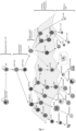
- directed differentiation of multipotent hematopoietic stem and progenitor cells to a definitive DC fate by expression of the DC-enriched transcription factors can be achieved (forcing differentiation along the hematopoietic tree depicted in Figure 1 ) .
- nucleic acids encoding the DC inducing factors are introduced into a cell, using viral vectors or without viral vectors, via one or repeated transfections, and the expression of the gene products and/or translation of the RNA molecules result in cells that are morphologically, biochemically, and functionally similar to DCs, as described herein.
- iDCs induced DCs
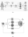
- these induced DCs (iDCs) after priming with the adequate antigens have the ability to capture, process and present them to effectors cells of the immune system (macrophages, T-cells, B-cells, NK cells) eliciting antigen-specific immune responses against cancer, viral and parasitic/bacterial infections ( Figure 2 ).
- the TFs can for example be used in cancer cells ( in situ or ex vivo ) to force them to present their own antigens to immune cells ( Figure 3 ).
- This method represents a feasible strategy to increase the clinical outcome of anticancer immunotherapies as it bypasses cancer evasion mechanisms and increases tumor immunogenicity.
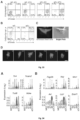
- Mouse Embryonic Fibroblasts harboring a DC-specific reporter (Clec9a-Cre X R26-stop- tdTomato) were used, and the activation of the reporter gene was used to shown DC-inducing TFs.
- Clec9a- tomato reporter mouse the tdTomato fluorescent protein is expressed exclusively by CDPs, pre-DCs and in cDCs (10).
- Macrophages, other immune lineages or monocyte-derived DCs in culture do not express Clec9a and therefore the tdTomato protein ( Figure 5A ) .
- Clec9a gene expression is selectively restricted to CDPs and their progeny (pre-cDCs and cDCs) ( Figure 5B ).
- Results from gene expression analysis of cDC and precursors also highlighted that Clec9a expression is acquired after commitment to cDC lineage in CDPs and pre-DCs and not before in Monocyte DC progenitors (MDPs) ( Figure 5C ) (11).
- Clec9a is expressed in CDPs, both pre-DCs and cDC subset, reaching high levels in the cDC1 subset ( Figure 6A ) (21).
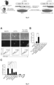
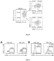
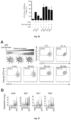
- Double transgenic Clec9a-tdTomato reporter MEFs were isolated from E13.5 embryos and excluded from any contaminating tdTomato+ or CD45+ cell that could be already committed to the hematopoietic lineage ( Figure 7A and 7B ) by Fluorescent- Activated Cell Sorting (FACS). MEFs used for screening and in the following experiments were tdTomato- CD45- with a purity of 99.8% ( Figure 7C ).
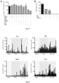
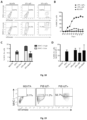
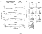
- Clec9a reporter MEFs were transduced with combinations of candidate TFs and evaluated for tdTomato expression ( Figure 8 ). After transduction with the 18 candidate TFs or one of the pools of 4 TFs, the emergence of tdTomato+ cells 5 days after adding Dox was observed ( Figure 9A, Figure 9B ). The pool comprising of Pu.1, Irf4, Irf8 and Batf3 generated more tdTomato+ cells than 18 TFs (2.36% versus 0.59%, respectively) suggesting that the minimal combination of factors required to induced reporter activation is contained within this pool. TdTomato+ cells were not detected after transduction with control M2rtTA vector. We then removed each of the factors individually ( Figure 9C ). Pu.1, Irf8 and Batf3 (PIB) removal reduced reporter activation while removal of Irf4 did not have an impact. These results suggest that PIB are essential for DC reprogramming.
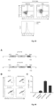
- TdTomato+ cells started to be detected between day 1 and day 2 and peaked between day 5 and day 7 ( Figure 10B ). Removal of PU.1, IRF8 or BATF3 completely abolished reporter activation whereas their individual expression was not sufficient to generate tdTomato+ cells ( Figure 10C ).
- PIB constitute the minimal combination of TFs for Clec9a activation and induced Dendritic Cell (iDC) generation.
- tdTomato+ cells display increased size and complexity ( Figure 10D ), consistent with the observed stellate morphology and the establishment of dendrites characteristic of DCs ( Figure 10E , Figure 10F ) .
- pan-hematopoietic marker CD45 is expressed in approximately 20% of PIB-transduced MEFs, with approximately 6.6% of tdT+ cells included in this population ( Figure 12 ).
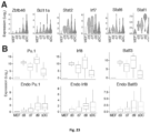
- PU.1, IRF8, BATF3 and TCF4 transcripts are expressed in single DC precursor cells ( Figure 15 ). While Pu.1 is equally expressed in MDPs, CDPs and Pre-DCs, IRF8 expression markedly increases in CDPs and is maintained in pre-DCs. BATF3 and TCF4 are only up- regulated at a later stage, in pre-DCs. Moreover, the combined expression of PU.1+IRF8+BATF3 is mostly enriched in CD8a+ DCs among 96 cells and tissues ( Figure 16A ) .
- Pu.1 levels are higher in both pre-DC stages, while Irf8 and Batf3 are specifically enriched in pre-cDC1 and cDC1 subsets ( Figure 16B ) .
- Clec9a, Pu.1, Irf8 and Batf3 display increased expression of in CD103+ DCs belonging to cDC1 subset ( Figure 16C ).
- CIITA Class II Transactivator
- CIITApl promoter I
- IRF4 Due to the described involvement of IRF4 in inducing MHC-II expression through interaction with CIITA (17), it was evaluated whether IRF4 could compensate for Pu.1 in the generation of MHC-II+ cells within the tdTomato+ population. It was therefore assessed the expression of MHC-II in tdTomato+ cells generated by 4TFs (including IRF4) or their individual exclusion ( Figure 18D ). Inclusion of IRF4 in the pool did not increase MHC-II expression on tdTomato+ cells and IRF4 could not substitute for the loss of Pu.1. Accordingly, IRF4 and PU.1 were found to synergistically promote MHC-II expression through CIITA promoter III in B cells but not DCs (17). During reprogramming to iDCs, no synergism with PU.1 was observed, which was strictly required for MHC-II expression in tdTomato+ cells.
- MHC class I molecules key molecules for the establishment of APC functionality.
- 56.7% of tdTomato+ cells at day 7 expressed MHC-I at the surface ( Figure 19 ).
- the tdT- compartment contained a lower percentage of MHC-I+ cells (11.2%) ( Figure 19 ).
- CD80 and CD86 The expression the co-stimulatory molecules CD80 and CD86, required for efficient antigen presentation ( Figure 20 ) was evaluated.
- CD80 and CD86 are expressed in 35.2% of tdTomato+MHC-II+ cells in contrast to only 12.9% of tdTomato+ MHC- II- cells.
- This characterization of the expression of MHC-II, CD80 and CD86 at the cell surface of iDCs suggests that a cohort of tdTomato+ MHC-II+ cells would be competent in antigen presentation.
- An additional co-stimulatory molecule, CD40 is expressed in 16.1% of tdTomato+ cells, comparing to only 2.8% of tdTomato- cells ( Figure 21A ).
- cDCs in particular cDC1 subset, have been described to respond to microbial stimulation up- regulating the expression of co-stimulatory molecules and becoming more effective APCs (25). Accordingly, tdTomato+ cells up-regulate the expression of CD40 (4-fold increase) at cell surface after toll-like receptor TLR4 stimulation (LPS) ( Figure 21B ).
- Cluster I comprises highly expressed genes in MEFs, which are silenced during DC reprogramming. These include typical fibroblast markers, such as Col5a2, Grem1, Lox, Acta2 and Thy1 ( Figure 22E ).
- Cluster II includes transcripts enriched at day 3 and day 7, suggesting activation during the initial stages of reprogramming. This cluster comprises genes such as Eea1 and Aldh1a2 that are associated with intracellular trafficking and metabolism as well as type I interferon (IFN) signaling ( Ifit3 ) ( Figure 22F ).
- IFN type I interferon
- Cluster III encompasses genes enriched at day 9 ( Figure 22D ).
- Cluster IV includes genes enriched in sDC1s and reprogrammed iDCs ( Figure 22D ), such as the pan-hematopoietic marker Cd45 and the general DC marker Cd11c ( Figure 22F ).
- cDC1-restricted genes were also upregulated, such as the Clec9 ⁇ gene and Tlr3 (22), and the key regulator of MHC class I-dependent immune responses ( Nlrc5 ) necessary for antigen cross-presentation, a key feature of cDC1s (23).
- Clec9 ⁇ gene and Tlr3 22
- Nlrc5 MHC class I-dependent immune responses
- GSEA gene set enrichment analysis
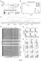
- the transcriptional networks for step-wise transitions during iDC reprogramming were evaluated ( Figure 25A ). It was observed that the transition of MEFs to day 3 was associated with the expression of a dense TF network that highly connected to the PIB reprogramming factors; the transition of day 3 to day 7 was softer, characterized by a less dense TF network, which do not include the PIB factors; and the transition of day 7 to day 9, characterized by a dense TF network which can be divided in 2 clusters of TFs, one denser that includes the cDC marker Zbtb46, and one composed by fewer TFs including the PIB factors.
- GSEA also showed that 4705 vs 167 gene sets for immunological signatures were upregulated on State 3 when compared with State 2, such as Mature Stimulatory DC, IFNy and IFN ⁇ stimulated DC gene sets ( Figure 28B ). As State 3 contained the majority of day 9 iDCs, it was sought to confirm that similar maturation trait was observed when comparing sDC1s (na ⁇ ve) with day 9 iDCs. GSEA showed that antigen processing and presentation and DC maturation gene sets are enriched at day 9 iDCs ( Figure 29A ). Interestingly, Stat6, which is associated with immature DCs, was up regulated in sDC1s, whilst Stat1, described to increase with maturation, was up regulated in day 9 iDCs ( Figure 23A ).
- the induced dendritic cells while similar in functional characteristics, differ in their gene expression from the naturally occurring endogenous dendritic cells ( Table 5 ). Table 5. Top 500 differentially expressed genes between day 9 iDCs and sDC1 cells ordered by fold change. Day 9 up (vs sDC1) Fold change Day9 Down (vs sDC1) Fold Change Cd74 8.378400519 AY036118 3.630967107 Ucp2 7.999114666 Sfi1 3.546674749 Grn 7.650480054 Gprc5c 3.440084658 Cdkn1a 7.079169574 Olfr648 3.00881167 S100a11 7.048233116 Rsph9 2.959047864 Gapdh 6.979316778 Tmsb4x 2.890522248 Cct8 6.923788452 Mtmr1 2.654214297 Ly6e 6.336631107 Il15 2.574244187 Ubb 6.19368097 Fanci 2.24583185 B2
- TLRs Toll-like receptors
- iDCs The capacity of iDCs to mount an antigen- specific immune response was evaluated. First it was evaluated whether iDCs would be able to engulf particles by incubation with 1 ⁇ m FITC-labeled latex beads. After incubation tdTomato+ cells contained numerous fluorescent beads in the cytoplasm ( Figure 32 ), suggesting that iDCs have established the competence for phagocytosis.
- tdTomato+ cells After overnight incubation with labeled dead cells, 65.7% of purified tdTomato+ cells have incorporated dead cell material in contrast to only 10.5% of tdTomato- ( Figure 33B ). Uptake of dead cells was further analysed by live imaging and it was observed that tdTomato+ cells avidly accumulated dead cell material in the cytoplasm ( Figure 33C ). TdTomato+ cells move actively and, upon encountering a dead cell, projected cellular protrusions to incorporate and engulf it ( Figure 33D ).
- iDCs express genes encoding TLR ( Tlr3 and Tlr4 ) and other mediators of TLR signaling, including MyD88-dependent (TRAM (encoded by Ticam2), and Traf6 ) and independent ( IKK ⁇ ) pathways ( Figure 34A ). Also, we have confirmed that iDCs express key mediators of receptor-mediated endocytosis (Fcgr2b, Tfr2 and Mrc1) and macropinocytosis of dead cells ( Axl, Lrp1 and Scarf1), further suggesting that iDCs have acquired the ability for sense and incorporate antigens ( Figure 34B ).
- OT-II cells MHC class II-restricted ovalbumin-specific T cells isolated from lymph nodes and spleen of OT-II Rag2 KO mice (18).
- OVA 323-339 processed antigenic peptide
- OT-II CD4 T-cells were co-cultured with iDCs when given the Ovalbumin protein (OVA) or pre-processed antigenic peptide (OVA 323-339).
- Functional DCs are able to capture the protein, process and present the processed antigenic peptide in the context of MHC-II.
- Induced CD4+ T cell proliferation was measured by CFSE dilution and the activation of the T-cell activation marker CD44 after 7 days of co-culture.
- 56% of CD4+ T cells diluted CFSE when co-cultured with PIB-generated iDCs in the presence of OVA protein ( Figure 35B ).
- 38.2% of CD4+ T cells diluted CFSE content which suggests that inclusion of TCF4 in the reprogramming pool does not increase the stimulatory ability of iDCs.
- Splenic MHC-II+CD11c+ DCs were used as controls and generated 24.1% of proliferative T-cells.
- addition of LPS stimuli, which is commonly employed to induce DC “maturation” increased the antigen-specific stimulatory ability of MEFs transduced with PIB (1.5-fold) or PIB+TCF4 (2-fold) and also splenic DCs (3-fold) ( Figure 35B and Figure 36 ).
- LPS stimuli which is commonly employed to induce DC “maturation”
- MEFs transduced with PIB (1.5-fold) or PIB+TCF4 (2-fold) and also splenic DCs (3-fold) Figure 35B and Figure 36 .
- T-cells that were not co-cultured did not proliferate with or without LPS stimuli.
- T-cell activation markers such as CD44.
- OT-II CD4 T cells When given the pre-processed antigen, OT-II CD4 T cells diluted CFSE and upregulated the expression of CD44 when co-cultured with both PIB-generated iDCs and splenic MHC-II+CD11c+ DCs ( Figure 35C ).
- iDCs display comparable ability to induce CD44 expression in OT-II T cells when compared with splenic DCs (52.2% versus 63.8%).
- This data supports iDCs' functional ability to incorporate and process OVA protein followed by presentation of processed Ovalbumin peptides in MHC-II complexes at cell surface.
- iDCs acquire ability to export antigens to cytosol and express key genes essential for cross-presentation ability.
- Cross-presentation via the cytosolic pathway involves antigen export from endocytic compartments to the cytosol.
- the ability of iDCs to perform antigen export was evaluated using a cytofluorimetry- based assay ( Figure 37A ).
- a cytofluorimetry- based assay Remarkably, after 90-minute incubation with b-lactamase, approximately 80% of CCF4-loaded iDCs expressed cleaved CCF4.
- iDCs were able to uptake b-lactamase and efficiently export it into the cytoplasm, leading to the generation of cleaved CCF4.
- iDCs express genes involved in cross-presentation pathway, such as Cybb, Atg7, Tap1 and Tap2 ( Figure 37B ).
- Functional DCs are able to capture the exogenous protein, process and perform cross-presentation of the processed antigenic peptide in the context of MHC-I, inducing activation of CD8+ T cells.
- Induced CD8+ T cell proliferation was measured by CFSE dilution and the activation of the T-cell activation marker CD44 after 4 days of co-culture.
- Splenic MHC-II+CD11c+ DCs were used as controls and generated 18.05 ⁇ 0.78% of proliferative and CD44+ T-cells.
- HDFs Human Dermal Fibroblasts
- Coding regions of PU.1, IRF8 and BATF3 were cloned into polycistronic inducible lentiviral vectors that express the three nucleic acid sequences, each of them separated by 2A peptide sequences ( Figure 44A ).
- the 3 TFs were included in different orders, PU.1, IRF8 and BATF3 (PIB) or IRF8, PU.1 and BATF3 (IPB).
- PIB IRF8 and BATF3
- IPB IRF8 PU.1 and BATF3
- Clec9a reporter activation was observed in 10.8% and 6.6% of MEFs transduced with the polycistronic vectors (PIB and IPB, respectively).
- Coding regions of each candidate TF were individually cloned into an inducible lentiviral pFUW-TetO vector (6) in which the expression of the TFs is under the control of the tetracycline operator and a minimal CMV promoter.
- a previously described lentiviral vector containing the reverse tetracycline transactivator M2rtTA under the control of a constitutively active human ubiquitin C promoter (FUW-M2rtTA) was used in combination.
- Human Embryonic Kidney (HEK) 293T cells were transfected with a mixture of TF-encoding plasmids, packaging constructs and the VSV-G envelope protein. Viral supernatants were harvested after 36, 48 and 60 hours, filtered (0.45 ⁇ m, Corning) and used fresh or concentrated 40-fold with Amicon ultra centrifugal filters (Millipore).
- Polypeptide variants or family members having the same or a similar activity as the reference polypeptide encoded by the sequences provided in the sequence list can be used in the compositions, methods, and kits described herein.
- variants of a particular polypeptide encoding a DC inducing factor for use in the compositions, methods, and kits described herein will have at least about 95%, at least about 96%, at least about 97%, at least about 98%, at least about 99% or more sequence identity to that particular reference polynucleotide or polypeptide as determined by sequence alignment programs and parameters described herein and known to those skilled in the art.
- BATF3 Basic Leucine Zipper ATF-Like Transcription Factor
- SEQ. ID. 1 mRNA (SEQ. ID. 1) and a codon optimized, or different codons encoding the same amino acids, are naturally also contemplated to be covered by the reference to the nucleic acid as set forth herein.
- PU.1 Homo sapiens Spi-1 proto-oncogene
- SEQ. ID. 7 mRNA
- a codon optimized, or different codons encoding the same amino acids are naturally also contemplated to be covered by the reference to the nucleic acid as set forth herein.
- IRF8 Interferon Regulatory Factor 8
- mRNA SEQ. ID. 5
- a codon optimized, or different codons encoding the same amino acids are naturally also contemplated to be covered by the reference to the nucleic acid as set forth herein.
- TCF4 Homo sapiens Transcription factor 4
- mRNA SEQ. ID. 13
- a codon optimized, or different codons encoding the same amino acids are naturally also contemplated to be covered by the reference to the nucleic acid as set forth herein.
- compositions and methods are isolated amino acid sequences, and isolated DNA or RNA nucleic acid sequences encoding one or more DC inducing factors for use in making iDCs.
- the nucleic acid sequence or construct encoding the DC inducing factor(s), such as PU.1, IRF8, BATF3 and TCF4 is inserted or operably linked into a suitable expression vector for transfection of cells using standard molecular biology techniques.
- a "vector" refers to a nucleic acid molecule, such as a dsDNA molecule that provides a useful biological or biochemical property to an inserted nucleotide sequence, such as the nucleic acid constructs or replacement cassettes described herein.
- a vector can have one or more restriction endonuclease recognition sites (whether type I, II or Ils) at which the sequences can be cut in a determinable fashion without loss of an essential biological function of the vector, and into which a nucleic acid fragment can be spliced or inserted in order to bring about its replication and cloning.
- Vectors can also comprise one or more recombination sites that permit exchange of nucleic acid sequences between two nucleic acid molecules.
- Vectors can further provide primer sites, e.g., for PCR, transcriptional and/or translational initiation and/or regulation sites, recombination signals, replicons, additional selectable markers, etc.
- a vector can further comprise one or more selectable markers suitable for use in the identification of cells transformed with the vector.
- the expression vector is a viral vector.
- Some viral-mediated expression methods employ retrovirus, adenovirus, lentivirus, herpes virus, pox virus, and adeno-associated virus (AAV) vectors, and such expression methods have been used in gene delivery and are well known in the art.
- the viral vector is a retrovirus.
- Retroviruses provide a convenient platform for gene delivery. A selected gene can be inserted into a vector and packaged in retroviral particles using techniques known in the art. The recombinant virus can then be isolated and delivered to target cells of the subject either in vivo or ex vivo.
- a number of retroviral systems have been described. See, e.g., U.S. Pat. No. 5,219,740 ; Miller and Rosman (1989) BioTechniques 7:980-90 ; Miller, A. D. (1990) Human Gene Therapy 1:5-14 ; Scarpa et al. (1991) Virology 180:849-52 ; Burns et al.
- the retrovirus is replication deficient.
- Retroviral vector systems exploit the fact that a minimal vector containing the 5' and 3' LTRs and the packaging signal are sufficient to allow vector packaging, infection and integration into target cells, provided that the viral structural proteins are supplied in trans in the packaging cell line. Fundamental advantages of retroviral vectors for gene transfer include efficient infection and gene expression in most cell types, precise single copy vector integration into target cell chromosomal DNA and ease of manipulation of the retroviral genome.
- the viral vector is an adenovirus-based expression vector.
- adenoviruses persist extrachromosomally, thus minimizing the risks associated with insertional mutagenesis ( Haj-Ahmad and Graham (1986) J. Virol. 57:267-74 ; Bett et al. (1993) J. Virol. 67:5911-21 ; Mittereder et al. (1994) Human Gene Therapy 5:717-29 ; Seth et al. (1994) J. Virol. 68:933-40 ; Barr et al. (1994) Gene Therapy 1:51-58 ; Berkner, K. L.
- Adenoviral vectors infect a wide variety of cells, have a broad host-range, exhibit high efficiencies of infectivity, direct expression of heterologous genes at high levels, and achieve long-term expression of those genes in vivo.
- the virus is fully infective as a cell-free virion so injection of producer cell lines is not necessary.
- adenovirus is not associated with severe human pathology, and the recombinant vectors derived from the virus can be rendered replication defective by deletions in the early-region 1 ("E1") of the viral genome.
- E1 early-region 1
- Adenoviral vectors for use in the compositions, methods, and kits described herein can be derived from any of the various adenoviral serotypes, including, without limitation, any of the over 40 serotype strains of adenovirus, such as serotypes 2, 5, 12, 40, and 41.
- the adenoviral vectors used herein are preferably replication-deficient and contain the DC inducing factor of interest operably linked to a suitable promoter.
- the nucleic acid sequences encoding the DC inducing factor(s), such as, PU.1, IRF8, BATF3 and TCF4 are introduced or delivered using one or more inducible lentiviral vectors.
- Control of expression of DC inducing factors delivered using one or more inducible lentiviral vectors can be achieved, in some embodiments, by contacting a cell having at least one DC inducing factor in an expression vector under the control of or operably linked to an inducible promoter, with a regulatory agent (e.g., doxycycline) or other inducing agent.
- a regulatory agent e.g., doxycycline
- induction of expression refers to the expression of a gene, such as a DC inducing factor encoded by an inducible viral vector, in the presence of an inducing agent, for example, or in the presence of one or more agents or factors that cause endogenous expression of the gene in a cell.
- a doxycycline (Dox) inducible lentiviral system is used.
- lentiviruses are able to transduce quiescent cells making them amenable for transducing a wider variety of hematopoietic cell types.
- the pFUW-tetO lentivirus system has been shown to transduce primary hematopoietic progenitor cells with high efficiency.
- the nucleic acid sequences encoding the DC inducing factor(s), such as PU.1 (SEQ. ID. 7, SEQ. ID. 8), IRF8 (SEQ. ID. 5, SEQ. ID. 6), BATF3 (SEQ. ID. 1, SEQ. ID. 2) and/or TCF4 (SEQ. ID. 13, SEQ. ID. 14), are introduced or delivered using a non-integrating vector (e.g., adenovirus). While integrating vectors, such as retroviral vectors, incorporate into the host cell genome and can potentially disrupt normal gene function, non-integrating vectors control expression of a gene product by extra-chromosomal transcription.
- a non-integrating vector e.g., adenovirus
- non-integrating vectors do not become part of the host genome, non-integrating vectors tend to express a nucleic acid transiently in a cell population. This is due in part to the fact that the non-integrating vectors are often rendered replication deficient.
- non-integrating vectors have several advantages over retroviral vectors including, but not limited to: (1) no disruption of the host genome, and (2) transient expression, and (3) no remaining viral integration products.
- Some non-limiting examples of non-integrating vectors for use with the methods described herein include adenovirus, baculovirus, alphavirus, picornavirus, and vaccinia virus.
- the non-integrating viral vector is an adenovirus.
- advantages of non-integrating viral vectors include the ability to produce them in high titers, their stability in vivo, and their efficient infection of host cells.
- Nucleic acid constructs and vectors for use in generating iDCs in the compositions and methods described herein can further comprise, in some embodiments, one or more sequences encoding selection markers for positive and negative selection of cells.
- selection marker sequences can typically provide properties of resistance or sensitivity to antibiotics that are not normally found in the cells in the absence of introduction of the nucleic acid construct.
- a selectable marker can be used in conjunction with a selection agent, such as an antibiotic, to select in culture for cells expressing the inserted nucleic acid construct.
- Sequences encoding positive selection markers typically provide antibiotic resistance, i.e., when the positive selection marker sequence is present in the genome of a cell, the cell is sensitive to the antibiotic or agent.
- Sequences encoding negative selection markers typically provide sensitivity to an antibiotic or agent, i.e., when the negative selection marker is present in the genome of a cell, the cell is sensitive to the antibiotic or agent.
- Nucleic acid constructs and vectors for use in making iDCs in the compositions and methods described herein can further comprise, in some embodiments, other nucleic acid elements for the regulation, expression, stabilization of the construct or of other vector genetic elements, for example, promoters, enhancers, TATA-box, ribosome binding sites, IRES, as known to one of ordinary skill in the art.
- other nucleic acid elements for the regulation, expression, stabilization of the construct or of other vector genetic elements for example, promoters, enhancers, TATA-box, ribosome binding sites, IRES, as known to one of ordinary skill in the art.
- the DC inducing factor(s) such as PU.1 (SEQ. ID. 7, SEQ. ID. 8), IRF8 (SEQ. ID. 5, SEQ. ID. 6), BATF3 (SEQ. ID. 1, SEQ. ID. 2) and/or TCF4 (SEQ. ID. 13, SEQ. ID. 14), are provided as synthetic, modified RNAs, or introduced or delivered into a cell as a synthetic, modified RNA, as described in US Patent Publication US 2012/046346 A1 .
- the methods can involve repeated contacting of the cells or involve repeated transfections of the synthetic, modified RNAs encoding DC inducing factors, such as for example, at least 2, at least 3, at least 4, at least 5, at least 6, at least 7, at least 8, at least 9, at least 10, at least 11, at least 12, at least 13, at least 14, at least 15, at least 16, at least 17, at least 18, at least 19, at least 20, at least 25, at least 30, or more transfections.
- modified mRNAs for use in the compositions, methods, and kits described herein can comprise any additional modifications known to one of skill in the art and as described in US Patent Publications US 2012/0046346 A1 and US 2012/251618 A1 , and PCT Publication WO 2012/019168 .
- Such other components include, for example, a 5' cap (e.g., the Anti-Reverse Cap Analog (ARCA) cap, which contains a 5'-5'-triphosphate guanine-guanine linkage where one guanine contains an N7 methyl group as well as a 3'-O-methyl group; caps created using recombinant Vaccinia Virus Capping Enzyme and recombinant 2'-O-methyltransferase enzyme, which can create a canonical 5'-triphosphate linkage between the 5'-most nucleotide of an mRNA and a guanine nucleotide where the guanine contains an N7 methylation and the ultimate 5'-nucleotide contains a 2'-O-methyl generating the Cap1 structure); a poly(A) tail (e.g., a poly-A tail greater than 30 nucleotides in length, greater than 35 nucleotides in length, at least 40 nucleotides,
- the modified mRNAs for use in the compositions and methods described herein can further comprise an internal ribosome entry site (IRES).
- IRES can act as the sole ribosome binding site, or can serve as one of multiple ribosome binding sites of an mRNA.
- An mRNA containing more than one functional ribosome binding site can encode several peptides or polypeptides, such as the DC inducing factors described herein, that are translated independently by the ribosomes ("multicistronic mRNA").
- multicistronic mRNA When nucleic acids are provided with an IRES, further optionally provided is a second translatable region. Examples of IRES sequences that can be used according to the disclosure include without limitation, those from picornaviruses (e.g.
- FMDV pest viruses
- CFFV pest viruses
- PV polio viruses
- ECMV encephalomyocarditis viruses
- FMDV foot-and-mouth disease viruses
- HCV hepatitis C viruses
- CSFV classical swine fever viruses
- MLV murine leukemia virus
- SW simian immune deficiency viruses
- CrPV cricket paralysis viruses
- the synthetic, modified RNA molecule comprises at least one modified nucleoside. In some embodiments of the compositions and methods described herein, the synthetic, modified RNA molecule comprises at least two modified nucleosides.
- the modified nucleosides are selected from the group consisting of 5-methylcytosine (5mC), N6- methyladenosine (m6A), 3,2'-O-dimethyluridine (m4U), 2-thiouridine (s2U), 2' fluorouridine, pseudouridine, 2'-O-methyluridine (Um), 2'deoxy uridine (2' dU), 4-thiouridine (s4U), 5- methyluridine (m5U), 2'-O-methyladenosine (m6A), N6,2'-O-dimethyladenosine (m6Am), N6,N6,2'-O-trimethyladenosine (m62Am), 2'-O-methylcytidine (Cm), 7-methylguanosine (m7G), 2'-O-methylguanosine (Gm), N2,7-dimethylguanosine (m2,7G), N2,N2,7-trimethyl
- Modified mRNAs need not be uniformly modified along the entire length of the molecule.
- Different nucleotide modifications and/or backbone structures can exist at various positions in the nucleic acid.
- the nucleotide analogs or other modification(s) can be located at any position(s) of a nucleic acid such that the function of the nucleic acid is not substantially decreased.
- a modification can also be a 5' or 3' terminal modification.
- the nucleic acids can contain at a minimum one and at maximum 100% modified nucleotides, or any intervening percentage, such as at least 50% modified nucleotides, at least 80% modified nucleotides, or at least 90% modified nucleotides.
- each occurrence of a given nucleoside in a molecule is modified (e.g., each cytosine is a modified cytosine e.g., 5-methylcytosine, each uracil is a modified uracil, e.g., pseudouracil, etc.).
- the modified mRNAs can comprise a modified pyrimidine such as uracil or cytosine.
- at least 25%, at least 50%, at least 80%, at least 90% or 100% of the uracil in the nucleic acid are replaced with a modified uracil.
- modified uracil can be replaced by a compound having a single unique structure, or can be replaced by a plurality of compounds having different structures (e.g., 2, 3, 4 or more unique structures).
- at least 25%, at least 50%, at least 80%, at least 90% or 100% of the cytosine in the nucleic acid may be replaced with a modified cytosine.
- the modified cytosine can be replaced by a compound having a single unique structure, or can be replaced by a plurality of compounds having different structures (e.g., 2, 3, 4 or more unique structures) (e.g., some cytosines modified as 5mC, others modified as 2'-O-methylcytosine or other cytosine analog).
- Such multi-modified synthetic RNA molecules can be produced by using a ribonucleoside blend or mixture comprising all the desired modified nucleosides, such that when the RNA molecules are being synthesized, only the desired modified nucleosides are incorporated into the resulting RNA molecule encoding the DC inducing factor.
- modified nucleic acids comprising a degradation domain, which is capable of being acted on in a directed manner within a cell.
- iDCs can be generated by delivery of DC inducing factors in the form of nucleic acid (DNA or RNA) or amino acid sequences
- iDC induction can be induced using other methods, such as, for example, by treatment of cells with an agent, such as a small molecule or cocktail of small molecules, that induce expression one or more of the DC inducing factors.
- Detection of expression of DC inducing factors introduced into cells or induced in a cell population using the compositions and methods described herein can be achieved by any of several techniques known to those of skill in the art including, for example, Western blot analysis, immunocytochemistry, and fluorescence-mediated detection.
- one or more DC activities or parameters can be measured, such as, differential expression of surface antigens.
- the generation of induced DCs using the compositions, methods, and kits described herein preferably causes the appearance of the cell surface phenotype characteristic of endogenous DCs, such as CLEC9A, MHC-I, MHC-II, CD40, CD80, CD86, CD103, for example.
- DCs are most reliably distinguished from other immune cells by their functional behavior. Functional aspects of DC phenotypes, or dendritic cell activities, such as the ability of a dendritic cell to induce antigen specific T cell responses, can be easily determined by one of skill in the art using routine methods known in the art, and as described herein, for example, in the Drawings, i.e., FIGS. 1-44 . In some embodiments of the aspects described herein, functional assays to identify reprogramming factors can be used. For example, in some embodiments, antigen presentation and antigen cross-presentation assays can be used to confirm antigen-specific induction of T cell responses (antigen presentation potential) of iDCs generated using the compositions, methods, and kits thereof.
- Cytokine secretion can be used to confirm immune-modulatory properties of iDCs generated using the compositions, methods, and kits described herein.
- Ability to engulf particles, proteins and dead cells of iDCs generated using the compositions, methods, and kits described herein can be evaluated by culturing transduced cells in the presence of labelled beads, ovalbumine or dead cells, followed by flow cytometry analysis, respectively.
- cellular parameter refers to measureable components or qualities of endogenous or natural DCs, particularly components that can be accurately measured.
- a cellular parameter can be any measurable parameter related to a phenotype, function, or behavior of a cell.
- Such cellular parameters include, changes in characteristics and markers of a DC or DC population, including but not limited to changes in viability, cell growth, expression of one or more or a combination of markers, such as cell surface determinants, such as receptors, proteins, including conformational or posttranslational modification thereof, lipids, carbohydrates, organic or inorganic molecules, nucleic acids, e.g. mRNA, DNA, global gene expression patterns, etc.
- Such cellular parameters can be measured using any of a variety of assays known to one of skill in the art. For example, viability and cell growth can be measured by assays such as Trypan blue exclusion, CFSE dilution, and 3H-thymidine incorporation. Expression of protein or polypeptide markers can be measured, for example, using flow cytometric assays, Western blot techniques, or microscopy methods. Gene expression profiles can be assayed, for example, using RNA-sequencing methodologies and quantitative or semi-quantitative real-time PCR assays. A cellular parameter can also refer to a functional parameter or functional activity. While most cellular parameters will provide a quantitative readout, in some instances a semi-quantitative or qualitative result can be acceptable.
- Readouts can include a single determined value, or can include mean, median value or the variance, etc. Characteristically a range of parameter readout values can be obtained for each parameter from a multiplicity of the same assays. Variability is expected and a range of values for each of the set of test parameters will be obtained using standard statistical methods with a common statistical method used to provide single values.
- additional factors and agents can be used to enhance iDC reprogramming.
- factors and agents that modify epigenetic pathways can be used to facilitate reprogramming into iDCs.
- any primary somatic cell type can be used for producing iDCs or reprogramming somatic cells to iDCs according to the presently described compositions, methods, and kits.
- Such primary somatic cell types also include other stem cell types, including pluripotent stem cells, such as induced pluripotent stem cells (iPS cells); other multipotent stem cells; oligopotent stem cells; and (5) unipotent stem cells.
- pluripotent stem cells such as induced pluripotent stem cells (iPS cells); other multipotent stem cells; oligopotent stem cells; and (5) unipotent stem cells.
- primary somatic cells useful in the various aspects and embodiments of the methods described herein include, but are not limited to, fibroblast, epithelial, endothelial, neuronal, adipose, cardiac, skeletal muscle, hematopoietic or immune cells, hepatic, splenic, lung, circulating blood cells, gastrointestinal, renal, bone marrow, and pancreatic cells, as well as stem cells from which those cells are derived.
- the cell can be a primary cell isolated from any somatic tissue including, but not limited to, spleen, bone marrow, blood, brain, liver, lung, gut, stomach, intestine, fat, muscle, uterus, skin, spleen, endocrine organ, bone, etc.
- somatic cell further encompasses, in some embodiments, primary cells grown in culture, provided that the somatic cells are not immortalized. Where the cell is maintained under in vitro conditions, conventional tissue culture conditions and methods can be used, and are known to those of skill in the art. Isolation and culture methods for various primary somatic cells are well within the abilities of one skilled in the art.
- the somatic cell is a fibroblast cell.
- the somatic cell can be a hematopoietic lineage cell.
- the somatic cell can be a cancer cell or a tumor cell.
- a somatic cell to be reprogrammed or made into an iDC cell is a cell of hematopoietic origin.
- hematopoietic-derived cell hematopoietic-derived differentiated cell
- hematopoietic lineage cell hematopoietic lineage cell
- cell of hematopoietic origin refer to cells derived or differentiated from a multipotent hematopoietic stem cell (HSC).
- hematopoietic lineage cells for use with the compositions, methods, and kits described herein include multipotent, oligopotent, and lineage-restricted hematopoietic progenitor cells, granulocytes (e.g., promyelocytes, neutrophils, eosinophils, basophils), erythrocytes (e.g., reticulocytes, erythrocytes), thrombocytes (e.g., megakaryoblasts, platelet producing megakaryocytes, platelets), monocytes (e.g., monocytes, macrophages), dendritic cells, and lymphocytes (e.g., T-lymphocytes, which carry T-cell receptors (TCRs), B- lymphocytes or B cells, which express immunoglobulin and produce antibodies, NK cells, NKT cells, and innate lymphocytes).
- granulocytes e.g., promyelocytes, neutr
- hematopoietic progenitor cells refer to multipotent, oligopotent, and lineage-restricted hematopoietic cells capable of differentiating into two or more cell types of the hematopoietic system, including, but not limited to, granulocytes, monocytes, erythrocytes, megakaryocytes, and lymphocytes B-cells and T-cells.
- Hematopoietic progenitor cells encompass multi-potent progenitor cells (MPPs), common myeloid progenitor cells (CMPs), common lymphoid progenitor cells (CLPs), granulocyte-monocyte progenitor cells (GMPs), and pre-megakaryocyte-erythrocyte progenitor cell.
- MPPs multi-potent progenitor cells
- CMPs common myeloid progenitor cells
- CLPs common lymphoid progenitor cells
- GFPs granulocyte-monocyte progenitor cells
- pre-megakaryocyte-erythrocyte progenitor cell pre-megakaryocyte-erythrocyte progenitor cell.
- Lineage-restricted hematopoietic progenitor cells include megakaryocyte- erythrocyte progenitor cells (MEP), ProB cells, PreB cells, PreProB cells, ProT cells, double- negative T cells, pro-NK cells, pre-granulocyte/macrophage cells, granulocyte/macrophage progenitor (GMP) cells, and pro-mast cells (ProMCs).
- MEP megakaryocyte- erythrocyte progenitor cells
- ProB cells PreB cells
- PreProB cells ProT cells
- double- negative T cells pro-NK cells
- pre-granulocyte/macrophage cells pre-granulocyte/macrophage progenitor (GMP) cells
- ProMCs pro-mast cells
- Cells of hematopoietic origin for use in the compositions and methods described herein can be obtained from any source known to comprise these cells, such as fetal tissues, umbilical cord blood, bone marrow, peripheral blood, mobilized peripheral blood, spleen, liver, thymus, lymph, etc. Cells obtained from these sources can be expanded ex vivo using any method acceptable to those skilled in the art prior to use in with the compositions, methods, and kits for making iDCs described herein. For example, cells can be sorted, fractionated, treated to remove specific cell types, or otherwise manipulated to obtain a population of cells for use in the methods described herein using any procedure acceptable to those skilled in the art.
- Mononuclear lymphocytes may be collected, for example, by repeated lymphocytophereses using a continuous flow cell separator as described in U.S. Pat. No. 4,690,915 , or isolated using an affinity purification step of CLP method, such as flow-cytometry using a cytometer, magnetic separation, using antibody or protein coated beads, affinity chromatography, or solid-support affinity separation where cells are retained on a substrate according to their expression or lack of expression of a specific protein or type of protein, or batch purification using one or more antibodies against one or more surface antigens specifically expressed by the cell type of interest.
- Cells of hematopoietic origin can also be obtained from peripheral blood.
- the subject Prior to harvest of the cells from peripheral blood, the subject can be treated with a cytokine, such as e.g., granulocyte- colony stimulating factor, to promote cell migration from the bone marrow to the blood compartment and/or promote activation and/or proliferation of the population of interest.
- a cytokine such as e.g., granulocyte- colony stimulating factor
- Any method suitable for identifying surface proteins can be employed to isolate cells of hematopoietic origin from a heterogeneous population.
- a clonal population of cells of hematopoietic origin such as lymphocytes, is obtained.
- the cells of hematopoietic origin are not a clonal population.
- a somatic cell can be obtained from any mammalian species, with non-limiting examples including a murine, bovine, simian, porcine, equine, ovine, or human cell.
- the somatic cell is a human cell.
- the cell is from a non-human organism, such as a non-human mammal.
- the methods for making iDCs described herein involve culturing or expanding somatic cells, such as cells of hematopoietic origin, in any culture medium that is available and well-known to one of ordinary skill in the art.
- somatic cells such as cells of hematopoietic origin
- Such media include, but are not limited to, Dulbecco's Modified Eagle's Medium ® (DMEM), DMEM F12 Medium ® , Eagle's Minimum Essential Medium ® , F-12K Medium ® , Iscove's Modified Dulbecco's Medium ® , RPMI-1640 Medium ® , and serum-free medium for culture and expansion of DCs.
- DMEM Dulbecco's Modified Eagle's Medium ®
- DMEM F12 Medium ® Eagle's Minimum Essential Medium ®
- F-12K Medium ® F-12K Medium
- Iscove's Modified Dulbecco's Medium ® RPMI-1640 Medium ®
- the medium used with the methods described herein can, in some embodiments, be supplemented with one or more immunostimulatory cytokine.
- immunostimulatory cytokine include, but are not limited to, G-CSF, GM-CSF, TNF- ⁇ , IL-4, the Flt-3 ligand and the kit ligand.
- the immunostimulatory cytokine is selected from the group consisting of the interleukins (e.g., IL-1 ⁇ , IL-1 ⁇ , IL-2, IL-3, IL-4, IL-6, IL-8, IL-9, IL-10, IL-12, IL-18, IL-19, IL-20), the interferons (e.g., IFN- ⁇ , IFN- ⁇ , IFN-y), tumor necrosis factor (TNF), transforming growth factor- ⁇ (TGF- ⁇ ), granulocyte colony stimulating factor (G-CSF), macrophage colony stimulating factor (M-CSF), granulocyte-macrophage colony stimulating factor (GM-CSF), the Flt-3 ligand and the kit ligand.
- the interleukins e.g., IL-1 ⁇ , IL-1 ⁇ , IL-2, IL-3, IL-4, IL-6, IL-8, IL-9, IL-10, IL-12, IL-18, IL-19,
- Cells in culture can be maintained either in suspension or attached to a solid support, such as extracellular matrix components or plating on feeder cells, for example.
- a solid support such as extracellular matrix components or plating on feeder cells, for example.
- Cells being used in the methods described herein can require additional factors that encourage their attachment to a solid support, in some embodiments, such as type I and type II collagen, chondroitin sulfate, fibronectin, "superfibronectin” and fibronectin-like polymers, gelatin, poly-D and poly-L-lysine, thrombospondin and vitronectin.
- the cells are suitable for growth in suspension cultures.
- Suspension-competent host cells are generally monodisperse or grow in loose aggregates without substantial aggregation.
- Suspension-competent host cells include cells that are suitable for suspension culture without adaptation or manipulation (e.g., cells of hematopoietic origin, such as lymphoid cells) and cells that have been made suspension-competent by modification or adaptation of attachment-dependent cells (e.g., epithelial cells, fibroblasts).
- a variety of means for administering cells to subjects are known to those of skill in the art. Such methods can include systemic injection, for example, i.v. injection, or implantation of cells into a target site in a subject.
- Cells may be inserted into a delivery device which facilitates introduction by injection or implantation into the subject.
- delivery devices can include tubes, e.g., catheters, for injecting cells and fluids into the body of a recipient subject.
- the cells can be prepared for delivery in a variety of different forms. For example, the cells can be suspended in a solution or gel or embedded in a support matrix when contained in such a delivery device. Cells can be mixed with a pharmaceutically acceptable carrier or diluent in which the cells remain viable.
- the cells produced by the methods described herein can be used to prepare cells to treat or alleviate several cancers and tumors including, but not limited to, breast cancer, prostate cancer, lymphoma, skin cancer, pancreatic cancer, colon cancer, melanoma, malignant melanoma, ovarian cancer, brain cancer, primary brain carcinoma, head-neck cancer, glioma, glioblastoma, liver cancer, bladder cancer, non- small cell lung cancer, head or neck carcinoma, breast carcinoma, ovarian carcinoma, lung carcinoma, small-cell lung carcinoma, Wilms' tumor, cervical carcinoma, testicular carcinoma, bladder carcinoma, pancreatic carcinoma, stomach carcinoma, colon carcinoma, prostatic carcinoma, genitourinary carcinoma, thyroid carcinoma, esophageal carcinoma, myeloma, multiple myeloma, adrenal carcinoma, renal cell carcinoma, endometrial carcinoma, adrenal cortex carcinoma, malignant pancreatic insulinoma, malignant carcinoid carcinoma, choriocarcinoma, mycosis fungoides
- the methods of the disclosure can be used to prevent or eliminate infection by pathogens known to predispose to certain cancers.
- Pathogens of particular interest for use in the cancer vaccines provided herein include the hepatitis B virus (hepatocellular carcinoma), hepatitis C virus (heptomas), Epstein Barr virus (EBV) (Burkitt lymphoma, nasopharynx cancer, PTLD in immunosuppressed individuals), HTLVL (adult T cell leukemia), oncogenic human papilloma viruses types 16, 18, 33, 45 (adult cervical cancer), and the bacterium Helicobacter pylori (B cell gastric lymphoma).
- EBV Epstein Barr virus
- HTLVL adult T cell leukemia
- HTLVL adult T cell leukemia
- oncogenic human papilloma viruses types 16, 18, 33, 45 adult cervical cancer
- Helicobacter pylori B cell gastric lymphoma
- infectious virus examples include, but are not limited to, infectious virus that infect mammals, and more particularly humans.
- infectious virus examples include, but are not limited to: Retroviridae (e.g., human immunodeficiency viruses, such as HIV-I (also referred to as HTLV-III, LAV or HTLV-III/LAV, or HIV-III; and other isolates, such as HIV-LP; Picornaviridae (e.g. polio viruses, hepatitis A virus; enteroviruses, human Coxsackie viruses, rhinoviruses, echoviruses); Calciviridae (e.g.
- Togaviridae e.g. equine encephalitis viruses, rubella viruses
- Flaviridae e.g. dengue viruses, encephalitis viruses, yellow fever viruses
- Coronoviridae e.g. coronaviruses such as the SARS coronavirus
- Rhabdoviradae e.g. vesicular stomatitis viruses, rabies viruses
- Filoviridae e.g. ebola viruses
- Paramyxoviridae e.g. parainfluenza viruses, mumps virus, measles virus, respiratory syncytial virus
- Orthomyxoviridae e.g.
- influenza viruses Bungaviridae (e.g. Hantaan viruses, bunga viruses, phleboviruses and Nairo viruses); Arena viridae (hemorrhagic fever viruses); Reoviridae (e.g.
- reoviruses reoviruses, orbiviurses and rotaviruses
- Bir-naviridae Hepadnaviridae (Hepatitis B virus); Parvovirida (parvoviruses); Papovaviridae (papilloma viruses, polyoma viruses); Adenoviridae (most adenoviruses); Herpesviridae herpes simplex virus (HSV) 1 and 2, varicella zoster virus, cytomegalovirus (CMV), herpes virus; P.oxyiridae (variola viruses, vaccinia viruses, pox viruses); and Iridoviridae (e.g. African swine fever virus); and unclassified viruses (e.g.
- the methods of the disclosure can be used to target gram negative and gram positive bacteria in vertebrate animals.
- gram positive bacteria include, but are not limited to Pasteurella sp., Staphylococci sp., and Streptococcus sp.
- Gram negative bacteria include, but are not limited to, Escherichia coli, Pseudomonas sp. , and Salmonella sp.
- infectious bacteria include but are not limited to: Helicobacter pyloris, Borella burgdorferi, Legionella pneumophilia, Mycobacteria sp. (e.g. M. tuberculosis, M. avium, M. intracellular e, M.
- infectious fungi include, but are not limited to: Cryptococcus neoformans, Histoplasma capsulatum, Coccidioides immitis, Blastomyces dermatitidis, Chlamydia trachomatis, and Candida albicans.
- the methods of the disclosure can be used to target parasites such as intracellular parasites and obligate intracellular parasites.
- parasites include but are not limited to Plasmodium- falciparum, Plasmodium ovale, Plasmodium malariae, Plasmdodium vivax, Plasmodium knowlesi, Babesia microti, Babesia divergens, Trypanosoma cruzi, Toxoplasma gondii, Trichinella spiralis, Leishmania major, Leishmania donovani, Leishmania braziliensis, Leishmania tropica, Trypanosoma gambiense, Trypanosoma rhodesiense, Wuchereria bancrofti, Brugia malayi, Brugia timori, Ascaris lumbricoides, Onchocerca volvulus and Schistosoma mansoni.
- modified induced dendritic cells can be used to induce a tolerogenic response including the suppression of a future or existing immune response, to one or more target antigens.
- induced DCs are useful for treating or preventing an undesirable immune response including, for example, transplant rejection, graft versus host disease, allergies, parasitic diseases, inflammatory diseases and autoimmune diseases.
- transplant rejection which can be treated or prevented in accordance with the present disclosure, include rejections associated with transplantation of bone marrow and of organs such as heart, liver, pancreas, kidney, lung, eye, skin etc.
- allergies examples include seasonal respiratory allergies; allergy to aeroallergens such as hayfever; allergy treatable by reducing serum IgE and eosinophilia; asthma; eczema; animal allergies, food allergies; latex allergies; dermatitis; or allergies treatable by allergic desensitisation.
- Autoimmune diseases that can be treated or prevented by the present disclosure include, for example, psoriasis, systemic lupus erythematosus, myasthenia gravis, stiff-man syndrome, thyroiditis, Sydenham chorea, rheumatoid arthritis, diabetes and multiple sclerosis.
- inflammatory disease examples include Crohn's disease, chronic inflammatory eye diseases, chronic inflammatory lung diseases and chronic inflammatory liver diseases, autoimmune haemolytic anaemia, idiopathic leucopoenia, ulcerative colitis, dermatomyositis, scleroderma, mixed connective tissue disease, irritable bowel syndrome, systemic lupus erythromatosus (SLE), multiple sclerosis, myasthenia gravis, Guillan-Barre syndrome (antiphospholipid syndrome), primary myxoedema, thyrotoxicosis, pernicious anaemia, autoimmune atrophic gastris, Addison's disease, insulin-dependent diabetes mellitus (IDDM), Goodpasture's syndrome, Behcet's syndrome, Sjogren's syndrome, rheumatoid arthritis, sympathetic ophthalmia, Hashimoto's disease/hypothyroiditis, celiac disease/dermatitis herpetiformis, and demye
- Pharmaceutically acceptable carriers and diluents include saline, aqueous buffer solutions, solvents and/or dispersion media.
- the use of such carriers and diluents is well known in the art.
- the solution is preferably sterile and fluid.
- the solution prior to the introduction of cells, the solution is stable under the conditions of manufacture and storage and preserved against the contaminating action of microorganisms such as bacteria and fungi through the use of, for example, parabens, chlorobutanol, phenol, ascorbic acid, thimerosal, and the like.
- the mode of cell administration is relatively non-invasive, for example by intravenous injection, pulmonary delivery through inhalation, topical, or intranasal administration.
- the route of cell administration will depend on the tissue to be treated and may include implantation. Methods for cell delivery are known to those of skill in the art and can be extrapolated by one skilled in the art of medicine for use with the methods and compositions described herein.
- Mouse Embryonic Fibroblasts were isolated and purified in the following way: Clec9aCre/Cre animals (10) were crossed with Rosa26-stopflox- tdTomato reporter mice (The Jackson Laboratory) to generate double homozygous Clec9a Cre/Cre Rosa tdTomato/tdTomato (C9A-tdTomato) mice. C57BL/6 mice, Rag2 constitutive knock- out (KO)/OT-II random transgenic (Rag2KO/OT-II) mice and Rag2KO/OT-I random transgenic mice were acquired from Charles River and Taconic, respectively (17-19). All animals were housed under controlled temperature (23 ⁇ 2 °C), subject to a fixed 12-h light/dark cycle, with free access to food and water.
- MEFs Primary cultures of MEFs were isolated from E13.5 embryos of C9A-tdTomato or C57BL/6 mice (6, 10). Head, fetal liver and all internal organs were removed and the remaining tissue was mechanically dissociated. Dissected tissue was enzymatic digested using 0.12% trypsin/0.1 mM Ethylenediaminetetraacetic acid (EDTA) solution (3 mL per embryo), and incubation at 37°C for 15 min. Additional 3 mL of same solution per embryo were added, followed by another 15 min incubation period. A single cell suspension was obtained and plated in 0.1% gelatin-coated 10-cm tissue culture dishes in growth media.
- EDTA Ethylenediaminetetraacetic acid
- MEFs were sorted to remove residual CD45+ and tdTomato+ cells that could represent cells with hematopoietic potential. MEFs used for screening and in the following experiments were tdTomato-CD45- with a purity of 99.8% and expanded up to 4 passages.
- HEK293T cells MEFs and Human Dermal Fibroblasts (HDFs, ScienCell) were maintained in growth medium [Dulbecco's modified eagle medium (DMEM) supplemented with 10% (v/v) FBS, 2mM L-Glutamine and antibiotics (10 ⁇ g/ml Penicillin and Streptomycin)], OP-9 and OP-9-DL1 cell lines were cultured in Minimum Essential Medium (MEM) Alpha containing 20% FBS, 1mM L-Glutamine and penicillin/streptomycin (10 ⁇ g/ml). OP-9 and OP-9-DL1 were routinely passaged at 80% confluency. All cells were maintained at 37°C and 5% (v/v) CO2. All tissue culture reagents were from Thermo Fisher Scientific unless stated otherwise.
- C9A-tdTomato MEFs were seeded at a density of 40,000 cells per well on 0.1% gelatin coated 6-well plates. Cells were incubated overnight with a ratio of 1:1 FUW-TetO-TFs and FUW-M2rtTA lentiviral particles in growth media supplemented with 8 ⁇ g/mL polybrene. When testing combinations of TFs, equal MOls of each individual viral particles were applied. Cells were transduced twice in consecutive days and after overnight incubation, media was replaced with fresh growth media. After the second transduction, growth media was supplemented with Doxycycline (1 ⁇ g/mL) - day 0.
- tdTomato+ cells were analyzed 1-15 days post-transduction.
- variations of culture conditions were applied, namely RPMI-1640, Lipopolysaccharide (LPS, 100 ng/ml, Sigma), 2- Mercaptoethanol (1x10⁴ ⁇ M; 2-ME), L-glutamine (2 ⁇ mol/ml), GM-CSF (10 ng/ml, STEMCELL Technologies), IL-4 (20 ng/ml, STEMCELL Technologies) and Flt3l (100 ng/ml, STEMCELL Technologies).
- Flow cytometry analysis was performed in the following way: Transduced C9A-tdTomato MEFs or transduced human fibroblasts were dissociated with TrypLE Express, resuspended in 200 ⁇ L PBS 5% FBS and kept at 4°C prior analysis in BD Accuri C6 Flow Cytometer (BD Biosciences). Sample acquisition was performed with the configuration 3-blue-1-red (533/30 filter in FL1; 585/40 in FL2, 670 LP in FL3 and 675/25 in FL4). tdTomato fluorescence was analyzed in the FL2 channel.
- CD45 or MHC-II cell surface marker expression For the analysis of CD45 or MHC-II cell surface marker expression, dissociated cells were incubated with APC-Cy7 rat anti-mouse CD45 antibody or Alexa Fluor 647 rat anti-mouse I-A/I-E diluted in PBS 5% FBS at 4°C for 30 minutes in the presence of rat serum (1/100, GeneTex) to block unspecific binding. Cells were washed with PBS 5% FBS, resuspended in PBS 5% FBS and analyzed in a BD Accuri C6 Flow cytometer. CD45 APC-Cy7 and I-A/I-E Alexa Fluor 647 fluorescence were analyzed in FL4 channel.
- dissociated cells were stained with Alexa Fluor 647 rat anti-mouse I-A/I-E, BV650 rat anti-mouse CD80 and PE-CY7 rat anti-mouse CD86 and analyzed in BD FACSAria III (BD Biosciences).
- dissociated cells were stained with APC mouse anti-human CLEC9A and FITC mouse anti-human HLA-DR.
- Fluorescence activated cell sorting was performed in the following way: To purify C9A-tdTomato MEFs, cells were incubated at 4°C for 30 minutes with APC-Cy7 anti-CD45 antibody diluted in PBS 5% FBS. Subsequently, MEFs were washed with PBS 5% FBS, resuspended in PBS 5% FBS and tdTomato- CD45- MEFs were purified in BD FACSAria III. When described tdTomato+ cells were purified using BD FACSAria III and cultured in the absence or presence of doxycycline.
- FACS Fluorescence activated cell sorting
- splenic DCs For the isolation of splenic DCs, splenic cells were incubated with Alexa Fluor 647 rat anti-mouse I-A/I-E, FITC rat anti-mouse CD11c and APC-Cy7 rat anti-mouse CD8a. CD11c+MHCII+CD8a+ splenic DCs were purified in BD FACSAria III (BD Biosciences). FACS data was processed in FlowJo software.
- GPSforGenes software was used to calculate the specificity of Pu.1, Irf8 and Batf3 combination for the DC lineage.
- Gene expression data was downloaded from BioGPS database (GeneAtlas MOE430), transformed to log-space and normalized to bring the expression values to 0-1 range for each gene across different samples. The resulting data was then searched for samples with the highest averaged expression for Pu.1+Irf8+Batf3.
- Custom R scripts were used to perform t-distributed stochastic neighbor embedding (tSNE) (Monocle and scatter package), principal component analysis (PCA) (Monocle and scatter package), hierarchical clustering (SC3 package), variance analysis and to construct heat maps, box plots, scatter plots, violin plots, dendrograms, bar graphs, and histograms.
- tSNE stochastic neighbor embedding
- PCA Principal component analysis
- SC3 package hierarchical clustering
- variance analysis and to construct heat maps, box plots, scatter plots, violin plots, dendrograms, bar graphs, and histograms.
- ggplot2 gplots
- graphics and pheatmap packages were used to generate data graphs.
- Endogenous expression of genes was determined using STAR v2.5.3a with default settings.
- a window was defined based on -10kb, start of the gene and end of the gene, +10kb, which correspond to 5' and 3' untranslated regions (UTRs) and used to calculate the number of reads in the UTRs using multicov from bedtools v2.27.0.
- UTRs untranslated regions
- DC lineage of iDCs was determined by using cDC1 and cDC2 gene signatures from Schlitzer (11). The majority of genes were highly expressed in MEF, and across all our condition. These genes were discarded. Moreover, as sDC cells were purified for the cDC1 markers CD11c, MHC-II and CD8 ⁇ , genes that were expressed in sDC but at the same time were found in cDC2 signature list were discarded. cDC1/cDC2 gene lists were then used to performed hierarchical clustering. Next, only clusters in which median expression of genes in MEF cells were significantly lower compared to day3, day7 and day9 were selected.
- the Monocle package an algorithm that uses independent component analysis with minimal spanning tree to connect cells along a pseudotemporally ordered path, was used to order cells on a pseudotime course during MEF to iDC cell reprogramming.
- Monocle analysis was performed based on cDC1 and cDC2 genes from Schlitzer, 2015 (11) as genes, which define a cell's progress, as this was an alternative approach to prove that day9 are cDC1-like cells.
- the resulting trajectories were visualized using Monocle functions. Since single-cell trajectories included branches, branched expression analysis modeling (BEAM) was used, a special statistical test implemented in Monocle package in order to find differentially expressed genes between the branches.
- BEAM branched expression analysis modeling
- TSCAN was used, which combines clustering with pseudotime analysis, by building a minimum-spanning tree to connect the clusters.
- TSCAN in contrast to Monocle, can use all genes to order the cells.
- Gene ontology was performed using using Enrichr (http://amp.pharm.mssm.edu/Enrichr/) and Database for Annotation, Visualization and Integrated Discovery (DAVID) clustered functional analysis (david.ncifcrf.gov/).
- MicroRNA target interaction analysis was performed using miRTarBase 2017, Enrichr website (http://amp.pharm.mssm.edu/Enrichr/).
- GSEA Gene set enrichment analysis
- TF network analysis was computed by pairwise correlation matrix using Pearson correlation.
- TFs were selected based on DBD: Transcription factor prediction database (http://www.transcriptionfactor.org/) in mouse.
- DBD Transcription factor prediction database
- TFs were selected with a Pearson correlation of greater than 0.35 with at least five other TFs.
- permutations were used in order to determine the probability of TFs passing this threshold by chance. 100 permutations were performed and all of them resulted in 0 TFs that pass this threshold.
- the function graph.adjacency() of igraph R package was used, which took Pearson pairwise correlation matrix for the selected TFs for 3 pair of conditions.
- Methylcelluloseose clonogenic assays were performed in the fallowing way: PIB-transduced MEFs at day 3, 5, 7, 10 and 25 after addition of Dox were assayed in 1% methylcellulose media (Methocult M3434, Stem Cell Technologies). Sorted sDC1 (MHC-II+CD11c+CD8a+) as well as unsorted splenocytes and bone marrow cells were used as control. Hematopoietic colonies were scored and counted after 7-10 days of culture in 5% CO 2 at 37oC.
- Bead incorporation assay was evaluated in the following way: transduced C9A-tdTomato MEF or transduced HDF cultures were incubated with 2.5% yellow-green fluorescent-coupled solid latex beads (carboxylate-modified polystyrene, Sigma) at 1:1000 ratio in growth medium. Sixteen hours later, cells were washed twice in PBS 5% FBS and analyzed under an inverted microscope. DAPI (1 ⁇ g/mL, Sigma) was used for nuclear staining.
- HEK293T cells were exposed to ultra-violet (UV) irradiation to induce cell death and labelled with CellVue ® Claret Far Red Fluorescent Cell Linker Kit (Sigma), according to manufacturer's instructions. Transduced MEFs were incubated with Far red-labelled dead cells overnight, and analysed in BD Accuri C6.
- Inflammatory cytokine assay was performed in the following way: Levels of the cytokines interleukin- 6 (IL-6), interleukin-10 (IL-10), interleukin-12p70 (IL-12p70), interferon-y (IFN-y) and tumor necrosis factor (TNF) were assessed in supernatants of iDCs cultures 10 days after Dox supplementation. At day 9, 100 ng/mL LPS or 25 ⁇ g/mL of Polyinosinic-polycytidylic acid (Polyl:C) (Invivogen) were added for overnight stimulation.
- Polyinosinic-polycytidylic acid Polyinosinic-polycytidylic acid
- CBA Mouse Inflammation Kit (BD Biosciences), according to manufacturer's instructions. Acquisition was performed with a BD Accuri C6 and data were analyzed using FCAP array software, version 3.0 (BD Biosciences).
- the limit of detection in CBA was: IL-6, 20.91 pg/ml; IL-10, 10.55 pg/ml; IFN- ⁇ , 18.2 pg/ml; TNF, 18.13 pg/ml; IL-12p70, 20.05 pg/ml.
- Splenic DC isolation was evaluated in the following way: Freshly isolated spleens were homogenized using the frosted ends of 2 sterile slides. Cells were harvested in PBS supplemented with 2% FBS and filtered through a 70 ⁇ m cell strainers (BD Biosciences). Red blood cells were lysed with BD Pharm Lyse (BD Biosciences) for 8 min at room temperature. MHC-II+CD11c+ DCs were purified by FACS (BD FACSAria III, BD Biosciences) and immediately used for antigen presenting assays.
- FACS BD FACSAria III, BD Biosciences
- CD4+ T cell isolation and antigen presenting assays was evaluated in the following way: CD4+ T cells from spleen of Rag2KO/OT-II mice were enriched using Dynabeads Untouched Mouse CD4 Cells Kit (BD Biosciences), according to manufacturer's instructions. Enriched CD4+ T cells were labeled with CFSE 5 ⁇ M at room temperature for 10 min, washed, and counted before cultured with APCs.
- iDCs cultures at day 8 after the addition of Dox or splenic CD11c+ MHC-II+ DCs cells were incubated with OVA protein (10 ⁇ g/mL) or OVA323-339 peptide (10 ⁇ g/mL) in the presence or absence of 100 ng/mL of LPS and co-cultured with untouched CFSE-labeled OT-II CD4+ T cells.
- iDC cultures (20000 cells) or 20000 splenic CD11c+ MHC-II+ DCs were incubated with 20000 CFSE-labeled CD4+ T cells in 96-well round-bottom tissue culture plates. T cell proliferation (dilution of CFSE staining) and activation (CD44 expression) were assessed by flow cytometry after 7 days of co-culture.
- CD8+ T cell isolation and antigen cross-presentation was evaluated in the following way: CD8+ T cells from spleen of Rag2KO/OT-I mice were enriched using Dynabeads Untouched Mouse CD8 Cells Kit (BD Biosciences), according to manufacturer's instructions. Enriched CD8+ T cells were labelled with CFSE 5 ⁇ M at room temperature for 10 min, washed, and counted before cultured with APCs.
- iDCs cultures at day 8 after the addition of Dox or splenic CD11c+ MHC-II+ DCs cells were incubated with OVA protein (10 ⁇ g/mL) in the presence of 25 ⁇ g/mL of polyl:C and co-cultured with untouched CFSE-labelled OT-I CD8+ T cells.
- iDC cultures (20000 cells) or 20000 splenic CD11c+ MHC-II+ DCs were incubated with 20000 CFSE-labelled CD8+ T cells in 96-well round- bottom tissue culture plates. T cell proliferation (dilution of CFSE staining) and activation (CD44 expression) were assessed by flow cytometry after 4 days of co-culture.
- Hybridoma cross-presentation assays were performed in the following way: PIB-transduced Clec9a-tdTomato MEFs at day 16 after addition of Dox were dissociated with TrypLE Express, resuspended in growth media and incubated for 4 hours with different concentrations of OVA protein. After being extensively washed, PIB- transduced MEFs (100,000 cells) were co-cultured with 100,000 B3Z cells in 96-well round- bottom tissue culture plates in the presence or absence of 100 ng/mL LPS or 25 ⁇ g/mL PIC.
- the efficiency of antigen export to the cytosol by Clec9a- tdTomato+ cells were analyzed by cytofluorimetry-based assay. Briefly, PIB-transduced MEFs at day 16 after addition of Dox were dissociated with TrypLE Express, resuspended in loading buffer and loaded with 1 ⁇ M CCF4-AM for 30 min at room temperature. Cells were then washed and incubated with 2 mg/mL ⁇ -lactamase at 37°C for 30, 60 and 90 minutes. To stop the reaction, cells were transferred to ice cold PBS. Immediately before flow cytometry analysis in a BD FACSArialll, the cells were stained with Fixable Viability Dye eFluor 780 (eBioscience). The percentage of live Clec9a-tdTomato+ cells with a high blue-to-green (V450/V500) fluorescence ratio was used as a measure of the efficiency of antigen export into the cytosol.
- the disclosure includes embodiments in which exactly one member of the group is present in, employed in, or otherwise relevant to a given product or process.
- the disclosure also includes embodiments in which more than one, or all of the group members are present in, employed in, or otherwise relevant to a given product or process.
- any particular embodiment of the present disclosure may be explicitly excluded from any one or more of the claims. Where ranges are given, any value within the range may explicitly be excluded from any one or more of the claims. Any embodiment, element, feature, application, or aspect of the compositions and/or methods of the present disclosure, can be excluded from any one or more claims. For purposes of brevity, all of the embodiments in which one or more elements, features, purposes, or aspects is excluded are not set forth explicitly herein.
Landscapes
- Health & Medical Sciences (AREA)
- Life Sciences & Earth Sciences (AREA)
- Engineering & Computer Science (AREA)
- Chemical & Material Sciences (AREA)
- Genetics & Genomics (AREA)
- Zoology (AREA)
- Biomedical Technology (AREA)
- Organic Chemistry (AREA)
- General Health & Medical Sciences (AREA)
- Bioinformatics & Cheminformatics (AREA)
- Biotechnology (AREA)
- Wood Science & Technology (AREA)
- Immunology (AREA)
- General Engineering & Computer Science (AREA)
- Medicinal Chemistry (AREA)
- Biochemistry (AREA)
- Hematology (AREA)
- Pharmacology & Pharmacy (AREA)
- Microbiology (AREA)
- Animal Behavior & Ethology (AREA)
- Veterinary Medicine (AREA)
- Public Health (AREA)
- Epidemiology (AREA)
- Cell Biology (AREA)
- Biophysics (AREA)
- Molecular Biology (AREA)
- Proteomics, Peptides & Aminoacids (AREA)
- Gastroenterology & Hepatology (AREA)
- Virology (AREA)
- Plant Pathology (AREA)
- Physics & Mathematics (AREA)
- Marine Sciences & Fisheries (AREA)
- Toxicology (AREA)
- Developmental Biology & Embryology (AREA)
- Medicines Containing Material From Animals Or Micro-Organisms (AREA)
- Micro-Organisms Or Cultivation Processes Thereof (AREA)
- Medicines That Contain Protein Lipid Enzymes And Other Medicines (AREA)
- Medicines Containing Antibodies Or Antigens For Use As Internal Diagnostic Agents (AREA)
- Pharmaceuticals Containing Other Organic And Inorganic Compounds (AREA)
Claims (14)
- Construction ou vecteur comprenant une combinaison d'au moins deux séquences polynucléotidiques codant pour au moins deux facteurs de transcription choisis dans le groupe constitué de : BATF3, IRF8 et PU.1, dans lequel les au moins deux facteurs de transcription sont BATF3 et PU.1 ou IRF8 et PU.1.
- Construction ou vecteur selon la revendication 1, dans lequel les au moins deux facteurs de transcription sont codés par une séquence au moins à 90 %, telle qu'au moins à 95 %, identique à une séquence choisie dans le groupe constitué de :a. SEQ. ID. 1 ou SEQ. ID. 2 (BATF3),b. SEQ. ID. 5 ou SEQ. ID. 6 (IRF8), etc. SEQ. ID. 7 ou SEQ. ID. 8 (PU.1).
- Construction ou vecteur selon l'une quelconque des revendications précédentes, dans lequel la combinaison des au moins deux facteurs de transcription est dans l'ordre séquentiel suivant de 5' à 3' :PU.1, IRF8, BATF3 ; ouIRF8, PU.1, BATF3.
- Vecteur selon l'une quelconque des revendications précédentes, dans lequel le vecteur est un vecteur viral.
- Vecteur selon la revendication 4, dans lequel le vecteur viral est choisi dans le groupe constitué : d'un vecteur lentiviral, rétroviral, adénoviral, herpétique, poxviral et viral adéno-associé.
- Composition comprenant une combinaison d'au moins deux facteurs de transcription isolés codés par une séquence au moins 90 % identique à une séquence choisie dans une liste constituée de : BATF3 (SEQ. ID. 1, SEQ. ID. 2), IRF8 (SEQ. ID. 5, SEQ. ID. 6), PU.1 (SEQ. ID. 7, SEQ. ID. 8), TCF4 (SEQ. ID. 13, EQ. ID. 14), et leurs mélanges, pour une utilisation en médecine, dans laquelle les au moins deux facteurs de transcription sont BATF3 et PU.1 ou IRF8 et PU.1.
- Composition comprenant une combinaison d'au moins deux facteurs de transcription isolés codés par une séquence au moins 90 % identique à une séquence choisie dans une liste constituée de : BATF3 (SEQ. ID. 1, SEQ. ID. 2), IRF8 (SEQ. ID. 5, SEQ. ID. 6), PU.1 (SEQ. ID. 7, SEQ. ID. 8), TCF4 (SEQ. ID. 13, SEQ. ID. 14), et leurs mélanges, pour une utilisation dans le traitement d'une maladie choisie dans le groupe constitué : du cancer, des maladies neurodégénératives et des maladies infectieuses, dans laquelle les au moins deux facteurs de transcription sont BATF3 et PU.1 ou IRF8 et PU.1.
- Composition pour une utilisation selon la revendication 7, dans laquelle le cancer est choisi dans le groupe constitué : d'une tumeur bénigne, d'une tumeur maligne, d'un cancer précoce, d'un carcinome basocellulaire, d'une dysplasie cervicale, d'un sarcome des tissus mous, d'une tumeur germinale, d'un rétinoblastome, d'un lymphome de Hodgkin, d'un cancer du sang, d'un cancer de la prostate, d'un cancer de l'ovaire, d'un cancer du col de l'utérus, d'un cancer de l'utérus, d'un cancer du vagin, d'un cancer du sein, d'un cancer du naso-pharynx, d'un cancer de la trachée, d'un cancer du larynx, d'un cancer des bronches, d'un cancer des bronchioles, d'un cancer du poumon, d'un cancer des organes creux, d'un cancer de l'œsophage, d'un cancer de l'estomac, d'un cancer des voies biliaires, d'un cancer de l'intestin, d'un cancer du côlon, d'un cancer colorectal, d'un cancer du rectum, d'un cancer de la vessie, d'un cancer de l'uretère, d'un cancer du rein, d'un cancer du foie, d'un cancer de la vésicule biliaire, d'un cancer de la rate, d'un cancer du cerveau, d'un cancer du système lymphatique, d'un cancer des os, d'un cancer du pancréas, d'une leucémie, d'un cancer de la peau et d'un myélome.
- Procédé de reprogrammation ou d'induction d'une cellule en une cellule dendritique ou une cellule présentatrice d'antigène, comprenant les étapes suivantes :a. la transduction d'une cellule avec un ou plusieurs vecteurs comprenant au moins deux séquences polynucléotidiques codant pour au moins deux facteurs de transcription choisis dans le groupe constitué de : BATF3, IRFS et PU.1, dans lequel les au moins deux facteurs de transcription sont BATF3 et PU.1 ou IRFS et PU.1 ; etb. la culture de la cellule transduite dans un milieu cellulaire qui favorise la croissance de cellules dendritiques ou de cellules présentatrices d'antigènes.
- Procédé selon la revendication 9, dans lequel les au moins deux facteurs de transcription sont choisis dans le groupe constitué de :BATF3 et PU.1 ;IRF8 et PU.1 ; etBATF3, IRF8 et PU.1.
- Procédé selon l'une quelconque des revendications 9 et 10, dans lequel la combinaison d'au moins deux facteurs de transcription isolés est dans l'ordre séquentiel suivant de 5' à 3' :PU.1, IRF8, BATF3 ; ouIRF8, PU.1, BATF3.
- Procédé selon l'une quelconque des revendications 9 à 11, dans lequel l'étape de transduction comprend également au moins un vecteur comprenant des acides nucléiques codant pour des cytokines immunostimulatrices et/ou un ARNsi ciblant l'IL-10.
- Procédé selon l'une quelconque des revendications 9 à 12, dans lequel la cellule est choisie dans le groupe constitué de : cellule différenciée, cellule dérivée de l'endoderme, cellule dérivée du mésoderme, cellule dérivée de l'ectoderme, cellule souche multipotente comportant une cellule souche mésenchymateuse, cellule souche hématopoïétique, cellule souche intestinale, cellule souche pluripotente, fibroblaste humain ou de souris, cellule souche du cordon ombilical de mammifère, et cellule et lignée cellulaire cancéreuses.
- Cellule présentatrice d'antigène induite ou une cellule dendritique induite obtenue par un procédé comprenant les étapes suivantes :a. la transduction d'une cellule choisie dans une liste constituée : d'une cellule souche ou d'une cellule différenciée, et de mélanges de celles-ci, telle qu'une cellule ou une lignée cellulaire cancéreuse,
avec un ou plusieurs vecteurs comprenant au moins deux séquences d'acide nucléique codant pour une séquence au moins 90 % identique, de préférence au moins 95 % identique, à une séquence d'un groupe constitué de SEQ. ID. 1, SEQ. ID. 2, SEQ. ID. 5, SEQ. ID. 6, SEQ. ID. 7, SEQ. ID. 8, SEQ. ID. 13, SEQ. ID. 14, et leurs mélanges ; dans laquelle les au moins deux séquences d'acide nucléique sont BATF3 (SEQ. ID. 1 ou SEQ. ID. 2) et PU.1 (SEQ. ID. 7 ou SEQ. ID. 8) ou IRF8 (SEQ. ID. 5 ou SEQ. ID. 6) et PU.1 (SEQ. ID. 7 ou SEQ. ID. 8) ; etb. la culture de la cellule transduite dans un milieu cellulaire qui favorise la croissance de cellules dendritiques ou de cellules présentatrices d'antigènes.
Applications Claiming Priority (5)
| Application Number | Priority Date | Filing Date | Title |
|---|---|---|---|
| PT11001217 | 2017-04-05 | ||
| EP17171166.6A EP3385373A1 (fr) | 2017-04-05 | 2017-05-15 | Compositions pour la reprogrammation de cellules en cellules dendritiques ou en cellules présentatrices d'antigènes, leurs procédés et utilisations |
| PT11026317 | 2017-08-24 | ||
| PT11026717 | 2017-08-25 | ||
| PCT/IB2018/052378 WO2018185709A1 (fr) | 2017-04-05 | 2018-04-05 | Compositions destinées à la reprogrammation de cellules en cellules dendritiques ou en cellules présentatrices d'antigène, procédés et utilisations associés |
Publications (2)
| Publication Number | Publication Date |
|---|---|
| EP3607055A1 EP3607055A1 (fr) | 2020-02-12 |
| EP3607055B1 true EP3607055B1 (fr) | 2025-05-14 |
Family
ID=63712920
Family Applications (1)
| Application Number | Title | Priority Date | Filing Date |
|---|---|---|---|
| EP18723960.3A Active EP3607055B1 (fr) | 2017-04-05 | 2018-04-05 | Compositions pour la reprogrammation de cellules en cellules dendritiques ou en cellules présentatrices d'antigènes, leurs procédés et utilisations |
Country Status (9)
| Country | Link |
|---|---|
| US (2) | US11345891B2 (fr) |
| EP (1) | EP3607055B1 (fr) |
| JP (1) | JP7303743B2 (fr) |
| CN (1) | CN110088272B (fr) |
| CA (1) | CA3040626A1 (fr) |
| DK (1) | DK3607055T3 (fr) |
| IL (1) | IL266131B1 (fr) |
| SG (1) | SG11201903353WA (fr) |
| WO (1) | WO2018185709A1 (fr) |
Families Citing this family (17)
| Publication number | Priority date | Publication date | Assignee | Title |
|---|---|---|---|---|
| GB201619876D0 (en) | 2016-11-24 | 2017-01-11 | Cambridge Entpr Ltd | Controllable transcription |
| JP7648543B2 (ja) * | 2019-05-27 | 2025-03-18 | ヴェストファーリッシュ ヴィルヘルム-ユニバーシテート ミュンスター | ヒト多能性幹細胞からのミクログリアの迅速かつ確定的な作製 |
| CN112574952B (zh) * | 2019-09-30 | 2025-04-15 | 英基生物医药(香港)有限公司 | 一种工程化人体免疫细胞、其制备方法及应用 |
| KR20220077916A (ko) * | 2019-10-10 | 2022-06-09 | 아스가르드 테라퓨틱스 아베 | 세포를 형질세포양 수지상 세포 또는 인터페론 유형 i-생산 세포로 재프로그래밍하기 위한 조성물, 방법 및 이의 용도 |
| WO2021087234A1 (fr) * | 2019-11-01 | 2021-05-06 | The Board Of Trustees Of The Leland Stanford Junior University | Reprogrammation de lignée en tant qu'immunothérapie anticancéreuse |
| KR20220105161A (ko) * | 2019-11-25 | 2022-07-26 | 아스가르드 테라퓨틱스 아베 | 항원 제시를 위해 세포를 2형 수지상 세포 성분으로 재프로그래밍하기 위한 조성물, 그 방법 및 용도 |
| CA3161103A1 (fr) * | 2019-12-16 | 2021-06-24 | Carl Deselm | Cellules dendritiques a recepteur antigenique chimerique (car-dc) et leurs procedes de fabrication et d'utilisation |
| US20230046425A1 (en) * | 2019-12-23 | 2023-02-16 | University Of Florida Research Foundation, Incorporated | Immunotherapy for direct reprogramming of cancer cells into immune cells/antigen presenting cells/dendritic cells |
| CA3204787A1 (fr) * | 2021-02-05 | 2022-08-11 | Julie STEINBRINK | Methodes pour detecter et traiter une infection fongique |
| IL308291A (en) * | 2021-05-19 | 2024-01-01 | Asgard Therapeutics Ab | Reprogramming of cells to type 1 conventional dendritic cells or antigen-presenting cells |
| CN114252611B (zh) * | 2021-12-07 | 2022-09-27 | 暨南大学附属第一医院(广州华侨医院) | 一种筛选前列腺癌潜在生物标志物的方法及其应用 |
| CN114324126B (zh) * | 2022-01-07 | 2024-03-19 | 南京鼓楼医院 | 一种改变早期b细胞分化的方法 |
| WO2023176806A1 (fr) * | 2022-03-18 | 2023-09-21 | 国立研究開発法人産業技術総合研究所 | Procédé de reprogrammation de fibroblaste ou de cellule de type fibroblaste en cellule dendritique de type 2 classique |
| WO2025120097A1 (fr) * | 2023-12-05 | 2025-06-12 | Asgard Therapeutics Ab | Stratégies à base d'arn pour la reprogrammation de cellules dendritiques et leurs utilisations |
| WO2026003067A1 (fr) * | 2024-06-25 | 2026-01-02 | Asgard Therapeutics Ab | Facteurs de transcription et modulateurs de reprogrammation pour reprogrammer des cellules en cellules cdc1, compositions et procédés associés |
| CN120290474B (zh) * | 2025-04-15 | 2025-12-30 | 广州正源生物技术有限公司 | 一种dc细胞的高效诱导培养试剂盒及其诱导培养方法 |
| CN121023040A (zh) * | 2025-09-30 | 2025-11-28 | 长江大学 | 与非洲猪瘟免疫相关的分子标记、引物、试剂盒及应用 |
Family Cites Families (25)
| Publication number | Priority date | Publication date | Assignee | Title |
|---|---|---|---|---|
| JPH0746994B2 (ja) | 1984-10-04 | 1995-05-24 | 味の素株式会社 | 発酵法によるl−アミノ酸の製造法 |
| US4690915A (en) | 1985-08-08 | 1987-09-01 | The United States Of America As Represented By The Department Of Health And Human Services | Adoptive immunotherapy as a treatment modality in humans |
| US5219740A (en) | 1987-02-13 | 1993-06-15 | Fred Hutchinson Cancer Research Center | Retroviral gene transfer into diploid fibroblasts for gene therapy |
| CA2058820C (fr) | 1991-04-25 | 2003-07-15 | Kotikanyad Sreekrishna | Cassettes d'expression et vecteurs pour la secretion de serum albumine humaine dans des cellules pichia pastoris |
| JPH07501450A (ja) | 1991-11-29 | 1995-02-16 | モンサント カンパニー | 環状ジグアニレート代謝酵素 |
| DE4228457A1 (de) | 1992-08-27 | 1994-04-28 | Beiersdorf Ag | Herstellung von heterodimerem PDGF-AB mit Hilfe eines bicistronischen Vektorsystems in Säugerzellen |
| EP0672161B1 (fr) | 1992-11-05 | 1999-09-22 | Xyrofin Oy | Production de xylitol par recombinaison et hote utilise pour ce procede |
| FR2722208B1 (fr) | 1994-07-05 | 1996-10-04 | Inst Nat Sante Rech Med | Nouveau site interne d'entree des ribosomes, vecteur le contenant et utilisation therapeutique |
| WO1996006926A1 (fr) | 1994-08-30 | 1996-03-07 | Ajinomoto Co., Inc. | Procede pour produire de la l-valine et de la l-leucine |
| JP3966583B2 (ja) | 1997-06-23 | 2007-08-29 | 協和醗酵工業株式会社 | 発酵法によるl−アミノ酸の製造法 |
| US8148129B2 (en) * | 2006-06-30 | 2012-04-03 | The Regents Of The University Of California | Generation of potent dominant negative transcriptional inhibitors |
| CN105861443A (zh) * | 2007-04-07 | 2016-08-17 | 怀特黑德生物医学研究所 | 体细胞重编程 |
| US20090215875A1 (en) * | 2008-02-25 | 2009-08-27 | Deneault Eric | Methods and kits for expanding hematopoietic stem cells |
| WO2010117879A1 (fr) * | 2009-04-08 | 2010-10-14 | Ld Biopharma, Inc. | Génération de cellules ips par transduction de protéine de facteurs déterminant la puissance recombinants |
| CA2785832A1 (fr) * | 2010-01-04 | 2011-07-07 | Curna, Inc. | Traitement de maladies liees au facteur de regulation de l'interferon 8 (irf8) par l'inhibition du produit de transcription antisens naturel de l'irf8 |
| US8802438B2 (en) | 2010-04-16 | 2014-08-12 | Children's Medical Center Corporation | Compositions, kits, and methods for making induced pluripotent stem cells using synthetic modified RNAs |
| WO2012019168A2 (fr) | 2010-08-06 | 2012-02-09 | Moderna Therapeutics, Inc. | Acides nucléiques modifiés et leurs procédés d'utilisation |
| US9228204B2 (en) * | 2011-02-14 | 2016-01-05 | University Of Utah Research Foundation | Constructs for making induced pluripotent stem cells |
| AU2012236099A1 (en) | 2011-03-31 | 2013-10-03 | Moderna Therapeutics, Inc. | Delivery and formulation of engineered nucleic acids |
| WO2013036829A1 (fr) | 2011-09-09 | 2013-03-14 | Genentech, Inc | Traitement de maladies inflammatoires induites par les th17 |
| WO2013116307A1 (fr) * | 2012-01-30 | 2013-08-08 | Mount Sinai School Of Medicine | Méthode de programmation de cellules différenciées en cellules souches hématopoïétiques |
| US9828582B2 (en) | 2013-03-19 | 2017-11-28 | Duke University | Compositions and methods for the induction and tuning of gene expression |
| WO2014197748A2 (fr) | 2013-06-05 | 2014-12-11 | Duke University | Édition et régulation géniques à guidage arn |
| WO2016120651A1 (fr) * | 2015-01-30 | 2016-08-04 | Debreceni Egyetem | Génération améliorée dépendante de runx3 de cellules dendritiques |
| WO2021087234A1 (fr) | 2019-11-01 | 2021-05-06 | The Board Of Trustees Of The Leland Stanford Junior University | Reprogrammation de lignée en tant qu'immunothérapie anticancéreuse |
-
2018
- 2018-04-05 WO PCT/IB2018/052378 patent/WO2018185709A1/fr not_active Ceased
- 2018-04-05 JP JP2019524083A patent/JP7303743B2/ja active Active
- 2018-04-05 DK DK18723960.3T patent/DK3607055T3/da active
- 2018-04-05 CA CA3040626A patent/CA3040626A1/fr active Pending
- 2018-04-05 SG SG11201903353WA patent/SG11201903353WA/en unknown
- 2018-04-05 CN CN201880005047.3A patent/CN110088272B/zh active Active
- 2018-04-05 US US16/342,803 patent/US11345891B2/en active Active
- 2018-04-05 EP EP18723960.3A patent/EP3607055B1/fr active Active
-
2019
- 2019-04-18 IL IL266131A patent/IL266131B1/en unknown
-
2022
- 2022-04-26 US US17/729,681 patent/US20220325244A1/en active Pending
Also Published As
| Publication number | Publication date |
|---|---|
| US20200017832A1 (en) | 2020-01-16 |
| JP7303743B2 (ja) | 2023-07-05 |
| DK3607055T3 (da) | 2025-08-18 |
| WO2018185709A1 (fr) | 2018-10-11 |
| WO2018185709A9 (fr) | 2019-08-22 |
| US11345891B2 (en) | 2022-05-31 |
| IL266131A (en) | 2019-06-30 |
| IL266131B1 (en) | 2026-01-01 |
| CN110088272B (zh) | 2023-08-04 |
| CA3040626A1 (fr) | 2018-10-11 |
| CN110088272A (zh) | 2019-08-02 |
| EP3607055A1 (fr) | 2020-02-12 |
| JP2020513191A (ja) | 2020-05-07 |
| US20220325244A1 (en) | 2022-10-13 |
| SG11201903353WA (en) | 2019-05-30 |
Similar Documents
| Publication | Publication Date | Title |
|---|---|---|
| US20220325244A1 (en) | Compositions for reprogramming cells into dendritic cells or antigen presenting cells, methods and uses thereof | |
| AU2015243922B2 (en) | Production of engineered T-cells by sleeping beauty transposon coupled with methotrexate selection | |
| US11141471B2 (en) | Universal donor checkpoint inhibitor silenced/gene edited cord blood killer cells | |
| EP3385373A1 (fr) | Compositions pour la reprogrammation de cellules en cellules dendritiques ou en cellules présentatrices d'antigènes, leurs procédés et utilisations | |
| US20240139239A1 (en) | Compositions for reprogramming cells into dendritic cells type 2 competent for antigen presentation, methods and uses thereof | |
| US20250186489A1 (en) | Reprogramming of cells to type 1 conventional dendritic cells or antigen-presenting cells | |
| US12600947B2 (en) | Composition for reprogramming cells into plasmacytoid dendritic cells or interferon type I-producing cells, methods and uses thereof | |
| EP3442544B1 (fr) | Méthodes améliorées d'administration de gènes | |
| US20140037599A1 (en) | Compositions and Methods of Treating T Cell Deficiency | |
| HK40023977A (en) | Compositions for reprogramming cells into dendritic cells or antigen presenting cells, methods and uses thereof | |
| HK40023977B (en) | Compositions for reprogramming cells into dendritic cells or antigen presenting cells, methods and uses thereof | |
| ES3035814T3 (en) | Compositions for reprogramming cells into dendritic cells or antigen presenting cells, methods and uses thereof | |
| WO2024047563A1 (fr) | Matériaux et procédés d'ingénierie d'hypo-immunogénicité | |
| WO2024034656A1 (fr) | PROCÉDÉ DE PRODUCTION DE CELLULES PROLIFÉRATIVES DE TYPE MACROPHAGE (pMAC) | |
| WO2025117773A2 (fr) | Compositions et méthodes de traitement de la néoplasie | |
| JP2026509809A (ja) | 遺伝子改変された胎盤由来の粘膜関連不変t細胞(mait細胞)及びその用途 | |
| Knippertz | Genetic and physical modification of human monocyte-derived dendritic cells in order to improve vaccination protocols |
Legal Events
| Date | Code | Title | Description |
|---|---|---|---|
| STAA | Information on the status of an ep patent application or granted ep patent |
Free format text: STATUS: UNKNOWN |
|
| STAA | Information on the status of an ep patent application or granted ep patent |
Free format text: STATUS: THE INTERNATIONAL PUBLICATION HAS BEEN MADE |
|
| PUAI | Public reference made under article 153(3) epc to a published international application that has entered the european phase |
Free format text: ORIGINAL CODE: 0009012 |
|
| STAA | Information on the status of an ep patent application or granted ep patent |
Free format text: STATUS: REQUEST FOR EXAMINATION WAS MADE |
|
| 17P | Request for examination filed |
Effective date: 20190430 |
|
| AK | Designated contracting states |
Kind code of ref document: A1 Designated state(s): AL AT BE BG CH CY CZ DE DK EE ES FI FR GB GR HR HU IE IS IT LI LT LU LV MC MK MT NL NO PL PT RO RS SE SI SK SM TR |
|
| AX | Request for extension of the european patent |
Extension state: BA ME |
|
| RAP1 | Party data changed (applicant data changed or rights of an application transferred) |
Owner name: ASGARD THERAPEUTICS AB |
|
| DAV | Request for validation of the european patent (deleted) | ||
| DAX | Request for extension of the european patent (deleted) | ||
| STAA | Information on the status of an ep patent application or granted ep patent |
Free format text: STATUS: EXAMINATION IS IN PROGRESS |
|
| 17Q | First examination report despatched |
Effective date: 20200817 |
|
| REG | Reference to a national code |
Ref country code: HK Ref legal event code: DE Ref document number: 40023977 Country of ref document: HK |
|
| RAP3 | Party data changed (applicant data changed or rights of an application transferred) |
Owner name: ASGARD THERAPEUTICS AB |
|
| REG | Reference to a national code |
Ref legal event code: R079 Free format text: PREVIOUS MAIN CLASS: C12N0005078400 Ref country code: DE Ref legal event code: R079 Ref document number: 602018081904 Country of ref document: DE Free format text: PREVIOUS MAIN CLASS: C12N0005078400 Ipc: A61K0038170000 |
|
| RIC1 | Information provided on ipc code assigned before grant |
Ipc: C12N 5/0784 20100101ALI20241010BHEP Ipc: A61K 35/15 20150101ALI20241010BHEP Ipc: C07K 14/47 20060101ALI20241010BHEP Ipc: A61K 38/17 20060101AFI20241010BHEP |
|
| GRAP | Despatch of communication of intention to grant a patent |
Free format text: ORIGINAL CODE: EPIDOSNIGR1 |
|
| STAA | Information on the status of an ep patent application or granted ep patent |
Free format text: STATUS: GRANT OF PATENT IS INTENDED |
|
| INTG | Intention to grant announced |
Effective date: 20241127 |
|
| GRAS | Grant fee paid |
Free format text: ORIGINAL CODE: EPIDOSNIGR3 |
|
| GRAA | (expected) grant |
Free format text: ORIGINAL CODE: 0009210 |
|
| STAA | Information on the status of an ep patent application or granted ep patent |
Free format text: STATUS: THE PATENT HAS BEEN GRANTED |
|
| AK | Designated contracting states |
Kind code of ref document: B1 Designated state(s): AL AT BE BG CH CY CZ DE DK EE ES FI FR GB GR HR HU IE IS IT LI LT LU LV MC MK MT NL NO PL PT RO RS SE SI SK SM TR |
|
| REG | Reference to a national code |
Ref country code: GB Ref legal event code: FG4D |
|
| REG | Reference to a national code |
Ref country code: CH Ref legal event code: EP |
|
| REG | Reference to a national code |
Ref country code: IE Ref legal event code: FG4D |
|
| REG | Reference to a national code |
Ref country code: DE Ref legal event code: R096 Ref document number: 602018081904 Country of ref document: DE |
|
| P01 | Opt-out of the competence of the unified patent court (upc) registered |
Free format text: CASE NUMBER: APP_22367/2025 Effective date: 20250513 |
|
| REG | Reference to a national code |
Ref country code: NL Ref legal event code: FP |
|
| REG | Reference to a national code |
Ref country code: FI Ref legal event code: FGE |
|
| REG | Reference to a national code |
Ref country code: DK Ref legal event code: T3 Effective date: 20250807 |
|
| REG | Reference to a national code |
Ref country code: SE Ref legal event code: TRGR |
|
| REG | Reference to a national code |
Ref country code: ES Ref legal event code: FG2A Ref document number: 3035814 Country of ref document: ES Kind code of ref document: T3 Effective date: 20250909 |
|
| PG25 | Lapsed in a contracting state [announced via postgrant information from national office to epo] |
Ref country code: PT Free format text: LAPSE BECAUSE OF FAILURE TO SUBMIT A TRANSLATION OF THE DESCRIPTION OR TO PAY THE FEE WITHIN THE PRESCRIBED TIME-LIMIT Effective date: 20250915 |
|
| REG | Reference to a national code |
Ref country code: LT Ref legal event code: MG9D |
|
| PG25 | Lapsed in a contracting state [announced via postgrant information from national office to epo] |
Ref country code: GR Free format text: LAPSE BECAUSE OF FAILURE TO SUBMIT A TRANSLATION OF THE DESCRIPTION OR TO PAY THE FEE WITHIN THE PRESCRIBED TIME-LIMIT Effective date: 20250815 |
|
| PG25 | Lapsed in a contracting state [announced via postgrant information from national office to epo] |
Ref country code: BG Free format text: LAPSE BECAUSE OF FAILURE TO SUBMIT A TRANSLATION OF THE DESCRIPTION OR TO PAY THE FEE WITHIN THE PRESCRIBED TIME-LIMIT Effective date: 20250514 |
|
| PG25 | Lapsed in a contracting state [announced via postgrant information from national office to epo] |
Ref country code: HR Free format text: LAPSE BECAUSE OF FAILURE TO SUBMIT A TRANSLATION OF THE DESCRIPTION OR TO PAY THE FEE WITHIN THE PRESCRIBED TIME-LIMIT Effective date: 20250514 |
|
| PG25 | Lapsed in a contracting state [announced via postgrant information from national office to epo] |
Ref country code: RS Free format text: LAPSE BECAUSE OF FAILURE TO SUBMIT A TRANSLATION OF THE DESCRIPTION OR TO PAY THE FEE WITHIN THE PRESCRIBED TIME-LIMIT Effective date: 20250814 |
|
| PG25 | Lapsed in a contracting state [announced via postgrant information from national office to epo] |
Ref country code: IS Free format text: LAPSE BECAUSE OF FAILURE TO SUBMIT A TRANSLATION OF THE DESCRIPTION OR TO PAY THE FEE WITHIN THE PRESCRIBED TIME-LIMIT Effective date: 20250914 |
|
| PG25 | Lapsed in a contracting state [announced via postgrant information from national office to epo] |
Ref country code: LV Free format text: LAPSE BECAUSE OF FAILURE TO SUBMIT A TRANSLATION OF THE DESCRIPTION OR TO PAY THE FEE WITHIN THE PRESCRIBED TIME-LIMIT Effective date: 20250514 |
|
| PG25 | Lapsed in a contracting state [announced via postgrant information from national office to epo] |
Ref country code: SM Free format text: LAPSE BECAUSE OF FAILURE TO SUBMIT A TRANSLATION OF THE DESCRIPTION OR TO PAY THE FEE WITHIN THE PRESCRIBED TIME-LIMIT Effective date: 20250514 |
|
| PG25 | Lapsed in a contracting state [announced via postgrant information from national office to epo] |
Ref country code: EE Free format text: LAPSE BECAUSE OF FAILURE TO SUBMIT A TRANSLATION OF THE DESCRIPTION OR TO PAY THE FEE WITHIN THE PRESCRIBED TIME-LIMIT Effective date: 20250514 |
|
| PG25 | Lapsed in a contracting state [announced via postgrant information from national office to epo] |
Ref country code: RO Free format text: LAPSE BECAUSE OF FAILURE TO SUBMIT A TRANSLATION OF THE DESCRIPTION OR TO PAY THE FEE WITHIN THE PRESCRIBED TIME-LIMIT Effective date: 20250514 Ref country code: SK Free format text: LAPSE BECAUSE OF FAILURE TO SUBMIT A TRANSLATION OF THE DESCRIPTION OR TO PAY THE FEE WITHIN THE PRESCRIBED TIME-LIMIT Effective date: 20250514 |
|
| REG | Reference to a national code |
Ref country code: DE Ref legal event code: R097 Ref document number: 602018081904 Country of ref document: DE |
|
| PLBE | No opposition filed within time limit |
Free format text: ORIGINAL CODE: 0009261 |
|
| STAA | Information on the status of an ep patent application or granted ep patent |
Free format text: STATUS: NO OPPOSITION FILED WITHIN TIME LIMIT |
|
| REG | Reference to a national code |
Ref country code: CH Ref legal event code: L10 Free format text: ST27 STATUS EVENT CODE: U-0-0-L10-L00 (AS PROVIDED BY THE NATIONAL OFFICE) Effective date: 20260325 |
|
| PGFP | Annual fee paid to national office [announced via postgrant information from national office to epo] |
Ref country code: GB Payment date: 20260326 Year of fee payment: 9 |
|
| PGFP | Annual fee paid to national office [announced via postgrant information from national office to epo] |
Ref country code: FI Payment date: 20260320 Year of fee payment: 9 Ref country code: BE Payment date: 20260324 Year of fee payment: 9 |
|
| PGFP | Annual fee paid to national office [announced via postgrant information from national office to epo] |
Ref country code: NL Payment date: 20260326 Year of fee payment: 9 |
|
| PGFP | Annual fee paid to national office [announced via postgrant information from national office to epo] |
Ref country code: TR Payment date: 20260327 Year of fee payment: 9 |
|
| 26N | No opposition filed |
Effective date: 20260217 |