EP3397263B1 - T-lymphocyt-herstellungsverfahren und dadurch hergestellte t-lymphocyten - Google Patents
T-lymphocyt-herstellungsverfahren und dadurch hergestellte t-lymphocyten Download PDFInfo
- Publication number
- EP3397263B1 EP3397263B1 EP16882676.6A EP16882676A EP3397263B1 EP 3397263 B1 EP3397263 B1 EP 3397263B1 EP 16882676 A EP16882676 A EP 16882676A EP 3397263 B1 EP3397263 B1 EP 3397263B1
- Authority
- EP
- European Patent Office
- Prior art keywords
- cells
- cell
- antigen
- population
- specific
- Prior art date
- Legal status (The legal status is an assumption and is not a legal conclusion. Google has not performed a legal analysis and makes no representation as to the accuracy of the status listed.)
- Active
Links
- 210000001744 T-lymphocyte Anatomy 0.000 title claims description 543
- 238000004519 manufacturing process Methods 0.000 title description 10
- 210000004027 cell Anatomy 0.000 claims description 282
- 239000000427 antigen Substances 0.000 claims description 222
- 108091007433 antigens Proteins 0.000 claims description 222
- 102000036639 antigens Human genes 0.000 claims description 222
- 102000004388 Interleukin-4 Human genes 0.000 claims description 175
- 108090000978 Interleukin-4 Proteins 0.000 claims description 175
- 229940028885 interleukin-4 Drugs 0.000 claims description 166
- 238000000034 method Methods 0.000 claims description 165
- 230000006698 induction Effects 0.000 claims description 133
- 210000000612 antigen-presenting cell Anatomy 0.000 claims description 131
- 102000004127 Cytokines Human genes 0.000 claims description 114
- 108090000695 Cytokines Proteins 0.000 claims description 114
- 230000010261 cell growth Effects 0.000 claims description 99
- 210000003819 peripheral blood mononuclear cell Anatomy 0.000 claims description 94
- 102000003812 Interleukin-15 Human genes 0.000 claims description 91
- 108090000172 Interleukin-15 Proteins 0.000 claims description 91
- 108010002586 Interleukin-7 Proteins 0.000 claims description 90
- 229940100994 interleukin-7 Drugs 0.000 claims description 89
- 102100032937 CD40 ligand Human genes 0.000 claims description 85
- 102100039620 Granulocyte-macrophage colony-stimulating factor Human genes 0.000 claims description 66
- 108010029697 CD40 Ligand Proteins 0.000 claims description 63
- 108010017213 Granulocyte-Macrophage Colony-Stimulating Factor Proteins 0.000 claims description 62
- 108010047761 Interferon-alpha Proteins 0.000 claims description 50
- 102000006992 Interferon-alpha Human genes 0.000 claims description 50
- 238000012258 culturing Methods 0.000 claims description 43
- OHDXDNUPVVYWOV-UHFFFAOYSA-N n-methyl-1-(2-naphthalen-1-ylsulfanylphenyl)methanamine Chemical compound CNCC1=CC=CC=C1SC1=CC=CC2=CC=CC=C12 OHDXDNUPVVYWOV-UHFFFAOYSA-N 0.000 claims description 41
- 108090000765 processed proteins & peptides Proteins 0.000 claims description 41
- 102000004196 processed proteins & peptides Human genes 0.000 claims description 37
- 210000001151 cytotoxic T lymphocyte Anatomy 0.000 claims description 16
- 210000004369 blood Anatomy 0.000 claims description 15
- 239000008280 blood Substances 0.000 claims description 15
- 108010002335 Interleukin-9 Proteins 0.000 claims description 13
- 102000000585 Interleukin-9 Human genes 0.000 claims description 13
- 229940118526 interleukin-9 Drugs 0.000 claims description 13
- 101000767629 Human papillomavirus type 18 Protein E7 Proteins 0.000 claims description 12
- 238000010186 staining Methods 0.000 claims description 12
- 101000954519 Human papillomavirus type 18 Protein E6 Proteins 0.000 claims description 7
- 108091034117 Oligonucleotide Proteins 0.000 claims description 7
- JLCPHMBAVCMARE-UHFFFAOYSA-N [3-[[3-[[3-[[3-[[3-[[3-[[3-[[3-[[3-[[3-[[3-[[5-(2-amino-6-oxo-1H-purin-9-yl)-3-[[3-[[3-[[3-[[3-[[3-[[5-(2-amino-6-oxo-1H-purin-9-yl)-3-[[5-(2-amino-6-oxo-1H-purin-9-yl)-3-hydroxyoxolan-2-yl]methoxy-hydroxyphosphoryl]oxyoxolan-2-yl]methoxy-hydroxyphosphoryl]oxy-5-(5-methyl-2,4-dioxopyrimidin-1-yl)oxolan-2-yl]methoxy-hydroxyphosphoryl]oxy-5-(6-aminopurin-9-yl)oxolan-2-yl]methoxy-hydroxyphosphoryl]oxy-5-(6-aminopurin-9-yl)oxolan-2-yl]methoxy-hydroxyphosphoryl]oxy-5-(6-aminopurin-9-yl)oxolan-2-yl]methoxy-hydroxyphosphoryl]oxy-5-(6-aminopurin-9-yl)oxolan-2-yl]methoxy-hydroxyphosphoryl]oxyoxolan-2-yl]methoxy-hydroxyphosphoryl]oxy-5-(5-methyl-2,4-dioxopyrimidin-1-yl)oxolan-2-yl]methoxy-hydroxyphosphoryl]oxy-5-(4-amino-2-oxopyrimidin-1-yl)oxolan-2-yl]methoxy-hydroxyphosphoryl]oxy-5-(5-methyl-2,4-dioxopyrimidin-1-yl)oxolan-2-yl]methoxy-hydroxyphosphoryl]oxy-5-(5-methyl-2,4-dioxopyrimidin-1-yl)oxolan-2-yl]methoxy-hydroxyphosphoryl]oxy-5-(6-aminopurin-9-yl)oxolan-2-yl]methoxy-hydroxyphosphoryl]oxy-5-(6-aminopurin-9-yl)oxolan-2-yl]methoxy-hydroxyphosphoryl]oxy-5-(4-amino-2-oxopyrimidin-1-yl)oxolan-2-yl]methoxy-hydroxyphosphoryl]oxy-5-(4-amino-2-oxopyrimidin-1-yl)oxolan-2-yl]methoxy-hydroxyphosphoryl]oxy-5-(4-amino-2-oxopyrimidin-1-yl)oxolan-2-yl]methoxy-hydroxyphosphoryl]oxy-5-(6-aminopurin-9-yl)oxolan-2-yl]methoxy-hydroxyphosphoryl]oxy-5-(4-amino-2-oxopyrimidin-1-yl)oxolan-2-yl]methyl [5-(6-aminopurin-9-yl)-2-(hydroxymethyl)oxolan-3-yl] hydrogen phosphate Polymers Cc1cn(C2CC(OP(O)(=O)OCC3OC(CC3OP(O)(=O)OCC3OC(CC3O)n3cnc4c3nc(N)[nH]c4=O)n3cnc4c3nc(N)[nH]c4=O)C(COP(O)(=O)OC3CC(OC3COP(O)(=O)OC3CC(OC3COP(O)(=O)OC3CC(OC3COP(O)(=O)OC3CC(OC3COP(O)(=O)OC3CC(OC3COP(O)(=O)OC3CC(OC3COP(O)(=O)OC3CC(OC3COP(O)(=O)OC3CC(OC3COP(O)(=O)OC3CC(OC3COP(O)(=O)OC3CC(OC3COP(O)(=O)OC3CC(OC3COP(O)(=O)OC3CC(OC3COP(O)(=O)OC3CC(OC3COP(O)(=O)OC3CC(OC3COP(O)(=O)OC3CC(OC3COP(O)(=O)OC3CC(OC3COP(O)(=O)OC3CC(OC3CO)n3cnc4c(N)ncnc34)n3ccc(N)nc3=O)n3cnc4c(N)ncnc34)n3ccc(N)nc3=O)n3ccc(N)nc3=O)n3ccc(N)nc3=O)n3cnc4c(N)ncnc34)n3cnc4c(N)ncnc34)n3cc(C)c(=O)[nH]c3=O)n3cc(C)c(=O)[nH]c3=O)n3ccc(N)nc3=O)n3cc(C)c(=O)[nH]c3=O)n3cnc4c3nc(N)[nH]c4=O)n3cnc4c(N)ncnc34)n3cnc4c(N)ncnc34)n3cnc4c(N)ncnc34)n3cnc4c(N)ncnc34)O2)c(=O)[nH]c1=O JLCPHMBAVCMARE-UHFFFAOYSA-N 0.000 claims description 7
- 108091029430 CpG site Proteins 0.000 claims description 6
- 230000003834 intracellular effect Effects 0.000 claims description 6
- 101000767631 Human papillomavirus type 16 Protein E7 Proteins 0.000 claims description 5
- 102000000589 Interleukin-1 Human genes 0.000 claims description 5
- 108010002352 Interleukin-1 Proteins 0.000 claims description 5
- 101000954493 Human papillomavirus type 16 Protein E6 Proteins 0.000 claims description 4
- 102000000704 Interleukin-7 Human genes 0.000 claims 7
- 241000701806 Human papillomavirus Species 0.000 description 172
- 239000002609 medium Substances 0.000 description 141
- 206010028980 Neoplasm Diseases 0.000 description 93
- 102100021592 Interleukin-7 Human genes 0.000 description 83
- 201000011510 cancer Diseases 0.000 description 64
- 102000017420 CD3 protein, epsilon/gamma/delta subunit Human genes 0.000 description 63
- 108050005493 CD3 protein, epsilon/gamma/delta subunit Proteins 0.000 description 63
- 210000004940 nucleus Anatomy 0.000 description 63
- 210000000170 cell membrane Anatomy 0.000 description 58
- 210000000805 cytoplasm Anatomy 0.000 description 51
- 102000004190 Enzymes Human genes 0.000 description 48
- 108090000790 Enzymes Proteins 0.000 description 48
- 229940088598 enzyme Drugs 0.000 description 48
- 108090000623 proteins and genes Proteins 0.000 description 48
- 101000716102 Homo sapiens T-cell surface glycoprotein CD4 Proteins 0.000 description 38
- 102100036011 T-cell surface glycoprotein CD4 Human genes 0.000 description 38
- 102000027257 transmembrane receptors Human genes 0.000 description 37
- 108091008578 transmembrane receptors Proteins 0.000 description 37
- 108091000080 Phosphotransferase Proteins 0.000 description 34
- 102000020233 phosphotransferase Human genes 0.000 description 34
- 208000036142 Viral infection Diseases 0.000 description 33
- 230000009385 viral infection Effects 0.000 description 33
- 102100034922 T-cell surface glycoprotein CD8 alpha chain Human genes 0.000 description 32
- -1 e.g. Proteins 0.000 description 32
- 239000000203 mixture Substances 0.000 description 32
- RZVAJINKPMORJF-UHFFFAOYSA-N Acetaminophen Chemical class CC(=O)NC1=CC=C(O)C=C1 RZVAJINKPMORJF-UHFFFAOYSA-N 0.000 description 30
- 102000004169 proteins and genes Human genes 0.000 description 26
- 108010074328 Interferon-gamma Proteins 0.000 description 25
- 239000000243 solution Substances 0.000 description 24
- UZOVYGYOLBIAJR-UHFFFAOYSA-N 4-isocyanato-4'-methyldiphenylmethane Chemical compound C1=CC(C)=CC=C1CC1=CC=C(N=C=O)C=C1 UZOVYGYOLBIAJR-UHFFFAOYSA-N 0.000 description 23
- 101000868215 Homo sapiens CD40 ligand Proteins 0.000 description 23
- 210000001723 extracellular space Anatomy 0.000 description 23
- 235000018102 proteins Nutrition 0.000 description 23
- 102100037850 Interferon gamma Human genes 0.000 description 22
- IAZDPXIOMUYVGZ-UHFFFAOYSA-N Dimethylsulphoxide Chemical compound CS(C)=O IAZDPXIOMUYVGZ-UHFFFAOYSA-N 0.000 description 21
- 238000010899 nucleation Methods 0.000 description 21
- 150000001875 compounds Chemical class 0.000 description 20
- 238000002474 experimental method Methods 0.000 description 20
- 210000005087 mononuclear cell Anatomy 0.000 description 20
- 230000014509 gene expression Effects 0.000 description 18
- 230000000638 stimulation Effects 0.000 description 17
- ZZVUWRFHKOJYTH-UHFFFAOYSA-N diphenhydramine Chemical compound C=1C=CC=CC=1C(OCCN(C)C)C1=CC=CC=C1 ZZVUWRFHKOJYTH-UHFFFAOYSA-N 0.000 description 16
- 229960000520 diphenhydramine Drugs 0.000 description 16
- 229960005489 paracetamol Drugs 0.000 description 16
- CMSMOCZEIVJLDB-UHFFFAOYSA-N Cyclophosphamide Chemical compound ClCCN(CCCl)P1(=O)NCCCO1 CMSMOCZEIVJLDB-UHFFFAOYSA-N 0.000 description 15
- 229960004397 cyclophosphamide Drugs 0.000 description 15
- MZOFCQQQCNRIBI-VMXHOPILSA-N (3s)-4-[[(2s)-1-[[(2s)-1-[[(1s)-1-carboxy-2-hydroxyethyl]amino]-4-methyl-1-oxopentan-2-yl]amino]-5-(diaminomethylideneamino)-1-oxopentan-2-yl]amino]-3-[[2-[[(2s)-2,6-diaminohexanoyl]amino]acetyl]amino]-4-oxobutanoic acid Chemical compound OC[C@@H](C(O)=O)NC(=O)[C@H](CC(C)C)NC(=O)[C@H](CCCN=C(N)N)NC(=O)[C@H](CC(O)=O)NC(=O)CNC(=O)[C@@H](N)CCCCN MZOFCQQQCNRIBI-VMXHOPILSA-N 0.000 description 14
- 108060008682 Tumor Necrosis Factor Proteins 0.000 description 14
- 102000000852 Tumor Necrosis Factor-alpha Human genes 0.000 description 14
- 230000000694 effects Effects 0.000 description 13
- 239000003112 inhibitor Substances 0.000 description 13
- 238000011282 treatment Methods 0.000 description 13
- 230000008569 process Effects 0.000 description 11
- 238000013518 transcription Methods 0.000 description 11
- 230000035897 transcription Effects 0.000 description 11
- 210000004443 dendritic cell Anatomy 0.000 description 10
- LOKCTEFSRHRXRJ-UHFFFAOYSA-I dipotassium trisodium dihydrogen phosphate hydrogen phosphate dichloride Chemical compound P(=O)(O)(O)[O-].[K+].P(=O)(O)([O-])[O-].[Na+].[Na+].[Cl-].[K+].[Cl-].[Na+] LOKCTEFSRHRXRJ-UHFFFAOYSA-I 0.000 description 10
- 210000004700 fetal blood Anatomy 0.000 description 10
- GIUYCYHIANZCFB-FJFJXFQQSA-N fludarabine phosphate Chemical compound C1=NC=2C(N)=NC(F)=NC=2N1[C@@H]1O[C@H](COP(O)(O)=O)[C@@H](O)[C@@H]1O GIUYCYHIANZCFB-FJFJXFQQSA-N 0.000 description 10
- 230000002519 immonomodulatory effect Effects 0.000 description 10
- 239000002953 phosphate buffered saline Substances 0.000 description 10
- BASFCYQUMIYNBI-UHFFFAOYSA-N platinum Chemical compound [Pt] BASFCYQUMIYNBI-UHFFFAOYSA-N 0.000 description 10
- 210000004881 tumor cell Anatomy 0.000 description 10
- 102000003814 Interleukin-10 Human genes 0.000 description 9
- 108090000174 Interleukin-10 Proteins 0.000 description 9
- 108010002616 Interleukin-5 Proteins 0.000 description 9
- FAPWRFPIFSIZLT-UHFFFAOYSA-M Sodium chloride Chemical compound [Na+].[Cl-] FAPWRFPIFSIZLT-UHFFFAOYSA-M 0.000 description 9
- 239000000872 buffer Substances 0.000 description 9
- 238000002512 chemotherapy Methods 0.000 description 9
- 238000003306 harvesting Methods 0.000 description 9
- 241000341655 Human papillomavirus type 16 Species 0.000 description 8
- 229940076838 Immune checkpoint inhibitor Drugs 0.000 description 8
- 102000037984 Inhibitory immune checkpoint proteins Human genes 0.000 description 8
- 108091008026 Inhibitory immune checkpoint proteins Proteins 0.000 description 8
- 102100039897 Interleukin-5 Human genes 0.000 description 8
- 102000004160 Phosphoric Monoester Hydrolases Human genes 0.000 description 8
- 108090000608 Phosphoric Monoester Hydrolases Proteins 0.000 description 8
- 208000000102 Squamous Cell Carcinoma of Head and Neck Diseases 0.000 description 8
- 238000004458 analytical method Methods 0.000 description 8
- 210000003719 b-lymphocyte Anatomy 0.000 description 8
- 230000008859 change Effects 0.000 description 8
- 239000003795 chemical substances by application Substances 0.000 description 8
- 229940119744 dextran 40 Drugs 0.000 description 8
- 239000012636 effector Substances 0.000 description 8
- 229960000390 fludarabine Drugs 0.000 description 8
- 201000000459 head and neck squamous cell carcinoma Diseases 0.000 description 8
- 239000012274 immune-checkpoint protein inhibitor Substances 0.000 description 8
- FZWBNHMXJMCXLU-BLAUPYHCSA-N isomaltotriose Chemical compound O[C@@H]1[C@@H](O)[C@H](O)[C@@H](CO)O[C@@H]1OC[C@@H]1[C@@H](O)[C@H](O)[C@@H](O)[C@@H](OC[C@@H](O)[C@@H](O)[C@H](O)[C@@H](O)C=O)O1 FZWBNHMXJMCXLU-BLAUPYHCSA-N 0.000 description 8
- 230000000306 recurrent effect Effects 0.000 description 8
- 210000002966 serum Anatomy 0.000 description 8
- 102100023701 C-C motif chemokine 18 Human genes 0.000 description 7
- 102100037362 Fibronectin Human genes 0.000 description 7
- 101000978371 Homo sapiens C-C motif chemokine 18 Proteins 0.000 description 7
- 101001027128 Homo sapiens Fibronectin Proteins 0.000 description 7
- 241000701044 Human gammaherpesvirus 4 Species 0.000 description 7
- 238000003556 assay Methods 0.000 description 7
- 239000011324 bead Substances 0.000 description 7
- 230000024245 cell differentiation Effects 0.000 description 7
- 229910052739 hydrogen Inorganic materials 0.000 description 7
- 229940076144 interleukin-10 Drugs 0.000 description 7
- 239000000047 product Substances 0.000 description 7
- 230000001105 regulatory effect Effects 0.000 description 7
- 238000002560 therapeutic procedure Methods 0.000 description 7
- 125000004209 (C1-C8) alkyl group Chemical group 0.000 description 6
- 102100024222 B-lymphocyte antigen CD19 Human genes 0.000 description 6
- 125000004648 C2-C8 alkenyl group Chemical group 0.000 description 6
- 125000004649 C2-C8 alkynyl group Chemical group 0.000 description 6
- AOJJSUZBOXZQNB-TZSSRYMLSA-N Doxorubicin Chemical compound O([C@H]1C[C@@](O)(CC=2C(O)=C3C(=O)C=4C=CC=C(C=4C(=O)C3=C(O)C=21)OC)C(=O)CO)[C@H]1C[C@H](N)[C@H](O)[C@H](C)O1 AOJJSUZBOXZQNB-TZSSRYMLSA-N 0.000 description 6
- 108010074032 HLA-A2 Antigen Proteins 0.000 description 6
- 102000025850 HLA-A2 Antigen Human genes 0.000 description 6
- 101000980825 Homo sapiens B-lymphocyte antigen CD19 Proteins 0.000 description 6
- 102000003816 Interleukin-13 Human genes 0.000 description 6
- 108090000176 Interleukin-13 Proteins 0.000 description 6
- 108010002350 Interleukin-2 Proteins 0.000 description 6
- 102000000588 Interleukin-2 Human genes 0.000 description 6
- 102000000440 Melanoma-associated antigen Human genes 0.000 description 6
- 108050008953 Melanoma-associated antigen Proteins 0.000 description 6
- 239000012980 RPMI-1640 medium Substances 0.000 description 6
- 230000002159 abnormal effect Effects 0.000 description 6
- 230000004913 activation Effects 0.000 description 6
- 125000000217 alkyl group Chemical group 0.000 description 6
- 238000011319 anticancer therapy Methods 0.000 description 6
- 125000001797 benzyl group Chemical group [H]C1=C([H])C([H])=C(C([H])=C1[H])C([H])([H])* 0.000 description 6
- 238000000684 flow cytometry Methods 0.000 description 6
- 238000009472 formulation Methods 0.000 description 6
- 108020001507 fusion proteins Proteins 0.000 description 6
- 102000037865 fusion proteins Human genes 0.000 description 6
- 239000003102 growth factor Substances 0.000 description 6
- 239000001257 hydrogen Substances 0.000 description 6
- 206010061289 metastatic neoplasm Diseases 0.000 description 6
- 230000004044 response Effects 0.000 description 6
- DGVVWUTYPXICAM-UHFFFAOYSA-N β‐Mercaptoethanol Chemical compound OCCS DGVVWUTYPXICAM-UHFFFAOYSA-N 0.000 description 6
- RJBDSRWGVYNDHL-XNJNKMBASA-N (2S,4R,5S,6S)-2-[(2S,3R,4R,5S,6R)-5-[(2S,3R,4R,5R,6R)-3-acetamido-4,5-dihydroxy-6-(hydroxymethyl)oxan-2-yl]oxy-2-[(2R,3S,4R,5R,6R)-4,5-dihydroxy-2-(hydroxymethyl)-6-[(E,2R,3S)-3-hydroxy-2-(octadecanoylamino)octadec-4-enoxy]oxan-3-yl]oxy-3-hydroxy-6-(hydroxymethyl)oxan-4-yl]oxy-5-amino-6-[(1S,2R)-2-[(2S,4R,5S,6S)-5-amino-2-carboxy-4-hydroxy-6-[(1R,2R)-1,2,3-trihydroxypropyl]oxan-2-yl]oxy-1,3-dihydroxypropyl]-4-hydroxyoxane-2-carboxylic acid Chemical compound CCCCCCCCCCCCCCCCCC(=O)N[C@H](CO[C@@H]1O[C@H](CO)[C@@H](O[C@@H]2O[C@H](CO)[C@H](O[C@@H]3O[C@H](CO)[C@H](O)[C@H](O)[C@H]3NC(C)=O)[C@H](O[C@@]3(C[C@@H](O)[C@H](N)[C@H](O3)[C@H](O)[C@@H](CO)O[C@@]3(C[C@@H](O)[C@H](N)[C@H](O3)[C@H](O)[C@H](O)CO)C(O)=O)C(O)=O)[C@H]2O)[C@H](O)[C@H]1O)[C@@H](O)\C=C\CCCCCCCCCCCCC RJBDSRWGVYNDHL-XNJNKMBASA-N 0.000 description 5
- 108010022366 Carcinoembryonic Antigen Proteins 0.000 description 5
- 102100025475 Carcinoembryonic antigen-related cell adhesion molecule 5 Human genes 0.000 description 5
- 206010008342 Cervix carcinoma Diseases 0.000 description 5
- 108010024986 Cyclin-Dependent Kinase 2 Proteins 0.000 description 5
- 102100036239 Cyclin-dependent kinase 2 Human genes 0.000 description 5
- 102000001398 Granzyme Human genes 0.000 description 5
- 108060005986 Granzyme Proteins 0.000 description 5
- 101001078158 Homo sapiens Integrin alpha-1 Proteins 0.000 description 5
- 101000994375 Homo sapiens Integrin alpha-4 Proteins 0.000 description 5
- 101000994369 Homo sapiens Integrin alpha-5 Proteins 0.000 description 5
- 101000994365 Homo sapiens Integrin alpha-6 Proteins 0.000 description 5
- 101000935043 Homo sapiens Integrin beta-1 Proteins 0.000 description 5
- 101000735213 Homo sapiens Palladin Proteins 0.000 description 5
- 101000777277 Homo sapiens Serine/threonine-protein kinase Chk2 Proteins 0.000 description 5
- 101000824971 Homo sapiens Sperm surface protein Sp17 Proteins 0.000 description 5
- 102100025323 Integrin alpha-1 Human genes 0.000 description 5
- 102100032818 Integrin alpha-4 Human genes 0.000 description 5
- 102100032817 Integrin alpha-5 Human genes 0.000 description 5
- 102100032816 Integrin alpha-6 Human genes 0.000 description 5
- 102100025304 Integrin beta-1 Human genes 0.000 description 5
- 102000004889 Interleukin-6 Human genes 0.000 description 5
- 108090001005 Interleukin-6 Proteins 0.000 description 5
- 102000016943 Muramidase Human genes 0.000 description 5
- 108010014251 Muramidase Proteins 0.000 description 5
- 108010062010 N-Acetylmuramoyl-L-alanine Amidase Proteins 0.000 description 5
- 239000012270 PD-1 inhibitor Substances 0.000 description 5
- 239000012668 PD-1-inhibitor Substances 0.000 description 5
- 102100035031 Palladin Human genes 0.000 description 5
- 102100031075 Serine/threonine-protein kinase Chk2 Human genes 0.000 description 5
- 208000006105 Uterine Cervical Neoplasms Diseases 0.000 description 5
- 125000003118 aryl group Chemical group 0.000 description 5
- 230000000903 blocking effect Effects 0.000 description 5
- 125000004432 carbon atom Chemical group C* 0.000 description 5
- 201000010881 cervical cancer Diseases 0.000 description 5
- 201000010099 disease Diseases 0.000 description 5
- 208000037265 diseases, disorders, signs and symptoms Diseases 0.000 description 5
- 230000012010 growth Effects 0.000 description 5
- 125000001072 heteroaryl group Chemical group 0.000 description 5
- 125000004435 hydrogen atom Chemical group [H]* 0.000 description 5
- 230000006872 improvement Effects 0.000 description 5
- 238000001990 intravenous administration Methods 0.000 description 5
- GURKHSYORGJETM-WAQYZQTGSA-N irinotecan hydrochloride (anhydrous) Chemical compound Cl.C1=C2C(CC)=C3CN(C(C4=C([C@@](C(=O)OC4)(O)CC)C=4)=O)C=4C3=NC2=CC=C1OC(=O)N(CC1)CCC1N1CCCCC1 GURKHSYORGJETM-WAQYZQTGSA-N 0.000 description 5
- 238000002955 isolation Methods 0.000 description 5
- 229960000274 lysozyme Drugs 0.000 description 5
- 235000010335 lysozyme Nutrition 0.000 description 5
- 239000004325 lysozyme Substances 0.000 description 5
- 231100000682 maximum tolerated dose Toxicity 0.000 description 5
- 230000001394 metastastic effect Effects 0.000 description 5
- 229940121655 pd-1 inhibitor Drugs 0.000 description 5
- 229910052697 platinum Inorganic materials 0.000 description 5
- 208000037821 progressive disease Diseases 0.000 description 5
- 230000004083 survival effect Effects 0.000 description 5
- 230000003612 virological effect Effects 0.000 description 5
- 239000011534 wash buffer Substances 0.000 description 5
- 102100036732 Actin, aortic smooth muscle Human genes 0.000 description 4
- 102100034540 Adenomatous polyposis coli protein Human genes 0.000 description 4
- 102100023635 Alpha-fetoprotein Human genes 0.000 description 4
- 102100027314 Beta-2-microglobulin Human genes 0.000 description 4
- 102100031658 C-X-C chemokine receptor type 5 Human genes 0.000 description 4
- 102100027207 CD27 antigen Human genes 0.000 description 4
- 229940045513 CTLA4 antagonist Drugs 0.000 description 4
- 108010078791 Carrier Proteins Proteins 0.000 description 4
- 102100025064 Cellular tumor antigen p53 Human genes 0.000 description 4
- 102100035167 Coiled-coil domain-containing protein 54 Human genes 0.000 description 4
- 102100032857 Cyclin-dependent kinase 1 Human genes 0.000 description 4
- 102100023593 Fibroblast growth factor receptor 1 Human genes 0.000 description 4
- 101710182386 Fibroblast growth factor receptor 1 Proteins 0.000 description 4
- GHASVSINZRGABV-UHFFFAOYSA-N Fluorouracil Chemical compound FC1=CNC(=O)NC1=O GHASVSINZRGABV-UHFFFAOYSA-N 0.000 description 4
- 101001066288 Gallus gallus GATA-binding factor 3 Proteins 0.000 description 4
- 102000053171 Glial Fibrillary Acidic Human genes 0.000 description 4
- 101710193519 Glial fibrillary acidic protein Proteins 0.000 description 4
- 101000929319 Homo sapiens Actin, aortic smooth muscle Proteins 0.000 description 4
- 101000924577 Homo sapiens Adenomatous polyposis coli protein Proteins 0.000 description 4
- 101000922405 Homo sapiens C-X-C chemokine receptor type 5 Proteins 0.000 description 4
- 101000914511 Homo sapiens CD27 antigen Proteins 0.000 description 4
- 101000721661 Homo sapiens Cellular tumor antigen p53 Proteins 0.000 description 4
- 101000737052 Homo sapiens Coiled-coil domain-containing protein 54 Proteins 0.000 description 4
- 101001077600 Homo sapiens Insulin receptor substrate 2 Proteins 0.000 description 4
- 101001046683 Homo sapiens Integrin alpha-L Proteins 0.000 description 4
- 101001015037 Homo sapiens Integrin beta-7 Proteins 0.000 description 4
- 101001033249 Homo sapiens Interleukin-1 beta Proteins 0.000 description 4
- 101000624625 Homo sapiens M-phase inducer phosphatase 1 Proteins 0.000 description 4
- 101000578784 Homo sapiens Melanoma antigen recognized by T-cells 1 Proteins 0.000 description 4
- 101000628547 Homo sapiens Metalloreductase STEAP1 Proteins 0.000 description 4
- 101000623901 Homo sapiens Mucin-16 Proteins 0.000 description 4
- 101000713602 Homo sapiens T-box transcription factor TBX21 Proteins 0.000 description 4
- 101000635938 Homo sapiens Transforming growth factor beta-1 proprotein Proteins 0.000 description 4
- 101001022129 Homo sapiens Tyrosine-protein kinase Fyn Proteins 0.000 description 4
- 102100025092 Insulin receptor substrate 2 Human genes 0.000 description 4
- 102100022339 Integrin alpha-L Human genes 0.000 description 4
- 102100033016 Integrin beta-7 Human genes 0.000 description 4
- 102100039065 Interleukin-1 beta Human genes 0.000 description 4
- 102100026236 Interleukin-8 Human genes 0.000 description 4
- 102000015696 Interleukins Human genes 0.000 description 4
- 108010063738 Interleukins Proteins 0.000 description 4
- 102100023326 M-phase inducer phosphatase 1 Human genes 0.000 description 4
- 102100022430 Melanocyte protein PMEL Human genes 0.000 description 4
- 102100028389 Melanoma antigen recognized by T-cells 1 Human genes 0.000 description 4
- 102100026712 Metalloreductase STEAP1 Human genes 0.000 description 4
- 102100034256 Mucin-1 Human genes 0.000 description 4
- 102100023123 Mucin-16 Human genes 0.000 description 4
- 102000012288 Phosphopyruvate Hydratase Human genes 0.000 description 4
- 108010022181 Phosphopyruvate Hydratase Proteins 0.000 description 4
- 102100021768 Phosphoserine aminotransferase Human genes 0.000 description 4
- 101710120463 Prostate stem cell antigen Proteins 0.000 description 4
- 102100036735 Prostate stem cell antigen Human genes 0.000 description 4
- 102100035703 Prostatic acid phosphatase Human genes 0.000 description 4
- 102100032420 Protein S100-A9 Human genes 0.000 description 4
- 102100030086 Receptor tyrosine-protein kinase erbB-2 Human genes 0.000 description 4
- 108091008874 T cell receptors Proteins 0.000 description 4
- 102000016266 T-Cell Antigen Receptors Human genes 0.000 description 4
- 102100036840 T-box transcription factor TBX21 Human genes 0.000 description 4
- 102000040945 Transcription factor Human genes 0.000 description 4
- 108091023040 Transcription factor Proteins 0.000 description 4
- 102000003425 Tyrosinase Human genes 0.000 description 4
- 108060008724 Tyrosinase Proteins 0.000 description 4
- 102100035221 Tyrosine-protein kinase Fyn Human genes 0.000 description 4
- 241000700605 Viruses Species 0.000 description 4
- 108010026331 alpha-Fetoproteins Proteins 0.000 description 4
- 239000005557 antagonist Substances 0.000 description 4
- 230000001093 anti-cancer Effects 0.000 description 4
- 230000000840 anti-viral effect Effects 0.000 description 4
- 230000000890 antigenic effect Effects 0.000 description 4
- 239000002246 antineoplastic agent Substances 0.000 description 4
- 230000003833 cell viability Effects 0.000 description 4
- 239000011248 coating agent Substances 0.000 description 4
- 238000000576 coating method Methods 0.000 description 4
- 210000004748 cultured cell Anatomy 0.000 description 4
- 230000007812 deficiency Effects 0.000 description 4
- 238000001514 detection method Methods 0.000 description 4
- 239000013024 dilution buffer Substances 0.000 description 4
- 230000001973 epigenetic effect Effects 0.000 description 4
- 210000005046 glial fibrillary acidic protein Anatomy 0.000 description 4
- 201000010536 head and neck cancer Diseases 0.000 description 4
- 208000014829 head and neck neoplasm Diseases 0.000 description 4
- 201000005787 hematologic cancer Diseases 0.000 description 4
- 208000024200 hematopoietic and lymphoid system neoplasm Diseases 0.000 description 4
- 125000000592 heterocycloalkyl group Chemical group 0.000 description 4
- FDGQSTZJBFJUBT-UHFFFAOYSA-N hypoxanthine Chemical compound O=C1NC=NC2=C1NC=N2 FDGQSTZJBFJUBT-UHFFFAOYSA-N 0.000 description 4
- 229940124622 immune-modulator drug Drugs 0.000 description 4
- 238000001802 infusion Methods 0.000 description 4
- 238000011068 loading method Methods 0.000 description 4
- 210000003071 memory t lymphocyte Anatomy 0.000 description 4
- 239000003607 modifier Substances 0.000 description 4
- 229960003301 nivolumab Drugs 0.000 description 4
- 230000037361 pathway Effects 0.000 description 4
- 229960002621 pembrolizumab Drugs 0.000 description 4
- 239000008194 pharmaceutical composition Substances 0.000 description 4
- 108010043671 prostatic acid phosphatase Proteins 0.000 description 4
- 108010014186 ras Proteins Proteins 0.000 description 4
- 102000016914 ras Proteins Human genes 0.000 description 4
- 150000003839 salts Chemical class 0.000 description 4
- 238000013207 serial dilution Methods 0.000 description 4
- 239000011780 sodium chloride Substances 0.000 description 4
- 230000003068 static effect Effects 0.000 description 4
- 239000000758 substrate Substances 0.000 description 4
- 108010052500 Calgranulin A Proteins 0.000 description 3
- 108010052495 Calgranulin B Proteins 0.000 description 3
- GAGWJHPBXLXJQN-UORFTKCHSA-N Capecitabine Chemical compound C1=C(F)C(NC(=O)OCCCCC)=NC(=O)N1[C@H]1[C@H](O)[C@H](O)[C@@H](C)O1 GAGWJHPBXLXJQN-UORFTKCHSA-N 0.000 description 3
- 108010025464 Cyclin-Dependent Kinase 4 Proteins 0.000 description 3
- 108010025468 Cyclin-Dependent Kinase 6 Proteins 0.000 description 3
- 102100026804 Cyclin-dependent kinase 6 Human genes 0.000 description 3
- 102100022307 DNA polymerase alpha catalytic subunit Human genes 0.000 description 3
- 108090000379 Fibroblast growth factor 2 Proteins 0.000 description 3
- 102000003974 Fibroblast growth factor 2 Human genes 0.000 description 3
- 102100033201 G2/mitotic-specific cyclin-B2 Human genes 0.000 description 3
- 101710113436 GTPase KRas Proteins 0.000 description 3
- 241000711549 Hepacivirus C Species 0.000 description 3
- 101000902558 Homo sapiens DNA polymerase alpha catalytic subunit Proteins 0.000 description 3
- 101000868643 Homo sapiens G2/mitotic-specific cyclin-B1 Proteins 0.000 description 3
- 101000713023 Homo sapiens G2/mitotic-specific cyclin-B2 Proteins 0.000 description 3
- 101000852998 Homo sapiens Interleukin-27 subunit alpha Proteins 0.000 description 3
- 101000624643 Homo sapiens M-phase inducer phosphatase 3 Proteins 0.000 description 3
- 101000605639 Homo sapiens Phosphatidylinositol 4,5-bisphosphate 3-kinase catalytic subunit alpha isoform Proteins 0.000 description 3
- 101000721642 Homo sapiens Phosphatidylinositol 4-phosphate 3-kinase C2 domain-containing subunit alpha Proteins 0.000 description 3
- 101000788755 Homo sapiens RING finger and CHY zinc finger domain-containing protein 1 Proteins 0.000 description 3
- 101000686246 Homo sapiens Ras-related protein R-Ras Proteins 0.000 description 3
- XDXDZDZNSLXDNA-TZNDIEGXSA-N Idarubicin Chemical compound C1[C@H](N)[C@H](O)[C@H](C)O[C@H]1O[C@@H]1C2=C(O)C(C(=O)C3=CC=CC=C3C3=O)=C3C(O)=C2C[C@@](O)(C(C)=O)C1 XDXDZDZNSLXDNA-TZNDIEGXSA-N 0.000 description 3
- DGAQECJNVWCQMB-PUAWFVPOSA-M Ilexoside XXIX Chemical compound C[C@@H]1CC[C@@]2(CC[C@@]3(C(=CC[C@H]4[C@]3(CC[C@@H]5[C@@]4(CC[C@@H](C5(C)C)OS(=O)(=O)[O-])C)C)[C@@H]2[C@]1(C)O)C)C(=O)O[C@H]6[C@@H]([C@H]([C@@H]([C@H](O6)CO)O)O)O.[Na+] DGAQECJNVWCQMB-PUAWFVPOSA-M 0.000 description 3
- 102100022297 Integrin alpha-X Human genes 0.000 description 3
- 102000014150 Interferons Human genes 0.000 description 3
- 108010050904 Interferons Proteins 0.000 description 3
- 102100036678 Interleukin-27 subunit alpha Human genes 0.000 description 3
- FBOZXECLQNJBKD-ZDUSSCGKSA-N L-methotrexate Chemical compound C=1N=C2N=C(N)N=C(N)C2=NC=1CN(C)C1=CC=C(C(=O)N[C@@H](CCC(O)=O)C(O)=O)C=C1 FBOZXECLQNJBKD-ZDUSSCGKSA-N 0.000 description 3
- 108010000817 Leuprolide Proteins 0.000 description 3
- 206010058467 Lung neoplasm malignant Diseases 0.000 description 3
- 102100023330 M-phase inducer phosphatase 3 Human genes 0.000 description 3
- ZDZOTLJHXYCWBA-VCVYQWHSSA-N N-debenzoyl-N-(tert-butoxycarbonyl)-10-deacetyltaxol Chemical compound O([C@H]1[C@H]2[C@@](C([C@H](O)C3=C(C)[C@@H](OC(=O)[C@H](O)[C@@H](NC(=O)OC(C)(C)C)C=4C=CC=CC=4)C[C@]1(O)C3(C)C)=O)(C)[C@@H](O)C[C@H]1OC[C@]12OC(=O)C)C(=O)C1=CC=CC=C1 ZDZOTLJHXYCWBA-VCVYQWHSSA-N 0.000 description 3
- 102000007399 Nuclear hormone receptor Human genes 0.000 description 3
- 108020005497 Nuclear hormone receptor Proteins 0.000 description 3
- 206010031096 Oropharyngeal cancer Diseases 0.000 description 3
- 239000012271 PD-L1 inhibitor Substances 0.000 description 3
- 102100038332 Phosphatidylinositol 4,5-bisphosphate 3-kinase catalytic subunit alpha isoform Human genes 0.000 description 3
- 102100025058 Phosphatidylinositol 4-phosphate 3-kinase C2 domain-containing subunit alpha Human genes 0.000 description 3
- 229920001213 Polysorbate 20 Polymers 0.000 description 3
- 102100032442 Protein S100-A8 Human genes 0.000 description 3
- 102100025427 RING finger and CHY zinc finger domain-containing protein 1 Human genes 0.000 description 3
- 102100024683 Ras-related protein R-Ras Human genes 0.000 description 3
- OTKJDMGTUTTYMP-ROUUACIJSA-N Safingol ( L-threo-sphinganine) Chemical compound CCCCCCCCCCCCCCC[C@H](O)[C@@H](N)CO OTKJDMGTUTTYMP-ROUUACIJSA-N 0.000 description 3
- 102100037311 Serine/threonine-protein kinase D3 Human genes 0.000 description 3
- 102100036234 Synaptonemal complex protein 1 Human genes 0.000 description 3
- 230000037453 T cell priming Effects 0.000 description 3
- 102100027222 T-lymphocyte activation antigen CD80 Human genes 0.000 description 3
- 108700025316 aldesleukin Proteins 0.000 description 3
- 230000002744 anti-aggregatory effect Effects 0.000 description 3
- 239000003443 antiviral agent Substances 0.000 description 3
- 230000006907 apoptotic process Effects 0.000 description 3
- 230000015572 biosynthetic process Effects 0.000 description 3
- 239000006285 cell suspension Substances 0.000 description 3
- 229960005395 cetuximab Drugs 0.000 description 3
- 239000003153 chemical reaction reagent Substances 0.000 description 3
- 230000006378 damage Effects 0.000 description 3
- 230000003247 decreasing effect Effects 0.000 description 3
- 238000011161 development Methods 0.000 description 3
- 230000018109 developmental process Effects 0.000 description 3
- 230000004069 differentiation Effects 0.000 description 3
- 238000003114 enzyme-linked immunosorbent spot assay Methods 0.000 description 3
- 229960000752 etoposide phosphate Drugs 0.000 description 3
- LIQODXNTTZAGID-OCBXBXKTSA-N etoposide phosphate Chemical compound COC1=C(OP(O)(O)=O)C(OC)=CC([C@@H]2C3=CC=4OCOC=4C=C3[C@@H](O[C@H]3[C@@H]([C@@H](O)[C@@H]4O[C@H](C)OC[C@H]4O3)O)[C@@H]3[C@@H]2C(OC3)=O)=C1 LIQODXNTTZAGID-OCBXBXKTSA-N 0.000 description 3
- 238000011156 evaluation Methods 0.000 description 3
- 238000009093 first-line therapy Methods 0.000 description 3
- 229960002949 fluorouracil Drugs 0.000 description 3
- SDUQYLNIPVEERB-QPPQHZFASA-N gemcitabine Chemical compound O=C1N=C(N)C=CN1[C@H]1C(F)(F)[C@H](O)[C@@H](CO)O1 SDUQYLNIPVEERB-QPPQHZFASA-N 0.000 description 3
- 210000002443 helper t lymphocyte Anatomy 0.000 description 3
- 238000000338 in vitro Methods 0.000 description 3
- 208000015181 infectious disease Diseases 0.000 description 3
- GFIJNRVAKGFPGQ-LIJARHBVSA-N leuprolide Chemical compound CCNC(=O)[C@@H]1CCCN1C(=O)[C@H](CCCNC(N)=N)NC(=O)[C@H](CC(C)C)NC(=O)[C@@H](CC(C)C)NC(=O)[C@@H](NC(=O)[C@H](CO)NC(=O)[C@H](CC=1C2=CC=CC=C2NC=1)NC(=O)[C@H](CC=1N=CNC=1)NC(=O)[C@H]1NC(=O)CC1)CC1=CC=C(O)C=C1 GFIJNRVAKGFPGQ-LIJARHBVSA-N 0.000 description 3
- 229960004338 leuprorelin Drugs 0.000 description 3
- 210000002540 macrophage Anatomy 0.000 description 3
- 210000001616 monocyte Anatomy 0.000 description 3
- 229940127084 other anti-cancer agent Drugs 0.000 description 3
- 229940121656 pd-l1 inhibitor Drugs 0.000 description 3
- 210000005259 peripheral blood Anatomy 0.000 description 3
- 239000011886 peripheral blood Substances 0.000 description 3
- 239000000256 polyoxyethylene sorbitan monolaurate Substances 0.000 description 3
- 235000010486 polyoxyethylene sorbitan monolaurate Nutrition 0.000 description 3
- UVSMNLNDYGZFPF-UHFFFAOYSA-N pomalidomide Chemical compound O=C1C=2C(N)=CC=CC=2C(=O)N1C1CCC(=O)NC1=O UVSMNLNDYGZFPF-UHFFFAOYSA-N 0.000 description 3
- 238000004886 process control Methods 0.000 description 3
- 230000028327 secretion Effects 0.000 description 3
- 230000011664 signaling Effects 0.000 description 3
- 229910052708 sodium Inorganic materials 0.000 description 3
- 239000011734 sodium Substances 0.000 description 3
- 239000012453 solvate Substances 0.000 description 3
- 229950006050 spiromustine Drugs 0.000 description 3
- RCINICONZNJXQF-MZXODVADSA-N taxol Chemical compound O([C@@H]1[C@@]2(C[C@@H](C(C)=C(C2(C)C)[C@H](C([C@]2(C)[C@@H](O)C[C@H]3OC[C@]3([C@H]21)OC(C)=O)=O)OC(=O)C)OC(=O)[C@H](O)[C@@H](NC(=O)C=1C=CC=CC=1)C=1C=CC=CC=1)O)C(=O)C1=CC=CC=C1 RCINICONZNJXQF-MZXODVADSA-N 0.000 description 3
- 238000012360 testing method Methods 0.000 description 3
- XLYOFNOQVPJJNP-UHFFFAOYSA-N water Chemical compound O XLYOFNOQVPJJNP-UHFFFAOYSA-N 0.000 description 3
- HZSBSRAVNBUZRA-RQDPQJJXSA-J (1r,2r)-cyclohexane-1,2-diamine;tetrachloroplatinum(2+) Chemical compound Cl[Pt+2](Cl)(Cl)Cl.N[C@@H]1CCCC[C@H]1N HZSBSRAVNBUZRA-RQDPQJJXSA-J 0.000 description 2
- FELGMEQIXOGIFQ-CYBMUJFWSA-N (3r)-9-methyl-3-[(2-methylimidazol-1-yl)methyl]-2,3-dihydro-1h-carbazol-4-one Chemical compound CC1=NC=CN1C[C@@H]1C(=O)C(C=2C(=CC=CC=2)N2C)=C2CC1 FELGMEQIXOGIFQ-CYBMUJFWSA-N 0.000 description 2
- SWXOGPJRIDTIRL-DOUNNPEJSA-N (4r,7s,10s,13r,16s,19r)-10-(4-aminobutyl)-n-[(2s)-1-amino-3-(1h-indol-3-yl)-1-oxopropan-2-yl]-19-[[(2r)-2-amino-3-phenylpropanoyl]amino]-16-[(4-hydroxyphenyl)methyl]-13-(1h-indol-3-ylmethyl)-6,9,12,15,18-pentaoxo-7-propan-2-yl-1,2-dithia-5,8,11,14,17-pent Chemical compound C([C@H]1C(=O)N[C@H](CC=2C3=CC=CC=C3NC=2)C(=O)N[C@@H](CCCCN)C(=O)N[C@H](C(N[C@@H](CSSC[C@@H](C(=O)N1)NC(=O)[C@H](N)CC=1C=CC=CC=1)C(=O)N[C@@H](CC=1C2=CC=CC=C2NC=1)C(N)=O)=O)C(C)C)C1=CC=C(O)C=C1 SWXOGPJRIDTIRL-DOUNNPEJSA-N 0.000 description 2
- 125000006272 (C3-C7) cycloalkyl group Chemical group 0.000 description 2
- LKJPYSCBVHEWIU-KRWDZBQOSA-N (R)-bicalutamide Chemical compound C([C@@](O)(C)C(=O)NC=1C=C(C(C#N)=CC=1)C(F)(F)F)S(=O)(=O)C1=CC=C(F)C=C1 LKJPYSCBVHEWIU-KRWDZBQOSA-N 0.000 description 2
- 108091032973 (ribonucleotides)n+m Proteins 0.000 description 2
- FONKWHRXTPJODV-DNQXCXABSA-N 1,3-bis[2-[(8s)-8-(chloromethyl)-4-hydroxy-1-methyl-7,8-dihydro-3h-pyrrolo[3,2-e]indole-6-carbonyl]-1h-indol-5-yl]urea Chemical compound C1([C@H](CCl)CN2C(=O)C=3NC4=CC=C(C=C4C=3)NC(=O)NC=3C=C4C=C(NC4=CC=3)C(=O)N3C4=CC(O)=C5NC=C(C5=C4[C@H](CCl)C3)C)=C2C=C(O)C2=C1C(C)=CN2 FONKWHRXTPJODV-DNQXCXABSA-N 0.000 description 2
- OOMDVERDMZLRFX-UHFFFAOYSA-N 2,2-bis(aminomethyl)propane-1,3-diol;cyclobutane-1,1-dicarboxylic acid;platinum Chemical compound [Pt].NCC(CN)(CO)CO.OC(=O)C1(C(O)=O)CCC1 OOMDVERDMZLRFX-UHFFFAOYSA-N 0.000 description 2
- UEJJHQNACJXSKW-UHFFFAOYSA-N 2-(2,6-dioxopiperidin-3-yl)-1H-isoindole-1,3(2H)-dione Chemical compound O=C1C2=CC=CC=C2C(=O)N1C1CCC(=O)NC1=O UEJJHQNACJXSKW-UHFFFAOYSA-N 0.000 description 2
- QXLQZLBNPTZMRK-UHFFFAOYSA-N 2-[(dimethylamino)methyl]-1-(2,4-dimethylphenyl)prop-2-en-1-one Chemical compound CN(C)CC(=C)C(=O)C1=CC=C(C)C=C1C QXLQZLBNPTZMRK-UHFFFAOYSA-N 0.000 description 2
- LKKMLIBUAXYLOY-UHFFFAOYSA-N 3-Amino-1-methyl-5H-pyrido[4,3-b]indole Chemical compound N1C2=CC=CC=C2C2=C1C=C(N)N=C2C LKKMLIBUAXYLOY-UHFFFAOYSA-N 0.000 description 2
- UZFPOOOQHWICKY-UHFFFAOYSA-N 3-[13-[1-[1-[8,12-bis(2-carboxyethyl)-17-(1-hydroxyethyl)-3,7,13,18-tetramethyl-21,24-dihydroporphyrin-2-yl]ethoxy]ethyl]-18-(2-carboxyethyl)-8-(1-hydroxyethyl)-3,7,12,17-tetramethyl-22,23-dihydroporphyrin-2-yl]propanoic acid Chemical compound N1C(C=C2C(=C(CCC(O)=O)C(C=C3C(=C(C)C(C=C4N5)=N3)CCC(O)=O)=N2)C)=C(C)C(C(C)O)=C1C=C5C(C)=C4C(C)OC(C)C1=C(N2)C=C(N3)C(C)=C(C(O)C)C3=CC(C(C)=C3CCC(O)=O)=NC3=CC(C(CCC(O)=O)=C3C)=NC3=CC2=C1C UZFPOOOQHWICKY-UHFFFAOYSA-N 0.000 description 2
- QNKJFXARIMSDBR-UHFFFAOYSA-N 3-[2-[bis(2-chloroethyl)amino]ethyl]-1,3-diazaspiro[4.5]decane-2,4-dione Chemical compound O=C1N(CCN(CCCl)CCCl)C(=O)NC11CCCCC1 QNKJFXARIMSDBR-UHFFFAOYSA-N 0.000 description 2
- WEVYNIUIFUYDGI-UHFFFAOYSA-N 3-[6-[4-(trifluoromethoxy)anilino]-4-pyrimidinyl]benzamide Chemical compound NC(=O)C1=CC=CC(C=2N=CN=C(NC=3C=CC(OC(F)(F)F)=CC=3)C=2)=C1 WEVYNIUIFUYDGI-UHFFFAOYSA-N 0.000 description 2
- CLPFFLWZZBQMAO-UHFFFAOYSA-N 4-(5,6,7,8-tetrahydroimidazo[1,5-a]pyridin-5-yl)benzonitrile Chemical compound C1=CC(C#N)=CC=C1C1N2C=NC=C2CCC1 CLPFFLWZZBQMAO-UHFFFAOYSA-N 0.000 description 2
- AKJHMTWEGVYYSE-AIRMAKDCSA-N 4-HPR Chemical compound C=1C=C(O)C=CC=1NC(=O)/C=C(\C)/C=C/C=C(C)C=CC1=C(C)CCCC1(C)C AKJHMTWEGVYYSE-AIRMAKDCSA-N 0.000 description 2
- 102100030310 5,6-dihydroxyindole-2-carboxylic acid oxidase Human genes 0.000 description 2
- 101710163881 5,6-dihydroxyindole-2-carboxylic acid oxidase Proteins 0.000 description 2
- XAUDJQYHKZQPEU-KVQBGUIXSA-N 5-aza-2'-deoxycytidine Chemical compound O=C1N=C(N)N=CN1[C@@H]1O[C@H](CO)[C@@H](O)C1 XAUDJQYHKZQPEU-KVQBGUIXSA-N 0.000 description 2
- NMUSYJAQQFHJEW-KVTDHHQDSA-N 5-azacytidine Chemical compound O=C1N=C(N)N=CN1[C@H]1[C@H](O)[C@H](O)[C@@H](CO)O1 NMUSYJAQQFHJEW-KVTDHHQDSA-N 0.000 description 2
- 101710163573 5-hydroxyisourate hydrolase Proteins 0.000 description 2
- WYWHKKSPHMUBEB-UHFFFAOYSA-N 6-Mercaptoguanine Natural products N1C(N)=NC(=S)C2=C1N=CN2 WYWHKKSPHMUBEB-UHFFFAOYSA-N 0.000 description 2
- RTHKPHCVZVYDFN-UHFFFAOYSA-N 9-amino-5-(2-aminopyrimidin-4-yl)pyrido[3',2':4,5]pyrrolo[1,2-c]pyrimidin-4-ol Chemical compound NC1=NC=CC(C=2C3=C(O)C=CN=C3N3C(N)=NC=CC3=2)=N1 RTHKPHCVZVYDFN-UHFFFAOYSA-N 0.000 description 2
- 102100033347 AP-2 complex subunit beta Human genes 0.000 description 2
- 102100030840 AT-rich interactive domain-containing protein 4B Human genes 0.000 description 2
- 108010085238 Actins Proteins 0.000 description 2
- 102000007469 Actins Human genes 0.000 description 2
- 102100034542 Acyl-CoA (8-3)-desaturase Human genes 0.000 description 2
- 102100034544 Acyl-CoA 6-desaturase Human genes 0.000 description 2
- 102100024321 Alkaline phosphatase, placental type Human genes 0.000 description 2
- 102100032959 Alpha-actinin-4 Human genes 0.000 description 2
- 101710115256 Alpha-actinin-4 Proteins 0.000 description 2
- 206010061424 Anal cancer Diseases 0.000 description 2
- 102000052593 Anaphase-Promoting Complex-Cyclosome Apc11 Subunit Human genes 0.000 description 2
- 208000007860 Anus Neoplasms Diseases 0.000 description 2
- 102100027308 Apoptosis regulator BAX Human genes 0.000 description 2
- 102100026789 Aryl hydrocarbon receptor repressor Human genes 0.000 description 2
- 108010024976 Asparaginase Proteins 0.000 description 2
- 102000015790 Asparaginase Human genes 0.000 description 2
- 108010008014 B-Cell Maturation Antigen Proteins 0.000 description 2
- 102000006942 B-Cell Maturation Antigen Human genes 0.000 description 2
- 102100021631 B-cell lymphoma 6 protein Human genes 0.000 description 2
- 102100038080 B-cell receptor CD22 Human genes 0.000 description 2
- 102000036365 BRCA1 Human genes 0.000 description 2
- 108700020463 BRCA1 Proteins 0.000 description 2
- 101150072950 BRCA1 gene Proteins 0.000 description 2
- 102000015735 Beta-catenin Human genes 0.000 description 2
- 108060000903 Beta-catenin Proteins 0.000 description 2
- CIUUIPMOFZIWIZ-UHFFFAOYSA-N Bropirimine Chemical compound NC1=NC(O)=C(Br)C(C=2C=CC=CC=2)=N1 CIUUIPMOFZIWIZ-UHFFFAOYSA-N 0.000 description 2
- 102100036301 C-C chemokine receptor type 7 Human genes 0.000 description 2
- 102100023702 C-C motif chemokine 13 Human genes 0.000 description 2
- 102100036848 C-C motif chemokine 20 Human genes 0.000 description 2
- 102100039396 C-X-C motif chemokine 16 Human genes 0.000 description 2
- 102100039398 C-X-C motif chemokine 2 Human genes 0.000 description 2
- LDZJNMJIPNOYGA-UHFFFAOYSA-N C1=C(OC(C)=O)C(OC)=CC=C1C1=C2C3=CC(OC)=C(OC(C)=O)C=C3C=CN2C2=C1C(C=C(OC)C(OC(C)=O)=C1)=C1OC2=O Chemical compound C1=C(OC(C)=O)C(OC)=CC=C1C1=C2C3=CC(OC)=C(OC(C)=O)C=C3C=CN2C2=C1C(C=C(OC)C(OC(C)=O)=C1)=C1OC2=O LDZJNMJIPNOYGA-UHFFFAOYSA-N 0.000 description 2
- 102100025221 CD70 antigen Human genes 0.000 description 2
- 108060001253 CD99 Proteins 0.000 description 2
- 102000024905 CD99 Human genes 0.000 description 2
- FVLVBPDQNARYJU-XAHDHGMMSA-N C[C@H]1CCC(CC1)NC(=O)N(CCCl)N=O Chemical compound C[C@H]1CCC(CC1)NC(=O)N(CCCl)N=O FVLVBPDQNARYJU-XAHDHGMMSA-N 0.000 description 2
- 101100005789 Caenorhabditis elegans cdk-4 gene Proteins 0.000 description 2
- 108010028326 Calbindin 2 Proteins 0.000 description 2
- 102100021849 Calretinin Human genes 0.000 description 2
- GAGWJHPBXLXJQN-UHFFFAOYSA-N Capecitabine Natural products C1=C(F)C(NC(=O)OCCCCC)=NC(=O)N1C1C(O)C(O)C(C)O1 GAGWJHPBXLXJQN-UHFFFAOYSA-N 0.000 description 2
- 102100026548 Caspase-8 Human genes 0.000 description 2
- ZEOWTGPWHLSLOG-UHFFFAOYSA-N Cc1ccc(cc1-c1ccc2c(n[nH]c2c1)-c1cnn(c1)C1CC1)C(=O)Nc1cccc(c1)C(F)(F)F Chemical compound Cc1ccc(cc1-c1ccc2c(n[nH]c2c1)-c1cnn(c1)C1CC1)C(=O)Nc1cccc(c1)C(F)(F)F ZEOWTGPWHLSLOG-UHFFFAOYSA-N 0.000 description 2
- 102100025053 Cell division control protein 45 homolog Human genes 0.000 description 2
- 102100027047 Cell division control protein 6 homolog Human genes 0.000 description 2
- 102100038099 Cell division cycle protein 20 homolog Human genes 0.000 description 2
- 102100039550 Chemokine-like factor Human genes 0.000 description 2
- 108010007718 Chromogranins Proteins 0.000 description 2
- 102000007345 Chromogranins Human genes 0.000 description 2
- PTOAARAWEBMLNO-KVQBGUIXSA-N Cladribine Chemical compound C1=NC=2C(N)=NC(Cl)=NC=2N1[C@H]1C[C@H](O)[C@@H](CO)O1 PTOAARAWEBMLNO-KVQBGUIXSA-N 0.000 description 2
- 102100037291 Coatomer subunit gamma-2 Human genes 0.000 description 2
- 102100025278 Coxsackievirus and adenovirus receptor Human genes 0.000 description 2
- 108050006400 Cyclin Proteins 0.000 description 2
- 102100025176 Cyclin-A1 Human genes 0.000 description 2
- 102100025191 Cyclin-A2 Human genes 0.000 description 2
- 102000013701 Cyclin-Dependent Kinase 4 Human genes 0.000 description 2
- 108010025454 Cyclin-Dependent Kinase 5 Proteins 0.000 description 2
- 101710106279 Cyclin-dependent kinase 1 Proteins 0.000 description 2
- 102100026805 Cyclin-dependent-like kinase 5 Human genes 0.000 description 2
- 102100031476 Cytochrome P450 1A1 Human genes 0.000 description 2
- 102100027417 Cytochrome P450 1B1 Human genes 0.000 description 2
- 102100035298 Cytokine SCM-1 beta Human genes 0.000 description 2
- 102100026234 Cytokine receptor common subunit gamma Human genes 0.000 description 2
- 102100037579 D-3-phosphoglycerate dehydrogenase Human genes 0.000 description 2
- 102100039524 DNA endonuclease RBBP8 Human genes 0.000 description 2
- 102100040792 DNA primase small subunit Human genes 0.000 description 2
- 102100039116 DNA repair protein RAD50 Human genes 0.000 description 2
- 102100025450 DNA replication factor Cdt1 Human genes 0.000 description 2
- 102100030960 DNA replication licensing factor MCM2 Human genes 0.000 description 2
- 102100039606 DNA replication licensing factor MCM3 Human genes 0.000 description 2
- 102100021389 DNA replication licensing factor MCM4 Human genes 0.000 description 2
- 102100034001 DNA replication licensing factor MCM5 Human genes 0.000 description 2
- 102100033587 DNA topoisomerase 2-alpha Human genes 0.000 description 2
- 102100036912 Desmin Human genes 0.000 description 2
- 108010044052 Desmin Proteins 0.000 description 2
- 101100216227 Dictyostelium discoideum anapc3 gene Proteins 0.000 description 2
- 102100024746 Dihydrofolate reductase Human genes 0.000 description 2
- ZQZFYGIXNQKOAV-OCEACIFDSA-N Droloxifene Chemical compound C=1C=CC=CC=1C(/CC)=C(C=1C=C(O)C=CC=1)\C1=CC=C(OCCN(C)C)C=C1 ZQZFYGIXNQKOAV-OCEACIFDSA-N 0.000 description 2
- 101150029707 ERBB2 gene Proteins 0.000 description 2
- 102400001368 Epidermal growth factor Human genes 0.000 description 2
- 101800003838 Epidermal growth factor Proteins 0.000 description 2
- 102000018651 Epithelial Cell Adhesion Molecule Human genes 0.000 description 2
- 108010066687 Epithelial Cell Adhesion Molecule Proteins 0.000 description 2
- 108700039887 Essential Genes Proteins 0.000 description 2
- 108090000380 Fibroblast growth factor 5 Proteins 0.000 description 2
- 102100028073 Fibroblast growth factor 5 Human genes 0.000 description 2
- 102100032790 Flotillin-1 Human genes 0.000 description 2
- 102100039717 G antigen 1 Human genes 0.000 description 2
- 102000003688 G-Protein-Coupled Receptors Human genes 0.000 description 2
- 108090000045 G-Protein-Coupled Receptors Proteins 0.000 description 2
- 102100037859 G1/S-specific cyclin-D3 Human genes 0.000 description 2
- 102100037854 G1/S-specific cyclin-E2 Human genes 0.000 description 2
- 102100024515 GDP-L-fucose synthase Human genes 0.000 description 2
- 108010062427 GDP-mannose 4,6-dehydratase Proteins 0.000 description 2
- 102000002312 GDPmannose 4,6-dehydratase Human genes 0.000 description 2
- 102100037777 Galactokinase Human genes 0.000 description 2
- 102100034013 Gamma-glutamyl phosphate reductase Human genes 0.000 description 2
- 102100041003 Glutamate carboxypeptidase 2 Human genes 0.000 description 2
- 102100033369 Glutathione S-transferase A4 Human genes 0.000 description 2
- 102100023523 Glutathione S-transferase Mu 4 Human genes 0.000 description 2
- DHMQDGOQFOQNFH-UHFFFAOYSA-N Glycine Chemical compound NCC(O)=O DHMQDGOQFOQNFH-UHFFFAOYSA-N 0.000 description 2
- 102100039619 Granulocyte colony-stimulating factor Human genes 0.000 description 2
- 102100031153 Growth arrest and DNA damage-inducible protein GADD45 beta Human genes 0.000 description 2
- 102100031258 HLA class II histocompatibility antigen, DM beta chain Human genes 0.000 description 2
- 102100036243 HLA class II histocompatibility antigen, DQ alpha 1 chain Human genes 0.000 description 2
- 102100036241 HLA class II histocompatibility antigen, DQ beta 1 chain Human genes 0.000 description 2
- 102100040505 HLA class II histocompatibility antigen, DR alpha chain Human genes 0.000 description 2
- 108010036972 HLA-A11 Antigen Proteins 0.000 description 2
- 102100031573 Hematopoietic progenitor cell antigen CD34 Human genes 0.000 description 2
- 108090000100 Hepatocyte Growth Factor Proteins 0.000 description 2
- 102100021866 Hepatocyte growth factor Human genes 0.000 description 2
- 102100037907 High mobility group protein B1 Human genes 0.000 description 2
- 101000732341 Homo sapiens AP-2 complex subunit beta Proteins 0.000 description 2
- 101000792935 Homo sapiens AT-rich interactive domain-containing protein 4B Proteins 0.000 description 2
- 101000971234 Homo sapiens B-cell lymphoma 6 protein Proteins 0.000 description 2
- 101000884305 Homo sapiens B-cell receptor CD22 Proteins 0.000 description 2
- 101000716065 Homo sapiens C-C chemokine receptor type 7 Proteins 0.000 description 2
- 101000978379 Homo sapiens C-C motif chemokine 13 Proteins 0.000 description 2
- 101000713099 Homo sapiens C-C motif chemokine 20 Proteins 0.000 description 2
- 101000889133 Homo sapiens C-X-C motif chemokine 16 Proteins 0.000 description 2
- 101000889128 Homo sapiens C-X-C motif chemokine 2 Proteins 0.000 description 2
- 101000934356 Homo sapiens CD70 antigen Proteins 0.000 description 2
- 101000934421 Homo sapiens Cell division control protein 45 homolog Proteins 0.000 description 2
- 101000914465 Homo sapiens Cell division control protein 6 homolog Proteins 0.000 description 2
- 101000888518 Homo sapiens Chemokine-like factor Proteins 0.000 description 2
- 101000952951 Homo sapiens Coatomer subunit gamma-2 Proteins 0.000 description 2
- 101000858031 Homo sapiens Coxsackievirus and adenovirus receptor Proteins 0.000 description 2
- 101000934314 Homo sapiens Cyclin-A1 Proteins 0.000 description 2
- 101000934320 Homo sapiens Cyclin-A2 Proteins 0.000 description 2
- 101000725164 Homo sapiens Cytochrome P450 1B1 Proteins 0.000 description 2
- 101000804771 Homo sapiens Cytokine SCM-1 beta Proteins 0.000 description 2
- 101001055227 Homo sapiens Cytokine receptor common subunit gamma Proteins 0.000 description 2
- 101000746134 Homo sapiens DNA endonuclease RBBP8 Proteins 0.000 description 2
- 101000611567 Homo sapiens DNA primase small subunit Proteins 0.000 description 2
- 101000743929 Homo sapiens DNA repair protein RAD50 Proteins 0.000 description 2
- 101000914265 Homo sapiens DNA replication factor Cdt1 Proteins 0.000 description 2
- 101000583807 Homo sapiens DNA replication licensing factor MCM2 Proteins 0.000 description 2
- 101000963174 Homo sapiens DNA replication licensing factor MCM3 Proteins 0.000 description 2
- 101000615280 Homo sapiens DNA replication licensing factor MCM4 Proteins 0.000 description 2
- 101000847538 Homo sapiens Flotillin-1 Proteins 0.000 description 2
- 101000886137 Homo sapiens G antigen 1 Proteins 0.000 description 2
- 101000738559 Homo sapiens G1/S-specific cyclin-D3 Proteins 0.000 description 2
- 101000738575 Homo sapiens G1/S-specific cyclin-E2 Proteins 0.000 description 2
- 101001052793 Homo sapiens GDP-L-fucose synthase Proteins 0.000 description 2
- 101001024874 Homo sapiens Galactokinase Proteins 0.000 description 2
- 101001133924 Homo sapiens Gamma-glutamyl phosphate reductase Proteins 0.000 description 2
- 101000892862 Homo sapiens Glutamate carboxypeptidase 2 Proteins 0.000 description 2
- 101000906399 Homo sapiens Glutathione S-transferase Mu 4 Proteins 0.000 description 2
- 101000746373 Homo sapiens Granulocyte-macrophage colony-stimulating factor Proteins 0.000 description 2
- 101001066164 Homo sapiens Growth arrest and DNA damage-inducible protein GADD45 beta Proteins 0.000 description 2
- 101000777663 Homo sapiens Hematopoietic progenitor cell antigen CD34 Proteins 0.000 description 2
- 101001025337 Homo sapiens High mobility group protein B1 Proteins 0.000 description 2
- 101001046668 Homo sapiens Integrin alpha-X Proteins 0.000 description 2
- 101001015004 Homo sapiens Integrin beta-3 Proteins 0.000 description 2
- 101001001420 Homo sapiens Interferon gamma receptor 1 Proteins 0.000 description 2
- 101001003142 Homo sapiens Interleukin-12 receptor subunit beta-1 Proteins 0.000 description 2
- 101001003138 Homo sapiens Interleukin-12 receptor subunit beta-2 Proteins 0.000 description 2
- 101001055144 Homo sapiens Interleukin-2 receptor subunit alpha Proteins 0.000 description 2
- 101000599056 Homo sapiens Interleukin-6 receptor subunit beta Proteins 0.000 description 2
- 101001055222 Homo sapiens Interleukin-8 Proteins 0.000 description 2
- 101001050321 Homo sapiens Junctional adhesion molecule C Proteins 0.000 description 2
- 101001008953 Homo sapiens Kinesin-like protein KIF11 Proteins 0.000 description 2
- 101000605743 Homo sapiens Kinesin-like protein KIF23 Proteins 0.000 description 2
- 101001018097 Homo sapiens L-selectin Proteins 0.000 description 2
- 101000780202 Homo sapiens Long-chain-fatty-acid-CoA ligase 6 Proteins 0.000 description 2
- 101000804764 Homo sapiens Lymphotactin Proteins 0.000 description 2
- 101000934372 Homo sapiens Macrosialin Proteins 0.000 description 2
- 101000857849 Homo sapiens Mannose-1-phosphate guanyltransferase alpha Proteins 0.000 description 2
- 101001008874 Homo sapiens Mast/stem cell growth factor receptor Kit Proteins 0.000 description 2
- 101000620359 Homo sapiens Melanocyte protein PMEL Proteins 0.000 description 2
- 101001036406 Homo sapiens Melanoma-associated antigen C1 Proteins 0.000 description 2
- 101001013097 Homo sapiens Methylmalonate-semialdehyde dehydrogenase [acylating], mitochondrial Proteins 0.000 description 2
- 101000628785 Homo sapiens Microsomal glutathione S-transferase 3 Proteins 0.000 description 2
- 101001018141 Homo sapiens Mitogen-activated protein kinase kinase kinase 2 Proteins 0.000 description 2
- 101001133056 Homo sapiens Mucin-1 Proteins 0.000 description 2
- 101001128464 Homo sapiens Myosin light chain 6B Proteins 0.000 description 2
- 101001024872 Homo sapiens N-acetylgalactosamine kinase Proteins 0.000 description 2
- 101000973778 Homo sapiens NAD(P)H dehydrogenase [quinone] 1 Proteins 0.000 description 2
- 101000581981 Homo sapiens Neural cell adhesion molecule 1 Proteins 0.000 description 2
- 101000873418 Homo sapiens P-selectin glycoprotein ligand 1 Proteins 0.000 description 2
- 101000615933 Homo sapiens Phosphoserine aminotransferase Proteins 0.000 description 2
- 101000615965 Homo sapiens Phosphoserine phosphatase Proteins 0.000 description 2
- 101000610208 Homo sapiens Poly(A) polymerase gamma Proteins 0.000 description 2
- 101000574318 Homo sapiens Protein phosphatase 1L Proteins 0.000 description 2
- 101000666171 Homo sapiens Protein-glutamine gamma-glutamyltransferase 2 Proteins 0.000 description 2
- 101001091538 Homo sapiens Pyruvate kinase PKM Proteins 0.000 description 2
- 101001110313 Homo sapiens Ras-related C3 botulinum toxin substrate 2 Proteins 0.000 description 2
- 101000712576 Homo sapiens Ras-related C3 botulinum toxin substrate 3 Proteins 0.000 description 2
- 101001012157 Homo sapiens Receptor tyrosine-protein kinase erbB-2 Proteins 0.000 description 2
- 101001062222 Homo sapiens Receptor-binding cancer antigen expressed on SiSo cells Proteins 0.000 description 2
- 101000738771 Homo sapiens Receptor-type tyrosine-protein phosphatase C Proteins 0.000 description 2
- 101000591201 Homo sapiens Receptor-type tyrosine-protein phosphatase kappa Proteins 0.000 description 2
- 101001073409 Homo sapiens Retrotransposon-derived protein PEG10 Proteins 0.000 description 2
- 101000973629 Homo sapiens Ribosome quality control complex subunit NEMF Proteins 0.000 description 2
- 101001087372 Homo sapiens Securin Proteins 0.000 description 2
- 101000851593 Homo sapiens Separin Proteins 0.000 description 2
- 101000729945 Homo sapiens Serine/threonine-protein kinase PLK2 Proteins 0.000 description 2
- 101000691614 Homo sapiens Serine/threonine-protein kinase PLK3 Proteins 0.000 description 2
- 101000582914 Homo sapiens Serine/threonine-protein kinase PLK4 Proteins 0.000 description 2
- 101000614399 Homo sapiens Serine/threonine-protein phosphatase 2A regulatory subunit B'' subunit beta Proteins 0.000 description 2
- 101000633445 Homo sapiens Structural maintenance of chromosomes protein 2 Proteins 0.000 description 2
- 101000740519 Homo sapiens Syndecan-4 Proteins 0.000 description 2
- 101000914484 Homo sapiens T-lymphocyte activation antigen CD80 Proteins 0.000 description 2
- 101000904152 Homo sapiens Transcription factor E2F1 Proteins 0.000 description 2
- 101001050288 Homo sapiens Transcription factor Jun Proteins 0.000 description 2
- 101000851376 Homo sapiens Tumor necrosis factor receptor superfamily member 8 Proteins 0.000 description 2
- 101000671653 Homo sapiens U3 small nucleolar RNA-associated protein 14 homolog A Proteins 0.000 description 2
- 101000785626 Homo sapiens Zinc finger E-box-binding homeobox 1 Proteins 0.000 description 2
- 101000976604 Homo sapiens Zinc finger protein 420 Proteins 0.000 description 2
- VEXZGXHMUGYJMC-UHFFFAOYSA-N Hydrochloric acid Chemical compound Cl VEXZGXHMUGYJMC-UHFFFAOYSA-N 0.000 description 2
- 108010052919 Hydroxyethylthiazole kinase Proteins 0.000 description 2
- 108010027436 Hydroxymethylpyrimidine kinase Proteins 0.000 description 2
- UGQMRVRMYYASKQ-UHFFFAOYSA-N Hypoxanthine nucleoside Natural products OC1C(O)C(CO)OC1N1C(NC=NC2=O)=C2N=C1 UGQMRVRMYYASKQ-UHFFFAOYSA-N 0.000 description 2
- 108090000723 Insulin-Like Growth Factor I Proteins 0.000 description 2
- 108010030506 Integrin alpha6beta4 Proteins 0.000 description 2
- 108010047852 Integrin alphaVbeta3 Proteins 0.000 description 2
- 102100032999 Integrin beta-3 Human genes 0.000 description 2
- 102100035678 Interferon gamma receptor 1 Human genes 0.000 description 2
- 102100020790 Interleukin-12 receptor subunit beta-1 Human genes 0.000 description 2
- 102100020792 Interleukin-12 receptor subunit beta-2 Human genes 0.000 description 2
- 102100026878 Interleukin-2 receptor subunit alpha Human genes 0.000 description 2
- 108010017411 Interleukin-21 Receptors Proteins 0.000 description 2
- 102100030699 Interleukin-21 receptor Human genes 0.000 description 2
- 102100037795 Interleukin-6 receptor subunit beta Human genes 0.000 description 2
- 108090001007 Interleukin-8 Proteins 0.000 description 2
- 108010044467 Isoenzymes Proteins 0.000 description 2
- 102100023429 Junctional adhesion molecule C Human genes 0.000 description 2
- 108010076876 Keratins Proteins 0.000 description 2
- 102000011782 Keratins Human genes 0.000 description 2
- 102100027629 Kinesin-like protein KIF11 Human genes 0.000 description 2
- 102100038406 Kinesin-like protein KIF23 Human genes 0.000 description 2
- 102100031413 L-dopachrome tautomerase Human genes 0.000 description 2
- 101710093778 L-dopachrome tautomerase Proteins 0.000 description 2
- 102100033467 L-selectin Human genes 0.000 description 2
- 239000005517 L01XE01 - Imatinib Substances 0.000 description 2
- 102100034337 Long-chain-fatty-acid-CoA ligase 6 Human genes 0.000 description 2
- 102100035304 Lymphotactin Human genes 0.000 description 2
- 102100025136 Macrosialin Human genes 0.000 description 2
- 208000032271 Malignant tumor of penis Diseases 0.000 description 2
- 102100025302 Mannose-1-phosphate guanyltransferase alpha Human genes 0.000 description 2
- 102100027754 Mast/stem cell growth factor receptor Kit Human genes 0.000 description 2
- 102100024131 Matrix metalloproteinase-25 Human genes 0.000 description 2
- 229930126263 Maytansine Natural products 0.000 description 2
- 102000018697 Membrane Proteins Human genes 0.000 description 2
- 108010052285 Membrane Proteins Proteins 0.000 description 2
- 102000003735 Mesothelin Human genes 0.000 description 2
- 108090000015 Mesothelin Proteins 0.000 description 2
- 102100029676 Methylmalonate-semialdehyde dehydrogenase [acylating], mitochondrial Human genes 0.000 description 2
- 102100026723 Microsomal glutathione S-transferase 2 Human genes 0.000 description 2
- 102100026722 Microsomal glutathione S-transferase 3 Human genes 0.000 description 2
- 102100033058 Mitogen-activated protein kinase kinase kinase 2 Human genes 0.000 description 2
- HRHKSTOGXBBQCB-UHFFFAOYSA-N Mitomycin E Natural products O=C1C(N)=C(C)C(=O)C2=C1C(COC(N)=O)C1(OC)C3N(C)C3CN12 HRHKSTOGXBBQCB-UHFFFAOYSA-N 0.000 description 2
- 108010008707 Mucin-1 Proteins 0.000 description 2
- 208000034578 Multiple myelomas Diseases 0.000 description 2
- 108060008487 Myosin Chemical class 0.000 description 2
- 102000003505 Myosin Human genes 0.000 description 2
- 102100031828 Myosin light chain 6B Human genes 0.000 description 2
- NWIBSHFKIJFRCO-WUDYKRTCSA-N Mytomycin Chemical compound C1N2C(C(C(C)=C(N)C3=O)=O)=C3[C@@H](COC(N)=O)[C@@]2(OC)[C@@H]2[C@H]1N2 NWIBSHFKIJFRCO-WUDYKRTCSA-N 0.000 description 2
- 102100037774 N-acetylgalactosamine kinase Human genes 0.000 description 2
- LYPFDBRUNKHDGX-SOGSVHMOSA-N N1C2=CC=C1\C(=C1\C=CC(=N1)\C(=C1\C=C/C(/N1)=C(/C1=N/C(/CC1)=C2/C1=CC(O)=CC=C1)C1=CC(O)=CC=C1)\C1=CC(O)=CC=C1)C1=CC(O)=CC=C1 Chemical compound N1C2=CC=C1\C(=C1\C=CC(=N1)\C(=C1\C=C/C(/N1)=C(/C1=N/C(/CC1)=C2/C1=CC(O)=CC=C1)C1=CC(O)=CC=C1)\C1=CC(O)=CC=C1)C1=CC(O)=CC=C1 LYPFDBRUNKHDGX-SOGSVHMOSA-N 0.000 description 2
- 102100022365 NAD(P)H dehydrogenase [quinone] 1 Human genes 0.000 description 2
- 102100027347 Neural cell adhesion molecule 1 Human genes 0.000 description 2
- 108010088373 Neurofilament Proteins Proteins 0.000 description 2
- 102000008763 Neurofilament Proteins Human genes 0.000 description 2
- 108010062309 Nuclear Receptor Interacting Protein 1 Proteins 0.000 description 2
- 102100029558 Nuclear receptor-interacting protein 1 Human genes 0.000 description 2
- 206010057444 Oropharyngeal neoplasm Diseases 0.000 description 2
- 101000935036 Ovis aries Integrin beta-1 Proteins 0.000 description 2
- 102100034925 P-selectin glycoprotein ligand 1 Human genes 0.000 description 2
- 102000036673 PRAME Human genes 0.000 description 2
- 108060006580 PRAME Proteins 0.000 description 2
- 229930012538 Paclitaxel Natural products 0.000 description 2
- 208000002471 Penile Neoplasms Diseases 0.000 description 2
- 206010034299 Penile cancer Diseases 0.000 description 2
- 102000035195 Peptidases Human genes 0.000 description 2
- 108091005804 Peptidases Proteins 0.000 description 2
- 102100021762 Phosphoserine phosphatase Human genes 0.000 description 2
- 206010035226 Plasma cell myeloma Diseases 0.000 description 2
- 108010038512 Platelet-Derived Growth Factor Proteins 0.000 description 2
- 102000010780 Platelet-Derived Growth Factor Human genes 0.000 description 2
- 102100040153 Poly(A) polymerase gamma Human genes 0.000 description 2
- RJKFOVLPORLFTN-LEKSSAKUSA-N Progesterone Chemical compound C1CC2=CC(=O)CC[C@]2(C)[C@@H]2[C@@H]1[C@@H]1CC[C@H](C(=O)C)[C@@]1(C)CC2 RJKFOVLPORLFTN-LEKSSAKUSA-N 0.000 description 2
- 102100036691 Proliferating cell nuclear antigen Human genes 0.000 description 2
- 102100027584 Protein c-Fos Human genes 0.000 description 2
- 229940123924 Protein kinase C inhibitor Drugs 0.000 description 2
- 102100025775 Protein phosphatase 1L Human genes 0.000 description 2
- 102100033947 Protein regulator of cytokinesis 1 Human genes 0.000 description 2
- 102100038095 Protein-glutamine gamma-glutamyltransferase 2 Human genes 0.000 description 2
- 108020005115 Pyruvate Kinase Proteins 0.000 description 2
- 102000013009 Pyruvate Kinase Human genes 0.000 description 2
- 102100034911 Pyruvate kinase PKM Human genes 0.000 description 2
- 102000002490 Rad51 Recombinase Human genes 0.000 description 2
- 108010068097 Rad51 Recombinase Proteins 0.000 description 2
- 102100022129 Ras-related C3 botulinum toxin substrate 2 Human genes 0.000 description 2
- 102100033481 Ras-related C3 botulinum toxin substrate 3 Human genes 0.000 description 2
- 101710100968 Receptor tyrosine-protein kinase erbB-2 Proteins 0.000 description 2
- 102100029165 Receptor-binding cancer antigen expressed on SiSo cells Human genes 0.000 description 2
- 102100037422 Receptor-type tyrosine-protein phosphatase C Human genes 0.000 description 2
- 102100034089 Receptor-type tyrosine-protein phosphatase kappa Human genes 0.000 description 2
- 102100023606 Retinoic acid receptor alpha Human genes 0.000 description 2
- 102100035844 Retrotransposon-derived protein PEG10 Human genes 0.000 description 2
- 102000002278 Ribosomal Proteins Human genes 0.000 description 2
- 108010000605 Ribosomal Proteins Proteins 0.000 description 2
- 102100022213 Ribosome quality control complex subunit NEMF Human genes 0.000 description 2
- 102100022353 Ribosyldihydronicotinamide dehydrogenase [quinone] Human genes 0.000 description 2
- 108010044012 STAT1 Transcription Factor Proteins 0.000 description 2
- 102100033004 Securin Human genes 0.000 description 2
- 102100036750 Separin Human genes 0.000 description 2
- 102100021225 Serine hydroxymethyltransferase, cytosolic Human genes 0.000 description 2
- 102100034606 Serine hydroxymethyltransferase, mitochondrial Human genes 0.000 description 2
- 102100031462 Serine/threonine-protein kinase PLK2 Human genes 0.000 description 2
- 102100026209 Serine/threonine-protein kinase PLK3 Human genes 0.000 description 2
- 102100030267 Serine/threonine-protein kinase PLK4 Human genes 0.000 description 2
- 102000054727 Serum Amyloid A Human genes 0.000 description 2
- 108700028909 Serum Amyloid A Proteins 0.000 description 2
- 101710173693 Short transient receptor potential channel 1 Proteins 0.000 description 2
- 101710173694 Short transient receptor potential channel 2 Proteins 0.000 description 2
- 102100029904 Signal transducer and activator of transcription 1-alpha/beta Human genes 0.000 description 2
- 102100037253 Solute carrier family 45 member 3 Human genes 0.000 description 2
- 102000013275 Somatomedins Human genes 0.000 description 2
- 102100029540 Structural maintenance of chromosomes protein 2 Human genes 0.000 description 2
- 101710143177 Synaptonemal complex protein 1 Proteins 0.000 description 2
- 102100037220 Syndecan-4 Human genes 0.000 description 2
- 230000006052 T cell proliferation Effects 0.000 description 2
- 108700019889 TEL-AML1 fusion Proteins 0.000 description 2
- 102100033455 TGF-beta receptor type-2 Human genes 0.000 description 2
- 101150031162 TM4SF1 gene Proteins 0.000 description 2
- 108010017842 Telomerase Proteins 0.000 description 2
- 102000036693 Thrombopoietin Human genes 0.000 description 2
- 108010041111 Thrombopoietin Proteins 0.000 description 2
- 102100033504 Thyroglobulin Human genes 0.000 description 2
- 108010034949 Thyroglobulin Proteins 0.000 description 2
- 102000002658 Thyroid Nuclear Factor 1 Human genes 0.000 description 2
- 108010057966 Thyroid Nuclear Factor 1 Proteins 0.000 description 2
- 108090001097 Transcription Factor DP1 Proteins 0.000 description 2
- 102000004853 Transcription Factor DP1 Human genes 0.000 description 2
- 102100024026 Transcription factor E2F1 Human genes 0.000 description 2
- 102100023132 Transcription factor Jun Human genes 0.000 description 2
- 102100034030 Transient receptor potential cation channel subfamily M member 8 Human genes 0.000 description 2
- 102100034902 Transmembrane 4 L6 family member 1 Human genes 0.000 description 2
- 102000005924 Triose-Phosphate Isomerase Human genes 0.000 description 2
- 108700015934 Triose-phosphate isomerases Proteins 0.000 description 2
- 108010050144 Triptorelin Pamoate Proteins 0.000 description 2
- LVTKHGUGBGNBPL-UHFFFAOYSA-N Trp-P-1 Chemical compound N1C2=CC=CC=C2C2=C1C(C)=C(N)N=C2C LVTKHGUGBGNBPL-UHFFFAOYSA-N 0.000 description 2
- 102100040403 Tumor necrosis factor receptor superfamily member 6 Human genes 0.000 description 2
- 102100036857 Tumor necrosis factor receptor superfamily member 8 Human genes 0.000 description 2
- 102100040099 U3 small nucleolar RNA-associated protein 14 homolog A Human genes 0.000 description 2
- 102100029640 UDP-glucose 6-dehydrogenase Human genes 0.000 description 2
- 108010073929 Vascular Endothelial Growth Factor A Proteins 0.000 description 2
- 102000005789 Vascular Endothelial Growth Factors Human genes 0.000 description 2
- 108010019530 Vascular Endothelial Growth Factors Proteins 0.000 description 2
- 102100023048 Very long-chain acyl-CoA synthetase Human genes 0.000 description 2
- 206010047741 Vulval cancer Diseases 0.000 description 2
- HCHKCACWOHOZIP-UHFFFAOYSA-N Zinc Chemical compound [Zn] HCHKCACWOHOZIP-UHFFFAOYSA-N 0.000 description 2
- 102100026457 Zinc finger E-box-binding homeobox 1 Human genes 0.000 description 2
- 102100023565 Zinc finger protein 420 Human genes 0.000 description 2
- ODEDPKNSRBCSDO-UHFFFAOYSA-N [2-(hexadecylsulfanylmethyl)-3-methoxypropyl] 2-(trimethylazaniumyl)ethyl phosphate Chemical compound CCCCCCCCCCCCCCCCSCC(COC)COP([O-])(=O)OCC[N+](C)(C)C ODEDPKNSRBCSDO-UHFFFAOYSA-N 0.000 description 2
- 239000002253 acid Substances 0.000 description 2
- USZYSDMBJDPRIF-SVEJIMAYSA-N aclacinomycin A Chemical compound O([C@H]1[C@@H](O)C[C@@H](O[C@H]1C)O[C@H]1[C@H](C[C@@H](O[C@H]1C)O[C@H]1C[C@]([C@@H](C2=CC=3C(=O)C4=CC=CC(O)=C4C(=O)C=3C(O)=C21)C(=O)OC)(O)CC)N(C)C)[C@H]1CCC(=O)[C@H](C)O1 USZYSDMBJDPRIF-SVEJIMAYSA-N 0.000 description 2
- 229960004176 aclarubicin Drugs 0.000 description 2
- SMPZPKRDRQOOHT-UHFFFAOYSA-N acronycine Chemical compound CN1C2=CC=CC=C2C(=O)C2=C1C(C=CC(C)(C)O1)=C1C=C2OC SMPZPKRDRQOOHT-UHFFFAOYSA-N 0.000 description 2
- RJURFGZVJUQBHK-UHFFFAOYSA-N actinomycin D Natural products CC1OC(=O)C(C(C)C)N(C)C(=O)CN(C)C(=O)C2CCCN2C(=O)C(C(C)C)NC(=O)C1NC(=O)C1=C(N)C(=O)C(C)=C2OC(C(C)=CC=C3C(=O)NC4C(=O)NC(C(N5CCCC5C(=O)N(C)CC(=O)N(C)C(C(C)C)C(=O)OC4C)=O)C(C)C)=C3N=C21 RJURFGZVJUQBHK-UHFFFAOYSA-N 0.000 description 2
- 229950004955 adozelesin Drugs 0.000 description 2
- BYRVKDUQDLJUBX-JJCDCTGGSA-N adozelesin Chemical compound C1=CC=C2OC(C(=O)NC=3C=C4C=C(NC4=CC=3)C(=O)N3C[C@H]4C[C@]44C5=C(C(C=C43)=O)NC=C5C)=CC2=C1 BYRVKDUQDLJUBX-JJCDCTGGSA-N 0.000 description 2
- 239000000556 agonist Substances 0.000 description 2
- 229960005310 aldesleukin Drugs 0.000 description 2
- 229960000473 altretamine Drugs 0.000 description 2
- 125000000539 amino acid group Chemical group 0.000 description 2
- 229960001220 amsacrine Drugs 0.000 description 2
- XCPGHVQEEXUHNC-UHFFFAOYSA-N amsacrine Chemical compound COC1=CC(NS(C)(=O)=O)=CC=C1NC1=C(C=CC=C2)C2=NC2=CC=CC=C12 XCPGHVQEEXUHNC-UHFFFAOYSA-N 0.000 description 2
- 229960002932 anastrozole Drugs 0.000 description 2
- YBBLVLTVTVSKRW-UHFFFAOYSA-N anastrozole Chemical compound N#CC(C)(C)C1=CC(C(C)(C#N)C)=CC(CN2N=CN=C2)=C1 YBBLVLTVTVSKRW-UHFFFAOYSA-N 0.000 description 2
- 229940045985 antineoplastic platinum compound Drugs 0.000 description 2
- 201000011165 anus cancer Diseases 0.000 description 2
- 229960002756 azacitidine Drugs 0.000 description 2
- VSRXQHXAPYXROS-UHFFFAOYSA-N azanide;cyclobutane-1,1-dicarboxylic acid;platinum(2+) Chemical group [NH2-].[NH2-].[Pt+2].OC(=O)C1(C(O)=O)CCC1 VSRXQHXAPYXROS-UHFFFAOYSA-N 0.000 description 2
- XFILPEOLDIKJHX-QYZOEREBSA-N batimastat Chemical compound C([C@@H](C(=O)NC)NC(=O)[C@H](CC(C)C)[C@H](CSC=1SC=CC=1)C(=O)NO)C1=CC=CC=C1 XFILPEOLDIKJHX-QYZOEREBSA-N 0.000 description 2
- 229950001858 batimastat Drugs 0.000 description 2
- 108010056708 bcr-abl Fusion Proteins Proteins 0.000 description 2
- 102000004441 bcr-abl Fusion Proteins Human genes 0.000 description 2
- 108010081355 beta 2-Microglobulin Proteins 0.000 description 2
- 229960000997 bicalutamide Drugs 0.000 description 2
- 229950008548 bisantrene Drugs 0.000 description 2
- 229950006844 bizelesin Drugs 0.000 description 2
- 229950009494 bropirimine Drugs 0.000 description 2
- 229960004117 capecitabine Drugs 0.000 description 2
- 150000001720 carbohydrates Chemical class 0.000 description 2
- 235000014633 carbohydrates Nutrition 0.000 description 2
- 229910052799 carbon Inorganic materials 0.000 description 2
- 229960004562 carboplatin Drugs 0.000 description 2
- BBZDXMBRAFTCAA-AREMUKBSSA-N carzelesin Chemical compound C1=2NC=C(C)C=2C([C@H](CCl)CN2C(=O)C=3NC4=CC=C(C=C4C=3)NC(=O)C3=CC4=CC=C(C=C4O3)N(CC)CC)=C2C=C1OC(=O)NC1=CC=CC=C1 BBZDXMBRAFTCAA-AREMUKBSSA-N 0.000 description 2
- 229950007509 carzelesin Drugs 0.000 description 2
- 238000011072 cell harvest Methods 0.000 description 2
- 210000002421 cell wall Anatomy 0.000 description 2
- 238000012512 characterization method Methods 0.000 description 2
- NQGMIPUYCWIEAW-OVCLIPMQSA-N chembl1834105 Chemical compound O/N=C/C1=C(SC)C(OC)=CC(C=2N=CC=CC=2)=N1 NQGMIPUYCWIEAW-OVCLIPMQSA-N 0.000 description 2
- 229960004316 cisplatin Drugs 0.000 description 2
- DQLATGHUWYMOKM-UHFFFAOYSA-L cisplatin Chemical compound N[Pt](N)(Cl)Cl DQLATGHUWYMOKM-UHFFFAOYSA-L 0.000 description 2
- 229960002436 cladribine Drugs 0.000 description 2
- 101150014604 cpg-3 gene Proteins 0.000 description 2
- 239000002577 cryoprotective agent Substances 0.000 description 2
- 229960003603 decitabine Drugs 0.000 description 2
- 230000001934 delay Effects 0.000 description 2
- 230000001419 dependent effect Effects 0.000 description 2
- 210000005045 desmin Anatomy 0.000 description 2
- WVYXNIXAMZOZFK-UHFFFAOYSA-N diaziquone Chemical compound O=C1C(NC(=O)OCC)=C(N2CC2)C(=O)C(NC(=O)OCC)=C1N1CC1 WVYXNIXAMZOZFK-UHFFFAOYSA-N 0.000 description 2
- 229950002389 diaziquone Drugs 0.000 description 2
- OTKJDMGTUTTYMP-UHFFFAOYSA-N dihydrosphingosine Natural products CCCCCCCCCCCCCCCC(O)C(N)CO OTKJDMGTUTTYMP-UHFFFAOYSA-N 0.000 description 2
- 238000007865 diluting Methods 0.000 description 2
- 229960003668 docetaxel Drugs 0.000 description 2
- NOPFSRXAKWQILS-UHFFFAOYSA-N docosan-1-ol Chemical compound CCCCCCCCCCCCCCCCCCCCCCO NOPFSRXAKWQILS-UHFFFAOYSA-N 0.000 description 2
- 231100000371 dose-limiting toxicity Toxicity 0.000 description 2
- 229960004679 doxorubicin Drugs 0.000 description 2
- 229950004203 droloxifene Drugs 0.000 description 2
- 229950009791 durvalumab Drugs 0.000 description 2
- 230000008030 elimination Effects 0.000 description 2
- 238000003379 elimination reaction Methods 0.000 description 2
- 229940116977 epidermal growth factor Drugs 0.000 description 2
- 210000004955 epithelial membrane Anatomy 0.000 description 2
- HCZKYJDFEPMADG-UHFFFAOYSA-N erythro-nordihydroguaiaretic acid Natural products C=1C=C(O)C(O)=CC=1CC(C)C(C)CC1=CC=C(O)C(O)=C1 HCZKYJDFEPMADG-UHFFFAOYSA-N 0.000 description 2
- FRPJXPJMRWBBIH-RBRWEJTLSA-N estramustine Chemical compound ClCCN(CCCl)C(=O)OC1=CC=C2[C@H]3CC[C@](C)([C@H](CC4)O)[C@@H]4[C@@H]3CCC2=C1 FRPJXPJMRWBBIH-RBRWEJTLSA-N 0.000 description 2
- 229940011871 estrogen Drugs 0.000 description 2
- 239000000262 estrogen Substances 0.000 description 2
- 239000000328 estrogen antagonist Substances 0.000 description 2
- WCDWBPCFGJXFJZ-UHFFFAOYSA-N etanidazole Chemical compound OCCNC(=O)CN1C=CN=C1[N+]([O-])=O WCDWBPCFGJXFJZ-UHFFFAOYSA-N 0.000 description 2
- 229950006566 etanidazole Drugs 0.000 description 2
- VJJPUSNTGOMMGY-MRVIYFEKSA-N etoposide Chemical compound COC1=C(O)C(OC)=CC([C@@H]2C3=CC=4OCOC=4C=C3[C@@H](O[C@H]3[C@@H]([C@@H](O)[C@@H]4O[C@H](C)OC[C@H]4O3)O)[C@@H]3[C@@H]2C(OC3)=O)=C1 VJJPUSNTGOMMGY-MRVIYFEKSA-N 0.000 description 2
- 229950011548 fadrozole Drugs 0.000 description 2
- NMUSYJAQQFHJEW-ARQDHWQXSA-N fazarabine Chemical compound O=C1N=C(N)N=CN1[C@H]1[C@@H](O)[C@H](O)[C@@H](CO)O1 NMUSYJAQQFHJEW-ARQDHWQXSA-N 0.000 description 2
- 229950005096 fazarabine Drugs 0.000 description 2
- 229950003662 fenretinide Drugs 0.000 description 2
- 239000012530 fluid Substances 0.000 description 2
- 238000001943 fluorescence-activated cell sorting Methods 0.000 description 2
- VVIAGPKUTFNRDU-ABLWVSNPSA-N folinic acid Chemical compound C1NC=2NC(N)=NC(=O)C=2N(C=O)C1CNC1=CC=C(C(=O)N[C@@H](CCC(O)=O)C(O)=O)C=C1 VVIAGPKUTFNRDU-ABLWVSNPSA-N 0.000 description 2
- 239000012634 fragment Substances 0.000 description 2
- 230000006870 function Effects 0.000 description 2
- CHPZKNULDCNCBW-UHFFFAOYSA-N gallium nitrate Chemical compound [Ga+3].[O-][N+]([O-])=O.[O-][N+]([O-])=O.[O-][N+]([O-])=O CHPZKNULDCNCBW-UHFFFAOYSA-N 0.000 description 2
- 229960005277 gemcitabine Drugs 0.000 description 2
- RWSXRVCMGQZWBV-WDSKDSINSA-N glutathione Chemical compound OC(=O)[C@@H](N)CCC(=O)N[C@@H](CS)C(=O)NCC(O)=O RWSXRVCMGQZWBV-WDSKDSINSA-N 0.000 description 2
- UUVWYPNAQBNQJQ-UHFFFAOYSA-N hexamethylmelamine Chemical compound CN(C)C1=NC(N(C)C)=NC(N(C)C)=N1 UUVWYPNAQBNQJQ-UHFFFAOYSA-N 0.000 description 2
- 229950006905 ilmofosine Drugs 0.000 description 2
- KTUFNOKKBVMGRW-UHFFFAOYSA-N imatinib Chemical compound C1CN(C)CCN1CC1=CC=C(C(=O)NC=2C=C(NC=3N=C(C=CN=3)C=3C=NC=CC=3)C(C)=CC=2)C=C1 KTUFNOKKBVMGRW-UHFFFAOYSA-N 0.000 description 2
- 229960002751 imiquimod Drugs 0.000 description 2
- DOUYETYNHWVLEO-UHFFFAOYSA-N imiquimod Chemical compound C1=CC=CC2=C3N(CC(C)C)C=NC3=C(N)N=C21 DOUYETYNHWVLEO-UHFFFAOYSA-N 0.000 description 2
- 238000003364 immunohistochemistry Methods 0.000 description 2
- 238000007901 in situ hybridization Methods 0.000 description 2
- 229940079322 interferon Drugs 0.000 description 2
- 229940096397 interleukin-8 Drugs 0.000 description 2
- XKTZWUACRZHVAN-VADRZIEHSA-N interleukin-8 Chemical compound C([C@H](NC(=O)[C@H](CC(O)=O)NC(=O)[C@H](CC=1C2=CC=CC=C2NC=1)NC(=O)[C@@H](NC(C)=O)CCSC)C(=O)N[C@@H](CC(O)=O)C(=O)N[C@@H](CC(O)=O)C(=O)N[C@@H](CC(C)C)C(=O)N[C@@H](CC(N)=O)C(=O)N[C@@H](CC=1C=CC=CC=1)C(=O)N[C@@H]([C@@H](C)O)C(=O)NCC(=O)N[C@@H](CCSC)C(=O)N1[C@H](CCC1)C(=O)N1[C@H](CCC1)C(=O)N[C@@H](C)C(=O)N[C@H](CC(O)=O)C(=O)N[C@H](CCC(O)=O)C(=O)N[C@H](CC(O)=O)C(=O)N[C@H](CC=1C=CC(O)=CC=1)C(=O)N[C@H](CO)C(=O)N1[C@H](CCC1)C(N)=O)C1=CC=CC=C1 XKTZWUACRZHVAN-VADRZIEHSA-N 0.000 description 2
- 229940047122 interleukins Drugs 0.000 description 2
- 229960004768 irinotecan Drugs 0.000 description 2
- 108010021336 lanreotide Proteins 0.000 description 2
- GOTYRUGSSMKFNF-UHFFFAOYSA-N lenalidomide Chemical compound C1C=2C(N)=CC=CC=2C(=O)N1C1CCC(=O)NC1=O GOTYRUGSSMKFNF-UHFFFAOYSA-N 0.000 description 2
- 229960003881 letrozole Drugs 0.000 description 2
- HPJKCIUCZWXJDR-UHFFFAOYSA-N letrozole Chemical compound C1=CC(C#N)=CC=C1C(N1N=CN=C1)C1=CC=C(C#N)C=C1 HPJKCIUCZWXJDR-UHFFFAOYSA-N 0.000 description 2
- 208000032839 leukemia Diseases 0.000 description 2
- 201000005202 lung cancer Diseases 0.000 description 2
- 208000020816 lung neoplasm Diseases 0.000 description 2
- 238000002826 magnetic-activated cell sorting Methods 0.000 description 2
- 239000003550 marker Substances 0.000 description 2
- 229960003951 masoprocol Drugs 0.000 description 2
- HCZKYJDFEPMADG-TXEJJXNPSA-N masoprocol Chemical compound C([C@H](C)[C@H](C)CC=1C=C(O)C(O)=CC=1)C1=CC=C(O)C(O)=C1 HCZKYJDFEPMADG-TXEJJXNPSA-N 0.000 description 2
- 239000000463 material Substances 0.000 description 2
- WKPWGQKGSOKKOO-RSFHAFMBSA-N maytansine Chemical compound CO[C@@H]([C@@]1(O)C[C@](OC(=O)N1)([C@H]([C@@H]1O[C@@]1(C)[C@@H](OC(=O)[C@H](C)N(C)C(C)=O)CC(=O)N1C)C)[H])\C=C\C=C(C)\CC2=CC(OC)=C(Cl)C1=C2 WKPWGQKGSOKKOO-RSFHAFMBSA-N 0.000 description 2
- 201000001441 melanoma Diseases 0.000 description 2
- LWYJUZBXGAFFLP-OCNCTQISSA-N menogaril Chemical compound O1[C@@]2(C)[C@H](O)[C@@H](N(C)C)[C@H](O)[C@@H]1OC1=C3C(=O)C(C=C4C[C@@](C)(O)C[C@H](C4=C4O)OC)=C4C(=O)C3=C(O)C=C12 LWYJUZBXGAFFLP-OCNCTQISSA-N 0.000 description 2
- 229950002676 menogaril Drugs 0.000 description 2
- GLVAUDGFNGKCSF-UHFFFAOYSA-N mercaptopurine Chemical compound S=C1NC=NC2=C1NC=N2 GLVAUDGFNGKCSF-UHFFFAOYSA-N 0.000 description 2
- HRHKSTOGXBBQCB-VFWICMBZSA-N methylmitomycin Chemical compound O=C1C(N)=C(C)C(=O)C2=C1[C@@H](COC(N)=O)[C@@]1(OC)[C@H]3N(C)[C@H]3CN12 HRHKSTOGXBBQCB-VFWICMBZSA-N 0.000 description 2
- 210000003205 muscle Anatomy 0.000 description 2
- PUPNJSIFIXXJCH-UHFFFAOYSA-N n-(4-hydroxyphenyl)-2-(1,1,3-trioxo-1,2-benzothiazol-2-yl)acetamide Chemical compound C1=CC(O)=CC=C1NC(=O)CN1S(=O)(=O)C2=CC=CC=C2C1=O PUPNJSIFIXXJCH-UHFFFAOYSA-N 0.000 description 2
- NJSMWLQOCQIOPE-OCHFTUDZSA-N n-[(e)-[10-[(e)-(4,5-dihydro-1h-imidazol-2-ylhydrazinylidene)methyl]anthracen-9-yl]methylideneamino]-4,5-dihydro-1h-imidazol-2-amine Chemical compound N1CCN=C1N\N=C\C(C1=CC=CC=C11)=C(C=CC=C2)C2=C1\C=N\NC1=NCCN1 NJSMWLQOCQIOPE-OCHFTUDZSA-N 0.000 description 2
- AEMBWNDIEFEPTH-UHFFFAOYSA-N n-tert-butyl-n-ethylnitrous amide Chemical compound CCN(N=O)C(C)(C)C AEMBWNDIEFEPTH-UHFFFAOYSA-N 0.000 description 2
- 210000000581 natural killer T-cell Anatomy 0.000 description 2
- 210000005044 neurofilament Anatomy 0.000 description 2
- NQDJXKOVJZTUJA-UHFFFAOYSA-N nevirapine Chemical compound C12=NC=CC=C2C(=O)NC=2C(C)=CC=NC=2N1C1CC1 NQDJXKOVJZTUJA-UHFFFAOYSA-N 0.000 description 2
- 238000010606 normalization Methods 0.000 description 2
- 229960000435 oblimersen Drugs 0.000 description 2
- MIMNFCVQODTQDP-NDLVEFNKSA-N oblimersen Chemical compound O=C1NC(=O)C(C)=CN1[C@@H]1O[C@H](COP(S)(=O)O[C@@H]2[C@H](O[C@H](C2)N2C3=NC=NC(N)=C3N=C2)COP(O)(=S)O[C@@H]2[C@H](O[C@H](C2)N2C(N=C(N)C=C2)=O)COP(O)(=S)O[C@@H]2[C@H](O[C@H](C2)N2C(N=C(N)C=C2)=O)COP(O)(=S)O[C@@H]2[C@H](O[C@H](C2)N2C3=C(C(NC(N)=N3)=O)N=C2)COP(O)(=S)O[C@@H]2[C@H](O[C@H](C2)N2C(N=C(N)C=C2)=O)COP(O)(=S)O[C@@H]2[C@H](O[C@H](C2)N2C3=C(C(NC(N)=N3)=O)N=C2)COP(O)(=S)O[C@@H]2[C@H](O[C@H](C2)N2C(NC(=O)C(C)=C2)=O)COP(O)(=S)O[C@@H]2[C@H](O[C@H](C2)N2C3=C(C(NC(N)=N3)=O)N=C2)COP(O)(=S)O[C@@H]2[C@H](O[C@H](C2)N2C(N=C(N)C=C2)=O)COP(O)(=S)O[C@@H]2[C@H](O[C@H](C2)N2C3=C(C(NC(N)=N3)=O)N=C2)COP(O)(=S)O[C@@H]2[C@H](O[C@H](C2)N2C3=NC=NC(N)=C3N=C2)COP(O)(=S)O[C@@H]2[C@H](O[C@H](C2)N2C(N=C(N)C=C2)=O)COP(O)(=S)O[C@@H]2[C@H](O[C@H](C2)N2C(N=C(N)C=C2)=O)COP(O)(=S)O[C@@H]2[C@H](O[C@H](C2)N2C(N=C(N)C=C2)=O)COP(O)(=S)O[C@@H]2[C@H](O[C@H](C2)N2C(NC(=O)C(C)=C2)=O)COP(O)(=S)O[C@@H]2[C@H](O[C@H](C2)N2C(N=C(N)C=C2)=O)COP(O)(=S)O[C@@H]2[C@H](O[C@H](C2)N2C(NC(=O)C(C)=C2)=O)CO)[C@@H](O)C1 MIMNFCVQODTQDP-NDLVEFNKSA-N 0.000 description 2
- 229960005343 ondansetron Drugs 0.000 description 2
- 238000001543 one-way ANOVA Methods 0.000 description 2
- 229950008017 ormaplatin Drugs 0.000 description 2
- 201000006958 oropharynx cancer Diseases 0.000 description 2
- 229960001592 paclitaxel Drugs 0.000 description 2
- 229960001972 panitumumab Drugs 0.000 description 2
- 229960001744 pegaspargase Drugs 0.000 description 2
- 108010001564 pegaspargase Proteins 0.000 description 2
- VPAWVRUHMJVRHU-VGDKGRGNSA-N perfosfamide Chemical compound OO[C@@H]1CCO[P@@](=O)(N(CCCl)CCCl)N1 VPAWVRUHMJVRHU-VGDKGRGNSA-N 0.000 description 2
- 229950009351 perfosfamide Drugs 0.000 description 2
- NDTYTMIUWGWIMO-UHFFFAOYSA-N perillyl alcohol Chemical compound CC(=C)C1CCC(CO)=CC1 NDTYTMIUWGWIMO-UHFFFAOYSA-N 0.000 description 2
- 108010031345 placental alkaline phosphatase Proteins 0.000 description 2
- 150000003058 platinum compounds Chemical class 0.000 description 2
- 229920001481 poly(stearyl methacrylate) Polymers 0.000 description 2
- 238000003752 polymerase chain reaction Methods 0.000 description 2
- 229960004293 porfimer sodium Drugs 0.000 description 2
- 229950004406 porfiromycin Drugs 0.000 description 2
- 238000012545 processing Methods 0.000 description 2
- 230000000770 proinflammatory effect Effects 0.000 description 2
- 108010079891 prostein Proteins 0.000 description 2
- 235000019833 protease Nutrition 0.000 description 2
- 239000003881 protein kinase C inhibitor Substances 0.000 description 2
- RXWNCPJZOCPEPQ-NVWDDTSBSA-N puromycin Chemical compound C1=CC(OC)=CC=C1C[C@H](N)C(=O)N[C@H]1[C@@H](O)[C@H](N2C3=NC=NC(=C3N=C2)N(C)C)O[C@@H]1CO RXWNCPJZOCPEPQ-NVWDDTSBSA-N 0.000 description 2
- 230000009467 reduction Effects 0.000 description 2
- MOCVYVBNJQIVOV-TVQRCGJNSA-N rohitukine Chemical compound O[C@@H]1CN(C)CC[C@@H]1C1=C(O)C=C(O)C2=C1OC(C)=CC2=O MOCVYVBNJQIVOV-TVQRCGJNSA-N 0.000 description 2
- 229950008902 safingol Drugs 0.000 description 2
- CGFVUVWMYIHGHS-UHFFFAOYSA-N saintopin Chemical compound C1=C(O)C=C2C=C(C(=O)C=3C(=C(O)C=C(C=3)O)C3=O)C3=C(O)C2=C1O CGFVUVWMYIHGHS-UHFFFAOYSA-N 0.000 description 2
- 238000012216 screening Methods 0.000 description 2
- 229960003440 semustine Drugs 0.000 description 2
- 238000000926 separation method Methods 0.000 description 2
- XBUIKNRVGYFSHL-IAVQPKKASA-M sodium;[(1e,3r,4r,6r,7z,9z,11e)-3,6,13-trihydroxy-3-methyl-1-[(2r)-6-oxo-2,3-dihydropyran-2-yl]trideca-1,7,9,11-tetraen-4-yl] hydrogen phosphate Chemical compound [Na+].OC/C=C/C=C\C=C/[C@H](O)C[C@@H](OP(O)([O-])=O)[C@@](O)(C)\C=C\[C@H]1CC=CC(=O)O1 XBUIKNRVGYFSHL-IAVQPKKASA-M 0.000 description 2
- 238000007619 statistical method Methods 0.000 description 2
- PVYJZLYGTZKPJE-UHFFFAOYSA-N streptonigrin Chemical compound C=1C=C2C(=O)C(OC)=C(N)C(=O)C2=NC=1C(C=1N)=NC(C(O)=O)=C(C)C=1C1=CC=C(OC)C(OC)=C1O PVYJZLYGTZKPJE-UHFFFAOYSA-N 0.000 description 2
- 239000000725 suspension Substances 0.000 description 2
- 208000024891 symptom Diseases 0.000 description 2
- 101150047061 tag-72 gene Proteins 0.000 description 2
- URLYINUFLXOMHP-HTVVRFAVSA-N tcn-p Chemical compound C=12C3=NC=NC=1N(C)N=C(N)C2=CN3[C@@H]1O[C@H](COP(O)(O)=O)[C@@H](O)[C@H]1O URLYINUFLXOMHP-HTVVRFAVSA-N 0.000 description 2
- 229960001674 tegafur Drugs 0.000 description 2
- WFWLQNSHRPWKFK-ZCFIWIBFSA-N tegafur Chemical compound O=C1NC(=O)C(F)=CN1[C@@H]1OCCC1 WFWLQNSHRPWKFK-ZCFIWIBFSA-N 0.000 description 2
- 229960002197 temoporfin Drugs 0.000 description 2
- NRUKOCRGYNPUPR-QBPJDGROSA-N teniposide Chemical compound COC1=C(O)C(OC)=CC([C@@H]2C3=CC=4OCOC=4C=C3[C@@H](O[C@H]3[C@@H]([C@@H](O)[C@@H]4O[C@@H](OC[C@H]4O3)C=3SC=CC=3)O)[C@@H]3[C@@H]2C(OC3)=O)=C1 NRUKOCRGYNPUPR-QBPJDGROSA-N 0.000 description 2
- 229960001278 teniposide Drugs 0.000 description 2
- 229960003433 thalidomide Drugs 0.000 description 2
- 229960002175 thyroglobulin Drugs 0.000 description 2
- 229950002376 tirapazamine Drugs 0.000 description 2
- QVMPZNRFXAKISM-UHFFFAOYSA-N tirapazamine Chemical compound C1=CC=C2[N+]([O-])=NC(=N)N(O)C2=C1 QVMPZNRFXAKISM-UHFFFAOYSA-N 0.000 description 2
- TVPNFKRGOFJQOO-UHFFFAOYSA-N topsentin b1 Chemical compound C1=CC=C2C(C3=CN=C(N3)C(=O)C=3C4=CC=C(C=C4NC=3)O)=CNC2=C1 TVPNFKRGOFJQOO-UHFFFAOYSA-N 0.000 description 2
- 108010020589 trehalose-6-phosphate synthase Proteins 0.000 description 2
- 229960001099 trimetrexate Drugs 0.000 description 2
- NOYPYLRCIDNJJB-UHFFFAOYSA-N trimetrexate Chemical compound COC1=C(OC)C(OC)=CC(NCC=2C(=C3C(N)=NC(N)=NC3=CC=2)C)=C1 NOYPYLRCIDNJJB-UHFFFAOYSA-N 0.000 description 2
- VXKHXGOKWPXYNA-PGBVPBMZSA-N triptorelin Chemical compound C([C@@H](C(=O)N[C@H](CC=1C2=CC=CC=C2NC=1)C(=O)N[C@@H](CC(C)C)C(=O)N[C@@H](CCCNC(N)=N)C(=O)N1[C@@H](CCC1)C(=O)NCC(N)=O)NC(=O)[C@H](CO)NC(=O)[C@H](CC=1C2=CC=CC=C2NC=1)NC(=O)[C@H](CC=1N=CNC=1)NC(=O)[C@H]1NC(=O)CC1)C1=CC=C(O)C=C1 VXKHXGOKWPXYNA-PGBVPBMZSA-N 0.000 description 2
- 229960004824 triptorelin Drugs 0.000 description 2
- 229940121358 tyrosine kinase inhibitor Drugs 0.000 description 2
- VBEQCZHXXJYVRD-GACYYNSASA-N uroanthelone Chemical compound C([C@@H](C(=O)N[C@H](C(=O)N[C@@H](CS)C(=O)N[C@@H](CC(N)=O)C(=O)N[C@@H](CS)C(=O)N[C@H](C(=O)N[C@@H]([C@@H](C)CC)C(=O)NCC(=O)N[C@@H](CC=1C=CC(O)=CC=1)C(=O)N[C@@H](CO)C(=O)NCC(=O)N[C@@H](CC(O)=O)C(=O)N[C@@H](CCCNC(N)=N)C(=O)N[C@@H](CS)C(=O)N[C@@H](CCC(N)=O)C(=O)N[C@@H]([C@@H](C)O)C(=O)N[C@@H](CCCNC(N)=N)C(=O)N[C@@H](CC(O)=O)C(=O)N[C@@H](CC(C)C)C(=O)N[C@@H](CCCNC(N)=N)C(=O)N[C@@H](CC=1C2=CC=CC=C2NC=1)C(=O)N[C@@H](CC=1C2=CC=CC=C2NC=1)C(=O)N[C@@H](CCC(O)=O)C(=O)N[C@@H](CC(C)C)C(=O)N[C@@H](CCCNC(N)=N)C(O)=O)C(C)C)[C@@H](C)O)NC(=O)[C@H](CO)NC(=O)[C@H](CC(O)=O)NC(=O)[C@H](CC(C)C)NC(=O)[C@H](CO)NC(=O)[C@H](CCC(O)=O)NC(=O)[C@@H](NC(=O)[C@H](CC=1NC=NC=1)NC(=O)[C@H](CCSC)NC(=O)[C@H](CS)NC(=O)[C@@H](NC(=O)CNC(=O)CNC(=O)[C@H](CC(N)=O)NC(=O)[C@H](CC(C)C)NC(=O)[C@H](CS)NC(=O)[C@H](CC=1C=CC(O)=CC=1)NC(=O)CNC(=O)[C@H](CC(O)=O)NC(=O)[C@H](CC=1C=CC(O)=CC=1)NC(=O)[C@H](CO)NC(=O)[C@H](CO)NC(=O)[C@H]1N(CCC1)C(=O)[C@H](CS)NC(=O)CNC(=O)[C@H]1N(CCC1)C(=O)[C@H](CC=1C=CC(O)=CC=1)NC(=O)[C@H](CO)NC(=O)[C@@H](N)CC(N)=O)C(C)C)[C@@H](C)CC)C1=CC=C(O)C=C1 VBEQCZHXXJYVRD-GACYYNSASA-N 0.000 description 2
- 206010046885 vaginal cancer Diseases 0.000 description 2
- 208000013139 vaginal neoplasm Diseases 0.000 description 2
- 229960002730 vapreotide Drugs 0.000 description 2
- 108700029852 vapreotide Proteins 0.000 description 2
- ZQFGRJWRSLZCSQ-ZSFNYQMMSA-N verteporfin Chemical compound C=1C([C@@]2([C@H](C(=O)OC)C(=CC=C22)C(=O)OC)C)=NC2=CC(C(=C2C=C)C)=NC2=CC(C(=C2CCC(O)=O)C)=NC2=CC2=NC=1C(C)=C2CCC(=O)OC ZQFGRJWRSLZCSQ-ZSFNYQMMSA-N 0.000 description 2
- 229960003895 verteporfin Drugs 0.000 description 2
- 229960001771 vorozole Drugs 0.000 description 2
- XLMPPFTZALNBFS-INIZCTEOSA-N vorozole Chemical compound C1([C@@H](C2=CC=C3N=NN(C3=C2)C)N2N=CN=C2)=CC=C(Cl)C=C1 XLMPPFTZALNBFS-INIZCTEOSA-N 0.000 description 2
- 201000005102 vulva cancer Diseases 0.000 description 2
- 229950003017 zeniplatin Drugs 0.000 description 2
- 229910052725 zinc Inorganic materials 0.000 description 2
- 239000011701 zinc Substances 0.000 description 2
- OPFTUNCRGUEPRZ-UHFFFAOYSA-N (+)-beta-Elemen Natural products CC(=C)C1CCC(C)(C=C)C(C(C)=C)C1 OPFTUNCRGUEPRZ-UHFFFAOYSA-N 0.000 description 1
- BMKDZUISNHGIBY-ZETCQYMHSA-N (+)-dexrazoxane Chemical compound C([C@H](C)N1CC(=O)NC(=O)C1)N1CC(=O)NC(=O)C1 BMKDZUISNHGIBY-ZETCQYMHSA-N 0.000 description 1
- OPFTUNCRGUEPRZ-QLFBSQMISA-N (-)-beta-elemene Chemical compound CC(=C)[C@@H]1CC[C@@](C)(C=C)[C@H](C(C)=C)C1 OPFTUNCRGUEPRZ-QLFBSQMISA-N 0.000 description 1
- 229930007631 (-)-perillyl alcohol Natural products 0.000 description 1
- OTWVIYXCRFLDJW-QMVMUTFZSA-N (1-hydroxy-1-phosphonooxyethyl) dihydrogen phosphate;rhenium-186 Chemical compound [186Re].OP(=O)(O)OC(O)(C)OP(O)(O)=O OTWVIYXCRFLDJW-QMVMUTFZSA-N 0.000 description 1
- GCPUVEMWOWMALU-HZMBPMFUSA-N (1s,3s)-1-hydroxy-8-methoxy-3-methyl-1,2,3,4-tetrahydrobenzo[a]anthracene-7,12-dione Chemical compound C1[C@H](C)C[C@H](O)C2=C1C=CC1=C2C(=O)C(C=CC=C2OC)=C2C1=O GCPUVEMWOWMALU-HZMBPMFUSA-N 0.000 description 1
- MNHVIVWFCMBFCV-AVGNSLFASA-N (2S)-2-[[(2S)-2-[[(4S)-4-amino-4-carboxybutanoyl]amino]-6-diazo-5-oxohexanoyl]amino]-6-diazo-5-oxohexanoic acid Chemical compound OC(=O)[C@@H](N)CCC(=O)N[C@@H](CCC(=O)C=[N+]=[N-])C(=O)N[C@@H](CCC(=O)C=[N+]=[N-])C(O)=O MNHVIVWFCMBFCV-AVGNSLFASA-N 0.000 description 1
- MXABZXILAJGOTL-AUYMZICSSA-N (2S)-N-[(2S)-1-[(2S)-1-[(2S,3S)-1-[(2S)-1-[2-[(2S)-1,3-dihydroxy-1-[(E)-1-hydroxy-1-[(2S,3S)-1-hydroxy-3-methyl-1-[[(2Z,6S,9S,12R)-5,8,11-trihydroxy-9-(2-methylpropyl)-6-propan-2-yl-1-thia-4,7,10-triazacyclotrideca-2,4,7,10-tetraen-12-yl]imino]pentan-2-yl]iminobut-2-en-2-yl]iminopropan-2-yl]imino-2-hydroxyethyl]imino-1,5-dihydroxy-5-iminopentan-2-yl]imino-1-hydroxy-3-methylpentan-2-yl]imino-1-hydroxy-3-methylbutan-2-yl]imino-1-hydroxy-3-phenylpropan-2-yl]-2-[[(2S)-2-[[(2S)-2-[[(Z)-2-[[(2S)-2-[[(Z)-2-[[(2S)-2-[[[(2S)-1-[(Z)-2-[[(2S)-2-(dimethylamino)-1-hydroxypropylidene]amino]but-2-enoyl]pyrrolidin-2-yl]-hydroxymethylidene]amino]-1-hydroxypropylidene]amino]-1-hydroxybut-2-enylidene]amino]-1-hydroxy-3-phenylpropylidene]amino]-1-hydroxybut-2-enylidene]amino]-1-hydroxy-3-methylbutylidene]amino]-1-hydroxypropylidene]amino]pentanediimidic acid Chemical compound CC[C@H](C)[C@H](\N=C(/O)[C@@H](\N=C(/O)[C@H](Cc1ccccc1)\N=C(/O)[C@H](CCC(O)=N)\N=C(/O)[C@H](C)\N=C(/O)[C@@H](\N=C(/O)\C(=C\C)\N=C(/O)[C@H](Cc1ccccc1)\N=C(/O)\C(=C\C)\N=C(/O)[C@H](C)\N=C(/O)[C@@H]1CCCN1C(=O)\C(=C\C)\N=C(/O)[C@H](C)N(C)C)C(C)C)C(C)C)C(\O)=N\[C@@H](CCC(O)=N)C(\O)=N\C\C(O)=N\[C@@H](CO)C(\O)=N\C(=C\C)\C(\O)=N\[C@@H]([C@@H](C)CC)C(\O)=N\[C@H]1CS\C=C/N=C(O)\[C@@H](\N=C(O)/[C@H](CC(C)C)\N=C1\O)C(C)C MXABZXILAJGOTL-AUYMZICSSA-N 0.000 description 1
- BUSGWUFLNHIBPT-XYBORKQMSA-N (2e,4e,6e)-7-[(1r,5r,6s)-3-[[(2e,4e)-5-cyclohexylpenta-2,4-dienoyl]amino]-5-hydroxy-2-oxo-7-oxabicyclo[4.1.0]hept-3-en-5-yl]hepta-2,4,6-trienoic acid Chemical compound C([C@]([C@H]1O[C@H]1C1=O)(O)/C=C/C=C/C=C/C(=O)O)=C1NC(=O)\C=C\C=C\C1CCCCC1 BUSGWUFLNHIBPT-XYBORKQMSA-N 0.000 description 1
- LCADVYTXPLBAGB-AUQKUMLUSA-N (2e,4e,6z,8e,10e,14e)-13-hydroxy-n-(1-hydroxypropan-2-yl)-2,10,12,14,16-pentamethyl-18-phenyloctadeca-2,4,6,8,10,14-hexaenamide Chemical compound OCC(C)NC(=O)C(\C)=C\C=C\C=C/C=C/C(/C)=C/C(C)C(O)C(\C)=C\C(C)CCC1=CC=CC=C1 LCADVYTXPLBAGB-AUQKUMLUSA-N 0.000 description 1
- FKHUGQZRBPETJR-RXSRXONKSA-N (2r)-2-[[(4r)-4-[[(2s)-2-[[(2r)-2-[(3r,4r,5s,6r)-3-acetamido-2,5-dihydroxy-6-(hydroxymethyl)oxan-4-yl]oxypropanoyl]amino]propanoyl]amino]-5-amino-5-oxopentanoyl]amino]-6-(octadecanoylamino)hexanoic acid Chemical compound CCCCCCCCCCCCCCCCCC(=O)NCCCC[C@H](C(O)=O)NC(=O)CC[C@H](C(N)=O)NC(=O)[C@H](C)NC(=O)[C@@H](C)O[C@H]1[C@H](O)[C@@H](CO)OC(O)[C@@H]1NC(C)=O FKHUGQZRBPETJR-RXSRXONKSA-N 0.000 description 1
- SWTGJCNCBUCXSS-ISUZDFFFSA-N (2r)-3,4-dihydroxy-2-[(4s)-2-phenyl-1,3-dioxolan-4-yl]-2h-furan-5-one Chemical compound OC1=C(O)C(=O)O[C@@H]1[C@H]1OC(C=2C=CC=CC=2)OC1 SWTGJCNCBUCXSS-ISUZDFFFSA-N 0.000 description 1
- RCGXNDQKCXNWLO-WLEIXIPESA-N (2r)-n-[(2s)-5-amino-1-[[(2r,3r)-1-[[(3s,6z,9s,12r,15r,18r,19s)-9-benzyl-15-[(2r)-butan-2-yl]-6-ethylidene-19-methyl-2,5,8,11,14,17-hexaoxo-3,12-di(propan-2-yl)-1-oxa-4,7,10,13,16-pentazacyclononadec-18-yl]amino]-3-methyl-1-oxopentan-2-yl]amino]-1-oxopent Chemical compound N([C@@H](CCCN)C(=O)N[C@H]([C@H](C)CC)C(=O)N[C@H]1C(N[C@@H](C(=O)N[C@@H](C(=O)N[C@@H](CC=2C=CC=CC=2)C(=O)NC(/C(=O)N[C@H](C(=O)O[C@H]1C)C(C)C)=C\C)C(C)C)[C@H](C)CC)=O)C(=O)[C@H]1CCCN1C(=O)[C@H](NC(=O)[C@@H](NC(=O)[C@@H](NC(=O)[C@H](NC(=O)CCCC(C)C)C(C)C)[C@@H](C)O)C(C)C)C(C)C RCGXNDQKCXNWLO-WLEIXIPESA-N 0.000 description 1
- PAYBYKKERMGTSS-MNCSTQPFSA-N (2r,3r,3as,9ar)-7-fluoro-2-(hydroxymethyl)-6-imino-2,3,3a,9a-tetrahydrofuro[1,2][1,3]oxazolo[3,4-a]pyrimidin-3-ol Chemical compound N=C1C(F)=CN2[C@@H]3O[C@H](CO)[C@@H](O)[C@@H]3OC2=N1 PAYBYKKERMGTSS-MNCSTQPFSA-N 0.000 description 1
- WDQLRUYAYXDIFW-RWKIJVEZSA-N (2r,3r,4s,5r,6r)-4-[(2s,3r,4s,5r,6r)-3,5-dihydroxy-4-[(2r,3r,4s,5s,6r)-3,4,5-trihydroxy-6-(hydroxymethyl)oxan-2-yl]oxy-6-[[(2r,3r,4s,5s,6r)-3,4,5-trihydroxy-6-(hydroxymethyl)oxan-2-yl]oxymethyl]oxan-2-yl]oxy-6-(hydroxymethyl)oxane-2,3,5-triol Chemical compound O[C@@H]1[C@@H](CO)O[C@@H](O)[C@H](O)[C@H]1O[C@H]1[C@H](O)[C@@H](O[C@H]2[C@@H]([C@@H](O)[C@H](O)[C@@H](CO)O2)O)[C@H](O)[C@@H](CO[C@H]2[C@@H]([C@@H](O)[C@H](O)[C@@H](CO)O2)O)O1 WDQLRUYAYXDIFW-RWKIJVEZSA-N 0.000 description 1
- NOENHWMKHNSHGX-IZOOSHNJSA-N (2s)-1-[(2s)-2-[[(2s)-2-[[(2r)-2-[[(2r)-2-[[(2s)-2-[[(2r)-2-[[(2s)-2-[[(2r)-2-acetamido-3-naphthalen-2-ylpropanoyl]amino]-3-(4-chlorophenyl)propanoyl]amino]-3-pyridin-3-ylpropanoyl]amino]-3-hydroxypropanoyl]amino]-3-(4-hydroxyphenyl)propanoyl]amino]-6-(ca Chemical compound C([C@H](C(=O)N[C@H](CCCCNC(N)=O)C(=O)N[C@@H](CC(C)C)C(=O)N[C@@H](CCCCNC(C)C)C(=O)N1[C@@H](CCC1)C(=O)N[C@H](C)C(N)=O)NC(=O)[C@H](CO)NC(=O)[C@@H](CC=1C=NC=CC=1)NC(=O)[C@H](CC=1C=CC(Cl)=CC=1)NC(=O)[C@@H](CC=1C=C2C=CC=CC2=CC=1)NC(C)=O)C1=CC=C(O)C=C1 NOENHWMKHNSHGX-IZOOSHNJSA-N 0.000 description 1
- ZZKNRXZVGOYGJT-VKHMYHEASA-N (2s)-2-[(2-phosphonoacetyl)amino]butanedioic acid Chemical compound OC(=O)C[C@@H](C(O)=O)NC(=O)CP(O)(O)=O ZZKNRXZVGOYGJT-VKHMYHEASA-N 0.000 description 1
- XDZGQQRZJDKPTG-HBNQUELISA-N (2s)-2-[(3s,6s)-6-[2-[(1r,2r,4as,8as)-1-hydroxy-2,4a,5,5,8a-pentamethyl-2,3,4,6,7,8-hexahydronaphthalen-1-yl]ethyl]-6-methyldioxan-3-yl]propanoic acid Chemical compound O1O[C@H]([C@H](C)C(O)=O)CC[C@@]1(C)CC[C@]1(O)[C@@]2(C)CCCC(C)(C)[C@]2(C)CC[C@H]1C XDZGQQRZJDKPTG-HBNQUELISA-N 0.000 description 1
- CUCSSYAUKKIDJV-FAXBSAIASA-N (2s)-2-[[(2r)-2-[[(2s)-2-[[(2r)-2-[[(2s)-2-amino-5-(diaminomethylideneamino)pentanoyl]amino]-3-(1h-indol-3-yl)propanoyl]-methylamino]-3-phenylpropanoyl]amino]-3-(1h-indol-3-yl)propanoyl]amino]-n-[(2s)-1-amino-4-methylsulfanyl-1-oxobutan-2-yl]-4-methylpent Chemical compound C([C@@H](C(=O)N[C@H](CC=1C2=CC=CC=C2NC=1)C(=O)N[C@@H](CC(C)C)C(=O)N[C@@H](CCSC)C(N)=O)N(C)C(=O)[C@@H](CC=1C2=CC=CC=C2NC=1)NC(=O)[C@@H](N)CCCN=C(N)N)C1=CC=CC=C1 CUCSSYAUKKIDJV-FAXBSAIASA-N 0.000 description 1
- ZUQBAQVRAURMCL-DOMZBBRYSA-N (2s)-2-[[4-[2-[(6r)-2-amino-4-oxo-5,6,7,8-tetrahydro-1h-pyrido[2,3-d]pyrimidin-6-yl]ethyl]benzoyl]amino]pentanedioic acid Chemical compound C([C@@H]1CC=2C(=O)N=C(NC=2NC1)N)CC1=CC=C(C(=O)N[C@@H](CCC(O)=O)C(O)=O)C=C1 ZUQBAQVRAURMCL-DOMZBBRYSA-N 0.000 description 1
- JRBXPUUAYKCCLQ-QMMMGPOBSA-N (2s)-2-amino-2-[3-hydroxy-4-(hydroxymethyl)phenyl]acetic acid Chemical compound OC(=O)[C@@H](N)C1=CC=C(CO)C(O)=C1 JRBXPUUAYKCCLQ-QMMMGPOBSA-N 0.000 description 1
- HJNZCKLMRAOTMA-BRBGIFQRSA-N (2s)-n-[(2s)-1-[[(2s)-1-[[(2s)-1-[[(2s)-1-[[(2r)-1-[[(2s)-1-[[(2s)-5-(diaminomethylideneamino)-1-[(2s)-2-(ethylcarbamoyl)pyrrolidin-1-yl]-1-oxopentan-2-yl]amino]-4-methyl-1-oxopentan-2-yl]amino]-3-(2-methyl-1h-indol-3-yl)-1-oxopropan-2-yl]amino]-3-(4-hydr Chemical compound CCNC(=O)[C@@H]1CCCN1C(=O)[C@H](CCCNC(N)=N)NC(=O)[C@H](CC(C)C)NC(=O)[C@H](NC(=O)[C@H](CC=1C=CC(O)=CC=1)NC(=O)[C@H](CO)NC(=O)[C@H](CC=1C2=CC=CC=C2NC=1)NC(=O)[C@H](CC=1N=CNC=1)NC(=O)[C@H]1NC(=O)CC1)CC1=C(C)NC2=CC=CC=C12 HJNZCKLMRAOTMA-BRBGIFQRSA-N 0.000 description 1
- LOGFVTREOLYCPF-KXNHARMFSA-N (2s,3r)-2-[[(2r)-1-[(2s)-2,6-diaminohexanoyl]pyrrolidine-2-carbonyl]amino]-3-hydroxybutanoic acid Chemical compound C[C@@H](O)[C@@H](C(O)=O)NC(=O)[C@H]1CCCN1C(=O)[C@@H](N)CCCCN LOGFVTREOLYCPF-KXNHARMFSA-N 0.000 description 1
- HWMMBHOXHRVLCU-QOUANJGESA-N (2s,4s,5s)-4-[(1e,3e,5e)-7-[(2r,6r)-6-[(2r,3s,4ar,12bs)-2,3,4a,8,12b-pentahydroxy-3-methyl-1,7,12-trioxo-2,4-dihydrobenzo[a]anthracen-9-yl]-2-methyloxan-3-yl]oxy-7-oxohepta-1,3,5-trienyl]-2,5-dimethyl-1,3-dioxolane-2-carboxylic acid Chemical compound C[C@@H]1O[C@](C)(C(O)=O)O[C@H]1\C=C\C=C\C=C\C(=O)OC1[C@@H](C)O[C@@H](C=2C(=C3C(=O)C4=C([C@]5(C(=O)[C@H](O)[C@@](C)(O)C[C@@]5(O)C=C4)O)C(=O)C3=CC=2)O)CC1 HWMMBHOXHRVLCU-QOUANJGESA-N 0.000 description 1
- NAALWFYYHHJEFQ-ZASNTINBSA-N (2s,5r,6r)-6-[[(2r)-2-[[6-[4-[bis(2-hydroxyethyl)sulfamoyl]phenyl]-2-oxo-1h-pyridine-3-carbonyl]amino]-2-(4-hydroxyphenyl)acetyl]amino]-3,3-dimethyl-7-oxo-4-thia-1-azabicyclo[3.2.0]heptane-2-carboxylic acid Chemical compound N([C@@H](C(=O)N[C@H]1[C@H]2SC([C@@H](N2C1=O)C(O)=O)(C)C)C=1C=CC(O)=CC=1)C(=O)C(C(N1)=O)=CC=C1C1=CC=C(S(=O)(=O)N(CCO)CCO)C=C1 NAALWFYYHHJEFQ-ZASNTINBSA-N 0.000 description 1
- RDIMTXDFGHNINN-UHFFFAOYSA-N (3R,9R,10R)-1-heptadecen-4,6-diyne-3,9,10-triol Natural products CCCCCCCC(O)C(O)CC#CC#CC(O)C=C RDIMTXDFGHNINN-UHFFFAOYSA-N 0.000 description 1
- TVIRNGFXQVMMGB-OFWIHYRESA-N (3s,6r,10r,13e,16s)-16-[(2r,3r,4s)-4-chloro-3-hydroxy-4-phenylbutan-2-yl]-10-[(3-chloro-4-methoxyphenyl)methyl]-6-methyl-3-(2-methylpropyl)-1,4-dioxa-8,11-diazacyclohexadec-13-ene-2,5,9,12-tetrone Chemical compound C1=C(Cl)C(OC)=CC=C1C[C@@H]1C(=O)NC[C@@H](C)C(=O)O[C@@H](CC(C)C)C(=O)O[C@H]([C@H](C)[C@@H](O)[C@@H](Cl)C=2C=CC=CC=2)C/C=C/C(=O)N1 TVIRNGFXQVMMGB-OFWIHYRESA-N 0.000 description 1
- FRCJDPPXHQGEKS-BCHFMIIMSA-N (4S,5R)-N-[4-[(2,3-dihydroxybenzoyl)amino]butyl]-N-[3-[(2,3-dihydroxybenzoyl)amino]propyl]-2-(2-hydroxyphenyl)-5-methyl-4,5-dihydro-1,3-oxazole-4-carboxamide Chemical compound C[C@H]1OC(=N[C@@H]1C(=O)N(CCCCNC(=O)c1cccc(O)c1O)CCCNC(=O)c1cccc(O)c1O)c1ccccc1O FRCJDPPXHQGEKS-BCHFMIIMSA-N 0.000 description 1
- GTEXXGIEZVKSLH-YPMHNXCESA-N (4as,12br)-8,10-dihydroxy-2,5,5,9-tetramethyl-3,4,4a,12b-tetrahydronaphtho[2,3-c]isochromene-7,12-dione Chemical compound O=C1C2=CC(O)=C(C)C(O)=C2C(=O)C2=C1[C@@H]1C=C(C)CC[C@@H]1C(C)(C)O2 GTEXXGIEZVKSLH-YPMHNXCESA-N 0.000 description 1
- DEQANNDTNATYII-OULOTJBUSA-N (4r,7s,10s,13r,16s,19r)-10-(4-aminobutyl)-19-[[(2r)-2-amino-3-phenylpropanoyl]amino]-16-benzyl-n-[(2r,3r)-1,3-dihydroxybutan-2-yl]-7-[(1r)-1-hydroxyethyl]-13-(1h-indol-3-ylmethyl)-6,9,12,15,18-pentaoxo-1,2-dithia-5,8,11,14,17-pentazacycloicosane-4-carboxa Chemical compound C([C@@H](N)C(=O)N[C@H]1CSSC[C@H](NC(=O)[C@H]([C@@H](C)O)NC(=O)[C@H](CCCCN)NC(=O)[C@@H](CC=2C3=CC=CC=C3NC=2)NC(=O)[C@H](CC=2C=CC=CC=2)NC1=O)C(=O)N[C@H](CO)[C@H](O)C)C1=CC=CC=C1 DEQANNDTNATYII-OULOTJBUSA-N 0.000 description 1
- PUDHBTGHUJUUFI-SCTWWAJVSA-N (4r,7s,10s,13r,16s,19r)-10-(4-aminobutyl)-n-[(2s,3r)-1-amino-3-hydroxy-1-oxobutan-2-yl]-19-[[(2r)-2-amino-3-naphthalen-2-ylpropanoyl]amino]-16-[(4-hydroxyphenyl)methyl]-13-(1h-indol-3-ylmethyl)-6,9,12,15,18-pentaoxo-7-propan-2-yl-1,2-dithia-5,8,11,14,17-p Chemical compound C([C@H]1C(=O)N[C@H](CC=2C3=CC=CC=C3NC=2)C(=O)N[C@@H](CCCCN)C(=O)N[C@H](C(N[C@@H](CSSC[C@@H](C(=O)N1)NC(=O)[C@H](N)CC=1C=C2C=CC=CC2=CC=1)C(=O)N[C@@H]([C@@H](C)O)C(N)=O)=O)C(C)C)C1=CC=C(O)C=C1 PUDHBTGHUJUUFI-SCTWWAJVSA-N 0.000 description 1
- HLAKJNQXUARACO-ZDUSSCGKSA-N (5'r)-5'-hydroxy-2',5',7'-trimethylspiro[cyclopropane-1,6'-indene]-4'-one Chemical compound O=C([C@@]1(O)C)C2=CC(C)=CC2=C(C)C21CC2 HLAKJNQXUARACO-ZDUSSCGKSA-N 0.000 description 1
- WTSKMKRYHATLLL-UHFFFAOYSA-N (6-benzoyloxy-3-cyanopyridin-2-yl) 3-[3-(ethoxymethyl)-5-fluoro-2,6-dioxopyrimidine-1-carbonyl]benzoate Chemical compound O=C1N(COCC)C=C(F)C(=O)N1C(=O)C1=CC=CC(C(=O)OC=2C(=CC=C(OC(=O)C=3C=CC=CC=3)N=2)C#N)=C1 WTSKMKRYHATLLL-UHFFFAOYSA-N 0.000 description 1
- LKBBOPGQDRPCDS-YAOXHJNESA-N (7s,9r,10r)-7-[(2r,4s,5s,6s)-4-amino-5-hydroxy-6-methyloxan-2-yl]oxy-9-ethyl-4,6,9,10,11-pentahydroxy-8,10-dihydro-7h-tetracene-5,12-dione Chemical compound O([C@H]1C[C@]([C@@H](C2=C(O)C=3C(=O)C4=CC=CC(O)=C4C(=O)C=3C(O)=C21)O)(O)CC)[C@H]1C[C@H](N)[C@H](O)[C@H](C)O1 LKBBOPGQDRPCDS-YAOXHJNESA-N 0.000 description 1
- MWWSFMDVAYGXBV-FGBSZODSSA-N (7s,9s)-7-[(2r,4s,5r,6s)-4-amino-5-hydroxy-6-methyloxan-2-yl]oxy-6,9,11-trihydroxy-9-(2-hydroxyacetyl)-4-methoxy-8,10-dihydro-7h-tetracene-5,12-dione;hydron;chloride Chemical compound Cl.O([C@H]1C[C@@](O)(CC=2C(O)=C3C(=O)C=4C=CC=C(C=4C(=O)C3=C(O)C=21)OC)C(=O)CO)[C@H]1C[C@H](N)[C@@H](O)[C@H](C)O1 MWWSFMDVAYGXBV-FGBSZODSSA-N 0.000 description 1
- GYPCWHHQAVLMKO-XXKQIVDLSA-N (7s,9s)-7-[(2r,4s,5s,6s)-4-amino-5-hydroxy-6-methyloxan-2-yl]oxy-6,9,11-trihydroxy-9-[(e)-n-[(1-hydroxy-2,2,6,6-tetramethylpiperidin-4-ylidene)amino]-c-methylcarbonimidoyl]-4-methoxy-8,10-dihydro-7h-tetracene-5,12-dione;hydrochloride Chemical group Cl.O([C@H]1C[C@@](O)(CC=2C(O)=C3C(=O)C=4C=CC=C(C=4C(=O)C3=C(O)C=21)OC)C(\C)=N\N=C1CC(C)(C)N(O)C(C)(C)C1)[C@H]1C[C@H](N)[C@H](O)[C@H](C)O1 GYPCWHHQAVLMKO-XXKQIVDLSA-N 0.000 description 1
- RCFNNLSZHVHCEK-YGCMNLPTSA-N (7s,9s)-7-[(2s,4r,6s)-4-amino-6-methyloxan-2-yl]oxy-6,9,11-trihydroxy-9-(2-hydroxyacetyl)-4-methoxy-8,10-dihydro-7h-tetracene-5,12-dione;hydrochloride Chemical compound Cl.O([C@H]1C[C@@](O)(CC=2C(O)=C3C(=O)C=4C=CC=C(C=4C(=O)C3=C(O)C=21)OC)C(=O)CO)[C@H]1C[C@H](N)C[C@H](C)O1 RCFNNLSZHVHCEK-YGCMNLPTSA-N 0.000 description 1
- FPVKHBSQESCIEP-UHFFFAOYSA-N (8S)-3-(2-deoxy-beta-D-erythro-pentofuranosyl)-3,6,7,8-tetrahydroimidazo[4,5-d][1,3]diazepin-8-ol Natural products C1C(O)C(CO)OC1N1C(NC=NCC2O)=C2N=C1 FPVKHBSQESCIEP-UHFFFAOYSA-N 0.000 description 1
- VHZXNQKVFDBFIK-NBBHSKLNSA-N (8r,9s,10r,13s,14s,16r)-16-fluoro-10,13-dimethyl-1,2,3,4,7,8,9,11,12,14,15,16-dodecahydrocyclopenta[a]phenanthren-17-one Chemical compound C1CCC[C@]2(C)[C@H]3CC[C@](C)(C([C@H](F)C4)=O)[C@@H]4[C@@H]3CC=C21 VHZXNQKVFDBFIK-NBBHSKLNSA-N 0.000 description 1
- IEXUMDBQLIVNHZ-YOUGDJEHSA-N (8s,11r,13r,14s,17s)-11-[4-(dimethylamino)phenyl]-17-hydroxy-17-(3-hydroxypropyl)-13-methyl-1,2,6,7,8,11,12,14,15,16-decahydrocyclopenta[a]phenanthren-3-one Chemical compound C1=CC(N(C)C)=CC=C1[C@@H]1C2=C3CCC(=O)C=C3CC[C@H]2[C@H](CC[C@]2(O)CCCO)[C@@]2(C)C1 IEXUMDBQLIVNHZ-YOUGDJEHSA-N 0.000 description 1
- FDKXTQMXEQVLRF-ZHACJKMWSA-N (E)-dacarbazine Chemical compound CN(C)\N=N\c1[nH]cnc1C(N)=O FDKXTQMXEQVLRF-ZHACJKMWSA-N 0.000 description 1
- MHFRGQHAERHWKZ-HHHXNRCGSA-N (R)-edelfosine Chemical compound CCCCCCCCCCCCCCCCCCOC[C@@H](OC)COP([O-])(=O)OCC[N+](C)(C)C MHFRGQHAERHWKZ-HHHXNRCGSA-N 0.000 description 1
- OJRZEKJECRTBPJ-NGAMADIESA-N (z,5s)-5-acetamido-1-diazonio-6-hydroxy-6-oxohex-1-en-2-olate Chemical compound CC(=O)N[C@H](C(O)=O)CC\C([O-])=C\[N+]#N OJRZEKJECRTBPJ-NGAMADIESA-N 0.000 description 1
- 125000001989 1,3-phenylene group Chemical group [H]C1=C([H])C([*:1])=C([H])C([*:2])=C1[H] 0.000 description 1
- 125000001140 1,4-phenylene group Chemical group [H]C1=C([H])C([*:2])=C([H])C([H])=C1[*:1] 0.000 description 1
- OUPZKGBUJRBPGC-HLTSFMKQSA-N 1,5-bis[[(2r)-oxiran-2-yl]methyl]-3-[[(2s)-oxiran-2-yl]methyl]-1,3,5-triazinane-2,4,6-trione Chemical compound O=C1N(C[C@H]2OC2)C(=O)N(C[C@H]2OC2)C(=O)N1C[C@H]1CO1 OUPZKGBUJRBPGC-HLTSFMKQSA-N 0.000 description 1
- UBCHPRBFMUDMNC-UHFFFAOYSA-N 1-(1-adamantyl)ethanamine Chemical compound C1C(C2)CC3CC2CC1(C(N)C)C3 UBCHPRBFMUDMNC-UHFFFAOYSA-N 0.000 description 1
- UOAFGUOASVSLPK-UHFFFAOYSA-N 1-(2-chloroethyl)-3-(2,2-dimethylpropyl)-1-nitrosourea Chemical compound CC(C)(C)CNC(=O)N(N=O)CCCl UOAFGUOASVSLPK-UHFFFAOYSA-N 0.000 description 1
- YQYBWJPESSJLTK-HXFLIBJXSA-N 1-(2-chloroethyl)-3-[(2r,3s,4r,6s)-3-hydroxy-2-(hydroxymethyl)-6-methoxyoxan-4-yl]-1-nitrosourea Chemical compound CO[C@@H]1C[C@@H](NC(=O)N(CCCl)N=O)[C@H](O)[C@@H](CO)O1 YQYBWJPESSJLTK-HXFLIBJXSA-N 0.000 description 1
- RCLLNBVPCJDIPX-UHFFFAOYSA-N 1-(2-chloroethyl)-3-[2-(dimethylsulfamoyl)ethyl]-1-nitrosourea Chemical compound CN(C)S(=O)(=O)CCNC(=O)N(N=O)CCCl RCLLNBVPCJDIPX-UHFFFAOYSA-N 0.000 description 1
- JQJSFAJISYZPER-UHFFFAOYSA-N 1-(4-chlorophenyl)-3-(2,3-dihydro-1h-inden-5-ylsulfonyl)urea Chemical compound C1=CC(Cl)=CC=C1NC(=O)NS(=O)(=O)C1=CC=C(CCC2)C2=C1 JQJSFAJISYZPER-UHFFFAOYSA-N 0.000 description 1
- SNYUHPPZINRDSG-UHFFFAOYSA-N 1-(oxiran-2-ylmethyl)-4-[1-(oxiran-2-ylmethyl)piperidin-4-yl]piperidine Chemical compound C1CC(C2CCN(CC3OC3)CC2)CCN1CC1CO1 SNYUHPPZINRDSG-UHFFFAOYSA-N 0.000 description 1
- ZKFNOUUKULVDOB-UHFFFAOYSA-N 1-amino-1-phenylmethyl phosphonic acid Chemical compound OP(=O)(O)C(N)C1=CC=CC=C1 ZKFNOUUKULVDOB-UHFFFAOYSA-N 0.000 description 1
- 101710175516 14 kDa zinc-binding protein Proteins 0.000 description 1
- NJWBUDCAWGTQAS-UHFFFAOYSA-N 2-(chrysen-6-ylmethylamino)-2-methylpropane-1,3-diol;methanesulfonic acid Chemical compound CS(O)(=O)=O.C1=CC=C2C(CNC(CO)(CO)C)=CC3=C(C=CC=C4)C4=CC=C3C2=C1 NJWBUDCAWGTQAS-UHFFFAOYSA-N 0.000 description 1
- PDWUPXJEEYOOTR-UHFFFAOYSA-N 2-[(3-iodophenyl)methyl]guanidine Chemical compound NC(=N)NCC1=CC=CC(I)=C1 PDWUPXJEEYOOTR-UHFFFAOYSA-N 0.000 description 1
- KPRFMAZESAKTEJ-UHFFFAOYSA-N 2-[1-amino-4-[2,5-dioxo-4-(1-phenylethyl)pyrrolidin-3-yl]-1-oxobutan-2-yl]-5-carbamoylheptanedioic acid;azane Chemical compound [NH4+].[NH4+].C=1C=CC=CC=1C(C)C1C(CCC(C(CCC(CC([O-])=O)C(N)=O)C([O-])=O)C(N)=O)C(=O)NC1=O KPRFMAZESAKTEJ-UHFFFAOYSA-N 0.000 description 1
- XXVLKDRPHSFIIB-UHFFFAOYSA-N 2-[2-(dimethylamino)ethyl]-5-nitrobenzo[de]isoquinoline-1,3-dione Chemical compound [O-][N+](=O)C1=CC(C(N(CCN(C)C)C2=O)=O)=C3C2=CC=CC3=C1 XXVLKDRPHSFIIB-UHFFFAOYSA-N 0.000 description 1
- MHXVDXXARZCVRK-WCWDXBQESA-N 2-[2-[4-[(e)-3,3,3-trifluoro-1,2-diphenylprop-1-enyl]phenoxy]ethylamino]ethanol Chemical compound C1=CC(OCCNCCO)=CC=C1C(\C=1C=CC=CC=1)=C(C(F)(F)F)/C1=CC=CC=C1 MHXVDXXARZCVRK-WCWDXBQESA-N 0.000 description 1
- PXJJOGITBQXZEQ-JTHROIFXSA-M 2-[4-[(z)-1,2-diphenylbut-1-enyl]phenoxy]ethyl-trimethylazanium;iodide Chemical compound [I-].C=1C=CC=CC=1C(/CC)=C(C=1C=CC(OCC[N+](C)(C)C)=CC=1)/C1=CC=CC=C1 PXJJOGITBQXZEQ-JTHROIFXSA-M 0.000 description 1
- HYHJFNXFVPGMBI-UHFFFAOYSA-N 2-[[2-chloroethyl(nitroso)carbamoyl]-methylamino]acetamide Chemical compound NC(=O)CN(C)C(=O)N(CCCl)N=O HYHJFNXFVPGMBI-UHFFFAOYSA-N 0.000 description 1
- QCXJFISCRQIYID-IAEPZHFASA-N 2-amino-1-n-[(3s,6s,7r,10s,16s)-3-[(2s)-butan-2-yl]-7,11,14-trimethyl-2,5,9,12,15-pentaoxo-10-propan-2-yl-8-oxa-1,4,11,14-tetrazabicyclo[14.3.0]nonadecan-6-yl]-4,6-dimethyl-3-oxo-9-n-[(3s,6s,7r,10s,16s)-7,11,14-trimethyl-2,5,9,12,15-pentaoxo-3,10-di(propa Chemical compound C[C@H]1OC(=O)[C@H](C(C)C)N(C)C(=O)CN(C)C(=O)[C@@H]2CCCN2C(=O)[C@H](C(C)C)NC(=O)[C@H]1NC(=O)C1=C(N=C2C(C(=O)N[C@@H]3C(=O)N[C@H](C(N4CCC[C@H]4C(=O)N(C)CC(=O)N(C)[C@@H](C(C)C)C(=O)O[C@@H]3C)=O)[C@@H](C)CC)=C(N)C(=O)C(C)=C2O2)C2=C(C)C=C1 QCXJFISCRQIYID-IAEPZHFASA-N 0.000 description 1
- VDCRFBBZFHHYGT-IOSLPCCCSA-N 2-amino-9-[(2r,3r,4s,5r)-3,4-dihydroxy-5-(hydroxymethyl)oxolan-2-yl]-7-prop-2-enyl-3h-purine-6,8-dione Chemical compound O=C1N(CC=C)C=2C(=O)NC(N)=NC=2N1[C@@H]1O[C@H](CO)[C@@H](O)[C@H]1O VDCRFBBZFHHYGT-IOSLPCCCSA-N 0.000 description 1
- NIXVOFULDIFBLB-QVRNUERCSA-N 2-amino-9-[(2r,3r,4s,5r)-3,4-dihydroxy-5-(hydroxymethyl)oxolan-2-yl]purine-6-sulfinamide Chemical compound C12=NC(N)=NC(S(N)=O)=C2N=CN1[C@@H]1O[C@H](CO)[C@@H](O)[C@H]1O NIXVOFULDIFBLB-QVRNUERCSA-N 0.000 description 1
- DSWLRNLRVBAVFC-UHFFFAOYSA-N 2-methylsulfinyl-1-pyridin-2-ylethanone Chemical compound CS(=O)CC(=O)C1=CC=CC=N1 DSWLRNLRVBAVFC-UHFFFAOYSA-N 0.000 description 1
- NDMPLJNOPCLANR-UHFFFAOYSA-N 3,4-dihydroxy-15-(4-hydroxy-18-methoxycarbonyl-5,18-seco-ibogamin-18-yl)-16-methoxy-1-methyl-6,7-didehydro-aspidospermidine-3-carboxylic acid methyl ester Natural products C1C(CC)(O)CC(CC2(C(=O)OC)C=3C(=CC4=C(C56C(C(C(O)C7(CC)C=CCN(C67)CC5)(O)C(=O)OC)N4C)C=3)OC)CN1CCC1=C2NC2=CC=CC=C12 NDMPLJNOPCLANR-UHFFFAOYSA-N 0.000 description 1
- GRLUHXSUZYFZCW-UHFFFAOYSA-N 3-(8,8-diethyl-2-aza-8-germaspiro[4.5]decan-2-yl)-n,n-dimethylpropan-1-amine;dihydrochloride Chemical compound Cl.Cl.C1C[Ge](CC)(CC)CCC11CN(CCCN(C)C)CC1 GRLUHXSUZYFZCW-UHFFFAOYSA-N 0.000 description 1
- QGJZLNKBHJESQX-UHFFFAOYSA-N 3-Epi-Betulin-Saeure Natural products C1CC(O)C(C)(C)C2CCC3(C)C4(C)CCC5(C(O)=O)CCC(C(=C)C)C5C4CCC3C21C QGJZLNKBHJESQX-UHFFFAOYSA-N 0.000 description 1
- GTJXPMSTODOYNP-BTKVJIOYSA-N 3-[(e)-1-[4-[2-(dimethylamino)ethoxy]phenyl]-2-phenylbut-1-enyl]phenol;2-hydroxypropane-1,2,3-tricarboxylic acid Chemical compound OC(=O)CC(O)(C(O)=O)CC(O)=O.C=1C=CC=CC=1C(/CC)=C(C=1C=C(O)C=CC=1)\C1=CC=C(OCCN(C)C)C=C1 GTJXPMSTODOYNP-BTKVJIOYSA-N 0.000 description 1
- WUIABRMSWOKTOF-OYALTWQYSA-N 3-[[2-[2-[2-[[(2s,3r)-2-[[(2s,3s,4r)-4-[[(2s,3r)-2-[[6-amino-2-[(1s)-3-amino-1-[[(2s)-2,3-diamino-3-oxopropyl]amino]-3-oxopropyl]-5-methylpyrimidine-4-carbonyl]amino]-3-[(2r,3s,4s,5s,6s)-3-[(2r,3s,4s,5r,6r)-4-carbamoyloxy-3,5-dihydroxy-6-(hydroxymethyl)ox Chemical compound OS([O-])(=O)=O.N([C@H](C(=O)N[C@H](C)[C@@H](O)[C@H](C)C(=O)N[C@@H]([C@H](O)C)C(=O)NCCC=1SC=C(N=1)C=1SC=C(N=1)C(=O)NCCC[S+](C)C)[C@@H](O[C@H]1[C@H]([C@@H](O)[C@H](O)[C@H](CO)O1)O[C@@H]1[C@H]([C@@H](OC(N)=O)[C@H](O)[C@@H](CO)O1)O)C=1NC=NC=1)C(=O)C1=NC([C@H](CC(N)=O)NC[C@H](N)C(N)=O)=NC(N)=C1C WUIABRMSWOKTOF-OYALTWQYSA-N 0.000 description 1
- WELIVEBWRWAGOM-UHFFFAOYSA-N 3-amino-n-[2-[2-(3-aminopropanoylamino)ethyldisulfanyl]ethyl]propanamide Chemical compound NCCC(=O)NCCSSCCNC(=O)CCN WELIVEBWRWAGOM-UHFFFAOYSA-N 0.000 description 1
- CLOUCVRNYSHRCF-UHFFFAOYSA-N 3beta-Hydroxy-20(29)-Lupen-3,27-oic acid Natural products C1CC(O)C(C)(C)C2CCC3(C)C4(C(O)=O)CCC5(C)CCC(C(=C)C)C5C4CCC3C21C CLOUCVRNYSHRCF-UHFFFAOYSA-N 0.000 description 1
- PDQGEKGUTOTUNV-TZSSRYMLSA-N 4'-deoxy-4'-iododoxorubicin Chemical compound O([C@H]1C[C@@](O)(CC=2C(O)=C3C(=O)C=4C=CC=C(C=4C(=O)C3=C(O)C=21)OC)C(=O)CO)[C@H]1C[C@H](N)[C@H](I)[C@H](C)O1 PDQGEKGUTOTUNV-TZSSRYMLSA-N 0.000 description 1
- AOJJSUZBOXZQNB-VTZDEGQISA-N 4'-epidoxorubicin Chemical compound O([C@H]1C[C@@](O)(CC=2C(O)=C3C(=O)C=4C=CC=C(C=4C(=O)C3=C(O)C=21)OC)C(=O)CO)[C@H]1C[C@H](N)[C@@H](O)[C@H](C)O1 AOJJSUZBOXZQNB-VTZDEGQISA-N 0.000 description 1
- LIETVYHJBSLSSW-UHFFFAOYSA-N 4,6,9-trihydroxy-8-methyl-3,4-dihydro-2h-anthracen-1-one Chemical compound OC1CCC(=O)C2=C1C=C1C=C(O)C=C(C)C1=C2O LIETVYHJBSLSSW-UHFFFAOYSA-N 0.000 description 1
- JARCFMKMOFFIGZ-UHFFFAOYSA-N 4,6-dioxo-n-phenyl-2-sulfanylidene-1,3-diazinane-5-carboxamide Chemical compound O=C1NC(=S)NC(=O)C1C(=O)NC1=CC=CC=C1 JARCFMKMOFFIGZ-UHFFFAOYSA-N 0.000 description 1
- HQFSNUYUXXPVKL-UHFFFAOYSA-N 4-[(4-fluorophenyl)methyl]-2-[1-(2-phenylethyl)azepan-4-yl]phthalazin-1-one Chemical compound C1=CC(F)=CC=C1CC(C1=CC=CC=C1C1=O)=NN1C1CCN(CCC=2C=CC=CC=2)CCC1 HQFSNUYUXXPVKL-UHFFFAOYSA-N 0.000 description 1
- OUQPTBCOEKUHBH-LSDHQDQOSA-N 4-[2-[4-[(e)-2-(5,5,8,8-tetramethyl-6,7-dihydronaphthalen-2-yl)prop-1-enyl]phenoxy]ethyl]morpholine Chemical compound C=1C=C(C(CCC2(C)C)(C)C)C2=CC=1C(/C)=C/C(C=C1)=CC=C1OCCN1CCOCC1 OUQPTBCOEKUHBH-LSDHQDQOSA-N 0.000 description 1
- CTSNHMQGVWXIEG-UHFFFAOYSA-N 4-amino-n-(5-chloroquinoxalin-2-yl)benzenesulfonamide Chemical compound C1=CC(N)=CC=C1S(=O)(=O)NC1=CN=C(C(Cl)=CC=C2)C2=N1 CTSNHMQGVWXIEG-UHFFFAOYSA-N 0.000 description 1
- SGOOQMRIPALTEL-UHFFFAOYSA-N 4-hydroxy-N,1-dimethyl-2-oxo-N-phenyl-3-quinolinecarboxamide Chemical compound OC=1C2=CC=CC=C2N(C)C(=O)C=1C(=O)N(C)C1=CC=CC=C1 SGOOQMRIPALTEL-UHFFFAOYSA-N 0.000 description 1
- AKJHMTWEGVYYSE-FXILSDISSA-N 4-hydroxyphenyl retinamide Chemical compound C=1C=C(O)C=CC=1NC(=O)\C=C(/C)\C=C\C=C(/C)\C=C\C1=C(C)CCCC1(C)C AKJHMTWEGVYYSE-FXILSDISSA-N 0.000 description 1
- NSUDGNLOXMLAEB-UHFFFAOYSA-N 5-(2-formyl-3-hydroxyphenoxy)pentanoic acid Chemical compound OC(=O)CCCCOC1=CC=CC(O)=C1C=O NSUDGNLOXMLAEB-UHFFFAOYSA-N 0.000 description 1
- PXLPCZJACKUXGP-UHFFFAOYSA-N 5-(3,4-dichlorophenyl)-6-ethylpyrimidine-2,4-diamine Chemical compound CCC1=NC(N)=NC(N)=C1C1=CC=C(Cl)C(Cl)=C1 PXLPCZJACKUXGP-UHFFFAOYSA-N 0.000 description 1
- NMUSYJAQQFHJEW-UHFFFAOYSA-N 5-Azacytidine Natural products O=C1N=C(N)N=CN1C1C(O)C(O)C(CO)O1 NMUSYJAQQFHJEW-UHFFFAOYSA-N 0.000 description 1
- APNRZHLOPQFNMR-WEIUTZTHSA-N 5-[(e)-5-[(1s)-2,2-dimethyl-6-methylidenecyclohexyl]-3-methylpent-2-enyl]phenazin-1-one Chemical compound C12=CC=CC=C2N=C(C(C=CC=2)=O)C=2N1C\C=C(/C)CC[C@@H]1C(=C)CCCC1(C)C APNRZHLOPQFNMR-WEIUTZTHSA-N 0.000 description 1
- IDPUKCWIGUEADI-UHFFFAOYSA-N 5-[bis(2-chloroethyl)amino]uracil Chemical compound ClCCN(CCCl)C1=CNC(=O)NC1=O IDPUKCWIGUEADI-UHFFFAOYSA-N 0.000 description 1
- DQOGWKZQQBYYMW-LQGIGNHCSA-N 5-methyl-6-[(3,4,5-trimethoxyanilino)methyl]quinazoline-2,4-diamine;(2s,3s,4s,5r,6s)-3,4,5,6-tetrahydroxyoxane-2-carboxylic acid Chemical compound O[C@H]1O[C@H](C(O)=O)[C@@H](O)[C@H](O)[C@H]1O.COC1=C(OC)C(OC)=CC(NCC=2C(=C3C(N)=NC(N)=NC3=CC=2)C)=C1 DQOGWKZQQBYYMW-LQGIGNHCSA-N 0.000 description 1
- PXBZKHOQHTVCSQ-QZTJIDSGSA-N 5-nitro-2-[(2r)-1-[2-[[(2r)-2-(5-nitro-1,3-dioxobenzo[de]isoquinolin-2-yl)propyl]amino]ethylamino]propan-2-yl]benzo[de]isoquinoline-1,3-dione Chemical compound [O-][N+](=O)C1=CC(C(N([C@@H](CNCCNC[C@@H](C)N2C(C=3C=C(C=C4C=CC=C(C=34)C2=O)[N+]([O-])=O)=O)C)C2=O)=O)=C3C2=CC=CC3=C1 PXBZKHOQHTVCSQ-QZTJIDSGSA-N 0.000 description 1
- ATCGGEJZONJOCL-UHFFFAOYSA-N 6-(2,5-dichlorophenyl)-1,3,5-triazine-2,4-diamine Chemical compound NC1=NC(N)=NC(C=2C(=CC=C(Cl)C=2)Cl)=N1 ATCGGEJZONJOCL-UHFFFAOYSA-N 0.000 description 1
- VJXSSYDSOJBUAV-UHFFFAOYSA-N 6-(2,5-dimethoxy-benzyl)-5-methyl-pyrido[2,3-d]pyrimidine-2,4-diamine Chemical compound COC1=CC=C(OC)C(CC=2C(=C3C(N)=NC(N)=NC3=NC=2)C)=C1 VJXSSYDSOJBUAV-UHFFFAOYSA-N 0.000 description 1
- OTSZCHORPMQCBZ-UHFFFAOYSA-N 6-[(3-chlorophenyl)-imidazol-1-ylmethyl]-1h-benzimidazole;hydron;chloride Chemical compound Cl.ClC1=CC=CC(C(C=2C=C3NC=NC3=CC=2)N2C=NC=C2)=C1 OTSZCHORPMQCBZ-UHFFFAOYSA-N 0.000 description 1
- LRHPCRBOMKRVOA-UHFFFAOYSA-N 6-[2-(2-hydroxyethylamino)ethyl]indeno[1,2-c]isoquinoline-5,11-dione Chemical compound C12=CC=CC=C2C(=O)N(CCNCCO)C2=C1C(=O)C1=CC=CC=C12 LRHPCRBOMKRVOA-UHFFFAOYSA-N 0.000 description 1
- KXBCLNRMQPRVTP-UHFFFAOYSA-N 6-amino-1,5-dihydroimidazo[4,5-c]pyridin-4-one Chemical compound O=C1NC(N)=CC2=C1N=CN2 KXBCLNRMQPRVTP-UHFFFAOYSA-N 0.000 description 1
- ZNTIXVYOBQDFFV-UHFFFAOYSA-N 6-amino-1,5-dihydroimidazo[4,5-c]pyridin-4-one;methanesulfonic acid Chemical compound CS(O)(=O)=O.O=C1NC(N)=CC2=C1N=CN2 ZNTIXVYOBQDFFV-UHFFFAOYSA-N 0.000 description 1
- LJIRBXZDQGQUOO-KVTDHHQDSA-N 6-amino-3-[(2r,3r,4s,5r)-3,4-dihydroxy-5-(hydroxymethyl)oxolan-2-yl]-1,4-dihydro-1,3,5-triazin-2-one Chemical compound C1NC(N)=NC(=O)N1[C@H]1[C@H](O)[C@H](O)[C@@H](CO)O1 LJIRBXZDQGQUOO-KVTDHHQDSA-N 0.000 description 1
- VVIAGPKUTFNRDU-UHFFFAOYSA-N 6S-folinic acid Natural products C1NC=2NC(N)=NC(=O)C=2N(C=O)C1CNC1=CC=C(C(=O)NC(CCC(O)=O)C(O)=O)C=C1 VVIAGPKUTFNRDU-UHFFFAOYSA-N 0.000 description 1
- GOYNNCPGHOBFCK-UHFFFAOYSA-N 7-[4-(dimethylamino)-5-[(2,9-dimethyl-3-oxo-4,4a,5a,6,7,9,9a,10a-octahydrodipyrano[4,2-a:4',3'-e][1,4]dioxin-7-yl)oxy]-6-methyloxan-2-yl]oxy-9-ethyl-4,6,9,10,11-pentahydroxy-8,10-dihydro-7h-tetracene-5,12-dione Chemical compound O=C1C2=C(O)C=CC=C2C(=O)C2=C1C(O)=C1C(OC3OC(C)C(OC4OC(C)C5OC6OC(C)C(=O)CC6OC5C4)C(C3)N(C)C)CC(CC)(O)C(O)C1=C2O GOYNNCPGHOBFCK-UHFFFAOYSA-N 0.000 description 1
- KABRXLINDSPGDF-UHFFFAOYSA-N 7-bromoisoquinoline Chemical compound C1=CN=CC2=CC(Br)=CC=C21 KABRXLINDSPGDF-UHFFFAOYSA-N 0.000 description 1
- GOJJWDOZNKBUSR-UHFFFAOYSA-N 7-sulfamoyloxyheptyl sulfamate Chemical compound NS(=O)(=O)OCCCCCCCOS(N)(=O)=O GOJJWDOZNKBUSR-UHFFFAOYSA-N 0.000 description 1
- LPDLEICKXUVJHW-QJILNLRNSA-N 78nz2pmp25 Chemical compound OS(O)(=O)=O.O([C@]12[C@H](OC(C)=O)[C@]3(CC)C=CCN4CC[C@@]5([C@H]34)[C@H]1N(C)C1=C5C=C(C(=C1)OC)[C@]1(C(=O)OC)C3=C(C4=CC=CC=C4N3)CCN3C[C@H](C1)C[C@@](C3)(O)CC)C(=O)N(CCCl)C2=O LPDLEICKXUVJHW-QJILNLRNSA-N 0.000 description 1
- ZGXJTSGNIOSYLO-UHFFFAOYSA-N 88755TAZ87 Chemical compound NCC(=O)CCC(O)=O ZGXJTSGNIOSYLO-UHFFFAOYSA-N 0.000 description 1
- 101150059521 AHRR gene Proteins 0.000 description 1
- 101150046097 ANAPC11 gene Proteins 0.000 description 1
- 102000000872 ATM Human genes 0.000 description 1
- ZKHQWZAMYRWXGA-KQYNXXCUSA-J ATP(4-) Chemical compound C1=NC=2C(N)=NC=NC=2N1[C@@H]1O[C@H](COP([O-])(=O)OP([O-])(=O)OP([O-])([O-])=O)[C@@H](O)[C@H]1O ZKHQWZAMYRWXGA-KQYNXXCUSA-J 0.000 description 1
- 102100022907 Acrosin-binding protein Human genes 0.000 description 1
- 208000031261 Acute myeloid leukaemia Diseases 0.000 description 1
- ZKHQWZAMYRWXGA-UHFFFAOYSA-N Adenosine triphosphate Natural products C1=NC=2C(N)=NC=NC=2N1C1OC(COP(O)(=O)OP(O)(=O)OP(O)(O)=O)C(O)C1O ZKHQWZAMYRWXGA-UHFFFAOYSA-N 0.000 description 1
- ITPDYQOUSLNIHG-UHFFFAOYSA-N Amiodarone hydrochloride Chemical compound [Cl-].CCCCC=1OC2=CC=CC=C2C=1C(=O)C1=CC(I)=C(OCC[NH+](CC)CC)C(I)=C1 ITPDYQOUSLNIHG-UHFFFAOYSA-N 0.000 description 1
- 229920000856 Amylose Polymers 0.000 description 1
- BOJKULTULYSRAS-OTESTREVSA-N Andrographolide Chemical compound C([C@H]1[C@]2(C)CC[C@@H](O)[C@]([C@H]2CCC1=C)(CO)C)\C=C1/[C@H](O)COC1=O BOJKULTULYSRAS-OTESTREVSA-N 0.000 description 1
- NQGMIPUYCWIEAW-UHFFFAOYSA-N Antibiotic SF 2738 Natural products COc1cc(nc(C=NO)c1SC)-c1ccccn1 NQGMIPUYCWIEAW-UHFFFAOYSA-N 0.000 description 1
- 108020000948 Antisense Oligonucleotides Proteins 0.000 description 1
- 108050006685 Apoptosis regulator BAX Proteins 0.000 description 1
- MJINRRBEMOLJAK-DCAQKATOSA-N Arg-Lys-Asp Chemical compound OC(=O)C[C@@H](C(O)=O)NC(=O)[C@H](CCCCN)NC(=O)[C@@H](N)CCCN=C(N)N MJINRRBEMOLJAK-DCAQKATOSA-N 0.000 description 1
- DRCNRVYVCHHIJP-AQBORDMYSA-N Arg-Lys-Glu-Val-Tyr Chemical compound NC(N)=NCCC[C@H](N)C(=O)N[C@@H](CCCCN)C(=O)N[C@@H](CCC(O)=O)C(=O)N[C@@H](C(C)C)C(=O)N[C@H](C(O)=O)CC1=CC=C(O)C=C1 DRCNRVYVCHHIJP-AQBORDMYSA-N 0.000 description 1
- 239000004475 Arginine Substances 0.000 description 1
- BFYIZQONLCFLEV-DAELLWKTSA-N Aromasine Chemical compound O=C1C=C[C@]2(C)[C@H]3CC[C@](C)(C(CC4)=O)[C@@H]4[C@@H]3CC(=C)C2=C1 BFYIZQONLCFLEV-DAELLWKTSA-N 0.000 description 1
- 108090000448 Aryl Hydrocarbon Receptors Proteins 0.000 description 1
- 102100026792 Aryl hydrocarbon receptor Human genes 0.000 description 1
- 102100030907 Aryl hydrocarbon receptor nuclear translocator Human genes 0.000 description 1
- 108700032558 Aspergillus restrictus MITF Proteins 0.000 description 1
- 101150065175 Atm gene Proteins 0.000 description 1
- 241001263178 Auriparus Species 0.000 description 1
- 238000012935 Averaging Methods 0.000 description 1
- YOZSEGPJAXTSFZ-ZETCQYMHSA-N Azatyrosine Chemical compound OC(=O)[C@@H](N)CC1=CC=C(O)C=N1 YOZSEGPJAXTSFZ-ZETCQYMHSA-N 0.000 description 1
- 102100035526 B melanoma antigen 1 Human genes 0.000 description 1
- 208000032791 BCR-ABL1 positive chronic myelogenous leukemia Diseases 0.000 description 1
- VGGGPCQERPFHOB-MCIONIFRSA-N Bestatin Chemical compound CC(C)C[C@H](C(O)=O)NC(=O)[C@@H](O)[C@H](N)CC1=CC=CC=C1 VGGGPCQERPFHOB-MCIONIFRSA-N 0.000 description 1
- DIZWSDNSTNAYHK-XGWVBXMLSA-N Betulinic acid Natural products CC(=C)[C@@H]1C[C@H]([C@H]2CC[C@]3(C)[C@H](CC[C@@H]4[C@@]5(C)CC[C@H](O)C(C)(C)[C@@H]5CC[C@@]34C)[C@@H]12)C(=O)O DIZWSDNSTNAYHK-XGWVBXMLSA-N 0.000 description 1
- 108010006654 Bleomycin Proteins 0.000 description 1
- 101000964894 Bos taurus 14-3-3 protein zeta/delta Proteins 0.000 description 1
- 241000219198 Brassica Species 0.000 description 1
- 235000003351 Brassica cretica Nutrition 0.000 description 1
- 235000003343 Brassica rupestris Nutrition 0.000 description 1
- COVZYZSDYWQREU-UHFFFAOYSA-N Busulfan Chemical compound CS(=O)(=O)OCCCCOS(C)(=O)=O COVZYZSDYWQREU-UHFFFAOYSA-N 0.000 description 1
- 102100023698 C-C motif chemokine 17 Human genes 0.000 description 1
- 102100036845 C-C motif chemokine 22 Human genes 0.000 description 1
- 102100031102 C-C motif chemokine 4 Human genes 0.000 description 1
- 210000001266 CD8-positive T-lymphocyte Anatomy 0.000 description 1
- 108700020472 CDC20 Proteins 0.000 description 1
- QAGYKUNXZHXKMR-UHFFFAOYSA-N CPD000469186 Natural products CC1=C(O)C=CC=C1C(=O)NC(C(O)CN1C(CC2CCCCC2C1)C(=O)NC(C)(C)C)CSC1=CC=CC=C1 QAGYKUNXZHXKMR-UHFFFAOYSA-N 0.000 description 1
- 102100025570 Cancer/testis antigen 1 Human genes 0.000 description 1
- 101001110283 Canis lupus familiaris Ras-related C3 botulinum toxin substrate 1 Proteins 0.000 description 1
- DLGOEMSEDOSKAD-UHFFFAOYSA-N Carmustine Chemical compound ClCCNC(=O)N(N=O)CCCl DLGOEMSEDOSKAD-UHFFFAOYSA-N 0.000 description 1
- 108010031425 Casein Kinases Proteins 0.000 description 1
- 102000005403 Casein Kinases Human genes 0.000 description 1
- JDVVGAQPNNXQDW-WCMLQCRESA-N Castanospermine Natural products O[C@H]1[C@@H](O)[C@H]2[C@@H](O)CCN2C[C@H]1O JDVVGAQPNNXQDW-WCMLQCRESA-N 0.000 description 1
- JDVVGAQPNNXQDW-TVNFTVLESA-N Castinospermine Chemical compound C1[C@H](O)[C@@H](O)[C@H](O)[C@H]2[C@@H](O)CCN21 JDVVGAQPNNXQDW-TVNFTVLESA-N 0.000 description 1
- 108090000994 Catalytic RNA Proteins 0.000 description 1
- 102000053642 Catalytic RNA Human genes 0.000 description 1
- 101150023302 Cdc20 gene Proteins 0.000 description 1
- 108010066813 Chitinase-3-Like Protein 1 Proteins 0.000 description 1
- 102000018704 Chitinase-3-Like Protein 1 Human genes 0.000 description 1
- JWBOIMRXGHLCPP-UHFFFAOYSA-N Chloditan Chemical compound C=1C=CC=C(Cl)C=1C(C(Cl)Cl)C1=CC=C(Cl)C=C1 JWBOIMRXGHLCPP-UHFFFAOYSA-N 0.000 description 1
- 208000010833 Chronic myeloid leukaemia Diseases 0.000 description 1
- VWFCHDSQECPREK-LURJTMIESA-N Cidofovir Chemical compound NC=1C=CN(C[C@@H](CO)OCP(O)(O)=O)C(=O)N=1 VWFCHDSQECPREK-LURJTMIESA-N 0.000 description 1
- PPASFTRHCXASPY-UHFFFAOYSA-N Cl.Cl.NCCCNc1ccc2c3c(nn2CCNCCO)c4c(O)ccc(O)c4C(=O)c13 Chemical compound Cl.Cl.NCCCNc1ccc2c3c(nn2CCNCCO)c4c(O)ccc(O)c4C(=O)c13 PPASFTRHCXASPY-UHFFFAOYSA-N 0.000 description 1
- 206010009944 Colon cancer Diseases 0.000 description 1
- 208000001333 Colorectal Neoplasms Diseases 0.000 description 1
- HVXBOLULGPECHP-WAYWQWQTSA-N Combretastatin A4 Chemical compound C1=C(O)C(OC)=CC=C1\C=C/C1=CC(OC)=C(OC)C(OC)=C1 HVXBOLULGPECHP-WAYWQWQTSA-N 0.000 description 1
- 108010028773 Complement C5 Proteins 0.000 description 1
- 102100031506 Complement C5 Human genes 0.000 description 1
- DFDTZECTHJFPHE-UHFFFAOYSA-N Crambescidin 816 Natural products C1CC=CC(CC)OC11NC(N23)=NC4(OC(C)CCC4)C(C(=O)OCCCCCCCCCCCCCCCC(=O)N(CCCN)CC(O)CCN)C3(O)CCC2C1 DFDTZECTHJFPHE-UHFFFAOYSA-N 0.000 description 1
- LUEYTMPPCOCKBX-UHFFFAOYSA-N Curacin A Natural products C=CCC(OC)CCC(C)=CC=CCCC=CC1CSC(C2C(C2)C)=N1 LUEYTMPPCOCKBX-UHFFFAOYSA-N 0.000 description 1
- LUEYTMPPCOCKBX-KWYHTCOPSA-N Curacin A Chemical compound C=CC[C@H](OC)CC\C(C)=C\C=C\CC\C=C/[C@@H]1CSC([C@H]2[C@H](C2)C)=N1 LUEYTMPPCOCKBX-KWYHTCOPSA-N 0.000 description 1
- 102100025571 Cutaneous T-cell lymphoma-associated antigen 1 Human genes 0.000 description 1
- 102100036252 Cyclin-dependent kinase 4 Human genes 0.000 description 1
- UHDGCWIWMRVCDJ-CCXZUQQUSA-N Cytarabine Chemical compound O=C1N=C(N)C=CN1[C@H]1[C@@H](O)[C@H](O)[C@@H](CO)O1 UHDGCWIWMRVCDJ-CCXZUQQUSA-N 0.000 description 1
- 108010074918 Cytochrome P-450 CYP1A1 Proteins 0.000 description 1
- 102100031655 Cytochrome b5 Human genes 0.000 description 1
- PQNNIEWMPIULRS-UHFFFAOYSA-N Cytostatin Natural products CC=CC=CC=CC(O)C(C)C(OP(O)(O)=O)CCC(C)C1OC(=O)C=CC1C PQNNIEWMPIULRS-UHFFFAOYSA-N 0.000 description 1
- SPKNARKFCOPTSY-UHFFFAOYSA-N D-asperlin Natural products CC1OC1C1C(OC(C)=O)C=CC(=O)O1 SPKNARKFCOPTSY-UHFFFAOYSA-N 0.000 description 1
- 108020004414 DNA Proteins 0.000 description 1
- 102000053602 DNA Human genes 0.000 description 1
- 230000033616 DNA repair Effects 0.000 description 1
- 108010092160 Dactinomycin Proteins 0.000 description 1
- 206010011968 Decreased immune responsiveness Diseases 0.000 description 1
- GJKXGJCSJWBJEZ-XRSSZCMZSA-N Deslorelin Chemical compound CCNC(=O)[C@@H]1CCCN1C(=O)[C@H](CCCN=C(N)N)NC(=O)[C@H](CC(C)C)NC(=O)[C@H](NC(=O)[C@H](CC=1C=CC(O)=CC=1)NC(=O)[C@H](CO)NC(=O)[C@H](CC=1C2=CC=CC=C2NC=1)NC(=O)[C@H](CC=1NC=NC=1)NC(=O)[C@H]1NC(=O)CC1)CC1=CNC2=CC=CC=C12 GJKXGJCSJWBJEZ-XRSSZCMZSA-N 0.000 description 1
- 206010051392 Diapedesis Diseases 0.000 description 1
- 101001008952 Dictyostelium discoideum Kinesin-related protein 11 Proteins 0.000 description 1
- BXZVVICBKDXVGW-NKWVEPMBSA-N Didanosine Chemical compound O1[C@H](CO)CC[C@@H]1N1C(NC=NC2=O)=C2N=C1 BXZVVICBKDXVGW-NKWVEPMBSA-N 0.000 description 1
- KYHUYMLIVQFXRI-SJPGYWQQSA-N Didemnin B Chemical compound CN([C@H](CC(C)C)C(=O)N[C@@H]1C(=O)N[C@@H]([C@H](CC(=O)O[C@H](C(=O)[C@H](C)C(=O)N[C@@H](CC(C)C)C(=O)N2CCC[C@H]2C(=O)N(C)[C@@H](CC=2C=CC(OC)=CC=2)C(=O)O[C@@H]1C)C(C)C)O)[C@@H](C)CC)C(=O)[C@@H]1CCCN1C(=O)[C@H](C)O KYHUYMLIVQFXRI-SJPGYWQQSA-N 0.000 description 1
- HWMMBHOXHRVLCU-UHFFFAOYSA-N Dioxamycin Natural products CC1OC(C)(C(O)=O)OC1C=CC=CC=CC(=O)OC1C(C)OC(C=2C(=C3C(=O)C4=C(C5(C(=O)C(O)C(C)(O)CC5(O)C=C4)O)C(=O)C3=CC=2)O)CC1 HWMMBHOXHRVLCU-UHFFFAOYSA-N 0.000 description 1
- MWWSFMDVAYGXBV-RUELKSSGSA-N Doxorubicin hydrochloride Chemical compound Cl.O([C@H]1C[C@@](O)(CC=2C(O)=C3C(=O)C=4C=CC=C(C=4C(=O)C3=C(O)C=21)OC)C(=O)CO)[C@H]1C[C@H](N)[C@H](O)[C@H](C)O1 MWWSFMDVAYGXBV-RUELKSSGSA-N 0.000 description 1
- CYQFCXCEBYINGO-DLBZAZTESA-N Dronabinol Natural products C1=C(C)CC[C@H]2C(C)(C)OC3=CC(CCCCC)=CC(O)=C3[C@H]21 CYQFCXCEBYINGO-DLBZAZTESA-N 0.000 description 1
- 208000006402 Ductal Carcinoma Diseases 0.000 description 1
- VQNATVDKACXKTF-UHFFFAOYSA-N Duocarmycin SA Natural products COC1=C(OC)C(OC)=C2NC(C(=O)N3C4=CC(=O)C5=C(C64CC6C3)C=C(N5)C(=O)OC)=CC2=C1 VQNATVDKACXKTF-UHFFFAOYSA-N 0.000 description 1
- DYEFUKCXAQOFHX-UHFFFAOYSA-N Ebselen Chemical compound [se]1C2=CC=CC=C2C(=O)N1C1=CC=CC=C1 DYEFUKCXAQOFHX-UHFFFAOYSA-N 0.000 description 1
- XPOQHMRABVBWPR-UHFFFAOYSA-N Efavirenz Natural products O1C(=O)NC2=CC=C(Cl)C=C2C1(C(F)(F)F)C#CC1CC1 XPOQHMRABVBWPR-UHFFFAOYSA-N 0.000 description 1
- 238000011510 Elispot assay Methods 0.000 description 1
- NBEALWAVEGMZQY-UHFFFAOYSA-N Enpromate Chemical compound C=1C=CC=CC=1C(C#C)(C=1C=CC=CC=1)OC(=O)NC1CCCCC1 NBEALWAVEGMZQY-UHFFFAOYSA-N 0.000 description 1
- HTIJFSOGRVMCQR-UHFFFAOYSA-N Epirubicin Natural products COc1cccc2C(=O)c3c(O)c4CC(O)(CC(OC5CC(N)C(=O)C(C)O5)c4c(O)c3C(=O)c12)C(=O)CO HTIJFSOGRVMCQR-UHFFFAOYSA-N 0.000 description 1
- VAPSMQAHNAZRKC-PQWRYPMOSA-N Epristeride Chemical compound C1C=C2C=C(C(O)=O)CC[C@]2(C)[C@@H]2[C@@H]1[C@@H]1CC[C@H](C(=O)NC(C)(C)C)[C@@]1(C)CC2 VAPSMQAHNAZRKC-PQWRYPMOSA-N 0.000 description 1
- 206010015108 Epstein-Barr virus infection Diseases 0.000 description 1
- 241000588698 Erwinia Species 0.000 description 1
- ITIONVBQFUNVJV-UHFFFAOYSA-N Etomidoline Chemical compound C12=CC=CC=C2C(=O)N(CC)C1NC(C=C1)=CC=C1OCCN1CCCCC1 ITIONVBQFUNVJV-UHFFFAOYSA-N 0.000 description 1
- 102100024516 F-box only protein 5 Human genes 0.000 description 1
- 108010029961 Filgrastim Proteins 0.000 description 1
- 101000888214 Flaveria pringlei Serine hydroxymethyltransferase 1, mitochondrial Proteins 0.000 description 1
- 101001067614 Flaveria pringlei Serine hydroxymethyltransferase 2, mitochondrial Proteins 0.000 description 1
- 102100020714 Fragile X mental retardation 1 neighbor protein Human genes 0.000 description 1
- 102100032340 G2/mitotic-specific cyclin-B1 Human genes 0.000 description 1
- 102100039788 GTPase NRas Human genes 0.000 description 1
- 108010010803 Gelatin Proteins 0.000 description 1
- 108010024636 Glutathione Proteins 0.000 description 1
- 239000004471 Glycine Substances 0.000 description 1
- 108010050568 HLA-DM antigens Proteins 0.000 description 1
- 108010086786 HLA-DQA1 antigen Proteins 0.000 description 1
- 108010065026 HLA-DQB1 antigen Proteins 0.000 description 1
- 108010067802 HLA-DR alpha-Chains Proteins 0.000 description 1
- 102000002812 Heat-Shock Proteins Human genes 0.000 description 1
- 108010004889 Heat-Shock Proteins Proteins 0.000 description 1
- 102000001554 Hemoglobins Human genes 0.000 description 1
- 108010054147 Hemoglobins Proteins 0.000 description 1
- 229920002971 Heparan sulfate Polymers 0.000 description 1
- HTTJABKRGRZYRN-UHFFFAOYSA-N Heparin Chemical compound OC1C(NC(=O)C)C(O)OC(COS(O)(=O)=O)C1OC1C(OS(O)(=O)=O)C(O)C(OC2C(C(OS(O)(=O)=O)C(OC3C(C(O)C(O)C(O3)C(O)=O)OS(O)(=O)=O)C(CO)O2)NS(O)(=O)=O)C(C(O)=O)O1 HTTJABKRGRZYRN-UHFFFAOYSA-N 0.000 description 1
- 101000756551 Homo sapiens Acrosin-binding protein Proteins 0.000 description 1
- 101000848239 Homo sapiens Acyl-CoA (8-3)-desaturase Proteins 0.000 description 1
- 101000848255 Homo sapiens Acyl-CoA 6-desaturase Proteins 0.000 description 1
- 101000824278 Homo sapiens Acyl-[acyl-carrier-protein] hydrolase Proteins 0.000 description 1
- 101000937797 Homo sapiens Apoptosis regulator BAX Proteins 0.000 description 1
- 101000793115 Homo sapiens Aryl hydrocarbon receptor nuclear translocator Proteins 0.000 description 1
- 101000874316 Homo sapiens B melanoma antigen 1 Proteins 0.000 description 1
- 101000978362 Homo sapiens C-C motif chemokine 17 Proteins 0.000 description 1
- 101000713083 Homo sapiens C-C motif chemokine 22 Proteins 0.000 description 1
- 101000777471 Homo sapiens C-C motif chemokine 4 Proteins 0.000 description 1
- 101000856237 Homo sapiens Cancer/testis antigen 1 Proteins 0.000 description 1
- 101000884317 Homo sapiens Cell division cycle protein 20 homolog Proteins 0.000 description 1
- 101000856239 Homo sapiens Cutaneous T-cell lymphoma-associated antigen 1 Proteins 0.000 description 1
- 101000941690 Homo sapiens Cytochrome P450 1A1 Proteins 0.000 description 1
- 101000922386 Homo sapiens Cytochrome b5 Proteins 0.000 description 1
- 101000739890 Homo sapiens D-3-phosphoglycerate dehydrogenase Proteins 0.000 description 1
- 101001017545 Homo sapiens DNA replication licensing factor MCM5 Proteins 0.000 description 1
- 101001018431 Homo sapiens DNA replication licensing factor MCM7 Proteins 0.000 description 1
- 101000801505 Homo sapiens DNA topoisomerase 2-alpha Proteins 0.000 description 1
- 101001052797 Homo sapiens F-box only protein 5 Proteins 0.000 description 1
- 101000932499 Homo sapiens Fragile X mental retardation 1 neighbor protein Proteins 0.000 description 1
- 101000744505 Homo sapiens GTPase NRas Proteins 0.000 description 1
- 101000870514 Homo sapiens Glutathione S-transferase A4 Proteins 0.000 description 1
- 101001046870 Homo sapiens Hypoxia-inducible factor 1-alpha Proteins 0.000 description 1
- 101000599940 Homo sapiens Interferon gamma Proteins 0.000 description 1
- 101001139130 Homo sapiens Krueppel-like factor 5 Proteins 0.000 description 1
- 101001054842 Homo sapiens Leucine zipper protein 4 Proteins 0.000 description 1
- 101000780205 Homo sapiens Long-chain-fatty-acid-CoA ligase 5 Proteins 0.000 description 1
- 101000627852 Homo sapiens Matrix metalloproteinase-25 Proteins 0.000 description 1
- 101000628796 Homo sapiens Microsomal glutathione S-transferase 2 Proteins 0.000 description 1
- 101000974356 Homo sapiens Nuclear receptor coactivator 3 Proteins 0.000 description 1
- 101000582950 Homo sapiens Platelet factor 4 Proteins 0.000 description 1
- 101000880770 Homo sapiens Protein SSX2 Proteins 0.000 description 1
- 101000861454 Homo sapiens Protein c-Fos Proteins 0.000 description 1
- 101000679365 Homo sapiens Putative tyrosine-protein phosphatase TPTE Proteins 0.000 description 1
- 101001112293 Homo sapiens Retinoic acid receptor alpha Proteins 0.000 description 1
- 101001040808 Homo sapiens Serine hydroxymethyltransferase, cytosolic Proteins 0.000 description 1
- 101001067604 Homo sapiens Serine hydroxymethyltransferase, mitochondrial Proteins 0.000 description 1
- 101001026885 Homo sapiens Serine/threonine-protein kinase D3 Proteins 0.000 description 1
- 101000825254 Homo sapiens Sperm protein associated with the nucleus on the X chromosome B1 Proteins 0.000 description 1
- 101000643620 Homo sapiens Synaptonemal complex protein 1 Proteins 0.000 description 1
- 101000946850 Homo sapiens T-lymphocyte activation antigen CD86 Proteins 0.000 description 1
- 101000712669 Homo sapiens TGF-beta receptor type-2 Proteins 0.000 description 1
- 101000666379 Homo sapiens Transcription factor Dp family member 3 Proteins 0.000 description 1
- 101000611023 Homo sapiens Tumor necrosis factor receptor superfamily member 6 Proteins 0.000 description 1
- 101000836268 Homo sapiens U4/U6.U5 tri-snRNP-associated protein 1 Proteins 0.000 description 1
- 101000939529 Homo sapiens UDP-glucose 6-dehydrogenase Proteins 0.000 description 1
- 101000856240 Homo sapiens cTAGE family member 2 Proteins 0.000 description 1
- 102000008100 Human Serum Albumin Human genes 0.000 description 1
- 108091006905 Human Serum Albumin Proteins 0.000 description 1
- UFHFLCQGNIYNRP-UHFFFAOYSA-N Hydrogen Chemical compound [H][H] UFHFLCQGNIYNRP-UHFFFAOYSA-N 0.000 description 1
- AVXURJPOCDRRFD-UHFFFAOYSA-N Hydroxylamine Chemical class ON AVXURJPOCDRRFD-UHFFFAOYSA-N 0.000 description 1
- VSNHCAURESNICA-UHFFFAOYSA-N Hydroxyurea Chemical compound NC(=O)NO VSNHCAURESNICA-UHFFFAOYSA-N 0.000 description 1
- 206010021143 Hypoxia Diseases 0.000 description 1
- 102100022875 Hypoxia-inducible factor 1-alpha Human genes 0.000 description 1
- XQFRJNBWHJMXHO-RRKCRQDMSA-N IDUR Chemical compound C1[C@H](O)[C@@H](CO)O[C@H]1N1C(=O)NC(=O)C(I)=C1 XQFRJNBWHJMXHO-RRKCRQDMSA-N 0.000 description 1
- MPBVHIBUJCELCL-UHFFFAOYSA-N Ibandronate Chemical compound CCCCCN(C)CCC(O)(P(O)(O)=O)P(O)(O)=O MPBVHIBUJCELCL-UHFFFAOYSA-N 0.000 description 1
- XDXDZDZNSLXDNA-UHFFFAOYSA-N Idarubicin Natural products C1C(N)C(O)C(C)OC1OC1C2=C(O)C(C(=O)C3=CC=CC=C3C3=O)=C3C(O)=C2CC(O)(C(C)=O)C1 XDXDZDZNSLXDNA-UHFFFAOYSA-N 0.000 description 1
- JJKOTMDDZAJTGQ-DQSJHHFOSA-N Idoxifene Chemical compound C=1C=CC=CC=1C(/CC)=C(C=1C=CC(OCCN2CCCC2)=CC=1)/C1=CC=C(I)C=C1 JJKOTMDDZAJTGQ-DQSJHHFOSA-N 0.000 description 1
- 101000668058 Infectious salmon anemia virus (isolate Atlantic salmon/Norway/810/9/99) RNA-directed RNA polymerase catalytic subunit Proteins 0.000 description 1
- 108700022013 Insecta cecropin B Proteins 0.000 description 1
- 108010078049 Interferon alpha-2 Proteins 0.000 description 1
- 102000003777 Interleukin-1 beta Human genes 0.000 description 1
- 108090000193 Interleukin-1 beta Proteins 0.000 description 1
- KJQFBVYMGADDTQ-CVSPRKDYSA-N L-buthionine-(S,R)-sulfoximine Chemical compound CCCCS(=N)(=O)CC[C@H](N)C(O)=O KJQFBVYMGADDTQ-CVSPRKDYSA-N 0.000 description 1
- GSDBGCKBBJVPNC-BYPYZUCNSA-N L-lombricine Chemical compound NC(=[NH2+])NCCOP([O-])(=O)OC[C@H]([NH3+])C([O-])=O GSDBGCKBBJVPNC-BYPYZUCNSA-N 0.000 description 1
- 108010043135 L-methionine gamma-lyase Proteins 0.000 description 1
- GUBGYTABKSRVRQ-QKKXKWKRSA-N Lactose Natural products OC[C@H]1O[C@@H](O[C@H]2[C@H](O)[C@@H](O)C(O)O[C@@H]2CO)[C@H](O)[C@@H](O)[C@H]1O GUBGYTABKSRVRQ-QKKXKWKRSA-N 0.000 description 1
- 208000031671 Large B-Cell Diffuse Lymphoma Diseases 0.000 description 1
- ZHTRILQJTPJGNK-FYBAATNNSA-N Leinamycin Chemical compound N([C@@H](C=1SC=C(N=1)\C=C/C=C/C(=O)[C@H](O)/C=C(C)/CC1)C)C(=O)C[C@@]21S(=O)SC(=O)[C@]2(C)O ZHTRILQJTPJGNK-FYBAATNNSA-N 0.000 description 1
- ZHTRILQJTPJGNK-UHFFFAOYSA-N Leinamycin Natural products C1CC(C)=CC(O)C(=O)C=CC=CC(N=2)=CSC=2C(C)NC(=O)CC21S(=O)SC(=O)C2(C)O ZHTRILQJTPJGNK-UHFFFAOYSA-N 0.000 description 1
- 108010062867 Lenograstim Proteins 0.000 description 1
- 229920001491 Lentinan Polymers 0.000 description 1
- LMVRPBWWHMVLPC-KBPJCXPTSA-N Leptolstatin Natural products CC(CC=CC(=CC(C)C(=O)C(C)C(O)C(C)CC(=CCO)C)C)C=C(C)/C=C/C1CC=CC(=O)O1 LMVRPBWWHMVLPC-KBPJCXPTSA-N 0.000 description 1
- 102100026910 Leucine zipper protein 4 Human genes 0.000 description 1
- HLFSDGLLUJUHTE-SNVBAGLBSA-N Levamisole Chemical compound C1([C@H]2CN3CCSC3=N2)=CC=CC=C1 HLFSDGLLUJUHTE-SNVBAGLBSA-N 0.000 description 1
- GQYIWUVLTXOXAJ-UHFFFAOYSA-N Lomustine Chemical compound ClCCN(N=O)C(=O)NC1CCCCC1 GQYIWUVLTXOXAJ-UHFFFAOYSA-N 0.000 description 1
- 102100034318 Long-chain-fatty-acid-CoA ligase 5 Human genes 0.000 description 1
- 206010025323 Lymphomas Diseases 0.000 description 1
- 108060004872 MIF Proteins 0.000 description 1
- BLOFGONIVNXZME-UHFFFAOYSA-N Mannostatin A Natural products CSC1C(N)C(O)C(O)C1O BLOFGONIVNXZME-UHFFFAOYSA-N 0.000 description 1
- 102000004318 Matrilysin Human genes 0.000 description 1
- 108090000855 Matrilysin Proteins 0.000 description 1
- 102000000422 Matrix Metalloproteinase 3 Human genes 0.000 description 1
- 102000003939 Membrane transport proteins Human genes 0.000 description 1
- 108090000301 Membrane transport proteins Proteins 0.000 description 1
- 108700021154 Metallothionein 3 Proteins 0.000 description 1
- 102100028708 Metallothionein-3 Human genes 0.000 description 1
- 206010063569 Metastatic squamous cell carcinoma Diseases 0.000 description 1
- 229930192392 Mitomycin Natural products 0.000 description 1
- 101000836267 Mus musculus U4/U6.U5 tri-snRNP-associated protein 1 Proteins 0.000 description 1
- HFPXYDFQVINJBV-UHFFFAOYSA-N Mycaperoxide B Natural products O1OC(C(C)C(O)=O)CCC1(C)CCC1(O)C2(C)CCCC(C)(C)C2CCC1C HFPXYDFQVINJBV-UHFFFAOYSA-N 0.000 description 1
- 208000033761 Myelogenous Chronic BCR-ABL Positive Leukemia Diseases 0.000 description 1
- 208000033776 Myeloid Acute Leukemia Diseases 0.000 description 1
- USVMJSALORZVDV-SDBHATRESA-N N(6)-(Delta(2)-isopentenyl)adenosine Chemical compound C1=NC=2C(NCC=C(C)C)=NC=NC=2N1[C@@H]1O[C@H](CO)[C@@H](O)[C@H]1O USVMJSALORZVDV-SDBHATRESA-N 0.000 description 1
- WUKZPHOXUVCQOR-UHFFFAOYSA-N N-(1-azabicyclo[2.2.2]octan-3-yl)-6-chloro-4-methyl-3-oxo-1,4-benzoxazine-8-carboxamide Chemical compound C1N(CC2)CCC2C1NC(=O)C1=CC(Cl)=CC2=C1OCC(=O)N2C WUKZPHOXUVCQOR-UHFFFAOYSA-N 0.000 description 1
- BNQSTAOJRULKNX-UHFFFAOYSA-N N-(6-acetamidohexyl)acetamide Chemical compound CC(=O)NCCCCCCNC(C)=O BNQSTAOJRULKNX-UHFFFAOYSA-N 0.000 description 1
- QJMCKEPOKRERLN-UHFFFAOYSA-N N-3,4-tridhydroxybenzamide Chemical compound ONC(=O)C1=CC=C(O)C(O)=C1 QJMCKEPOKRERLN-UHFFFAOYSA-N 0.000 description 1
- 108010017405 NRH - quinone oxidoreductase2 Proteins 0.000 description 1
- 108010021717 Nafarelin Proteins 0.000 description 1
- GTEXXGIEZVKSLH-UHFFFAOYSA-N Naphterpin Natural products O=C1C2=CC(O)=C(C)C(O)=C2C(=O)C2=C1C1C=C(C)CCC1C(C)(C)O2 GTEXXGIEZVKSLH-UHFFFAOYSA-N 0.000 description 1
- 102400000058 Neuregulin-1 Human genes 0.000 description 1
- 108090000556 Neuregulin-1 Proteins 0.000 description 1
- BUSGWUFLNHIBPT-UHFFFAOYSA-N Nisamycin Natural products O=C1C2OC2C(C=CC=CC=CC(=O)O)(O)C=C1NC(=O)C=CC=CC1CCCCC1 BUSGWUFLNHIBPT-UHFFFAOYSA-N 0.000 description 1
- KYRVNWMVYQXFEU-UHFFFAOYSA-N Nocodazole Chemical compound C1=C2NC(NC(=O)OC)=NC2=CC=C1C(=O)C1=CC=CS1 KYRVNWMVYQXFEU-UHFFFAOYSA-N 0.000 description 1
- KGTDRFCXGRULNK-UHFFFAOYSA-N Nogalamycin Natural products COC1C(OC)(C)C(OC)C(C)OC1OC1C2=C(O)C(C(=O)C3=C(O)C=C4C5(C)OC(C(C(C5O)N(C)C)O)OC4=C3C3=O)=C3C=C2C(C(=O)OC)C(C)(O)C1 KGTDRFCXGRULNK-UHFFFAOYSA-N 0.000 description 1
- 229940122313 Nucleoside reverse transcriptase inhibitor Drugs 0.000 description 1
- 229960005524 O6-benzylguanine Drugs 0.000 description 1
- KRWMERLEINMZFT-UHFFFAOYSA-N O6-benzylguanine Chemical compound C=12NC=NC2=NC(N)=NC=1OCC1=CC=CC=C1 KRWMERLEINMZFT-UHFFFAOYSA-N 0.000 description 1
- 108010016076 Octreotide Proteins 0.000 description 1
- 108700020796 Oncogene Proteins 0.000 description 1
- VTAZRSXSBIHBMH-UHFFFAOYSA-N Ophiocordin Natural products OC1=CC(C(=O)O)=CC(O)=C1C(=O)C1=C(O)C=CC=C1C(=O)NC1C(OC(=O)C=2C=CC(O)=CC=2)CCCNC1 VTAZRSXSBIHBMH-UHFFFAOYSA-N 0.000 description 1
- LKBBOPGQDRPCDS-UHFFFAOYSA-N Oxaunomycin Natural products C12=C(O)C=3C(=O)C4=C(O)C=CC=C4C(=O)C=3C(O)=C2C(O)C(CC)(O)CC1OC1CC(N)C(O)C(C)O1 LKBBOPGQDRPCDS-UHFFFAOYSA-N 0.000 description 1
- 239000002033 PVDF binder Substances 0.000 description 1
- VYOQBYCIIJYKJA-UHFFFAOYSA-N Palauamine Natural products C1N2C(=O)C3=CC=CN3C3N=C(N)NC32C2C1C(CN)C(Cl)C12NC(N)=NC1O VYOQBYCIIJYKJA-UHFFFAOYSA-N 0.000 description 1
- 208000009608 Papillomavirus Infections Diseases 0.000 description 1
- FRCJDPPXHQGEKS-UHFFFAOYSA-N Parabactin Natural products CC1OC(=NC1C(=O)N(CCCCNC(=O)c1cccc(O)c1O)CCCNC(=O)c1cccc(O)c1O)c1ccccc1O FRCJDPPXHQGEKS-UHFFFAOYSA-N 0.000 description 1
- 229930040373 Paraformaldehyde Natural products 0.000 description 1
- 108010057150 Peplomycin Proteins 0.000 description 1
- 229940083963 Peptide antagonist Drugs 0.000 description 1
- APNRZHLOPQFNMR-UHFFFAOYSA-N Phenazinomycin Natural products C12=CC=CC=C2N=C(C(C=CC=2)=O)C=2N1CC=C(C)CCC1C(=C)CCCC1(C)C APNRZHLOPQFNMR-UHFFFAOYSA-N 0.000 description 1
- 108010051404 Phosphatidylinositol-4-Phosphate 3-Kinase Proteins 0.000 description 1
- 102000013576 Phosphatidylinositol-4-Phosphate 3-Kinase Human genes 0.000 description 1
- 108010047620 Phytohemagglutinins Proteins 0.000 description 1
- KMSKQZKKOZQFFG-HSUXVGOQSA-N Pirarubicin Chemical compound O([C@H]1[C@@H](N)C[C@@H](O[C@H]1C)O[C@H]1C[C@@](O)(CC=2C(O)=C3C(=O)C=4C=CC=C(C=4C(=O)C3=C(O)C=21)OC)C(=O)CO)[C@H]1CCCCO1 KMSKQZKKOZQFFG-HSUXVGOQSA-N 0.000 description 1
- 102000010752 Plasminogen Inactivators Human genes 0.000 description 1
- 108010077971 Plasminogen Inactivators Proteins 0.000 description 1
- 102100030304 Platelet factor 4 Human genes 0.000 description 1
- 239000010103 Podophyllin Substances 0.000 description 1
- 229920003171 Poly (ethylene oxide) Polymers 0.000 description 1
- 108010000598 Polycomb Repressive Complex 1 Proteins 0.000 description 1
- 208000009052 Precursor T-Cell Lymphoblastic Leukemia-Lymphoma Diseases 0.000 description 1
- HFVNWDWLWUCIHC-GUPDPFMOSA-N Prednimustine Chemical compound O=C([C@@]1(O)CC[C@H]2[C@H]3[C@@H]([C@]4(C=CC(=O)C=C4CC3)C)[C@@H](O)C[C@@]21C)COC(=O)CCCC1=CC=C(N(CCCl)CCCl)C=C1 HFVNWDWLWUCIHC-GUPDPFMOSA-N 0.000 description 1
- 206010060862 Prostate cancer Diseases 0.000 description 1
- 229940079156 Proteasome inhibitor Drugs 0.000 description 1
- 102100037686 Protein SSX2 Human genes 0.000 description 1
- 101710090875 Protein c-Fos Proteins 0.000 description 1
- PICZCWCKOLHDOJ-UHFFFAOYSA-N Pseudoaxinellin Natural products N1C(=O)C2CCCN2C(=O)C(CC(N)=O)NC(=O)C(C(C)C)NC(=O)C2CCCN2C(=O)C(C(C)C)NC(=O)C(C(C)C)NC(=O)C1CC1=CC=CC=C1 PICZCWCKOLHDOJ-UHFFFAOYSA-N 0.000 description 1
- 102100022578 Putative tyrosine-protein phosphatase TPTE Human genes 0.000 description 1
- XESARGFCSKSFID-UHFFFAOYSA-N Pyrazofurin Natural products OC1=C(C(=O)N)NN=C1C1C(O)C(O)C(CO)O1 XESARGFCSKSFID-UHFFFAOYSA-N 0.000 description 1
- RWRDLPDLKQPQOW-UHFFFAOYSA-N Pyrrolidine Chemical compound C1CCNC1 RWRDLPDLKQPQOW-UHFFFAOYSA-N 0.000 description 1
- 238000002123 RNA extraction Methods 0.000 description 1
- 239000012979 RPMI medium Substances 0.000 description 1
- 238000011529 RT qPCR Methods 0.000 description 1
- 102000003901 Ras GTPase-activating proteins Human genes 0.000 description 1
- 108090000231 Ras GTPase-activating proteins Proteins 0.000 description 1
- 229940078123 Ras inhibitor Drugs 0.000 description 1
- 206010038111 Recurrent cancer Diseases 0.000 description 1
- 201000000582 Retinoblastoma Diseases 0.000 description 1
- OWPCHSCAPHNHAV-UHFFFAOYSA-N Rhizoxin Natural products C1C(O)C2(C)OC2C=CC(C)C(OC(=O)C2)CC2CC2OC2C(=O)OC1C(C)C(OC)C(C)=CC=CC(C)=CC1=COC(C)=N1 OWPCHSCAPHNHAV-UHFFFAOYSA-N 0.000 description 1
- IWUCXVSUMQZMFG-AFCXAGJDSA-N Ribavirin Chemical compound N1=C(C(=O)N)N=CN1[C@H]1[C@H](O)[C@H](O)[C@@H](CO)O1 IWUCXVSUMQZMFG-AFCXAGJDSA-N 0.000 description 1
- NCDNCNXCDXHOMX-UHFFFAOYSA-N Ritonavir Natural products C=1C=CC=CC=1CC(NC(=O)OCC=1SC=NC=1)C(O)CC(CC=1C=CC=CC=1)NC(=O)C(C(C)C)NC(=O)N(C)CC1=CSC(C(C)C)=N1 NCDNCNXCDXHOMX-UHFFFAOYSA-N 0.000 description 1
- 239000006146 Roswell Park Memorial Institute medium Substances 0.000 description 1
- GCPUVEMWOWMALU-UHFFFAOYSA-N Rubiginone B1 Natural products C1C(C)CC(O)C2=C1C=CC1=C2C(=O)C(C=CC=C2OC)=C2C1=O GCPUVEMWOWMALU-UHFFFAOYSA-N 0.000 description 1
- 108010005173 SERPIN-B5 Proteins 0.000 description 1
- 108091006525 SLC27A2 Proteins 0.000 description 1
- 206010039491 Sarcoma Diseases 0.000 description 1
- YADVRLOQIWILGX-MIWLTHJTSA-N Sarcophytol A Chemical compound CC(C)C/1=C/C=C(C)/CC\C=C(C)\CC\C=C(C)\C[C@@H]\1O YADVRLOQIWILGX-MIWLTHJTSA-N 0.000 description 1
- 101100010298 Schizosaccharomyces pombe (strain 972 / ATCC 24843) pol2 gene Proteins 0.000 description 1
- MTCFGRXMJLQNBG-UHFFFAOYSA-N Serine Natural products OCC(N)C(O)=O MTCFGRXMJLQNBG-UHFFFAOYSA-N 0.000 description 1
- 102100040471 Serine/threonine-protein phosphatase 2A regulatory subunit B'' subunit beta Human genes 0.000 description 1
- 102100030333 Serpin B5 Human genes 0.000 description 1
- 229920000519 Sizofiran Polymers 0.000 description 1
- 108020004459 Small interfering RNA Proteins 0.000 description 1
- OCOKWVBYZHBHLU-UHFFFAOYSA-N Sobuzoxane Chemical compound C1C(=O)N(COC(=O)OCC(C)C)C(=O)CN1CCN1CC(=O)N(COC(=O)OCC(C)C)C(=O)C1 OCOKWVBYZHBHLU-UHFFFAOYSA-N 0.000 description 1
- 102100022326 Sperm protein associated with the nucleus on the X chromosome B1 Human genes 0.000 description 1
- 102100022441 Sperm surface protein Sp17 Human genes 0.000 description 1
- UIRKNQLZZXALBI-MSVGPLKSSA-N Squalamine Chemical compound C([C@@H]1C[C@H]2O)[C@@H](NCCCNCCCCN)CC[C@]1(C)[C@@H]1[C@@H]2[C@@H]2CC[C@H]([C@H](C)CC[C@H](C(C)C)OS(O)(=O)=O)[C@@]2(C)CC1 UIRKNQLZZXALBI-MSVGPLKSSA-N 0.000 description 1
- UIRKNQLZZXALBI-UHFFFAOYSA-N Squalamine Natural products OC1CC2CC(NCCCNCCCCN)CCC2(C)C2C1C1CCC(C(C)CCC(C(C)C)OS(O)(=O)=O)C1(C)CC2 UIRKNQLZZXALBI-UHFFFAOYSA-N 0.000 description 1
- 229920002472 Starch Polymers 0.000 description 1
- XNKLLVCARDGLGL-JGVFFNPUSA-N Stavudine Chemical compound O=C1NC(=O)C(C)=CN1[C@H]1C=C[C@@H](CO)O1 XNKLLVCARDGLGL-JGVFFNPUSA-N 0.000 description 1
- QAOWNCQODCNURD-UHFFFAOYSA-L Sulfate Chemical compound [O-]S([O-])(=O)=O QAOWNCQODCNURD-UHFFFAOYSA-L 0.000 description 1
- 230000006044 T cell activation Effects 0.000 description 1
- 230000005867 T cell response Effects 0.000 description 1
- 208000029052 T-cell acute lymphoblastic leukemia Diseases 0.000 description 1
- 101710180188 T-lymphocyte activation antigen CD80 Proteins 0.000 description 1
- 102100034924 T-lymphocyte activation antigen CD86 Human genes 0.000 description 1
- 210000000662 T-lymphocyte subset Anatomy 0.000 description 1
- CYQFCXCEBYINGO-UHFFFAOYSA-N THC Natural products C1=C(C)CCC2C(C)(C)OC3=CC(CCCCC)=CC(O)=C3C21 CYQFCXCEBYINGO-UHFFFAOYSA-N 0.000 description 1
- WXZSUBHBYQYTNM-UHFFFAOYSA-N Tetrazomine Natural products C1=CC=2CC(N34)C(N5C)C(CO)CC5C4OCC3C=2C(OC)=C1NC(=O)C1NCCCC1O WXZSUBHBYQYTNM-UHFFFAOYSA-N 0.000 description 1
- 210000000447 Th1 cell Anatomy 0.000 description 1
- 210000004241 Th2 cell Anatomy 0.000 description 1
- UPGGKUQISSWRJJ-XLTUSUNSSA-N Thiocoraline Chemical compound O=C([C@H]1CSSC[C@@H](N(C(=O)CNC2=O)C)C(=O)N(C)[C@@H](C(SC[C@@H](C(=O)NCC(=O)N1C)NC(=O)C=1C(=CC3=CC=CC=C3N=1)O)=O)CSC)N(C)[C@H](CSC)C(=O)SC[C@@H]2NC(=O)C1=NC2=CC=CC=C2C=C1O UPGGKUQISSWRJJ-XLTUSUNSSA-N 0.000 description 1
- FOCVUCIESVLUNU-UHFFFAOYSA-N Thiotepa Chemical compound C1CN1P(N1CC1)(=S)N1CC1 FOCVUCIESVLUNU-UHFFFAOYSA-N 0.000 description 1
- 108010078233 Thymalfasin Proteins 0.000 description 1
- 102000011923 Thyrotropin Human genes 0.000 description 1
- 108010061174 Thyrotropin Proteins 0.000 description 1
- ATJFFYVFTNAWJD-UHFFFAOYSA-N Tin Chemical compound [Sn] ATJFFYVFTNAWJD-UHFFFAOYSA-N 0.000 description 1
- IVTVGDXNLFLDRM-HNNXBMFYSA-N Tomudex Chemical compound C=1C=C2NC(C)=NC(=O)C2=CC=1CN(C)C1=CC=C(C(=O)N[C@@H](CCC(O)=O)C(O)=O)S1 IVTVGDXNLFLDRM-HNNXBMFYSA-N 0.000 description 1
- IWEQQRMGNVVKQW-OQKDUQJOSA-N Toremifene citrate Chemical compound OC(=O)CC(O)(C(O)=O)CC(O)=O.C1=CC(OCCN(C)C)=CC=C1C(\C=1C=CC=CC=1)=C(\CCCl)C1=CC=CC=C1 IWEQQRMGNVVKQW-OQKDUQJOSA-N 0.000 description 1
- 102100038129 Transcription factor Dp family member 3 Human genes 0.000 description 1
- 108010082684 Transforming Growth Factor-beta Type II Receptor Proteins 0.000 description 1
- 102100030742 Transforming growth factor beta-1 proprotein Human genes 0.000 description 1
- 108010046308 Type II DNA Topoisomerases Proteins 0.000 description 1
- 102100027244 U4/U6.U5 tri-snRNP-associated protein 1 Human genes 0.000 description 1
- VGQOVCHZGQWAOI-UHFFFAOYSA-N UNPD55612 Natural products N1C(O)C2CC(C=CC(N)=O)=CN2C(=O)C2=CC=C(C)C(O)=C12 VGQOVCHZGQWAOI-UHFFFAOYSA-N 0.000 description 1
- LEHOTFFKMJEONL-UHFFFAOYSA-N Uric Acid Chemical compound N1C(=O)NC(=O)C2=C1NC(=O)N2 LEHOTFFKMJEONL-UHFFFAOYSA-N 0.000 description 1
- TVWHNULVHGKJHS-UHFFFAOYSA-N Uric acid Natural products N1C(=O)NC(=O)C2NC(=O)NC21 TVWHNULVHGKJHS-UHFFFAOYSA-N 0.000 description 1
- 102000003990 Urokinase-type plasminogen activator Human genes 0.000 description 1
- 108090000435 Urokinase-type plasminogen activator Proteins 0.000 description 1
- HDOVUKNUBWVHOX-QMMMGPOBSA-N Valacyclovir Chemical compound N1C(N)=NC(=O)C2=C1N(COCCOC(=O)[C@@H](N)C(C)C)C=N2 HDOVUKNUBWVHOX-QMMMGPOBSA-N 0.000 description 1
- 108010003205 Vasoactive Intestinal Peptide Proteins 0.000 description 1
- 102400000015 Vasoactive intestinal peptide Human genes 0.000 description 1
- JXLYSJRDGCGARV-WWYNWVTFSA-N Vinblastine Natural products O=C(O[C@H]1[C@](O)(C(=O)OC)[C@@H]2N(C)c3c(cc(c(OC)c3)[C@]3(C(=O)OC)c4[nH]c5c(c4CCN4C[C@](O)(CC)C[C@H](C3)C4)cccc5)[C@@]32[C@H]2[C@@]1(CC)C=CCN2CC3)C JXLYSJRDGCGARV-WWYNWVTFSA-N 0.000 description 1
- WREGKURFCTUGRC-POYBYMJQSA-N Zalcitabine Chemical compound O=C1N=C(N)C=CN1[C@@H]1O[C@H](CO)CC1 WREGKURFCTUGRC-POYBYMJQSA-N 0.000 description 1
- MHDDZDPNIDVLNK-ZGIWMXSJSA-N Zanoterone Chemical compound C1C2=NN(S(C)(=O)=O)C=C2C[C@]2(C)[C@H]3CC[C@](C)([C@](CC4)(O)C#C)[C@@H]4[C@@H]3CC[C@H]21 MHDDZDPNIDVLNK-ZGIWMXSJSA-N 0.000 description 1
- OGQICQVSFDPSEI-UHFFFAOYSA-N Zorac Chemical compound N1=CC(C(=O)OCC)=CC=C1C#CC1=CC=C(SCCC2(C)C)C2=C1 OGQICQVSFDPSEI-UHFFFAOYSA-N 0.000 description 1
- ZZWKZQDOSJAGGF-VRSYWUPDSA-N [(1s,2e,7s,10e,12r,13r,15s)-12-hydroxy-7-methyl-9-oxo-8-oxabicyclo[11.3.0]hexadeca-2,10-dien-15-yl] 2-(dimethylamino)acetate Chemical compound O[C@@H]1\C=C\C(=O)O[C@@H](C)CCC\C=C\[C@@H]2C[C@H](OC(=O)CN(C)C)C[C@H]21 ZZWKZQDOSJAGGF-VRSYWUPDSA-N 0.000 description 1
- VUPBDWQPEOWRQP-RTUCOMKBSA-N [(2R,3S,4S,5R,6R)-2-[(2R,3S,4S,5S,6S)-2-[(1S,2S)-3-[[(2R,3S)-5-[[(2S,3R)-1-[[2-[4-[4-[[4-amino-6-[3-(4-aminobutylamino)propylamino]-6-oxohexyl]carbamoyl]-1,3-thiazol-2-yl]-1,3-thiazol-2-yl]-1-[(2S,3R,4R,5S,6S)-5-amino-3,4-dihydroxy-6-methyloxan-2-yl]oxy-2-hydroxyethyl]amino]-3-hydroxy-1-oxobutan-2-yl]amino]-3-hydroxy-5-oxopentan-2-yl]amino]-2-[[6-amino-2-[(1S)-3-amino-1-[[(2S)-2,3-diamino-3-oxopropyl]amino]-3-oxopropyl]-5-methylpyrimidine-4-carbonyl]amino]-1-(1H-imidazol-5-yl)-3-oxopropoxy]-4,5-dihydroxy-6-(hydroxymethyl)oxan-3-yl]oxy-3,5-dihydroxy-6-(hydroxymethyl)oxan-4-yl] carbamate Chemical compound C[C@@H](O)[C@H](NC(=O)C[C@H](O)[C@@H](C)NC(=O)[C@@H](NC(=O)c1nc(nc(N)c1C)[C@H](CC(N)=O)NC[C@H](N)C(N)=O)[C@H](O[C@@H]1O[C@@H](CO)[C@@H](O)[C@H](O)[C@@H]1O[C@H]1O[C@H](CO)[C@@H](O)[C@H](OC(N)=O)[C@@H]1O)c1cnc[nH]1)C(=O)NC(O[C@@H]1O[C@@H](C)[C@@H](N)[C@@H](O)[C@H]1O)C(O)c1nc(cs1)-c1nc(cs1)C(=O)NCCCC(N)CC(=O)NCCCNCCCCN VUPBDWQPEOWRQP-RTUCOMKBSA-N 0.000 description 1
- SPKNARKFCOPTSY-XWPZMVOTSA-N [(2r,3s)-2-[(2s,3r)-3-methyloxiran-2-yl]-6-oxo-2,3-dihydropyran-3-yl] acetate Chemical compound C[C@H]1O[C@@H]1[C@H]1[C@@H](OC(C)=O)C=CC(=O)O1 SPKNARKFCOPTSY-XWPZMVOTSA-N 0.000 description 1
- IVCRCPJOLWECJU-XQVQQVTHSA-N [(7r,8r,9s,10r,13s,14s,17s)-7,13-dimethyl-3-oxo-2,6,7,8,9,10,11,12,14,15,16,17-dodecahydro-1h-cyclopenta[a]phenanthren-17-yl] acetate Chemical compound C1C[C@]2(C)[C@@H](OC(C)=O)CC[C@H]2[C@@H]2[C@H](C)CC3=CC(=O)CC[C@@H]3[C@H]21 IVCRCPJOLWECJU-XQVQQVTHSA-N 0.000 description 1
- PQNNIEWMPIULRS-SUTYWZMXSA-N [(8e,10e,12e)-7-hydroxy-6-methyl-2-(3-methyl-6-oxo-2,3-dihydropyran-2-yl)tetradeca-8,10,12-trien-5-yl] dihydrogen phosphate Chemical compound C\C=C\C=C\C=C\C(O)C(C)C(OP(O)(O)=O)CCC(C)C1OC(=O)C=CC1C PQNNIEWMPIULRS-SUTYWZMXSA-N 0.000 description 1
- IFJUINDAXYAPTO-UUBSBJJBSA-N [(8r,9s,13s,14s,17s)-17-[2-[4-[4-[bis(2-chloroethyl)amino]phenyl]butanoyloxy]acetyl]oxy-13-methyl-6,7,8,9,11,12,14,15,16,17-decahydrocyclopenta[a]phenanthren-3-yl] benzoate Chemical compound C([C@@H]1[C@@H](C2=CC=3)CC[C@]4([C@H]1CC[C@@H]4OC(=O)COC(=O)CCCC=1C=CC(=CC=1)N(CCCl)CCCl)C)CC2=CC=3OC(=O)C1=CC=CC=C1 IFJUINDAXYAPTO-UUBSBJJBSA-N 0.000 description 1
- KMLCRELJHYKIIL-UHFFFAOYSA-N [1-(azanidylmethyl)cyclohexyl]methylazanide;platinum(2+);sulfuric acid Chemical compound [Pt+2].OS(O)(=O)=O.[NH-]CC1(C[NH-])CCCCC1 KMLCRELJHYKIIL-UHFFFAOYSA-N 0.000 description 1
- JJULHOZRTCDZOH-JGJFOBQESA-N [1-[[[(2r,3s,4s,5r)-5-(4-amino-2-oxopyrimidin-1-yl)-3,4-dihydroxyoxolan-2-yl]methoxy-hydroxyphosphoryl]oxy-hydroxyphosphoryl]oxy-3-octadecylsulfanylpropan-2-yl] hexadecanoate Chemical compound O[C@H]1[C@H](O)[C@@H](COP(O)(=O)OP(O)(=O)OCC(CSCCCCCCCCCCCCCCCCCC)OC(=O)CCCCCCCCCCCCCCC)O[C@H]1N1C(=O)N=C(N)C=C1 JJULHOZRTCDZOH-JGJFOBQESA-N 0.000 description 1
- XSMVECZRZBFTIZ-UHFFFAOYSA-M [2-(aminomethyl)cyclobutyl]methanamine;2-oxidopropanoate;platinum(4+) Chemical compound [Pt+4].CC([O-])C([O-])=O.NCC1CCC1CN XSMVECZRZBFTIZ-UHFFFAOYSA-M 0.000 description 1
- NAFFDQVVNWTDJD-UHFFFAOYSA-L [4-(azanidylmethyl)oxan-4-yl]methylazanide;cyclobutane-1,1-dicarboxylate;platinum(4+) Chemical compound [Pt+4].[NH-]CC1(C[NH-])CCOCC1.[O-]C(=O)C1(C([O-])=O)CCC1 NAFFDQVVNWTDJD-UHFFFAOYSA-L 0.000 description 1
- JURAJLFHWXNPHG-UHFFFAOYSA-N [acetyl(methylcarbamoyl)amino] n-methylcarbamate Chemical compound CNC(=O)ON(C(C)=O)C(=O)NC JURAJLFHWXNPHG-UHFFFAOYSA-N 0.000 description 1
- GZOSMCIZMLWJML-VJLLXTKPSA-N abiraterone Chemical compound C([C@H]1[C@H]2[C@@H]([C@]3(CC[C@H](O)CC3=CC2)C)CC[C@@]11C)C=C1C1=CC=CN=C1 GZOSMCIZMLWJML-VJLLXTKPSA-N 0.000 description 1
- 229960000853 abiraterone Drugs 0.000 description 1
- 238000009825 accumulation Methods 0.000 description 1
- IPBVNPXQWQGGJP-UHFFFAOYSA-N acetic acid phenyl ester Natural products CC(=O)OC1=CC=CC=C1 IPBVNPXQWQGGJP-UHFFFAOYSA-N 0.000 description 1
- RUGAHXUZHWYHNG-NLGNTGLNSA-N acetic acid;(4r,7s,10s,13r,16s,19r)-10-(4-aminobutyl)-n-[(2s,3r)-1-amino-3-hydroxy-1-oxobutan-2-yl]-19-[[(2r)-2-amino-3-naphthalen-2-ylpropanoyl]amino]-16-[(4-hydroxyphenyl)methyl]-13-(1h-indol-3-ylmethyl)-6,9,12,15,18-pentaoxo-7-propan-2-yl-1,2-dithia-5, Chemical compound CC(O)=O.CC(O)=O.CC(O)=O.CC(O)=O.CC(O)=O.C([C@H]1C(=O)N[C@H](CC=2C3=CC=CC=C3NC=2)C(=O)N[C@@H](CCCCN)C(=O)N[C@H](C(N[C@@H](CSSC[C@@H](C(=O)N1)NC(=O)[C@H](N)CC=1C=C2C=CC=CC2=CC=1)C(=O)N[C@@H]([C@@H](C)O)C(N)=O)=O)C(C)C)C1=CC=C(O)C=C1.C([C@H]1C(=O)N[C@H](CC=2C3=CC=CC=C3NC=2)C(=O)N[C@@H](CCCCN)C(=O)N[C@H](C(N[C@@H](CSSC[C@@H](C(=O)N1)NC(=O)[C@H](N)CC=1C=C2C=CC=CC2=CC=1)C(=O)N[C@@H]([C@@H](C)O)C(N)=O)=O)C(C)C)C1=CC=C(O)C=C1 RUGAHXUZHWYHNG-NLGNTGLNSA-N 0.000 description 1
- IGCAUIJHGNYDKE-UHFFFAOYSA-N acetic acid;1,4-bis[2-(2-hydroxyethylamino)ethylamino]anthracene-9,10-dione Chemical compound CC([O-])=O.CC([O-])=O.O=C1C2=CC=CC=C2C(=O)C2=C1C(NCC[NH2+]CCO)=CC=C2NCC[NH2+]CCO IGCAUIJHGNYDKE-UHFFFAOYSA-N 0.000 description 1
- 229960004150 aciclovir Drugs 0.000 description 1
- MKUXAQIIEYXACX-UHFFFAOYSA-N aciclovir Chemical compound N1C(N)=NC(=O)C2=C1N(COCCO)C=N2 MKUXAQIIEYXACX-UHFFFAOYSA-N 0.000 description 1
- QAWIHIJWNYOLBE-OKKQSCSOSA-N acivicin Chemical compound OC(=O)[C@@H](N)[C@@H]1CC(Cl)=NO1 QAWIHIJWNYOLBE-OKKQSCSOSA-N 0.000 description 1
- 229950008427 acivicin Drugs 0.000 description 1
- 229950000616 acronine Drugs 0.000 description 1
- RJURFGZVJUQBHK-IIXSONLDSA-N actinomycin D Chemical compound C[C@H]1OC(=O)[C@H](C(C)C)N(C)C(=O)CN(C)C(=O)[C@@H]2CCCN2C(=O)[C@@H](C(C)C)NC(=O)[C@H]1NC(=O)C1=C(N)C(=O)C(C)=C2OC(C(C)=CC=C3C(=O)N[C@@H]4C(=O)N[C@@H](C(N5CCC[C@H]5C(=O)N(C)CC(=O)N(C)[C@@H](C(C)C)C(=O)O[C@@H]4C)=O)C(C)C)=C3N=C21 RJURFGZVJUQBHK-IIXSONLDSA-N 0.000 description 1
- 201000011186 acute T cell leukemia Diseases 0.000 description 1
- HLAKJNQXUARACO-UHFFFAOYSA-N acylfulvene Natural products CC1(O)C(=O)C2=CC(C)=CC2=C(C)C21CC2 HLAKJNQXUARACO-UHFFFAOYSA-N 0.000 description 1
- 238000011374 additional therapy Methods 0.000 description 1
- DPGOLRILOKERAV-AAWJQDODSA-N adecypenol Chemical compound OC1C(CO)=CCC1(O)N1C(N=CNC[C@H]2O)C2N=C1 DPGOLRILOKERAV-AAWJQDODSA-N 0.000 description 1
- WJSAFKJWCOMTLH-UHFFFAOYSA-N adecypenol Natural products OC1C(O)C(CO)=CC1N1C(NC=NCC2O)=C2N=C1 WJSAFKJWCOMTLH-UHFFFAOYSA-N 0.000 description 1
- 229960001456 adenosine triphosphate Drugs 0.000 description 1
- 229940009456 adriamycin Drugs 0.000 description 1
- 229940064305 adrucil Drugs 0.000 description 1
- 125000003545 alkoxy group Chemical group 0.000 description 1
- SHGAZHPCJJPHSC-YCNIQYBTSA-N all-trans-retinoic acid Chemical compound OC(=O)\C=C(/C)\C=C\C=C(/C)\C=C\C1=C(C)CCCC1(C)C SHGAZHPCJJPHSC-YCNIQYBTSA-N 0.000 description 1
- 229950010817 alvocidib Drugs 0.000 description 1
- BIIVYFLTOXDAOV-YVEFUNNKSA-N alvocidib Chemical compound O[C@@H]1CN(C)CC[C@@H]1C1=C(O)C=C(O)C2=C1OC(C=1C(=CC=CC=1)Cl)=CC2=O BIIVYFLTOXDAOV-YVEFUNNKSA-N 0.000 description 1
- DKNWSYNQZKUICI-UHFFFAOYSA-N amantadine Chemical compound C1C(C2)CC3CC2CC1(N)C3 DKNWSYNQZKUICI-UHFFFAOYSA-N 0.000 description 1
- 229960003805 amantadine Drugs 0.000 description 1
- 229950010949 ambamustine Drugs 0.000 description 1
- 229950004821 ambomycin Drugs 0.000 description 1
- 229960001097 amifostine Drugs 0.000 description 1
- JKOQGQFVAUAYPM-UHFFFAOYSA-N amifostine Chemical compound NCCCNCCSP(O)(O)=O JKOQGQFVAUAYPM-UHFFFAOYSA-N 0.000 description 1
- 150000001413 amino acids Chemical class 0.000 description 1
- 229960002749 aminolevulinic acid Drugs 0.000 description 1
- 230000003321 amplification Effects 0.000 description 1
- 229960002550 amrubicin Drugs 0.000 description 1
- VJZITPJGSQKZMX-XDPRQOKASA-N amrubicin Chemical compound O([C@H]1C[C@](CC2=C(O)C=3C(=O)C4=CC=CC=C4C(=O)C=3C(O)=C21)(N)C(=O)C)[C@H]1C[C@H](O)[C@H](O)CO1 VJZITPJGSQKZMX-XDPRQOKASA-N 0.000 description 1
- 229960001694 anagrelide Drugs 0.000 description 1
- OTBXOEAOVRKTNQ-UHFFFAOYSA-N anagrelide Chemical compound N1=C2NC(=O)CN2CC2=C(Cl)C(Cl)=CC=C21 OTBXOEAOVRKTNQ-UHFFFAOYSA-N 0.000 description 1
- ASLUCFFROXVMFL-UHFFFAOYSA-N andrographolide Natural products CC1(CO)C(O)CCC2(C)C(CC=C3/C(O)OCC3=O)C(=C)CCC12 ASLUCFFROXVMFL-UHFFFAOYSA-N 0.000 description 1
- 230000033115 angiogenesis Effects 0.000 description 1
- 239000004037 angiogenesis inhibitor Substances 0.000 description 1
- 229940121369 angiogenesis inhibitor Drugs 0.000 description 1
- 108010070670 antarelix Proteins 0.000 description 1
- VGQOVCHZGQWAOI-HYUHUPJXSA-N anthramycin Chemical compound N1[C@@H](O)[C@@H]2CC(\C=C\C(N)=O)=CN2C(=O)C2=CC=C(C)C(O)=C12 VGQOVCHZGQWAOI-HYUHUPJXSA-N 0.000 description 1
- 230000002280 anti-androgenic effect Effects 0.000 description 1
- 230000003466 anti-cipated effect Effects 0.000 description 1
- 230000003092 anti-cytokine Effects 0.000 description 1
- 229940046836 anti-estrogen Drugs 0.000 description 1
- 230000001833 anti-estrogenic effect Effects 0.000 description 1
- 229940121363 anti-inflammatory agent Drugs 0.000 description 1
- 239000002260 anti-inflammatory agent Substances 0.000 description 1
- 239000000051 antiandrogen Substances 0.000 description 1
- 229940041181 antineoplastic drug Drugs 0.000 description 1
- 239000003963 antioxidant agent Substances 0.000 description 1
- 230000003078 antioxidant effect Effects 0.000 description 1
- 239000000074 antisense oligonucleotide Substances 0.000 description 1
- 238000012230 antisense oligonucleotides Methods 0.000 description 1
- IOASYARYEYRREA-LQAJYKIKSA-N aphidicolin glycinate Chemical compound C1[C@]23[C@]4(C)CC[C@H](O)[C@](C)(CO)[C@H]4CC[C@@H]3C[C@@H]1[C@@](COC(=O)CN)(O)CC2 IOASYARYEYRREA-LQAJYKIKSA-N 0.000 description 1
- ODKSFYDXXFIFQN-UHFFFAOYSA-N arginine Natural products OC(=O)C(N)CCCNC(N)=N ODKSFYDXXFIFQN-UHFFFAOYSA-N 0.000 description 1
- 108010055530 arginyl-tryptophyl-N-methylphenylalanyl-tryptophyl-leucyl-methioninamide Proteins 0.000 description 1
- 229960003272 asparaginase Drugs 0.000 description 1
- DCXYFEDJOCDNAF-UHFFFAOYSA-M asparaginate Chemical compound [O-]C(=O)C(N)CC(N)=O DCXYFEDJOCDNAF-UHFFFAOYSA-M 0.000 description 1
- TWHSQQYCDVSBRK-UHFFFAOYSA-N asulacrine Chemical compound C12=CC=CC(C)=C2N=C2C(C(=O)NC)=CC=CC2=C1NC1=CC=C(NS(C)(=O)=O)C=C1OC TWHSQQYCDVSBRK-UHFFFAOYSA-N 0.000 description 1
- 229950011088 asulacrine Drugs 0.000 description 1
- PEPMWUSGRKINHX-TXTPUJOMSA-N atamestane Chemical compound C1C[C@@H]2[C@@]3(C)C(C)=CC(=O)C=C3CC[C@H]2[C@@H]2CCC(=O)[C@]21C PEPMWUSGRKINHX-TXTPUJOMSA-N 0.000 description 1
- 229950004810 atamestane Drugs 0.000 description 1
- 229950006933 atrimustine Drugs 0.000 description 1
- 239000012752 auxiliary agent Substances 0.000 description 1
- 229940120638 avastin Drugs 0.000 description 1
- 108010093161 axinastatin 1 Proteins 0.000 description 1
- PICZCWCKOLHDOJ-GHTSNYPWSA-N axinastatin 1 Chemical compound C([C@H]1C(=O)N[C@H](C(=O)N[C@H](C(=O)N2CCC[C@H]2C(=O)N[C@H](C(N[C@@H](CC(N)=O)C(=O)N2CCC[C@H]2C(=O)N1)=O)C(C)C)C(C)C)C(C)C)C1=CC=CC=C1 PICZCWCKOLHDOJ-GHTSNYPWSA-N 0.000 description 1
- 108010093000 axinastatin 2 Proteins 0.000 description 1
- OXNAATCTZCSVKR-AVGVIDKOSA-N axinastatin 2 Chemical compound C([C@H]1C(=O)N[C@H](C(=O)N[C@H](C(N2CCC[C@H]2C(=O)N[C@@H](C(=O)N[C@@H](CC(N)=O)C(=O)N2CCC[C@H]2C(=O)N1)C(C)C)=O)CC(C)C)C(C)C)C1=CC=CC=C1 OXNAATCTZCSVKR-AVGVIDKOSA-N 0.000 description 1
- UZCPCRPHNVHKKP-UHFFFAOYSA-N axinastatin 2 Natural products CC(C)CC1NC(=O)C2CCCN2C(=O)C(NC(=O)C(CC(=O)N)NC(=O)C3CCCN3C(=O)C(Cc4ccccc4)NC(=O)C(NC1=O)C(C)C)C(C)C UZCPCRPHNVHKKP-UHFFFAOYSA-N 0.000 description 1
- 108010092978 axinastatin 3 Proteins 0.000 description 1
- ANLDPEXRVVIABH-WUUSPZRJSA-N axinastatin 3 Chemical compound C([C@H]1C(=O)N[C@H](C(N[C@@H](CC(C)C)C(=O)N2CCC[C@H]2C(=O)N[C@@H](C(=O)N[C@@H](CC(N)=O)C(=O)N2CCC[C@H]2C(=O)N1)C(C)C)=O)[C@@H](C)CC)C1=CC=CC=C1 ANLDPEXRVVIABH-WUUSPZRJSA-N 0.000 description 1
- RTGMQVUKARGBNM-UHFFFAOYSA-N axinastatin 3 Natural products CCC(C)C1NC(=O)C(CC(C)C)NC(=O)C2CCCN2C(=O)C(NC(=O)C(CC(=O)N)NC(=O)C3CCCN3C(=O)C(Cc4ccccc4)NC1=O)C(C)C RTGMQVUKARGBNM-UHFFFAOYSA-N 0.000 description 1
- OPWOOOGFNULJAQ-UHFFFAOYSA-L azane;cyclopentanamine;2-hydroxybutanedioate;platinum(2+) Chemical compound N.[Pt+2].NC1CCCC1.[O-]C(=O)C(O)CC([O-])=O OPWOOOGFNULJAQ-UHFFFAOYSA-L 0.000 description 1
- KLNFSAOEKUDMFA-UHFFFAOYSA-N azanide;2-hydroxyacetic acid;platinum(2+) Chemical compound [NH2-].[NH2-].[Pt+2].OCC(O)=O KLNFSAOEKUDMFA-UHFFFAOYSA-N 0.000 description 1
- 229950005951 azasetron Drugs 0.000 description 1
- HRXVDDOKERXBEY-UHFFFAOYSA-N azatepa Chemical compound C1CN1P(=O)(N1CC1)N(CC)C1=NN=CS1 HRXVDDOKERXBEY-UHFFFAOYSA-N 0.000 description 1
- MIXLRUYCYZPSOQ-HXPMCKFVSA-N azatoxin Chemical compound COC1=C(O)C(OC)=CC([C@@H]2C3=C(C4=CC=CC=C4N3)C[C@@H]3N2C(OC3)=O)=C1 MIXLRUYCYZPSOQ-HXPMCKFVSA-N 0.000 description 1
- 229950004295 azotomycin Drugs 0.000 description 1
- 150000004200 baccatin III derivatives Chemical class 0.000 description 1
- XYUFCXJZFZPEJD-PGRDOPGGSA-N balanol Chemical compound OC(=O)C1=CC=CC(O)=C1C(=O)C1=C(O)C=C(C(=O)O[C@H]2[C@H](CNCCC2)NC(=O)C=2C=CC(O)=CC=2)C=C1O XYUFCXJZFZPEJD-PGRDOPGGSA-N 0.000 description 1
- 230000008901 benefit Effects 0.000 description 1
- 125000005605 benzo group Chemical group 0.000 description 1
- 229950005567 benzodepa Drugs 0.000 description 1
- MMIMIFULGMZVPO-UHFFFAOYSA-N benzyl 3-bromo-2,6-dinitro-5-phenylmethoxybenzoate Chemical compound [O-][N+](=O)C1=C(C(=O)OCC=2C=CC=CC=2)C([N+](=O)[O-])=C(Br)C=C1OCC1=CC=CC=C1 MMIMIFULGMZVPO-UHFFFAOYSA-N 0.000 description 1
- VFIUCBTYGKMLCM-UHFFFAOYSA-N benzyl n-[bis(aziridin-1-yl)phosphoryl]carbamate Chemical compound C=1C=CC=CC=1COC(=O)NP(=O)(N1CC1)N1CC1 VFIUCBTYGKMLCM-UHFFFAOYSA-N 0.000 description 1
- QGJZLNKBHJESQX-FZFNOLFKSA-N betulinic acid Chemical compound C1C[C@H](O)C(C)(C)[C@@H]2CC[C@@]3(C)[C@]4(C)CC[C@@]5(C(O)=O)CC[C@@H](C(=C)C)[C@@H]5[C@H]4CC[C@@H]3[C@]21C QGJZLNKBHJESQX-FZFNOLFKSA-N 0.000 description 1
- 229960000397 bevacizumab Drugs 0.000 description 1
- 235000010290 biphenyl Nutrition 0.000 description 1
- 239000004305 biphenyl Substances 0.000 description 1
- QKSKPIVNLNLAAV-UHFFFAOYSA-N bis(2-chloroethyl) sulfide Chemical compound ClCCSCCCl QKSKPIVNLNLAAV-UHFFFAOYSA-N 0.000 description 1
- 229950002370 bisnafide Drugs 0.000 description 1
- NPSOIFAWYAHWOH-UHFFFAOYSA-N bistratene A Natural products O1C(CC(=O)C=CC)CCC(O2)(O)CC(C)C2CCCNC(=O)C(C)C2OC(CCC(C)C=C(C)C(C)O)CCCCC(C)C1CC(=O)NC2 NPSOIFAWYAHWOH-UHFFFAOYSA-N 0.000 description 1
- 229960004395 bleomycin sulfate Drugs 0.000 description 1
- KQNZDYYTLMIZCT-KQPMLPITSA-N brefeldin A Chemical compound O[C@@H]1\C=C\C(=O)O[C@@H](C)CCC\C=C\[C@@H]2C[C@H](O)C[C@H]21 KQNZDYYTLMIZCT-KQPMLPITSA-N 0.000 description 1
- JUMGSHROWPPKFX-UHFFFAOYSA-N brefeldin-A Natural products CC1CCCC=CC2(C)CC(O)CC2(C)C(O)C=CC(=O)O1 JUMGSHROWPPKFX-UHFFFAOYSA-N 0.000 description 1
- PZOHOALJQOFNTB-UHFFFAOYSA-M brequinar sodium Chemical compound [Na+].N1=C2C=CC(F)=CC2=C(C([O-])=O)C(C)=C1C(C=C1)=CC=C1C1=CC=CC=C1F PZOHOALJQOFNTB-UHFFFAOYSA-M 0.000 description 1
- 229950002361 budotitane Drugs 0.000 description 1
- 108700002839 cactinomycin Proteins 0.000 description 1
- 229950009908 cactinomycin Drugs 0.000 description 1
- 229960002882 calcipotriol Drugs 0.000 description 1
- LWQQLNNNIPYSNX-UROSTWAQSA-N calcipotriol Chemical compound C1([C@H](O)/C=C/[C@@H](C)[C@@H]2[C@]3(CCCC(/[C@@H]3CC2)=C\C=C\2C([C@@H](O)C[C@H](O)C/2)=C)C)CC1 LWQQLNNNIPYSNX-UROSTWAQSA-N 0.000 description 1
- 229960005084 calcitriol Drugs 0.000 description 1
- LSUTUUOITDQYNO-UHFFFAOYSA-N calphostin C Chemical compound C=12C3=C4C(CC(C)OC(=O)C=5C=CC=CC=5)=C(OC)C(O)=C(C(C=C5OC)=O)C4=C5C=1C(OC)=CC(=O)C2=C(O)C(OC)=C3CC(C)OC(=O)OC1=CC=C(O)C=C1 LSUTUUOITDQYNO-UHFFFAOYSA-N 0.000 description 1
- IVFYLRMMHVYGJH-PVPPCFLZSA-N calusterone Chemical compound C1C[C@]2(C)[C@](O)(C)CC[C@H]2[C@@H]2[C@@H](C)CC3=CC(=O)CC[C@]3(C)[C@H]21 IVFYLRMMHVYGJH-PVPPCFLZSA-N 0.000 description 1
- 229950009823 calusterone Drugs 0.000 description 1
- 229940088954 camptosar Drugs 0.000 description 1
- VSJKWCGYPAHWDS-FQEVSTJZSA-N camptothecin Chemical class C1=CC=C2C=C(CN3C4=CC5=C(C3=O)COC(=O)[C@]5(O)CC)C4=NC2=C1 VSJKWCGYPAHWDS-FQEVSTJZSA-N 0.000 description 1
- 229940022399 cancer vaccine Drugs 0.000 description 1
- 238000009566 cancer vaccine Methods 0.000 description 1
- 229950009338 caracemide Drugs 0.000 description 1
- 229950005155 carbetimer Drugs 0.000 description 1
- WNRZHQBJSXRYJK-UHFFFAOYSA-N carboxyamidotriazole Chemical compound NC1=C(C(=O)N)N=NN1CC(C=C1Cl)=CC(Cl)=C1C(=O)C1=CC=C(Cl)C=C1 WNRZHQBJSXRYJK-UHFFFAOYSA-N 0.000 description 1
- XREUEWVEMYWFFA-CSKJXFQVSA-N carminomycin Chemical compound C1[C@H](N)[C@H](O)[C@H](C)O[C@H]1O[C@@H]1C2=C(O)C(C(=O)C3=C(O)C=CC=C3C3=O)=C3C(O)=C2C[C@@](O)(C(C)=O)C1 XREUEWVEMYWFFA-CSKJXFQVSA-N 0.000 description 1
- 229960005243 carmustine Drugs 0.000 description 1
- 210000000845 cartilage Anatomy 0.000 description 1
- 229950001725 carubicin Drugs 0.000 description 1
- 230000003197 catalytic effect Effects 0.000 description 1
- 229950010667 cedefingol Drugs 0.000 description 1
- 229960000590 celecoxib Drugs 0.000 description 1
- RZEKVGVHFLEQIL-UHFFFAOYSA-N celecoxib Chemical compound C1=CC(C)=CC=C1C1=CC(C(F)(F)F)=NN1C1=CC=C(S(N)(=O)=O)C=C1 RZEKVGVHFLEQIL-UHFFFAOYSA-N 0.000 description 1
- 230000012820 cell cycle checkpoint Effects 0.000 description 1
- 239000002458 cell surface marker Substances 0.000 description 1
- 238000002659 cell therapy Methods 0.000 description 1
- 108700008462 cetrorelix Proteins 0.000 description 1
- 229960003230 cetrorelix Drugs 0.000 description 1
- SBNPWPIBESPSIF-MHWMIDJBSA-N cetrorelix Chemical compound C([C@@H](C(=O)N[C@H](CCCNC(N)=O)C(=O)N[C@@H](CC(C)C)C(=O)N[C@@H](CCCNC(N)=N)C(=O)N1[C@@H](CCC1)C(=O)N[C@H](C)C(N)=O)NC(=O)[C@H](CO)NC(=O)[C@@H](CC=1C=NC=CC=1)NC(=O)[C@@H](CC=1C=CC(Cl)=CC=1)NC(=O)[C@@H](CC=1C=C2C=CC=CC2=CC=1)NC(C)=O)C1=CC=C(O)C=C1 SBNPWPIBESPSIF-MHWMIDJBSA-N 0.000 description 1
- HZCWPKGYTCJSEB-UHFFFAOYSA-N chembl118841 Chemical compound C12=CC(OC)=CC=C2NC2=C([N+]([O-])=O)C=CC3=C2C1=NN3CCCN(C)C HZCWPKGYTCJSEB-UHFFFAOYSA-N 0.000 description 1
- OWSKEUBOCMEJMI-KPXOXKRLSA-N chembl2105946 Chemical compound [N-]=[N+]=CC(=O)CC[C@H](NC(=O)[C@@H](N)C)C(=O)N[C@H](CCC(=O)C=[N+]=[N-])C(O)=O OWSKEUBOCMEJMI-KPXOXKRLSA-N 0.000 description 1
- UKTAZPQNNNJVKR-KJGYPYNMSA-N chembl2368925 Chemical compound C1=CC=C2C(C(O[C@@H]3C[C@@H]4C[C@H]5C[C@@H](N4CC5=O)C3)=O)=CNC2=C1 UKTAZPQNNNJVKR-KJGYPYNMSA-N 0.000 description 1
- ZWVZORIKUNOTCS-OAQYLSRUSA-N chembl401930 Chemical compound C1([C@H](O)CNC2=C(C(NC=C2)=O)C=2NC=3C=C(C=C(C=3N=2)C)N2CCOCC2)=CC=CC(Cl)=C1 ZWVZORIKUNOTCS-OAQYLSRUSA-N 0.000 description 1
- DCKFXSZUWVWFEU-JECTWPLRSA-N chembl499423 Chemical compound O1[C@@H](CC)CCCC[C@]11NC(N23)=N[C@]4(O[C@H](C)CCC4)[C@@H](C(=O)OCCCCCCCCCCCCCCCC(=O)N(CCCN)C[C@@H](O)CCN)[C@@]3(O)CC[C@H]2C1 DCKFXSZUWVWFEU-JECTWPLRSA-N 0.000 description 1
- 238000006243 chemical reaction Methods 0.000 description 1
- 229960004630 chlorambucil Drugs 0.000 description 1
- JCKYGMPEJWAADB-UHFFFAOYSA-N chlorambucil Chemical compound OC(=O)CCCC1=CC=C(N(CCCl)CCCl)C=C1 JCKYGMPEJWAADB-UHFFFAOYSA-N 0.000 description 1
- 125000001309 chloro group Chemical group Cl* 0.000 description 1
- RPKLZQLYODPWTM-KBMWBBLPSA-N cholanoic acid Chemical compound C1CC2CCCC[C@]2(C)[C@@H]2[C@@H]1[C@@H]1CC[C@H]([C@@H](CCC(O)=O)C)[C@@]1(C)CC2 RPKLZQLYODPWTM-KBMWBBLPSA-N 0.000 description 1
- 229960004407 chorionic gonadotrophin Drugs 0.000 description 1
- 108091006090 chromatin-associated proteins Proteins 0.000 description 1
- 230000001684 chronic effect Effects 0.000 description 1
- ARUGKOZUKWAXDS-SEWALLKFSA-N cicaprost Chemical compound C1\C(=C/COCC(O)=O)C[C@@H]2[C@@H](C#C[C@@H](O)[C@@H](C)CC#CCC)[C@H](O)C[C@@H]21 ARUGKOZUKWAXDS-SEWALLKFSA-N 0.000 description 1
- 229950000634 cicaprost Drugs 0.000 description 1
- 229960000724 cidofovir Drugs 0.000 description 1
- 229950011359 cirolemycin Drugs 0.000 description 1
- JKNIRLKHOOMGOJ-UHFFFAOYSA-N cladochrome D Natural products COC1=C(CC(C)OC(=O)Oc2ccc(O)cc2)c3c4C(=C(OC)C(=O)c5c(O)cc(OC)c(c45)c6c(OC)cc(O)c(C1=O)c36)CC(C)OC(=O)c7ccc(O)cc7 JKNIRLKHOOMGOJ-UHFFFAOYSA-N 0.000 description 1
- SRJYZPCBWDVSGO-UHFFFAOYSA-N cladochrome E Natural products COC1=CC(O)=C(C(C(OC)=C(CC(C)OC(=O)OC=2C=CC(O)=CC=2)C2=3)=O)C2=C1C1=C(OC)C=C(O)C(C(C=2OC)=O)=C1C=3C=2CC(C)OC(=O)C1=CC=CC=C1 SRJYZPCBWDVSGO-UHFFFAOYSA-N 0.000 description 1
- GKIRPKYJQBWNGO-OCEACIFDSA-N clomifene Chemical class C1=CC(OCCN(CC)CC)=CC=C1C(\C=1C=CC=CC=1)=C(\Cl)C1=CC=CC=C1 GKIRPKYJQBWNGO-OCEACIFDSA-N 0.000 description 1
- VNFPBHJOKIVQEB-UHFFFAOYSA-N clotrimazole Chemical compound ClC1=CC=CC=C1C(N1C=NC=C1)(C=1C=CC=CC=1)C1=CC=CC=C1 VNFPBHJOKIVQEB-UHFFFAOYSA-N 0.000 description 1
- 229960004022 clotrimazole Drugs 0.000 description 1
- 201000010897 colon adenocarcinoma Diseases 0.000 description 1
- 208000029742 colonic neoplasm Diseases 0.000 description 1
- 201000010989 colorectal carcinoma Diseases 0.000 description 1
- 238000004040 coloring Methods 0.000 description 1
- 229960005537 combretastatin A-4 Drugs 0.000 description 1
- HVXBOLULGPECHP-UHFFFAOYSA-N combretastatin A4 Natural products C1=C(O)C(OC)=CC=C1C=CC1=CC(OC)=C(OC)C(OC)=C1 HVXBOLULGPECHP-UHFFFAOYSA-N 0.000 description 1
- 150000004814 combretastatins Chemical class 0.000 description 1
- 230000009918 complex formation Effects 0.000 description 1
- GLESHRYLRAOJPS-DHCFDGJBSA-N conagenin Chemical compound C[C@@H](O)[C@H](C)[C@@H](O)C(=O)N[C@@](C)(CO)C(O)=O GLESHRYLRAOJPS-DHCFDGJBSA-N 0.000 description 1
- 230000003750 conditioning effect Effects 0.000 description 1
- 229940111134 coxibs Drugs 0.000 description 1
- SBRXTSOCZITGQG-UHFFFAOYSA-N crisnatol Chemical compound C1=CC=C2C(CNC(CO)(CO)C)=CC3=C(C=CC=C4)C4=CC=C3C2=C1 SBRXTSOCZITGQG-UHFFFAOYSA-N 0.000 description 1
- 229950007258 crisnatol Drugs 0.000 description 1
- PSNOPSMXOBPNNV-VVCTWANISA-N cryptophycin 1 Chemical class C1=C(Cl)C(OC)=CC=C1C[C@@H]1C(=O)NC[C@@H](C)C(=O)O[C@@H](CC(C)C)C(=O)O[C@H]([C@H](C)[C@@H]2[C@H](O2)C=2C=CC=CC=2)C/C=C/C(=O)N1 PSNOPSMXOBPNNV-VVCTWANISA-N 0.000 description 1
- 108010090203 cryptophycin 8 Proteins 0.000 description 1
- 239000003255 cyclooxygenase 2 inhibitor Substances 0.000 description 1
- PESYEWKSBIWTAK-UHFFFAOYSA-N cyclopenta-1,3-diene;titanium(2+) Chemical compound [Ti+2].C=1C=C[CH-]C=1.C=1C=C[CH-]C=1 PESYEWKSBIWTAK-UHFFFAOYSA-N 0.000 description 1
- 108010041566 cypemycin Proteins 0.000 description 1
- 229960000684 cytarabine Drugs 0.000 description 1
- YJTVZHOYBAOUTO-URBBEOKESA-N cytarabine ocfosfate Chemical compound O[C@H]1[C@H](O)[C@@H](COP(O)(=O)OCCCCCCCCCCCCCCCCCC)O[C@H]1N1C(=O)N=C(N)C=C1 YJTVZHOYBAOUTO-URBBEOKESA-N 0.000 description 1
- 229950006614 cytarabine ocfosfate Drugs 0.000 description 1
- 230000016396 cytokine production Effects 0.000 description 1
- 230000001461 cytolytic effect Effects 0.000 description 1
- 231100000433 cytotoxic Toxicity 0.000 description 1
- 229940127089 cytotoxic agent Drugs 0.000 description 1
- 239000002254 cytotoxic agent Substances 0.000 description 1
- 231100000599 cytotoxic agent Toxicity 0.000 description 1
- 230000001472 cytotoxic effect Effects 0.000 description 1
- 230000003013 cytotoxicity Effects 0.000 description 1
- 231100000135 cytotoxicity Toxicity 0.000 description 1
- YCWXIQRLONXJLF-PFFGJIDWSA-N d06307 Chemical compound OS(O)(=O)=O.C([C@]1([C@@H]2O1)CC)N(CCC=1C3=CC=CC=C3NC=11)C[C@H]2C[C@]1(C(=O)OC)C1=CC([C@]23[C@H]([C@@]([C@H](OC(C)=O)[C@]4(CC)C=CCN([C@H]34)CC2)(O)C(=O)OC)N2C)=C2C=C1OC.C([C@]1([C@@H]2O1)CC)N(CCC=1C3=CC=CC=C3NC=11)C[C@H]2C[C@]1(C(=O)OC)C1=CC([C@]23[C@H]([C@@]([C@H](OC(C)=O)[C@]4(CC)C=CCN([C@H]34)CC2)(O)C(=O)OC)N2C)=C2C=C1OC YCWXIQRLONXJLF-PFFGJIDWSA-N 0.000 description 1
- 229960003901 dacarbazine Drugs 0.000 description 1
- 229960000640 dactinomycin Drugs 0.000 description 1
- STQGQHZAVUOBTE-VGBVRHCVSA-N daunorubicin Chemical compound O([C@H]1C[C@@](O)(CC=2C(O)=C3C(=O)C=4C=CC=C(C=4C(=O)C3=C(O)C=21)OC)C(C)=O)[C@H]1C[C@H](N)[C@H](O)[C@H](C)O1 STQGQHZAVUOBTE-VGBVRHCVSA-N 0.000 description 1
- 229960003109 daunorubicin hydrochloride Drugs 0.000 description 1
- 230000034994 death Effects 0.000 description 1
- 230000002950 deficient Effects 0.000 description 1
- 239000008367 deionised water Substances 0.000 description 1
- 229910021641 deionized water Inorganic materials 0.000 description 1
- 230000003111 delayed effect Effects 0.000 description 1
- CYQFCXCEBYINGO-IAGOWNOFSA-N delta1-THC Chemical compound C1=C(C)CC[C@H]2C(C)(C)OC3=CC(CCCCC)=CC(O)=C3[C@@H]21 CYQFCXCEBYINGO-IAGOWNOFSA-N 0.000 description 1
- 238000013461 design Methods 0.000 description 1
- 108700025485 deslorelin Proteins 0.000 description 1
- 229960005408 deslorelin Drugs 0.000 description 1
- 229960003957 dexamethasone Drugs 0.000 description 1
- UREBDLICKHMUKA-CXSFZGCWSA-N dexamethasone Chemical compound C1CC2=CC(=O)C=C[C@]2(C)[C@]2(F)[C@@H]1[C@@H]1C[C@@H](C)[C@@](C(=O)CO)(O)[C@@]1(C)C[C@@H]2O UREBDLICKHMUKA-CXSFZGCWSA-N 0.000 description 1
- VPOCYEOOFRNHNL-RQDPQJJXSA-J dexormaplatin Chemical compound Cl[Pt](Cl)(Cl)Cl.N[C@@H]1CCCC[C@H]1N VPOCYEOOFRNHNL-RQDPQJJXSA-J 0.000 description 1
- 229950001640 dexormaplatin Drugs 0.000 description 1
- 229960000605 dexrazoxane Drugs 0.000 description 1
- SGTNSNPWRIOYBX-HHHXNRCGSA-N dexverapamil Chemical compound C1=C(OC)C(OC)=CC=C1CCN(C)CCC[C@@](C#N)(C(C)C)C1=CC=C(OC)C(OC)=C1 SGTNSNPWRIOYBX-HHHXNRCGSA-N 0.000 description 1
- 229950005878 dexverapamil Drugs 0.000 description 1
- 229950010621 dezaguanine Drugs 0.000 description 1
- 238000003745 diagnosis Methods 0.000 description 1
- 229960002656 didanosine Drugs 0.000 description 1
- KYHUYMLIVQFXRI-UHFFFAOYSA-N didemnin B Natural products CC1OC(=O)C(CC=2C=CC(OC)=CC=2)N(C)C(=O)C2CCCN2C(=O)C(CC(C)C)NC(=O)C(C)C(=O)C(C(C)C)OC(=O)CC(O)C(C(C)CC)NC(=O)C1NC(=O)C(CC(C)C)N(C)C(=O)C1CCCN1C(=O)C(C)O KYHUYMLIVQFXRI-UHFFFAOYSA-N 0.000 description 1
- 108010061297 didemnins Proteins 0.000 description 1
- 235000014113 dietary fatty acids Nutrition 0.000 description 1
- PZXJOHSZQAEJFE-UHFFFAOYSA-N dihydrobetulinic acid Natural products C1CC(O)C(C)(C)C2CCC3(C)C4(C)CCC5(C(O)=O)CCC(C(C)C)C5C4CCC3C21C PZXJOHSZQAEJFE-UHFFFAOYSA-N 0.000 description 1
- 108020001096 dihydrofolate reductase Proteins 0.000 description 1
- 238000010790 dilution Methods 0.000 description 1
- 239000012895 dilution Substances 0.000 description 1
- XEYBRNLFEZDVAW-ARSRFYASSA-N dinoprostone Chemical compound CCCCC[C@H](O)\C=C\[C@H]1[C@H](O)CC(=O)[C@@H]1C\C=C/CCCC(O)=O XEYBRNLFEZDVAW-ARSRFYASSA-N 0.000 description 1
- 229940042399 direct acting antivirals protease inhibitors Drugs 0.000 description 1
- 150000002016 disaccharides Chemical class 0.000 description 1
- CZLKTMHQYXYHOO-QTNFYWBSSA-L disodium;(2s)-2-[(2-phosphonatoacetyl)amino]butanedioic acid Chemical compound [Na+].[Na+].OC(=O)C[C@@H](C(O)=O)NC(=O)CP([O-])([O-])=O CZLKTMHQYXYHOO-QTNFYWBSSA-L 0.000 description 1
- SVJSWELRJWVPQD-KJWOGLQMSA-L disodium;(2s)-2-[[4-[2-[(6r)-2-amino-4-oxo-5,6,7,8-tetrahydro-1h-pyrido[2,3-d]pyrimidin-6-yl]ethyl]benzoyl]amino]pentanedioate Chemical compound [Na+].[Na+].C([C@@H]1CC=2C(=O)N=C(NC=2NC1)N)CC1=CC=C(C(=O)N[C@@H](CCC([O-])=O)C([O-])=O)C=C1 SVJSWELRJWVPQD-KJWOGLQMSA-L 0.000 description 1
- 239000012153 distilled water Substances 0.000 description 1
- CETRZFQIITUQQL-UHFFFAOYSA-N dmso dimethylsulfoxide Chemical compound CS(C)=O.CS(C)=O CETRZFQIITUQQL-UHFFFAOYSA-N 0.000 description 1
- 229960000735 docosanol Drugs 0.000 description 1
- 229960003413 dolasetron Drugs 0.000 description 1
- ZWAOHEXOSAUJHY-ZIYNGMLESA-N doxifluridine Chemical compound O[C@@H]1[C@H](O)[C@@H](C)O[C@H]1N1C(=O)NC(=O)C(F)=C1 ZWAOHEXOSAUJHY-ZIYNGMLESA-N 0.000 description 1
- 229950005454 doxifluridine Drugs 0.000 description 1
- 229960002918 doxorubicin hydrochloride Drugs 0.000 description 1
- NOTIQUSPUUHHEH-UXOVVSIBSA-N dromostanolone propionate Chemical compound C([C@@H]1CC2)C(=O)[C@H](C)C[C@]1(C)[C@@H]1[C@@H]2[C@@H]2CC[C@H](OC(=O)CC)[C@@]2(C)CC1 NOTIQUSPUUHHEH-UXOVVSIBSA-N 0.000 description 1
- 229960004242 dronabinol Drugs 0.000 description 1
- 229950004683 drostanolone propionate Drugs 0.000 description 1
- 239000003814 drug Substances 0.000 description 1
- 239000003937 drug carrier Substances 0.000 description 1
- 229950005133 duazomycin Drugs 0.000 description 1
- 229930192837 duazomycin Natural products 0.000 description 1
- VQNATVDKACXKTF-XELLLNAOSA-N duocarmycin Chemical compound COC1=C(OC)C(OC)=C2NC(C(=O)N3C4=CC(=O)C5=C([C@@]64C[C@@H]6C3)C=C(N5)C(=O)OC)=CC2=C1 VQNATVDKACXKTF-XELLLNAOSA-N 0.000 description 1
- 229960005510 duocarmycin SA Drugs 0.000 description 1
- 229950010033 ebselen Drugs 0.000 description 1
- 229950005678 ecomustine Drugs 0.000 description 1
- FSIRXIHZBIXHKT-MHTVFEQDSA-N edatrexate Chemical compound C=1N=C2N=C(N)N=C(N)C2=NC=1CC(CC)C1=CC=C(C(=O)N[C@@H](CCC(O)=O)C(O)=O)C=C1 FSIRXIHZBIXHKT-MHTVFEQDSA-N 0.000 description 1
- 229950006700 edatrexate Drugs 0.000 description 1
- 229950011461 edelfosine Drugs 0.000 description 1
- 229960001776 edrecolomab Drugs 0.000 description 1
- 229960003804 efavirenz Drugs 0.000 description 1
- XPOQHMRABVBWPR-ZDUSSCGKSA-N efavirenz Chemical compound C([C@]1(C2=CC(Cl)=CC=C2NC(=O)O1)C(F)(F)F)#CC1CC1 XPOQHMRABVBWPR-ZDUSSCGKSA-N 0.000 description 1
- 230000002900 effect on cell Effects 0.000 description 1
- MGQRRMONVLMKJL-KWJIQSIXSA-N elsamitrucin Chemical compound O1[C@H](C)[C@H](O)[C@H](OC)[C@@H](N)[C@H]1O[C@@H]1[C@](O)(C)[C@@H](O)[C@@H](C)O[C@H]1OC1=CC=CC2=C(O)C(C(O3)=O)=C4C5=C3C=CC(C)=C5C(=O)OC4=C12 MGQRRMONVLMKJL-KWJIQSIXSA-N 0.000 description 1
- 229950002339 elsamitrucin Drugs 0.000 description 1
- 229940073038 elspar Drugs 0.000 description 1
- 229950005450 emitefur Drugs 0.000 description 1
- 239000003995 emulsifying agent Substances 0.000 description 1
- 230000002121 endocytic effect Effects 0.000 description 1
- 210000002472 endoplasmic reticulum Anatomy 0.000 description 1
- 238000005516 engineering process Methods 0.000 description 1
- JOZGNYDSEBIJDH-UHFFFAOYSA-N eniluracil Chemical compound O=C1NC=C(C#C)C(=O)N1 JOZGNYDSEBIJDH-UHFFFAOYSA-N 0.000 description 1
- 229950010625 enloplatin Drugs 0.000 description 1
- 229950001022 enpromate Drugs 0.000 description 1
- YJGVMLPVUAXIQN-UHFFFAOYSA-N epipodophyllotoxin Natural products COC1=C(OC)C(OC)=CC(C2C3=CC=4OCOC=4C=C3C(O)C3C2C(OC3)=O)=C1 YJGVMLPVUAXIQN-UHFFFAOYSA-N 0.000 description 1
- 229950004926 epipropidine Drugs 0.000 description 1
- 229960001904 epirubicin Drugs 0.000 description 1
- 229960003265 epirubicin hydrochloride Drugs 0.000 description 1
- 229950009537 epristeride Drugs 0.000 description 1
- 229940082789 erbitux Drugs 0.000 description 1
- 229950001426 erbulozole Drugs 0.000 description 1
- KLEPCGBEXOCIGS-QPPBQGQZSA-N erbulozole Chemical compound C1=CC(NC(=O)OCC)=CC=C1SC[C@@H]1O[C@@](CN2C=NC=C2)(C=2C=CC(OC)=CC=2)OC1 KLEPCGBEXOCIGS-QPPBQGQZSA-N 0.000 description 1
- 229940051398 erwinaze Drugs 0.000 description 1
- 229960001842 estramustine Drugs 0.000 description 1
- 229960001766 estramustine phosphate sodium Drugs 0.000 description 1
- IIUMCNJTGSMNRO-VVSKJQCTSA-L estramustine sodium phosphate Chemical compound [Na+].[Na+].ClCCN(CCCl)C(=O)OC1=CC=C2[C@H]3CC[C@](C)([C@H](CC4)OP([O-])([O-])=O)[C@@H]4[C@@H]3CCC2=C1 IIUMCNJTGSMNRO-VVSKJQCTSA-L 0.000 description 1
- HYSIJEPDMLSIQJ-UHFFFAOYSA-N ethanolate;1-phenylbutane-1,3-dione;titanium(4+) Chemical compound [Ti+4].CC[O-].CC[O-].CC(=O)[CH-]C(=O)C1=CC=CC=C1.CC(=O)[CH-]C(=O)C1=CC=CC=C1 HYSIJEPDMLSIQJ-UHFFFAOYSA-N 0.000 description 1
- XPGDODOEEWLHOI-GSDHBNRESA-N ethyl (2s)-2-[[(2s)-2-[[(2s)-2-amino-3-(4-fluorophenyl)propanoyl]amino]-3-[3-[bis(2-chloroethyl)amino]phenyl]propanoyl]amino]-4-methylsulfanylbutanoate Chemical compound C([C@@H](C(=O)N[C@@H](CCSC)C(=O)OCC)NC(=O)[C@@H](N)CC=1C=CC(F)=CC=1)C1=CC=CC(N(CCCl)CCCl)=C1 XPGDODOEEWLHOI-GSDHBNRESA-N 0.000 description 1
- 125000001495 ethyl group Chemical group [H]C([H])([H])C([H])([H])* 0.000 description 1
- HZQPPNNARUQMJA-IMIWJGOWSA-N ethyl n-[4-[[(2r,4r)-2-(2,4-dichlorophenyl)-2-(imidazol-1-ylmethyl)-1,3-dioxolan-4-yl]methylsulfanyl]phenyl]carbamate;hydrochloride Chemical compound Cl.C1=CC(NC(=O)OCC)=CC=C1SC[C@@H]1O[C@@](CN2C=NC=C2)(C=2C(=CC(Cl)=CC=2)Cl)OC1 HZQPPNNARUQMJA-IMIWJGOWSA-N 0.000 description 1
- ISVXIZFUEUVXPG-UHFFFAOYSA-N etiopurpurin Chemical compound CC1C2(CC)C(C(=O)OCC)=CC(C3=NC(C(=C3C)CC)=C3)=C2N=C1C=C(N1)C(CC)=C(C)C1=CC1=C(CC)C(C)=C3N1 ISVXIZFUEUVXPG-UHFFFAOYSA-N 0.000 description 1
- 229960005420 etoposide Drugs 0.000 description 1
- 229960000255 exemestane Drugs 0.000 description 1
- 238000013401 experimental design Methods 0.000 description 1
- 239000000284 extract Substances 0.000 description 1
- 229960004396 famciclovir Drugs 0.000 description 1
- GGXKWVWZWMLJEH-UHFFFAOYSA-N famcyclovir Chemical compound N1=C(N)N=C2N(CCC(COC(=O)C)COC(C)=O)C=NC2=C1 GGXKWVWZWMLJEH-UHFFFAOYSA-N 0.000 description 1
- 108010052621 fas Receptor Proteins 0.000 description 1
- 229930195729 fatty acid Natural products 0.000 description 1
- 239000000194 fatty acid Substances 0.000 description 1
- 210000002950 fibroblast Anatomy 0.000 description 1
- 229960004177 filgrastim Drugs 0.000 description 1
- DBEPLOCGEIEOCV-WSBQPABSSA-N finasteride Chemical compound N([C@@H]1CC2)C(=O)C=C[C@]1(C)[C@@H]1[C@@H]2[C@@H]2CC[C@H](C(=O)NC(C)(C)C)[C@@]2(C)CC1 DBEPLOCGEIEOCV-WSBQPABSSA-N 0.000 description 1
- 229960004039 finasteride Drugs 0.000 description 1
- 238000011354 first-line chemotherapy Methods 0.000 description 1
- 229950006000 flezelastine Drugs 0.000 description 1
- 229960000961 floxuridine Drugs 0.000 description 1
- ODKNJVUHOIMIIZ-RRKCRQDMSA-N floxuridine Chemical compound C1[C@H](O)[C@@H](CO)O[C@H]1N1C(=O)NC(=O)C(F)=C1 ODKNJVUHOIMIIZ-RRKCRQDMSA-N 0.000 description 1
- 229960005304 fludarabine phosphate Drugs 0.000 description 1
- 125000001153 fluoro group Chemical group F* 0.000 description 1
- 229950005682 flurocitabine Drugs 0.000 description 1
- 235000008191 folinic acid Nutrition 0.000 description 1
- 239000011672 folinic acid Substances 0.000 description 1
- 229950004217 forfenimex Drugs 0.000 description 1
- 229960004421 formestane Drugs 0.000 description 1
- OSVMTWJCGUFAOD-KZQROQTASA-N formestane Chemical compound O=C1CC[C@]2(C)[C@H]3CC[C@](C)(C(CC4)=O)[C@@H]4[C@@H]3CCC2=C1O OSVMTWJCGUFAOD-KZQROQTASA-N 0.000 description 1
- UXTSQCOOUJTIAC-UHFFFAOYSA-N fosquidone Chemical compound C=1N2CC3=CC=CC=C3C(C)C2=C(C(C2=CC=C3)=O)C=1C(=O)C2=C3OP(O)(=O)OCC1=CC=CC=C1 UXTSQCOOUJTIAC-UHFFFAOYSA-N 0.000 description 1
- 229950005611 fosquidone Drugs 0.000 description 1
- 229950010404 fostriecin Drugs 0.000 description 1
- 229960004783 fotemustine Drugs 0.000 description 1
- YAKWPXVTIGTRJH-UHFFFAOYSA-N fotemustine Chemical compound CCOP(=O)(OCC)C(C)NC(=O)N(CCCl)N=O YAKWPXVTIGTRJH-UHFFFAOYSA-N 0.000 description 1
- 229940044658 gallium nitrate Drugs 0.000 description 1
- 229950004410 galocitabine Drugs 0.000 description 1
- 229960002963 ganciclovir Drugs 0.000 description 1
- IRSCQMHQWWYFCW-UHFFFAOYSA-N ganciclovir Chemical compound O=C1NC(N)=NC2=C1N=CN2COC(CO)CO IRSCQMHQWWYFCW-UHFFFAOYSA-N 0.000 description 1
- 150000002270 gangliosides Chemical class 0.000 description 1
- 108700032141 ganirelix Proteins 0.000 description 1
- 229960003794 ganirelix Drugs 0.000 description 1
- GJNXBNATEDXMAK-PFLSVRRQSA-N ganirelix Chemical compound C([C@@H](C(=O)N[C@H](CCCCN=C(NCC)NCC)C(=O)N[C@@H](CC(C)C)C(=O)N[C@@H](CCCCN=C(NCC)NCC)C(=O)N1[C@@H](CCC1)C(=O)N[C@H](C)C(N)=O)NC(=O)[C@H](CO)NC(=O)[C@@H](CC=1C=NC=CC=1)NC(=O)[C@@H](CC=1C=CC(Cl)=CC=1)NC(=O)[C@@H](CC=1C=C2C=CC=CC2=CC=1)NC(C)=O)C1=CC=C(O)C=C1 GJNXBNATEDXMAK-PFLSVRRQSA-N 0.000 description 1
- 229920000159 gelatin Polymers 0.000 description 1
- 239000002406 gelatinase inhibitor Substances 0.000 description 1
- 235000019322 gelatine Nutrition 0.000 description 1
- 235000011852 gelatine desserts Nutrition 0.000 description 1
- 229960005144 gemcitabine hydrochloride Drugs 0.000 description 1
- 238000003633 gene expression assay Methods 0.000 description 1
- 229940080856 gleevec Drugs 0.000 description 1
- ZDXPYRJPNDTMRX-UHFFFAOYSA-N glutamine Natural products OC(=O)C(N)CCC(N)=O ZDXPYRJPNDTMRX-UHFFFAOYSA-N 0.000 description 1
- 229960003180 glutathione Drugs 0.000 description 1
- 125000005843 halogen group Chemical group 0.000 description 1
- 229940022353 herceptin Drugs 0.000 description 1
- 239000003276 histone deacetylase inhibitor Substances 0.000 description 1
- SOCGJDYHNGLZEC-UHFFFAOYSA-N hydron;n-methyl-n-[4-[(7-methyl-3h-imidazo[4,5-f]quinolin-9-yl)amino]phenyl]acetamide;chloride Chemical compound Cl.C1=CC(N(C(C)=O)C)=CC=C1NC1=CC(C)=NC2=CC=C(NC=N3)C3=C12 SOCGJDYHNGLZEC-UHFFFAOYSA-N 0.000 description 1
- 229960001330 hydroxycarbamide Drugs 0.000 description 1
- 229940031574 hydroxymethyl cellulose Drugs 0.000 description 1
- 229920003063 hydroxymethyl cellulose Polymers 0.000 description 1
- BTXNYTINYBABQR-UHFFFAOYSA-N hypericin Chemical compound C12=C(O)C=C(O)C(C(C=3C(O)=CC(C)=C4C=33)=O)=C2C3=C2C3=C4C(C)=CC(O)=C3C(=O)C3=C(O)C=C(O)C1=C32 BTXNYTINYBABQR-UHFFFAOYSA-N 0.000 description 1
- 229940005608 hypericin Drugs 0.000 description 1
- PHOKTTKFQUYZPI-UHFFFAOYSA-N hypericin Natural products Cc1cc(O)c2c3C(=O)C(=Cc4c(O)c5c(O)cc(O)c6c7C(=O)C(=Cc8c(C)c1c2c(c78)c(c34)c56)O)O PHOKTTKFQUYZPI-UHFFFAOYSA-N 0.000 description 1
- 230000001146 hypoxic effect Effects 0.000 description 1
- 229960005236 ibandronic acid Drugs 0.000 description 1
- 229940099279 idamycin Drugs 0.000 description 1
- 229960000908 idarubicin Drugs 0.000 description 1
- 229960001176 idarubicin hydrochloride Drugs 0.000 description 1
- 229950002248 idoxifene Drugs 0.000 description 1
- TZBDEVBNMSLVKT-UHFFFAOYSA-N idramantone Chemical compound C1C(C2)CC3CC1(O)CC2C3=O TZBDEVBNMSLVKT-UHFFFAOYSA-N 0.000 description 1
- 229950009926 idramantone Drugs 0.000 description 1
- 229960001101 ifosfamide Drugs 0.000 description 1
- HOMGKSMUEGBAAB-UHFFFAOYSA-N ifosfamide Chemical compound ClCCNP1(=O)OCCCN1CCCl HOMGKSMUEGBAAB-UHFFFAOYSA-N 0.000 description 1
- NITYDPDXAAFEIT-DYVFJYSZSA-N ilomastat Chemical compound C1=CC=C2C(C[C@@H](C(=O)NC)NC(=O)[C@H](CC(C)C)CC(=O)NO)=CNC2=C1 NITYDPDXAAFEIT-DYVFJYSZSA-N 0.000 description 1
- 229960003696 ilomastat Drugs 0.000 description 1
- 229960002411 imatinib Drugs 0.000 description 1
- 238000013394 immunophenotyping Methods 0.000 description 1
- 229960001438 immunostimulant agent Drugs 0.000 description 1
- 239000003022 immunostimulating agent Substances 0.000 description 1
- 230000003308 immunostimulating effect Effects 0.000 description 1
- 229960001936 indinavir Drugs 0.000 description 1
- CBVCZFGXHXORBI-PXQQMZJSSA-N indinavir Chemical compound C([C@H](N(CC1)C[C@@H](O)C[C@@H](CC=2C=CC=CC=2)C(=O)N[C@H]2C3=CC=CC=C3C[C@H]2O)C(=O)NC(C)(C)C)N1CC1=CC=CN=C1 CBVCZFGXHXORBI-PXQQMZJSSA-N 0.000 description 1
- 239000000411 inducer Substances 0.000 description 1
- 230000002401 inhibitory effect Effects 0.000 description 1
- 238000002347 injection Methods 0.000 description 1
- 239000007924 injection Substances 0.000 description 1
- 102000028416 insulin-like growth factor binding Human genes 0.000 description 1
- 108091022911 insulin-like growth factor binding Proteins 0.000 description 1
- 229940047124 interferons Drugs 0.000 description 1
- 238000007918 intramuscular administration Methods 0.000 description 1
- VBUWHHLIZKOSMS-RIWXPGAOSA-N invicorp Chemical compound C([C@@H](C(=O)N[C@@H](CC(C)C)C(=O)N[C@@H](CC(N)=O)C(=O)N[C@@H](CO)C(=O)N[C@@H]([C@@H](C)CC)C(=O)N[C@@H](CC(C)C)C(=O)N[C@@H](CC(N)=O)C(O)=O)NC(=O)[C@H](CCCCN)NC(=O)[C@H](CCCCN)NC(=O)[C@@H](NC(=O)[C@H](C)NC(=O)[C@H](CCSC)NC(=O)[C@H](CCC(N)=O)NC(=O)[C@H](CCCCN)NC(=O)[C@H](CCCNC(N)=N)NC(=O)[C@H](CC(C)C)NC(=O)[C@H](CCCNC(N)=N)NC(=O)[C@@H](NC(=O)[C@H](CC=1C=CC(O)=CC=1)NC(=O)[C@H](CC(N)=O)NC(=O)[C@H](CC(O)=O)NC(=O)[C@@H](NC(=O)[C@H](CC=1C=CC=CC=1)NC(=O)[C@@H](NC(=O)[C@H](C)NC(=O)[C@H](CC(O)=O)NC(=O)[C@H](CO)NC(=O)[C@@H](N)CC=1NC=NC=1)C(C)C)[C@@H](C)O)[C@@H](C)O)C(C)C)C1=CC=C(O)C=C1 VBUWHHLIZKOSMS-RIWXPGAOSA-N 0.000 description 1
- 229960003795 iobenguane (123i) Drugs 0.000 description 1
- 229960005386 ipilimumab Drugs 0.000 description 1
- 229950010897 iproplatin Drugs 0.000 description 1
- 229960000779 irinotecan hydrochloride Drugs 0.000 description 1
- 229950000855 iroplact Drugs 0.000 description 1
- 229950010984 irsogladine Drugs 0.000 description 1
- RWXRJSRJIITQAK-ZSBIGDGJSA-N itasetron Chemical compound C12=CC=CC=C2NC(=O)N1C(=O)N[C@H](C1)C[C@H]2CC[C@@H]1N2C RWXRJSRJIITQAK-ZSBIGDGJSA-N 0.000 description 1
- 229950007654 itasetron Drugs 0.000 description 1
- GQWYWHOHRVVHAP-DHKPLNAMSA-N jaspamide Chemical compound C1([C@@H]2NC(=O)[C@@H](CC=3C4=CC=CC=C4NC=3Br)N(C)C(=O)[C@H](C)NC(=O)[C@@H](C)C/C(C)=C/[C@H](C)C[C@@H](OC(=O)C2)C)=CC=C(O)C=C1 GQWYWHOHRVVHAP-DHKPLNAMSA-N 0.000 description 1
- 108010052440 jasplakinolide Proteins 0.000 description 1
- GQWYWHOHRVVHAP-UHFFFAOYSA-N jasplakinolide Natural products C1C(=O)OC(C)CC(C)C=C(C)CC(C)C(=O)NC(C)C(=O)N(C)C(CC=2C3=CC=CC=C3NC=2Br)C(=O)NC1C1=CC=C(O)C=C1 GQWYWHOHRVVHAP-UHFFFAOYSA-N 0.000 description 1
- 108010091711 kahalalide F Proteins 0.000 description 1
- 230000002147 killing effect Effects 0.000 description 1
- 229940043355 kinase inhibitor Drugs 0.000 description 1
- 239000008101 lactose Substances 0.000 description 1
- 229960001627 lamivudine Drugs 0.000 description 1
- JTEGQNOMFQHVDC-NKWVEPMBSA-N lamivudine Chemical compound O=C1N=C(N)C=CN1[C@H]1O[C@@H](CO)SC1 JTEGQNOMFQHVDC-NKWVEPMBSA-N 0.000 description 1
- 229960002437 lanreotide Drugs 0.000 description 1
- 229960001739 lanreotide acetate Drugs 0.000 description 1
- 229960004942 lenalidomide Drugs 0.000 description 1
- 229960002618 lenograstim Drugs 0.000 description 1
- 229940115286 lentinan Drugs 0.000 description 1
- 229960001691 leucovorin Drugs 0.000 description 1
- 210000000265 leukocyte Anatomy 0.000 description 1
- KDQAABAKXDWYSZ-SDCRJXSCSA-N leurosidine sulfate Chemical compound OS(O)(=O)=O.C([C@H](C[C@]1(C(=O)OC)C=2C(=CC3=C([C@]45[C@H]([C@@]([C@H](OC(C)=O)[C@]6(CC)C=CCN([C@H]56)CC4)(O)C(=O)OC)N3C)C=2)OC)C[C@](C2)(O)CC)N2CCC2=C1NC1=CC=CC=C21 KDQAABAKXDWYSZ-SDCRJXSCSA-N 0.000 description 1
- 229960001614 levamisole Drugs 0.000 description 1
- UGFHIPBXIWJXNA-UHFFFAOYSA-N liarozole Chemical compound ClC1=CC=CC(C(C=2C=C3NC=NC3=CC=2)N2C=NC=C2)=C1 UGFHIPBXIWJXNA-UHFFFAOYSA-N 0.000 description 1
- 229950007056 liarozole Drugs 0.000 description 1
- 239000003446 ligand Substances 0.000 description 1
- 229940040452 linolenate Drugs 0.000 description 1
- DTOSIQBPPRVQHS-PDBXOOCHSA-M linolenate Chemical compound CC\C=C/C\C=C/C\C=C/CCCCCCCC([O-])=O DTOSIQBPPRVQHS-PDBXOOCHSA-M 0.000 description 1
- 108010020270 lissoclinamide 7 Proteins 0.000 description 1
- RBBBWKUBQVARPL-SWQMWMPHSA-N lissoclinamide 7 Chemical compound C([C@H]1C(=O)N2CCC[C@H]2C2=N[C@@H]([C@H](O2)C)C(=O)N[C@@H](C=2SC[C@H](N=2)C(=O)N[C@H](CC=2C=CC=CC=2)C=2SC[C@H](N=2)C(=O)N1)C(C)C)C1=CC=CC=C1 RBBBWKUBQVARPL-SWQMWMPHSA-N 0.000 description 1
- RBBBWKUBQVARPL-UHFFFAOYSA-N lissoclinamide 7 Natural products N1C(=O)C(N=2)CSC=2C(CC=2C=CC=CC=2)NC(=O)C(N=2)CSC=2C(C(C)C)NC(=O)C(C(O2)C)N=C2C2CCCN2C(=O)C1CC1=CC=CC=C1 RBBBWKUBQVARPL-UHFFFAOYSA-N 0.000 description 1
- 229950008991 lobaplatin Drugs 0.000 description 1
- 229950000909 lometrexol Drugs 0.000 description 1
- 229960002247 lomustine Drugs 0.000 description 1
- 230000007774 longterm Effects 0.000 description 1
- 229960003538 lonidamine Drugs 0.000 description 1
- WDRYRZXSPDWGEB-UHFFFAOYSA-N lonidamine Chemical compound C12=CC=CC=C2C(C(=O)O)=NN1CC1=CC=C(Cl)C=C1Cl WDRYRZXSPDWGEB-UHFFFAOYSA-N 0.000 description 1
- YROQEQPFUCPDCP-UHFFFAOYSA-N losoxantrone Chemical compound OCCNCCN1N=C2C3=CC=CC(O)=C3C(=O)C3=C2C1=CC=C3NCCNCCO YROQEQPFUCPDCP-UHFFFAOYSA-N 0.000 description 1
- 229950008745 losoxantrone Drugs 0.000 description 1
- XDMHALQMTPSGEA-UHFFFAOYSA-N losoxantrone hydrochloride Chemical compound Cl.Cl.OCCNCCN1N=C2C3=CC=CC(O)=C3C(=O)C3=C2C1=CC=C3NCCNCCO XDMHALQMTPSGEA-UHFFFAOYSA-N 0.000 description 1
- 229950005634 loxoribine Drugs 0.000 description 1
- 239000000314 lubricant Substances 0.000 description 1
- 201000005296 lung carcinoma Diseases 0.000 description 1
- RVFGKBWWUQOIOU-NDEPHWFRSA-N lurtotecan Chemical compound O=C([C@]1(O)CC)OCC(C(N2CC3=4)=O)=C1C=C2C3=NC1=CC=2OCCOC=2C=C1C=4CN1CCN(C)CC1 RVFGKBWWUQOIOU-NDEPHWFRSA-N 0.000 description 1
- 229950002654 lurtotecan Drugs 0.000 description 1
- 230000002101 lytic effect Effects 0.000 description 1
- FVVLHONNBARESJ-NTOWJWGLSA-H magnesium;potassium;trisodium;(2r,3s,4r,5r)-2,3,4,5,6-pentahydroxyhexanoate;acetate;tetrachloride;nonahydrate Chemical compound O.O.O.O.O.O.O.O.O.[Na+].[Na+].[Na+].[Mg+2].[Cl-].[Cl-].[Cl-].[Cl-].[K+].CC([O-])=O.OC[C@@H](O)[C@@H](O)[C@H](O)[C@@H](O)C([O-])=O FVVLHONNBARESJ-NTOWJWGLSA-H 0.000 description 1
- 238000007898 magnetic cell sorting Methods 0.000 description 1
- 229950001474 maitansine Drugs 0.000 description 1
- BLOFGONIVNXZME-YDMGZANHSA-N mannostatin A Chemical compound CS[C@@H]1[C@@H](N)[C@@H](O)[C@@H](O)[C@H]1O BLOFGONIVNXZME-YDMGZANHSA-N 0.000 description 1
- 229950008959 marimastat Drugs 0.000 description 1
- OCSMOTCMPXTDND-OUAUKWLOSA-N marimastat Chemical compound CNC(=O)[C@H](C(C)(C)C)NC(=O)[C@H](CC(C)C)[C@H](O)C(=O)NO OCSMOTCMPXTDND-OUAUKWLOSA-N 0.000 description 1
- 239000011159 matrix material Substances 0.000 description 1
- 108090000440 matrix metalloproteinase 25 Proteins 0.000 description 1
- 239000003771 matrix metalloproteinase inhibitor Substances 0.000 description 1
- 229940121386 matrix metalloproteinase inhibitor Drugs 0.000 description 1
- 230000035800 maturation Effects 0.000 description 1
- 229960002868 mechlorethamine hydrochloride Drugs 0.000 description 1
- QZIQJVCYUQZDIR-UHFFFAOYSA-N mechlorethamine hydrochloride Chemical compound Cl.ClCCN(C)CCCl QZIQJVCYUQZDIR-UHFFFAOYSA-N 0.000 description 1
- 229960004296 megestrol acetate Drugs 0.000 description 1
- RQZAXGRLVPAYTJ-GQFGMJRRSA-N megestrol acetate Chemical compound C1=C(C)C2=CC(=O)CC[C@]2(C)[C@@H]2[C@@H]1[C@@H]1CC[C@@](C(C)=O)(OC(=O)C)[C@@]1(C)CC2 RQZAXGRLVPAYTJ-GQFGMJRRSA-N 0.000 description 1
- 229960003846 melengestrol acetate Drugs 0.000 description 1
- 229960001924 melphalan Drugs 0.000 description 1
- SGDBTWWWUNNDEQ-LBPRGKRZSA-N melphalan Chemical compound OC(=O)[C@@H](N)CC1=CC=C(N(CCCl)CCCl)C=C1 SGDBTWWWUNNDEQ-LBPRGKRZSA-N 0.000 description 1
- 229960001428 mercaptopurine Drugs 0.000 description 1
- 108700025096 meterelin Proteins 0.000 description 1
- KPQJSSLKKBKWEW-RKDOVGOJSA-N methanesulfonic acid;5-nitro-2-[(2r)-1-[2-[[(2r)-2-(5-nitro-1,3-dioxobenzo[de]isoquinolin-2-yl)propyl]amino]ethylamino]propan-2-yl]benzo[de]isoquinoline-1,3-dione Chemical compound CS(O)(=O)=O.CS(O)(=O)=O.[O-][N+](=O)C1=CC(C(N([C@@H](CNCCNC[C@@H](C)N2C(C=3C=C(C=C4C=CC=C(C=34)C2=O)[N+]([O-])=O)=O)C)C2=O)=O)=C3C2=CC=CC3=C1 KPQJSSLKKBKWEW-RKDOVGOJSA-N 0.000 description 1
- 229960000485 methotrexate Drugs 0.000 description 1
- DASQOOZCTWOQPA-GXKRWWSZSA-L methotrexate disodium Chemical compound [Na+].[Na+].C=1N=C2N=C(N)N=C(N)C2=NC=1CN(C)C1=CC=C(C(=O)N[C@@H](CCC([O-])=O)C([O-])=O)C=C1 DASQOOZCTWOQPA-GXKRWWSZSA-L 0.000 description 1
- 229960003058 methotrexate sodium Drugs 0.000 description 1
- 229960004503 metoclopramide Drugs 0.000 description 1
- TTWJBBZEZQICBI-UHFFFAOYSA-N metoclopramide Chemical compound CCN(CC)CCNC(=O)C1=CC(Cl)=C(N)C=C1OC TTWJBBZEZQICBI-UHFFFAOYSA-N 0.000 description 1
- VQJHOPSWBGJHQS-UHFFFAOYSA-N metoprine, methodichlorophen Chemical compound CC1=NC(N)=NC(N)=C1C1=CC=C(Cl)C(Cl)=C1 VQJHOPSWBGJHQS-UHFFFAOYSA-N 0.000 description 1
- QTFKTBRIGWJQQL-UHFFFAOYSA-N meturedepa Chemical compound C1C(C)(C)N1P(=O)(NC(=O)OCC)N1CC1(C)C QTFKTBRIGWJQQL-UHFFFAOYSA-N 0.000 description 1
- 229950009847 meturedepa Drugs 0.000 description 1
- HPNSFSBZBAHARI-UHFFFAOYSA-N micophenolic acid Natural products OC1=C(CC=C(C)CCC(O)=O)C(OC)=C(C)C2=C1C(=O)OC2 HPNSFSBZBAHARI-UHFFFAOYSA-N 0.000 description 1
- 238000010208 microarray analysis Methods 0.000 description 1
- 239000011325 microbead Substances 0.000 description 1
- 239000004005 microsphere Substances 0.000 description 1
- BMGQWWVMWDBQGC-IIFHNQTCSA-N midostaurin Chemical class CN([C@H]1[C@H]([C@]2(C)O[C@@H](N3C4=CC=CC=C4C4=C5C(=O)NCC5=C5C6=CC=CC=C6N2C5=C43)C1)OC)C(=O)C1=CC=CC=C1 BMGQWWVMWDBQGC-IIFHNQTCSA-N 0.000 description 1
- 229960003248 mifepristone Drugs 0.000 description 1
- VKHAHZOOUSRJNA-GCNJZUOMSA-N mifepristone Chemical compound C1([C@@H]2C3=C4CCC(=O)C=C4CC[C@H]3[C@@H]3CC[C@@]([C@]3(C2)C)(O)C#CC)=CC=C(N(C)C)C=C1 VKHAHZOOUSRJNA-GCNJZUOMSA-N 0.000 description 1
- 229960003775 miltefosine Drugs 0.000 description 1
- PQLXHQMOHUQAKB-UHFFFAOYSA-N miltefosine Chemical compound CCCCCCCCCCCCCCCCOP([O-])(=O)OCC[N+](C)(C)C PQLXHQMOHUQAKB-UHFFFAOYSA-N 0.000 description 1
- 229950008541 mirimostim Drugs 0.000 description 1
- CFCUWKMKBJTWLW-BKHRDMLASA-N mithramycin Chemical compound O([C@@H]1C[C@@H](O[C@H](C)[C@H]1O)OC=1C=C2C=C3C[C@H]([C@@H](C(=O)C3=C(O)C2=C(O)C=1C)O[C@@H]1O[C@H](C)[C@@H](O)[C@H](O[C@@H]2O[C@H](C)[C@H](O)[C@H](O[C@@H]3O[C@H](C)[C@@H](O)[C@@](C)(O)C3)C2)C1)[C@H](OC)C(=O)[C@@H](O)[C@@H](C)O)[C@H]1C[C@@H](O)[C@H](O)[C@@H](C)O1 CFCUWKMKBJTWLW-BKHRDMLASA-N 0.000 description 1
- DRCJGCOYHLTVNR-ZUIZSQJWSA-N mitindomide Chemical compound C1=C[C@@H]2[C@@H]3[C@H]4C(=O)NC(=O)[C@H]4[C@@H]3[C@H]1[C@@H]1C(=O)NC(=O)[C@H]21 DRCJGCOYHLTVNR-ZUIZSQJWSA-N 0.000 description 1
- 229950001314 mitindomide Drugs 0.000 description 1
- 229950002137 mitocarcin Drugs 0.000 description 1
- 229950000911 mitogillin Drugs 0.000 description 1
- 229960003539 mitoguazone Drugs 0.000 description 1
- MXWHMTNPTTVWDM-NXOFHUPFSA-N mitoguazone Chemical compound NC(N)=N\N=C(/C)\C=N\N=C(N)N MXWHMTNPTTVWDM-NXOFHUPFSA-N 0.000 description 1
- VFKZTMPDYBFSTM-GUCUJZIJSA-N mitolactol Chemical compound BrC[C@H](O)[C@@H](O)[C@@H](O)[C@H](O)CBr VFKZTMPDYBFSTM-GUCUJZIJSA-N 0.000 description 1
- 229950010913 mitolactol Drugs 0.000 description 1
- 108010026677 mitomalcin Proteins 0.000 description 1
- 229950007612 mitomalcin Drugs 0.000 description 1
- 229960004857 mitomycin Drugs 0.000 description 1
- 229950001745 mitonafide Drugs 0.000 description 1
- 229950005715 mitosper Drugs 0.000 description 1
- 229960000350 mitotane Drugs 0.000 description 1
- 229960001156 mitoxantrone Drugs 0.000 description 1
- KKZJGLLVHKMTCM-UHFFFAOYSA-N mitoxantrone Chemical compound O=C1C2=C(O)C=CC(O)=C2C(=O)C2=C1C(NCCNCCO)=CC=C2NCCNCCO KKZJGLLVHKMTCM-UHFFFAOYSA-N 0.000 description 1
- ZAHQPTJLOCWVPG-UHFFFAOYSA-N mitoxantrone dihydrochloride Chemical compound Cl.Cl.O=C1C2=C(O)C=CC(O)=C2C(=O)C2=C1C(NCCNCCO)=CC=C2NCCNCCO ZAHQPTJLOCWVPG-UHFFFAOYSA-N 0.000 description 1
- 229960004169 mitoxantrone hydrochloride Drugs 0.000 description 1
- 238000012986 modification Methods 0.000 description 1
- 230000004048 modification Effects 0.000 description 1
- 229950008012 mofarotene Drugs 0.000 description 1
- VOWOEBADKMXUBU-UHFFFAOYSA-J molecular oxygen;tetrachlorite;hydrate Chemical compound O.O=O.[O-]Cl=O.[O-]Cl=O.[O-]Cl=O.[O-]Cl=O VOWOEBADKMXUBU-UHFFFAOYSA-J 0.000 description 1
- 108010032806 molgramostim Proteins 0.000 description 1
- 229960003063 molgramostim Drugs 0.000 description 1
- 229940035032 monophosphoryl lipid a Drugs 0.000 description 1
- FOYWNSCCNCUEPU-UHFFFAOYSA-N mopidamol Chemical compound C12=NC(N(CCO)CCO)=NC=C2N=C(N(CCO)CCO)N=C1N1CCCCC1 FOYWNSCCNCUEPU-UHFFFAOYSA-N 0.000 description 1
- 229950010718 mopidamol Drugs 0.000 description 1
- 230000001002 morphogenetic effect Effects 0.000 description 1
- AARXZCZYLAFQQU-UHFFFAOYSA-N motexafin gadolinium Chemical compound [Gd].CC(O)=O.CC(O)=O.C1=C([N-]2)C(CC)=C(CC)C2=CC(C(=C2C)CCCO)=NC2=CN=C2C=C(OCCOCCOCCOC)C(OCCOCCOCCOC)=CC2=NC=C2C(C)=C(CCCO)C1=N2 AARXZCZYLAFQQU-UHFFFAOYSA-N 0.000 description 1
- WIQKYZYFTAEWBF-UHFFFAOYSA-L motexafin lutetium hydrate Chemical compound O.[Lu+3].CC([O-])=O.CC([O-])=O.C1=C([N-]2)C(CC)=C(CC)C2=CC(C(=C2C)CCCO)=NC2=CN=C2C=C(OCCOCCOCCOC)C(OCCOCCOCCOC)=CC2=NC=C2C(C)=C(CCCO)C1=N2 WIQKYZYFTAEWBF-UHFFFAOYSA-L 0.000 description 1
- 235000010460 mustard Nutrition 0.000 description 1
- 229960000951 mycophenolic acid Drugs 0.000 description 1
- HPNSFSBZBAHARI-RUDMXATFSA-N mycophenolic acid Chemical compound OC1=C(C\C=C(/C)CCC(O)=O)C(OC)=C(C)C2=C1C(=O)OC2 HPNSFSBZBAHARI-RUDMXATFSA-N 0.000 description 1
- PAVKBQLPQCDVNI-UHFFFAOYSA-N n',n'-diethyl-n-(9-methoxy-5,11-dimethyl-6h-pyrido[4,3-b]carbazol-1-yl)propane-1,3-diamine Chemical compound N1C2=CC=C(OC)C=C2C2=C1C(C)=C1C=CN=C(NCCCN(CC)CC)C1=C2C PAVKBQLPQCDVNI-UHFFFAOYSA-N 0.000 description 1
- CRJGESKKUOMBCT-PMACEKPBSA-N n-[(2s,3s)-1,3-dihydroxyoctadecan-2-yl]acetamide Chemical compound CCCCCCCCCCCCCCC[C@H](O)[C@H](CO)NC(C)=O CRJGESKKUOMBCT-PMACEKPBSA-N 0.000 description 1
- NKFHKYQGZDAKMX-PPRKPIOESA-N n-[(e)-1-[(2s,4s)-4-[(2r,4s,5s,6s)-4-amino-5-hydroxy-6-methyloxan-2-yl]oxy-2,5,12-trihydroxy-7-methoxy-6,11-dioxo-3,4-dihydro-1h-tetracen-2-yl]ethylideneamino]benzamide;hydrochloride Chemical compound Cl.O([C@H]1C[C@@](O)(CC=2C(O)=C3C(=O)C=4C=CC=C(C=4C(=O)C3=C(O)C=21)OC)C(\C)=N\NC(=O)C=1C=CC=CC=1)[C@H]1C[C@H](N)[C@H](O)[C@H](C)O1 NKFHKYQGZDAKMX-PPRKPIOESA-N 0.000 description 1
- TVYPSLDUBVTDIS-FUOMVGGVSA-N n-[1-[(2r,3r,4s,5r)-3,4-dihydroxy-5-methyloxolan-2-yl]-5-fluoro-2-oxopyrimidin-4-yl]-3,4,5-trimethoxybenzamide Chemical compound COC1=C(OC)C(OC)=CC(C(=O)NC=2C(=CN(C(=O)N=2)[C@H]2[C@@H]([C@H](O)[C@@H](C)O2)O)F)=C1 TVYPSLDUBVTDIS-FUOMVGGVSA-N 0.000 description 1
- ARKYUICTMUZVEW-UHFFFAOYSA-N n-[5-[[5-[(3-amino-3-iminopropyl)carbamoyl]-1-methylpyrrol-3-yl]carbamoyl]-1-methylpyrrol-3-yl]-4-[[4-[bis(2-chloroethyl)amino]benzoyl]amino]-1-methylpyrrole-2-carboxamide Chemical compound C1=C(C(=O)NCCC(N)=N)N(C)C=C1NC(=O)C1=CC(NC(=O)C=2N(C=C(NC(=O)C=3C=CC(=CC=3)N(CCCl)CCCl)C=2)C)=CN1C ARKYUICTMUZVEW-UHFFFAOYSA-N 0.000 description 1
- UMJJGDUYVQCBMC-UHFFFAOYSA-N n-ethyl-n'-[3-[3-(ethylamino)propylamino]propyl]propane-1,3-diamine Chemical compound CCNCCCNCCCNCCCNCC UMJJGDUYVQCBMC-UHFFFAOYSA-N 0.000 description 1
- WRINSSLBPNLASA-FOCLMDBBSA-N n-methyl-n-[(e)-(n-methylanilino)diazenyl]aniline Chemical compound C=1C=CC=CC=1N(C)\N=N\N(C)C1=CC=CC=C1 WRINSSLBPNLASA-FOCLMDBBSA-N 0.000 description 1
- RWHUEXWOYVBUCI-ITQXDASVSA-N nafarelin Chemical compound C([C@@H](C(=O)N[C@H](CC=1C=C2C=CC=CC2=CC=1)C(=O)N[C@@H](CC(C)C)C(=O)N[C@@H](CCCN=C(N)N)C(=O)N1[C@@H](CCC1)C(=O)NCC(N)=O)NC(=O)[C@H](CO)NC(=O)[C@H](CC=1C2=CC=CC=C2NC=1)NC(=O)[C@H](CC=1NC=NC=1)NC(=O)[C@H]1NC(=O)CC1)C1=CC=C(O)C=C1 RWHUEXWOYVBUCI-ITQXDASVSA-N 0.000 description 1
- 229960002333 nafarelin Drugs 0.000 description 1
- 229960004127 naloxone Drugs 0.000 description 1
- UZHSEJADLWPNLE-GRGSLBFTSA-N naloxone Chemical compound O=C([C@@H]1O2)CC[C@@]3(O)[C@H]4CC5=CC=C(O)C2=C5[C@@]13CCN4CC=C UZHSEJADLWPNLE-GRGSLBFTSA-N 0.000 description 1
- JZGDNMXSOCDEFQ-UHFFFAOYSA-N napavin Chemical compound C1C(CC)(O)CC(C2)CN1CCC(C1=CC=CC=C1N1)=C1C2(C(=O)OC)C(C(=C1)OC)=CC2=C1N(C)C1C2(C23)CCN3CC=CC2(CC)C(O)C1(O)C(=O)NCCNC1=CC=C(N=[N+]=[N-])C=C1[N+]([O-])=O JZGDNMXSOCDEFQ-UHFFFAOYSA-N 0.000 description 1
- 108010032539 nartograstim Proteins 0.000 description 1
- 229950010676 nartograstim Drugs 0.000 description 1
- 210000000822 natural killer cell Anatomy 0.000 description 1
- 229950007221 nedaplatin Drugs 0.000 description 1
- 229960000884 nelfinavir Drugs 0.000 description 1
- QAGYKUNXZHXKMR-HKWSIXNMSA-N nelfinavir Chemical compound CC1=C(O)C=CC=C1C(=O)N[C@H]([C@H](O)CN1[C@@H](C[C@@H]2CCCC[C@@H]2C1)C(=O)NC(C)(C)C)CSC1=CC=CC=C1 QAGYKUNXZHXKMR-HKWSIXNMSA-N 0.000 description 1
- CTMCWCONSULRHO-UHQPFXKFSA-N nemorubicin Chemical compound C1CO[C@H](OC)CN1[C@@H]1[C@H](O)[C@H](C)O[C@@H](O[C@@H]2C3=C(O)C=4C(=O)C5=C(OC)C=CC=C5C(=O)C=4C(O)=C3C[C@](O)(C2)C(=O)CO)C1 CTMCWCONSULRHO-UHQPFXKFSA-N 0.000 description 1
- 229950010159 nemorubicin Drugs 0.000 description 1
- QZGIWPZCWHMVQL-UIYAJPBUSA-N neocarzinostatin chromophore Chemical compound O1[C@H](C)[C@H](O)[C@H](O)[C@@H](NC)[C@H]1O[C@@H]1C/2=C/C#C[C@H]3O[C@@]3([C@@H]3OC(=O)OC3)C#CC\2=C[C@H]1OC(=O)C1=C(O)C=CC2=C(C)C=C(OC)C=C12 QZGIWPZCWHMVQL-UIYAJPBUSA-N 0.000 description 1
- MQYXUWHLBZFQQO-UHFFFAOYSA-N nepehinol Natural products C1CC(O)C(C)(C)C2CCC3(C)C4(C)CCC5(C)CCC(C(=C)C)C5C4CCC3C21C MQYXUWHLBZFQQO-UHFFFAOYSA-N 0.000 description 1
- PUUSSSIBPPTKTP-UHFFFAOYSA-N neridronic acid Chemical compound NCCCCCC(O)(P(O)(O)=O)P(O)(O)=O PUUSSSIBPPTKTP-UHFFFAOYSA-N 0.000 description 1
- 229950010733 neridronic acid Drugs 0.000 description 1
- 229960000689 nevirapine Drugs 0.000 description 1
- 229960002653 nilutamide Drugs 0.000 description 1
- XWXYUMMDTVBTOU-UHFFFAOYSA-N nilutamide Chemical compound O=C1C(C)(C)NC(=O)N1C1=CC=C([N+]([O-])=O)C(C(F)(F)F)=C1 XWXYUMMDTVBTOU-UHFFFAOYSA-N 0.000 description 1
- 229940125745 nitric oxide modulator Drugs 0.000 description 1
- 125000000449 nitro group Chemical group [O-][N+](*)=O 0.000 description 1
- 229950006344 nocodazole Drugs 0.000 description 1
- KGTDRFCXGRULNK-JYOBTZKQSA-N nogalamycin Chemical compound CO[C@@H]1[C@@](OC)(C)[C@@H](OC)[C@H](C)O[C@H]1O[C@@H]1C2=C(O)C(C(=O)C3=C(O)C=C4[C@@]5(C)O[C@H]([C@H]([C@@H]([C@H]5O)N(C)C)O)OC4=C3C3=O)=C3C=C2[C@@H](C(=O)OC)[C@@](C)(O)C1 KGTDRFCXGRULNK-JYOBTZKQSA-N 0.000 description 1
- 229950009266 nogalamycin Drugs 0.000 description 1
- 229940042402 non-nucleoside reverse transcriptase inhibitor Drugs 0.000 description 1
- 239000002726 nonnucleoside reverse transcriptase inhibitor Substances 0.000 description 1
- 229940087419 nonoxynol-9 Drugs 0.000 description 1
- 229920004918 nonoxynol-9 Polymers 0.000 description 1
- 108020004017 nuclear receptors Proteins 0.000 description 1
- 238000003199 nucleic acid amplification method Methods 0.000 description 1
- 229960002700 octreotide Drugs 0.000 description 1
- 229950011093 onapristone Drugs 0.000 description 1
- ZLLOIFNEEWYATC-XMUHMHRVSA-N osaterone Chemical compound C1=C(Cl)C2=CC(=O)OC[C@]2(C)[C@@H]2[C@@H]1[C@@H]1CC[C@@](C(=O)C)(O)[C@@]1(C)CC2 ZLLOIFNEEWYATC-XMUHMHRVSA-N 0.000 description 1
- 229950006466 osaterone Drugs 0.000 description 1
- 230000003204 osmotic effect Effects 0.000 description 1
- 229960001756 oxaliplatin Drugs 0.000 description 1
- DWAFYCQODLXJNR-BNTLRKBRSA-L oxaliplatin Chemical compound O1C(=O)C(=O)O[Pt]11N[C@@H]2CCCC[C@H]2N1 DWAFYCQODLXJNR-BNTLRKBRSA-L 0.000 description 1
- 229950000370 oxisuran Drugs 0.000 description 1
- VYOQBYCIIJYKJA-VORKOXQSSA-N palau'amine Chemical compound N([C@@]12[C@@H](Cl)[C@@H]([C@@H]3[C@@H]2[C@]24N=C(N)N[C@H]2N2C=CC=C2C(=O)N4C3)CN)C(N)=N[C@H]1O VYOQBYCIIJYKJA-VORKOXQSSA-N 0.000 description 1
- ZFYKZAKRJRNXGF-XRZRNGJYSA-N palmitoyl rhizoxin Chemical compound O1C(=O)C2OC2CC(CC(=O)O2)CC2C(C)\C=C\C2OC2(C)C(OC(=O)CCCCCCCCCCCCCCC)CC1C(C)C(OC)C(\C)=C\C=C\C(\C)=C\C1=COC(C)=N1 ZFYKZAKRJRNXGF-XRZRNGJYSA-N 0.000 description 1
- WRUUGTRCQOWXEG-UHFFFAOYSA-N pamidronate Chemical compound NCCC(O)(P(O)(O)=O)P(O)(O)=O WRUUGTRCQOWXEG-UHFFFAOYSA-N 0.000 description 1
- 229960003978 pamidronic acid Drugs 0.000 description 1
- RDIMTXDFGHNINN-IKGGRYGDSA-N panaxytriol Chemical compound CCCCCCC[C@H](O)[C@@H](O)CC#CC#C[C@H](O)C=C RDIMTXDFGHNINN-IKGGRYGDSA-N 0.000 description 1
- ZCKMUKZQXWHXOF-UHFFFAOYSA-N panaxytriol Natural products CCC(C)C(C)C(C)C(C)C(C)C(O)C(O)CC#CC#CC(O)C=C ZCKMUKZQXWHXOF-UHFFFAOYSA-N 0.000 description 1
- 229950003440 panomifene Drugs 0.000 description 1
- 229920002866 paraformaldehyde Polymers 0.000 description 1
- 239000002245 particle Substances 0.000 description 1
- LPHSYQSMAGVYNT-UHFFFAOYSA-N pazelliptine Chemical compound N1C2=CC=NC=C2C2=C1C(C)=C1C=CN=C(NCCCN(CC)CC)C1=C2 LPHSYQSMAGVYNT-UHFFFAOYSA-N 0.000 description 1
- 229950006361 pazelliptine Drugs 0.000 description 1
- DOHVAKFYAHLCJP-UHFFFAOYSA-N peldesine Chemical compound C1=2NC(N)=NC(=O)C=2NC=C1CC1=CC=CN=C1 DOHVAKFYAHLCJP-UHFFFAOYSA-N 0.000 description 1
- 229950000039 peldesine Drugs 0.000 description 1
- 229950006960 peliomycin Drugs 0.000 description 1
- 239000008188 pellet Substances 0.000 description 1
- VOKSWYLNZZRQPF-GDIGMMSISA-N pentazocine Chemical compound C1C2=CC=C(O)C=C2[C@@]2(C)[C@@H](C)[C@@H]1N(CC=C(C)C)CC2 VOKSWYLNZZRQPF-GDIGMMSISA-N 0.000 description 1
- 229960005301 pentazocine Drugs 0.000 description 1
- 229960003820 pentosan polysulfate sodium Drugs 0.000 description 1
- 229960002340 pentostatin Drugs 0.000 description 1
- FPVKHBSQESCIEP-JQCXWYLXSA-N pentostatin Chemical compound C1[C@H](O)[C@@H](CO)O[C@H]1N1C(N=CNC[C@H]2O)=C2N=C1 FPVKHBSQESCIEP-JQCXWYLXSA-N 0.000 description 1
- 229950003180 peplomycin Drugs 0.000 description 1
- QIMGFXOHTOXMQP-GFAGFCTOSA-N peplomycin Chemical compound N([C@H](C(=O)N[C@H](C)[C@@H](O)[C@H](C)C(=O)N[C@@H]([C@H](O)C)C(=O)NCCC=1SC=C(N=1)C=1SC=C(N=1)C(=O)NCCCN[C@@H](C)C=1C=CC=CC=1)[C@@H](O[C@H]1[C@H]([C@@H](O)[C@H](O)[C@H](CO)O1)O[C@@H]1[C@H]([C@@H](OC(N)=O)[C@H](O)[C@@H](CO)O1)O)C=1NC=NC=1)C(=O)C1=NC([C@H](CC(N)=O)NC[C@H](N)C(N)=O)=NC(N)=C1C QIMGFXOHTOXMQP-GFAGFCTOSA-N 0.000 description 1
- 239000000137 peptide hydrolase inhibitor Substances 0.000 description 1
- WTWWXOGTJWMJHI-UHFFFAOYSA-N perflubron Chemical compound FC(F)(F)C(F)(F)C(F)(F)C(F)(F)C(F)(F)C(F)(F)C(F)(F)C(F)(F)Br WTWWXOGTJWMJHI-UHFFFAOYSA-N 0.000 description 1
- 229960001217 perflubron Drugs 0.000 description 1
- 235000005693 perillyl alcohol Nutrition 0.000 description 1
- 239000000546 pharmaceutical excipient Substances 0.000 description 1
- LCADVYTXPLBAGB-GNCBHIOISA-N phenalamide A1 Natural products CC(CO)NC(=O)C(=CC=CC=C/C=C/C(=C/C(C)C(O)C(=CC(C)CCc1ccccc1)C)/C)C LCADVYTXPLBAGB-GNCBHIOISA-N 0.000 description 1
- 125000001997 phenyl group Chemical group [H]C1=C([H])C([H])=C(*)C([H])=C1[H] 0.000 description 1
- 229940049953 phenylacetate Drugs 0.000 description 1
- WLJVXDMOQOGPHL-UHFFFAOYSA-N phenylacetic acid Chemical compound OC(=O)CC1=CC=CC=C1 WLJVXDMOQOGPHL-UHFFFAOYSA-N 0.000 description 1
- ZUOUZKKEUPVFJK-UHFFFAOYSA-N phenylbenzene Natural products C1=CC=CC=C1C1=CC=CC=C1 ZUOUZKKEUPVFJK-UHFFFAOYSA-N 0.000 description 1
- 239000003757 phosphotransferase inhibitor Substances 0.000 description 1
- 230000001885 phytohemagglutinin Effects 0.000 description 1
- 229960002139 pilocarpine hydrochloride Drugs 0.000 description 1
- RNAICSBVACLLGM-GNAZCLTHSA-N pilocarpine hydrochloride Chemical compound Cl.C1OC(=O)[C@@H](CC)[C@H]1CC1=CN=CN1C RNAICSBVACLLGM-GNAZCLTHSA-N 0.000 description 1
- 229960000952 pipobroman Drugs 0.000 description 1
- NJBFOOCLYDNZJN-UHFFFAOYSA-N pipobroman Chemical compound BrCCC(=O)N1CCN(C(=O)CCBr)CC1 NJBFOOCLYDNZJN-UHFFFAOYSA-N 0.000 description 1
- NUKCGLDCWQXYOQ-UHFFFAOYSA-N piposulfan Chemical compound CS(=O)(=O)OCCC(=O)N1CCN(C(=O)CCOS(C)(=O)=O)CC1 NUKCGLDCWQXYOQ-UHFFFAOYSA-N 0.000 description 1
- 229950001100 piposulfan Drugs 0.000 description 1
- 229960001221 pirarubicin Drugs 0.000 description 1
- XESARGFCSKSFID-FLLFQEBCSA-N pirazofurin Chemical compound OC1=C(C(=O)N)NN=C1[C@H]1[C@H](O)[C@H](O)[C@@H](CO)O1 XESARGFCSKSFID-FLLFQEBCSA-N 0.000 description 1
- 229950001030 piritrexim Drugs 0.000 description 1
- 239000002797 plasminogen activator inhibitor Substances 0.000 description 1
- 229920003023 plastic Polymers 0.000 description 1
- 239000004033 plastic Substances 0.000 description 1
- 238000007747 plating Methods 0.000 description 1
- 229960003171 plicamycin Drugs 0.000 description 1
- JKPDEYAOCSQBSZ-OEUJLIAZSA-N plomestane Chemical compound O=C1CC[C@]2(CC#C)[C@H]3CC[C@](C)(C(CC4)=O)[C@@H]4[C@@H]3CCC2=C1 JKPDEYAOCSQBSZ-OEUJLIAZSA-N 0.000 description 1
- 229950004541 plomestane Drugs 0.000 description 1
- 229940068585 podofilox Drugs 0.000 description 1
- 229940068582 podophyllin Drugs 0.000 description 1
- YJGVMLPVUAXIQN-XVVDYKMHSA-N podophyllotoxin Chemical compound COC1=C(OC)C(OC)=CC([C@@H]2C3=CC=4OCOC=4C=C3[C@H](O)[C@@H]3[C@@H]2C(OC3)=O)=C1 YJGVMLPVUAXIQN-XVVDYKMHSA-N 0.000 description 1
- 229920000768 polyamine Polymers 0.000 description 1
- 229920002981 polyvinylidene fluoride Polymers 0.000 description 1
- 229960000688 pomalidomide Drugs 0.000 description 1
- 230000034190 positive regulation of NF-kappaB transcription factor activity Effects 0.000 description 1
- 239000002243 precursor Substances 0.000 description 1
- 229960004694 prednimustine Drugs 0.000 description 1
- 229960004618 prednisone Drugs 0.000 description 1
- XOFYZVNMUHMLCC-ZPOLXVRWSA-N prednisone Chemical compound O=C1C=C[C@]2(C)[C@H]3C(=O)C[C@](C)([C@@](CC4)(O)C(=O)CO)[C@@H]4[C@@H]3CCC2=C1 XOFYZVNMUHMLCC-ZPOLXVRWSA-N 0.000 description 1
- 238000002360 preparation method Methods 0.000 description 1
- 239000003755 preservative agent Substances 0.000 description 1
- 229960001586 procarbazine hydrochloride Drugs 0.000 description 1
- 238000013064 process characterization Methods 0.000 description 1
- 239000000186 progesterone Substances 0.000 description 1
- 229960003387 progesterone Drugs 0.000 description 1
- 229940087463 proleukin Drugs 0.000 description 1
- 125000001436 propyl group Chemical group [H]C([*])([H])C([H])([H])C([H])([H])[H] 0.000 description 1
- UQOQENZZLBSFKO-POPPZSFYSA-N prostaglandin J2 Chemical compound CCCCC[C@H](O)\C=C\[C@@H]1[C@@H](C\C=C/CCCC(O)=O)C=CC1=O UQOQENZZLBSFKO-POPPZSFYSA-N 0.000 description 1
- 239000003207 proteasome inhibitor Substances 0.000 description 1
- 239000003528 protein farnesyltransferase inhibitor Substances 0.000 description 1
- 239000003806 protein tyrosine phosphatase inhibitor Substances 0.000 description 1
- SSKVDVBQSWQEGJ-UHFFFAOYSA-N pseudohypericin Natural products C12=C(O)C=C(O)C(C(C=3C(O)=CC(O)=C4C=33)=O)=C2C3=C2C3=C4C(C)=CC(O)=C3C(=O)C3=C(O)C=C(O)C1=C32 SSKVDVBQSWQEGJ-UHFFFAOYSA-N 0.000 description 1
- 239000000784 purine nucleoside phosphorylase inhibitor Substances 0.000 description 1
- 229940117820 purinethol Drugs 0.000 description 1
- 229950010131 puromycin Drugs 0.000 description 1
- MKSVFGKWZLUTTO-FZFAUISWSA-N puromycin dihydrochloride Chemical compound Cl.Cl.C1=CC(OC)=CC=C1C[C@H](N)C(=O)N[C@H]1[C@@H](O)[C@H](N2C3=NC=NC(=C3N=C2)N(C)C)O[C@@H]1CO MKSVFGKWZLUTTO-FZFAUISWSA-N 0.000 description 1
- 238000001959 radiotherapy Methods 0.000 description 1
- 229960004432 raltitrexed Drugs 0.000 description 1
- NTHPAPBPFQJABD-LLVKDONJSA-N ramosetron Chemical compound C12=CC=CC=C2N(C)C=C1C(=O)[C@H]1CC(NC=N2)=C2CC1 NTHPAPBPFQJABD-LLVKDONJSA-N 0.000 description 1
- 229950001588 ramosetron Drugs 0.000 description 1
- 230000007420 reactivation Effects 0.000 description 1
- 230000009257 reactivity Effects 0.000 description 1
- 229940044601 receptor agonist Drugs 0.000 description 1
- 239000000018 receptor agonist Substances 0.000 description 1
- 229940044551 receptor antagonist Drugs 0.000 description 1
- 239000002464 receptor antagonist Substances 0.000 description 1
- 230000022983 regulation of cell cycle Effects 0.000 description 1
- 229950002225 retelliptine Drugs 0.000 description 1
- 201000006845 reticulosarcoma Diseases 0.000 description 1
- 208000029922 reticulum cell sarcoma Diseases 0.000 description 1
- 229940061969 rheumatrex Drugs 0.000 description 1
- OWPCHSCAPHNHAV-LMONGJCWSA-N rhizoxin Chemical compound C/C([C@H](OC)[C@@H](C)[C@@H]1C[C@H](O)[C@]2(C)O[C@@H]2/C=C/[C@@H](C)[C@]2([H])OC(=O)C[C@@](C2)(C[C@@H]2O[C@H]2C(=O)O1)[H])=C\C=C\C(\C)=C\C1=COC(C)=N1 OWPCHSCAPHNHAV-LMONGJCWSA-N 0.000 description 1
- 229960000329 ribavirin Drugs 0.000 description 1
- HZCAHMRRMINHDJ-DBRKOABJSA-N ribavirin Natural products O[C@@H]1[C@H](O)[C@@H](CO)O[C@H]1N1N=CN=C1 HZCAHMRRMINHDJ-DBRKOABJSA-N 0.000 description 1
- 229960004356 riboprine Drugs 0.000 description 1
- 108091092562 ribozyme Proteins 0.000 description 1
- 229960000888 rimantadine Drugs 0.000 description 1
- 229960000311 ritonavir Drugs 0.000 description 1
- NCDNCNXCDXHOMX-XGKFQTDJSA-N ritonavir Chemical compound N([C@@H](C(C)C)C(=O)N[C@H](C[C@H](O)[C@H](CC=1C=CC=CC=1)NC(=O)OCC=1SC=NC=1)CC=1C=CC=CC=1)C(=O)N(C)CC1=CSC(C(C)C)=N1 NCDNCNXCDXHOMX-XGKFQTDJSA-N 0.000 description 1
- 229960004641 rituximab Drugs 0.000 description 1
- 239000003419 rna directed dna polymerase inhibitor Substances 0.000 description 1
- 229950003733 romurtide Drugs 0.000 description 1
- 108700033545 romurtide Proteins 0.000 description 1
- 229960003522 roquinimex Drugs 0.000 description 1
- 229960001852 saquinavir Drugs 0.000 description 1
- QWAXKHKRTORLEM-UGJKXSETSA-N saquinavir Chemical compound C([C@@H]([C@H](O)CN1C[C@H]2CCCC[C@H]2C[C@H]1C(=O)NC(C)(C)C)NC(=O)[C@H](CC(N)=O)NC(=O)C=1N=C2C=CC=CC2=CC=1)C1=CC=CC=C1 QWAXKHKRTORLEM-UGJKXSETSA-N 0.000 description 1
- YADVRLOQIWILGX-UHFFFAOYSA-N sarcophytol N Natural products CC(C)C1=CC=C(C)CCC=C(C)CCC=C(C)CC1O YADVRLOQIWILGX-UHFFFAOYSA-N 0.000 description 1
- 108010038379 sargramostim Proteins 0.000 description 1
- 229960002530 sargramostim Drugs 0.000 description 1
- 230000009758 senescence Effects 0.000 description 1
- 230000019491 signal transduction Effects 0.000 description 1
- 229950009089 simtrazene Drugs 0.000 description 1
- 229950001403 sizofiran Drugs 0.000 description 1
- 229950010372 sobuzoxane Drugs 0.000 description 1
- 229940006198 sodium phenylacetate Drugs 0.000 description 1
- QUCDWLYKDRVKMI-UHFFFAOYSA-M sodium;3,4-dimethylbenzenesulfonate Chemical compound [Na+].CC1=CC=C(S([O-])(=O)=O)C=C1C QUCDWLYKDRVKMI-UHFFFAOYSA-M 0.000 description 1
- 229950004225 sonermin Drugs 0.000 description 1
- 229950004796 sparfosic acid Drugs 0.000 description 1
- 229950009641 sparsomycin Drugs 0.000 description 1
- XKLZIVIOZDNKEQ-CLQLPEFOSA-N sparsomycin Chemical compound CSC[S@](=O)C[C@H](CO)NC(=O)\C=C\C1=C(C)NC(=O)NC1=O XKLZIVIOZDNKEQ-CLQLPEFOSA-N 0.000 description 1
- XKLZIVIOZDNKEQ-UHFFFAOYSA-N sparsomycin Natural products CSCS(=O)CC(CO)NC(=O)C=CC1=C(C)NC(=O)NC1=O XKLZIVIOZDNKEQ-UHFFFAOYSA-N 0.000 description 1
- YBZRLMLGUBIIDN-NZSGCTDASA-N spicamycin Chemical compound O1[C@@H](C(O)CO)[C@H](NC(=O)CNC(=O)CCCCCCCCCCCCC(C)C)[C@@H](O)[C@@H](O)[C@H]1NC1=NC=NC2=C1N=CN2 YBZRLMLGUBIIDN-NZSGCTDASA-N 0.000 description 1
- YBZRLMLGUBIIDN-UHFFFAOYSA-N spicamycin Natural products O1C(C(O)CO)C(NC(=O)CNC(=O)CCCCCCCCCCCCC(C)C)C(O)C(O)C1NC1=NC=NC2=C1NC=N2 YBZRLMLGUBIIDN-UHFFFAOYSA-N 0.000 description 1
- 229950004330 spiroplatin Drugs 0.000 description 1
- 108010032486 splenopentin Proteins 0.000 description 1
- ICXJVZHDZFXYQC-UHFFFAOYSA-N spongistatin 1 Natural products OC1C(O2)(O)CC(O)C(C)C2CCCC=CC(O2)CC(O)CC2(O2)CC(OC)CC2CC(=O)C(C)C(OC(C)=O)C(C)C(=C)CC(O2)CC(C)(O)CC2(O2)CC(OC(C)=O)CC2CC(=O)OC2C(O)C(CC(=C)CC(O)C=CC(Cl)=C)OC1C2C ICXJVZHDZFXYQC-UHFFFAOYSA-N 0.000 description 1
- HAOCRCFHEPRQOY-JKTUOYIXSA-N spongistatin-1 Chemical compound C([C@@H]1C[C@@H](C[C@@]2(C[C@@H](O)C[C@@H](C2)\C=C/CCC[C@@H]2[C@H](C)[C@@H](O)C[C@](O2)(O)[C@H]2O)O1)OC)C(=O)[C@@H](C)[C@@H](OC(C)=O)[C@H](C)C(=C)C[C@H](O1)C[C@](C)(O)C[C@@]1(O1)C[C@@H](OC(C)=O)C[C@@H]1CC(=O)O[C@H]1[C@H](O)[C@@H](CC(=C)C(C)[C@H](O)\C=C\C(Cl)=C)O[C@@H]2[C@@H]1C HAOCRCFHEPRQOY-JKTUOYIXSA-N 0.000 description 1
- 229950001248 squalamine Drugs 0.000 description 1
- 239000003381 stabilizer Substances 0.000 description 1
- 235000019698 starch Nutrition 0.000 description 1
- 239000008107 starch Substances 0.000 description 1
- 229960001203 stavudine Drugs 0.000 description 1
- 230000004936 stimulating effect Effects 0.000 description 1
- 239000011550 stock solution Substances 0.000 description 1
- 238000003860 storage Methods 0.000 description 1
- 229960001052 streptozocin Drugs 0.000 description 1
- ZSJLQEPLLKMAKR-GKHCUFPYSA-N streptozocin Chemical compound O=NN(C)C(=O)N[C@H]1[C@@H](O)O[C@H](CO)[C@@H](O)[C@@H]1O ZSJLQEPLLKMAKR-GKHCUFPYSA-N 0.000 description 1
- 108091007196 stromelysin Proteins 0.000 description 1
- 229950007841 sulofenur Drugs 0.000 description 1
- 239000006228 supernatant Substances 0.000 description 1
- 239000013589 supplement Substances 0.000 description 1
- 230000009469 supplementation Effects 0.000 description 1
- FIAFUQMPZJWCLV-UHFFFAOYSA-N suramin Chemical compound OS(=O)(=O)C1=CC(S(O)(=O)=O)=C2C(NC(=O)C3=CC=C(C(=C3)NC(=O)C=3C=C(NC(=O)NC=4C=C(C=CC=4)C(=O)NC=4C(=CC=C(C=4)C(=O)NC=4C5=C(C=C(C=C5C(=CC=4)S(O)(=O)=O)S(O)(=O)=O)S(O)(=O)=O)C)C=CC=3)C)=CC=C(S(O)(=O)=O)C2=C1 FIAFUQMPZJWCLV-UHFFFAOYSA-N 0.000 description 1
- 229960005314 suramin Drugs 0.000 description 1
- 229960005566 swainsonine Drugs 0.000 description 1
- FXUAIOOAOAVCGD-UHFFFAOYSA-N swainsonine Natural products C1CCC(O)C2C(O)C(O)CN21 FXUAIOOAOAVCGD-UHFFFAOYSA-N 0.000 description 1
- FXUAIOOAOAVCGD-FKSUSPILSA-N swainsonine Chemical compound C1CC[C@H](O)[C@H]2[C@H](O)[C@H](O)CN21 FXUAIOOAOAVCGD-FKSUSPILSA-N 0.000 description 1
- 229940095374 tabloid Drugs 0.000 description 1
- VAZAPHZUAVEOMC-UHFFFAOYSA-N tacedinaline Chemical compound C1=CC(NC(=O)C)=CC=C1C(=O)NC1=CC=CC=C1N VAZAPHZUAVEOMC-UHFFFAOYSA-N 0.000 description 1
- 108700003774 talisomycin Proteins 0.000 description 1
- 229950002687 talisomycin Drugs 0.000 description 1
- 108010021891 tallimustine Proteins 0.000 description 1
- 229950005667 tallimustine Drugs 0.000 description 1
- 229950010168 tauromustine Drugs 0.000 description 1
- 150000004579 taxol derivatives Chemical class 0.000 description 1
- 229940063683 taxotere Drugs 0.000 description 1
- 229960000565 tazarotene Drugs 0.000 description 1
- 239000003277 telomerase inhibitor Substances 0.000 description 1
- RNVNXVVEDMSRJE-UHFFFAOYSA-N teloxantrone hydrochloride Chemical compound Cl.Cl.OCCNCCN1NC2=C3C(=O)C=CC(=O)C3=C(O)C3=C2C1=CC=C3NCCNC RNVNXVVEDMSRJE-UHFFFAOYSA-N 0.000 description 1
- FBWNMEQMRUMQSO-UHFFFAOYSA-N tergitol NP-9 Chemical compound CCCCCCCCCC1=CC=C(OCCOCCOCCOCCOCCOCCOCCOCCOCCO)C=C1 FBWNMEQMRUMQSO-UHFFFAOYSA-N 0.000 description 1
- 229950008703 teroxirone Drugs 0.000 description 1
- 210000001550 testis Anatomy 0.000 description 1
- 229960005353 testolactone Drugs 0.000 description 1
- BPEWUONYVDABNZ-DZBHQSCQSA-N testolactone Chemical compound O=C1C=C[C@]2(C)[C@H]3CC[C@](C)(OC(=O)CC4)[C@@H]4[C@@H]3CCC2=C1 BPEWUONYVDABNZ-DZBHQSCQSA-N 0.000 description 1
- WXZSUBHBYQYTNM-WMDJANBXSA-N tetrazomine Chemical compound C=1([C@@H]2CO[C@@H]3[C@H]4C[C@@H](CO)[C@H](N4C)[C@@H](N23)CC=1C=C1)C(OC)=C1NC(=O)C1NCCC[C@H]1O WXZSUBHBYQYTNM-WMDJANBXSA-N 0.000 description 1
- ZCTJIMXXSXQXRI-UHFFFAOYSA-N thaliblastine Natural products CN1CCC2=CC(OC)=C(OC)C3=C2C1CC1=C3C=C(OC)C(OC2=C(CC3C4=CC(OC)=C(OC)C=C4CCN3C)C=C(C(=C2)OC)OC)=C1 ZCTJIMXXSXQXRI-UHFFFAOYSA-N 0.000 description 1
- ZCTJIMXXSXQXRI-KYJUHHDHSA-N thalicarpine Chemical compound CN1CCC2=CC(OC)=C(OC)C3=C2[C@@H]1CC1=C3C=C(OC)C(OC2=C(C[C@H]3C4=CC(OC)=C(OC)C=C4CCN3C)C=C(C(=C2)OC)OC)=C1 ZCTJIMXXSXQXRI-KYJUHHDHSA-N 0.000 description 1
- 238000010257 thawing Methods 0.000 description 1
- 230000001225 therapeutic effect Effects 0.000 description 1
- 108010062880 thiocoraline Proteins 0.000 description 1
- UPGGKUQISSWRJJ-UHFFFAOYSA-N thiocoraline Natural products CN1C(=O)CNC(=O)C(NC(=O)C=2C(=CC3=CC=CC=C3N=2)O)CSC(=O)C(CSC)N(C)C(=O)C(N(C(=O)CNC2=O)C)CSSCC1C(=O)N(C)C(CSC)C(=O)SCC2NC(=O)C1=NC2=CC=CC=C2C=C1O UPGGKUQISSWRJJ-UHFFFAOYSA-N 0.000 description 1
- 229960001196 thiotepa Drugs 0.000 description 1
- NZVYCXVTEHPMHE-ZSUJOUNUSA-N thymalfasin Chemical compound CC(=O)N[C@@H](CO)C(=O)N[C@@H](CC(O)=O)C(=O)N[C@@H](C)C(=O)N[C@@H](C)C(=O)N[C@@H](C(C)C)C(=O)N[C@@H](CC(O)=O)C(=O)N[C@@H]([C@@H](C)O)C(=O)N[C@@H](CO)C(=O)N[C@@H](CO)C(=O)N[C@@H](CCC(O)=O)C(=O)N[C@@H]([C@@H](C)CC)C(=O)N[C@@H]([C@@H](C)O)C(=O)N[C@@H]([C@@H](C)O)C(=O)N[C@@H](CCCCN)C(=O)N[C@@H](CC(O)=O)C(=O)N[C@@H](CC(C)C)C(=O)N[C@@H](CCCCN)C(=O)N[C@@H](CCC(O)=O)C(=O)N[C@@H](CCCCN)C(=O)N[C@@H](CCCCN)C(=O)N[C@@H](CCC(O)=O)C(=O)N[C@@H](C(C)C)C(=O)N[C@@H](C(C)C)C(=O)N[C@@H](CCC(O)=O)C(=O)N[C@@H](CCC(O)=O)C(=O)N[C@@H](C)C(=O)N[C@@H](CCC(O)=O)C(=O)N[C@@H](CC(N)=O)C(O)=O NZVYCXVTEHPMHE-ZSUJOUNUSA-N 0.000 description 1
- 229960004231 thymalfasin Drugs 0.000 description 1
- 108010013515 thymopoietin receptor Proteins 0.000 description 1
- 229950010183 thymotrinan Drugs 0.000 description 1
- YFTWHEBLORWGNI-UHFFFAOYSA-N tiamiprine Chemical compound CN1C=NC([N+]([O-])=O)=C1SC1=NC(N)=NC2=C1NC=N2 YFTWHEBLORWGNI-UHFFFAOYSA-N 0.000 description 1
- 229950011457 tiamiprine Drugs 0.000 description 1
- 229960003723 tiazofurine Drugs 0.000 description 1
- FVRDYQYEVDDKCR-DBRKOABJSA-N tiazofurine Chemical compound NC(=O)C1=CSC([C@H]2[C@@H]([C@H](O)[C@@H](CO)O2)O)=N1 FVRDYQYEVDDKCR-DBRKOABJSA-N 0.000 description 1
- 229960003087 tioguanine Drugs 0.000 description 1
- MNRILEROXIRVNJ-UHFFFAOYSA-N tioguanine Chemical compound N1C(N)=NC(=S)C2=NC=N[C]21 MNRILEROXIRVNJ-UHFFFAOYSA-N 0.000 description 1
- 210000001519 tissue Anatomy 0.000 description 1
- 229940035307 toposar Drugs 0.000 description 1
- ONYVJPZNVCOAFF-UHFFFAOYSA-N topsentin Natural products Oc1ccc2cc([nH]c2c1)C(=O)c3ncc([nH]3)c4c[nH]c5ccccc45 ONYVJPZNVCOAFF-UHFFFAOYSA-N 0.000 description 1
- 229960005026 toremifene Drugs 0.000 description 1
- XFCLJVABOIYOMF-QPLCGJKRSA-N toremifene Chemical compound C1=CC(OCCN(C)C)=CC=C1C(\C=1C=CC=CC=1)=C(\CCCl)C1=CC=CC=C1 XFCLJVABOIYOMF-QPLCGJKRSA-N 0.000 description 1
- 229960004167 toremifene citrate Drugs 0.000 description 1
- 238000013519 translation Methods 0.000 description 1
- 230000032258 transport Effects 0.000 description 1
- 229960001727 tretinoin Drugs 0.000 description 1
- 229940111528 trexall Drugs 0.000 description 1
- 229950003873 triciribine Drugs 0.000 description 1
- 229960000538 trimetrexate glucuronate Drugs 0.000 description 1
- 229960003688 tropisetron Drugs 0.000 description 1
- UIVFDCIXTSJXBB-ITGUQSILSA-N tropisetron Chemical compound C1=CC=C[C]2C(C(=O)O[C@H]3C[C@H]4CC[C@@H](C3)N4C)=CN=C21 UIVFDCIXTSJXBB-ITGUQSILSA-N 0.000 description 1
- 230000005909 tumor killing Effects 0.000 description 1
- WMPQMBUXZHMEFZ-YJPJVVPASA-N turosteride Chemical compound CN([C@@H]1CC2)C(=O)CC[C@]1(C)[C@@H]1[C@@H]2[C@@H]2CC[C@H](C(=O)N(C(C)C)C(=O)NC(C)C)[C@@]2(C)CC1 WMPQMBUXZHMEFZ-YJPJVVPASA-N 0.000 description 1
- 229950007816 turosteride Drugs 0.000 description 1
- 239000005483 tyrosine kinase inhibitor Substances 0.000 description 1
- 229950009811 ubenimex Drugs 0.000 description 1
- 229960001055 uracil mustard Drugs 0.000 description 1
- SPDZFJLQFWSJGA-UHFFFAOYSA-N uredepa Chemical compound C1CN1P(=O)(NC(=O)OCC)N1CC1 SPDZFJLQFWSJGA-UHFFFAOYSA-N 0.000 description 1
- 229950006929 uredepa Drugs 0.000 description 1
- 229940116269 uric acid Drugs 0.000 description 1
- AUFUWRKPQLGTGF-FMKGYKFTSA-N uridine triacetate Chemical compound CC(=O)O[C@@H]1[C@H](OC(C)=O)[C@@H](COC(=O)C)O[C@H]1N1C(=O)NC(=O)C=C1 AUFUWRKPQLGTGF-FMKGYKFTSA-N 0.000 description 1
- 229960005356 urokinase Drugs 0.000 description 1
- 229940093257 valacyclovir Drugs 0.000 description 1
- 230000004862 vasculogenesis Effects 0.000 description 1
- 229950008261 velaresol Drugs 0.000 description 1
- XLQGICHHYYWYIU-UHFFFAOYSA-N veramine Natural products O1C2CC3C4CC=C5CC(O)CCC5(C)C4CC=C3C2(C)C(C)C21CCC(C)CN2 XLQGICHHYYWYIU-UHFFFAOYSA-N 0.000 description 1
- 229960004982 vinblastine sulfate Drugs 0.000 description 1
- KDQAABAKXDWYSZ-PNYVAJAMSA-N vinblastine sulfate Chemical compound OS(O)(=O)=O.C([C@H](C[C@]1(C(=O)OC)C=2C(=CC3=C([C@]45[C@H]([C@@]([C@H](OC(C)=O)[C@]6(CC)C=CCN([C@H]56)CC4)(O)C(=O)OC)N3C)C=2)OC)C[C@@](C2)(O)CC)N2CCC2=C1NC1=CC=CC=C21 KDQAABAKXDWYSZ-PNYVAJAMSA-N 0.000 description 1
- AQTQHPDCURKLKT-JKDPCDLQSA-N vincristine sulfate Chemical compound OS(O)(=O)=O.C([C@@H](C[C@]1(C(=O)OC)C=2C(=CC3=C([C@]45[C@H]([C@@]([C@H](OC(C)=O)[C@]6(CC)C=CCN([C@H]56)CC4)(O)C(=O)OC)N3C=O)C=2)OC)C[C@@](C2)(O)CC)N2CCC2=C1NC1=CC=CC=C21 AQTQHPDCURKLKT-JKDPCDLQSA-N 0.000 description 1
- 229960002110 vincristine sulfate Drugs 0.000 description 1
- 229960004355 vindesine Drugs 0.000 description 1
- UGGWPQSBPIFKDZ-KOTLKJBCSA-N vindesine Chemical compound C([C@@H](C[C@]1(C(=O)OC)C=2C(=CC3=C([C@]45[C@H]([C@@]([C@H](O)[C@]6(CC)C=CCN([C@H]56)CC4)(O)C(N)=O)N3C)C=2)OC)C[C@@](C2)(O)CC)N2CCC2=C1N=C1[C]2C=CC=C1 UGGWPQSBPIFKDZ-KOTLKJBCSA-N 0.000 description 1
- 229960005212 vindesine sulfate Drugs 0.000 description 1
- BCXOZISMDZTYHW-IFQBWSDRSA-N vinepidine sulfate Chemical compound OS(O)(=O)=O.C([C@H](C[C@]1(C(=O)OC)C=2C(=CC3=C([C@]45[C@H]([C@@]([C@H](OC(C)=O)[C@]6(CC)C=CCN([C@H]56)CC4)(O)C(=O)OC)N3C=O)C=2)OC)C[C@@H](C2)CC)N2CCC2=C1NC1=CC=CC=C21 BCXOZISMDZTYHW-IFQBWSDRSA-N 0.000 description 1
- 229960002066 vinorelbine Drugs 0.000 description 1
- GBABOYUKABKIAF-GHYRFKGUSA-N vinorelbine Chemical compound C1N(CC=2C3=CC=CC=C3NC=22)CC(CC)=C[C@H]1C[C@]2(C(=O)OC)C1=CC([C@]23[C@H]([C@]([C@H](OC(C)=O)[C@]4(CC)C=CCN([C@H]34)CC2)(O)C(=O)OC)N2C)=C2C=C1OC GBABOYUKABKIAF-GHYRFKGUSA-N 0.000 description 1
- 229960002166 vinorelbine tartrate Drugs 0.000 description 1
- GBABOYUKABKIAF-IWWDSPBFSA-N vinorelbinetartrate Chemical compound C1N(CC=2C3=CC=CC=C3NC=22)CC(CC)=C[C@H]1C[C@]2(C(=O)OC)C1=CC(C23[C@H]([C@@]([C@H](OC(C)=O)[C@]4(CC)C=CCN([C@H]34)CC2)(O)C(=O)OC)N2C)=C2C=C1OC GBABOYUKABKIAF-IWWDSPBFSA-N 0.000 description 1
- 229920002554 vinyl polymer Polymers 0.000 description 1
- 230000007502 viral entry Effects 0.000 description 1
- 238000005406 washing Methods 0.000 description 1
- DVPVGSLIUJPOCJ-XXRQFBABSA-N x1j761618a Chemical compound OS(O)(=O)=O.OS(O)(=O)=O.OS(O)(=O)=O.C([C@H](C[C@]1(C(=O)OC)C=2C(=CC3=C([C@]45[C@H]([C@@]([C@H](OC(=O)CN(C)C)[C@]6(CC)C=CCN([C@H]56)CC4)(O)C(=O)OC)N3C)C=2)OC)C[C@@](C2)(O)CC)N2CCC2=C1NC1=CC=CC=C21.C([C@H](C[C@]1(C(=O)OC)C=2C(=CC3=C([C@]45[C@H]([C@@]([C@H](OC(=O)CN(C)C)[C@]6(CC)C=CCN([C@H]56)CC4)(O)C(=O)OC)N3C)C=2)OC)C[C@@](C2)(O)CC)N2CCC2=C1NC1=CC=CC=C21 DVPVGSLIUJPOCJ-XXRQFBABSA-N 0.000 description 1
- 229940053867 xeloda Drugs 0.000 description 1
- 229940055760 yervoy Drugs 0.000 description 1
- 229960000523 zalcitabine Drugs 0.000 description 1
- ARAIBEBZBOPLMB-UFGQHTETSA-N zanamivir Chemical compound CC(=O)N[C@@H]1[C@@H](N=C(N)N)C=C(C(O)=O)O[C@H]1[C@H](O)[C@H](O)CO ARAIBEBZBOPLMB-UFGQHTETSA-N 0.000 description 1
- 229950005561 zanoterone Drugs 0.000 description 1
- 229960002555 zidovudine Drugs 0.000 description 1
- HBOMLICNUCNMMY-XLPZGREQSA-N zidovudine Chemical compound O=C1NC(=O)C(C)=CN1[C@@H]1O[C@H](CO)[C@@H](N=[N+]=[N-])C1 HBOMLICNUCNMMY-XLPZGREQSA-N 0.000 description 1
- 229950009268 zinostatin Drugs 0.000 description 1
- FYQZGCBXYVWXSP-STTFAQHVSA-N zinostatin stimalamer Chemical compound O1[C@H](C)[C@H](O)[C@H](O)[C@@H](NC)[C@H]1OC1C/2=C/C#C[C@H]3O[C@@]3([C@H]3OC(=O)OC3)C#CC\2=C[C@H]1OC(=O)C1=C(C)C=CC2=C(C)C=C(OC)C=C12 FYQZGCBXYVWXSP-STTFAQHVSA-N 0.000 description 1
- 229950009233 zinostatin stimalamer Drugs 0.000 description 1
- 150000003952 β-lactams Chemical class 0.000 description 1
Images

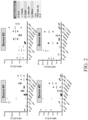


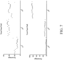


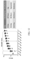


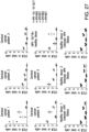


Classifications
-
- C—CHEMISTRY; METALLURGY
- C12—BIOCHEMISTRY; BEER; SPIRITS; WINE; VINEGAR; MICROBIOLOGY; ENZYMOLOGY; MUTATION OR GENETIC ENGINEERING
- C12N—MICROORGANISMS OR ENZYMES; COMPOSITIONS THEREOF; PROPAGATING, PRESERVING, OR MAINTAINING MICROORGANISMS; MUTATION OR GENETIC ENGINEERING; CULTURE MEDIA
- C12N5/00—Undifferentiated human, animal or plant cells, e.g. cell lines; Tissues; Cultivation or maintenance thereof; Culture media therefor
- C12N5/06—Animal cells or tissues; Human cells or tissues
- C12N5/0602—Vertebrate cells
- C12N5/0634—Cells from the blood or the immune system
- C12N5/0636—T lymphocytes
- C12N5/0638—Cytotoxic T lymphocytes [CTL] or lymphokine activated killer cells [LAK]
-
- A—HUMAN NECESSITIES
- A61—MEDICAL OR VETERINARY SCIENCE; HYGIENE
- A61K—PREPARATIONS FOR MEDICAL, DENTAL OR TOILETRY PURPOSES
- A61K35/00—Medicinal preparations containing materials or reaction products thereof with undetermined constitution
- A61K35/12—Materials from mammals; Compositions comprising non-specified tissues or cells; Compositions comprising non-embryonic stem cells; Genetically modified cells
- A61K35/14—Blood; Artificial blood
- A61K35/17—Lymphocytes; B-cells; T-cells; Natural killer cells; Interferon-activated or cytokine-activated lymphocytes
-
- A—HUMAN NECESSITIES
- A61—MEDICAL OR VETERINARY SCIENCE; HYGIENE
- A61K—PREPARATIONS FOR MEDICAL, DENTAL OR TOILETRY PURPOSES
- A61K39/00—Medicinal preparations containing antigens or antibodies
- A61K39/0005—Vertebrate antigens
- A61K39/0011—Cancer antigens
-
- A—HUMAN NECESSITIES
- A61—MEDICAL OR VETERINARY SCIENCE; HYGIENE
- A61K—PREPARATIONS FOR MEDICAL, DENTAL OR TOILETRY PURPOSES
- A61K39/00—Medicinal preparations containing antigens or antibodies
- A61K39/12—Viral antigens
-
- A—HUMAN NECESSITIES
- A61—MEDICAL OR VETERINARY SCIENCE; HYGIENE
- A61K—PREPARATIONS FOR MEDICAL, DENTAL OR TOILETRY PURPOSES
- A61K39/00—Medicinal preparations containing antigens or antibodies
- A61K39/46—Cellular immunotherapy
- A61K39/461—Cellular immunotherapy characterised by the cell type used
- A61K39/4611—T-cells, e.g. tumor infiltrating lymphocytes [TIL], lymphokine-activated killer cells [LAK] or regulatory T cells [Treg]
-
- A—HUMAN NECESSITIES
- A61—MEDICAL OR VETERINARY SCIENCE; HYGIENE
- A61K—PREPARATIONS FOR MEDICAL, DENTAL OR TOILETRY PURPOSES
- A61K39/00—Medicinal preparations containing antigens or antibodies
- A61K39/46—Cellular immunotherapy
- A61K39/464—Cellular immunotherapy characterised by the antigen targeted or presented
- A61K39/464838—Viral antigens
-
- A—HUMAN NECESSITIES
- A61—MEDICAL OR VETERINARY SCIENCE; HYGIENE
- A61K—PREPARATIONS FOR MEDICAL, DENTAL OR TOILETRY PURPOSES
- A61K45/00—Medicinal preparations containing active ingredients not provided for in groups A61K31/00 - A61K41/00
- A61K45/06—Mixtures of active ingredients without chemical characterisation, e.g. antiphlogistics and cardiaca
-
- A—HUMAN NECESSITIES
- A61—MEDICAL OR VETERINARY SCIENCE; HYGIENE
- A61P—SPECIFIC THERAPEUTIC ACTIVITY OF CHEMICAL COMPOUNDS OR MEDICINAL PREPARATIONS
- A61P31/00—Antiinfectives, i.e. antibiotics, antiseptics, chemotherapeutics
- A61P31/12—Antivirals
-
- A—HUMAN NECESSITIES
- A61—MEDICAL OR VETERINARY SCIENCE; HYGIENE
- A61P—SPECIFIC THERAPEUTIC ACTIVITY OF CHEMICAL COMPOUNDS OR MEDICINAL PREPARATIONS
- A61P35/00—Antineoplastic agents
-
- A—HUMAN NECESSITIES
- A61—MEDICAL OR VETERINARY SCIENCE; HYGIENE
- A61P—SPECIFIC THERAPEUTIC ACTIVITY OF CHEMICAL COMPOUNDS OR MEDICINAL PREPARATIONS
- A61P35/00—Antineoplastic agents
- A61P35/04—Antineoplastic agents specific for metastasis
-
- A—HUMAN NECESSITIES
- A61—MEDICAL OR VETERINARY SCIENCE; HYGIENE
- A61P—SPECIFIC THERAPEUTIC ACTIVITY OF CHEMICAL COMPOUNDS OR MEDICINAL PREPARATIONS
- A61P37/00—Drugs for immunological or allergic disorders
- A61P37/02—Immunomodulators
-
- A—HUMAN NECESSITIES
- A61—MEDICAL OR VETERINARY SCIENCE; HYGIENE
- A61P—SPECIFIC THERAPEUTIC ACTIVITY OF CHEMICAL COMPOUNDS OR MEDICINAL PREPARATIONS
- A61P43/00—Drugs for specific purposes, not provided for in groups A61P1/00-A61P41/00
-
- C—CHEMISTRY; METALLURGY
- C07—ORGANIC CHEMISTRY
- C07K—PEPTIDES
- C07K16/00—Immunoglobulins [IGs], e.g. monoclonal or polyclonal antibodies
- C07K16/18—Immunoglobulins [IGs], e.g. monoclonal or polyclonal antibodies against material from animals or humans
- C07K16/28—Immunoglobulins [IGs], e.g. monoclonal or polyclonal antibodies against material from animals or humans against receptors, cell surface antigens or cell surface determinants
- C07K16/2803—Immunoglobulins [IGs], e.g. monoclonal or polyclonal antibodies against material from animals or humans against receptors, cell surface antigens or cell surface determinants against the immunoglobulin superfamily
- C07K16/2818—Immunoglobulins [IGs], e.g. monoclonal or polyclonal antibodies against material from animals or humans against receptors, cell surface antigens or cell surface determinants against the immunoglobulin superfamily against CD28 or CD152
-
- C—CHEMISTRY; METALLURGY
- C07—ORGANIC CHEMISTRY
- C07K—PEPTIDES
- C07K16/00—Immunoglobulins [IGs], e.g. monoclonal or polyclonal antibodies
- C07K16/18—Immunoglobulins [IGs], e.g. monoclonal or polyclonal antibodies against material from animals or humans
- C07K16/28—Immunoglobulins [IGs], e.g. monoclonal or polyclonal antibodies against material from animals or humans against receptors, cell surface antigens or cell surface determinants
- C07K16/2803—Immunoglobulins [IGs], e.g. monoclonal or polyclonal antibodies against material from animals or humans against receptors, cell surface antigens or cell surface determinants against the immunoglobulin superfamily
- C07K16/2827—Immunoglobulins [IGs], e.g. monoclonal or polyclonal antibodies against material from animals or humans against receptors, cell surface antigens or cell surface determinants against the immunoglobulin superfamily against B7 molecules, e.g. CD80, CD86
-
- A—HUMAN NECESSITIES
- A61—MEDICAL OR VETERINARY SCIENCE; HYGIENE
- A61K—PREPARATIONS FOR MEDICAL, DENTAL OR TOILETRY PURPOSES
- A61K2239/00—Indexing codes associated with cellular immunotherapy of group A61K39/46
- A61K2239/31—Indexing codes associated with cellular immunotherapy of group A61K39/46 characterized by the route of administration
-
- A—HUMAN NECESSITIES
- A61—MEDICAL OR VETERINARY SCIENCE; HYGIENE
- A61K—PREPARATIONS FOR MEDICAL, DENTAL OR TOILETRY PURPOSES
- A61K2239/00—Indexing codes associated with cellular immunotherapy of group A61K39/46
- A61K2239/38—Indexing codes associated with cellular immunotherapy of group A61K39/46 characterised by the dose, timing or administration schedule
-
- C—CHEMISTRY; METALLURGY
- C12—BIOCHEMISTRY; BEER; SPIRITS; WINE; VINEGAR; MICROBIOLOGY; ENZYMOLOGY; MUTATION OR GENETIC ENGINEERING
- C12N—MICROORGANISMS OR ENZYMES; COMPOSITIONS THEREOF; PROPAGATING, PRESERVING, OR MAINTAINING MICROORGANISMS; MUTATION OR GENETIC ENGINEERING; CULTURE MEDIA
- C12N2501/00—Active agents used in cell culture processes, e.g. differentation
- C12N2501/20—Cytokines; Chemokines
- C12N2501/22—Colony stimulating factors (G-CSF, GM-CSF)
-
- C—CHEMISTRY; METALLURGY
- C12—BIOCHEMISTRY; BEER; SPIRITS; WINE; VINEGAR; MICROBIOLOGY; ENZYMOLOGY; MUTATION OR GENETIC ENGINEERING
- C12N—MICROORGANISMS OR ENZYMES; COMPOSITIONS THEREOF; PROPAGATING, PRESERVING, OR MAINTAINING MICROORGANISMS; MUTATION OR GENETIC ENGINEERING; CULTURE MEDIA
- C12N2501/00—Active agents used in cell culture processes, e.g. differentation
- C12N2501/20—Cytokines; Chemokines
- C12N2501/23—Interleukins [IL]
- C12N2501/2304—Interleukin-4 (IL-4)
-
- C—CHEMISTRY; METALLURGY
- C12—BIOCHEMISTRY; BEER; SPIRITS; WINE; VINEGAR; MICROBIOLOGY; ENZYMOLOGY; MUTATION OR GENETIC ENGINEERING
- C12N—MICROORGANISMS OR ENZYMES; COMPOSITIONS THEREOF; PROPAGATING, PRESERVING, OR MAINTAINING MICROORGANISMS; MUTATION OR GENETIC ENGINEERING; CULTURE MEDIA
- C12N2501/00—Active agents used in cell culture processes, e.g. differentation
- C12N2501/20—Cytokines; Chemokines
- C12N2501/23—Interleukins [IL]
- C12N2501/2307—Interleukin-7 (IL-7)
-
- C—CHEMISTRY; METALLURGY
- C12—BIOCHEMISTRY; BEER; SPIRITS; WINE; VINEGAR; MICROBIOLOGY; ENZYMOLOGY; MUTATION OR GENETIC ENGINEERING
- C12N—MICROORGANISMS OR ENZYMES; COMPOSITIONS THEREOF; PROPAGATING, PRESERVING, OR MAINTAINING MICROORGANISMS; MUTATION OR GENETIC ENGINEERING; CULTURE MEDIA
- C12N2501/00—Active agents used in cell culture processes, e.g. differentation
- C12N2501/20—Cytokines; Chemokines
- C12N2501/23—Interleukins [IL]
- C12N2501/2315—Interleukin-15 (IL-15)
-
- C—CHEMISTRY; METALLURGY
- C12—BIOCHEMISTRY; BEER; SPIRITS; WINE; VINEGAR; MICROBIOLOGY; ENZYMOLOGY; MUTATION OR GENETIC ENGINEERING
- C12N—MICROORGANISMS OR ENZYMES; COMPOSITIONS THEREOF; PROPAGATING, PRESERVING, OR MAINTAINING MICROORGANISMS; MUTATION OR GENETIC ENGINEERING; CULTURE MEDIA
- C12N2501/00—Active agents used in cell culture processes, e.g. differentation
- C12N2501/20—Cytokines; Chemokines
- C12N2501/24—Interferons [IFN]
-
- C—CHEMISTRY; METALLURGY
- C12—BIOCHEMISTRY; BEER; SPIRITS; WINE; VINEGAR; MICROBIOLOGY; ENZYMOLOGY; MUTATION OR GENETIC ENGINEERING
- C12N—MICROORGANISMS OR ENZYMES; COMPOSITIONS THEREOF; PROPAGATING, PRESERVING, OR MAINTAINING MICROORGANISMS; MUTATION OR GENETIC ENGINEERING; CULTURE MEDIA
- C12N2501/00—Active agents used in cell culture processes, e.g. differentation
- C12N2501/50—Cell markers; Cell surface determinants
- C12N2501/52—CD40, CD40-ligand (CD154)
-
- C—CHEMISTRY; METALLURGY
- C12—BIOCHEMISTRY; BEER; SPIRITS; WINE; VINEGAR; MICROBIOLOGY; ENZYMOLOGY; MUTATION OR GENETIC ENGINEERING
- C12N—MICROORGANISMS OR ENZYMES; COMPOSITIONS THEREOF; PROPAGATING, PRESERVING, OR MAINTAINING MICROORGANISMS; MUTATION OR GENETIC ENGINEERING; CULTURE MEDIA
- C12N2506/00—Differentiation of animal cells from one lineage to another; Differentiation of pluripotent cells
- C12N2506/11—Differentiation of animal cells from one lineage to another; Differentiation of pluripotent cells from blood or immune system cells
- C12N2506/115—Differentiation of animal cells from one lineage to another; Differentiation of pluripotent cells from blood or immune system cells from monocytes, from macrophages
-
- C—CHEMISTRY; METALLURGY
- C12—BIOCHEMISTRY; BEER; SPIRITS; WINE; VINEGAR; MICROBIOLOGY; ENZYMOLOGY; MUTATION OR GENETIC ENGINEERING
- C12N—MICROORGANISMS OR ENZYMES; COMPOSITIONS THEREOF; PROPAGATING, PRESERVING, OR MAINTAINING MICROORGANISMS; MUTATION OR GENETIC ENGINEERING; CULTURE MEDIA
- C12N2710/00—MICROORGANISMS OR ENZYMES; COMPOSITIONS THEREOF; PROPAGATING, PRESERVING, OR MAINTAINING MICROORGANISMS; MUTATION OR GENETIC ENGINEERING; CULTURE MEDIA dsDNA viruses
- C12N2710/00011—Details
- C12N2710/20011—Papillomaviridae
- C12N2710/20034—Use of virus or viral component as vaccine, e.g. live-attenuated or inactivated virus, VLP, viral protein
Definitions
- the disclosure herein relates to the field of immunology, and more specifically, to the generation of antigen-specific T lymphocytes and methods of use thereof.
- T cell antigen-specific T cells e.g ., cytotoxic T lymphocytes (CTLs) ex vivo involves T cell antigen-specific activation followed, e.g ,. by T cell proliferation.
- Full activation of such T cells involves ligation of T cell receptor (TCR) with cognate MHC-peptide complexes presented by antigen presenting cells (APC), including dendritic cells (DC), macrophages, and B cells, and ligations of co-stimulatory molecules between T cells and APC.
- APC antigen presenting cells
- DC dendritic cells
- B cells ligations of co-stimulatory molecules between T cells and APC.
- cell-free MHC-peptide complexes are used in lieu of MHC-peptide complexes presented by APCs. If co-stimulation activation is deficient, T cells will become either partially activated, undergo apoptosis, or enter into anergy status.
- Both B cells and monocytes typically express HLA molecules required for MHC-peptide complex formation, but lack high expression of co-stimulatory molecules, such as CD80 and CD86. Without further activation or maturation to obtain fully activated APC, a failed or suboptimal antigen-specific T cell priming and amplification may ensue. Thus, a separate APC induction and isolation step is usually necessary prior to antigen-specific T cell priming ex vivo, by producing either mature dendritic cells (mDC) from the precursor monocytes, or activated B cells.
- mDC mature dendritic cells
- T cells 66 to 76 stimulated peripheral blood T cells with monocyte-derived dendritic cells loaded with pepmixes spanning E6/E7, in the presence or absence of specific assessory cytokines.
- monocyte-derived dendritic cells loaded with pepmixes spanning E6/E7, in the presence or absence of specific assessory cytokines.
- the resulting T -cell lines were further expanded with pepmix-loaded activated B-cell blasts.
- T cells cytotoxic T lymphocytes
- peripheral blood mononuclear cells without a separate step of generating and isolating antigen-presenting cells, such as dendritic cells, macrophages, and B cells, and with a single round of antigen stimulation, as defined in the claims.
- a method of producing a cell population comprising antigen-specific T cells, optionally cytotoxic T lymphocytes (CTLs), comprising the steps of: (a) culturing peripheral blood mononuclear cells (PBMCs) isolated from a subject in an antigen presenting cell (APC) induction medium comprising interleukin 4 (IL-4) and soluble CD40 ligand (sCD40L) and/or comprising granulocyte-macrophage colony-stimulating factor (GM-CSF) and interferon ⁇ (IFN- ⁇ ), to produce a first population of cells; (b) culturing the first population of cells in the presence of one or more antigens, to produce a second population of cells; and (c) culturing the second population of cells in a T cell expansion medium comprising interleukin 7 (IL-7), interleukin 15 (IL-15), and IL-4, to produce a third population of cells; wherein the third population of cells comprises T cells that are CD
- APC
- the third population of cells comprises T cells that are additionally CD4+; and/or additionally CD8+.
- the method further comprises a step of culturing the third population of cells in a second T cell expansion medium comprising IL-7 and IL-15, but not comprising IL-4, to create a fourth population of cells; wherein the fourth population of cells comprises T cells that are CD3+ and specific for the one or more antigens added in step (b).
- the one or more antigens are a pool of peptides; preferably wherein: (i) the pool of peptides covers the complete sequence of human HPV16E6, HPV16E7, HPV18E6, and HPV18E7 proteins, and/or (ii) the peptides are at a concentration of 1 ⁇ g/mL.
- the APC induction medium comprises IL-4 and sCD40L; preferably wherein the APC induction medium comprises 8-12 ng/mL of IL-4 and 0.8-1.2 ⁇ g/mL of sCD40L; or preferably wherein the APC induction medium comprises 10 ng/mL of IL-4 and 1 ⁇ g/mL of sCD40L.
- the APC induction medium further comprises synthetic oligonucleotides with unmethylated CpG dinucleotide motifs; and/or wherein the APC induction medium further comprises GM-CSF and IFN- ⁇ .
- the T cell expansion medium comprises 40-60 ng/mL of IL-7, 7-11 ng/mL of IL-15, and 45-65 ng/mL of IL-4; preferably wherein the T cell expansion medium comprises 50 ng/mL of IL-7, 9 ng/mL of IL-15, and 55 ng/mL of IL-4.
- the second T cell expansion medium comprises 40-60 ng/mL of IL-7 and 7-11 ng/mL of IL-15; preferably wherein the second T cell expansion medium comprises 50 ng/mL of IL-7 and 9 ng/mL of IL-15.
- the duration of step (a) is 1-3 days, preferably 1 day.
- the PBMCs are: (i) isolated from whole blood, buffy coat, or an enriched leukapheresis product; and/or (ii) seeded in the APC induction medium at a density of 4-6 ⁇ 10 6 /cm 2 , preferably at a density of 5 ⁇ 10 6 /cm 2 .
- the method further comprises a step of isolating T cells that are CD3+ from the third population of cells or the fourth population of cells.
- the antigen-specific CD3+ cells are identified by intracellular cytokine staining (ICCS).
- step (b) is performed in the APC induction medium.
- the culturing steps are performed in a G-REX ® device.
- the duration of step (c) is 8-16 days, preferably 12 days.
- a cell population comprising antigen-specific T cells comprising antigen-specific T cells, e.g., CTLs
- methods of producing a cell population comprising antigen-specific T cells comprising the steps of: (a) isolating blood mononuclear cells, e.g., peripheral blood mononuclear cells (PBMCs) or cord blood mononuclear cells, from a subject; (b) culturing said blood mononuclear cells, e.g ., PBMCs or cord blood mononuclear cells, in an antigen presenting cell (APC) induction medium comprising interleukin 4 (IL-4) and soluble CD40 ligand (sCD40L) and/or comprising granulocyte-macrophage colony-stimulating factor (GM-CSF) and interferon, e.g ., interferon ⁇ (IFN- ⁇ ), to produce a first population of cells; (c) culturing the first population of cells in the presence of
- the method disclosed herein further comprises a step of culturing the third population of cells in a second T cell expansion medium comprising IL-7 and IL-15, but not comprising IL-4, to create a fourth population of cells; wherein the fourth population of cells comprises T cells that are CD3+ and specific for an antigen added in step (c).
- the method disclosed herein further comprises a step of isolating T cells that are CD3+ from the third population of cells or the fourth population of cells.
- step (c) is performed in APC induction medium.
- the T cell expansion medium comprises IL-4.
- the subject is a human.
- the antigen is a human antigen.
- the antigen is a full-length protein. In certain aspects, the antigen is a fragment of a protein. In certain aspects, the antigen is a peptide representing a part of the protein. In certain aspects, the APCs produce the antigen from exogenous genetic material.
- the third population of cells comprises T cells that are additionally CD4+. In certain aspects, the third population of cells comprises T cells that are additionally CD8+. In certain aspects, the third population of cells comprises T cells that are additional CD4+ and CD8+.
- the fourth population of cells comprises T cells that are additionally CD4+. In certain aspects, the fourth population of cells comprises T cells that are additionally CD8+. In certain aspects, the fourth population of cells comprises T cells that are additionally CD4+ and CD8+.
- the APC induction medium comprises IL-4 and sCD40L. In certain aspects, the APC induction medium comprises 1-50 ng/mL of IL-4 and 0.1-5 ⁇ g/mL of sCD40L. In certain aspects, the APC induction medium comprises 8-12 ng/mL of IL-4 and 0.8-1.2 ⁇ g/mL of sCD40L. In certain aspects, the APC induction medium comprises 10 ng/mL of IL-4 and 1 ⁇ g/mL of sCD40L.
- the APC induction medium further comprises synthetic oligonucleotides with one or more unmethylated CpG dinucleotide motifs. In certain aspects, the APC induction medium further comprises GM-CSF and IFN- ⁇ . In certain aspects, the APC induction medium consists essentially of GM-CSF and IFN- ⁇ .
- the one or more antigens are, or are contained within, a pool of peptides, e.g ., lyophilized peptides.
- the pool of lyophilized peptides cover the sequences of human HPV16E6, HPV16E7, HPV18E6, and/or HPV18E7 proteins.
- the peptides are at a concentration of 1 ⁇ g/mL.
- the T cell expansion medium comprises 10-100 ng/mL of IL-7, 2-20 ng/mL of IL-15, and 10-100 ng/mL of IL-4. In certain aspects, the T cell expansion medium comprises 40-60 ng/mL of IL-7, 7-11 ng/mL of IL-15, and 45-65 ng/mL of IL-4. In specific aspects, the T cell expansion medium comprises 50 ng/mL of IL-7, 9 ng/mL of IL-15, and 55 ng/mL of IL-4.
- the second T cell expansion medium comprises 10-100 ng/mL of IL-7 and 2-20 ng/mL of IL-15. In certain aspects, the second T cell expansion medium comprises 40-60 ng/mL of IL-7 and 7-11 ng/mL of IL-15. In specific aspects, the second T cell expansion medium comprises 50 ng/mL of IL-7 and 9 ng/mL of IL-15.
- the duration of step (a) is 1-3 days. In specific aspects, the duration of step (a) is 1 day. In certain aspects, the duration of step (c) is 8-16 days. In specific aspects, the duration of step (c) is 12 days.
- the PBMCs are isolated from whole blood, buffy coat, or an enriched leukapheresis product.
- the PBMCs are seeded in the APC induction medium at a density of 4-6 ⁇ 10 6 /cm 2 .
- the PBMCs are seeded at a density of 5 ⁇ 10 6 /cm 2 .
- the PBMCs are seeded at a density greater than 5 ⁇ 10 6 /cm 2 .
- the antigen-specific CD3+ cells are identified by, or are identifiable by, intracellular cytokine staining (ICCS).
- the culturing steps are performed in a gas-permeable enclosure, e.g ., a G-REX ® device. In certain aspects, one or more culturing steps are performed in T-flasks. In certain aspects, one or more culturing steps are performed in bags. In specific aspects, the bags are static bags. In certain aspects, the bags are gas-permeable. In certain aspects, one or more culturing steps are performed in a WAVE TM bioreactor (GE Healthcare Life Sciences). In certain aspects, one or more culturing steps are performed in spinner flasks.
- populations of antigen-specific T cells produced by a method comprising the steps of: (a) isolating blood mononuclear cells, e.g ., PBMCs or cord blood mononuclear cells, from a subject; (b) culturing said blood mononuclear cells, e.g., PBMCs or cord blood mononuclear cells, in an antigen presenting cell (APC) induction medium comprising interleukin 4 (IL-4) and soluble CD40 ligand (sCD40L) and/or comprising granulocyte-macrophage colony-stimulating factor (GM-CSF) and interferon ⁇ (IFN- ⁇ ), to produce a first population of cells; (c) culturing the first population of cells in the presence of one or more antigens, to produce a second population of cells; and (d) culturing the second population of cells in a T cell expansion medium comprising interleukin 7
- APC antigen presenting cell
- the populations of cells are produced by a method which further comprises a step of culturing the third population of cells in a second T cell expansion medium comprising IL-7 and IL-15, but not comprising IL-4, to create a fourth population of cells; wherein the fourth population of cells comprises T cells that are CD3+ and specific for an antigen added in step (c).
- the populations of cells are produced by a method which further comprises a step of isolating T cells that are CD3+ from the third population of cells or the fourth population of cells.
- step (c) is performed in APC induction medium.
- compositions e.g ., pharmaceutical compositions, comprising the antigen-specific T cells described herein.
- 0.5%, 0.6%, 0.7%, 0.8%, 0.9%, 1%, 2%, 3%, 4%, 5%, 6%, 7%, 8%, 9%, 10%, 15%, 20%, 25%, 30%, 35%, 40%, or 45% or more of the cells in the composition are antigen-specific T cells.
- 50% of the cells in the composition are antigen-specific T cells.
- the compositions comprise a pharmaceutically acceptable excipient.
- Disclosed herein and not part of the invention are methods of treating a cancer or a viral infection comprising administering to a patient in need thereof a population of antigen-specific T cells produced by the methods described herein, wherein the isolated blood mononuclear cells, e.g., PBMCs or cord blood mononuclear cells, are autologous to the patient. Also disclosed herein are methods of treating a cancer or a viral infection comprising administering to a patient in need thereof a population of antigen-specific T cells produced by the methods described herein, wherein the isolated blood mononuclear cells, e.g., PBMCs or cord blood mononuclear cells, are not autologous to the patient.
- the isolated blood mononuclear cells e.g., PBMCs or cord blood mononuclear cells
- the viral infection is a human papillomavirus (HPV) infection.
- the viral infection is an HPV infection and the antigen is an HPV antigen.
- the viral infection is an Epstein-Barr virus (EBV) infection.
- the viral infection is an EBV infection and the antigen is an EBV antigen.
- the antigens are expressed in tumor cells comprising the viral infection.
- disclosed herein are methods of administering a population of antigen-specific T cells to a patient in need thereof.
- the method further comprises administering to said patient an immunomodulatory drug or an epigenetic modifier.
- the method further comprises administering to said patient an immune checkpoint inhibitor.
- the immune checkpoint inhibitor is selected from the group consisting of an anti-CTLA-4 antibody, an anti-PD1 antibody, and an anti-PD-L1 antibody.
- the cancer is an HPV positive (HPV+) cancer.
- HPV+ cancer is head and neck cancer.
- the head and neck cancer is squamous cell carcinoma of the head and neck.
- the head and neck cancer is oropharyngeal cancer.
- the HPV+ cancer is penile cancer.
- the HPV+ cancer is cervical cancer.
- the HPV+ cancer is anal cancer. In certain examples, the HPV+ cancer is vulval cancer. In certain examples, the HPV+ cancer is vaginal cancer. In certain examples, the HPV+ cancer is lung cancer. In certain examples, the HPV+ cancer is metastatic. In certain examples, the HPV+ cancer is recurrent. In certain examples, the HPV+ cancer is metastatic and recurrent. In certain examples, the method further comprises administering to said patient an immunomodulatory drug or an epigenetic modifier. In certain examples, the method further comprises administering to said patient an immune checkpoint inhibitor. In certain examples, the immune checkpoint inhibitor is selected from the group consisting of an anti-CTLA-4 antibody, an anti-PD1 antibody, and an anti-PD-L1 antibody.
- T cells cytotoxic T lymphocytes
- PBMCs PBMCs
- antigen-presenting cells such as dendritic cells, macrophages, and B cells
- T cells T cells
- Table 1 List of abbreviations for key terms.
- Term Full name AE adverse event APC antigen presenting cells CBA cytometric beads array
- Cy cyclophosphamide DC dendritic cells DLT dose-limiting toxicity DMSO dimethyl sulfoxide Flu fludarabine GM-CSF granulocyte-macrophage HPV Human papillomavirus ICCS intracellular cytokine staining iDC immature dendritic cells IFN- ⁇ interferon alpha IFN- ⁇ interferon gamma IL-10 interleukin 10 IL-13 interleukin 13 IL-15 interleukin 15 IL-1 ⁇ interleukin 1 beta IL-2 interleukin 2 IL-4 interleukin 4 IL-5 interleukin 5 IL-6 interleukin 6 IL-7 interleukin 7 IM intramuscular IMDM Iscove's Modified Dulbecco's IV intravenous mDC mature dendritic cells MTD maximum tolerated dose OS overall survival
- cytotoxic T lymphocytes comprising the steps of: (a) culturing PBMCs isolated from a subject in an antigen presenting cell (APC) induction medium comprising interleukin 4 (IL-4) and soluble CD40 ligand (sCD40L) and/or comprising granulocyte-macrophage colony-stimulating factor (GM-CSF) and interferon ⁇ (IFN- ⁇ ), to produce a first population of cells; (b) culturing the first population of cells in the presence of one or more antigens, to produce a second population of cells; and (c) culturing the second population of cells in a T cell expansion medium comprising interleukin 7 (IL-7), interleukin 15 (IL-15), and IL-4, to produce a third population of cells; wherein the third population of cells comprises T cells that are CD3+ and
- the method further comprises a step of culturing the third population of cells in a second T cell expansion medium comprising IL-7 and IL-15, but not comprising IL-4, to create a fourth population of cells; wherein the fourth population of cells comprises T cells that are CD3+ and specific for an antigen added in step (b).
- the method further comprises a step of isolating T cells that are CD3+ from the third population of cells or the fourth population of cells.
- step (b) is performed in APC induction medium.
- the subject is a human.
- the antigen is a human antigen.
- the antigen is a full-length protein.
- the antigen is a fragment of a protein.
- the antigen is a peptide representing a part of a protein.
- the APCs produce the antigen from exogenous genetic material.
- one or more steps are repeated in order to increase the yield of the target population of cells generated by those one or more steps.
- a step is repeated with cells that remain unmodified after said step.
- one or more steps are repeated with cells that remain unmodified after said steps.
- steps (a) to (c) are repeated one time.
- steps (a) to (c) are repeated two times.
- steps (a) to (c) are repeated three times.
- steps (a) to (c) are repeated four times.
- steps (a) to (c) are repeated five times. In certain aspects, steps (a) to (c) are repeated with PBMCs, remaining after step (c). In certain aspects, one or more of steps (a), (b), and (c) are repeated one, two, three, four, or five times.
- the third population of cells comprises T cells that are additionally CD4+. In certain aspects, the third population of cells comprises T cells that are additionally CD8+. In certain aspects, the third population of cells comprises T cells that are additionally CD4+ and CD8+.
- the fourth population of cells comprises T cells that are additionally CD4+. In certain aspects, the fourth population of cells comprises T cells that are additionally CD8+. In certain aspects, the fourth population of cells comprises T cells that are additionally CD4+ and CD8+.
- the APC induction medium comprises IL-4 and sCD40L. In certain aspects, the APC induction medium comprises 1-50 ng/mL of IL-4 and 0.1-5 ⁇ g/mL of sCD40L. In certain aspects, the APC induction medium comprises 8-12 ng/mL of IL-4 and 0.8-1.2 ⁇ g/mL of sCD40L. In certain aspects, the APC induction medium comprises 10 ng/mL of IL-4 and 1 ⁇ g/mL of sCD40L.
- the APC induction medium further comprises synthetic oligonucleotides with one or more unmethylated CpG dinucleotide motifs. In certain aspects, the APC induction medium further comprises GM-CSF and IFN- ⁇ . In certain aspects, the APC induction medium comprises GM-CSF and IFN- ⁇ . In certain aspects, the APC induction medium consists essentially of GM-CSF and IFN- ⁇ .
- the APC induction medium further comprises IMDM, 2-mercaptoethanol, Pen Strep, and/or human serum.
- the APC induction medium further comprises RPMI-1640, Click's media, human serum, L-GLUTAMAX TM (Life Technologies), Pen Strep, and/or 2-mercaptoethanol.
- the APC induction medium further comprises RPMI-1640, Click's media, human serum, glutamine, Pen Strep, and/or 2-mercaptoethanol.
- the IL-4 in the APC induction medium is at a concentration of 40 ng/mL. In certain aspects, the IL-4 in the APC induction medium is at a concentration of 10 ng/mL. In certain aspects, the CD40L in the APC induction medium is at a concentration of 1 ⁇ g/mL. In certain aspects, the CpG dinucleotide motifs in the APC induction medium are at a concentration of 4 ⁇ g/mL. In certain aspects, the GM-CSF in the APC induction medium is at a concentration of 800 U/mL. In certain aspects, the IFN- ⁇ in the APC induction medium is at a concentration of 1000 U/mL.
- the one or more antigens are a pool of peptides, e.g ., a pool of lyophilized peptides.
- the one or more antigens are an HPV peptide mixture, e.g ., PEPMIX TM .
- the one or more antigens are a pool of overlapping 15-mers spanning E6 and E7 proteins from HPV types 16 and 18.
- the pool of peptides cover the sequences of one or more of, or all of, human HPV16E6, HPV16E7, HPV18E6, and/or HPV18E7 proteins.
- the one or more antigens comprise one or more antigens from a single virus, e.g ., only from HPV16 or only from HPV18.
- the peptides are at a concentration of 0.5-1 ⁇ g/mL. In specific aspects, the peptides are at a concentration of 1 ⁇ g/mL. In specific aspects, the peptides are at a concentration of 0.5 ⁇ g/mL.
- the T cell expansion medium comprises 10-100 ng/mL of IL-7, 2-20 ng/mL of IL-15, and 10-100 ng/mL of IL-4. In certain aspects, the T cell expansion medium comprises 40-60 ng/mL of IL-7, 7-11 ng/mL of IL-15, and 45-65 ng/mL of IL-4. In specific aspects, the T cell expansion medium comprises 50 ng/mL of IL-7, 9 ng/mL of IL-15, and 55 ng/mL of IL-4. In certain aspects, addition of the cytokines from the T cell expansion medium is performed 2 hours, 24 hours, or 48 hours after addition of the one or more antigens.
- addition of the cytokines from the T cell expansion medium is performed 2 hours after addition of the one or more antigens. In specific aspects, addition of the cytokines from the T cell expansion medium is performed 24 hours after addition of the one or more antigens. In specific aspects, addition of the cytokines from the T cell expansion medium is performed 48 hours after addition of the one or more antigens.
- the T cell expansion medium comprises 1 ng/mL IL-15, 15 ng/mL IL-4, and 10 ng/mL IL-7. In certain aspects, the T cell expansion medium comprises 1 ng/mL IL-15, 15 ng/mL IL-4, and 50 ng/mL IL-7. In certain aspects, the T cell expansion medium comprises 1 ng/mL IL-15, 35 ng/mL IL-4, and 30 ng/mL IL-7. In certain aspects, the T cell expansion medium comprises 1 ng/mL IL-15, 55 ng/mL IL-4, and 10 ng/mL IL-7.
- the T cell expansion medium comprises 1 ng/mL IL-15, 55 ng/mL IL-4, and 50 ng/mL IL-7. In certain aspects, the T cell expansion medium comprises 5.5 ng/mL IL-15, 15 ng/mL IL-4, and 10 ng/mL IL-7. In certain aspects, the T cell expansion medium comprises 5.5 ng/mL IL-15, 15 ng/mL IL-4, and 50 ng/mL IL-7. In certain aspects, the T cell expansion medium comprises 5.5 ng/mL IL-15, 35 ng/mL IL-4, and 30 ng/mL IL-7.
- the T cell expansion medium comprises 5.5 ng/mL IL-15, 55 ng/mL IL-4, and 10 ng/mL IL-7. In certain aspects, the T cell expansion medium comprises 5.5 ng/mL IL-15, 55 ng/mL IL-4, and 50 ng/mL IL-7. In certain aspects, the T cell expansion medium comprises 10 ng/mL IL-15, 15 ng/mL IL-4, and 10 ng/mL IL-7. In certain aspects, the T cell expansion medium comprises 10 ng/mL IL-15, 15 ng/mL IL-4, and 50 ng/mL IL-7.
- the T cell expansion medium comprises 10 ng/mL IL-15, 35 ng/mL IL-4, and 30 ng/mL IL-7. In certain aspects, the T cell expansion medium comprises 10 ng/mL IL-15, 55 ng/mL IL-4, and 10 ng/mL IL-7. In certain aspects, the T cell expansion medium comprises 10 ng/mL IL-15, 55 ng/mL IL-4, and 50 ng/mL IL-7. In certain aspects, the T cell expansion medium comprises 55 ng/mL IL-4 and 50 ng/mL IL-7. In certain aspects, the T cell expansion medium comprises 5.5 ng/mL IL-15 and 50 ng/mL IL-7. In certain aspects, the T cell expansion medium consists essentially of 5.5 ng/mL IL-15 and 50 ng/mL IL-7.
- the second T cell expansion medium comprises 10-100 ng/mL of IL-7 and 2-20 ng/mL of IL-15. In certain aspects, the second T cell expansion medium comprises 40-60 ng/mL of IL-7 and 7-11 ng/mL of IL-15. In specific aspects, the second T cell expansion medium comprises 50 ng/mL of IL-7 and 9 ng/mL of IL-15.
- the duration of step (a) is 1-3 days. In specific aspects, the duration of step (a) is 1 day. In certain aspects, the duration of step (c) is 8-16 days. In specific aspects, the duration of step (c) is 12 days.
- IL-4 is removed from the T cell expansion medium prior to the end of T cell expansion. In certain aspects, IL-4 is not removed from the T cell expansion medium prior to the end of T cell expansion.
- the PBMCs are isolated from whole blood, buffy coat, or an enriched leukapheresis product. In certain aspects, the PBMCs are seeded into the APC induction medium at a density of 4-6 ⁇ 10 6 /cm 2 . In specific aspects, the PBMCs are seeded at a density of 5 ⁇ 10 6 /cm 2 . In specific aspects, the PBMCs are seeded at a density greater than 5 ⁇ 10 6 /cm 2 .
- the antigen-specific CD3+ cells are identified by intracellular cytokine staining (ICCS).
- the culturing steps are performed in a gas-permeable container, e.g ., plastic bag or a G-REX ® device.
- one or more culturing steps are performed in T-flasks.
- one or more culturing steps are performed in bags.
- the bags are static bags.
- the bags are gas-permeable.
- one or more culturing steps are performed in a WAVE TM bioreactor.
- one or more culturing steps are performed in a spinner flasks.
- T cells can be isolated or enriched by staining cells, in one embodiment, with antibodies to CD3, and selecting for CD3+ cells. T cells may also be isolated with antibodies to CD4 and/or CD8, and selecting for CD4+ and/or CD8+ cells.
- T cells for example, T cells produced by the methods described herein can also be isolated or enriched by removal of cells other than T cells in a population of cells that comprise the T cells, e.g ., T cells produced by the methods described herein.
- T cells e.g ., cells produced using the methods described herein, may be isolated or enriched by depletion of cells displaying non-T cell cell markers. Cells isolated by these methods may be additionally sorted.
- Antigen-specific T cells e.g., antigen-specific CTLs, e.g. HPV-specific T cells and CTLs, may, for example, be identified using an ICCS assay, a flow cytometry based assay which detects the production and accumulation of cytokines within the endoplasmic reticulum after cell stimulation.
- ICCS can be used in combination with other flow cytometry protocols for immunophenotyping using cell surface markers to detect antigen reactivity of T cells.
- a ICCS assay is used to measure antigen-specific T cell, e.g., HPV-T cell, frequency at various time points of the culture process of the T cells.
- ICCS works via the following principles: 1) antigen specific T cells can be activated by the same specific antigen re-stimulation, 2) in response to the antigen specific re-stimulation, T cell can produce cytokines (such as IFN- ⁇ , TNF- ⁇ , IL-2, et al) and express activation markers (such as CD154, CD137, et al), 3) an inhibitor of protein transport (e.g . brefeldin A) is added during the re-stimulation to retain the cytokines within the cell, 4) the cells are then fixed in paraformaldehyde and permeabilized, and 5) the anti-cytokine antibody is added and the cells can be analyzed by flow cytometer.
- cytokines such as IFN- ⁇ , TNF- ⁇ , IL-2, et al
- activation markers such as CD154, CD137, et al
- an inhibitor of protein transport e.g . brefeldin A
- a non-antigen stimulation control is set up along with the specific antigen stimulation conditions.
- the antigen-specific, e.g., HPV, T cell number is counted by measuring the T cells that have at least one of the cytokine production or up-regulated activation markers, and subtracting the number from the non-antigen stimulation control.
- the panel used for flow cytometry is a 6 marker panel, including CD3, CD4, CD8, CD154 (CD40L), IFN- ⁇ and TNF- ⁇ .
- CD3, CD4 and CD8 are T cell lineage markers that can define a total T cell population, optionally including natural killer T (NKT) cells, and CD4/CD8 T cell ratio.
- IFN- ⁇ and TNF- ⁇ are cytokines that T cells produce after overnight re-activation.
- CD154 is a T cell activation marker that is up-regulated in newly antigen-activated T cells.
- Determination and/or isolation of T cells may be performed, for example, using a T cell purity panel comprising antibodies against CD19, CD56, CD3, CD11a, CD4, and CD8. Determination and/or isolation of T cells at a certain stage of differentiation may be performed using a T cell differentiation panel comprising antibodies against CD3, CD62L, CD4, CD27, CCR7, and CD45RO.
- T cell differentiation panel comprising antibodies against CD3, CD62L, CD4, CD27, CCR7, and CD45RO.
- secreted cytokines can also be measured, for example, by an 8-plex cytometrix beads array (CBA).
- the panel may include one or more sets of cytokines selected from the group consisting of Th1 cytokines (e.g ., IL-2, IFN- ⁇ and TNF- ⁇ ), Th2 cytokines (e.g., IL-5, IL-13), Treg cytokines ( e.g., IL-10), and T cell effector function related cytokines (e.g ., Granzyme B and GM-CSF).
- Th1 cytokines e.g IL-2, IFN- ⁇ and TNF- ⁇
- Th2 cytokines e.g., IL-5, IL-13
- Treg cytokines e.g., IL-10
- T cell effector function related cytokines e.g ., Granzyme B and GM-CSF.
- transcription factors may be measured, for example, one or more of T-bet, GATA-3, and FoxP3.
- Cell separation e.g ., separating T cells produced by the methods described herein from non-T cells, can be accomplished by, e.g., flow cytometry, fluorescence-activated cell sorting (FACS), or, in one embodiment, magnetic cell sorting using microbeads conjugated with specific antibodies.
- the cells may be isolated, e.g ., using a magnetic activated cell sorting (MACS) technique, a method for separating particles based on their ability to bind magnetic beads (e.g., about 0.5-100 ⁇ m diameter) that comprise one or more specific antibodies, e.g ., anti-CD3 antibodies.
- MCS magnetic activated cell sorting
- Magnetic cell separation can be performed and automated using, e.g., an AUTOMACS TM Separator (Miltenyi) or a CLINIMACS ® System (Miltenyi).
- a variety of useful modifications can be performed on the magnetic microspheres, including covalent addition of antibody that specifically recognizes a particular cell surface molecule or hapten.
- the beads are then mixed with the cells to allow binding.
- Cells are then passed through a magnetic field to separate out cells having the specific cell surface marker.
- these cells can then be isolated and re-mixed with magnetic beads coupled to an antibody against additional cell surface markers.
- the cells are again passed through a magnetic field, isolating cells that bound both the antibodies.
- Such cells can then be diluted into separate dishes, such as microtiter dishes for clonal isolation.
- the populations of antigen-specific T cells disclosed herein and not part of the invention are specific for a specific antigen or a set of related antigens, e.g ., specific to antigens from the same protein. In one aspect, the populations of antigen-specific T cells are specific for a set of unrelated antigens, e.g ., specific to antigens from different proteins.
- the T cells are produced by the method of the invention comprising the steps of: (a) culturing PBMCs isolated from a subject in an antigen presenting cell (APC) induction medium comprising interleukin 4 (IL-4) and soluble CD40 ligand (sCD40L) and/or comprising granulocyte-macrophage colony-stimulating factor (GM-CSF) and interferon ⁇ (IFN- ⁇ ), to produce a first population of cells; (b) culturing the first population of cells in the presence of one or more antigens, to produce a second population of cells; and (c) culturing the second population of cells in a T cell expansion medium comprising interleukin 7 (IL-7), interleukin 15 (IL-15), and IL-4, to produce a third population of cells; wherein the third population of cells comprises T cells that are CD3+ and specific for an antigen added in step (b).
- APC antigen presenting cell
- IL-4 soluble CD40 lig
- the populations of cells are produced by a method of the invention which further comprises a step of culturing the third population of cells in a second T cell expansion medium comprising IL-7 and IL-15, but not comprising IL-4, to create a fourth population of cells; wherein the fourth population of cells comprises T cells that are CD3+ and specific for an antigen added in step (b).
- the populations of cells disclosed herein and not part of the invention are produced by a method of the invention which further comprises a step of isolating T cells that are CD3+ from the third population of cells or the fourth population of cells.
- the populations of cells are produced by a method of the invention wherein step (b) is performed in APC induction medium.
- the third population of cells comprises T cells that are additionally CD4+. In certain aspects, the third population of cells comprises T cells that are additionally CD8+. In certain aspects, the third population of cells comprises T cells that are additional CD4+ and CD8+.
- the fourth population of cells comprises T cells that are additionally CD4+. In certain aspects, the fourth population of cells comprises T cells that are additionally CD8+. In certain aspects, the fourth population of cells comprises T cells that are additionally CD4+ and CD8+.
- the APC induction medium comprises IL-4 and sCD40L. In certain aspects, the APC induction medium comprises 8-12 ng/mL of IL-4 and 0.8-1.2 ⁇ g/mL of sCD40L. In certain aspects, the APC induction medium comprises 10 ng/mL of IL-4 and 1 ⁇ g/mL of sCD40L.
- the one or more antigens are, or are comprised within, a pool of peptides, e.g ., lyophilized peptides.
- the peptides are each derived from, e.g., represent a portion of, a sequence of a protein against which the T cells are to be activated.
- the peptides are 10-mers, 11-mers, 12-mers, 13-mers, 14-mers, 15-mers, 16-mers, 17-mers, 18-mers, 19-mers or 20-mers.
- the pool of peptides are non-overlapping across a specific protein sequence from which they are derived.
- the pool of peptides are overlapping across a specific protein sequence from which they are derived. In specific embodiments, such peptides may successively overlap by 1, 2, 3, 4, 5, 6, 7, 8, 9, 10, 11, 12, 13, 14, 15, 16, 17, 18, 19, 20, 21, 22, 23, 24, or 25 amino acids.
- the protein is a Tumor-Associated Antigen (TAA). In other embodiments, the protein is a Tumor-Specific Antigen (TSA).
- TAA Tumor-Associated Antigen
- TSA Tumor-Specific Antigen
- the peptides cover the sequences of human HPV16E6, HPV16E7, HPV18E6, and/or HPV18E7 proteins.
- such peptides comprise every amino acid residue present in human HPV16E6, HPV16E7, HPV18E6, and/or HPV18E7 proteins. In certain aspects, such peptides comprise a portion of the amino acid residues present in human HPV16E6, HPV16E7, HPV18E6, and/or HPV18E7 proteins. In a specific embodiment, the peptides cover the sequences of human HPV16E6, HPV16E7, HPV18E6, and HPV18E7 proteins. In a specific embodiment, the peptides cover the sequences of human HPV16E6 and/or HPV16E7 proteins and do not cover HPV18E6, and/or HPV18E7 proteins.
- the peptides cover the sequences of human HPV18E6 and/or HPV18E7 proteins and do not cover HPV16E6, and/or HPV16E7 proteins. In specific aspects, the peptides are at a concentration of 1 ⁇ g/mL. In certain aspects, the one or more antigens are produced from exogenous genetic material by the APCs.
- the antigen is selected from the group consisting of B-cell maturation antigen (BCMA), II,13R ⁇ 2, Her2, prostate stem cell antigen (PSCA), alpha-fetoprotein (AFP), carcinoembryonic antigen (CEA), cancer antigen-125 (CA-125), CA19-9, calretinin, MUC-1, epithelial membrane protein (EMA), epithelial tumor antigen (ETA), tyrosinase, melanoma-associated antigen (MAGE), CD19, CD34, CD45, CD99, CD117, chromogranin, cytokeratin, desmin, glial fibrillary acidic protein (GFAP), gross cystic disease fluid protein (GCDFP-15), HMB-45 antigen, high molecular weight melanoma-associated antigen (HMW-MAA), protein melan-A (melanoma antigen recognized by T lymphocytes; MART-1), myo-D1, muscle-specific actin (MSA), neurofilament
- the antigen is a tumor-associated antigen or a tumor-specific antigen.
- the tumor-associated antigen or tumor-specific antigen is Her2, prostate stem cell antigen (PSCA), alpha-fetoprotein (AFP), carcinoembryonic antigen (CEA), cancer antigen-125 (CA-125), CA19-9, calretinin, MUC-1, epithelial membrane protein (EMA), epithelial tumor antigen (ETA), tyrosinase, melanoma-associated antigen (MAGE), CD19, CD34, CD45, CD99, CD117, chromogranin, cytokeratin, desmin, glial fibrillary acidic protein (GFAP), gross cystic disease fluid protein (GCDFP-15), HMB-45 antigen, high molecular weight melanoma-associated antigen (HMW-MAA), protein melan-A (melanoma antigen recognized by T lymphocytes; MART-1), myo-D
- HMW-MAA high molecular
- the TAA or TSA is a cancer/testis (CT) antigen, e.g ., BAGE, CAGE, CTAGE, FATE, GAGE, HCA661, HOM-TES-85, MAGEA, MAGEB, MAGEC, NA88, NY-ESO-1, NY-SAR-35, OY-TES-1, SPANXB1, SPA17, SSX, SYCP1, or TPTE.
- CT cancer/testis
- the TAA or TSA is a carbohydrate or ganglioside, e.g ., fuc-GM1, GM2 (oncofetal antigen-immunogenic-1; OFA-I-1); GD2 (OFA-I-2), GM3, GD3, and the like.
- the TAA or TSA is alpha-actinin-4, Bage-1, BCR-ABL, Bcr-Abl fusion protein, beta-catenin, CA 125, CA 15-3 (CA 27.29 ⁇ BCAA), CA 195, CA 242, CA-50, CAM43, Casp-8, cdc27, cdk4, cdkn2a, CEA, coa-1, dek-can fusion protein, EBNA, EF2, Epstein Barr virus antigens, ETV6-AML1 fusion protein, HLA-A2, HLA-A11, hsp70-2, KIAAO205, Mart2, Mum-1, 2, and 3, neo-PAP, myosin class I, OS-9, pml-RAR ⁇ fusion protein, PTPRK, K-ras, N-ras, triosephosphate isomerase, Gage 3,4,5,6,7, GnTV, Herv-K-mel, Lü-1, NA-88, NY
- said tumor-associated antigen or tumor-specific antigen is integrin ⁇ v ⁇ 3 (CD61), galactin, K-Ras (V-Ki-ras2 Kirsten rat sarcoma viral oncogene), or Ral-B.
- integrin ⁇ v ⁇ 3 CD61
- galactin galactin
- K-Ras V-Ki-ras2 Kirsten rat sarcoma viral oncogene
- Ral-B tumor-associated and tumor-specific antigens are known to those in the art.
- the antigen is an antigen not considered to be a TSA or a TAA, but is nevertheless associated with tumor cells, or damage caused by a tumor.
- the antigen is a neoantigen.
- the antigen is, e.g., a growth factor, cytokine or interleukin, e.g ., a growth factor, cytokine, or interleukin associated with angiogenesis or vasculogenesis.
- Such growth factors, cytokines, or interleukins can include, e.g ., vascular endothelial growth factor (VEGF), basic fibroblast growth factor (bFGF), platelet-derived growth factor (PDGF), hepatocyte growth factor (HGF), insulin-like growth factor (IGF), or interleukin-8 (IL-8).
- VEGF vascular endothelial growth factor
- bFGF basic fibroblast growth factor
- PDGF platelet-derived growth factor
- HGF hepatocyte growth factor
- IGF insulin-like growth factor
- IL-8 interleukin-8
- Tumors can also create a hypoxic environment local to the tumor.
- the antigen is a hypoxia-associated factor, e.g., HIF-1 ⁇ , HIF-1 ⁇ , HIF-2 ⁇ , HIP-2 ⁇ , HIF-3 ⁇ , or HIP-3 ⁇ .
- the antigen is a DAMP, e.g ., a heat shock protein, chromatin-associated protein high mobility group box 1 (HMGB1), S100A8 (MRP8, calgranulin A), S100A9 (MRP14, calgranulin B), serum amyloid A (SAA), or can be a deoxyribonucleic acid, adenosine triphosphate, uric acid, or heparin sulfate.
- DAMP damage associated molecular pattern molecules
- the antigen is virus-associated, e.g ., HPV associated. In specific embodiments, the antigen is HPV associated. In specific embodiments, the antigen is EBV associated.
- the T cell expansion medium comprises 40-60 ng/mL of IL-7, 7-11 ng/mL of IL-15, and 45-65 ng/mL of IL-4. In specific aspects, the T cell expansion medium comprises 50 ng/mL of IL-7, 9 ng/mL of IL-15, and 55 ng/mL of IL-4.
- the second T cell expansion medium comprises 40-60 ng/mL of IL-7 and 7-11 ng/mL of IL-15. In specific aspects, the second T cell expansion medium comprises 50 ng/mL of IL-7 and 9 ng/mL of IL-15.
- the duration of step (a) is 1-3 days. In specific aspects, the duration of step (a) is 1 day. In certain aspects, the duration of step (c) is 8-16 days. In specific aspects, the duration of step (c) is 12 days.
- the PBMCs are isolated from whole blood, buffy coat, or an enriched leukapheresis product. In certain aspects, the PBMCs are seeded in the APC induction medium at a density of 4-6 ⁇ 10 6 /cm 2 . In specific aspects, the PBMCs are seeded at a density of 5 ⁇ 10 6 /cm 2 . In specific aspects, the PBMCs are seeded at a density greater than 5 ⁇ 10 6 /cm 2 .
- the antigen-specific CD3+ cells are identified by intracellular cytokine staining (ICCS).
- the culturing steps are performed in a G-REX ® device.
- one or more culturing steps are performed in T-flasks.
- one or more culturing steps are performed in bags.
- the bags are static bags.
- the bags are gas-permeable.
- one or more culturing steps are performed in a WAVE TM bioreactor.
- one or more culturing steps are performed in a spinner flasks.
- the APC induction medium further comprises synthetic oligonucleotides with unmethylated CpG dinucleotide motifs. In certain aspects, the APC induction medium further comprises GM-CSF and IFN- ⁇ . In certain aspects, the APC induction medium comprises GM-CSF and IFN- ⁇ .
- said T cell population comprises about 70% or more, in some embodiments, 75%, 80%, 85%, 90%, 95%, 98%, or 99% CD3 + cells. In another specific embodiment, said T cell population comprises no less than 80%, 85%, 90%, 95%, 98%, or 99% CD3 + cells. In another specific embodiment, said T cell population comprises between 70%-75%, 75%-80%, 80%-85%, 85%-90%, 90%-95%, or 95%-99% CD3+ cells.
- said CD3 + cells in said T cell population comprise CD3+ cells that are additionally CD4+. In certain embodiments, said CD3+ cells in said T cell population comprise CD3+ cells that are additionally CD8+. In certain embodiments, said CD3 + cells in said T cell population comprise CD3+ cells that are additionally CD4+ and CD8+. In certain embodiments, said T cell population comprises CD137+ cells. In certain embodiments, said T cell population comprises CD154+ cells. In certain embodiments, said T cell population comprises cells that express Th1 cytokines. In certain embodiments, said T cell population comprises cells that express IFN- ⁇ . In certain embodiments, said T cell population comprises cells that express TNF- ⁇ . In certain embodiments, said T cell population comprises cells that express GM-CSF.
- said T cell population comprises cells that express granzyme B. In certain embodiments, said T cell population comprises CD3+, CD137+, and CD154+ cells. In certain embodiments, said T cell population comprises CD3+, CD4+, CD137+, and CD154+ cells. In certain embodiments, said T cell population comprises CD3+, CD8+, CD137+, and CD154+ cells. In certain embodiments, said T cell population comprises CD3+, CD4+, CD8+, CD137+, and CD154+ cells. In certain embodiments, said T cell population comprises CD3+, CD137+, CD154+ cells, and cells that express one or more Th1 cytokines.
- said T cell population comprises CD3+, CD4+, CD137+, CD154+ cells, and cells that express one or more Th1 cytokines. In certain embodiments, said T cell population comprises CD3+, CD8+, CD137+, CD154+ cells, and cells that express one or more Th1 cytokines. In certain embodiments, said T cell population comprises CD3+, CD4+, CD8+, CD137+, CD154+ cells, and cells that express one or more Th1 cytokines.
- T cells e.g., the T cells produced by any of the methods provided herein, e.g., T cells that are HPV-specific, show increased expression of one or more genes as compared to PBMCs, e.g ., matched PBMCs, from which they are produced, wherein said one or more genes are one or more of ACSLS, ACSL6, ACTA2, AHRR, ALDH18A1, ALDH6A1, ANAPC11, AP2B1, BAX, BCL6, BRCA1, C5, CCL13, CCL17, CCL18, CCL20, CCL22, CCL4, CCNA1, CCNA2, CCNB1, CCNB2, CCND3, CCNE2, CD40LG, CD80, CD86, CDC20, CDC25A, CDC25A, CDC25C, CDC45, CDC6, CDK1, CDK1, CDK2, CDK2, CDK4, CDK5, CDK6, CDT1, CHEK2, CKLF, COPG2,
- the increased expression is 5-fold, 10-fold, 15-fold, 20-fold, 25-fold, 30-fold, 40-fold, 50-fold, 60-fold, 70-fold, 80-fold, 90-fold, 100-fold or more.
- T cells e.g., the T cells produced by any of the methods provided herein, e.g., T cells that are HPV-specific, show increased expression of one or more genes as compared to upulsed cells that have otherwise been produced by the methods described herein (that is, T cells not exposed to a PepMix), wherein said one or more genes are one or more of CCL18 (chemokine (C-C motif) Ligand 18); CH13L1 (Chitinase-3-like protein); FN1 (Fibronectin 1); LYZ (Lysozyme); RCHY1 (Ring finger and CHY zinc finger 1); and PALLD (Palladin, cytoskeletal associated protein).
- Such T cells may be used in, e.g.,
- compositions e.g ., a pharmaceutical composition, comprising a T cell population produced using the methods described herein.
- said T cell population comprises at least 50% of cells in the composition.
- said T cell population e.g. , CD3 + cells, comprises at least 80%, 85%, 90%. 95%, 98% or 99% of cells in the composition.
- no more than 0.5%, 0.6%, 0.7%, 0.8%, 0.9%, 1%, 2%, 3%, 4%, 5%, 6%, 7%, 8%, 9%, 10%, 15%, 20%, 25%, 30%, 35%, 40%, or 45% of the cells in said T cell population are CD3+ cells.
- said CD3+ cells are CD4+ and/or CD8+. In certain embodiments, said CD3+ cells are CD137+. In certain embodiments, said CD3+ cells are CD154+. In certain embodiments, said CD3+ cells express Th1 cytokines. In certain embodiments, said CD3+ cells express IFN- ⁇ . In certain embodiments, CD3+ cells express TNF- ⁇ . In certain embodiments, CD3+ cells express GM-CSF. In certain embodiments, CD3+ cells express granzyme B. In certain embodiments, said CD3+ cells are CD137+ and CD154+. In certain embodiments, said CD3+ cells are CD4+, CD137+, and CD154+.
- said CD3+ cells are CD8+, CD137+, and CD154+. In certain embodiments, said CD3+ cells are CD4+, CD8+, CD137+, and CD154+. In certain embodiments, said CD3+ cells are CD137+, CD154+, and express one or more Th1 cytokines. In certain embodiments, said CD3+ cells are CD4+, CD137+, CD154+, and express one or more Th1 cytokines. In certain embodiments, said CD3+ cells are CD8+, CD137+, CD154+, and express one or more Th1 cytokines. In certain embodiments, said CD3+ cells are CD4+, CD8+, CD137+, CD154+, and express one or more Th1 cytokines.
- said T cells in said composition are from the same individual as the individual for whom reintroduction of, e.g ., treatment with, the T cells is intended ( i.e., the T cells are autologous to the intended recipient).
- said T cells in said composition are from a different individual than the individual for whom reintroduction of, e.g ., treatment with, the T cells is intended ( i.e., the T cells are not autologous to the intended recipient).
- said composition additionally comprises an immunomodulatory compound or thalidomide.
- the immunomodulatory compound is a compound described below. See, e.g ., U.S. Patent No. 7,498,171 .
- the immunomodulatory compound is an amino-substituted isoindoline.
- the immunomodulatory compound is 3-(4-amino-1-oxo-1,3-dihydroisoindol-2-yl)-piperidine-2,6-dione; 3-(4'aminoisolindoline-1'-one)-1-piperidine-2,6-dione; 4-(amino)-2-(2,6-dioxo(3-piperidyl))-isoindoline-1,3-dione; or 4-Amino-2-(2,6-dioxopiperidin-3-yl)isoindole-1,3-dione.
- the immunomodulatory compound is pomalidomide, or lenalidomide.
- said immunomodulatory compound is a compound having the structure
- the composition additionally comprises one or more anticancer compounds, e.g ., one or more of the anticancer compounds described below.
- the composition additionally comprises an immunomodulatory drug or an epigenetic modifier.
- the composition comprises an immune checkpoint inhibitor.
- the immune checkpoint inhibitor is selected from the group consisting of an anti-CTLA-4 antibody, an anti-PD1 antibody, and an anti-PD-L1 antibody.
- the antigen-specific T cells are formulated in a pharmaceutically-acceptable solution.
- the pharmaceutically-acceptable solution is suitable for the delivery of living cells.
- the pharmaceutically-acceptable solution is, for example, saline solution (such as Ringer's solution), gelatins, carbohydrates ( e.g ., lactose, amylose, starch, or the like), fatty acid esters, hydroxymethylcellulose, or polyvinyl pyrolidine.
- the pharmaceutically-acceptable solution is sterilized prior to addition of the cells.
- the pharmaceutically-acceptable solution may be mixed with auxiliary agents such as lubricants, preservatives, stabilizers, emulsifiers, salts for influencing osmotic pressure, buffers, and coloring.
- auxiliary agents such as lubricants, preservatives, stabilizers, emulsifiers, salts for influencing osmotic pressure, buffers, and coloring.
- Pharmaceutical carriers suitable for use in formulating the cells are known in the art and are described, for example, in WO 96/05309 .
- the pharmaceutically-acceptable solution comprises one or more of dextran-40, human serum albumin, Plasma Lyte A, sodium chloride, and dimethyl sulfoxide.
- the antigen-specific T cells are formulated in a solution with a cryoprotectant.
- the cryoprotectant is dimethyl sulfoxide (DMSO).
- the antigen-specific T cells produced by the methods described herein are provided in one or more infusion bags.
- Disclosed herein are methods of treating a cancer or a viral infection comprising administering to a patient in need thereof a population of antigen-specific T cells produced by the methods described herein, e.g ., from isolated blood mononuclear cells, e.g., PBMCs or cord blood mononuclear cells.
- isolated blood mononuclear cells e.g., PBMCs or cord blood mononuclear cells
- the isolated blood mononuclear cells e.g., PBMCs or cord blood mononuclear cells
- the isolated blood mononuclear cells e.g., PBMCs or cord blood mononuclear cells, are not autologous to the patient.
- the antigen-specific T cells produced using the methods described herein can be used in methods of treating individuals having cancer, e.g ., individuals having solid tumor cells and/or blood cancer cells, or persons having a viral infection.
- the antigen-specific T cells produced using the methods described herein can also be used in methods of treating individuals at risk of developing, or to prevent the development of, cancer.
- a method of treating an individual having a cancer for example, a blood cancer or a solid tumor, comprising administering to said individual a therapeutically effective amount of antigen-specific T cells produced using the methods described herein.
- a method of treating an individual at risk of developing cancer for example, a blood cancer or a solid tumor, comprising administering to said individual a therapeutically effective amount of antigen-specific T cells produced using the methods described herein.
- the T cells are specific to an antigen expressed by the cancer.
- the T cells are specific to one or more of the tumor-associated antigens or tumor-specific antigens disclosed in Section 5.3, above.
- the T cells are antigen-specific T cells produced by any of the methods described herein.
- the individual has a deficiency of T cells, e.g ., a deficiency of T cells active against the individual's cancer.
- the individual has too few T cells.
- the individual has T cells with suppressed activity.
- an "effective amount" is an amount that, e.g ., results in a detectable improvement of, lessening of the progression of, or elimination of, one or more symptoms of a cancer from which the individual suffers.
- the individual has primary ductal carcinoma, leukemia, acute T cell leukemia, chronic myeloid lymphoma, acute myelogenous leukemia, multiple myeloma, chronic myelogenous leukemia, lung carcinoma, colon adenocarcinoma, histiocytic lymphoma, multiple myeloma, retinoblastoma, or colorectal carcinoma.
- the HPV+ cancer is head and neck cancer.
- the HPV+ cancer is squamous cell carcinoma of the head and neck (SCCHN).
- the HPV+ cancer is oropharyngeal cancer.
- the HPV+ cancer is penile cancer.
- the HPV+ cancer is cervical cancer.
- the HPV+ cancer is anal cancer.
- the HPV+ cancer is vulval cancer.
- the HPV+ cancer is vaginal cancer.
- the HPV+ cancer is lung cancer.
- the HPV+ cancer is HPV-16+.
- the HPV+ cancer is HPV-18+. In specific embodiments, the HPV+ cancer is HPV-16+ and HPV-18+. In certain embodiments, the HPV+ cancer is metastatic. In certain embodiments, the HPV+ cancer is recurrent. In certain embodiments, the HPV+ cancer is metastatic and recurrent. Also disclosed herein are methods of treating an individual who has an HPV+ cancer, as determined by immunohistochemistry. Also disclosed herein are methods of treating an individual who has an HPV+ cancer, as determined by polymerase chain reaction. Also disclosed herein are methods of treating an individual who has an HPV+ cancer, as determined by RNA fluorescence in situ hybridization.
- the PD-1 inhibitor is pembrolizumab (KEYTRUDA ® ; Merck). In certain embodiments, the PD-1 inhibitor is nivolumab (OPDIVO ® ; Bristol-Myers Squibb). Also disclosed herein are methods of treating an individual with an HPV+ cancer who has been previously treated with a PD-L1 inhibitor. In certain embodiments, the PD-L1 inhibitor is durvalumab (MedImmune).
- the method further comprises administering to any of the above said individuals an immunomodulatory drug or an epigenetic modifier.
- the method further comprises administering to said individual an immune checkpoint inhibitor.
- the immune checkpoint inhibitor is selected from the group consisting of an anti-CTLA-4 antibody, an anti-PD1 antibody, and an anti-PD-L1 antibody.
- the PD-1 inhibitor is pembrolizumab (KEYTRUDA ® ; Merck).
- the PD-1 inhibitor is nivolumab (OPDIVO ® ; Bristol-Myers Squibb).
- the PD-L1 inhibitor is durvalumab (MedImmune).
- the anti-CTLA4 antibody is ipilimumab (YERVOY ® ; Bristol-Meyers Squibb).
- administration of an isolated population of T cells or a pharmaceutical composition thereof to a subject is by injection, infusion, intravenous (IV) administration, intrafemoral administration, or intratumor administration.
- administration of an isolated population of T cells or a pharmaceutical composition thereof to a subject is performed with a devise, a matrix, or a scaffold.
- Treatment of an individual having cancer using the antigen-specific T cells produced using the methods described herein can be part of an anticancer therapy regimen that includes one or more other anticancer agents.
- treatment of an individual having cancer using the T cells produced using the methods described herein can be used to supplement an anticancer therapy that includes one or more other anticancer agents.
- anticancer agents are well-known in the art and include anti-inflammatory agents, immumodulatory agents, cytotoxic agents, cancer vaccines, chemotherapeutics, HDAC inhibitors, and siRNAs. Specific anticancer agents that may be administered to an individual having cancer, e.g.
- an individual having tumor cells, in addition to the antigen-specific T cells produced using the methods described herein include, but are not limited to: acivicin; aclarubicin; acodazole hydrochloride; acronine; adozelesin; adriamycin; adrucil; aldesleukin; altretamine; ambomycin; ametantrone acetate; amsacrine; anastrozole; anthramycin; asparaginase ( e.g ., from Erwinia chrysan; Erwinaze); asperlin; avastin (bevacizumab); azacitidine; azetepa; azotomycin; batimastat; benzodepa; bicalutamide; bisantrene hydrochloride; bisnafide dimesylate; bizelesin; bleomycin sulfate; brequinar sodium; bropirimine
- anti-cancer drugs include, but are not limited to: 20-epi-1,25 dihydroxyvitamin D3; 5-azacytidine; 5-ethynyluracil; abiraterone; aclarubicin; acylfulvene; adecypenol; adozelesin; aldesleukin; ALL-TK antagonists; altretamine; ambamustine; amidox; amifostine; aminolevulinic acid; amrubicin; amsacrine; anagrelide; anastrozole; andrographolide; angiogenesis inhibitors; antagonist D; antagonist G; antarelix; anti-dorsalizing morphogenetic protein-1; antiandrogen, prostatic carcinoma; antiestrogen; antineoplaston; antisense oligonucleotides; aphidicolin glycinate; apoptosis gene modulators; apoptosis regulators; apurinic acid; ara-C
- Disclosed herein is a method of treating an individual having a viral infection, e.g., and active viral infection or a dormant viral infection, comprising administering to said individual a therapeutically effective amount of antigen-specific T cells produced using the methods described herein.
- the individual has a deficiency of T cells, e.g ., a deficiency of T cells active against the individual's viral infection.
- the individual has too few T cells.
- the individual has T cells with suppressed activity.
- said administering comprises administering an immunomodulatory compound, e.g ., an immunomodulatory compound described above, or thalidomide, to said individual in addition to said antigen-specific T cells produced using the methods described herein, wherein said amount is an amount that, e.g., results in a detectable improvement of, lessening of the progression of, or elimination of, one or more symptoms of said viral infection.
- the viral infection is an infection by a virus of the human papillomavirus (HPV) family.
- the viral infection is HPV.
- the viral infection is a member of the herpes family.
- the viral infection is EBV.
- the viral infection is a member of the hepatitis C virus (HCV) family.
- the viral infection is HCV.
- the antigen-specific T cells produced using the methods described herein are administered to an individual having a viral infection as part of an antiviral therapy regimen that includes one or more other antiviral agents. In other embodiments, the antigen-specific T cells produced using the methods described herein are administered to an individual at risk of developing a viral infection as part of an antiviral therapy regimen that includes one or more other antiviral agents.
- antiviral agents that may be administered to an individual having a viral infection include, but are not limited to: imiquimod, podofilox, podophyllin, interferon alpha (IFN ⁇ ), reticolos, nonoxynol-9, acyclovir, famciclovir, valaciclovir, ganciclovir, cidofovir; amantadine, rimantadine; ribavirin; zanamavir and oseltaumavir; protease inhibitors such as indinavir, nelfinavir, ritonavir, or saquinavir; nucleoside reverse transcriptase inhibitors such as didanosine, lamivudine, stavudine, zalcitabine, or zidovudine; and non-nucleoside reverse transcriptase inhibitors such as nevirapine, or efavirenz.
- IFN ⁇ interferon alpha
- T cells e.g., antigen-specific T cells, produced using the methods described herein are used, e.g ., administered to an individual, in any amount or number that results in a detectable therapeutic benefit to the individual, e.g ., an effective amount, wherein the individual has a viral infection, cancer, or tumor cells, for example, an individual having tumor cells, a solid tumor or a blood cancer, e.g., a cancer patient.
- Such cells can be administered to such an individual by absolute numbers of cells, e.g., said individual can be administered at about, at least about, or at most about, 1 ⁇ 10 4 , 5 ⁇ 10 4 , 1 ⁇ 10 5 , 5 ⁇ 10 5 , 1 ⁇ 10 6 , 5 ⁇ 10 6 , 1 ⁇ 10 7 , 5 ⁇ 10 7 , 1 ⁇ 10 8 , 5 ⁇ 10 8 , 1 ⁇ 10 9 , 5 ⁇ 10 9 , 1 ⁇ 10 10 , 5 ⁇ 10 10 , or 1 ⁇ 10 11 T cells produced using the methods described herein.
- antigen-specific T cells produced using the methods described herein can be administered to such an individual by relative numbers of cells, e.g ., said individual can be administered at about, at least about, or at most about, 1 ⁇ 10 5 , 5 ⁇ 10 5 , 1 ⁇ 10 6 , 5 ⁇ 10 6 , 1 ⁇ 10 7 , 5 ⁇ 10 7 , 1 ⁇ 10 8 , 5 ⁇ 10 8 , 1 ⁇ 10 9 , 5 ⁇ 10 9 , 1 ⁇ 10 10 , 5 ⁇ 10 10 , or 1 ⁇ 10 11 T cells produced using the methods described herein per kilogram of the individual.
- antigen-specific T cells produced using the methods described herein can be administered to such an individual by relative numbers of cells, e.g ., said individual can be administered at about, at least about, or at most about, 1 ⁇ 10 5 , 5 ⁇ 10 5 , 1 ⁇ 10 6 , 5 ⁇ 10 6 , 1 ⁇ 10 7 , 5 ⁇ 10 7 , 1 ⁇ 10 8 , or 5 ⁇ 10 8 T cells produced using the methods described herein per kilogram of the individual.
- antigen-specific T cells produced using the methods described herein are administered to a subject according to the subject's body surface area. In certain embodiments, antigen-specific T cells produced using the methods described herein are administered to a subject at a dose of 1 ⁇ 10 7 to 1 ⁇ 10 10 cells/m 2 . In certain embodiments, antigen-specific T cells produced using the methods described herein are administered to a subject at a dose of 5 ⁇ 10 7 , 1.5 ⁇ 10 8 , 4.5 ⁇ 10 8 , 1.3 ⁇ 10 9 , or 2 ⁇ 10 9 cells/m 2 . In a specific embodiment, antigen-specific T cells produced using the methods described herein are administered to a subject at a dose of 5 ⁇ 10 7 cells/m 2 .
- antigen-specific T cells produced using the methods described herein are administered to a subject at a dose of 1.5 ⁇ 10 8 cells/m 2 . In a specific embodiment, antigen-specific T cells produced using the methods described herein are administered to a subject at a dose of 4.5 ⁇ 10 8 cells/m 2 . In a specific embodiment, antigen-specific T cells produced using the methods described herein are administered to a subject at a dose of 1.3 ⁇ 10 9 cells/m 2 . In a specific embodiment, antigen-specific T cells produced using the methods described herein are administered to a subject at a dose of 2 ⁇ 10 9 cells/m 2 .
- the antigen-specific T cells produced using the methods described herein are administered to a subject at a dose of 1 ⁇ 10 4 , 5 ⁇ 10 4 , 1 ⁇ 10 5 , 5 ⁇ 10 5 , 1 ⁇ 10 6 , 5 ⁇ 10 6 , 1 ⁇ 10 7 , 5 ⁇ 10 7 , 1 ⁇ 10 8 , 5 ⁇ 10 8 , 1 ⁇ 10 9 , 5 ⁇ 10 9 , or 1 ⁇ 10 10 cells per kilogram.
- the subject undergoes leukapheresis prior to administration of antigen-specific T cells produced using the methods described herein.
- the subject receives a first therapy prior to administration of antigen-specific T cells produced using the methods described herein.
- the first therapy is chemotherapy.
- the first therapy is 1, 2, 3, 4, 5, or 6 rounds of chemotherapy.
- the subject receives one round of chemotherapy prior to administration of antigen-specific T cells produced using the methods described herein.
- the subject receives two rounds of chemotherapy prior to administration of antigen-specific T cells produced using the methods described herein.
- the subject receives three rounds of chemotherapy prior to administration of antigen-specific T cells produced using the methods described herein. In more specific embodiments, the subject receives four rounds of chemotherapy prior to administration of antigen-specific T cells produced using the methods described herein. In more specific embodiments, the subject receives five rounds of chemotherapy prior to administration of antigen-specific T cells produced using the methods described herein. In more specific embodiments, the subject receives six rounds of chemotherapy prior to administration of antigen-specific T cells produced using the methods described herein.
- T cells to be administered to the subject are manufactured, tested, and released for use over the course of 28 days.
- the subject undergoes a lymphodepletion regiment prior to administration of antigen-specific T cells produced using the methods described herein.
- the lymphodepletion regimen is 1, 2, 3, 4, 5, or 6 days. In specific embodiments, the lymphodepletion regimen is 3 days.
- the lymphodepletion regimen is a single agent lymphodepletion regiment. In certain embodiments, the lymphodepletion regimen is a two agent lymphodepletion regiment. In certain embodiments, the single agent is cyclophosphamide.
- the two agents are cyclophosphamide and fludarabine.
- the cyclophosphamide is administered at 300 mg/m 2 /day. In specific embodiments, the cyclophosphamide is administered at 300 mg/m 2 /day for three days. In certain embodiments, the cyclophosphamide is administered at 900 mg/m 2 /day. In specific embodiments, the cyclophosphamide is administered at 900 mg/m 2 /day for three days. In certain embodiments, the cyclophosphamide is administered at 300 mg/m 2 /day and the fludarabine is administered at 30 mg/kg/day.
- the cyclophosphamide is administered at 300 mg/m 2 /day for three days and the fludarabine is administered at 30 mg/kg/day for two days.
- administration of antigen-specific T cells produced using the methods described herein begins 1, 2, 3, 4, or 5 days after the last day of the lymphodepletion regimen. In specific embodiments, administration of antigen-specific T cells produced using the methods described herein begins two days after the last day of the lymphodepletion regimen.
- acetaminophen is administered to the subject prior to administration of antigen-specific T cells produced using the methods described herein.
- diphenhydramine is administered to the subject prior to administration of antigen-specific T cells produced using the methods described herein.
- acetaminophen and diphenhydramine are administered to the subject prior to administration of antigen-specific T cells produced using the methods described herein.
- acetaminophen is administered to the subject after administration of antigen-specific T cells produced using the methods described herein.
- acetaminophen is administered four hours after administration of antigen-specific T cells produced using the methods described herein.
- diphenhydramine is administered to the subject after administration of antigen-specific T cells produced using the methods described herein. In specific embodiments, diphenhydramine is administered four hours after administration of antigen-specific T cells produced using the methods described herein. In certain embodiments, acetaminophen and diphenhydramine are administered to the subject after administration of antigen-specific T cells produced using the methods described herein. In specific embodiments, acetaminophen and diphenhydramine are administered four hours after administration of antigen-specific T cells produced using the methods described herein. In certain embodiments, acetaminophen is administered to the subject prior to and after administration of antigen-specific T cells produced using the methods described herein.
- diphenhydramine is administered to the subject prior to and after administration of antigen-specific T cells produced using the methods described herein.
- acetaminophen and diphenhydramine are administered to the subject prior to and after administration of antigen-specific T cells produced using the methods described herein.
- acetaminophen is administered orally.
- diphenhydramine is administered orally.
- diphenhydramine is administered intravenously.
- diphenhydramine is administered intramuscularly.
- acetaminophen is administered at a dose of 300-1000 mg. In specific embodiments, acetaminophen is administered at a dose of 650 mg.
- diphenhydramine is administered at a dose of 10-40 mg. In specific embodiments, diphenhydramine is administered at a dose of 25 mg. In a specific embodiment, acetaminophen is administered orally at 650 mg and diphenhydramine is administered orally at 25 mg both before and after administration of antigen-specific T cells produced using the methods described herein.
- the antigen-specific T cells produced using the methods described herein can be administered once to an individual having a viral infection, an individual having cancer, or an individual having tumor cells (whether detectable or undetectable), during a course of anticancer therapy; or can be administered multiple times, e.g., once every 1, 2, 3, 4, 5, 6, 7, 8, 9, 10, 11, 12, 13, 14, 15, 16, 17, 18, 19, 20, 21, 22 or 23 hours, or once every 1, 2, 3, 4, 5, 6 or 7 days, or once every 1, 2, 3, 4, 5, 6, 7, 8, 9, 10, 24, 36 or more weeks during therapy.
- T cells produced using the methods described herein are administered once to an individual having a viral infection, an individual having cancer, or an individual having tumor cells, during a course of anticancer therapy.
- T cells produced using the methods described herein are administered once to an individual at risk of developing a viral infection, an individual at risk of developing cancer, or an individual at risk of developing tumor cells, during a course of anticancer therapy.
- T cells produced using the methods described herein are administered once to an individual to prevent recurrence of a viral infection, cancer, or tumor cells, during a course of anticancer therapy.
- antigen-specific T cells produced using the methods described herein can be administered once to an individual having a viral infection every 1, 2, 3, 4, 5, 6, 7, 8, 9, 10, 11, 12, 13, 14, 15, 16, 17, 18, 19, 20, 21, 22 or 23 hours.
- antigen-specific T cells produced using the methods described herein can be administered once to an individual having a viral infection every 1, 2, 3, 4, 5, 6 or 7 days. In specific embodiments, antigen-specific T cells produced using the methods described herein can be administered once to an individual having a viral infection every 1, 2, 3, 4, 5, 6, 7, 8, 9, 10, 24, 36 or more weeks. In specific embodiments, antigen-specific T cells produced using the methods described herein can be administered once to an individual having cancer every 1, 2, 3, 4, 5, 6, 7, 8, 9, 10, 11, 12, 13, 14, 15, 16, 17, 18, 19, 20, 21, 22 or 23 hours. In specific embodiments, antigen-specific T cells produced using the methods described herein can be administered once to an individual having cancer every 1, 2, 3, 4, 5, 6 or 7 days.
- antigen-specific T cells produced using the methods described herein can be administered once to an individual having cancer every 1, 2, 3, 4, 5, 6, 7, 8, 9, 10, 24, 36 or more weeks.
- the antiviral compound or anticancer compound, and cells can be administered to the individual together, e.g., in the same formulation; separately, e.g ., in separate formulations, at approximately the same time; or can be administered separately, e.g ., on different dosing schedules or at different times of the day.
- the antigen-specific T cells produced using the methods described herein can be administered without regard to whether T cells produced using the methods described herein have been administered to the individual in the past.
- the T cells are administered to the individual before a second agent.
- a second agent is administered before the T cells.
- the T cells and the second agent are administered simultaneously.
- a pharmaceutical pack or kit comprising one or more containers comprising one or more of the compositions described herein, e.g ., a composition comprising antigen-specific T cells produced by a method described herein.
- the kit may be usable standalone or may be used with automated cell production machinery in order to produce the T cells described herein.
- Optionally associated with such container(s) can be a notice in the form prescribed by a governmental agency regulating the manufacture, use or sale of pharmaceuticals or biological products, which notice reflects approval by the agency of manufacture, use or sale for human administration.
- kits disclosed herein can be used in accordance with the methods described herein, e.g ., methods of treating cancer and/or methods of treating viral infection.
- a kit comprises antigen-specific T cells produced by a method described herein or a composition thereof, in one or more containers.
- a kit comprising a T cell population produced by a method described herein, or a composition thereof.
- a kit comprising the components, e.g ., the media or the components thereof, necessary for producing the antigen-specific T cells by the methods described herein.
- a kit comprises (i) T cells produced by a method described herein or a composition thereof, in one or more containers, and (ii) acetaminophen in one or more containers, and/or (iii) diphenhydramine in one or more containers.
- a kit comprises (i) T cells produced by a method described herein or a composition thereof, in one or more containers, and (ii) cyclophosphamide in one or more containers, and/or (iii) fludarabine in one or more containers.
- a kit comprises (i) T cells produced by a method described herein or a composition thereof, in one or more containers, and (ii) acetaminophen in one or more containers, and/or (iii) diphenhydramine in one or more containers, and (iv) cyclophosphamide in one or more containers, and/or (v) fludarabine in one or more containers.
- Combinations of APC induction cocktails were evaluated in HPV T cell generation (Table 2). Table 2. Combinations of APC induction cocktails Condition# Cytokine Cocktail Description C1 IL-4/CD40L C2 IL-4/CD40L/CpG C3 GM-CSF/FN- ⁇ C4 GM-CSF/IL-4 + TNF- ⁇ /IL-1 ⁇ /IL-6 C5 GM-CSF /I L-4/CD40L C6 GM-CSF/IL-4/CD40L/CpG C7 GM-CSF/IFN- ⁇ /IL-4/CD40L C8 GM-CSF/IFN- ⁇ /IL-4/CD40L/CpG C9 GM-CSF/IL-4/CD40L+ TNF- ⁇ /IL-1 ⁇ /IL-6 C10 GM-CSF/IL-4/CD40L/CpG + TNF- ⁇ /IL-1 ⁇ /IL-6
- PBMCs were cultured in G-REX ® -24 from the initial seeding till the final T cell harvest. PGE 2 was eliminated from the process. PBMC were incubated with APC induction cytokine for 3 days prior to HPV PEPMIX TM stimulation. The other process elements in this experiment are listed in Table 3. Table 3. Other parameters for the APC induction cocktail experiment.
- HPV T cell number was counted using ViCELL XR after harvest at day 13 post antigen loading.
- FIG 1 cells expanded to a different extent under the same APC induction conditions in different donors, ranging from about 2 fold to 6 fold expansion.
- HPV T cell frequency and the yield are shown in Figures 2 and 3 , respectively.
- HPV T cell yield takes both total cell yield and HPV T cell frequency into consideration, making it a reasonable factor for culturing condition evaluation. Accordingly, the mean HPV T cell yield is obtained by averaging the T cell yield of the replicates for the same condition.
- IL-4 15ng/ml to 55ng/ml
- IL-7 10ng/ml to 50ng/ml
- IL-15 1ng/ml to 10ng/ml.
- the detailed conditions are listed as Table 4.
- the process used 1 day APC induction with CpG, sCD40L and IL-4 as shown in Table 5.
- the dose of PEPMIX TM used was 1 ⁇ g/ml. Table 4. Expansion cytokine combinations.
- APC induction conditions for the expansion cytokine combination experiment Reagent Stock Conc. Final Conc. 2xcytokine cocktail Volume/1ml IL-4 100 ⁇ g/ml 20ng/ml 40ng/ml 0.4 ⁇ l CD40L 500 ⁇ g/ml 1 ⁇ g/ml 2 ⁇ g/ml 4 ⁇ l CpG 4mg/ml 5ug/ml 10 ⁇ g/ml 2.5 ⁇ l
- T cell frequency was measured by ICCS ( Figure 5 ).
- T cell yield was illustrated in Figure 6 .
- the conditions without IL-15 or with lower concentration (1ng/ml) of IL-15 generated lower HPV T cell in all the donors.
- cytometrix beads array 8-plex cytometrix beads array (CBA).
- the panel included Th1 cytokines (IL-2, IFN- ⁇ and TNF- ⁇ ), Th2 cytokines (IL-5, IL-13), Treg (IL-10) and T cell effector function related cytokines (Granzyme B and GM-CSF).
- the donor variations were seen in the amount and varieties regarding the secreted cytokines.
- the condition with three cytokines in their highest concentrations was one of the conditions that had higher Th1 cytokines and lower Th2 or Treg cytokines.
- T cell differentiation markers were also measured in 2 of the 4 donors by antibody staining and flow cytometer analysis. The Tcm, Tern and Teff percentage within CD45RO positive T cell population were determined as shown in Figure 7 . CD45RO+CD62Lhi T cell population was also determined ( Figure 8 ), since it was reported to have positive correlation with clinical efficacy in adoptive T cell therapy. After 12 day expansion, majority of the T cells were effect T cells in one of the donors. It was difficult to see any difference in their differentiation markers under different cytokine conditions since more than 80% of the T cells were effectors. In the other donor, it clearly showed that higher IL-15 concentration have driven more T cell differentiation. The condition without IL-15 had higher Tcm and less Teff. However, there was no significant difference in the CD45RO+CD62Lhi populations.
- PBMC Frozen PBMC are thawed three days in a row and treated with APC induction cocktail as shown in Table 6 for simultaneous HPV PEPMIX TM stimulation post different APC induction durations.
- PBMC are stimulated with APC induction cocktail for 3 days in the presence or absence of 2ng/ml IL-7.
- the APC induction conditions are shown in Table 6, and general experiment conditions are listed in Table 7. Table 6.
- IL-4 and CD40L-treated cells generate highest yield after HPV PEPMIX TM stimulation, whereas GM-CSF and IFN- ⁇ -induced conditions show lowest cell expansion, regardless the duration of the APC induction.
- Sequential addition of components in cocktail IL-4/CD40L/CpG/GMCSF/IFN- ⁇ appears to compromise cell expansion compared to concurrent addition.
- the presence of IL-7 during APC induction minimally improves cell yield, if at all.
- HPV T cells were identified using ICCS. Opposite to the trend of cell expansion, T cells generated from GM-CSF and IFN- ⁇ -induced PBMC showed the highest HPV T cell frequency compared with cells from other APC induction cocktail treatment for a given APC induction duration ( Figure 10 ). Sequential addition of cocktail component of IL-4/CD40L/CpG/GMCSF/IFN- ⁇ improves HPV T cell frequency more or less in the presence or absence of IL-7.
- HPV T cell yield is shown in Figure 11 .
- the pattern of T cell yield of different conditions is similar to that of T cell frequency as shown in Figure 10 , although the differences are generally to a less extent.
- the experiment was designed to address the following goals by using PBMC from 11 healthy donors: define the growth curve of current T cell process, define the duration of T cell signals, define T cell purity in T cell harvests on different days, explore dynamics of T cell differentiation markers, and generate product materials for microarray analysis and composition IP in multiple donors.
- Cell viability and cell size were measured by ViCell, and the results as shown in Figure 13 .
- Cell viability started to drop after day 12 and it was much lower on day 16.
- the cell size also decreased with culture extension.
- the average diameter of the cells on day 12 were about 9 to 10 ⁇ m, it dropped to 8 ⁇ m by day 16.
- the cell number, expansion fold, T cell frequency and T cell yield were shown in Figure 14 .
- Total cell number or fold expansion increased with culture extension from 12 days to 16 days. The average fold change was about 7 on day 12 and it reached more than 10 fold on day 16. There were no significant changes about T cell frequency between day 12 and day 16. The total T cell yield was higher on day 16 than day 12 or day 14 since it had greater cell expansion.
- T cell purity was defined by antibody staining and flow cytometry analysis, using T cell purity panel of CD19, CD56, CD3, CD11c, CD4, and CD8. The results demonstrated that T cell purity was greater than 85% from all the donors by day 12 and it slightly increased with culture extension. The small percentage of non-T cells are mainly NK and NKT cells. B cells were less than 1% from all the donors. Majority of the T cells generated from this process were CD4 T cells, it accounted about 80%. The CD4/CD8 T cell ratio decreased with culture extension. See Figure 15 and Figure 16 .
- T cell differentiation status was also checked by antibody staining and flow cytometry analysis using T cell differentiation panel of CD3, CD62L, CD4, CD27, CCR7, and CD45RO. From day 12 to day 16, CD45RO+ memory T cell population increased. There was no significant difference in CD45RO+CD62Lhi population between day 12, day 14 and day 16 ( Figure 17 ). Within the CD45RO+ population, percentage of central memory T cell percentage decreased, while effect T cell population increased ( Figure 18 ).
- HPV antigen specific T cells were generated from all 11 donors. The growth curve was determined; the log phase was between 8 to 13 days. The T cell purity increases with culture extension. 10/11 donors purity were more than 85 % by day 12. They were more than 90% for all the donors after day 14.
- the non-T cell populations were mainly NK and NK T cells, less than 1% of B cells. There was significant increase in T cell expansion and T cell yield from day 12 to day 16, however accompanied with the decrease of cell viability and cell size, as well as a more differentiated phenotype.
- the seeding density of 1 ⁇ 10 7 /well, 5 ⁇ 10 6 /well, and 2.5 ⁇ 10 6 /well equals to 5 ⁇ 10 6 /cm 2 , 2.5 ⁇ 10 6 /cm 2 , and 1.25 ⁇ 10 6 /cm 2 , respectively.
- the cells are treated with APC induction cocktail IL-4 and CD40L at various concentrations for 24 hour prior to HPV PEPMIX TM stimulation.
- the general experiment conditions are listed in Table 9. Table 9. Other parameters for PBMC seeding density experiment.
- HPV T cell frequency is also analyzed ( Figure 19 ). Generally, highest HPV T cell frequency is generated from the condition where the PBMC are seeded at highest density (1 ⁇ 10 7 /well). The HPV T cell frequency is positively proportional to the seeding density. There is no clear pattern regarding the influence of different concentration of IL-4 or CD40L on the HPV T cell frequency across all three donors.
- HPV T cell yield is shown in Figure 20 .
- the pattern of HPV T cell yield is comparable to HPV T cell frequency.
- the impact from different seeding density is more pronounced in T cell yield than T cell frequency, attributed to the added effect from both cell yield and T cell frequency.
- All conditions with seeding density at 1 ⁇ 10 7 /well are superior to other conditions with lower seeding densities in all donors except for one condition from donor#1.
- Statistical analysis conducted between 6 APC induction conditions with the seeding density at 1 ⁇ 10 7 /well shows no significant difference (P>0.05).
- HPV PEPMIX TM has been used at 1 ⁇ g/ml for each of the 4 HPV PEPMIX TM types in previous definition experiments. In this experiment, HPV PEPMIX TM are dosed at 0.5, 1, and 2 ⁇ g/ml for optimal dose determination. Timing for initial T cell expansion cytokine supplementation is also defined by delaying the IL-4/IL-7/IL-15 addition until 2 hours, 24 hours, and 48 hours post HPV PEPMIX TM loading. IL-4 presence throughout the T cell expansion, or discontinued after initial feeding is also evaluated when T cell expansion cytokine is added after 48-hour delay. The experiment design is summarized in Table 10.
- IL-4 and CD40L are used at 10ng/ml and 1 ⁇ g/ml, respectively. Furthermore, PBMC are seeded at 1 ⁇ 10 7 /well. HPV T cells are expanded for 12 days.
- cytokine profile is also measured in the T cells upon antigen re-stimulation using cytometric beads array.
- the measured cytokines are listed in Table 12.
- Type Cytokines Th1 IL-2, TNF- ⁇ , IFN- ⁇ Th2 IL-5, IL-13 Pro-inflammatory GM-CSF Immune regulatory IL-10 Cytotoxic Granzyme B
- Th1 Transcription factor for Th1 (T-bet), Th2 (GATA-3), and Treg (FoxP3) are also analyzed in harvested T cell after the completion of process.
- T cell are harvested at day 12 post HPV PEPMIX TM stimulation.
- the viable cells are counted using ViCell as described in experiment procedure.
- the results of cell expansion fold are shown in Figure 21 .
- HPV T cell frequency is analyzed and shown in Figure 22 .
- Higher HPV T cell frequency is obtained under many conditions where IMDM media is used as APC induction media across all donors, especially in the case where the HPV T cell frequency is generally low for a given donor, such as Donor #2.
- HPV T cell frequency is positively proportional to the cytokine delay length at donor #1.
- donor #2 and #3 the differences of HPV T cell frequency between different cytokine delays are not evident except that 24-hr and 48-hr delay generates slightly higher HPV T cell than 2-hr delay in donor #3.
- IL-4 discontinuation slightly improves HPV T cell frequency where IMDM is used for APC induction, but this improvement is not exhibited under the condition where T cell media is used for APC induction.
- HPV PEPMIX TM at 2 ⁇ g/ml stimulates highest HPV T cell frequency in donor #1 and #2. Stimulation with HPV PEPMIX TM at 0.5 ⁇ g/ml generates lowest HPV T cell frequency among all tested conditions in donor #1.
- HPV T cell yield is shown in Figure 23 .
- the T cell yield shows a similar pattern to HPV T cell frequency. Slightly higher HPV T cell yield is obtained under many conditions where IMDM media is used as APC induction media across all donors, especially in the case where the HPV T cell frequency is generally low for a given donor, such as Donor #2.
- HPV T cell yield is positively proportional to the cytokine delay length at donor #1. For donor #2 and #3, the differences of HPV T cell frequency between different cytokine delays are not evident except that 24-hr and 48- hr delay generates slightly higher HPV T cell than 2-hr delay in donor #3.
- IL-4 discontinuation slightly improves HPV T cell yield where IMDM is used for APC induction, but this improvement is not exhibited under the condition where T cell media is used for APC induction.
- HPV PEPMIX TM at 2 ⁇ g/ml stimulates slightly higher HPV T cell yield than 1 ⁇ g/ml counterpart in donor #1 and #2. Stimulation with HPV PEPMIX TM at 0.5 ⁇ g/ml generates lowest HPV T cell frequency among all tested conditions in donor #1.
- IFN- ⁇ -producing HPV T cell analysis The results of IFN- ⁇ -producing HPV T cell frequency is shown in Figure 24 . Different than the trend in total HPV T cell frequency in Figure 22 , most of cells cultured with IMDM for APC induction generate higher IFN- ⁇ -producing T cell frequency than their counterpart culture with T cell media during APC induction. IFN- ⁇ -producing T cell is almost undetectable in cells from donor #2.
- IL-4 withdrawal during T cell expansion in IMDM induction conditions slightly improves IFN- ⁇ -producing HPV T cell frequency in all tested donors.
- Cytokines secreted by HPV T cell upon antigen re-stimulation are also measured.
- Th1/Th2 ratio is acquired by dividing representative Th1 cytokine IFN- ⁇ level with representative Th2 cytokine IL-5 level ( Figure 25 ). The higher the ratio of Th1/Th2, the more dominant Th1 response in the microenvironment.
- IMDM for APC induction show higher Th1/Th2 than those cultured with T cell media.
- This Example describes several protocols for producing antigen-specific T cells.
- Protocol 1 A leukapheresis unit is obtained from an individual.
- PBMCs are isolated from the unit and frozen in a formulation comprising 40% v/v HSA and 5% DMSO in Plasma-Lyte TM
- the PBMCs are thawed, and cultured and APC induction is initiated in 6-well plates in IMDM medium comprising IL-4 (10 ng/mL) and CD40L (1 ⁇ g/mL).
- T cells within the PBMCs are activated using T cell medium comprising a PepMix derived from at least one viral or tumor antigen at 1 ⁇ g/mL.
- the T cells are expanded in T cell medium comprising 55 ng/mL IL-4, 50 ng/mL IL-7, and 9 ng/mL IL-15.
- the medium is replaced with T cell medium comprising 50 ng/mL IL-7 and 9 ng/mL IL-15.
- the T cells are collected, concentrated in a centrifuge and washed at least once in Plasma-Lyte TM comprising 10% v/v HSA.
- the cells are then resuspended in Plasma-Lyte TM comprising 10% v/v HSA and 6% w/v Dextran-40, and diluted to a final cell concentration of 1-20 ⁇ 10 6 cells/mL in a solution comprising RPMI medium (32% v/v), 12.5% w/v HSA, and 5% v/v DMSO.
- the cells are then frozen for shipping to the point of delivery.
- Protocol 2 A leukapheresis unit is obtained from an individual.
- PBMCs are isolated from the unit and frozen in a formulation comprising 40% v/v HSA and 5% w/v DMSO in Plasma-Lyte TM
- the PBMCs are thawed, and cultured and APC induction is initiated in a G-REX 100 mL culture flask in IMDM medium comprising IL-4 (10 ng/mL) and CD40L (1 ⁇ g/mL).
- T cells within the PBMCs are activated using T cell medium comprising a PepMix derived from at least one viral or tumor antigen at 1 ⁇ g/mL.
- the medium is removed and the T cells are expanded in replacement T cell medium comprising 55 ng/mL IL-4, 50 ng/mL IL-7, and 9 ng/mL IL-15.
- the medium is replaced with T cell medium comprising 50 ng/mL IL-7 and 9 ng/mL IL-15, and the cells are cultured for an additional seven days.
- the T cells are collected, concentrated in a centrifuge and washed at least once in Plasma-Lyte TM comprising 10% v/v HSA.
- the cells are then resuspended in Plasma-Lyte TM comprising 10% v/v HSA and 6% w/v Dextran-40, and diluted to an intermediate cell concentration of 8-35 ⁇ 10 6 cells/mL in the same solution.
- the cells After passage of the cells through a 60 ⁇ m in-line strainer, the cells are brought to a final cell concentration of 1-20 ⁇ 10 6 cells/mL in a solution comprising Plasma-Lyte TM (32% v/v), 0.17% w/v NaCl, 5.5% w/v Dextran-40, 10% w/v HSA, and 5% v/v DMSO.
- the cells are then frozen for shipping to the point of delivery.
- Protocol 3 A leukapheresis unit is obtained from an individual.
- PBMCs are isolated from the unit and washed and frozen in a formulation comprising 40% v/v HSA and 5% w/v DMSO in Plasma-Lyte TM .
- the PBMCs are thawed, and cultured and APC induction is initiated in a WAVE 100 mL bioreactor in IMDM medium comprising IL-4 (10 ng/mL) and CD40L (1 ⁇ g/mL).
- T cells within the PBMCs are activated using T cell medium comprising a PepMix derived from at least one viral or tumor antigen at 1 ⁇ g/mL.
- the medium is removed and the T cells are expanded in replacement T cell medium comprising 55 ng/mL IL-4, 50 ng/mL IL-7, and 9 ng/mL IL-15.
- the medium is replaced with T cell medium comprising 50 ng/mL IL-7 and 9 ng/mL IL-15, and the cells are cultured for an additional seven days.
- the T cells are collected, concentrated in a washed at least once in Plasma-Lyte TM comprising 10% v/v HSA using a Lovo Cell Processing System (Fresenius Kabi).
- the cells are then resuspended in Plasma-Lyte TM comprising 10% v/v HSA and 6% w/v Dextran-40, and diluted to an intermediate cell concentration of 8-35 ⁇ 10 6 cells/mL in the same solution.
- the cells After passage of the cells through a 60 ⁇ m in-line strainer, the cells are brought to a final cell concentration of 1-20 ⁇ 10 6 cells/mL in a solution comprising Plasma-Lyte TM (32% v/v), 0.17% w/v NaCl, 5.5% w/v Dextran-40, 10% w/v HSA, and 5% v/v DMSO.
- the cells are then frozen for shipping to the point of delivery.
- Protocol 4 A leukapheresis unit is obtained from an individual. PBMCs are isolated from the unit and washed, using a Cell Saver 5 Centrifuge and frozen in a formulation comprising 10% w/v HSA and 5% v/v DMSO in Plasma-Lyte TM (32% v/v). On Day 0, the PBMCs are thawed, and cultured and APC induction is initiated in a Grex 100mL culture device in IMDM medium comprising IL-4 (10 ng/mL) and CD40L (1 ⁇ g/mL).
- T cells within the PBMCs are activated in the Grex device using T cell medium comprising a PepMix derived from at least one viral or tumor antigen at 1 ⁇ g/mL, e.g., HPV16 and HPV18 E6 and E8 protein PepMixes at 1 ⁇ g/mL each.
- T cells are expanded in replacement T cell medium (added to existing medium) comprising 55 ng/mL IL-4, 50 ng/mL IL-7, and 9 ng/mL IL-15.
- the medium is supplemented in the same Grex device with T cell medium comprising 50 ng/mL IL-7 and 9 ng/mL IL-15.
- the T cells are collected, and washed at least once in Plasma-Lyte TM comprising 10% v/v HSA using a Lovo Cell Processing System (Fresenius Kabi). The cells are then resuspended in Plasma-Lyte TM comprising 10% w/v HSA and 6% w/v Dextran-40, and diluted to an intermediate cell concentration of 8-35 ⁇ 10 6 cells/mL in the same solution.
- Plasma-Lyte TM comprising 10% v/v HSA using a Lovo Cell Processing System (Fresenius Kabi).
- the cells are then resuspended in Plasma-Lyte TM comprising 10% w/v HSA and 6% w/v Dextran-40, and diluted to an intermediate cell concentration of 8-35 ⁇ 10 6 cells/mL in the same solution.
- the resulting cell suspension is then filtered through a 60 ⁇ m in-line strainer, and diluted to -20 ⁇ 10 6 cells/mL in a solution comprising Plasma-Lyte TM 32% v/v, Dextran-40 5.5% w/v, HAS 10% w/v, NaCl 0.17% w/v, and DMSO 5% v/v.
- the cell suspension is then transferred to cell a storage bag and frozen for later use.
- the Grex device may be substituted with a WAVE TM Bioreactor, static bag, or the like.
- T cells generated as in Example 7 were tested for their functional properties.
- HPV16/18 in Normal and Cancer Patients T cells were generated as described in Example 7 from five cervical cancer patients, one healthy donor positive for HPV 16 and HPV 18, and three healthy donors of unknown HPV status. The T cells were than activated with HPV 16 or HPV 18 E6 or E7 antigenic peptides, or E6 and E7 antigenic peptides from both HPV 16 and HPV 18 ( Fig. 27 ). The T cells from the cervical cancer patients responded to both HPV 16 and HPV 18 antigens, but particularly HPV 16 antigens. The T cells from healthy donors responded to both HPV 16 and HPV 18 antigens, but particularly HPV 16 antigens.
- T cells generated as described in Example 7 and then tested against autologous targets or HLA-A2 matched targets in vitro at effector to target ratios of 2.5:1, 5:1, 10:1, 20:1, and 40: 1 ( Fig. 28 ).
- T cells killed autologous targets ( Fig. 28A ) and HLA-A2 matched targets ( Fig. 28B ).
- the target cells were not pulsed with HPV, little to no killing was observed.
- HPV Antigen Specificity and Effector Activity Cells from seven donors were tested before (PBMCs) and after (T cells) generation of T cells via the protocols described in Example 7. The HPV antigen specificity was tested by measuring surface expression of CD137 ( Fig. 29A ). Effector activity was measured in HPV-stimulated PBMCs and in HPV or non-HPV stimulated T cells by measuring secretion of IFN- ⁇ ( Fig. 29B ), GM-CSF ( Fig. 29C ), and TNF- ⁇ ( Fig. 29D ). Only the T cells, and not the PBMCs from which they were derived, secreted pro-inflammatory Th1 cytokines.
- This Example details gene expression differences between PBMCs obtained from patients and the HPV-specific T cells produced by the method of Example 7, above, using a PepMix comprising HPV16 and HPV18 E6 and E7 protein-derived peptides.
- housekeeping genes beta-2-microglobulin (B2M), hypoxanthine phosphoribosylotransferase (HPRT), and Large Ribosomal Protein (RPLPO)
- IPA Ingenuity Pathway Analysis-Biostatistic
- PBMCs Genes overexpressed in the T cells vs. PBMCs are listed in Table 13 below, and are identified by standard gene symbol, Entrez gene name, Affymetrix ID number, and Entrez gene ID.
- the "Exp Fold Change” column identifies the fold-change in expression of the indicated gene in T cells over PBMCs. Additionally, the genes are grouped according to the particular pathway of which the genes are a part. Table 13. Genes overexpressed in the T cells vs.
- PBMCs Symbol Entrez Gene Name Affymetrix Exp Fold Change Location Type(s) Entrez Gene ID for Huma n CELL CYCLE CONTROL CDC6 cell division cycle 6 203967_at 12.37 Nucleus other 990 CDC45 cell division cycle 45 204126_s_at 66.74 Nucleus other 8318 CDK2 cyclin dependent kinase 2 211804_s_at 6.86 Nucleus kinase 1017 CDK4 cyclin dependent kinase 4 202246_s_at 5.09 Nucleus kinase 1019 CDK5 cyclin dependent kinase 5 204247_s_at 46.05 Nucleus kinase 1020 CDK6 cyclin dependent kinase 6 235287_at 11.25 Nucleus kinase 1021 CDT1 chromatin licensing and DNA replication factor 1 209832_s_at 21.78 Nucleus other 81620 CHEK2 checkpoint kinase 2 2
- ten lines of HPV T cell and process control cells were generated using PBMC from 10 healthy donors.
- Pan T cells were purified from HPV T cell, process control cells and their donor matched PBMCs.
- RNA extraction from the 30 samples was performed.
- the gene expression profile of the purified T cells was characterized by using Affymetrix U133A Plus 2.0 array.
- the fold changes of gene expression were obtained by normalization with housekeeping genes (beta-2-microglobulin (B2M), hypoxanthine phosphoribosylotransferase (HPRT), and Large Ribosomal Protein (RPLPO)) followed by comparison of expression levels between samples.
- housekeeping genes beta-2-microglobulin (B2M), hypoxanthine phosphoribosylotransferase (HPRT), and Large Ribosomal Protein (RPLPO)
- CCL18 chemokine (C-C motif) Ligand 18
- CH13L1 Choitinase-3-like protein
- FN1 Fibronectin 1
- LYZ Lisozyme
- RCHY1 Ring finger and CHY zinc finger 1
- PALLD Palladin, cytoskeletal associated protein
- This Example describes an assay to quantify HPV-specific T cells in patient PBMC samples.
- the assay may also be used to reveal frequencies and cytokine signatures of antigen-specific T cells, e.g., cytotoxic T lymphocytes, and to quantify antigen-specific T cells in response to HPV antigen stimulation in PBMC.
- a TAA antigen solution is made into 100ug/ml each in distilled water using equal amounts of PepTivator HPV16E6 (Miltenyi, Cat#130-095-998); PepTivator HPV16E7 (Miltenyi, Cat#130-096-000); PepTivator HPV18E6 (Miltenyi, Cat#130-096-006); PepTivator HPV18E7 (Miltenyi, Cat#130-096-007).
- ViCell Beckman Coulter, Fullerton, CA
- AID EliSpot Reader AID GmbH, Strassberg, Germany
- Coating Buffer capture antibody stock is diluted at 1:200 in PBS (Stock: Purified NA/LE Anti-Human IPN ⁇ , 1mg/ml).
- Blocking Buffer 5% v/v human AB serum is prepared in RPMI1640.
- Wash Buffer I 1 ⁇ PBS containing 0.05% Tween-20 is prepared, e.g., by adding 0.5 ml Tween-20 into 1 L PBS.
- Wash Buffer II 1 ⁇ PBS.
- Dilution Buffer 1 ⁇ PBS containing 5% human AB serum.
- Substrate Solution No more than 15 minutes prior to use, one drop (20 ⁇ l) of AEC chromogen is mixed with each 1ml of AEC substrate, and mixed thoroughly.
- Capture antibodies specific for IFN ⁇ are coated onto a 96-well PVDF plate as follows.
- Coating buffer is prepared by diluting capture antibody stock at 1:200 in PBS (Stock: Purified NA/LE Anti-Human IFN ⁇ , 1mg/ml). Plates are coated by adding 100 ⁇ l of coating buffer to each well of the 96 well plate. The plate is then stored at 4°C overnight. The plate is then blocked with blocking buffer.
- Blocking buffer is prepared as 5% Human AB serum in RPMI1640, e.g., by adding 25ml of human AB serum into 475ml of RPMI1640. Excess coating antibody is discarded. 200 ⁇ l/well Blocking Solution is added to each well and incubated for 2 hours at room temperature.
- HPV antigen is diluted into CTL-Test Plus medium at 2 ⁇ g/ml, for example, to make 5ml antigen solution by adding 25ul antigen stock solution into 5ml of medium.
- the blocking buffer is decanted, and 50 ⁇ l of antigen solution is added into each well of the 96 well plate, and the plate is incubated at 37°C for 10-20 minutes before plating cells.
- PBMCs are plated by thawing the PBMCs in a 50ml of conical tube, and diluting the cells by slowly adding pre-warmed 37 °C Anti-Aggregate medium.
- PBMCs are centrifuged for 5 min at 400g, the supernatant is decanted, and the resulting cell pellet is resuspended into anti-Aggregate medium. Cell number is determined using the ViCell. PBMC are centrifuged and and resuspended into CTL-Test Plus medium at 20 million cells per ml. 1.5-fold serial dilution of PBMC suspension from the highest concentration are made in a new U bottom 96 well plate, as indicated in Table 14 below. Each cell concentration is preferably performed in triplicate. Table 14.
- PBMC in a different density 50 ⁇ l of PBMC in a different density is transferred to the assay plate immediately placed into a 37°C incubator. The plates are then incubated for 48hours without disturbance. Captured cytokine (IFNA ⁇ ) is detected using biotinylated anti-human IFN ⁇ (detection antibody). The cell suspension is aspirated, the wells washed 2X with deionized water, and 3 ⁇ with 200 ⁇ l/well wash buffer I. 1:250 detection antibody is prepared in dilution buffer to make 2 ⁇ g/ml detection antibody solution, by adding 400 ⁇ l of 0.5mg/ml detection antibody stock into 100ml of dilution buffer. 100 ⁇ l is added per well and incubated for 2h at room temperature.
- cytokine is visualized by color development using 1:100 enzyme conjugate (Streptavidin-HRP) in Dilution Buffer,adding 100 ⁇ l/well diluted enzyme reagent, and incubating for 1 hour at room temperature. The enzyme conjugate solution is then discarded and the wells washed 4X with 200 ⁇ l/well wash buffer I, and 2X with 200 ⁇ l/well Wash Buffer II. 100 ⁇ l of Final Substrate Solution is then added to each well, and color development is monitored from 5 - 60 min. The reaction is stopped by washing wells with DI water, and the plates are air-dried until completely dry. The resulting spots are enumerated with an AID EliSpot Reader with IFN ⁇ spot reader program selected according to manufacturer instructions.
- AID EliSpot Reader with IFN ⁇ spot reader program selected according to manufacturer instructions.
- This Example describes a phase 1, multicenter, open-label, dose-escalating safety study of human peripheral blood-derived, culture expanded, autologous, T cell infusion in subjects with HPV+ recurrent and/or metastatic squamous cell carcinoma of the head and neck (SCCHN) following first-line chemotherapy with or without radiation therapy.
- SCCHN head and neck
- First-line therapy for the purposes of this study is the first treatment administered for the diagnosis of recurrent disease or metastatic disease.
- the study enrolls up to 84 subjects.
- the study consists of a Pre Standard of Care Screening Period, a Standard of Care Treatment Period, a T cell Screening Period, a T cell Treatment Period (Day -4 to Day 0), which includes a 3-day conditioning treatment (Day -4 to Day -2), and a T cell infusion on Day 0, and a Follow-up Period beginning on Day 1 to the time of progressive disease (PD).
- a Long Term Follow-up Period starts after PD or start of new therapy and includes collection of information for survival and additional therapies administered for SCCHN every three months until death, loss to follow-up, or withdrawal of consent, whichever occurs first.
- the anticipated participation time for subjects is five years.
- Subjects have recurrent and/or metastatic oropharyngeal cancer that is p16 and/or HPV-16 and/or HPV-18 positive as determined by immunohistochemistry, polymerase chain reaction, or ribonuclic acid fluorescence in situ hybridization.
- the primary objective of the study is to assess the safety and determine the maximum tolerated dose (MTD) of the T cells administered intravenously (IV) in subjects with HPV+ recurrent and/or metastatic SCCHN as defined by National Comprehensive Cancer Network (NCCN) guidelines.
- MTD maximum tolerated dose
- the secondary objectives of the study are to explore potential clinical efficacy by assessing tumor response as measured by the Response Evaluation Criteria in Solid Tumors (RECIST v 1.1) guidelines, and by assessing duration and rate of disease control, progression-free survival (PFS) and overall survival (OS), and to evaluate the tolerability of the pre-specified lymphodepletion regimens as measured by safety.
- RECIST v 1.1 Solid Tumors
- PFS progression-free survival
- OS overall survival
- the study investigates up to 5 dose-level cohorts utilizing a low cyclophosphamide (LD-Cy) only regimen for lymphodepletion. Once the MTD of the T cells is identified, two alternative lymphodepletion regimens are investigated, high dose cyclophosphamide (HD-Cy) and cyclophosphamide and fludarabine (Cy-Flu).
- LD-Cy low cyclophosphamide
- CD-Flu fludarabine
- Subjects undergo leukapheresis for the purposes of manufacturing T cells. After the subject undergoes leukapheresis, the subject receives front-line therapy as medically indicated, at the discretion of the treating physician, for example, subjects may receive up to 6 cycles of chemotherapy following standard of care practices. During front-line therapy, the subject undergoes clinical and radiological disease evaluation by the treating physician. Based on the totality of the data treating physician may advise the sponsor to proceed with T cell manufacturing for the subject.
- Subject-specific T cells are then manufactured, tested and released for use over the course of approximately 28 days. T cell manufacturing is performed according to the methods described herein. Once the T cells are successfully manufactured and released for use, the subject may initiate treatment, the timing of which is at the discretion of the treating physician, but takes place after front-line chemotherapy is completed. In order to accommodate variances in length of first-line therapy and the possibility of early progression, subjects may be enrolled to the study for leukapheresis without a known date of T cell treatment.
- the study enrolls up to a maximum of 84 T cell-treated subjects. No fewer than 3 dose-limiting toxicity (DLT)-evaluable subjects are investigated at each dose level cohort prior to any recommendation for dose escalation. In the event of the DLT, the cohort is expanded and no fewer than 6 DLT-evaluable subjects are investigated prior to dose level cohort escalation. In the event that 2 DLTs occur, enrollment to that specific cohort is halted at which time MTD recommendation is evaluated. Due to the variability of duration of first-line therapy, and the possibility of subjects not being treated with T cells, no more than 18 subjects may undergo leukapheresis without treatment of T cells or early termination at any given time during the escalation phase of the study. Once the conditioning regimen and MTD of T cells is identified, an 18 subject expansion is enrolled to provide additional safety information and contribute to additional efficacy data.
- DLT dose-limiting toxicity
- the T cell treatment period starts with a three day lymphodepletion regimen starting on Day -4 to Day -2.
- a single agent regimen is utilized, specifically LD-Cy which includes cyclophosphamide at 300 mg/m 2 /day administered on Day -4 to -2.
- T cells are administered on Day 0.
- Each subject receives a single dose of T cells, which may require multiple infusion bags.
- Subjects are sequentially assigned to one of up to five dosing cohorts, based on time of entry into the study:
- HD-Cy which includes Cyclophosphamide at a higher dose of 900 mg/m 2 /day for three days starting on Day -4 to -2
- Cy-Flu which includes cyclophosphamide 300 mg/m 2 /day for three days in combination with fludarabine 30 mg/kg/day for two days starting Day -4 to Day-3.
- Subjects are pre-medicated with acetaminophen 650 mg orally (PO) and diphenhydramine 25 mg (PO/IV/intramuscularly [IM]) before and approximately 4 hours after the T cell infusion. Subjects are monitored for at least 24 hours post end of T cell infusion.
- acetaminophen 650 mg orally (PO) and diphenhydramine 25 mg (PO/IV/intramuscularly [IM]) before and approximately 4 hours after the T cell infusion.
- Subjects are monitored for at least 24 hours post end of T cell infusion.
- the decision to increase the size of the cohort is based upon the number of DLT events. For the first cohort, not more than 1 subject may begin treatment in any 14 day period.
- AEs adverse events
- NCI National Cancer Institute
- CCAE common terminology criteria for adverse events
- T cell administration After completion of the T cell administration, subjects are followed until PD. Thereafter, subjects are followed for survival until death, loss to follow up, or withdrawal of consent, whichever occurs first.
Landscapes
- Health & Medical Sciences (AREA)
- Life Sciences & Earth Sciences (AREA)
- Chemical & Material Sciences (AREA)
- Immunology (AREA)
- General Health & Medical Sciences (AREA)
- Organic Chemistry (AREA)
- Engineering & Computer Science (AREA)
- Medicinal Chemistry (AREA)
- Pharmacology & Pharmacy (AREA)
- Animal Behavior & Ethology (AREA)
- Public Health (AREA)
- Veterinary Medicine (AREA)
- Biomedical Technology (AREA)
- Genetics & Genomics (AREA)
- Bioinformatics & Cheminformatics (AREA)
- Cell Biology (AREA)
- Biotechnology (AREA)
- Zoology (AREA)
- Microbiology (AREA)
- Wood Science & Technology (AREA)
- Epidemiology (AREA)
- Biochemistry (AREA)
- Nuclear Medicine, Radiotherapy & Molecular Imaging (AREA)
- General Chemical & Material Sciences (AREA)
- Chemical Kinetics & Catalysis (AREA)
- Hematology (AREA)
- Virology (AREA)
- Mycology (AREA)
- General Engineering & Computer Science (AREA)
- Oncology (AREA)
- Biophysics (AREA)
- Molecular Biology (AREA)
- Proteomics, Peptides & Aminoacids (AREA)
- Developmental Biology & Embryology (AREA)
- Communicable Diseases (AREA)
- Micro-Organisms Or Cultivation Processes Thereof (AREA)
- Medicines Containing Material From Animals Or Micro-Organisms (AREA)
- Medicines That Contain Protein Lipid Enzymes And Other Medicines (AREA)
- Medicines Containing Antibodies Or Antigens For Use As Internal Diagnostic Agents (AREA)
Claims (15)
- Verfahren zur Erzeugung einer antigenspezifische T-Zellen, gegebenenfalls cytotoxische T-Lymphozyten (CTL) umfassenden Zellpopulation, umfassend die Schritte:(a) Kultivieren von einem Individuum entnommenen mononukleären Zellen des peripheren Bluts (Peripheral Blood Mononuclear Cells, PBMC) in einem APC(Antigen Presenting Cell)-Induktionsmedium, das Interleukin 4 (IL-4) und löslichen CD40-Ligand (sCD40L) umfasst und/oder GM-CSF (GranulocyteMacrophage- Colony-stimulating Factor) und Interferon α (IFN-α) umfasst, unter Erhalt einer ersten Population von Zellen;(b) Kultivieren der ersten Population von Zellen in Gegenwart eines oder mehrerer Antigene, unter Erhalt einer zweiten Population von Zellen; und(c) Kultivieren der zweiten Population von Zellen in einem T-Zell-Expansionsmedium, das Interleukin 7 (IL-7), Interleukin 15 (IL-15) und IL-4 umfasst, unter Erhalt einer dritten Population von Zellen;wobei die dritte Population von Zellen T-Zellen umfasst, die CD3+ und spezifisch für die in Schritt (b) zugesetzten ein oder mehreren Antigene sind.
- Verfahren nach Anspruch 1, wobei die dritte Population von Zellen T-Zellen umfasst, die zusätzlich CD4+ sind; und/oder zusätzlich CD8+ sind.
- Verfahren nach einem der Ansprüche 1-2, ferner umfassend einen Schritt, bei dem die dritte Population von Zellen in einem zweiten T-Zell-Expansionsmedium, das IL-7 und IL-15, aber nicht IL-4 umfasst, kultiviert wird, so dass eine vierte Population von Zellen erzeugt wird; wobei die vierte Population von Zellen T-Zellen umfasst, die CD3+ und spezifisch für die in Schritt (b) zugesetzten ein oder mehreren Antigene sind.
- Verfahren nach einem der Ansprüche 1-3, wobei es sich bei den ein oder mehreren Antigenen um ein Reservoir von Peptiden handelt; vorzugsweise wobei:(i) das Reservoir von Peptiden die komplette Sequenz der menschlichen HPV16E6-, HPV16E7-, HPV18E6- und HPV18E7-Proteine abdeckt und/oder(ii) die Peptide in einer Konzentration von 1 µg/ml vorliegen.
- Verfahren nach einem der Ansprüche 1-4, wobei das APC-Induktionsmedium IL-4 und sCD40L umfasst; vorzugsweise wobei das APC-Induktionsmedium 8-12 ng/ml IL-4 und 0,8-1,2 µg/ml sCD40L umfasst; oder vorzugsweise wobei das APC-Induktionsmedium 10 ng/ml IL-4 und 1 µg/ml sCD40L umfasst.
- Verfahren nach Anspruch 5, wobei das APC-Induktionsmedium ferner synthetische Oligonukleotide mit nicht methylierten CpG-Dinukleotidmotiven umfasst; und/oder wobei das APC-Induktionsmedium ferner GM-CSF und IFN-α umfasst.
- Verfahren nach einem der Ansprüche 1-6, wobei das T-Zell-Expansionsmedium 40-60 ng/ml IL-7, 7-11 ng/ml IL-15 und 45-65 ng/ml IL-4 umfasst; vorzugsweise wobei das T-Zell-Expansionsmedium 50 ng/ml IL-7, 9 ng/ml IL-15 und 55 ng/ml IL-4 umfasst.
- Verfahren nach einem der Ansprüche 3-7, wobei das zweite T-Zell-Expansionsmedium 40-60 ng/ml IL-7 und 7-11 ng/ml IL-15 umfasst; vorzugsweise wobei das zweite T-Zell-Expansionsmedium 50 ng/ml IL-7 und 9 ng/ml IL-15 umfasst.
- Verfahren nach einem der Ansprüche 1-8, wobei Schritt (a) 1-3 Tage, bevorzugt 1 Tag dauert.
- Verfahren nach einem der Ansprüche 1-9, wobei die PBMC:(i) aus Vollblut, Buffy Coat oder einem angereicherten Leukaphereseprodukt isoliert werden; und/oder(ii) im APC-Induktionsmedium mit einer Dichte von 4-6 × 10⁶/cm2, bevorzugt mit einer Dichte von 5 × I06/cm2 ausgesät werden.
- Verfahren nach einem der Ansprüche 1-10, ferner umfassend einen Schritt, bei dem T-Zellen, die CD3+ sind, aus der dritten Population von Zellen oder der vierten Population von Zellen isoliert werden.
- Verfahren nach einem der Ansprüche 1-11, wobei die antigenspezifischen CD3+-Zellen durch intrazelluläre Cytokinfärbung (Intracellular Cytokine Staining, ICCS) identifiziert werden.
- Verfahren nach einem der Ansprüche 1-12, wobei Schritt (b) im APC-Induktionsmedium erfolgt.
- Verfahren nach einem der Ansprüche 1-13, wobei die Kulturschritte in einer G-REX®-Vorrichtung erfolgen.
- Verfahren nach einem der Ansprüche 1-14, wobei Schritt (c) 8-16 Tage, bevorzugt 12 Tage dauert.
Applications Claiming Priority (2)
| Application Number | Priority Date | Filing Date | Title |
|---|---|---|---|
| US201562272969P | 2015-12-30 | 2015-12-30 | |
| PCT/US2016/069269 WO2017117418A1 (en) | 2015-12-30 | 2016-12-29 | T lymphocyte production methods and t lymphocytes produced thereby |
Publications (3)
| Publication Number | Publication Date |
|---|---|
| EP3397263A1 EP3397263A1 (de) | 2018-11-07 |
| EP3397263A4 EP3397263A4 (de) | 2019-06-26 |
| EP3397263B1 true EP3397263B1 (de) | 2023-09-20 |
Family
ID=59225801
Family Applications (1)
| Application Number | Title | Priority Date | Filing Date |
|---|---|---|---|
| EP16882676.6A Active EP3397263B1 (de) | 2015-12-30 | 2016-12-29 | T-lymphocyt-herstellungsverfahren und dadurch hergestellte t-lymphocyten |
Country Status (8)
| Country | Link |
|---|---|
| US (1) | US11834675B2 (de) |
| EP (1) | EP3397263B1 (de) |
| JP (1) | JP7019578B2 (de) |
| CN (1) | CN109310709A (de) |
| AR (1) | AR107278A1 (de) |
| ES (1) | ES2964746T3 (de) |
| TW (1) | TW201734204A (de) |
| WO (1) | WO2017117418A1 (de) |
Families Citing this family (35)
| Publication number | Priority date | Publication date | Assignee | Title |
|---|---|---|---|---|
| US9867652B2 (en) | 2008-04-29 | 2018-01-16 | Virginia Tech Intellectual Properties, Inc. | Irreversible electroporation using tissue vasculature to treat aberrant cell masses or create tissue scaffolds |
| US10245098B2 (en) | 2008-04-29 | 2019-04-02 | Virginia Tech Intellectual Properties, Inc. | Acute blood-brain barrier disruption using electrical energy based therapy |
| US10238447B2 (en) | 2008-04-29 | 2019-03-26 | Virginia Tech Intellectual Properties, Inc. | System and method for ablating a tissue site by electroporation with real-time monitoring of treatment progress |
| US9283051B2 (en) | 2008-04-29 | 2016-03-15 | Virginia Tech Intellectual Properties, Inc. | System and method for estimating a treatment volume for administering electrical-energy based therapies |
| US9198733B2 (en) | 2008-04-29 | 2015-12-01 | Virginia Tech Intellectual Properties, Inc. | Treatment planning for electroporation-based therapies |
| US11382681B2 (en) | 2009-04-09 | 2022-07-12 | Virginia Tech Intellectual Properties, Inc. | Device and methods for delivery of high frequency electrical pulses for non-thermal ablation |
| US11638603B2 (en) | 2009-04-09 | 2023-05-02 | Virginia Tech Intellectual Properties, Inc. | Selective modulation of intracellular effects of cells using pulsed electric fields |
| WO2010138919A2 (en) | 2009-05-28 | 2010-12-02 | Angiodynamics, Inc. | System and method for synchronizing energy delivery to the cardiac rhythm |
| US9895189B2 (en) | 2009-06-19 | 2018-02-20 | Angiodynamics, Inc. | Methods of sterilization and treating infection using irreversible electroporation |
| EP2627274B1 (de) | 2010-10-13 | 2022-12-14 | AngioDynamics, Inc. | System zur elektrischen ableitung des gewebes eines patienten |
| US9078665B2 (en) | 2011-09-28 | 2015-07-14 | Angiodynamics, Inc. | Multiple treatment zone ablation probe |
| AU2015259303B2 (en) | 2014-05-12 | 2021-10-28 | Arena, Christopher B. | Selective modulation of intracellular effects of cells using pulsed electric fields |
| WO2016100325A1 (en) | 2014-12-15 | 2016-06-23 | Virginia Tech Intellectual Properties, Inc. | Devices, systems, and methods for real-time monitoring of electrophysical effects during tissue treatment |
| EP3344575B1 (de) | 2015-09-04 | 2020-04-15 | SQZ Biotechnologies Company | Intrazelluläres einbringen von biomolekülen in zellen mit einer zellwand |
| TWI808055B (zh) | 2016-05-11 | 2023-07-11 | 美商滬亞生物國際有限公司 | Hdac 抑制劑與 pd-1 抑制劑之組合治療 |
| TWI794171B (zh) | 2016-05-11 | 2023-03-01 | 美商滬亞生物國際有限公司 | Hdac抑制劑與pd-l1抑制劑之組合治療 |
| US10905492B2 (en) | 2016-11-17 | 2021-02-02 | Angiodynamics, Inc. | Techniques for irreversible electroporation using a single-pole tine-style internal device communicating with an external surface electrode |
| CN107557333B (zh) * | 2017-10-16 | 2018-07-31 | 北京鼎成肽源生物技术有限公司 | 一种用于细胞治疗的新抗原呈递细胞的制备方法 |
| US11607537B2 (en) | 2017-12-05 | 2023-03-21 | Virginia Tech Intellectual Properties, Inc. | Method for treating neurological disorders, including tumors, with electroporation |
| BR112020013848A2 (pt) * | 2018-01-08 | 2020-12-01 | Iovance Biotherapeutics, Inc. | métodos para expandir linfócitos infiltrantes de tumor e para tratar um indivíduo com câncer, população de linfócitos infiltrantes de tumor, e, método para avaliar fatores de transcrição |
| US11713446B2 (en) | 2018-01-08 | 2023-08-01 | Iovance Biotherapeutics, Inc. | Processes for generating TIL products enriched for tumor antigen-specific T-cells |
| US11311329B2 (en) | 2018-03-13 | 2022-04-26 | Virginia Tech Intellectual Properties, Inc. | Treatment planning for immunotherapy based treatments using non-thermal ablation techniques |
| US11925405B2 (en) | 2018-03-13 | 2024-03-12 | Virginia Tech Intellectual Properties, Inc. | Treatment planning system for immunotherapy enhancement via non-thermal ablation |
| CA3101509A1 (en) * | 2018-04-20 | 2019-10-24 | Children's National Medical Center | Fixed ratio ex vivo activated mixed lymphocyte products for use in the treatment of cancer |
| CN108823161A (zh) * | 2018-07-06 | 2018-11-16 | 上海中溢精准医疗科技有限公司 | 一种细胞毒性t细胞与淋巴管内皮细胞共培养的方法 |
| WO2020102701A1 (en) * | 2018-11-16 | 2020-05-22 | Rapa Therapeutics, Llc | Methods for the manufacture of th1/tc1 phenotype t cells |
| EP3931309A4 (de) | 2019-02-28 | 2022-12-21 | SQZ Biotechnologies Company | Verabreichung von biomolekülen an pbmcs zur veränderung einer immunantwort |
| JP2022529741A (ja) * | 2019-04-26 | 2022-06-23 | アロジーン セラピューティクス,インコーポレイテッド | 同種car t細胞を製造する方法 |
| US11950835B2 (en) | 2019-06-28 | 2024-04-09 | Virginia Tech Intellectual Properties, Inc. | Cycled pulsing to mitigate thermal damage for multi-electrode irreversible electroporation therapy |
| CN110964698B (zh) * | 2019-12-25 | 2022-05-10 | 杭州中赢生物医疗科技有限公司 | 一种人工抗原递呈细胞及其制备方法和应用 |
| WO2022066768A1 (en) * | 2020-09-22 | 2022-03-31 | Virginia Tech Intellectual Properties, Inc. | Electroporation-based platform for generation of tumor-activated t cells |
| CN113005084A (zh) * | 2021-03-17 | 2021-06-22 | 山西怀瑾基业生物科技有限公司 | 一种将体外富集的pbmc扩增活化cd8+t细胞群的方法 |
| KR20230001726A (ko) * | 2021-06-29 | 2023-01-05 | 가톨릭대학교 산학협력단 | 면역 억제 t 세포의 대량 생산 방법 |
| CN113881632B (zh) * | 2021-09-29 | 2023-09-29 | 四川省医学科学院·四川省人民医院 | 一种提高dc细胞活性的细胞培养基及培养方法 |
| CN115011548B (zh) * | 2022-07-11 | 2023-10-13 | 中国人民解放军总医院 | 细菌感染肺类器官模型与免疫微环境共存体系的构建方法 |
Family Cites Families (13)
| Publication number | Priority date | Publication date | Assignee | Title |
|---|---|---|---|---|
| US6309853B1 (en) | 1994-08-17 | 2001-10-30 | The Rockfeller University | Modulators of body weight, corresponding nucleic acids and proteins, and diagnostic and therapeutic uses thereof |
| JP2001509677A (ja) * | 1997-01-31 | 2001-07-24 | エピミューン,インコーポレイティド | Ctlを活性化するためのペプチドおよびペプチド担持抗原提示細胞 |
| GB9726890D0 (en) * | 1997-12-20 | 1998-02-18 | Ici Plc | Polymerisation method |
| US20030082806A1 (en) | 2001-04-27 | 2003-05-01 | Xcyte Therapies, Inc. | Maturation of antigen-presenting cells using activated T cells |
| US7498171B2 (en) | 2002-04-12 | 2009-03-03 | Anthrogenesis Corporation | Modulation of stem and progenitor cell differentiation, assays, and uses thereof |
| JP5079697B2 (ja) * | 2005-08-05 | 2012-11-21 | ヘルムホルツ ツェントラム ミュンヘン ドイチェス フォーシュングスツェントラム フュール ゲズントハイト ウント ウンヴェルト ゲーエムベーハー | 抗原特異的t細胞の生成方法 |
| KR20090033375A (ko) * | 2006-06-30 | 2009-04-02 | 베일러 리서치 인스티튜트 | Gm-csf 및 인터페론 알파의 사용에 의해 생성되고 열처리 및 사멸된 암 세포가 로딩된 수지상 세포 |
| KR20090114466A (ko) * | 2007-02-23 | 2009-11-03 | 베일러 리서치 인스티튜트 | 덱틴-1을 통한 사람 항원제시세포 활성화의 치료학적 적용 |
| US8652482B2 (en) * | 2007-10-03 | 2014-02-18 | The Board Of Trustees Of The University Of Arkansas | HPV E6 protein T cell epitopes and uses thereof |
| US9476028B2 (en) * | 2011-04-13 | 2016-10-25 | Immunicum Ab | Method for proliferation of antigen-specific T cells |
| WO2014121840A1 (en) * | 2013-02-07 | 2014-08-14 | Medizinische Hochschule Hannover | Induced dendritic cells and uses thereof |
| EP3022294B1 (de) * | 2013-07-15 | 2019-11-20 | The U.S.A. as represented by the Secretary, Department of Health and Human Services | Verfahren zur herstellung von anti-hpv-antigen-t-zellen |
| CN104946588A (zh) * | 2015-07-07 | 2015-09-30 | 英普乐孚生物技术(上海)有限公司 | 一种抗原特异性t淋巴细胞的分离和高效扩增培养方法 |
-
2016
- 2016-12-29 JP JP2018534655A patent/JP7019578B2/ja active Active
- 2016-12-29 WO PCT/US2016/069269 patent/WO2017117418A1/en active Application Filing
- 2016-12-29 US US16/067,536 patent/US11834675B2/en active Active
- 2016-12-29 CN CN201680082913.XA patent/CN109310709A/zh active Pending
- 2016-12-29 EP EP16882676.6A patent/EP3397263B1/de active Active
- 2016-12-29 ES ES16882676T patent/ES2964746T3/es active Active
- 2016-12-30 TW TW105144320A patent/TW201734204A/zh unknown
- 2016-12-30 AR ARP160104094A patent/AR107278A1/es unknown
Also Published As
| Publication number | Publication date |
|---|---|
| EP3397263A4 (de) | 2019-06-26 |
| EP3397263A1 (de) | 2018-11-07 |
| JP7019578B2 (ja) | 2022-02-15 |
| JP2019500045A (ja) | 2019-01-10 |
| US20210163890A1 (en) | 2021-06-03 |
| TW201734204A (zh) | 2017-10-01 |
| WO2017117418A1 (en) | 2017-07-06 |
| US11834675B2 (en) | 2023-12-05 |
| ES2964746T3 (es) | 2024-04-09 |
| CN109310709A (zh) | 2019-02-05 |
| AR107278A1 (es) | 2018-04-11 |
Similar Documents
| Publication | Publication Date | Title |
|---|---|---|
| EP3397263B1 (de) | T-lymphocyt-herstellungsverfahren und dadurch hergestellte t-lymphocyten | |
| US11154574B2 (en) | Tumor infiltrating lymphocytes and methods of therapy | |
| US20210008109A1 (en) | Methods of treating hematological disorders, solid tumors, or infectious diseases using natural killer cells | |
| US20230028680A1 (en) | Expansion of natural killer cells and ilc3 cells with novel aromatic compounds | |
| US20200016207A1 (en) | TRANSGENIC c-MPL PROVIDES LIGAND-DEPENDENT CO-STIMULATION AND CYTOKINE SIGNALS TO TCR-ENGINEERED T CELLS | |
| US20220378773A1 (en) | Methods for treating leukemia and use of a leukemic stem cell signature to predict clinical sensitivity to therapies | |
| US11332747B2 (en) | Bispecific aptamer for treating cancer | |
| US20210162076A1 (en) | Aptamer Compositions and Methods of Use Thereof | |
| Wang et al. | Safety and clinical response to combined immunotherapy with autologous iNKT cells and PD-1+ CD8+ T cells in patients failing first-line chemotherapy in stage IV pancreatic cancer | |
| Ingersoll et al. | Comparison of the cytotoxic response against ovarian cancer by immune effector cells isolated and expanded from normal donors and ovarian cancer patients | |
| US20060073589A1 (en) | Rapid generation of activated mononuclear antigen presenting cells from monocytes | |
| US20220000919A1 (en) | Placental derived natural killer cells for treatment of coronavirus infections | |
| VIVO | S56 Poster Presentations/Experimental Hematology 43 (2015) S51–S106 | |
| NZ733149A (en) | Methods of treating hematological disorders, solid tumors, or infectious diseases using natural killer cells |
Legal Events
| Date | Code | Title | Description |
|---|---|---|---|
| STAA | Information on the status of an ep patent application or granted ep patent |
Free format text: STATUS: THE INTERNATIONAL PUBLICATION HAS BEEN MADE |
|
| PUAI | Public reference made under article 153(3) epc to a published international application that has entered the european phase |
Free format text: ORIGINAL CODE: 0009012 |
|
| STAA | Information on the status of an ep patent application or granted ep patent |
Free format text: STATUS: REQUEST FOR EXAMINATION WAS MADE |
|
| 17P | Request for examination filed |
Effective date: 20180719 |
|
| AK | Designated contracting states |
Kind code of ref document: A1 Designated state(s): AL AT BE BG CH CY CZ DE DK EE ES FI FR GB GR HR HU IE IS IT LI LT LU LV MC MK MT NL NO PL PT RO RS SE SI SK SM TR |
|
| AX | Request for extension of the european patent |
Extension state: BA ME |
|
| DAV | Request for validation of the european patent (deleted) | ||
| DAX | Request for extension of the european patent (deleted) | ||
| A4 | Supplementary search report drawn up and despatched |
Effective date: 20190528 |
|
| RIC1 | Information provided on ipc code assigned before grant |
Ipc: A61K 39/00 20060101ALI20190522BHEP Ipc: A61K 35/14 20150101AFI20190522BHEP Ipc: A61K 38/20 20060101ALI20190522BHEP Ipc: A61K 45/06 20060101ALI20190522BHEP Ipc: A61K 38/19 20060101ALI20190522BHEP Ipc: C12N 5/0783 20100101ALI20190522BHEP Ipc: A61K 39/12 20060101ALI20190522BHEP Ipc: A61K 38/21 20060101ALI20190522BHEP Ipc: A61K 35/15 20150101ALI20190522BHEP Ipc: A61K 35/17 20150101ALI20190522BHEP |
|
| REG | Reference to a national code |
Ref country code: HK Ref legal event code: DE Ref document number: 1261962 Country of ref document: HK |
|
| STAA | Information on the status of an ep patent application or granted ep patent |
Free format text: STATUS: EXAMINATION IS IN PROGRESS |
|
| 17Q | First examination report despatched |
Effective date: 20200723 |
|
| STAA | Information on the status of an ep patent application or granted ep patent |
Free format text: STATUS: EXAMINATION IS IN PROGRESS |
|
| STAA | Information on the status of an ep patent application or granted ep patent |
Free format text: STATUS: EXAMINATION IS IN PROGRESS |
|
| GRAP | Despatch of communication of intention to grant a patent |
Free format text: ORIGINAL CODE: EPIDOSNIGR1 |
|
| STAA | Information on the status of an ep patent application or granted ep patent |
Free format text: STATUS: GRANT OF PATENT IS INTENDED |
|
| RIC1 | Information provided on ipc code assigned before grant |
Ipc: C12N 5/0783 20100101ALI20230313BHEP Ipc: A61P 43/00 20060101ALI20230313BHEP Ipc: A61P 37/02 20060101ALI20230313BHEP Ipc: A61P 35/04 20060101ALI20230313BHEP Ipc: A61P 35/00 20060101ALI20230313BHEP Ipc: A61P 31/12 20060101ALI20230313BHEP Ipc: A61K 35/15 20150101ALI20230313BHEP Ipc: A61K 35/17 20150101ALI20230313BHEP Ipc: A61K 38/19 20060101ALI20230313BHEP Ipc: A61K 38/20 20060101ALI20230313BHEP Ipc: A61K 38/21 20060101ALI20230313BHEP Ipc: A61K 39/00 20060101ALI20230313BHEP Ipc: A61K 39/12 20060101ALI20230313BHEP Ipc: A61K 45/06 20060101ALI20230313BHEP Ipc: A61K 35/14 20150101AFI20230313BHEP |
|
| INTG | Intention to grant announced |
Effective date: 20230331 |
|
| RIN1 | Information on inventor provided before grant (corrected) |
Inventor name: SHAH, KRUTI Inventor name: WIWI, CHRISTOPHER Inventor name: KARASCIEWICZ-MENDEZ, KATHY, E. Inventor name: LIU, WEI Inventor name: LU, XIAOHUA Inventor name: LIANG, BITAO |
|
| P01 | Opt-out of the competence of the unified patent court (upc) registered |
Effective date: 20230525 |
|
| GRAS | Grant fee paid |
Free format text: ORIGINAL CODE: EPIDOSNIGR3 |
|
| GRAA | (expected) grant |
Free format text: ORIGINAL CODE: 0009210 |
|
| STAA | Information on the status of an ep patent application or granted ep patent |
Free format text: STATUS: THE PATENT HAS BEEN GRANTED |
|
| AK | Designated contracting states |
Kind code of ref document: B1 Designated state(s): AL AT BE BG CH CY CZ DE DK EE ES FI FR GB GR HR HU IE IS IT LI LT LU LV MC MK MT NL NO PL PT RO RS SE SI SK SM TR |
|
| REG | Reference to a national code |
Ref country code: GB Ref legal event code: FG4D |
|
| REG | Reference to a national code |
Ref country code: CH Ref legal event code: EP |
|
| REG | Reference to a national code |
Ref country code: IE Ref legal event code: FG4D |
|
| REG | Reference to a national code |
Ref country code: DE Ref legal event code: R096 Ref document number: 602016082947 Country of ref document: DE |
|
| REG | Reference to a national code |
Ref country code: LT Ref legal event code: MG9D |
|
| PG25 | Lapsed in a contracting state [announced via postgrant information from national office to epo] |
Ref country code: GR Free format text: LAPSE BECAUSE OF FAILURE TO SUBMIT A TRANSLATION OF THE DESCRIPTION OR TO PAY THE FEE WITHIN THE PRESCRIBED TIME-LIMIT Effective date: 20231221 |
|
| PGFP | Annual fee paid to national office [announced via postgrant information from national office to epo] |
Ref country code: GB Payment date: 20231109 Year of fee payment: 8 |
|
| REG | Reference to a national code |
Ref country code: NL Ref legal event code: MP Effective date: 20230920 |
|
| PG25 | Lapsed in a contracting state [announced via postgrant information from national office to epo] |
Ref country code: SE Free format text: LAPSE BECAUSE OF FAILURE TO SUBMIT A TRANSLATION OF THE DESCRIPTION OR TO PAY THE FEE WITHIN THE PRESCRIBED TIME-LIMIT Effective date: 20230920 Ref country code: RS Free format text: LAPSE BECAUSE OF FAILURE TO SUBMIT A TRANSLATION OF THE DESCRIPTION OR TO PAY THE FEE WITHIN THE PRESCRIBED TIME-LIMIT Effective date: 20230920 Ref country code: NO Free format text: LAPSE BECAUSE OF FAILURE TO SUBMIT A TRANSLATION OF THE DESCRIPTION OR TO PAY THE FEE WITHIN THE PRESCRIBED TIME-LIMIT Effective date: 20231220 Ref country code: LV Free format text: LAPSE BECAUSE OF FAILURE TO SUBMIT A TRANSLATION OF THE DESCRIPTION OR TO PAY THE FEE WITHIN THE PRESCRIBED TIME-LIMIT Effective date: 20230920 Ref country code: LT Free format text: LAPSE BECAUSE OF FAILURE TO SUBMIT A TRANSLATION OF THE DESCRIPTION OR TO PAY THE FEE WITHIN THE PRESCRIBED TIME-LIMIT Effective date: 20230920 Ref country code: HR Free format text: LAPSE BECAUSE OF FAILURE TO SUBMIT A TRANSLATION OF THE DESCRIPTION OR TO PAY THE FEE WITHIN THE PRESCRIBED TIME-LIMIT Effective date: 20230920 Ref country code: GR Free format text: LAPSE BECAUSE OF FAILURE TO SUBMIT A TRANSLATION OF THE DESCRIPTION OR TO PAY THE FEE WITHIN THE PRESCRIBED TIME-LIMIT Effective date: 20231221 Ref country code: FI Free format text: LAPSE BECAUSE OF FAILURE TO SUBMIT A TRANSLATION OF THE DESCRIPTION OR TO PAY THE FEE WITHIN THE PRESCRIBED TIME-LIMIT Effective date: 20230920 |
|
| PGFP | Annual fee paid to national office [announced via postgrant information from national office to epo] |
Ref country code: IT Payment date: 20231110 Year of fee payment: 8 Ref country code: FR Payment date: 20231108 Year of fee payment: 8 |
|
| REG | Reference to a national code |
Ref country code: AT Ref legal event code: MK05 Ref document number: 1612894 Country of ref document: AT Kind code of ref document: T Effective date: 20230920 |
|
| PG25 | Lapsed in a contracting state [announced via postgrant information from national office to epo] |
Ref country code: NL Free format text: LAPSE BECAUSE OF FAILURE TO SUBMIT A TRANSLATION OF THE DESCRIPTION OR TO PAY THE FEE WITHIN THE PRESCRIBED TIME-LIMIT Effective date: 20230920 |
|
| RAP4 | Party data changed (patent owner data changed or rights of a patent transferred) |
Owner name: CELGENE CORPORATION |
|
| PG25 | Lapsed in a contracting state [announced via postgrant information from national office to epo] |
Ref country code: IS Free format text: LAPSE BECAUSE OF FAILURE TO SUBMIT A TRANSLATION OF THE DESCRIPTION OR TO PAY THE FEE WITHIN THE PRESCRIBED TIME-LIMIT Effective date: 20240120 |
|
| REG | Reference to a national code |
Ref country code: ES Ref legal event code: FG2A Ref document number: 2964746 Country of ref document: ES Kind code of ref document: T3 Effective date: 20240409 |
|
| PGFP | Annual fee paid to national office [announced via postgrant information from national office to epo] |
Ref country code: ES Payment date: 20240116 Year of fee payment: 8 |
|
| PG25 | Lapsed in a contracting state [announced via postgrant information from national office to epo] |
Ref country code: AT Free format text: LAPSE BECAUSE OF FAILURE TO SUBMIT A TRANSLATION OF THE DESCRIPTION OR TO PAY THE FEE WITHIN THE PRESCRIBED TIME-LIMIT Effective date: 20230920 |
|
| PG25 | Lapsed in a contracting state [announced via postgrant information from national office to epo] |
Ref country code: SM Free format text: LAPSE BECAUSE OF FAILURE TO SUBMIT A TRANSLATION OF THE DESCRIPTION OR TO PAY THE FEE WITHIN THE PRESCRIBED TIME-LIMIT Effective date: 20230920 Ref country code: RO Free format text: LAPSE BECAUSE OF FAILURE TO SUBMIT A TRANSLATION OF THE DESCRIPTION OR TO PAY THE FEE WITHIN THE PRESCRIBED TIME-LIMIT Effective date: 20230920 Ref country code: IS Free format text: LAPSE BECAUSE OF FAILURE TO SUBMIT A TRANSLATION OF THE DESCRIPTION OR TO PAY THE FEE WITHIN THE PRESCRIBED TIME-LIMIT Effective date: 20240120 Ref country code: EE Free format text: LAPSE BECAUSE OF FAILURE TO SUBMIT A TRANSLATION OF THE DESCRIPTION OR TO PAY THE FEE WITHIN THE PRESCRIBED TIME-LIMIT Effective date: 20230920 Ref country code: CZ Free format text: LAPSE BECAUSE OF FAILURE TO SUBMIT A TRANSLATION OF THE DESCRIPTION OR TO PAY THE FEE WITHIN THE PRESCRIBED TIME-LIMIT Effective date: 20230920 Ref country code: AT Free format text: LAPSE BECAUSE OF FAILURE TO SUBMIT A TRANSLATION OF THE DESCRIPTION OR TO PAY THE FEE WITHIN THE PRESCRIBED TIME-LIMIT Effective date: 20230920 Ref country code: SK Free format text: LAPSE BECAUSE OF FAILURE TO SUBMIT A TRANSLATION OF THE DESCRIPTION OR TO PAY THE FEE WITHIN THE PRESCRIBED TIME-LIMIT Effective date: 20230920 Ref country code: PT Free format text: LAPSE BECAUSE OF FAILURE TO SUBMIT A TRANSLATION OF THE DESCRIPTION OR TO PAY THE FEE WITHIN THE PRESCRIBED TIME-LIMIT Effective date: 20240122 |