EP3368658B1 - Expansion of non-haematopoietic tissue-resident gamma delta t cells and uses of these cells - Google Patents
Expansion of non-haematopoietic tissue-resident gamma delta t cells and uses of these cells Download PDFInfo
- Publication number
- EP3368658B1 EP3368658B1 EP16788539.1A EP16788539A EP3368658B1 EP 3368658 B1 EP3368658 B1 EP 3368658B1 EP 16788539 A EP16788539 A EP 16788539A EP 3368658 B1 EP3368658 B1 EP 3368658B1
- Authority
- EP
- European Patent Office
- Prior art keywords
- cells
- cell
- lymphocytes
- resident
- tissue
- Prior art date
- Legal status (The legal status is an assumption and is not a legal conclusion. Google has not performed a legal analysis and makes no representation as to the accuracy of the status listed.)
- Active
Links
Images


Classifications
-
- C—CHEMISTRY; METALLURGY
- C12—BIOCHEMISTRY; BEER; SPIRITS; WINE; VINEGAR; MICROBIOLOGY; ENZYMOLOGY; MUTATION OR GENETIC ENGINEERING
- C12N—MICROORGANISMS OR ENZYMES; COMPOSITIONS THEREOF; PROPAGATING, PRESERVING, OR MAINTAINING MICROORGANISMS; MUTATION OR GENETIC ENGINEERING; CULTURE MEDIA
- C12N5/00—Undifferentiated human, animal or plant cells, e.g. cell lines; Tissues; Cultivation or maintenance thereof; Culture media therefor
- C12N5/06—Animal cells or tissues; Human cells or tissues
- C12N5/0602—Vertebrate cells
- C12N5/0634—Cells from the blood or the immune system
- C12N5/0636—T lymphocytes
- C12N5/0638—Cytotoxic T lymphocytes [CTL] or lymphokine activated killer cells [LAK]
-
- A—HUMAN NECESSITIES
- A61—MEDICAL OR VETERINARY SCIENCE; HYGIENE
- A61K—PREPARATIONS FOR MEDICAL, DENTAL OR TOILETRY PURPOSES
- A61K35/00—Medicinal preparations containing materials or reaction products thereof with undetermined constitution
- A61K35/12—Materials from mammals; Compositions comprising non-specified tissues or cells; Compositions comprising non-embryonic stem cells; Genetically modified cells
- A61K35/14—Blood; Artificial blood
- A61K35/17—Lymphocytes; B-cells; T-cells; Natural killer cells; Interferon-activated or cytokine-activated lymphocytes
-
- A—HUMAN NECESSITIES
- A61—MEDICAL OR VETERINARY SCIENCE; HYGIENE
- A61K—PREPARATIONS FOR MEDICAL, DENTAL OR TOILETRY PURPOSES
- A61K40/00—Cellular immunotherapy
- A61K40/10—Cellular immunotherapy characterised by the cell type used
- A61K40/11—T-cells, e.g. tumour infiltrating lymphocytes [TIL] or regulatory T [Treg] cells; Lymphokine-activated killer [LAK] cells
-
- A—HUMAN NECESSITIES
- A61—MEDICAL OR VETERINARY SCIENCE; HYGIENE
- A61K—PREPARATIONS FOR MEDICAL, DENTAL OR TOILETRY PURPOSES
- A61K40/00—Cellular immunotherapy
- A61K40/40—Cellular immunotherapy characterised by antigens that are targeted or presented by cells of the immune system
- A61K40/41—Vertebrate antigens
- A61K40/42—Cancer antigens
-
- A—HUMAN NECESSITIES
- A61—MEDICAL OR VETERINARY SCIENCE; HYGIENE
- A61P—SPECIFIC THERAPEUTIC ACTIVITY OF CHEMICAL COMPOUNDS OR MEDICINAL PREPARATIONS
- A61P31/00—Antiinfectives, i.e. antibiotics, antiseptics, chemotherapeutics
- A61P31/12—Antivirals
-
- A—HUMAN NECESSITIES
- A61—MEDICAL OR VETERINARY SCIENCE; HYGIENE
- A61P—SPECIFIC THERAPEUTIC ACTIVITY OF CHEMICAL COMPOUNDS OR MEDICINAL PREPARATIONS
- A61P31/00—Antiinfectives, i.e. antibiotics, antiseptics, chemotherapeutics
- A61P31/12—Antivirals
- A61P31/14—Antivirals for RNA viruses
- A61P31/18—Antivirals for RNA viruses for HIV
-
- A—HUMAN NECESSITIES
- A61—MEDICAL OR VETERINARY SCIENCE; HYGIENE
- A61P—SPECIFIC THERAPEUTIC ACTIVITY OF CHEMICAL COMPOUNDS OR MEDICINAL PREPARATIONS
- A61P31/00—Antiinfectives, i.e. antibiotics, antiseptics, chemotherapeutics
- A61P31/12—Antivirals
- A61P31/20—Antivirals for DNA viruses
- A61P31/22—Antivirals for DNA viruses for herpes viruses
-
- A—HUMAN NECESSITIES
- A61—MEDICAL OR VETERINARY SCIENCE; HYGIENE
- A61P—SPECIFIC THERAPEUTIC ACTIVITY OF CHEMICAL COMPOUNDS OR MEDICINAL PREPARATIONS
- A61P35/00—Antineoplastic agents
-
- G—PHYSICS
- G01—MEASURING; TESTING
- G01N—INVESTIGATING OR ANALYSING MATERIALS BY DETERMINING THEIR CHEMICAL OR PHYSICAL PROPERTIES
- G01N33/00—Investigating or analysing materials by specific methods not covered by groups G01N1/00 - G01N31/00
- G01N33/48—Biological material, e.g. blood, urine; Haemocytometers
- G01N33/50—Chemical analysis of biological material, e.g. blood, urine; Testing involving biospecific ligand binding methods; Immunological testing
- G01N33/5005—Chemical analysis of biological material, e.g. blood, urine; Testing involving biospecific ligand binding methods; Immunological testing involving human or animal cells
- G01N33/5008—Chemical analysis of biological material, e.g. blood, urine; Testing involving biospecific ligand binding methods; Immunological testing involving human or animal cells for testing or evaluating the effect of chemical or biological compounds, e.g. drugs, cosmetics
- G01N33/5044—Chemical analysis of biological material, e.g. blood, urine; Testing involving biospecific ligand binding methods; Immunological testing involving human or animal cells for testing or evaluating the effect of chemical or biological compounds, e.g. drugs, cosmetics involving specific cell types
- G01N33/5047—Cells of the immune system
- G01N33/505—Cells of the immune system involving T-cells
-
- C—CHEMISTRY; METALLURGY
- C12—BIOCHEMISTRY; BEER; SPIRITS; WINE; VINEGAR; MICROBIOLOGY; ENZYMOLOGY; MUTATION OR GENETIC ENGINEERING
- C12N—MICROORGANISMS OR ENZYMES; COMPOSITIONS THEREOF; PROPAGATING, PRESERVING, OR MAINTAINING MICROORGANISMS; MUTATION OR GENETIC ENGINEERING; CULTURE MEDIA
- C12N2501/00—Active agents used in cell culture processes, e.g. differentation
- C12N2501/20—Cytokines; Chemokines
- C12N2501/23—Interleukins [IL]
- C12N2501/2302—Interleukin-2 (IL-2)
-
- C—CHEMISTRY; METALLURGY
- C12—BIOCHEMISTRY; BEER; SPIRITS; WINE; VINEGAR; MICROBIOLOGY; ENZYMOLOGY; MUTATION OR GENETIC ENGINEERING
- C12N—MICROORGANISMS OR ENZYMES; COMPOSITIONS THEREOF; PROPAGATING, PRESERVING, OR MAINTAINING MICROORGANISMS; MUTATION OR GENETIC ENGINEERING; CULTURE MEDIA
- C12N2501/00—Active agents used in cell culture processes, e.g. differentation
- C12N2501/20—Cytokines; Chemokines
- C12N2501/23—Interleukins [IL]
- C12N2501/2315—Interleukin-15 (IL-15)
Definitions
- the present invention relates to a method for expanding non-haematopoietic tissue-resident ⁇ T cells ex vivo.
- non-haematopoietic tissue-resident ⁇ T cells refers to subsets of T lymphocytes that reside within non-haematopoietic tissues, rather than within lymphoid organs and blood.
- Such cells include non-V ⁇ 2 cells, e.g. V ⁇ 1, V ⁇ 3 and V ⁇ 5 cells.
- the invention also relates to the use of these cells in adoptive T cell therapy and in chimeric antigen receptor therapy, as well as their use in a method of screening for checkpoint modulators.
- T cell immunotherapy for cancer has focussed on the evident capacity of subsets of CD8+ (1-4) and CD4+ ⁇ (5, 6) T cells to recognise cancer cells and to mediate host-protective functional potentials, particularly when de-repressed by clinically mediated antagonism of inhibitory pathways exerted by PD1(7, 8), CTLA4 (9, 10) and other receptors (11). Nonetheless, many questions remain.
- ⁇ T cells a third lineage of lymphocytes with somatically-generated receptors that are as strongly conserved evolutionarily as ⁇ T cells and B cells.
- ⁇ T cells There are in essence two sub-groups of human ⁇ T cells: one that is dominant in human peripheral blood, mostly expressing a V ⁇ 2 T Cell Receptor (TCR); and one that is dominant in non-haematopoietic tissues, the majority expressing a V ⁇ 1 TCR, with smaller populations expressing TCRs that contain a V ⁇ 3 or a V ⁇ 5 chain or some other non-V ⁇ 2 chain (14).
- TCR V ⁇ 2 T Cell Receptor
- V ⁇ 2 cells comprise at steady-state only a small and highly variable component of blood T cells (0.01-5%), but the cells expand rapidly, transiently reaching up to -25% of CD3+ cells, following challenge by a broad spectrum of agents, including numerous bacteria and parasites (14).
- a major basis for this response is the V ⁇ 2 TCR-mediated recognition of low molecular weight "phospho-moieties", including hydroxyl-methyl but-2-enyl pyrophosphate (HMBPP) (15), an intermediate in a critical microbial pathway of synthesis of cholesterol and of other lipids that are used to modify proteins, e.g. by geranylation or famesylation.
- this synthesis occurs via the mevalonate pathway, one intermediate of which - isopentenyl pyrophosphate (IPP) - is expressed at very high levels in virus-infected and transformed cells, and is also a target of V ⁇ 2 TCR-mediated recognition (16).
- IPP isopentenyl pyrophosphate
- V ⁇ 2 T cells express high levels of the NKG2D receptor that can activate or co-stimulate (together with the TCR) the cells' cytolytic potentials upon engaging NKG2D ligands, e.g. MICA, MICB, and ULBP.
- NKG2D ligands e.g. MICA, MICB, and ULBP.
- Those ligands are host proteins that are upregulated when cells are exposed to agents such as oxidative or osmotic stress or ultraviolet light. These agents promote hyper-active signalling of the epidermal growth factor receptor (EGFR) pathway, which is also commonly dysregulated in human solid tumours (17).
- EGFR epidermal growth factor receptor
- V ⁇ 2 T cells The capacity of V ⁇ 2 T cells to detect transformed cells using their TCRs and/or NKG2D (18-20), together with their powerful cytolytic capabilities, and an overt potential to present antigens to CD8+ T cells (21), have collectively provoked the view that V ⁇ 2 T cells might be clinically exploited to deliver cancer immunotherapy. This may be achieved by the cells' adoptive transfer, in which regard the failure of ⁇ T cells to be restricted by MHC significantly and beneficially limits the potential for graft-versus-host disease (GvHD) (22).
- GvHD graft-versus-host disease
- blood-resident V ⁇ 9V ⁇ 2 ⁇ T cells can be expanded ex vivo by addition of cytokines such as Interleukin (IL)-2, together with exogenous TCR-activating agents such as phospho-moieties (e.g. BrHPP), or together with clinically-approved bisphosphonates (e.g. Zoledronic acid) which inhibit farnesyl pyrophosphate synthase in the mevalonate pathway, thereby inducing accumulation of the TCR-activating moiety, IPP.
- cytokines such as Interleukin (IL)-2
- exogenous TCR-activating agents such as phospho-moieties (e.g. BrHPP)
- clinically-approved bisphosphonates e.g. Zoledronic acid
- the patients' own ⁇ T cells may be activated in situ using either pharmacologically modified forms of HMBPP, or clinically-approved aminobisphosphonates.
- Chimeric antigen receptor T cell (CAR-T) therapy is showing promise in the clinic for B cell malignancies.
- CAR-T Chimeric antigen receptor T cell
- the performance of CAR-T cells has to date been below expectations showing less effectiveness at inducing complete tumour responses and high incident rates of off-tumour cytotoxicity (24).
- peripheral blood ⁇ T cells a major obstacle to the success of CAR-T approaches for solid tumours is the likely inefficiency of systemic CAR-T cells to migrate to the sites of malignancy and to reside there in a functionally efficacious state (25).
- CAR-T cells have to overcome immunosuppressive signals in the tumour microenvironment, e.g. those transmitted via the PD1 receptor.
- ⁇ T cells for CAR-T approaches, because they can be transduced with tumour-reactive chimeric antigen specific TCRs, while retaining their innate capabilities of recognising transformed cells using receptors such as NKG2D. Thus they may simultaneously bring to bear upon tumours adaptive (TCR) and innate (NKG2D)-mediated effects.
- TCR tumour adaptive
- NKG2D tumour-reactive chimeric antigen specific TCRs
- TCR tumour adaptive
- NKG2D innate
- T cells migrate to non-haematopoietic tissues as part of their development and as such are distinct from those T cells, e.g. tissue-resident TCR ⁇ + memory T cells (so-called TRM cells) that infiltrate the tissue after systemic priming.
- TRM cells tissue-resident TCR ⁇ + memory T cells
- Tissue-resident ⁇ T cells are most well studied in mice, where they have been shown to be prevalent in skin, gut, and reproductive tissues, among other sites. Many such cells have been shown to harbour innate-like functional potentials whereby they can respond to challenges through activation of the NKG2D receptor.
- the inventors have recently obtained data demonstrating that human skin and intestine likewise harbour large compartments of non-haematopoietic tissue-resident ⁇ T cells with innate-like activities.
- Non-haematopoietic tissue-associated ⁇ T cells also commonly express NKG2D (14). Given these properties, and the cells' physiologic residence within non-haematopoietic tissues such as the skin and gut, the adoptive transfer of such cells to cancer patients might be considerably more effective at targeting solid tumours and potentially other immunopathologies.
- non-V ⁇ 2 cells for immunotherapy requires either a means to expand the cells in situ or to harvest them and expand them ex vivo prior to re-infusion.
- the latter approach has been adopted because there are no known TCR-activating agents that have the proven capacity to expand large numbers of non-V ⁇ 2 cells in situ.
- TCR-activating agents that have the proven capacity to expand large numbers of non-V ⁇ 2 cells in situ.
- some researchers have attempted to expand the very small numbers of non-V ⁇ 2 cells from the blood wherein V ⁇ 2-expressing cells are the dominant subset, making the assumption that these cells are equivalent to tissue-resident non-V ⁇ 2 cells.
- non-V ⁇ 2 ⁇ T cells found in the blood expand substantially during active CMV infection, show superior reactivity toward CMV by comparison to V ⁇ 2 T cells, and seem able to protect the human foetus in cases of CMV infection in utero. Additionally, CMV-reactive non-V ⁇ 2 ⁇ T cells seemingly protect transplant patients from CMV re-activation during immunosuppression, and via cross reactivity to transformed cells, decrease the risk of secondary malignancies (26). Similarly, there are data suggesting that ⁇ T cells play beneficial roles in controlling HIV infection, in which instance non-V ⁇ 2 ⁇ T cells are expanded in the blood relative to V ⁇ 2 T cells (24).
- Blood resident non-V ⁇ 2 cells have been expanded ex vivo by addition of exogenous agents that either directly activate TCR signalling, e.g. by using an agent such as an anti-CD3 antibody, pan ⁇ -TCR-specific antibody or phytohemagglutinin (PHA), or by co-culturing stimulated non-V ⁇ 2 T cells with artificial Antigen Presenting cells (aAPC), wherein direct contact between the ⁇ T cells and the aAPC is required for non-V ⁇ 2 T cell expansion ex vivo (41-44).
- exogenous agents that either directly activate TCR signalling, e.g. by using an agent such as an anti-CD3 antibody, pan ⁇ -TCR-specific antibody or phytohemagglutinin (PHA), or by co-culturing stimulated non-V ⁇ 2 T cells with artificial Antigen Presenting cells (aAPC), wherein direct contact between the ⁇ T cells and the aAPC is required for non-V ⁇ 2 T cell expansion ex vivo (41-44).
- cells have been expanded by promoting NKG2D receptor signalling by use of immobilised recombinant MICA (an NKG2D ligand), for example as was used to sustain the proliferation of ⁇ T cell cultures ex vivo from epithelial cancer-infiltrating lymphocytes (TILs) (28).
- immobilised recombinant MICA an NKG2D ligand
- TILs epithelial cancer-infiltrating lymphocytes
- the present inventors have isolated a distinct and large population of ⁇ T cells normally resident within non-haematopoietic tissues and with unique properties relative to ⁇ T cells and blood-resident ⁇ T cells.
- the inventors have found that the cells show strong, TCR-independent, innate-like responses to NKG2D-ligands and to cytokines.
- ⁇ T cells resident in skin and other non-haematopoietic tissues are profoundly and specifically suppressed by co-culturing these cells in contact with autologous dermal fibroblasts and potentially other stromal components, such as keratinocytes and endothelial cells. Relief of such interactions permits the cells to be rapidly expanded in large quantities for potential clinical applications.
- non-haematopoietic tissue-resident ⁇ T cells can be expanded without deliberate addition of any exogenous agents that activate their TCR or NKG2D signalling pathways.
- ⁇ T cells from human or non-human animal non-haematopoietic tissue, such as skin and intestine.
- the expansion is promoted by disrupting the contact of non-haematopoietic tissue-derived non-V ⁇ 2 T cells with autologous fibroblasts and potentially other stromal components, and is sustained by culture in interleukin-2 (IL-2) and/or interleukin-15 (IL-15).
- IL-2 interleukin-2
- IL-15 interleukin-15
- FIGS. 3A , 3C , and 3D The expansion is highly selective since the expansion of ⁇ T cells or of V ⁇ 2-expressing T cells or of NK cells is not induced by the disruption of their contacts with autologous fibroblasts.
- This expansion of non-haematopoietic tissue-resident ⁇ T cells by release from fibroblast-mediated checkpoint modulation also led to the "spontaneous" activation of the cells' effector potentials ( FIGS. 5A and 5B ), which are highly desirable in the context of anti-tumour activity.
- These developments permit non-haematopoietic tissue-resident ⁇ T cells to be expanded in culture and activated for potential use as "off-the-shelf" cell infusions to patients.
- tissue-resident ⁇ T cells by fibroblasts (or other stromal or epithelial cells) should permit non-haematopoietic tissue-resident ⁇ T cells to be activated in situ, for example in a cancer patient, via checkpoint-blockade.
- ⁇ T cells from non-haematopoietic tissues (e.g. skin) has identified clear differences from blood-derived V ⁇ 1 T cells.
- skin-derived V ⁇ 1 T cells show markers of prior T cell activation such as CD69 expression, ICOS and TIM3 positivity as well as little or no expression of the classic co-stimulatory molecule, CD28 ( FIG. 10A ).
- CD69 expression markers of prior T cell activation
- ICOS the classic co-stimulatory molecule
- TIM3 positivity as well as little or no expression of the classic co-stimulatory molecule, CD28 ( FIG. 10A ).
- V ⁇ 1 T cells derived from human blood do not express CD69 or TIM3, express only minor levels of ICOS, and are also positive to some degree for expression of CD28.
- NKG2D expression by blood-derived V ⁇ 1 T cells is much lower compared to its expression by skin-derived V ⁇ 1 T cells, and whereas skin-derived V ⁇ 1 T cells show innate-like reactivity to NKG2D ligands such as recombinant MICA in the absence of stimulation of the T cell receptor, blood-derived V ⁇ 1 T cells do not ( FIG. 10B ).
- ⁇ T cells from non-haematopoietic tissues as described herein may also display increased expression of CCR3, CD39, CD11b, IL-13 and/or CD9 relative to blood-derived V ⁇ 1 T cells and other lymphocyte populations.
- the invention provides a method for expanding non-haematopoietic tissue-resident ⁇ T cells in vitro, the method comprising culturing lymphocytes obtained from non-haematopoietic tissue of humans or non-human animals in the presence of interleukin-15 (IL-15), wherein the lymphocytes are not in direct contact with stromal or epithelial cells during culture.
- IL-15 interleukin-15
- the lymphocytes are not in direct contact with fibroblasts during culture
- the ⁇ T cells are ordinarily resident in non-haematopoietic tissue in vivo.
- the method comprises culturing the lymphocytes obtained from human or non-human animal non-haematopoietic tissue in the presence of IL-2 and IL-15.
- the lymphocytes obtained from human or non-human animal non-haematopoietic tissue may be cultured in the absence of TCR activators or co-stmulators that induce T cell activation.
- the lymphocytes may be cultured in the absence of TCR pathway agonists, e.g. CD3 activators, such as anti-CD3 antibodies, in the presence or absence of CD28 activators.
- a suitable ⁇ expansion medium for use in methods of the invention may be devoid of T cell activation activity, for example ⁇ T cell or blood ⁇ T cell activation activity, and may not activate or co-stimulate TCRs.
- the ⁇ expansion medium may be free or substantially free of agents or factors that activate T cell signalling, such as TCR activators or co-stimulators, including exogenously added TCR pathway agonists.
- the ⁇ expansion medium comprises IL-15.
- the ⁇ expansion medium may comprise one or more additional growth factors, such as cytokines, in additional to IL-15. Suitable growth factors do not display T cell activation activity.
- the ⁇ expansion medium may be devoid of growth factors other than IL-15; for example the ⁇ expansion medium may consist of a basal medium supplemented with IL-15.
- the lymphocytes obtained from human or non-human animal non-haematopoietic tissue may be cultured in the absence of stromal or epithelial cells.
- the stromal or epithelial cells may be removed prior to culture.
- the lymphocytes obtained from human or non-human animal non-haematopoietic tissue may be cultured in the absence of fibroblasts.
- the fibroblasts may be removed prior to culture.
- the lymphocytes may be obtained from any suitable human or non-human animal non-haematopoietic tissue, such as skin, the gastrointestinal tract (e.g. colon or ileum), mammary gland tissue, lung, liver, pancreas, adipose tissue or prostate.
- suitable human or non-human animal non-haematopoietic tissue such as skin, the gastrointestinal tract (e.g. colon or ileum), mammary gland tissue, lung, liver, pancreas, adipose tissue or prostate.
- the non-haematopoietic tissue-resident ⁇ T cells are preferably non-V ⁇ 2 cells, most commonly expressing TCRs containing V ⁇ 1 chains, i.e. V ⁇ 1 cells.
- the non-haematopoietic tissue-resident ⁇ T cells may also include so-called double negative (DN) ⁇ T cells, defined as expressing ⁇ TCRs containing neither V ⁇ 1 nor V ⁇ 2 chains.
- DN double negative
- lymphocytes may be obtained from a sample of human or non-human animal non-haematopoietic tissue.
- a method may comprise providing a sample of human or non-human animal non-haematopoietic tissue and separating the lymphocytes from the non-haematopoietic cells of said sample to produce a population of lymphocytes that is substantially free of stromal cells.
- a method for expanding ⁇ T cells including (i) providing a population of ⁇ T cells obtained from a non-haematopoietic tissue; and (ii) culturing the ⁇ T cells substantially free of stromal cell contact to produce an expanded population of ⁇ T cells.
- the population of ⁇ T cells obtained from the non-haematopoietic tissue may be a substantially pure population of ⁇ T cells.
- the population of ⁇ T cells obtained from the non-haematopoietic tissue are preferably non-V ⁇ 2 cells, most commonly expressing TCRs containing V ⁇ 1 chains, i.e. V ⁇ 1 cells.
- the population of ⁇ T cells may also comprise DN ⁇ T cells.
- the population of ⁇ T cells obtained from the non-haematopoietic tissue may express one or more additional tissue-resident ⁇ T cell markers, such as CLA, IL13, CCL1, CD103 and CCR8.
- the population of v ⁇ T cells obtained from the non-haematopoietic tissue mav comprise V ⁇ 1 + CCR8 + ⁇ T cells.
- the ⁇ T cells may be cultured in the absence of contact with stromal cells to produce the expanded population of ⁇ T cells (i.e. there is no contact between ⁇ T cells and stromal cells in the cell culture).
- the ⁇ T cells may be cultured in the absence of TCR activation signals or co-stimulatory signals.
- the culturing step may be performed in stromal cell-conditioned medium, and in the presence of IL-15.
- the ⁇ T cells may be cultured in a ⁇ expansion medium comprising IL-2 and IL-15.
- a suitable ⁇ expansion medium may not activate or co-stimulate TCRs.
- the ⁇ expansion medium may be free or substantially free of agents or factors that activate T cell signalling, such as TCR activators or co-stimulators, including TCR pathway agonists.
- the ⁇ expansion medium may comprise IL-2 and IL-15.
- the ⁇ expansion medium may comprise one or more additional growth factors, such as cytokines, in addition to IL-2 and IL-15. Suitable growth factors do not display T cell activation activity.
- the ⁇ expansion medium may be devoid of growth factors other than IL-2 and IL-15; for example the ⁇ expansion medium may consist of a basal medium supplemented with IL-2 and IL-15.
- a method for expanding ⁇ T cells including: (i) providing a non-haematopoietic tissue, the tissue comprising non-haematopoietic cells and ⁇ T cells; (ii) separating the ⁇ T cells from the non-haematopoietic cells to produce a population including ⁇ T cells that is substantially free of stromal cells; and (iii) culturing the population of step (ii) in the absence of TCR activation signals or co-stimulatory signals to produce an expanded population of ⁇ T cells.
- the ⁇ T cells may, for example, be separated from ⁇ T cells.
- the population of step (ii) may be a substantially pure population of ⁇ T cells.
- the ⁇ T cells in the population of step (ii) may comprise non-V ⁇ 2 ⁇ T cells, most commonly expressing TCRs containing V ⁇ 1 chains, i.e. V ⁇ 1 ⁇ T cells.
- the ⁇ T cells in the population of step (ii) may also comprise DN ⁇ T cells.
- the ⁇ T cells in the population of step (ii) may also comprise ⁇ T cells that express one or more additional tissue-resident v ⁇ T cell markers. such as CLA. CD103 and CCR8.
- the ⁇ T cells in the population of step (ii) may comprise V ⁇ 1 + CCR8 + ⁇ T cells.
- the culturing of step (iii) may be substantially free of stromal cell contact with the population of step (ii) and/or is in the absence of TCR activation signals or co-stimulatory signals.
- the culturing may be performed without contact between ⁇ T cells and stromal cells.
- step (iii) may be in stromal cell-conditioned medium or in the presence of IL-2, IL-15, or a combination thereof.
- the ⁇ T cells may be cultured in a ⁇ expansion medium comprising IL-2 and/or IL-15.
- a suitable ⁇ expansion medium may not activate or co-stimulate TCRs.
- the ⁇ expansion medium may be free or substantially free of agents or factors that activate T cell signalling,
- TCR activators or co-stimulators such as TCR pathway agonists.
- ⁇ expansion medium may comprise one or more additional growth factors, such as cytokines, in addition to IL-2 and/or IL-15. Suitable growth factors do not display T cell activation activity.
- the ⁇ expansion medium may consist of a basal medium supplemented with IL-2 and/or IL-15.
- An expanded population of ⁇ T cells according to the second or third aspect may comprise at least 5-fold, at least 10-fold, at least 15-fold, at least 20-fold, at least 30-fold, at least 40-fold, at least 50-fold, at least 100-fold, at least 500-fold, at least 1000-fold or at least 10000-fold more ⁇ T cells than the ⁇ T cells obtained from non-haematopoietic tissue or separated from non-haematopoietic cells.
- the expanded population may generate within 3 days, 5 days, 7 days, 10 days, 14 days, 21 days, or 28 days of culture.
- the ⁇ T cells in the expanded population are preferably V ⁇ 2 - T cells.
- the expanded population of ⁇ T cells may comprise at least 50%, at least 60%, at least 70%, at least 80%, at least 90%, or at least 95% V ⁇ 1 + cells.
- the expanded population of ⁇ T cells may be at least 5%, at least 10%, at least 15%, at least 20%, at least 25%, at least 30%, at least 40%, at least 50%, at least 60%, at least 70%, at least 80% or at least 90% positive for one, two or all three of CCR4, CCR8 and CD103.
- the expanded population may be at least 10% positive for CD103, at least 30% positive for CCR4 and at least 60% positive for CCR8.
- a checkpoint modulator of non-haematopoietic tissue-resident ⁇ T cells comprising:
- RNA targeting agent such as small interfering RNA (siRNA) or small hairpin RNA (shRNA) or by gene editing, e.g. using the CRISPR/Cas system.
- siRNA small interfering RNA
- shRNA small hairpin RNA
- Disclosed herein is a method of treating a subject by adoptive T cell therapy, wherein the method comprises administering the non-haematopoietic tissue-resident ⁇ T cells obtained by a method disclosed above to a subject in need thereof.
- the subject is preferably a human.
- the subject is preferably a human cancer patient or a virus-infected patient, e.g. a CMV-infected patient or an HIV-infected patient.
- non-haematopoietic tissue-resident ⁇ T cells obtained by the method of the frst, second or third aspect of the invention for use in a method of treatment of a human or non-human animal by adoptive T cell therapy.
- a non-haematopoietic tissue-resident ⁇ T cell may have one or more of the following properties:
- the human may be a human cancer patient or a virus-infected patient, e.g. a CMV-infected or HIV-infected patient, wherein the CMV or HIV infection is MICA-related.
- Disclosed herein is a method of treating a subject by chimeric antigen receptor therapy, the method comprising admnistering the non-haematopoietic tissue-resident ⁇ T cells obtained by the method of the frst, second or third aspect of the invention to a subject in need thereof.
- the subject is preferably a human.
- the subject may be a human cancer patient.
- non-haematopoietic tissue-resident ⁇ T cells obtained by the method of disclosed above for use in a method of treatment of a human or non-human animal by chimeric antigen receptor therapy.
- the human may be a human cancer patient.
- Gamma delta T cells represent a small subset of T cells that express on their surface a distinct, defining T-cell receptor (TCR). This TCR is made up of one gamma ( ⁇ ) and one delta ( ⁇ ) chain.
- non-haematopoietic tissue localization of the second human ⁇ T cell subtype makes it harder to sample and there has been no established means for culturing the cells.
- the present invention relates to a method of expanding non-haematopoietic tissue-resident ⁇ T cells, alternatively referred to herein as non-haematopoietic tissue-native ⁇ T cells.
- These ⁇ T cells are ordinarily resident in non-haematopoietic tissues.
- Non-haematopoietic tissue-resident ⁇ T cells for use as described herein may originate from or be obtained from a non-haematopoietic tissue.
- Non-haematopoietic tissues may contain non-haematopoietic cells and ⁇ T cells.
- the method described herein provides a means of expanding ⁇ T cells from any human or non-human animal non-haematopoietic tissue that can be removed from a patient, including skin, the gastrointestinal tract (e.g. colon), mammary gland tissue, lung, prostate, liver, spleen and pancreas.
- the ⁇ T cells may also be resident in human cancer tissues, e.g. tumours of breast and prostate.
- the ⁇ T cells may be from human cancer tissues.
- the ⁇ T cells may be from non-haematopoietic tissue other than human cancer tissue.
- the ⁇ T cells that are dominant in the blood are primarily V ⁇ 2 T cells, while the ⁇ T cells that are dominant in the non-haematopoietic tissues are primarily V ⁇ 1 T cells, such that V ⁇ 1 T cells comprise about 70-80% of the non-haematopoietic tissue-resident ⁇ T cell population.
- V ⁇ 2 T cells are also found in non-haematopoietic tissues, e.g. in the gut, where they can comprise about 10-20% of ⁇ T cells ( FIG. 6 ).
- Some ⁇ T cells that are resident in non-haematopoietic tissues express neither V ⁇ 1 nor V ⁇ 2 TCR and we have named them double negative (DN) ⁇ T cells. These DN ⁇ T cells are likely to be mostly V ⁇ 3-expressing with a minority of V ⁇ 5-expressing T cells.
- the ⁇ T cells that are ordinarily resident in non-haematopoietic tissues and that are expanded by the method of the invention are preferably non-V ⁇ 2 T cells, e.g. V ⁇ 1 T cells, with the inclusion of a smaller amount of DN ⁇ T cells.
- double negative ⁇ T cells refer to ⁇ T cells that express the ⁇ receptors (i.e., stain positive for pan-TCR) but are negative for V ⁇ 1 and V ⁇ 2 receptors.
- DN ⁇ T cells include those that express V ⁇ receptors other than V ⁇ 1 and V ⁇ 2 (e.g., V ⁇ 3, V ⁇ 4, V ⁇ 5, or V ⁇ 8).
- a cell can be characterized as positive for a marker (e.g., V ⁇ 1 + ) if its expression of the marker is higher than a negative control cell as determined by standard FACS gating methods.
- a method described herein may comprise culturing lymphocytes obtained from human or non-human animal non-haematopoietic tissue in vitro.
- the lymphocytes may be obtained from any suitable human or non-human animal non-haematopoietic tissue.
- Non-haematopoietic tissue is a tissue other than blood, bone marrow, or thymus tissue.
- the ⁇ T cells are not obtained from particular types of samples of biological fluids, such as blood or synovial fluid.
- suitable human or non-human animal non-haematopoietic tissues include skin or a portion thereof (e.g., dermis, epidermis), the gastrointestinal tract (e.g.
- ⁇ T cells may also be resident in human cancer tissues, e.g. breast and prostate. In some embodiments, the ⁇ T cells are not obtained from human cancer tissue.
- Non-haematopoietic tissue samples may be obtained by standard techniques e.g., by explant (e.g., biopsy).
- the lymphocytes may be obtained by any suitable method that allows isolation of lymphocytes from human or non-human animal non-haematopoietic tissue.
- One such method is set out in Clark et al. (29), which describes a three-dimensional skin explant protocol for isolating lymphocytes from human skin.
- An explant may be adhered to a synthetic scaffold to facilitate lymphocyte egress from the explant onto the scaffold.
- a synthetic scaffold refers to a non-native three-dimensional structure suitable to support cell growth.
- Synthetic scaffolds may be constructed from materials such as polymers (e.g., natural or synthetic polymers, e.g., poly vinyl pyrolidones, polymethylmethacrylate, methyl cellulose, polystyrene, polypropylene, polyurethane), ceramics (e.g., tricalcium phosphate, calcium aluminate, calcium hydroxyapatite), or metals (tantalum, titanium, platinum and metals in the same element group as platinum, niobium, hafnium, tungsten, and combinations of alloys thereof).
- polymers e.g., natural or synthetic polymers, e.g., poly vinyl pyrolidones, polymethylmethacrylate, methyl cellulose, polystyrene, polypropylene, polyurethane
- ceramics e.g., tricalcium phosphate, calcium aluminate, calcium hydroxyapatite
- metals tantalum, titanium, platinum and metals in the same element group as platinum
- Biological factors e.g., collagens (e.g., collagen I or collagen II), fibronectins, laminins, integrins, angiogenic factors, antiinflammatory factors, glycosaminoglycans, vitrogens, antibodies and fragments thereof, cytokines (e.g., interleukin-2 (IL-2) or interleukin-15 (IL-15), and combinations thereof) may be coated onto the scaffold surface or encapsulated within the scaffold material to enhance cell adhesion, migration, survival, or proliferation, according to methods known in the art.
- IL-2 interleukin-2
- IL-15 interleukin-15
- any suitable non-haematopoietic tissue may be used, such as skin, the gastrointestinal tract (e.g. colon), mammary gland tissue, lung, prostate, liver, spleen and pancreas.
- the non-haematopoietic tissue-resident ⁇ T cells are preferably obtained from human tissue. However, they may be obtained from non-haematopoietic tissue from any suitable non-human animal, such as mice, rats, dogs, horses and pigs.
- a critical step is the deliberate separation, e.g. after some days or weeks of culture, of non-haematopoietic tissue-resident T cells (e.g. within a mixed lymphocyte population, which may for example comprise ⁇ , ⁇ 2 and non- ⁇ 2 T cells) away from the non-haematopoietic cells, (e.g. stromal cells, particularly fibroblasts) of the tissue from which the T cells were obtained, and the cells' subsequent culture as lymphocytes in cytokines as described below.
- stromal cells particularly fibroblasts
- separation refers to the act of breaking or prohibiting physical contact between distinct cell populations. Separation may be performed, e.g., by forcefully pipetting a mixed population of cells to break inter-membrane associations, or by inducing "crawl-out" of a population of cells from, e.g., a tissue matrix, by culturing with, e.g., chemokines or cytokines, as described by Carrasco et al (30). Separation may be maintained during culture using a transwell culture system or by similar culture methods that prevent physical contact between distinct cell populations.
- lymphocytes obtained from human or non-human animal non-haematopoietic tissue may be cultured for at least 3 days, at least 4 days, at least 5 days, at least 6 days, at least 7 days, at least 8 days, at least 9 days, at least 10 days, at least 2 weeks, at least 3 weeks, or at least 4 weeks.
- the method comprises culturing the lymphocytes obtained from human or non-human animal non-haematopoietic tissue in the presence of IL-2.
- concentration of IL-2 is preferably at least 10 international units/ml (IU/ml, or U/ml), at least 20 U/ml, at least 30 U/ml, at least 40 U/ml, at least 50 U/ml, at least 60 U/ml, at least 70 U/ml, at least 80 U/ml, at least 90 U/ml or at least 100 U/ml.
- IL-2 to promote the expansion of skin-derived ⁇ T cells is not obvious because the cells express very low levels of the high affinity IL-2-receptor, known as CD25 ( FIG. 1D ). However, this receptor is upregulated on discrete subsets of ⁇ T cells by dissociation from other cell types, such as stromal or epithelial cells (e.g. fibroblasts) (see FIGS. 3B and 4B ) rendering the cells highly susceptible to IL-2.
- stromal or epithelial cells e.g. fibroblasts
- IL-2 refers to wild-type IL-2 (e.g., native or recombinant) or an agent that acts as an agonist for one or more IL-2 receptor (IL-2R) subunits (e.g., IL-2 muteins, long-acting IL-2 analogues, subunits thereof, receptor complexes thereof).
- IL-2R IL-2 receptor
- Such agents can support proliferation of an IL-2-dependent cell line, CTLL-2 (33; American Type Culture Collection (ATCC ® ) TIB 214).
- CTLL-2 33; American Type Culture Collection (ATCC ® ) TIB 214.
- Mature human IL-2 occurs as a 133 amino acid sequence (less the signal peptide, consisting of an additional 20 N-terminal amino acids), as described in Fujita, et al. (34).
- An IL-2 mutein is a polypeptide wherein specific substitutions to the interleukin-2 protein have been made while retaining the ability to bind IL-2R ⁇ , such as those described in US 2014/0046026 .
- the IL-2 muteins can be characterized by amino acid insertions, deletions, substitutions and modifications at one or more sites in or at the other residues of the native IL-2 polypeptide chain. In accordance with this disclosure any such insertions, deletions, substitutions and modifications result in an IL-2 mutein that retains the IL-2R ⁇ binding activity.
- Exemplary muteins can include substitutions of 1, 2, 3, 4, 5, 6, 7, 8, 9, 10 or more amino acids.
- Nucleic acid encoding human IL-2 can be obtained by conventional procedures such as polymerase chain reaction (PCR).
- the amino acid sequence of human IL-2 (Gene ID 3558) is found in Genbank under accession locator NP_000577.2 GI: 28178861.
- the murine (Mus musculus) IL-2 amino acid sequence (Gene ID 16183) is found in Genbank under accession locator NP_032392.1 GI: 7110653.
- the lymphocytes are preferably cultured in the presence of IL-2 and IL-15, as the addition of IL-15 in combination with IL-2 results in enhanced expansion of proliferative non-haematopoietic tissue-resident ⁇ T cells compared to IL-2 alone.
- concentration of IL-15 is preferably at least 10 ng/ml.
- IL-15 like IL-2, is a known T-cell growth factor that can support proliferation of an IL-2-dependent cell line, CTLL-2.
- IL-15 was first reported by Grabstein et al (35) as a 114-amino acid mature protein.
- the term, "IL-15" as used herein, means native or recombinant IL-15 and muteins, analogs, subunits thereof, or complexes thereof (e.g., receptor complexes, e.g., sushi peptides, as described in WO2007/046006 ), and each of which will stimulate proliferation of CTLL-2 cells.
- CTLL-2 proliferation assays supernatants of cells transfected with recombinantly expressed precursor and in-frame fusions of mature forms of IL-15 can induce CTLL-2 cell proliferation.
- IL-15 also means IL-15 as derived from a variety of mammalian species, including, for example, human, simian, bovine, porcine, equine and murine.
- An IL-15 "mutein” or “variant”, as referred to herein, is a polypeptide substantially homologous to a sequence of a native mammalian IL-15 but that has an amino acid sequence different from a native mammalian IL-15 polypeptide because of an amino acid deletion, insertion or substitution. Variants may comprise conservatively substituted sequences, meaning that a given amino acid residue is replaced by a residue having similar physiochemical characteristics.
- Naturally occurring IL-15 variants are also encompassed by the invention.
- examples of such variants are proteins that result from alternate mRNA splicing events or from proteolytic cleavage of the IL-15 protein, wherein the IL-15 binding property is retained. Alternate splicing of mRNA may yield a truncated but biologically active IL-15 protein. Variations attributable to proteolysis include, for example, differences in the N- or C-termini upon expression in different types of host cells, due to proteolytic removal of one or more terminal amino acids from the IL-15 protein (generally from 1-10 amino acids).
- Human IL-15 can be obtained according to the procedures described by Grabstein et al (35) or by conventional procedures such as polymerase chain reaction (PCR). A deposit of human IL-15 cDNA was made with the ATCC ® on Feb. 19, 1993 and assigned accession number 69245.
- the amino acid sequence of human IL-15 (Gene ID 3600) is found in Genbank under accession locator NP000576.1 GI: 10835153 (isoform 1) and NP_751915.1 GI: 26787986 (isoform 2).
- the murine ( Mus musculus ) IL-15 amino acid sequence (Gene ID 16168) is found in Genbank under accession locator NP_001241676.1 GI: 363000984.
- the lymphocytes are cultured in the absence of IL-6, IL-23 and IL-1B, or in the presence of low concentrations of these cytokines (e.g. less than 20 ng/ml), as the addition of this combination of cytokines appears to reduce proliferation of non-haematopoietic tissue-resident ⁇ T cells. This is surprising, as these cytokines would have been expected to promote proliferation.
- the lymphocytes obtained from non-haematopoietic tissue may be cultured in the absence of agents that activate T cell signalling (e.g. T cell receptor (TCR) pathway agonists).
- T cell receptor (TCR) pathway agonists e.g. T cell receptor (TCR) pathway agonists
- the lymphocytes obtained from non-haematopoietic tissue may be cultured in a medium that does not support or induce the proliferation or activation of ⁇ T cells and blood-resident ⁇ T cells.
- a suitable medium may be free or substantially free of TCR agonists or other agents that activate T cell signalling.
- the culture of ⁇ T cells from haematopoietic tissues requires the presence of an agent that activates T cell signalling, such as zoledronate (41, 42) or an anti-CD3 antibody, such as OKT3 (43).
- T cell signalling modulators function by sequential activation of the Src-related protein tyrosine kinases (PTKs), LcK and Fyn, and zeta-chain (TCR) associated protein kinase of 70kDA (ZAP70).
- PTKs Src-related protein tyrosine kinases
- LcK and Fyn LcK and Fyn
- ZAP70 zeta-chain associated protein kinase of 70kDA
- PTKs lead to phosphorylation of polypeptides including linker activator for T cells (LAT), which leads to downstream stimulation through extracellular signal regulated kinase (ERK), c-Jun N-terminal kinase (JNK), and nuclear factor of activated T-cells (NFAT).
- LAT linker activator for T cells
- ERK extracellular signal regulated kinase
- JNK c-Jun N-terminal kinase
- NFAT nuclear factor of activated T-cells
- Co-stimulation for example through CD28 and CD45, can enhance phosphorylation and enhance TCR signalling pathways.
- any agent that targets a part of the TCR or co-stimulatory pathway can activate T cell signalling.
- Agents that activate T cell signalling may be soluble or membrane bound and may, for example, be presented on cells, such as artificial antigen presenting cells (aAPCs). Suitable aAPCs for activating T cell signalling are known in the art
- the lymphocytes may be cultured in the absence of exogenously added T cell receptor pathway agonists, such as CD3 and/or CD28 activators (e.g. anti-CD3 and/or anti-CD28 monoclonal antibodies); phytohemagglutinin (PHA); concanavalin A, synthetic phosphoantigens, such as BrHPP (bromohydrin pyrophosphate), 2M3B1PP (2-methyl-3-butenyl-1-pyrophosphate), HMBPP ((E)-4-Hydroxy-3-methyl-but-2-enyl pyrophosphate), or IPP (isopentenyl pyrophosphate); N-bisphosphonates, such as zoledronate; recombinant CD70; anti-CD2 monoclonal antibodies; anti-CD27 monoclonal antibodies; anti-pan-TCR ⁇ antibodies; anti-CD277 monoclonal antibodies; or artificial antigen presenting cells (aAPCs).
- CD3 and/or CD28 activators
- T cell signalling also include cell-surface bound molecules, such as MHC or HLA complexes associated with antigen-presenting cells (APCs) or artificial APCs.
- APCs antigen-presenting cells
- Suitable methods of activating T cells by exogenously adding TCR pathway agonists are well known in the art and summarized in Figure 1 of Deniger et al (44).
- the lymphocytes are cultured in a medium that is free or substantially free of exogenously added T cell receptor pathway agonists. Addition of such T cell receptor signalling activating agents is not required for expansion of non-haematopoietic tissue resident ⁇ T cells using the method of the invention. In contrast, the expansion of haematopoietic tissue-derived ⁇ T cells requires the presence of both IL-2 and a T cell receptor signalling activating agent, such as zoledronate (41, 42).
- the lymphocytes may be cultured in conditioned media from stromal cell cultures to provide supplements for ⁇ T cell growth.
- the ⁇ T cells may be cultured in a ⁇ expansion medium comprising IL-2 and IL-15.
- a suitable ⁇ expansion medium is devoid of T cell activation activity, for example ⁇ T cell or blood ⁇ T cell activation activity, and may, for example, be free or substantially free of TCR agonists or co-stimulatory agents.
- the ⁇ expansion medium may comprise one or more additional growth factors, such as cytokines, in additional-to IL-2 and IL-15. Suitable growth factors do not display T cell activation activity.
- the ⁇ expansion medium may be devoid of growth factors other than IL-2 and IL-15; for example the ⁇ expansion medium may consist of a basal medium supplemented with IL-2 and IL-15.
- basal culture media suitable for use in the proliferation of ⁇ T cells are available, in particular complete media, such as AIM-V, Iscoves medium and RPMI-1640 (Life Technologies).
- the medium may be supplemented with other media factors, such as serum, serum proteins and selective agents, such as antibiotics.
- RPMI-1640 medium containing 2 mM glutamine, 10% FBS, 10 mM HEPES, pH 7.2,1% penicillin-streptomycin, sodium pyruvate (1 mM; Life Technologies), non-essential amino acids (e.g.
- the basal medium may be supplemented with IL-2 and/er IL-15 at standard concentrations which may readily be determined by the skilled person by routine experimentation.
- cells are cultured at 37°C in a humidified atmosphere containing 5% CO 2 in a suitable culture medium.
- the ⁇ T cells may be cultured as described herein in any suitable system, including stirred tank fermenters, airlift fermenters, roller bottles, culture bags or dishes, and other bioreactors, in particular hollow fibre bioreactors.
- suitable system including stirred tank fermenters, airlift fermenters, roller bottles, culture bags or dishes, and other bioreactors, in particular hollow fibre bioreactors.
- stirred tank fermenters airlift fermenters, roller bottles, culture bags or dishes, and other bioreactors, in particular hollow fibre bioreactors.
- bioreactors in particular hollow fibre bioreactors.
- the lymphocytes are not in direct contact with stromal or epithelial cells. This is because direct contact of the lymphocytes with stromal or epithelial cells appears to inhibit expansion of tissue-resident ⁇ T cells.
- Stromal cells are non-haematopoietic connective tissue cells of any organ and support the function of the parenchymal cells of that organ.
- stromal cells include fibroblasts, pericytes, mesenchymal cells, keratinocytes, endothelial cells and non-haematological tumour cells.
- the lymphocytes are not in direct contact with fibroblasts during culture.
- Epithelial cells are non-haematopoietic cells that line the cavities and surfaces of blood vessels and organs throughout the body. They are normally squamous, columnar or cuboidal in shape and can be arranged as a single layer of cells, or as layers of two or more cells.
- Fibroblasts and/or other stromal or epithelial cells are preferably present during culture of the lymphocytes, as factors secreted by these cells may promote the expansion of non-haematopoietic tissue-resident ⁇ T cells, but are not in direct contact with the lymphocytes, as direct contact inhibits expansion of non-haematopoietic tissue-resident ⁇ T cells.
- the lymphocytes may be cultured in transwells, which allow physical separation of the lymphocytes and fibroblasts.
- fibroblast cell lines which may be used include human foreskin fibroblasts (e.g. BJ (ATCC ® CRL-2522 TM )), normal skin fibroblasts (e.g. CCD-1059Sk (ATCC ® CRL-2072 TM )) and lung fibroblasts (e.g. HEL 299 (ATCC ® CRL-137 TM )).
- non-haematopoietic tissue resident lymphocytes can be harvested and separated from stromal cells, such as dermal fibroblasts, e.g. by firm pipetting.
- the lymphocyte harvest may further be washed through a 40 ⁇ m nylon mesh in order to retain fibroblast aggregates that may have become loose during the process.
- Lymphocytes may also be isolated using fluorescent or magnetic associated cell sorting using, for example, CD45 antibodies. In order to minimise activation of the T cells, they may also be sorted solely on the criteria of their forward and side scatter properties. Lymphocytes may then be grown in isolation of stromal cells (e.g. fibroblasts) or in their presence but not in direct contact.
- the lymphocytes may be grown in a transwell basket with a confluent monolayer of fibroblasts in the cell culture well below to allow the exchange of soluble growth factors produced by the fibroblasts, without allowing any direct contact.
- the fibroblasts may be cultured in a transwell basket with the lymphocytes growing in the cell culture well below.
- One may also use conditioned media by non-haematopoietic cells (e.g., fibroblasts) to supplement lymphocyte expansions.
- Conditioned media contains soluble factors secreted by non-haematopoietic cells (e.g., stromal cells, such as fibroblasts).
- Conditioned media may or may not contain the cells that secreted the factors.
- ⁇ T cells can be cultured in the presence of cells that secrete conditioning factors during ⁇ T cell culture.
- the non-haematopoietic cells can be removed from the media prior to ⁇ T cell culture, leaving their secreted factors in the media.
- Conditioned media also includes media that has been supplemented with non-haematopoietic cell factors that have been previously prepared (e.g., as a concentrate or a lyophilized powder).
- stromal or epithelial cells are preferably present during culture of the lymphocytes (but are not in direct contact with the lymphocytes), they may be removed so that the lymphocytes are cultured in the absence of stromal or epithelial cells, e.g. in the absence of fibroblasts.
- the expanded ⁇ T cells may be further cultured in the presence of one or more agents that activate T cell signalling, such as exogenously added T cell receptor pathway agonists, and/or one or more growth factors, such as cytokines.
- agents that activate T cell signalling such as exogenously added T cell receptor pathway agonists
- growth factors such as cytokines.
- non-haematopoietic tissue resident ⁇ T cells may be isolated or further purified, stored, admixed with other reagents, such as pharmaceutically acceptable excipients and/or used, as required.
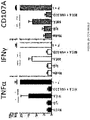
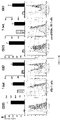
- Non-haematopoietic tissue-resident ⁇ T cells produced by the method of the invention may be distinguished from other blood-derived ⁇ T cells in that they respond to NKG2D ligand (MICA), which is strongly associated with malignancy, in the absence of any T cell receptor stimulating ligand, for example by increased production of TNF ⁇ , IFN ⁇ , and CD107a ( FIGS. 2A to 2D , 10A and 10B ). They also execute a cytotoxic T cell response without undergoing any exogenous pharmacological or ligand mediated activation of the T cell receptor and are therefore cytotoxic in the absence of stimulation ( FIG. 3 and FIG. 5 ).
- MICA NKG2D ligand
- the non-haematopoietic tissue-resident ⁇ T cells produced by the method of the invention are unique in their ability to respond and proliferate in the absence of addition of any exogenous agents activating T cell receptor signalling ( FIG. 3 ).
- the non-haematopoietic tissue-resident ⁇ T cells produced by the method of the invention also stained positive for CD69 and PD-1, lacked expression of CD28, and showed only low levels of CD25 (see FIG. 1D ). This combination of markers is not expressed by blood-derived ⁇ T cells.
- Non-haematopoietic tissue-resident ⁇ T cells produced by methods of the invention may be culturable in the presence of IL2 and/or IL15 without TCR agonists or other growth factors.
- a non-haematopoietic tissue-resident ⁇ T cell may grow in a medium consisting of RPMI 1640 medium supplemented with IL-2.
- a non-haematopoietic tissue-resident ⁇ T cell produced by the method of the invention may thus have one or more of the following properties:
- a non-haematopoietic tissue-resident ⁇ T cell produced by the method of the invention produces IL-13 in the absence of TCR agonists and/or produces IFN ⁇ in response to an NKG2D ligand in the absence of TCR agonists.
- the ⁇ T cells obtained by the method of the invention may be used in a method of screening for a checkpoint modulator of non-haematopoietic tissue resident ⁇ T cells.
- the identification of such checkpoint modulators may be useful for developing cancer immunotherapies, as the checkpoint modulator is a potential target for cancer therapy.
- non-haematopoietic tissue-resident ⁇ T cells may be cultured in vitro in direct contact with stromal or epithelial cells (e.g. fibroblasts) in the presence or absence of the test compound.
- stromal or epithelial cells e.g. fibroblasts
- the rate of proliferation or degree of activation of the non-haematopoietic tissue-resident ⁇ T cells is determined in the presence and absence of the test compound. If the rate of proliferation or degree of activation is higher in the presence of the test compound than in the absence of the test compound, then the test compound is likely to be a candidate checkpoint modulator. This is because the test compound is able to relieve contact inhibition by the stromal or epithelial cells (e.g. fibroblasts).
- test compound is chosen based on its potential to modulate a checkpoint for tissue-resident T cells.
- the apparent increase in non-haematopoietic tissue resident ⁇ T cell proliferation could also be due to inhibition of cell death and can be measured by an increase in T cell count or markers thereof and by reduced expression of markers of programmed cell death. Increased activation can be assessed by measuring release of cytokines such as IFN ⁇ which are secreted by activated tissue resident ⁇ T cells.
- non-haematopoietic tissue resident ⁇ T cells may be cultured in vitro in direct contact with stromal or epithelial cells (e.g. fibroblasts), wherein expression of a test gene within the ⁇ T cells and/or the stromal or epithelial cells (e.g. fibroblasts) or both the cell types is altered.
- stromal or epithelial cells e.g. fibroblasts
- expression of a test gene within the ⁇ T cells and/or in the stromal or epithelial cells may, for example, be altered by an RNA targeting agent, such as small interfering RNA (siRNA) or small hairpin RNA (shRNA) or by gene editing, e.g. using the CRISPR/Cas system.
- siRNA small interfering RNA
- shRNA small hairpin RNA
- the rate of proliferation or degree of activation of the non-haematopoietic tissue-resident ⁇ T cells is determined in the presence and absence of alteration of expression of the test gene in the stromal or epithelial cells (e.g. fibroblasts) and/or ⁇ T cells. If the rate of proliferation or activation is higher in the presence of alteration of expression of the test gene in stromal or epithelial cells (e.g. fibroblasts) and/or ⁇ T cells than in the absence of alteration of the test gene in stromal or epithelial cells (e.g. fibroblasts) and/or ⁇ T cells, then the test gene is likely to be a candidate checkpoint gene.
- the rate of killing of stromal or epithelial cells is higher in the presence of the test compound than in the absence of the test compound or higher in the presence of alteration of expression of the test gene in stromal or epithelial cells (e.g. fibroblasts) and/or ⁇ T cells than in the absence of alteration of the test gene in stromal or epithelial cells (e.g. fibroblasts) and/or ⁇ T cells, then the test compound is likely to be a checkpoint modulator or the test gene is likely to be a candidate checkpoint gene.
- the rate of killing may, for example, be measured by the quantitation of molecules released by dying cells.
- a higher rate of cell killing in the presence of the test compound or in the presence of alteration of expression of the test gene in stromal or epithelial cells (e.g. fibroblasts) and/or ⁇ T cells indicates that the checkpoint that was repressing cell killing has been relieved and one may therefore conclude that the test compound is likely to be a checkpoint modulator or the test gene is likely to be a candidate checkpoint gene.
- fibroblast cell lines which may be used in each embodiment include human foreskin fibroblasts (e.g. BJ (ATCC ® CRL-2522 TM )), normal skin fibroblasts (e.g. CCD-1059Sk (ATCC ® CRL-2072 TM )) and lung fibroblasts (e.g. HEL 299 (ATCC ® CRL-137 TM )).
- human foreskin fibroblasts e.g. BJ (ATCC ® CRL-2522 TM )
- normal skin fibroblasts e.g. CCD-1059Sk (ATCC ® CRL-2072 TM )
- lung fibroblasts e.g. HEL 299 (ATCC ® CRL-137 TM )
- the rate of proliferation and or activation of the ⁇ T cells in the presence of the test compound or in the presence of alteration of the test gene may be at least 1.5 times, at least 2 times, at least 3 times, at least 4 times or at least 5 times higher than in the absence of the test compound or in the absence of alteration of the test gene.
- Cell cycling can be measured by a number of means, such as absolute cell numbers on day 0 and day 7, levels of Ki-67 and CD25 expression (which are cell cycling markers) and using cell culture dyes, such CFSE or CELLTRACE TM violet.
- Cell activation can be measured by the production of effector proteins such as IFN- ⁇ .
- the apparent increase in proliferation of non-haematopoietic tissue-resident ⁇ T cells could also be due to inhibition of cell death and can be measured by an increase in T cell count or markers thereof and by reduced expression of markers of programmed cell death.
- the ⁇ T cells obtained by the method of the invention may be used as a medicament, for example for adoptive T cell therapy.
- the therapy may be autologous, i.e., the ⁇ T cells may be transferred back into the same patient from which they were obtained, or the therapy may be allogeneic, i.e. the ⁇ T cells from one person may be transferred into a different patient.
- a method of treatment may comprise;
- the donor individual and the recipient individual may be the same or different.
- the ⁇ T cells may be administered to the patient or subject in need of treatment by any suitable method.
- the ⁇ T cells may be administered to the patient or subject in need of treatment intravenously or intra-tumourally.
- the patient or subject to be treated is preferably a human cancer patient or a virus-infected patient, e.g., a CMV-infected or HIV infected patient.
- ⁇ T cells are non-MHC restricted, they do not recognise a host into which they are transferred as foreign, which means that they are less likely to cause graft-versus-host disease. This means that they can be used "off the shelf” and transferred into any recipient, e.g., for allogeneic adoptive T cell therapy.
- tissue-resident V ⁇ 1 T and DN ⁇ T cells are also more likely to home to and be retained within tumour masses than their systemic blood-resident counterparts and adoptive transfer of these cells is likely to be more effective at targeting solid tumours and potentially other non-haematopoietic tissue-associated immunopathologies.
- a method of treatment of an individual with a tumour in a non-haematopoietic tissue may comprise;
- Non-haematopoietic tissue-resident ⁇ T cells obtained by methods of the invention express NKG2D and respond to a NKG2D ligand (e.g. MICA), which is strongly associated with malignancy. They also express a cytotoxic profile in the absence of any activation and are therefore likely to be effective at killing tumour cells.
- the non-haematopoietic tissue-resident ⁇ T cells obtained as described herein may express one or more, preferably all of IFN- ⁇ , TNF- ⁇ , GM-CSF, CCL4, IL-13, Granulysin, Granzyme A and B, and Perforin in the absence of any activation.
- IL-17A may not be expressed.
- the non-haematopoietic tissue-resident ⁇ T cells obtained by the method of the invention may also be used for CAR-T therapy.
- the engineered TCR may make the T cells specific for malignant cells and therefore useful for cancer immunotherapy.
- the T cells may recognise cancer cells expressing a tumour antigen, such as a tumour associated antigen (TAA) that is not expressed by normal somatic cells from the subject tissue.
- TAA tumour associated antigen
- the CAR-modified T cells may be used for adoptive T cell therapy of, for example, cancer patients.
- non-haematopoietic tissue-resident ⁇ T cells obtained by the method of the invention are likely to be particularly good vehicles for CAR-T approaches, as they can be transduced with chimeric antigen-specific TCRs while retaining their innate-like capabilities of recognising transformed cells, and are likely to have better tumour penetration and retention capabilities than either blood-resident ⁇ T cells or conventional, systemic ⁇ T cells.
- their lack of MHC dependent antigen presentation reduces the potential for graft-versus-host disease and permits them to target tumours expressing low levels of MHC.
- their non-reliance upon conventional co-stimulation, for example via engagement of CD28 enhances the targeting of tumours expressing low levels of ligands for co-stimulatory receptors.
- Cancer may be characterised by the abnormal proliferation of malignant cancer cells and may include leukaemias, such as acute myeloid leukaemia (AML), chronic myeloid leukaemia (CML), acute lymphoblastic leukaemia (ALL) and chronic lymphocytic leukaemia (CLL), lymphomas, such as Hodgkin lymphoma, non-Hodgkin lymphoma and multiple myeloma, and solid cancers such as sarcomas, skin cancer, melanoma, bladder cancer, brain cancer, breast cancer, uterus cancer, ovary cancer, prostate cancer, lung cancer, colorectal cancer, cervical cancer, liver cancer, head and neck cancer, oesophageal cancer, pancreas cancer, renal cancer, adrenal cancer, stomach cancer, testicular cancer, cancer of the gall bladder and biliary tracts, thyroid cancer, thymus cancer, cancer of bone, and cerebral cancer.
- leukaemias such as acute myeloid leukaemia (AML),
- Cancer cells within a cancer patient may be immunologically distinct from normal somatic cells in the individual (i.e. the cancerous tumour may be immunogenic).
- the cancer cells may be capable of eliciting a systemic immune response in the cancer patient against one or more antigens expressed by the cancer cells.
- the antigens that elicit the immune response may be tumour antigens or may be shared by normal cells.
- a patient with cancer may display at least one identifiable sign, symptom, or laboratory finding that is sufficient to make a diagnosis of cancer in accordance with clinical standards known in the art. Examples of such clinical standards can be found in textbooks of medicine such as Harrison's Principles of Internal Medicine (40).
- a diagnosis of a cancer in an individual may include identification of a particular cell type (e.g. a cancer cell) in a sample of a body fluid or tissue obtained from the individual.
- a patient, subject, or individual suitable for treatment as described above may be a mammal, such as a rodent (e.g. a guinea pig, a hamster, a rat, a mouse), murine (e.g. a mouse), canine (e.g. a dog), feline (e.g. a cat), equine (e.g. a horse), a primate, simian (e.g. a monkey or ape), a monkey (e.g. a marmoset or baboon), an ape (e.g. a gorilla, chimpanzee, orang-utan or gibbon), or a human.
- a rodent e.g. a guinea pig, a hamster, a rat, a mouse
- murine e.g. a mouse
- canine e.g. a dog
- feline e.g. a cat
- the patient, subject, or individual is a human.
- non-human mammals especially mammals that are conventionally used as models for demonstrating therapeutic efficacy in humans (e.g. murine, primate, porcine, canine, or rabbit) may be employed.
- the patient, subject, or individual may have minimal residual disease (MRD) after an initial cancer treatment.
- MRD minimal residual disease
- Treatment may be any treatment and therapy, whether of a human or an animal (e.g. in veterinary applications), in which some desired therapeutic effect is achieved, for example, the inhibition or delay of the progress of the condition, and includes a reduction in the rate of progress, a halt in the rate of progress, amelioration of the condition, cure or remission (whether partial or total) of the condition, preventing, delaying, abating or arresting one or more symptoms and/or signs of the condition or prolonging survival of a subject or patient beyond that expected in the absence of treatment.
- some desired therapeutic effect is achieved, for example, the inhibition or delay of the progress of the condition, and includes a reduction in the rate of progress, a halt in the rate of progress, amelioration of the condition, cure or remission (whether partial or total) of the condition, preventing, delaying, abating or arresting one or more symptoms and/or signs of the condition or prolonging survival of a subject or patient beyond that expected in the absence of treatment.
- Treatment as a prophylactic measure is also included.
- a patient, subject, or individual susceptible to or at risk of the occurrence or re-occurrence of cancer may be treated as described herein. Such treatment may prevent or delay the occurrence or re-occurrence of cancer in the patient, subject, or individual.
- treatment may include inhibiting cancer growth, including complete cancer remission, and/or inhibiting cancer metastasis.
- Cancer growth generally refers to any one of a number of indices that indicate change within the cancer to a more developed form.
- indices for measuring an inhibition of cancer growth include a decrease in cancer cell survival, a decrease in tumor volume or morphology (for example, as determined using computed tomographic (CT), sonography, or other imaging method), a delayed tumor growth, a destruction of tumor vasculature, improved performance in delayed hypersensitivity skin test, an increase in the activity of cytolytic T-lymphocytes, and a decrease in levels of tumor-specific antigens.
- Reducing immune suppression in cancerous tumors in an individual may improve the capacity of the individual to resist cancer growth, in particular growth of a cancer already present the subject and/or decrease the propensity for cancer growth in the individual.
- a three-dimensional skin explant protocol was established, as described elsewhere (29).
- the 9 mm x 9 mm x 1.5 mm Cellfoam Matrices (Cytomatrix Pty Ltd, Victoria, Australia) were autoclaved, then incubated in a solution of 100 mg/ml rat tail collagen I (BD Biosciences) in PBS for 30 minutes at room temperature, followed by one rinse in PBS.
- Samples of adult human skin were obtained within 3-6 hours of cutaneous surgery. Subcutaneous fat was removed and the remaining skin tissue was minced into fragments measuring approximately 1 mm ⁇ 1 mm. Approximately five skin fragments/explants were placed and pressed down onto the surface of each matrix.
- 'Skin-T' media Iscove's Modified Dulbecco's Medium (IMDM; Life Technologies) with 10% heat-inactivated foetal bovine serum (Life Technologies), L-glutamine (292 ⁇ g/ml; Life Technologies), penicillin (100 units/ml; Life Technologies), streptomycin (100 ⁇ g/ml; Life Technologies), N-2-hydroxyethylpiperazine-N-2-ethane sulfonic acid (HEPES; 0.01 M; Life Technologies), sodium pyruvate (1mM; Life Technologies), minimal essential media (MEM) non-essential amino acids (1X; Life Technologies) and 3.5 ⁇ l/L 2-mercaptoethanol (Life Technologies).
- IMDM Iscove's Modified Dulbecco's Medium
- L-glutamine 292 ⁇ g/ml
- penicillin 100 units/ml
- streptomycin 100 ⁇ g/ml
- HEPES N-2-hydroxyethylpiperazine-N-2-
- the matrices were transferred to a 50ml centrifuge tube (Corning) containing 10 ml Hanks Balanced Salt Solution (HBSS; Life Technologies) with 0.01 mM HEPES (up to 12 matrices/tube).
- the matrices were rinsed with the cell suspension using a 10 ml pipette, and the cell suspension passed through a 70- ⁇ m filter (BD Biosciences) into a fresh 50ml centrifuge tube (Corning).
- the 'washing' of the matrices was repeated two further times.
- the media from the culture well was also aspirated and passed through a 70- ⁇ m filter (BD Biosciences) into fresh 50ml centrifuge tube (Corning).
- IL-2 100IU/ml
- IL-15 20ng/ml
- Cells were seeded at 2 ⁇ 10 5 /well into 96 well flat bottom plates (Corning) or at 2 ⁇ 10 6 /well into 12 well plates (Corning) for expansion. Cells were monitored daily by microscopy, fed with fresh media and cytokines added 3 times a week. Upon full confluence and cell aggregates being visible, cells were split 1:1 into additional wells and plates, respectively. Cells were harvested and analysed using flow cytometry or used for functional assays after 7, 14 or 21 days depending on the assay.
- ⁇ T cells were re-suspended in 1ml FACS buffer (PBS containing 2% heat-inactivated foetal bovine serum and 0.01M EDTA) and stained for the ⁇ T cell receptor (Biolegend, clone IP26, 1:50) for 30 minutes in the dark on ice and all negative cells sorted using an Aria sorter running DIVA (BD Biosciences).
- FACS buffer PBS containing 2% heat-inactivated foetal bovine serum and 0.01M EDTA
- fibroblasts reached confluence and co culture experiments were started using RPMI and the cytokines indicated adding 2 ⁇ 10 5 mixed skin lymphocytes in the case of 48 well plates, or 3 ⁇ 10 5 lymphocytes in the case of 24 well plates, bottom wells as well as transwells.
- Flow cytometry was performed using the following antibodies, coupled to the indicated fluorochromes: Ki-67-BV421, CD3-BV510, V ⁇ 1-PeVio770, TIM-3-PE, CD9-PE, CCR3-BV421, CD39-BV421. All samples were also always stained for viability, using eFluor770NIR. Commercial antibodies were purchased from Biolegend or Miltenyi. Viability dye (near IR) was from eBioscience. Ki-67 staining was performed on cells fixed and permeabilised using the Foxp3 staining buffer set (eBioscience). Once each experiment was finished, the cell population was washed in PBS and split in half.
- V ⁇ 1 T cells from human skin and human blood V ⁇ 1 T cells were sorted (FACS), centrifuged and the cell pellet re-suspended in RLT buffer.
- RNA was prepared using the RNA-Micro-plus kit (QIAGEN). RNA libraries were generated using the KAPA Stranded RNA-seq Kit with RiboErase (HMR) (KAPA BIOSYSTEMS). Paired-end sequencing on HiSeq 2500 (illumina) using rapid run chemistry (read length: 100bp). 101 base-pair paired-end reads were aligned and quantified using RSEM (v1.2.11) with Bowtie2. Reads were aligned to the human transcriptome, the count values have been log2 transformed and quantile normalised.
- V ⁇ 1 T cells from human skin were stimulated with PMA and lonomycin or plate bound anti-CD3 mAb (OKT3, 5 ⁇ g/ml) for 24 hours. Supernatants were taken afterwards and analysed using ProcartaPlex Human Cytokine & Chemokine Panel 1A (34 plex) from eBioscience. Assays were analysed using a Luminex FlexMap3D (Luminex Corp). Data was analysed in Microsoft Excel, the mean of 3 donors (run in duplicates) is shown. Error bars indicate standard deviation.
- Blood derived ⁇ T cells within PBMCs can only be expanded if stimulated with TCR ligands (in the case of V ⁇ 2, e.g. IPP, HMBPP, bisphosphonates) (41, 42) or antibody supplementation to either cross-link the TCR receptor (mAbs) or the TCR associated kinase CD3 (43).
- TCR ligands in the case of V ⁇ 2, e.g. IPP, HMBPP, bisphosphonates
- mAbs TCR receptor
- TCR associated kinase CD3 43
- the same effect of TCR cross-linking can also be achieved using, lectins such as PHA.
- the ⁇ T cells in PBMCs survive for several days but fail to expand and remain in their initial composition of T cell subsets with minor variations.
- Blood from healthy volunteers was used to isolate PBMCs by layering whole blood onto Ficoll followed by centrifugation at 400g for 20 minutes to separate red blood cells, blood plasma and white lymohocytes/monocytes. White blood cells were carefully harvested through a stripett and washed four times in cold PBS.
- ⁇ TCR monoclonal antibody (20 ⁇ g/ml, clone B1, Biolegend) 90 minutes prior to cell transfer.
- Cells were grown for 14 days, media changed ever 2-3 days and fresh cytokines added. Upon reaching confluence, cells were split 1:1. Under these conditions, after 14 days, the original minor population of ⁇ T cells is normally highly activated through their TCR (as indicated by upregulation if CD69 and CD25) and largely enriched consisting of mainly V ⁇ 2 T cells but also V ⁇ 1 T cells (up to 30% of all ⁇ T cells).
- V ⁇ 1 T cells can subsequently be isolated using FACS for functional, phenotypic or genetic analysis.
- Human ⁇ T cells are abundant in the skin, express a non-V ⁇ 2 TCR and participate in the human lymphoid stress surveillance response
- FIGS 1A and 3D we found a substantial population of ⁇ T cells (mean 8.513% of CD45+ cells, ⁇ 6.564%) in our donors ( FIGS 1A and 3D ).
- This subset representation of lymphocytes after organotypic culture was highly reproducible in approximately 100 donors, and was comparable with freshly digested skin samples, differing only in a slightly increased ⁇ population, but of practical utility, offering much larger and purer lymphocyte populations compared to standard tissue digestion protocols.
- tissue compartmentalisation of human ⁇ T cells based on their TCR delta chain, most human skin ⁇ T cells expressed a V ⁇ 1 TCR chain paired with various ⁇ chains identified by flow cytometry.
- Skin-resident ⁇ T cells grown in this fashion showed a non-terminally differentiated memory phenotype lacking expression of CD45RA and expressing variable levels of the co-stimulatory molecule CCR7.
- strong expression of the surface protein CD69 together with the expression of programmed death receptor 1 (PD-1); low to absent levels of IL-2 receptor ⁇ (CD25); and a lack of the co-stimulatory molecule CD28 draw the picture of previously activated or chronically activated T cells ( FIG. 1D ).
- V ⁇ 1 and DN cells show expression of skin and tissue homing markers, such as CLA, CCR4, CCR8 and integrin ⁇ E (CD103) (see FIG. 7 ).
- tissue-homing marker-set might conceivably prove beneficial in an immunotherapy setting.
- skin-resident ⁇ T cells show high levels of expression for the activatory receptor NKG2D ( FIG. 2A ), implying a possible role of these cells in the lymphoid stress surveillance response.
- NKG2D ligands such as MICA, MICB and ULBPs, respectively, are up-regulated by cells in response to DNA damage, EGF-receptor activation and oxidative stress and may therefore allow T cells expressing NKG2D to identify and eradicate stressed or transformed cells, thereby maintaining tissue homeostasis.
- activated skin-resident V ⁇ 1+ and DN ⁇ T cells executed a pro-inflammatory TH1 biased cytokine program (staining positive for IFN ⁇ , TNF- ⁇ and GM-CSF) when activated by either PMA/ionomycin or by NKG2D ligands, e.g. recombinant MICA protein ( FIGS. 2A and 2B ), thereby asserting the cells' innate-like responses. Indeed, the responses to MICA were almost completely abrogated by blocking the NKG2D receptor by means of an antibody ( FIGS. 2B and 2C ).
- ⁇ T cells are known to secrete IL-17 under certain disease settings such as psoriasis and within some types of tumours.
- ⁇ T cells expanded by the method of the invention produce low level or no IL-17 even upon extensive activation ( FIGS. 2B and 8 ).
- tissue-resident CD4-expressing ⁇ T cells did produce IL-17 upon TCR activation ( FIG. 2B ).
- ⁇ T cells showed a much more diverse cytokine repertoire in response to PMA/ionomycin compared to V ⁇ 1+ and DN ⁇ T cells, which were limited to a TH1 biased programme associated with host-protection.
- V ⁇ 1 T cells In order to further study human tissue, ⁇ T cells and their biology, we transferred mixed skin lymphocytes into cell culture wells and supplemented them with IL-2 in order to maintain viability over time. Interestingly, we quickly found that by segregation away from stromal and epithelial cells present in the organotypic culture, V ⁇ 1 T cells uniquely showed signs of activation and proliferation without any other added stimulus. Within fewer than 7 days, V ⁇ 1+ and DN ⁇ T cells uniquely and massively up regulated the nuclear factor Ki-67 and increased surface expression of IL-2 receptor ⁇ (CD25) ( FIGS. 3B and 4B ).
- MFI of the cell cycling-associated nuclear factor KI-67 increased from 2664.5( ⁇ 1876.1) to 8457.7( ⁇ 4574.2) in 14 days in V ⁇ 1+ and DN ⁇ T cells, whereas in ⁇ T cells the MFI decreased from 592.8( ⁇ 390.5) to 284.7( ⁇ 140.1) over the same time course ( FIG. 3C ).
- This phenomenon of selective, skin-resident ⁇ T cell outgrowth could be further supported using additional recombinant IL-15, which increased the viability and total number of lymphocytes.
- V ⁇ 1+ and DN ⁇ T cells never occurred in organotypic culture systems in which there was extensive fibroblast outgrowth. We therefore grew out autologous fibroblasts in order to test directly whether their co-culture with V ⁇ 1+ and DN ⁇ T cells would inhibit the T cells' expansion.
- transwells that prevented the T lymphocytes from directly contacting the fibroblasts within the same wells, but permitted them to be influenced by any soluble factors made by the fibroblasts.
- V ⁇ 1+ and DN ⁇ T cells started proliferating in wells in which there were no fibroblasts and in those in which the T cells were prevented from directly contacting the fibroblasts.
- ⁇ T cell proliferation was low under all conditions.
- the growth rate over two weeks of V ⁇ 1+ and DN ⁇ T cells was considerably reduced, from 22.6 (SEM 8.07) fold in wells without fibroblast contact to 3.3 (SEM 0.17) fold ( FIG.
- the phenotype of skin-resident V ⁇ 1+ and DN ⁇ T cells and their striking functional potentials are reflective of pre-activated T cells that are ordinarily kept in check by their neighbouring dermal fibroblasts via a contact-dependent mechanism that is yet to be characterised. Inactivating that mechanism by releasing the T cells from contact with the fibroblasts selectively permits the striking expansion of V ⁇ 1+ and DN ⁇ T cells, whereas other T cells in the skin are unaffected.
- ⁇ T cells wholly unexpectedly, expanding ⁇ T cells (provoked only by their separation from fibroblasts) spontaneously produced large amounts of TH1 associated cytokines such as IFN- ⁇ (12,383.46 ⁇ 16,618.90 pg/ml), GM-CSF (4,316.73 ⁇ 4,534.96 pg/ml) and the proinflammatory chemokines CCL4 (14,877.34 ⁇ 10,935.64 pg/ml) and CCL3 (1,303.07 ⁇ 757.23 pg/ml) ( FIG. 5A ).
- IFN- ⁇ (12,383.46 ⁇ 16,618.90 pg/ml
- GM-CSF 4,316.73 ⁇ 4,534.96 pg/ml
- proinflammatory chemokines CCL4 (14,877.34 ⁇ 10,935.64 pg/ml) and CCL3 (1,303.07 ⁇ 757.23 pg/ml)
- the cells produced large amounts of spontaneous IL-13, associated with atopic responses, during expansion and in contrast to freshly isolated skin-derived, TCR-activated ⁇ T cells; other cytokines were produced at much lower levels or not at all e.g. IL-17A ( FIG. 8 ).
- the high effector potentials of the cells could be increased further following stimulation with recombinant MICA (NKG2D ligand), with anti CD3 cross-linking, or with PMA/lonomycin.
- V ⁇ 1+ and DN ⁇ T cells showed very high cytotoxic activity towards Hela cells (cervix) and Caco2 (colon), in a dose-dependent fashion, far superior to conventional tissue ⁇ T cells ( FIG. 5B ). Furthermore, ⁇ -cell mediated cytotoxicity could be strongly inhibited by blocking the NKG2D receptor using soluble monoclonal antibodies indicating that this receptor is at least partly involved in tumour surveillance by derepressed human skin-derived ⁇ T cells. We furthermore confirmed the cytotoxic potential of these cells using other targets: HCT1954, MDAMB231 (both breast carcinoma) and HCT116 (colon) ( FIG. 9 ).
- tissue derived V ⁇ 1 T cells may be well equipped to be use as an adjuvant to monoclonal antibody therapies such as CD20 or Her2 therapies as they will be guided by the antibody to sides of malignancies and metastasis, recognize opsonized tumour cells and kill targets via ADCC.
- monoclonal antibody therapies such as CD20 or Her2 therapies as they will be guided by the antibody to sides of malignancies and metastasis, recognize opsonized tumour cells and kill targets via ADCC.
Landscapes
- Health & Medical Sciences (AREA)
- Life Sciences & Earth Sciences (AREA)
- Engineering & Computer Science (AREA)
- General Health & Medical Sciences (AREA)
- Chemical & Material Sciences (AREA)
- Immunology (AREA)
- Organic Chemistry (AREA)
- Virology (AREA)
- Veterinary Medicine (AREA)
- Public Health (AREA)
- Animal Behavior & Ethology (AREA)
- Biomedical Technology (AREA)
- Biotechnology (AREA)
- Medicinal Chemistry (AREA)
- Pharmacology & Pharmacy (AREA)
- Nuclear Medicine, Radiotherapy & Molecular Imaging (AREA)
- General Chemical & Material Sciences (AREA)
- Chemical Kinetics & Catalysis (AREA)
- Cell Biology (AREA)
- Bioinformatics & Cheminformatics (AREA)
- Zoology (AREA)
- Wood Science & Technology (AREA)
- Genetics & Genomics (AREA)
- Molecular Biology (AREA)
- Oncology (AREA)
- Communicable Diseases (AREA)
- Hematology (AREA)
- Epidemiology (AREA)
- Biochemistry (AREA)
- Microbiology (AREA)
- Tropical Medicine & Parasitology (AREA)
- General Engineering & Computer Science (AREA)
- AIDS & HIV (AREA)
- Urology & Nephrology (AREA)
- Physics & Mathematics (AREA)
- Toxicology (AREA)
- Food Science & Technology (AREA)
- Developmental Biology & Embryology (AREA)
- Analytical Chemistry (AREA)
- General Physics & Mathematics (AREA)
Priority Applications (1)
| Application Number | Priority Date | Filing Date | Title |
|---|---|---|---|
| EP22182997.1A EP4148124A1 (en) | 2015-10-30 | 2016-10-31 | Expansion of non-haematopoietic tissue-resident gamma delta t cells and uses of these cells |
Applications Claiming Priority (3)
| Application Number | Priority Date | Filing Date | Title |
|---|---|---|---|
| GBGB1519198.4A GB201519198D0 (en) | 2015-10-30 | 2015-10-30 | Expansion of gamma delta t cells resident in non-haematopoietic tissue and uses of these cells |
| GBGB1612731.8A GB201612731D0 (en) | 2016-07-22 | 2016-07-22 | Expansion of non-haematopoietic tissue-resident gamma delta t cells and uses of these cells |
| PCT/EP2016/076264 WO2017072367A1 (en) | 2015-10-30 | 2016-10-31 | EXPANSION OF NON-HAEMATOPOIETIC TISSUE-RESIDENT γδ T CELLS AND USES OF THESE CELLS |
Related Child Applications (1)
| Application Number | Title | Priority Date | Filing Date |
|---|---|---|---|
| EP22182997.1A Division EP4148124A1 (en) | 2015-10-30 | 2016-10-31 | Expansion of non-haematopoietic tissue-resident gamma delta t cells and uses of these cells |
Publications (2)
| Publication Number | Publication Date |
|---|---|
| EP3368658A1 EP3368658A1 (en) | 2018-09-05 |
| EP3368658B1 true EP3368658B1 (en) | 2022-07-06 |
Family
ID=57211532
Family Applications (2)
| Application Number | Title | Priority Date | Filing Date |
|---|---|---|---|
| EP16788539.1A Active EP3368658B1 (en) | 2015-10-30 | 2016-10-31 | Expansion of non-haematopoietic tissue-resident gamma delta t cells and uses of these cells |
| EP22182997.1A Pending EP4148124A1 (en) | 2015-10-30 | 2016-10-31 | Expansion of non-haematopoietic tissue-resident gamma delta t cells and uses of these cells |
Family Applications After (1)
| Application Number | Title | Priority Date | Filing Date |
|---|---|---|---|
| EP22182997.1A Pending EP4148124A1 (en) | 2015-10-30 | 2016-10-31 | Expansion of non-haematopoietic tissue-resident gamma delta t cells and uses of these cells |
Country Status (16)
| Country | Link |
|---|---|
| US (2) | US11618885B2 (enExample) |
| EP (2) | EP3368658B1 (enExample) |
| JP (3) | JP2018533373A (enExample) |
| KR (2) | KR20250093576A (enExample) |
| CN (1) | CN108473958A (enExample) |
| AU (2) | AU2016344779B2 (enExample) |
| CA (1) | CA3003077A1 (enExample) |
| DK (1) | DK3368658T3 (enExample) |
| ES (1) | ES2925261T3 (enExample) |
| IL (3) | IL295838B2 (enExample) |
| MX (2) | MX2018005207A (enExample) |
| MY (1) | MY198084A (enExample) |
| PH (1) | PH12018500926A1 (enExample) |
| PL (1) | PL3368658T3 (enExample) |
| PT (1) | PT3368658T (enExample) |
| WO (1) | WO2017072367A1 (enExample) |
Families Citing this family (33)
| Publication number | Priority date | Publication date | Assignee | Title |
|---|---|---|---|---|
| GB201707048D0 (en) | 2017-05-03 | 2017-06-14 | King S College London | Expansion of gamma delta cells, compositions, and methods of use thereof |
| WO2018229163A1 (en) | 2017-06-14 | 2018-12-20 | King's College London | Methods of activating v delta 2 negative gamma delta t cells |
| EP3681996B1 (en) | 2017-09-15 | 2024-08-07 | King's College London | Compositions and methods for enhancing gamma delta t cells in the gut |
| CN109777778B (zh) * | 2017-11-14 | 2023-07-18 | 中国科学院广州生物医药与健康研究院 | 一种基因改造的γδT细胞 |
| GB201804701D0 (en) | 2018-03-23 | 2018-05-09 | Gammadelta Therapeutics Ltd | Lymphocytes expressing heterologous targeting constructs |
| WO2020027193A1 (ja) * | 2018-07-31 | 2020-02-06 | 国立大学法人三重大学 | γδ型T細胞への遺伝子導入方法 |
| GB201818243D0 (en) | 2018-11-08 | 2018-12-26 | Gammadelta Therapeutics Ltd | Methods for isolating and expanding cells |
| CN113423820A (zh) * | 2018-11-08 | 2021-09-21 | 伽马三角洲疗法有限公司 | 分离和扩增细胞的方法 |
| PH12021551475A1 (en) | 2019-01-07 | 2022-05-02 | Shattuck Labs Inc | Heterodimeric proteins for modulating gamma delta t cells |
| US11098093B2 (en) | 2019-01-07 | 2021-08-24 | Shattuck Labs, Inc. | Heterodimeric proteins for modulating gamma delta T cells |
| CN112029719B (zh) * | 2019-06-04 | 2024-05-03 | 北京医明佳和生物科技有限公司 | 纯化的双阴性t细胞及其制备和应用 |
| WO2021016652A1 (en) * | 2019-07-29 | 2021-02-04 | The University Of Melbourne | METHODS AND COMPOSITIONS FOR MONITORING, TREATING AND PREVENTING CMV INFECTION OR GRAFT REJECTION USING γδ T-CELLS |
| JP2022545196A (ja) | 2019-08-16 | 2022-10-26 | ガンマデルタ セラピューティクス リミテッド | 抗tcrデルタ可変1抗体の治療的使用 |
| JP7764364B2 (ja) | 2019-08-16 | 2025-11-05 | ガンマデルタ セラピューティクス リミティッド | エクスビボγδ T細胞集団 |
| CN115151565A (zh) | 2019-10-31 | 2022-10-04 | 莫佛塞斯公司 | 包含抗CD19抗体和γδT细胞的抗肿瘤组合疗法 |
| EP4114415A1 (en) | 2020-03-06 | 2023-01-11 | Sorrento Therapeutics, Inc. | Innate immunity killer cells targeting psma positive tumor cells |
| KR20230013022A (ko) | 2020-03-20 | 2023-01-26 | 감마델타 테라퓨틱스 엘티디 | 골수성 악성종양의 치료를 위한 v 델타1+ t 세포 |
| GB202006989D0 (en) * | 2020-05-12 | 2020-06-24 | Gammadelta Therapeutics Ltd | Methods for isolating gamma delta t cells |
| PL4269444T3 (pl) | 2020-08-14 | 2025-01-13 | GammaDelta Therapeutics Limited | Wieloswoiste przeciwciała anty-tcr zmienne delta 1 |
| GB202013962D0 (en) | 2020-09-04 | 2020-10-21 | Gammadelta Therapeutics Ltd | Immunotherapy composition |
| AU2022224391A1 (en) | 2021-02-17 | 2023-08-24 | Gammadelta Therapeutics Ltd | Anti-tcr delta variable 1 antibodies |
| GB202105113D0 (en) | 2021-04-09 | 2021-05-26 | Gammadelta Therapeutics Ltd | Novel method |
| CA3228168A1 (en) | 2021-08-03 | 2023-02-09 | Gammadelta Therapeutics Ltd | Engineering of gamma delta t cells and compositions thereof |
| WO2023053994A1 (ja) * | 2021-09-29 | 2023-04-06 | アイ ピース, インコーポレイテッド | 幹細胞の製造方法 |
| CA3208653A1 (en) | 2022-02-17 | 2022-08-25 | Mihriban Tuna | Multispecific anti-tcr delta variable 1 antibodies |
| JPWO2023176896A1 (enExample) * | 2022-03-18 | 2023-09-21 | ||
| EP4504247A1 (en) | 2022-04-04 | 2025-02-12 | Gammadelta Therapeutics Ltd | Novel gene armoring |
| WO2024209429A1 (en) | 2023-04-07 | 2024-10-10 | Takeda Pharmaceutical Company Limited | T cells for use in treating relapsed or refractory acute myeloid leukemia |
| CN121532496A (zh) | 2023-07-20 | 2026-02-13 | 武田药品工业株式会社 | 用于制备γδT细胞的无网格方法 |
| CN118165933B (zh) * | 2024-05-14 | 2024-07-16 | 成都美杰赛尔生物科技有限公司 | 组合物、提高cd39-cd69-t细胞比例的方法 |
| WO2026013159A1 (en) | 2024-07-10 | 2026-01-15 | Cytospire Therapeutics Limited | Antibodies binding to constant domains of gamma delta t-cell receptors |
| WO2026013158A1 (en) | 2024-07-10 | 2026-01-15 | Cytospire Therapeutics Limited | Antibodies binding to constant domains of gamma delta t-cell receptors |
| CN118853566B (zh) * | 2024-09-26 | 2025-05-09 | 青岛华赛伯曼医学细胞生物有限公司 | 一种肿瘤浸润淋巴细胞的培养方法及培养基 |
Family Cites Families (12)
| Publication number | Priority date | Publication date | Assignee | Title |
|---|---|---|---|---|
| ATE421573T1 (de) | 1998-11-04 | 2009-02-15 | Therapure Biopharma Inc | Verfahren zur herstellung von gamma delta t zellen |
| US20060246520A1 (en) * | 2003-05-23 | 2006-11-02 | Institut National De La Sante Et De La Recherche Medicale | Gamma delta t cell-mediated therapy |
| EP1777294A1 (en) | 2005-10-20 | 2007-04-25 | Institut National De La Sante Et De La Recherche Medicale (Inserm) | IL-15Ralpha sushi domain as a selective and potent enhancer of IL-15 action through IL-15Rbeta/gamma, and hyperagonist (IL15Ralpha sushi -IL15) fusion proteins |
| WO2009131730A2 (en) * | 2008-01-31 | 2009-10-29 | University Of Iowa Research Foundation | IMMUNOGENIC COMPOSITIONS FOR ACTIVATING γδ T CELLS |
| CA2860170C (en) | 2010-12-22 | 2022-06-14 | The Board Of Trustees Of The Leland Stanford Junior University | Superagonists and antagonists of interleukin-2 |
| PT2710123T (pt) * | 2011-05-19 | 2017-10-13 | Inst De Medicina Molecular | Linha celular de linfócitos compreendendo células t gama-delta, método de composição e produção resultante dos mesmos |
| EP3060059A4 (en) | 2013-10-25 | 2017-11-01 | Board of Regents, The University of Texas System | Polyclonal gamma delta t cells for immunotherapy |
| RU2739770C2 (ru) * | 2014-06-11 | 2020-12-28 | полибайосепт ГмбХ | Экспансия лимфоцитов с использованием композиции цитокинов для активной клеточной иммунотерапии |
| CN104293734B (zh) * | 2014-09-28 | 2018-01-09 | 上海云舜生物技术有限公司 | 一种人γδT细胞的制备方法 |
| CN104711224A (zh) * | 2015-01-09 | 2015-06-17 | 天津大学 | 一种提高人Vδ2T细胞扩增效率的体外培养方法及应用 |
| AU2016274633B2 (en) * | 2015-06-09 | 2022-04-21 | Gammadelta Therapeutics Ltd | Methods for the production of TCR gamma delta+ T cells |
| WO2016198906A1 (fr) * | 2015-06-10 | 2016-12-15 | Arcelormittal | Acier a haute résistance et procédé de fabrication |
-
2016
- 2016-10-31 WO PCT/EP2016/076264 patent/WO2017072367A1/en not_active Ceased
- 2016-10-31 KR KR1020257019399A patent/KR20250093576A/ko active Pending
- 2016-10-31 IL IL295838A patent/IL295838B2/en unknown
- 2016-10-31 PT PT167885391T patent/PT3368658T/pt unknown
- 2016-10-31 US US15/771,875 patent/US11618885B2/en active Active
- 2016-10-31 IL IL315329A patent/IL315329A/en unknown
- 2016-10-31 MY MYPI2018000604A patent/MY198084A/en unknown
- 2016-10-31 CN CN201680074800.5A patent/CN108473958A/zh active Pending
- 2016-10-31 DK DK16788539.1T patent/DK3368658T3/da active
- 2016-10-31 CA CA3003077A patent/CA3003077A1/en active Pending
- 2016-10-31 KR KR1020187015244A patent/KR102821381B1/ko active Active
- 2016-10-31 PL PL16788539.1T patent/PL3368658T3/pl unknown
- 2016-10-31 MX MX2018005207A patent/MX2018005207A/es unknown
- 2016-10-31 EP EP16788539.1A patent/EP3368658B1/en active Active
- 2016-10-31 EP EP22182997.1A patent/EP4148124A1/en active Pending
- 2016-10-31 JP JP2018541524A patent/JP2018533373A/ja active Pending
- 2016-10-31 AU AU2016344779A patent/AU2016344779B2/en active Active
- 2016-10-31 ES ES16788539T patent/ES2925261T3/es active Active
-
2018
- 2018-04-26 IL IL258985A patent/IL258985B2/en unknown
- 2018-04-26 MX MX2022011003A patent/MX2022011003A/es unknown
- 2018-04-30 PH PH12018500926A patent/PH12018500926A1/en unknown
-
2021
- 2021-09-10 JP JP2021147372A patent/JP7451468B2/ja active Active
-
2022
- 2022-09-29 US US17/936,811 patent/US20230099491A1/en active Pending
- 2022-12-09 AU AU2022283768A patent/AU2022283768A1/en active Pending
-
2024
- 2024-03-06 JP JP2024033438A patent/JP2024069306A/ja active Pending
Non-Patent Citations (3)
| Title |
|---|
| WOOLF R * + ET AL: "Cutaneous V[delta]1+ cells provide evidence for human innate-like T-cells", IMMUNO, WILEY-BLACKWELL PUBLISHING LTD, GB, vol. 143, no. Suppl. 2, 1 December 2014 (2014-12-01), pages 51, XP009192997, ISSN: 0019-2805 * |
| WOOLF R ET AL: "Human skin harbours a resident T cell subset with rapid, innate-like responsiveness: a new perspective on tissue immune-surveillance", JOURNAL OF INVESTIGATIVE DERMATOLOGY, ELSEVIER, NL, vol. 136, no. 5, 1 January 2015 (2015-01-01), pages S72, XP009192991, ISSN: 0022-202X, DOI: 10.1016/J.JID.2016.02.500 * |
| WOOLF R ET AL: "V?1+ T cells in human skin provide evidence for innate-like T cells with implications for tissue lymphoid stress surveillance", BRITISH JOURNAL OF DERMATO, OXFORD : WILEY-BLACKWELL, UK, vol. 172, no. 5, 1 May 2015 (2015-05-01), pages e48, XP009192995, ISSN: 0007-0963 * |
Also Published As
Similar Documents
| Publication | Publication Date | Title |
|---|---|---|
| US20230099491A1 (en) | Expansion of non-haematopoietic tissue-resident gamma delta t cells and uses of these cells | |
| US12458698B2 (en) | Expansion of γδ T cells, compositions, and methods of use thereof | |
| HK40090397A (en) | Expansion of non-haematopoietic tissue-resident gamma delta t cells and uses of these cells | |
| RU2784566C2 (ru) | РАЗМНОЖЕНИЕ И ПРИМЕНЕНИЕ НЕГЕМОПОЭТИЧЕСКИХ ТКАНЕРЕЗИДЕНТНЫХ γδ Т-КЛЕТОК | |
| HK1253954B (en) | Expansion of non-haematopoietic tissue-resident gamma delta t cells and uses of these cells | |
| BR112018008293B1 (pt) | Método in vitro para a expansão de células t gama delta não hematopoiéticas | |
| EA049808B1 (ru) | ЭКСПАНСИЯ γΔ-Т-КЛЕТОК, КОМПОЗИЦИИ И СПОСОБЫ ИХ ПРИМЕНЕНИЯ | |
| EA044224B1 (ru) | ЭКСПАНСИЯ γδ-T-КЛЕТОК, КОМПОЗИЦИИ И СПОСОБЫ ИХ ПРИМЕНЕНИЯ | |
| BR122025001918A2 (pt) | Métodos de expansão de células t gama delta, composições, seu uso e uso de células t gama delta expandidas |
Legal Events
| Date | Code | Title | Description |
|---|---|---|---|
| STAA | Information on the status of an ep patent application or granted ep patent |
Free format text: STATUS: UNKNOWN |
|
| STAA | Information on the status of an ep patent application or granted ep patent |
Free format text: STATUS: THE INTERNATIONAL PUBLICATION HAS BEEN MADE |
|
| PUAI | Public reference made under article 153(3) epc to a published international application that has entered the european phase |
Free format text: ORIGINAL CODE: 0009012 |
|
| STAA | Information on the status of an ep patent application or granted ep patent |
Free format text: STATUS: REQUEST FOR EXAMINATION WAS MADE |
|
| 17P | Request for examination filed |
Effective date: 20180524 |
|
| AK | Designated contracting states |
Kind code of ref document: A1 Designated state(s): AL AT BE BG CH CY CZ DE DK EE ES FI FR GB GR HR HU IE IS IT LI LT LU LV MC MK MT NL NO PL PT RO RS SE SI SK SM TR |
|
| AX | Request for extension of the european patent |
Extension state: BA ME |
|
| DAV | Request for validation of the european patent (deleted) | ||
| STAA | Information on the status of an ep patent application or granted ep patent |
Free format text: STATUS: EXAMINATION IS IN PROGRESS |
|
| REG | Reference to a national code |
Ref country code: HK Ref legal event code: DE Ref document number: 1253954 Country of ref document: HK |
|
| 17Q | First examination report despatched |
Effective date: 20190621 |
|
| GRAP | Despatch of communication of intention to grant a patent |
Free format text: ORIGINAL CODE: EPIDOSNIGR1 |
|
| STAA | Information on the status of an ep patent application or granted ep patent |
Free format text: STATUS: GRANT OF PATENT IS INTENDED |
|
| INTG | Intention to grant announced |
Effective date: 20220117 |
|
| GRAS | Grant fee paid |
Free format text: ORIGINAL CODE: EPIDOSNIGR3 |
|
| GRAA | (expected) grant |
Free format text: ORIGINAL CODE: 0009210 |
|
| STAA | Information on the status of an ep patent application or granted ep patent |
Free format text: STATUS: THE PATENT HAS BEEN GRANTED |
|
| RAP3 | Party data changed (applicant data changed or rights of an application transferred) |
Owner name: THE FRANCIS CRICK INSTITUTE LIMITED Owner name: KING'S COLLEGE LONDON Owner name: CANCER RESEARCH TECHNOLOGY LIMITED |
|
| AK | Designated contracting states |
Kind code of ref document: B1 Designated state(s): AL AT BE BG CH CY CZ DE DK EE ES FI FR GB GR HR HU IE IS IT LI LT LU LV MC MK MT NL NO PL PT RO RS SE SI SK SM TR |
|
| REG | Reference to a national code |
Ref country code: AT Ref legal event code: REF Ref document number: 1502907 Country of ref document: AT Kind code of ref document: T Effective date: 20220715 Ref country code: CH Ref legal event code: EP |
|
| REG | Reference to a national code |
Ref country code: DE Ref legal event code: R096 Ref document number: 602016073354 Country of ref document: DE |
|
| REG | Reference to a national code |
Ref country code: DK Ref legal event code: T3 Effective date: 20220726 |
|
| REG | Reference to a national code |
Ref country code: IE Ref legal event code: FG4D |
|
| REG | Reference to a national code |
Ref country code: FI Ref legal event code: FGE |
|
| REG | Reference to a national code |
Ref country code: PT Ref legal event code: SC4A Ref document number: 3368658 Country of ref document: PT Date of ref document: 20220817 Kind code of ref document: T Free format text: AVAILABILITY OF NATIONAL TRANSLATION Effective date: 20220809 |
|
| REG | Reference to a national code |
Ref country code: SE Ref legal event code: TRGR |
|
| REG | Reference to a national code |
Ref country code: NL Ref legal event code: FP |
|
| REG | Reference to a national code |
Ref country code: ES Ref legal event code: FG2A Ref document number: 2925261 Country of ref document: ES Kind code of ref document: T3 Effective date: 20221014 |
|
| REG | Reference to a national code |
Ref country code: LT Ref legal event code: MG9D |
|
| REG | Reference to a national code |
Ref country code: NO Ref legal event code: T2 Effective date: 20220706 |
|
| REG | Reference to a national code |
Ref country code: GR Ref legal event code: EP Ref document number: 20220401952 Country of ref document: GR Effective date: 20221109 |
|
| PG25 | Lapsed in a contracting state [announced via postgrant information from national office to epo] |
Ref country code: RS Free format text: LAPSE BECAUSE OF FAILURE TO SUBMIT A TRANSLATION OF THE DESCRIPTION OR TO PAY THE FEE WITHIN THE PRESCRIBED TIME-LIMIT Effective date: 20220706 Ref country code: LV Free format text: LAPSE BECAUSE OF FAILURE TO SUBMIT A TRANSLATION OF THE DESCRIPTION OR TO PAY THE FEE WITHIN THE PRESCRIBED TIME-LIMIT Effective date: 20220706 Ref country code: LT Free format text: LAPSE BECAUSE OF FAILURE TO SUBMIT A TRANSLATION OF THE DESCRIPTION OR TO PAY THE FEE WITHIN THE PRESCRIBED TIME-LIMIT Effective date: 20220706 |
|
| PG25 | Lapsed in a contracting state [announced via postgrant information from national office to epo] |
Ref country code: IS Free format text: LAPSE BECAUSE OF FAILURE TO SUBMIT A TRANSLATION OF THE DESCRIPTION OR TO PAY THE FEE WITHIN THE PRESCRIBED TIME-LIMIT Effective date: 20221106 Ref country code: HR Free format text: LAPSE BECAUSE OF FAILURE TO SUBMIT A TRANSLATION OF THE DESCRIPTION OR TO PAY THE FEE WITHIN THE PRESCRIBED TIME-LIMIT Effective date: 20220706 |
|
| REG | Reference to a national code |
Ref country code: DE Ref legal event code: R097 Ref document number: 602016073354 Country of ref document: DE |
|
| PG25 | Lapsed in a contracting state [announced via postgrant information from national office to epo] |
Ref country code: SM Free format text: LAPSE BECAUSE OF FAILURE TO SUBMIT A TRANSLATION OF THE DESCRIPTION OR TO PAY THE FEE WITHIN THE PRESCRIBED TIME-LIMIT Effective date: 20220706 Ref country code: RO Free format text: LAPSE BECAUSE OF FAILURE TO SUBMIT A TRANSLATION OF THE DESCRIPTION OR TO PAY THE FEE WITHIN THE PRESCRIBED TIME-LIMIT Effective date: 20220706 |
|
| PLBE | No opposition filed within time limit |
Free format text: ORIGINAL CODE: 0009261 |
|
| STAA | Information on the status of an ep patent application or granted ep patent |
Free format text: STATUS: NO OPPOSITION FILED WITHIN TIME LIMIT |
|
| PG25 | Lapsed in a contracting state [announced via postgrant information from national office to epo] |
Ref country code: SK Free format text: LAPSE BECAUSE OF FAILURE TO SUBMIT A TRANSLATION OF THE DESCRIPTION OR TO PAY THE FEE WITHIN THE PRESCRIBED TIME-LIMIT Effective date: 20220706 Ref country code: MC Free format text: LAPSE BECAUSE OF FAILURE TO SUBMIT A TRANSLATION OF THE DESCRIPTION OR TO PAY THE FEE WITHIN THE PRESCRIBED TIME-LIMIT Effective date: 20220706 Ref country code: EE Free format text: LAPSE BECAUSE OF FAILURE TO SUBMIT A TRANSLATION OF THE DESCRIPTION OR TO PAY THE FEE WITHIN THE PRESCRIBED TIME-LIMIT Effective date: 20220706 |
|
| 26N | No opposition filed |
Effective date: 20230411 |
|
| P01 | Opt-out of the competence of the unified patent court (upc) registered |
Effective date: 20230510 |
|
| PG25 | Lapsed in a contracting state [announced via postgrant information from national office to epo] |
Ref country code: LU Free format text: LAPSE BECAUSE OF NON-PAYMENT OF DUE FEES Effective date: 20221031 Ref country code: AL Free format text: LAPSE BECAUSE OF FAILURE TO SUBMIT A TRANSLATION OF THE DESCRIPTION OR TO PAY THE FEE WITHIN THE PRESCRIBED TIME-LIMIT Effective date: 20220706 |
|
| PG25 | Lapsed in a contracting state [announced via postgrant information from national office to epo] |
Ref country code: SI Free format text: LAPSE BECAUSE OF FAILURE TO SUBMIT A TRANSLATION OF THE DESCRIPTION OR TO PAY THE FEE WITHIN THE PRESCRIBED TIME-LIMIT Effective date: 20220706 |
|
| REG | Reference to a national code |
Ref country code: AT Ref legal event code: UEP Ref document number: 1502907 Country of ref document: AT Kind code of ref document: T Effective date: 20220706 |
|
| PG25 | Lapsed in a contracting state [announced via postgrant information from national office to epo] |
Ref country code: HU Free format text: LAPSE BECAUSE OF FAILURE TO SUBMIT A TRANSLATION OF THE DESCRIPTION OR TO PAY THE FEE WITHIN THE PRESCRIBED TIME-LIMIT; INVALID AB INITIO Effective date: 20161031 |
|
| PG25 | Lapsed in a contracting state [announced via postgrant information from national office to epo] |
Ref country code: CY Free format text: LAPSE BECAUSE OF FAILURE TO SUBMIT A TRANSLATION OF THE DESCRIPTION OR TO PAY THE FEE WITHIN THE PRESCRIBED TIME-LIMIT Effective date: 20220706 |
|
| PG25 | Lapsed in a contracting state [announced via postgrant information from national office to epo] |
Ref country code: MK Free format text: LAPSE BECAUSE OF FAILURE TO SUBMIT A TRANSLATION OF THE DESCRIPTION OR TO PAY THE FEE WITHIN THE PRESCRIBED TIME-LIMIT Effective date: 20220706 |
|
| PG25 | Lapsed in a contracting state [announced via postgrant information from national office to epo] |
Ref country code: BG Free format text: LAPSE BECAUSE OF FAILURE TO SUBMIT A TRANSLATION OF THE DESCRIPTION OR TO PAY THE FEE WITHIN THE PRESCRIBED TIME-LIMIT Effective date: 20220706 |
|
| PG25 | Lapsed in a contracting state [announced via postgrant information from national office to epo] |
Ref country code: MT Free format text: LAPSE BECAUSE OF FAILURE TO SUBMIT A TRANSLATION OF THE DESCRIPTION OR TO PAY THE FEE WITHIN THE PRESCRIBED TIME-LIMIT Effective date: 20220706 |
|
| PGFP | Annual fee paid to national office [announced via postgrant information from national office to epo] |
Ref country code: FI Payment date: 20250923 Year of fee payment: 10 Ref country code: PT Payment date: 20250926 Year of fee payment: 10 |
|
| PGFP | Annual fee paid to national office [announced via postgrant information from national office to epo] |
Ref country code: DK Payment date: 20250923 Year of fee payment: 10 |
|
| PGFP | Annual fee paid to national office [announced via postgrant information from national office to epo] |
Ref country code: GR Payment date: 20250925 Year of fee payment: 10 Ref country code: NO Payment date: 20250925 Year of fee payment: 10 |
|
| PGFP | Annual fee paid to national office [announced via postgrant information from national office to epo] |
Ref country code: PL Payment date: 20250929 Year of fee payment: 10 Ref country code: NL Payment date: 20250923 Year of fee payment: 10 Ref country code: IT Payment date: 20250929 Year of fee payment: 10 |
|
| PGFP | Annual fee paid to national office [announced via postgrant information from national office to epo] |
Ref country code: GB Payment date: 20250923 Year of fee payment: 10 Ref country code: BE Payment date: 20250923 Year of fee payment: 10 |
|
| PGFP | Annual fee paid to national office [announced via postgrant information from national office to epo] |
Ref country code: FR Payment date: 20250924 Year of fee payment: 10 |
|
| PGFP | Annual fee paid to national office [announced via postgrant information from national office to epo] |
Ref country code: SE Payment date: 20250923 Year of fee payment: 10 |
|
| PGFP | Annual fee paid to national office [announced via postgrant information from national office to epo] |
Ref country code: CZ Payment date: 20250929 Year of fee payment: 10 Ref country code: IE Payment date: 20250925 Year of fee payment: 10 |
|
| REG | Reference to a national code |
Ref country code: CH Ref legal event code: U11 Free format text: ST27 STATUS EVENT CODE: U-0-0-U10-U11 (AS PROVIDED BY THE NATIONAL OFFICE) Effective date: 20251101 |
|
| PGFP | Annual fee paid to national office [announced via postgrant information from national office to epo] |
Ref country code: DE Payment date: 20250923 Year of fee payment: 10 |
|
| PGFP | Annual fee paid to national office [announced via postgrant information from national office to epo] |
Ref country code: AT Payment date: 20250926 Year of fee payment: 10 |
|
| PGFP | Annual fee paid to national office [announced via postgrant information from national office to epo] |
Ref country code: TR Payment date: 20251001 Year of fee payment: 10 |
|
| PGFP | Annual fee paid to national office [announced via postgrant information from national office to epo] |
Ref country code: CH Payment date: 20251101 Year of fee payment: 10 |
|
| PGFP | Annual fee paid to national office [announced via postgrant information from national office to epo] |
Ref country code: ES Payment date: 20251103 Year of fee payment: 10 |