EP3198274B1 - Antigenspezifische immunfärbung von t-zellen - Google Patents
Antigenspezifische immunfärbung von t-zellen Download PDFInfo
- Publication number
- EP3198274B1 EP3198274B1 EP15794816.7A EP15794816A EP3198274B1 EP 3198274 B1 EP3198274 B1 EP 3198274B1 EP 15794816 A EP15794816 A EP 15794816A EP 3198274 B1 EP3198274 B1 EP 3198274B1
- Authority
- EP
- European Patent Office
- Prior art keywords
- fluorescent dye
- polyelectrolyte
- mhc class
- antigen
- cell
- Prior art date
- Legal status (The legal status is an assumption and is not a legal conclusion. Google has not performed a legal analysis and makes no representation as to the accuracy of the status listed.)
- Active
Links
Images
Classifications
-
- G—PHYSICS
- G01—MEASURING; TESTING
- G01N—INVESTIGATING OR ANALYSING MATERIALS BY DETERMINING THEIR CHEMICAL OR PHYSICAL PROPERTIES
- G01N33/00—Investigating or analysing materials by specific methods not covered by groups G01N1/00 - G01N31/00
- G01N33/48—Biological material, e.g. blood, urine; Haemocytometers
- G01N33/50—Chemical analysis of biological material, e.g. blood, urine; Testing involving biospecific ligand binding methods; Immunological testing
- G01N33/53—Immunoassay; Biospecific binding assay; Materials therefor
- G01N33/543—Immunoassay; Biospecific binding assay; Materials therefor with an insoluble carrier for immobilising immunochemicals
- G01N33/54313—Immunoassay; Biospecific binding assay; Materials therefor with an insoluble carrier for immobilising immunochemicals the carrier being characterised by its particulate form
- G01N33/5432—Liposomes or microcapsules
-
- G—PHYSICS
- G01—MEASURING; TESTING
- G01N—INVESTIGATING OR ANALYSING MATERIALS BY DETERMINING THEIR CHEMICAL OR PHYSICAL PROPERTIES
- G01N33/00—Investigating or analysing materials by specific methods not covered by groups G01N1/00 - G01N31/00
- G01N33/48—Biological material, e.g. blood, urine; Haemocytometers
- G01N33/50—Chemical analysis of biological material, e.g. blood, urine; Testing involving biospecific ligand binding methods; Immunological testing
- G01N33/53—Immunoassay; Biospecific binding assay; Materials therefor
- G01N33/569—Immunoassay; Biospecific binding assay; Materials therefor for microorganisms, e.g. protozoa, bacteria, viruses
- G01N33/56966—Animal cells
- G01N33/56972—White blood cells
-
- G—PHYSICS
- G01—MEASURING; TESTING
- G01N—INVESTIGATING OR ANALYSING MATERIALS BY DETERMINING THEIR CHEMICAL OR PHYSICAL PROPERTIES
- G01N33/00—Investigating or analysing materials by specific methods not covered by groups G01N1/00 - G01N31/00
- G01N33/48—Biological material, e.g. blood, urine; Haemocytometers
- G01N33/50—Chemical analysis of biological material, e.g. blood, urine; Testing involving biospecific ligand binding methods; Immunological testing
- G01N33/58—Chemical analysis of biological material, e.g. blood, urine; Testing involving biospecific ligand binding methods; Immunological testing involving labelled substances
- G01N33/585—Chemical analysis of biological material, e.g. blood, urine; Testing involving biospecific ligand binding methods; Immunological testing involving labelled substances with a particulate label, e.g. coloured latex
- G01N33/586—Liposomes, microcapsules or cells
Definitions
- the invention relates to a method for antigen-specific immunostaining of T cells and a staining kit therefor.
- MHC class I proteins are transmembrane proteins involved in the cellular immune response that bind antigenic peptides from the cell interior of an antigen presenting cell (APC), for example, from the cytosol or lumen of the endocytic organelles, and at the cell surface, to the cytotoxic T cells ( CTL) (class I antigen presentation).
- APC antigen presenting cell
- CTL cytotoxic T cells
- the specificity (reactivity with a particular class I peptide complex) of a CTL is due to the fact that it carries on its surface only one kind of TCR (i.e., only TCRs of a single unique sequence).
- the immune response is effective because those CTL that react with self-peptides (peptides that are generated from the body's own proteins) are eliminated in the thymus.
- recognition of an antigenic peptide by the TCR of a CTL means that the APC produces foreign proteins that could be from viruses or intracellular parasites (bacteria, protozoa). Overproduction of endogenous peptides in malignant degenerated cells, for example in tumors, can also lead to such recognition reactions.
- MHC class I proteins also plays a role in allergic reactions, rejection of transplants and a number of autoimmune diseases such as multiple sclerosis and spondyloarthropathies (eg, ankylosing spondylitis or rheumatoid arthritis).
- the study of immune responses mediated by MHC class I proteins often requires the detection of those CTL that react with a particular class I peptide complex (epitope). Responses to a single immunodominant epitope often account for 10-20% of the total T cell population in an organism, and tracking the T cell frequency (ie, the proportion of those CTL that react with a particular epitope in the total amount the CTL in the organism) allows, for example, to closely follow the immune response (and, for example, success or failure of a therapy). Corresponding detection methods are, for example, in Hadrup and Schumacher 2010 ( Hadrup SR, Schumacher TN 2010. MHC-based detection of antigen-specific CD8 + cell responses. Cancer Immunol Immunother.59 (9): 1425-33.
- Anikeeva et al 2006 ( Anikeeva N, Lebedeva T, Clapp AR, Goldman ER, Dustin ML, Mattoussi H, Sykulev Y. 2006, Quantum dot / peptide-MHC biosensors reveal CD8-dependent cooperation between self and viral antigens that augment the T cell response, Proc Natl Acad Sci USA 103 (45): 16846-51 describe a method in which quantum dots ("QD”) of semiconductor material are used to study the interaction between MHC class I proteins and CTL.
- QD quantum dots
- pMHC-I proteins fluorescence-labeled viral antigen-loaded MHC class I proteins
- WO 2010/037395 A2 and WO 2009/003492 A1 describe MHC multimers consisting of a multimerization domain to which one or more MHC-peptide complexes are linked by one or more linkers.
- the multimerization domain may be globular, cell-like Structures, including membrane-based structures, such as micelles, liposomes, and other membrane structures.
- the MHC multimer can be used to identify antigen-specific T cells by flow cytometry, where the MHC multimer is labeled with a fluorescent dye.
- AU 2004203482 A1 describes membrane vesicles (exosomes) containing or carrying recombinant MHC molecules or peptide-MHC complexes and their use as immunogenic agents or diagnostic or therapeutic tools.
- the vesicles may have in their interior or on their surface a marker, for example fluorescence markers.
- DE 10042023 A1 describes polyelectrolyte capsules in which solid particles of signal-generating substances, eg fluorophores, are encapsulated by one or more layers of a polyelectrolyte and which carry affinity molecules on their outer surface which specifically recognize and bind target molecules in a sample.
- the polyelectrolyte capsules are used for signal generation in the optical, electrochemical or chemical detection of target molecules.
- WO2010037395 describes MHC-peptide complexes and their use for diagnosis, treatment or vaccination against a disease in a subject. More specifically, the invention discloses MHC complexes comprising cancer antigenic peptides and their use.
- WO2009003492 describes methods for the production, labeling and use of MHC multimers, in particular new methods for generating MHC multimers and methods for improving existing and new MHC multimers.
- the invention also describes improved methods for the use of MHC multimers in the analysis of T cells, including diagnostic and prognostic methods.
- AU2004203482 relates to membrane vesicles comprising molecules, in particular antigenic molecules, of predetermined structure, and their use, in particular vesicles (exosomes) comprising recombinant molecules of the large histocompatibility complex, and their use as immunogen or for diagnostic and therapeutic purposes.
- the invention also relates to methods for producing the vesicles, genetic constructs, cells and compositions useful for carrying out the methods.
- DE 10042023 relates to capsules which encapsulate solid particles of signal-producing substances and carry affinity molecules on their outer surface which specifically recognize and bind target molecules in a sample.
- the invention relates to the use of these capsules for signal generation in the optical, electrochemical or chemical detection of target molecules. A method of detection and a kit is described.
- the object of the present invention is to enable a simple and reliable antigen-specific detection of T cells.
- the present invention provides a method for antigen-specific immunostaining of T cells, wherein a polyelectrolyte microcapsule externally having an antigen-presenting MHC class I molecule having a T cell receptor carrying T cells under conditions that allow interaction between the T cell receptor and the antigen presenting MHC class I molecule, such that a complex of T cell, antigen presenting MHC class I molecule and polyelectrolyte microcapsule as defined in claim 1.
- the method according to the invention provides an easily performed but sensitive antigen-specific detection of T cells or T cell populations.
- the method according to the invention can be configured very flexibly and provides a simple and sensitive detection by means of fluorescence labeling of individual or else several components of the complex of T cell, antigen-presenting MHC class I molecule and polyelectrolyte microcapsule.
- polyelectrolyte microcapsule (abbreviated to PEMC) is here understood to mean a hollow body having a multilayer polyelectrolyte shell which has an average diameter of 100 nm to 10 ⁇ m.
- Polyelectrolyte microcapsules can be prepared, for example, by successive adsorption of oppositely charged polyelectrolyte layers on a charged die body, which can subsequently be dissolved (see, for example, US Pat WO 1999/047252 A2 ; DE 10042023 C2 ; WO 2005057163 A2 ; Tong and Gao 2008, Multilayer microcapsules with tailored structures for bio-related applications, J. Mater. Chem., 2008, 18, 3799-3812 ).
- polyelectrolytes polymers having ionizable or ionically dissociable groups which may be part or substituents of the polymer chain.
- the number of ionizable or ionically dissociable groups is preferably so great that the polymers in ionic form (polyion) are water-soluble or swellable in water.
- Polyelectrolytes can be linear or branched. Depending on the type of dissociable groups, a distinction is made between cationic (polybases) and anionic polyelectrolytes (polyacids).
- Cationic polyelectrolytes contain predominantly positively charged (basic) groups, anionic polyelectrolytes predominantly anionic (acidic) groups.
- An example of a cationic polyelectrolyte is polyallylamine or polyallylamine hydrochloride (PAH), an example of an anionic polyelectrolyte is polyacrylic acid (PAA).
- MHC class I molecule By “MHC class I molecule”, “class I molecule”, “MHC-I molecule”, MHC class I protein ",” class I protein “or” MHC-I " Protein "is understood to be a major histocompatibility complex class I protein MHC class I proteins are transmembrane proteins encoded by the major histocompatibility complex (MHC) and involved in the cellular immune response MHC class I proteins bind peptides from within the cell, such as from the cytosol or lumen of the endocytic organelles, and present them to the cell surface as cytotoxic T cells (antigen presentation) .
- a MHC-I molecule is usually composed of a heavy ⁇ -chain (also abbreviated to "hc", molecular weight about 44 kDa, about 350 amino acids) with three extracellular domains ( ⁇ 1, ⁇ 2 and ⁇ 3) and a transmembrane domain as well as a non-covalently bound ⁇ -chain (also " ⁇ 2-microglobulin” or
- Hurt MHC class I proteins are also referred to as human leukocyte antigen (HLA) proteins.
- HLA human leukocyte antigen
- the term encompasses both classical MHC class I proteins such as HLA-A, HLA-B and HLA-C as well as non-classical MHC class I proteins such as HLA-E, HLA-F and HLA-G.
- the term also encompasses peptide-binding fragments of all of these proteins, especially the extracellular domain, as well as fusion proteins consisting of the heavy chain (or a peptide-binding fragment of the heavy chain), optionally a linker, and the light chain.
- fragment are here in particular connected partial sequences of at least 10, 15, 20, 25, 30, 35, 40, 45, 50, 60, 65, 70, 80, 90 or at least 100 amino acids, preferably at least 110, 120, 130, 140, 150, 160, 170, 180, 190 or at least 200 amino acids.
- MHC class I protein also includes MHC class I proteins from non-human vertebrate species, for example from mouse (Mus musculus), rat (Rattus norvegicus), bovine (Bos taurus), horse (Equus equus) or green monkey (Macaca mulatta).
- the murine MHC class I proteins are referred to, for example, as H-2 proteins encoded by the gene loci H-2K, H-2L and H-2D.
- H-2 proteins encoded by the gene loci H-2K, H-2L and H-2D.
- a particular allotype of a mouse MHC class I protein is H-2Kb or H-2Ld.
- a complex of peptide and MHC class I protein i. MHC class I protein with attached peptide is also referred to herein as "peptide / MHC class I protein complex", “class I peptide complex” or “peptide / MHC-I”. As an abbreviation for such a complex also "pMHC-I” is used.
- the term “antigen-presenting MHC class I protein” is used synonymously with the complex. "Antigen presenting” in this context means that the MHC class I protein has a peptide (antigen) bound.
- PEMC polyelectrolyte microcapsule
- pMHC-I / PEMC complex peptide / MHC class I protein
- saying peptide refers to an (antigenic) peptide that is or is bound to an MHC class I protein and is subject to the method of the invention.
- T cells is understood to mean T lymphocytes.
- T-lymphocytes carry MHC receptors (T-cell receptors, TCRs) in their cell membrane, with the help of which they can recognize peptide antigens bound to MHC-I proteins of other cells.
- TCL cytotoxic T cells
- Suitable polyelectrolyte microcapsules are known to the person skilled in the art or can be prepared by him on the basis of his specialist knowledge. It is preferred to use polyelectrolyte microcapsules that are on average smaller than average T cells, preferably at least a factor of 2, 3, 4, 5, 6, 7, 8, 9, 10, 15, 20, 30, 40 or 50. For example, preferred polyelectrolyte microcapsules have an average Diameters of 100-6000 nm, 100-900 nm, 100-800 nm, 100-700 nm, 100-600 nm, 100-500 nm, 150-300 nm, or 3000-6000 nm.
- Range indications such as "100-1000” are always to be understood here so that each intermediate value is also disclosed.
- An arbitrary smaller area of the area should also be disclosed, wherein the smaller area also includes areas which do not include any of the limit values of the area.
- an indication such as "100-1000” includes not only areas such as “100-950” or “600-1000” but also areas of "250-700” or "200-900” or “200-400", where the individual values within the range are expressly included, and not just its limits.
- the term that the polyelectrolyte microcapsule "has an antigen-presenting MHC class I molecule outside” means that the MHC class I molecule is attached to the shell of the polyelectrolyte microcapsule such that the MHC class I molecule with the (antigenic) peptide binding region is directed to the outside of the polyelectrolyte microcapsule environment.
- the MHC class I molecule with the C-terminus of its heavy chain is attached to the outer shell of the polyelectrolyte microcapsule or anchored in the outer shell.
- “tacked” means that under conditions normally encountered during the practice of the present invention, the contact between the MHC class I molecule and the polyelectrolyte microcapsule shell is not lost.
- the term “stapled” is also used here synonymously with the terms "bound” or "anchored”. Bonding modes are known to those skilled in the art and include any covalent and non-covalent bonds.
- a complex of antigen-presenting MHC class I molecule and polyelectrolyte microcapsule (pMHC-I / PEMC complex) is contacted with a TCR-carrying T cell such that the TCR of the T cell pMHC-I molecules in the pMHC-I / PEMC complex bind to form a complex of T cell, antigen presenting MHC class I molecule and polyelectrolyte microcapsule (pMHC-I / PEMC / T cell complex) is formed.
- the antigen-presenting MHC class I molecule is bound directly or indirectly to the polyelectrolyte microcapsule by covalent and / or non-covalent bonding. In direct binding it can For example, be a covalent or electrostatic bond.
- the MHC class I molecule is indirectly linked to the polyelectrolyte microcapsule via a suitable coupling system.
- a coupling system is meant a system of mutually-binding coupling elements, eg molecules, which mediate a stable indirect connection between the polyelectrolyte microcapsule and the MHC class I molecule.
- Suitable coupling systems are known to the person skilled in the art and preferably comprise a coupling system in which a first coupling element can bind a plurality of correspondingly binding second coupling elements, such as the avidin-biotin and streptavidin-biotin system, in order to increase the avidity of the coupling system.
- the coupling system is an avidin-biotin and / or streptavidin-biotin system, wherein avidin or streptavidin, preferably streptavidin, preferably directly to the polyelectrolyte microcapsule and biotin directly to the antigen presenting MHC class I.
- Molecule preferably at the C-terminus, is bound.
- Directly bound in particular means covalently bonded.
- streptavidin may be covalently bound to free carboxyl groups of the polyelectrolyte.
- Methods for biotinylating MHC class I molecules are well known to those skilled in the art and include, for example, enzymatic methods using a biotin ligase (BirA). Because of the low affinity of a single class I peptide complex to a single TCR, it is advantageous for the binding of a T cell to the complex of polyelectrolyte microcapsule and antigen presenting MHC class I molecule (pMHC-I molecule) To use multimers of pMHC-I molecule that bind tight enough to the TCR of a T cell due to the avidity effect. Such a multimer can be obtained by the streptavidin-mediated tetramerization of biotinylated pMHC-I molecules.
- a fluorescence labeling of the pMHC-I / PEMC / T-cell complex is provided in order, for example, to enable detection by means of fluorescence-activated cell sorting (FACS).
- FACS fluorescence-activated cell sorting
- the fluorescent label can be provided on different components of the complexes mentioned or produced during the process. Suitable fluorophores (fluorescent dyes) as well as methods for fluorescence labeling are known to the person skilled in the art or can be easily determined on the basis of routine experiments.
- fluorescent dyes examples include fluorescein, tetramethylrhodamine, the so-called “Alexa Fluor” dyes (Molecular Probes Inc., Eugene, USA, eg Alexa Fluor® 488), the “Chromeo TM” dyes (Active Motif Inc., Carlsbad , USA, eg Chromeo 488), or "DyLight®” dyes (Dyomics GmbH, Jena, Germany; Thermo Fisher Scientific Inc., Rockford, USA, eg DyLight 488). The list of these examples is not exhaustive.
- a polyelectrolyte microcapsule which has a fluorescent dye inside.
- the fluorescent dye is enclosed by the shell inside the polyelectrolyte microcapsule.
- the fluorescent dye inside the polyelectrolyte microcapsule may be a fluorescent dye coupled to dextran or a protein.
- the fluorescent dye is preferably not bound to a quantum dot.
- the fluorescent dye is preferably selected so that it remains trapped within the polyelectrolyte microcapsule at least over the detection period of the shell of the polyelectrolyte microcapsule and is not substantially released.
- fluorescence labeling of the polyelectrolyte microcapsule in particular in a configuration with fluorescent dye (s) in the interior of the polyelectrolyte microcapsule, a heavy loading with fluorophores (fluorescent dyes) is possible, resulting in a stronger and easier to detect signal.
- the polyelectrolyte microcapsule has, on the outside, an antigen-presenting MHC class I molecule and, on the inside, a first fluorescent dye.
- the fluorescent dye is not located inside the polyelectrolyte microcapsule are merely reference examples.
- a fluorescence-labeled coupling system wherein preferably a first coupling element of the coupling system, for example avidin and / or streptavidin bound externally to the polyelectrolyte microcapsule, is fluorescently labeled.
- the fluorescently-labeled coupling element can also be attached, for example, by direct or indirect binding of a to the Coupling element directed fluorescently labeled antibody to be fluorescently labeled.
- "indirect" in this context means that a primary unlabeled antibody is bound to the coupling element and a secondary fluorescent labeled antibody directed against the primary antibody is bound to the primary antibody.
- a fluorescently labeled antibody can also be used which binds directly or indirectly to the polyelectrolyte microcapsule and / or the antigen-presenting MHC class I molecule in the complex of T cell, antigen-presenting MHC molecule. Class I molecule and polyelectrolyte microcapsule is bound.
- indirectly in this context means that a primary unlabelled antibody is bound to the polyelectrolyte microcapsule, and / or the antigen presenting MHC class I molecule, and a secondary fluorescent labeled antibody directed against the primary antibody is bound to the primary antibody ,
- a fluorescent-labeled antibody binding to the polyelectrolyte microcapsule and / or the antigen-presenting MHC class I molecule is preferably used when the polyelectrolyte microcapsule does not contain a fluorescent dye.
- a fluorescently labeled antibody is used in addition, which is bound directly or indirectly to the T cell.
- "indirectly” in this context means that a primary unlabeled antibody is bound to the T cell and a secondary fluorescent labeled antibody directed against the primary antibody is bound to the primary antibody.
- a fluorescently labeled antibody that binds specifically to the T cell receptor or a coreceptor of the T cell receptor, or a fluorescently labeled antibody that specifically binds to a second antibody specific to the T cell receptor can be used or coreceptor of the T cell receptor binds.
- a primary antibody directed against the CD8 coreceptor and a fluorescently labeled secondary antibody directed against the primary antibody can be used. Suitable antibodies are known to the person skilled in the art and are commercially available.
- a Förster resonance energy transfer can advantageously be used to increase the detection improve.
- a combination of at least two different fluorescent dyes is used, which can act as a donor and acceptor fluorescent dye.
- the type and dimensions of said components are coordinated so that a Förster resonance energy transfer (FRET) can take place.
- the components can be coordinated so that the smallest possible distances of the donor and acceptor fluorescent dyes result.
- the fluorescent dye in the interior of the polyelectrolyte microcapsule may be, for example, a donor fluorescent dye and the fluorescent dye with which a fluorescently labeled antibody is labeled can be an acceptor fluorescent dye or, conversely, the fluorescent dye in the interior of the polyelectrolyte microcapsule can be an acceptor fluorescent dye.
- a donor and acceptor fluorescent dye, a polyelectrolyte microcapsule, an antigen presenting MHC class I molecule and a fluorescently labeled antibody are used so that in the complex of polyelectrolyte microcapsule, antigen presenting MHC class I molecule, T Cell and fluorescently labeled antibody Förster resonance energy transfer is enabled.
- the present invention relates to a kit for antigen-specific immunostaining of T cells.
- the kit in its broadest interpretation is described in claim 7.
- Embodiments hereafter in which the polyelectrolyte microcapsule does not have a fluorescent dye trapped in its interior are merely to be considered as reference examples.
- the kit according to the invention comprises at least one polyelectrolyte microcapsule having an interior space and a polyelectrolyte shell surrounding the interior space, the polyelectrolyte shell having at least one peptide / MHC class I protein complex bound, such that the peptide / MHC class -I protein complex with its T cell receptor binding binding site is directed to the space surrounding the polyelectrolyte shell outside.
- the kit according to the invention comprises at least one polyelectrolyte microcapsule having a fluorescent dye, wherein the fluorescent dye is preferably enclosed in its interior or bound to the polyelectrolyte shell, wherein the phrase "bound to the polyelectrolyte shell” also includes the case that Fluorescent dye is anchored in the polyelectrolyte shell.
- the at least one peptide / MHC class I protein complex is bound indirectly to the polyelectrolyte shell by means of a coupling system comprising a first coupling element and a second coupling element.
- the first coupling element is preferably avidin and / or streptavidin, with the second coupling element preferably biotin.
- the kit further comprises at least one T cell-cell-binding fluorescent-labeled primary antibody or a T-cell-binding non-fluorescent-labeled primary antibody and a fluorescent-labeled secondary antibody.
- the kit may of course also comprise other components, for example suitable buffer solutions or components for their preparation.
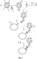
- FIG. 1 Schematic representation of an embodiment of the method according to the invention.
- FIG. 1 schematically shows an embodiment of the method according to the invention. For a better overview, only one molecule or complex etc. is shown in each case.
- PEMC polyelectrolyte microcapsule
- the polyelectrolyte microcapsule 1 includes one Fluorescent dye 4 a.
- Streptavidin 5 is now covalently bound to the shell 4 of the PEMC 1 so that the binding sites of the streptavidin 5 point to the space 16 around the PEMC 1 and are accessible there.
- the complex 10 of PEMC 1 and streptavidin 5 is contacted with a complex 9 of a folded and biotin 8-linked MHC-I molecule and an antigenic peptide 7. It forms a PEMC / pMHC-I complex 11.
- FIG. 1 For clarity, only one streptavidin and one pMHC-I molecule are shown.
- Complex 9 is formed by folding an unfolded starting MHC-I molecule 6 and binding an antigenic peptide 7 and biotin 8 thereto.
- Complex 11 is contacted with T cells isolated from blood 12 so that the T cell receptor (TCR) 13 can bind to pMHC complex 9.
- TCR T cell receptor
- the formed complex 14 of PEMC 1, pMHC molecule 9 and T cell 12 is contacted with a fluorescently labeled anti-TCR antibody 15.
- the reaction mixture is then subjected to flow cytometry.
- a system of a primary and a fluorescently labeled secondary antibody can also be used here.
- the (primary) antibody does not have to be directed against the TCR, but may also target an associated coreceptor, e.g. CD8, judge.
- the following amounts are for a single pellet from a one liter culture.
- sucrose solution (25% sucrose, 1 mM EDTA, 1 mM PMSF and 10 mM DTT, 10 mM Tris, pH 8.0). Transfer to 50 ml Falcon tubes and freeze at -20 ° C for storage.
- Lyse the cells by thawing in a 30 ° C water bath Swing the tubes to ensure thawing is fast and consistent. From now on, always keep the samples at 4 ° C (on ice).
- the reaction solution is filtered through a 0.22 ⁇ m vacuum filter (bottle top filter) into a new 250 ml flask.
- An Amicon stirred cell is assembled and purged with about 200 ml of filtered dd H 2 O at 60 psi.
- the refolding reaction mixture is concentrated to about 20 ml at 60 psi in the Amicon stirred cell.
- the retentate is collected in a 50 ml Falcon tube and the stirred cell is purged with permeate until there is about 25 ml of retentate.
- the Amicon cell is disassembled and cleaned with 70% EtOH.
- the permeate is collected for the second refolding reaction. This is achieved by adding 250 .mu.l l PMSF 1000x to the permeate. To 8 mg of thawed MHC heavy chain, add 8 M urea to 20 mg / ml and mix with 700 ⁇ l injection buffer. The mixture is then taken up in a 1 ml syringe with a 26G needle and added to the permeate. Finally, 7 mg thawed MHC light chain is mixed with 780 ⁇ l injection buffer and added to the permeate in the same manner as the heavy chain. The permeate is stirred at high speed during the addition of the heavy and light chain. The reaction mixture is then incubated for 12 h at 10 ° C with gentle shaking.
Landscapes
- Health & Medical Sciences (AREA)
- Life Sciences & Earth Sciences (AREA)
- Engineering & Computer Science (AREA)
- Immunology (AREA)
- Hematology (AREA)
- Urology & Nephrology (AREA)
- Cell Biology (AREA)
- Biomedical Technology (AREA)
- Chemical & Material Sciences (AREA)
- Molecular Biology (AREA)
- Food Science & Technology (AREA)
- General Health & Medical Sciences (AREA)
- Biotechnology (AREA)
- Medicinal Chemistry (AREA)
- Physics & Mathematics (AREA)
- Analytical Chemistry (AREA)
- Biochemistry (AREA)
- Microbiology (AREA)
- General Physics & Mathematics (AREA)
- Pathology (AREA)
- Zoology (AREA)
- Tropical Medicine & Parasitology (AREA)
- Virology (AREA)
- Peptides Or Proteins (AREA)
- Investigating Or Analysing Biological Materials (AREA)
Description
- Die Erfindung betrifft ein Verfahren zur antigenspezifischen Immunfärbung von T-Zellen sowie ein Färbekit hierfür.
- MHC-Klasse-I-Proteine sind an der zellulären Immunantwort beteiligte Transmembranproteine, die antigene Peptide aus dem Zellinneren einer antigenpräsentierenden Zelle (APC), beispielsweise aus dem Cytosol oder dem Lumen der endocytischen Organellen, binden und an der Zelloberfläche den zytotoxischen T-Zellen (CTL) präsentieren (Klasse-I-Antigenpräsentation). Die Bindung eines T-Zell-Rezeptors (TCR) einer CTL an einen Klasse-I-Peptid-Komplex einer APC führt, abhängig vom Ort der Reaktion im Körper, dem Typ der APC (B-Zelle, dendritische Zelle, etc.) und dem Aktivierungszustand der CTL, zur Aktivierung der CTL und/oder zur Induktion des Zelltods (Apoptose) der APC durch die CTL. Die Spezifität (Reaktionsfähigkeit mit einem bestimmten Klasse-I-Peptid-Komplex) einer CTL liegt dabei darin begründet, dass sie an ihrer Oberfläche nur eine Art TCR (d.h. nur TCRs einer einzelnen eindeutigen Sequenz) trägt. Die Immunantwort ist effektiv, weil solche CTL, die mit Selbst-Peptiden (Peptiden, die die aus körpereigenen Proteinen erzeugt werden) reagieren, im Thymus eliminiert werden. Aus diesem Grund bedeutet die Erkennung eines antigenen Peptids durch den TCR einer CTL, dass die APC fremde Proteine produziert, die von Viren oder intrazellulären Parasiten (Bakterien, Protozoen) stammen könnten. Auch eine Überproduktion endogener Peptide in malignen entarteten Zellen, zum Beispiel in Tumoren, kann zu solchen Erkennungsreaktionen führen.
- Nahezu alle in der Zelle vorhandenen Proteine werden in Peptide zersetzt, die dann im endoplasmatischen Retikulum (ER), einer membranumgrenzten Organelle im Zellinneren, an MHC-Klasse-I-Proteine binden. Anschließend wird der Komplex aus Peptid und MHC-Klasse-I-Protein (pMHC-I) an die Zelloberfläche transportiert und steht dort für die Erkennung durch CTL zur Verfügung. Wenn nun aufgrund einer tumorigenen malignen Entartung neuartige oder mutierte Proteine produziert werden, oder wenn durch eine virale oder bakterielle Infektion virale oder bakterielle Proteine aus dem genetischen Material des Virus oder Bakteriums produziert werden, werden diese ,neuartigen' Proteine ebenfalls in Peptide zersetzt, die dann im Komplex mit MHC-Klasse-I-Proteinen auf der Zelloberfläche präsentiert werden. Diese ,neuartigen' Peptide sind verschieden von den zelleigenen und lösen eine Erkennung durch die CTL aus. Die Präsentation durch MHC-Klasse-I-Proteine spielt ebenfalls eine Rolle in allergischen Reaktionen, der Abstoßung von Transplantaten und einer Anzahl von Autoimmunkrankheiten wie multipler Sklerose und Spondyloarthropathien (z.B. Morbus Bechterew oder rheumatoider Arthritis).
- Die Untersuchung der Immunantworten, die durch MHC-Klasse-I-Proteine vermittelt werden, erfordert oft den Nachweis derjenigen CTL, die mit einem bestimmten Klasse-I-Peptid-Komplex (Epitop) reagieren. Reaktionen auf ein einziges immunodominantes Epitop machen oft 10-20% der gesamten T-Zell-Population in einem Organismus aus, und die Verfolgung der T-Zell-Frequenz (d.h. des Anteils derjenigen CTL, die mit einem bestimmten Epitop reagieren, an der Gesamtmenge der CTL im Organismus) erlaubt es beispielsweise, die Immunantwort (und z.B. Gelingen oder Fehlschlagen einer Therapie) genau zu verfolgen. Entsprechende Nachweisverfahren sind beispielsweise in Hadrup und Schumacher 2010 (Hadrup SR, Schumacher TN 2010. MHC-based detection of antigen-specific CD8+ T cell responses. Cancer Immunol Immunother.59(9):1425-33. doi: 10.1007/s00262-010-0824-2) und Andersen et al. 2012 (Andersen RS, Kvistborg P, Frøsig TM, Pedersen NW, Lyngaa R, Bakker AH, Shu CJ, Straten Pt, Schumacher TN, Hadrup SR. 2012. Parallel detection of antigen-specific T cell responses by combinatorial encoding of MHC multimers. Nat Protoc. 7(5):891-902. doi: 10.1038/nprot.2012.037) beschrieben.
- Anikeeva et al 2006 (Anikeeva N, Lebedeva T, Clapp AR, Goldman ER, Dustin ML, Mattoussi H, Sykulev Y. 2006, Quantum dot/peptide-MHC biosensors reveal strong CD8-dependent cooperation between self and viral antigens that augment the T cell response, Proc Natl Acad Sci U.S.A. 103(45):16846-51) beschreiben ein Verfahren, bei dem lumineszierende Quantenpunkte ("Quantum Dots"; QD) aus Halbleitermaterial zur Untersuchung der Wechselwirkung zwischen MHC-Klasse-I-Proteinen und CTL eingesetzt werden. Bei dem Verfahren werden zur Imitierung von APC fluoreszenzmarkierte mit viralen Antigenen beladene MHC-Klasse-I-Proteine (pMHC-I-Proteine) mit ihrem C-Terminus auf einem solchen Quantenpunkt immobilisiert und mit antiviralen CD8+-T-Zellen in Kontakt gebracht.
-
WO 2010/037395 A2 undWO 2009/003492 A1 beschreiben MHC-Multimere bestehend aus einer Multimerisierungsdomäne, an die ein oder mehrere MHC-Peptid-Komplexe durch einen oder mehrere Linker gebunden sind. Bei der Multimerisierungsdomäne kann es sich um Kügelchen, zellähnliche Strukturen, einschließlich membranbasierter Strukturen, beispielsweise Micellen, Liposomen und andere Membranstrukturen handeln. Das MHC-Multimer kann zur Identifizierung antigenspezifischer T-Zellen mittels Durchflusszytometrie verwendet werden, wobei das MHC-Multimer mit einem Fluoreszenzfarbstoff markiert ist. -
AU 2004203482 A1 -
DE 10042023 A1 beschreibt Polyelektrolyt-Kapseln, bei denen feste Teilchen signalerzeugender Substanzen, z.B. Fluorophore, von einer oder mehreren Schichten eines Polyelektrolyten eingekapselt sind, und die an ihrer äußeren Oberfläche Affinitätsmoleküle tragen, die Zielmoleküle in einer Probe spezifisch erkennen und daran binden. Die Polyelektrolyt-Kapseln finden Verwendung zur Signalerzeugung beim optischen, elektrochemischen oder chemischen Nachweis von Zielmolekülen. -
WO2010037395 beschreibt MHC-Peptidkomplexe und deren Verwendung zur Diagnose, Behandlung oder Impfung gegen eine Krankheit bei einer Person. Genauer gesagt, offenbart die Erfindung MHC-Komplexe, die krebsantigene Peptide umfassen, und deren Verwendung. -
WO2009003492 beschreibt Verfahren zur Herstellung, Kennzeichnung und Verwendung von MHC-Multimeren, insbesondere neue Methoden zur Erzeugung von MHC-Multimeren und Methoden zur Verbesserung bestehender und neuer MHC-Multimere. Die Erfindung beschreibt auch verbesserte Methoden für den Einsatz von MHC-Multimeren bei der Analyse von T-Zellen einschließlich diagnostischer und prognostischer Methoden. -
AU2004203482 -
DE 10042023 betrifft betrifft Kapseln, die feste Teilchen signalerzeugender Substanzen einkapseln und an ihrer äußeren Oberfläche Affinitätsmoleküle tragen, die Zielmoleküle in einer Probe spezifisch erkennen und daran binden. Gegenstand der Erfindung ist die Verwendung dieser Kapseln zur Signalerzeugung beim optischen, elektrochemischen oder chemischen Nachweis von Zielmolekülen. Es wird ein Nachweisverfahren und ein Kit beschrieben. - Aufgabe der vorliegenden Erfindung ist es, einen einfachen und zuverlässigen antigenspezifischen Nachweis von T-Zellen zu ermöglichen.
- Zur Lösung der Aufgabe stellt die vorliegende Erfindung in einem ersten Aspekt ein Verfahren zur antigenspezifischen Immunfärbung von T-Zellen bereit, wobei eine Polyelektrolyt-Mikrokapsel, die außen ein antigenpräsentierendes MHC-Klasse-I-Molekül aufweist, mit einer einen T-Zell-Rezeptor tragenden T-Zelle unter Bedingungen in Kontakt gebracht wird, die eine Wechselwirkung zwischen dem T-Zell-Rezeptor und dem antigenpräsentierenden MHC-Klasse-I-Molekül erlauben, so dass ein Komplex aus T-Zelle, antigenpräsentierendem MHC-Klasse-I-Molekül und Polyelektrolyt-Mikrokapsel gebildet werden kann, so wie in Anspruch 1 definiert.
- Das erfindungsgemäße Verfahren stellt einen einfach durchzuführenden, dabei aber empfindlichen antigenspezifischen Nachweis von T-Zellen oder T-Zellpopulationen bereit. Das erfindungsgemäße Verfahren ist sehr flexibel ausgestaltbar, und stellt einen einfachen und empfindlichen Nachweis mittels Fluoreszenzmarkierung einzelner oder auch mehrerer Komponenten des Komplexes aus T-Zelle, antigenpräsentierendem MHC-Klasse-I-Molekül und Polyelektrolyt-Mikrokapsel bereit.
- Unter einer "Polyelektrolyt-Mikrokapsel" (abgekürzt auch PEMC) wird hier ein Hohlkörper mit einer mehrschichtigen Polyelektrolythülle verstanden, die einen mittleren Durchmesser von 100 nm bis 10 µm aufweist. Polyelektrolyt-Mikrokapseln können beispielsweise durch aufeinander folgende Adsorption entgegengesetzt geladener Polyelektrolytschichten auf einen geladenen Matrizenkörper, der anschließend aufgelöst werden kann, hergestellt werden (s. z.B.
WO 1999/047252 A2 ;DE 10042023 C2 ;WO 2005057163 A2 ; Tong und Gao 2008, Multilayer microcapsules with tailored structures for bio-related applications, J. Mater. Chem., 2008, 18, 3799-3812). - Unter "Polyelektrolyten" werden Polymere mit ionisierbaren oder ionisch dissoziierbaren Gruppen verstanden, die Bestandteil oder Substituent der Polymerkette sein können. Dabei ist die Anzahl der ionisierbaren oder ionisch dissoziierbaren Gruppen vorzugsweise so groß, dass die Polymere in ionischer Form (Polyion) wasserlöslich oder in Wasser quellbar sind. Polyelektrolyte können linear oder verzweigt sein. Je nach Art der dissoziierbaren Gruppen unterscheidet man kationische (Polybasen) und anionische Polyelektrolyte (Polysäuren). Kationische Polyelektrolyte enthalten überwiegend positiv geladene (basische) Gruppen, anionische Polyelektrolyte überwiegend anionische (saure) Gruppen. Ein Beispiel für einen kationischen Polyelektrolyten ist Polyallylamin oder Polyallylamin-Hydrochlorid (PAH), ein Beispiel für einen anionischen Polyelektrolyten ist Polyacrylsäure (PAA).
- Unter einem "MHC-Klasse-I-Molekül", "Klasse-I-Molekül", "MHC-I-Molekül", MHC-Klasse-I-Protein", "Klasse-I-Protein" oder "MHC-I-Protein" wird ein Haupthistokompatibilitätskomplex-Klasse-I-Protein verstanden. MHC-Klasse-I-Proteine sind vom Haupthistokompatibilitätskomplex (engl. "Major Histocompatibility Complex", MHC) kodierte Transmembranproteine und an der zellulären Immunantwort beteiligt. MHC-Klasse-I-Proteine binden Peptide aus dem Zellinneren, wie beispielsweise aus dem Cytosol oder dem Lumen der endocytischen Organellen und präsentieren diese an der Zelloberfläche den zytotoxischen T-Zellen als (Antigenpräsentation). Ein MHC-I-Molekül besteht in der Regel aus einer schweren α-Kette (auch kurz "hc", Molekulargewicht ca. 44 kDa, etwa 350 Aminosäuren) mit drei extrazellulären Domänen (α1, α2 und α3) und einer Transmembrandomäne sowie einer nicht kovalent daran gebundenen leichten β-Kette (auch "β2-Mikroglobulin" oder kurz "β2m", ca. 12 kDa, 99 Aminosäuren). Menschliche MHC-Klasse-I-Proteine werden auch als HLA-Proteine (HLA = human leukocyte antigen) bezeichnet. Der Begriff umfasst sowohl klassische MHC-Klasse-I-Proteine wie HLA-A, HLA-B und HLA-C als auch nicht-klassische MHC-Klasse-I-Proteine wie HLA-E, HLA-F und HLA-G. Zudem umfasst der Begriff auch peptidbindende Fragmente aller dieser Proteine, insbesondere die extrazelluläre Domäne, sowie Fusionsproteine, die aus der schweren Kette (oder einem peptidbindenden Fragment der schweren Kette), ggfs. einem Linker, und der leichten Kette bestehen. Unter einem "Fragment" werden hier insbesondere zusammenhängende Teilsequenzen von mindestens 10, 15, 20, 25, 30, 35, 40, 45, 50, 60, 65, 70, 80, 90 oder mindestens 100 Aminosäuren, bevorzugt mindestens 110, 120, 130, 140, 150, 160, 170, 180, 190 oder mindestens 200 Aminosäuren verstanden. Der Begriff "MHC-Klasse-I-Protein" umfasst auch MHC-Klasse-I-Proteine nicht-menschlicher Wirbeltierspezies, beispielsweise von Maus (Mus musculus), Ratte (Rattus norvegicus), Rind (Bos taurus), Pferd (Equus equus) oder Grüner Meerkatze (Macaca mulatta). Die MHC-Klasse-I-Proteine der Maus werden beispielsweise als H-2-Proteine bezeichnet, die von den Genloci H-2K, H-2L und H-2D kodiert werden. Ein bestimmter Allotyp eines Maus-MHC-Klasse-I-Proteins ist beispielsweise H-2Kb oder H-2Ld.
- Ein Komplex aus Peptid und MHC-Klasse-I-Protein, d.h. ein MHC-Klasse-I-Protein mit daran gebundenem Peptid, wird hier auch als "Peptid/MHC-Klasse-I-Protein-Komplex", "Klasse-I-Peptid-Komplex" oder "Peptid/MHC-I" bezeichnet. Als Abkürzung für einen solchen Komplex wird auch "pMHC-I" verwendet. Der Begriff "antigenpräsentierendes MHC-Klasse-I-Protein" wird synonym zu dem Komplex verwendet. "Antigenpräsentierend" bedeutet in diesem Zusammenhang, dass das MHC-Klasse-I-Protein ein Peptid (Antigen) gebunden aufweist.
- Wenn hier von einem Komplex aus Polyelektrolyt-Mikrokapsel (PEMC) und Peptid/MHC-Klasse-I-Protein (pMHC-I/PEMC-Komplex) gesprochen wird, umfasst dieser Begriff auch ein etwaiges Kopplungssystem, mit dessen Hilfe das antigenpräsentierende MHC-Klasse-I-Protein an die Polyelektrolyt-Mikrokapsel gebunden ist.
- Wenn hier der Begriff "Untersuchungspeptid" verwendet wird, ist damit ein (antigenisches) Peptid gemeint, das an ein MHC-Klasse-I-Protein gebunden wird oder ist und dem erfindungsgemäßen Verfahren unterworfen wird.
- Unter dem Begriff "T-Zellen" werden T-Lymphozyten verstanden. T-Lymphozyten tragen MHC-Rezeptoren (T-Zell-Rezeptoren, TCR) in ihrer Zellmembran, mit deren Hilfe sie an MHC-I-Proteine anderer Zellen gebundene Peptidantigene erkennen können. Mit dem Begriff "zytotoxische T-Zellen" (CTL) werden CD8+-T-Lymphozyten bezeichnet.
- Geeignete Polyelektrolyt-Mikrokapseln sind dem Fachmann bekannt oder können von ihm anhand seines Fachwissens hergestellt werden. Es ist bevorzugt, Polyelektrolyt-Mikrokapseln zu verwenden, die durchschnittlich kleiner sind als durchschnittliche T-Zellen, bevorzugt um mindestens den Faktor 2, 3, 4, 5, 6, 7, 8, 9, 10, 15, 20, 30, 40 oder 50. Bevorzugte Polyelektrolyt-Mikrokapseln haben beispielsweise einen durchschnittlichen Durchmesser von 100-6000 nm, 100-900 nm, 100-800 nm, 100-700 nm, 100-600 nm, 100-500 nm, 150-300 nm, oder 3000-6000 nm.
- Bereichsangaben wie "100-1000" sind hier immer so zu verstehen, dass jeder Zwischenwert mit offenbart ist. Auch ein beliebiger kleinerer Bereich aus dem Bereich soll dabei mit offenbart sein, wobei unter dem kleineren Bereich auch Bereiche zu verstehen sind, die keinen der Grenzwerte des Bereichs umfassen. So umfasst eine Angabe wie "100-1000" nicht nur Bereiche wie "100-950" oder "600-1000", sondern auch Bereiche von "250-700" oder "200-900" oder "200-400", wobei die einzelnen Werte innerhalb des Bereichs ausdrücklich mit erfasst sind, und nicht nur dessen Grenzwerte.
- Der Ausdruck, wonach die Polyelektrolyt-Mikrokapsel "außen ein antigenpräsentierendes MHC-Klasse-I-Molekül aufweist", bedeutet, dass das MHC-Klasse-I-Molekül so an die Hülle der Polyelektrolyt-Mikrokapsel geheftet ist, dass das MHC-Klasse-I-Molekül mit dem das (antigenische) Peptid bindenden Bereich zur außerhalb der Polyelektrolyt-Mikrokapsel liegenden Umgebung gerichtet ist. Vorzugsweise ist das MHC-Klasse-I-Molekül mit dem C-Terminus seiner schweren Kette an die äußere Hülle der Polyelektrolyt-Mikrokapsel gebunden oder in der äußeren Hülle verankert. "Geheftet" bedeutet hier, dass unter Bedingungen, wie sie normalerweise während der Durchführung des erfindungsgemäßen Verfahrens herrschen, der Kontakt zwischen MHC-Klasse-I-Molekül und Polyelektrolyt-Mikrokapsel-Hülle nicht verloren geht. Der Begriff "geheftet" wird hier gegebenenfalls auch synonym zu den Begriffen "gebunden" oder "verankert" verwendet. Bindungsarten sind dem Fachmann bekannt und schließen beliebige kovalente und nicht-kovalente Bindungen ein.
- Bei dem erfindungsgemäßen Verfahren wird ein Komplex aus antigenpräsentierendem MHC-Klasse-I-Molekül und Polyelektrolyt-Mikrokapsel (pMHC-I/PEMC-Komplex) mit einer TCR-tragenden T-Zelle so in Kontakt gebracht, dass der TCR der T-Zelle an die pMHC-I-Moleküle in dem pMHC-I/PEMC-Komplex bindet, wodurch ein Komplex aus T-Zelle, antigenpräsentierendem MHC-Klasse-I-Molekül und Polyelektrolyt-Mikrokapsel (pMHC-I/PEMC/T-Zell-Komplex) gebildet wird.
- Das antigenpräsentierende MHC-Klasse-I-Molekül ist direkt oder indirekt mittels kovalenter und/oder nicht-kovalenter Bindung an die Polyelektrolyt-Mikrokapsel gebunden. Bei der direkten Bindung kann es sich beispielsweise um eine kovalente oder elektrostatische Bindung handeln. Bevorzugt ist das MHC-Klasse-I-Molekül indirekt über ein geeignetes Kopplungssystem an die Polyelektrolyt-Mikrokapsel gebunden. Unter einem "Kopplungssystem" wird ein System aus sich gegenseitig bindenden Kopplungselementen, z.B. Molekülen, verstanden, die eine stabile indirekte Verbindung zwischen der Polyelektrolyt-Mikrokapsel und dem MHC-Klasse-I-Molekül vermitteln. Geeignete Kopplungssysteme sind dem Fachmann bekannt und umfassen bevorzugt ein Kopplungssystem, bei dem ein erstes Kopplungselement mehrere entsprechend daran bindende zweite Kopplungselemente binden kann, wie beispielsweise das Avidin-Biotin und Streptavidin-Biotin-System, um die Avidität des Kopplungssystems zu erhöhen.
- In einer bevorzugten Ausführungsform des erfindungsgemäßen Verfahrens ist das Kopplungssystem ein Avidin-Biotin- und/oder Streptavidin-Biotin-System, wobei Avidin oder Streptavidin, bevorzugt Steptavidin, vorzugsweise direkt an die Polyelektrolyt-Mikrokapsel und Biotin direkt an das antigenpräsentierende MHC-Klasse-I-Molekül, vorzugsweise an dessen C-Terminus, gebunden ist. "Direkt gebunden" bedeutet insbesondere kovalent gebunden. Streptavidin kann beispielsweise kovalent an freie Carboxylgruppen des Polyelektrolyten gebunden werden. Verfahren zur Biotinylierung von MHC-Klasse-I-Molekülen sind dem Fachmann gut bekannt und umfassen beispielsweise enzymatische Verfahren unter Verwendung einer Biotinligase (BirA). Aufgrund der niedrigen Affinität eines einzelnen Klasse-I-Peptid-Komplexes zu einem einzelnen TCR ist es für die Bindung einer T-Zelle an den Komplex aus Polyelektrolyt-Mikrokapsel und antigenpräsentierendem MHC-Klasse-I-Molekül (pMHC-I-Molekül) vorteilhaft, Multimere von pMHC-I-Molekül zu verwenden, die aufgrund des Aviditätseffektes fest genug an den TCR einer T-Zelle binden. Ein derartiges Multimer kann durch die Streptavidin-vermittelte Tetramerisierung von biotinylierten pMHC-I-Molekülen erhalten werden.
- Bei dem erfindungsgemäßen Verfahren ist eine Fluoreszenzmarkierung des pMHC-I/PEMC/T-Zell-Komplexes vorgesehen, um beispielsweise eine Detektion mittels fluoreszenzbasierter Durchflusszytometrie (fluorescence-activated cell sorting, FACS) zu ermöglichen. Dabei wird die Fluoreszenz des Komplexes gemessen, wodurch Rückschlüsse auf Menge bzw. Anteil der mit dem pMHC-I-Molekül reagierenden T-Zellen in der untersuchten Probe gezogen werden können.
- Die Fluoreszenzmarkierung kann je nach Einsatzzweck an unterschiedlichen Komponenten der genannten Komplexe vorgesehen bzw. während des Verfahrens erzeugt werden. Geeignete Fluorophore (Fluoreszenzfarbstoffe) sowie Verfahren zur Fluoreszenzmarkierung sind dem Fachmann bekannt oder leicht anhand von Routineversuchen zu ermitteln. Beispiele für geeignete Fluoreszenzfarbstoffe sind Fluorescein, Tetramethylrhodamin, die so genannten "Alexa-Fluor"-Farbstoffe (Molecular Probes Inc., Eugene, USA, z.B. Alexa Fluor® 488), die "Chromeo™"-Farbstoffe (Active Motif Inc., Carlsbad, USA, z.B. Chromeo 488), oder "DyLight®"-Farbstoffe (Dyomics GmbH, Jena, Deutschland; Thermo Fisher Scientific Inc., Rockford, USA, z.B. DyLight 488). Die Liste dieser Beispiele ist nicht erschöpfend.
- Bei dem erfindungsgemäßen Verfahren wird eine Polyelektrolyt-Mikrokapsel verwendet, die innen einen Fluoreszenzfarbstoff aufweist. Bevorzugt ist der Fluroreszenzfarbstoff von der Hülle im Inneren der Polyelektrolyt-Mikrokapsel eingeschlossen. Bei dem Fluoreszenzfarbstoff im Innern der Polyelektrolyt-Mikrokapsel kann es sich um einen an Dextran oder ein Protein gekoppelten Fluoreszenzfarbstoff handeln. Der Fluoreszenzfarbstoff ist vorzugsweise nicht an einen Quantenpunkt gebunden. Der Fluoreszenzfarbstoff ist vorzugsweise so ausgewählt, dass er im Innern der Polyelektrolyt-Mikrokapsel zumindest über den Nachweiszeitraum von der Hülle der Polyelektrolyt-Mikrokapsel eingeschlossen bleibt und im Wesentlichen nicht freigesetzt wird. Durch Fluoreszenzmarkierung der Polyelektrolyt-Mikrokapsel, insbesondere bei einer Ausgestaltung mit Fluoreszenzfarbstoff(en) im Innern der Polyelektrolyt-Mikrokapsel, ist eine starke Beladung mit Fluorophoren (Fluoreszenzfarbstoffen) möglich, wodurch ein stärkeres und einfacher zu detektierendes Signal resultiert.
- Im beanspruchten Verfahren weist die Polyelektrolyt-Mikrokapsel, außen ein antigenpräsentierendes MHC-Klasse-I-Molekül und innen einen ersten Fluoreszenzfarbstoff auf. Im Folgenden beschriebene Ausführungsformen, bei denen sich der Fluoreszenzfarbstoff nicht innen in der Polyelektrolyt-Mikrokapsel befindet, stellen lediglich Referenzbeispiele dar.
- Alternativ oder zusätzlich kann auch ein fluoreszenzmarkiertes Kopplungssystem verwendet werden, wobei vorzugsweise ein außen an die Polyelektrolyt-Mikrokapsel gebundenes erstes Kopplungselement des Kopplungssystems, beispielsweise Avidin und/oder Streptavidin, fluoreszenzmarkiert ist. Das fluoreszenzmarkierte Kopplungselement kann beispielsweise auch durch direkte oder indirekte Bindung eines gegen das Kopplungselement gerichteten fluoreszenzmarkierten Antikörpers fluoreszenzmarkiert sein. "Indirekt" bedeutet in diesem Zusammenhang beispielsweise, dass ein primärer nicht markierter Antikörper an das Kopplungselement gebunden und ein gegen den primären Antikörper gerichteter sekundärer fluoreszenzmarkierter Antikörper an den primären Antikörper gebunden wird.
- Weiter alternativ oder gegebenenfalls auch zusätzlich zu den obigen Fluoreszenzmarkierungen kann auch ein fluoreszenzmarkierter Antikörper eingesetzt werden, der direkt oder indirekt an die Polyelektrolyt-Mikrokapsel und/oder das antigenpräsentierende MHC-Klasse-I-Molekül in dem Komplex aus T-Zelle, antigenpräsentierendem MHC-Klasse-I-Molekül und Polyelektrolyt-Mikrokapsel gebunden wird. "Indirekt" bedeutet in diesem Zusammenhang beispielsweise, dass ein primärer nicht markierter Antikörper an die Polyelektrolyt-Mikrokapsel, und/oder das antigenpräsentierende MHC-Klasse-I-Molekül gebunden und ein gegen den primären Antikörper gerichteter sekundärer fluoreszenzmarkierter Antikörper an den primären Antikörper gebunden wird. Ein an die Polyelektrolyt-Mikrokapsel und/oder das antigenpräsentierende MHC-Klasse-I-Molekül bindender fluoreszenzmarkierter Antikörper wird beispielsweise bevorzugt dann eingesetzt, wenn die Polyelektrolyt-Mikrokapsel keinen Fluoreszenzfarbstoff enthält.
- Bei dem erfindungsgemäßen Verfahren wird zusätzlich auch ein fluoreszenzmarkierter Antikörper eingesetzt, der direkt oder indirekt an die T-Zelle gebunden wird. "Indirekt" bedeutet in diesem Zusammenhang beispielsweise, dass ein primärer nicht markierter Antikörper an die T-Zelle gebunden und ein gegen den primären Antikörper gerichteter sekundärer fluoreszenzmarkierter Antikörper an den primären Antikörper gebunden wird. Beispielsweise kann ein fluoreszenzmarkierter Antikörper verwendet werden, der spezifisch an den T-Zell-Rezeptor oder einen Corezeptor des T-Zell-Rezeptors bindet, oder ein fluoreszenzmarkierter Antikörper, der spezifisch an einen zweiten Antikörper bindet, der spezifisch an den T-Zell-Rezeptor oder Corezeptor des T-Zell-Rezeptors bindet. Beispielsweise kann ein primärer gegen den CD8-Corezeptor gerichteter Antikörper und ein fluoreszenzmarkierter gegen den primären Antikörper gerichteter sekundärer Antikörper verwendet werden. Geeignete Antikörper sind dem Fachmann bekannt und kommerziell erhältlich.
- In einer besonders bevorzugten Ausführungsform umfasst das erfindungsgemäße Verfahren folgende Schritte:
- a. Herstellen einer Polyelektrolyt-Mikrokapsel, die innen und/oder außen, vorzugsweise innen, einen Fluoreszenzfarbstoff und außen ein gebundenes erstes Kopplungselement eines Kopplungssystems, vorzugsweise Avidin oder Streptavidin, aufweist,
- b. Herstellen eines gefalteten, ein Antigen präsentierenden MHC-Klasse-I-Moleküls, das ein zweites Kopplungselement des Kopplungssystems, vorzugsweise Biotin, aufweist, das mit dem ersten Kopplungselement eine Bindung eingehen kann,
- c. Inkontaktbringen des in Schritt a) hergestellten Komplexes aus Polyelektrolyt-Mikrokapsel und erstem Kopplungselement mit dem in Schritt b) hergestellten Komplex aus antigenpräsentierendem MHC-Klasse-I-Molekül und dem zweiten Kopplungselement unter Bedingungen, die eine Bindung zwischen dem ersten und dem zweiten Kopplungselement erlauben,
- d. Inkontaktbringen des in Schritt c) gebildeten Komplexes aus Polyelektrolyt-Mikrokapsel und antigenpräsentierendem MHC-Klasse-I-Molekül mit einer einen T-Zellrezeptor tragenden T-Zelle unter Bedingungen, die eine Bindung zwischen T-Zellrezeptor und antigenpräsentierendem MHC-Klasse-I-Molekül unter Bildung eines Komplexes aus Polyelektrolyt-Mikrokapsel, antigenpräsentierendem MHC-Klasse-I-Molekül und T-Zelle erlauben,
- e. Inkontaktbringen des in Schritt d) gebildeten Komplexes aus Polyelektrolyt-Mikrokapsel, antigenpräsentierendem MHC-Klasse-I-Molekül und T-Zelle mit einem fluoreszenzmarkierten Antikörper, der spezifisch an den T-Zell-Rezeptor oder einen Corezeptor des T-Zell-Rezeptors bindet, oder einem fluoreszenzmarkierten Antikörper, der spezifisch an einen zweiten Antikörper bindet, der spezifisch an den T-Zell-Rezeptor oder Corezeptor des T-Zell-Rezeptors bindet, und
- f. Messen der Fluoreszenz des Komplexes aus Polyelektrolyt-Mikrokapsel, antigenpräsentierendem MHC-Klasse-I-Molekül und T-Zelle.
- Die Herstellung eines gefalteten, ein Antigen präsentierenden MHC-Klasse-I-Moleküls ist beispielsweise in der
WO 2013/102458 A1 beschrieben. - Durch gleichzeitige Fluoreszenzmarkierung mindestens einer Komponente des pMHC-I/PEMC-Komplexes und der T-Zelle in dem pMHC-I/PEMC/T-Zell-Komplex kann in vorteilhafter Weise ein Förster-Resonanzenergietransfer (FRET) genutzt werden, um die Detektion zu verbessern. Dazu wird eine Kombination aus mindestens zwei unterschiedlichen Fluoreszenzfarbstoffen eingesetzt, die als Donor- und Akzeptor-Fluoreszenzfarbstoff fungieren können. Die Art und die Abmessungen der genannten Komponenten werden so aufeinander abgestimmt, dass ein Förster-Resonanzenergietransfer (FRET) erfolgen kann. Beispielsweise können die Komponenten so aufeinander abgestimmt werden, dass möglichst geringe Abstände der Donor- und Akzeptor-Fluoreszenzfarbstoffe resultieren.
- In einer Ausgestaltung der Erfindung kann der Fluoreszenzfarbstoff im Inneren der Polyelektrolyt-Mikrokapsel beispielsweise ein Donor-Fluoreszenzfarbstoff und der Fluoreszenzfarbstoff, mit dem ein fluoreszenzmarkierter Antikörper markiert ist, ein Akzeptor-Fluoreszenzfarbstoff sein, oder umgekehrt der Fluoreszenzfarbstoff im Inneren der Polyelektrolyt-Mikrokapsel ein Akzeptor-Fluoreszenzfarbstoff und der Fluoreszenzfarbstoff, mit dem der fluoreszenzmarkierte Antikörper markiert ist, ein Donor-Fluoreszenzfarbstoff sein. Vorzugsweise werden ein Donor- und Akzeptor-Fluoreszenzfarbstoff, eine Polyelektrolyt-Mikrokapsel, ein antigenpräsentierendes MHC-Klasse-I-Molekül und ein fluoreszenzmarkierter Antikörper verwendet, so dass in dem Komplex aus Polyelektrolyt-Mikrokapsel, antigenpräsentierendem MHC-Klasse-I-Molekül, T-Zelle und fluoreszenzmarkiertem Antikörper ein Förster-Resonanzenergietransfer ermöglicht wird.
- In einem weiteren Aspekt betrifft die vorliegende Erfindung ein Kit zur antigenspezifischen Immunfärbung von T-Zellen. Der Kit in seiner breitesten Auslegung ist im Anspruch 7 beschrieben. Ausführungsformen im Folgenden, bei denen die Polyelektrolyt-Mikrokapsel nicht einen Fluoreszenzfarbstoff eingeschlossen in ihrem Innenraum aufweist, sind lediglich als Referenzbeispiele anzusehen.
- Das erfindungsgemäße Kit umfasst mindestens eine Polyelektrolyt-Mikrokapsel mit einem Innenraum und einer den Innenraum umgebenden Polyelektrolyt-Hülle, wobei die Polyelektrolyt-Hülle mindestens einen Peptid/MHC-Klasse-I-Protein-Komplex gebunden aufweist, so dass der Peptid/MHC-Klasse-I-Protein-Komplex mit seiner einen T-Zell-Rezeptor bindenden Bindungsstelle zu dem die Polyelektrolyt-Hülle außen umgebenden Raum gerichtet ist.
- Das erfindungsgemäße Kit umfasst mindestens eine Polyelektrolyt-Mikrokapsel, die einen Fluoreszenzfarbstoff aufweist, wobei der Fluoreszenzfarbstoff vorzugsweise in ihren Innenraum eingeschlossen oder an die Polyelektrolyt-Hülle gebunden ist, wobei die Formulierung "an die Polyelektrolyt-Hülle gebunden" auch den Fall umfasst, dass der Fluoreszenzfarbstoff in der Polyelektrolyt-Hülle verankert ist.
- In einer besonders bevorzugten Ausführungsform ist bei dem erfindungsgemäßen Kit der mindestens eine Peptid/MHC-Klasse-I-Protein-Komplex indirekt mittels eines Kopplungssystems aus einem ersten Kopplungselement und einem zweiten Kopplungselement an die Polyelektrolyt-Hülle gebunden. Bei dem ersten Kopplungselement handelt es sich bevorzugt um Avidin und/oder Streptavidin, bei dem zweiten Kopplungselement bevorzugt um Biotin.
- Das Kit umfasst ferner mindestens einen an eine T-Zelle bindenden fluoreszenzmarkierten primären Antikörper oder einen an eine T-Zelle bindenden nicht fluoreszenzmarkierten primären Antikörper und einen fluoreszenzmarkierten sekundären Antikörper.
- Das Kit kann selbstverständlich auch andere Komponenten umfassen, beispielsweise geeignete Pufferlösungen oder Komponenten zu deren Herstellung.
- Im Folgenden wird die Erfindung rein zu Veranschaulichungszwecken anhand einer Figur und eines beispielhaften Protokolls für die Durchführung einer Ausführungsform des erfindungsgemäßen Verfahrens näher beschrieben.
-
Figur 1 Schematische Darstellung einer Ausführungsform des erfindungsgemäßen Verfahrens. -
Figur 1 zeigt schematisch eine Ausführungsform des erfindungsgemäßen Verfahrens. Zur besseren Übersicht ist jeweils nur ein Molekül bzw. Komplex etc. dargestellt. Links oben ist eine Polyelektrolyt-Mikrokapsel (PEMC) 1 mit einem Innenraum 2 und einer den Innenraum 2 umgebenden mehrschichtigen Polyelektrolyt-Hülle 3 dargestellt. Die Polyelektrolyt-Mikrokapsel 1 schließt einen Fluoreszenzfarbstoff 4 ein. An die Hülle 4 der PEMC 1 wird nun Streptavidin 5 kovalent gebunden, so dass die Bindungsstellen des Streptavidin 5 zu dem Raum 16 um die PEMC 1 herum weisen und dort zugänglich sind. Der Komplex 10 aus PEMC 1 und Streptavidin 5 wird mit einem Komplex 9 aus einem gefalteten und an Biotin 8 gebundenen MHC-I-Molekül und einem antigenischen Peptid 7 in Kontakt gebracht. Es bildet sich ein PEMC/pMHC-I-Komplex 11. InFigur 1 sind der besseren Übersicht halber lediglich ein Streptavidin- und ein pMHC-I-Molekül dargestellt. - Der Komplex 9 wird gebildet, indem ein ungefaltetes Ausgangs-MHC-I-Molekül 6 gefaltet und ein antigenisches Peptid 7 und Biotin 8 daran gebunden werden. Der Komplex 11 wird mit aus Blut isolierten T-Zellen 12 in Kontakt gebracht, so dass der T-Zell-Rezeptor (TCR) 13 an den pMHC-Komplex 9 binden kann. Der gebildete Komplex 14 aus PEMC 1, pMHC-Molekül 9 und T-Zelle 12 wird mit einem fluoreszenzmarkierten Anti-TCR-Antikörper 15 in Kontakt gebracht. Die Reaktionsmischung wird anschließend einer Durchflusszytometrie unterzogen. Selbstverständlich kann hier auch ein System aus einem primären und einem fluoreszenzmarkierten sekundären Antikörper verwendet werden. Ebenso muss sich der (primäre) Antikörper nicht gegen den TCR richten, sondern kann sich auch gegen einen damit verbundenen Corezeptor, z.B. CD8, richten.
-
- a. Man löse 2 mg/ml PAH (Poly(allylamin-hydrochlorid), MG = 58,000) und PAA (Poly(acrylsäure), MG = 60.000) (0,286 ml 35% w/v) in 0,5 M NaCl-Lösung (in H2O).
-
- a. Man füge 2 ml H2O zu 6 µM CaCO3-Partikeln und beschalle für 5 min
- b. Man füge 5 µl 1 mg/ml Fluoreszenzfarbstoff (z. B. Dextran oder ein mit einem Fluoreszenzfarbstoff gekoppeltes Protein, beispielsweise Fluorescein oder Tetramethylrhodamin) zu den Partikeln und inkubiere für 30 min unter Schütteln
- c. Man schlage die CaCO3-Partikel für 2 min bei 3000 UPM nieder; Überstand entfernen
- d. Man wasche die CaCO3-Partikel 3x mit H2O
-
- a. Adsorption von Polymer: 10 min Schütteln bei 1200 UPM (2 ml)
- b. Man wasche nach der Adsorption dreimal mit Wasser, füge dann das zweite Polymer hinzu.
-
- a. Man messe die Partikelzahl und Fluoreszenz mittels Durchflusszytometrie (FACS).
-
- a. Man entferne die EDTA-Lösung durch einen weiteren Zentrifugationsschritt. Die Kapseln können in H2O bei Raumtemperatur gelagert werden, es sei denn, der eingekapselte Farbstoff benötigt besondere Bedingungen (z.B. Puffer, pH, Temperatur).
- Vernetze die Kapseln/Partikel mit 10mg/mL (1-Ethyl-3-[3dimethylaminopropyl]carbodiimid-Hydrochlorid) über Nacht bei RT (Raumtemperatur) in MES (2-(N-Morpholino) ethansulfonsäure)-Puffer, pH 6,0.
- Zum Quenching von EDC 3x mit Wasser waschen, bei 3000 UPM für 2 min zentrifugieren.
- Aktiviere die -COOH-Gruppe auf den Kapseln mit EDC (0,4 M) und Sulfo-NHS (N-Hydroxysuccinimid, 0,1 M) durch Inkubation für 1 h bei RT (in phosphatgepufferter Salzlösung (PBS), pH-Wert 7,2)
- Zum Quenching von EDC 3x mit kaltem Wasser waschen, bei 3000 UPM für 2 min zentrifugieren
- Füge Streptavidin zu einer Endkonzentration von 5 µg/mL hinzu und inkubiere unter Schütteln über Nacht bei RT (PBS pH 8,2)
- Blockiere die verbleibenden NHS-Ester durch Behandlung mit 50 mM Tris-Cl (pH 8,8) für 30 min bei RT.
- Wasche 3x mit Wasser, Zentrifugiere bei 3000 UPM für 2 min.
- Qualitätskontrolle: Füge Biotin-Fluorescein und messe die Partikelzahl und Fluoreszenz mittels Durchflusszytometrie (FACS).
-
- 50 ml Saccharoselösung:
- 500 µl 1 M Tris-Cl pH 8,0
- 12,5 g Saccharose
- 100 µl 500 mM EDTA
- Wasser zu 50 ml
- (DTT erst unmittelbar vor Gebrauch zufügen)
- 50 ml Reinigungspuffer:
- 2,5 ml 1 M Tris-Cl pH 8,0
- 12,5 g Saccharose
- 2,5 ml 20% Triton X-100
- 500 µl 500 mM EDTA
- Wasser zu 50 ml
- (DTT erst unmittelbar vor Gebrauch zufügen)
- 50 ml Harnstoffpuffer:
- 20 ml 5 M NaCl (oder: 5,84 g)
- 6,01 g Harnstoff
- 1,25 ml 1 M Tris-CI pH 8,4
- (DTT erst unmittelbar vor Gebrauch zufügen)
- 50 ml TBS:
- 1,5 ml 5 M NaCl
- 1 ml 1 M Tris-Cl pH 7,5
- (füge 1/100 v/v 500 mM PMSF unmittelbar vor Gebrauch hinzu)
- 50 ml Harnstoff-Resolubilisierungspuffer
- 24 g Harnstoff
- 2,5 ml 1 M HEPES pH 6,5
- Ziehe 20-ml-Kulturen E. coli, Stamm Rosetta oder BL21, enthaltend die schwere Kette, β2m oder das H-2Db/hβ2m-Einzelketten-Konstrukt in einem pGMT7 Vektor, in LB-Medium über Nacht bei 37 °C, unter Selektion durch 100 µg/ml Ampicillin und Chloramphenicol, heran. Beispiele: β2m-Expressionsplasmid pHN1+, in SPB542 (Expressionsstamm, abgeleitet von BL21); H-2Db-Volllängen-Expressionsplasmid pGMT7Db, in SPB595 (Rosetta). Siehe Sunil Kumar Saini, Esam Tolba Abualrous, Anca-Sarmiza Tigan, Kathryn Covella, Ursula Wellbrock, Sebastian Springer (2013), Not all empty MHC class I molecules are molten globules: Tryptophan fluorescence reveals a two-step mechanism of thermal denaturation, Molecular Immunology, 54, 386-396, http://dx.doi.org/10.1016/j.molimm.2013.01.004.
- Zentrifugiere bei 3000 x g für 10 Minuten, und verwende den Niederschlag, um 1 L-Kulturen der gleichen Zusammensetzung anzuimpfen.
- Ziehe die 1-L-Kulturen zu einer OD600 zwischen 0,4 und 0,5 an, füge dann IPTG (Isopropyl-β-D-1-thiogalactopyranosid) zu, um die Expression zu induzieren (Endkonzentration 0,5 mM).
- Ziehe die Kulturen über 4 Stunden zu einer OD600 von 1,0 an, bevor durch Zentrifugation bei 13.000 × g für 10 Minuten geerntet wird
- Die folgenden Mengen sind für ein einzelnes Pellet aus einer 1-Liter-Kultur.
- Resuspendiere das Pellet in 20 ml Saccharoselösung (25% Saccharose, 1 mM EDTA, 1 mM PMSF und 10 mM DTT, 10 mM Tris, pH 8,0). Überführe in 50 ml Falcon-Röhrchen und friere zur Aufbewahrung bei -20 ° C ein.
- Lysiere die Zellen durch Auftauen in einem 30-°C- Wasserbad. Schwenke die Röhrchen, um sicherzustellen, dass das Auftauen schnell und gleichmäßig erfolgt. Halte die Proben von nun an immer bei 4 °C (auf Eis).
- Beschalle (kleine Spitze, 3 x 20 Sekunden), bis die Lösung nicht mehr viskos ist. Nehme eine 50 µl Probe aus dem Lysat und beschrifte sie mit #1.
- Zentrifugiere die Zelllysate in Oak-Ridge-Röhrchen (JA25.50 Rotor) bei 40.000 x g, 4 °C für 15 Minuten. Nehme eine Probe aus dem Überstand und beschrifte sie #2. Überstand verwerfen.
- Resuspendiere das Pellet in 20 ml Detergenspuffer (25% Saccharose, 1% Triton X100, 5 mM EDTA und 2 mM DTT, 50 mM Tris, pH 8,0) durch Ultraschallbehandlung wie oben. Nehme eine Probe aus der Suspension und beschrifte sie mit #3. Zentrifugiere wie oben, und verwerfe den Überstand.
- Wiederhole diesen Vorgang der Resuspendierung, Ultraschallbehandlung und Zentrifugation für zwei weitere Waschschritte in
- a. Harnstoff-Puffer (2 M NaCl, 2 M Harnstoff und 2 mM DTT, 25 mM Tris, pH 8,4) (Probe #4) und
- b. Tris-gepufferter Kochsalzlösung (150 mM NaCl und 0,5 mM PMSF, 20 mM Tris, pH 7,5) (Probe #5).
- Nach dem letzten Waschschritt resuspendiere das Pellet in 10 ml 8 M Harnstoff (enthaltend 50 mM HEPES pH 6,5 und 100 µM β-Mercaptoethanol) durch Ultraschallbehandlung (Probe #6), und löse durch sanftes Schütteln bei 4 ° C.
- Nach 48 Stunden zentrifugiere die gelöste Proteinsuspension bei 40.000 g, 4 °C, für 15 Minuten. Gegebenenfalls muss ein zweites Mal zentrifugiert oder sogar durch ein 0,22-µm-Filter filtriert werden, um das unlösliche Sediment loszuwerden.
- Aliquotiere den Überstand (Probe #7) und lagere bei minus 20 °C
- Um die Qualität des Verfahrens zu beurteilen, lasse jeweils 1 µl der Proben #1-7 auf einem SDS-PAGE-Gel mit einem Größenmarker und etwas gereinigtem Protein aus einer früheren Aufbereitung laufen
- Bestimme die Konzentration des gereinigten Proteins durch Extinktion:
- a. Verdünne 1/20 in 6 M Guanidinium-Hydrochlorid.
- b. Messe die Extinktion bei 280 nm gegen Wasser.
- c. Berechne den Extinktionskoeffizienten aus der Sequenz des Proteins mit einem Online-Werkzeug wie http://www.basic.northwestern.edu/biotools/proteincalc.html. (Beispielsweise kann der Extinktionskoeffizient der lumenalen Domäne der schweren Kette von Db 84830 M-1cm-1 betragen (und das Molekulargewicht beträgt 33025 g/mol, so dass eine 1 mg/ml Lösung eine E280 von 2,57 hat).
- d. Anhand des berechneten Extinktionskoeffizienten und der eigenen Messung wird die Konzentration und die Ausbeute des Proteins ermittelt. Die Ausbeute aus einem Liter Kultur sollte in einem Bereich von 15-25 mg Protein liegen.
-
- HLA-Schwerkette-Einschlusskörper, 20 oder mehr mg/ml in Harnstoffpuffer, gelagert bei -80 °C, 8-mg-Aliquots
- MHC-Leichtkette-Einschlusskörper, 10 oder mehr mg/ml in Harnstoffpuffer, gelagert bei -80 °C, 7-mg-Aliquots,
- Untersuchungspeptid: 2,5-7,5 mg
- Rückfaltungspuffer: Mag gebe in einen 250 ml-Kolben (Schott, blauer Deckel) mit flachem Boden: 17,42 g L-Arginin-Base (Fluka), 5,95 g HEPES (Roth), 1 ml EDTA (Sigma) 0,5 M pH 8, 200 ml dd H2O, Magnetrührstab. Lege bis zum Erreichen von 10 °C auf Eis, stelle dann den pH-Wert mit konzentrierter HCl (in der Regel 5 bis 10 ml) auf pH 7,76, fülle mit dd H2O auf 250 ml auf, autoklaviere, lagere bei 4 °C
- TBS: 20 mM Tris (Sigma), pH 8,0, 4 °C, 150 mM NaCl (Merck). Filtrieren und Autoklavieren, Lagerung bei 4 °C.
- Harnstoffpuffer: 8 M Harnstoff (UCB), 10 mM Tris, pH 8 (Sigma), 100 mM NaH2PO4 (Merck), 0,1 mM EDTA (Roth), 0,1 mM DTT. Lagere Harnstoff als 10 M Stammlösung mit "Duolite"-Indikatorharz (Merck) und filtriere unmittelbar vor der Verwendung.
- Injektionspuffer: 3 M Guanidin-HCl (Fluka), 10 mM Na-Acetat (Sigma), 10 mM EDTA (Sigma), mit HCl auf pH 4,2 einstellen und als 10-ml-Aliquots bei -80 ° C lagern
- Regenerationspuffer für Amicon-Membran: 0,1 M NaOH, 100 ppm NaOCl Reduziertes Glutathion (Sigma)
- Oxidiertes Glutathion (Sigma) 1000 x PMSF (Sigma): 200 mM Stammlösung in Methanol (Merck), lagere 250-µl-Aliquots bei -80 °C
- 1000 x Leupeptin (Roche): 1 mg/ml Stammlösung in H2O, lagere 250-µl-Aliquots bei -80 °C
- 1000 x Pepstatin (Roche): 0,7 mg/ml Stammlösung in Methanol (Merck), lagere 250-µl-Aliquots bei -80 °C
- BirA-Enzym (Affinity Biosciences, oder selbst hergestellt). Nach dem Auftauen nicht wieder einfrieren; Lagerung bei -80 ° C
- D-Biotin (Sigma) 100 mM in 200 mM Tris (Sigma) 35-µl-Aliquots, Lagerung bei -80 °C
- ATP 100 mM in H2O auf pH 7,0 eingestellt, Lagerung von 300-µl-Aliquots bei -80 °C.
- MgCl2 1 M
- Tris 1 M
- Protein-Assay-Kit (z.B. Roti-Nanoquant).
- 1 Millipore PBTK-Membran, 76 mm Durchmesser, NMWL 30.000
- 3 Millipore Amicon Ultra-15-Aufkonzentrierungsvorrichtungen (MWCO = 10 kDa-Membran)
- 1 Millipore Stericup Express 0,22 µm Vakuumfiltrationsvorrichtung (250 ml) .
- FPLC-System mit Superdex-75-Säule (26 mm Durchmesser), Fraktionssammler und UV-Recorder, (z.B. Pharmacia)
-
- Wiege 2,5 bis 7,5 mg Peptid ab, füge DMSO zu 10 mg/ml hinzu. Träufle rasch mit 1-ml-Pipette, damit es nicht einfriert.
- Füge Folgendes zu einer Flasche Rückfaltungspuffer:
- 385 mg reduziertes Glutathion
- 77,5 mg oxidiertes Glutathion
- 250 µl PMSF 1000x (vorsichtig öffnen: Methanol)
-
- Taue 8 mg MHC-Schwerkette auf und füge 8 M Harnstoff zu 20 mg/ml hinzu. Füge 700 µl Injektionspuffer hinzu. Lade in eine 1-ml-Spritze mit einer 26G-Nadel. Rühre den Rückfaltungspuffer mit hoher Geschwindigkeit und spritze die schwere Kette kräftig und so nahe am Rührer wie möglich hinein.
- Taue 7 mg MHC-Leichtkette auf und füge 780 µl Injektionspuffer hinzu. Behandle wie die schwere Kette.
- Inkubiere die Reaktionsmischung über Nacht (12 h im Idealfall) bei 10 °C unter leichtem Schütteln oder Rühren
-
- Füge 8 mg schwere Kette (keine leichte Kette!) Mit Harnstoff- und Injektionspuffer wie oben beschrieben einmal am Morgen und einmal am Abend (12-h-Intervalle im Idealfall)
- Die Reaktionslösung wird durch ein 0,22-µm-Vakuum-Filter (Flaschenaufsatzfilter) in einen neuen 250-ml-Kolben filtriert. Eine Amicon-Rührzelle wird zusammengebaut und mit etwa 200 ml filtriertem dd H2O bei 60 psi gespült. Die Rückfaltungsreaktionsmischung wird bei 60 psi in der Amicon-Rührzelle auf etwa 20 ml eingeengt. Das Retentat wird in einem 50-ml-Falcon-Röhrchen gesammelt und die Rührzelle mit Permeat gespült, bis etwa 25 ml Retentat vorliegt. Die Amicon-Zelle wird zerlegt und mit 70% EtOH gereinigt.
- Das Permeat wird für die zweite Rückfaltungsreaktion gesammelt. Diese wird durch Zugabe von 250 µl l PMSF 1000x zum Permeat erreicht. Zu 8 mg aufgetauter MHC-Schwerkette wird 8 M Harnstoff zu 20 mg/ml gegeben, und mit 700 µl Injektionspuffer gemischt. Die Mischung wird dann in eine 1-ml-Spritze mit einer 26G-Nadel aufgenommen und zum Permeat hinzugegeben. Schließlich wird 7 mg aufgetaute MHC-Leichtkette mit 780 µl Injektionspuffer gemischt und in der gleichen Weise wie die schwere Kette zum Permeat gegeben. Das Permeat wird während der Zugabe der schweren und leichten Kette bei hoher Geschwindigkeit gerührt. Die Reaktionsmischung wird dann für 12 h bei 10 °C unter sanftem Schütteln inkubiert.
-
- Man füge 8 mg schwere Kette (keine leichte Kette!) mit Harnstoff- und Injektionspuffer wie oben beschrieben einmal am Morgen und einmal am Abend (12-h-Intervalle im Idealfall) hinzu.
-
- Filtriere die Reaktionsmischung aus der zweiten Rückfaltung durch 0,22-µm-Vakuumfilter (von Tag 3) (Flaschenaufsatzfilter) in einen neuen 250-ml-Kolben. Entsorge den Filter.
- Montiere die Amicon-Rührzelle und spüle mit rund 200 ml filtriertem dd H20 bei 60 psi.
- Konzentriere die filtrierte Reaktionsmischung auf 20 ml bei 60 psi in der gerührten Amicon-Rührzelle. Sammle das Retentat und spüle die Rührzelle mit Permeat, bis 25 ml Retentat vorliegen.
- Zerlege die Amicon-Zelle und reinige alle Teile mit 70% EtOH
- Zentrifugiere die Retentate (von Tag 3 und 5) bei 3200 g für 5 Minuten
- Konzentriere die zentrifugierten Retentate der Rührzelle schrittweise in der Amicon-Ultra-15-Vorrichtung, bis 4-5 ml Retentat vorliegen
- Spüle die Vorrichtung mit ca. 1 ml Permeat, bis insgesamt 5-6 ml Retentat vorliegen.
-
- Zentrifugiere das Retentat von Tag 5 bei 3200 g für 5 Minuten
- Führe mit dem 5-6 ml Retentat eine Größenausschluss-Chromatographie auf einer S75-Säule (TBS als Puffer) durch. Sammle 4-ml-Fraktionen aus 100-200 ml. Zeichne die UV-Absorption bei 280 nm auf.
- Sammle Fraktionen des Monomer-Peaks in einem 50-ml-Falcon-Röhrchen und gebe sofort PMSF, Leupeptin und Pepstatin zu l x Endkonzentration hinzu
- Konzentriere in einem Ultra-15-Gerät auf 3-4 ml. Spüle mit Permeat, bis 5,0 ml Retentat vorliegen.
- Hinzufügen: 400 µl 1 M Tris pH 8 25 °C, 25 µl 1 M MgCl2, 250 µl ATP 100 mM. Mische durch Umdrehen des Röhrchens.
- Füge 10-20 µg BirA Enzym und 28,5 µl 100 mM Biotin hinzu. Gut mischen durch mehrmaliges Umdrehen des Röhrchens.
- Inkubation für 12-16 Stunden in einem Wasserbad bei 27,0 °C
-
- Zentrifugiere die Biotinylierungsreaktionsmischung bei 3200 g für 5 Minuten
- Führe eine weitere FPLC-Trennung mit der Biotinylierungsreaktionsmischung auf einer S75-Säule (TBS als Puffer) durch, und sammle Fraktionen und zeichne die UV-Werte auf, wie oben beschrieben
- Sammle biotinylierten Monomerpeak und füge sofort PMSF, Leupeptin und Pepstatin zu lx Endkonzentration hinzu
- Prääquilibriere mit der Ultra-15-Vorrichtung mit 5 ml TBS. Konzentriere den Peak auf 250-350 µl. Sammle das Retentat und spüle das Gerät mit 100 µl Permeat
- Bestimme die Proteinkonzentration unter Verwendung eines handelsüblichen Standard-Proteinbestimmungskits.
- Stelle die Proben mit TBS auf 2,0 mg/ml Endkonzentration an
- Aliquotiere zu 50-µg-Aliquots in ordnungsgemäß gekennzeichneten lichtundurchlässigen Röhrchen. Lagere bei -80 °C
-
- 1. Füge biotinylierte MHC-Klasse-I-Proteine hinzu und Inkubation für 2 Stunden
- 2. Wasche 3 x mit Wasser, Zentrifugiere bei 3000 UPM für 2 min
- 3. Löse die Partikel mit 0,2 M EDTA (pH 7,2) für 30 Minuten auf
- 4. Qualitätskontrolle I: Füge Antikörper gegen Klasse I und fluoreszenzmarkierten sekundären Antikörper hinzu und messe Partikelzahl und Fluoreszenz mittels Durchflusszytometrie (FACS).
- 5. Qualitätskontrolle II: Tausche das an Klasse-I-Protein gebundene Peptid gegen ein Peptid, das mit einem Fluorophor wie Fluorescein oder Tetramethylrhodamin markiert ist, und messe Partikelzahl und Fluoreszenz mittels Durchflusszytometrie (FACS)
-
- Schritt 1-3: 60 ml Ficoll, ca.150 ml PBS (-Ca-Mg)
- Schritt 5: Einfriermedium; Kryoröhrchen
-
- 4x 15 ml Ficoll pro Falcon-Röhrchen vorlegen
- Blut in 150 ml-Zellkulturflasche einfüllen
- Blut mit PBS auf 125 ml auffüllen
- Je 30 ml Blutgemisch über Ficoll in ein Falcon-Röhrchen schichten
- Zentrifugieren: 2000 UPM, 30 min, 21°C (keine Bremse).
-
- Thrombozyten/PBS-Schicht bis auf 5ml von oben abnehmen
- Lymphozytenschicht abnehmen (höchstens 10 ml), Fraktionen von 2 Falcon-Röhrchen in einem sammeln; mit PBS auf 50ml auffüllen
- Zentrifugieren: 1500 UPM, 10 min, 20 °C
-
- Waschschritt: auf 50 ml mit PBS auffüllen, 1300 UPM, 10 min zentrifugieren.
-
- Lymphozyten in einem Falcon-Röhrchen sammeln, auf 50 ml auffüllen
- Probe zum Zellen zählen entnehmen
- Zentrifugieren: 1100 UPM, 10 min, 20 °C
-
- Zellzahl bestimmen:
- 10 µl Zellen mit 90 µl PBS versetzen.
- 10 µl dieser Zellsuspension mit 10 µl einer 0.4%-Lösung von Trypanblau (CAS 72-57-1) in PBS mischen.
- Mischung in ein Haemocytometer nach Neubauer pipettieren und Zellen zählen.
-
- Einfrieren, beschriften: möglichst viele 108/2ml-Zelleinheiten. Die letzte 108/2ml-Einheit wird mit 3 ml verdünnt und in Form von 5 x 2∗107/1ml eingefroren
-
- PFEA ("FACS-Puffer"): 500 ml PBS; 2% FeS; 2 mM EDTA; 0,02% NaN3 PBS-EDTA: 500 ml PBS; 2 mM EDTA; 0,02% NaN3
- Tetramer-Färbungspuffer (TSB), auch Tetramer-Lösung genannt: 50% PBS; 50% FeS; 2 mM EDTA
-
- In 96-Napf-Platte etwa 0.5-l x 106 Zellen ausplattieren und abzentrifugieren (4 °C, 2 min, 1800 UPM). Anschließend Platte abflicken.
-
- Waschschritt mit PBS-EDTA (Zellen in 150 µl aufnehmen und abzentrifugieren)
- Aqua live/dead 1:400 oder 1:200 (wurde Aqua live/dead vor <2 Wochen eingefroren 1:400, bei 2-4 Wochen 1:200) in PBS-EDTA verdünnen.
- 50 µl pro Napf resuspendieren
- Inkubation 20 min bei 4 °C im Dunkeln
- Waschschritt mit PBS-EDTA
-
- Pro Napf: 105-108 MHC-Mikrokapseln (Optimierung je nach Target) in TSB ansetzen und mischen.
- Pro Napf werden die Zellen in 50 µl der angesetzten Lösung resuspendiert (Lösung vorsichtig aus dem Überstand abnehmen! Pellett nicht aufwirbeln)
- Inkubation 30 min bei RT im Dunklen
- Ein Waschschritt mit PFEA
-
- Antikörper (z.B. CDB-PerCP) in PFEA ansetzen
- Färbung in 50 µl/well resuspendieren
- Inkubation 20 min bei 4 °C im Dunklen
- Ein Waschschritt mit PFEA
- Zellen in 200 µl PFEA aufnehmen, falls am gleichen Tag gemessen wird. Ansonsten fixieren
-
- Zellen in 100 µl PFEA mit 1% Formaldehyd resuspendieren
- Inkubation 20 min bei 4 °C im Dunklen
- Ein Waschschritt mit PFEA
- Zellen in 200 µl PFEA aufnehmen, bei 4°C im Dunkeln lagern
-
- Für jede Probe 200 000 Zellen verwenden.
- Nach dem Mischen von Zellen und Tetrameren werden die Proben für 30 Minuten bei 37 °C inkubiert.
- Danach Zellen auf ein Volumen von 1,5 ml mit PBS-Natriumazid auffüllen, in ein entsprechendes Probenröhrchen überführen und in der Durchflusszytometrie analysieren.
- Hierzu werden beispielsweise das Gerät CyFlowSpace von Partec und die Software FloMax Version 2.82 verwendet.
- Die Achseneinstellung der verschiedenen Graphen kann beispielsweise wie folgt erfolgen:
- - - Histogramm 1: Counts / forward scatter (FSC)
- - - Histogramm 2: Counts / side scatter (SSC)
- - - Histogramm 3: SSC / FSC
- - - Histogramm 4: Counts / Fluoreszenz 1 (FL1: 516-556 nm; logarithmische Skala)
- - - Histogramm 5: Counts / Fluoreszenz 3 (FL3: 650-700 nm; logarithmische Skala)
- - - Histogramm 6: FL1 / FSC
- - - Histogramm 7: FL3 / FL1 (logarithmische Skala)
- - - Histogramm 8: FL3 / FSC
- Zunächst werden unbehandelte Zellen zur Bestimmung der Größe der Zellen und der Hintergrundfluoreszenz gemessen. Dazu wird im Histogramm 3 die Region mit den intakten Zellen mit dem "Gate R1" markiert. Diese Region wird auf alle anderen Zellzählungen Histogramme angewendet. In Folge werden dann nur noch Zellen aus der markierten Region in den Histogrammen gezeigt.
- Im nächsten Schritt legt man ein im Histogramm 4 den Schwellenwert RN1 fest, über dem die Zellen als fuoreszenzmarkiert gelten.
- Die Proben werden dann mit diesen Einstellungen gemessen.
Claims (8)
- Verfahren zur antigenspezifischen Immunfärbung von T-Zellen, wobei- eine Polyelektrolyt-Mikrokapsel, die außen ein antigenpräsentierendes MHC-Klasse-I-Molekül und innen einen ersten Fluoreszenzfarbstoff aufweist, mit einer einen T-Zell-Rezeptor tragenden T-Zelle unter Bedingungen in Kontakt gebracht wird, die eine Wechselwirkung zwischen dem T-Zell-Rezeptor und dem antigenpräsentierenden MHC-Klasse-I-Molekül erlauben, so dass ein Komplex aus T-Zelle, antigenpräsentierendem MHC-Klasse-I-Molekül und Polyelektrolyt-Mikrokapsel gebildet werden kann,- ein mit einem zweiten Fluoreszenzfarbstoff markierter Antikörper direkt oder indirekt an die T-Zelle in dem Komplex aus T-Zelle, antigenpräsentierendem MHC-Klasse-I-Molekül und Polyelektrolyt-Mikrokapsel gebunden wird,- der erste und zweite Fluoreszenzfarbstoff unterschiedlich sind und als Donor- und Akzeptor-Fluoreszenzfarbstoff fungieren können, und- ein Donor- und Akzeptor-Fluoreszenzfarbstoff, eine Polyelektrolyt-Mikrokapsel, ein antigenpräsentierendes MHC-Klasse-I-Molekül und ein mit einem zweiten Fluoreszenzfarbstoff markierter Antikörper verwendet werden, so dass in dem Komplex aus Polyelektrolyt-Mikrokapsel, antigenpräsentierendem MHC-Klasse-I-Molekül, T-Zelle und mit einem zweiten Fluoreszenzfarbstoff markierter Antikörper ein Förster-Resonanzenergietransfer ermöglicht wird.
- Verfahren nach Anspruch 1, wobei das antigenpräsentierende MHC-Klasse-I-Molekül direkt oder indirekt mittels kovalenter und/oder nicht-kovalenter Bindung an die Polyelektrolyt-Mikrokapsel gebunden ist
- Verfahren nach Anspruch 2, wobei Avidin und/oder Streptavidin direkt an die Polyelektrolyt-Mikrokapsel und Biotin direkt an das antigenpräsentierende MHC-Klasse-I-Molekül gebunden ist
- Verfahren nach einem der vorhergehenden Ansprüche, wobeia) der Fluoreszenzfarbstoff im Inneren der Polyelektrolyt-Mikrokapsel ein Donor-Fluoreszenzfarbstoff und der zweite Fluoreszenzfarbstoff, mit dem der Antikörper markiert ist, ein Akzeptor-Fluoreszenzfarbstoff ist, oder b) der Fluoreszenzfarbstoff im Inneren der Polyelektrolyt-Mikrokapsel ein Akzeptor-Fluoreszenzfarbstoff und der zweite Fluoreszenzfarbstoff, mit dem der Antikörper markiert ist, ein Donor-Fluoreszenzfarbstoff ist.
- Verfahren nach Anspruch 4, wobei ein mit einem zweiten Fluoreszenzfarbstoff markierter Antikörper verwendet wird, der spezifisch an den T-Zell-Rezeptor oder einen Corezeptor des T-Zell-Rezeptors bindet, oder ein mit einem zweiten Fluoreszenzfarbstoff markierter Antikörper verwendet wird, der spezifisch an einen zweiten Antikörper bindet, der spezifisch an den T-Zell-Rezeptor oder Corezeptor des T-Zell-Rezeptors bindet.
- Verfahren nach einem der Ansprüche 4 bis 5, umfassend das Messen der Fluoreszenz des Komplexes aus Polyelektrolyt-Mikrokapsel, antigenpräsentierendem MHC-Klasse-I-Molekül, T-Zelle und mit einem zweiten Fluoreszenzfarbstoff markierten Antikörper.
- Kit zur antigenspezifischen Immunfärbung von T-Zellen, umfassenda) mindestens eine Polyelektrolyt-Mikrokapsel (1) mit einem Innenraum (2) und einer den Innenraum (2) umgebenden Polyelektrolyt-Hülle (3), wobei die Polyelektrolyt-Hülle (3) mindestens einen Peptid/MHC-Klasse-I-Protein-Komplex gebunden aufweist, so dass der Peptid/MHC-Klasse-I-Protein-Komplex mit seiner einen T-Zell-Rezeptor (13) bindenden Bindungsstelle zu dem die Polyelektrolyt-Hülle (3) außen umgebenden Raum (16) gerichtet ist, und wobei die Polyelektrolyt-Mikrokapsel (1) einen Fluoreszenzfarbstoff (4) eingeschlossen in ihrem Innenraum (2) aufweist, undb) mindestens einen mit einem zweiten Fluoreszenzfarbstoff markierten primären Antikörper (15) oder einen nicht mit einem zweiten Fluoreszenzfarbstoff markierten primären Antikörper und einen mit einem zweiten Fluoreszenzfarbstoff markierten sekundären Antikörper.
- Kit nach Anspruch 9, wobei der mindestens eine Peptid/MHC-Klasse-I-Protein-Komplex indirekt mittels eines Kopplungssystems aus einem ersten Kopplungselement (5) und einem zweiten Kopplungselement (8) an die Polyelektrolyt-Hülle (3) gebunden ist, wobei das erste Kopplungselement (5) bevorzugt Avidin und/oder Streptavidin, und das zweite Kopplungselement (8) bevorzugt Biotin ist.
Applications Claiming Priority (2)
| Application Number | Priority Date | Filing Date | Title |
|---|---|---|---|
| DE102014113688.8A DE102014113688A1 (de) | 2014-09-22 | 2014-09-22 | Antigenspezifische immunfärbung von t-zellen |
| PCT/DE2015/100365 WO2016045660A1 (de) | 2014-09-22 | 2015-09-02 | Antigenspezifische immunfärbung von t-zellen |
Publications (2)
| Publication Number | Publication Date |
|---|---|
| EP3198274A1 EP3198274A1 (de) | 2017-08-02 |
| EP3198274B1 true EP3198274B1 (de) | 2019-11-06 |
Family
ID=54544865
Family Applications (1)
| Application Number | Title | Priority Date | Filing Date |
|---|---|---|---|
| EP15794816.7A Active EP3198274B1 (de) | 2014-09-22 | 2015-09-02 | Antigenspezifische immunfärbung von t-zellen |
Country Status (3)
| Country | Link |
|---|---|
| EP (1) | EP3198274B1 (de) |
| DE (1) | DE102014113688A1 (de) |
| WO (1) | WO2016045660A1 (de) |
Family Cites Families (9)
| Publication number | Priority date | Publication date | Assignee | Title |
|---|---|---|---|---|
| ATE524165T1 (de) | 1998-03-19 | 2011-09-15 | Max Planck Gesellschaft | Verfahren zur herstellung von kapseln mit einer polyelektrolythülle |
| FR2785543B1 (fr) * | 1998-11-05 | 2003-02-28 | Inst Nat Sante Rech Med | Exosomes modifies et utilisations |
| DE10010264A1 (de) * | 2000-03-02 | 2001-09-13 | Novosom Gmbh | Stabilisierte Liposomen und Hüllstrukturen |
| DE10042023C2 (de) | 2000-08-08 | 2003-04-10 | Biognostic Ag | Kapseln, die feste Teilchen signalerzeugender Substanzen einkapseln, und deren Verwendung bei Bioassays zum Nachweis von Zielmolekülen in einer Probe |
| WO2005044224A2 (en) * | 2003-05-02 | 2005-05-19 | Case Western Reserve University | Drug delivery system based on polymer nanoshells |
| EP1678476A4 (de) | 2003-10-20 | 2007-05-23 | Univ Rice William M | Verfahren zur herstellung von mikrokapseln aus polymeren und geladenen nanopartikeln |
| EP2167537A2 (de) * | 2007-07-03 | 2010-03-31 | Dako Denmark A/S | Zusammengestellte verfahren für die analyse und sortierung von proben |
| US20110318380A1 (en) * | 2008-10-01 | 2011-12-29 | Dako Denmark A/S | MHC Multimers in Cancer Vaccines and Immune Monitoring |
| EP2800974B1 (de) | 2012-01-04 | 2016-12-14 | Jacobs University Bremen GmbH | Verfahren zum herstellen eines untersuchungsreagens und kit zum analysieren einer t-zellen-frequenz |
-
2014
- 2014-09-22 DE DE102014113688.8A patent/DE102014113688A1/de not_active Withdrawn
-
2015
- 2015-09-02 WO PCT/DE2015/100365 patent/WO2016045660A1/de not_active Ceased
- 2015-09-02 EP EP15794816.7A patent/EP3198274B1/de active Active
Non-Patent Citations (1)
| Title |
|---|
| None * |
Also Published As
| Publication number | Publication date |
|---|---|
| WO2016045660A1 (de) | 2016-03-31 |
| EP3198274A1 (de) | 2017-08-02 |
| DE102014113688A1 (de) | 2016-03-24 |
Similar Documents
| Publication | Publication Date | Title |
|---|---|---|
| DE69900468T2 (de) | Löslicher t-zell rezeptor | |
| DE69632577T2 (de) | Mhc-antigen-komplexe zur detektion und reinigung antigen-spezifischer t-zellen | |
| Zhao et al. | cis-B7: CD28 interactions at invaginated synaptic membranes provide CD28 co-stimulation and promote CD8+ T cell function and anti-tumor immunity | |
| DE60319745T2 (de) | Modifizierte löslicher t-zellen-rezeptor | |
| US10611818B2 (en) | MHC multimers in tuberculosis diagnostics, vaccine and therapeutics | |
| US8268964B2 (en) | MHC peptide complexes and uses thereof in infectious diseases | |
| DE69833949T2 (de) | Mutierte klasse-i-mhc-moleküle | |
| DE69628154T2 (de) | Antigen präsentierendes system und aktivierung von t-zellen | |
| DE69838648T2 (de) | Verwendung von multivalenten chimären peptidbeladenen mhc/ig-molekülen zum nachweis, zur aktivierung oder unterdrückung einer antigenspezifischen t-zellabhängigen immunantwort | |
| WO2013030620A2 (de) | Gen codiert für ein mhc-klasse-i-molekül, plasmid, expressionssystem protein, multimer, reagenz und kit zum analysieren einer t-zellen-frequenz | |
| EP2800974B1 (de) | Verfahren zum herstellen eines untersuchungsreagens und kit zum analysieren einer t-zellen-frequenz | |
| DE10117858A1 (de) | MHC-Tetramere | |
| AU2020262502A1 (en) | Self-assembling protein nanocage decorated with antibodies (SAPNA) and parts thereof | |
| DE60223040T2 (de) | Epitop-untersuchung mit hla | |
| JPH02503715A (ja) | 氷核形成免疫検定 | |
| DE102014113688A1 (de) | Antigenspezifische immunfärbung von t-zellen | |
| DE202010003498U1 (de) | Gen codiert für ein MHC-Klasse-I-Molekül, Plasmid, Expressionssystem Protein, Multimer, Reagenz und Kit zum Analysieren einer T-Zellen-Frequenz | |
| US20200347103A1 (en) | Mhc multimers in tuberculosis diagnostics, vaccine and therapeutics | |
| KR101616705B1 (ko) | 특이적 결합분자―나노섬유 복합체 및 이를 이용한 세포활성화 방법 | |
| DE202012100019U1 (de) | T-Zellen-Frequenz | |
| MXPA00011368A (en) | Multivalent t cell receptor complexes | |
| DE102013227106A1 (de) | In-vitro Verfahren zur Diagnose einer aktiven Lupus Nephritis |
Legal Events
| Date | Code | Title | Description |
|---|---|---|---|
| STAA | Information on the status of an ep patent application or granted ep patent |
Free format text: STATUS: THE INTERNATIONAL PUBLICATION HAS BEEN MADE |
|
| PUAI | Public reference made under article 153(3) epc to a published international application that has entered the european phase |
Free format text: ORIGINAL CODE: 0009012 |
|
| STAA | Information on the status of an ep patent application or granted ep patent |
Free format text: STATUS: REQUEST FOR EXAMINATION WAS MADE |
|
| 17P | Request for examination filed |
Effective date: 20170424 |
|
| AK | Designated contracting states |
Kind code of ref document: A1 Designated state(s): AL AT BE BG CH CY CZ DE DK EE ES FI FR GB GR HR HU IE IS IT LI LT LU LV MC MK MT NL NO PL PT RO RS SE SI SK SM TR |
|
| AX | Request for extension of the european patent |
Extension state: BA ME |
|
| DAV | Request for validation of the european patent (deleted) | ||
| DAX | Request for extension of the european patent (deleted) | ||
| STAA | Information on the status of an ep patent application or granted ep patent |
Free format text: STATUS: EXAMINATION IS IN PROGRESS |
|
| 17Q | First examination report despatched |
Effective date: 20180214 |
|
| GRAP | Despatch of communication of intention to grant a patent |
Free format text: ORIGINAL CODE: EPIDOSNIGR1 |
|
| STAA | Information on the status of an ep patent application or granted ep patent |
Free format text: STATUS: GRANT OF PATENT IS INTENDED |
|
| INTG | Intention to grant announced |
Effective date: 20190613 |
|
| GRAS | Grant fee paid |
Free format text: ORIGINAL CODE: EPIDOSNIGR3 |
|
| GRAA | (expected) grant |
Free format text: ORIGINAL CODE: 0009210 |
|
| STAA | Information on the status of an ep patent application or granted ep patent |
Free format text: STATUS: THE PATENT HAS BEEN GRANTED |
|
| AK | Designated contracting states |
Kind code of ref document: B1 Designated state(s): AL AT BE BG CH CY CZ DE DK EE ES FI FR GB GR HR HU IE IS IT LI LT LU LV MC MK MT NL NO PL PT RO RS SE SI SK SM TR |
|
| REG | Reference to a national code |
Ref country code: GB Ref legal event code: FG4D Free format text: NOT ENGLISH |
|
| REG | Reference to a national code |
Ref country code: CH Ref legal event code: EP Ref country code: AT Ref legal event code: REF Ref document number: 1199475 Country of ref document: AT Kind code of ref document: T Effective date: 20191115 |
|
| REG | Reference to a national code |
Ref country code: DE Ref legal event code: R096 Ref document number: 502015010881 Country of ref document: DE |
|
| REG | Reference to a national code |
Ref country code: IE Ref legal event code: FG4D Free format text: LANGUAGE OF EP DOCUMENT: GERMAN |
|
| REG | Reference to a national code |
Ref country code: NL Ref legal event code: MP Effective date: 20191106 |
|
| REG | Reference to a national code |
Ref country code: LT Ref legal event code: MG4D |
|
| PG25 | Lapsed in a contracting state [announced via postgrant information from national office to epo] |
Ref country code: LV Free format text: LAPSE BECAUSE OF FAILURE TO SUBMIT A TRANSLATION OF THE DESCRIPTION OR TO PAY THE FEE WITHIN THE PRESCRIBED TIME-LIMIT Effective date: 20191106 Ref country code: NO Free format text: LAPSE BECAUSE OF FAILURE TO SUBMIT A TRANSLATION OF THE DESCRIPTION OR TO PAY THE FEE WITHIN THE PRESCRIBED TIME-LIMIT Effective date: 20200206 Ref country code: GR Free format text: LAPSE BECAUSE OF FAILURE TO SUBMIT A TRANSLATION OF THE DESCRIPTION OR TO PAY THE FEE WITHIN THE PRESCRIBED TIME-LIMIT Effective date: 20200207 Ref country code: PT Free format text: LAPSE BECAUSE OF FAILURE TO SUBMIT A TRANSLATION OF THE DESCRIPTION OR TO PAY THE FEE WITHIN THE PRESCRIBED TIME-LIMIT Effective date: 20200306 Ref country code: PL Free format text: LAPSE BECAUSE OF FAILURE TO SUBMIT A TRANSLATION OF THE DESCRIPTION OR TO PAY THE FEE WITHIN THE PRESCRIBED TIME-LIMIT Effective date: 20191106 Ref country code: LT Free format text: LAPSE BECAUSE OF FAILURE TO SUBMIT A TRANSLATION OF THE DESCRIPTION OR TO PAY THE FEE WITHIN THE PRESCRIBED TIME-LIMIT Effective date: 20191106 Ref country code: SE Free format text: LAPSE BECAUSE OF FAILURE TO SUBMIT A TRANSLATION OF THE DESCRIPTION OR TO PAY THE FEE WITHIN THE PRESCRIBED TIME-LIMIT Effective date: 20191106 Ref country code: BG Free format text: LAPSE BECAUSE OF FAILURE TO SUBMIT A TRANSLATION OF THE DESCRIPTION OR TO PAY THE FEE WITHIN THE PRESCRIBED TIME-LIMIT Effective date: 20200206 Ref country code: NL Free format text: LAPSE BECAUSE OF FAILURE TO SUBMIT A TRANSLATION OF THE DESCRIPTION OR TO PAY THE FEE WITHIN THE PRESCRIBED TIME-LIMIT Effective date: 20191106 Ref country code: FI Free format text: LAPSE BECAUSE OF FAILURE TO SUBMIT A TRANSLATION OF THE DESCRIPTION OR TO PAY THE FEE WITHIN THE PRESCRIBED TIME-LIMIT Effective date: 20191106 |
|
| PG25 | Lapsed in a contracting state [announced via postgrant information from national office to epo] |
Ref country code: RS Free format text: LAPSE BECAUSE OF FAILURE TO SUBMIT A TRANSLATION OF THE DESCRIPTION OR TO PAY THE FEE WITHIN THE PRESCRIBED TIME-LIMIT Effective date: 20191106 Ref country code: HR Free format text: LAPSE BECAUSE OF FAILURE TO SUBMIT A TRANSLATION OF THE DESCRIPTION OR TO PAY THE FEE WITHIN THE PRESCRIBED TIME-LIMIT Effective date: 20191106 Ref country code: IS Free format text: LAPSE BECAUSE OF FAILURE TO SUBMIT A TRANSLATION OF THE DESCRIPTION OR TO PAY THE FEE WITHIN THE PRESCRIBED TIME-LIMIT Effective date: 20200306 |
|
| PG25 | Lapsed in a contracting state [announced via postgrant information from national office to epo] |
Ref country code: AL Free format text: LAPSE BECAUSE OF FAILURE TO SUBMIT A TRANSLATION OF THE DESCRIPTION OR TO PAY THE FEE WITHIN THE PRESCRIBED TIME-LIMIT Effective date: 20191106 |
|
| PG25 | Lapsed in a contracting state [announced via postgrant information from national office to epo] |
Ref country code: RO Free format text: LAPSE BECAUSE OF FAILURE TO SUBMIT A TRANSLATION OF THE DESCRIPTION OR TO PAY THE FEE WITHIN THE PRESCRIBED TIME-LIMIT Effective date: 20191106 Ref country code: CZ Free format text: LAPSE BECAUSE OF FAILURE TO SUBMIT A TRANSLATION OF THE DESCRIPTION OR TO PAY THE FEE WITHIN THE PRESCRIBED TIME-LIMIT Effective date: 20191106 Ref country code: ES Free format text: LAPSE BECAUSE OF FAILURE TO SUBMIT A TRANSLATION OF THE DESCRIPTION OR TO PAY THE FEE WITHIN THE PRESCRIBED TIME-LIMIT Effective date: 20191106 Ref country code: EE Free format text: LAPSE BECAUSE OF FAILURE TO SUBMIT A TRANSLATION OF THE DESCRIPTION OR TO PAY THE FEE WITHIN THE PRESCRIBED TIME-LIMIT Effective date: 20191106 Ref country code: DK Free format text: LAPSE BECAUSE OF FAILURE TO SUBMIT A TRANSLATION OF THE DESCRIPTION OR TO PAY THE FEE WITHIN THE PRESCRIBED TIME-LIMIT Effective date: 20191106 |
|
| REG | Reference to a national code |
Ref country code: DE Ref legal event code: R097 Ref document number: 502015010881 Country of ref document: DE |
|
| PG25 | Lapsed in a contracting state [announced via postgrant information from national office to epo] |
Ref country code: SK Free format text: LAPSE BECAUSE OF FAILURE TO SUBMIT A TRANSLATION OF THE DESCRIPTION OR TO PAY THE FEE WITHIN THE PRESCRIBED TIME-LIMIT Effective date: 20191106 Ref country code: SM Free format text: LAPSE BECAUSE OF FAILURE TO SUBMIT A TRANSLATION OF THE DESCRIPTION OR TO PAY THE FEE WITHIN THE PRESCRIBED TIME-LIMIT Effective date: 20191106 |
|
| PLBE | No opposition filed within time limit |
Free format text: ORIGINAL CODE: 0009261 |
|
| STAA | Information on the status of an ep patent application or granted ep patent |
Free format text: STATUS: NO OPPOSITION FILED WITHIN TIME LIMIT |
|
| 26N | No opposition filed |
Effective date: 20200807 |
|
| PG25 | Lapsed in a contracting state [announced via postgrant information from national office to epo] |
Ref country code: SI Free format text: LAPSE BECAUSE OF FAILURE TO SUBMIT A TRANSLATION OF THE DESCRIPTION OR TO PAY THE FEE WITHIN THE PRESCRIBED TIME-LIMIT Effective date: 20191106 |
|
| PG25 | Lapsed in a contracting state [announced via postgrant information from national office to epo] |
Ref country code: IT Free format text: LAPSE BECAUSE OF FAILURE TO SUBMIT A TRANSLATION OF THE DESCRIPTION OR TO PAY THE FEE WITHIN THE PRESCRIBED TIME-LIMIT Effective date: 20191106 |
|
| PG25 | Lapsed in a contracting state [announced via postgrant information from national office to epo] |
Ref country code: MC Free format text: LAPSE BECAUSE OF FAILURE TO SUBMIT A TRANSLATION OF THE DESCRIPTION OR TO PAY THE FEE WITHIN THE PRESCRIBED TIME-LIMIT Effective date: 20191106 |
|
| REG | Reference to a national code |
Ref country code: CH Ref legal event code: PL |
|
| REG | Reference to a national code |
Ref country code: BE Ref legal event code: MM Effective date: 20200930 |
|
| PG25 | Lapsed in a contracting state [announced via postgrant information from national office to epo] |
Ref country code: LU Free format text: LAPSE BECAUSE OF NON-PAYMENT OF DUE FEES Effective date: 20200902 |
|
| PG25 | Lapsed in a contracting state [announced via postgrant information from national office to epo] |
Ref country code: BE Free format text: LAPSE BECAUSE OF NON-PAYMENT OF DUE FEES Effective date: 20200930 Ref country code: CH Free format text: LAPSE BECAUSE OF NON-PAYMENT OF DUE FEES Effective date: 20200930 Ref country code: IE Free format text: LAPSE BECAUSE OF NON-PAYMENT OF DUE FEES Effective date: 20200902 Ref country code: LI Free format text: LAPSE BECAUSE OF NON-PAYMENT OF DUE FEES Effective date: 20200930 |
|
| REG | Reference to a national code |
Ref country code: AT Ref legal event code: MM01 Ref document number: 1199475 Country of ref document: AT Kind code of ref document: T Effective date: 20200902 |
|
| PG25 | Lapsed in a contracting state [announced via postgrant information from national office to epo] |
Ref country code: AT Free format text: LAPSE BECAUSE OF NON-PAYMENT OF DUE FEES Effective date: 20200902 |
|
| PG25 | Lapsed in a contracting state [announced via postgrant information from national office to epo] |
Ref country code: TR Free format text: LAPSE BECAUSE OF FAILURE TO SUBMIT A TRANSLATION OF THE DESCRIPTION OR TO PAY THE FEE WITHIN THE PRESCRIBED TIME-LIMIT Effective date: 20191106 Ref country code: MT Free format text: LAPSE BECAUSE OF FAILURE TO SUBMIT A TRANSLATION OF THE DESCRIPTION OR TO PAY THE FEE WITHIN THE PRESCRIBED TIME-LIMIT Effective date: 20191106 Ref country code: CY Free format text: LAPSE BECAUSE OF FAILURE TO SUBMIT A TRANSLATION OF THE DESCRIPTION OR TO PAY THE FEE WITHIN THE PRESCRIBED TIME-LIMIT Effective date: 20191106 |
|
| PG25 | Lapsed in a contracting state [announced via postgrant information from national office to epo] |
Ref country code: MK Free format text: LAPSE BECAUSE OF FAILURE TO SUBMIT A TRANSLATION OF THE DESCRIPTION OR TO PAY THE FEE WITHIN THE PRESCRIBED TIME-LIMIT Effective date: 20191106 |
|
| REG | Reference to a national code |
Ref country code: DE Ref legal event code: R081 Ref document number: 502015010881 Country of ref document: DE Owner name: CONSTRUCTOR UNIVERSITY BREMEN GGMBH, DE Free format text: FORMER OWNER: JACOBS UNIVERSITY BREMEN GGMBH, 28759 BREMEN, DE |
|
| P01 | Opt-out of the competence of the unified patent court (upc) registered |
Effective date: 20230530 |
|
| PGFP | Annual fee paid to national office [announced via postgrant information from national office to epo] |
Ref country code: DE Payment date: 20250627 Year of fee payment: 11 |
|
| PGFP | Annual fee paid to national office [announced via postgrant information from national office to epo] |
Ref country code: GB Payment date: 20250923 Year of fee payment: 11 |
|
| PGFP | Annual fee paid to national office [announced via postgrant information from national office to epo] |
Ref country code: FR Payment date: 20250925 Year of fee payment: 11 |