EP3186397B1 - Use of circulating cell biomarkers in the blood for detection and diagnosis of diseases and methods of isolating them - Google Patents
Use of circulating cell biomarkers in the blood for detection and diagnosis of diseases and methods of isolating them Download PDFInfo
- Publication number
- EP3186397B1 EP3186397B1 EP15836011.5A EP15836011A EP3186397B1 EP 3186397 B1 EP3186397 B1 EP 3186397B1 EP 15836011 A EP15836011 A EP 15836011A EP 3186397 B1 EP3186397 B1 EP 3186397B1
- Authority
- EP
- European Patent Office
- Prior art keywords
- camls
- degree
- cancer
- ctcs
- expression
- Prior art date
- Legal status (The legal status is an assumption and is not a legal conclusion. Google has not performed a legal analysis and makes no representation as to the accuracy of the status listed.)
- Active
Links
- 238000000034 method Methods 0.000 title claims description 80
- 210000004369 blood Anatomy 0.000 title claims description 48
- 239000008280 blood Substances 0.000 title claims description 48
- 238000001514 detection method Methods 0.000 title description 19
- 239000000090 biomarker Substances 0.000 title description 15
- 201000010099 disease Diseases 0.000 title description 9
- 208000037265 diseases, disorders, signs and symptoms Diseases 0.000 title description 9
- 238000003745 diagnosis Methods 0.000 title description 6
- 210000003040 circulating cell Anatomy 0.000 title 1
- 206010028980 Neoplasm Diseases 0.000 claims description 205
- 201000011510 cancer Diseases 0.000 claims description 149
- 230000014509 gene expression Effects 0.000 claims description 115
- 210000004027 cell Anatomy 0.000 claims description 107
- 210000000265 leukocyte Anatomy 0.000 claims description 78
- 239000003550 marker Substances 0.000 claims description 67
- 239000011148 porous material Substances 0.000 claims description 52
- 238000011282 treatment Methods 0.000 claims description 51
- 238000010186 staining Methods 0.000 claims description 44
- 239000012472 biological sample Substances 0.000 claims description 38
- 102100024216 Programmed cell death 1 ligand 1 Human genes 0.000 claims description 36
- 108010074708 B7-H1 Antigen Proteins 0.000 claims description 32
- 101000738771 Homo sapiens Receptor-type tyrosine-protein phosphatase C Proteins 0.000 claims description 32
- 102100037422 Receptor-type tyrosine-protein phosphatase C Human genes 0.000 claims description 32
- 108010066687 Epithelial Cell Adhesion Molecule Proteins 0.000 claims description 27
- 102000018651 Epithelial Cell Adhesion Molecule Human genes 0.000 claims description 27
- 230000008859 change Effects 0.000 claims description 27
- 102000013127 Vimentin Human genes 0.000 claims description 25
- 108010065472 Vimentin Proteins 0.000 claims description 25
- 238000003556 assay Methods 0.000 claims description 25
- 210000005048 vimentin Anatomy 0.000 claims description 25
- 239000000523 sample Substances 0.000 claims description 23
- 206010006187 Breast cancer Diseases 0.000 claims description 20
- 208000026310 Breast neoplasm Diseases 0.000 claims description 20
- 210000001519 tissue Anatomy 0.000 claims description 17
- 208000020816 lung neoplasm Diseases 0.000 claims description 16
- 206010058467 Lung neoplasm malignant Diseases 0.000 claims description 15
- 201000005202 lung cancer Diseases 0.000 claims description 15
- 201000009030 Carcinoma Diseases 0.000 claims description 14
- 102000011782 Keratins Human genes 0.000 claims description 13
- 108010076876 Keratins Proteins 0.000 claims description 13
- 210000005259 peripheral blood Anatomy 0.000 claims description 13
- 239000011886 peripheral blood Substances 0.000 claims description 13
- 208000000236 Prostatic Neoplasms Diseases 0.000 claims description 12
- 210000000481 breast Anatomy 0.000 claims description 12
- 230000007717 exclusion Effects 0.000 claims description 12
- 102100033421 Keratin, type I cytoskeletal 18 Human genes 0.000 claims description 11
- 102100033420 Keratin, type I cytoskeletal 19 Human genes 0.000 claims description 11
- 102100023972 Keratin, type II cytoskeletal 8 Human genes 0.000 claims description 11
- 108010066327 Keratin-18 Proteins 0.000 claims description 11
- 108010066302 Keratin-19 Proteins 0.000 claims description 11
- 108010070511 Keratin-8 Proteins 0.000 claims description 11
- 206010060862 Prostate cancer Diseases 0.000 claims description 11
- 238000001471 micro-filtration Methods 0.000 claims description 11
- 210000002307 prostate Anatomy 0.000 claims description 11
- 102100039116 DNA repair protein RAD50 Human genes 0.000 claims description 10
- 101000743929 Homo sapiens DNA repair protein RAD50 Proteins 0.000 claims description 10
- 206010039491 Sarcoma Diseases 0.000 claims description 10
- 210000003743 erythrocyte Anatomy 0.000 claims description 10
- 102000003729 Neprilysin Human genes 0.000 claims description 9
- 108090000028 Neprilysin Proteins 0.000 claims description 9
- 102000012349 Uroplakins Human genes 0.000 claims description 9
- 108010061861 Uroplakins Proteins 0.000 claims description 9
- 239000000463 material Substances 0.000 claims description 9
- 206010029260 Neuroblastoma Diseases 0.000 claims description 8
- 238000000684 flow cytometry Methods 0.000 claims description 8
- 238000003312 immunocapture Methods 0.000 claims description 8
- 201000001441 melanoma Diseases 0.000 claims description 8
- 208000001333 Colorectal Neoplasms Diseases 0.000 claims description 7
- 229920001917 Ficoll Polymers 0.000 claims description 7
- 102100041030 Pancreas/duodenum homeobox protein 1 Human genes 0.000 claims description 7
- 101710144033 Pancreas/duodenum homeobox protein 1 Proteins 0.000 claims description 7
- 206010061902 Pancreatic neoplasm Diseases 0.000 claims description 7
- 230000006037 cell lysis Effects 0.000 claims description 7
- 210000004072 lung Anatomy 0.000 claims description 7
- 208000015486 malignant pancreatic neoplasm Diseases 0.000 claims description 7
- 201000002528 pancreatic cancer Diseases 0.000 claims description 7
- 208000008443 pancreatic carcinoma Diseases 0.000 claims description 7
- 206010009944 Colon cancer Diseases 0.000 claims description 6
- 102100024616 Platelet endothelial cell adhesion molecule Human genes 0.000 claims description 6
- 238000012544 monitoring process Methods 0.000 claims description 6
- 210000003934 vacuole Anatomy 0.000 claims description 6
- 101000946889 Homo sapiens Monocyte differentiation antigen CD14 Proteins 0.000 claims description 5
- 102100035877 Monocyte differentiation antigen CD14 Human genes 0.000 claims description 5
- 210000001185 bone marrow Anatomy 0.000 claims description 5
- 210000001165 lymph node Anatomy 0.000 claims description 5
- 102100022014 Angiopoietin-1 receptor Human genes 0.000 claims description 4
- 102100023126 Cell surface glycoprotein MUC18 Human genes 0.000 claims description 4
- 101000753291 Homo sapiens Angiopoietin-1 receptor Proteins 0.000 claims description 4
- 102100022297 Integrin alpha-X Human genes 0.000 claims description 4
- 210000001175 cerebrospinal fluid Anatomy 0.000 claims description 4
- 210000002919 epithelial cell Anatomy 0.000 claims description 4
- 230000000877 morphologic effect Effects 0.000 claims description 4
- 210000002700 urine Anatomy 0.000 claims description 4
- 101100341519 Homo sapiens ITGAX gene Proteins 0.000 claims description 3
- 210000005266 circulating tumour cell Anatomy 0.000 claims 18
- 208000005443 Circulating Neoplastic Cells Diseases 0.000 description 152
- FWBHETKCLVMNFS-UHFFFAOYSA-N 4',6-Diamino-2-phenylindol Chemical compound C1=CC(C(=N)N)=CC=C1C1=CC2=CC=C(C(N)=N)C=C2N1 FWBHETKCLVMNFS-UHFFFAOYSA-N 0.000 description 17
- 210000000805 cytoplasm Anatomy 0.000 description 15
- 238000002512 chemotherapy Methods 0.000 description 13
- 238000012216 screening Methods 0.000 description 12
- 238000002560 therapeutic procedure Methods 0.000 description 12
- 208000008839 Kidney Neoplasms Diseases 0.000 description 11
- 206010038389 Renal cancer Diseases 0.000 description 11
- 201000010982 kidney cancer Diseases 0.000 description 11
- 238000001959 radiotherapy Methods 0.000 description 11
- 210000001744 T-lymphocyte Anatomy 0.000 description 10
- 238000001574 biopsy Methods 0.000 description 9
- 239000003814 drug Substances 0.000 description 9
- 229940079593 drug Drugs 0.000 description 9
- 230000008569 process Effects 0.000 description 9
- 210000004881 tumor cell Anatomy 0.000 description 9
- 210000001124 body fluid Anatomy 0.000 description 8
- 230000007423 decrease Effects 0.000 description 8
- 238000009169 immunotherapy Methods 0.000 description 8
- 238000007479 molecular analysis Methods 0.000 description 8
- 108020004414 DNA Proteins 0.000 description 7
- 238000004458 analytical method Methods 0.000 description 7
- 238000012512 characterization method Methods 0.000 description 7
- 238000012360 testing method Methods 0.000 description 7
- 230000005945 translocation Effects 0.000 description 7
- 206010005003 Bladder cancer Diseases 0.000 description 6
- 206010061535 Ovarian neoplasm Diseases 0.000 description 6
- 208000007097 Urinary Bladder Neoplasms Diseases 0.000 description 6
- 230000001640 apoptogenic effect Effects 0.000 description 6
- 238000002591 computed tomography Methods 0.000 description 6
- 102000015694 estrogen receptors Human genes 0.000 description 6
- 108010038795 estrogen receptors Proteins 0.000 description 6
- 239000012634 fragment Substances 0.000 description 6
- 108090000623 proteins and genes Proteins 0.000 description 6
- 230000004044 response Effects 0.000 description 6
- 201000005112 urinary bladder cancer Diseases 0.000 description 6
- 206010064571 Gene mutation Diseases 0.000 description 5
- 102100041003 Glutamate carboxypeptidase 2 Human genes 0.000 description 5
- 101000892862 Homo sapiens Glutamate carboxypeptidase 2 Proteins 0.000 description 5
- 206010033128 Ovarian cancer Diseases 0.000 description 5
- 230000003321 amplification Effects 0.000 description 5
- 238000002595 magnetic resonance imaging Methods 0.000 description 5
- 238000003199 nucleic acid amplification method Methods 0.000 description 5
- 229920001481 poly(stearyl methacrylate) Polymers 0.000 description 5
- 102000004169 proteins and genes Human genes 0.000 description 5
- 238000010791 quenching Methods 0.000 description 5
- 230000035945 sensitivity Effects 0.000 description 5
- 238000002626 targeted therapy Methods 0.000 description 5
- 108091003079 Bovine Serum Albumin Proteins 0.000 description 4
- 108010022366 Carcinoembryonic Antigen Proteins 0.000 description 4
- 102100025475 Carcinoembryonic antigen-related cell adhesion molecule 5 Human genes 0.000 description 4
- 101001117317 Homo sapiens Programmed cell death 1 ligand 1 Proteins 0.000 description 4
- 102000007066 Prostate-Specific Antigen Human genes 0.000 description 4
- 108010072866 Prostate-Specific Antigen Proteins 0.000 description 4
- 238000011109 contamination Methods 0.000 description 4
- 230000034994 death Effects 0.000 description 4
- 231100000517 death Toxicity 0.000 description 4
- 239000003596 drug target Substances 0.000 description 4
- 230000003511 endothelial effect Effects 0.000 description 4
- 238000001914 filtration Methods 0.000 description 4
- 238000002509 fluorescent in situ hybridization Methods 0.000 description 4
- 210000002540 macrophage Anatomy 0.000 description 4
- 230000035772 mutation Effects 0.000 description 4
- 239000013610 patient sample Substances 0.000 description 4
- 230000000171 quenching effect Effects 0.000 description 4
- 239000000439 tumor marker Substances 0.000 description 4
- 102100023990 60S ribosomal protein L17 Human genes 0.000 description 3
- 238000012879 PET imaging Methods 0.000 description 3
- 101710089372 Programmed cell death protein 1 Proteins 0.000 description 3
- 239000002246 antineoplastic agent Substances 0.000 description 3
- 238000012790 confirmation Methods 0.000 description 3
- 238000004720 dielectrophoresis Methods 0.000 description 3
- 230000000694 effects Effects 0.000 description 3
- 238000001962 electrophoresis Methods 0.000 description 3
- 239000007788 liquid Substances 0.000 description 3
- 238000009607 mammography Methods 0.000 description 3
- 230000001394 metastastic effect Effects 0.000 description 3
- 206010061289 metastatic neoplasm Diseases 0.000 description 3
- 210000000056 organ Anatomy 0.000 description 3
- 230000001575 pathological effect Effects 0.000 description 3
- 102000003998 progesterone receptors Human genes 0.000 description 3
- 108090000468 progesterone receptors Proteins 0.000 description 3
- 238000012163 sequencing technique Methods 0.000 description 3
- 102100031650 C-X-C chemokine receptor type 4 Human genes 0.000 description 2
- 101710082513 C-X-C chemokine receptor type 4 Proteins 0.000 description 2
- 210000001239 CD8-positive, alpha-beta cytotoxic T lymphocyte Anatomy 0.000 description 2
- 206010061818 Disease progression Diseases 0.000 description 2
- 101001012157 Homo sapiens Receptor tyrosine-protein kinase erbB-2 Proteins 0.000 description 2
- 206010027476 Metastases Diseases 0.000 description 2
- 102100030086 Receptor tyrosine-protein kinase erbB-2 Human genes 0.000 description 2
- 208000006265 Renal cell carcinoma Diseases 0.000 description 2
- 208000005718 Stomach Neoplasms Diseases 0.000 description 2
- 208000036142 Viral infection Diseases 0.000 description 2
- 238000009534 blood test Methods 0.000 description 2
- 239000010839 body fluid Substances 0.000 description 2
- 229940098773 bovine serum albumin Drugs 0.000 description 2
- 230000015556 catabolic process Effects 0.000 description 2
- 238000000576 coating method Methods 0.000 description 2
- 230000000593 degrading effect Effects 0.000 description 2
- 238000002405 diagnostic procedure Methods 0.000 description 2
- 230000005750 disease progression Effects 0.000 description 2
- 239000012091 fetal bovine serum Substances 0.000 description 2
- 206010017758 gastric cancer Diseases 0.000 description 2
- 239000011521 glass Substances 0.000 description 2
- 230000036541 health Effects 0.000 description 2
- 238000003384 imaging method Methods 0.000 description 2
- 230000028993 immune response Effects 0.000 description 2
- 238000002955 isolation Methods 0.000 description 2
- 210000004731 jugular vein Anatomy 0.000 description 2
- 230000009401 metastasis Effects 0.000 description 2
- 238000002493 microarray Methods 0.000 description 2
- 238000001000 micrograph Methods 0.000 description 2
- 208000015347 renal cell adenocarcinoma Diseases 0.000 description 2
- 238000011160 research Methods 0.000 description 2
- 238000007447 staining method Methods 0.000 description 2
- 238000010561 standard procedure Methods 0.000 description 2
- 201000011549 stomach cancer Diseases 0.000 description 2
- 238000001356 surgical procedure Methods 0.000 description 2
- 230000004083 survival effect Effects 0.000 description 2
- 229960005486 vaccine Drugs 0.000 description 2
- 210000001631 vena cava inferior Anatomy 0.000 description 2
- 230000009385 viral infection Effects 0.000 description 2
- 238000012800 visualization Methods 0.000 description 2
- 102100033793 ALK tyrosine kinase receptor Human genes 0.000 description 1
- 206010004593 Bile duct cancer Diseases 0.000 description 1
- -1 CA15-3/CA27.29 Proteins 0.000 description 1
- 230000005778 DNA damage Effects 0.000 description 1
- 231100000277 DNA damage Toxicity 0.000 description 1
- 206010061819 Disease recurrence Diseases 0.000 description 1
- 101150029707 ERBB2 gene Proteins 0.000 description 1
- 208000000461 Esophageal Neoplasms Diseases 0.000 description 1
- 208000022072 Gallbladder Neoplasms Diseases 0.000 description 1
- 206010051066 Gastrointestinal stromal tumour Diseases 0.000 description 1
- 208000032612 Glial tumor Diseases 0.000 description 1
- 206010018338 Glioma Diseases 0.000 description 1
- 101000984753 Homo sapiens Serine/threonine-protein kinase B-raf Proteins 0.000 description 1
- 241000701074 Human alphaherpesvirus 2 Species 0.000 description 1
- 102000018071 Immunoglobulin Fc Fragments Human genes 0.000 description 1
- 108010091135 Immunoglobulin Fc Fragments Proteins 0.000 description 1
- 238000000636 Northern blotting Methods 0.000 description 1
- 206010030155 Oesophageal carcinoma Diseases 0.000 description 1
- 238000002944 PCR assay Methods 0.000 description 1
- 102100027103 Serine/threonine-protein kinase B-raf Human genes 0.000 description 1
- 238000002105 Southern blotting Methods 0.000 description 1
- 102000009843 Thyroglobulin Human genes 0.000 description 1
- 108010034949 Thyroglobulin Proteins 0.000 description 1
- 208000024770 Thyroid neoplasm Diseases 0.000 description 1
- 230000002159 abnormal effect Effects 0.000 description 1
- 238000013459 approach Methods 0.000 description 1
- 239000011324 bead Substances 0.000 description 1
- 208000026900 bile duct neoplasm Diseases 0.000 description 1
- 230000008499 blood brain barrier function Effects 0.000 description 1
- 210000000601 blood cell Anatomy 0.000 description 1
- 210000001218 blood-brain barrier Anatomy 0.000 description 1
- 210000000988 bone and bone Anatomy 0.000 description 1
- 210000004556 brain Anatomy 0.000 description 1
- 208000030270 breast disease Diseases 0.000 description 1
- 230000005773 cancer-related death Effects 0.000 description 1
- 230000021164 cell adhesion Effects 0.000 description 1
- 239000002771 cell marker Substances 0.000 description 1
- 230000001413 cellular effect Effects 0.000 description 1
- 238000005119 centrifugation Methods 0.000 description 1
- 238000006243 chemical reaction Methods 0.000 description 1
- 239000003153 chemical reaction reagent Substances 0.000 description 1
- 239000012829 chemotherapy agent Substances 0.000 description 1
- 208000006990 cholangiocarcinoma Diseases 0.000 description 1
- 239000011248 coating agent Substances 0.000 description 1
- 239000012141 concentrate Substances 0.000 description 1
- 230000002596 correlated effect Effects 0.000 description 1
- 230000000875 corresponding effect Effects 0.000 description 1
- 238000012258 culturing Methods 0.000 description 1
- 208000030381 cutaneous melanoma Diseases 0.000 description 1
- 238000004163 cytometry Methods 0.000 description 1
- 230000003247 decreasing effect Effects 0.000 description 1
- 238000006731 degradation reaction Methods 0.000 description 1
- 238000011161 development Methods 0.000 description 1
- 239000000975 dye Substances 0.000 description 1
- 230000008030 elimination Effects 0.000 description 1
- 238000003379 elimination reaction Methods 0.000 description 1
- 238000005516 engineering process Methods 0.000 description 1
- 201000004101 esophageal cancer Diseases 0.000 description 1
- 239000011554 ferrofluid Substances 0.000 description 1
- 239000007850 fluorescent dye Substances 0.000 description 1
- 201000010175 gallbladder cancer Diseases 0.000 description 1
- 201000011243 gastrointestinal stromal tumor Diseases 0.000 description 1
- 230000009395 genetic defect Effects 0.000 description 1
- 230000036449 good health Effects 0.000 description 1
- 210000003714 granulocyte Anatomy 0.000 description 1
- 238000009830 intercalation Methods 0.000 description 1
- 210000003734 kidney Anatomy 0.000 description 1
- 210000004185 liver Anatomy 0.000 description 1
- 230000001926 lymphatic effect Effects 0.000 description 1
- 230000003211 malignant effect Effects 0.000 description 1
- 210000001616 monocyte Anatomy 0.000 description 1
- 201000003731 mucosal melanoma Diseases 0.000 description 1
- 230000017074 necrotic cell death Effects 0.000 description 1
- 238000013188 needle biopsy Methods 0.000 description 1
- 102000039446 nucleic acids Human genes 0.000 description 1
- 108020004707 nucleic acids Proteins 0.000 description 1
- 150000007523 nucleic acids Chemical class 0.000 description 1
- 230000002611 ovarian Effects 0.000 description 1
- 238000012335 pathological evaluation Methods 0.000 description 1
- 238000007747 plating Methods 0.000 description 1
- 238000002203 pretreatment Methods 0.000 description 1
- 230000005855 radiation Effects 0.000 description 1
- 210000002966 serum Anatomy 0.000 description 1
- 201000003708 skin melanoma Diseases 0.000 description 1
- 230000007480 spreading Effects 0.000 description 1
- 239000006228 supernatant Substances 0.000 description 1
- 208000024891 symptom Diseases 0.000 description 1
- 229940126585 therapeutic drug Drugs 0.000 description 1
- 229960002175 thyroglobulin Drugs 0.000 description 1
- 201000002510 thyroid cancer Diseases 0.000 description 1
Images


Classifications
-
- G—PHYSICS
- G01—MEASURING; TESTING
- G01N—INVESTIGATING OR ANALYSING MATERIALS BY DETERMINING THEIR CHEMICAL OR PHYSICAL PROPERTIES
- G01N33/00—Investigating or analysing materials by specific methods not covered by groups G01N1/00 - G01N31/00
- G01N33/48—Biological material, e.g. blood, urine; Haemocytometers
- G01N33/50—Chemical analysis of biological material, e.g. blood, urine; Testing involving biospecific ligand binding methods; Immunological testing
- G01N33/53—Immunoassay; Biospecific binding assay; Materials therefor
- G01N33/574—Immunoassay; Biospecific binding assay; Materials therefor for cancer
- G01N33/57484—Immunoassay; Biospecific binding assay; Materials therefor for cancer involving compounds serving as markers for tumor, cancer, neoplasia, e.g. cellular determinants, receptors, heat shock/stress proteins, A-protein, oligosaccharides, metabolites
- G01N33/57492—Immunoassay; Biospecific binding assay; Materials therefor for cancer involving compounds serving as markers for tumor, cancer, neoplasia, e.g. cellular determinants, receptors, heat shock/stress proteins, A-protein, oligosaccharides, metabolites involving compounds localized on the membrane of tumor or cancer cells
-
- C—CHEMISTRY; METALLURGY
- C12—BIOCHEMISTRY; BEER; SPIRITS; WINE; VINEGAR; MICROBIOLOGY; ENZYMOLOGY; MUTATION OR GENETIC ENGINEERING
- C12N—MICROORGANISMS OR ENZYMES; COMPOSITIONS THEREOF; PROPAGATING, PRESERVING, OR MAINTAINING MICROORGANISMS; MUTATION OR GENETIC ENGINEERING; CULTURE MEDIA
- C12N5/00—Undifferentiated human, animal or plant cells, e.g. cell lines; Tissues; Cultivation or maintenance thereof; Culture media therefor
- C12N5/06—Animal cells or tissues; Human cells or tissues
- C12N5/0602—Vertebrate cells
- C12N5/0693—Tumour cells; Cancer cells
- C12N5/0694—Cells of blood, e.g. leukemia cells, myeloma cells
-
- C—CHEMISTRY; METALLURGY
- C12—BIOCHEMISTRY; BEER; SPIRITS; WINE; VINEGAR; MICROBIOLOGY; ENZYMOLOGY; MUTATION OR GENETIC ENGINEERING
- C12Q—MEASURING OR TESTING PROCESSES INVOLVING ENZYMES, NUCLEIC ACIDS OR MICROORGANISMS; COMPOSITIONS OR TEST PAPERS THEREFOR; PROCESSES OF PREPARING SUCH COMPOSITIONS; CONDITION-RESPONSIVE CONTROL IN MICROBIOLOGICAL OR ENZYMOLOGICAL PROCESSES
- C12Q1/00—Measuring or testing processes involving enzymes, nucleic acids or microorganisms; Compositions therefor; Processes of preparing such compositions
- C12Q1/68—Measuring or testing processes involving enzymes, nucleic acids or microorganisms; Compositions therefor; Processes of preparing such compositions involving nucleic acids
- C12Q1/6876—Nucleic acid products used in the analysis of nucleic acids, e.g. primers or probes
- C12Q1/6883—Nucleic acid products used in the analysis of nucleic acids, e.g. primers or probes for diseases caused by alterations of genetic material
- C12Q1/6886—Nucleic acid products used in the analysis of nucleic acids, e.g. primers or probes for diseases caused by alterations of genetic material for cancer
-
- G—PHYSICS
- G01—MEASURING; TESTING
- G01N—INVESTIGATING OR ANALYSING MATERIALS BY DETERMINING THEIR CHEMICAL OR PHYSICAL PROPERTIES
- G01N33/00—Investigating or analysing materials by specific methods not covered by groups G01N1/00 - G01N31/00
- G01N33/48—Biological material, e.g. blood, urine; Haemocytometers
- G01N33/50—Chemical analysis of biological material, e.g. blood, urine; Testing involving biospecific ligand binding methods; Immunological testing
- G01N33/53—Immunoassay; Biospecific binding assay; Materials therefor
- G01N33/569—Immunoassay; Biospecific binding assay; Materials therefor for microorganisms, e.g. protozoa, bacteria, viruses
- G01N33/56983—Viruses
-
- C—CHEMISTRY; METALLURGY
- C12—BIOCHEMISTRY; BEER; SPIRITS; WINE; VINEGAR; MICROBIOLOGY; ENZYMOLOGY; MUTATION OR GENETIC ENGINEERING
- C12Q—MEASURING OR TESTING PROCESSES INVOLVING ENZYMES, NUCLEIC ACIDS OR MICROORGANISMS; COMPOSITIONS OR TEST PAPERS THEREFOR; PROCESSES OF PREPARING SUCH COMPOSITIONS; CONDITION-RESPONSIVE CONTROL IN MICROBIOLOGICAL OR ENZYMOLOGICAL PROCESSES
- C12Q2600/00—Oligonucleotides characterized by their use
- C12Q2600/112—Disease subtyping, staging or classification
-
- C—CHEMISTRY; METALLURGY
- C12—BIOCHEMISTRY; BEER; SPIRITS; WINE; VINEGAR; MICROBIOLOGY; ENZYMOLOGY; MUTATION OR GENETIC ENGINEERING
- C12Q—MEASURING OR TESTING PROCESSES INVOLVING ENZYMES, NUCLEIC ACIDS OR MICROORGANISMS; COMPOSITIONS OR TEST PAPERS THEREFOR; PROCESSES OF PREPARING SUCH COMPOSITIONS; CONDITION-RESPONSIVE CONTROL IN MICROBIOLOGICAL OR ENZYMOLOGICAL PROCESSES
- C12Q2600/00—Oligonucleotides characterized by their use
- C12Q2600/118—Prognosis of disease development
-
- C—CHEMISTRY; METALLURGY
- C12—BIOCHEMISTRY; BEER; SPIRITS; WINE; VINEGAR; MICROBIOLOGY; ENZYMOLOGY; MUTATION OR GENETIC ENGINEERING
- C12Q—MEASURING OR TESTING PROCESSES INVOLVING ENZYMES, NUCLEIC ACIDS OR MICROORGANISMS; COMPOSITIONS OR TEST PAPERS THEREFOR; PROCESSES OF PREPARING SUCH COMPOSITIONS; CONDITION-RESPONSIVE CONTROL IN MICROBIOLOGICAL OR ENZYMOLOGICAL PROCESSES
- C12Q2600/00—Oligonucleotides characterized by their use
- C12Q2600/158—Expression markers
-
- G—PHYSICS
- G01—MEASURING; TESTING
- G01N—INVESTIGATING OR ANALYSING MATERIALS BY DETERMINING THEIR CHEMICAL OR PHYSICAL PROPERTIES
- G01N2800/00—Detection or diagnosis of diseases
- G01N2800/08—Hepato-biliairy disorders other than hepatitis
- G01N2800/085—Liver diseases, e.g. portal hypertension, fibrosis, cirrhosis, bilirubin
Definitions
- the present invention generally relates to the discovery and characterization of biomarkers in blood and other body fluids that can be used to monitor the efficacy of cancer treatments among other important goals.
- the methods of the invention may be used alone or in combination with circulating tumor cells, free plasma and serum DNA cancer markers, cancer-associated protein markers and other biomarkers.
- CTCs circulating tumor cells
- cells indicative or characteristic of certain medical conditions may be larger and/or less flexible than other cells found in selected bodily fluids. Accordingly, by collecting such larger and/or less flexible cells from a sample of a bodily fluid, it may be possible to diagnose a medical condition based on the cells collected.
- the identification and characterization of biomarkers in blood and other body fluids that can be used to screen a subject for medical conditions would provide additional tools to a clinician.
- the present invention is directed to this and other important goals.
- the present invention uses a type of cell with unique characteristics that is found in the blood of cancer patients having solid tumors, including carcinoma, sarcoma, neuroblastoma and melanoma.
- the cell termed "circulating Cancer Associated Macrophage-Like cell” (CAML)
- AML circulating Cancer Associated Macrophage-Like cell
- Five morphologies associated with CAMLs have been characterized and described ( Adams, D., et al., Circulating giant macrophages as a potential biomarker of solid tumors. PNAS 2014, 111(9):3514-3519 and WO 2013/181532 ).
- CAMLs are shown through the data presented herein to have clinical utility in that they can be used as a biomarker for a variety of medical applications.
- CAMLs have been found consistently in the peripheral blood of subjects having stage I to stage IV cancer of epithelial origin by microfiltration using precision microfilters. Additional CAML morphologies, found in blood of cancer patients, are presented herein.
- CAMLs Medical applications associated with CAMLs include, but are not limited to, use of the cell as a biomarker to provide a diagnosis of cancer, in particular, in the early detection of cancer, in the early detection of cancer relapse or recurrence, and in the determination of cancer mutation.
- CAMLs can also be used as a biomarker in determining appropriate courses of therapy; in particular, the cells can be used in a rapid determination of effectiveness of chemotherapy and radiation therapy treatment response.
- CAMLs may be used independently as a cancer marker, or in combination with CTCs, cell-free DNA, proteins and other biomarkers to provide a more complete understanding of the patient's disease.
- the invention is directed to methods for determining cancer stage in a subject, comprising characterizing selected characteristics of CAMLs in a biological sample from a subject having a cancer, wherein said characterizing indicates cancer stage in the subject, wherein the selected characteristics are (i) number of CAMLs and (ii) average size of CAMLs, and wherein when measured in a sample of peripheral blood in a volume of 7.5ml, then 5 or more CAMLs and about 70% CAMLs larger than 40 microns in diameter is indicative of stage III cancer; and 5 or more CAMLs and about 80% CAMLs larger than 40 microns in diameter is indicative of stage IV cancer.
- the identification of the specific type of cancer can be performed by standard methods, such as staining for cancer markers.
- the identification of the specific type of cancer can also be performed by staining for cancer markers and then re-staining the same cells for additional markers.
- the methods encompassed by this embodiment also include characterizing selected characteristics of CTCs in the biological sample, wherein the selected characteristics of the CTCs are one or more characteristics selected from the group consisting of (i) number of CTCs; (ii) number of WBCs bound to the CTCs; (iii) degree of cytokeratin 8 expression; (iv) degree of cytokeratin 18 expression; (v) degree of cytokeratin 19 expression; (vi) degree of EpCAM expression; (vii) degree of vimentin expression; (viii) degree of PD-L1 expression; (ix) degree of uroplakin expression; (x) cytokeratin morphology; (xi) location of markers; and (xii) intensity of marker staining, preferably where
- the invention is directed to methods for monitoring efficacy of a cancer treatment, comprising (a) assaying selected characteristics of CAMLs in a biological sample from a subject undergoing cancer treatment, and (b) comparing assay values for the selected characteristics determined in (a) to assay values for the same characteristics assayed in a similar biological sample from the same subject at one or more time points before, during or after completion of treatment, wherein a change in assay values indicates efficacy of the cancer treatment in the subject, wherein the selected characteristics are (i) number of CAMLs and (ii) average size of CAMLs, and wherein a change in number and average size of the CAMLs indicates efficacy of the cancer treatment.
- the methods encompassed by this embodiment also include (a) assaying one or more selected characteristics of CTCs in the biological sample, and (b) comparing assay values for the one or more selected characteristics determined in (a) to assay values for the same characteristics assayed in a similar biological sample from the same subject at one or more time points before, during or after completion of treatment, wherein the selected characteristics of the CTCs are one or more characteristics selected from the group consisting of (i) number of CTCs; (ii) number of WBCs bound to the CTCs; (iii) degree of cytokeratin 8 expression; (iv) degree of cytokeratin 18 expression; (v) degree of cytokeratin 19 expression; (vi) degree of EpCAM expression; (vii) degree of vimentin expression; (viii) degree of PD-L1 expression; (ix) degree of uroplakin expression; (x) cytokeratin morphology; (xi) location of markers; and (xii) intensity of marker sta
- the selected characteristics of the CAMLs in the sample include: (i) number of CAMLs; and (ii) average size of the CAMLs (CAML cell sizes range from about 20 micron to about 300 microns in diameter); and can include one or more additional characteristics selected from the group consisting of:
- the selected characteristics of the CTCs in the sample are one or more characteristics selected from the group consisting of:
- the CAMLs can be collected from the biological sample using one or more means selected from the group consisting of size exclusion methodology, red blood cell lysis, FICOLL, a microfluidic chip, and flow cytometry, or a combination thereof, preferably wherein the microfilter has precision pore geometry and uniform pore distribution.
- the numbers of CAMLs, CTCs and/or WBCs bound to CTCs are determined simultaneously using a microfilter.
- Suitable microfilters can have a variety of pore sizes and shapes.
- the microfilters have pores ranging in size from about 5 microns to about 10 microns in size, and may include round, race-track shaped, oval, square and rectangular pore shapes.
- the microfilter has precision pore geometry and uniform pore distribution.
- the CAMLs and CTCs are collected from the biological sample using one or more means selected from the group consisting of size exclusion methodology, red blood cell lysis, FICOLL, a microfluidic chip, and flow cytometry, or a combination thereof, preferably wherein the size exclusion methodology comprises use of a microfilter having a pore size of about 7-8 microns, preferably wherein the microfilter has precision pore geometry and uniform pore distribution.
- the number of CAMLs and CTCs is determined using a microfluidic chip based on physical size-based sorting, hydrodynamic size-based sorting, grouping, trapping, immunocapture, concentrating large cells, or eliminating small cells based on size.
- the number of CAMLs and CTCs is determined using a low-pressure microfiltration assay.
- the biological sample is one or more selected from the group consisting of peripheral blood, blood, lymph nodes, bone marrow, cerebral spinal fluid, tissue, and urine.
- the sample may be a fresh sample or a cryo-preserved sample that is thawed.
- the biological sample is peripheral blood.
- the blood is antecubital-vein blood, inferior-vena-cava blood or jugular-vein blood.
- the cancer is one or more of a solid tumor, Stage I cancer, Stage II cancer, Stage III cancer, Stage IV cancer, carcinoma, sarcoma, neuroblastoma, melanoma, epithelial cell cancer, breast cancer, prostate cancer, lung cancer, pancreatic cancer, colorectal cancer, and other solid tumor cancers.
- the anti-cancer therapeutic may be one or more of chemotherapy, radiation therapy, immunotherapy, vaccine therapy, targeted therapy, and/or a combination of therapies.
- CAMLs are detected and/or collected using one or more means selected from the group consisting of size exclusion methodology, immunocapture, red blood cell lysis, white blood cell depletion, FICOLL, electrophoresis, dielectrophoresis, flow cytometry and microfluidic chip, or a combination thereof.
- size exclusion methodology comprises use of a microfilter.
- Suitable microfilters can have a variety of pore sizes and shapes. When CAMLs alone are being detected and/or collected, the pore sizes can range from about 15 microns to about 20 microns. When both CAMLs and CTCs are being detected and/or collected, the pore sizes can range from about 5 microns to about 10 microns. The larger pore sizes will eliminate most of the WBC contamination on the filter. The pores may have a round, race-track shaped, oval, square and rectangular pore shape. In a preferred aspect, the microfilter has precision pore geometry and uniform pore distribution.
- CAMLs are detected and/or collected using a microfluidic chip based on physical size-based sorting, hydrodynamic size-based sorting, grouping, trapping, immunocapture, concentrating large cells, or eliminating small cells based on size.
- the CAMLs are detected and/or collected using a low-pressure microfiltration assay.
- Cancer is the most feared illness in the world, affecting all populations and ethnicities in all countries. In the United States alone, there are more than 12 million cancer patients, with 1.7 million new cancer cases and almost 0.6 million deaths per year. Cancer death worldwide is estimated to be about 8 million annually, of which 3 million occur in developed countries where patients have available treatment.
- biomarker that can (i) provide early detection of all solid tumors, especially for at risk groups such as smokers for lung cancer, (ii) confirm other indications of cancer, such as high PSA for prostate cancer, and/or (iii) provide early detection of cancer recurrence.
- the current testing standard is a tissue biopsy, which is used to determine the cancer subtype, because therapeutic drugs are frequently effective only for specific subtypes.
- the biopsy method varies by location, but is invasive and can be risky.
- CT computed tomography
- MRI magnetic resonance imaging
- CTCs circulating tumor cells
- a cell type is presented that is more consistently found in the blood of solid tumor patients from stage I-IV.
- These cells are macrophage-like cells that contain the same tumor markers as the primary tumor and they are termed circulating Cancer Associated Macrophage-Like cells (CAMLs) herein.
- CAMLs Cancer Associated Macrophage-Like cells
- CTCs and CAMLs can be found from the same patient sample at the same time by size exclusion methods, such as by microfiltration methods.
- Microfilters can be formed with pores big enough to let all red blood cells and majority of white blood cells through and retain larger cells such as CTCs and CAMLs. Size exclusion methods have also been implemented by microfluidic chips.
- CAMLs have many clinical utilities when used alone. Furthermore, CAMLs can be combined with other markers such as CTCs, free DNA in blood and free proteins in blood to further improve sensitivity and specificity of a diagnosis. This is especially true for CAMLs and CTCs because they can be isolated and identified at the same time.
- CTCs for many solid tumors express a number of cytokeratins (CKs). CK 8, 18, & 19 are the most commonly used in diagnostics, but surveying need not be limited to these markers.
- the surface of solid tumor CTCs usually express epithelial cell adhesion molecule (EpCAM). However, this expression is not uniform or consistent.
- CTCs should not express any CD45, because it is a white blood cell marker. In assays to identify tumor associated cells, such as CTCs and CAMLs, it is sufficient to use antibody against markers associated with the solid tumor such as CK 8, 18, & 19, or antibody against CD45 or DAPI.
- PDCTCs for solid tumors express CK 8, 18, & 19, and can be identified by the following characteristics:
- An apoptotic CTC from a cancer that express CK 8, 18, & 19 is identified by the following characteristics:
- An apoptotic CTC for solid tumors that express cytokeratins thus includes those CTCs having one, two or three of the following characteristics: (a) degrading nucleus; (b) expression of one or more of cytokeratin 8, 18 and 19, and wherein the cytokeratin is fragmented in the form of spots; and (c) CD45 negative phenotype.
- CTCs from renal cell cancer (RCC) and sarcomas express vimentin.
- CTC from bladder cancer usually express uroplakin and CK 8, 18 & 19 is weak. It is possible to stain the cells for many different markers of interest.
- CTCs for use in the methods of the present invention can be characterized based on one or more of the following selected characteristics: (i) number of CTCs; (ii) number of WBCs bound to the CTCs; (iii) degree of cytokeratin 8 expression; (iv) degree of cytokeratin 18 expression; (v) degree of cytokeratin 19 expression; (vi) degree of EpCAM expression; (vii) degree of vimentin expression; (viii) degree of PD-L1 expression; (ix) degree of uroplakin expression; (x) cytokeratin morphology; (xi) location of markers (the location markers appear in CTCs, e.g., cytoplasm versus nucleus, can change at the different time points); and (xii) intensity of marker staining.
- the number of CTC characteristics used in the methods of the invention can be 1, 2, 3, 4, 5, 6, 7, 8, 9, 10, 11, or all 12.
- CAMLs for use in the methods of the present invention are characterized based on the following selected characteristics: (i) number of CAMLs; and (ii) average size of the CAMLs (CAML cell sizes range from about 20 micron to about 300 microns in diameter).
- Additional characteristics include one or more of (i) individual size of the CAMLs; (ii) average size of the nuclei of the CAMLs (CAMLs have a large atypical nucleus having a size of about 14-64 ⁇ m in diameter); (iii) morphological shape of the CAMLs (CAML shapes include spindle, tadpole, round, oblong, two legs, more than two legs, thin legs, or amorphous); (iv) CD14 positive phenotype; (v) degree of CD45 expression; (vi) degree of EpCAM expression; (vii) degree of vimentin expression; (viii) degree of PD-L1 expression; (ix) degree of CD11C marker expression; (x) degree of CD146 marker expression; (xi) degree of CD202b marker expression; (xii) degree of CD31 marker expression; (xiii) degree of RAD50 marker expression; (xiv) degree of PD-1 marker expression; (xv) degree of CCR marker expression; (x
- CAMLs by H&E or other colorimetric stains.
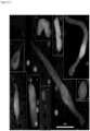
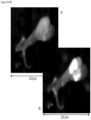
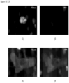
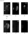
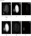
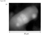
- FIG. 1 contains a collage of CAMLs showing many of the different CAML morphologies and signal variation from separate prostate, breast and pancreatic patient samples ( Adams, D., et al., Circulating giant macrophages as a potential biomarker of solid tumors. PNAS 2014, 111(9):3514-3519 ): ( FIG. 1A ) pancreatic, ( FIG. 1B ) breast, ( FIG. 1C ) breast, ( FIG. 1D ) breast, ( FIG. 1E ) prostate, ( FIG. 1F ) pancreatic, ( FIG. 1G ) pancreatic, ( FIG. 1H ) prostate, and ( FIG. 1I ) prostate. Examples of morphology variants are as follows: amorphous ( FIG.
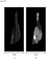
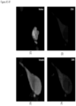
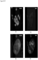
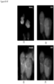
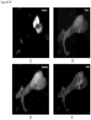
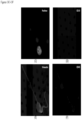
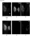
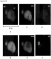
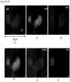
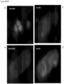
- FIGS. 2-13 show CAMLs stained with DAPI, CD10, vimentin and CD45, where ( FIG. 2A ) frames show merged microscope image DAPI (blue), CD10 (green), vimentin (red) and CD45 (violet), ( FIG. 2B ) frames show merged microscope image DAPI (white), CD10 (green), vimentin (red) and CD45 (violet), ( FIG. 2C ) frames show DAPI (white), ( FIG. 2D ) frames show CD10 (white), ( FIG. 2E ) frames show vimentin (white), and ( FIG. 2F ) frames show CD45 (white). Frame ( FIG. 2B ) with nucleus in white provides better image quality for some cells. Those cells possess the properties of CAMLs described in paragraphs above. The choice of the stain was chosen because the source of the cells is kidney cancer patients.
- FIGS. 2A-2F show engulfed DNA material at the end of top leg.
- FIGS. 3A-3F show a CAML in the process of engulfing a cell, where the DNA material is already in the CAML and some degraded cell cytoplasm is still partially outside the CAML.
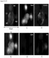
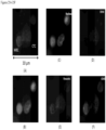
- FIGS. 4A-4F show a CAML in the process of engulfing degraded cellular material. This is most visible in the vimentin channel.
- FIGS. 5A-5F show a CAML with four engulfed CD45 positive white blood cells.
- FIGS. 6A-6F show a CAML with two engulfed CD45 positive white blood cells.
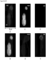
- FIGS. 7A-7F appear to show a CAML in the process of dividing.
- FIGS. 8A-8F show two similar side-by-side CAMLs suggesting the two cells might have come from the same origin.
- FIGS. 9A-9F show another example of two similar side-by-side CAMLs.
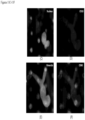
- FIGS. 10-12 show additional morphologies of CAML not identified in FIG. 1 .
- FIGS. 10A-10F show a CAML with two small legs on the left side of cell.
- FIGS. 11A-11F show a CAML with two legs on the same side.
- FIGS. 12A-12F show a CAML with one leg on the right side and two legs on the left side of the nucleus.
- FIGS. 13A-13F show a CAML with very thin legs and large single nucleus.
- FIGS. 14A-14F show a CAML found in patient with HSV-2 viral infection.
- T-cells are a subtype of white blood cell.
- the CD45 marker stains WBCs and is not specific to T-cells.
- T-cells can be differentiated from granulocytes by the morphology of the nucleus.
- T-cells have a single nucleus approximately round and smaller than 8 microns.
- Filtration of the blood can capture white blood cells (WBCs) bound to the CTCs.
- WBCs white blood cells
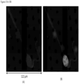
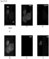
- FIGS. 15-18 show WBCs bond to CTCs found in the blood of breast cancer patients.
- the markers used for the breast cancer patients are DAPI, CK 8, 18 & 19, EpCAM and CD45.
- FIGS. 15-16 show the CTCs are apoptotic with CK 8, 18, and 19 degraded to spots.
- FIG. 18 shows a much degraded CTC losing both CK and EpCAM markers with no cytoplasm. It is often observed that the nuclei of the WBC and the CTCs are pulled towards each other as shown in FIG. 16 .
- FIGS. 19-21 shows WBCs bond to CTCs found in the blood of bladder cancer patients.
- the markers used for the bladder cancer patients are DAPI, CK 8, 18 & 19, EpCAM and CD45.
- EpCAM is degraded to spots.
- the cytoplasm of the WBC (marked in FIG. 19A ) and the CTC are in the process of merging with EpCAM around the WBC.
- the WBC still expresses CD45.
- FIGS. 20A-20F show a still relatively intact CTC bound to a WBC.
- FIGS. 21A-21F show a naked CTC nucleus without cytoplasm and the WBC (marked in FIG. 21A ) still expressing CD45, but much weaker than WBCs not bound to the CTC (not marked in FIG. 21A ).
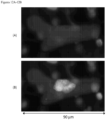
- FIGS. 22-28 show WBCs bond to a CTC found in the blood of kidney cancer patients.
- the markers used for these patient samples are DAPI, CD10, vimentin and CD45.
- FIGS. 22A-22F show a CTC from mesenchymal kidney cancer with high expression of vimentin. It is tightly bound to the WBC.
- FIGS. 23-28 show WBCs bond to a CTC found in blood of non-mesenchymal kidney cancer patients expressing lower level of vimentin than shown in FIG. 22 .
- the nuclei and the cytoplasm of the WBCs and CTCs are pulled toward each other.
- CD10, vimentin and CD45 markers all become very weak after WBCs bond to the CTCs. The amount of the cytoplasm decreases and eventually can all be lost.
- FIG. 30 shows that the percentage of the patients having CAMLs in stages I, II, III and IV are 87%, 100%, 91% and 97%, respectively.
- CAMLs were found to be more common than PDCTCs.
- Patient samples from 12 different solid tumors were analyzed: breast, prostate, pancreatic, lung, colorectal, uterine, neuroblastoma, esophageal, kidney, bladder, sarcoma, and ovarian.
- CAMLs were found in all those types of cancer (data not shown).
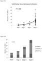
- the number of CAMLs in patients undergoing chemotherapy is only weakly associated with cancer stage at the time of pre-treatment clinical assessment, FIG. 32 , and is highly correlative with stage after pathological confirmation, FIG. 33A .
- FIG. 33B breaks down the number of CAMLs based on size for different stages. CAMLs in later stages have more CAMLs with larger sizes.
- CAMLs can be found in high percentages in all stages of solid tumors.
- CAMLs as a cancer screening marker was evaluated for breast cancer.
- a double-blind prospective study was conducted of 41 subjects where mammography was judged abnormal.
- a double blinded test was performed: (i) 7.5 mL of peripheral blood samples were taken to test for CAMLs and (ii) tissue diagnosis by core needle biopsy was performed. Though mammography could not distinguish the subpopulations in this group, CAML presence did differentiate between benign and malignant breast disease with a sensitivity of 90% and a specificity of 72% (data not shown).
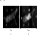
- Typical fluorescent microscopes usually use four or five fluorescent channels to minimize bleed through of fluorescent emissions into unintended fluorescent channels. One channel is taken by DAPI for imaging the nucleus. Often there is a need to evaluate more than three markers. Given these shortcomings, a method that allows analysis of up to approximately 12 different markers on the same cell was developed that can be used in conjunction with each of the methods disclosed in the present disclosure. After the process of filtration and staining the cells on the filter with a first set of markers, the cells of interest are identified and imaged. To evaluate more markers on the same cells, a quenching/stripping step was developed followed by a re-staining technique. This required the cell to stay at the same location to allow reimaging of the same cell.
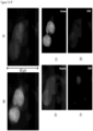
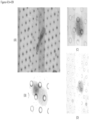
- Top row of FIG. 34 shows a CAML A with standard CTC stains: DAPI, CK 8, 18, 19, EpCAM and CD45.
- the second row shows the re-staining of the same cells after quenching and re-stained for markers of interest: PD-L1, CCR and PD-1.
- Third row of FIG. 34 shows a CAML B with standard CTC stains.
- the fourth row shows the re-staining of the same CAML B after quenching for markers of interest: PD-L1, CCR and PD-1.
- This re-staining method is particularly suitable for cells fixed on the microfilter. Their location is fixed so they can be reimaged to evaluate different markers. CTCs and other cells on the filter can also be re-stained using this technique. This re-staining method is very useful for analyzing the cancer type, companion diagnostic, therapy response, cancer screening, and a variety of research applications.
- CAMLs and CTCs can potentially be used to determine cancer subtyping for gene mutations, translocations and amplifications, by various PCR assays, microarrays, FISH assays and sequencing.
- Single cell molecular analysis is becoming common, and single cell analysis of CAMLs are particularly interesting. Some assays require more than one nucleus and/or cell to reduce errors.
- the present invention thus includes methods of molecular analysis of a single CAML cell where a single CAML cell is obtained and molecular analysis is conducted on the single cell.
- molecular analysis there is no limitation on the particular type of molecular analysis that may be conducted on the single cell and such means include, but are not limited to, nucleic acid sequencing, northern blot analysis and southern blot analysis.
- a method to collect CTCs and CAMLs using the microfiltration device is described.
- the important step to allow removal of the cells is to coat the filter to prevent the cells from sticking, for example coating using fetal bovine serum (FBS) or bovine serum albumin (BSA). Other coatings to prevent cell adhesion are also applicable.
- FBS fetal bovine serum
- BSA bovine serum albumin
- the sample flows through the filter to collect cells larger than the pores.
- Method 1 remove the filter from the filter holder and place in a dish or glass slide with cells on top, and cover with appropriate liquid, such as PBS; cells can be directly picking cells off the filter using micromanipulators.
- Method 2 attach a syringe filled with PBS to the bottom of the holder with the cells on the filter and backwash the cells off the filter.
- the cells may be concentrated by centrifugation and removing the supernatant.
- the cells need to be stained to enable visualization under the microscope.
- One non-limiting choice of stains is fluorescent intercalating dyes.
- Another example is to stain for cell surface markers such as EpCAM, CD45, and/or other markers.
- There are various ways to pick out cells of interest from the dish such as micromanipulators, or instruments such as CellColector, and other instruments.
- CAMLs can be used as a source of tissue for companion diagnostics to determine the specific drug to be prescribed to the patient.
- companion diagnostics utilize tissue biopsies to stain for markers for drug targets, perform FISH assays, and conduct other molecular assays to look for gene mutations, amplification or translocations by PCR, microarrays, sequencing, etc.
- tissue biopsies are FISH for HER2 amplification, FISH for ALK translocation, PD-L1 in tissue, AR and ER in tissue, etc.
- CTCs and CAMLs can be harvested repeatedly and used in place of the tissue biopsies. Also the same sample can be re-stained repeatedly to evaluate the efficacy of multiple drugs.
- Liquid Cell Biopsy provides a minimally invasive method to monitor treatment response in a patient.
- the following approaches can be adopted to monitor efficacy of a cancer treatment, comprising:
- FIG. 31 shows that chemotherapy responders seem to exhibit an increase in CAMLs shortly after chemotherapy treatment. In contrast, the number of CAMLs from target therapy did not show an increase above the no treatment control.
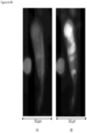
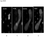
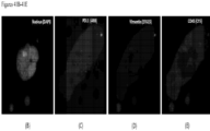
- the top row of FIG. 35 show a CAML from a lung cancer patient before radiation therapy stained for DAPI, PD-L1, RAD50 and PD-1.
- the bottom two rows show two different CAMLs after radiation therapy also stained for DAPI, PD-L1, RAD50 and PD-1.
- FIG. 36 shows a cluster of CTCs from a lung cancer patient after treatment with radiation using the standard CTC stains: DAPI, CK 8, 18, 19, EpCAM and CD45.
- the bottom row shows the same CTC cluster after quenching and re-staining for markers related to radiation therapy and immune response: PD-L1, RAD50 and PD-1.
- a fourth example is related to immunotherapy.
- Immunotherapies that enable the body to kill a tumor have shown impressive results for many types of cancers.
- An example of an immunotherapy drug is an antibody against PD-L1 on the surface of the tumor cells; and another sample of an immunotherapy drug is an antibody against PD-1 on the surface of the killer T-cells. Both types of immunotherapy drug enable killer T-cells to kill the tumor cells for some of the patients.
- FIG. 37 shows an example of number of CAMLs collected at four time intervals spread approximately by two to three weeks. The treatment of PD-1 was on dates T1 and T3, after providing the blood sample for Liquid Cell Biopsy. The decrease in number of CAMLs after treatment maybe an indication that the patient is not responding to treatment.
- FIG 38 shows the corresponding ranges of sizes of the CAMLs as the CAMLs are also decreasing in size. This information suggests that there might be a decrease of tumor debris in the blood as a result of treatment, suggesting that patient may not be responding to treatment.
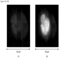
- FIG. 39 is a CAML before T1 showing very bright PD-L1. The signal of PD-L1 above background noise is about a factor of 8 of the background noise.
- FIG. 40 is a CAML at T1 collected just before treatment. The PD-L1 signal is now weak. The signal of PD-L1 above background noise is less than a factor of 2 of the background noise, indicating potential poor response.
- FIG. 41 is a CAML at T3. The PD-L1 signal is now very weak. The signal of PD-L1 above background noise is less than a factor of 1 of the background noise; this is an indication of loss of drug target, such as PD-L1.
- a fifth example is related to monitoring the success of surgery.
- Figure S5 in paper by Adams et al. (Circulating giant macrophages as a potential biomarker of solid tumors. PNAS 2014, 111(9):3514-3519 ) showed that surgery reduced the number of CAMLs. Continued presence of CAMLs in the blood of the patient could indicate that cancer might not be completely eradicated.
- Cells larger and/or less flexible than other cells present in a bodily fluid may be collected by filtering the bodily fluid.
- targeted cells indicative of a condition e.g., CAMLs and CTCs
- CAMLs and CTCs may be collected by passing a bodily fluid through a filter having openings that are too small for the target cells to pass through, but large enough for other cells to pass through.
- analyses of the target cells may be performed. Such analyses may include, for example, identifying, counting, characterizing expressions of markers, obtaining molecular analysis, and/or culturing the collected cells.
- CAMLs, pathologically-definable CTCs, and apoptotic CTCs are larger than red blood cells and most white blood cells.
- CellSieve TM microfilters (Creatv MicroTech) are one example of precision microfilters.
- CellSieve TM microfilters are transparent and nonfluorescent making them ideal for microscope imaging analysis. Pore sizes of 7-8 microns eliminated all the red blood cells and 99.99% of the white blood cells.
- Methods to fabricate microfilters producing uniform pore size and distribution are described in WO 2011/139445 , and PCT/US12/66390 .
- Microfilters made by a track etch method have randomly located pores that can overlap resulting in effectively large pores. They might lose some CAMLs and CTCs.
- Filtration is the best method to identify WBC bond to a CTC. Because both the WBC and CTCs lose their markers and lose cytoplasm, immunocapture and flow cytometry are less suitable methods to isolate them.
- CTCs and WBCs bound to CTCs may be detected in conjunction with the detection of CAMLs.
- detection may be simultaneously or sequential detection, and can utilize the same or different means.
- simultaneous detection using a microfilter having a pore size that selects for both cell types may be used.
- Suitable microfilters can have a variety of pore sizes and shapes. Microfilters having pores of about 7-8 microns in size are acceptable, and include round, rectangular and race track pore shapes. Microfilters having round pores of about 7-8 microns in size are especially optimal when polymeric microfilters are used.
- the microfilter has precision pore geometry and uniform pore distribution.
- Methods for isolating CAMLs and/or CTCs from a biological sample and counting the isolated cells using a camera can be used.
- a camera such as a cell phone camera
- the ability to count CAMLs and/or CTCs based on colorimetric staining may be sufficient for some applications.
- cancer tends to be diagnosed in late stages which translates to limited treatment options and dim outcomes.
- a method to provide a low cost diagnostic based on the counts of CAMLs and/or CTCs in a sample can adopt one or more of the following concepts.
- the large pore size in the filters will reduce the WBC contamination. Manual draw will reduce the cost. Colorimetric stain is low cost. Cell phone cameras can visualize CAMLs due to the large size of the cells.
- CAMLs and CTCs described herein make them well-suited for use in clinical methodology including methods of screening and diagnosis diseases such as cancer, monitoring treatment, monitoring of disease progression and recurrence.
- CAMLs, CTCs or T-cells bound to tumor cells After CAMLs, CTCs or T-cells bound to tumor cells are found, it may be possible to identify the type of tumor by staining, and in some instances, re-staining these cells with markers associated with the type of tumor.
- the National Cancer Institute tumor marker FactSheet lists many cancer markers (see the NCI website having the URL ending in "cancer.gov/cancertopics/factsheet/detection/tumor-markers" and "cancer.gov/about-cancer/diagnosis-staging/diagnosis/tumor-markers-fact-sheet#q5"). Cancer markers are not limited to this list. A few examples of markers listed below can be used to stain CAMLs and CTCs to provide initial indications of the cancer type:
- one marker may be sufficient for some types of cancer.
- the following is an illustration of analyzing CAMLs and CTCs for cancer screening up to four types of epithelial cancers using microfiltration method:
- CT scans for lung can show unusual findings as small as 4 mm in size. It is now a recommended screening method for lung cancer. To verify that an initial finding is lung cancer, tissue biopsy is needed. Tissue biopsy for lung is very challenging and it is associated with higher risk of causing undesirable effects. Presence of CAMLs with associated lung cancer markers such as cytokeratin fragments 21-1, and other markers can be used to provide a non-invasive step towards determine lung cancer.
- CAMLs including the markers of CA125, Ova1 for ovarian cancer, and CEA, CA15-3/CA27.29, ER, PR and HER2 for breast cancer can be performed.
- markers can be PSMA for prostate cancer, CEA for colorectal cancer, cytokeratin fragments 21-1 for lung cancer and PDX-1 for pancreatic cancer.
- the procedure and markers can vary depending on the CAML and CTC isolation method, the microscope, cancer types of interest, etc. In summary, it is possible to screen for one specific cancer, a few cancers, or any solid tumors in the category of carcinomas, sarcomas, neuroblastomas and melanomas.
- the markers do not need to be limited to the ones describe here.
- the invention is directed to methods for determining cancer stage in a subject, comprising characterizing selected characteristics of CAMLs in a biological sample from a subject having a cancer, wherein the selected characteristics are (i) number of CAMLs and (ii) average size of CAMLs, and wherein when measured in a sample of peripheral blood in a volume of 7.5ml, then 5 or more CAMLs and about 70% CAMLs larger than 40 microns in diameter is indicative of stage III cancer; and 5 or more CAMLs and about 80% CAMLs larger than 40 microns in diameter is indicative of stage IV cancer.
- the identification of the specific type of cancer can be performed by standard methods, such as staining for cancer markers.
- the methods encompassed by this embodiment also include characterizing selected characteristics of CTCs in the biological sample, wherein the selected characteristics of the CTCs are one or more characteristics selected from the group consisting of (i) number of CTCs; (ii) number of WBCs bound to the CTCs; (iii) degree of cytokeratin 8 expression; (iv) degree of cytokeratin 18 expression; (v) degree of cytokeratin 19 expression; (vi) degree of EpCAM expression; (vii) degree of vimentin expression; (viii) degree of PD-L1 expression; (ix) degree of uroplakin expression; (x) cytokeratin morphology; (xi) location of markers; and (xii) intensity of marker staining, preferably wherein the selected characteristics of the CTCs are one or more of: (i) number of CTCs; (ii) number of WBCs bound to the CTCs; (iii) degree of cytokeratin 8 expression; (iv) degree of
- the CAMLs and/or CTCs are collected from the biological sample prior to characterization.
- the number of CAMLs in stage III and IV cancer are typically 5 or more in a sample of peripheral blood in a volume of 7.5 ml.
- the percentage of CAMLs larger than 40 microns in size by diameter is about 70% for Stage III and about 80% for Stage IV patients.
- the invention is directed to methods for monitoring efficacy of a cancer treatment, comprising (a) assaying selected characteristics of CAMLs in a biological sample from a subject undergoing cancer treatment, and (b) comparing assay values for the selected characteristics determined in (a) to assay values for the same characteristics assayed in a similar biological sample from the same subject at one or more time points before, during or after completion of treatment, wherein a change in one or more assay values indicates efficacy of the cancer treatment in the subject wherein the selected characteristics are (i) number of CAMLs and (ii) average size of CAMLs, and wherein a change in number and average size of the CAMLs indicates efficacy of the cancer treatment.
- the methods encompassed by this embodiment also include (a) assaying one or more selected characteristics of CTCs in the biological sample, and (b) comparing assay values for the one or more selected characteristics determined in (a) to assay values for the same characteristics assayed in a similar biological sample from the same subject at one or more time points before, during or after completion of treatment, wherein the selected characteristics of the CTCs are one or more characteristics selected from the group consisting of (i) number of CTCs; (ii) number of WBCs bound to the CTCs; (iii) degree of cytokeratin 8 expression; (iv) degree of cytokeratin 18 expression; (v) degree of cytokeratin 19 expression; (vi) degree of EpCAM expression; (vii) degree of vimentin expression; (viii) degree of PD-L1 expression; (ix) degree of uroplakin expression; (x) cytokeratin morphology; (xi) location of markers; and (xii) intensity of marker sta
- the selected characteristics of the CAMLs are: (i) change of the number of CAMLs from the same subject at different time points after treatments; (ii) change in average size of CAMLs at the different time points; and one or more of (iii) change in intensity of markers in the CAMLs at the different time points; and (iv) change of location of markers from nucleus to cytoplasm or vice versa in the CAMLs.
- the selected characteristics of the CTCs are one or more of: (i) change of the number of CTCs in the biological samples at different time points after treatment; (ii) change in intensity of markers in the CTCs at the different time points; (iii) change of location of markers from nucleus to cytoplasm or vice versa; (iv) change in number of WBCs bound to CTCs in the biological sample; and (v) change in number of WBCs bound to CTCs in the biological samples at different time points after treatments.
- CAMLs and/or CTCs and/or WBCs bound to CTCs will be an indication of treatment efficacy, where the change may be an increase or a decrease in the number of CAMLs and/or CTCs and/or WBCs bound to CTCs.
- the information gathered on CAMLs, CTCs and CTC bound to WBC can be used independent of each other.
- the information gathered on CAMLs, CTCs and CTC bound to WBC can also be used together.
- CAMLs and/or CTCs can be an indication of treatment efficacy, where the change in the size may be an increase or a decrease in size of CAMLs and/or CTCs and/or WBCs bound to CTCs.
- the information gathered on CAMLs and CTCs can be used independently or together.
- CAMLs The capability of tracking CAMLs provides a novel opportunity to routinely monitor necrosis and chemotherapy or radiation therapy response. If the chemotherapy is not working, the CAMLs number will not increase. This can be used in parallel with CTC detection. If the treatment is working, the number of pathologically-definable CTCs will decrease and number of apoptotic CTCs will increase. However, CTCs cannot always be detected. If CTCs are detected at the same time as CAMLs, the sensitivity and specificity can be improved. For many cancers there are large array of chemotherapy agents. If the patient is not responding to one type of chemotherapy, the patient can quickly switch to another.
- the biological sample may be any suspected of containing CTCs, WBCs bound to CTCs, and/or CAMLs.
- the biological sample is one or more selected from the group consisting of peripheral blood, blood, lymph nodes, bone marrow, cerebral spinal fluid, tissue, and urine.
- the sample may be a fresh sample or a cryo-preserved sample that is thawed.
- the biological sample is peripheral blood.
- the blood is antecubital-vein blood, inferior-vena-cava blood or jugular-vein blood.
- Circulating monocytes have the ability to enter any tissue compartment of the body, including lymph nodes, bone marrow, most organs, and even cross the blood brain barrier.
- the detection of CAMLs is therefore not limited to blood, and the cells can also be found in lymph nodes, bone marrow, cerebral spinal fluid, most organs, and urine.
- the cancer is one or more of a solid tumor, Stage I cancer, Stage II cancer, Stage III cancer, Stage IV cancer, carcinoma, sarcoma, neuroblastoma, melanoma, epithelial cell cancer, breast cancer, prostate cancer, lung cancer, pancreatic cancer, colorectal cancer, and other solid tumor cancers.
- a solid tumor Stage I cancer, Stage II cancer, Stage III cancer, Stage IV cancer, carcinoma, sarcoma, neuroblastoma, melanoma, epithelial cell cancer, breast cancer, prostate cancer, lung cancer, pancreatic cancer, colorectal cancer, and other solid tumor cancers.
- CAMLs can be found in stage I and II of cancer, CAMLs can be used as screening for early detection of carcinomas, sarcomas, neuroblastomas and melanomas.
- Carcinomas are cancer of epithelial origin especially for high risk patients for breast, prostate, lung, pancreatic, colorectal and other cancers. Specificity of the type of cancer can be determined by re-staining for various cancer site specific markers on the same cells captured on the microfilter. Some examples are (i) use antibody against PSMA to specifically identifying prostate cancer, (ii) use antibody against PDX-1 to specifically identifying lung cancer, (iii) antibody against CA125 for ovarian cancer, and (iv) clorotoxin to identify glioma.
- CAMLs can be used to determine early recurrence of cancer when the cancer was under remission.
- CT, MRI and PET imaging are used to monitor the patient's tumor, requiring the tumor to change in size substantially to notice the difference. Patients can therefore lose valuable time in beginning treatment when only subtle size changes occur.
- CAMLs, alone or in combination with CTCs, can provide early detection of return of cancer. Non-invasive blood test of CAMLs and CTCs is much lower in cost than CT, MRI and PET imaging.
- the CAMLs can also potentially be used to determine cancer subtyping or gene mutations, translocations or amplification. There are a number of cancerous nuclei in each CAML. Thus, molecular analysis of the nucleus for genetic mutation, genetic defects, and gene translocations can provide information to determine treatments. There are drugs that specifically target certain gene mutations, translocation or amplifications. CAMLs can be used along or in parallel with CTCs for molecular analysis.
- the volume of the biological sample will vary based on the source and/or identity of the sample.
- the volume of blood may range from about 0.5 ml to about 50 ml, about 1 ml to about 40 ml, about 2 ml to about 30 ml, about 3 ml to about 25 ml, about 4 ml to about 20 ml, about 5 ml to about 15 ml, about 6 ml to about 10 ml, or about 7 ml to about 8 ml.
- a suitable volume also includes about 1, 1.5, 2, 2.5, 3, 3.5, 4, 4.5, 5, 5.5, 6, 6.5, 7, 7.5, 8, 8.5, 9, 9.5 or 10 ml.
- Blood volume typically used for detection of CTCs is 7.5 mL. Larger volumes of blood will provide more sensitivity and consistency, but smaller volumes such as 3.5 mL may be sufficient. For many CTC detection methods, larger volumes of blood are not practical for a variety of reasons. However, microfiltration of blood to capture CTCs and/or CAMLs allows more flexibility to increase the sample size. Blood volumes of 50 mL have been shown to be successfully screened using CellSieve TM microfilters with 160,000 pores. The suitable volume of blood to capture CAMLs would be 7.5 ml.
- the anti-cancer therapeutic may be one or more of chemotherapy, radiation therapy, immunotherapy, vaccine therapy, targeted therapy, and/or a combination of therapies.
- CAMLs are detected and/or collected using one or more means selected from the group consisting of size exclusion methodology, immunocapture, red blood cell lysis, white blood cell depletion, FICOLL, electrophoresis, dielectrophoresis, flow cytometry and microfluidic chip, or a combination thereof.
- the size exclusion methodology comprises use of a microfilter. Suitable microfilters can have a variety of pore sizes and shapes. When CAMLs alone are being detected and/or collected, the pore sizes can range from about 15 microns to about 20 microns.
- the pore sizes can range from about 5 microns to about 10 microns, preferably 7-8 microns.
- the larger pore sizes will eliminate most of the WBC contamination on the filter.
- the pores may have a round, race-track shaped, oval, square and rectangular pore shape.
- the microfilter has precision pore geometry and uniform pore distribution.
- CAMLs are detected and/or collected using a microfluidic chip based on physical size-based sorting, hydrodynamic size-based sorting, grouping, trapping, immunocapture, concentrating large cells, or eliminating small cells based on size.
- the CAMLs are detected and/or collected using a low-pressure microfiltration assay.
- CAMLs provide a robust indicator of cancer presence.
- the sensitivity and specificity of the utility of CAMLs can be further improved in combination with simultaneous detection of CTCs and CTCs bond to WBCs.
- Cancer screening is a strategy used in a population to identify an unrecognized disease in individuals without signs or symptoms, with pre-symptomatic or unrecognized symptomatic disease. As such, screening tests are somewhat unique in that they are performed on persons apparently in good health. A screening test is not a diagnostic test. Diagnostic testing is a procedure performed to confirm, or determine the presence of disease in an individual suspected of having the disease.
- CAMLs can be used as a cancer diagnostic to provide additional non-invasive diagnostics to confirm other screening techniques, such as mammography, PSA test, presence of CA125, CT, MRI or PET imaging.
Landscapes
- Health & Medical Sciences (AREA)
- Life Sciences & Earth Sciences (AREA)
- Engineering & Computer Science (AREA)
- Chemical & Material Sciences (AREA)
- Immunology (AREA)
- Biomedical Technology (AREA)
- Oncology (AREA)
- Organic Chemistry (AREA)
- Cell Biology (AREA)
- Biotechnology (AREA)
- Molecular Biology (AREA)
- Hematology (AREA)
- Wood Science & Technology (AREA)
- Zoology (AREA)
- Genetics & Genomics (AREA)
- Analytical Chemistry (AREA)
- Pathology (AREA)
- Microbiology (AREA)
- Biochemistry (AREA)
- General Health & Medical Sciences (AREA)
- Urology & Nephrology (AREA)
- Proteomics, Peptides & Aminoacids (AREA)
- Bioinformatics & Cheminformatics (AREA)
- Physics & Mathematics (AREA)
- Hospice & Palliative Care (AREA)
- General Engineering & Computer Science (AREA)
- General Physics & Mathematics (AREA)
- Medicinal Chemistry (AREA)
- Food Science & Technology (AREA)
- Virology (AREA)
- Biophysics (AREA)
- Tropical Medicine & Parasitology (AREA)
- Measuring Or Testing Involving Enzymes Or Micro-Organisms (AREA)
- Investigating Or Analysing Biological Materials (AREA)
- Micro-Organisms Or Cultivation Processes Thereof (AREA)
Applications Claiming Priority (5)
| Application Number | Priority Date | Filing Date | Title |
|---|---|---|---|
| US201462041540P | 2014-08-25 | 2014-08-25 | |
| US201562131051P | 2015-03-10 | 2015-03-10 | |
| US201562138744P | 2015-03-26 | 2015-03-26 | |
| US201562166492P | 2015-05-26 | 2015-05-26 | |
| PCT/US2015/046782 WO2016033103A1 (en) | 2014-08-25 | 2015-08-25 | Use of circulating cell biomarkers in the blood for detection and diagnosis of diseases and methods of isolating them |
Publications (4)
| Publication Number | Publication Date |
|---|---|
| EP3186397A1 EP3186397A1 (en) | 2017-07-05 |
| EP3186397A4 EP3186397A4 (en) | 2018-02-28 |
| EP3186397B1 true EP3186397B1 (en) | 2024-02-07 |
| EP3186397C0 EP3186397C0 (en) | 2024-02-07 |
Family
ID=55400461
Family Applications (1)
| Application Number | Title | Priority Date | Filing Date |
|---|---|---|---|
| EP15836011.5A Active EP3186397B1 (en) | 2014-08-25 | 2015-08-25 | Use of circulating cell biomarkers in the blood for detection and diagnosis of diseases and methods of isolating them |
Country Status (7)
| Country | Link |
|---|---|
| US (2) | US10871491B2 (ja) |
| EP (1) | EP3186397B1 (ja) |
| JP (4) | JP6657509B2 (ja) |
| CN (2) | CN107109477B (ja) |
| AU (2) | AU2015306692B2 (ja) |
| CA (1) | CA2959072A1 (ja) |
| WO (1) | WO2016033103A1 (ja) |
Families Citing this family (15)
| Publication number | Priority date | Publication date | Assignee | Title |
|---|---|---|---|---|
| EP3186397B1 (en) * | 2014-08-25 | 2024-02-07 | Creatv Microtech, Inc. | Use of circulating cell biomarkers in the blood for detection and diagnosis of diseases and methods of isolating them |
| CN109311989A (zh) * | 2016-04-14 | 2019-02-05 | 创新微技术公司 | 在癌症疗法的治疗决定中的pd-l1表达的应用方法 |
| CA3053562A1 (en) * | 2017-02-16 | 2018-08-23 | Creatv Microtech, Inc. | Methods for predicting overall and progression free survival in subjects having cancer using circulating cancer associated macrophage-like cells (camls) |
| CA3056654A1 (en) * | 2017-03-31 | 2018-10-04 | Creatv Microtech, Inc. | Methods of using giant cell nucleic acid characterization in cancer screening, diagnostics, treatment and recurrence |
| CN107643238A (zh) * | 2017-09-19 | 2018-01-30 | 中山大学附属第医院 | 一种定量检测血液循环微粒浓度的方法 |
| JP7337805B2 (ja) * | 2017-12-27 | 2023-09-04 | テサロ, インコーポレイテッド | がんを処置する方法 |
| WO2019178226A1 (en) | 2018-03-13 | 2019-09-19 | Creatv Microtech, Inc. | Methods for monitoring treatment response and disease progression in subjects using circulating cells |
| CN109856388A (zh) * | 2018-11-29 | 2019-06-07 | 北京优迅医学检验实验室有限公司 | 循环肿瘤细胞的捕获方法及捕获试剂盒 |
| US20220128544A1 (en) | 2019-02-13 | 2022-04-28 | Creatv Microtech, Inc. | Ccr5-based methods for predicting overall and progression free survival in subjects having cancer |
| CN112257508B (zh) * | 2020-06-30 | 2022-03-11 | 华为技术有限公司 | 识别物体的卫生状况方法及相关电子设备 |
| KR102448588B1 (ko) * | 2020-10-08 | 2022-09-27 | 가톨릭대학교 산학협력단 | 췌장암 진단용 바이오마커 및 이의 용도 |
| CN112462065A (zh) * | 2020-11-16 | 2021-03-09 | 浙江博真生物科技有限公司 | 一种用于检测实体肿瘤的抗体组合物、试剂盒及检测方法 |
| US20230175073A1 (en) * | 2021-12-07 | 2023-06-08 | Epic Sciences, Inc. | Enhanced characterization of breast cancer |
| CN115718095B (zh) * | 2022-03-28 | 2023-09-01 | 南京诺源医疗器械有限公司 | 一种循环肿瘤细胞自动扫描方法及装置 |
| CN115792245A (zh) * | 2023-02-02 | 2023-03-14 | 北京市肿瘤防治研究所 | 一种用于同时对外周血胃癌循环肿瘤细胞活性和免疫状态进行检测的方法 |
Family Cites Families (20)
| Publication number | Priority date | Publication date | Assignee | Title |
|---|---|---|---|---|
| DE10238046A1 (de) * | 2002-08-20 | 2004-03-04 | Giesing, Michael, Prof. Dr.med. | Verfahren zum Untersuchen von Körperflüssigkeiten auf Krebszellen, dessen Verwendung, entsprechende Analysekits und Verwendung bestimmter Wirkstoffe zur Behandlung von Krebs |
| US20060178833A1 (en) | 2005-02-04 | 2006-08-10 | Bauer Kenneth D | System for and method of providing diagnostic information through microscopic imaging |
| US9134237B2 (en) * | 2005-09-20 | 2015-09-15 | Janssen Diagnotics, LLC | High sensitivity multiparameter method for rare event analysis in a biological sample |
| US20090317836A1 (en) | 2006-01-30 | 2009-12-24 | The Scripps Research Institute | Methods for Detection of Circulating Tumor Cells and Methods of Diagnosis of Cancer in Mammalian Subject |
| EA201400710A1 (ru) | 2008-01-18 | 2015-03-31 | Президент Энд Феллоуз Оф Гарвард Колледж | Способы детекции признаков заболеваний или состояний в жидкостях организма |
| CA2736178A1 (en) * | 2008-09-05 | 2010-03-11 | Peter Kuhn | Methods for the detection of circulating tumor cells |
| JP2012506048A (ja) | 2008-10-20 | 2012-03-08 | ベリデックス・エルエルシー | マウスにおける循環乳癌細胞の連続的変化を監視するための前臨床方法 |
| WO2010111388A2 (en) | 2009-03-24 | 2010-09-30 | Biocept, Inc. | Devices and methods of cell capture and analysis |
| WO2010135603A2 (en) | 2009-05-20 | 2010-11-25 | California Institute Of Technology | Method for cancer detection, diagnosis and prognosis |
| WO2011002649A1 (en) | 2009-06-30 | 2011-01-06 | Veridex, Llc | Analysis of circulating tumor-related microparticles |
| US20140315295A1 (en) | 2013-03-15 | 2014-10-23 | Creatv Microtech, Inc. | Polymer microfilters, devices comprising the same, methods of manufacturing the same, and uses thereof |
| US11613115B2 (en) | 2010-05-03 | 2023-03-28 | Creatv Microtech, Inc. | Polymer microfilters and methods of manufacturing the same |
| US20120040361A1 (en) * | 2010-07-22 | 2012-02-16 | National Tsing Hua University | Methods and compositions for detection of lethal system and uses thereof |
| CN104094116B (zh) * | 2012-01-24 | 2017-05-31 | 辉瑞公司 | 在哺乳动物个体中检测5t4阳性循环肿瘤细胞的方法和诊断5t4阳性癌症的方法 |
| WO2013146993A1 (ja) * | 2012-03-28 | 2013-10-03 | 株式会社オンチップ・バイオテクノロジーズ | 末梢循環腫瘍細胞単位の悪性度の検出方法及びそのキット |
| ES2680243T3 (es) * | 2012-06-01 | 2018-09-05 | Creatv Microtech, Inc. | Captura, identificación y uso de un nuevo marcador de tumores sólidos en fluidos corporales |
| US11156596B2 (en) * | 2012-06-01 | 2021-10-26 | Creatv Microtech, Inc. | Capture, identification and use of a new biomarker of solid tumors in body fluids |
| CN103725589B (zh) * | 2012-10-10 | 2015-07-08 | 中国科学院化学研究所 | 用于循环肿瘤细胞富集和检测的生物芯片及其制备方法 |
| EP3186397B1 (en) * | 2014-08-25 | 2024-02-07 | Creatv Microtech, Inc. | Use of circulating cell biomarkers in the blood for detection and diagnosis of diseases and methods of isolating them |
| CN109311989A (zh) * | 2016-04-14 | 2019-02-05 | 创新微技术公司 | 在癌症疗法的治疗决定中的pd-l1表达的应用方法 |
-
2015
- 2015-08-25 EP EP15836011.5A patent/EP3186397B1/en active Active
- 2015-08-25 AU AU2015306692A patent/AU2015306692B2/en active Active
- 2015-08-25 CA CA2959072A patent/CA2959072A1/en active Pending
- 2015-08-25 JP JP2017510378A patent/JP6657509B2/ja active Active
- 2015-08-25 US US15/506,439 patent/US10871491B2/en active Active
- 2015-08-25 WO PCT/US2015/046782 patent/WO2016033103A1/en active Application Filing
- 2015-08-25 CN CN201580058052.7A patent/CN107109477B/zh active Active
- 2015-08-25 CN CN202011560678.7A patent/CN112877426A/zh active Pending
-
2019
- 2019-11-22 JP JP2019211754A patent/JP6912064B2/ja active Active
-
2020
- 2020-12-21 US US17/128,807 patent/US20210132073A1/en active Pending
-
2021
- 2021-06-29 JP JP2021107492A patent/JP7215805B2/ja active Active
- 2021-10-21 AU AU2021254611A patent/AU2021254611A1/en active Pending
-
2023
- 2023-01-13 JP JP2023003897A patent/JP2023040252A/ja active Pending
Also Published As
| Publication number | Publication date |
|---|---|
| AU2021254611A1 (en) | 2021-11-18 |
| EP3186397A1 (en) | 2017-07-05 |
| JP2021170012A (ja) | 2021-10-28 |
| JP6657509B2 (ja) | 2020-03-04 |
| CN112877426A (zh) | 2021-06-01 |
| US10871491B2 (en) | 2020-12-22 |
| JP6912064B2 (ja) | 2021-07-28 |
| JP2023040252A (ja) | 2023-03-22 |
| AU2015306692A1 (en) | 2017-03-09 |
| EP3186397C0 (en) | 2024-02-07 |
| CA2959072A1 (en) | 2016-03-03 |
| JP7215805B2 (ja) | 2023-01-31 |
| JP2020042037A (ja) | 2020-03-19 |
| US20210132073A1 (en) | 2021-05-06 |
| CN107109477A (zh) | 2017-08-29 |
| EP3186397A4 (en) | 2018-02-28 |
| US20180238893A1 (en) | 2018-08-23 |
| CN107109477B (zh) | 2021-01-12 |
| WO2016033103A1 (en) | 2016-03-03 |
| AU2015306692B2 (en) | 2021-07-22 |
| JP2017527799A (ja) | 2017-09-21 |
Similar Documents
| Publication | Publication Date | Title |
|---|---|---|
| US20210132073A1 (en) | Use of circulating cell biomarkers in the blood for detection and diagnosis of diseases and methods of isolating them | |
| CA2874691C (en) | Capture, identification and use of a new biomarker of solid tumors in body fluids | |
| KR101716555B1 (ko) | 포유동물 대상체에서 5t4-양성 순환 종양 세포를 검출하는 방법 및 5t4-양성 암을 진단하는 방법 | |
| AU2018220513B2 (en) | Methods for predicting overall and progression free survival in subjects having cancer using circulating cancer associated macrophage-like cells (CAMLs) | |
| US20220042970A1 (en) | Capture, identification and use of a new biomarker of solid tumors in body fluids | |
| KR20240033068A (ko) | 순환성 암 관련 대식세포 유사 세포(caml)의 변화를 이용한 암 치료 반응의 예측 및/또는 모니터링 방법 | |
| Hideyuki Abe et al. | Heterogeneity of Anaplastic Lymphoma Kinase Gene Rearrangement in Non–Small-Cell Lung Carcinomas | |
| 安倍秀幸 et al. | Heterogeneity of anaplastic lymphoma kinase gene rearrangement in non-small-cell lung carcinomas: a comparative study between small biopsy and excision samples |
Legal Events
| Date | Code | Title | Description |
|---|---|---|---|
| STAA | Information on the status of an ep patent application or granted ep patent |
Free format text: STATUS: THE INTERNATIONAL PUBLICATION HAS BEEN MADE |
|
| PUAI | Public reference made under article 153(3) epc to a published international application that has entered the european phase |
Free format text: ORIGINAL CODE: 0009012 |
|
| STAA | Information on the status of an ep patent application or granted ep patent |
Free format text: STATUS: REQUEST FOR EXAMINATION WAS MADE |
|
| 17P | Request for examination filed |
Effective date: 20170221 |
|
| AK | Designated contracting states |
Kind code of ref document: A1 Designated state(s): AL AT BE BG CH CY CZ DE DK EE ES FI FR GB GR HR HU IE IS IT LI LT LU LV MC MK MT NL NO PL PT RO RS SE SI SK SM TR |
|
| AX | Request for extension of the european patent |
Extension state: BA ME |
|
| DAV | Request for validation of the european patent (deleted) | ||
| DAX | Request for extension of the european patent (deleted) | ||
| A4 | Supplementary search report drawn up and despatched |
Effective date: 20180130 |
|
| RIC1 | Information provided on ipc code assigned before grant |
Ipc: C12N 5/00 20060101ALI20180124BHEP Ipc: C12Q 1/68 20180101AFI20180124BHEP Ipc: C12Q 1/6886 20180101ALI20180124BHEP Ipc: G01N 33/574 20060101ALI20180124BHEP Ipc: C12N 5/09 20100101ALI20180124BHEP |
|
| REG | Reference to a national code |
Ref country code: HK Ref legal event code: DE Ref document number: 1236046 Country of ref document: HK |
|
| STAA | Information on the status of an ep patent application or granted ep patent |
Free format text: STATUS: EXAMINATION IS IN PROGRESS |
|
| 17Q | First examination report despatched |
Effective date: 20190514 |
|
| STAA | Information on the status of an ep patent application or granted ep patent |
Free format text: STATUS: EXAMINATION IS IN PROGRESS |
|
| STAA | Information on the status of an ep patent application or granted ep patent |
Free format text: STATUS: EXAMINATION IS IN PROGRESS |
|
| GRAP | Despatch of communication of intention to grant a patent |
Free format text: ORIGINAL CODE: EPIDOSNIGR1 |
|
| STAA | Information on the status of an ep patent application or granted ep patent |
Free format text: STATUS: GRANT OF PATENT IS INTENDED |
|
| INTG | Intention to grant announced |
Effective date: 20230816 |
|
| GRAS | Grant fee paid |
Free format text: ORIGINAL CODE: EPIDOSNIGR3 |
|
| GRAA | (expected) grant |
Free format text: ORIGINAL CODE: 0009210 |
|
| STAA | Information on the status of an ep patent application or granted ep patent |
Free format text: STATUS: THE PATENT HAS BEEN GRANTED |
|
| AK | Designated contracting states |
Kind code of ref document: B1 Designated state(s): AL AT BE BG CH CY CZ DE DK EE ES FI FR GB GR HR HU IE IS IT LI LT LU LV MC MK MT NL NO PL PT RO RS SE SI SK SM TR |
|
| REG | Reference to a national code |
Ref country code: GB Ref legal event code: FG4D |
|
| REG | Reference to a national code |
Ref country code: CH Ref legal event code: EP |
|
| REG | Reference to a national code |
Ref country code: IE Ref legal event code: FG4D |
|
| REG | Reference to a national code |
Ref country code: DE Ref legal event code: R096 Ref document number: 602015087492 Country of ref document: DE |
|
| U01 | Request for unitary effect filed |
Effective date: 20240207 |
|
| U07 | Unitary effect registered |
Designated state(s): AT BE BG DE DK EE FI FR IT LT LU LV MT NL PT SE SI Effective date: 20240212 |
|
| REG | Reference to a national code |
Ref country code: ES Ref legal event code: FG2A Ref document number: 2970741 Country of ref document: ES Kind code of ref document: T3 Effective date: 20240530 |