EP3118220B1 - Protein - Google Patents
Protein Download PDFInfo
- Publication number
- EP3118220B1 EP3118220B1 EP16183598.8A EP16183598A EP3118220B1 EP 3118220 B1 EP3118220 B1 EP 3118220B1 EP 16183598 A EP16183598 A EP 16183598A EP 3118220 B1 EP3118220 B1 EP 3118220B1
- Authority
- EP
- European Patent Office
- Prior art keywords
- cancer
- ogta076
- antibody
- subject
- antibodies
- Prior art date
- Legal status (The legal status is an assumption and is not a legal conclusion. Google has not performed a legal analysis and makes no representation as to the accuracy of the status listed.)
- Active
Links
- 108090000623 proteins and genes Proteins 0.000 title description 224
- 102000004169 proteins and genes Human genes 0.000 title description 143
- 238000000034 method Methods 0.000 claims description 208
- 206010061535 Ovarian neoplasm Diseases 0.000 claims description 191
- 206010033128 Ovarian cancer Diseases 0.000 claims description 187
- 239000012634 fragment Substances 0.000 claims description 181
- 208000020816 lung neoplasm Diseases 0.000 claims description 178
- 206010058467 Lung neoplasm malignant Diseases 0.000 claims description 177
- 201000005202 lung cancer Diseases 0.000 claims description 177
- 210000004027 cell Anatomy 0.000 claims description 130
- 241000282414 Homo sapiens Species 0.000 claims description 88
- 230000000694 effects Effects 0.000 claims description 80
- 230000027455 binding Effects 0.000 claims description 79
- 230000001225 therapeutic effect Effects 0.000 claims description 41
- 238000011282 treatment Methods 0.000 claims description 40
- 239000000427 antigen Substances 0.000 claims description 36
- 108091007433 antigens Proteins 0.000 claims description 35
- 102000036639 antigens Human genes 0.000 claims description 35
- 238000012216 screening Methods 0.000 claims description 31
- 102100033486 Lymphocyte antigen 75 Human genes 0.000 claims description 18
- 101710157884 Lymphocyte antigen 75 Proteins 0.000 claims description 18
- 238000012544 monitoring process Methods 0.000 claims description 18
- 238000002560 therapeutic procedure Methods 0.000 claims description 18
- 230000009870 specific binding Effects 0.000 claims description 14
- 108010021625 Immunoglobulin Fragments Proteins 0.000 claims description 11
- 102000008394 Immunoglobulin Fragments Human genes 0.000 claims description 10
- 238000000338 in vitro Methods 0.000 claims description 10
- 230000001472 cytotoxic effect Effects 0.000 claims description 8
- 230000002285 radioactive effect Effects 0.000 claims description 8
- 239000003560 cancer drug Substances 0.000 claims description 7
- 230000008859 change Effects 0.000 claims description 7
- 231100000433 cytotoxic Toxicity 0.000 claims description 7
- 230000003013 cytotoxicity Effects 0.000 claims description 7
- 231100000135 cytotoxicity Toxicity 0.000 claims description 7
- 230000000295 complement effect Effects 0.000 claims description 6
- 239000012642 immune effector Substances 0.000 claims description 3
- 229940127121 immunoconjugate Drugs 0.000 claims description 3
- 229940121354 immunomodulator Drugs 0.000 claims description 3
- 230000001939 inductive effect Effects 0.000 claims description 3
- 231100000023 Cell-mediated cytotoxicity Toxicity 0.000 claims description 2
- 206010057250 Cell-mediated cytotoxicity Diseases 0.000 claims description 2
- 239000000611 antibody drug conjugate Substances 0.000 claims description 2
- 229940049595 antibody-drug conjugate Drugs 0.000 claims description 2
- 230000005890 cell-mediated cytotoxicity Effects 0.000 claims description 2
- 108090000765 processed proteins & peptides Proteins 0.000 description 228
- 102000004196 processed proteins & peptides Human genes 0.000 description 208
- 229920001184 polypeptide Polymers 0.000 description 174
- 150000001875 compounds Chemical class 0.000 description 164
- 206010009944 Colon cancer Diseases 0.000 description 161
- 208000001333 Colorectal Neoplasms Diseases 0.000 description 160
- 208000008839 Kidney Neoplasms Diseases 0.000 description 151
- 206010038389 Renal cancer Diseases 0.000 description 151
- 201000010982 kidney cancer Diseases 0.000 description 151
- 206010061902 Pancreatic neoplasm Diseases 0.000 description 150
- 201000007270 liver cancer Diseases 0.000 description 150
- 208000014018 liver neoplasm Diseases 0.000 description 150
- 208000015486 malignant pancreatic neoplasm Diseases 0.000 description 150
- 201000002528 pancreatic cancer Diseases 0.000 description 150
- 208000008443 pancreatic carcinoma Diseases 0.000 description 150
- 208000032852 chronic lymphocytic leukemia Diseases 0.000 description 149
- 235000018102 proteins Nutrition 0.000 description 137
- 208000028018 Lymphocytic leukaemia Diseases 0.000 description 130
- 239000003153 chemical reaction reagent Substances 0.000 description 76
- 150000007523 nucleic acids Chemical class 0.000 description 73
- 102000039446 nucleic acids Human genes 0.000 description 71
- 108020004707 nucleic acids Proteins 0.000 description 71
- 230000014509 gene expression Effects 0.000 description 70
- 206010028980 Neoplasm Diseases 0.000 description 68
- 238000012360 testing method Methods 0.000 description 62
- 201000011510 cancer Diseases 0.000 description 57
- 239000000203 mixture Substances 0.000 description 57
- 108020004414 DNA Proteins 0.000 description 51
- 239000000523 sample Substances 0.000 description 48
- 108020001507 fusion proteins Proteins 0.000 description 43
- 102000037865 fusion proteins Human genes 0.000 description 43
- 210000001519 tissue Anatomy 0.000 description 42
- 239000000499 gel Substances 0.000 description 36
- 239000003795 chemical substances by application Substances 0.000 description 34
- 238000004519 manufacturing process Methods 0.000 description 33
- 239000002773 nucleotide Substances 0.000 description 33
- 125000003729 nucleotide group Chemical group 0.000 description 33
- 208000037265 diseases, disorders, signs and symptoms Diseases 0.000 description 31
- 241001465754 Metazoa Species 0.000 description 30
- 235000001014 amino acid Nutrition 0.000 description 29
- 238000003556 assay Methods 0.000 description 29
- 229940024606 amino acid Drugs 0.000 description 28
- 201000010099 disease Diseases 0.000 description 27
- 230000002163 immunogen Effects 0.000 description 27
- 150000001413 amino acids Chemical class 0.000 description 26
- 238000003745 diagnosis Methods 0.000 description 26
- 238000005516 engineering process Methods 0.000 description 26
- 238000002474 experimental method Methods 0.000 description 25
- 108020004999 messenger RNA Proteins 0.000 description 25
- 239000013598 vector Substances 0.000 description 25
- 230000006870 function Effects 0.000 description 24
- 238000001514 detection method Methods 0.000 description 23
- 108060003951 Immunoglobulin Proteins 0.000 description 21
- 239000013604 expression vector Substances 0.000 description 21
- 102000018358 immunoglobulin Human genes 0.000 description 21
- 239000003814 drug Substances 0.000 description 20
- 230000000890 antigenic effect Effects 0.000 description 19
- 125000003275 alpha amino acid group Chemical group 0.000 description 18
- 238000009396 hybridization Methods 0.000 description 18
- 108010029485 Protein Isoforms Proteins 0.000 description 17
- 102000001708 Protein Isoforms Human genes 0.000 description 17
- 210000004072 lung Anatomy 0.000 description 17
- 238000004458 analytical method Methods 0.000 description 16
- 210000004185 liver Anatomy 0.000 description 16
- 239000000243 solution Substances 0.000 description 16
- 239000002671 adjuvant Substances 0.000 description 15
- 230000010056 antibody-dependent cellular cytotoxicity Effects 0.000 description 15
- 239000012472 biological sample Substances 0.000 description 15
- 239000003446 ligand Substances 0.000 description 15
- 238000004949 mass spectrometry Methods 0.000 description 15
- 108010003723 Single-Domain Antibodies Proteins 0.000 description 14
- 238000013459 approach Methods 0.000 description 14
- 239000000463 material Substances 0.000 description 14
- 239000000047 product Substances 0.000 description 14
- 210000002966 serum Anatomy 0.000 description 14
- 240000004808 Saccharomyces cerevisiae Species 0.000 description 13
- 235000014680 Saccharomyces cerevisiae Nutrition 0.000 description 13
- 239000002299 complementary DNA Substances 0.000 description 13
- 238000003384 imaging method Methods 0.000 description 13
- 238000003018 immunoassay Methods 0.000 description 13
- 230000008569 process Effects 0.000 description 13
- 206010041067 Small cell lung cancer Diseases 0.000 description 12
- 210000000170 cell membrane Anatomy 0.000 description 12
- 238000002347 injection Methods 0.000 description 12
- 239000007924 injection Substances 0.000 description 12
- 210000003734 kidney Anatomy 0.000 description 12
- 230000002611 ovarian Effects 0.000 description 12
- 239000008194 pharmaceutical composition Substances 0.000 description 12
- 241000894006 Bacteria Species 0.000 description 11
- 108010047041 Complementarity Determining Regions Proteins 0.000 description 11
- 108010052285 Membrane Proteins Proteins 0.000 description 11
- 238000010171 animal model Methods 0.000 description 11
- 239000003550 marker Substances 0.000 description 11
- 238000011321 prophylaxis Methods 0.000 description 11
- 239000000126 substance Substances 0.000 description 11
- 230000004083 survival effect Effects 0.000 description 11
- 241000588724 Escherichia coli Species 0.000 description 10
- 230000034994 death Effects 0.000 description 10
- 231100000517 death Toxicity 0.000 description 10
- 238000009472 formulation Methods 0.000 description 10
- 230000001965 increasing effect Effects 0.000 description 10
- 230000004048 modification Effects 0.000 description 10
- 238000012986 modification Methods 0.000 description 10
- 208000002154 non-small cell lung carcinoma Diseases 0.000 description 10
- 238000002360 preparation method Methods 0.000 description 10
- 238000000746 purification Methods 0.000 description 10
- 238000001356 surgical procedure Methods 0.000 description 10
- XLYOFNOQVPJJNP-UHFFFAOYSA-N water Substances O XLYOFNOQVPJJNP-UHFFFAOYSA-N 0.000 description 10
- QTBSBXVTEAMEQO-UHFFFAOYSA-N Acetic acid Chemical compound CC(O)=O QTBSBXVTEAMEQO-UHFFFAOYSA-N 0.000 description 9
- WEVYAHXRMPXWCK-UHFFFAOYSA-N Acetonitrile Chemical compound CC#N WEVYAHXRMPXWCK-UHFFFAOYSA-N 0.000 description 9
- LFQSCWFLJHTTHZ-UHFFFAOYSA-N Ethanol Chemical compound CCO LFQSCWFLJHTTHZ-UHFFFAOYSA-N 0.000 description 9
- 108090001090 Lectins Proteins 0.000 description 9
- 102000004856 Lectins Human genes 0.000 description 9
- 241000699660 Mus musculus Species 0.000 description 9
- 108091030071 RNAI Proteins 0.000 description 9
- 229940079593 drug Drugs 0.000 description 9
- 230000009368 gene silencing by RNA Effects 0.000 description 9
- 239000002523 lectin Substances 0.000 description 9
- 239000013612 plasmid Substances 0.000 description 9
- 102000005962 receptors Human genes 0.000 description 9
- 108020003175 receptors Proteins 0.000 description 9
- 238000006467 substitution reaction Methods 0.000 description 9
- 208000024891 symptom Diseases 0.000 description 9
- 102000004190 Enzymes Human genes 0.000 description 8
- 108090000790 Enzymes Proteins 0.000 description 8
- 241000699670 Mus sp. Species 0.000 description 8
- 241000700605 Viruses Species 0.000 description 8
- 210000004978 chinese hamster ovary cell Anatomy 0.000 description 8
- 238000010276 construction Methods 0.000 description 8
- 229940088598 enzyme Drugs 0.000 description 8
- 238000001502 gel electrophoresis Methods 0.000 description 8
- 239000011521 glass Substances 0.000 description 8
- 230000006698 induction Effects 0.000 description 8
- 210000004923 pancreatic tissue Anatomy 0.000 description 8
- 229960005486 vaccine Drugs 0.000 description 8
- 238000001262 western blot Methods 0.000 description 8
- 108010054477 Immunoglobulin Fab Fragments Proteins 0.000 description 7
- 102000001706 Immunoglobulin Fab Fragments Human genes 0.000 description 7
- 241000699666 Mus <mouse, genus> Species 0.000 description 7
- 241000283973 Oryctolagus cuniculus Species 0.000 description 7
- 241000700159 Rattus Species 0.000 description 7
- FAPWRFPIFSIZLT-UHFFFAOYSA-M Sodium chloride Chemical compound [Na+].[Cl-] FAPWRFPIFSIZLT-UHFFFAOYSA-M 0.000 description 7
- 238000012217 deletion Methods 0.000 description 7
- 230000037430 deletion Effects 0.000 description 7
- 230000002255 enzymatic effect Effects 0.000 description 7
- 210000004408 hybridoma Anatomy 0.000 description 7
- 230000028993 immune response Effects 0.000 description 7
- 229940099472 immunoglobulin a Drugs 0.000 description 7
- 238000003364 immunohistochemistry Methods 0.000 description 7
- 210000004962 mammalian cell Anatomy 0.000 description 7
- 238000011275 oncology therapy Methods 0.000 description 7
- 238000004393 prognosis Methods 0.000 description 7
- 230000002829 reductive effect Effects 0.000 description 7
- 239000006228 supernatant Substances 0.000 description 7
- 108091026890 Coding region Proteins 0.000 description 6
- PEDCQBHIVMGVHV-UHFFFAOYSA-N Glycerine Chemical compound OCC(O)CO PEDCQBHIVMGVHV-UHFFFAOYSA-N 0.000 description 6
- 241000238631 Hexapoda Species 0.000 description 6
- 102000018697 Membrane Proteins Human genes 0.000 description 6
- DBMJMQXJHONAFJ-UHFFFAOYSA-M Sodium laurylsulphate Chemical compound [Na+].CCCCCCCCCCCCOS([O-])(=O)=O DBMJMQXJHONAFJ-UHFFFAOYSA-M 0.000 description 6
- 230000001594 aberrant effect Effects 0.000 description 6
- 238000007792 addition Methods 0.000 description 6
- 210000003719 b-lymphocyte Anatomy 0.000 description 6
- 230000001580 bacterial effect Effects 0.000 description 6
- 210000004369 blood Anatomy 0.000 description 6
- 239000008280 blood Substances 0.000 description 6
- 238000010367 cloning Methods 0.000 description 6
- 239000013068 control sample Substances 0.000 description 6
- 238000009509 drug development Methods 0.000 description 6
- 239000000284 extract Substances 0.000 description 6
- 239000007850 fluorescent dye Substances 0.000 description 6
- 229960001438 immunostimulant agent Drugs 0.000 description 6
- 239000003022 immunostimulating agent Substances 0.000 description 6
- 230000003308 immunostimulating effect Effects 0.000 description 6
- 238000000099 in vitro assay Methods 0.000 description 6
- 238000011580 nude mouse model Methods 0.000 description 6
- 238000002823 phage display Methods 0.000 description 6
- 150000003839 salts Chemical class 0.000 description 6
- 239000007790 solid phase Substances 0.000 description 6
- 238000003786 synthesis reaction Methods 0.000 description 6
- 238000013519 translation Methods 0.000 description 6
- 241000282693 Cercopithecidae Species 0.000 description 5
- 108700024394 Exon Proteins 0.000 description 5
- 241000282412 Homo Species 0.000 description 5
- 241001529936 Murinae Species 0.000 description 5
- 108091028043 Nucleic acid sequence Proteins 0.000 description 5
- 108091034117 Oligonucleotide Proteins 0.000 description 5
- JLCPHMBAVCMARE-UHFFFAOYSA-N [3-[[3-[[3-[[3-[[3-[[3-[[3-[[3-[[3-[[3-[[3-[[5-(2-amino-6-oxo-1H-purin-9-yl)-3-[[3-[[3-[[3-[[3-[[3-[[5-(2-amino-6-oxo-1H-purin-9-yl)-3-[[5-(2-amino-6-oxo-1H-purin-9-yl)-3-hydroxyoxolan-2-yl]methoxy-hydroxyphosphoryl]oxyoxolan-2-yl]methoxy-hydroxyphosphoryl]oxy-5-(5-methyl-2,4-dioxopyrimidin-1-yl)oxolan-2-yl]methoxy-hydroxyphosphoryl]oxy-5-(6-aminopurin-9-yl)oxolan-2-yl]methoxy-hydroxyphosphoryl]oxy-5-(6-aminopurin-9-yl)oxolan-2-yl]methoxy-hydroxyphosphoryl]oxy-5-(6-aminopurin-9-yl)oxolan-2-yl]methoxy-hydroxyphosphoryl]oxy-5-(6-aminopurin-9-yl)oxolan-2-yl]methoxy-hydroxyphosphoryl]oxyoxolan-2-yl]methoxy-hydroxyphosphoryl]oxy-5-(5-methyl-2,4-dioxopyrimidin-1-yl)oxolan-2-yl]methoxy-hydroxyphosphoryl]oxy-5-(4-amino-2-oxopyrimidin-1-yl)oxolan-2-yl]methoxy-hydroxyphosphoryl]oxy-5-(5-methyl-2,4-dioxopyrimidin-1-yl)oxolan-2-yl]methoxy-hydroxyphosphoryl]oxy-5-(5-methyl-2,4-dioxopyrimidin-1-yl)oxolan-2-yl]methoxy-hydroxyphosphoryl]oxy-5-(6-aminopurin-9-yl)oxolan-2-yl]methoxy-hydroxyphosphoryl]oxy-5-(6-aminopurin-9-yl)oxolan-2-yl]methoxy-hydroxyphosphoryl]oxy-5-(4-amino-2-oxopyrimidin-1-yl)oxolan-2-yl]methoxy-hydroxyphosphoryl]oxy-5-(4-amino-2-oxopyrimidin-1-yl)oxolan-2-yl]methoxy-hydroxyphosphoryl]oxy-5-(4-amino-2-oxopyrimidin-1-yl)oxolan-2-yl]methoxy-hydroxyphosphoryl]oxy-5-(6-aminopurin-9-yl)oxolan-2-yl]methoxy-hydroxyphosphoryl]oxy-5-(4-amino-2-oxopyrimidin-1-yl)oxolan-2-yl]methyl [5-(6-aminopurin-9-yl)-2-(hydroxymethyl)oxolan-3-yl] hydrogen phosphate Polymers Cc1cn(C2CC(OP(O)(=O)OCC3OC(CC3OP(O)(=O)OCC3OC(CC3O)n3cnc4c3nc(N)[nH]c4=O)n3cnc4c3nc(N)[nH]c4=O)C(COP(O)(=O)OC3CC(OC3COP(O)(=O)OC3CC(OC3COP(O)(=O)OC3CC(OC3COP(O)(=O)OC3CC(OC3COP(O)(=O)OC3CC(OC3COP(O)(=O)OC3CC(OC3COP(O)(=O)OC3CC(OC3COP(O)(=O)OC3CC(OC3COP(O)(=O)OC3CC(OC3COP(O)(=O)OC3CC(OC3COP(O)(=O)OC3CC(OC3COP(O)(=O)OC3CC(OC3COP(O)(=O)OC3CC(OC3COP(O)(=O)OC3CC(OC3COP(O)(=O)OC3CC(OC3COP(O)(=O)OC3CC(OC3COP(O)(=O)OC3CC(OC3CO)n3cnc4c(N)ncnc34)n3ccc(N)nc3=O)n3cnc4c(N)ncnc34)n3ccc(N)nc3=O)n3ccc(N)nc3=O)n3ccc(N)nc3=O)n3cnc4c(N)ncnc34)n3cnc4c(N)ncnc34)n3cc(C)c(=O)[nH]c3=O)n3cc(C)c(=O)[nH]c3=O)n3ccc(N)nc3=O)n3cc(C)c(=O)[nH]c3=O)n3cnc4c3nc(N)[nH]c4=O)n3cnc4c(N)ncnc34)n3cnc4c(N)ncnc34)n3cnc4c(N)ncnc34)n3cnc4c(N)ncnc34)O2)c(=O)[nH]c1=O JLCPHMBAVCMARE-UHFFFAOYSA-N 0.000 description 5
- 230000004913 activation Effects 0.000 description 5
- 230000003321 amplification Effects 0.000 description 5
- 230000000237 anti-lymphoid effect Effects 0.000 description 5
- 230000002946 anti-pancreatic effect Effects 0.000 description 5
- 230000004071 biological effect Effects 0.000 description 5
- 230000015572 biosynthetic process Effects 0.000 description 5
- 230000001413 cellular effect Effects 0.000 description 5
- 238000005119 centrifugation Methods 0.000 description 5
- 238000006243 chemical reaction Methods 0.000 description 5
- 238000003776 cleavage reaction Methods 0.000 description 5
- 238000013270 controlled release Methods 0.000 description 5
- 210000000987 immune system Anatomy 0.000 description 5
- 238000001727 in vivo Methods 0.000 description 5
- 150000002500 ions Chemical class 0.000 description 5
- 210000004379 membrane Anatomy 0.000 description 5
- 239000012528 membrane Substances 0.000 description 5
- 238000003199 nucleic acid amplification method Methods 0.000 description 5
- 150000002482 oligosaccharides Polymers 0.000 description 5
- 230000001105 regulatory effect Effects 0.000 description 5
- 230000007017 scission Effects 0.000 description 5
- 208000000587 small cell lung carcinoma Diseases 0.000 description 5
- 150000003384 small molecules Chemical class 0.000 description 5
- 230000009261 transgenic effect Effects 0.000 description 5
- 208000029729 tumor suppressor gene on chromosome 11 Diseases 0.000 description 5
- 210000002700 urine Anatomy 0.000 description 5
- 102100033400 4F2 cell-surface antigen heavy chain Human genes 0.000 description 4
- 235000014469 Bacillus subtilis Nutrition 0.000 description 4
- 102000004127 Cytokines Human genes 0.000 description 4
- 108090000695 Cytokines Proteins 0.000 description 4
- 238000002965 ELISA Methods 0.000 description 4
- 102100031726 Endoplasmic reticulum junction formation protein lunapark Human genes 0.000 description 4
- 108010087819 Fc receptors Proteins 0.000 description 4
- 102000009109 Fc receptors Human genes 0.000 description 4
- 108010070675 Glutathione transferase Proteins 0.000 description 4
- 102000005720 Glutathione transferase Human genes 0.000 description 4
- 101000800023 Homo sapiens 4F2 cell-surface antigen heavy chain Proteins 0.000 description 4
- 101000941029 Homo sapiens Endoplasmic reticulum junction formation protein lunapark Proteins 0.000 description 4
- 206010027476 Metastases Diseases 0.000 description 4
- 238000011579 SCID mouse model Methods 0.000 description 4
- 108090000631 Trypsin Proteins 0.000 description 4
- 102000004142 Trypsin Human genes 0.000 description 4
- 238000005273 aeration Methods 0.000 description 4
- 238000001042 affinity chromatography Methods 0.000 description 4
- 238000013019 agitation Methods 0.000 description 4
- 210000004102 animal cell Anatomy 0.000 description 4
- 239000005557 antagonist Substances 0.000 description 4
- -1 aromatic amino acids Chemical class 0.000 description 4
- 239000011324 bead Substances 0.000 description 4
- 230000008901 benefit Effects 0.000 description 4
- 239000011230 binding agent Substances 0.000 description 4
- 239000000872 buffer Substances 0.000 description 4
- 239000000969 carrier Substances 0.000 description 4
- 230000024245 cell differentiation Effects 0.000 description 4
- 230000036755 cellular response Effects 0.000 description 4
- 238000004587 chromatography analysis Methods 0.000 description 4
- 239000003085 diluting agent Substances 0.000 description 4
- LOKCTEFSRHRXRJ-UHFFFAOYSA-I dipotassium trisodium dihydrogen phosphate hydrogen phosphate dichloride Chemical compound P(=O)(O)(O)[O-].[K+].P(=O)(O)([O-])[O-].[Na+].[Na+].[Cl-].[K+].[Cl-].[Na+] LOKCTEFSRHRXRJ-UHFFFAOYSA-I 0.000 description 4
- 238000007876 drug discovery Methods 0.000 description 4
- 238000001962 electrophoresis Methods 0.000 description 4
- 239000000839 emulsion Substances 0.000 description 4
- 238000005194 fractionation Methods 0.000 description 4
- 230000002068 genetic effect Effects 0.000 description 4
- 238000001114 immunoprecipitation Methods 0.000 description 4
- 208000015181 infectious disease Diseases 0.000 description 4
- 238000001802 infusion Methods 0.000 description 4
- 230000003834 intracellular effect Effects 0.000 description 4
- 238000001990 intravenous administration Methods 0.000 description 4
- 238000001819 mass spectrum Methods 0.000 description 4
- 230000009401 metastasis Effects 0.000 description 4
- 230000035772 mutation Effects 0.000 description 4
- 210000000822 natural killer cell Anatomy 0.000 description 4
- 239000008188 pellet Substances 0.000 description 4
- 239000002953 phosphate buffered saline Substances 0.000 description 4
- 210000002381 plasma Anatomy 0.000 description 4
- 230000004481 post-translational protein modification Effects 0.000 description 4
- 239000013615 primer Substances 0.000 description 4
- 238000000159 protein binding assay Methods 0.000 description 4
- 238000003259 recombinant expression Methods 0.000 description 4
- 238000011160 research Methods 0.000 description 4
- 238000007423 screening assay Methods 0.000 description 4
- 238000012163 sequencing technique Methods 0.000 description 4
- 239000011780 sodium chloride Substances 0.000 description 4
- 238000001228 spectrum Methods 0.000 description 4
- 238000004885 tandem mass spectrometry Methods 0.000 description 4
- 229940124597 therapeutic agent Drugs 0.000 description 4
- 239000012588 trypsin Substances 0.000 description 4
- 241001515965 unidentified phage Species 0.000 description 4
- 239000003981 vehicle Substances 0.000 description 4
- 241000201370 Autographa californica nucleopolyhedrovirus Species 0.000 description 3
- 241000193830 Bacillus <bacterium> Species 0.000 description 3
- 244000063299 Bacillus subtilis Species 0.000 description 3
- 102100023995 Beta-nerve growth factor Human genes 0.000 description 3
- 102000014914 Carrier Proteins Human genes 0.000 description 3
- 102000053642 Catalytic RNA Human genes 0.000 description 3
- 108090000994 Catalytic RNA Proteins 0.000 description 3
- 241000700198 Cavia Species 0.000 description 3
- 241000588722 Escherichia Species 0.000 description 3
- HTTJABKRGRZYRN-UHFFFAOYSA-N Heparin Chemical compound OC1C(NC(=O)C)C(O)OC(COS(O)(=O)=O)C1OC1C(OS(O)(=O)=O)C(O)C(OC2C(C(OS(O)(=O)=O)C(OC3C(C(O)C(O)C(O3)C(O)=O)OS(O)(=O)=O)C(CO)O2)NS(O)(=O)=O)C(C(O)=O)O1 HTTJABKRGRZYRN-UHFFFAOYSA-N 0.000 description 3
- 208000017604 Hodgkin disease Diseases 0.000 description 3
- 102000006496 Immunoglobulin Heavy Chains Human genes 0.000 description 3
- 108010019476 Immunoglobulin Heavy Chains Proteins 0.000 description 3
- SHZGCJCMOBCMKK-DHVFOXMCSA-N L-fucopyranose Chemical group C[C@@H]1OC(O)[C@@H](O)[C@H](O)[C@@H]1O SHZGCJCMOBCMKK-DHVFOXMCSA-N 0.000 description 3
- 102100029193 Low affinity immunoglobulin gamma Fc region receptor III-A Human genes 0.000 description 3
- 108060001084 Luciferase Proteins 0.000 description 3
- 239000005089 Luciferase Substances 0.000 description 3
- 241000829100 Macaca mulatta polyomavirus 1 Species 0.000 description 3
- 241000124008 Mammalia Species 0.000 description 3
- OVRNDRQMDRJTHS-UHFFFAOYSA-N N-acelyl-D-glucosamine Natural products CC(=O)NC1C(O)OC(CO)C(O)C1O OVRNDRQMDRJTHS-UHFFFAOYSA-N 0.000 description 3
- MBLBDJOUHNCFQT-LXGUWJNJSA-N N-acetylglucosamine Natural products CC(=O)N[C@@H](C=O)[C@@H](O)[C@H](O)[C@H](O)CO MBLBDJOUHNCFQT-LXGUWJNJSA-N 0.000 description 3
- 108010025020 Nerve Growth Factor Proteins 0.000 description 3
- 241000235648 Pichia Species 0.000 description 3
- 108020004511 Recombinant DNA Proteins 0.000 description 3
- VYPSYNLAJGMNEJ-UHFFFAOYSA-N Silicium dioxide Chemical compound O=[Si]=O VYPSYNLAJGMNEJ-UHFFFAOYSA-N 0.000 description 3
- CZMRCDWAGMRECN-UGDNZRGBSA-N Sucrose Chemical compound O[C@H]1[C@H](O)[C@@H](CO)O[C@@]1(CO)O[C@@H]1[C@H](O)[C@@H](O)[C@H](O)[C@@H](CO)O1 CZMRCDWAGMRECN-UGDNZRGBSA-N 0.000 description 3
- 229930006000 Sucrose Natural products 0.000 description 3
- ZMANZCXQSJIPKH-UHFFFAOYSA-N Triethylamine Chemical compound CCN(CC)CC ZMANZCXQSJIPKH-UHFFFAOYSA-N 0.000 description 3
- 241000700618 Vaccinia virus Species 0.000 description 3
- 239000000556 agonist Substances 0.000 description 3
- 239000002246 antineoplastic agent Substances 0.000 description 3
- 108091008324 binding proteins Proteins 0.000 description 3
- 230000037396 body weight Effects 0.000 description 3
- 210000000988 bone and bone Anatomy 0.000 description 3
- 230000015556 catabolic process Effects 0.000 description 3
- 230000003197 catalytic effect Effects 0.000 description 3
- 230000004663 cell proliferation Effects 0.000 description 3
- 238000002512 chemotherapy Methods 0.000 description 3
- 230000002759 chromosomal effect Effects 0.000 description 3
- 210000000349 chromosome Anatomy 0.000 description 3
- 229940127089 cytotoxic agent Drugs 0.000 description 3
- 239000003145 cytotoxic factor Substances 0.000 description 3
- 230000007423 decrease Effects 0.000 description 3
- 230000003247 decreasing effect Effects 0.000 description 3
- 238000006731 degradation reaction Methods 0.000 description 3
- 230000018109 developmental process Effects 0.000 description 3
- 150000001982 diacylglycerols Chemical class 0.000 description 3
- 208000035475 disorder Diseases 0.000 description 3
- 239000003937 drug carrier Substances 0.000 description 3
- 239000000975 dye Substances 0.000 description 3
- 238000002710 external beam radiation therapy Methods 0.000 description 3
- 230000004927 fusion Effects 0.000 description 3
- 229960002897 heparin Drugs 0.000 description 3
- 229920000669 heparin Polymers 0.000 description 3
- 206010073071 hepatocellular carcinoma Diseases 0.000 description 3
- 231100000844 hepatocellular carcinoma Toxicity 0.000 description 3
- 230000006801 homologous recombination Effects 0.000 description 3
- 238000002744 homologous recombination Methods 0.000 description 3
- 229940096329 human immunoglobulin a Drugs 0.000 description 3
- AFQIYTIJXGTIEY-UHFFFAOYSA-N hydrogen carbonate;triethylazanium Chemical compound OC(O)=O.CCN(CC)CC AFQIYTIJXGTIEY-UHFFFAOYSA-N 0.000 description 3
- RAXXELZNTBOGNW-UHFFFAOYSA-N imidazole Natural products C1=CNC=N1 RAXXELZNTBOGNW-UHFFFAOYSA-N 0.000 description 3
- 230000001900 immune effect Effects 0.000 description 3
- 230000003053 immunization Effects 0.000 description 3
- 230000005847 immunogenicity Effects 0.000 description 3
- 229940072221 immunoglobulins Drugs 0.000 description 3
- 239000004615 ingredient Substances 0.000 description 3
- 239000003112 inhibitor Substances 0.000 description 3
- 230000002401 inhibitory effect Effects 0.000 description 3
- 230000005764 inhibitory process Effects 0.000 description 3
- 238000011221 initial treatment Methods 0.000 description 3
- 230000003993 interaction Effects 0.000 description 3
- 238000007912 intraperitoneal administration Methods 0.000 description 3
- 238000007914 intraventricular administration Methods 0.000 description 3
- 238000002955 isolation Methods 0.000 description 3
- 208000032839 leukemia Diseases 0.000 description 3
- 239000002502 liposome Substances 0.000 description 3
- 230000004807 localization Effects 0.000 description 3
- 210000004698 lymphocyte Anatomy 0.000 description 3
- 230000001404 mediated effect Effects 0.000 description 3
- 201000001441 melanoma Diseases 0.000 description 3
- XYONNSVDNIRXKZ-UHFFFAOYSA-N methanethiosulfonic acid S-methyl ester Natural products CSS(C)(=O)=O XYONNSVDNIRXKZ-UHFFFAOYSA-N 0.000 description 3
- 230000000813 microbial effect Effects 0.000 description 3
- 229950006780 n-acetylglucosamine Drugs 0.000 description 3
- 229940053128 nerve growth factor Drugs 0.000 description 3
- 239000003921 oil Substances 0.000 description 3
- 230000003287 optical effect Effects 0.000 description 3
- 230000036961 partial effect Effects 0.000 description 3
- 230000007170 pathology Effects 0.000 description 3
- 239000000546 pharmaceutical excipient Substances 0.000 description 3
- 238000002600 positron emission tomography Methods 0.000 description 3
- 230000002265 prevention Effects 0.000 description 3
- 238000012545 processing Methods 0.000 description 3
- 230000000069 prophylactic effect Effects 0.000 description 3
- 238000011002 quantification Methods 0.000 description 3
- 230000004044 response Effects 0.000 description 3
- 238000003757 reverse transcription PCR Methods 0.000 description 3
- 238000012552 review Methods 0.000 description 3
- 108091092562 ribozyme Proteins 0.000 description 3
- 239000012723 sample buffer Substances 0.000 description 3
- 238000000926 separation method Methods 0.000 description 3
- 239000005720 sucrose Substances 0.000 description 3
- 238000013518 transcription Methods 0.000 description 3
- 230000035897 transcription Effects 0.000 description 3
- 238000001890 transfection Methods 0.000 description 3
- 238000011830 transgenic mouse model Methods 0.000 description 3
- 241000701161 unidentified adenovirus Species 0.000 description 3
- YBJHBAHKTGYVGT-ZKWXMUAHSA-N (+)-Biotin Chemical compound N1C(=O)N[C@@H]2[C@H](CCCCC(=O)O)SC[C@@H]21 YBJHBAHKTGYVGT-ZKWXMUAHSA-N 0.000 description 2
- VBICKXHEKHSIBG-UHFFFAOYSA-N 1-monostearoylglycerol Chemical compound CCCCCCCCCCCCCCCCCC(=O)OCC(O)CO VBICKXHEKHSIBG-UHFFFAOYSA-N 0.000 description 2
- 229920000936 Agarose Polymers 0.000 description 2
- 102000002260 Alkaline Phosphatase Human genes 0.000 description 2
- 108020004774 Alkaline Phosphatase Proteins 0.000 description 2
- GUBGYTABKSRVRQ-XLOQQCSPSA-N Alpha-Lactose Chemical compound O[C@@H]1[C@@H](O)[C@@H](O)[C@@H](CO)O[C@H]1O[C@@H]1[C@@H](CO)O[C@H](O)[C@H](O)[C@H]1O GUBGYTABKSRVRQ-XLOQQCSPSA-N 0.000 description 2
- 108020000948 Antisense Oligonucleotides Proteins 0.000 description 2
- 241000228212 Aspergillus Species 0.000 description 2
- 206010005003 Bladder cancer Diseases 0.000 description 2
- 241000283690 Bos taurus Species 0.000 description 2
- 206010006187 Breast cancer Diseases 0.000 description 2
- 208000026310 Breast neoplasm Diseases 0.000 description 2
- 208000011691 Burkitt lymphomas Diseases 0.000 description 2
- 241000282472 Canis lupus familiaris Species 0.000 description 2
- 101710132601 Capsid protein Proteins 0.000 description 2
- 206010008342 Cervix carcinoma Diseases 0.000 description 2
- AOJJSUZBOXZQNB-TZSSRYMLSA-N Doxorubicin Chemical compound O([C@H]1C[C@@](O)(CC=2C(O)=C3C(=O)C=4C=CC=C(C=4C(=O)C3=C(O)C=21)OC)C(=O)CO)[C@H]1C[C@H](N)[C@H](O)[C@H](C)O1 AOJJSUZBOXZQNB-TZSSRYMLSA-N 0.000 description 2
- 102000001301 EGF receptor Human genes 0.000 description 2
- 108060006698 EGF receptor Proteins 0.000 description 2
- 241000196324 Embryophyta Species 0.000 description 2
- LYCAIKOWRPUZTN-UHFFFAOYSA-N Ethylene glycol Chemical compound OCCO LYCAIKOWRPUZTN-UHFFFAOYSA-N 0.000 description 2
- 241000282326 Felis catus Species 0.000 description 2
- 241000287828 Gallus gallus Species 0.000 description 2
- WQZGKKKJIJFFOK-GASJEMHNSA-N Glucose Natural products OC[C@H]1OC(O)[C@H](O)[C@@H](O)[C@@H]1O WQZGKKKJIJFFOK-GASJEMHNSA-N 0.000 description 2
- 108700023372 Glycosyltransferases Proteins 0.000 description 2
- 102000004269 Granulocyte Colony-Stimulating Factor Human genes 0.000 description 2
- 108010017080 Granulocyte Colony-Stimulating Factor Proteins 0.000 description 2
- 108010017213 Granulocyte-Macrophage Colony-Stimulating Factor Proteins 0.000 description 2
- 102100039620 Granulocyte-macrophage colony-stimulating factor Human genes 0.000 description 2
- 208000010747 Hodgkins lymphoma Diseases 0.000 description 2
- 108090000144 Human Proteins Proteins 0.000 description 2
- 102000003839 Human Proteins Human genes 0.000 description 2
- 108700005091 Immunoglobulin Genes Proteins 0.000 description 2
- 102000013463 Immunoglobulin Light Chains Human genes 0.000 description 2
- 108010065825 Immunoglobulin Light Chains Proteins 0.000 description 2
- 108010067060 Immunoglobulin Variable Region Proteins 0.000 description 2
- 102000017727 Immunoglobulin Variable Region Human genes 0.000 description 2
- 102000000589 Interleukin-1 Human genes 0.000 description 2
- 108010002352 Interleukin-1 Proteins 0.000 description 2
- 102000000588 Interleukin-2 Human genes 0.000 description 2
- 108010002350 Interleukin-2 Proteins 0.000 description 2
- 102000004889 Interleukin-6 Human genes 0.000 description 2
- 108090001005 Interleukin-6 Proteins 0.000 description 2
- KFZMGEQAYNKOFK-UHFFFAOYSA-N Isopropanol Chemical compound CC(C)O KFZMGEQAYNKOFK-UHFFFAOYSA-N 0.000 description 2
- GUBGYTABKSRVRQ-QKKXKWKRSA-N Lactose Natural products OC[C@H]1O[C@@H](O[C@H]2[C@H](O)[C@@H](O)C(O)O[C@@H]2CO)[C@H](O)[C@@H](O)[C@H]1O GUBGYTABKSRVRQ-QKKXKWKRSA-N 0.000 description 2
- 101710099301 Low affinity immunoglobulin gamma Fc region receptor III-A Proteins 0.000 description 2
- NWIBSHFKIJFRCO-WUDYKRTCSA-N Mytomycin Chemical compound C1N2C(C(C(C)=C(N)C3=O)=O)=C3[C@@H](COC(N)=O)[C@@]2(OC)[C@@H]2[C@H]1N2 NWIBSHFKIJFRCO-WUDYKRTCSA-N 0.000 description 2
- OVRNDRQMDRJTHS-FMDGEEDCSA-N N-acetyl-beta-D-glucosamine Chemical compound CC(=O)N[C@H]1[C@H](O)O[C@H](CO)[C@@H](O)[C@@H]1O OVRNDRQMDRJTHS-FMDGEEDCSA-N 0.000 description 2
- ZDZOTLJHXYCWBA-VCVYQWHSSA-N N-debenzoyl-N-(tert-butoxycarbonyl)-10-deacetyltaxol Chemical compound O([C@H]1[C@H]2[C@@](C([C@H](O)C3=C(C)[C@@H](OC(=O)[C@H](O)[C@@H](NC(=O)OC(C)(C)C)C=4C=CC=CC=4)C[C@]1(O)C3(C)C)=O)(C)[C@@H](O)C[C@H]1OC[C@]12OC(=O)C)C(=O)C1=CC=CC=C1 ZDZOTLJHXYCWBA-VCVYQWHSSA-N 0.000 description 2
- 108020004711 Nucleic Acid Probes Proteins 0.000 description 2
- 208000008589 Obesity Diseases 0.000 description 2
- 108020005187 Oligonucleotide Probes Proteins 0.000 description 2
- 229930012538 Paclitaxel Natural products 0.000 description 2
- 108010004729 Phycoerythrin Proteins 0.000 description 2
- 241000276498 Pollachius virens Species 0.000 description 2
- 101710182846 Polyhedrin Proteins 0.000 description 2
- 206010060862 Prostate cancer Diseases 0.000 description 2
- 208000000236 Prostatic Neoplasms Diseases 0.000 description 2
- 208000006265 Renal cell carcinoma Diseases 0.000 description 2
- 108700008625 Reporter Genes Proteins 0.000 description 2
- 238000010847 SEQUEST Methods 0.000 description 2
- 241000235070 Saccharomyces Species 0.000 description 2
- 229920002472 Starch Polymers 0.000 description 2
- 229940123237 Taxane Drugs 0.000 description 2
- 108020005038 Terminator Codon Proteins 0.000 description 2
- 241000723873 Tobacco mosaic virus Species 0.000 description 2
- XSQUKJJJFZCRTK-UHFFFAOYSA-N Urea Chemical compound NC(N)=O XSQUKJJJFZCRTK-UHFFFAOYSA-N 0.000 description 2
- 208000006105 Uterine Cervical Neoplasms Diseases 0.000 description 2
- 210000001015 abdomen Anatomy 0.000 description 2
- 230000021736 acetylation Effects 0.000 description 2
- 238000006640 acetylation reaction Methods 0.000 description 2
- 239000004480 active ingredient Substances 0.000 description 2
- 239000013543 active substance Substances 0.000 description 2
- 230000004075 alteration Effects 0.000 description 2
- WNROFYMDJYEPJX-UHFFFAOYSA-K aluminium hydroxide Chemical compound [OH-].[OH-].[OH-].[Al+3] WNROFYMDJYEPJX-UHFFFAOYSA-K 0.000 description 2
- 239000003708 ampul Substances 0.000 description 2
- 239000004037 angiogenesis inhibitor Substances 0.000 description 2
- 230000000692 anti-sense effect Effects 0.000 description 2
- 230000005875 antibody response Effects 0.000 description 2
- 239000000074 antisense oligonucleotide Substances 0.000 description 2
- 238000012230 antisense oligonucleotides Methods 0.000 description 2
- VSRXQHXAPYXROS-UHFFFAOYSA-N azanide;cyclobutane-1,1-dicarboxylic acid;platinum(2+) Chemical compound [NH2-].[NH2-].[Pt+2].OC(=O)C1(C(O)=O)CCC1 VSRXQHXAPYXROS-UHFFFAOYSA-N 0.000 description 2
- WQZGKKKJIJFFOK-VFUOTHLCSA-N beta-D-glucose Chemical compound OC[C@H]1O[C@@H](O)[C@H](O)[C@@H](O)[C@@H]1O WQZGKKKJIJFFOK-VFUOTHLCSA-N 0.000 description 2
- 102000005936 beta-Galactosidase Human genes 0.000 description 2
- 108010005774 beta-Galactosidase Proteins 0.000 description 2
- 230000002146 bilateral effect Effects 0.000 description 2
- 230000008512 biological response Effects 0.000 description 2
- 230000000903 blocking effect Effects 0.000 description 2
- 244000309464 bull Species 0.000 description 2
- 229960004562 carboplatin Drugs 0.000 description 2
- 238000000423 cell based assay Methods 0.000 description 2
- 238000004113 cell culture Methods 0.000 description 2
- 230000004715 cellular signal transduction Effects 0.000 description 2
- 201000010881 cervical cancer Diseases 0.000 description 2
- 210000000038 chest Anatomy 0.000 description 2
- 235000013330 chicken meat Nutrition 0.000 description 2
- DQLATGHUWYMOKM-UHFFFAOYSA-L cisplatin Chemical compound N[Pt](N)(Cl)Cl DQLATGHUWYMOKM-UHFFFAOYSA-L 0.000 description 2
- 229960004316 cisplatin Drugs 0.000 description 2
- 210000001072 colon Anatomy 0.000 description 2
- 238000012875 competitive assay Methods 0.000 description 2
- 230000001010 compromised effect Effects 0.000 description 2
- 239000000599 controlled substance Substances 0.000 description 2
- 238000007796 conventional method Methods 0.000 description 2
- 238000005520 cutting process Methods 0.000 description 2
- 125000000151 cysteine group Chemical group N[C@@H](CS)C(=O)* 0.000 description 2
- 238000013461 design Methods 0.000 description 2
- 238000011161 development Methods 0.000 description 2
- 238000002405 diagnostic procedure Methods 0.000 description 2
- 230000029087 digestion Effects 0.000 description 2
- 229960003668 docetaxel Drugs 0.000 description 2
- 238000012377 drug delivery Methods 0.000 description 2
- 239000012636 effector Substances 0.000 description 2
- VJJPUSNTGOMMGY-MRVIYFEKSA-N etoposide Chemical compound COC1=C(O)C(OC)=CC([C@@H]2C3=CC=4OCOC=4C=C3[C@@H](O[C@H]3[C@@H]([C@@H](O)[C@@H]4O[C@H](C)OC[C@H]4O3)O)[C@@H]3[C@@H]2C(OC3)=O)=C1 VJJPUSNTGOMMGY-MRVIYFEKSA-N 0.000 description 2
- 229960005420 etoposide Drugs 0.000 description 2
- 210000003527 eukaryotic cell Anatomy 0.000 description 2
- 238000000605 extraction Methods 0.000 description 2
- 238000001914 filtration Methods 0.000 description 2
- 238000000684 flow cytometry Methods 0.000 description 2
- GNBHRKFJIUUOQI-UHFFFAOYSA-N fluorescein Chemical compound O1C(=O)C2=CC=CC=C2C21C1=CC=C(O)C=C1OC1=CC(O)=CC=C21 GNBHRKFJIUUOQI-UHFFFAOYSA-N 0.000 description 2
- MHMNJMPURVTYEJ-UHFFFAOYSA-N fluorescein-5-isothiocyanate Chemical compound O1C(=O)C2=CC(N=C=S)=CC=C2C21C1=CC=C(O)C=C1OC1=CC(O)=CC=C21 MHMNJMPURVTYEJ-UHFFFAOYSA-N 0.000 description 2
- SDUQYLNIPVEERB-QPPQHZFASA-N gemcitabine Chemical compound O=C1N=C(N)C=CN1[C@H]1C(F)(F)[C@H](O)[C@@H](CO)O1 SDUQYLNIPVEERB-QPPQHZFASA-N 0.000 description 2
- 229960005277 gemcitabine Drugs 0.000 description 2
- RWSXRVCMGQZWBV-WDSKDSINSA-N glutathione Chemical compound OC(=O)[C@@H](N)CCC(=O)N[C@@H](CS)C(=O)NCC(O)=O RWSXRVCMGQZWBV-WDSKDSINSA-N 0.000 description 2
- 230000013595 glycosylation Effects 0.000 description 2
- 238000006206 glycosylation reaction Methods 0.000 description 2
- 239000008187 granular material Substances 0.000 description 2
- 125000000487 histidyl group Chemical group [H]N([H])C(C(=O)O*)C([H])([H])C1=C([H])N([H])C([H])=N1 0.000 description 2
- 230000002209 hydrophobic effect Effects 0.000 description 2
- HOMGKSMUEGBAAB-UHFFFAOYSA-N ifosfamide Chemical compound ClCCNP1(=O)OCCCN1CCCl HOMGKSMUEGBAAB-UHFFFAOYSA-N 0.000 description 2
- 229960001101 ifosfamide Drugs 0.000 description 2
- 230000036039 immunity Effects 0.000 description 2
- 238000002649 immunization Methods 0.000 description 2
- 239000007943 implant Substances 0.000 description 2
- 238000007901 in situ hybridization Methods 0.000 description 2
- 238000011065 in-situ storage Methods 0.000 description 2
- 238000010348 incorporation Methods 0.000 description 2
- 238000011534 incubation Methods 0.000 description 2
- 238000003780 insertion Methods 0.000 description 2
- 230000037431 insertion Effects 0.000 description 2
- 229940100601 interleukin-6 Drugs 0.000 description 2
- 238000000021 kinase assay Methods 0.000 description 2
- 239000008101 lactose Substances 0.000 description 2
- 150000002611 lead compounds Chemical class 0.000 description 2
- 150000002632 lipids Chemical class 0.000 description 2
- 239000007788 liquid Substances 0.000 description 2
- 238000011068 loading method Methods 0.000 description 2
- 239000006166 lysate Substances 0.000 description 2
- HQKMJHAJHXVSDF-UHFFFAOYSA-L magnesium stearate Chemical compound [Mg+2].CCCCCCCCCCCCCCCCCC([O-])=O.CCCCCCCCCCCCCCCCCC([O-])=O HQKMJHAJHXVSDF-UHFFFAOYSA-L 0.000 description 2
- 238000002595 magnetic resonance imaging Methods 0.000 description 2
- 230000003211 malignant effect Effects 0.000 description 2
- 239000011159 matrix material Substances 0.000 description 2
- 229910021645 metal ion Inorganic materials 0.000 description 2
- MYWUZJCMWCOHBA-VIFPVBQESA-N methamphetamine Chemical compound CN[C@@H](C)CC1=CC=CC=C1 MYWUZJCMWCOHBA-VIFPVBQESA-N 0.000 description 2
- BDAGIHXWWSANSR-UHFFFAOYSA-N methanoic acid Natural products OC=O BDAGIHXWWSANSR-UHFFFAOYSA-N 0.000 description 2
- LEBYISUPSSNHTJ-UHFFFAOYSA-N methoxy-methyl-oxo-sulfanylidene-$l^{6}-sulfane Chemical compound COS(C)(=O)=S LEBYISUPSSNHTJ-UHFFFAOYSA-N 0.000 description 2
- 239000011859 microparticle Substances 0.000 description 2
- 238000010369 molecular cloning Methods 0.000 description 2
- 239000003068 molecular probe Substances 0.000 description 2
- 230000001613 neoplastic effect Effects 0.000 description 2
- 239000002853 nucleic acid probe Substances 0.000 description 2
- 210000004940 nucleus Anatomy 0.000 description 2
- 235000020824 obesity Nutrition 0.000 description 2
- 235000019198 oils Nutrition 0.000 description 2
- 239000002751 oligonucleotide probe Substances 0.000 description 2
- 229920001542 oligosaccharide Polymers 0.000 description 2
- 210000001672 ovary Anatomy 0.000 description 2
- 229960001592 paclitaxel Drugs 0.000 description 2
- 210000000496 pancreas Anatomy 0.000 description 2
- 239000002245 particle Substances 0.000 description 2
- 230000037361 pathway Effects 0.000 description 2
- 238000010647 peptide synthesis reaction Methods 0.000 description 2
- 239000012071 phase Substances 0.000 description 2
- 150000003058 platinum compounds Chemical class 0.000 description 2
- 238000003752 polymerase chain reaction Methods 0.000 description 2
- 238000010188 recombinant method Methods 0.000 description 2
- 230000006798 recombination Effects 0.000 description 2
- 208000015347 renal cell adenocarcinoma Diseases 0.000 description 2
- 230000001177 retroviral effect Effects 0.000 description 2
- PYWVYCXTNDRMGF-UHFFFAOYSA-N rhodamine B Chemical compound [Cl-].C=12C=CC(=[N+](CC)CC)C=C2OC2=CC(N(CC)CC)=CC=C2C=1C1=CC=CC=C1C(O)=O PYWVYCXTNDRMGF-UHFFFAOYSA-N 0.000 description 2
- 210000003296 saliva Anatomy 0.000 description 2
- 230000028327 secretion Effects 0.000 description 2
- 238000002741 site-directed mutagenesis Methods 0.000 description 2
- 229940126586 small molecule drug Drugs 0.000 description 2
- 239000002904 solvent Substances 0.000 description 2
- 241000894007 species Species 0.000 description 2
- 238000010183 spectrum analysis Methods 0.000 description 2
- 239000008107 starch Substances 0.000 description 2
- 235000019698 starch Nutrition 0.000 description 2
- 230000004936 stimulating effect Effects 0.000 description 2
- 239000000758 substrate Substances 0.000 description 2
- 239000000829 suppository Substances 0.000 description 2
- 239000004094 surface-active agent Substances 0.000 description 2
- 230000002194 synthesizing effect Effects 0.000 description 2
- 230000009885 systemic effect Effects 0.000 description 2
- DKPFODGZWDEEBT-QFIAKTPHSA-N taxane Chemical class C([C@]1(C)CCC[C@@H](C)[C@H]1C1)C[C@H]2[C@H](C)CC[C@@H]1C2(C)C DKPFODGZWDEEBT-QFIAKTPHSA-N 0.000 description 2
- RCINICONZNJXQF-MZXODVADSA-N taxol Chemical compound O([C@@H]1[C@@]2(C[C@@H](C(C)=C(C2(C)C)[C@H](C([C@]2(C)[C@@H](O)C[C@H]3OC[C@]3([C@H]21)OC(C)=O)=O)OC(=O)C)OC(=O)[C@H](O)[C@@H](NC(=O)C=1C=CC=CC=1)C=1C=CC=CC=1)O)C(=O)C1=CC=CC=C1 RCINICONZNJXQF-MZXODVADSA-N 0.000 description 2
- 229940126585 therapeutic drug Drugs 0.000 description 2
- 239000003053 toxin Substances 0.000 description 2
- 231100000765 toxin Toxicity 0.000 description 2
- 108700012359 toxins Proteins 0.000 description 2
- 238000010361 transduction Methods 0.000 description 2
- 230000026683 transduction Effects 0.000 description 2
- 230000014621 translational initiation Effects 0.000 description 2
- 210000004881 tumor cell Anatomy 0.000 description 2
- 241000701447 unidentified baculovirus Species 0.000 description 2
- GBABOYUKABKIAF-IELIFDKJSA-N vinorelbine Chemical compound C1N(CC=2C3=CC=CC=C3NC=22)CC(CC)=C[C@H]1C[C@]2(C(=O)OC)C1=CC([C@]23[C@H]([C@@]([C@H](OC(C)=O)[C@]4(CC)C=CCN([C@H]34)CC2)(O)C(=O)OC)N2C)=C2C=C1OC GBABOYUKABKIAF-IELIFDKJSA-N 0.000 description 2
- 229960002066 vinorelbine Drugs 0.000 description 2
- 238000012800 visualization Methods 0.000 description 2
- DIGQNXIGRZPYDK-WKSCXVIASA-N (2R)-6-amino-2-[[2-[[(2S)-2-[[2-[[(2R)-2-[[(2S)-2-[[(2R,3S)-2-[[2-[[(2S)-2-[[2-[[(2S)-2-[[(2S)-2-[[(2R)-2-[[(2S,3S)-2-[[(2R)-2-[[(2S)-2-[[(2S)-2-[[(2S)-2-[[2-[[(2S)-2-[[(2R)-2-[[2-[[2-[[2-[(2-amino-1-hydroxyethylidene)amino]-3-carboxy-1-hydroxypropylidene]amino]-1-hydroxy-3-sulfanylpropylidene]amino]-1-hydroxyethylidene]amino]-1-hydroxy-3-sulfanylpropylidene]amino]-1,3-dihydroxypropylidene]amino]-1-hydroxyethylidene]amino]-1-hydroxypropylidene]amino]-1,3-dihydroxypropylidene]amino]-1,3-dihydroxypropylidene]amino]-1-hydroxy-3-sulfanylpropylidene]amino]-1,3-dihydroxybutylidene]amino]-1-hydroxy-3-sulfanylpropylidene]amino]-1-hydroxypropylidene]amino]-1,3-dihydroxypropylidene]amino]-1-hydroxyethylidene]amino]-1,5-dihydroxy-5-iminopentylidene]amino]-1-hydroxy-3-sulfanylpropylidene]amino]-1,3-dihydroxybutylidene]amino]-1-hydroxy-3-sulfanylpropylidene]amino]-1,3-dihydroxypropylidene]amino]-1-hydroxyethylidene]amino]-1-hydroxy-3-sulfanylpropylidene]amino]-1-hydroxyethylidene]amino]hexanoic acid Chemical compound C[C@@H]([C@@H](C(=N[C@@H](CS)C(=N[C@@H](C)C(=N[C@@H](CO)C(=NCC(=N[C@@H](CCC(=N)O)C(=NC(CS)C(=N[C@H]([C@H](C)O)C(=N[C@H](CS)C(=N[C@H](CO)C(=NCC(=N[C@H](CS)C(=NCC(=N[C@H](CCCCN)C(=O)O)O)O)O)O)O)O)O)O)O)O)O)O)O)N=C([C@H](CS)N=C([C@H](CO)N=C([C@H](CO)N=C([C@H](C)N=C(CN=C([C@H](CO)N=C([C@H](CS)N=C(CN=C(C(CS)N=C(C(CC(=O)O)N=C(CN)O)O)O)O)O)O)O)O)O)O)O)O DIGQNXIGRZPYDK-WKSCXVIASA-N 0.000 description 1
- ASWBNKHCZGQVJV-UHFFFAOYSA-N (3-hexadecanoyloxy-2-hydroxypropyl) 2-(trimethylazaniumyl)ethyl phosphate Chemical compound CCCCCCCCCCCCCCCC(=O)OCC(O)COP([O-])(=O)OCC[N+](C)(C)C ASWBNKHCZGQVJV-UHFFFAOYSA-N 0.000 description 1
- MWWSFMDVAYGXBV-MYPASOLCSA-N (7r,9s)-7-[(2r,4s,5s,6s)-4-amino-5-hydroxy-6-methyloxan-2-yl]oxy-6,9,11-trihydroxy-9-(2-hydroxyacetyl)-4-methoxy-8,10-dihydro-7h-tetracene-5,12-dione;hydrochloride Chemical compound Cl.O([C@@H]1C[C@@](O)(CC=2C(O)=C3C(=O)C=4C=CC=C(C=4C(=O)C3=C(O)C=21)OC)C(=O)CO)[C@H]1C[C@H](N)[C@H](O)[C@H](C)O1 MWWSFMDVAYGXBV-MYPASOLCSA-N 0.000 description 1
- UFBJCMHMOXMLKC-UHFFFAOYSA-N 2,4-dinitrophenol Chemical compound OC1=CC=C([N+]([O-])=O)C=C1[N+]([O-])=O UFBJCMHMOXMLKC-UHFFFAOYSA-N 0.000 description 1
- MIJDSYMOBYNHOT-UHFFFAOYSA-N 2-(ethylamino)ethanol Chemical compound CCNCCO MIJDSYMOBYNHOT-UHFFFAOYSA-N 0.000 description 1
- OSWFIVFLDKOXQC-UHFFFAOYSA-N 4-(3-methoxyphenyl)aniline Chemical compound COC1=CC=CC(C=2C=CC(N)=CC=2)=C1 OSWFIVFLDKOXQC-UHFFFAOYSA-N 0.000 description 1
- CJIJXIFQYOPWTF-UHFFFAOYSA-N 7-hydroxycoumarin Natural products O1C(=O)C=CC2=CC(O)=CC=C21 CJIJXIFQYOPWTF-UHFFFAOYSA-N 0.000 description 1
- 108010066676 Abrin Proteins 0.000 description 1
- 102000012440 Acetylcholinesterase Human genes 0.000 description 1
- 108010022752 Acetylcholinesterase Proteins 0.000 description 1
- 206010069754 Acquired gene mutation Diseases 0.000 description 1
- HRPVXLWXLXDGHG-UHFFFAOYSA-N Acrylamide Chemical compound NC(=O)C=C HRPVXLWXLXDGHG-UHFFFAOYSA-N 0.000 description 1
- 206010000871 Acute monocytic leukaemia Diseases 0.000 description 1
- 108010000239 Aequorin Proteins 0.000 description 1
- 102100021266 Alpha-(1,6)-fucosyltransferase Human genes 0.000 description 1
- 208000024827 Alzheimer disease Diseases 0.000 description 1
- QGZKDVFQNNGYKY-UHFFFAOYSA-O Ammonium Chemical compound [NH4+] QGZKDVFQNNGYKY-UHFFFAOYSA-O 0.000 description 1
- 102400000068 Angiostatin Human genes 0.000 description 1
- 108010079709 Angiostatins Proteins 0.000 description 1
- 201000001320 Atherosclerosis Diseases 0.000 description 1
- 108090001008 Avidin Proteins 0.000 description 1
- 208000028564 B-cell non-Hodgkin lymphoma Diseases 0.000 description 1
- 206010006811 Bursitis Diseases 0.000 description 1
- OYPRJOBELJOOCE-UHFFFAOYSA-N Calcium Chemical compound [Ca] OYPRJOBELJOOCE-UHFFFAOYSA-N 0.000 description 1
- 241000701489 Cauliflower mosaic virus Species 0.000 description 1
- 102000000844 Cell Surface Receptors Human genes 0.000 description 1
- 108010001857 Cell Surface Receptors Proteins 0.000 description 1
- 108010077544 Chromatin Proteins 0.000 description 1
- 208000006545 Chronic Obstructive Pulmonary Disease Diseases 0.000 description 1
- 108090000317 Chymotrypsin Proteins 0.000 description 1
- 206010052358 Colorectal cancer metastatic Diseases 0.000 description 1
- 208000035473 Communicable disease Diseases 0.000 description 1
- 241000557626 Corvus corax Species 0.000 description 1
- 241000186216 Corynebacterium Species 0.000 description 1
- 241000699802 Cricetulus griseus Species 0.000 description 1
- CMSMOCZEIVJLDB-UHFFFAOYSA-N Cyclophosphamide Chemical compound ClCCN(CCCl)P1(=O)NCCCO1 CMSMOCZEIVJLDB-UHFFFAOYSA-N 0.000 description 1
- IGXWBGJHJZYPQS-SSDOTTSWSA-N D-Luciferin Chemical compound OC(=O)[C@H]1CSC(C=2SC3=CC=C(O)C=C3N=2)=N1 IGXWBGJHJZYPQS-SSDOTTSWSA-N 0.000 description 1
- FBPFZTCFMRRESA-KVTDHHQDSA-N D-Mannitol Chemical compound OC[C@@H](O)[C@@H](O)[C@H](O)[C@H](O)CO FBPFZTCFMRRESA-KVTDHHQDSA-N 0.000 description 1
- SHZGCJCMOBCMKK-UHFFFAOYSA-N D-mannomethylose Natural products CC1OC(O)C(O)C(O)C1O SHZGCJCMOBCMKK-UHFFFAOYSA-N 0.000 description 1
- 101150074155 DHFR gene Proteins 0.000 description 1
- 108020001019 DNA Primers Proteins 0.000 description 1
- 230000004544 DNA amplification Effects 0.000 description 1
- 239000003155 DNA primer Substances 0.000 description 1
- XPDXVDYUQZHFPV-UHFFFAOYSA-N Dansyl Chloride Chemical compound C1=CC=C2C(N(C)C)=CC=CC2=C1S(Cl)(=O)=O XPDXVDYUQZHFPV-UHFFFAOYSA-N 0.000 description 1
- CYCGRDQQIOGCKX-UHFFFAOYSA-N Dehydro-luciferin Natural products OC(=O)C1=CSC(C=2SC3=CC(O)=CC=C3N=2)=N1 CYCGRDQQIOGCKX-UHFFFAOYSA-N 0.000 description 1
- 206010012289 Dementia Diseases 0.000 description 1
- 229920002307 Dextran Polymers 0.000 description 1
- 238000009007 Diagnostic Kit Methods 0.000 description 1
- BWGNESOTFCXPMA-UHFFFAOYSA-N Dihydrogen disulfide Chemical compound SS BWGNESOTFCXPMA-UHFFFAOYSA-N 0.000 description 1
- 102000016607 Diphtheria Toxin Human genes 0.000 description 1
- 108010053187 Diphtheria Toxin Proteins 0.000 description 1
- 206010061818 Disease progression Diseases 0.000 description 1
- 241000255581 Drosophila <fruit fly, genus> Species 0.000 description 1
- 208000032928 Dyslipidaemia Diseases 0.000 description 1
- 206010014561 Emphysema Diseases 0.000 description 1
- 102400001047 Endostatin Human genes 0.000 description 1
- 108010079505 Endostatins Proteins 0.000 description 1
- YQYJSBFKSSDGFO-UHFFFAOYSA-N Epihygromycin Natural products OC1C(O)C(C(=O)C)OC1OC(C(=C1)O)=CC=C1C=C(C)C(=O)NC1C(O)C(O)C2OCOC2C1O YQYJSBFKSSDGFO-UHFFFAOYSA-N 0.000 description 1
- 241000283086 Equidae Species 0.000 description 1
- 241001452028 Escherichia coli DH1 Species 0.000 description 1
- 241001646716 Escherichia coli K-12 Species 0.000 description 1
- 229910052693 Europium Inorganic materials 0.000 description 1
- 108010074860 Factor Xa Proteins 0.000 description 1
- 208000007300 Fibrolamellar hepatocellular carcinoma Diseases 0.000 description 1
- 241000724791 Filamentous phage Species 0.000 description 1
- BJGNCJDXODQBOB-UHFFFAOYSA-N Fivefly Luciferin Natural products OC(=O)C1CSC(C=2SC3=CC(O)=CC=C3N=2)=N1 BJGNCJDXODQBOB-UHFFFAOYSA-N 0.000 description 1
- 241000700662 Fowlpox virus Species 0.000 description 1
- PNNNRSAQSRJVSB-SLPGGIOYSA-N Fucose Natural products C[C@H](O)[C@@H](O)[C@H](O)[C@H](O)C=O PNNNRSAQSRJVSB-SLPGGIOYSA-N 0.000 description 1
- 208000015872 Gaucher disease Diseases 0.000 description 1
- 108010010803 Gelatin Proteins 0.000 description 1
- 108010024636 Glutathione Proteins 0.000 description 1
- 102000051366 Glycosyltransferases Human genes 0.000 description 1
- 108060005986 Granzyme Proteins 0.000 description 1
- 102000001398 Granzyme Human genes 0.000 description 1
- 108010043121 Green Fluorescent Proteins Proteins 0.000 description 1
- 102000004144 Green Fluorescent Proteins Human genes 0.000 description 1
- HVLSXIKZNLPZJJ-TXZCQADKSA-N HA peptide Chemical compound C([C@@H](C(=O)N[C@@H](CC(O)=O)C(=O)N[C@@H](C(C)C)C(=O)N1[C@@H](CCC1)C(=O)N[C@@H](CC(O)=O)C(=O)N[C@@H](CC=1C=CC(O)=CC=1)C(=O)N[C@@H](C)C(O)=O)NC(=O)[C@H]1N(CCC1)C(=O)[C@@H](N)CC=1C=CC(O)=CC=1)C1=CC=C(O)C=C1 HVLSXIKZNLPZJJ-TXZCQADKSA-N 0.000 description 1
- 208000021519 Hodgkin lymphoma Diseases 0.000 description 1
- 101000819490 Homo sapiens Alpha-(1,6)-fucosyltransferase Proteins 0.000 description 1
- 101000917858 Homo sapiens Low affinity immunoglobulin gamma Fc region receptor III-A Proteins 0.000 description 1
- 101000917839 Homo sapiens Low affinity immunoglobulin gamma Fc region receptor III-B Proteins 0.000 description 1
- 101000623901 Homo sapiens Mucin-16 Proteins 0.000 description 1
- 101000583175 Homo sapiens Prolactin-inducible protein Proteins 0.000 description 1
- 101001012157 Homo sapiens Receptor tyrosine-protein kinase erbB-2 Proteins 0.000 description 1
- 101000914514 Homo sapiens T-cell-specific surface glycoprotein CD28 Proteins 0.000 description 1
- 101000669447 Homo sapiens Toll-like receptor 4 Proteins 0.000 description 1
- 108010001336 Horseradish Peroxidase Proteins 0.000 description 1
- 241000701024 Human betaherpesvirus 5 Species 0.000 description 1
- 101100321817 Human parvovirus B19 (strain HV) 7.5K gene Proteins 0.000 description 1
- 206010020772 Hypertension Diseases 0.000 description 1
- DGAQECJNVWCQMB-PUAWFVPOSA-M Ilexoside XXIX Chemical compound C[C@@H]1CC[C@@]2(CC[C@@]3(C(=CC[C@H]4[C@]3(CC[C@@H]5[C@@]4(CC[C@@H](C5(C)C)OS(=O)(=O)[O-])C)C)[C@@H]2[C@]1(C)O)C)C(=O)O[C@H]6[C@@H]([C@H]([C@@H]([C@H](O6)CO)O)O)O.[Na+] DGAQECJNVWCQMB-PUAWFVPOSA-M 0.000 description 1
- 102000009786 Immunoglobulin Constant Regions Human genes 0.000 description 1
- 108010009817 Immunoglobulin Constant Regions Proteins 0.000 description 1
- 102100037850 Interferon gamma Human genes 0.000 description 1
- 102000006992 Interferon-alpha Human genes 0.000 description 1
- 108010047761 Interferon-alpha Proteins 0.000 description 1
- 102000003996 Interferon-beta Human genes 0.000 description 1
- 108090000467 Interferon-beta Proteins 0.000 description 1
- 108010074328 Interferon-gamma Proteins 0.000 description 1
- 241000235649 Kluyveromyces Species 0.000 description 1
- 241000235058 Komagataella pastoris Species 0.000 description 1
- CKLJMWTZIZZHCS-REOHCLBHSA-N L-aspartic acid Chemical compound OC(=O)[C@@H](N)CC(O)=O CKLJMWTZIZZHCS-REOHCLBHSA-N 0.000 description 1
- HNDVDQJCIGZPNO-YFKPBYRVSA-N L-histidine Chemical compound OC(=O)[C@@H](N)CC1=CN=CN1 HNDVDQJCIGZPNO-YFKPBYRVSA-N 0.000 description 1
- AGPKZVBTJJNPAG-WHFBIAKZSA-N L-isoleucine Chemical compound CC[C@H](C)[C@H](N)C(O)=O AGPKZVBTJJNPAG-WHFBIAKZSA-N 0.000 description 1
- 239000005411 L01XE02 - Gefitinib Substances 0.000 description 1
- 239000005551 L01XE03 - Erlotinib Substances 0.000 description 1
- NNJVILVZKWQKPM-UHFFFAOYSA-N Lidocaine Chemical compound CCN(CC)CC(=O)NC1=C(C)C=CC=C1C NNJVILVZKWQKPM-UHFFFAOYSA-N 0.000 description 1
- DDWFXDSYGUXRAY-UHFFFAOYSA-N Luciferin Natural products CCc1c(C)c(CC2NC(=O)C(=C2C=C)C)[nH]c1Cc3[nH]c4C(=C5/NC(CC(=O)O)C(C)C5CC(=O)O)CC(=O)c4c3C DDWFXDSYGUXRAY-UHFFFAOYSA-N 0.000 description 1
- 206010025323 Lymphomas Diseases 0.000 description 1
- KDXKERNSBIXSRK-UHFFFAOYSA-N Lysine Natural products NCCCCC(N)C(O)=O KDXKERNSBIXSRK-UHFFFAOYSA-N 0.000 description 1
- 239000004472 Lysine Substances 0.000 description 1
- 229930195725 Mannitol Natural products 0.000 description 1
- 108010090054 Membrane Glycoproteins Proteins 0.000 description 1
- 102000012750 Membrane Glycoproteins Human genes 0.000 description 1
- 208000001145 Metabolic Syndrome Diseases 0.000 description 1
- 102000003792 Metallothionein Human genes 0.000 description 1
- 108090000157 Metallothionein Proteins 0.000 description 1
- 206010054949 Metaplasia Diseases 0.000 description 1
- 208000019695 Migraine disease Diseases 0.000 description 1
- GXCLVBGFBYZDAG-UHFFFAOYSA-N N-[2-(1H-indol-3-yl)ethyl]-N-methylprop-2-en-1-amine Chemical compound CN(CCC1=CNC2=C1C=CC=C2)CC=C GXCLVBGFBYZDAG-UHFFFAOYSA-N 0.000 description 1
- BKAYIFDRRZZKNF-VIFPVBQESA-N N-acetylcarnosine Chemical compound CC(=O)NCCC(=O)N[C@H](C(O)=O)CC1=CN=CN1 BKAYIFDRRZZKNF-VIFPVBQESA-N 0.000 description 1
- 229930193140 Neomycin Natural products 0.000 description 1
- 206010029260 Neuroblastoma Diseases 0.000 description 1
- 238000000636 Northern blotting Methods 0.000 description 1
- BZQFBWGGLXLEPQ-UHFFFAOYSA-N O-phosphoryl-L-serine Natural products OC(=O)C(N)COP(O)(O)=O BZQFBWGGLXLEPQ-UHFFFAOYSA-N 0.000 description 1
- 108700020796 Oncogene Proteins 0.000 description 1
- 108700026244 Open Reading Frames Proteins 0.000 description 1
- 240000007594 Oryza sativa Species 0.000 description 1
- 235000007164 Oryza sativa Nutrition 0.000 description 1
- 208000007571 Ovarian Epithelial Carcinoma Diseases 0.000 description 1
- 238000012408 PCR amplification Methods 0.000 description 1
- 101150012394 PHO5 gene Proteins 0.000 description 1
- 208000002193 Pain Diseases 0.000 description 1
- 235000019483 Peanut oil Nutrition 0.000 description 1
- 102000057297 Pepsin A Human genes 0.000 description 1
- 108090000284 Pepsin A Proteins 0.000 description 1
- 108091005804 Peptidases Proteins 0.000 description 1
- 102000007079 Peptide Fragments Human genes 0.000 description 1
- 108010033276 Peptide Fragments Proteins 0.000 description 1
- 108010067902 Peptide Library Proteins 0.000 description 1
- KHGNFPUMBJSZSM-UHFFFAOYSA-N Perforine Natural products COC1=C2CCC(O)C(CCC(C)(C)O)(OC)C2=NC2=C1C=CO2 KHGNFPUMBJSZSM-UHFFFAOYSA-N 0.000 description 1
- 241000577979 Peromyscus spicilegus Species 0.000 description 1
- 108010053210 Phycocyanin Proteins 0.000 description 1
- 206010035226 Plasma cell myeloma Diseases 0.000 description 1
- 108010038512 Platelet-Derived Growth Factor Proteins 0.000 description 1
- 102000010780 Platelet-Derived Growth Factor Human genes 0.000 description 1
- 239000004698 Polyethylene Substances 0.000 description 1
- ZLMJMSJWJFRBEC-UHFFFAOYSA-N Potassium Chemical compound [K] ZLMJMSJWJFRBEC-UHFFFAOYSA-N 0.000 description 1
- 241000288906 Primates Species 0.000 description 1
- 206010036790 Productive cough Diseases 0.000 description 1
- 102100030350 Prolactin-inducible protein Human genes 0.000 description 1
- 208000025844 Prostatic disease Diseases 0.000 description 1
- 239000004365 Protease Substances 0.000 description 1
- 108010026552 Proteome Proteins 0.000 description 1
- 101000762949 Pseudomonas aeruginosa (strain ATCC 15692 / DSM 22644 / CIP 104116 / JCM 14847 / LMG 12228 / 1C / PRS 101 / PAO1) Exotoxin A Proteins 0.000 description 1
- 108010066717 Q beta Replicase Proteins 0.000 description 1
- 230000010799 Receptor Interactions Effects 0.000 description 1
- 102100030086 Receptor tyrosine-protein kinase erbB-2 Human genes 0.000 description 1
- 108010008281 Recombinant Fusion Proteins Proteins 0.000 description 1
- 102000007056 Recombinant Fusion Proteins Human genes 0.000 description 1
- 108020005091 Replication Origin Proteins 0.000 description 1
- 201000000582 Retinoblastoma Diseases 0.000 description 1
- 102100037486 Reverse transcriptase/ribonuclease H Human genes 0.000 description 1
- 108010039491 Ricin Proteins 0.000 description 1
- 229910003797 SPO1 Inorganic materials 0.000 description 1
- 229910003798 SPO2 Inorganic materials 0.000 description 1
- 101100150136 Saccharomyces cerevisiae (strain ATCC 204508 / S288c) SPO1 gene Proteins 0.000 description 1
- 101100478210 Schizosaccharomyces pombe (strain 972 / ATCC 24843) spo2 gene Proteins 0.000 description 1
- MTCFGRXMJLQNBG-UHFFFAOYSA-N Serine Natural products OCC(N)C(O)=O MTCFGRXMJLQNBG-UHFFFAOYSA-N 0.000 description 1
- 206010040943 Skin Ulcer Diseases 0.000 description 1
- VMHLLURERBWHNL-UHFFFAOYSA-M Sodium acetate Chemical compound [Na+].CC([O-])=O VMHLLURERBWHNL-UHFFFAOYSA-M 0.000 description 1
- 241000256248 Spodoptera Species 0.000 description 1
- 241000256251 Spodoptera frugiperda Species 0.000 description 1
- 241000295644 Staphylococcaceae Species 0.000 description 1
- 108010088160 Staphylococcal Protein A Proteins 0.000 description 1
- 108091081024 Start codon Proteins 0.000 description 1
- 208000005718 Stomach Neoplasms Diseases 0.000 description 1
- 108010090804 Streptavidin Proteins 0.000 description 1
- 241000187747 Streptomyces Species 0.000 description 1
- 241000701093 Suid alphaherpesvirus 1 Species 0.000 description 1
- 241000282887 Suidae Species 0.000 description 1
- 108010008038 Synthetic Vaccines Proteins 0.000 description 1
- 230000006052 T cell proliferation Effects 0.000 description 1
- 208000029052 T-cell acute lymphoblastic leukemia Diseases 0.000 description 1
- 206010042970 T-cell chronic lymphocytic leukaemia Diseases 0.000 description 1
- 102100027213 T-cell-specific surface glycoprotein CD28 Human genes 0.000 description 1
- PZBFGYYEXUXCOF-UHFFFAOYSA-N TCEP Chemical compound OC(=O)CCP(CCC(O)=O)CCC(O)=O PZBFGYYEXUXCOF-UHFFFAOYSA-N 0.000 description 1
- FEWJPZIEWOKRBE-UHFFFAOYSA-N Tartaric Acid Chemical class [H+].[H+].[O-]C(=O)C(O)C(O)C([O-])=O FEWJPZIEWOKRBE-UHFFFAOYSA-N 0.000 description 1
- 108090000190 Thrombin Proteins 0.000 description 1
- 102000003978 Tissue Plasminogen Activator Human genes 0.000 description 1
- 108090000373 Tissue Plasminogen Activator Proteins 0.000 description 1
- 102100039360 Toll-like receptor 4 Human genes 0.000 description 1
- 102000004357 Transferases Human genes 0.000 description 1
- 108090000992 Transferases Proteins 0.000 description 1
- 108700019146 Transgenes Proteins 0.000 description 1
- 241000223259 Trichoderma Species 0.000 description 1
- 235000021307 Triticum Nutrition 0.000 description 1
- 244000098338 Triticum aestivum Species 0.000 description 1
- 239000013504 Triton X-100 Substances 0.000 description 1
- 229920004890 Triton X-100 Polymers 0.000 description 1
- 108060008682 Tumor Necrosis Factor Proteins 0.000 description 1
- 102000000852 Tumor Necrosis Factor-alpha Human genes 0.000 description 1
- 208000007097 Urinary Bladder Neoplasms Diseases 0.000 description 1
- 206010046865 Vaccinia virus infection Diseases 0.000 description 1
- JXLYSJRDGCGARV-WWYNWVTFSA-N Vinblastine Natural products O=C(O[C@H]1[C@](O)(C(=O)OC)[C@@H]2N(C)c3c(cc(c(OC)c3)[C@]3(C(=O)OC)c4[nH]c5c(c4CCN4C[C@](O)(CC)C[C@H](C3)C4)cccc5)[C@@]32[C@H]2[C@@]1(CC)C=CCN2CC3)C JXLYSJRDGCGARV-WWYNWVTFSA-N 0.000 description 1
- 238000012452 Xenomouse strains Methods 0.000 description 1
- 201000010390 abdominal obesity-metabolic syndrome 1 Diseases 0.000 description 1
- 238000010521 absorption reaction Methods 0.000 description 1
- 229940022698 acetylcholinesterase Drugs 0.000 description 1
- 239000002253 acid Substances 0.000 description 1
- 230000001154 acute effect Effects 0.000 description 1
- 210000004100 adrenal gland Anatomy 0.000 description 1
- 238000007818 agglutination assay Methods 0.000 description 1
- 230000000735 allogeneic effect Effects 0.000 description 1
- 108010004469 allophycocyanin Proteins 0.000 description 1
- VREFGVBLTWBCJP-UHFFFAOYSA-N alprazolam Chemical compound C12=CC(Cl)=CC=C2N2C(C)=NN=C2CN=C1C1=CC=CC=C1 VREFGVBLTWBCJP-UHFFFAOYSA-N 0.000 description 1
- 229960000473 altretamine Drugs 0.000 description 1
- 229910021502 aluminium hydroxide Inorganic materials 0.000 description 1
- 230000009435 amidation Effects 0.000 description 1
- 238000007112 amidation reaction Methods 0.000 description 1
- 150000001412 amines Chemical class 0.000 description 1
- 125000000539 amino acid group Chemical group 0.000 description 1
- 238000003277 amino acid sequence analysis Methods 0.000 description 1
- 125000003277 amino group Chemical group 0.000 description 1
- BFNBIHQBYMNNAN-UHFFFAOYSA-N ammonium sulfate Chemical compound N.N.OS(O)(=O)=O BFNBIHQBYMNNAN-UHFFFAOYSA-N 0.000 description 1
- 229910052921 ammonium sulfate Inorganic materials 0.000 description 1
- 239000001166 ammonium sulphate Substances 0.000 description 1
- 235000011130 ammonium sulphate Nutrition 0.000 description 1
- 238000005571 anion exchange chromatography Methods 0.000 description 1
- 150000001450 anions Chemical class 0.000 description 1
- 238000000137 annealing Methods 0.000 description 1
- 229940045799 anthracyclines and related substance Drugs 0.000 description 1
- 230000003466 anti-cipated effect Effects 0.000 description 1
- 230000000118 anti-neoplastic effect Effects 0.000 description 1
- 230000009830 antibody antigen interaction Effects 0.000 description 1
- 230000030741 antigen processing and presentation Effects 0.000 description 1
- 229940041181 antineoplastic drug Drugs 0.000 description 1
- 230000006907 apoptotic process Effects 0.000 description 1
- 239000012062 aqueous buffer Substances 0.000 description 1
- 239000003125 aqueous solvent Substances 0.000 description 1
- 206010003246 arthritis Diseases 0.000 description 1
- 229940009098 aspartate Drugs 0.000 description 1
- FZCSTZYAHCUGEM-UHFFFAOYSA-N aspergillomarasmine B Natural products OC(=O)CNC(C(O)=O)CNC(C(O)=O)CC(O)=O FZCSTZYAHCUGEM-UHFFFAOYSA-N 0.000 description 1
- 208000006673 asthma Diseases 0.000 description 1
- 210000004082 barrier epithelial cell Anatomy 0.000 description 1
- 230000009286 beneficial effect Effects 0.000 description 1
- 229960000397 bevacizumab Drugs 0.000 description 1
- 230000008827 biological function Effects 0.000 description 1
- 230000031018 biological processes and functions Effects 0.000 description 1
- 239000000090 biomarker Substances 0.000 description 1
- 238000001574 biopsy Methods 0.000 description 1
- 229960002685 biotin Drugs 0.000 description 1
- 235000020958 biotin Nutrition 0.000 description 1
- 239000011616 biotin Substances 0.000 description 1
- 230000006287 biotinylation Effects 0.000 description 1
- 238000007413 biotinylation Methods 0.000 description 1
- 201000001531 bladder carcinoma Diseases 0.000 description 1
- 238000004820 blood count Methods 0.000 description 1
- 238000009534 blood test Methods 0.000 description 1
- 210000004556 brain Anatomy 0.000 description 1
- 208000030270 breast disease Diseases 0.000 description 1
- 238000013276 bronchoscopy Methods 0.000 description 1
- 210000004899 c-terminal region Anatomy 0.000 description 1
- 239000011575 calcium Substances 0.000 description 1
- 229910052791 calcium Inorganic materials 0.000 description 1
- 239000001506 calcium phosphate Substances 0.000 description 1
- 229910000389 calcium phosphate Inorganic materials 0.000 description 1
- 235000011010 calcium phosphates Nutrition 0.000 description 1
- 230000009400 cancer invasion Effects 0.000 description 1
- 210000000234 capsid Anatomy 0.000 description 1
- 239000002775 capsule Substances 0.000 description 1
- 239000004202 carbamide Substances 0.000 description 1
- 238000007623 carbamidomethylation reaction Methods 0.000 description 1
- 150000001720 carbohydrates Chemical class 0.000 description 1
- 235000014633 carbohydrates Nutrition 0.000 description 1
- 125000003178 carboxy group Chemical group [H]OC(*)=O 0.000 description 1
- 238000005277 cation exchange chromatography Methods 0.000 description 1
- 230000030833 cell death Effects 0.000 description 1
- 230000022534 cell killing Effects 0.000 description 1
- 239000013592 cell lysate Substances 0.000 description 1
- 238000001516 cell proliferation assay Methods 0.000 description 1
- 239000001913 cellulose Substances 0.000 description 1
- 229920002678 cellulose Polymers 0.000 description 1
- 210000003169 central nervous system Anatomy 0.000 description 1
- 238000012512 characterization method Methods 0.000 description 1
- 238000007385 chemical modification Methods 0.000 description 1
- 229940044683 chemotherapy drug Drugs 0.000 description 1
- 210000003483 chromatin Anatomy 0.000 description 1
- 229960002376 chymotrypsin Drugs 0.000 description 1
- 208000019425 cirrhosis of liver Diseases 0.000 description 1
- 230000004186 co-expression Effects 0.000 description 1
- 239000011248 coating agent Substances 0.000 description 1
- 238000000576 coating method Methods 0.000 description 1
- 230000001427 coherent effect Effects 0.000 description 1
- 238000004440 column chromatography Methods 0.000 description 1
- 238000002648 combination therapy Methods 0.000 description 1
- 230000000052 comparative effect Effects 0.000 description 1
- 230000002860 competitive effect Effects 0.000 description 1
- 238000002591 computed tomography Methods 0.000 description 1
- 238000013170 computed tomography imaging Methods 0.000 description 1
- 239000012141 concentrate Substances 0.000 description 1
- 230000001268 conjugating effect Effects 0.000 description 1
- 239000000356 contaminant Substances 0.000 description 1
- 238000012937 correction Methods 0.000 description 1
- 230000008878 coupling Effects 0.000 description 1
- 238000010168 coupling process Methods 0.000 description 1
- 238000005859 coupling reaction Methods 0.000 description 1
- 230000037029 cross reaction Effects 0.000 description 1
- 239000000287 crude extract Substances 0.000 description 1
- 239000012228 culture supernatant Substances 0.000 description 1
- 238000012258 culturing Methods 0.000 description 1
- 125000004122 cyclic group Chemical group 0.000 description 1
- 229960004397 cyclophosphamide Drugs 0.000 description 1
- XUJNEKJLAYXESH-UHFFFAOYSA-N cysteine Natural products SCC(N)C(O)=O XUJNEKJLAYXESH-UHFFFAOYSA-N 0.000 description 1
- 235000018417 cysteine Nutrition 0.000 description 1
- 210000000172 cytosol Anatomy 0.000 description 1
- 239000002254 cytotoxic agent Substances 0.000 description 1
- 231100000599 cytotoxic agent Toxicity 0.000 description 1
- 230000002950 deficient Effects 0.000 description 1
- 238000004925 denaturation Methods 0.000 description 1
- 230000036425 denaturation Effects 0.000 description 1
- 230000001419 dependent effect Effects 0.000 description 1
- 238000001212 derivatisation Methods 0.000 description 1
- 239000003599 detergent Substances 0.000 description 1
- 229950006137 dexfosfoserine Drugs 0.000 description 1
- 239000008121 dextrose Substances 0.000 description 1
- 206010012601 diabetes mellitus Diseases 0.000 description 1
- 238000003748 differential diagnosis Methods 0.000 description 1
- 239000000539 dimer Substances 0.000 description 1
- 230000005750 disease progression Effects 0.000 description 1
- 208000037765 diseases and disorders Diseases 0.000 description 1
- 238000010494 dissociation reaction Methods 0.000 description 1
- 230000005593 dissociations Effects 0.000 description 1
- 238000009826 distribution Methods 0.000 description 1
- 208000007784 diverticulitis Diseases 0.000 description 1
- 239000002552 dosage form Substances 0.000 description 1
- 231100000673 dose–response relationship Toxicity 0.000 description 1
- 229940115080 doxil Drugs 0.000 description 1
- 229960004679 doxorubicin Drugs 0.000 description 1
- 229940126534 drug product Drugs 0.000 description 1
- 238000007877 drug screening Methods 0.000 description 1
- 238000004520 electroporation Methods 0.000 description 1
- 230000008030 elimination Effects 0.000 description 1
- 238000003379 elimination reaction Methods 0.000 description 1
- 238000010828 elution Methods 0.000 description 1
- 239000003995 emulsifying agent Substances 0.000 description 1
- 238000005538 encapsulation Methods 0.000 description 1
- 230000002124 endocrine Effects 0.000 description 1
- 230000002121 endocytic effect Effects 0.000 description 1
- 210000002472 endoplasmic reticulum Anatomy 0.000 description 1
- 239000003623 enhancer Substances 0.000 description 1
- 230000002708 enhancing effect Effects 0.000 description 1
- 238000001952 enzyme assay Methods 0.000 description 1
- 230000004890 epithelial barrier function Effects 0.000 description 1
- 210000000981 epithelium Anatomy 0.000 description 1
- 229960001433 erlotinib Drugs 0.000 description 1
- AAKJLRGGTJKAMG-UHFFFAOYSA-N erlotinib Chemical compound C=12C=C(OCCOC)C(OCCOC)=CC2=NC=NC=1NC1=CC=CC(C#C)=C1 AAKJLRGGTJKAMG-UHFFFAOYSA-N 0.000 description 1
- 238000012869 ethanol precipitation Methods 0.000 description 1
- OGPBJKLSAFTDLK-UHFFFAOYSA-N europium atom Chemical compound [Eu] OGPBJKLSAFTDLK-UHFFFAOYSA-N 0.000 description 1
- 238000011156 evaluation Methods 0.000 description 1
- 230000007717 exclusion Effects 0.000 description 1
- 210000001723 extracellular space Anatomy 0.000 description 1
- 210000004996 female reproductive system Anatomy 0.000 description 1
- 239000000835 fiber Substances 0.000 description 1
- 201000004098 fibrolamellar carcinoma Diseases 0.000 description 1
- 235000013312 flour Nutrition 0.000 description 1
- 239000012530 fluid Substances 0.000 description 1
- ZFKJVJIDPQDDFY-UHFFFAOYSA-N fluorescamine Chemical compound C12=CC=CC=C2C(=O)OC1(C1=O)OC=C1C1=CC=CC=C1 ZFKJVJIDPQDDFY-UHFFFAOYSA-N 0.000 description 1
- 235000019253 formic acid Nutrition 0.000 description 1
- 230000022244 formylation Effects 0.000 description 1
- 238000006170 formylation reaction Methods 0.000 description 1
- 238000013467 fragmentation Methods 0.000 description 1
- 238000006062 fragmentation reaction Methods 0.000 description 1
- 125000002446 fucosyl group Chemical group C1([C@@H](O)[C@H](O)[C@H](O)[C@@H](O1)C)* 0.000 description 1
- 230000033581 fucosylation Effects 0.000 description 1
- 230000005714 functional activity Effects 0.000 description 1
- 238000002825 functional assay Methods 0.000 description 1
- 101150023212 fut8 gene Proteins 0.000 description 1
- 206010017758 gastric cancer Diseases 0.000 description 1
- 229960002584 gefitinib Drugs 0.000 description 1
- XGALLCVXEZPNRQ-UHFFFAOYSA-N gefitinib Chemical compound C=12C=C(OCCCN3CCOCC3)C(OC)=CC2=NC=NC=1NC1=CC=C(F)C(Cl)=C1 XGALLCVXEZPNRQ-UHFFFAOYSA-N 0.000 description 1
- 238000012817 gel-diffusion technique Methods 0.000 description 1
- 239000008273 gelatin Substances 0.000 description 1
- 229920000159 gelatin Polymers 0.000 description 1
- 235000019322 gelatine Nutrition 0.000 description 1
- 235000011852 gelatine desserts Nutrition 0.000 description 1
- 238000001415 gene therapy Methods 0.000 description 1
- 238000012268 genome sequencing Methods 0.000 description 1
- 210000004602 germ cell Anatomy 0.000 description 1
- 208000005017 glioblastoma Diseases 0.000 description 1
- 239000008103 glucose Substances 0.000 description 1
- 229930195712 glutamate Natural products 0.000 description 1
- WHUUTDBJXJRKMK-VKHMYHEASA-L glutamate group Chemical group N[C@@H](CCC(=O)[O-])C(=O)[O-] WHUUTDBJXJRKMK-VKHMYHEASA-L 0.000 description 1
- 229960003180 glutathione Drugs 0.000 description 1
- YQEMORVAKMFKLG-UHFFFAOYSA-N glycerine monostearate Natural products CCCCCCCCCCCCCCCCCC(=O)OC(CO)CO YQEMORVAKMFKLG-UHFFFAOYSA-N 0.000 description 1
- SVUQHVRAGMNPLW-UHFFFAOYSA-N glycerol monostearate Natural products CCCCCCCCCCCCCCCCC(=O)OCC(O)CO SVUQHVRAGMNPLW-UHFFFAOYSA-N 0.000 description 1
- 102000045442 glycosyltransferase activity proteins Human genes 0.000 description 1
- 108700014210 glycosyltransferase activity proteins Proteins 0.000 description 1
- PCHJSUWPFVWCPO-UHFFFAOYSA-N gold Chemical compound [Au] PCHJSUWPFVWCPO-UHFFFAOYSA-N 0.000 description 1
- 239000011544 gradient gel Substances 0.000 description 1
- 239000005090 green fluorescent protein Substances 0.000 description 1
- 239000003102 growth factor Substances 0.000 description 1
- 229960000789 guanidine hydrochloride Drugs 0.000 description 1
- PJJJBBJSCAKJQF-UHFFFAOYSA-N guanidinium chloride Chemical compound [Cl-].NC(N)=[NH2+] PJJJBBJSCAKJQF-UHFFFAOYSA-N 0.000 description 1
- 230000036541 health Effects 0.000 description 1
- 238000010438 heat treatment Methods 0.000 description 1
- 208000006359 hepatoblastoma Diseases 0.000 description 1
- UUVWYPNAQBNQJQ-UHFFFAOYSA-N hexamethylmelamine Chemical compound CN(C)C1=NC(N(C)C)=NC(N(C)C)=N1 UUVWYPNAQBNQJQ-UHFFFAOYSA-N 0.000 description 1
- 238000004128 high performance liquid chromatography Methods 0.000 description 1
- HNDVDQJCIGZPNO-UHFFFAOYSA-N histidine Natural products OC(=O)C(N)CC1=CN=CN1 HNDVDQJCIGZPNO-UHFFFAOYSA-N 0.000 description 1
- 210000005260 human cell Anatomy 0.000 description 1
- 238000004191 hydrophobic interaction chromatography Methods 0.000 description 1
- 150000004679 hydroxides Chemical class 0.000 description 1
- WGCNASOHLSPBMP-UHFFFAOYSA-N hydroxyacetaldehyde Natural products OCC=O WGCNASOHLSPBMP-UHFFFAOYSA-N 0.000 description 1
- 238000012872 hydroxylapatite chromatography Methods 0.000 description 1
- 238000009802 hysterectomy Methods 0.000 description 1
- 238000003365 immunocytochemistry Methods 0.000 description 1
- 230000000951 immunodiffusion Effects 0.000 description 1
- 230000016784 immunoglobulin production Effects 0.000 description 1
- 230000001024 immunotherapeutic effect Effects 0.000 description 1
- 238000009169 immunotherapy Methods 0.000 description 1
- 239000002596 immunotoxin Substances 0.000 description 1
- 230000001771 impaired effect Effects 0.000 description 1
- 230000001976 improved effect Effects 0.000 description 1
- 230000006872 improvement Effects 0.000 description 1
- 238000010874 in vitro model Methods 0.000 description 1
- 238000005462 in vivo assay Methods 0.000 description 1
- 229910052500 inorganic mineral Inorganic materials 0.000 description 1
- 230000010354 integration Effects 0.000 description 1
- 210000004347 intestinal mucosa Anatomy 0.000 description 1
- 238000007918 intramuscular administration Methods 0.000 description 1
- 238000007913 intrathecal administration Methods 0.000 description 1
- 238000011835 investigation Methods 0.000 description 1
- 238000005342 ion exchange Methods 0.000 description 1
- 238000004255 ion exchange chromatography Methods 0.000 description 1
- 229960004768 irinotecan Drugs 0.000 description 1
- UWKQSNNFCGGAFS-XIFFEERXSA-N irinotecan Chemical compound C1=C2C(CC)=C3CN(C(C4=C([C@@](C(=O)OC4)(O)CC)C=4)=O)C=4C3=NC2=CC=C1OC(=O)N(CC1)CCC1N1CCCCC1 UWKQSNNFCGGAFS-XIFFEERXSA-N 0.000 description 1
- 229960000310 isoleucine Drugs 0.000 description 1
- AGPKZVBTJJNPAG-UHFFFAOYSA-N isoleucine Natural products CCC(C)C(N)C(O)=O AGPKZVBTJJNPAG-UHFFFAOYSA-N 0.000 description 1
- JJWLVOIRVHMVIS-UHFFFAOYSA-N isopropylamine Chemical compound CC(C)N JJWLVOIRVHMVIS-UHFFFAOYSA-N 0.000 description 1
- 108010045069 keyhole-limpet hemocyanin Proteins 0.000 description 1
- 230000002147 killing effect Effects 0.000 description 1
- 230000000009 lactational effect Effects 0.000 description 1
- 238000002357 laparoscopic surgery Methods 0.000 description 1
- 231100001231 less toxic Toxicity 0.000 description 1
- 229960004194 lidocaine Drugs 0.000 description 1
- 238000007834 ligase chain reaction Methods 0.000 description 1
- 125000005647 linker group Chemical group 0.000 description 1
- 238000004811 liquid chromatography Methods 0.000 description 1
- 239000003589 local anesthetic agent Substances 0.000 description 1
- 230000007774 longterm Effects 0.000 description 1
- HWYHZTIRURJOHG-UHFFFAOYSA-N luminol Chemical compound O=C1NNC(=O)C2=C1C(N)=CC=C2 HWYHZTIRURJOHG-UHFFFAOYSA-N 0.000 description 1
- 201000005243 lung squamous cell carcinoma Diseases 0.000 description 1
- 210000003563 lymphoid tissue Anatomy 0.000 description 1
- 239000008176 lyophilized powder Substances 0.000 description 1
- 235000010335 lysozyme Nutrition 0.000 description 1
- ZLNQQNXFFQJAID-UHFFFAOYSA-L magnesium carbonate Chemical compound [Mg+2].[O-]C([O-])=O ZLNQQNXFFQJAID-UHFFFAOYSA-L 0.000 description 1
- 239000001095 magnesium carbonate Substances 0.000 description 1
- 229910000021 magnesium carbonate Inorganic materials 0.000 description 1
- 235000019359 magnesium stearate Nutrition 0.000 description 1
- 230000036210 malignancy Effects 0.000 description 1
- 239000000594 mannitol Substances 0.000 description 1
- 235000010355 mannitol Nutrition 0.000 description 1
- 238000013507 mapping Methods 0.000 description 1
- 238000001254 matrix assisted laser desorption--ionisation time-of-flight mass spectrum Methods 0.000 description 1
- 238000001840 matrix-assisted laser desorption--ionisation time-of-flight mass spectrometry Methods 0.000 description 1
- 238000005259 measurement Methods 0.000 description 1
- 208000011661 metabolic syndrome X Diseases 0.000 description 1
- 229910052751 metal Inorganic materials 0.000 description 1
- 239000002184 metal Substances 0.000 description 1
- 150000002739 metals Chemical class 0.000 description 1
- 230000015689 metaplastic ossification Effects 0.000 description 1
- 244000005700 microbiome Species 0.000 description 1
- 239000003094 microcapsule Substances 0.000 description 1
- 238000000520 microinjection Methods 0.000 description 1
- 206010027599 migraine Diseases 0.000 description 1
- 230000003278 mimic effect Effects 0.000 description 1
- 239000011707 mineral Substances 0.000 description 1
- 235000010446 mineral oil Nutrition 0.000 description 1
- 239000002480 mineral oil Substances 0.000 description 1
- 238000005065 mining Methods 0.000 description 1
- 210000003470 mitochondria Anatomy 0.000 description 1
- 230000002438 mitochondrial effect Effects 0.000 description 1
- 229960004857 mitomycin Drugs 0.000 description 1
- 239000003607 modifier Substances 0.000 description 1
- 229940035032 monophosphoryl lipid a Drugs 0.000 description 1
- 210000002200 mouth mucosa Anatomy 0.000 description 1
- 201000006417 multiple sclerosis Diseases 0.000 description 1
- 238000002703 mutagenesis Methods 0.000 description 1
- 231100000350 mutagenesis Toxicity 0.000 description 1
- 201000000050 myeloid neoplasm Diseases 0.000 description 1
- ZTLGJPIZUOVDMT-UHFFFAOYSA-N n,n-dichlorotriazin-4-amine Chemical compound ClN(Cl)C1=CC=NN=N1 ZTLGJPIZUOVDMT-UHFFFAOYSA-N 0.000 description 1
- 229940086322 navelbine Drugs 0.000 description 1
- 239000006199 nebulizer Substances 0.000 description 1
- 238000013188 needle biopsy Methods 0.000 description 1
- 239000013642 negative control Substances 0.000 description 1
- 229960004927 neomycin Drugs 0.000 description 1
- 230000007935 neutral effect Effects 0.000 description 1
- 230000036963 noncompetitive effect Effects 0.000 description 1
- 238000009806 oophorectomy Methods 0.000 description 1
- 201000008482 osteoarthritis Diseases 0.000 description 1
- 201000008968 osteosarcoma Diseases 0.000 description 1
- 239000006179 pH buffering agent Substances 0.000 description 1
- 201000005163 papillary serous adenocarcinoma Diseases 0.000 description 1
- 208000024641 papillary serous cystadenocarcinoma Diseases 0.000 description 1
- 230000005298 paramagnetic effect Effects 0.000 description 1
- 230000003950 pathogenic mechanism Effects 0.000 description 1
- 239000000312 peanut oil Substances 0.000 description 1
- 230000006320 pegylation Effects 0.000 description 1
- 206010034260 pelvic mass Diseases 0.000 description 1
- 210000004197 pelvis Anatomy 0.000 description 1
- 101150019841 penP gene Proteins 0.000 description 1
- 229940111202 pepsin Drugs 0.000 description 1
- 239000000816 peptidomimetic Substances 0.000 description 1
- 229930192851 perforin Natural products 0.000 description 1
- 210000005105 peripheral blood lymphocyte Anatomy 0.000 description 1
- 210000003819 peripheral blood mononuclear cell Anatomy 0.000 description 1
- 210000001322 periplasm Anatomy 0.000 description 1
- 210000003200 peritoneal cavity Anatomy 0.000 description 1
- 210000002824 peroxisome Anatomy 0.000 description 1
- 239000003208 petroleum Substances 0.000 description 1
- 239000008177 pharmaceutical agent Substances 0.000 description 1
- 229940124531 pharmaceutical excipient Drugs 0.000 description 1
- 239000000825 pharmaceutical preparation Substances 0.000 description 1
- 229940080469 phosphocellulose Drugs 0.000 description 1
- BZQFBWGGLXLEPQ-REOHCLBHSA-N phosphoserine Chemical compound OC(=O)[C@@H](N)COP(O)(O)=O BZQFBWGGLXLEPQ-REOHCLBHSA-N 0.000 description 1
- USRGIUJOYOXOQJ-GBXIJSLDSA-N phosphothreonine Chemical compound OP(=O)(O)O[C@H](C)[C@H](N)C(O)=O USRGIUJOYOXOQJ-GBXIJSLDSA-N 0.000 description 1
- DCWXELXMIBXGTH-UHFFFAOYSA-N phosphotyrosine Chemical compound OC(=O)C(N)CC1=CC=C(OP(O)(O)=O)C=C1 DCWXELXMIBXGTH-UHFFFAOYSA-N 0.000 description 1
- 238000002428 photodynamic therapy Methods 0.000 description 1
- ZWLUXSQADUDCSB-UHFFFAOYSA-N phthalaldehyde Chemical compound O=CC1=CC=CC=C1C=O ZWLUXSQADUDCSB-UHFFFAOYSA-N 0.000 description 1
- 239000006187 pill Substances 0.000 description 1
- 229920001983 poloxamer Polymers 0.000 description 1
- 229920002401 polyacrylamide Polymers 0.000 description 1
- 229920000447 polyanionic polymer Polymers 0.000 description 1
- 229920000573 polyethylene Polymers 0.000 description 1
- 229920005862 polyol Polymers 0.000 description 1
- 150000003077 polyols Chemical class 0.000 description 1
- 239000013641 positive control Substances 0.000 description 1
- 239000011591 potassium Substances 0.000 description 1
- 229910052700 potassium Inorganic materials 0.000 description 1
- 239000000843 powder Substances 0.000 description 1
- 239000002243 precursor Substances 0.000 description 1
- 230000001855 preneoplastic effect Effects 0.000 description 1
- MFDFERRIHVXMIY-UHFFFAOYSA-N procaine Chemical compound CCN(CC)CCOC(=O)C1=CC=C(N)C=C1 MFDFERRIHVXMIY-UHFFFAOYSA-N 0.000 description 1
- 229960004919 procaine Drugs 0.000 description 1
- 210000001236 prokaryotic cell Anatomy 0.000 description 1
- 230000035755 proliferation Effects 0.000 description 1
- 229940021993 prophylactic vaccine Drugs 0.000 description 1
- QQONPFPTGQHPMA-UHFFFAOYSA-N propylene Natural products CC=C QQONPFPTGQHPMA-UHFFFAOYSA-N 0.000 description 1
- 125000004805 propylene group Chemical group [H]C([H])([H])C([H])([*:1])C([H])([H])[*:2] 0.000 description 1
- 201000007094 prostatitis Diseases 0.000 description 1
- 230000001681 protective effect Effects 0.000 description 1
- 108020001580 protein domains Proteins 0.000 description 1
- 230000009145 protein modification Effects 0.000 description 1
- 238000001742 protein purification Methods 0.000 description 1
- 239000012460 protein solution Substances 0.000 description 1
- 230000006337 proteolytic cleavage Effects 0.000 description 1
- 230000002685 pulmonary effect Effects 0.000 description 1
- 238000003908 quality control method Methods 0.000 description 1
- 239000001397 quillaja saponaria molina bark Substances 0.000 description 1
- 239000000700 radioactive tracer Substances 0.000 description 1
- 238000003127 radioimmunoassay Methods 0.000 description 1
- 238000001959 radiotherapy Methods 0.000 description 1
- 230000035484 reaction time Effects 0.000 description 1
- 101150079601 recA gene Proteins 0.000 description 1
- 230000010837 receptor-mediated endocytosis Effects 0.000 description 1
- 238000005215 recombination Methods 0.000 description 1
- 238000011084 recovery Methods 0.000 description 1
- 230000000306 recurrent effect Effects 0.000 description 1
- 230000009467 reduction Effects 0.000 description 1
- 210000001350 reed-sternberg cell Anatomy 0.000 description 1
- 108091008146 restriction endonucleases Proteins 0.000 description 1
- 210000001995 reticulocyte Anatomy 0.000 description 1
- 210000000574 retroperitoneal space Anatomy 0.000 description 1
- 238000010839 reverse transcription Methods 0.000 description 1
- 235000009566 rice Nutrition 0.000 description 1
- CVHZOJJKTDOEJC-UHFFFAOYSA-N saccharin Chemical compound C1=CC=C2C(=O)NS(=O)(=O)C2=C1 CVHZOJJKTDOEJC-UHFFFAOYSA-N 0.000 description 1
- 229930182490 saponin Natural products 0.000 description 1
- 150000007949 saponins Chemical class 0.000 description 1
- 238000013391 scatchard analysis Methods 0.000 description 1
- 201000000980 schizophrenia Diseases 0.000 description 1
- 230000003248 secreting effect Effects 0.000 description 1
- 238000013538 segmental resection Methods 0.000 description 1
- 238000011896 sensitive detection Methods 0.000 description 1
- 230000035945 sensitivity Effects 0.000 description 1
- 125000003607 serino group Chemical group [H]N([H])[C@]([H])(C(=O)[*])C(O[H])([H])[H] 0.000 description 1
- 239000008159 sesame oil Substances 0.000 description 1
- 235000011803 sesame oil Nutrition 0.000 description 1
- 238000007873 sieving Methods 0.000 description 1
- 230000019491 signal transduction Effects 0.000 description 1
- 239000000741 silica gel Substances 0.000 description 1
- 229910002027 silica gel Inorganic materials 0.000 description 1
- 238000002603 single-photon emission computed tomography Methods 0.000 description 1
- 238000004513 sizing Methods 0.000 description 1
- 235000020183 skimmed milk Nutrition 0.000 description 1
- 231100000019 skin ulcer Toxicity 0.000 description 1
- 239000011734 sodium Substances 0.000 description 1
- 229910052708 sodium Inorganic materials 0.000 description 1
- 239000001632 sodium acetate Substances 0.000 description 1
- 235000017281 sodium acetate Nutrition 0.000 description 1
- 238000002415 sodium dodecyl sulfate polyacrylamide gel electrophoresis Methods 0.000 description 1
- RYYKJJJTJZKILX-UHFFFAOYSA-M sodium octadecanoate Chemical compound [Na+].CCCCCCCCCCCCCCCCCC([O-])=O RYYKJJJTJZKILX-UHFFFAOYSA-M 0.000 description 1
- 239000007787 solid Substances 0.000 description 1
- 239000006104 solid solution Substances 0.000 description 1
- 230000037439 somatic mutation Effects 0.000 description 1
- 238000001179 sorption measurement Methods 0.000 description 1
- 239000003549 soybean oil Substances 0.000 description 1
- 235000012424 soybean oil Nutrition 0.000 description 1
- 230000003595 spectral effect Effects 0.000 description 1
- 210000000952 spleen Anatomy 0.000 description 1
- 239000007921 spray Substances 0.000 description 1
- 210000003802 sputum Anatomy 0.000 description 1
- 208000024794 sputum Diseases 0.000 description 1
- 230000010473 stable expression Effects 0.000 description 1
- 238000010186 staining Methods 0.000 description 1
- 239000012192 staining solution Substances 0.000 description 1
- 238000010561 standard procedure Methods 0.000 description 1
- 239000007858 starting material Substances 0.000 description 1
- 239000008227 sterile water for injection Substances 0.000 description 1
- 201000011549 stomach cancer Diseases 0.000 description 1
- 238000013517 stratification Methods 0.000 description 1
- 238000012437 strong cation exchange chromatography Methods 0.000 description 1
- 238000002305 strong-anion-exchange chromatography Methods 0.000 description 1
- 238000007920 subcutaneous administration Methods 0.000 description 1
- 239000000725 suspension Substances 0.000 description 1
- 238000013268 sustained release Methods 0.000 description 1
- 239000012730 sustained-release form Substances 0.000 description 1
- 239000003826 tablet Substances 0.000 description 1
- 239000000454 talc Substances 0.000 description 1
- 229910052623 talc Inorganic materials 0.000 description 1
- 238000002626 targeted therapy Methods 0.000 description 1
- 230000008685 targeting Effects 0.000 description 1
- 235000002906 tartaric acid Nutrition 0.000 description 1
- 208000001608 teratocarcinoma Diseases 0.000 description 1
- 238000010257 thawing Methods 0.000 description 1
- 229940021747 therapeutic vaccine Drugs 0.000 description 1
- 210000000115 thoracic cavity Anatomy 0.000 description 1
- 238000003161 three-hybrid assay Methods 0.000 description 1
- 229960004072 thrombin Drugs 0.000 description 1
- 230000001732 thrombotic effect Effects 0.000 description 1
- 210000001541 thymus gland Anatomy 0.000 description 1
- 229960000187 tissue plasminogen activator Drugs 0.000 description 1
- 230000000699 topical effect Effects 0.000 description 1
- 229960000303 topotecan Drugs 0.000 description 1
- UCFGDBYHRUNTLO-QHCPKHFHSA-N topotecan Chemical compound C1=C(O)C(CN(C)C)=C2C=C(CN3C4=CC5=C(C3=O)COC(=O)[C@]5(O)CC)C4=NC2=C1 UCFGDBYHRUNTLO-QHCPKHFHSA-N 0.000 description 1
- 231100000331 toxic Toxicity 0.000 description 1
- 230000002588 toxic effect Effects 0.000 description 1
- 230000001988 toxicity Effects 0.000 description 1
- 231100000419 toxicity Toxicity 0.000 description 1
- 230000002103 transcriptional effect Effects 0.000 description 1
- 230000009466 transformation Effects 0.000 description 1
- 230000032258 transport Effects 0.000 description 1
- 238000011269 treatment regimen Methods 0.000 description 1
- 150000003626 triacylglycerols Chemical class 0.000 description 1
- QORWJWZARLRLPR-UHFFFAOYSA-H tricalcium bis(phosphate) Chemical compound [Ca+2].[Ca+2].[Ca+2].[O-]P([O-])([O-])=O.[O-]P([O-])([O-])=O QORWJWZARLRLPR-UHFFFAOYSA-H 0.000 description 1
- 238000003160 two-hybrid assay Methods 0.000 description 1
- 238000002604 ultrasonography Methods 0.000 description 1
- 238000012285 ultrasound imaging Methods 0.000 description 1
- ORHBXUUXSCNDEV-UHFFFAOYSA-N umbelliferone Chemical compound C1=CC(=O)OC2=CC(O)=CC=C21 ORHBXUUXSCNDEV-UHFFFAOYSA-N 0.000 description 1
- HFTAFOQKODTIJY-UHFFFAOYSA-N umbelliferone Natural products Cc1cc2C=CC(=O)Oc2cc1OCC=CC(C)(C)O HFTAFOQKODTIJY-UHFFFAOYSA-N 0.000 description 1
- 241001430294 unidentified retrovirus Species 0.000 description 1
- 238000011144 upstream manufacturing Methods 0.000 description 1
- 201000005112 urinary bladder cancer Diseases 0.000 description 1
- 208000010570 urinary bladder carcinoma Diseases 0.000 description 1
- 208000007089 vaccinia Diseases 0.000 description 1
- 230000002792 vascular Effects 0.000 description 1
- 235000013311 vegetables Nutrition 0.000 description 1
- 229960003048 vinblastine Drugs 0.000 description 1
- JXLYSJRDGCGARV-XQKSVPLYSA-N vincaleukoblastine Chemical compound C([C@@H](C[C@]1(C(=O)OC)C=2C(=CC3=C([C@]45[C@H]([C@@]([C@H](OC(C)=O)[C@]6(CC)C=CCN([C@H]56)CC4)(O)C(=O)OC)N3C)C=2)OC)C[C@@](C2)(O)CC)N2CCC2=C1NC1=CC=CC=C21 JXLYSJRDGCGARV-XQKSVPLYSA-N 0.000 description 1
- CILBMBUYJCWATM-PYGJLNRPSA-N vinorelbine ditartrate Chemical compound OC(=O)[C@H](O)[C@@H](O)C(O)=O.OC(=O)[C@H](O)[C@@H](O)C(O)=O.C1N(CC=2C3=CC=CC=C3NC=22)CC(CC)=C[C@H]1C[C@]2(C(=O)OC)C1=CC([C@]23[C@H]([C@@]([C@H](OC(C)=O)[C@]4(CC)C=CCN([C@H]34)CC2)(O)C(=O)OC)N2C)=C2C=C1OC CILBMBUYJCWATM-PYGJLNRPSA-N 0.000 description 1
- 230000003612 virological effect Effects 0.000 description 1
- 238000005406 washing Methods 0.000 description 1
- 238000009736 wetting Methods 0.000 description 1
- 239000000080 wetting agent Substances 0.000 description 1
- 210000005253 yeast cell Anatomy 0.000 description 1
Images


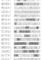
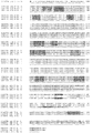

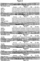

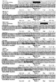


Classifications
-
- A—HUMAN NECESSITIES
- A61—MEDICAL OR VETERINARY SCIENCE; HYGIENE
- A61K—PREPARATIONS FOR MEDICAL, DENTAL OR TOILETRY PURPOSES
- A61K39/00—Medicinal preparations containing antigens or antibodies
- A61K39/395—Antibodies; Immunoglobulins; Immune serum, e.g. antilymphocytic serum
- A61K39/39533—Antibodies; Immunoglobulins; Immune serum, e.g. antilymphocytic serum against materials from animals
- A61K39/39558—Antibodies; Immunoglobulins; Immune serum, e.g. antilymphocytic serum against materials from animals against tumor tissues, cells, antigens
-
- A—HUMAN NECESSITIES
- A61—MEDICAL OR VETERINARY SCIENCE; HYGIENE
- A61P—SPECIFIC THERAPEUTIC ACTIVITY OF CHEMICAL COMPOUNDS OR MEDICINAL PREPARATIONS
- A61P35/00—Antineoplastic agents
-
- A—HUMAN NECESSITIES
- A61—MEDICAL OR VETERINARY SCIENCE; HYGIENE
- A61P—SPECIFIC THERAPEUTIC ACTIVITY OF CHEMICAL COMPOUNDS OR MEDICINAL PREPARATIONS
- A61P35/00—Antineoplastic agents
- A61P35/02—Antineoplastic agents specific for leukemia
-
- A—HUMAN NECESSITIES
- A61—MEDICAL OR VETERINARY SCIENCE; HYGIENE
- A61P—SPECIFIC THERAPEUTIC ACTIVITY OF CHEMICAL COMPOUNDS OR MEDICINAL PREPARATIONS
- A61P43/00—Drugs for specific purposes, not provided for in groups A61P1/00-A61P41/00
-
- C—CHEMISTRY; METALLURGY
- C07—ORGANIC CHEMISTRY
- C07K—PEPTIDES
- C07K14/00—Peptides having more than 20 amino acids; Gastrins; Somatostatins; Melanotropins; Derivatives thereof
- C07K14/435—Peptides having more than 20 amino acids; Gastrins; Somatostatins; Melanotropins; Derivatives thereof from animals; from humans
- C07K14/705—Receptors; Cell surface antigens; Cell surface determinants
-
- C—CHEMISTRY; METALLURGY
- C07—ORGANIC CHEMISTRY
- C07K—PEPTIDES
- C07K16/00—Immunoglobulins [IGs], e.g. monoclonal or polyclonal antibodies
- C07K16/18—Immunoglobulins [IGs], e.g. monoclonal or polyclonal antibodies against material from animals or humans
- C07K16/28—Immunoglobulins [IGs], e.g. monoclonal or polyclonal antibodies against material from animals or humans against receptors, cell surface antigens or cell surface determinants
- C07K16/30—Immunoglobulins [IGs], e.g. monoclonal or polyclonal antibodies against material from animals or humans against receptors, cell surface antigens or cell surface determinants from tumour cells
- C07K16/3023—Lung
-
- C—CHEMISTRY; METALLURGY
- C07—ORGANIC CHEMISTRY
- C07K—PEPTIDES
- C07K16/00—Immunoglobulins [IGs], e.g. monoclonal or polyclonal antibodies
- C07K16/18—Immunoglobulins [IGs], e.g. monoclonal or polyclonal antibodies against material from animals or humans
- C07K16/28—Immunoglobulins [IGs], e.g. monoclonal or polyclonal antibodies against material from animals or humans against receptors, cell surface antigens or cell surface determinants
- C07K16/30—Immunoglobulins [IGs], e.g. monoclonal or polyclonal antibodies against material from animals or humans against receptors, cell surface antigens or cell surface determinants from tumour cells
- C07K16/303—Liver or Pancreas
-
- C—CHEMISTRY; METALLURGY
- C07—ORGANIC CHEMISTRY
- C07K—PEPTIDES
- C07K16/00—Immunoglobulins [IGs], e.g. monoclonal or polyclonal antibodies
- C07K16/18—Immunoglobulins [IGs], e.g. monoclonal or polyclonal antibodies against material from animals or humans
- C07K16/28—Immunoglobulins [IGs], e.g. monoclonal or polyclonal antibodies against material from animals or humans against receptors, cell surface antigens or cell surface determinants
- C07K16/30—Immunoglobulins [IGs], e.g. monoclonal or polyclonal antibodies against material from animals or humans against receptors, cell surface antigens or cell surface determinants from tumour cells
- C07K16/3038—Kidney, bladder
-
- C—CHEMISTRY; METALLURGY
- C07—ORGANIC CHEMISTRY
- C07K—PEPTIDES
- C07K16/00—Immunoglobulins [IGs], e.g. monoclonal or polyclonal antibodies
- C07K16/18—Immunoglobulins [IGs], e.g. monoclonal or polyclonal antibodies against material from animals or humans
- C07K16/28—Immunoglobulins [IGs], e.g. monoclonal or polyclonal antibodies against material from animals or humans against receptors, cell surface antigens or cell surface determinants
- C07K16/30—Immunoglobulins [IGs], e.g. monoclonal or polyclonal antibodies against material from animals or humans against receptors, cell surface antigens or cell surface determinants from tumour cells
- C07K16/3046—Stomach, Intestines
-
- C—CHEMISTRY; METALLURGY
- C07—ORGANIC CHEMISTRY
- C07K—PEPTIDES
- C07K16/00—Immunoglobulins [IGs], e.g. monoclonal or polyclonal antibodies
- C07K16/18—Immunoglobulins [IGs], e.g. monoclonal or polyclonal antibodies against material from animals or humans
- C07K16/28—Immunoglobulins [IGs], e.g. monoclonal or polyclonal antibodies against material from animals or humans against receptors, cell surface antigens or cell surface determinants
- C07K16/30—Immunoglobulins [IGs], e.g. monoclonal or polyclonal antibodies against material from animals or humans against receptors, cell surface antigens or cell surface determinants from tumour cells
- C07K16/3061—Blood cells
-
- C—CHEMISTRY; METALLURGY
- C07—ORGANIC CHEMISTRY
- C07K—PEPTIDES
- C07K16/00—Immunoglobulins [IGs], e.g. monoclonal or polyclonal antibodies
- C07K16/18—Immunoglobulins [IGs], e.g. monoclonal or polyclonal antibodies against material from animals or humans
- C07K16/28—Immunoglobulins [IGs], e.g. monoclonal or polyclonal antibodies against material from animals or humans against receptors, cell surface antigens or cell surface determinants
- C07K16/30—Immunoglobulins [IGs], e.g. monoclonal or polyclonal antibodies against material from animals or humans against receptors, cell surface antigens or cell surface determinants from tumour cells
- C07K16/3069—Reproductive system, e.g. ovaria, uterus, testes, prostate
-
- C—CHEMISTRY; METALLURGY
- C12—BIOCHEMISTRY; BEER; SPIRITS; WINE; VINEGAR; MICROBIOLOGY; ENZYMOLOGY; MUTATION OR GENETIC ENGINEERING
- C12Q—MEASURING OR TESTING PROCESSES INVOLVING ENZYMES, NUCLEIC ACIDS OR MICROORGANISMS; COMPOSITIONS OR TEST PAPERS THEREFOR; PROCESSES OF PREPARING SUCH COMPOSITIONS; CONDITION-RESPONSIVE CONTROL IN MICROBIOLOGICAL OR ENZYMOLOGICAL PROCESSES
- C12Q1/00—Measuring or testing processes involving enzymes, nucleic acids or microorganisms; Compositions therefor; Processes of preparing such compositions
- C12Q1/68—Measuring or testing processes involving enzymes, nucleic acids or microorganisms; Compositions therefor; Processes of preparing such compositions involving nucleic acids
- C12Q1/6876—Nucleic acid products used in the analysis of nucleic acids, e.g. primers or probes
- C12Q1/6883—Nucleic acid products used in the analysis of nucleic acids, e.g. primers or probes for diseases caused by alterations of genetic material
- C12Q1/6886—Nucleic acid products used in the analysis of nucleic acids, e.g. primers or probes for diseases caused by alterations of genetic material for cancer
-
- G—PHYSICS
- G01—MEASURING; TESTING
- G01N—INVESTIGATING OR ANALYSING MATERIALS BY DETERMINING THEIR CHEMICAL OR PHYSICAL PROPERTIES
- G01N33/00—Investigating or analysing materials by specific methods not covered by groups G01N1/00 - G01N31/00
- G01N33/48—Biological material, e.g. blood, urine; Haemocytometers
- G01N33/50—Chemical analysis of biological material, e.g. blood, urine; Testing involving biospecific ligand binding methods; Immunological testing
- G01N33/53—Immunoassay; Biospecific binding assay; Materials therefor
- G01N33/574—Immunoassay; Biospecific binding assay; Materials therefor for cancer
- G01N33/57407—Specifically defined cancers
- G01N33/57419—Specifically defined cancers of colon
-
- G—PHYSICS
- G01—MEASURING; TESTING
- G01N—INVESTIGATING OR ANALYSING MATERIALS BY DETERMINING THEIR CHEMICAL OR PHYSICAL PROPERTIES
- G01N33/00—Investigating or analysing materials by specific methods not covered by groups G01N1/00 - G01N31/00
- G01N33/48—Biological material, e.g. blood, urine; Haemocytometers
- G01N33/50—Chemical analysis of biological material, e.g. blood, urine; Testing involving biospecific ligand binding methods; Immunological testing
- G01N33/53—Immunoassay; Biospecific binding assay; Materials therefor
- G01N33/574—Immunoassay; Biospecific binding assay; Materials therefor for cancer
- G01N33/57407—Specifically defined cancers
- G01N33/57423—Specifically defined cancers of lung
-
- G—PHYSICS
- G01—MEASURING; TESTING
- G01N—INVESTIGATING OR ANALYSING MATERIALS BY DETERMINING THEIR CHEMICAL OR PHYSICAL PROPERTIES
- G01N33/00—Investigating or analysing materials by specific methods not covered by groups G01N1/00 - G01N31/00
- G01N33/48—Biological material, e.g. blood, urine; Haemocytometers
- G01N33/50—Chemical analysis of biological material, e.g. blood, urine; Testing involving biospecific ligand binding methods; Immunological testing
- G01N33/53—Immunoassay; Biospecific binding assay; Materials therefor
- G01N33/574—Immunoassay; Biospecific binding assay; Materials therefor for cancer
- G01N33/57407—Specifically defined cancers
- G01N33/57426—Specifically defined cancers leukemia
-
- G—PHYSICS
- G01—MEASURING; TESTING
- G01N—INVESTIGATING OR ANALYSING MATERIALS BY DETERMINING THEIR CHEMICAL OR PHYSICAL PROPERTIES
- G01N33/00—Investigating or analysing materials by specific methods not covered by groups G01N1/00 - G01N31/00
- G01N33/48—Biological material, e.g. blood, urine; Haemocytometers
- G01N33/50—Chemical analysis of biological material, e.g. blood, urine; Testing involving biospecific ligand binding methods; Immunological testing
- G01N33/53—Immunoassay; Biospecific binding assay; Materials therefor
- G01N33/574—Immunoassay; Biospecific binding assay; Materials therefor for cancer
- G01N33/57407—Specifically defined cancers
- G01N33/57438—Specifically defined cancers of liver, pancreas or kidney
-
- G—PHYSICS
- G01—MEASURING; TESTING
- G01N—INVESTIGATING OR ANALYSING MATERIALS BY DETERMINING THEIR CHEMICAL OR PHYSICAL PROPERTIES
- G01N33/00—Investigating or analysing materials by specific methods not covered by groups G01N1/00 - G01N31/00
- G01N33/48—Biological material, e.g. blood, urine; Haemocytometers
- G01N33/50—Chemical analysis of biological material, e.g. blood, urine; Testing involving biospecific ligand binding methods; Immunological testing
- G01N33/53—Immunoassay; Biospecific binding assay; Materials therefor
- G01N33/574—Immunoassay; Biospecific binding assay; Materials therefor for cancer
- G01N33/57407—Specifically defined cancers
- G01N33/57449—Specifically defined cancers of ovaries
-
- C—CHEMISTRY; METALLURGY
- C07—ORGANIC CHEMISTRY
- C07K—PEPTIDES
- C07K2317/00—Immunoglobulins specific features
- C07K2317/70—Immunoglobulins specific features characterized by effect upon binding to a cell or to an antigen
- C07K2317/73—Inducing cell death, e.g. apoptosis, necrosis or inhibition of cell proliferation
- C07K2317/734—Complement-dependent cytotoxicity [CDC]
-
- Y—GENERAL TAGGING OF NEW TECHNOLOGICAL DEVELOPMENTS; GENERAL TAGGING OF CROSS-SECTIONAL TECHNOLOGIES SPANNING OVER SEVERAL SECTIONS OF THE IPC; TECHNICAL SUBJECTS COVERED BY FORMER USPC CROSS-REFERENCE ART COLLECTIONS [XRACs] AND DIGESTS
- Y10—TECHNICAL SUBJECTS COVERED BY FORMER USPC
- Y10T—TECHNICAL SUBJECTS COVERED BY FORMER US CLASSIFICATION
- Y10T436/00—Chemistry: analytical and immunological testing
- Y10T436/14—Heterocyclic carbon compound [i.e., O, S, N, Se, Te, as only ring hetero atom]
- Y10T436/142222—Hetero-O [e.g., ascorbic acid, etc.]
- Y10T436/143333—Saccharide [e.g., DNA, etc.]
Definitions
- the present invention relates to the identification of membrane protein associated with lung cancer, and ovarian cancer which has utility as a marker for lung cancer, and ovarian cancermetastases and which also forms a biological target against which therapeutic antibodies (or other affinity reagents) or other pharmaceutical agents can be made.
- Lung cancer is the most common form of cancer worldwide (accounting for about 12% of cancer cases) and the main cause of death from cancer (accounting for about 18% of deaths).
- Global incidence of lung cancer is over 1,300,000 per year, with the number of deaths over 1,100,000.
- In the USA there are about 170,000 new cases per year (about 13% of all cancers), with about 160,000 deaths (about 28% of cancer deaths).
- Lung cancer is much more prevalent among men than women. Nearly 70% of people diagnosed with lung cancer are older than 65; fewer than 3% of all cases are found in people under the age of 45.
- SCLC small cell type
- NSCLC non-small cell
- Lung cancer is a life-threatening disease because it often metastasises even before it can be detected on a chest x-ray. Usually symptoms of lung cancer do not appear until the disease is in an advanced stage. So far, there is no screening test that has been shown to improve a person's chance for a cure. Imaging tests such as a chest x-ray, CT scan, MRI scan or PET scan may be used to detect lung cancer. Tests to confirm the diagnosis are then performed and include sputum cytology, needle biopsy, bronchoscopy, endobronchial ultrasound and complete blood count (CBC).
- CBC complete blood count
- NSCLC is staged using the American Joint Committee on Cancer (AJCC) TNM system - Stage 0 - Stage IV.
- the 5-year survival rates by stage are as follows: stage I: 47%; stage II; 26%; stage III: 8% and stage IV: 2%.
- SCLC has a 2-stage system - limited stage and extensive stage. About two thirds of SCLC patients have extensive disease at diagnosis. If SCLC is found very early and is localised to the lung alone, the 5-year survival rate is around 21%, but only 6% of patients fall into this category. Where the cancer has spread, the 5-year survival is around 11%. For patients with extensive disease, the 5-year survival is just 2%.
- the main treatment for SCLC is chemotherapy, either alone or in combination with external beam radiation therapy and very rarely, surgery.
- Chemotherapeutic agents used for NSCLC and SCLC include cisplatin, carboplatin, mitomycin C, ifosfamide, vinblastine, gemcitabine, etoposide, vinorelbine, paclitaxel, docetaxel and irinotecan.
- Ovarian cancer accounts for about 1.9% of cancer cases globally and around 1.8% of deaths.
- Global incidence of ovarian cancer is around 205,000, predominantly in postmenopausal women in developed countries, with around 125,000 deaths.
- About 85% to 90% of ovarian cancers are epithelial ovarian carcinomas.
- About 5% of ovarian cancers are germ cell tumours and a smaller percentage are stromal tumours.
- Ovarian cancer is the eighth most common cancer among women. In the USA, about 20,200 new cases of ovarian cancer are diagnosed each year and it accounts for about 3% of all cancers in women. The risk of developing and dying from ovarian cancer is higher for white women than black women. Around two-thirds of women with ovarian cancer are 55 or older.
- Ovarian cancer ranks fifth in cancer deaths among women in the USA, accounting for more deaths than any other cancer of the female reproductive system. There are around 15,300 deaths in the USA from ovarian cancer each year. It has been estimated that approximately US$2.2 billion is spent in the USA each year on treating ovarian cancer.
- Imaging tests such as ultrasound, computed tomography and magnetic resonance imaging can confirm whether a pelvic mass is present.
- Blood tests, including a CA-125 test and a laparoscopy are performed. Ovarian cancer is then confirmed by biopsy.
- Ovarian cancer is staged using the American Joint Committee on Cancer (AJCC) TNM system - stage I - IV.
- the FIGO (International Federation of Gynecology and Obstetrics) system is also used.
- Ovarian cancers are also given a grade from 1-3. About 76% of women with ovarian cancer survive 1 year after diagnosis, and 45% survive longer than 5 years after diagnosis. If diagnosed and treated while the cancer has not spread outside the ovary, the 5-year survival rate is 94%. However, only 19% of all ovarian cancers are found at this early stage.
- Surgery for ovarian cancer includes hysterectomy, bilateral salpingectomy, bilateral oophorectomy and omentectomy. Debulking is performed in women in whom the cancer has spread widely throughout their abdomen.
- Intraperitoneal (IP) chemotherapy using a combination therapy using a platinum compound, such as cisplatin or carboplatin, and a taxane, such as paclitaxel or docetaxel is the standard approach. Tumour recurrence is sometimes treated with additional cycles of a platinum compound and/or a taxane. In other cases, recurrence is treated with other drugs, such as topotecan, anthracyclines such as doxorubicin (Adriamycin) and liposomal doxorubicin (Doxil), gemcitabine, cyclophosphamide, vinorelbine (Navelbine), hexamethylmelamine, ifosfamide, and etoposide. Resistance to currently-available chemotherapeutic agents is a major problem. Although complete clinical response is achieved in 75% of patients after initial treatment, most will develop recurrent disease and require re-treatment.
- the major challenges in treatment of lung cancer and ovarian cancer is to improve early detection rates, to find new non-invasive markers that can be used to follow disease progression and identify relapse, and to find improved and less toxic therapies, especially for more advanced disease where 5 year survival is still poor.
- targets which are more specific to the cancer cells e.g. ones which are expressed on the surface of the tumour cells so that they can be attacked by promising new approaches like immunotherapeutics and targeted toxins.
- WO2004/053138 describes DEC 205 splice variants associated with Hodgkin's disease. These splice variants have been identified on Reed-Sternberg cells and thus suggested to have application in therapy and investigation of this disease.
- WO2005/016962 describes compositions containing novel proteins and methods of using those compositions for the diagnosis and treatment of immune related dieseases.
- the present invention provides methods and compositions for diagnosis and therapy of lung cancer, and ovarian cancer, also described in the present disclosure are methods for colorectal cancer, kidney cancer, liver cancer, lung cancer, lymphoid leukaemia (particularly chronic lymphocytic leukaemia), ovarian cancer or pancreatic cancer patients' stratification, for monitoring the effectiveness of colorectal cancer, kidney cancer, liver cancer, lung cancer, lymphoid leukaemia (particularly chronic lymphocytic leukaemia), ovarian cancer or pancreatic cancer treatment, and for drug development for treatment of colorectal cancer, kidney cancer, liver cancer, lung cancer, lymphoid leukaemia (particularly chronic lymphocytic leukaemia), ovarian cancer or pancreatic cancer.
- an antibody which is capable of specific binding to Lymphocyte antigen 75 for use in treatment of ovarian cancer or lung cancer wherein said antibody contains or is conjugated to a therapeutic moiety.
- the antibody for use is an isolated monoclonal antibody, or an antigen-binding portion thereof, or an antibody fragment.
- the therapeutic moiety is a cytotoxic moiety or a radioactive isotope or wherein the affinity reagent is an antibody drug conjugate.
- the antibody is a fully human or humanised antibody that is capable of inducing antibody-directed dell-mediated cytotoxicity (ADCC)
- the disclosure provides a method of detecting, diagnosing and/or screening for or monitoring the progression of colorectal cancer, kidney cancer, liver cancer, lung cancer, lymphoid leukaemia (particularly chronic lymphocytic leukaemia), ovarian cancer or pancreatic cancer or of monitoring the effect of an anti-colorectal cancer, anti-kidney cancer, anti-liver cancer, anti-lung cancer, anti-lymphoid leukaemia (particularly chronic lymphocytic leukaemia), anti-ovarian cancer or anti-pancreatic cancer drug or therapy in a subject which comprises detecting the presence or level of OGTA076, or one or more fragments thereof, or the presence or level of nucleic acid encoding OGTA076 or the presence or level of the activity of OGTA076 or which comprises detecting a change in the level thereof in said subject.
- Also described is a method of detecting, diagnosing and/or screening for colorectal cancer, kidney cancer, liver cancer, lung cancer, lymphoid leukaemia (particularly chronic lymphocytic leukaemia), ovarian cancer or pancreatic cancer in a candidate subject which comprises detecting the presence of OGTA076, or one or more fragments thereof, or the presence of nucleic acid encoding OGTA076 or the presence of the activity of OGTA076 in said candidate subject, in which either (a) the presence of an elevated level of OGTA076 or said one or more fragments thereof or an elevated level of nucleic acid encoding OGTA076 or the presence of an elevated level of OGTA076 activity in the candidate subject as compared with the level in a healthy subject or (b) the presence of a detectable level of OGTA076 or said one or more fragments thereof or a detectable level of nucleic acid encoding OGTA076 or the presence of a detectable level of OGTA076 activity in
- a method of monitoring the progression of colorectal cancer, kidney cancer, liver cancer, lung cancer, lymphoid leukaemia (particularly chronic lymphocytic leukaemia), ovarian cancer or pancreatic cancer in a subject or of monitoring the effect of an anti-colorectal cancer, anti-kidney cancer, anti-liver cancer, anti-lung cancer, anti-lymphoid leukaemia (particularly chronic lymphocytic leukaemia), anti-ovarian cancer or anti-pancreatic cancer drug or therapy which comprises detecting the presence of OGTA076, or one or more fragments thereof, or the presence of nucleic acid encoding OGTA076 or the presence of the activity of OGTA076 in said candidate subject at a first time point and at a later time point, the presence of an elevated or lowered level of OGTA076 or said one or more fragments thereof or an elevated or lowered level of nucleic acid encoding OGTA076 or the presence of an elevated or lowered level of OGTA060
- the presence of OGTA076, or one or more fragments thereof, or the presence of nucleic acid encoding OGTA076 or the presence of the activity of OGTA076 may, for example, be detected by analysis of a biological sample obtained from said subject.
- the method described may typically include the step of obtaining a biological sample for analysis from said subject. However, in one or more examples the method does not include the step of obtaining the biological sample.
- the biological sample used can be from any source such as a serum sample or a tissue sample e.g. colorectal, kidney, liver, lung, lymphoid, ovarian or pancreatic tissue.
- a serum sample or a tissue sample e.g. colorectal, kidney, liver, lung, lymphoid, ovarian or pancreatic tissue.
- a tissue sample e.g. colorectal, kidney, liver, lung, lymphoid, ovarian or pancreatic tissue.
- a tissue sample e.g. colorectal, kidney, liver, lung, lymphoid, ovarian or pancreatic tissue.
- a tissue sample e.g. colorectal, kidney, liver, lung, lymphoid, ovarian or pancreatic tissue.
- the presence of OGTA076, or one or more fragments thereof, or the presence of nucleic acid encoding OGTA076 or the presence of the activity of OGTA076 may be detected by analysis in situ.
- methods of diagnosis described herein may be at least partly, or wholly, performed in vitro.
- the presence of OGTA076, or one or more fragments thereof, or the presence of nucleic acid encoding OGTA076 or the presence of the activity of OGTA076 is detected quantitatively.
- quantitatively detecting may comprise:
- the presence of OGTA076, or one or more fragments thereof, or the presence of nucleic acid encoding OGTA076 or the presence of the activity of OGTA076 may be detected quantitatively by means involving use of an imaging technology.
- the method of the disclosure involves use of immunohistochemistry on tissue sections in order to determine the presence of OGTA076, or one or more fragments thereof, or the presence of nucleic acid encoding OGTA076 or the presence of the activity of OGTA076, and thereby to localise colorectal cancer, kidney cancer, liver cancer, lung cancer, lymphoid leukaemia (particularly chronic lymphocytic leukaemia), ovarian cancer or pancreatic cancer cells.
- the presence of OGTA076 or one or more epitope-containing fragments thereof is detected, for example using an affinity reagent capable of specific binding to OGTA076 or one or more fragments thereof, such as an antibody.
- the activity of OGTA076 is detected.
- a method of detecting, diagnosing and/or screening for or monitoring the progression of lung canceror ovarian cancer or of monitoring the effect of an anti-lung canceror anti-ovarian cancer drug or therapy in a subject which comprises detecting the presence or level of antibodies capable of immunospecific binding to OGTA076, or one or more epitope-containing fragments thereof or which comprises detecting a change in the level thereof in said subject.
- a method of detecting, diagnosing and/or screening for lung cancer or ovarian cancer in a subject which comprises detecting the presence of antibodies capable of immunospecific binding to OGTA076, or one or more epitope-containing fragments thereof in said subject, in which (a) the presence of an elevated level of antibodies capable of immunospecific binding to OGTA076 or said one or more epitope-containing fragments thereof in said subject as compared with the level in a healthy subject or (b) the presence of a detectable level of antibodies capable of immunospecific binding to OGTA076 or said one or more epitope-containing fragments thereof in said subject as compared with a corresponding undetectable level in a healthy subject indicates the presence of lung cancer or ovarian cancer in said subject.
- One particular method of detecting, diagnosing and/or screening for lung cancer or ovarian cancer comprises:
- the presence of antibodies capable of immunospecific binding to OGTA076, or one or more epitope-containing fragments thereof is typically detected by analysis of a biological sample obtained from said subject (exemplary biological samples are mentioned above, e.g. the sample is a sample of colorectal, kidney, liver, lung, lymphoid, ovarian or pancreatic tissue, or else a sample of blood or saliva).
- the method may include the step of obtaining said biological sample for analysis from said subject.
- the antibodies that may be detected include IgA, IgM and IgG antibodies.
- the level that may be detected in the candidate subject who has colorectal cancer, kidney cancer, liver cancer, lung cancer, lymphoid leukaemia (particularly chronic lymphocytic leukaemia), ovarian cancer or pancreatic cancer is 2 or more fold higher than the level in the healthy subject.
- OGTA076, or a fragment thereof for use in screening for, detecting and/or diagnosing disease such as cancer, and especially colorectal cancer, kidney cancer, liver cancer, lung cancer, lymphoid leukaemia (particularly chronic lymphocytic leukaemia), ovarian cancer or pancreatic cancer.
- an affinity reagent capable of specific binding to OGTA076 or a fragment thereof, for example an affinity reagent which contains or is conjugated to a detectable label or contains or is conjugated to a therapeutic moiety such as a cytotoxic moiety.
- the affinity reagent may, for example, be an antibody.
- hybridizing agent capable of hybridizing to nucleic acid encoding OGTA076, for example, a hybridizing agent which contains or is conjugated to a detectable label.
- a hybridizing agent is an inhibitory RNA (RNAi).
- RNAi inhibitory RNA
- Other examples include anti-sense oligonucleotides and ribozymes.
- the disclosure also describes a kit containing OGTA076 and/or one or more fragments thereof or containing one or more aforementioned affinity reagents and/or hybridizing agents or containing one or more agents capable of detecting the activity of OGTA076 together with instructions for their use in an aforementioned method.
- the kit may further contain reagents capable of detecting and reporting the binding of said affinity reagents and/or hybridizing agents to their binding partners.
- composition comprising a therapeutically effective amount of an affinity reagent capable of specific binding to OGTA076 or a fragment thereof.
- a pharmaceutically acceptable diluent or carrier and a pharmaceutical composition comprising one or more affinity reagents or hybridizing reagents as aforesaid and a pharmaceutically acceptable diluent or carrier.
- the cancer to be detected, prevented or treated is colorectal cancer.
- the cancer to be detected, prevented or treated is kidney cancer.
- the cancer to be detected, prevented or treated is liver cancer.
- the cancer to be detected, prevented or treated is lung cancer.
- the cancer to be detected, prevented or treated is lymphoid leukaemia (particularly chronic lymphocytic leukaemia).
- the cancer to be detected, prevented or treated is ovarian cancer.
- the cancer to be detected, prevented or treated is pancreatic cancer.
- the invention described in detail below provides methods and compositions for clinical screening, diagnosis and prognosis of lung cancer and ovarian cancer. Also described are methods and compositions for clinical screening, diagnosis and prognosis of colorectal cancer, kidney cancer, liver cancer, lymphoid leukaemia (particularly chronic lymphocytic leukaemia) or pancreatic cancer in a mammalian subject for identifying patients most likely to respond to a particular therapeutic treatment, for monitoring the results of colorectal cancer, kidney cancer, liver cancer, lung cancer, lymphoid leukaemia (particularly chronic lymphocytic leukaemia), ovarian cancer or pancreatic cancer therapy, for drug screening and drug development.
- the invention also encompasses the administration of therapeutic compositions to a mammalian subject to treat lung cancer or ovarian cancer.
- the disclosure also describes the administration of therapeutic compositions to a mammalian subject to treat or prevent colorectal cancer, kidney cancer, liver cancer, lymphoid leukaemia (particularly chronic lymphocytic leukaemia), or pancreatic cancer.
- the mammalian subject may be a non-human mammal, for example a human, such as a human adult, i.e. a human subject at least 21 (for example at least 35, at least 50, at least 60, at least 70, or at least 80) years old.
- the methods and compositions of the present invention are specially suited for screening, diagnosis and prognosis of a living subject, but may also be used for postmortem diagnosis in a subject, for example, to identify family members at risk of developing the same disease.
- one-dimensional electrophoresis or isobaric tags for relative and absolute quantification are used to analyze colorectal cancer, kidney cancer, liver cancer, lung cancer, lymphoid leukaemia (particularly chronic lymphocytic leukaemia), ovarian cancer or pancreatic cancer tissue samples from a subject, preferably a living subject, in order to measure the expression of the protein for screening or diagnosis of colorectal cancer, kidney cancer, liver cancer, lung cancer, lymphoid leukaemia (particularly chronic lymphocytic leukaemia), ovarian cancer or pancreatic cancer, to determine the prognosis of a colorectal cancer, kidney cancer, liver cancer, lung cancer, lymphoid leukaemia (particularly chronic lymphocytic leukaemia), ovarian cancer or pancreatic cancer patient, to monitor the effectiveness of colorectal cancer, kidney cancer, liver cancer, lung cancer, lymphoid leukaemia (particularly chronic lymphocytic leukaemia),
- OGTA076 refers to the protein illustrated in Figure 1 in all its isoforms, in particular in its three different isoforms detected experimentally by ID gel electrophoresis and iTRAQ analysis of colorectal cancer, kidney cancer, liver cancer, lung cancer, lymphoid leukaemia (particularly chronic lymphocytic leukaemia), ovarian cancer and pancreatic cancer tissue samples (OGTA076a [SEQ ID No: 1]; OGTA076b [SEQ ID No: 2] and OGTA076c [SEQ ID No: 3]). Protein derivatives of these sequences may also be useful for the same purposes as described herein.
- This protein has been identified in membrane protein extracts of colorectal cancer, kidney cancer, liver cancer, lung cancer, lymphoid leukaemia (particularly chronic lymphocytic leukaemia), ovarian cancer and pancreatic cancer tissue samples from colorectal cancer, kidney cancer, liver cancer, lung cancer, lymphoid leukaemia (particularly chronic lymphocytic leukaemia), ovarian cancer and pancreatic cancer patients, through the methods and apparatus of the Preferred Technologies (ID gel electrophoresis or iTRAQ together with tryptic digest of membrane protein extracts).
- ID gel electrophoresis or iTRAQ together with tryptic digest of membrane protein extracts.
- Lymphocyte antigen 75 is expressed in spleen, thymus, colon and peripheral blood lymphocytes. It has been detected in myeloid and B lymphoid cell lines. Isoforms OGTA076b and OGTA076c are expressed in malignant Hodgkin's lymphoma cells called Hodgkin's and Reed-Sternberg (HRS) cells. Lymphocyte antigen 75 acts as an endocytic receptor to direct captured antigens from the extracellular space to a specialized antigen-processing compartment. It causes reduced proliferation of B-lymphocytes.
- the protein of the disclosure is useful as are fragments particularly epitope containing fragments e.g. antigenic or immunogenic fragments thereof and derivatives thereof.
- Epitope containing fragments including antigenic or immunogenic fragments will typically be of length 12 amino acids or more e.g. 20 amino acids or more e.g. 50 or 100 amino acids or more.
- Fragments may be 95% or more of the length of the full protein e.g. 90% or more e.g. 75% or 50% or 25% or 10% or more of the length of the full protein.
- DNA encoding the protein of the disclosure is also useful as are fragments thereof e.g. DNA encoding fragments of the protein of the disclosure such as immunogenic fragments thereof.
- Fragments of nucleic acid (e.g. DNA) encoding the protein of the disclosure may be 95% or more of the length of the full coding region e.g. 90% or more e.g. 75% or 50% or 25% or 10% or more of the length of the full coding region.
- Fragments of nucleic acid (e.g. DNA) may be 36 nucleotides or more e.g. 60 nucleotides or more e.g. 150 or 300 nucleotides or more in length.
- Derivatives of the protein of the disclosure include variants on the sequence in which one or more (e.g. 1-20 such as 15 amino acids, or up to 20% such as up to 10% or 5% or 1% by number of amino acids based on the total length of the protein) deletions, insertions or substitutions have been made. Substitutions may typically be conservative substitutions.
- Derivatives will typically have essentially the same biological function as the protein from which they are derived.
- Derivatives will typically be comparably antigenic or immunogenic to the protein from which they are derived.
- Derivatives will typically have either the ligand-binding activity, or the active receptor-complex forming ability, or preferably both, of the protein from which they are derived.
- Tables 1a-1f below illustrates the different occurrences of OGTA076 as detected by iTRAQ and mass spectrometry of membrane protein extracts of colorectal, kidney, liver, lung and ovarian tissue samples from colorectal cancer, kidney cancer, liver cancer, non-small cell lung cancer, small cell lung cancer and ovarian cancer patients respectively.
- the first column provides the samples batch number
- the second column gives the iTRAQ experiment number for those samples
- the last column provides a list of the sequences observed by mass spectrometry and the corresponding SEQ ID Nos.
- Table 2a-2c below illustrates the different occurrences of OGTA076 as detected by 1D gel electrophoresis and mass spectrometry of membrane protein extracts of colorectal, lymphoid and pancreatic tissue samples from colorectal cancer, chronic lymphocytic leukaemia and pancreatic cancer patients respectively.
- the first column provides the molecular weight
- the second column gives information on the subfractionation protocol used, if any (see Example 1 below)
- the last column provides a list of the sequences observed by mass spectrometry and the corresponding SEQ ID Nos.
- each laboratory will establish a reference range in subjects free from colorectal cancer, kidney cancer, liver cancer, lung cancer, lymphoid leukaemia (particularly chronic lymphocytic leukaemia), ovarian cancer and pancreatic cancer according to the analytical protocol and detection technique in use, as is conventional in the diagnostic art.
- OGTA076 can be used for detection, prognosis, diagnosis, or monitoring of colorectal cancer, kidney cancer, liver cancer, lung cancer, lymphoid leukaemia (particularly chronic lymphocytic leukaemia), ovarian cancer or pancreatic cancer or for drug development.
- tissue from a subject e.g. a subject suspected of having colorectal cancer, kidney cancer, liver cancer, lung cancer, lymphoid leukaemia (particularly chronic lymphocytic leukaemia), ovarian cancer or pancreatic cancer
- ID gel electrophoresis or iTRAQ for detection of OGTA076.
- An increased abundance of OGTA076 in the tissue from the subject relative to tissue from a subject or subjects free from colorectal cancer, kidney cancer, liver cancer, lung cancer, lymphoid leukaemia (particularly chronic lymphocytic leukaemia), ovarian cancer and pancreatic cancer (e.g. a control sample) or a previously determined reference range indicates the presence of colorectal cancer, kidney cancer, liver cancer, lung cancer, lymphoid leukaemia (particularly chronic lymphocytic leukaemia), ovarian cancer or pancreatic cancer.
- OGTA076 is "isolated" when it is present in a preparation that is substantially free of contaminating proteins, i.e. a preparation in which less than 10% (for example less than 5%, such as less than 1%) of the total protein present is contaminating protein(s).
- a contaminating protein is a protein having a significantly different amino acid sequence from that of isolated OGTA076, as determined by mass spectral analysis.
- a "significantly different" sequence is one that permits the contaminating protein to be resolved from OGTA076 by mass spectral analysis, performed according to the Reference Protocols.
- OGTA076 can be assayed by any method known to those skilled in the art, including but not limited to, the Preferred Technologies described herein, kinase assays, enzyme assays, binding assays and other functional assays, immunoassays, and western blotting.
- OGTA076 is separated on a 1-D gel by virtue of its MW and visualized by staining the gel.
- OGTA076 is stained with a fluorescent dye and imaged with a fluorescence scanner. Sypro Red (Molecular Probes, Inc., Eugene, Oregon) is a suitable dye for this purpose.
- Sypro Red Molecular Probes, Inc., Eugene, Oregon
- a preferred fluorescent dye is disclosed in U.S. Application No. 09/412,168, filed on October 5, 1999 ,.
- OGTA076 is analysed using isobaric tags for relative and absolute quantification (iTRAQ).
- OGTA076 can be detected in an immunoassay.
- an immunoassay is performed by contacting a sample from a subject to be tested with an anti-OGTA076 antibody (or other affinity reagent) under conditions such that binding (e.g. immunospecific binding) can occur if OGTA076 is present, and detecting or measuring the amount of any binding (e.g. immunospecific binding) by the agent.
- OGTA076 binding agents can be produced by the methods and techniques taught herein.
- OGTA076 may be detected by virtue of the detection of a fragment thereof e.g. an epitope containing (e.g. an immunogenic or antigenic) fragment thereof. Fragments may have a length of at least 10, more typically at least 20 amino acids e.g. at least 50 or 100 amino acids e.g. at least 200 or 500 amino acids e.g. at least 1000 or 1500 amino acids.
- OGTA076 is differentially expressed in patients/subjects with colorectal cancer, kidney cancer, liver cancer, lung cancer, lymphoid leukaemia (particularly chronic lymphocytic leukaemia), ovarian cancer or pancreatic cancer. Differentially expressed is intended to refer to a higher or lower level of expression in a relevant sample in comparison to cancer free/healthy sample.
- binding of an affinity reagent in tissue sections can be used to detect aberrant OGTA076 localization or an aberrant level of OGTA076.
- an antibody (or other affinity reagent) to OGTA076 can be used to assay a patient tissue (e.g. a colorectal, kidney, liver, lung, lymphoid, ovarian or pancreatic tissue) for the level of OGTA076 where an aberrant level of OGTA076 is indicative of colorectal cancer, kidney cancer, liver cancer, lung cancer, lymphoid leukaemia (particularly chronic lymphocytic leukaemia), ovarian cancer or pancreatic cancer.
- an "aberrant level” means a level that is increased compared with the level in a subject free from colorectal cancer, kidney cancer, liver cancer, lung cancer, lymphoid leukaemia (particularly chronic lymphocytic leukaemia), ovarian cancer and pancreatic cancer or a reference level.
- any suitable immunoassay can be used, including, without limitation, competitive and non-competitive assay systems using techniques such as western blots, radioimmunoassays, ELISA (enzyme linked immunosorbent assay), "sandwich” immunoassays, immunoprecipitation assays, precipitin reactions, gel diffusion precipitin reactions, immunodiffusion assays, agglutination assays, complement-fixation assays, immunoradiometric assays, fluorescent immunoassays and protein A immunoassays.
- competitive and non-competitive assay systems using techniques such as western blots, radioimmunoassays, ELISA (enzyme linked immunosorbent assay), "sandwich” immunoassays, immunoprecipitation assays, precipitin reactions, gel diffusion precipitin reactions, immunodiffusion assays, agglutination assays, complement-fixation assays, immunoradiometric assays, fluorescent immunoas
- OGTA076 can be detected in a fluid sample (e.g. blood, urine, or saliva) by means of a two-step sandwich assay.
- a capture reagent e.g. an anti-OGTA076 antibody or other affinity reagent
- the capture reagent can optionally be immobilized on a solid phase.
- a directly or indirectly labeled detection reagent is used to detect the captured OGTA076.
- the detection reagent is a lectin.
- any lectin can be used for this purpose that, for example binds to OGTA076 rather than to other isoforms that have the same core protein as OGTA076 or to other proteins that share the antigenic determinant recognized by the antibody.
- the chosen lectin binds OGTA076 with at least 2-fold greater affinity, more preferably at least 5-fold greater affinity, still more preferably at least 10-fold greater affinity, than to said other isoforms that have the same core protein as OGTA076 or to said other proteins that share the antigenic determinant recognized by the affinity reagent.
- a lectin that is suitable for detecting OGTA076 can readily be identified by methods well known in the art, for instance upon testing one or more lectins enumerated in Table I on pages 158-159 of Sumar et al., Lectins as Indicators of Disease-Associated Glycoforms, In: Gabius H-J & Gabius S (eds.), 1993 , Lectins and Glycobiology, at pp. 158 -.
- the detection reagent is an antibody (or other affinity reagent), e.g. an antibody that specifically (e.g.
- immunospecifically detects other post-translational modifications, such as an antibody that immunospecifically binds to phosphorylated amino acids.
- antibodies include those that bind to phosphotyrosine (BD Transduction Laboratories, catalog nos.: P1 1230-050/P11230-150; P11120; P38820; P39020), those that bind to phosphoserine (Zymed Laboratories Inc., South San Francisco, CA, catalog no. 61-8100) and those that bind to phosphothreonine (Zymed Laboratories Inc., South San Francisco, CA, catalogue nos. 71-8200, 13-9200).
- phosphotyrosine BD Transduction Laboratories, catalog nos.: P1 1230-050/P11230-150; P11120; P38820; P39020
- those that bind to phosphoserine Zymed Laboratories Inc., South San Francisco, CA, catalog no. 61-8100
- phosphothreonine Zymed Laboratories
- a gene encoding OGTA076, a related gene, or related nucleic acid sequences or subsequences, including complementary sequences can also be used in hybridization assays.
- a nucleotide encoding OGTA076, or subsequences thereof comprising at least 8 nucleotides, for example at least 12 nucleotides, such as at least 15 nucleotides can be used as a hybridization probe.
- Hybridization assays can be used for detection, prognosis, diagnosis, or monitoring of conditions, disorders, or disease states, associated with aberrant expression of the gene encoding OGTA076, or for differential diagnosis of subjects with signs or symptoms suggestive of colorectal cancer, kidney cancer, liver cancer, lung cancer, lymphoid leukaemia (particularly chronic lymphocytic leukaemia), ovarian cancer or pancreatic cancer.
- a hybridization assay can be carried out by a method comprising contacting a subject's sample containing nucleic acid with a nucleic acid probe capable of hybridizing to a DNA or RNA that encodes OGTA076, under conditions such that hybridization can occur, and detecting or measuring any resulting hybridization.
- nucleic acid encoding OGTA076 e.g. DNA or more suitably RNA
- a hybridizing agent capable of hybridizing to nucleic acid encoding OGTA076.
- One such exemplary method comprises:
- kits comprising an anti-OGTA076 antibody (or other affinity reagent).
- a kit may optionally comprise one or more of the following: (1) instructions for using the anti-OGTA076 affinity reagent for diagnosis, prognosis, therapeutic monitoring or any combination of these applications; (2) a labeled binding partner to the affinity reagent; (3) a solid phase (such as a reagent strip) upon which the anti-OGTA076 affinity reagent is immobilized; and (4) a label or insert indicating regulatory approval for diagnostic, prognostic or therapeutic use or any combination thereof.
- the anti-OGTA076 affinity reagent itself can be labeled with a detectable marker, e.g. a chemiluminescent, enzymatic, fluorescent, or radioactive moiety.
- a detectable marker e.g. a chemiluminescent, enzymatic, fluorescent, or radioactive moiety.
- kits comprising a nucleic acid probe capable of hybridizing to nucleic acid, suitably RNA, encoding OGTA076.
- a kit comprises in one or more containers a pair of primers (e.g. each in the size range of 6-30 nucleotides, more preferably 10-30 nucleotides and still more preferably 10-20 nucleotides) that under appropriate reaction conditions can prime amplification of at least a portion of a nucleic acid encoding OGTA076, such as by polymerase chain reaction (see, e.g.
- a kit can optionally further comprise a predetermined amount of OGTA076 or a nucleic acid encoding OGTA076, e.g. for use as a standard or control.
- the diagnostic methods and compositions of the present invention can assist in monitoring a clinical study, e.g. to evaluate drugs for therapy of colorectal cancer, kidney cancer, liver cancer, lung cancer, lymphoid leukaemia (particularly chronic lymphocytic leukaemia), ovarian cancer or pancreatic cancer.
- candidate molecules are tested for their ability to restore OGTA076 levels in a subject having colorectal cancer, kidney cancer, liver cancer, lung cancer, lymphoid leukaemia (particularly chronic lymphocytic leukaemia), ovarian cancer or pancreatic cancer to levels found in subjects free from colorectal cancer, kidney cancer, liver cancer, lung cancer, lymphoid leukaemia (particularly chronic lymphocytic leukaemia), ovarian cancer and pancreatic cancer or, in a treated subject, to preserve OGTA076 levels at or near non-colorectal cancer, non-kidney cancer, non-liver cancer, non-lung cancer, non-lymphoid leukaemia (particularly chronic lymphocytic leukaemia), non-ovarian cancer or non-pancreatic cancer values.
- the methods and compositions of the present invention are used to screen candidates for a clinical study to identify individuals having colorectal cancer, kidney cancer, liver cancer, lung cancer, lymphoid leukaemia (particularly chronic lymphocytic leukaemia), ovarian cancer or pancreatic cancer; such individuals can then be excluded from the study or can be placed in a separate cohort for treatment or analysis.
- a DNA of the present disclosure can be obtained by isolation as a cDNA fragment from cDNA libraries using as starter materials commercial mRNAs and determining and identifying the nucleotide sequences thereof. That is, specifically, clones are randomly isolated from cDNA libraries, which are prepared according to Ohara et al's method (DNA Research Vol.4, 53-59 (1997 )). Next, through hybridization, duplicated clones (which appear repeatedly) are removed and then in vitro transcription and translation are carried out. Nucleotide sequences of both termini of clones, for which products of 50 kDa or more are confirmed, are determined. Furthermore, databases of known genes are searched for homology using the thus obtained terminal nucleotide sequences as queries.
- the entire nucleotide sequence of a clone revealed to be novel as a result is determined.
- the 5' and 3' terminal sequences of cDNA are related to a human genome sequence. Then an unknown long-chain gene is confirmed in a region between the sequences, and the full-length of the cDNA is analyzed. In this way, an unknown gene that is unable to be obtained by a conventional cloning method that depends on known genes can be systematically cloned.
- all of the regions of a human-derived gene containing a DNA of the present disclosure can also be prepared using a PCR method such as RACE while paying sufficient attention to prevent artificial errors from taking place in short fragments or obtained sequences.
- clones having DNA of the present disclosure can be obtained.
- a synthetic DNA primer having an appropriate nucleotide sequence of a portion of a polypeptide of the present disclosure is produced, followed by amplification by the PCR method using an appropriate library.
- selection can be carried out by hybridization of the DNA of the present disclosure with a DNA that has been incorporated into an appropriate vector and labeled with a DNA fragment or a synthetic DNA encoding some or all of the regions of the polypeptide of the present disclosure. Hybridization can be carried out by, for example, the method described in Current Protocols in Molecular Biology (edited by Frederick M. Ausubel et al., 1987 ).
- DNA of the present disclosure may be any DNA, as long as they contain nucleotide sequences encoding the polypeptides of the present disclosure as described above.
- a DNA may be a cDNA identified and isolated from cDNA libraries or the like that are derived from colorectal, kidney, liver, lung, lymphoid, ovarian or pancreatic tissue.
- Such a DNA may also be a synthetic DNA or the like.
- Vectors for use in library construction may be any of bacteriophages, plasmids, cosmids, phargemids, or the like.
- amplification can be carried out by a direct reverse transcription coupled polymerase chain reaction (hereinafter abbreviated as "RT-PCR method").
- RT-PCR method a direct reverse transcription coupled polymerase chain reaction
- DNA encoding the above polypeptide consisting of an amino acid sequence that is substantially identical to the amino acid sequence of OGTA076 or DNA encoding the above polypeptide consisting of an amino acid sequence derived from the amino acid sequence of OGTA076 by deletion, substitution, or addition of one or more amino acids composing a portion of the amino acid sequence can be easily produced by an appropriate combination of, for example, a site-directed mutagenesis method, a gene homologous recombination method, a primer elongation method, and the PCR method known by persons skilled in the art.
- a possible method for causing a polypeptide to have substantially equivalent biological activity is substitution of homologous amino acids (e.g.
- amino acids within functional domains contained in the polypeptide of the present disclosure are preferably conserved.
- examples of DNA of the present disclosure include DNA comprising a nucleotide sequence that encodes the amino acid sequence of OGTA076 and DNA hybridizing under stringent conditions to the DNA and encoding a polypeptide (protein) having biological activity (function) equivalent to the function of the polypeptide consisting of the amino acid sequence of OGTA076.
- an example of such DNA capable of hybridizing to DNA comprising the nucleotide sequence that encodes the amino acid sequence of OGTA076 is DNA comprising a nucleotide sequence that has a degree of overall mean homology with the entire nucleotide sequence of the DNA, such as approximately 80% or more, preferably approximately 90% or more, and more preferably approximately 95% or more.
- Hybridization can be carried out according to a method known in the art such as a method described in Current Protocols in Molecular Biology (edited by Frederick M. Ausubel et al., 1987 ) or a method according thereto.
- stringent conditions are, for example, conditions of approximately “1*SSC, 0.1% SDS, and 37°C, more stringent conditions of approximately "0.5*SSC, 0.1% SDS, and 42°C, or even more stringent conditions of approximately "0.2*SSC, 0.1% SDS, and 65°C.
- stringent hybridization conditions With more stringent hybridization conditions, the isolation of a DNA having high homology with a probe sequence can be expected.
- the above combinations of SSC, SDS, and temperature conditions are given for illustrative purposes. Stringency similar to the above can be achieved by persons skilled in the art using an appropriate combination of the above factors or other factors (for example, probe concentration, probe length, and reaction time for hybridization) for determination of hybridization stringency.
- a cloned DNA of the present disclosure can be directly used or used, if desired, after digestion with a restriction enzyme or addition of a linker, depending on purposes.
- the DNA may have ATG as a translation initiation codon at the 5' terminal side and have TAA, TGA, or TAG as a translation termination codon at the 3' terminal side. These translation initiation and translation termination codons can also be added using an appropriate synthetic DNA adapter.
- OGTA076 may, for example be provided in isolated form.
- the OGTA076 polypeptide has been purified to at least to some extent.
- OGTA076 polypeptide may be provided in substantially pure form, that is to say free, to a substantial extent, from other proteins.
- OGTA076 polypeptide can also be produced using recombinant methods, synthetically produced or produced by a combination of these methods.
- OGTA076 can be easily prepared by any method known by persons skilled in the art, which involves producing an expression vector containing a DNA of the present disclosure or a gene containing a DNA of the present disclosure, culturing a transformant transformed using the expression vector, generating and accumulating a polypeptide of the present disclosure or a recombinant protein containing the polypeptide, and then collecting the resultant.
- Recombinant OGTA076 polypeptide may be prepared by processes well known in the art from genetically engineered host cells comprising expression systems. Accordingly, the present disclosure also relates to expression systems which comprise an OGTA076 polypeptide or nucleic acid, to host cells which are genetically engineered with such expression systems and to the production of OGTA076 polypeptide by recombinant techniques. For recombinant OGTA076 polypeptide production, host cells can be genetically engineered to incorporate expression systems or portions thereof for nucleic acids.
- incorporation can be performed using methods well known in the art, such as, calcium phosphate transfection, DEAD-dextran mediated transfection, transvection, microinjection, cationic lipid-mediated transfection, electroporation, transduction, scrape loading, ballistic introduction or infection (see e.g. Davis et al., Basic Methods in Molecular Biology, 1986 and Sambrook et al. , Molecular Cloning: A Laboratory Manual, 2nd Ed., Cold Spring Harbour laboratory Press, Cold Spring Harbour, NY, 1989 ).
- bacteria of the genus Escherichia, Streptococci, Staphylococci, Streptomyces, bacteria of the genus Bacillus, yeast, Aspergillus cells, insect cells, insects, and animal cells are used.
- bacteria of the genus Escherichia which are used herein, include Escherichia coli K12 and DH1 ( Proc. Natl. Acad. Sci. U.S.A., Vol. 60, 160 (1968 )), JM103 ( Nucleic Acids Research, Vol. 9, 309 (1981 )), JA221 ( Journal of Molecular Biology, Vol. 120, 517 (1978 )), and HB101 ( Journal of Molecular Biology, Vol.
- Bacillus subtilis MI114 Gene, Vol. 24, 255 (1983 )
- 207-21 Journal of Biochemistry, Vol. 95, 87 (1984 )
- yeast for example, Saccaromyces cerevisiae AH22, AH22R-, NA87-11A, DKD-5D, and 20B-12
- Schizosaccaromyces pombe NCYC1913 and NCYC2036, and Pichia pastoris are used.
- insect cells for example, Drosophila S2 and Spodoptera Sf9 cells are used.
- animal cells for example, COS-7 and Vero monkey cells, CHO Chinese hamster cells (hereinafter abbreviated as CHO cells), dhfr-gene-deficient CHO cells, mouse L cells, mouse AtT-20 cells, mouse myeloma cells, rat GH3 cells, human FL cells, COS, HeLa, C127,3T3, HEK 293, BHK and Bowes melanoma cells are used.
- CHO cells Chinese hamster cells
- dhfr-gene-deficient CHO cells mouse L cells
- mouse AtT-20 cells mouse myeloma cells
- rat GH3 cells human FL cells
- COS HeLa
- Cell-free translation systems can also be employed to produce recombinant polypeptides (e.g. rabbit reticulocyte lysate, wheat germ lysate, SP6/T7 in vitro T&T and RTS 100 E. Coli HY transcription and translation kits from Roche Diagnostics Ltd., Lewes, UK and the TNT Quick coupled Transcription/Translation System from Promega UK, Southampton, UK).
- recombinant polypeptides e.g. rabbit reticulocyte lysate, wheat germ lysate, SP6/T7 in vitro T&T and RTS 100 E. Coli HY transcription and translation kits from Roche Diagnostics Ltd., Lewes, UK and the TNT Quick coupled Transcription/Translation System from Promega UK, Southampton, UK.
- the expression vector can be produced according to a method known in the art.
- the vector can be produced by (1) excising a DNA fragment containing a DNA of the present disclosure or a gene containing a DNA of the present disclosure and (2) ligating the DNA fragment downstream of the promoter in an appropriate expression vector.
- a wide variety of expression systems can be used, such as and without limitation, chromosomal, episomal and virus-derived systems, e.g. plasmids derived from Escherichia coli (e.g. pBR322, pBR325, pUC18, and pUC118), plasmids derived from Bacillus subtilis (e.g.
- pUB110, pTP5, and pC194 from bacteriophage, from transposons, from yeast episomes (e.g. pSH19 and pSH15), from insertion elements, from yeast chromosomal elements, from viruses such as baculoviruses, papova viruses such as SV40, vaccinia viruses, adenoviruses, fowl pox viruses, pseudorabies viruses and retroviruses, and vectors derived from combinations thereof, such as those derived from plasmid and bacteriophage (such as [lambda] phage) genetic elements, such as cosmids and phagemids.
- the expression systems may contain control regions that regulate as well as engender expression.
- Promoters to be used in the present disclosure may be any promoters as long as they are appropriate for hosts to be used for gene expression.
- a host is Escherichia coli
- an SPO1 promoter, an SPO2 promoter, a penP promoter, and the like are preferred.
- yeast a PHO5 promoter, a PGK promoter, a GAP promoter, an ADH promoter, and the like are preferred.
- promoters for use in this case include an SRa promoter, an SV40 promoter, an LTR promoter, a CMV promoter, and an HSV-TK promoter.
- any system or vector that is able to maintain, propagate or express a nucleic acid to produce a polypeptide in a host may be used.
- nucleic acid sequence may be inserted into an expression system by any variety of well known and routine techniques, such as those set forth in Sambrook et al., supra.
- Appropriate secretion signals may be incorporated into the OGTA076 polypeptide to allow secretion of the translated protein into the lumen of the endoplasmic reticulum, the periplasmic space or the extracellular environment. These signals may be endogenous to the OGTA076 polypeptide or they may be heterologous signals. Transformation of the host cells can be carried out according to methods known in the art. For example, the following documents can be referred to: Proc. Natl. Acad. Sci. U.S.A., Vol. 69, 2110 (1972 ); Gene, Vol.
- the bacteria when hosts are bacteria of the genus Escherichia, the bacteria are generally cultured at approximately 15°C to 43°C for approximately 3 to 24 hours. If necessary, aeration or agitation can also be added.
- the bacteria When hosts are bacteria of the genus Bacillus, the bacteria are generally cultured at approximately 30°C to 40°C for approximately 6 to 24 hours. If necessary, aeration or agitation can also be added.
- transformants whose hosts are yeast are cultured, culture is generally carried out at approximately 20°C to 35°C for approximately 24 to 72 hours using media with pH adjusted to be approximately 5 to 8. If necessary, aeration or agitation can also be added.
- transformants whose hosts are animal cells When transformants whose hosts are animal cells are cultured, the cells are generally cultured at approximately 30°C to 40°C for approximately 15 to 60 hours using media with the pH adjusted to be approximately 6 to 8. If necessary, aeration or agitation can also be added.
- an OGTA076 polypeptide is to be expressed for use in cell-based screening assays, it is preferred that the polypeptide be produced at the cell surface. In this event, the cells may be harvested prior to use in the screening assay. If the OGTA076 polypeptide is secreted into the medium, the medium can be recovered in order to isolate said polypeptide. If produced intracellularly, the cells must first be lysed before the OGTA076 polypeptide is recovered.
- OGTA076 polypeptide can be recovered and purified from recombinant cell cultures or from other biological sources by well known methods including, ammonium sulphate or ethanol precipitation, acid extraction, anion or cation exchange chromatography, phosphocellulose chromatography, affinity chromatography, hydrophobic interaction chromatography, hydroxylapatite chromatography, molecular sieving chromatography, centrifugation methods, electrophoresis methods and lectin chromatography. In one example, a combination of these methods is used. In another example, high performance liquid chromatography is used. In a further example, an antibody which specifically binds to an OGTA076 polypeptide can be used to deplete a sample comprising an OGTA076 polypeptide of said polypeptide or to purify said polypeptide.
- microbial bodies or cells are collected by a known method, they are suspended in an appropriate buffer, the microbial bodies or the cells are disrupted by, for example, ultrasonic waves, lysozymes, and/or freeze-thawing, the resultant is then subjected to centrifugation or filtration, and then a crude extract of the protein can be obtained.
- the buffer may also contain a protein denaturation agent such as urea or guanidine hydrochloride or a surfactant such as Triton X-100(TM).
- polypeptide (protein) of the present disclosure can be converted into a salt by a known method or a method according thereto.
- polypeptide (protein) of the present disclosure when the polypeptide (protein) of the present disclosure is obtained in the form of a salt, it can be converted into a free protein or peptide or another salt by a known method or a method according thereto.
- an appropriate protein modification enzyme such as trypsin or chymotrypsin is caused to act on a protein produced by a recombinant before or after purification, so that modification can be arbitrarily added or a polypeptide can be partially removed.
- the presence of a polypeptide (protein) of the present disclosure or a salt thereof can be measured by various binding assays, enzyme immunoassays using specific antibodies, and the like.
- OGTA076 polypeptide can be obtained from a biological sample from any source, such as and without limitation, a blood sample or tissue sample, e.g. a colorectal, kidney, liver, lung, lymphoid, ovarian or pancreatic tissue sample.
- a biological sample from any source, such as and without limitation, a blood sample or tissue sample, e.g. a colorectal, kidney, liver, lung, lymphoid, ovarian or pancreatic tissue sample.
- OGTA076 polypeptide may be in the form of a "mature protein" or may be part of a larger protein such as a fusion protein. It is often advantageous to include an additional amino acid sequence which contains secretory or leader sequences, a pre-, pro- or prepro-protein sequence, or a sequence which aids in purification such as an affinity tag, for example, but without limitation, multiple histidine residues, a FLAG tag, HA tag or myc tag.
- an affinity tag for example, but without limitation, multiple histidine residues, a FLAG tag, HA tag or myc tag.
- an additional sequence that may provide stability during recombinant production may also be used. Such sequences may be optionally removed as required by incorporating a cleavable sequence as an additional sequence or part thereof.
- an OGTA076 polypeptide may be fused to other moieties including other polypeptides or proteins (for example, glutathione S-transferase and protein A). Such a fusion protein can be cleaved using an appropriate protease, and then separated into each protein.
- additional sequences and affinity tags are well known in the art.
- features known in the art such as an enhancer, a splicing signal, a polyA addition signal, a selection marker, and an SV40 replication origin can be added to an expression vector, if desired.
- immunoaffinity reagent - monoclonal antibodies phage display antibodies and smaller antibody-derived molecules such as Affibodies, Domain Antibodies (dAbs), Nanobodies or UniBodies.
- Affibodies e.g. Affibodies, domain antibodies, Nanobodies or UniBodies
- dAbs Domain Antibodies
- UniBodies e.g. Affibodies, domain antibodies, Nanobodies or UniBodies
- affinity reagents e.g. Affibodies, domain antibodies, Nanobodies or UniBodies
- Such substances may be said to be capable of immunospecific binding to OGTA076.
- affinity agent shall be construed to embrace immunoaffinity reagents and other substances capable of specific binding to OGTA076 including but not limited to ligands, lectins, streptavidins, antibody mimetics and synthetic binding agents.
- an OGTA076 analog, an OGTA076-related protein or a fragment or derivative of any of the foregoing may be used as an immunogen to generate antibodies which immunospecifically bind such an immunogen.
- immunogens can be isolated by any convenient means, including the methods described above.
- the term "antibody” as used herein refers to a peptide or polypeptide derived from, modeled after or substantially encoded by an immunoglobulin gene or immunoglobulin genes, or fragments thereof, capable of specifically binding an antigen or epitope. See, e.g. Fundamental Immunology, 3rd Edition, W.E. Paul, ed., Raven Press, N.Y. (1993 ); Wilson (1994) J. Immunol.
- antibody includes antigen-binding portions, i.e., "antigen binding sites" (e.g.
- CDRs complementarity determining regions
- Antibodies of the invention include, but are not limited to polyclonal, monoclonal, bispecific, humanized or chimeric antibodies, single chain antibodies, Fab fragments and F(ab') 2 fragments, fragments produced by a Fab expression library, anti-idiotypic (anti-Id) antibodies, and epitope-binding fragments of any of the above.
- the immunoglobulin molecules of the invention can be of any class (e.g. IgG, IgE, IgM, IgD and IgA) or subclass of immunoglobulin molecule.
- the term “specifically binds” is not intended to indicate that an antibody binds exclusively to its intended target. Rather, an antibody “specifically binds” if its affinity for its intended target is typically about 5-fold greater when compared to its affinity for a non-target molecule. Suitably there is no significant cross-reaction or cross-binding with undesired substances, especially naturally occurring proteins or tissues of a healthy person or animal. In one embodiment the affinity of the antibody will be at least about 5 fold, for example 10 fold, such as 25-fold, particularly 50-fold, and especially 100-fold or more, greater for a target molecule than its affinity for a non-target molecule.
- specific binding between an antibody or other binding agent and an antigen means a binding affinity of at least 10 6 M -1 .
- Antibodies bind with affinities of at least about 10 7 M -1 , for example between about 10 8 M -1 to about 10 9 M -1 , about 10 9 M -1 to about 10 10 M -1 , or about 10 10 M -1 to about 10 11 M -1 may be particularly suitable for use in the invention.
- antibodies that recognize gene products of genes encoding OGTA076 are publicly available.
- methods known to those skilled in the art are used to produce antibodies that recognize OGTA076, an OGTA076 analog, an OGTA076-related polypeptide, or a fragment or derivative of any of the foregoing.
- One skilled in the art will recognize that many procedures are available for the production of antibodies, for example, as described in Antibodies, A Laboratory Manual, Ed Harlow and David Lane, Cold Spring Harbor Laboratory (1988), Cold Spring Harbor, N.Y .
- binding fragments or Fab fragments which mimic antibodies can also be prepared from genetic information by various procedures ( Antibody Engineering: A Practical Approach (Borrebaeck, C., ed.), 1995, Oxford University Press, Oxford; J. Immunol. 149, 3914-3920 (1992 )).
- antibodies to a specific domain of OGTA076 are produced.
- hydrophilic fragments of OGTA076 are used as immunogens for antibody production.
- screening for the desired antibody can be accomplished by techniques known in the art, e.g. ELISA (enzyme-linked immunosorbent assay).
- ELISA enzyme-linked immunosorbent assay
- a second OGTA076 homolog For selection of an antibody that specifically binds a first OGTA076 homolog but which does not specifically bind to (or binds less avidly to) a second OGTA076 homolog, one can select on the basis of positive binding to the first OGTA076 homolog and a lack of binding to (or reduced binding to) the second OGTA076 homolog.
- the present invention provides an antibody (preferably a monoclonal antibody) that binds with greater affinity (for example at least 2-fold, such as at least 5-fold, particularly at least 10-fold greater affinity) to OGTA076 than to a different isoform or isoforms (e.g. glycoforms) of OGTA076.
- Polyclonal antibodies which may be used in the methods of the invention are heterogeneous populations of antibody molecules derived from the sera of immunized animals. Unfractionated immune serum can also be used.
- Various procedures known in the art may be used for the production of polyclonal antibodies to OGTA076, a fragment of OGTA076, an OGTA076-related polypeptide, or a fragment of an OGTA076-related polypeptide.
- one way is to purify polypeptides of interest or to synthesize the polypeptides of interest using, e.g. solid phase peptide synthesis methods well known in the art. See, e.g. Guide to Protein Purification, Murray P. Deutcher, ed., Meth. Enzymol.
- the selected polypeptides may then be used to immunize by injection various host animals, including but not limited to rabbits, mice, rats, etc., to generate polyclonal or monoclonal antibodies.
- Example 1 The Preferred Technology described herein in Example 1 provides isolated OGTA076 suitable for such immunization. If OGTA076 is purified by gel electrophoresis, OGTA076 can be used for immunization with or without prior extraction from the polyacrylamide gel. Various adjuvants (i.e.
- immunostimulants may be used to enhance the immunological response, depending on the host species, including, but not limited to, complete or incomplete Freund's adjuvant, a mineral gel such as aluminum hydroxide, surface active substance such as lysolecithin, pluronic polyol, a polyanion, a peptide, an oil emulsion, keyhole limpet hemocyanin, dinitrophenol, and an adjuvant such as BCG (bacille Calmette-Guerin) or corynebacterium parvum. Additional adjuvants are also well known in the art.
- complete or incomplete Freund's adjuvant a mineral gel such as aluminum hydroxide, surface active substance such as lysolecithin, pluronic polyol, a polyanion, a peptide, an oil emulsion, keyhole limpet hemocyanin, dinitrophenol, and an adjuvant such as BCG (bacille Calmette-Guerin) or corynebacterium parvum. Additional adjuvants are also
- any technique which provides for the production of antibody molecules by continuous cell lines in culture may be used.
- the hybridoma technique originally developed by Kohler and Milstein (1975, Nature 256:495-497 ), as well as the trioma technique, the human B-cell hybridoma technique ( Kozbor et al., 1983, Immunology Today 4:72 ), and the EBV-hybridoma technique to produce human monoclonal antibodies Cole et al., 1985, in Monoclonal Antibodies and Cancer Therapy, Alan R.
- Such antibodies may be of any immunoglobulin class including IgG, IgM, IgE, IgA, IgD and any subclass thereof.
- the hybridoma producing the mAbs of the invention may be cultivated in vitro or in vivo.
- monoclonal antibodies can be produced in germ-free animals utilizing known technology ( PCT/US90/02545 ).
- the monoclonal antibodies include but are not limited to human monoclonal antibodies and chimeric monoclonal antibodies (e.g. human-mouse chimeras).
- a chimeric antibody is a molecule in which different portions are derived from different animal species, such as those having a human immunoglobulin constant region and a variable region derived from a murine mAb. (See, e.g. Cabilly et al., U.S. Patent No. 4,816,567 ; and Boss et al., U.S. Patent No.
- Humanized antibodies are antibody molecules from non-human species having one or more complementarity determining regions (CDRs) from the non-human species and a framework region from a human immunoglobulin molecule. (See, e.g. Queen, U.S. Patent No. 5,585,089 .)
- Chimeric and humanized monoclonal antibodies can be produced by recombinant DNA techniques known in the art, for example using methods described in PCT Publication No. WO 87/02671 ; European Patent Application 184,187 ; European Patent Application 171,496 ; European Patent Application 173,494 ; PCT Publication No. WO 86/01533 ; U.S. Patent No. 4,816,567 ; European Patent Application 125,023 ; Better et al., 1988, Science 240:1041-1043 ; Liu et al., 1987, Proc. Natl. Acad. Sci. USA 84:3439-3443 ; Liu et al., 1987, J. Immunol.
- Fully human antibodies are particularly desirable for therapeutic treatment of human subjects.
- Such antibodies can be produced using transgenic mice which are incapable of expressing endogenous immunoglobulin heavy and light chain genes, but which can express human heavy and light chain genes.
- the transgenic mice are immunized in the normal fashion with a selected antigen, e.g. all or a portion of OGTA076.
- Monoclonal antibodies directed against the antigen can be obtained using conventional hybridoma technology.
- the human immunoglobulin transgenes harbored by the transgenic mice rearrange during B cell differentiation, and subsequently undergo class switching and somatic mutation.
- Completely human antibodies which recognize a selected epitope can be generated using a technique referred to as "guided selection".
- a selected non-human monoclonal antibody e.g. a mouse antibody, is used to guide the selection of a completely human antibody recognizing the same epitope.
- the antibodies of the present invention can also be generated by the use of phage display technology to produce and screen libraries of polypeptides for binding to a selected target. See, e.g. Cwirla et al., Proc. Natl. Acad. Sci. USA 87, 6378-82, 1990 ; Devlin et al., Science 249, 404-6, 1990 , Scott and Smith, Science 249, 386-88, 1990 ; and Ladner et al., U.S. Pat. No. 5,571,698 .
- a basic concept of phage display methods is the establishment of a physical association between DNA encoding a polypeptide to be screened and the polypeptide.
- This physical association is provided by the phage particle, which displays a polypeptide as part of a capsid enclosing the phage genome which encodes the polypeptide.
- the establishment of a physical association between polypeptides and their genetic material allows simultaneous mass screening of very large numbers of phage bearing different polypeptides.
- Phage displaying a polypeptide with affinity to a target bind to the target and these phage are enriched by affinity screening to the target.
- the identity of polypeptides displayed from these phage can be determined from their respective genomes. Using these methods a polypeptide identified as having a binding affinity for a desired target can then be synthesized in bulk by conventional means. See, e.g. U.S. Patent No. 6,057,098 .
- phage can be utilized to display antigen binding domains expressed from a repertoire or combinatorial antibody library (e.g. human or murine).
- Phage expressing an antigen binding domain that binds the antigen of interest can be selected or identified with antigen, e.g. using labeled antigen or antigen bound or captured to a solid surface or bead.
- Phage used in these methods are typically filamentous phage including fd and M13 binding domains expressed from phage with Fab, Fv or disulfide stabilized Fv antibody domains recombinantly fused to either the phage gene III or gene VIII protein.
- Phage display methods that can be used to make the antibodies of the present invention include those disclosed in Brinkman et al., J.
- the antibody coding regions from the phage can be isolated and used to generate whole antibodies, including human antibodies, or any other desired antigen binding fragment, and expressed in any desired host, including mammalian cells, insect cells, plant cells, yeast, and bacteria, e.g. as described in detail below.
- Fab, Fab' and F(ab') 2 fragments can also be employed using methods known in the art such as those disclosed in PCT publication WO 92/22324 ; Mullinax et al., BioTechniques 12(6):864-869 (1992 ); and Sawai et al., AJRI 34:26-34 (1995 ); and Better et al., Science 240:1041-1043 (1988 ).
- the invention further provides for the use of bispecific antibodies, which can be made by methods known in the art.
- Traditional production of full length bispecific antibodies is based on the coexpression of two immunoglobulin heavy chain-light chain pairs, where the two chains have different specificities ( Milstein et al., 1983, Nature 305:537-539 ). Because of the random assortment of immunoglobulin heavy and light chains, these hybridomas (quadromas) produce a potential mixture of 10 different antibody molecules, of which only one has the correct bispecific structure. Purification of the correct molecule, which is usually done by affinity chromatography steps, is rather cumbersome, and the product yields are low. Similar procedures are disclosed in WO 93/08829, published 13 May 1993 , and in Traunecker et al., 1991, EMBO J. 10:3655-3659 .
- antibody variable domains with the desired binding specificities are fused to immunoglobulin constant domain sequences.
- the fusion preferably is with an immunoglobulin heavy chain constant domain, comprising at least part of the hinge, CH2, and CH3 regions. It is preferred to have the first heavy-chain constant region (CHI) containing the site necessary for light chain binding, present in at least one of the fusions.
- DNAs encoding the immunoglobulin heavy chain fusions and, if desired, the immunoglobulin light chain are inserted into separate expression vectors, and are co-transfected into a suitable host organism.
- the bispecific antibodies are composed of a hybrid immunoglobulin heavy chain with a first binding specificity in one arm, and a hybrid immunoglobulin heavy chain-light chain pair (providing a second binding specificity) in the other arm. It was found that this asymmetric structure facilitates the separation of the desired bispecific compound from unwanted immunoglobulin chain combinations, as the presence of an immunoglobulin light chain in only one half of the bispecific molecule provides for a facile way of separation. This approach is disclosed in WO 94/04690 published March 3, 1994 . For further details for generating bispecific antibodies see, for example, Suresh et al., Methods in Enzymology, 1986, 121:210 .
- the invention provides functionally active fragments, derivatives or analogs of the anti-OGTA076 immunoglobulin molecules.
- Functionally active means that the fragment, derivative or analog is able to elicit anti-anti-idiotype antibodies (i.e., tertiary antibodies) that recognize the same antigen that is recognized by the antibody from which the fragment, derivative or analog is derived.
- antigenicity of the idiotype of the immunoglobulin molecule may be enhanced by deletion of framework and CDR sequences that are C-terminal to the CDR sequence that specifically recognizes the antigen.
- synthetic peptides containing the CDR sequences can be used in binding assays with the antigen by any binding assay method known in the art.
- the present invention provides antibody fragments such as, but not limited to, F(ab') 2 fragments and Fab fragments.
- Antibody fragments which recognize specific epitopes may be generated by known techniques.
- F(ab') 2 fragments consist of the variable region, the light chain constant region and the CH1 domain of the heavy chain and are generated by pepsin digestion of the antibody molecule.
- Fab fragments are generated by reducing the disulfide bridges of the F(ab') 2 fragments.
- the invention also provides heavy chain and light chain dimers of the antibodies of the invention, or any minimal fragment thereof such as Fvs or single chain antibodies (SCAs) ( e.g. as described in U.S.
- Patent 4,946,778 Bird, 1988, Science 242:423-42 ; Huston et al., 1988, Proc. Natl. Acad. Sci. USA 85:5879-5883 ; and Ward et al., 1989, Nature 334:544-54 ), or any other molecule with the same specificity as the antibody of the invention.
- Single chain antibodies are formed by linking the heavy and light chain fragments of the Fv region via an amino acid bridge, resulting in a single chain polypeptide. Techniques for the assembly of functional Fv fragments in E. coli may be used ( Skerra et al., 1988, Science 242:1038-1041 ).
- the disclosure provides fusion proteins of the immunoglobulins of the invention (or functionally active fragments thereof), for example in which the immunoglobulin is fused via a covalent bond (e.g. a peptide bond), at either the N-terminus or the C-terminus to an amino acid sequence of another protein (or portion thereof, preferably at least 10, 20 or 50 amino acid portion of the protein) that is not the immunoglobulin.
- a covalent bond e.g. a peptide bond
- the immunoglobulin, or fragment thereof is covalently linked to the other protein at the N-terminus of the constant domain.
- such fusion proteins may facilitate purification, increase half-life in vivo, and enhance the delivery of an antigen across an epithelial barrier to the immune system.
- the immunoglobulins of the invention include analogs and derivatives that are modified, i.e., by the covalent attachment of any type of molecule as long as such covalent attachment does not impair immunospecific binding.
- the derivatives and analogs of the immunoglobulins include those that have been further modified, e.g. by glycosylation, acetylation, pegylation, phosphylation, amidation, derivatization by known protecting/blocking groups, proteolytic cleavage, linkage to a cellular ligand or other protein, etc. Any of numerous chemical modifications may be carried out by known techniques, including, but not limited to specific chemical cleavage, acetylation, formylation, etc. Additionally, the analog or derivative may contain one or more non-classical amino acids.
- the foregoing antibodies can be used in methods known in the art relating to the localization and activity of OGTA076, e.g. for imaging this protein, measuring levels thereof in appropriate physiological samples, in diagnostic methods, etc.
- Affibody molecules represent a new class of affinity proteins based on a 58-amino acid residue protein domain, derived from one of the IgG-binding domains of staphylococcal protein A. This three helix bundle domain has been used as a scaffold for the construction of combinatorial phagemid libraries, from which Affibody variants that target the desired molecules can be selected using phage display technology ( Nord K, Gunneriusson E, Ringdahl J, Stahl S, Uhlen M, Nygren PA, Binding proteins selected from combinatorial libraries of an ⁇ -helical bacterial receptor domain, Nat Biotechnol 1997; 15:772-7 .
- Affibody molecules in combination with their low molecular weight (6 kDa), make them suitable for a wide variety of applications, for instance, as detection reagents ( Ronmark J, Hansson M, Nguyen T, et al, Construction and characterization of Affibody-Fc chimeras produced in Escherichia coli, J Immunol Methods 2002;261:199-211 ) and to inhibit receptor interactions ( Sandstorm K, Xu Z, Forsberg G, Nygren PA, Inhibition of the CD28-CD80 co-stimulation signal by a CD28-binding Affibody ligand developed by combinatorial protein engineering, Protein Eng 2003;16:691-7 ). Further details of Affibodies
- Labelled Affibodies may also be useful in imaging applications for determining abundance of Isoforms.
- Domain Antibodies are the smallest functional binding units of antibodies, corresponding to the variable regions of either the heavy (V H ) or light (V L ) chains of human antibodies. Domain Antibodies have a molecular weight of approximately 13 kDa. Domantis has developed a series of large and highly functional libraries of fully human V H and V L dAbs (more than ten billion different sequences in each library), and uses these libraries to select dAbs that are specific to therapeutic targets. In contrast to many conventional antibodies, Domain Antibodies are well expressed in bacterial, yeast, and mammalian cell systems.
- Nanobodies are antibody-derived therapeutic proteins that contain the unique structural and functional properties of naturally-occurring heavy-chain antibodies. These heavy-chain antibodies contain a single variable domain (VHH) and two constant domains (C H 2 and C H 3). Importantly, the cloned and isolated VHH domain is a perfectly stable polypeptide harbouring the full antigen-binding capacity of the original heavy-chain antibody. Nanobodies have a high homology with the VH domains of human antibodies and can be further humanised without any loss of activity. Importantly, Nanobodies have a low immunogenic potential, which has been confirmed in primate studies with Nanobody lead compounds.
- Nanobodies combine the advantages of conventional antibodies with important features of small molecule drugs. Like conventional antibodies, Nanobodies show high target specificity, high affinity for their target and low inherent toxicity. However, like small molecule drugs they can inhibit enzymes and readily access receptor clefts. Furthermore, Nanobodies are extremely stable, can be administered by means other than injection (see e.g. WO 04/041867 ) and are easy to manufacture. Other advantages of Nanobodies include recognising uncommon or hidden epitopes as a result of their small size, binding into cavities or active sites of protein targets with high affinity and selectivity due to their unique 3-dimensional, drug format flexibility, tailoring of half-life and ease and speed of drug discovery.
- Nanobodies are encoded by single genes and are efficiently produced in almost all prokaryotic and eukaryotic hosts e.g. E. coli (see e.g. US 6,765,087 ), moulds (for example Aspergillus or Trichoderma ) and yeast (for example Saccharomyces, Kluyveromyces, Hansenula or Pichia ) (see e.g. US 6,838,254 ).
- E. coli see e.g. US 6,765,087
- moulds for example Aspergillus or Trichoderma
- yeast for example Saccharomyces, Kluyveromyces, Hansenula or Pichia
- the production process is scalable and multi-kilogram quantities of Nanobodies have been produced. Because Nanobodies exhibit a superior stability compared with conventional antibodies, they can be formulated as a long shelf-life, ready-to-use solution.
- the Nanoclone method (see e.g. WO 06/079372 ) is a proprietary method for generating Nanobodies against a desired target, based on automated high-throughout selection of B-cells.
- UniBody is a new proprietary antibody technology that creates a stable, smaller antibody format with an anticipated longer therapeutic window than current small antibody formats.
- IgG4 antibodies are considered inert and thus do not interact with the immune system.
- Genmab modified fully human IgG4 antibodies by eliminating the hinge region of the antibody.
- the half molecule fragment is very stable and is termed a UniBody. Halving the IgG4 molecule left only one area on the UniBody that can bind to disease targets and the UniBody therefore binds univalently to only one site on target cells. This univalent binding does not stimulate cancer cells to grow like bivalent antibodies might and opens the door for treatment of some types of cancer which ordinary antibodies cannot treat.
- the UniBody is about half the size of a regular IgG4 antibody. This small size can be a great benefit when treating some forms of cancer, allowing for better distribution of the molecule over larger solid tumors and potentially increasing efficacy.
- Fabs typically do not have a very long half-life. UniBodies, however, were cleared at a similar rate to whole IgG4 antibodies and were able to bind as well as whole antibodies and antibody fragments in pre-clinical studies. Other antibodies primarily work by killing the targeted cells whereas UniBodies only inhibit or silence the cells.
- the antibodies of the invention can be produced by any method known in the art for the synthesis of antibodies, in particular, by chemical synthesis or by recombinant expression, and are preferably produced by recombinant expression techniques.
- a nucleic acid encoding the antibody may be assembled from chemically synthesized oligonucleotides (e.g. as described in Kutmeier et al., 1994, BioTechniques 17:242 ), which, briefly, involves the synthesis of overlapping oligonucleotides containing portions of the sequence encoding antibody, annealing and ligation of those oligonucleotides, and then amplification of the ligated oligonucleotides by PCR.
- the nucleic acid encoding the antibody may be obtained by cloning the antibody. If a clone containing the nucleic acid encoding the particular antibody is not available, but the sequence of the antibody molecule is known, a nucleic acid encoding the antibody may be obtained from a suitable source (e.g. an antibody cDNA library, or cDNA library generated from any tissue or cells expressing the antibody) by PCR amplification using synthetic primers hybridizable to the 3' and 5' ends of the sequence or by cloning using an oligonucleotide probe specific for the particular gene sequence.
- a suitable source e.g. an antibody cDNA library, or cDNA library generated from any tissue or cells expressing the antibody
- antibodies specific for a particular antigen may be generated by any method known in the art, for example, by immunizing an animal, such as a rabbit, to generate polyclonal antibodies or, more preferably, by generating monoclonal antibodies.
- a clone encoding at least the Fab portion of the antibody may be obtained by screening Fab expression libraries (e.g. as described in Huse et al., 1989, Science 246:1275-1281 ) for clones of Fab fragments that bind the specific antigen or by screening antibody libraries (See, e.g. Clackson et al., 1991, Nature 352:624 ; Hane et al., 1997 Proc. Natl. Acad. Sci. USA 94:4937 ).
- nucleic acid encoding at least the variable domain of the antibody molecule may be introduced into a vector containing the nucleotide sequence encoding the constant region of the antibody molecule (see, e.g. PCT Publication WO 86/05807 ; PCT Publication WO 89/01036 ; and U.S. Patent No. 5,122,464 ).
- Vectors containing the complete light or heavy chain for co-expression with the nucleic acid to allow the expression of a complete antibody molecule are also available.
- the nucleic acid encoding the antibody can be used to introduce the nucleotide substitution(s) or deletion(s) necessary to substitute (or delete) the one or more variable region cysteine residues participating in an intrachain disulfide bond with an amino acid residue that does not contain a sulfhydyl group.
- Such modifications can be carried out by any method known in the art for the introduction of specific mutations or deletions in a nucleotide sequence, for example, but not limited to, chemical mutagenesis, in vitro site directed mutagenesis ( Hutchinson et al., 1978, J. Biol. Chem. 253:6551 ), PCT based methods, etc.
- a chimeric antibody is a molecule in which different portions are derived from different animal species, such as those having a variable region derived from a murine mAb and a human antibody constant region, e.g. humanized antibodies.
- the vector for the production of the antibody molecule may be produced by recombinant DNA technology using techniques well known in the art.
- methods for preparing the protein of the invention by expressing nucleic acid containing the antibody molecule sequences are described herein. Methods which are well known to those skilled in the art can be used to construct expression vectors containing an antibody molecule coding sequences and appropriate transcriptional and translational control signals. These methods include, for example, in vitro recombinant DNA techniques, synthetic techniques, and in vivo genetic recombination. See, for example, the techniques described in Sambrook et al.
- the expression vector is transferred to a host cell by conventional techniques and the transfected cells are then cultured by conventional techniques to produce an antibody of the invention.
- the host cells used to express a recombinant antibody of the invention may be either bacterial cells such as Escherichia coli, or, preferably, eukaryotic cells, especially for the expression of whole recombinant antibody molecule.
- mammalian cells such as Chinese hamster ovary cells (CHO), in conjunction with a vector such as the major intermediate early gene promoter element from human cytomegalovirus are an effective expression system for antibodies ( Foecking et al., 1986, Gene 45:101 ; Cockett et al., 1990, Bio/Technology 8:2 ).
- host-expression vector systems may be utilized to express an antibody molecule of the invention.
- Such host-expression systems represent vehicles by which the coding sequences of interest may be produced and subsequently purified, but also represent cells which may, when transformed or transfected with the appropriate nucleotide coding sequences, express the antibody molecule of the invention in situ.
- These include but are not limited to microorganisms such as bacteria ( e.g. E. coli, B. subtilis ) transformed with recombinant bacteriophage DNA, plasmid DNA or cosmid DNA expression vectors containing antibody coding sequences; yeast ( e.g.
- Saccharomyces, Pichia transformed with recombinant yeast expression vectors containing antibody coding sequences; insect cell systems infected with recombinant virus expression vectors (e.g. baculovirus) containing the antibody coding sequences; plant cell systems infected with recombinant virus expression vectors (e.g. cauliflower mosaic virus, CaMV; tobacco mosaic virus, TMV) or transformed with recombinant plasmid expression vectors (e.g. Ti plasmid) containing antibody coding sequences; or mammalian cell systems (e.g.
- COS COS, CHO, BHK, 293, 3T3 cells harboring recombinant expression constructs containing promoters derived from the genome of mammalian cells (e.g. metallothionein promoter) or from mammalian viruses (e.g. the adenovirus late promoter; the vaccinia virus 7.5K promoter).
- mammalian cells e.g. metallothionein promoter
- mammalian viruses e.g. the adenovirus late promoter; the vaccinia virus 7.5K promoter.
- a number of expression vectors may be advantageously selected depending upon the use intended for the antibody molecule being expressed.
- vectors which direct the expression of high levels of fusion protein products that are readily purified may be desirable.
- Such vectors include, but are not limited, to the E. coli expression vector pUR278 ( Ruther et al., 1983, EMBO J. 2:1791 ), in which the antibody coding sequence may be ligated individually into the vector in frame with the lac Z coding region so that a fusion protein is produced; pIN vectors ( Inouye & Inouye, 1985, Nucleic Acids Res.
- pGEX vectors may also be used to express foreign polypeptides as fusion proteins with glutathione S-transferase (GST).
- GST glutathione S-transferase
- fusion proteins are soluble and can easily be purified from lysed cells by adsorption and binding to a matrix glutathione-agarose beads followed by elution in the presence of free glutathione.
- the pGEX vectors are designed to include thrombin or factor Xa protease cleavage sites so that the cloned target gene product can be released from the GST moiety.
- Autographa californica nuclear polyhedrosis virus (AcNPV) is used as a vector to express foreign genes.
- the virus grows in Spodoptera frugiperda cells.
- the antibody coding sequence may be cloned individually into non-essential regions (for example the polyhedrin gene) of the virus and placed under control of an AcNPV promoter (for example the polyhedrin promoter).
- an AcNPV promoter for example the polyhedrin promoter.
- a number of viral-based expression systems e.g. an adenovirus expression system may be utilized.
- a host cell strain may be chosen which modulates the expression of the inserted sequences, or modifies and processes the gene product in the specific fashion desired. Such modifications (e.g. glycosylation) and processing (e.g. cleavage) of protein products may be important for the function of the protein.
- cell lines that stably express an antibody of interest can be produced by transfecting the cells with an expression vector comprising the nucleotide sequence of the antibody and the nucleotide sequence of a selectable (e.g. neomycin or hygromycin), and selecting for expression of the selectable marker.
- a selectable e.g. neomycin or hygromycin
- Such engineered cell lines may be particularly useful in screening and evaluation of compounds that interact directly or indirectly with the antibody molecule.
- the expression levels of the antibody molecule can be increased by vector amplification (for a review, see Bebbington and Hentschel, The use of vectors based on gene amplification for the expression of cloned genes in mammalian cells in DNA cloning, Vol.3. (Academic Press, New York, 1987 )).
- vector amplification for a review, see Bebbington and Hentschel, The use of vectors based on gene amplification for the expression of cloned genes in mammalian cells in DNA cloning, Vol.3. (Academic Press, New York, 1987 )).
- a marker in the vector system expressing antibody is amplifiable
- increase in the level of inhibitor present in culture of host cell will increase the number of copies of the marker gene. Since the amplified region is associated with the antibody gene, production of the antibody will also increase ( Crouse et al., 1983, Mol. Cell. Biol. 3:257 ).
- the host cell may be co-transfected with two expression vectors of the disclosure, the first vector encoding a heavy chain derived polypeptide and the second vector encoding a light chain derived polypeptide.
- the two vectors may contain identical selectable markers which enable equal expression of heavy and light chain polypeptides.
- a single vector may be used which encodes both heavy and light chain polypeptides. In such situations, the light chain should be placed before the heavy chain to avoid an excess of toxic free heavy chain ( Proudfoot, 1986, Nature 322:52 ; Kohler, 1980, Proc. Natl. Acad. Sci. USA 77:2197 ).
- the coding sequences for the heavy and light chains may comprise cDNA or genomic DNA.
- the antibody molecule of the invention may be purified by any method known in the art for purification of an antibody molecule, for example, by chromatography (e.g. ion exchange chromatography, affinity chromatography such as with protein A or specific antigen, and sizing column chromatography), centrifugation, differential solubility, or by any other standard technique for the purification of proteins.
- chromatography e.g. ion exchange chromatography, affinity chromatography such as with protein A or specific antigen, and sizing column chromatography
- centrifugation e.g. ion exchange chromatography, affinity chromatography such as with protein A or specific antigen, and sizing column chromatography
- differential solubility e.g. chromatography, centrifugation, differential solubility, or by any other standard technique for the purification of proteins.
- any fusion protein may be readily purified by utilizing an antibody specific for the fusion protein being expressed.
- a system described by Janknecht et al. allows for the ready purification of non-denatured fusion proteins expressed in human cell lines ( Janknecht et al., 1991, Proc. Natl. Acad. Sci. USA 88:8972-897 ).
- the gene of interest is subcloned into a vaccinia recombination plasmid such that the open reading frame of the gene is translationally fused to an amino-terminal tag consisting of six histidine residues.
- the tag serves as a matrix binding domain for the fusion protein. Extracts from cells infected with recombinant vaccinia virus are loaded onto Ni 2+ nitriloacetic acid-agarose columns and histidine-tagged proteins are selectively eluted with imidazole-containing buffers.
- the antibodies that are generated by these methods may then be selected by first screening for affinity and specificity with the purified polypeptide of interest and, if required, comparing the results to the affinity and specificity of the antibodies with polypeptides that are desired to be excluded from binding.
- the screening procedure can involve immobilization of the purified polypeptides in separate wells of microtiter plates. The solution containing a potential antibody or groups of antibodies is then placed into the respective microtiter wells and incubated for about 30 min to 2 h.
- microtiter wells are then washed and a labeled secondary antibody (for example, an anti-mouse antibody conjugated to alkaline phosphatase if the raised antibodies are mouse antibodies) is added to the wells and incubated for about 30 min and then washed. Substrate is added to the wells and a color reaction will appear where antibody to the immobilized polypeptide(s) is present.
- a labeled secondary antibody for example, an anti-mouse antibody conjugated to alkaline phosphatase if the raised antibodies are mouse antibodies
- the antibodies so identified may then be further analyzed for affinity and specificity in the assay design selected.
- the purified target protein acts as a standard with which to judge the sensitivity and specificity of the immunoassay using the antibodies that have been selected. Because the binding affinity of various antibodies may differ; certain antibody pairs (e.g. in sandwich assays) may interfere with one another sterically, etc., assay performance of an antibody may be a more important measure than absolute affinity and specificity of an antibody.
- antibodies may suitably be human or humanized animal (e.g. mouse) antibodies.
- Animal antibodies may be raised in animals using the human protein (e.g. OGTA076) as immunogen.
- Humanisation typically involves grafting CDRs identified thereby into human framework regions. Normally some subsequent retromutation to optimize the conformation of chains is required. Such processes are known to persons skilled in the art.
- anti-OGTA076 affinity reagents such as antibodies or fragments thereof are conjugated to a diagnostic moiety (such as a detectable label) or a therapeutic moiety.
- the antibodies can be used for diagnosis or to determine the efficacy of a given treatment regimen. Detection can be facilitated by coupling the antibody to a detectable substance (label).
- detectable substances include various enzymes, prosthetic groups, fluorescent materials, luminescent materials, bioluminescent materials, radioactive nuclides, positron emitting metals (for use in positron emission tomography), and nonradioactive paramagnetic metal ions. See generally U.S. Patent No. 4,741,900 for metal ions which can be conjugated to antibodies for use as diagnostics according to the present invention.
- Suitable enzymes include horseradish peroxidase, alkaline phosphatase, beta-galactosidase, or acetylcholinesterase; suitable prosthetic groups include streptavidin, avidin and biotin; suitable fluorescent materials include umbelliferone, fluorescein, fluorescein isothiocyanate, rhodamine, dichlorotriazinylamine fluorescein, dansyl chloride and phycoerythrin; suitable luminescent materials include luminol; suitable bioluminescent materials include luciferase, luciferin, and aequorin; and suitable radioactive nuclides include 125 I, 131 I, 111 In and 99 Tc. 68 Ga may also be employed.
- Anti-OGTA076 antibodies or fragments thereof as well as other affinity reagents can be conjugated to a therapeutic agent or drug moiety to modify a given biological response.
- An exemplary therapeutic agent to which the affinity reagent may be conjugated is a cytotoxic moiety.
- the therapeutic agent or drug moiety is not to be construed as limited to classical chemical therapeutic agents.
- the drug moiety may be a protein or polypeptide possessing a desired biological activity.
- Such proteins may include, for example, a toxin such as abrin, ricin A, pseudomonas exotoxin, or diphtheria toxin; a protein such as tumor necrosis factor, ⁇ -interferon, ⁇ -interferon, nerve growth factor, platelet derived growth factor, tissue plasminogen activator, a thrombotic agent or an anti-angiogenic agent, e.g.
- a toxin such as abrin, ricin A, pseudomonas exotoxin, or diphtheria toxin
- a protein such as tumor necrosis factor, ⁇ -interferon, ⁇ -interferon, nerve growth factor, platelet derived growth factor, tissue plasminogen activator, a thrombotic agent or an anti-angiogenic agent, e.g.
- angiostatin or endostatin or, a biological response modifier such as a lymphokine, interleukin-1 (IL-1), interleukin-2 (IL-2), interleukin-6 (IL-6), granulocyte macrophage colony stimulating factor (GM-CSF), granulocyte colony stimulating factor (G-CSF), nerve growth factor (NGF) or other growth factor.
- IL-1 interleukin-1
- IL-2 interleukin-2
- IL-6 interleukin-6
- GM-CSF granulocyte macrophage colony stimulating factor
- G-CSF granulocyte colony stimulating factor
- NGF nerve growth factor
- an antibody can be conjugated to a second antibody to form an antibody heteroconjugate as described by Segal in U.S. Patent No. 4,676,980 .
- An antibody with or without a therapeutic moiety conjugated to it can be used as a therapeutic that is administered alone or in combination with cytotoxic factor(s) and/or cytokine(s).
- the invention also provides for fully human, or humanised antibodies that induce antibody-directed cell-mediated cytotoxicity (ADCC).
- a fully human antibody is one in which the protein sequences are encoded by naturally occurring human immunoglobulin sequences, either from isolated antibody-producing human B-lymphocytes, or from transgenic murine B-lymphocytes of mice in which the murine immunoglobulin coding chromosomal regions have been replaced by orthologous human sequences.
- Transgenic antibodies of the latter type include, but are not restricted to, HuMab (Medarex, Inc, CA) and Xenomouse (Abgenix Inc., CA).
- a humanised antibody is one in which the constant region of a non-human antibody molecule of appropriate antigen specificity, is replaced by the constant region of a human antibody, for example of the IgG subtype, with appropriate effector functions ( Morrison et al., 1984, Proc. Natl. Acad. Sci. 81:851-855 ; Neuberger et al., 1984, Nature 312:604-608 ; Takeda et al., 1985, Nature 314:452-454 ).
- Appropriate effector functions include ADCC, which is a natural process by which fully-human antibodies or humanized antibodies, when bound to targets on the surface of cancer cells, switch on the cell killing properties of lymphocytes that are part of the normal immune system.
- NK cells Natural Killer cells
- ADCC activity may be detected and quantified by measuring release of Europium (Eu3+) from Eu3+ labelled, living cells in the presence of an antigen-specific antibody and peripheral blood mononuclear cells extracted from an immunocompetent, living human subject.
- Eu3+ Europium
- the ADCC process is described in detail in Janeway Jr. C.A. et al., Immunobiology, 5th ed., 2001, Garland Publishing, ISBN 0-8153-3642-X ; Pier G.B. et al., Immunology, Infection, and Immunity, 2004, p246-5 ; Albanell J.
- ADCC typically involves activation of NK cells and is dependent on the recognition of antibody-coated cells by Fc receptors on the surface of the NK cell.
- the Fc receptors recognize the Fc (crystalline) portion of antibodies such as IgG, bound specifically to the surface of a target cell.
- the Fc receptor that triggers activation of the NK cell is called CD16 or Fc ⁇ RIIIa. Once the Fc ⁇ RIIIa receptor is bound to the IgG Fc, the NK cell releases cytokines such as IFN- ⁇ , and cytotoxic granules containing perforin and granzymes that enter the target cell and promote cell death by triggering apoptosis.
- ADCC antibody-dependent cellular cytotoxicity
- modifications that alter interactions between the antibody constant region (Fc) and various receptors that are present on the surface of cells of the immune system.
- modifications include the reduction or absence of alphal,6-linked fucose moieties in the complex oligosaccharide chains that are normally added to the Fc of antibodies during natural or recombinant synthesis in mammalian cells.
- non-fucosylated anti-OGTA076 affinity reagents such as antibodies or fragments thereof are produced for the purpose of enhancing their ability to induce the ADCC response.
- the recombinant antibody is synthesized in a cell line that is impaired in its ability to add fucose in an alpha 1,6 linkage to the innermost N-acetylglucosamine of the N-linked biantennary complex-type Fc oligosaccharides.
- cell lines include, but are not limited to, the rat hybridoma YB2/0, which expresses a reduced level of the alpha 1,6-fucosyltransferase gene, FUT8.
- the antibody is synthesized in a cell line that is incapable of adding alpha 1,6-linked fucosyl moieties to complex oligosaccharide chains, due to the deletion of both copies of the FUT8 gene.
- cell lines include, but are not limited to, FUT8-/- CHO/DG44 cell lines.
- the fucosylation of a recombinant antibody is reduced or abolished by synthesis in a cell line that has been genetically engineered to overexpress a glycoprotein-modifying glycosyl transferase at a level that maximizes the production of complex N-linked oligosaccharides carrying bisecting N-acetylglucosamine.
- the antibody is synthesized in a Chinese Hamster Ovary cell line expressing the enzyme N-acetyl glucosamine transferase III (GnT III).
- GnT III N-acetyl glucosamine transferase III
- Cell lines stably transfected with suitable glycoprotein-modifying glycosyl transferases, and methods of synthesizing antibodies using these cells are described in WO9954342 .
- a non-fucosylated antibody or affinity reagent can be used as a therapeutic that is administered alone or in combination with cytotoxic factor(s) and/or cytokine(s).
- the amino acid sequences of the antibody Fc are altered in a way that enhances ADCC activation, without affecting ligand affinity. Examples of such modifications are described in Lazar et al., Proceedings of the National Academy of Sciences 2006, 103:p4005-4010 ; WO03074679 and WO2007039818 . In these examples, substitution of amino acids in the antibody Fc, such as aspartate for serine at position 239, and isoleucine for glutamate at position 332, altered the binding affinity of an antibody for Fc receptors, leading to an increase in ADCC activation.
- An antibody reagent with enhanced ADCC activation due to amino acid substitutions can be used as a therapeutic that is administered alone or in combination with cytotoxic factor(s) and/or cytokine(s).
- test samples of colorectal, kidney, liver, lung, lymphoid, ovarian or pancreatic tissue, serum, plasma or urine obtained from a subject suspected of having or known to have colorectal cancer, kidney cancer, liver cancer, lung cancer, lymphoid leukaemia (particularly chronic lymphocytic leukaemia), ovarian cancer or pancreatic cancer can be used for diagnosis or monitoring.
- a change in the abundance of OGTA076 in a test sample relative to a control sample indicates the presence of colorectal cancer, kidney cancer, liver cancer, lung cancer, lymphoid leukaemia (particularly chronic lymphocytic leukaemia), ovarian cancer or pancreatic cancer.
- the relative abundance of OGTA076 in a test sample compared to a control sample or a previously determined reference range indicates a subtype of colorectal cancer, kidney cancer, liver cancer, lung cancer, lymphoid leukaemia (particularly chronic lymphocytic leukaemia), ovarian cancer or pancreatic cancer (e.g. familial or sporadic colorectal cancer, fibrolamellar hepatocellular carcinoma, squamous cell lung carcinoma, T-cell chronic lymphocytic leukaemia, malignant papillary serous adenocarcinoma or endocrine tumours of the pancreas).
- lymphoid leukaemia particularly chronic lymphocytic leukaemia
- pancreatic cancer e.g. familial or sporadic colorectal cancer, fibrolamellar hepatocellular carcinoma, squamous cell lung carcinoma, T-cell chronic lymphocytic leukaemia, malignant papillary serous adeno
- the relative abundance of OGTA076 in a test sample relative to a control sample or a previously determined reference range indicates the degree or severity of colorectal cancer, kidney cancer, liver cancer, lung cancer, lymphoid leukaemia (particularly chronic lymphocytic leukaemia), ovarian cancer or pancreatic cancer (e.g. the likelihood for metastasis).
- detection of OGTA076 may optionally be combined with detection of one or more of additional biomarkers for colorectal cancer, kidney cancer, liver cancer, lung cancer, lymphoid leukaemia (particularly chronic lymphocytic leukaemia), ovarian cancer or pancreatic cancer.
- Any suitable method in the art can be employed to measure the level of OGTA076, including but not limited to the Preferred Technologies described herein, kinase assays, immunoassays to detect and/or visualize the OGTA076 (e.g. Western blot, immunoprecipitation followed by sodium dodecyl sulfate polyacrylamide gel electrophoresis, immunocytochemistry, etc.).
- a change in the abundance of mRNA encoding OGTA076 in a test sample relative to a control sample or a previously determined reference range indicates the presence of colorectal cancer, kidney cancer, liver cancer, lung cancer, lymphoid leukaemia (particularly chronic lymphocytic leukaemia), ovarian cancer or pancreatic cancer.
- Any suitable hybridization assay can be used to detect OGTA076 expression by detecting and/or visualizing mRNA encoding the OGTA076 (e.g. Northern assays, dot blots, in situ hybridization, etc.).
- labeled antibodies or other affinity reagents, derivatives and analogs thereof, which specifically bind to OGTA076 can be used for diagnostic purposes to detect, diagnose, or monitor colorectal cancer, kidney cancer, liver cancer, lung cancer, lymphoid leukaemia (particularly chronic lymphocytic leukaemia), ovarian cancer or pancreatic cancer.
- colorectal cancer, kidney cancer, liver cancer, lung cancer, lymphoid leukaemia (particularly chronic lymphocytic leukaemia), ovarian cancer or pancreatic cancer is detected in an animal, more preferably in a mammal and most preferably in a human.
- the disclosure provides methods for identifying agents (e.g. candidate compounds or test compounds) that bind to OGTA076 or have a stimulatory or inhibitory effect on the expression or activity of OGTA076.
- the disclosure also provides methods of identifying agents, candidate compounds or test compounds that bind to an OGTA076-related polypeptide or an OGTA076 fusion protein or have a stimulatory or inhibitory effect on the expression or activity of an OGTA076-related polypeptide or an OGTA076 fusion protein.
- agents, candidate compounds or test compounds include, but are not limited to, nucleic acids (e.g. DNA and RNA), carbohydrates, lipids, proteins, peptides, peptidomimetics, small molecules and other drugs.
- Agents can be obtained using any of the numerous approaches in combinatorial library methods known in the art, including: biological libraries; spatially addressable parallel solid phase or solution phase libraries; synthetic library methods requiring deconvolution; the "one-bead one-compound” library method; and synthetic library methods using affinity chromatography selection.
- biological libraries are limited to peptide libraries, while the other four approaches are applicable to peptide, non-peptide oligomer or small molecule libraries of compounds ( Lam, 1997, Anticancer Drug Des. 12:145 ; U.S. Patent No. 5,738,996 ; and U.S. Patent No. 5,807,683 ).
- Libraries of compounds may be presented, e.g. presented in solution (e.g. Houghten, 1992, Bio/Techniques 13:412-421 ), or on beads ( Lam, 1991, Nature 354:82-84 ), chips ( Fodor, 1993, Nature 364:555-556 ), bacteria ( U.S. Patent No. 5,223,409 ), spores (Patent Nos. 5,571,698 ; 5,403,484 ; and 5,223,409 ), plasmids ( Cull et al., 1992, Proc. Natl. Acad. Sci.
- agents that interact with (i.e. bind to) OGTA076, an OGTA076 fragment (e.g. a functionally active fragment), an OGTA076-related polypeptide, a fragment of an OGTA076-related polypeptide, or an OGTA076 fusion protein are identified in a cell-based assay system.
- cells expressing OGTA076, a fragment of an OGTA076, an OGTA076-related polypeptide, a fragment of the OGTA076-related polypeptide, or an OGTA076 fusion protein are contacted with a candidate compound or a control compound and the ability of the candidate compound to interact with the OGTA076 is determined.
- this assay may be used to screen a plurality (e.g. a library) of candidate compounds.
- the cell for example, can be of prokaryotic origin (e.g. E. coli ) or eukaryotic origin (e.g. yeast or mammalian).
- the cells can express OGTA076, a fragment of OGTA076, an OGTA076-related polypeptide, a fragment of the OGTA076-related polypeptide, or an OGTA076 fusion protein endogenously or be genetically engineered to express OGTA076, a fragment of OGTA076, an OGTA076-related polypeptide, a fragment of the OGTA076-related polypeptide, or an OGTA076 fusion protein.
- OGTA076, a fragment of OGTA076, an OGTA076-related polypeptide, a fragment of the OGTA076-related polypeptide, or an OGTA076 fusion protein or the candidate compound is labeled, for example with a radioactive label (such as 32 P, 35 S, and 125 I) or a fluorescent label (such as fluorescein isothiocyanate, rhodamine, phycoerythrin, phycocyanin, allophycocyanin, o-phthaldehyde or fluorescamine) to enable detection of an interaction between OGTA076 and a candidate compound.
- a radioactive label such as 32 P, 35 S, and 125 I
- a fluorescent label such as fluorescein isothiocyanate, rhodamine, phycoerythrin, phycocyanin, allophycocyanin, o-phthaldehyde or fluorescamine
- the ability of the candidate compound to interact directly or indirectly with OGTA076, a fragment of an OGTA076, an OGTA076-related polypeptide, a fragment of an OGTA076-related polypeptide, or an OGTA076 fusion protein can be determined by methods known to those of skill in the art.
- the interaction between a candidate compound and OGTA076, an OGTA076-related polypeptide, a fragment of an OGTA076-related polypeptide, or an OGTA076 fusion protein can be determined by flow cytometry, a scintillation assay, immunoprecipitation or western blot analysis.
- agents that interact with (i.e. bind to) OGTA076, an OGTA076 fragment (e.g. a functionally active fragment), an OGTA076-related polypeptide, a fragment of an OGTA076-related polypeptide, or an OGTA076 fusion protein are identified in a cell-free assay system.
- a native or recombinant OGTA076 or fragment thereof, or a native or recombinant OGTA076-related polypeptide or fragment thereof, or an OGTA076-fusion protein or fragment thereof is contacted with a candidate compound or a control compound and the ability of the candidate compound to interact with OGTA076 or OGTA076-related polypeptide, or OGTA076 fusion protein is determined. If desired, this assay may be used to screen a plurality (e.g. a library) of candidate compounds.
- OGTA076, an OGTA076 fragment, an OGTA076-related polypeptide, a fragment of an OGTA076-related polypeptide, or an OGTA076-fusion protein is first immobilized, by, for example, contacting OGTA076, an OGTA076 fragment, an OGTA076-related polypeptide, a fragment of an OGTA076-related polypeptide, or an OGTA076 fusion protein with an immobilized antibody (or other affinity reagent) which specifically recognizes and binds it, or by contacting a purified preparation of OGTA076, an OGTA076 fragment, an OGTA076-related polypeptide, fragment of an OGTA076-related polypeptide, or an OGTA076 fusion protein with a surface designed to bind proteins.
- an immobilized antibody or other affinity reagent
- an OGTA076, an OGTA076 fragment, an OGTA076-related polypeptide, a fragment of an OGTA076-related polypeptide, or an OGTA076 fusion protein may be partially or completely purified (e.g. partially or completely free of other polypeptides) or part of a cell lysate. Further, OGTA076, an OGTA076 fragment, an OGTA076-related polypeptide, or a fragment of an OGTA076-related polypeptide may be a fusion protein comprising OGTA076 or a biologically active portion thereof, or OGTA076-related polypeptide and a domain such as glutathionine-S-transferase.
- OGTA076, an OGTA076 fragment, an OGTA076-related polypeptide, a fragment of an OGTA076-related polypeptide or an OGTA076 fusion protein can be biotinylated using techniques well known to those of skill in the art (e.g. biotinylation kit, Pierce Chemicals; Rockford, IL).
- biotinylation kit Pierce Chemicals; Rockford, IL.
- the ability of the candidate compound to interact with OGTA076, an OGTA076 fragment, an OGTA076-related polypeptide, a fragment of an OGTA076-related polypeptide, or an OGTA076 fusion protein can be determined by methods known to those of skill in the art.
- a cell-based assay system is used to identify agents that bind to or modulate the activity of a protein, such as an enzyme, or a biologically active portion thereof, which is responsible for the production or degradation of OGTA076 or is responsible for the post-translational modification of OGTA076.
- a protein such as an enzyme, or a biologically active portion thereof, which is responsible for the production or degradation of OGTA076 or is responsible for the post-translational modification of OGTA076.
- a plurality e.g.
- a library of compounds are contacted with cells that naturally or recombinantly express: (i) OGTA076, an isoform of OGTA076, an OGTA076 homolog, an OGTA076-related polypeptide, an OGTA076 fusion protein, or a biologically active fragment of any of the foregoing; and (ii) a protein that is responsible for processing of OGTA076, an OGTA076 isoform, an OGTA076 homolog, an OGTA076-related polypeptide, an OGTA076 fusion protein, or a fragment in order to identify compounds that modulate the production, degradation, or post-translational modification of OGTA076, an OGTA076 isoform, an OGTA076 homolog, an OGTA076-related polypeptide, an OGTA076 fusion protein or fragment.
- compounds identified in the primary screen can then be assayed in a secondary screen against cells naturally or recombinantly expressing OGTA076.
- the ability of the candidate compound to modulate the production, degradation or post-translational modification of OGTA076, isoform, homolog, OGTA076-related polypeptide, or OGTA076 fusion protein can be determined by methods known to those of skill in the art, including without limitation, flow cytometry, a scintillation assay, immunoprecipitation and western blot analysis.
- agents that competitively interact with (i.e. bind to) OGTA076, an OGTA076 fragment, an OGTA076-related polypeptide, a fragment of an OGTA076-related polypeptide, or an OGTA076 fusion protein are identified in a competitive binding assay.
- cells expressing OGTA076, an OGTA076 fragment, an OGTA076-related polypeptide, a fragment of an OGTA076-related polypeptide, or an OGTA076 fusion protein are contacted with a candidate compound and a compound known to interact with OGTA076, an OGTA076 fragment, an OGTA076-related polypeptide, a fragment of an OGTA076-related polypeptide or an OGTA076 fusion protein; the ability of the candidate compound to preferentially interact with OGTA076, an OGTA076 fragment, an OGTA076-related polypeptide, a fragment of an OGTA076-related polypeptide, or an OGTA076 fusion protein is then determined.
- agents that preferentially interact with (i.e. bind to) OGTA076, an OGTA076 fragment, an OGTA076-related polypeptide or fragment of an OGTA076-related polypeptide are identified in a cell-free assay system by contacting OGTA076, an OGTA076 fragment, an OGTA076-related polypeptide, a fragment of an OGTA076-related polypeptide, or an OGTA076 fusion protein with a candidate compound and a compound known to interact with OGTA076, an OGTA076-related polypeptide or an OGTA076 fusion protein.
- the ability of the candidate compound to interact with OGTA076, an OGTA076 fragment, an OGTA076-related polypeptide, a fragment of an OGTA076-related polypeptide, or an OGTA076 fusion protein can be determined by methods known to those of skill in the art. These assays, whether cell-based or cell-free, can be used to screen a plurality (e.g. a library) of candidate compounds.
- agents that modulate i.e. upregulate or downregulate the expression or activity of OGTA076 or an OGTA076-related polypeptide are identified by contacting cells (e.g. cells of prokaryotic origin or eukaryotic origin) expressing OGTA076 or an OGTA076-related polypeptide with a candidate compound or a control compound (e.g. phosphate buffered saline (PBS)) and determining the expression of OGTA076, OGTA076-related polypeptide, or OGTA076 fusion protein, mRNA encoding OGTA076, or mRNA encoding the OGTA076-related polypeptide.
- a candidate compound or a control compound e.g. phosphate buffered saline (PBS)
- the level of expression of OGTA076, OGTA076-related polypeptide, mRNA encoding OGTA076, or mRNA encoding the OGTA076-related polypeptide in the presence of the candidate compound is compared to the level of expression of OGTA076, OGTA076-related polypeptide, mRNA encoding OGTA076, or mRNA encoding the OGTA076-related polypeptide in the absence of the candidate compound (e.g. in the presence of a control compound).
- the candidate compound can then be identified as a modulator of the expression of OGTA076, or the OGTA076-related polypeptide based on this comparison.
- the candidate compound when expression of OGTA076 or mRNA is significantly greater in the presence of the candidate compound than in its absence, the candidate compound is identified as a stimulator of expression of OGTA076 or mRNA.
- the candidate compound when expression of OGTA076 or mRNA is significantly less in the presence of the candidate compound than in its absence, the candidate compound is identified as an inhibitor of the expression of OGTA076 or mRNA.
- the level of expression of OGTA076 or the mRNA that encodes it can be determined by methods known to those of skill in the art. For example, mRNA expression can be assessed by Northern blot analysis or RT-PCR, and protein levels can be assessed by western blot analysis.
- agents that modulate the activity of OGTA076 or an OGTA076-related polypeptide are identified by contacting a preparation containing OGTA076 or OGTA076-related polypeptide or cells (e.g. prokaryotic or eukaryotic cells) expressing OGTA076 or OGTA076-related polypeptide with a test compound or a control compound and determining the ability of the test compound to modulate (e.g. stimulate or inhibit) the activity of OGTA076 or OGTA076-related polypeptide.
- a preparation containing OGTA076 or OGTA076-related polypeptide or cells e.g. prokaryotic or eukaryotic cells
- the activity of OGTA076 or an OGTA076-related polypeptide can be assessed by detecting induction of a cellular signal transduction pathway of OGTA076 or OGTA076-related polypeptide (e.g. intracellular Ca 2+ , diacylglycerol, IP3, etc.), detecting catalytic or enzymatic activity of the target on a suitable substrate, detecting the induction of a reporter gene (e.g. a regulatory element that is responsive to OGTA076 or an OGTA076-related polypeptide and is operably linked to a nucleic acid encoding a detectable marker, e.g. luciferase), or detecting a cellular response, for example, cellular differentiation, or cell proliferation.
- a reporter gene e.g. a regulatory element that is responsive to OGTA076 or an OGTA076-related polypeptide and is operably linked to a nucleic acid encoding a detectable marker, e.g.
- the candidate compound can then be identified as a modulator of the activity of OGTA076 or an OGTA076-related polypeptide by comparing the effects of the candidate compound to the control compound.
- Suitable control compounds include phosphate buffered saline (PBS) and normal saline (NS).
- agents that modulate i.e. upregulate or downregulate) the expression, activity or both the expression and activity of OGTA076 or an OGTA076-related polypeptide are identified in an animal model.
- suitable animals include, but are not limited to, mice, rats, rabbits, monkeys, guinea pigs, dogs and cats.
- the animal used represent a model of colorectal cancer, kidney cancer, liver cancer, lung cancer, lymphoid leukaemia (particularly chronic lymphocytic leukaemia), ovarian cancer or pancreatic cancer (e.g.
- xenografts of human colorectal cancer cell lines such as MDA-MB-345 in oestrogen-deprived SCID mice, Eccles et al. 1994 Cell Biophysics 24/25, 279 ; xenografts of renal cell cancer cell lines such as LABAZ1 in immune compromised mice, Zisman et al, Cancer Research 63, 4952-4959, August 15, 2003 ; xenografts of hepatocellular carcinoma cell lines such as MHCC97 in nude mice, Tian et al., Br J 5 Cancer 1999 Nov;81(5):814-21 ; xenografts of non small cell lung cancer cell lines such as A549 and H460 and xenografts of small cell lung cancer cell lines such as NCI-H345; xenografts of chronic lymphocytic leukaemia cell lines such as WSU-CLL in SCID mice, Mohammad et al, Leukaemia.
- xenografts of ovarian cancer cell lines such as IGROV1 in nude mice, Benard et al, Cancer Res. 1985 Oct;45(10):4970-9 or xenografts of pancreatic cancer cell lines such as MIA PaCa-2 in nude mice, Marincola et al., J Surg Res 1989 Dec;47(6):520-9 ).
- IGROV1 in nude mice
- pancreatic cancer cell lines such as MIA PaCa-2 in nude mice, Marincola et al., J Surg Res 1989 Dec;47(6):520-9
- These can be utilized to test compounds that modulate OGTA076 levels, since the pathology exhibited in these models is similar to that of colorectal cancer, kidney cancer, liver cancer, lung cancer, lymphoid leukaemia (particularly chronic lymphocytic leukaemia), ovarian cancer or pancreatic cancer.
- test compound or a control compound is administered (e.g. orally, rectally or parenterally such as intraperitoneally or intravenously) to a suitable animal and the effect on the expression, activity or both expression and activity of OGTA076 or OGTA076-related polypeptide is determined. Changes in the expression of OGTA076 or an OGTA076-related polypeptide can be assessed by the methods outlined above.
- OGTA076 or an OGTA076-related polypeptide is used as a "bait protein" in a two-hybrid assay or three hybrid assay to identify other proteins that bind to or interact with OGTA076 or an OGTA076-related polypeptide (see, e.g. U.S. Patent No. 5,283,317 ; Zervos et al. (1993) Cell 72:223-232 ; Madura et al. (1993) J. Biol. Chem. 268:12046-12054 ; Bartel et al. (1993) Bio/Techniques 14:920-924 ; Iwabuchi et al.
- binding proteins are also likely to be involved in the propagation of signals by OGTA076 as, for example, upstream or downstream elements of a signaling pathway involving OGTA076.
- This disclosure further provides novel agents identified by the above-described screening assays and uses thereof for treatments as described herein.
- the disclosure also provides the use of an agent which interacts with, or modulates the activity of, OGTA076 in the manufacture of a medicament for the treatment of colorectal cancer, kidney cancer, liver cancer, lung cancer, lymphoid leukaemia (particularly chronic lymphocytic leukaemia), ovarian cancer or pancreatic cancer.
- the disclosure provides for treatment or prevention of various diseases and disorders by administration of a therapeutic compound.
- a therapeutic compound include but are not limited to: OGTA076, OGTA076 analogs, OGTA076-related polypeptides and derivatives (including fragments) thereof; antibodies (or other affinity reagents) to the foregoing; nucleic acids encoding OGTA076, OGTA076 analogs, OGTA076-related polypeptides and fragments thereof; antisense nucleic acids to a gene encoding OGTA076 or an OGTA076-related polypeptide; and modulator (e.g. agonists and antagonists) of a gene encoding OGTA076 or an OGTA076-related polypeptide.
- modulator e.g. agonists and antagonists
- An important feature of the present disclosure is the identification of genes encoding OGTA076 involved in colorectal cancer, kidney cancer, liver cancer, lung cancer, lymphoid leukaemia (particularly chronic lymphocytic leukaemia), ovarian cancer or pancreatic cancer. Colorectal cancer, kidney cancer, liver cancer, lung cancer, lymphoid leukaemia
- ovarian cancer or pancreatic cancer can be treated (e.g. to ameliorate symptoms or to retard onset or progression) or prevented by administration of a therapeutic compound that reduces function or expression of OGTA076 in the serum or tissue of subjects having colorectal cancer, kidney cancer, liver cancer, lung cancer, lymphoid leukaemia (particularly chronic lymphocytic leukaemia), ovarian cancer or pancreatic cancer.
- a therapeutic compound that reduces function or expression of OGTA076 in the serum or tissue of subjects having colorectal cancer, kidney cancer, liver cancer, lung cancer, lymphoid leukaemia (particularly chronic lymphocytic leukaemia), ovarian cancer or pancreatic cancer.
- a therapeutic compound that reduces function or expression of OGTA076 in the serum or tissue of subjects having colorectal cancer, kidney cancer, liver cancer, lung cancer, lymphoid leukaemia (particularly chronic lymphocytic leukaemia), ovarian cancer or pancreatic cancer.
- one or more antibodies (or other affinity reagents) each specifically binding to OGTA076 are administered alone or in combination with one or more additional therapeutic compounds or treatments.
- a biological product such as an antibody (or other affinity reagent) is allogeneic to the subject to which it is administered.
- a human OGTA076 or a human OGTA076-related polypeptide, a nucleotide sequence encoding a human OGTA076 or a human OGTA076-related polypeptide, or an antibody (or other affinity reagent) to a human OGTA076 or a human OGTA076-related polypeptide is administered to a human subject for therapy (e.g. to ameliorate symptoms or to retard onset or progression) or prophylaxis.
- ADCC Antibody -Dependent Cell-mediated Cytotoxicity
- Colorectal cancer, kidney cancer, liver cancer, lung cancer, lymphoid leukaemia (particularly chronic lymphocytic leukaemia), ovarian cancer or pancreatic cancer is treated or prevented by administration to a subject suspected of having or known to have colorectal cancer, kidney cancer, liver cancer, lung cancer, lymphoid leukaemia (particularly chronic lymphocytic leukaemia), ovarian cancer or pancreatic cancer or to be at risk of developing colorectal cancer, kidney cancer, liver cancer, lung cancer, lymphoid leukaemia (particularly chronic lymphocytic leukaemia), ovarian cancer or pancreatic cancer of a compound that modulates (i.e. increases or decreases) the level or activity (i.e.
- OGTA076 that is differentially present in the serum or tissue of subjects having colorectal cancer, kidney cancer, liver cancer, lung cancer, lymphoid leukaemia (particularly chronic lymphocytic leukaemia), ovarian cancer or pancreatic cancer compared with serum or tissue of subjects free from colorectal cancer, kidney cancer, liver cancer, lung cancer, lymphoid leukaemia (particularly chronic lymphocytic leukaemia), ovarian cancer and pancreatic cancer.
- colorectal cancer, kidney cancer, liver cancer, lung cancer, lymphoid leukaemia (particularly chronic lymphocytic leukaemia), ovarian cancer or pancreatic cancer is treated or prevented by administering to a subject suspected of having or known to have colorectal cancer, kidney cancer, liver cancer, lung cancer, lymphoid leukaemia (particularly chronic lymphocytic leukaemia), ovarian cancer or pancreatic cancer or to be at risk of developing colorectal cancer, kidney cancer, liver cancer, lung cancer, lymphoid leukaemia (particularly chronic lymphocytic leukaemia), ovarian cancer or pancreatic cancer a compound that upregulates (i.e. increases) the level or activity (i.e.
- OGTA076 that are decreased in the serum or tissue of subjects having colorectal cancer, kidney cancer, liver cancer, lung cancer, lymphoid leukaemia (particularly chronic lymphocytic leukaemia), ovarian cancer or pancreatic cancer.
- a compound include, but are not limited to, OGTA076 antisense oligonucleotides, ribozymes, antibodies (or other affinity reagents) directed against OGTA076, and compounds that inhibit the enzymatic activity of OGTA076.
- Other useful compounds e.g. OGTA076 antagonists and small molecule OGTA076 antagonists, can be identified using in vitro assays.
- Colorectal cancer, kidney cancer, liver cancer, lung cancer, lymphoid leukaemia (particularly chronic lymphocytic leukaemia), ovarian cancer or pancreatic cancer is also treated or prevented by administration to a subject suspected of having or known to have colorectal cancer, kidney cancer, liver cancer, lung cancer, lymphoid leukaemia (particularly chronic lymphocytic leukaemia), ovarian cancer or pancreatic cancer or to be at risk of developing colorectal cancer, kidney cancer, liver cancer, lung cancer, lymphoid leukaemia (particularly chronic lymphocytic leukaemia), ovarian cancer or pancreatic cancer of a compound that downregulates the level or activity (i.e.
- OGTA076 that are increased in the serum or tissue of subjects having colorectal cancer, kidney cancer, liver cancer, lung cancer, lymphoid leukaemia (particularly chronic lymphocytic leukaemia), ovarian cancer or pancreatic cancer.
- a compound include but are not limited to: OGTA076, OGTA076 fragments and OGTA076-related polypeptides; nucleic acids encoding OGTA076, an OGTA076 fragment and an OGTA076-related polypeptide (e.g. for use in gene therapy); and, for those OGTA076 or OGTA076-related polypeptides with enzymatic activity, compounds or molecules known to modulate that enzymatic activity.
- Other compounds that can be used, e.g. OGTA076 agonists, can be identified using in in vitro assays.
- therapy or prophylaxis is tailored to the needs of an individual subject.
- compounds that promote the level or function of OGTA076 are therapeutically or prophylactically administered to a subject suspected of having or known to have colorectal cancer, kidney cancer, liver cancer, lung cancer, lymphoid leukaemia (particularly chronic lymphocytic leukaemia), ovarian cancer or pancreatic cancer, in whom the levels or functions of OGTA076 are absent or are decreased relative to a control or normal reference range.
- compounds that promote the level or function of OGTA076 are therapeutically or prophylactically administered to a subject suspected of having or known to have colorectal cancer, kidney cancer, liver cancer, lung cancer, lymphoid leukaemia (particularly chronic lymphocytic leukaemia), ovarian cancer or pancreatic cancer in whom the levels or functions of OGTA076 are increased relative to a control or to a reference range.
- compounds that decrease the level or function of OGTA076 are therapeutically or prophylactically administered to a subject suspected of having or known to have colorectal cancer, kidney cancer, liver cancer, lung cancer, lymphoid leukaemia (particularly chronic lymphocytic leukaemia), ovarian cancer or pancreatic cancer in whom the levels or functions of OGTA076 are increased relative to a control or to a reference range.
- compounds that decrease the level or function of OGTA076 are therapeutically or prophylactically administered to a subject suspected of having or known to have colorectal cancer, kidney cancer, liver cancer, lung cancer, lymphoid leukaemia (particularly chronic lymphocytic leukaemia), ovarian cancer or pancreatic cancer in whom the levels or functions of OGTA076 are decreased relative to a control or to a reference range.
- the change in OGTA076 function or level due to the administration of such compounds can be readily detected, e.g. by obtaining a sample (e.g.
- the compounds of the disclosure include but are not limited to any compound, e.g. a small organic molecule, protein, peptide, antibody (or other affinity reagent), nucleic acid, etc. that restores the OGTA076 profile towards normal.
- the compounds of the disclosure may be given in combination with any other chemotherapy drugs.
- an immunogenic composition or a pharmaceutical composition suitably a vaccine composition, comprising OGTA076 or an epitope containing fragment thereof, or nucleic acid encoding OGTA076 or a fragment thereof optionally together with an immunostimulant.
- the invention also extends to said composition(s) for use in treatment, for example for the treatment or prophylaxis of cancer, such as a cancer described herein.
- the invention further extends to use of said compostions for the manufacture of a medicament for the treatment or prophylaxis of cancer, for example a cancer described herein and the use of the composition(s) in treating same.
- a method of raising an immune response which comprises administering to a subject such compositions and a method for treating or preventing colorectal cancer, kidney cancer, liver cancer, lung cancer, lymphoid leukaemia (particularly chronic lymphocytic leukaemia), ovarian cancer or pancreatic cancer which comprises administering to a subject in need thereof a therapeutically effective amount of such compositions and such compositions for use in preventing or treating colorectal cancer, kidney cancer, liver cancer, lung cancer, lymphoid leukaemia (particularly chronic lymphocytic leukaemia), ovarian cancer or pancreatic cancer.
- OGTA076 may be useful as antigenic material, and may be used in the production of vaccines for treatment or prophylaxis of colorectal cancer, kidney cancer, liver cancer, lung cancer, lymphoid leukaemia (particularly chronic lymphocytic leukaemia), ovarian cancer or pancreatic cancer.
- Such material can be "antigenic” and/or “immunogenic”.
- antigenic is taken to mean that the protein is capable of being used to raise antibodies (or other affinity reagents) or indeed is capable of inducing an antibody response in a subject or experimental animal.
- Immunogenic is taken to mean that the protein is capable of eliciting an immune response, for example a protective immune response in a subject or experimental animal.
- the protein may be capable of not only generating an antibody response but, in addition, non-antibody based immune responses.
- Immunogenic also embraces whether the protein may elicit an immune-like response in an in-vitro setting e.g. a T-cell proliferation assay.
- homologues or derivatives of OGTA076 will also find use as antigenic/immunogenic material.
- proteins which include one or more additions, deletions, substitutions or the like are encompassed by the present disclosure.
- a program like BLASTx will align the longest stretch of similar sequences and assign a value to the fit. It is thus possible to obtain a comparison where several regions of similarity are found, each having a different score. Both types of analysis are contemplated in the present disclosure.
- homologues or derivatives having at least 60% similarity (as discussed above) with the proteins or polypeptides described herein are provided.
- homologues or derivatives having at least 70% similarity for example at least 80% similarity are provided, such as homologues or derivatives having at least 90% or even 95% similarity.
- homologues or derivatives have at least 60% sequence identity with the proteins or polypeptides described herein.
- homologues or derivatives have at least 70% identity, more preferably at least 80% identity. Most preferably, homologues or derivatives have at least 90% or even 95% identity.
- the homologues or derivatives could be fusion proteins, incorporating moieties which render purification easier, for example by effectively tagging the desired protein or polypeptide. It may be necessary to remove the "tag” or it may be the case that the fusion protein itself retains sufficient antigenicity to be useful.
- the fragments of the present disclosure should include one or more such epitopic regions or be sufficiently similar to such regions to retain their antigenic/immunogenic properties.
- the degree of identity is perhaps irrelevant, since they may be 100% identical to a particular part of a protein or polypeptide, homologue or derivative as described herein.
- the key issue, once again, is that the fragment retains the antigenic/immunogenic properties of the protein from which it is derived.
- homologues, derivatives and fragments possess at least a degree of the antigenicity/immunogenicity of the protein or polypeptide from which they are derived.
- antigenic/or immunogenic fragments of OGTA076, or of homologues or derivatives thereof are provided.
- OGTA076, or antigenic fragments thereof can be provided alone, as a purified or isolated preparation. They may be provided as part of a mixture with one or more other proteins of the disclosure, or antigenic fragments thereof.
- the disclosure provides an antigen composition comprising OGTA076 and/or one or more antigenic fragments thereof.
- Such a composition can be used for the detection and/or diagnosis of colorectal cancer, kidney cancer, liver cancer, lung cancer, lymphoid leukaemia (particularly chronic lymphocytic leukaemia), ovarian cancer or pancreatic cancer.
- Vaccine compositions according to the disclosure may be either a prophylactic or therapeutic vaccine composition.
- the vaccine compositions of the disclosure can include one or more adjuvants (immunostimulants).
- adjuvants include inorganic gels, such as aluminium hydroxide, and water-in-oil emulsions, such as incomplete Freund's adjuvant.
- Other useful adjuvants will be well known to the skilled person.
- Suitable adjuvants for use in vaccine compositions for the treatment of cancer include: 3De-O-acylated monophosphoryl lipid A (known as 3D-MPL or simply MPL see WO92/116556 ), a saponin, for example QS21 or QS7, and TLR4 agonists such as a CpG containing molecule, for example as disclosed in WO95/26204 .
- 3D-MPL 3De-O-acylated monophosphoryl lipid A
- saponin for example QS21 or QS7
- TLR4 agonists such as a CpG containing molecule, for example as disclosed in WO95/26204 .
- the adjuvants employed may be a combination of components, for example MPL and QS21 or MPL, QS21 and a CpG containing moiety.
- Adjuvants may be formulated as oil-in-water emulsions or liposomal formulations.
- Such preparations may include other vehicles.
- a preparation of oligonucleotides comprising 10 or more consecutive nucleotides complementary to a nucleotide sequence encoding OGTA076 or an OGTA076 peptide fragments is used as vaccines for the treatment of colorectal cancer, kidney cancer, liver cancer, lung cancer, lymphoid leukaemia (particularly chronic lymphocytic leukaemia), ovarian cancer or pancreatic cancer.
- Such preparations may include adjuvants or other vehicles.
- colorectal cancer, kidney cancer, liver cancer, lung cancer, lymphoid leukaemia (particularly chronic lymphocytic leukaemia), ovarian cancer or pancreatic cancer is treated or prevented by administration of a compound that antagonizes (inhibits) the level(s) and/or function(s) of OGTA076 which are elevated in the serum or tissue of subjects having colorectal cancer, kidney cancer, liver cancer, lung cancer, lymphoid leukaemia (particularly chronic lymphocytic leukaemia), ovarian cancer or pancreatic cancer as compared with serum or tissue of subjects free from colorectal cancer, kidney cancer, liver cancer, lung cancer, lymphoid leukaemia (particularly chronic lymphocytic leukaemia), ovarian cancer and pancreatic cancer.
- Compounds useful for this purpose include but are not limited to anti-OGTA076 antibodies (or other affinity reagents, and fragments and derivatives containing the binding region thereof), OGTA076 antisense or ribozyme nucleic acids, and nucleic acids encoding dysfunctional OGTA076 that are used to "knockout" endogenous OGTA076 function by homologous recombination (see, e.g. Capecchi, 1989, Science 244:1288-1292 ).
- Other compounds that inhibit OGTA076 function can be identified by use of known in vitro assays, e.g.
- assays for the ability of a test compound to inhibit binding of OGTA076 to another protein or a binding partner, or to inhibit a known OGTA076 function are assayed in vitro or in cell culture, but genetic assays may also be employed.
- the Preferred Technologies can also be used to detect levels of OGTA076 before and after the administration of the compound.
- suitable in vitro or in vivo assays are utilized to determine the effect of a specific compound and whether its administration is indicated for treatment of the affected tissue, as described in more detail below.
- a compound that inhibits OGTA076 function is administered therapeutically or prophylactically to a subject in whom an increased serum or tissue level or functional activity of OGTA076 (e.g. greater than the normal level or desired level) is detected as compared with serum or tissue of subjects free from colorectal cancer, kidney cancer, liver cancer, lung cancer, lymphoid leukaemia (particularly chronic lymphocytic leukaemia), ovarian cancer and pancreatic cancer or a predetermined reference range.
- Methods standard in the art can be employed to measure the increase in OGTA076 level or function, as outlined above.
- Preferred OGTA076 inhibitor compositions include small molecules, i.e. molecules of 1000 daltons or less. Such small molecules can be identified by the screening methods described herein.
- the present disclosure also provides assays for use in drug discovery in order to identify or verify the efficacy of compounds for treatment or prevention of colorectal cancer, kidney cancer, liver cancer, lung cancer, lymphoid leukaemia (particularly chronic lymphocytic leukaemia), ovarian cancer or pancreatic cancer.
- a method of screening for compounds that modulate the activity of OGTA076, comprising: (a) contacting OGTA076 or a biologically active portion thereof with a candidate compound; and (b) determining whether activity of OGTA076 is thereby modulated.
- Such a process may comprise (a) contacting OGTA076 or a biologically active portion thereof with a candidate compound in a sample; and (b) comparing the activity of OGTA076 or a biologically active portion thereof in said sample after contact with said candidate compound with the activity of OGTA076 or a biologically active portion thereof in said sample before contact with said candidate compound, or with a reference level of activity.
- the method of screening may be a method of screening for compounds that inhibit activity of OGTA076.
- OGTA076 or a biologically active portion thereof may, for example be expressed on or by a cell.
- OGTA076 or a biologically active portion thereof may, for example, be isolated from cells which express it.
- OGTA076 or a biologically active portion thereof may, for example, be immobilised onto a solid phase.
- Such a process may comprise (a) contacting cells expressing OGTA076 or nucleic acid encoding OGTA076 with a candidate compound in a sample; and (b) comparing the expression of OGTA076 or nucleic acid encoding OGTA076 by cells in said sample after contact with said candidate compound with the expression of OGTA076 or nucleic acid encoding OGTA076 of cells in said sample before contact with said candidate compound, or with a reference level of expression.
- the method may be a method of screening for compounds that inhibit expression of OGTA076 or nucleic acid encoding OGTA076.
- a compound obtainable by an aforementioned screening method a compound which modulates the activity or expression of OGTA076 or nucleic acid encoding OGTA076, for example a compound which inhibits the activity or expression of OGTA076 or nucleic acid encoding OGTA076.
- Such a compound is provided for use in treating or preventing colorectal cancer, kidney cancer, liver cancer, lung cancer, lymphoid leukaemia (particularly chronic lymphocytic leukaemia), ovarian cancer or pancreatic cancer.
- a method for treating or preventing colorectal cancer, kidney cancer, liver cancer, lung cancer, lymphoid leukaemia (particularly chronic lymphocytic leukaemia), ovarian cancer or pancreatic cancer which comprises administering to a subject in need thereof a therapeutically effective amount of such a compound.
- Test compounds can be assayed for their ability to restore OGTA076 levels in a subject having colorectal cancer, kidney cancer, liver cancer, lung cancer, lymphoid leukaemia (particularly chronic lymphocytic leukaemia), ovarian cancer or pancreatic cancer towards levels found in subjects free from colorectal cancer, kidney cancer, liver cancer, lung cancer, lymphoid leukaemia (particularly chronic lymphocytic leukaemia), ovarian cancer and pancreatic cancer or to produce similar changes in experimental animal models of colorectal cancer, kidney cancer, liver cancer, lung cancer, lymphoid leukaemia (particularly chronic lymphocytic leukaemia), ovarian cancer or pancreatic cancer.
- OGTA076 expression can be assayed by the Preferred Technologies, immunoassays, gel electrophoresis followed by visualization, detection of OGTA076 activity, or any other method taught herein or known to those skilled in the art.
- Such assays can be used to screen candidate drugs, in clinical monitoring or in drug development, where abundance of OGTA076 can serve as a surrogate marker for clinical disease.
- in vitro assays can be carried out with cells representative of cell types involved in a subject's disorder, to determine if a compound has a desired effect upon such cell types.
- Compounds for use in therapy can be tested in suitable animal model systems prior to testing in humans, including but not limited to rats, mice, chicken, cows, monkeys, rabbits, etc.
- suitable animal model systems prior to administration to humans, any animal model system known in the art may be used.
- animal models of colorectal cancer, kidney cancer, liver cancer, lung cancer, lymphoid leukaemia (particularly chronic lymphocytic leukaemia), ovarian cancer or pancreatic cancer include, but are not limited to xenografts of human colorectal cancer cell lines such as MDA-MB-345 in oestrogen-deprived SCID mice, Eccles et al.
- xenografts of renal cell cancer cell lines such as LABAZ1 in immune compromised mice, Zisman et al, Cancer Research 63, 4952-4959, August 15, 2003 ; xenografts of hepatocellular carcinoma cell lines such as MHCC97 in nude mice, Tian et al., Br J 5 Cancer 1999 Nov;81(5):814-21 ; xenografts of non small cell lung cancer cell lines such as A549 and H460 and xenografts of small cell lung cancer cell lines such as NCI-H345; xenografts of chronic lymphocytic leukaemia cell lines such as WSU-CLL in SCID mice, Mohammad et al, Leukaemia.
- xenografts of ovarian cancer cell lines such as IGROV1 in nude mice, Benard et al, Cancer Res. 1985 Oct;45(10):4970-9 or xenografts of pancreatic cancer cell lines such as MIA PaCa-2 in nude mice, Marincola et al., J Surg Res 1989 Dec;47(6):520-9 .
- xenografts of ovarian cancer cell lines such as IGROV1 in nude mice, Benard et al, Cancer Res. 1985 Oct;45(10):4970-9
- pancreatic cancer cell lines such as MIA PaCa-2 in nude mice, Marincola et al., J Surg Res 1989 Dec;47(6):520-9 .
- These can be utilized to test compounds that modulate OGTA076 levels, since the pathology exhibited in these models is similar to that of colorectal cancer, kidney cancer, liver cancer, lung cancer, lymphoid leukaemia (particularly chronic lymphocytic leukaemia),
- transgenic animals can be produced with "knock-out” mutations of the gene or genes encoding OGTA076.
- a "knock-out" mutation of a gene is a mutation that causes the mutated gene to not be expressed, or expressed in an aberrant form or at a low level, such that the activity associated with the gene product is nearly or entirely absent.
- the transgenic animal is a mammal; more preferably, the transgenic animal is a mouse.
- test compounds that modulate the expression of OGTA076 are identified in non-human animals (e.g. mice, rats, monkeys, rabbits, and guinea pigs), for example non-human animal models for colorectal cancer, kidney cancer, liver cancer, lung cancer, lymphoid leukaemia (particularly chronic lymphocytic leukaemia), ovarian cancer or pancreatic cancer, expressing OGTA076.
- non-human animals e.g. mice, rats, monkeys, rabbits, and guinea pigs
- a test compound or a control compound is administered to the animals, and the effect of the test compound on expression of OGTA076 is determined.
- a test compound that alters the expression of OGTA076 can be identified by comparing the level of OGTA076 (or mRNA encoding the same) in an animal or group of animals treated with a test compound with the level of OGTA076 or mRNA in an animal or group of animals treated with a control compound. Techniques known to those of skill in the art can be used to determine the mRNA and protein levels, for example, in situ hybridization. The animals may or may not be sacrificed to assay the effects of a test compound.
- test compounds that modulate the activity of OGTA076 or a biologically active portion thereof are identified in non-human animals (e.g. mice, rats, monkeys, rabbits, and guinea pigs), for example non-human animal models for colorectal cancer, kidney cancer, liver cancer, lung cancer, lymphoid leukaemia (particularly chronic lymphocytic leukaemia), ovarian cancer or pancreatic cancer, expressing OGTA076.
- a test compound or a control compound is administered to the animals, and the effect of a test compound on the activity of OGTA076 is determined.
- a test compound that alters the activity of OGTA076 can be identified by assaying animals treated with a control compound and animals treated with the test compound.
- the activity of OGTA076 can be assessed by detecting induction of a cellular second messenger of OGTA076 (e.g. intracellular Ca 2+ , diacylglycerol, IP3, etc.), detecting catalytic or enzymatic activity of OGTA076 or binding partner thereof, detecting the induction of a reporter gene (e.g. a regulatory element that is responsive to OGTA076 operably linked to a nucleic acid encoding a detectable marker, such as luciferase or green fluorescent protein), or detecting a cellular response (e.g. cellular differentiation or cell proliferation).
- a reporter gene e.g. a regulatory element that is responsive to OGTA076 operably linked to a nucleic acid encoding a detectable marker, such as luciferase or green fluorescent protein
- test compounds that modulate the level or expression of OGTA076 are identified in human subjects having colorectal cancer, kidney cancer, liver cancer, lung cancer, lymphoid leukaemia (particularly chronic lymphocytic leukaemia), ovarian cancer or pancreatic cancer, for example those having severe colorectal cancer, kidney cancer, liver cancer, lung cancer, lymphoid leukaemia (particularly chronic lymphocytic leukaemia), ovarian cancer or pancreatic cancer.
- a test compound or a control compound is administered to the human subject, and the effect of a test compound on OGTA076 expression is determined by analyzing the expression of OGTA076 or the mRNA encoding the same in a biological sample (e.g. serum, plasma, or urine).
- a test compound that alters the expression of OGTA076 can be identified by comparing the level of OGTA076 or mRNA encoding the same in a subject or group of subjects treated with a control compound to that in a subject or group of subjects treated with a test compound.
- alterations in the expression of OGTA076 can be identified by comparing the level of OGTA076 or mRNA encoding the same in a subject or group of subjects before and after the administration of a test compound. Techniques known to those of skill in the art can be used to obtain the biological sample and analyze the mRNA or protein expression. For example, the Preferred Technologies described herein can be used to assess changes in the level of OGTA076.
- test compounds that modulate the activity of OGTA076 are identified in human subjects having colorectal cancer, kidney cancer, liver cancer, lung cancer, lymphoid leukaemia (particularly chronic lymphocytic leukaemia), ovarian cancer or pancreatic cancer (preferably those with severe colorectal cancer, kidney cancer, liver cancer, lung cancer, lymphoid leukaemia (particularly chronic lymphocytic leukaemia), ovarian cancer or pancreatic cancer).
- a test compound or a control compound is administered to the human subject, and the effect of a test compound on the activity of OGTA076 is determined.
- a test compound that alters the activity of OGTA076 can be identified by comparing biological samples from subjects treated with a control compound to samples from subjects treated with the test compound.
- alterations in the activity of OGTA076 can be identified by comparing the activity of OGTA076 in a subject or group of subjects before and after the administration of a test compound.
- the activity of OGTA076 can be assessed by detecting in a biological sample (e.g. serum, plasma, or urine) induction of a cellular signal transduction pathway of OGTA076 (e.g.
- OGTA076 intracellular Ca 2+ , diacylglycerol, IP3, etc.
- catalytic or enzymatic activity of OGTA076 or a binding partner thereof or a cellular response, for example, cellular differentiation, or cell proliferation.
- Techniques known to those of skill in the art can be used to detect changes in the induction of a second messenger of OGTA076 or changes in a cellular response.
- RT-PCR can be used to detect changes in the induction of a cellular second messenger.
- a test compound that changes the level or expression of OGTA076 towards levels detected in control subjects e.g. humans free from colorectal cancer, kidney cancer, liver cancer, lung cancer, lymphoid leukaemia (particularly chronic lymphocytic leukaemia), ovarian cancer and pancreatic cancer
- control subjects e.g. humans free from colorectal cancer, kidney cancer, liver cancer, lung cancer, lymphoid leukaemia (particularly chronic lymphocytic leukaemia), ovarian cancer and pancreatic cancer
- a test compound that changes the activity of OGTA076 towards the activity found in control subjects e.g. humans free from colorectal cancer, kidney cancer, liver cancer, lung cancer, lymphoid leukaemia (particularly chronic lymphocytic leukaemia), ovarian cancer and pancreatic cancer
- test compounds that reduce the severity of one or more symptoms associated with colorectal cancer, kidney cancer, liver cancer, lung cancer, lymphoid leukaemia (particularly chronic lymphocytic leukaemia), ovarian cancer or pancreatic cancer are identified in human subjects having colorectal cancer, kidney cancer, liver cancer, lung cancer, lymphoid leukaemia (particularly chronic lymphocytic leukaemia), ovarian cancer or pancreatic cancer, for example subjects with severe colorectal cancer, kidney cancer, liver cancer, lung cancer, lymphoid leukaemia (particularly chronic lymphocytic leukaemia), ovarian cancer or pancreatic cancer.
- a test compound or a control compound is administered to the subjects, and the effect of a test compound on one or more symptoms of colorectal cancer, kidney cancer, liver cancer, lung cancer, lymphoid leukaemia (particularly chronic lymphocytic leukaemia), ovarian cancer or pancreatic cancer is determined.
- a test compound that reduces one or more symptoms can be identified by comparing the subjects treated with a control compound to the subjects treated with the test compound.
- Techniques known to physicians familiar with colorectal cancer, kidney cancer, liver cancer, lung cancer, lymphoid leukaemia (particularly chronic lymphocytic leukaemia), ovarian cancer or pancreatic cancer can be used to determine whether a test compound reduces one or more symptoms associated with colorectal cancer, kidney cancer, liver cancer, lung cancer, lymphoid leukaemia (particularly chronic lymphocytic leukaemia), ovarian cancer or pancreatic cancer.
- a test compound that reduces tumour burden in a subject having colorectal cancer, kidney cancer, liver cancer, lung cancer, lymphoid leukaemia (particularly chronic lymphocytic leukaemia), ovarian cancer or pancreatic cancer will be beneficial for subjects having colorectal cancer, kidney cancer, liver cancer, lung cancer, lymphoid leukaemia (particularly chronic lymphocytic leukaemia), ovarian cancer or pancreatic cancer.
- a test compound that reduces the severity of one or more symptoms associated with colorectal cancer, kidney cancer, liver cancer, lung cancer, lymphoid leukaemia (particularly chronic lymphocytic leukaemia), ovarian cancer or pancreatic cancer in a human having colorectal cancer, kidney cancer, liver cancer, lung cancer, lymphoid leukaemia (particularly chronic lymphocytic leukaemia), ovarian cancer or pancreatic cancer is selected for further testing or therapeutic use.
- the invention provides an antibody which is capable of specific binding to Lymphocyte antigen 75 for use in treatment of ovarian cancer or lung cancer wherein said antibody contains or is conjugated to a therapeutic moiety.
- Also disclosed are methods of treatment (and prophylaxis) comprising administering to a subject an effective amount of a compound of the disclosure.
- the compound is substantially purified (e.g. substantially free from substances that limit its effect or produce undesired side-effects).
- the subject is for example an animal, including but not limited to animals such as cows, pigs, horses, chickens, cats, dogs, etc., such as a mammal, and particularly a human.
- a non-human mammal is the subject.
- Formulations and methods of administration that can be employed when the compound comprises a nucleic acid are described above; additional appropriate formulations and routes of administration are described below.
- a compound of the invention e.g. encapsulation in liposomes, microparticles, microcapsules, recombinant cells capable of expressing the compound, receptor-mediated endocytosis (see, e.g. Wu and Wu, 1987, J. Biol. Chem. 262:4429-4432 ), construction of a nucleic acid as part of a retroviral or other vector, etc.
- Methods of introduction can be enteral or parenteral and include but are not limited to intradermal, intramuscular, intraperitoneal, intravenous, subcutaneous, intranasal, epidural, and oral routes.
- the compounds may be administered by any convenient route, for example by infusion or bolus injection, by absorption through epithelial or mucocutaneous linings (e.g. oral mucosa, rectal and intestinal mucosa, etc.) and may be administered together with other biologically active agents. Administration can be systemic or local.
- Pulmonary administration can also be employed, e.g. by use of an inhaler or nebulizer, and formulation with an aerosolizing agent.
- compositions of the invention may be desirable to administer the pharmaceutical compositions of the invention locally to the area in need of treatment; this may be achieved, for example, and not by way of limitation, by local infusion during surgery, topical application, e.g. by injection, by means of a catheter, or by means of an implant, said implant being of a porous, non-porous, or gelatinous material, including membranes, such as sialastic membranes, or fibers.
- administration can be by direct injection into colorectal, kidney, liver, lung, lymphoid, ovarian or pancreatic tissue or at the site (or former site) of a malignant tumor or neoplastic or pre-neoplastic tissue.
- the compound can be delivered in a vesicle, in particular a liposome (see Langer, 1990, Science 249:1527-1533 ; Treat et al., in Liposomes in the Therapy of Infectious Disease and Cancer, Lopez-Berestein and Fidler (eds.), Liss, New York, pp. 353-365 (1989 ); Lopez-Berestein, ibid., pp. 317-327 ; see generally ibid. )
- the compound can be delivered in a controlled release system.
- a pump maybe used (see Langer, supra; Sefton, 1987, CRC Crit. Ref. Biomed. Eng. 14:201 ; Buchwald et al., 1980, Surgery 88:507 ; Saudek et al., 1989, N. Engl. J. Med. 321:574 ).
- polymeric materials can be used (see Medical Applications of Controlled Release, Langer and Wise (eds.), CRC Pres., Boca Raton, Florida (1974 ); Controlled Drug Bioavailability, Drug Product Design and Performance, Smolen and Ball (eds.), Wiley, New York (1984 ); Ranger and Peppas, J., 1983, Macromol. Sci. Rev. Macromol. Chem. 23:61 ; see also Levy et al., 1985, Science 228:190 ; During et al., 1989, Ann. Neurol. 25:351 ; Howard et al., 1989, J. Neurosurg. 71:105 ).
- a controlled release system can be placed in proximity of the therapeutic target, i.e. the colon, kidney, liver, lung, ovary or pancreas thus requiring only a fraction of the systemic dose (see, e.g. Goodson, in Medical Applications of Controlled Release, supra, vol. 2, pp. 115-138 (1984 )).
- the nucleic acid can be administered in vivo to promote expression of its encoded protein, by constructing it as part of an appropriate nucleic acid expression vector and administering it so that it becomes intracellular, e.g. by use of a retroviral vector (see U.S. Patent No. 4,980,286 ), or by direct injection, or by use of microparticle bombardment (e.g. a gene gun; Biolistic, Dupont), or coating with lipids or cell-surface receptors or transfecting agents, or by administering it in linkage to a homeobox-like peptide which is known to enter the nucleus (see e.g.
- nucleic acid can be introduced intracellularly and incorporated within host cell DNA for expression, by homologous recombination.
- compositions comprise a therapeutically effective amount of a compound, and a pharmaceutically acceptable carrier.
- pharmaceutically acceptable means approved by a regulatory agency of the Federal or a state government or listed in the U.S. Pharmacopeia or other generally recognized pharmacopeia for use in animals, and more particularly in humans.
- carrier refers to a diluent, adjuvant, excipient, or vehicle with which the therapeutic is administered.
- Such pharmaceutical carriers can be sterile liquids, such as water and oils, including those of petroleum, animal, vegetable or synthetic origin, such as peanut oil, soybean oil, mineral oil, sesame oil and the like.
- Water may be a suitable carrier when the pharmaceutical composition is administered intravenously.
- Saline solutions and aqueous dextrose and glycerol solutions can also be employed as liquid carriers, particularly for injectable solutions.
- Suitable pharmaceutical excipients include starch, glucose, lactose, sucrose, gelatin, malt, rice, flour, chalk, silica gel, sodium stearate, glycerol monostearate, talc, sodium chloride, dried skim milk, glycerol, propylene, glycol, water, ethanol and the like.
- the composition if desired, can also contain minor amounts of wetting or emulsifying agents, or pH buffering agents.
- compositions can take the form of solutions, suspensions, emulsion, tablets, pills, capsules, powders, sustained-release formulations and the like.
- the composition can be formulated as a suppository, with traditional binders and carriers such as triglycerides.
- Oral formulation can include standard carriers such as pharmaceutical grades of mannitol, lactose, starch, magnesium stearate, sodium saccharine, cellulose, magnesium carbonate, etc. Examples of suitable pharmaceutical carriers are described in " Remington's Pharmaceutical Sciences” by E.W. Martin .
- Such compositions will contain a therapeutically effective amount of the compound, for example in purified form, together with a suitable amount of carrier so as to provide the form for proper administration to the subject.
- the formulation should suit the mode of administration.
- the composition is formulated in accordance with routine procedures as a pharmaceutical composition adapted for intravenous administration to human beings.
- compositions for intravenous administration are solutions in sterile isotonic aqueous buffer.
- the composition may also include a solubilizing agent and a local anesthetic such as lidocaine to ease pain at the site of the injection.
- the ingredients are supplied either separately or mixed together in unit dosage form, for example, as a dry lyophilized powder or water free concentrate in a hermetically sealed container such as an ampoule or sachette indicating the quantity of active agent.
- composition is to be administered by infusion, it can be dispensed with an infusion bottle containing sterile pharmaceutical grade water or saline.
- an ampoule of sterile water for injection or saline can be provided so that the ingredients may be mixed prior to administration.
- the compounds of the disclosure can be formulated as neutral or salt forms.
- Pharmaceutically acceptable salts include those formed with free amino groups such as those derived from hydrochloric, phosphoric, acetic, oxalic, tartaric acids, etc., and those formed with free carboxyl groups such as those derived from sodium, potassium, ammonium, calcium, ferric hydroxides, isopropylamine, triethylamine, 2-ethylamino ethanol, histidine, procaine, etc.
- the amount of the compound of the disclosure which will be effective in the treatment of colorectal cancer, kidney cancer, liver cancer, lung cancer, lymphoid leukaemia (particularly chronic lymphocytic leukaemia), ovarian cancer or pancreatic cancer can be determined by standard clinical techniques.
- in vitro assays may optionally be employed to help identify optimal dosage ranges.
- the precise dose to be employed in the formulation will also depend on the route of administration, and the seriousness of the disease or disorder, and should be decided according to the judgment of the practitioner and each subject's circumstances.
- suitable dosage ranges for intravenous administration are generally about 20-500 micrograms of active compound per kilogram body weight.
- Suitable dosage ranges for intranasal administration are generally about 0.01 pg/kg body weight to 100 for example 10 such as 1 mg/kg body weight. Effective doses may be extrapolated from dose-response curves derived from in vitro or animal model test systems.
- Suppositories generally contain active ingredient in the range of 0.5% to 10% by weight; oral formulations preferably contain 10% to 95% active ingredient.
- the disclosure also provides a pharmaceutical pack or kit comprising one or more containers filled with one or more of the ingredients of the pharmaceutical compositions of the disclosure.
- Optionally associated with such container(s) can be a notice in the form prescribed by a governmental agency regulating the manufacture, use or sale of pharmaceuticals or biological products, which notice reflects (a) approval by the agency of manufacture, use or sale for human administration, (b) directions for use, or both.
- An advantage of determining abundance of OGTA076 by imaging technology may be that such a method is non-invasive (save that reagents may need to be administered) and there is no need to extract a sample from the subject.
- Suitable imaging technologies include positron emission tomography (PET) and single photon emission computed tomography (SPECT).
- PET positron emission tomography
- SPECT single photon emission computed tomography
- a suitable label e.g. a radiotracer such as 18 F, 11 C or 123 I (see e.g. NeuroRx - The Journal of the American Society for Experimental NeuroTherapeutics (2005) 2(2), 348-360 and idem pages 361-371 for further details of the techniques).
- Radiotracers or other labels may be incorporated into OGTA076 by administration to the subject (e.g. by injection) of a suitably labelled specific ligand. Alternatively they may be incorporated into a binding affinity reagent (e.g.
- Affibodies for imaging see e.g. Orlova A, Magnusson M, Eriksson TL, Nilsson M, Larsson B, Hoiden-Guthenberg I, Widstrom C, Carlsson J, Tolmachev V, Stahl S, Nilsson FY, Tumor imaging using a picomolar affinity HER2 binding Affibody molecule, Cancer Res. 2006 Apr 15;66(8):4339-48 ).
- Immunohistochemistry is an excellent detection technique and may therefore be very useful in the diagnosis and treatment of colorectal cancer, kidney cancer, liver cancer, lung cancer, lymphoid leukaemia (particularly chronic lymphocytic leukaemia), ovarian cancer or pancreatic cancer.
- Immunohistochemistry may be used to detect, diagnose, or monitor colorectal cancer, kidney cancer, liver cancer, lung cancer, lymphoid leukaemia (particularly chronic lymphocytic leukaemia), ovarian cancer or pancreatic cancer through the localization of OGTA076 antigens in tissue sections by the use of labeled antibodies (or other affinity reagents), derivatives and analogs thereof, which specifically bind to OGTA076, as specific reagents through antigen-antibody interactions that are visualized by a marker such as fluorescent dye, enzyme, radioactive element or colloidal gold.
- a marker such as fluorescent dye, enzyme, radioactive element or colloidal gold.
- EXAMPLE 1 IDENTIFICATION OF MEMBRANE PROTEINS EXPRESSED IN COLORECTAL CANCER. CHRONIC LYMPHOCYTIC LEUKAEMIA AND PANCREATIC CANCER BLOOD AND TISSUE SAMPLES USING 1D GEL ELECTROPHORESIS
- membrane proteins extracted from colorectal cancer, chronic lymphocytic leukaemia and pancreatic cancer tissue samples were separated by ID gel and analysed.
- the cells recovered from the epithelium of a colorectal cancer, chronic lymphocytic leukaemia or pancreatic cancer were lysed and submitted to centrifugation at 1000G. The supernatant was taken, and it was subsequently centrifuged at 3000G. Once again, the supernatant was taken, and it was then centrifuged at 100 000G.
- the resulting pellet was recovered and put on 15-60% sucrose gradient.
- a Western blot was used to identify sub cellular markers, and the Plasma Membrane fractions were pooled.
- the pooled solution was either run directly on 1D gels (see section 1.1.4 below), or further fractionated into heparin binding and nucleotide binding fractions as described below.
- the pooled solution from 1.1.1 above was applied to a Heparin column, eluted from column and run on ID gels (see section 1.1.4 below).
- the pooled solution from 1.1.1 above was applied to a Cibacrom Blue 3GA column, eluted from column and run on ID gels (see section 1.1.4 below).
- Protein or membrane pellets were solubilised in ID sample buffer (1-2 ⁇ g/ ⁇ l). The sample buffer and protein mixture was then heated to 95°C for 3 min.
- a 9-16% acrylamide gradient gel was cast with a stacking gel and a stacking comb according to the procedure described in Ausubel F.M. et al., eds., 1989, Current Protocols in Molecular Biology, Vol. II, Green Publishing Associates, Inc., and John Wiley & Sons, Inc., New York, section 10.2 .
- the plates were then prised open, the gel placed in a tray of fixer (10% acetic acid, 40% ethanol, 50% water) and shaken overnight. Following this, the gel was primed by 30 minutes shaking in a primer solution (7.5% acetic acid (75ml), 0.05% SDS (5ml of 10%)). The gel was then incubated with a fluorescent dye (7.5% acetic acid, 0.06% OGS in-house dye (600 ⁇ l)) with shaking for 3 hrs. Sypro Red (Molecular Probes, Inc., Eugene, Oregon) is a suitable dye for this purpose. A preferred fluorescent dye is disclosed in U.S. Application No. 09/412,168 , filed on October 5.
- a computer-readable output was produced by imaging the fluorescently stained gels with an Apollo 3 scanner (Oxford Glycosciences, Oxford, UK). This scanner is developed from the scanner described in WO 96/36882 and in the Ph.D. thesis of David A. Basiji, entitled “Development of a High-throughput Fluorescence Scanner Employing Internal Reflection Optics and Phase-sensitive Detection (Total Internal Reflection, Electrophoresis)", University of Washington (1997), Volume 58/12-B of Dissertation Abstracts International, page 6686 .
- the latest embodiment of this instrument includes the following improvements: The gel is transported through the scanner on a precision lead-screw drive system. This is preferable to laying the glass plate on the belt-driven system that is defined in the Basiji thesis as it provides a reproducible means of accurately transporting the gel past the imaging optics.
- the gel is secured into the scanner against three alignment stops that rigidly hold the glass plate in a known position.
- the absolute position of the gel can be predicted and recorded. This ensures that accurate co-ordinates of each feature on the gel can be communicated to the cutting robot for excision.
- This cutting robot has an identical mounting arrangement for the glass plate to preserve the positional accuracy.
- the carrier that holds the gel in place has integral fluorescent markers (Designated M1, M2, M3) that are used to correct the image geometry and are a quality control feature to confirm that the scanning has been performed correctly.
- the optical components of the system have been inverted.
- the laser, mirror, waveguide and other optical components are now above the glass plate being scanned.
- the embodiment of the Basiji thesis has these underneath.
- the glass plate is therefore mounted onto the scanner gel side down, so that the optical path remains through the glass plate. By doing this, any particles of gel that may break away from the glass plate will fall onto the base of the instrument rather than into the optics.
- the gels were removed from the stain, rinsed with water and allowed to air dry briefly and imaged on the Apollo 3. After imaging, the gels were sealed in polyethylene bags containing a small volume of staining solution, and then stored at 4°C.
- Proteins were robotically excised from the gels by the process described in U.S. Patent No. 6,064,754 , Sections 5.4 and 5.6, 5.7, 5.8, as is applicable to 1D-electrophoresis, with modification to the robotic cutter as follows: the cutter begins at the top of the lane, and cuts a gel disc 1.7mm in diameter from the left edge of the lane. The cutter then moves 2mm to the right, and 0.7mm down and cuts a further disc. This is then repeated. The cutter then moves back to a position directly underneath the first gel cut, but offset by 2.2mm downwards, and the pattern of three diagonal cuts are repeated. This is continued for the whole length of the gel.
- Tryptic peptides were processed to generate tryptic digest peptides. Tryptic peptides were analyzed by mass spectrometry using a PerSeptive Biosystems Voyager- DETM STR Matrix-Assisted Laser Desorption Ionization Time-of-Flight (MALDI-TOF) mass spectrometer, and selected tryptic peptides were analyzed by tandem mass spectrometry (MS/MS) using a Micromass Quadrupole Time-of-Flight (Q-TOF) mass spectrometer (Micromass, Altrincham, U.K.) equipped with a nanoflowTM electrospray Z-spray source.
- MALDI-TOF PerSeptive Biosystems Voyager- DETM STR Matrix-Assisted Laser Desorption Ionization Time-of-Flight
- MS/MS tandem mass spectrometry
- Q-TOF Micromass Quadrupole Time-of-Flight
- the database searched was a database constructed of protein entries in the non-redundant database held by the National Centre for Biotechnology Information (NCBI) which is accessible at www.ncbi.nlm.nih.gov.
- NCBI National Centre for Biotechnology Information
- masses detected in MALDI-TOF mass spectra were assigned to tryptic digest peptides within the proteins identified.
- tandem mass spectra of the peptides were interpreted manually, using methods known in the art. (In the case of interpretation of low-energy fragmentation mass spectra of peptide ions see Gaskell et al., 1992, Rapid Commun. Mass Spectrom. 6:658-662 ).
- the process to identify OGTA076 uses the peptide sequences obtained experimentally by mass spectrometry described above of naturally occurring human proteins to identify and organize coding exons in the published human genome sequence.
- Discrete genetic units (exons, transcripts and genes) were identified using the following sequential steps:
- the process was used to generate approximately 1 million peptide sequences to identify protein-coding genes and their exons resulted in the identification of protein sequences for 18083 genes across 67 different tissues and 57 diseases including 506 genes in bladder cancer, 4,713 genes in breast cancer, 766 genes in burkitt's lymphoma, 1,371 genes in cervical cancer, 2,424 genes in chronic lymphocytic leukaemia, 949 genes in colorectal cancer, 1,782 genes in hepatocellular carcinoma, 1,004 genes in kidney cancer, 978 genes in lung cancer, 1,764 genes in melanoma, 1,033 genes in ovarian cancer, 2,961 genes in pancreatic cancer and 3,307 genes in prostate cancer, illustrated here by OGTA076 isolated and identified from colorectal cancer, chronic lymphocytic leukaemia, kidney cancer, liver cancer, lung cancer, ovarian cancer and pancreatic cancer samples.
- OGTA076 showed a high degree of specificity to colorectal cancer, chronic lymphocytic leukaemia, kidney cancer, liver cancer, lung cancer, ovarian cancer and pancreatic cancer indicative of the prognostic and diagnostic nature.
- Figure 2 shows the Protein Index for OGTA076.
- the protein index uses the mass spectrometry data to assign a score to each disease, relative to the global database.
- the Protein Index can then be used to identify cancer specific genes with a high score in cancer indications and low/negligible scores in normal and other diseases.
- the index contains ⁇ 1 million peptides sequenced via mass spectrometry from 56 diseases. For each gene, this yields a score for each disease and subcellular location. The results are summarized below:
- Figure 2 shows the Protein Index for OGTA076 is high in colorectal cancer, medium in chronic lymphocytic leukaemia, medium in kidney cancer, low in liver cancer, high in lung cancer, medium in ovarian cancer and low in pancreatic cancer plasma membrane and very low in normal plasma membrane and membrane.
- OGTA076 was not detected in any other diseases. This indicates that OGTA076 is potentially a good marker for colorectal cancer, kidney cancer, liver cancer, lung cancer, chronic lymphocytic leukaemia, ovarian cancer and pancreatic cancer.
- EXAMPLE 2 IDENTIFICATION OF MEMBRANE PROTEINS EXPRESSED IN COLORECTAL CANCER. KIDNEY CANCER. LIVER CANCER. LUNG CANCER OR OVARIAN CANCER BLOOD AND TISSUE SAMPLES
- membrane proteins extracted from colorectal cancer, kidney cancer, liver cancer, lung cancer and ovarian cancer tissue and normal adjacent colorectal, kidney, liver, lung and ovarian tissue samples were digested, labelled with Isotope Tagging for Absolute & Relative Quantitation reagents (iTRAQ; Applied Biosystems, Foster City, CA, USA) and resulting peptides sequenced by tandem mass spectrometry.
- iTRAQ Absolute & Relative Quantitation reagents
- the cells recovered from a colorectal cancer, kidney cancer, liver cancer, lung cancer or ovarian cancer or normal adjacent colorectal, kidney, liver, lung or ovarian tissue were lysed and submitted to centrifugation at 1000G.
- the supernatant was taken, and it was subsequently centrifuged at 3000G. Once again, the supernatant was taken, and it was then centrifuged at 100 000G.
- the resulting pellet was recovered and put on 15-60% sucrose gradient.
- a Western blot was used to identify sub cellular markers, and the Plasma Membrane fractions were pooled.
- Membrane protein pellets from colorectal cancer, kidney cancer, liver cancer, lung cancer or ovarian cancer and normal adjacent colorectal, kidney, liver, lung or ovarian tissue were solubilised in sample buffer (2-4 ⁇ g/ ⁇ l in 0.5% SDS) by the addition of buffer and then heating to 95°C for 3 min.
- TEAB triethylammonium bicarbonate
- the digested samples were dried under a vacuum and re-constituted with 30 ⁇ l of 0.5M TEAB solution. 70 ⁇ l ethanol was added to each of the four iTRAQ reagents (114/115/116/117) and one reagent added to each of the four samples analysed (two colorectal cancer, kidney cancer, liver cancer, lung cancer or ovarian cancer samples and two corresponding normal adjacent tissue samples) and left at room temperature for 1 hour. The specific reagent added to each sample was recorded. The four labeled samples were combined & vortexed.
- the combined sample was reduced to dryness under a vacuum and de-salted by loading onto a C18 spin column, washing with aqueous solvent and then eluting with 70% acetonitrile.
- the sample fraction was again reduced to dryness and then re-dissolved in 40 ⁇ l of solvent A (97.9 water, 2% acetonitrile, 0.1% formic acid) prior to ion exchange fractionation.
- the sample was fractionated by strong cation exchange chromatography using an Agilent 1200 chromatograph (Agilent, Santa Clara, CA, USA). Samples were eluted off an Agilent Zorbax Bio-SCXII column (3.5 ⁇ m; 50 x 0.8mm) using a 20 ⁇ l/min gradient of 0-100mM sodium acetate over 20 minutes and then to 1M over 10 minutes. 1 minute fractions were collected over the 30 minute run.
- Example 1 section 1.1.6 The process described in Example 1 section 1.1.6 was employed to discriminate the colorectal cancer, kidney cancer, liver cancer, lung cancer and ovarian cancer associated proteins in the experimental samples.
- Figure 2 shows the Protein Index for OGTA076. See Example 1 section 1.2 for a description of the Protein Index for OGTA076.
Priority Applications (3)
| Application Number | Priority Date | Filing Date | Title |
|---|---|---|---|
| PL16183598T PL3118220T3 (pl) | 2007-02-26 | 2008-02-26 | Białko |
| SI200832090T SI3118220T1 (sl) | 2007-02-26 | 2008-02-26 | Protein |
| HRP20191609 HRP20191609T1 (hr) | 2007-02-26 | 2019-09-05 | Protein |
Applications Claiming Priority (6)
| Application Number | Priority Date | Filing Date | Title |
|---|---|---|---|
| US90350907P | 2007-02-26 | 2007-02-26 | |
| US90351007P | 2007-02-26 | 2007-02-26 | |
| PCT/GB2008/050124 WO2008104803A2 (en) | 2007-02-26 | 2008-02-25 | Proteins |
| PCT/GB2008/050127 WO2008104806A2 (en) | 2007-02-26 | 2008-02-26 | Protein |
| EP08709647A EP2122360A2 (en) | 2007-02-26 | 2008-02-26 | Protein |
| EP12150462.5A EP2441775B1 (en) | 2007-02-26 | 2008-02-26 | Lymphocyte antigen 75 (Ly75) |
Related Parent Applications (2)
| Application Number | Title | Priority Date | Filing Date |
|---|---|---|---|
| EP08709647A Division EP2122360A2 (en) | 2007-02-26 | 2008-02-26 | Protein |
| EP12150462.5A Division EP2441775B1 (en) | 2007-02-26 | 2008-02-26 | Lymphocyte antigen 75 (Ly75) |
Publications (2)
| Publication Number | Publication Date |
|---|---|
| EP3118220A1 EP3118220A1 (en) | 2017-01-18 |
| EP3118220B1 true EP3118220B1 (en) | 2019-08-21 |
Family
ID=39486447
Family Applications (3)
| Application Number | Title | Priority Date | Filing Date |
|---|---|---|---|
| EP16183598.8A Active EP3118220B1 (en) | 2007-02-26 | 2008-02-26 | Protein |
| EP12150462.5A Active EP2441775B1 (en) | 2007-02-26 | 2008-02-26 | Lymphocyte antigen 75 (Ly75) |
| EP08709647A Withdrawn EP2122360A2 (en) | 2007-02-26 | 2008-02-26 | Protein |
Family Applications After (2)
| Application Number | Title | Priority Date | Filing Date |
|---|---|---|---|
| EP12150462.5A Active EP2441775B1 (en) | 2007-02-26 | 2008-02-26 | Lymphocyte antigen 75 (Ly75) |
| EP08709647A Withdrawn EP2122360A2 (en) | 2007-02-26 | 2008-02-26 | Protein |
Country Status (13)
| Country | Link |
|---|---|
| US (4) | US9200055B2 (sl) |
| EP (3) | EP3118220B1 (sl) |
| JP (2) | JP5945381B2 (sl) |
| CY (2) | CY1118169T1 (sl) |
| DK (3) | DK2441775T3 (sl) |
| ES (2) | ES2599477T3 (sl) |
| HR (2) | HRP20161338T1 (sl) |
| HU (2) | HUE046149T2 (sl) |
| LT (2) | LT2441775T (sl) |
| PL (2) | PL3118220T3 (sl) |
| PT (2) | PT3118220T (sl) |
| SI (2) | SI3118220T1 (sl) |
| WO (3) | WO2008104803A2 (sl) |
Families Citing this family (32)
| Publication number | Priority date | Publication date | Assignee | Title |
|---|---|---|---|---|
| WO2008104803A2 (en) | 2007-02-26 | 2008-09-04 | Oxford Genome Sciences (Uk) Limited | Proteins |
| JP2012501456A (ja) | 2008-08-28 | 2012-01-19 | アスチュート メディカル,インコーポレイテッド | 腎損傷および腎不全の診断および予後のための方法および組成物 |
| EP2813848A3 (en) | 2008-08-29 | 2015-03-11 | Astute Medical, Inc. | Methods and compositions for diagnosis and prognosis of renal injury and renal failure |
| NZ592358A (en) | 2008-10-21 | 2013-05-31 | Astute Medical Inc | Methods and compositions for diagnosis and prognosis of renal injury and renal failure using tissue inhibitor of metalloproteinase 2 (timp-2) |
| KR20110084280A (ko) | 2008-11-03 | 2011-07-21 | 알레시아 바이오쎄라퓨틱스 인코포레이티드 | 종양 항원의 생물 활성을 특이적으로 차단하는 항체 |
| EP2361018B1 (en) | 2008-11-10 | 2015-04-01 | Astute Medical, Inc. | Methods and compositions for diagnosis and prognosis of renal injury and renal failure |
| US9229010B2 (en) | 2009-02-06 | 2016-01-05 | Astute Medical, Inc. | Methods and compositions for diagnosis and prognosis of renal injury and renal failure |
| AU2010279249B2 (en) | 2009-02-06 | 2015-08-13 | Astute Medical, Inc. | Methods and compositions for diagnosis and prognosis of renal injury and renal failure |
| SG175734A1 (en) * | 2009-04-20 | 2011-12-29 | Oxford Biotherapeutics Ltd | Antibodies specific to cadherin-17 |
| US20120264148A1 (en) * | 2009-08-07 | 2012-10-18 | Dashurie Nezieri | Methods and compositions for diagnosis and prognosis of renal injury and renal failure |
| JP2013510322A (ja) | 2009-11-07 | 2013-03-21 | アスチュート メディカル,インコーポレイテッド | 腎損傷および腎不全の診断および予後診断のための方法ならびに組成物 |
| NZ600828A (en) | 2009-12-20 | 2014-09-26 | Astute Medical Inc | Methods and compositions for diagnosis and prognosis of renal injury and renal failure |
| EP2531620B1 (en) | 2010-02-05 | 2016-12-14 | Astute Medical, Inc. | Methods and compositions for diagnosis and prognosis of renal injury and renal failure |
| AU2011220413B2 (en) | 2010-02-26 | 2015-07-23 | Astute Medical, Inc. | Methods and compositions for diagnosis and prognosis of renal injury and renal failure |
| NZ703055A (en) | 2010-06-23 | 2016-07-29 | Astute Medical Inc | Methods and compositions for diagnosis and prognosis of renal injury and renal failure |
| CA2804297A1 (en) | 2010-06-23 | 2011-12-29 | Astute Medical, Inc. | Methods and compositions for diagnosis and prognosis of renal injury and renal failure |
| JP2012197258A (ja) * | 2011-03-23 | 2012-10-18 | Tohoku Univ | 個別化治療診断のためのマーカータンパク質絶対量の定量方法 |
| CA2829105C (en) | 2011-03-31 | 2019-09-24 | Alethia Biotherapeutics Inc. | Antibodies against kidney associated antigen 1 and antigen binding fragments thereof |
| AU2012311979B2 (en) * | 2011-09-22 | 2018-02-15 | Expression Pathology, Inc. | MRM/SRM assay for Death Receptor 5 protein |
| CA2850668A1 (en) * | 2011-10-04 | 2013-04-11 | Expression Pathology, Inc. | Srm/mrm assay for the ephrin type-a receptor 2 protein |
| US10935548B2 (en) | 2011-12-08 | 2021-03-02 | Astute Medical, Inc. | Methods for diagnosis and prognosis of renal injury and renal failure using insulin-like growth factor-binding protein 7 and metalloproteinase inhibitor 2 |
| PL2802351T3 (pl) | 2012-01-09 | 2019-11-29 | Adc Therapeutics Sa | Środki do leczenia potrójnie negatywnego raka piersi |
| CN104169926B (zh) | 2012-01-19 | 2017-06-09 | 耐克创新有限合伙公司 | 计算能量消耗的装置 |
| GB201220010D0 (en) * | 2012-11-07 | 2012-12-19 | Oxford Biotherapeutics Ltd | Therapeutic amd diagnostic target |
| JP6681195B2 (ja) | 2013-01-17 | 2020-04-15 | アスチュート メディカル,インコーポレイテッド | 腎損傷および腎不全の診断および予後診断のための方法ならびに組成物 |
| JP6657075B2 (ja) | 2013-10-11 | 2020-03-04 | オックスフォード バイオセラピューティックス リミテッドOxford Biotherapeutics Ltd | がん治療のためのly75に対する複合抗体 |
| TN2017000173A1 (en) | 2014-11-14 | 2018-10-19 | Novartis Ag | Antibody drug conjugates |
| JP2019523889A (ja) | 2016-06-06 | 2019-08-29 | アスチュート メディカル,インコーポレイテッド | インスリン様増殖因子結合タンパク質7およびメタロプロテアーゼ2の組織阻害剤を使用する急性腎臓傷害の管理 |
| GB201703876D0 (en) | 2017-03-10 | 2017-04-26 | Berlin-Chemie Ag | Pharmaceutical combinations |
| GB201809746D0 (en) | 2018-06-14 | 2018-08-01 | Berlin Chemie Ag | Pharmaceutical combinations |
| KR20240013732A (ko) | 2021-05-26 | 2024-01-30 | 옥스포드 바이오테라퓨틱스 리미티드 | 항-cd205 항체 및 면역 체크포인트 억제제를 포함하는 약학적 조합물 |
| WO2023089314A1 (en) | 2021-11-18 | 2023-05-25 | Oxford Biotherapeutics Limited | Pharmaceutical combinations |
Family Cites Families (114)
| Publication number | Priority date | Publication date | Assignee | Title |
|---|---|---|---|---|
| US4741900A (en) | 1982-11-16 | 1988-05-03 | Cytogen Corporation | Antibody-metal ion complexes |
| GB8308235D0 (en) | 1983-03-25 | 1983-05-05 | Celltech Ltd | Polypeptides |
| US4816567A (en) | 1983-04-08 | 1989-03-28 | Genentech, Inc. | Recombinant immunoglobin preparations |
| JPS6147500A (ja) | 1984-08-15 | 1986-03-07 | Res Dev Corp Of Japan | キメラモノクロ−ナル抗体及びその製造法 |
| EP0173494A3 (en) | 1984-08-27 | 1987-11-25 | The Board Of Trustees Of The Leland Stanford Junior University | Chimeric receptors by dna splicing and expression |
| GB8422238D0 (en) | 1984-09-03 | 1984-10-10 | Neuberger M S | Chimeric proteins |
| JPS61134325A (ja) | 1984-12-04 | 1986-06-21 | Teijin Ltd | ハイブリツド抗体遺伝子の発現方法 |
| JP2532858B2 (ja) | 1985-04-01 | 1996-09-11 | セルテツク リミテツド | 形質転換したミエロ―マ細胞系 |
| US4980286A (en) | 1985-07-05 | 1990-12-25 | Whitehead Institute For Biomedical Research | In vivo introduction and expression of foreign genetic material in epithelial cells |
| US4676980A (en) | 1985-09-23 | 1987-06-30 | The United States Of America As Represented By The Secretary Of The Department Of Health And Human Services | Target specific cross-linked heteroantibodies |
| WO1987002671A1 (en) | 1985-11-01 | 1987-05-07 | International Genetic Engineering, Inc. | Modular assembly of antibody genes, antibodies prepared thereby and use |
| GB8601597D0 (en) | 1986-01-23 | 1986-02-26 | Wilson R H | Nucleotide sequences |
| US5225539A (en) | 1986-03-27 | 1993-07-06 | Medical Research Council | Recombinant altered antibodies and methods of making altered antibodies |
| US4946778A (en) | 1987-09-21 | 1990-08-07 | Genex Corporation | Single polypeptide chain binding molecules |
| US5258498A (en) | 1987-05-21 | 1993-11-02 | Creative Biomolecules, Inc. | Polypeptide linkers for production of biosynthetic proteins |
| GB8717430D0 (en) | 1987-07-23 | 1987-08-26 | Celltech Ltd | Recombinant dna product |
| US5080891A (en) | 1987-08-03 | 1992-01-14 | Ddi Pharmaceuticals, Inc. | Conjugates of superoxide dismutase coupled to high molecular weight polyalkylene glycols |
| CA1323293C (en) | 1987-12-11 | 1993-10-19 | Keith C. Backman | Assay using template-dependent nucleic acid probe reorganization |
| DE768377T1 (de) | 1988-09-02 | 1998-01-02 | Dyax Corp | Herstellung und Auswahl von Rekombinantproteinen mit verschiedenen Bindestellen |
| US5223409A (en) | 1988-09-02 | 1993-06-29 | Protein Engineering Corp. | Directed evolution of novel binding proteins |
| WO1990005144A1 (en) | 1988-11-11 | 1990-05-17 | Medical Research Council | Single domain ligands, receptors comprising said ligands, methods for their production, and use of said ligands and receptors |
| US5530101A (en) | 1988-12-28 | 1996-06-25 | Protein Design Labs, Inc. | Humanized immunoglobulins |
| US6291158B1 (en) | 1989-05-16 | 2001-09-18 | Scripps Research Institute | Method for tapping the immunological repertoire |
| US5780225A (en) | 1990-01-12 | 1998-07-14 | Stratagene | Method for generating libaries of antibody genes comprising amplification of diverse antibody DNAs and methods for using these libraries for the production of diverse antigen combining molecules |
| AU7247191A (en) | 1990-01-11 | 1991-08-05 | Molecular Affinities Corporation | Production of antibodies using gene libraries |
| US5427908A (en) | 1990-05-01 | 1995-06-27 | Affymax Technologies N.V. | Recombinant library screening methods |
| GB9015198D0 (en) | 1990-07-10 | 1990-08-29 | Brien Caroline J O | Binding substance |
| US6172197B1 (en) | 1991-07-10 | 2001-01-09 | Medical Research Council | Methods for producing members of specific binding pairs |
| US5661016A (en) | 1990-08-29 | 1997-08-26 | Genpharm International Inc. | Transgenic non-human animals capable of producing heterologous antibodies of various isotypes |
| US5545806A (en) | 1990-08-29 | 1996-08-13 | Genpharm International, Inc. | Ransgenic non-human animals for producing heterologous antibodies |
| US5633425A (en) | 1990-08-29 | 1997-05-27 | Genpharm International, Inc. | Transgenic non-human animals capable of producing heterologous antibodies |
| KR100272077B1 (ko) | 1990-08-29 | 2000-11-15 | 젠팜인터내셔날,인코포레이티드 | 이종 항체를 생산할 수 있는 전이유전자를 가진 인간이외의 동물 |
| US5625126A (en) | 1990-08-29 | 1997-04-29 | Genpharm International, Inc. | Transgenic non-human animals for producing heterologous antibodies |
| GB9106048D0 (en) | 1991-03-21 | 1991-05-08 | Smithkline Beecham Biolog | Vaccines |
| US5698426A (en) | 1990-09-28 | 1997-12-16 | Ixsys, Incorporated | Surface expression libraries of heteromeric receptors |
| WO1992007088A1 (en) | 1990-10-22 | 1992-04-30 | Abbott Laboratories | Stabilized anoxic diagnostic reagent solution |
| ATE164395T1 (de) | 1990-12-03 | 1998-04-15 | Genentech Inc | Verfahren zur anreicherung von proteinvarianten mit geänderten bindungseigenschaften |
| WO1992018619A1 (en) | 1991-04-10 | 1992-10-29 | The Scripps Research Institute | Heterodimeric receptor libraries using phagemids |
| EP0590067A1 (en) | 1991-06-14 | 1994-04-06 | Xoma Corporation | Microbially-produced antibody fragments and their conjugates |
| WO1993008829A1 (en) | 1991-11-04 | 1993-05-13 | The Regents Of The University Of California | Compositions that mediate killing of hiv-infected cells |
| PT1696031E (pt) | 1991-12-02 | 2010-06-25 | Medical Res Council | Produção de auto-anticorpos a partir de reportórios de segmentos de anticorpo e exibidos em fagos |
| EP0616640B1 (en) | 1991-12-02 | 2004-09-01 | Medical Research Council | Production of anti-self antibodies from antibody segment repertoires and displayed on phage |
| US5733743A (en) | 1992-03-24 | 1998-03-31 | Cambridge Antibody Technology Limited | Methods for producing members of specific binding pairs |
| DE69308573T2 (de) | 1992-08-17 | 1997-08-07 | Genentech Inc | Bispezifische immunoadhesine |
| US6765087B1 (en) | 1992-08-21 | 2004-07-20 | Vrije Universiteit Brussel | Immunoglobulins devoid of light chains |
| EP0672131B1 (en) | 1992-10-30 | 2003-12-17 | The General Hospital Corporation | A novel cell cycle control protein |
| US5807683A (en) | 1992-11-19 | 1998-09-15 | Combichem, Inc. | Combinatorial libraries and methods for their use |
| DK0698097T3 (da) | 1993-04-29 | 2001-10-08 | Unilever Nv | Produktion af antistoffer eller (funktionaliserede) fragmenter deraf afledt af Camelidae-immunoglobuliner med tung kæde |
| JPH09506262A (ja) | 1993-12-08 | 1997-06-24 | ジェンザイム・コーポレイション | 特異的抗体の製造方法 |
| SE9400088D0 (sv) | 1994-01-14 | 1994-01-14 | Kabi Pharmacia Ab | Bacterial receptor structures |
| DK1231268T3 (da) | 1994-01-31 | 2005-11-21 | Univ Boston | Polyklonale antistofbiblioteker |
| WO1995026204A1 (en) | 1994-03-25 | 1995-10-05 | Isis Pharmaceuticals, Inc. | Immune stimulation by phosphorothioate oligonucleotide analogs |
| US5738996A (en) | 1994-06-15 | 1998-04-14 | Pence, Inc. | Combinational library composition and method |
| US5516637A (en) | 1994-06-10 | 1996-05-14 | Dade International Inc. | Method involving display of protein binding pairs on the surface of bacterial pili and bacteriophage |
| US5616502A (en) | 1995-05-19 | 1997-04-01 | Molecular Probes, Inc. | Non-specific protein staining using merocyanine dyes |
| JP2978435B2 (ja) | 1996-01-24 | 1999-11-15 | チッソ株式会社 | アクリロキシプロピルシランの製造方法 |
| GB9608510D0 (en) | 1996-04-25 | 1996-07-03 | Medical Res Council | Calcium dependent binding ligands |
| WO1997045449A1 (en) | 1996-05-29 | 1997-12-04 | Derek Nigel John Hart | Dendritic cell receptor |
| US6171816B1 (en) * | 1996-08-23 | 2001-01-09 | Human Genome Sciences, Inc. | Human XAG-1 polynucleotides and polypeptides |
| JP2002508652A (ja) * | 1996-08-23 | 2002-03-19 | ヒューマン ジノーム サイエンシーズ,インコーポレイテッド | 新規なヒト増殖因子 |
| GB9624927D0 (en) | 1996-11-29 | 1997-01-15 | Oxford Glycosciences Uk Ltd | Gels and their use |
| US6057098A (en) | 1997-04-04 | 2000-05-02 | Biosite Diagnostics, Inc. | Polyvalent display libraries |
| GB9710582D0 (en) | 1997-05-22 | 1997-07-16 | Oxford Glycosciences Uk Ltd | A method for de novo peptide sequence determination |
| GB9722131D0 (en) | 1997-10-20 | 1997-12-17 | Medical Res Council | Method |
| ATE458007T1 (de) | 1998-04-20 | 2010-03-15 | Glycart Biotechnology Ag | Glykosylierungs-engineering von antikörpern zur verbesserung der antikörperabhängigen zellvermittelten zytotoxizität |
| EP3031917A1 (en) | 1999-04-09 | 2016-06-15 | Kyowa Hakko Kirin Co., Ltd. | Method for controlling the activity of immunologically functional molecule |
| EP1290217A2 (en) | 2000-02-04 | 2003-03-12 | Aeomica, Inc. | Methods and apparatus for predicting, confirming, and displaying functional information derived from genomic sequence |
| GB0004576D0 (en) * | 2000-02-25 | 2000-04-19 | Oxford Glycosciences Uk Ltd | Proteins |
| CN100384875C (zh) * | 2000-02-25 | 2008-04-30 | 牛津格莱克科学(英国)有限公司 | 蛋白质 |
| US6900016B1 (en) | 2000-09-08 | 2005-05-31 | Applera Corporation | Polymorphisms in known genes associated with inflammatory autoimmune disease, methods of detection and uses thereof |
| WO2002031140A1 (fr) | 2000-10-06 | 2002-04-18 | Kyowa Hakko Kogyo Co., Ltd. | Cellules produisant des compositions d'anticorps |
| US7754208B2 (en) | 2001-01-17 | 2010-07-13 | Trubion Pharmaceuticals, Inc. | Binding domain-immunoglobulin fusion proteins |
| US7829084B2 (en) | 2001-01-17 | 2010-11-09 | Trubion Pharmaceuticals, Inc. | Binding constructs and methods for use thereof |
| ES2404689T3 (es) | 2001-04-05 | 2013-05-28 | The Johns Hopkins University | Vacunas quiméricas que comprenden el dominio lumenal de LAMP-1 o LAMP-2 |
| EP1517998A2 (en) * | 2001-06-18 | 2005-03-30 | EOS Biotechnology, Inc. | Methods of diagnosis of ovarian cancer, compositions and methods of screening for modulators of ovarian cancer |
| JP4303105B2 (ja) | 2001-06-28 | 2009-07-29 | ドマンティス リミテッド | 二重特異性リガンドとその利用 |
| EP1497454A2 (en) * | 2001-11-13 | 2005-01-19 | Protein Design Labs, Inc. | Methods of diagnosis of cancer, compositions and methods of screening for modulators of cancer |
| WO2003042661A2 (en) | 2001-11-13 | 2003-05-22 | Protein Design Labs, Inc. | Methods of diagnosis of cancer, compositions and methods of screening for modulators of cancer |
| WO2003048394A1 (en) * | 2001-12-03 | 2003-06-12 | Bristol-Myers Squibb Company | Polynucleotides and polypeptides associated with the development of osteoarthritis |
| US20030215835A1 (en) * | 2002-01-15 | 2003-11-20 | Zairen Sun | Differentially-regulated prostate cancer genes |
| US20040110226A1 (en) | 2002-03-01 | 2004-06-10 | Xencor | Antibody optimization |
| AU2003217966A1 (en) | 2002-03-07 | 2003-10-08 | Licentia, Ltd. | Lymphatic and blood endothelial cell genes |
| US20040110704A1 (en) | 2002-04-09 | 2004-06-10 | Kyowa Hakko Kogyo Co., Ltd. | Cells of which genome is modified |
| SI1517921T1 (sl) | 2002-06-28 | 2006-10-31 | Domantis Ltd | Dvojno-specificni ligandi z zvisano serumsko razpolovno dobo |
| CN1705492A (zh) | 2002-08-29 | 2005-12-07 | 新加坡国立大学 | 用于诱发抗过敏原保护性免疫应答的重组核酸 |
| EP1578364A4 (en) | 2002-09-16 | 2011-06-08 | Genentech Inc | COMPOSITIONS AND METHODS FOR TREATING IMMUNE DISEASES |
| AU2002951409A0 (en) * | 2002-09-16 | 2002-09-26 | North Western Adelaide Health Services | Methods for regulating cancer |
| EP1558646A2 (en) | 2002-11-08 | 2005-08-03 | Ablynx N.V. | Single domain antibodies directed against interferon- gamma and uses thereof |
| US20060281672A1 (en) | 2002-12-06 | 2006-12-14 | Hart Derek N J | Dec-205 (ly 75)/dcl-1 intergenic splice variants associated with hodgkin's disease, and uses thereof |
| AU2003290330A1 (en) | 2002-12-27 | 2004-07-22 | Domantis Limited | Dual specific single domain antibodies specific for a ligand and for the receptor of the ligand |
| EP1627062A1 (en) | 2003-05-14 | 2006-02-22 | Domantis Limited | A process for recovering polypeptides that unfold reversibly from a polypeptide repertoire |
| WO2005016952A2 (en) | 2003-05-19 | 2005-02-24 | Duke University | Polyvalent immunogen |
| AU2004220325B2 (en) | 2003-06-30 | 2011-05-12 | Domantis Limited | Polypeptides |
| WO2005019258A2 (en) * | 2003-08-11 | 2005-03-03 | Genentech, Inc. | Compositions and methods for the treatment of immune related diseases |
| CA2552658A1 (en) | 2004-01-07 | 2005-07-28 | Bristol-Myers Squibb Company | Biomarkers and methods for determining sensitivity to epidermal growth factor receptor modulators |
| US8273357B2 (en) * | 2004-07-16 | 2012-09-25 | Massachusetts Institute Of Technology | Antigen-carbohydrate conjugates |
| WO2006034455A2 (en) * | 2004-09-23 | 2006-03-30 | Vasgene Therapeutics, Inc. | Polipeptide compounds for inhibiting angiogenesis and tumor growth |
| CA2595682A1 (en) | 2005-01-31 | 2006-08-03 | Ablynx N.V. | Method for generating variable domain sequences of heavy chain antibodies |
| EP1695981A1 (de) | 2005-02-25 | 2006-08-30 | Forschungsverbund Berlin e.V. | Verfahren zum Redox-Potential-abhängigen Nachweis von Targetmolekülen durch wechselwirkende Polypeptide |
| US7608413B1 (en) * | 2005-03-25 | 2009-10-27 | Celera Corporation | Kidney disease targets and uses thereof |
| WO2007039818A2 (en) | 2005-05-09 | 2007-04-12 | Glycart Biotechnology Ag | Antigen binding molecules having modified fc regions and altered binding to fc receptors |
| WO2007030531A2 (en) | 2005-09-06 | 2007-03-15 | Molecular Image Inc. | Reagents for testing and molecular imaging of liver cancer |
| US7842466B1 (en) * | 2005-09-16 | 2010-11-30 | Celera Corporation | Colon disease targets and uses thereof |
| KR20080090406A (ko) | 2005-11-28 | 2008-10-08 | 젠맵 에이/에스 | 재조합 1가 항체 및 그의 제조 방법 |
| GB0611116D0 (en) * | 2006-06-06 | 2006-07-19 | Oxford Genome Sciences Uk Ltd | Proteins |
| WO2008016356A2 (en) | 2006-08-02 | 2008-02-07 | Genizon Biosciences | Genemap of the human genes associated with psoriasis |
| AU2007284651B2 (en) | 2006-08-09 | 2014-03-20 | Institute For Systems Biology | Organ-specific proteins and methods of their use |
| WO2008104803A2 (en) | 2007-02-26 | 2008-09-04 | Oxford Genome Sciences (Uk) Limited | Proteins |
| CN101888856B (zh) | 2007-11-07 | 2014-08-27 | 塞尔德克斯医疗公司 | 结合人树突和上皮细胞205(dec-205)的抗体 |
| US8168586B1 (en) | 2007-11-21 | 2012-05-01 | Celera Corporation | Cancer targets and uses thereof |
| DE102008006841B4 (de) | 2008-01-30 | 2013-07-11 | Wolfram Kangler | Verfahren und Vorrichtung zum Betrieb einer Brennkraftmaschine mit einem flüssigen Kraftstoffgemisch |
| EP2159291A1 (en) | 2008-09-01 | 2010-03-03 | Agendia B.V. | Means and method for determining tumor cell percentage in a sample |
| US20120128642A1 (en) | 2009-07-23 | 2012-05-24 | Jeffrey Keeler Teumer | Potency markers |
| US9002545B2 (en) | 2011-01-07 | 2015-04-07 | Wabtec Holding Corp. | Data improvement system and method |
-
2008
- 2008-02-25 WO PCT/GB2008/050124 patent/WO2008104803A2/en active Application Filing
- 2008-02-26 EP EP16183598.8A patent/EP3118220B1/en active Active
- 2008-02-26 PT PT16183598T patent/PT3118220T/pt unknown
- 2008-02-26 WO PCT/GB2008/050125 patent/WO2008104804A2/en active Application Filing
- 2008-02-26 ES ES12150462.5T patent/ES2599477T3/es active Active
- 2008-02-26 LT LTEP12150462.5T patent/LT2441775T/lt unknown
- 2008-02-26 DK DK12150462.5T patent/DK2441775T3/en active
- 2008-02-26 SI SI200832090T patent/SI3118220T1/sl unknown
- 2008-02-26 PL PL16183598T patent/PL3118220T3/pl unknown
- 2008-02-26 HU HUE16183598A patent/HUE046149T2/hu unknown
- 2008-02-26 PL PL12150462T patent/PL2441775T3/pl unknown
- 2008-02-26 EP EP12150462.5A patent/EP2441775B1/en active Active
- 2008-02-26 WO PCT/GB2008/050127 patent/WO2008104806A2/en active Application Filing
- 2008-02-26 PT PT121504625T patent/PT2441775T/pt unknown
- 2008-02-26 ES ES16183598T patent/ES2745014T3/es active Active
- 2008-02-26 SI SI200831674A patent/SI2441775T1/sl unknown
- 2008-02-26 DK DK16183598.8T patent/DK3118220T3/da active
- 2008-02-26 JP JP2009550765A patent/JP5945381B2/ja active Active
- 2008-02-26 HU HUE12150462A patent/HUE030894T2/en unknown
- 2008-02-26 DK DK16184605.0T patent/DK3118221T3/da active
- 2008-02-26 LT LTEP16183598.8T patent/LT3118220T/lt unknown
- 2008-02-26 EP EP08709647A patent/EP2122360A2/en not_active Withdrawn
-
2009
- 2009-08-26 US US12/547,736 patent/US9200055B2/en active Active
- 2009-08-26 US US12/547,772 patent/US20100098628A1/en not_active Abandoned
- 2009-08-26 US US12/547,743 patent/US20100098627A1/en not_active Abandoned
-
2014
- 2014-10-09 JP JP2014207923A patent/JP2015091797A/ja not_active Withdrawn
-
2015
- 2015-11-24 US US14/950,087 patent/US20160175437A1/en not_active Abandoned
-
2016
- 2016-10-13 HR HRP20161338TT patent/HRP20161338T1/hr unknown
- 2016-10-27 CY CY20161101094T patent/CY1118169T1/el unknown
-
2019
- 2019-09-05 HR HRP20191609 patent/HRP20191609T1/hr unknown
- 2019-09-18 CY CY20191100985T patent/CY1122048T1/el unknown
Non-Patent Citations (1)
| Title |
|---|
| None * |
Also Published As
Similar Documents
| Publication | Publication Date | Title |
|---|---|---|
| EP3118220B1 (en) | Protein | |
| US20210325396A1 (en) | Identification of protein associated with hepatocellular carcinoma, glioblastoma and lung cancer | |
| US8084034B2 (en) | Proteins | |
| EP2078202B1 (en) | Protein | |
| US20140024048A1 (en) | Protein | |
| US20110229480A1 (en) | Pta072 protein | |
| US20100284918A1 (en) | Protein | |
| EP2447719B1 (en) | Proteins | |
| EP2297580B1 (en) | Antibody against Ephrin Type-A receptor 7 for treatment of bladder cancer | |
| US20090238833A1 (en) | Protein | |
| EP2122359A2 (en) | Proteins |
Legal Events
| Date | Code | Title | Description |
|---|---|---|---|
| PUAI | Public reference made under article 153(3) epc to a published international application that has entered the european phase |
Free format text: ORIGINAL CODE: 0009012 |
|
| AC | Divisional application: reference to earlier application |
Ref document number: 2441775 Country of ref document: EP Kind code of ref document: P Ref document number: 2122360 Country of ref document: EP Kind code of ref document: P |
|
| AK | Designated contracting states |
Kind code of ref document: A1 Designated state(s): AT BE BG CH CY CZ DE DK EE ES FI FR GB GR HR HU IE IS IT LI LT LU LV MC MT NL NO PL PT RO SE SI SK TR |
|
| 17P | Request for examination filed |
Effective date: 20170713 |
|
| RBV | Designated contracting states (corrected) |
Designated state(s): AT BE BG CH CY CZ DE DK EE ES FI FR GB GR HR HU IE IS IT LI LT LU LV MC MT NL NO PL PT RO SE SI SK TR |
|
| 17Q | First examination report despatched |
Effective date: 20171012 |
|
| REG | Reference to a national code |
Ref country code: DE Ref legal event code: R079 Ref document number: 602008061014 Country of ref document: DE Free format text: PREVIOUS MAIN CLASS: C07K0014705000 Ipc: A61K0039395000 |
|
| GRAP | Despatch of communication of intention to grant a patent |
Free format text: ORIGINAL CODE: EPIDOSNIGR1 |
|
| RIC1 | Information provided on ipc code assigned before grant |
Ipc: A61K 39/395 20060101AFI20190201BHEP Ipc: C07K 14/705 20060101ALI20190201BHEP Ipc: C07K 16/30 20060101ALI20190201BHEP Ipc: G01N 33/574 20060101ALI20190201BHEP Ipc: C12Q 1/6886 20180101ALI20190201BHEP |
|
| INTG | Intention to grant announced |
Effective date: 20190304 |
|
| GRAS | Grant fee paid |
Free format text: ORIGINAL CODE: EPIDOSNIGR3 |
|
| GRAA | (expected) grant |
Free format text: ORIGINAL CODE: 0009210 |
|
| AC | Divisional application: reference to earlier application |
Ref document number: 2122360 Country of ref document: EP Kind code of ref document: P Ref document number: 2441775 Country of ref document: EP Kind code of ref document: P |
|
| AK | Designated contracting states |
Kind code of ref document: B1 Designated state(s): AT BE BG CH CY CZ DE DK EE ES FI FR GB GR HR HU IE IS IT LI LT LU LV MC MT NL NO PL PT RO SE SI SK TR |
|
| REG | Reference to a national code |
Ref country code: GB Ref legal event code: FG4D |
|
| REG | Reference to a national code |
Ref country code: CH Ref legal event code: EP |
|
| REG | Reference to a national code |
Ref country code: DE Ref legal event code: R081 Ref document number: 602008061014 Country of ref document: DE Owner name: OXFORD BIOTHERAPEUTICS LTD, OXFORD, GB Free format text: FORMER OWNER: OXFORD BIOTHERAPEUTICS LTD., ABINGDON, OXFORDSHIRE, GB |
|
| REG | Reference to a national code |
Ref country code: HR Ref legal event code: TUEP Ref document number: P20191609T Country of ref document: HR |
|
| REG | Reference to a national code |
Ref country code: DE Ref legal event code: R096 Ref document number: 602008061014 Country of ref document: DE |
|
| REG | Reference to a national code |
Ref country code: CH Ref legal event code: NV Representative=s name: MICHELI AND CIE SA, CH Ref country code: RO Ref legal event code: EPE |
|
| REG | Reference to a national code |
Ref country code: AT Ref legal event code: REF Ref document number: 1168962 Country of ref document: AT Kind code of ref document: T Effective date: 20190915 |
|
| REG | Reference to a national code |
Ref country code: IE Ref legal event code: FG4D |
|
| REG | Reference to a national code |
Ref country code: DK Ref legal event code: T3 Effective date: 20190919 |
|
| REG | Reference to a national code |
Ref country code: NL Ref legal event code: FP |
|
| REG | Reference to a national code |
Ref country code: PT Ref legal event code: SC4A Ref document number: 3118220 Country of ref document: PT Date of ref document: 20190927 Kind code of ref document: T Free format text: AVAILABILITY OF NATIONAL TRANSLATION Effective date: 20190910 |
|
| REG | Reference to a national code |
Ref country code: SE Ref legal event code: TRGR |
|
| REG | Reference to a national code |
Ref country code: AT Ref legal event code: UEP Ref document number: 1168962 Country of ref document: AT Kind code of ref document: T Effective date: 20190821 |
|
| REG | Reference to a national code |
Ref country code: NO Ref legal event code: T2 Effective date: 20190821 |
|
| REG | Reference to a national code |
Ref country code: EE Ref legal event code: FG4A Ref document number: E018076 Country of ref document: EE Effective date: 20190911 |
|
| REG | Reference to a national code |
Ref country code: SK Ref legal event code: T3 Ref document number: E 32034 Country of ref document: SK |
|
| REG | Reference to a national code |
Ref country code: HR Ref legal event code: T1PR Ref document number: P20191609 Country of ref document: HR |
|
| REG | Reference to a national code |
Ref country code: GR Ref legal event code: EP Ref document number: 20190402789 Country of ref document: GR Effective date: 20191128 |
|
| REG | Reference to a national code |
Ref country code: ES Ref legal event code: FG2A Ref document number: 2745014 Country of ref document: ES Kind code of ref document: T3 Effective date: 20200227 |
|
| REG | Reference to a national code |
Ref country code: HU Ref legal event code: AG4A Ref document number: E046149 Country of ref document: HU |
|
| REG | Reference to a national code |
Ref country code: HR Ref legal event code: ODRP Ref document number: P20191609 Country of ref document: HR Payment date: 20200218 Year of fee payment: 13 |
|
| REG | Reference to a national code |
Ref country code: DE Ref legal event code: R097 Ref document number: 602008061014 Country of ref document: DE |
|
| PLBE | No opposition filed within time limit |
Free format text: ORIGINAL CODE: 0009261 |
|
| STAA | Information on the status of an ep patent application or granted ep patent |
Free format text: STATUS: NO OPPOSITION FILED WITHIN TIME LIMIT |
|
| 26N | No opposition filed |
Effective date: 20200603 |
|
| REG | Reference to a national code |
Ref country code: HR Ref legal event code: ODRP Ref document number: P20191609 Country of ref document: HR Payment date: 20210218 Year of fee payment: 14 |
|
| REG | Reference to a national code |
Ref country code: HR Ref legal event code: ODRP Ref document number: P20191609 Country of ref document: HR Payment date: 20220203 Year of fee payment: 15 |
|
| REG | Reference to a national code |
Ref country code: HR Ref legal event code: ODRP Ref document number: P20191609 Country of ref document: HR Payment date: 20230201 Year of fee payment: 16 |
|
| PGFP | Annual fee paid to national office [announced via postgrant information from national office to epo] |
Ref country code: NO Payment date: 20230227 Year of fee payment: 16 Ref country code: FR Payment date: 20230223 Year of fee payment: 16 Ref country code: DK Payment date: 20230227 Year of fee payment: 16 |
|
| PGFP | Annual fee paid to national office [announced via postgrant information from national office to epo] |
Ref country code: TR Payment date: 20230215 Year of fee payment: 16 Ref country code: SE Payment date: 20230227 Year of fee payment: 16 Ref country code: PL Payment date: 20230131 Year of fee payment: 16 Ref country code: LV Payment date: 20230208 Year of fee payment: 16 Ref country code: IT Payment date: 20230221 Year of fee payment: 16 Ref country code: HR Payment date: 20230201 Year of fee payment: 16 Ref country code: CY Payment date: 20230202 Year of fee payment: 16 Ref country code: BE Payment date: 20230227 Year of fee payment: 16 |
|
| P01 | Opt-out of the competence of the unified patent court (upc) registered |
Effective date: 20230519 |
|
| PGFP | Annual fee paid to national office [announced via postgrant information from national office to epo] |
Ref country code: MT Payment date: 20230201 Year of fee payment: 16 |
|
| REG | Reference to a national code |
Ref country code: DE Ref legal event code: R081 Ref document number: 602008061014 Country of ref document: DE Owner name: OXFORD BIOTHERAPEUTICS LTD, OXFORD, GB Free format text: FORMER OWNER: OXFORD BIOTHERAPEUTICS LTD., ABINGDON, OXFORDSHIRE, GB |
|
| REG | Reference to a national code |
Ref country code: LU Ref legal event code: HC Owner name: OXFORD BIO THERAPEUTICS LIMITED; GB Free format text: FORMER OWNER: OXFORD BIOTHERAPEUTICS LTD Effective date: 20231010 |
|
| REG | Reference to a national code |
Ref country code: SK Ref legal event code: TC4A Ref document number: E 32034 Country of ref document: SK Owner name: OXFORD BIOTHERAPEUTICS LTD, OXFORD, GB Effective date: 20231023 |
|
| REG | Reference to a national code |
Ref country code: EE Ref legal event code: HE1A Ref document number: E018076 Country of ref document: EE Ref country code: EE Ref legal event code: HC1A Ref document number: E018076 Country of ref document: EE |
|
| REG | Reference to a national code |
Ref country code: NO Ref legal event code: CHAD Owner name: OXFORD BIOTHERAPEUTICS LTD, GB |
|
| REG | Reference to a national code |
Ref country code: HU Ref legal event code: HC9C Owner name: OXFORD BIOTHERAPEUTICS LTD, GB Free format text: FORMER OWNER(S): OXFORD BIOTHERAPEUTICS LTD, GB |
|
| REG | Reference to a national code |
Ref country code: SI Ref legal event code: SP73 Owner name: OXFORD BIOTHERAPEUTICS LTD; GB Effective date: 20240112 |
|
| REG | Reference to a national code |
Ref country code: HR Ref legal event code: ODRP Ref document number: P20191609 Country of ref document: HR Payment date: 20240220 Year of fee payment: 17 |
|
| REG | Reference to a national code |
Ref country code: HR Ref legal event code: PNAN Ref document number: P20191609 Country of ref document: HR Owner name: OXFORD BIOTHERAPEUTICS LTD, GB |
|
| PGFP | Annual fee paid to national office [announced via postgrant information from national office to epo] |
Ref country code: LU Payment date: 20240227 Year of fee payment: 17 |
|
| PGFP | Annual fee paid to national office [announced via postgrant information from national office to epo] |
Ref country code: GR Payment date: 20240227 Year of fee payment: 17 |
|
| PGFP | Annual fee paid to national office [announced via postgrant information from national office to epo] |
Ref country code: IS Payment date: 20240228 Year of fee payment: 17 |
|
| PGFP | Annual fee paid to national office [announced via postgrant information from national office to epo] |
Ref country code: LT Payment date: 20240220 Year of fee payment: 17 |
|
| PGFP | Annual fee paid to national office [announced via postgrant information from national office to epo] |
Ref country code: NL Payment date: 20240226 Year of fee payment: 17 Ref country code: IE Payment date: 20240227 Year of fee payment: 17 Ref country code: ES Payment date: 20240301 Year of fee payment: 17 |
|
| PGFP | Annual fee paid to national office [announced via postgrant information from national office to epo] |
Ref country code: AT Payment date: 20240221 Year of fee payment: 17 |
|
| PGFP | Annual fee paid to national office [announced via postgrant information from national office to epo] |
Ref country code: MC Payment date: 20240226 Year of fee payment: 17 |
|
| PGFP | Annual fee paid to national office [announced via postgrant information from national office to epo] |
Ref country code: RO Payment date: 20240223 Year of fee payment: 17 Ref country code: HU Payment date: 20240222 Year of fee payment: 17 Ref country code: FI Payment date: 20240226 Year of fee payment: 17 Ref country code: EE Payment date: 20240221 Year of fee payment: 17 Ref country code: DE Payment date: 20240228 Year of fee payment: 17 Ref country code: CZ Payment date: 20240221 Year of fee payment: 17 Ref country code: BG Payment date: 20240226 Year of fee payment: 17 Ref country code: GB Payment date: 20240227 Year of fee payment: 17 Ref country code: PT Payment date: 20240220 Year of fee payment: 17 Ref country code: CH Payment date: 20240301 Year of fee payment: 17 Ref country code: SK Payment date: 20240220 Year of fee payment: 17 |
|
| PGFP | Annual fee paid to national office [announced via postgrant information from national office to epo] |
Ref country code: SI Payment date: 20240221 Year of fee payment: 17 |