EP3102231B1 - Méthode de traitement de la maladie d'alzheimer - Google Patents
Méthode de traitement de la maladie d'alzheimer Download PDFInfo
- Publication number
- EP3102231B1 EP3102231B1 EP15710295.5A EP15710295A EP3102231B1 EP 3102231 B1 EP3102231 B1 EP 3102231B1 EP 15710295 A EP15710295 A EP 15710295A EP 3102231 B1 EP3102231 B1 EP 3102231B1
- Authority
- EP
- European Patent Office
- Prior art keywords
- antibody
- crenezumab
- patient
- antibodies
- abeta
- Prior art date
- Legal status (The legal status is an assumption and is not a legal conclusion. Google has not performed a legal analysis and makes no representation as to the accuracy of the status listed.)
- Active
Links
- 208000024827 Alzheimer disease Diseases 0.000 title claims description 166
- 238000000034 method Methods 0.000 title claims description 87
- 238000011282 treatment Methods 0.000 claims description 94
- 229950001954 crenezumab Drugs 0.000 claims description 90
- 229940068196 placebo Drugs 0.000 claims description 48
- 239000000902 placebo Substances 0.000 claims description 48
- 108010060159 Apolipoprotein E4 Proteins 0.000 claims description 40
- 206010072599 Amyloid related imaging abnormalities Diseases 0.000 claims description 37
- 230000000694 effects Effects 0.000 claims description 31
- 230000007423 decrease Effects 0.000 claims description 30
- 239000003814 drug Substances 0.000 claims description 27
- 230000009467 reduction Effects 0.000 claims description 24
- 239000003795 chemical substances by application Substances 0.000 claims description 20
- 230000001149 cognitive effect Effects 0.000 claims description 19
- 208000010877 cognitive disease Diseases 0.000 claims description 18
- 238000012360 testing method Methods 0.000 claims description 17
- DZHSAHHDTRWUTF-SIQRNXPUSA-N amyloid-beta polypeptide 42 Chemical compound C([C@@H](C(=O)N[C@@H](C)C(=O)N[C@@H](CCC(O)=O)C(=O)N[C@@H](CC(O)=O)C(=O)N[C@H](C(=O)NCC(=O)N[C@@H](CO)C(=O)N[C@@H](CC(N)=O)C(=O)N[C@@H](CCCCN)C(=O)NCC(=O)N[C@@H](C)C(=O)N[C@H](C(=O)N[C@@H]([C@@H](C)CC)C(=O)NCC(=O)N[C@@H](CC(C)C)C(=O)N[C@@H](CCSC)C(=O)N[C@@H](C(C)C)C(=O)NCC(=O)NCC(=O)N[C@@H](C(C)C)C(=O)N[C@@H](C(C)C)C(=O)N[C@@H]([C@@H](C)CC)C(=O)N[C@@H](C)C(O)=O)[C@@H](C)CC)C(C)C)NC(=O)[C@H](CC=1C=CC=CC=1)NC(=O)[C@@H](NC(=O)[C@H](CC(C)C)NC(=O)[C@H](CCCCN)NC(=O)[C@H](CCC(N)=O)NC(=O)[C@H](CC=1N=CNC=1)NC(=O)[C@H](CC=1N=CNC=1)NC(=O)[C@@H](NC(=O)[C@H](CCC(O)=O)NC(=O)[C@H](CC=1C=CC(O)=CC=1)NC(=O)CNC(=O)[C@H](CO)NC(=O)[C@H](CC(O)=O)NC(=O)[C@H](CC=1N=CNC=1)NC(=O)[C@H](CCCNC(N)=N)NC(=O)[C@H](CC=1C=CC=CC=1)NC(=O)[C@H](CCC(O)=O)NC(=O)[C@H](C)NC(=O)[C@@H](N)CC(O)=O)C(C)C)C(C)C)C1=CC=CC=C1 DZHSAHHDTRWUTF-SIQRNXPUSA-N 0.000 claims description 13
- 208000032843 Hemorrhage Diseases 0.000 claims description 11
- 206010030113 Oedema Diseases 0.000 claims description 11
- 230000006999 cognitive decline Effects 0.000 claims description 11
- 229940124597 therapeutic agent Drugs 0.000 claims description 11
- 101710137189 Amyloid-beta A4 protein Proteins 0.000 claims description 10
- 102100022704 Amyloid-beta precursor protein Human genes 0.000 claims description 10
- 101710151993 Amyloid-beta precursor protein Proteins 0.000 claims description 10
- 108010090849 Amyloid beta-Peptides Proteins 0.000 claims description 9
- 102000013455 Amyloid beta-Peptides Human genes 0.000 claims description 9
- 229950001863 bapineuzumab Drugs 0.000 claims description 9
- 239000003246 corticosteroid Substances 0.000 claims description 7
- 238000011161 development Methods 0.000 claims description 7
- 238000010253 intravenous injection Methods 0.000 claims description 7
- 230000001078 anti-cholinergic effect Effects 0.000 claims description 6
- 229950007874 solanezumab Drugs 0.000 claims description 6
- SNKZJIOFVMKAOJ-UHFFFAOYSA-N 3-Aminopropanesulfonate Chemical compound NCCCS(O)(=O)=O SNKZJIOFVMKAOJ-UHFFFAOYSA-N 0.000 claims description 5
- MKJIEFSOBYUXJB-HOCLYGCPSA-N (3S,11bS)-9,10-dimethoxy-3-isobutyl-1,3,4,6,7,11b-hexahydro-2H-pyrido[2,1-a]isoquinolin-2-one Chemical compound C1CN2C[C@H](CC(C)C)C(=O)C[C@H]2C2=C1C=C(OC)C(OC)=C2 MKJIEFSOBYUXJB-HOCLYGCPSA-N 0.000 claims description 4
- 108010043324 Amyloid Precursor Protein Secretases Proteins 0.000 claims description 4
- 102000002659 Amyloid Precursor Protein Secretases Human genes 0.000 claims description 4
- DFPAKSUCGFBDDF-UHFFFAOYSA-N Nicotinamide Chemical compound NC(=O)C1=CC=CN=C1 DFPAKSUCGFBDDF-UHFFFAOYSA-N 0.000 claims description 4
- RJKFOVLPORLFTN-LEKSSAKUSA-N Progesterone Chemical compound C1CC2=CC(=O)CC[C@]2(C)[C@@H]2[C@@H]1[C@@H]1CC[C@H](C(=O)C)[C@@]1(C)CC2 RJKFOVLPORLFTN-LEKSSAKUSA-N 0.000 claims description 4
- 102000005622 Receptor for Advanced Glycation End Products Human genes 0.000 claims description 4
- 108010045108 Receptor for Advanced Glycation End Products Proteins 0.000 claims description 4
- HWHLPVGTWGOCJO-UHFFFAOYSA-N Trihexyphenidyl Chemical group C1CCCCC1C(C=1C=CC=CC=1)(O)CCN1CCCCC1 HWHLPVGTWGOCJO-UHFFFAOYSA-N 0.000 claims description 4
- 102100022205 Tumor necrosis factor receptor superfamily member 21 Human genes 0.000 claims description 4
- 101710187751 Tumor necrosis factor receptor superfamily member 21 Proteins 0.000 claims description 4
- 229950008995 aducanumab Drugs 0.000 claims description 4
- 239000000544 cholinesterase inhibitor Substances 0.000 claims description 4
- ADEBPBSSDYVVLD-UHFFFAOYSA-N donepezil Chemical compound O=C1C=2C=C(OC)C(OC)=CC=2CC1CC(CC1)CCN1CC1=CC=CC=C1 ADEBPBSSDYVVLD-UHFFFAOYSA-N 0.000 claims description 4
- ASUTZQLVASHGKV-JDFRZJQESA-N galanthamine Chemical compound O1C(=C23)C(OC)=CC=C2CN(C)CC[C@]23[C@@H]1C[C@@H](O)C=C2 ASUTZQLVASHGKV-JDFRZJQESA-N 0.000 claims description 4
- 229950002508 gantenerumab Drugs 0.000 claims description 4
- 229940088597 hormone Drugs 0.000 claims description 4
- 239000005556 hormone Substances 0.000 claims description 4
- 229960003946 selegiline Drugs 0.000 claims description 4
- MEZLKOACVSPNER-GFCCVEGCSA-N selegiline Chemical compound C#CCN(C)[C@H](C)CC1=CC=CC=C1 MEZLKOACVSPNER-GFCCVEGCSA-N 0.000 claims description 4
- 229960005333 tetrabenazine Drugs 0.000 claims description 4
- 229940122041 Cholinesterase inhibitor Drugs 0.000 claims description 3
- 229940099433 NMDA receptor antagonist Drugs 0.000 claims description 3
- 229940121363 anti-inflammatory agent Drugs 0.000 claims description 3
- 239000002260 anti-inflammatory agent Substances 0.000 claims description 3
- 230000037396 body weight Effects 0.000 claims description 3
- 230000019771 cognition Effects 0.000 claims description 3
- 230000003291 dopaminomimetic effect Effects 0.000 claims description 3
- BUGYDGFZZOZRHP-UHFFFAOYSA-N memantine Chemical compound C1C(C2)CC3(C)CC1(C)CC2(N)C3 BUGYDGFZZOZRHP-UHFFFAOYSA-N 0.000 claims description 3
- 229960004640 memantine Drugs 0.000 claims description 3
- 239000003703 n methyl dextro aspartic acid receptor blocking agent Substances 0.000 claims description 3
- 239000011782 vitamin Substances 0.000 claims description 3
- 229940088594 vitamin Drugs 0.000 claims description 3
- 235000013343 vitamin Nutrition 0.000 claims description 3
- 229930003231 vitamin Natural products 0.000 claims description 3
- 150000003722 vitamin derivatives Chemical class 0.000 claims description 3
- WYDUSKDSKCASEF-LJQANCHMSA-N (1s)-1-cyclohexyl-1-phenyl-3-pyrrolidin-1-ylpropan-1-ol Chemical compound C([C@](O)(C1CCCCC1)C=1C=CC=CC=1)CN1CCCC1 WYDUSKDSKCASEF-LJQANCHMSA-N 0.000 claims description 2
- 102000040125 5-hydroxytryptamine receptor family Human genes 0.000 claims description 2
- 108091032151 5-hydroxytryptamine receptor family Proteins 0.000 claims description 2
- 101000619542 Homo sapiens E3 ubiquitin-protein ligase parkin Proteins 0.000 claims description 2
- 102000014150 Interferons Human genes 0.000 claims description 2
- 108010050904 Interferons Proteins 0.000 claims description 2
- WTDRDQBEARUVNC-LURJTMIESA-N L-DOPA Chemical compound OC(=O)[C@@H](N)CC1=CC=C(O)C(O)=C1 WTDRDQBEARUVNC-LURJTMIESA-N 0.000 claims description 2
- WTDRDQBEARUVNC-UHFFFAOYSA-N L-Dopa Natural products OC(=O)C(N)CC1=CC=C(O)C(O)=C1 WTDRDQBEARUVNC-UHFFFAOYSA-N 0.000 claims description 2
- 108010000817 Leuprolide Proteins 0.000 claims description 2
- 102000015499 Presenilins Human genes 0.000 claims description 2
- 108010050254 Presenilins Proteins 0.000 claims description 2
- XSVMFMHYUFZWBK-NSHDSACASA-N Rivastigmine Chemical compound CCN(C)C(=O)OC1=CC=CC([C@H](C)N(C)C)=C1 XSVMFMHYUFZWBK-NSHDSACASA-N 0.000 claims description 2
- 229960003805 amantadine Drugs 0.000 claims description 2
- DKNWSYNQZKUICI-UHFFFAOYSA-N amantadine Chemical compound C1C(C2)CC3CC2CC1(N)C3 DKNWSYNQZKUICI-UHFFFAOYSA-N 0.000 claims description 2
- 239000003242 anti bacterial agent Substances 0.000 claims description 2
- 239000003443 antiviral agent Substances 0.000 claims description 2
- 229960004046 apomorphine Drugs 0.000 claims description 2
- VMWNQDUVQKEIOC-CYBMUJFWSA-N apomorphine Chemical compound C([C@H]1N(C)CC2)C3=CC=C(O)C(O)=C3C3=C1C2=CC=C3 VMWNQDUVQKEIOC-CYBMUJFWSA-N 0.000 claims description 2
- GIJXKZJWITVLHI-PMOLBWCYSA-N benzatropine Chemical compound O([C@H]1C[C@H]2CC[C@@H](C1)N2C)C(C=1C=CC=CC=1)C1=CC=CC=C1 GIJXKZJWITVLHI-PMOLBWCYSA-N 0.000 claims description 2
- 229960001081 benzatropine Drugs 0.000 claims description 2
- 229960003003 biperiden Drugs 0.000 claims description 2
- YSXKPIUOCJLQIE-UHFFFAOYSA-N biperiden Chemical compound C1C(C=C2)CC2C1C(C=1C=CC=CC=1)(O)CCN1CCCCC1 YSXKPIUOCJLQIE-UHFFFAOYSA-N 0.000 claims description 2
- 229960002802 bromocriptine Drugs 0.000 claims description 2
- OZVBMTJYIDMWIL-AYFBDAFISA-N bromocriptine Chemical compound C1=CC(C=2[C@H](N(C)C[C@@H](C=2)C(=O)N[C@]2(C(=O)N3[C@H](C(N4CCC[C@H]4[C@]3(O)O2)=O)CC(C)C)C(C)C)C2)=C3C2=C(Br)NC3=C1 OZVBMTJYIDMWIL-AYFBDAFISA-N 0.000 claims description 2
- 229960004205 carbidopa Drugs 0.000 claims description 2
- TZFNLOMSOLWIDK-JTQLQIEISA-N carbidopa (anhydrous) Chemical compound NN[C@@](C(O)=O)(C)CC1=CC=C(O)C(O)=C1 TZFNLOMSOLWIDK-JTQLQIEISA-N 0.000 claims description 2
- ZZVUWRFHKOJYTH-UHFFFAOYSA-N diphenhydramine Chemical compound C=1C=CC=CC=1C(OCCN(C)C)C1=CC=CC=C1 ZZVUWRFHKOJYTH-UHFFFAOYSA-N 0.000 claims description 2
- 229960000520 diphenhydramine Drugs 0.000 claims description 2
- 229960003530 donepezil Drugs 0.000 claims description 2
- 229960003337 entacapone Drugs 0.000 claims description 2
- JRURYQJSLYLRLN-BJMVGYQFSA-N entacapone Chemical compound CCN(CC)C(=O)C(\C#N)=C\C1=CC(O)=C(O)C([N+]([O-])=O)=C1 JRURYQJSLYLRLN-BJMVGYQFSA-N 0.000 claims description 2
- FQHMMOGHDWAXDI-WUQHHHCFSA-N ergoloid mesylate Chemical compound CS(O)(=O)=O.C([C@H]1[C@]2(O)O3)CCN1C(=O)[C@H](C)N2C(=O)[C@]3(C(C)C)NC(=O)[C@H]1CN(C)[C@H](CC=2C3=C4C=CC=C3NC=2)[C@@H]4C1 FQHMMOGHDWAXDI-WUQHHHCFSA-N 0.000 claims description 2
- 229940011871 estrogen Drugs 0.000 claims description 2
- 239000000262 estrogen Substances 0.000 claims description 2
- 229940014144 folate Drugs 0.000 claims description 2
- OVBPIULPVIDEAO-LBPRGKRZSA-N folic acid Chemical compound C=1N=C2NC(N)=NC(=O)C2=NC=1CNC1=CC=C(C(=O)N[C@@H](CCC(O)=O)C(O)=O)C=C1 OVBPIULPVIDEAO-LBPRGKRZSA-N 0.000 claims description 2
- 235000019152 folic acid Nutrition 0.000 claims description 2
- 239000011724 folic acid Substances 0.000 claims description 2
- 229960003980 galantamine Drugs 0.000 claims description 2
- ASUTZQLVASHGKV-UHFFFAOYSA-N galanthamine hydrochloride Natural products O1C(=C23)C(OC)=CC=C2CN(C)CCC23C1CC(O)C=C2 ASUTZQLVASHGKV-UHFFFAOYSA-N 0.000 claims description 2
- 239000003862 glucocorticoid Substances 0.000 claims description 2
- 229940079322 interferon Drugs 0.000 claims description 2
- JNODQFNWMXFMEV-UHFFFAOYSA-N latrepirdine Chemical compound C1N(C)CCC2=C1C1=CC(C)=CC=C1N2CCC1=CC=C(C)N=C1 JNODQFNWMXFMEV-UHFFFAOYSA-N 0.000 claims description 2
- GFIJNRVAKGFPGQ-LIJARHBVSA-N leuprolide Chemical compound CCNC(=O)[C@@H]1CCCN1C(=O)[C@H](CCCNC(N)=N)NC(=O)[C@H](CC(C)C)NC(=O)[C@@H](CC(C)C)NC(=O)[C@@H](NC(=O)[C@H](CO)NC(=O)[C@H](CC=1C2=CC=CC=C2NC=1)NC(=O)[C@H](CC=1N=CNC=1)NC(=O)[C@H]1NC(=O)CC1)CC1=CC=C(O)C=C1 GFIJNRVAKGFPGQ-LIJARHBVSA-N 0.000 claims description 2
- 229960004338 leuprorelin Drugs 0.000 claims description 2
- 229960004502 levodopa Drugs 0.000 claims description 2
- 229960003966 nicotinamide Drugs 0.000 claims description 2
- 235000005152 nicotinamide Nutrition 0.000 claims description 2
- 239000011570 nicotinamide Substances 0.000 claims description 2
- 239000000041 non-steroidal anti-inflammatory agent Substances 0.000 claims description 2
- 229940021182 non-steroidal anti-inflammatory drug Drugs 0.000 claims description 2
- 102000045222 parkin Human genes 0.000 claims description 2
- 229960004851 pergolide Drugs 0.000 claims description 2
- 229960003089 pramipexole Drugs 0.000 claims description 2
- FASDKYOPVNHBLU-ZETCQYMHSA-N pramipexole Chemical compound C1[C@@H](NCCC)CCC2=C1SC(N)=N2 FASDKYOPVNHBLU-ZETCQYMHSA-N 0.000 claims description 2
- 229960005253 procyclidine Drugs 0.000 claims description 2
- 229960003387 progesterone Drugs 0.000 claims description 2
- 239000000186 progesterone Substances 0.000 claims description 2
- 229960000245 rasagiline Drugs 0.000 claims description 2
- RUOKEQAAGRXIBM-GFCCVEGCSA-N rasagiline Chemical compound C1=CC=C2[C@H](NCC#C)CCC2=C1 RUOKEQAAGRXIBM-GFCCVEGCSA-N 0.000 claims description 2
- 229960004136 rivastigmine Drugs 0.000 claims description 2
- 229960001879 ropinirole Drugs 0.000 claims description 2
- UHSKFQJFRQCDBE-UHFFFAOYSA-N ropinirole Chemical compound CCCN(CCC)CCC1=CC=CC2=C1CC(=O)N2 UHSKFQJFRQCDBE-UHFFFAOYSA-N 0.000 claims description 2
- 229960003179 rotigotine Drugs 0.000 claims description 2
- KFQYTPMOWPVWEJ-INIZCTEOSA-N rotigotine Chemical compound CCCN([C@@H]1CC2=CC=CC(O)=C2CC1)CCC1=CC=CS1 KFQYTPMOWPVWEJ-INIZCTEOSA-N 0.000 claims description 2
- 229960001685 tacrine Drugs 0.000 claims description 2
- YLJREFDVOIBQDA-UHFFFAOYSA-N tacrine Chemical compound C1=CC=C2C(N)=C(CCCC3)C3=NC2=C1 YLJREFDVOIBQDA-UHFFFAOYSA-N 0.000 claims description 2
- 102000013498 tau Proteins Human genes 0.000 claims description 2
- 108010026424 tau Proteins Proteins 0.000 claims description 2
- 229960004603 tolcapone Drugs 0.000 claims description 2
- MIQPIUSUKVNLNT-UHFFFAOYSA-N tolcapone Chemical compound C1=CC(C)=CC=C1C(=O)C1=CC(O)=C(O)C([N+]([O-])=O)=C1 MIQPIUSUKVNLNT-UHFFFAOYSA-N 0.000 claims description 2
- 229960001032 trihexyphenidyl Drugs 0.000 claims description 2
- 229960004664 xaliproden Drugs 0.000 claims description 2
- WJJYZXPHLSLMGE-UHFFFAOYSA-N xaliproden Chemical compound FC(F)(F)C1=CC=CC(C=2CCN(CCC=3C=C4C=CC=CC4=CC=3)CC=2)=C1 WJJYZXPHLSLMGE-UHFFFAOYSA-N 0.000 claims description 2
- CGIGDMFJXJATDK-UHFFFAOYSA-N indomethacin Chemical compound CC1=C(CC(O)=O)C2=CC(OC)=CC=C2N1C(=O)C1=CC=C(Cl)C=C1 CGIGDMFJXJATDK-UHFFFAOYSA-N 0.000 claims 2
- 230000003115 biocidal effect Effects 0.000 claims 1
- 229960000905 indomethacin Drugs 0.000 claims 1
- 230000000977 initiatory effect Effects 0.000 claims 1
- YEHCICAEULNIGD-MZMPZRCHSA-N pergolide Chemical compound C1=CC([C@H]2C[C@@H](CSC)CN([C@@H]2C2)CCC)=C3C2=CNC3=C1 YEHCICAEULNIGD-MZMPZRCHSA-N 0.000 claims 1
- 150000003839 salts Chemical class 0.000 claims 1
- 241000282414 Homo sapiens Species 0.000 description 107
- 125000003275 alpha amino acid group Chemical group 0.000 description 76
- 230000027455 binding Effects 0.000 description 66
- 210000004027 cell Anatomy 0.000 description 63
- 239000000427 antigen Substances 0.000 description 50
- 108091007433 antigens Proteins 0.000 description 49
- 102000036639 antigens Human genes 0.000 description 49
- 208000037265 diseases, disorders, signs and symptoms Diseases 0.000 description 46
- 239000000203 mixture Substances 0.000 description 36
- 201000010099 disease Diseases 0.000 description 35
- 238000006467 substitution reaction Methods 0.000 description 34
- 210000004556 brain Anatomy 0.000 description 33
- 150000007523 nucleic acids Chemical class 0.000 description 26
- 108020004707 nucleic acids Proteins 0.000 description 25
- 102000039446 nucleic acids Human genes 0.000 description 25
- 108091007491 NSP3 Papain-like protease domains Proteins 0.000 description 24
- 235000001014 amino acid Nutrition 0.000 description 24
- 108090000623 proteins and genes Proteins 0.000 description 24
- 208000010412 Glaucoma Diseases 0.000 description 23
- 108060003951 Immunoglobulin Proteins 0.000 description 23
- 206010002022 amyloidosis Diseases 0.000 description 23
- 239000012634 fragment Substances 0.000 description 23
- 102000018358 immunoglobulin Human genes 0.000 description 23
- 102000008394 Immunoglobulin Fragments Human genes 0.000 description 22
- 108010021625 Immunoglobulin Fragments Proteins 0.000 description 22
- 238000003556 assay Methods 0.000 description 22
- 239000000523 sample Substances 0.000 description 22
- 108090000765 processed proteins & peptides Proteins 0.000 description 21
- 102000004169 proteins and genes Human genes 0.000 description 19
- 210000001519 tissue Anatomy 0.000 description 19
- 229940024606 amino acid Drugs 0.000 description 18
- 150000001413 amino acids Chemical class 0.000 description 18
- 238000002595 magnetic resonance imaging Methods 0.000 description 18
- 235000018102 proteins Nutrition 0.000 description 18
- 230000006870 function Effects 0.000 description 17
- 238000012216 screening Methods 0.000 description 17
- 208000024891 symptom Diseases 0.000 description 17
- 229940079593 drug Drugs 0.000 description 16
- 238000009472 formulation Methods 0.000 description 16
- 230000004071 biological effect Effects 0.000 description 15
- 230000008859 change Effects 0.000 description 15
- 238000004519 manufacturing process Methods 0.000 description 15
- 238000002560 therapeutic procedure Methods 0.000 description 15
- 239000000872 buffer Substances 0.000 description 14
- 239000013598 vector Substances 0.000 description 14
- 206010012289 Dementia Diseases 0.000 description 13
- 230000010056 antibody-dependent cellular cytotoxicity Effects 0.000 description 13
- 208000037259 Amyloid Plaque Diseases 0.000 description 12
- 206010030348 Open-Angle Glaucoma Diseases 0.000 description 12
- 230000005750 disease progression Effects 0.000 description 12
- 238000002600 positron emission tomography Methods 0.000 description 12
- 102000004196 processed proteins & peptides Human genes 0.000 description 12
- 239000013074 reference sample Substances 0.000 description 12
- 230000004044 response Effects 0.000 description 12
- 230000001225 therapeutic effect Effects 0.000 description 12
- 108700028369 Alleles Proteins 0.000 description 11
- 206010061818 Disease progression Diseases 0.000 description 11
- 230000002411 adverse Effects 0.000 description 11
- 210000001175 cerebrospinal fluid Anatomy 0.000 description 11
- 208000035475 disorder Diseases 0.000 description 11
- 238000003780 insertion Methods 0.000 description 11
- 230000037431 insertion Effects 0.000 description 11
- 230000004410 intraocular pressure Effects 0.000 description 11
- 238000005516 engineering process Methods 0.000 description 10
- 230000014509 gene expression Effects 0.000 description 10
- 238000001727 in vivo Methods 0.000 description 10
- 238000001990 intravenous administration Methods 0.000 description 10
- 239000004475 Arginine Substances 0.000 description 9
- 230000004075 alteration Effects 0.000 description 9
- ODKSFYDXXFIFQN-UHFFFAOYSA-N arginine Natural products OC(=O)C(N)CCCNC(N)=N ODKSFYDXXFIFQN-UHFFFAOYSA-N 0.000 description 9
- 230000008901 benefit Effects 0.000 description 9
- 230000002490 cerebral effect Effects 0.000 description 9
- 230000004540 complement-dependent cytotoxicity Effects 0.000 description 9
- 238000012217 deletion Methods 0.000 description 9
- 230000037430 deletion Effects 0.000 description 9
- 230000004048 modification Effects 0.000 description 9
- 238000012986 modification Methods 0.000 description 9
- 210000001328 optic nerve Anatomy 0.000 description 9
- 239000008194 pharmaceutical composition Substances 0.000 description 9
- 239000012472 biological sample Substances 0.000 description 8
- 210000004369 blood Anatomy 0.000 description 8
- 239000008280 blood Substances 0.000 description 8
- 230000006735 deficit Effects 0.000 description 8
- 238000003745 diagnosis Methods 0.000 description 8
- 239000012636 effector Substances 0.000 description 8
- 229920001184 polypeptide Polymers 0.000 description 8
- 230000000982 vasogenic effect Effects 0.000 description 8
- 201000002862 Angle-Closure Glaucoma Diseases 0.000 description 7
- ROHFNLRQFUQHCH-YFKPBYRVSA-N L-leucine Chemical compound CC(C)C[C@H](N)C(O)=O ROHFNLRQFUQHCH-YFKPBYRVSA-N 0.000 description 7
- KDXKERNSBIXSRK-UHFFFAOYSA-N Lysine Natural products NCCCCC(N)C(O)=O KDXKERNSBIXSRK-UHFFFAOYSA-N 0.000 description 7
- 241001465754 Metazoa Species 0.000 description 7
- 208000022873 Ocular disease Diseases 0.000 description 7
- 125000000539 amino acid group Chemical group 0.000 description 7
- 230000000875 corresponding effect Effects 0.000 description 7
- 238000001514 detection method Methods 0.000 description 7
- YNDIAUKFXKEXSV-CRYLGTRXSA-N florbetapir F-18 Chemical compound C1=CC(NC)=CC=C1\C=C\C1=CC=C(OCCOCCOCC[18F])N=C1 YNDIAUKFXKEXSV-CRYLGTRXSA-N 0.000 description 7
- 210000004408 hybridoma Anatomy 0.000 description 7
- 150000002482 oligosaccharides Chemical class 0.000 description 7
- 230000002829 reductive effect Effects 0.000 description 7
- 239000000243 solution Substances 0.000 description 7
- CNIIGCLFLJGOGP-UHFFFAOYSA-N 2-(1-naphthalenylmethyl)-4,5-dihydro-1H-imidazole Chemical compound C=1C=CC2=CC=CC=C2C=1CC1=NCCN1 CNIIGCLFLJGOGP-UHFFFAOYSA-N 0.000 description 6
- BHKSYEZGBQDNRW-SCGRZTRASA-N C(CCC(=O)O)(=O)O.N[C@@H](CCCNC(N)=N)C(=O)O.N[C@@H](CCCNC(N)=N)C(=O)O Chemical compound C(CCC(=O)O)(=O)O.N[C@@H](CCCNC(N)=N)C(=O)O.N[C@@H](CCCNC(N)=N)C(=O)O BHKSYEZGBQDNRW-SCGRZTRASA-N 0.000 description 6
- SHZGCJCMOBCMKK-UHFFFAOYSA-N D-mannomethylose Natural products CC1OC(O)C(O)C(O)C1O SHZGCJCMOBCMKK-UHFFFAOYSA-N 0.000 description 6
- PNNNRSAQSRJVSB-SLPGGIOYSA-N Fucose Natural products C[C@H](O)[C@@H](O)[C@H](O)[C@H](O)C=O PNNNRSAQSRJVSB-SLPGGIOYSA-N 0.000 description 6
- 102100026120 IgG receptor FcRn large subunit p51 Human genes 0.000 description 6
- SHZGCJCMOBCMKK-DHVFOXMCSA-N L-fucopyranose Chemical compound C[C@@H]1OC(O)[C@@H](O)[C@H](O)[C@@H]1O SHZGCJCMOBCMKK-DHVFOXMCSA-N 0.000 description 6
- ZDXPYRJPNDTMRX-VKHMYHEASA-N L-glutamine Chemical compound OC(=O)[C@@H](N)CCC(N)=O ZDXPYRJPNDTMRX-VKHMYHEASA-N 0.000 description 6
- KDXKERNSBIXSRK-YFKPBYRVSA-N L-lysine Chemical compound NCCCC[C@H](N)C(O)=O KDXKERNSBIXSRK-YFKPBYRVSA-N 0.000 description 6
- 239000002202 Polyethylene glycol Substances 0.000 description 6
- 229920001213 Polysorbate 20 Polymers 0.000 description 6
- 108010003723 Single-Domain Antibodies Proteins 0.000 description 6
- 238000009825 accumulation Methods 0.000 description 6
- 108010064539 amyloid beta-protein (1-42) Proteins 0.000 description 6
- 230000006378 damage Effects 0.000 description 6
- 230000018109 developmental process Effects 0.000 description 6
- 238000010494 dissociation reaction Methods 0.000 description 6
- 230000005593 dissociations Effects 0.000 description 6
- 230000006872 improvement Effects 0.000 description 6
- 239000000463 material Substances 0.000 description 6
- 208000027061 mild cognitive impairment Diseases 0.000 description 6
- 230000000926 neurological effect Effects 0.000 description 6
- 229920001542 oligosaccharide Polymers 0.000 description 6
- 210000002381 plasma Anatomy 0.000 description 6
- 229920001223 polyethylene glycol Polymers 0.000 description 6
- 239000000256 polyoxyethylene sorbitan monolaurate Substances 0.000 description 6
- 235000010486 polyoxyethylene sorbitan monolaurate Nutrition 0.000 description 6
- 210000003994 retinal ganglion cell Anatomy 0.000 description 6
- 238000012552 review Methods 0.000 description 6
- 239000008362 succinate buffer Substances 0.000 description 6
- 230000000007 visual effect Effects 0.000 description 6
- NFGXHKASABOEEW-UHFFFAOYSA-N 1-methylethyl 11-methoxy-3,7,11-trimethyl-2,4-dodecadienoate Chemical compound COC(C)(C)CCCC(C)CC=CC(C)=CC(=O)OC(C)C NFGXHKASABOEEW-UHFFFAOYSA-N 0.000 description 5
- 208000002177 Cataract Diseases 0.000 description 5
- 102000004190 Enzymes Human genes 0.000 description 5
- 108090000790 Enzymes Proteins 0.000 description 5
- LYCAIKOWRPUZTN-UHFFFAOYSA-N Ethylene glycol Chemical compound OCCO LYCAIKOWRPUZTN-UHFFFAOYSA-N 0.000 description 5
- 101710177940 IgG receptor FcRn large subunit p51 Proteins 0.000 description 5
- DCXYFEDJOCDNAF-REOHCLBHSA-N L-asparagine Chemical compound OC(=O)[C@@H](N)CC(N)=O DCXYFEDJOCDNAF-REOHCLBHSA-N 0.000 description 5
- 241000699666 Mus <mouse, genus> Species 0.000 description 5
- 239000004480 active ingredient Substances 0.000 description 5
- 238000004458 analytical method Methods 0.000 description 5
- 239000000090 biomarker Substances 0.000 description 5
- 150000001875 compounds Chemical class 0.000 description 5
- 235000018417 cysteine Nutrition 0.000 description 5
- XUJNEKJLAYXESH-UHFFFAOYSA-N cysteine Natural products SCC(N)C(O)=O XUJNEKJLAYXESH-UHFFFAOYSA-N 0.000 description 5
- 230000003247 decreasing effect Effects 0.000 description 5
- 239000003937 drug carrier Substances 0.000 description 5
- 229940088598 enzyme Drugs 0.000 description 5
- 230000008717 functional decline Effects 0.000 description 5
- 230000013595 glycosylation Effects 0.000 description 5
- 238000006206 glycosylation reaction Methods 0.000 description 5
- 238000003384 imaging method Methods 0.000 description 5
- 238000000338 in vitro Methods 0.000 description 5
- 238000002823 phage display Methods 0.000 description 5
- 239000002953 phosphate buffered saline Substances 0.000 description 5
- 229940068977 polysorbate 20 Drugs 0.000 description 5
- 210000002966 serum Anatomy 0.000 description 5
- 238000011269 treatment regimen Methods 0.000 description 5
- 230000007082 Aβ accumulation Effects 0.000 description 4
- 201000004569 Blindness Diseases 0.000 description 4
- 208000005145 Cerebral amyloid angiopathy Diseases 0.000 description 4
- 206010067277 Cerebral microhaemorrhage Diseases 0.000 description 4
- 108010047041 Complementarity Determining Regions Proteins 0.000 description 4
- 206010063045 Effusion Diseases 0.000 description 4
- 108010087819 Fc receptors Proteins 0.000 description 4
- 102000009109 Fc receptors Human genes 0.000 description 4
- LRQKBLKVPFOOQJ-YFKPBYRVSA-N L-norleucine Chemical compound CCCC[C@H]([NH3+])C([O-])=O LRQKBLKVPFOOQJ-YFKPBYRVSA-N 0.000 description 4
- OVRNDRQMDRJTHS-UHFFFAOYSA-N N-acelyl-D-glucosamine Natural products CC(=O)NC1C(O)OC(CO)C(O)C1O OVRNDRQMDRJTHS-UHFFFAOYSA-N 0.000 description 4
- MBLBDJOUHNCFQT-LXGUWJNJSA-N N-acetylglucosamine Natural products CC(=O)N[C@@H](C=O)[C@@H](O)[C@H](O)[C@H](O)CO MBLBDJOUHNCFQT-LXGUWJNJSA-N 0.000 description 4
- 208000012902 Nervous system disease Diseases 0.000 description 4
- 206010067013 Normal tension glaucoma Diseases 0.000 description 4
- 206010061323 Optic neuropathy Diseases 0.000 description 4
- 230000009824 affinity maturation Effects 0.000 description 4
- 150000001720 carbohydrates Chemical group 0.000 description 4
- 238000010367 cloning Methods 0.000 description 4
- 230000000295 complement effect Effects 0.000 description 4
- 239000012530 fluid Substances 0.000 description 4
- 230000002068 genetic effect Effects 0.000 description 4
- 230000036541 health Effects 0.000 description 4
- 229940127121 immunoconjugate Drugs 0.000 description 4
- 230000016784 immunoglobulin production Effects 0.000 description 4
- 201000008319 inclusion body myositis Diseases 0.000 description 4
- 238000001802 infusion Methods 0.000 description 4
- 238000002347 injection Methods 0.000 description 4
- 239000007924 injection Substances 0.000 description 4
- 210000003292 kidney cell Anatomy 0.000 description 4
- 201000002978 low tension glaucoma Diseases 0.000 description 4
- 238000005259 measurement Methods 0.000 description 4
- 230000007246 mechanism Effects 0.000 description 4
- 125000001360 methionine group Chemical group N[C@@H](CCSC)C(=O)* 0.000 description 4
- 239000003094 microcapsule Substances 0.000 description 4
- 230000035772 mutation Effects 0.000 description 4
- 208000020911 optic nerve disease Diseases 0.000 description 4
- 229920000642 polymer Polymers 0.000 description 4
- 238000003752 polymerase chain reaction Methods 0.000 description 4
- 238000002360 preparation method Methods 0.000 description 4
- 201000006366 primary open angle glaucoma Diseases 0.000 description 4
- 238000000159 protein binding assay Methods 0.000 description 4
- -1 small molecule compounds Chemical class 0.000 description 4
- 239000007787 solid Substances 0.000 description 4
- 239000004094 surface-active agent Substances 0.000 description 4
- BYJAVTDNIXVSPW-UHFFFAOYSA-N tetryzoline Chemical compound N1CCN=C1C1C2=CC=CC=C2CCC1 BYJAVTDNIXVSPW-UHFFFAOYSA-N 0.000 description 4
- 230000009261 transgenic effect Effects 0.000 description 4
- 208000018282 ACys amyloidosis Diseases 0.000 description 3
- 241000894006 Bacteria Species 0.000 description 3
- WVDDGKGOMKODPV-UHFFFAOYSA-N Benzyl alcohol Chemical compound OCC1=CC=CC=C1 WVDDGKGOMKODPV-UHFFFAOYSA-N 0.000 description 3
- 241000282693 Cercopithecidae Species 0.000 description 3
- WQZGKKKJIJFFOK-QTVWNMPRSA-N D-mannopyranose Chemical compound OC[C@H]1OC(O)[C@@H](O)[C@@H](O)[C@@H]1O WQZGKKKJIJFFOK-QTVWNMPRSA-N 0.000 description 3
- 201000010374 Down Syndrome Diseases 0.000 description 3
- 241000196324 Embryophyta Species 0.000 description 3
- 208000007487 Familial Cerebral Amyloid Angiopathy Diseases 0.000 description 3
- PEDCQBHIVMGVHV-UHFFFAOYSA-N Glycerine Chemical compound OCC(O)CO PEDCQBHIVMGVHV-UHFFFAOYSA-N 0.000 description 3
- DHMQDGOQFOQNFH-UHFFFAOYSA-N Glycine Chemical compound NCC(O)=O DHMQDGOQFOQNFH-UHFFFAOYSA-N 0.000 description 3
- 206010053240 Glycogen storage disease type VI Diseases 0.000 description 3
- 208000032849 Hereditary cerebral hemorrhage with amyloidosis Diseases 0.000 description 3
- 108010001336 Horseradish Peroxidase Proteins 0.000 description 3
- 241000699670 Mus sp. Species 0.000 description 3
- OVRNDRQMDRJTHS-RTRLPJTCSA-N N-acetyl-D-glucosamine Chemical compound CC(=O)N[C@H]1C(O)O[C@H](CO)[C@@H](O)[C@@H]1O OVRNDRQMDRJTHS-RTRLPJTCSA-N 0.000 description 3
- 208000025966 Neurological disease Diseases 0.000 description 3
- 206010035226 Plasma cell myeloma Diseases 0.000 description 3
- DNIAPMSPPWPWGF-UHFFFAOYSA-N Propylene glycol Chemical compound CC(O)CO DNIAPMSPPWPWGF-UHFFFAOYSA-N 0.000 description 3
- 241000700159 Rattus Species 0.000 description 3
- GUGOEEXESWIERI-UHFFFAOYSA-N Terfenadine Chemical compound C1=CC(C(C)(C)C)=CC=C1C(O)CCCN1CCC(C(O)(C=2C=CC=CC=2)C=2C=CC=CC=2)CC1 GUGOEEXESWIERI-UHFFFAOYSA-N 0.000 description 3
- 206010044688 Trisomy 21 Diseases 0.000 description 3
- 241000251539 Vertebrata <Metazoa> Species 0.000 description 3
- 201000001326 acute closed-angle glaucoma Diseases 0.000 description 3
- 230000006933 amyloid-beta aggregation Effects 0.000 description 3
- 230000001387 anti-histamine Effects 0.000 description 3
- 239000000739 antihistaminic agent Substances 0.000 description 3
- 238000013459 approach Methods 0.000 description 3
- 238000001574 biopsy Methods 0.000 description 3
- 208000034158 bleeding Diseases 0.000 description 3
- 230000000740 bleeding effect Effects 0.000 description 3
- 235000014633 carbohydrates Nutrition 0.000 description 3
- 210000003169 central nervous system Anatomy 0.000 description 3
- 210000004978 chinese hamster ovary cell Anatomy 0.000 description 3
- 238000004590 computer program Methods 0.000 description 3
- 229920001577 copolymer Polymers 0.000 description 3
- 238000002784 cytotoxicity assay Methods 0.000 description 3
- 231100000263 cytotoxicity test Toxicity 0.000 description 3
- JXSJBGJIGXNWCI-UHFFFAOYSA-N diethyl 2-[(dimethoxyphosphorothioyl)thio]succinate Chemical compound CCOC(=O)CC(SP(=S)(OC)OC)C(=O)OCC JXSJBGJIGXNWCI-UHFFFAOYSA-N 0.000 description 3
- 208000037765 diseases and disorders Diseases 0.000 description 3
- 230000007717 exclusion Effects 0.000 description 3
- 238000001914 filtration Methods 0.000 description 3
- 230000033581 fucosylation Effects 0.000 description 3
- 208000035474 group of disease Diseases 0.000 description 3
- 230000001900 immune effect Effects 0.000 description 3
- 230000005847 immunogenicity Effects 0.000 description 3
- 229940072221 immunoglobulins Drugs 0.000 description 3
- 238000011534 incubation Methods 0.000 description 3
- 230000003993 interaction Effects 0.000 description 3
- 230000002045 lasting effect Effects 0.000 description 3
- 208000018769 loss of vision Diseases 0.000 description 3
- 231100000864 loss of vision Toxicity 0.000 description 3
- 208000002780 macular degeneration Diseases 0.000 description 3
- 201000000050 myeloid neoplasm Diseases 0.000 description 3
- 229960005016 naphazoline Drugs 0.000 description 3
- 210000004126 nerve fiber Anatomy 0.000 description 3
- 229960001802 phenylephrine Drugs 0.000 description 3
- SONNWYBIRXJNDC-VIFPVBQESA-N phenylephrine Chemical compound CNC[C@H](O)C1=CC=CC(O)=C1 SONNWYBIRXJNDC-VIFPVBQESA-N 0.000 description 3
- 229920000136 polysorbate Polymers 0.000 description 3
- 229950008882 polysorbate Drugs 0.000 description 3
- 230000002265 prevention Effects 0.000 description 3
- 239000000047 product Substances 0.000 description 3
- 230000002285 radioactive effect Effects 0.000 description 3
- 238000003127 radioimmunoassay Methods 0.000 description 3
- 241000894007 species Species 0.000 description 3
- 208000023366 superficial siderosis Diseases 0.000 description 3
- 229960005294 triamcinolone Drugs 0.000 description 3
- GFNANZIMVAIWHM-OBYCQNJPSA-N triamcinolone Chemical compound O=C1C=C[C@]2(C)[C@@]3(F)[C@@H](O)C[C@](C)([C@@]([C@H](O)C4)(O)C(=O)CO)[C@@H]4[C@@H]3CCC2=C1 GFNANZIMVAIWHM-OBYCQNJPSA-N 0.000 description 3
- 230000004393 visual impairment Effects 0.000 description 3
- XLYOFNOQVPJJNP-UHFFFAOYSA-N water Substances O XLYOFNOQVPJJNP-UHFFFAOYSA-N 0.000 description 3
- YBJHBAHKTGYVGT-ZKWXMUAHSA-N (+)-Biotin Chemical compound N1C(=O)N[C@@H]2[C@H](CCCCC(=O)O)SC[C@@H]21 YBJHBAHKTGYVGT-ZKWXMUAHSA-N 0.000 description 2
- HZAXFHJVJLSVMW-UHFFFAOYSA-N 2-Aminoethan-1-ol Chemical compound NCCO HZAXFHJVJLSVMW-UHFFFAOYSA-N 0.000 description 2
- FPQQSJJWHUJYPU-UHFFFAOYSA-N 3-(dimethylamino)propyliminomethylidene-ethylazanium;chloride Chemical compound Cl.CCN=C=NCCCN(C)C FPQQSJJWHUJYPU-UHFFFAOYSA-N 0.000 description 2
- FWMNVWWHGCHHJJ-SKKKGAJSSA-N 4-amino-1-[(2r)-6-amino-2-[[(2r)-2-[[(2r)-2-[[(2r)-2-amino-3-phenylpropanoyl]amino]-3-phenylpropanoyl]amino]-4-methylpentanoyl]amino]hexanoyl]piperidine-4-carboxylic acid Chemical compound C([C@H](C(=O)N[C@H](CC(C)C)C(=O)N[C@H](CCCCN)C(=O)N1CCC(N)(CC1)C(O)=O)NC(=O)[C@H](N)CC=1C=CC=CC=1)C1=CC=CC=C1 FWMNVWWHGCHHJJ-SKKKGAJSSA-N 0.000 description 2
- 208000023769 AA amyloidosis Diseases 0.000 description 2
- 206010065040 AIDS dementia complex Diseases 0.000 description 2
- 102100033639 Acetylcholinesterase Human genes 0.000 description 2
- 108010022752 Acetylcholinesterase Proteins 0.000 description 2
- 206010002023 Amyloidoses Diseases 0.000 description 2
- CIWBSHSKHKDKBQ-JLAZNSOCSA-N Ascorbic acid Chemical compound OC[C@H](O)[C@H]1OC(=O)C(O)=C1O CIWBSHSKHKDKBQ-JLAZNSOCSA-N 0.000 description 2
- 206010003591 Ataxia Diseases 0.000 description 2
- KUVIULQEHSCUHY-XYWKZLDCSA-N Beclometasone Chemical compound C1CC2=CC(=O)C=C[C@]2(C)[C@]2(Cl)[C@@H]1[C@@H]1C[C@H](C)[C@@](C(=O)COC(=O)CC)(OC(=O)CC)[C@@]1(C)C[C@@H]2O KUVIULQEHSCUHY-XYWKZLDCSA-N 0.000 description 2
- 102100021257 Beta-secretase 1 Human genes 0.000 description 2
- 206010007509 Cardiac amyloidosis Diseases 0.000 description 2
- 108090000397 Caspase 3 Proteins 0.000 description 2
- 102100029855 Caspase-3 Human genes 0.000 description 2
- LUKZNWIVRBCLON-GXOBDPJESA-N Ciclesonide Chemical compound C1([C@H]2O[C@@]3([C@H](O2)C[C@@H]2[C@@]3(C[C@H](O)[C@@H]3[C@@]4(C)C=CC(=O)C=C4CC[C@H]32)C)C(=O)COC(=O)C(C)C)CCCCC1 LUKZNWIVRBCLON-GXOBDPJESA-N 0.000 description 2
- 208000010859 Creutzfeldt-Jakob disease Diseases 0.000 description 2
- 241000699800 Cricetinae Species 0.000 description 2
- 241000699802 Cricetulus griseus Species 0.000 description 2
- 229920002307 Dextran Polymers 0.000 description 2
- 206010013887 Dysarthria Diseases 0.000 description 2
- 208000001976 Endocrine Gland Neoplasms Diseases 0.000 description 2
- ULGZDMOVFRHVEP-RWJQBGPGSA-N Erythromycin Chemical compound O([C@@H]1[C@@H](C)C(=O)O[C@@H]([C@@]([C@H](O)[C@@H](C)C(=O)[C@H](C)C[C@@](C)(O)[C@H](O[C@H]2[C@@H]([C@H](C[C@@H](C)O2)N(C)C)O)[C@H]1C)(C)O)CC)[C@H]1C[C@@](C)(OC)[C@@H](O)[C@H](C)O1 ULGZDMOVFRHVEP-RWJQBGPGSA-N 0.000 description 2
- 241000588724 Escherichia coli Species 0.000 description 2
- 241000233866 Fungi Species 0.000 description 2
- WQZGKKKJIJFFOK-GASJEMHNSA-N Glucose Natural products OC[C@H]1OC(O)[C@H](O)[C@@H](O)[C@@H]1O WQZGKKKJIJFFOK-GASJEMHNSA-N 0.000 description 2
- 108090000288 Glycoproteins Proteins 0.000 description 2
- 102000003886 Glycoproteins Human genes 0.000 description 2
- 206010018985 Haemorrhage intracranial Diseases 0.000 description 2
- 241000238631 Hexapoda Species 0.000 description 2
- 241000282412 Homo Species 0.000 description 2
- MHAJPDPJQMAIIY-UHFFFAOYSA-N Hydrogen peroxide Chemical compound OO MHAJPDPJQMAIIY-UHFFFAOYSA-N 0.000 description 2
- 208000008574 Intracranial Hemorrhages Diseases 0.000 description 2
- XEEYBQQBJWHFJM-UHFFFAOYSA-N Iron Chemical compound [Fe] XEEYBQQBJWHFJM-UHFFFAOYSA-N 0.000 description 2
- QNAYBMKLOCPYGJ-REOHCLBHSA-N L-alanine Chemical compound C[C@H](N)C(O)=O QNAYBMKLOCPYGJ-REOHCLBHSA-N 0.000 description 2
- 208000009829 Lewy Body Disease Diseases 0.000 description 2
- 201000002832 Lewy body dementia Diseases 0.000 description 2
- 239000004472 Lysine Substances 0.000 description 2
- 241000124008 Mammalia Species 0.000 description 2
- 208000026139 Memory disease Diseases 0.000 description 2
- 208000036626 Mental retardation Diseases 0.000 description 2
- 238000005481 NMR spectroscopy Methods 0.000 description 2
- 206010028980 Neoplasm Diseases 0.000 description 2
- 208000028389 Nerve injury Diseases 0.000 description 2
- 241000283973 Oryctolagus cuniculus Species 0.000 description 2
- 108090000854 Oxidoreductases Proteins 0.000 description 2
- 102000004316 Oxidoreductases Human genes 0.000 description 2
- 208000018737 Parkinson disease Diseases 0.000 description 2
- 208000012202 Pervasive developmental disease Diseases 0.000 description 2
- ISWSIDIOOBJBQZ-UHFFFAOYSA-N Phenol Natural products OC1=CC=CC=C1 ISWSIDIOOBJBQZ-UHFFFAOYSA-N 0.000 description 2
- 239000004372 Polyvinyl alcohol Substances 0.000 description 2
- 241000288906 Primates Species 0.000 description 2
- 206010036790 Productive cough Diseases 0.000 description 2
- NBBJYMSMWIIQGU-UHFFFAOYSA-N Propionic aldehyde Chemical compound CCC=O NBBJYMSMWIIQGU-UHFFFAOYSA-N 0.000 description 2
- 240000004808 Saccharomyces cerevisiae Species 0.000 description 2
- 206010039811 Secondary amyloidosis Diseases 0.000 description 2
- CDBYLPFSWZWCQE-UHFFFAOYSA-L Sodium Carbonate Chemical compound [Na+].[Na+].[O-]C([O-])=O CDBYLPFSWZWCQE-UHFFFAOYSA-L 0.000 description 2
- 208000006011 Stroke Diseases 0.000 description 2
- 206010047700 Vomiting Diseases 0.000 description 2
- 235000004279 alanine Nutrition 0.000 description 2
- 238000012867 alanine scanning Methods 0.000 description 2
- WQZGKKKJIJFFOK-PHYPRBDBSA-N alpha-D-galactose Chemical compound OC[C@H]1O[C@H](O)[C@H](O)[C@@H](O)[C@H]1O WQZGKKKJIJFFOK-PHYPRBDBSA-N 0.000 description 2
- VREFGVBLTWBCJP-UHFFFAOYSA-N alprazolam Chemical compound C12=CC(Cl)=CC=C2N2C(C)=NN=C2CN=C1C1=CC=CC=C1 VREFGVBLTWBCJP-UHFFFAOYSA-N 0.000 description 2
- 108010064397 amyloid beta-protein (1-40) Proteins 0.000 description 2
- 201000007201 aphasia Diseases 0.000 description 2
- 230000006907 apoptotic process Effects 0.000 description 2
- 208000029560 autism spectrum disease Diseases 0.000 description 2
- 230000003376 axonal effect Effects 0.000 description 2
- 239000008228 bacteriostatic water for injection Substances 0.000 description 2
- 238000001815 biotherapy Methods 0.000 description 2
- 210000004899 c-terminal region Anatomy 0.000 description 2
- 201000011510 cancer Diseases 0.000 description 2
- YCIMNLLNPGFGHC-UHFFFAOYSA-N catechol Chemical compound OC1=CC=CC=C1O YCIMNLLNPGFGHC-UHFFFAOYSA-N 0.000 description 2
- 238000004113 cell culture Methods 0.000 description 2
- 230000001413 cellular effect Effects 0.000 description 2
- 239000003153 chemical reaction reagent Substances 0.000 description 2
- 230000002759 chromosomal effect Effects 0.000 description 2
- 230000001684 chronic effect Effects 0.000 description 2
- 229960003728 ciclesonide Drugs 0.000 description 2
- MYSWGUAQZAJSOK-UHFFFAOYSA-N ciprofloxacin Chemical compound C12=CC(N3CCNCC3)=C(F)C=C2C(=O)C(C(=O)O)=CN1C1CC1 MYSWGUAQZAJSOK-UHFFFAOYSA-N 0.000 description 2
- 238000003776 cleavage reaction Methods 0.000 description 2
- 239000000470 constituent Substances 0.000 description 2
- 229960001334 corticosteroids Drugs 0.000 description 2
- 239000013078 crystal Substances 0.000 description 2
- 125000000151 cysteine group Chemical group N[C@@H](CS)C(=O)* 0.000 description 2
- 230000034994 death Effects 0.000 description 2
- 230000002950 deficient Effects 0.000 description 2
- 230000007850 degeneration Effects 0.000 description 2
- 239000003405 delayed action preparation Substances 0.000 description 2
- 230000002939 deleterious effect Effects 0.000 description 2
- 238000001212 derivatisation Methods 0.000 description 2
- 238000013461 design Methods 0.000 description 2
- 239000003085 diluting agent Substances 0.000 description 2
- 230000003292 diminished effect Effects 0.000 description 2
- 201000011523 endocrine gland cancer Diseases 0.000 description 2
- 239000013604 expression vector Substances 0.000 description 2
- 208000030533 eye disease Diseases 0.000 description 2
- 230000004438 eyesight Effects 0.000 description 2
- 239000000835 fiber Substances 0.000 description 2
- 229960000676 flunisolide Drugs 0.000 description 2
- 229960002714 fluticasone Drugs 0.000 description 2
- MGNNYOODZCAHBA-GQKYHHCASA-N fluticasone Chemical compound C1([C@@H](F)C2)=CC(=O)C=C[C@]1(C)[C@]1(F)[C@@H]2[C@@H]2C[C@@H](C)[C@@](C(=O)SCF)(O)[C@@]2(C)C[C@@H]1O MGNNYOODZCAHBA-GQKYHHCASA-N 0.000 description 2
- 229960000289 fluticasone propionate Drugs 0.000 description 2
- WMWTYOKRWGGJOA-CENSZEJFSA-N fluticasone propionate Chemical compound C1([C@@H](F)C2)=CC(=O)C=C[C@]1(C)[C@]1(F)[C@@H]2[C@@H]2C[C@@H](C)[C@@](C(=O)SCF)(OC(=O)CC)[C@@]2(C)C[C@@H]1O WMWTYOKRWGGJOA-CENSZEJFSA-N 0.000 description 2
- 230000004927 fusion Effects 0.000 description 2
- 239000008187 granular material Substances 0.000 description 2
- 229920001519 homopolymer Polymers 0.000 description 2
- 239000012216 imaging agent Substances 0.000 description 2
- 230000002163 immunogen Effects 0.000 description 2
- 230000005764 inhibitory process Effects 0.000 description 2
- 230000003834 intracellular effect Effects 0.000 description 2
- 230000002427 irreversible effect Effects 0.000 description 2
- 238000001155 isoelectric focusing Methods 0.000 description 2
- 210000003734 kidney Anatomy 0.000 description 2
- 201000010901 lateral sclerosis Diseases 0.000 description 2
- 210000005229 liver cell Anatomy 0.000 description 2
- RLSSMJSEOOYNOY-UHFFFAOYSA-N m-cresol Chemical compound CC1=CC=CC(O)=C1 RLSSMJSEOOYNOY-UHFFFAOYSA-N 0.000 description 2
- 210000004962 mammalian cell Anatomy 0.000 description 2
- 238000013507 mapping Methods 0.000 description 2
- 230000035800 maturation Effects 0.000 description 2
- 125000002496 methyl group Chemical group [H]C([H])([H])* 0.000 description 2
- 238000012544 monitoring process Methods 0.000 description 2
- 208000005264 motor neuron disease Diseases 0.000 description 2
- 238000010172 mouse model Methods 0.000 description 2
- 201000006417 multiple sclerosis Diseases 0.000 description 2
- 238000002703 mutagenesis Methods 0.000 description 2
- 231100000350 mutagenesis Toxicity 0.000 description 2
- 210000005036 nerve Anatomy 0.000 description 2
- 230000008764 nerve damage Effects 0.000 description 2
- 230000007971 neurological deficit Effects 0.000 description 2
- 210000002569 neuron Anatomy 0.000 description 2
- 230000007935 neutral effect Effects 0.000 description 2
- 231100000252 nontoxic Toxicity 0.000 description 2
- 230000003000 nontoxic effect Effects 0.000 description 2
- 210000001672 ovary Anatomy 0.000 description 2
- 230000001575 pathological effect Effects 0.000 description 2
- 230000007170 pathology Effects 0.000 description 2
- AQIXEPGDORPWBJ-UHFFFAOYSA-N pentan-3-ol Chemical compound CCC(O)CC AQIXEPGDORPWBJ-UHFFFAOYSA-N 0.000 description 2
- 210000003819 peripheral blood mononuclear cell Anatomy 0.000 description 2
- 239000000546 pharmaceutical excipient Substances 0.000 description 2
- 229920002451 polyvinyl alcohol Polymers 0.000 description 2
- 229920000036 polyvinylpyrrolidone Polymers 0.000 description 2
- 239000001267 polyvinylpyrrolidone Substances 0.000 description 2
- 235000013855 polyvinylpyrrolidone Nutrition 0.000 description 2
- 230000004481 post-translational protein modification Effects 0.000 description 2
- 239000003755 preservative agent Substances 0.000 description 2
- 230000008569 process Effects 0.000 description 2
- 201000002212 progressive supranuclear palsy Diseases 0.000 description 2
- QELSKZZBTMNZEB-UHFFFAOYSA-N propylparaben Chemical compound CCCOC(=O)C1=CC=C(O)C=C1 QELSKZZBTMNZEB-UHFFFAOYSA-N 0.000 description 2
- 230000005180 public health Effects 0.000 description 2
- MIXMJCQRHVAJIO-TZHJZOAOSA-N qk4dys664x Chemical compound O.C1([C@@H](F)C2)=CC(=O)C=C[C@]1(C)[C@@H]1[C@@H]2[C@@H]2C[C@H]3OC(C)(C)O[C@@]3(C(=O)CO)[C@@]2(C)C[C@@H]1O.C1([C@@H](F)C2)=CC(=O)C=C[C@]1(C)[C@@H]1[C@@H]2[C@@H]2C[C@H]3OC(C)(C)O[C@@]3(C(=O)CO)[C@@]2(C)C[C@@H]1O MIXMJCQRHVAJIO-TZHJZOAOSA-N 0.000 description 2
- 102000005962 receptors Human genes 0.000 description 2
- 108020003175 receptors Proteins 0.000 description 2
- 238000010188 recombinant method Methods 0.000 description 2
- GHMLBKRAJCXXBS-UHFFFAOYSA-N resorcinol Chemical compound OC1=CC=CC(O)=C1 GHMLBKRAJCXXBS-UHFFFAOYSA-N 0.000 description 2
- 230000000717 retained effect Effects 0.000 description 2
- 238000005070 sampling Methods 0.000 description 2
- 230000007017 scission Effects 0.000 description 2
- 230000035945 sensitivity Effects 0.000 description 2
- 230000001953 sensory effect Effects 0.000 description 2
- 238000013207 serial dilution Methods 0.000 description 2
- 150000003384 small molecules Chemical class 0.000 description 2
- 210000003802 sputum Anatomy 0.000 description 2
- 208000024794 sputum Diseases 0.000 description 2
- 150000003431 steroids Chemical class 0.000 description 2
- 230000002739 subcortical effect Effects 0.000 description 2
- 238000007920 subcutaneous administration Methods 0.000 description 2
- 238000010254 subcutaneous injection Methods 0.000 description 2
- 239000007929 subcutaneous injection Substances 0.000 description 2
- 239000000126 substance Substances 0.000 description 2
- 235000000346 sugar Nutrition 0.000 description 2
- 238000002198 surface plasmon resonance spectroscopy Methods 0.000 description 2
- 229960000337 tetryzoline Drugs 0.000 description 2
- 231100000331 toxic Toxicity 0.000 description 2
- 230000002588 toxic effect Effects 0.000 description 2
- 230000001052 transient effect Effects 0.000 description 2
- 238000012384 transportation and delivery Methods 0.000 description 2
- 208000001072 type 2 diabetes mellitus Diseases 0.000 description 2
- 230000008673 vomiting Effects 0.000 description 2
- 229920003169 water-soluble polymer Polymers 0.000 description 2
- 210000004885 white matter Anatomy 0.000 description 2
- HDTRYLNUVZCQOY-UHFFFAOYSA-N α-D-glucopyranosyl-α-D-glucopyranoside Natural products OC1C(O)C(O)C(CO)OC1OC1C(O)C(O)C(O)C(CO)O1 HDTRYLNUVZCQOY-UHFFFAOYSA-N 0.000 description 1
- MDKGKXOCJGEUJW-VIFPVBQESA-N (2s)-2-[4-(thiophene-2-carbonyl)phenyl]propanoic acid Chemical compound C1=CC([C@@H](C(O)=O)C)=CC=C1C(=O)C1=CC=CS1 MDKGKXOCJGEUJW-VIFPVBQESA-N 0.000 description 1
- UCTWMZQNUQWSLP-VIFPVBQESA-N (R)-adrenaline Chemical compound CNC[C@H](O)C1=CC=C(O)C(O)=C1 UCTWMZQNUQWSLP-VIFPVBQESA-N 0.000 description 1
- 229930182837 (R)-adrenaline Natural products 0.000 description 1
- XUBOMFCQGDBHNK-JTQLQIEISA-N (S)-gatifloxacin Chemical compound FC1=CC(C(C(C(O)=O)=CN2C3CC3)=O)=C2C(OC)=C1N1CCN[C@@H](C)C1 XUBOMFCQGDBHNK-JTQLQIEISA-N 0.000 description 1
- TWBNMYSKRDRHAT-RCWTXCDDSA-N (S)-timolol hemihydrate Chemical compound O.CC(C)(C)NC[C@H](O)COC1=NSN=C1N1CCOCC1.CC(C)(C)NC[C@H](O)COC1=NSN=C1N1CCOCC1 TWBNMYSKRDRHAT-RCWTXCDDSA-N 0.000 description 1
- WNXJIVFYUVYPPR-UHFFFAOYSA-N 1,3-dioxolane Chemical compound C1COCO1 WNXJIVFYUVYPPR-UHFFFAOYSA-N 0.000 description 1
- PNDPGZBMCMUPRI-HVTJNCQCSA-N 10043-66-0 Chemical compound [131I][131I] PNDPGZBMCMUPRI-HVTJNCQCSA-N 0.000 description 1
- SVUOLADPCWQTTE-UHFFFAOYSA-N 1h-1,2-benzodiazepine Chemical compound N1N=CC=CC2=CC=CC=C12 SVUOLADPCWQTTE-UHFFFAOYSA-N 0.000 description 1
- KGLPWQKSKUVKMJ-UHFFFAOYSA-N 2,3-dihydrophthalazine-1,4-dione Chemical class C1=CC=C2C(=O)NNC(=O)C2=C1 KGLPWQKSKUVKMJ-UHFFFAOYSA-N 0.000 description 1
- 102100031126 6-phosphogluconolactonase Human genes 0.000 description 1
- 108010029731 6-phosphogluconolactonase Proteins 0.000 description 1
- WLCZTRVUXYALDD-IBGZPJMESA-N 7-[[(2s)-2,6-bis(2-methoxyethoxycarbonylamino)hexanoyl]amino]heptoxy-methylphosphinic acid Chemical compound COCCOC(=O)NCCCC[C@H](NC(=O)OCCOC)C(=O)NCCCCCCCOP(C)(O)=O WLCZTRVUXYALDD-IBGZPJMESA-N 0.000 description 1
- CJIJXIFQYOPWTF-UHFFFAOYSA-N 7-hydroxycoumarin Natural products O1C(=O)C=CC2=CC(O)=CC=C21 CJIJXIFQYOPWTF-UHFFFAOYSA-N 0.000 description 1
- GSDSWSVVBLHKDQ-UHFFFAOYSA-N 9-fluoro-3-methyl-10-(4-methylpiperazin-1-yl)-7-oxo-2,3-dihydro-7H-[1,4]oxazino[2,3,4-ij]quinoline-6-carboxylic acid Chemical compound FC1=CC(C(C(C(O)=O)=C2)=O)=C3N2C(C)COC3=C1N1CCN(C)CC1 GSDSWSVVBLHKDQ-UHFFFAOYSA-N 0.000 description 1
- WQVFQXXBNHHPLX-ZKWXMUAHSA-N Ala-Ala-His Chemical compound C[C@H](N)C(=O)N[C@@H](C)C(=O)N[C@@H](Cc1cnc[nH]1)C(O)=O WQVFQXXBNHHPLX-ZKWXMUAHSA-N 0.000 description 1
- YYSWCHMLFJLLBJ-ZLUOBGJFSA-N Ala-Ala-Ser Chemical compound C[C@H](N)C(=O)N[C@@H](C)C(=O)N[C@@H](CO)C(O)=O YYSWCHMLFJLLBJ-ZLUOBGJFSA-N 0.000 description 1
- YYAVDNKUWLAFCV-ACZMJKKPSA-N Ala-Ser-Gln Chemical compound [H]N[C@@H](C)C(=O)N[C@@H](CO)C(=O)N[C@@H](CCC(N)=O)C(O)=O YYAVDNKUWLAFCV-ACZMJKKPSA-N 0.000 description 1
- 108010088751 Albumins Proteins 0.000 description 1
- 102000009027 Albumins Human genes 0.000 description 1
- 102000002260 Alkaline Phosphatase Human genes 0.000 description 1
- 108020004774 Alkaline Phosphatase Proteins 0.000 description 1
- 102100021266 Alpha-(1,6)-fucosyltransferase Human genes 0.000 description 1
- XYLJNLCSTIOKRM-UHFFFAOYSA-N Alphagan Chemical compound C1=CC2=NC=CN=C2C(Br)=C1NC1=NCCN1 XYLJNLCSTIOKRM-UHFFFAOYSA-N 0.000 description 1
- QGZKDVFQNNGYKY-OUBTZVSYSA-N Ammonia-15N Chemical compound [15NH3] QGZKDVFQNNGYKY-OUBTZVSYSA-N 0.000 description 1
- 208000000044 Amnesia Diseases 0.000 description 1
- 108091023037 Aptamer Proteins 0.000 description 1
- PTVGLOCPAVYPFG-CIUDSAMLSA-N Arg-Gln-Asp Chemical compound [H]N[C@@H](CCCNC(N)=N)C(=O)N[C@@H](CCC(N)=O)C(=O)N[C@@H](CC(O)=O)C(O)=O PTVGLOCPAVYPFG-CIUDSAMLSA-N 0.000 description 1
- PTNFNTOBUDWHNZ-GUBZILKMSA-N Asn-Arg-Met Chemical compound [H]N[C@@H](CC(N)=O)C(=O)N[C@@H](CCCNC(N)=N)C(=O)N[C@@H](CCSC)C(O)=O PTNFNTOBUDWHNZ-GUBZILKMSA-N 0.000 description 1
- MECFLTFREHAZLH-ACZMJKKPSA-N Asn-Glu-Cys Chemical compound C(CC(=O)O)[C@@H](C(=O)N[C@@H](CS)C(=O)O)NC(=O)[C@H](CC(=O)N)N MECFLTFREHAZLH-ACZMJKKPSA-N 0.000 description 1
- KHCNTVRVAYCPQE-CIUDSAMLSA-N Asn-Lys-Asn Chemical compound [H]N[C@@H](CC(N)=O)C(=O)N[C@@H](CCCCN)C(=O)N[C@@H](CC(N)=O)C(O)=O KHCNTVRVAYCPQE-CIUDSAMLSA-N 0.000 description 1
- DCXYFEDJOCDNAF-UHFFFAOYSA-N Asparagine Natural products OC(=O)C(N)CC(N)=O DCXYFEDJOCDNAF-UHFFFAOYSA-N 0.000 description 1
- 108090001008 Avidin Proteins 0.000 description 1
- MBUVEWMHONZEQD-UHFFFAOYSA-N Azeptin Chemical compound C1CN(C)CCCC1N1C(=O)C2=CC=CC=C2C(CC=2C=CC(Cl)=CC=2)=N1 MBUVEWMHONZEQD-UHFFFAOYSA-N 0.000 description 1
- 108091008875 B cell receptors Proteins 0.000 description 1
- 230000003844 B-cell-activation Effects 0.000 description 1
- 108090000363 Bacterial Luciferases Proteins 0.000 description 1
- 102100026189 Beta-galactosidase Human genes 0.000 description 1
- 101710150192 Beta-secretase 1 Proteins 0.000 description 1
- 241000283690 Bos taurus Species 0.000 description 1
- 108091003079 Bovine Serum Albumin Proteins 0.000 description 1
- 206010006187 Breast cancer Diseases 0.000 description 1
- VOVIALXJUBGFJZ-KWVAZRHASA-N Budesonide Chemical compound C1CC2=CC(=O)C=C[C@]2(C)[C@@H]2[C@@H]1[C@@H]1C[C@H]3OC(CCC)O[C@@]3(C(=O)CO)[C@@]1(C)C[C@@H]2O VOVIALXJUBGFJZ-KWVAZRHASA-N 0.000 description 1
- 241000282465 Canis Species 0.000 description 1
- 241000282472 Canis lupus familiaris Species 0.000 description 1
- OKTJSMMVPCPJKN-UHFFFAOYSA-N Carbon Chemical compound [C] OKTJSMMVPCPJKN-UHFFFAOYSA-N 0.000 description 1
- OKTJSMMVPCPJKN-OUBTZVSYSA-N Carbon-13 Chemical compound [13C] OKTJSMMVPCPJKN-OUBTZVSYSA-N 0.000 description 1
- 229940122072 Carbonic anhydrase inhibitor Drugs 0.000 description 1
- 229920002134 Carboxymethyl cellulose Polymers 0.000 description 1
- 102100026548 Caspase-8 Human genes 0.000 description 1
- 108090000538 Caspase-8 Proteins 0.000 description 1
- 102000000844 Cell Surface Receptors Human genes 0.000 description 1
- 108010001857 Cell Surface Receptors Proteins 0.000 description 1
- 206010008025 Cerebellar ataxia Diseases 0.000 description 1
- 108091006146 Channels Proteins 0.000 description 1
- 241000282552 Chlorocebus aethiops Species 0.000 description 1
- 102000011413 Chondroitinases and Chondroitin Lyases Human genes 0.000 description 1
- 108010023736 Chondroitinases and Chondroitin Lyases Proteins 0.000 description 1
- KRKNYBCHXYNGOX-UHFFFAOYSA-K Citrate Chemical compound [O-]C(=O)CC(O)(CC([O-])=O)C([O-])=O KRKNYBCHXYNGOX-UHFFFAOYSA-K 0.000 description 1
- 108020004705 Codon Proteins 0.000 description 1
- 206010010356 Congenital anomaly Diseases 0.000 description 1
- 108091035707 Consensus sequence Proteins 0.000 description 1
- PMATZTZNYRCHOR-CGLBZJNRSA-N Cyclosporin A Chemical compound CC[C@@H]1NC(=O)[C@H]([C@H](O)[C@H](C)C\C=C\C)N(C)C(=O)[C@H](C(C)C)N(C)C(=O)[C@H](CC(C)C)N(C)C(=O)[C@H](CC(C)C)N(C)C(=O)[C@@H](C)NC(=O)[C@H](C)NC(=O)[C@H](CC(C)C)N(C)C(=O)[C@H](C(C)C)NC(=O)[C@H](CC(C)C)N(C)C(=O)CN(C)C1=O PMATZTZNYRCHOR-CGLBZJNRSA-N 0.000 description 1
- 108010036949 Cyclosporine Proteins 0.000 description 1
- FBPFZTCFMRRESA-FSIIMWSLSA-N D-Glucitol Natural products OC[C@H](O)[C@H](O)[C@@H](O)[C@H](O)CO FBPFZTCFMRRESA-FSIIMWSLSA-N 0.000 description 1
- IGXWBGJHJZYPQS-SSDOTTSWSA-N D-Luciferin Chemical compound OC(=O)[C@H]1CSC(C=2SC3=CC=C(O)C=C3N=2)=N1 IGXWBGJHJZYPQS-SSDOTTSWSA-N 0.000 description 1
- FBPFZTCFMRRESA-KVTDHHQDSA-N D-Mannitol Chemical compound OC[C@@H](O)[C@@H](O)[C@H](O)[C@H](O)CO FBPFZTCFMRRESA-KVTDHHQDSA-N 0.000 description 1
- FBPFZTCFMRRESA-JGWLITMVSA-N D-glucitol Chemical compound OC[C@H](O)[C@@H](O)[C@H](O)[C@H](O)CO FBPFZTCFMRRESA-JGWLITMVSA-N 0.000 description 1
- 206010011891 Deafness neurosensory Diseases 0.000 description 1
- CYCGRDQQIOGCKX-UHFFFAOYSA-N Dehydro-luciferin Natural products OC(=O)C1=CSC(C=2SC3=CC(O)=CC=C3N=2)=N1 CYCGRDQQIOGCKX-UHFFFAOYSA-N 0.000 description 1
- 239000004375 Dextrin Substances 0.000 description 1
- 229920001353 Dextrin Polymers 0.000 description 1
- WYQPLTPSGFELIB-JTQPXKBDSA-N Difluprednate Chemical compound C1([C@@H](F)C2)=CC(=O)C=C[C@]1(C)[C@]1(F)[C@@H]2[C@@H]2CC[C@@](C(=O)COC(C)=O)(OC(=O)CCC)[C@@]2(C)C[C@@H]1O WYQPLTPSGFELIB-JTQPXKBDSA-N 0.000 description 1
- 208000035859 Drug effect increased Diseases 0.000 description 1
- KCXVZYZYPLLWCC-UHFFFAOYSA-N EDTA Chemical compound OC(=O)CN(CC(O)=O)CCN(CC(O)=O)CC(O)=O KCXVZYZYPLLWCC-UHFFFAOYSA-N 0.000 description 1
- 238000002965 ELISA Methods 0.000 description 1
- 241000283086 Equidae Species 0.000 description 1
- 206010073306 Exposure to radiation Diseases 0.000 description 1
- 206010015958 Eye pain Diseases 0.000 description 1
- 241000282326 Felis catus Species 0.000 description 1
- 108090000331 Firefly luciferases Proteins 0.000 description 1
- BJGNCJDXODQBOB-UHFFFAOYSA-N Fivefly Luciferin Natural products OC(=O)C1CSC(C=2SC3=CC(O)=CC=C3N=2)=N1 BJGNCJDXODQBOB-UHFFFAOYSA-N 0.000 description 1
- UUOUOERPONYGOS-CLCRDYEYSA-N Fluocinolone Chemical compound O=C1C=C[C@]2(C)[C@@]3(F)[C@@H](O)C[C@](C)([C@@]([C@H](O)C4)(O)C(=O)CO)[C@@H]4[C@@H]3C[C@H](F)C2=C1 UUOUOERPONYGOS-CLCRDYEYSA-N 0.000 description 1
- PXGOKWXKJXAPGV-UHFFFAOYSA-N Fluorine Chemical compound FF PXGOKWXKJXAPGV-UHFFFAOYSA-N 0.000 description 1
- 229910052688 Gadolinium Inorganic materials 0.000 description 1
- 108010015133 Galactose oxidase Proteins 0.000 description 1
- 108010010803 Gelatin Proteins 0.000 description 1
- CEAZRRDELHUEMR-URQXQFDESA-N Gentamicin Chemical compound O1[C@H](C(C)NC)CC[C@@H](N)[C@H]1O[C@H]1[C@H](O)[C@@H](O[C@@H]2[C@@H]([C@@H](NC)[C@@](C)(O)CO2)O)[C@H](N)C[C@@H]1N CEAZRRDELHUEMR-URQXQFDESA-N 0.000 description 1
- 229930182566 Gentamicin Natural products 0.000 description 1
- 241000223783 Glaucoma Species 0.000 description 1
- WQWMZOIPXWSZNE-WDSKDSINSA-N Gln-Asp-Gly Chemical compound [H]N[C@@H](CCC(N)=O)C(=O)N[C@@H](CC(O)=O)C(=O)NCC(O)=O WQWMZOIPXWSZNE-WDSKDSINSA-N 0.000 description 1
- YYOBUPFZLKQUAX-FXQIFTODSA-N Glu-Asn-Glu Chemical compound [H]N[C@@H](CCC(O)=O)C(=O)N[C@@H](CC(N)=O)C(=O)N[C@@H](CCC(O)=O)C(O)=O YYOBUPFZLKQUAX-FXQIFTODSA-N 0.000 description 1
- 108010073178 Glucan 1,4-alpha-Glucosidase Proteins 0.000 description 1
- 102100022624 Glucoamylase Human genes 0.000 description 1
- 239000004366 Glucose oxidase Substances 0.000 description 1
- 108010015776 Glucose oxidase Proteins 0.000 description 1
- 108010018962 Glucosephosphate Dehydrogenase Proteins 0.000 description 1
- 239000004471 Glycine Substances 0.000 description 1
- 108010026389 Gramicidin Proteins 0.000 description 1
- 206010019233 Headaches Diseases 0.000 description 1
- 101000819490 Homo sapiens Alpha-(1,6)-fucosyltransferase Proteins 0.000 description 1
- 101000894895 Homo sapiens Beta-secretase 1 Proteins 0.000 description 1
- 101001041117 Homo sapiens Hyaluronidase PH-20 Proteins 0.000 description 1
- 101000878605 Homo sapiens Low affinity immunoglobulin epsilon Fc receptor Proteins 0.000 description 1
- 108010003272 Hyaluronate lyase Proteins 0.000 description 1
- 102000001974 Hyaluronidases Human genes 0.000 description 1
- 206010020751 Hypersensitivity Diseases 0.000 description 1
- XQFRJNBWHJMXHO-RRKCRQDMSA-N IDUR Chemical compound C1[C@H](O)[C@@H](CO)O[C@H]1N1C(=O)NC(=O)C(I)=C1 XQFRJNBWHJMXHO-RRKCRQDMSA-N 0.000 description 1
- IOVUXUSIGXCREV-DKIMLUQUSA-N Ile-Leu-Phe Chemical compound CC[C@H](C)[C@H](N)C(=O)N[C@@H](CC(C)C)C(=O)N[C@H](C(O)=O)CC1=CC=CC=C1 IOVUXUSIGXCREV-DKIMLUQUSA-N 0.000 description 1
- DGAQECJNVWCQMB-PUAWFVPOSA-M Ilexoside XXIX Chemical class C[C@@H]1CC[C@@]2(CC[C@@]3(C(=CC[C@H]4[C@]3(CC[C@@H]5[C@@]4(CC[C@@H](C5(C)C)OS(=O)(=O)[O-])C)C)[C@@H]2[C@]1(C)O)C)C(=O)O[C@H]6[C@@H]([C@H]([C@@H]([C@H](O6)CO)O)O)O.[Na+] DGAQECJNVWCQMB-PUAWFVPOSA-M 0.000 description 1
- 102000009786 Immunoglobulin Constant Regions Human genes 0.000 description 1
- 108010009817 Immunoglobulin Constant Regions Proteins 0.000 description 1
- 108010054477 Immunoglobulin Fab Fragments Proteins 0.000 description 1
- 102000001706 Immunoglobulin Fab Fragments Human genes 0.000 description 1
- 102000006496 Immunoglobulin Heavy Chains Human genes 0.000 description 1
- 108010019476 Immunoglobulin Heavy Chains Proteins 0.000 description 1
- 206010061218 Inflammation Diseases 0.000 description 1
- 238000012695 Interfacial polymerization Methods 0.000 description 1
- ZCYVEMRRCGMTRW-AHCXROLUSA-N Iodine-123 Chemical compound [123I] ZCYVEMRRCGMTRW-AHCXROLUSA-N 0.000 description 1
- ZCVMWBYGMWKGHF-UHFFFAOYSA-N Ketotifene Chemical compound C1CN(C)CCC1=C1C2=CC=CC=C2CC(=O)C2=C1C=CS2 ZCVMWBYGMWKGHF-UHFFFAOYSA-N 0.000 description 1
- ODKSFYDXXFIFQN-BYPYZUCNSA-P L-argininium(2+) Chemical compound NC(=[NH2+])NCCC[C@H]([NH3+])C(O)=O ODKSFYDXXFIFQN-BYPYZUCNSA-P 0.000 description 1
- HNDVDQJCIGZPNO-YFKPBYRVSA-N L-histidine Chemical compound OC(=O)[C@@H](N)CC1=CN=CN1 HNDVDQJCIGZPNO-YFKPBYRVSA-N 0.000 description 1
- AGPKZVBTJJNPAG-WHFBIAKZSA-N L-isoleucine Chemical compound CC[C@H](C)[C@H](N)C(O)=O AGPKZVBTJJNPAG-WHFBIAKZSA-N 0.000 description 1
- FFEARJCKVFRZRR-BYPYZUCNSA-N L-methionine Chemical compound CSCC[C@H](N)C(O)=O FFEARJCKVFRZRR-BYPYZUCNSA-N 0.000 description 1
- 108010023244 Lactoperoxidase Proteins 0.000 description 1
- 102000045576 Lactoperoxidases Human genes 0.000 description 1
- 206010024264 Lethargy Diseases 0.000 description 1
- ROHFNLRQFUQHCH-UHFFFAOYSA-N Leucine Natural products CC(C)CC(N)C(O)=O ROHFNLRQFUQHCH-UHFFFAOYSA-N 0.000 description 1
- GSDSWSVVBLHKDQ-JTQLQIEISA-N Levofloxacin Chemical compound C([C@@H](N1C2=C(C(C(C(O)=O)=C1)=O)C=C1F)C)OC2=C1N1CCN(C)CC1 GSDSWSVVBLHKDQ-JTQLQIEISA-N 0.000 description 1
- NNJVILVZKWQKPM-UHFFFAOYSA-N Lidocaine Chemical compound CCN(CC)CC(=O)NC1=C(C)C=CC=C1C NNJVILVZKWQKPM-UHFFFAOYSA-N 0.000 description 1
- 102100038007 Low affinity immunoglobulin epsilon Fc receptor Human genes 0.000 description 1
- DDWFXDSYGUXRAY-UHFFFAOYSA-N Luciferin Natural products CCc1c(C)c(CC2NC(=O)C(=C2C=C)C)[nH]c1Cc3[nH]c4C(=C5/NC(CC(=O)O)C(C)C5CC(=O)O)CC(=O)c4c3C DDWFXDSYGUXRAY-UHFFFAOYSA-N 0.000 description 1
- 239000004907 Macro-emulsion Substances 0.000 description 1
- 229930195725 Mannitol Natural products 0.000 description 1
- GZENKSODFLBBHQ-ILSZZQPISA-N Medrysone Chemical compound C([C@@]12C)CC(=O)C=C1[C@@H](C)C[C@@H]1[C@@H]2[C@@H](O)C[C@]2(C)[C@@H](C(C)=O)CC[C@H]21 GZENKSODFLBBHQ-ILSZZQPISA-N 0.000 description 1
- 102000016943 Muramidase Human genes 0.000 description 1
- 108010014251 Muramidase Proteins 0.000 description 1
- 241001529936 Murinae Species 0.000 description 1
- 241000699660 Mus musculus Species 0.000 description 1
- 102100026933 Myelin-associated neurite-outgrowth inhibitor Human genes 0.000 description 1
- 108010062010 N-Acetylmuramoyl-L-alanine Amidase Proteins 0.000 description 1
- NQTADLQHYWFPDB-UHFFFAOYSA-N N-Hydroxysuccinimide Chemical compound ON1C(=O)CCC1=O NQTADLQHYWFPDB-UHFFFAOYSA-N 0.000 description 1
- 102000004868 N-Methyl-D-Aspartate Receptors Human genes 0.000 description 1
- 108090001041 N-Methyl-D-Aspartate Receptors Proteins 0.000 description 1
- OVRNDRQMDRJTHS-FMDGEEDCSA-N N-acetyl-beta-D-glucosamine Chemical compound CC(=O)N[C@H]1[C@H](O)O[C@H](CO)[C@@H](O)[C@@H]1O OVRNDRQMDRJTHS-FMDGEEDCSA-N 0.000 description 1
- 208000000592 Nasal Polyps Diseases 0.000 description 1
- 206010028813 Nausea Diseases 0.000 description 1
- 229930193140 Neomycin Natural products 0.000 description 1
- 208000001738 Nervous System Trauma Diseases 0.000 description 1
- 206010060860 Neurological symptom Diseases 0.000 description 1
- 241000238413 Octopus Species 0.000 description 1
- 208000021957 Ocular injury Diseases 0.000 description 1
- 108020005187 Oligonucleotide Probes Proteins 0.000 description 1
- 229910019142 PO4 Inorganic materials 0.000 description 1
- 208000002193 Pain Diseases 0.000 description 1
- 241000609499 Palicourea Species 0.000 description 1
- 206010057249 Phagocytosis Diseases 0.000 description 1
- WEMYTDDMDBLPMI-DKIMLUQUSA-N Phe-Ile-Lys Chemical compound CC[C@H](C)[C@@H](C(=O)N[C@@H](CCCCN)C(=O)O)NC(=O)[C@H](CC1=CC=CC=C1)N WEMYTDDMDBLPMI-DKIMLUQUSA-N 0.000 description 1
- KIQUCMUULDXTAZ-HJOGWXRNSA-N Phe-Tyr-Tyr Chemical compound N[C@@H](Cc1ccccc1)C(=O)N[C@@H](Cc1ccc(O)cc1)C(=O)N[C@@H](Cc1ccc(O)cc1)C(O)=O KIQUCMUULDXTAZ-HJOGWXRNSA-N 0.000 description 1
- 206010035015 Pigmentary glaucoma Diseases 0.000 description 1
- ONIBWKKTOPOVIA-UHFFFAOYSA-N Proline Natural products OC(=O)C1CCCN1 ONIBWKKTOPOVIA-UHFFFAOYSA-N 0.000 description 1
- KCLANYCVBBTKTO-UHFFFAOYSA-N Proparacaine Chemical compound CCCOC1=CC=C(C(=O)OCCN(CC)CC)C=C1N KCLANYCVBBTKTO-UHFFFAOYSA-N 0.000 description 1
- 239000013614 RNA sample Substances 0.000 description 1
- 241000700157 Rattus norvegicus Species 0.000 description 1
- 206010038848 Retinal detachment Diseases 0.000 description 1
- 241000283984 Rodentia Species 0.000 description 1
- 208000009966 Sensorineural Hearing Loss Diseases 0.000 description 1
- QMCDMHWAKMUGJE-IHRRRGAJSA-N Ser-Phe-Val Chemical compound [H]N[C@@H](CO)C(=O)N[C@@H](CC1=CC=CC=C1)C(=O)N[C@@H](C(C)C)C(O)=O QMCDMHWAKMUGJE-IHRRRGAJSA-N 0.000 description 1
- DKGRNFUXVTYRAS-UBHSHLNASA-N Ser-Ser-Trp Chemical compound [H]N[C@@H](CO)C(=O)N[C@@H](CO)C(=O)N[C@@H](CC1=CNC2=C1C=CC=C2)C(O)=O DKGRNFUXVTYRAS-UBHSHLNASA-N 0.000 description 1
- MTCFGRXMJLQNBG-UHFFFAOYSA-N Serine Natural products OCC(N)C(O)=O MTCFGRXMJLQNBG-UHFFFAOYSA-N 0.000 description 1
- 108010071390 Serum Albumin Proteins 0.000 description 1
- 102000007562 Serum Albumin Human genes 0.000 description 1
- VMHLLURERBWHNL-UHFFFAOYSA-M Sodium acetate Chemical compound [Na+].CC([O-])=O VMHLLURERBWHNL-UHFFFAOYSA-M 0.000 description 1
- 241000256251 Spodoptera frugiperda Species 0.000 description 1
- 229930006000 Sucrose Natural products 0.000 description 1
- CZMRCDWAGMRECN-UGDNZRGBSA-N Sucrose Chemical compound O[C@H]1[C@H](O)[C@@H](CO)O[C@@]1(CO)O[C@@H]1[C@H](O)[C@@H](O)[C@H](O)[C@@H](CO)O1 CZMRCDWAGMRECN-UGDNZRGBSA-N 0.000 description 1
- NHUHCSRWZMLRLA-UHFFFAOYSA-N Sulfisoxazole Chemical compound CC1=NOC(NS(=O)(=O)C=2C=CC(N)=CC=2)=C1C NHUHCSRWZMLRLA-UHFFFAOYSA-N 0.000 description 1
- COYHRQWNJDJCNA-NUJDXYNKSA-N Thr-Thr-Thr Chemical compound C[C@@H](O)[C@H](N)C(=O)N[C@@H]([C@@H](C)O)C(=O)N[C@@H]([C@@H](C)O)C(O)=O COYHRQWNJDJCNA-NUJDXYNKSA-N 0.000 description 1
- HDTRYLNUVZCQOY-WSWWMNSNSA-N Trehalose Natural products O[C@@H]1[C@@H](O)[C@@H](O)[C@@H](CO)O[C@@H]1O[C@@H]1[C@H](O)[C@@H](O)[C@@H](O)[C@@H](CO)O1 HDTRYLNUVZCQOY-WSWWMNSNSA-N 0.000 description 1
- KHPLUFDSWGDRHD-SLFFLAALSA-N Tyr-Tyr-Pro Chemical compound C1C[C@@H](N(C1)C(=O)[C@H](CC2=CC=C(C=C2)O)NC(=O)[C@H](CC3=CC=C(C=C3)O)N)C(=O)O KHPLUFDSWGDRHD-SLFFLAALSA-N 0.000 description 1
- 108010092464 Urate Oxidase Proteins 0.000 description 1
- 101150117115 V gene Proteins 0.000 description 1
- 244000000188 Vaccinium ovalifolium Species 0.000 description 1
- OIRDTQYFTABQOQ-UHTZMRCNSA-N Vidarabine Chemical compound C1=NC=2C(N)=NC=NC=2N1[C@@H]1O[C@H](CO)[C@@H](O)[C@@H]1O OIRDTQYFTABQOQ-UHTZMRCNSA-N 0.000 description 1
- 206010047513 Vision blurred Diseases 0.000 description 1
- 108010093894 Xanthine oxidase Proteins 0.000 description 1
- 102100033220 Xanthine oxidase Human genes 0.000 description 1
- 230000002159 abnormal effect Effects 0.000 description 1
- 239000008351 acetate buffer Substances 0.000 description 1
- 229960000571 acetazolamide Drugs 0.000 description 1
- BZKPWHYZMXOIDC-UHFFFAOYSA-N acetazolamide Chemical compound CC(=O)NC1=NN=C(S(N)(=O)=O)S1 BZKPWHYZMXOIDC-UHFFFAOYSA-N 0.000 description 1
- 229940022698 acetylcholinesterase Drugs 0.000 description 1
- 239000002253 acid Substances 0.000 description 1
- 230000002378 acidificating effect Effects 0.000 description 1
- 150000007513 acids Chemical class 0.000 description 1
- 239000013543 active substance Substances 0.000 description 1
- 230000001154 acute effect Effects 0.000 description 1
- 239000003463 adsorbent Substances 0.000 description 1
- 239000003732 agents acting on the eye Substances 0.000 description 1
- 230000002776 aggregation Effects 0.000 description 1
- 238000004220 aggregation Methods 0.000 description 1
- 230000032683 aging Effects 0.000 description 1
- 125000000217 alkyl group Chemical group 0.000 description 1
- 208000026935 allergic disease Diseases 0.000 description 1
- 230000007815 allergy Effects 0.000 description 1
- HDTRYLNUVZCQOY-LIZSDCNHSA-N alpha,alpha-trehalose Chemical compound O[C@@H]1[C@@H](O)[C@H](O)[C@@H](CO)O[C@@H]1O[C@@H]1[C@H](O)[C@@H](O)[C@H](O)[C@@H](CO)O1 HDTRYLNUVZCQOY-LIZSDCNHSA-N 0.000 description 1
- 230000003444 anaesthetic effect Effects 0.000 description 1
- 238000010171 animal model Methods 0.000 description 1
- 230000001772 anti-angiogenic effect Effects 0.000 description 1
- 230000001430 anti-depressive effect Effects 0.000 description 1
- 230000002924 anti-infective effect Effects 0.000 description 1
- 230000003110 anti-inflammatory effect Effects 0.000 description 1
- 230000002921 anti-spasmodic effect Effects 0.000 description 1
- 229940088710 antibiotic agent Drugs 0.000 description 1
- 238000009175 antibody therapy Methods 0.000 description 1
- 239000000935 antidepressant agent Substances 0.000 description 1
- 229940005513 antidepressants Drugs 0.000 description 1
- 238000010913 antigen-directed enzyme pro-drug therapy Methods 0.000 description 1
- 230000000890 antigenic effect Effects 0.000 description 1
- 239000003963 antioxidant agent Substances 0.000 description 1
- 235000006708 antioxidants Nutrition 0.000 description 1
- 239000002249 anxiolytic agent Substances 0.000 description 1
- 230000000949 anxiolytic effect Effects 0.000 description 1
- 229960002610 apraclonidine Drugs 0.000 description 1
- IEJXVRYNEISIKR-UHFFFAOYSA-N apraclonidine Chemical compound ClC1=CC(N)=CC(Cl)=C1NC1=NCCN1 IEJXVRYNEISIKR-UHFFFAOYSA-N 0.000 description 1
- 210000001742 aqueous humor Anatomy 0.000 description 1
- 239000007864 aqueous solution Substances 0.000 description 1
- 125000003118 aryl group Chemical group 0.000 description 1
- 235000010323 ascorbic acid Nutrition 0.000 description 1
- 229960005070 ascorbic acid Drugs 0.000 description 1
- 239000011668 ascorbic acid Substances 0.000 description 1
- 229960001230 asparagine Drugs 0.000 description 1
- 235000009582 asparagine Nutrition 0.000 description 1
- 125000000613 asparagine group Chemical group N[C@@H](CC(N)=O)C(=O)* 0.000 description 1
- 208000006673 asthma Diseases 0.000 description 1
- 230000002238 attenuated effect Effects 0.000 description 1
- 239000003693 atypical antipsychotic agent Substances 0.000 description 1
- 229940127236 atypical antipsychotics Drugs 0.000 description 1
- 229940090047 auto-injector Drugs 0.000 description 1
- 210000003050 axon Anatomy 0.000 description 1
- 229960004574 azelastine Drugs 0.000 description 1
- 229960004099 azithromycin Drugs 0.000 description 1
- MQTOSJVFKKJCRP-BICOPXKESA-N azithromycin Chemical compound O([C@@H]1[C@@H](C)C(=O)O[C@@H]([C@@]([C@H](O)[C@@H](C)N(C)C[C@H](C)C[C@@](C)(O)[C@H](O[C@H]2[C@@H]([C@H](C[C@@H](C)O2)N(C)C)O)[C@H]1C)(C)O)CC)[C@H]1C[C@@](C)(OC)[C@@H](O)[C@H](C)O1 MQTOSJVFKKJCRP-BICOPXKESA-N 0.000 description 1
- 210000003719 b-lymphocyte Anatomy 0.000 description 1
- 229940091616 bacitracin / polymyxin b Drugs 0.000 description 1
- 230000001580 bacterial effect Effects 0.000 description 1
- 229960004495 beclometasone Drugs 0.000 description 1
- 229950000210 beclometasone dipropionate Drugs 0.000 description 1
- 230000009286 beneficial effect Effects 0.000 description 1
- 229960000686 benzalkonium chloride Drugs 0.000 description 1
- 229960001950 benzethonium chloride Drugs 0.000 description 1
- UREZNYTWGJKWBI-UHFFFAOYSA-M benzethonium chloride Chemical compound [Cl-].C1=CC(C(C)(C)CC(C)(C)C)=CC=C1OCCOCC[N+](C)(C)CC1=CC=CC=C1 UREZNYTWGJKWBI-UHFFFAOYSA-M 0.000 description 1
- 229940049706 benzodiazepine Drugs 0.000 description 1
- 235000019445 benzyl alcohol Nutrition 0.000 description 1
- CADWTSSKOVRVJC-UHFFFAOYSA-N benzyl(dimethyl)azanium;chloride Chemical compound [Cl-].C[NH+](C)CC1=CC=CC=C1 CADWTSSKOVRVJC-UHFFFAOYSA-N 0.000 description 1
- 229960002071 bepotastine Drugs 0.000 description 1
- YWGDOWXRIALTES-NRFANRHFSA-N bepotastine Chemical compound C1CN(CCCC(=O)O)CCC1O[C@H](C=1N=CC=CC=1)C1=CC=C(Cl)C=C1 YWGDOWXRIALTES-NRFANRHFSA-N 0.000 description 1
- 229960004024 besifloxacin Drugs 0.000 description 1
- QFFGVLORLPOAEC-SNVBAGLBSA-N besifloxacin Chemical compound C1[C@H](N)CCCCN1C1=C(F)C=C2C(=O)C(C(O)=O)=CN(C3CC3)C2=C1Cl QFFGVLORLPOAEC-SNVBAGLBSA-N 0.000 description 1
- WQZGKKKJIJFFOK-VFUOTHLCSA-N beta-D-glucose Chemical compound OC[C@H]1O[C@@H](O)[C@H](O)[C@@H](O)[C@@H]1O WQZGKKKJIJFFOK-VFUOTHLCSA-N 0.000 description 1
- 108010005774 beta-Galactosidase Proteins 0.000 description 1
- SQVRNKJHWKZAKO-UHFFFAOYSA-N beta-N-Acetyl-D-neuraminic acid Natural products CC(=O)NC1C(O)CC(O)(C(O)=O)OC1C(O)C(O)CO SQVRNKJHWKZAKO-UHFFFAOYSA-N 0.000 description 1
- 229960002537 betamethasone Drugs 0.000 description 1
- UREBDLICKHMUKA-DVTGEIKXSA-N betamethasone Chemical compound C1CC2=CC(=O)C=C[C@]2(C)[C@]2(F)[C@@H]1[C@@H]1C[C@H](C)[C@@](C(=O)CO)(O)[C@@]1(C)C[C@@H]2O UREBDLICKHMUKA-DVTGEIKXSA-N 0.000 description 1
- 229960004324 betaxolol Drugs 0.000 description 1
- NWIUTZDMDHAVTP-UHFFFAOYSA-N betaxolol Chemical compound C1=CC(OCC(O)CNC(C)C)=CC=C1CCOCC1CC1 NWIUTZDMDHAVTP-UHFFFAOYSA-N 0.000 description 1
- 229960000397 bevacizumab Drugs 0.000 description 1
- 229960002470 bimatoprost Drugs 0.000 description 1
- AQOKCDNYWBIDND-FTOWTWDKSA-N bimatoprost Chemical compound CCNC(=O)CCC\C=C/C[C@H]1[C@@H](O)C[C@@H](O)[C@@H]1\C=C\[C@@H](O)CCC1=CC=CC=C1 AQOKCDNYWBIDND-FTOWTWDKSA-N 0.000 description 1
- 238000013357 binding ELISA Methods 0.000 description 1
- 239000011230 binding agent Substances 0.000 description 1
- 230000008827 biological function Effects 0.000 description 1
- 230000005540 biological transmission Effects 0.000 description 1
- 229960002685 biotin Drugs 0.000 description 1
- 235000020958 biotin Nutrition 0.000 description 1
- 239000011616 biotin Substances 0.000 description 1
- HUTDDBSSHVOYJR-UHFFFAOYSA-H bis[(2-oxo-1,3,2$l^{5},4$l^{2}-dioxaphosphaplumbetan-2-yl)oxy]lead Chemical compound [Pb+2].[Pb+2].[Pb+2].[O-]P([O-])([O-])=O.[O-]P([O-])([O-])=O HUTDDBSSHVOYJR-UHFFFAOYSA-H 0.000 description 1
- 230000000903 blocking effect Effects 0.000 description 1
- 230000008499 blood brain barrier function Effects 0.000 description 1
- 230000017531 blood circulation Effects 0.000 description 1
- 230000036770 blood supply Effects 0.000 description 1
- 210000004204 blood vessel Anatomy 0.000 description 1
- 210000001218 blood-brain barrier Anatomy 0.000 description 1
- 210000001124 body fluid Anatomy 0.000 description 1
- 229940098773 bovine serum albumin Drugs 0.000 description 1
- 230000006931 brain damage Effects 0.000 description 1
- 231100000874 brain damage Toxicity 0.000 description 1
- 208000029028 brain injury Diseases 0.000 description 1
- 210000005013 brain tissue Anatomy 0.000 description 1
- 229960003679 brimonidine Drugs 0.000 description 1
- 229960000722 brinzolamide Drugs 0.000 description 1
- HCRKCZRJWPKOAR-JTQLQIEISA-N brinzolamide Chemical compound CCN[C@H]1CN(CCCOC)S(=O)(=O)C2=C1C=C(S(N)(=O)=O)S2 HCRKCZRJWPKOAR-JTQLQIEISA-N 0.000 description 1
- 229960003655 bromfenac Drugs 0.000 description 1
- ZBPLOVFIXSTCRZ-UHFFFAOYSA-N bromfenac Chemical compound NC1=C(CC(O)=O)C=CC=C1C(=O)C1=CC=C(Br)C=C1 ZBPLOVFIXSTCRZ-UHFFFAOYSA-N 0.000 description 1
- 229960004436 budesonide Drugs 0.000 description 1
- LRHPLDYGYMQRHN-UHFFFAOYSA-N butyl alcohol Substances CCCCO LRHPLDYGYMQRHN-UHFFFAOYSA-N 0.000 description 1
- 125000000484 butyl group Chemical group [H]C([*])([H])C([H])([H])C([H])([H])C([H])([H])[H] 0.000 description 1
- ZTQSAGDEMFDKMZ-UHFFFAOYSA-N butyric aldehyde Natural products CCCC=O ZTQSAGDEMFDKMZ-UHFFFAOYSA-N 0.000 description 1
- 238000005251 capillar electrophoresis Methods 0.000 description 1
- 239000002775 capsule Substances 0.000 description 1
- 229960004484 carbachol Drugs 0.000 description 1
- AIXAANGOTKPUOY-UHFFFAOYSA-N carbachol Chemical compound [Cl-].C[N+](C)(C)CCOC(N)=O AIXAANGOTKPUOY-UHFFFAOYSA-N 0.000 description 1
- 239000002041 carbon nanotube Substances 0.000 description 1
- 229910021393 carbon nanotube Inorganic materials 0.000 description 1
- 239000003489 carbonate dehydratase inhibitor Substances 0.000 description 1
- 239000001768 carboxy methyl cellulose Substances 0.000 description 1
- 235000010948 carboxy methyl cellulose Nutrition 0.000 description 1
- 239000008112 carboxymethyl-cellulose Substances 0.000 description 1
- 239000000969 carrier Substances 0.000 description 1
- 229960001222 carteolol Drugs 0.000 description 1
- LWAFSWPYPHEXKX-UHFFFAOYSA-N carteolol Chemical compound N1C(=O)CCC2=C1C=CC=C2OCC(O)CNC(C)(C)C LWAFSWPYPHEXKX-UHFFFAOYSA-N 0.000 description 1
- 239000006143 cell culture medium Substances 0.000 description 1
- 210000003198 cerebellar cortex Anatomy 0.000 description 1
- 208000019065 cervical carcinoma Diseases 0.000 description 1
- 239000002738 chelating agent Substances 0.000 description 1
- 238000006243 chemical reaction Methods 0.000 description 1
- 229960005091 chloramphenicol Drugs 0.000 description 1
- WIIZWVCIJKGZOK-RKDXNWHRSA-N chloramphenicol Chemical compound ClC(Cl)C(=O)N[C@H](CO)[C@H](O)C1=CC=C([N+]([O-])=O)C=C1 WIIZWVCIJKGZOK-RKDXNWHRSA-N 0.000 description 1
- 239000000812 cholinergic antagonist Substances 0.000 description 1
- 210000000349 chromosome Anatomy 0.000 description 1
- 229960001265 ciclosporin Drugs 0.000 description 1
- 229960003405 ciprofloxacin Drugs 0.000 description 1
- 230000004087 circulation Effects 0.000 description 1
- 238000011260 co-administration Methods 0.000 description 1
- 230000004186 co-expression Effects 0.000 description 1
- 238000005354 coacervation Methods 0.000 description 1
- 230000007278 cognition impairment Effects 0.000 description 1
- 230000003930 cognitive ability Effects 0.000 description 1
- 238000002648 combination therapy Methods 0.000 description 1
- 208000030251 communication disease Diseases 0.000 description 1
- 238000012875 competitive assay Methods 0.000 description 1
- 230000024203 complement activation Effects 0.000 description 1
- 239000000356 contaminant Substances 0.000 description 1
- 239000013068 control sample Substances 0.000 description 1
- 238000007796 conventional method Methods 0.000 description 1
- 210000004087 cornea Anatomy 0.000 description 1
- 230000002596 correlated effect Effects 0.000 description 1
- 229960000265 cromoglicic acid Drugs 0.000 description 1
- IMZMKUWMOSJXDT-UHFFFAOYSA-N cromoglycic acid Chemical compound O1C(C(O)=O)=CC(=O)C2=C1C=CC=C2OCC(O)COC1=CC=CC2=C1C(=O)C=C(C(O)=O)O2 IMZMKUWMOSJXDT-UHFFFAOYSA-N 0.000 description 1
- 238000004132 cross linking Methods 0.000 description 1
- 238000012258 culturing Methods 0.000 description 1
- HPXRVTGHNJAIIH-UHFFFAOYSA-N cyclohexanol Chemical compound OC1CCCCC1 HPXRVTGHNJAIIH-UHFFFAOYSA-N 0.000 description 1
- 229930182912 cyclosporin Natural products 0.000 description 1
- 231100000433 cytotoxic Toxicity 0.000 description 1
- 229940127089 cytotoxic agent Drugs 0.000 description 1
- 239000002254 cytotoxic agent Substances 0.000 description 1
- 231100000599 cytotoxic agent Toxicity 0.000 description 1
- 230000001472 cytotoxic effect Effects 0.000 description 1
- 125000001295 dansyl group Chemical group [H]C1=C([H])C(N(C([H])([H])[H])C([H])([H])[H])=C2C([H])=C([H])C([H])=C(C2=C1[H])S(*)(=O)=O 0.000 description 1
- 239000000850 decongestant Substances 0.000 description 1
- 230000003111 delayed effect Effects 0.000 description 1
- 229960003715 demecarium bromide Drugs 0.000 description 1
- YHKBUDZECQDYBR-UHFFFAOYSA-L demecarium bromide Chemical compound [Br-].[Br-].C=1C=CC([N+](C)(C)C)=CC=1OC(=O)N(C)CCCCCCCCCCN(C)C(=O)OC1=CC=CC([N+](C)(C)C)=C1 YHKBUDZECQDYBR-UHFFFAOYSA-L 0.000 description 1
- 230000008021 deposition Effects 0.000 description 1
- 229960003957 dexamethasone Drugs 0.000 description 1
- UREBDLICKHMUKA-CXSFZGCWSA-N dexamethasone Chemical compound C1CC2=CC(=O)C=C[C@]2(C)[C@]2(F)[C@@H]1[C@@H]1C[C@@H](C)[C@@](C(=O)CO)(O)[C@@]1(C)C[C@@H]2O UREBDLICKHMUKA-CXSFZGCWSA-N 0.000 description 1
- 235000019425 dextrin Nutrition 0.000 description 1
- 206010012601 diabetes mellitus Diseases 0.000 description 1
- 239000000032 diagnostic agent Substances 0.000 description 1
- 229940039227 diagnostic agent Drugs 0.000 description 1
- 238000002059 diagnostic imaging Methods 0.000 description 1
- 239000012502 diagnostic product Substances 0.000 description 1
- 229960001259 diclofenac Drugs 0.000 description 1
- DCOPUUMXTXDBNB-UHFFFAOYSA-N diclofenac Chemical compound OC(=O)CC1=CC=CC=C1NC1=C(Cl)C=CC=C1Cl DCOPUUMXTXDBNB-UHFFFAOYSA-N 0.000 description 1
- 229960004875 difluprednate Drugs 0.000 description 1
- 230000029087 digestion Effects 0.000 description 1
- 239000000539 dimer Substances 0.000 description 1
- OCUJLLGVOUDECM-UHFFFAOYSA-N dipivefrin Chemical compound CNCC(O)C1=CC=C(OC(=O)C(C)(C)C)C(OC(=O)C(C)(C)C)=C1 OCUJLLGVOUDECM-UHFFFAOYSA-N 0.000 description 1
- 229960000966 dipivefrine Drugs 0.000 description 1
- LOKCTEFSRHRXRJ-UHFFFAOYSA-I dipotassium trisodium dihydrogen phosphate hydrogen phosphate dichloride Chemical compound P(=O)(O)(O)[O-].[K+].P(=O)(O)([O-])[O-].[Na+].[Na+].[Cl-].[K+].[Cl-].[Na+] LOKCTEFSRHRXRJ-UHFFFAOYSA-I 0.000 description 1
- 150000002016 disaccharides Chemical class 0.000 description 1
- 239000006185 dispersion Substances 0.000 description 1
- 238000004090 dissolution Methods 0.000 description 1
- 208000002173 dizziness Diseases 0.000 description 1
- 229960003933 dorzolamide Drugs 0.000 description 1
- IAVUPMFITXYVAF-XPUUQOCRSA-N dorzolamide Chemical compound CCN[C@H]1C[C@H](C)S(=O)(=O)C2=C1C=C(S(N)(=O)=O)S2 IAVUPMFITXYVAF-XPUUQOCRSA-N 0.000 description 1
- 239000002552 dosage form Substances 0.000 description 1
- 230000003828 downregulation Effects 0.000 description 1
- 238000012377 drug delivery Methods 0.000 description 1
- 230000009977 dual effect Effects 0.000 description 1
- 238000013399 early diagnosis Methods 0.000 description 1
- 230000004406 elevated intraocular pressure Effects 0.000 description 1
- 229960000325 emedastine Drugs 0.000 description 1
- KBUZBQVCBVDWKX-UHFFFAOYSA-N emedastine Chemical compound N=1C2=CC=CC=C2N(CCOCC)C=1N1CCCN(C)CC1 KBUZBQVCBVDWKX-UHFFFAOYSA-N 0.000 description 1
- 238000006911 enzymatic reaction Methods 0.000 description 1
- 229960003449 epinastine Drugs 0.000 description 1
- WHWZLSFABNNENI-UHFFFAOYSA-N epinastine Chemical compound C1C2=CC=CC=C2C2CN=C(N)N2C2=CC=CC=C21 WHWZLSFABNNENI-UHFFFAOYSA-N 0.000 description 1
- 229960005139 epinephrine Drugs 0.000 description 1
- 229960003276 erythromycin Drugs 0.000 description 1
- 210000003527 eukaryotic cell Anatomy 0.000 description 1
- 230000005284 excitation Effects 0.000 description 1
- 210000001723 extracellular space Anatomy 0.000 description 1
- 239000000284 extract Substances 0.000 description 1
- 210000003754 fetus Anatomy 0.000 description 1
- 238000000684 flow cytometry Methods 0.000 description 1
- 229940043075 fluocinolone Drugs 0.000 description 1
- GNBHRKFJIUUOQI-UHFFFAOYSA-N fluorescein Chemical compound O1C(=O)C2=CC=CC=C2C21C1=CC=C(O)C=C1OC1=CC(O)=CC=C21 GNBHRKFJIUUOQI-UHFFFAOYSA-N 0.000 description 1
- 229960001048 fluorometholone Drugs 0.000 description 1
- FAOZLTXFLGPHNG-KNAQIMQKSA-N fluorometholone Chemical compound C([C@@]12C)=CC(=O)C=C1[C@@H](C)C[C@@H]1[C@]2(F)[C@@H](O)C[C@]2(C)[C@@](O)(C(C)=O)CC[C@H]21 FAOZLTXFLGPHNG-KNAQIMQKSA-N 0.000 description 1
- 229960002390 flurbiprofen Drugs 0.000 description 1
- SYTBZMRGLBWNTM-UHFFFAOYSA-N flurbiprofen Chemical compound FC1=CC(C(C(O)=O)C)=CC=C1C1=CC=CC=C1 SYTBZMRGLBWNTM-UHFFFAOYSA-N 0.000 description 1
- 230000005714 functional activity Effects 0.000 description 1
- UIWYJDYFSGRHKR-UHFFFAOYSA-N gadolinium atom Chemical compound [Gd] UIWYJDYFSGRHKR-UHFFFAOYSA-N 0.000 description 1
- 229930182830 galactose Natural products 0.000 description 1
- 229960002963 ganciclovir Drugs 0.000 description 1
- IRSCQMHQWWYFCW-UHFFFAOYSA-N ganciclovir Chemical compound O=C1NC(N)=NC2=C1N=CN2COC(CO)CO IRSCQMHQWWYFCW-UHFFFAOYSA-N 0.000 description 1
- 229960003923 gatifloxacin Drugs 0.000 description 1
- 229920000159 gelatin Polymers 0.000 description 1
- 239000008273 gelatin Substances 0.000 description 1
- 235000019322 gelatine Nutrition 0.000 description 1
- 235000011852 gelatine desserts Nutrition 0.000 description 1
- 229960002518 gentamicin Drugs 0.000 description 1
- 210000004602 germ cell Anatomy 0.000 description 1
- 239000011521 glass Substances 0.000 description 1
- 239000008103 glucose Substances 0.000 description 1
- 229940116332 glucose oxidase Drugs 0.000 description 1
- 235000019420 glucose oxidase Nutrition 0.000 description 1
- ZDXPYRJPNDTMRX-UHFFFAOYSA-N glutamine Natural products OC(=O)C(N)CCC(N)=O ZDXPYRJPNDTMRX-UHFFFAOYSA-N 0.000 description 1
- 229960004905 gramicidin Drugs 0.000 description 1
- ZWCXYZRRTRDGQE-SORVKSEFSA-N gramicidina Chemical compound C1=CC=C2C(C[C@H](NC(=O)[C@@H](CC(C)C)NC(=O)[C@H](CC=3C4=CC=CC=C4NC=3)NC(=O)[C@@H](CC(C)C)NC(=O)[C@H](CC=3C4=CC=CC=C4NC=3)NC(=O)[C@@H](CC(C)C)NC(=O)[C@H](CC=3C4=CC=CC=C4NC=3)NC(=O)[C@H](C(C)C)NC(=O)[C@H](C(C)C)NC(=O)[C@@H](C(C)C)NC(=O)[C@H](C)NC(=O)[C@H](NC(=O)[C@H](C)NC(=O)CNC(=O)[C@@H](NC=O)C(C)C)CC(C)C)C(=O)NCCO)=CNC2=C1 ZWCXYZRRTRDGQE-SORVKSEFSA-N 0.000 description 1
- 229940093915 gynecological organic acid Drugs 0.000 description 1
- 231100000869 headache Toxicity 0.000 description 1
- 210000003958 hematopoietic stem cell Anatomy 0.000 description 1
- 125000000623 heterocyclic group Chemical group 0.000 description 1
- HNDVDQJCIGZPNO-UHFFFAOYSA-N histidine Natural products OC(=O)C(N)CC1=CN=CN1 HNDVDQJCIGZPNO-UHFFFAOYSA-N 0.000 description 1
- 210000005260 human cell Anatomy 0.000 description 1
- 229960002773 hyaluronidase Drugs 0.000 description 1
- 229920001477 hydrophilic polymer Polymers 0.000 description 1
- 230000002209 hydrophobic effect Effects 0.000 description 1
- 229920001600 hydrophobic polymer Polymers 0.000 description 1
- WGCNASOHLSPBMP-UHFFFAOYSA-N hydroxyacetaldehyde Natural products OCC=O WGCNASOHLSPBMP-UHFFFAOYSA-N 0.000 description 1
- 229940031574 hydroxymethyl cellulose Drugs 0.000 description 1
- 229920003063 hydroxymethyl cellulose Polymers 0.000 description 1
- 229940044700 hylenex Drugs 0.000 description 1
- 229960004716 idoxuridine Drugs 0.000 description 1
- 239000012642 immune effector Substances 0.000 description 1
- 230000003053 immunization Effects 0.000 description 1
- 238000002649 immunization Methods 0.000 description 1
- 229940121354 immunomodulator Drugs 0.000 description 1
- 238000009169 immunotherapy Methods 0.000 description 1
- 230000001771 impaired effect Effects 0.000 description 1
- 238000000099 in vitro assay Methods 0.000 description 1
- 238000007373 indentation Methods 0.000 description 1
- APFVFJFRJDLVQX-AHCXROLUSA-N indium-111 Chemical compound [111In] APFVFJFRJDLVQX-AHCXROLUSA-N 0.000 description 1
- 229940055742 indium-111 Drugs 0.000 description 1
- 230000036512 infertility Effects 0.000 description 1
- 230000004054 inflammatory process Effects 0.000 description 1
- 239000004615 ingredient Substances 0.000 description 1
- 238000001361 intraarterial administration Methods 0.000 description 1
- 210000003093 intracellular space Anatomy 0.000 description 1
- 238000007918 intramuscular administration Methods 0.000 description 1
- 238000007912 intraperitoneal administration Methods 0.000 description 1
- 238000002075 inversion recovery Methods 0.000 description 1
- 238000011835 investigation Methods 0.000 description 1
- 238000004255 ion exchange chromatography Methods 0.000 description 1
- 201000004614 iritis Diseases 0.000 description 1
- 229910052742 iron Inorganic materials 0.000 description 1
- 229960004752 ketorolac Drugs 0.000 description 1
- OZWKMVRBQXNZKK-UHFFFAOYSA-N ketorolac Chemical compound OC(=O)C1CCN2C1=CC=C2C(=O)C1=CC=CC=C1 OZWKMVRBQXNZKK-UHFFFAOYSA-N 0.000 description 1
- 229960004958 ketotifen Drugs 0.000 description 1
- 229940057428 lactoperoxidase Drugs 0.000 description 1
- 229960001160 latanoprost Drugs 0.000 description 1
- GGXICVAJURFBLW-CEYXHVGTSA-N latanoprost Chemical compound CC(C)OC(=O)CCC\C=C/C[C@H]1[C@@H](O)C[C@@H](O)[C@@H]1CC[C@@H](O)CCC1=CC=CC=C1 GGXICVAJURFBLW-CEYXHVGTSA-N 0.000 description 1
- 229960000831 levobunolol Drugs 0.000 description 1
- IXHBTMCLRNMKHZ-LBPRGKRZSA-N levobunolol Chemical compound O=C1CCCC2=C1C=CC=C2OC[C@@H](O)CNC(C)(C)C IXHBTMCLRNMKHZ-LBPRGKRZSA-N 0.000 description 1
- 229960003376 levofloxacin Drugs 0.000 description 1
- 229960004194 lidocaine Drugs 0.000 description 1
- 239000003446 ligand Substances 0.000 description 1
- 230000000670 limiting effect Effects 0.000 description 1
- 239000002502 liposome Substances 0.000 description 1
- 239000007788 liquid Substances 0.000 description 1
- 229960004305 lodoxamide Drugs 0.000 description 1
- RVGLGHVJXCETIO-UHFFFAOYSA-N lodoxamide Chemical compound OC(=O)C(=O)NC1=CC(C#N)=CC(NC(=O)C(O)=O)=C1Cl RVGLGHVJXCETIO-UHFFFAOYSA-N 0.000 description 1
- 230000007774 longterm Effects 0.000 description 1
- 229960001798 loteprednol Drugs 0.000 description 1
- YPZVAYHNBBHPTO-MXRBDKCISA-N loteprednol Chemical compound O=C1C=C[C@]2(C)[C@H]3[C@@H](O)C[C@](C)([C@@](CC4)(O)C(=O)OCCl)[C@@H]4[C@@H]3CCC2=C1 YPZVAYHNBBHPTO-MXRBDKCISA-N 0.000 description 1
- 239000000314 lubricant Substances 0.000 description 1
- 210000004072 lung Anatomy 0.000 description 1
- 210000005265 lung cell Anatomy 0.000 description 1
- 210000004880 lymph fluid Anatomy 0.000 description 1
- 210000004698 lymphocyte Anatomy 0.000 description 1
- 125000003588 lysine group Chemical group [H]N([H])C([H])([H])C([H])([H])C([H])([H])C([H])([H])C([H])(N([H])[H])C(*)=O 0.000 description 1
- 239000004325 lysozyme Substances 0.000 description 1
- 229960000274 lysozyme Drugs 0.000 description 1
- 235000010335 lysozyme Nutrition 0.000 description 1
- 230000005415 magnetization Effects 0.000 description 1
- WPBNNNQJVZRUHP-UHFFFAOYSA-L manganese(2+);methyl n-[[2-(methoxycarbonylcarbamothioylamino)phenyl]carbamothioyl]carbamate;n-[2-(sulfidocarbothioylamino)ethyl]carbamodithioate Chemical compound [Mn+2].[S-]C(=S)NCCNC([S-])=S.COC(=O)NC(=S)NC1=CC=CC=C1NC(=S)NC(=O)OC WPBNNNQJVZRUHP-UHFFFAOYSA-L 0.000 description 1
- 239000000594 mannitol Substances 0.000 description 1
- 235000010355 mannitol Nutrition 0.000 description 1
- 239000003550 marker Substances 0.000 description 1
- 230000008774 maternal effect Effects 0.000 description 1
- 239000011159 matrix material Substances 0.000 description 1
- 238000001840 matrix-assisted laser desorption--ionisation time-of-flight mass spectrometry Methods 0.000 description 1
- 235000012054 meals Nutrition 0.000 description 1
- 238000013160 medical therapy Methods 0.000 description 1
- 238000002483 medication Methods 0.000 description 1
- 229960001011 medrysone Drugs 0.000 description 1
- 239000012528 membrane Substances 0.000 description 1
- 230000006984 memory degeneration Effects 0.000 description 1
- 206010027175 memory impairment Diseases 0.000 description 1
- 208000023060 memory loss Diseases 0.000 description 1
- 230000003340 mental effect Effects 0.000 description 1
- 230000006996 mental state Effects 0.000 description 1
- 229910052751 metal Inorganic materials 0.000 description 1
- 239000002184 metal Chemical class 0.000 description 1
- 229960004083 methazolamide Drugs 0.000 description 1
- FLOSMHQXBMRNHR-DAXSKMNVSA-N methazolamide Chemical compound CC(=O)\N=C1/SC(S(N)(=O)=O)=NN1C FLOSMHQXBMRNHR-DAXSKMNVSA-N 0.000 description 1
- 229930182817 methionine Natural products 0.000 description 1
- 239000004292 methyl p-hydroxybenzoate Substances 0.000 description 1
- 235000010270 methyl p-hydroxybenzoate Nutrition 0.000 description 1
- 229960002216 methylparaben Drugs 0.000 description 1
- 229960002704 metipranolol Drugs 0.000 description 1
- BQIPXWYNLPYNHW-UHFFFAOYSA-N metipranolol Chemical compound CC(C)NCC(O)COC1=CC(C)=C(OC(C)=O)C(C)=C1C BQIPXWYNLPYNHW-UHFFFAOYSA-N 0.000 description 1
- 239000004530 micro-emulsion Substances 0.000 description 1
- 108010029942 microperoxidase Proteins 0.000 description 1
- 239000004005 microsphere Substances 0.000 description 1
- 238000002156 mixing Methods 0.000 description 1
- 230000004001 molecular interaction Effects 0.000 description 1
- 229960001664 mometasone Drugs 0.000 description 1
- QLIIKPVHVRXHRI-CXSFZGCWSA-N mometasone Chemical compound C1CC2=CC(=O)C=C[C@]2(C)[C@]2(Cl)[C@@H]1[C@@H]1C[C@@H](C)[C@@](C(=O)CCl)(O)[C@@]1(C)C[C@@H]2O QLIIKPVHVRXHRI-CXSFZGCWSA-N 0.000 description 1
- 229960002744 mometasone furoate Drugs 0.000 description 1
- WOFMFGQZHJDGCX-ZULDAHANSA-N mometasone furoate Chemical compound O([C@]1([C@@]2(C)C[C@H](O)[C@]3(Cl)[C@@]4(C)C=CC(=O)C=C4CC[C@H]3[C@@H]2C[C@H]1C)C(=O)CCl)C(=O)C1=CC=CO1 WOFMFGQZHJDGCX-ZULDAHANSA-N 0.000 description 1
- 210000001616 monocyte Anatomy 0.000 description 1
- 150000002772 monosaccharides Chemical class 0.000 description 1
- 229960003702 moxifloxacin Drugs 0.000 description 1
- FABPRXSRWADJSP-MEDUHNTESA-N moxifloxacin Chemical compound COC1=C(N2C[C@H]3NCCC[C@H]3C2)C(F)=CC(C(C(C(O)=O)=C2)=O)=C1N2C1CC1 FABPRXSRWADJSP-MEDUHNTESA-N 0.000 description 1
- 229950006780 n-acetylglucosamine Drugs 0.000 description 1
- 239000002088 nanocapsule Substances 0.000 description 1
- 239000002105 nanoparticle Substances 0.000 description 1
- 229960003255 natamycin Drugs 0.000 description 1
- 239000004311 natamycin Substances 0.000 description 1
- 235000010298 natamycin Nutrition 0.000 description 1
- NCXMLFZGDNKEPB-FFPOYIOWSA-N natamycin Chemical compound O[C@H]1[C@@H](N)[C@H](O)[C@@H](C)O[C@H]1O[C@H]1/C=C/C=C/C=C/C=C/C[C@@H](C)OC(=O)/C=C/[C@H]2O[C@@H]2C[C@H](O)C[C@](O)(C[C@H](O)[C@H]2C(O)=O)O[C@H]2C1 NCXMLFZGDNKEPB-FFPOYIOWSA-N 0.000 description 1
- 210000000822 natural killer cell Anatomy 0.000 description 1
- 230000008693 nausea Effects 0.000 description 1
- 229960004398 nedocromil Drugs 0.000 description 1
- RQTOOFIXOKYGAN-UHFFFAOYSA-N nedocromil Chemical compound CCN1C(C(O)=O)=CC(=O)C2=C1C(CCC)=C1OC(C(O)=O)=CC(=O)C1=C2 RQTOOFIXOKYGAN-UHFFFAOYSA-N 0.000 description 1
- 229960004927 neomycin Drugs 0.000 description 1
- 108010068617 neonatal Fc receptor Proteins 0.000 description 1
- 229960001002 nepafenac Drugs 0.000 description 1
- QEFAQIPZVLVERP-UHFFFAOYSA-N nepafenac Chemical compound NC(=O)CC1=CC=CC(C(=O)C=2C=CC=CC=2)=C1N QEFAQIPZVLVERP-UHFFFAOYSA-N 0.000 description 1
- 230000001537 neural effect Effects 0.000 description 1
- 210000002682 neurofibrillary tangle Anatomy 0.000 description 1
- 230000016273 neuron death Effects 0.000 description 1
- 231100000189 neurotoxic Toxicity 0.000 description 1
- 230000002887 neurotoxic effect Effects 0.000 description 1
- 239000002736 nonionic surfactant Substances 0.000 description 1
- 239000000820 nonprescription drug Substances 0.000 description 1
- 229960001180 norfloxacin Drugs 0.000 description 1
- OGJPXUAPXNRGGI-UHFFFAOYSA-N norfloxacin Chemical compound C1=C2N(CC)C=C(C(O)=O)C(=O)C2=CC(F)=C1N1CCNCC1 OGJPXUAPXNRGGI-UHFFFAOYSA-N 0.000 description 1
- 239000002773 nucleotide Substances 0.000 description 1
- 125000003729 nucleotide group Chemical group 0.000 description 1
- 238000013536 ocular coherence tomography Methods 0.000 description 1
- 229960001699 ofloxacin Drugs 0.000 description 1
- 239000002751 oligonucleotide probe Substances 0.000 description 1
- 229960004114 olopatadine Drugs 0.000 description 1
- JBIMVDZLSHOPLA-LSCVHKIXSA-N olopatadine Chemical compound C1OC2=CC=C(CC(O)=O)C=C2C(=C/CCN(C)C)\C2=CC=CC=C21 JBIMVDZLSHOPLA-LSCVHKIXSA-N 0.000 description 1
- 229940125702 ophthalmic agent Drugs 0.000 description 1
- 238000002577 ophthalmoscopy Methods 0.000 description 1
- 210000000056 organ Anatomy 0.000 description 1
- 150000007524 organic acids Chemical class 0.000 description 1
- 235000005985 organic acids Nutrition 0.000 description 1
- LXCFILQKKLGQFO-UHFFFAOYSA-N p-hydroxybenzoic acid methyl ester Natural products COC(=O)C1=CC=C(O)C=C1 LXCFILQKKLGQFO-UHFFFAOYSA-N 0.000 description 1
- 230000037361 pathway Effects 0.000 description 1
- 229960003407 pegaptanib Drugs 0.000 description 1
- 229960004439 pemirolast Drugs 0.000 description 1
- HIANJWSAHKJQTH-UHFFFAOYSA-N pemirolast Chemical compound CC1=CC=CN(C2=O)C1=NC=C2C=1N=NNN=1 HIANJWSAHKJQTH-UHFFFAOYSA-N 0.000 description 1
- 238000010647 peptide synthesis reaction Methods 0.000 description 1
- YYPWGCZOLGTTER-MZMPZRCHSA-N pergolide Chemical compound C1=CC=C2[C@H]3C[C@@H](CSC)CN(CCC)[C@@H]3CC3=CN=C1[C]32 YYPWGCZOLGTTER-MZMPZRCHSA-N 0.000 description 1
- 230000005043 peripheral vision Effects 0.000 description 1
- 230000002085 persistent effect Effects 0.000 description 1
- 230000005894 phagocytic removal Effects 0.000 description 1
- 230000008782 phagocytosis Effects 0.000 description 1
- NBIIXXVUZAFLBC-UHFFFAOYSA-K phosphate Chemical compound [O-]P([O-])([O-])=O NBIIXXVUZAFLBC-UHFFFAOYSA-K 0.000 description 1
- 239000010452 phosphate Substances 0.000 description 1
- 239000000049 pigment Substances 0.000 description 1
- 229920003023 plastic Polymers 0.000 description 1
- 239000004033 plastic Substances 0.000 description 1
- 229920000191 poly(N-vinyl pyrrolidone) Polymers 0.000 description 1
- 229920001308 poly(aminoacid) Polymers 0.000 description 1
- 229920001583 poly(oxyethylated polyols) Polymers 0.000 description 1
- 108010054442 polyalanine Proteins 0.000 description 1
- 108091033319 polynucleotide Proteins 0.000 description 1
- 102000040430 polynucleotide Human genes 0.000 description 1
- 239000002157 polynucleotide Substances 0.000 description 1
- 239000002243 precursor Substances 0.000 description 1
- 229960005205 prednisolone Drugs 0.000 description 1
- OIGNJSKKLXVSLS-VWUMJDOOSA-N prednisolone Chemical compound O=C1C=C[C@]2(C)[C@H]3[C@@H](O)C[C@](C)([C@@](CC4)(O)C(=O)CO)[C@@H]4[C@@H]3CCC2=C1 OIGNJSKKLXVSLS-VWUMJDOOSA-N 0.000 description 1
- 230000035935 pregnancy Effects 0.000 description 1
- 230000002335 preservative effect Effects 0.000 description 1
- 238000012545 processing Methods 0.000 description 1
- 238000004393 prognosis Methods 0.000 description 1
- 230000000750 progressive effect Effects 0.000 description 1
- 210000001236 prokaryotic cell Anatomy 0.000 description 1
- 230000001902 propagating effect Effects 0.000 description 1
- 229960003981 proparacaine Drugs 0.000 description 1
- 235000010232 propyl p-hydroxybenzoate Nutrition 0.000 description 1
- 239000004405 propyl p-hydroxybenzoate Substances 0.000 description 1
- 229960003415 propylparaben Drugs 0.000 description 1
- 230000001681 protective effect Effects 0.000 description 1
- 230000002797 proteolythic effect Effects 0.000 description 1
- 238000010791 quenching Methods 0.000 description 1
- 230000000171 quenching effect Effects 0.000 description 1
- 230000005855 radiation Effects 0.000 description 1
- 150000003254 radicals Chemical class 0.000 description 1
- 238000007420 radioactive assay Methods 0.000 description 1
- 229920005604 random copolymer Polymers 0.000 description 1
- 238000002708 random mutagenesis Methods 0.000 description 1
- 229960003876 ranibizumab Drugs 0.000 description 1
- 229910052761 rare earth metal Inorganic materials 0.000 description 1
- 150000002910 rare earth metals Chemical class 0.000 description 1
- 230000008707 rearrangement Effects 0.000 description 1
- 238000011160 research Methods 0.000 description 1
- 230000004264 retinal detachment Effects 0.000 description 1
- 208000004644 retinal vein occlusion Diseases 0.000 description 1
- 238000004007 reversed phase HPLC Methods 0.000 description 1
- PYWVYCXTNDRMGF-UHFFFAOYSA-N rhodamine B Chemical compound [Cl-].C=12C=CC(=[N+](CC)CC)C=C2OC2=CC(N(CC)CC)=CC=C2C=1C1=CC=CC=C1C(O)=O PYWVYCXTNDRMGF-UHFFFAOYSA-N 0.000 description 1
- 229960001487 rimexolone Drugs 0.000 description 1
- QTTRZHGPGKRAFB-OOKHYKNYSA-N rimexolone Chemical compound C1CC2=CC(=O)C=C[C@]2(C)[C@@H]2[C@@H]1[C@@H]1C[C@@H](C)[C@@](C(=O)CC)(C)[C@@]1(C)C[C@@H]2O QTTRZHGPGKRAFB-OOKHYKNYSA-N 0.000 description 1
- 208000023573 sensorineural hearing loss disease Diseases 0.000 description 1
- 210000000717 sertoli cell Anatomy 0.000 description 1
- SQVRNKJHWKZAKO-OQPLDHBCSA-N sialic acid Chemical compound CC(=O)N[C@@H]1[C@@H](O)C[C@@](O)(C(O)=O)OC1[C@H](O)[C@H](O)CO SQVRNKJHWKZAKO-OQPLDHBCSA-N 0.000 description 1
- 238000002741 site-directed mutagenesis Methods 0.000 description 1
- 229910052708 sodium Inorganic materials 0.000 description 1
- 239000011734 sodium Substances 0.000 description 1
- 239000001632 sodium acetate Substances 0.000 description 1
- 235000017281 sodium acetate Nutrition 0.000 description 1
- 229910000029 sodium carbonate Inorganic materials 0.000 description 1
- 238000002415 sodium dodecyl sulfate polyacrylamide gel electrophoresis Methods 0.000 description 1
- 238000005063 solubilization Methods 0.000 description 1
- 230000007928 solubilization Effects 0.000 description 1
- 230000000392 somatic effect Effects 0.000 description 1
- 230000002557 soporific effect Effects 0.000 description 1
- 239000000600 sorbitol Substances 0.000 description 1
- 230000002269 spontaneous effect Effects 0.000 description 1
- 239000003381 stabilizer Substances 0.000 description 1
- 238000007619 statistical method Methods 0.000 description 1
- SFVFIFLLYFPGHH-UHFFFAOYSA-M stearalkonium chloride Chemical compound [Cl-].CCCCCCCCCCCCCCCCCC[N+](C)(C)CC1=CC=CC=C1 SFVFIFLLYFPGHH-UHFFFAOYSA-M 0.000 description 1
- 210000000130 stem cell Anatomy 0.000 description 1
- 238000011146 sterile filtration Methods 0.000 description 1
- 210000002330 subarachnoid space Anatomy 0.000 description 1
- 239000005720 sucrose Substances 0.000 description 1
- 150000008163 sugars Chemical class 0.000 description 1
- 229960002673 sulfacetamide Drugs 0.000 description 1
- SKIVFJLNDNKQPD-UHFFFAOYSA-N sulfacetamide Chemical compound CC(=O)NS(=O)(=O)C1=CC=C(N)C=C1 SKIVFJLNDNKQPD-UHFFFAOYSA-N 0.000 description 1
- 229960000654 sulfafurazole Drugs 0.000 description 1
- 239000006228 supernatant Substances 0.000 description 1
- 230000001629 suppression Effects 0.000 description 1
- 229960004492 suprofen Drugs 0.000 description 1
- 239000000725 suspension Substances 0.000 description 1
- 230000002459 sustained effect Effects 0.000 description 1
- 230000007470 synaptic degeneration Effects 0.000 description 1
- 210000001179 synovial fluid Anatomy 0.000 description 1
- 238000012385 systemic delivery Methods 0.000 description 1
- 230000008685 targeting Effects 0.000 description 1
- 229960002372 tetracaine Drugs 0.000 description 1
- GKCBAIGFKIBETG-UHFFFAOYSA-N tetracaine Chemical compound CCCCNC1=CC=C(C(=O)OCCN(C)C)C=C1 GKCBAIGFKIBETG-UHFFFAOYSA-N 0.000 description 1
- 125000003396 thiol group Chemical group [H]S* 0.000 description 1
- 229960004605 timolol Drugs 0.000 description 1
- 238000004448 titration Methods 0.000 description 1
- 229960000707 tobramycin Drugs 0.000 description 1
- NLVFBUXFDBBNBW-PBSUHMDJSA-N tobramycin Chemical compound N[C@@H]1C[C@H](O)[C@@H](CN)O[C@@H]1O[C@H]1[C@H](O)[C@@H](O[C@@H]2[C@@H]([C@@H](N)[C@H](O)[C@@H](CO)O2)O)[C@H](N)C[C@@H]1N NLVFBUXFDBBNBW-PBSUHMDJSA-N 0.000 description 1
- 230000001988 toxicity Effects 0.000 description 1
- 231100000419 toxicity Toxicity 0.000 description 1
- 210000001585 trabecular meshwork Anatomy 0.000 description 1
- 238000001890 transfection Methods 0.000 description 1
- 238000012546 transfer Methods 0.000 description 1
- 238000011830 transgenic mouse model Methods 0.000 description 1
- 229960002368 travoprost Drugs 0.000 description 1
- MKPLKVHSHYCHOC-AHTXBMBWSA-N travoprost Chemical compound CC(C)OC(=O)CCC\C=C/C[C@H]1[C@@H](O)C[C@@H](O)[C@@H]1\C=C\[C@@H](O)COC1=CC=CC(C(F)(F)F)=C1 MKPLKVHSHYCHOC-AHTXBMBWSA-N 0.000 description 1
- 229960002117 triamcinolone acetonide Drugs 0.000 description 1
- YNDXUCZADRHECN-JNQJZLCISA-N triamcinolone acetonide Chemical compound C1CC2=CC(=O)C=C[C@]2(C)[C@]2(F)[C@@H]1[C@@H]1C[C@H]3OC(C)(C)O[C@@]3(C(=O)CO)[C@@]1(C)C[C@@H]2O YNDXUCZADRHECN-JNQJZLCISA-N 0.000 description 1
- VSQQQLOSPVPRAZ-RRKCRQDMSA-N trifluridine Chemical compound C1[C@H](O)[C@@H](CO)O[C@H]1N1C(=O)NC(=O)C(C(F)(F)F)=C1 VSQQQLOSPVPRAZ-RRKCRQDMSA-N 0.000 description 1
- 229960003962 trifluridine Drugs 0.000 description 1
- ORHBXUUXSCNDEV-UHFFFAOYSA-N umbelliferone Chemical compound C1=CC(=O)OC2=CC(O)=CC=C21 ORHBXUUXSCNDEV-UHFFFAOYSA-N 0.000 description 1
- HFTAFOQKODTIJY-UHFFFAOYSA-N umbelliferone Natural products Cc1cc2C=CC(=O)Oc2cc1OCC=CC(C)(C)O HFTAFOQKODTIJY-UHFFFAOYSA-N 0.000 description 1
- 241001515965 unidentified phage Species 0.000 description 1
- 238000011144 upstream manufacturing Methods 0.000 description 1
- 238000002255 vaccination Methods 0.000 description 1
- 208000019553 vascular disease Diseases 0.000 description 1
- 229960003636 vidarabine Drugs 0.000 description 1
- XLYOFNOQVPJJNP-OUBTZVSYSA-N water-17o Chemical compound [17OH2] XLYOFNOQVPJJNP-OUBTZVSYSA-N 0.000 description 1
- 230000003442 weekly effect Effects 0.000 description 1
- 238000001262 western blot Methods 0.000 description 1
Images


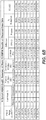


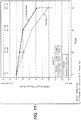
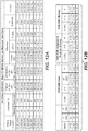
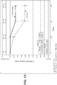


Classifications
-
- A—HUMAN NECESSITIES
- A61—MEDICAL OR VETERINARY SCIENCE; HYGIENE
- A61K—PREPARATIONS FOR MEDICAL, DENTAL OR TOILETRY PURPOSES
- A61K39/00—Medicinal preparations containing antigens or antibodies
-
- C—CHEMISTRY; METALLURGY
- C07—ORGANIC CHEMISTRY
- C07K—PEPTIDES
- C07K16/00—Immunoglobulins [IGs], e.g. monoclonal or polyclonal antibodies
- C07K16/18—Immunoglobulins [IGs], e.g. monoclonal or polyclonal antibodies against material from animals or humans
-
- A—HUMAN NECESSITIES
- A61—MEDICAL OR VETERINARY SCIENCE; HYGIENE
- A61K—PREPARATIONS FOR MEDICAL, DENTAL OR TOILETRY PURPOSES
- A61K31/00—Medicinal preparations containing organic active ingredients
- A61K31/56—Compounds containing cyclopenta[a]hydrophenanthrene ring systems; Derivatives thereof, e.g. steroids
-
- A—HUMAN NECESSITIES
- A61—MEDICAL OR VETERINARY SCIENCE; HYGIENE
- A61K—PREPARATIONS FOR MEDICAL, DENTAL OR TOILETRY PURPOSES
- A61K39/00—Medicinal preparations containing antigens or antibodies
- A61K39/395—Antibodies; Immunoglobulins; Immune serum, e.g. antilymphocytic serum
-
- A—HUMAN NECESSITIES
- A61—MEDICAL OR VETERINARY SCIENCE; HYGIENE
- A61K—PREPARATIONS FOR MEDICAL, DENTAL OR TOILETRY PURPOSES
- A61K39/00—Medicinal preparations containing antigens or antibodies
- A61K39/395—Antibodies; Immunoglobulins; Immune serum, e.g. antilymphocytic serum
- A61K39/39533—Antibodies; Immunoglobulins; Immune serum, e.g. antilymphocytic serum against materials from animals
- A61K39/3955—Antibodies; Immunoglobulins; Immune serum, e.g. antilymphocytic serum against materials from animals against proteinaceous materials, e.g. enzymes, hormones, lymphokines
-
- A—HUMAN NECESSITIES
- A61—MEDICAL OR VETERINARY SCIENCE; HYGIENE
- A61P—SPECIFIC THERAPEUTIC ACTIVITY OF CHEMICAL COMPOUNDS OR MEDICINAL PREPARATIONS
- A61P25/00—Drugs for disorders of the nervous system
- A61P25/28—Drugs for disorders of the nervous system for treating neurodegenerative disorders of the central nervous system, e.g. nootropic agents, cognition enhancers, drugs for treating Alzheimer's disease or other forms of dementia
-
- A—HUMAN NECESSITIES
- A61—MEDICAL OR VETERINARY SCIENCE; HYGIENE
- A61P—SPECIFIC THERAPEUTIC ACTIVITY OF CHEMICAL COMPOUNDS OR MEDICINAL PREPARATIONS
- A61P43/00—Drugs for specific purposes, not provided for in groups A61P1/00-A61P41/00
-
- A—HUMAN NECESSITIES
- A61—MEDICAL OR VETERINARY SCIENCE; HYGIENE
- A61K—PREPARATIONS FOR MEDICAL, DENTAL OR TOILETRY PURPOSES
- A61K39/00—Medicinal preparations containing antigens or antibodies
- A61K2039/505—Medicinal preparations containing antigens or antibodies comprising antibodies
-
- C—CHEMISTRY; METALLURGY
- C07—ORGANIC CHEMISTRY
- C07K—PEPTIDES
- C07K2317/00—Immunoglobulins specific features
- C07K2317/20—Immunoglobulins specific features characterized by taxonomic origin
- C07K2317/24—Immunoglobulins specific features characterized by taxonomic origin containing regions, domains or residues from different species, e.g. chimeric, humanized or veneered
-
- C—CHEMISTRY; METALLURGY
- C07—ORGANIC CHEMISTRY
- C07K—PEPTIDES
- C07K2317/00—Immunoglobulins specific features
- C07K2317/30—Immunoglobulins specific features characterized by aspects of specificity or valency
- C07K2317/34—Identification of a linear epitope shorter than 20 amino acid residues or of a conformational epitope defined by amino acid residues
-
- C—CHEMISTRY; METALLURGY
- C07—ORGANIC CHEMISTRY
- C07K—PEPTIDES
- C07K2317/00—Immunoglobulins specific features
- C07K2317/50—Immunoglobulins specific features characterized by immunoglobulin fragments
- C07K2317/56—Immunoglobulins specific features characterized by immunoglobulin fragments variable (Fv) region, i.e. VH and/or VL
Definitions
- AD Alzheimer's Disease
- MMSE Mini-Mental State Examination
- Approved medical therapies that inhibit acetylcholinesterase (“AChE”) activity or antagonize N-methyl-D-aspartate receptors in the brain may temporarily improve the symptoms of AD in some patients but do not modify the progression of the disease ( Cummings, N. Engl. J. Med. 2004; 351:56-67 ).
- AChE acetylcholinesterase
- amyloid plaques in the brain are a hallmark pathologic finding in AD, first reported by Alois Alzheimer in 1906.
- These amyloid plaques are primarily composed of Abeta peptides ( Haass and Selkoe, Nature 2007; 8:656-67 ) generated by the sequential cleavage of amyloid precursor protein ("APP") via ⁇ and ⁇ -secretase activity.
- Abeta particularly in its oligomerized forms, is toxic to neurons and is believed to be causative in AD.
- Therapies that reduce Abeta levels in the brain may alleviate cognitive dysfunction and block further synaptic loss, axon degeneration, and neuronal cell death.
- Abeta can be transported actively across the blood-brain barrier ( Deane et al., Stroke 2004; 35(Suppl I):2628-31 ).
- systemic delivery of antibodies to Abeta increases Abeta levels in plasma while reducing levels in the central nervous system (CNS) through several proposed mechanisms, including dissolution of brain Abeta plaque, phagocytic removal of opsonized Abeta, and finally via efflux of Abeta from the brain as a result of an equilibrium shift of Abeta resulting from circulating antibodies ( Morgan, Neurodegener. Dis. 2005; 2:261-6 ).
- bapineuzumab did appear to stabilize plaque levels and decreased phosphorylated tau levels in cerebrospinal fluid - suggesting that modification of these biomarkers alone is not necessarily predictive of clinical efficacy ( Miles et al., Scientific Reports 2013; 3:1-4 ).
- an antibody specific for monomeric Abeta that binds in the middle portion of the peptide the primary cognitive and functional endpoints were not met ( Eli Lilly and Company press release dated August 24, 2012, "Eli Lilly and Company Aanounces Top-Line Results on Solanezumab Phase 3 Clinical Trials in Patients with Alzheimer's Disease ").
- AD amyloid-related imaging abnormalities
- Crenezumab (also known as MABT5102A) is a fully humanized IgG4 monoclonal antibody to Abeta selected for its ability to bind both monomeric and oligomeric forms of Abeta in vitro. Crenezumab binds both Abeta1-40 and Abeta 1-42, inhibits Abeta aggregation, and promotes Abeta disaggregation. Because crenezumab is a human IgG4 backbone antibody, it has reduced Fe ⁇ receptor (“Fc ⁇ R”) binding affinity compared with human IgG1 or IgG2, which is predictive of reduced immune effector response.
- Fc ⁇ R Fe ⁇ receptor
- a humanized monoclonal anti-amyloid beta antibody with a conformational epitope specific for the middle region of amyloid beta (A ⁇ ) peptide is effective to treat mild to moderate AD, especially ApoE4 positive patients and patients with milder forms of AD, such as, but not limited to, mild AD, without an increased incidence of ARIA-E or ARIA-H. Accordingly, this application provides therapeutic agents for modulating the severity of AD and improved methods of using the same.
- the present invention is defined in the claims, and relates to Crenezumab for use in a method of treating Alzheimer's Disease (AD) comprising delaying the development of AD or slowing the progression of AD in a patient with an MMSE score of 20 or above, without increasing the risk of an adverse event in said patient relative to a placebo, wherein the adverse event is selected from: (i) Amyloid-Related Imaging Abnormality-Edema (ARIA-E) and(ii) Amyloid-Related Imaging Abnormality-Hemorrhage (ARIA-H); wherein the crenezumab is administered at a dose of 15 mg/kg to 100 mg/kg of the patient's body weight, said dose being administered repeatedly every four weeks or every month.
- AD Alzheimer's Disease
- the present application also describes methods of treating patients suffering from AD and other amyloidoses, comprising administering a humanized monoclonal anti-amyloid beta (A ⁇ or Abeta) antibody, or antigen-binding fragment thereof, that binds within residues 13 and 24 of amyloid ⁇ (1-42)(SEQ ID NO:1).
- a ⁇ or Abeta humanized monoclonal anti-amyloid beta
- the antibody, or antigen-binding fragment thereof is capable of binding fibrillar, oligomeric, and monomeric forms of Abeta.
- the antibody is an IgG4 antibody.
- the antibody, or antigen-binding fragment thereof comprises six hypervariable regions (HVRs) wherein HVR-H1 is SEQ ID NO:2, HVR-H2 is SEQ ID NO:3, HVR-H3 is SEQ ID NO:4, HVR-L1 is SEQ ID NO:6, HVR-L2 is SEQ ID NO:7, and HVR-L3 is SEQ ID NO:8.
- HVR-H1 is SEQ ID NO:2
- HVR-H3 is SEQ ID NO:4
- HVR-L1 is SEQ ID NO:6
- HVR-L2 is SEQ ID NO:7
- HVR-L3 is SEQ ID NO:8.
- the antibody, or antigen-binding fragment thereof comprises a heavy chain having the amino acid sequence of SEQ ID NO:5, comprising a heavy chain variable region, and a light chain having the amino acid sequence of SEQ ID NO:9, comprising a light chain variable region.
- the antibody is crenezumab.
- Suitable patients include patients suffering from mild to moderate AD, patients with an MMSE score of 18 to 26, patients suffering from mild AD, patients with an MMSE score of 20 or above (e.g ., 20-30, 20-26, 24-30, 21-26, 22-26, 22-28, 23-26, 24-26, or 25-26), patients suffering from early AD (including patients having mild cognitive impairement due to AD and patients having preclinical AD), amyloid positive patients (or patients having brain amyloid load consistent with that seen in patients diagnosed with AD), and ApoE4 positive patients suffering from mild to moderate or mild AD.
- the methods described herein are methods of reducing decline due to AD in patients suffering from early, mild, or mild to moderate AD.
- the decline is one or more of: clinical decline, cognitive decline, and functional decline.
- the decline is clinical decline.
- the decline is a decline in cognitive capacity or cognitive decline.
- the decline comprises a decline in functional capacity or functional decline.
- Various tests and scales have been developed to measure cognitive capacity (including memory) and/or function.
- one or more test is used to measure clinical, functional, or cognitive decline.
- a standard measurement of cognitive capacity is the Alzheimer's Disease Assessment Scale Cognitive (ADAS-Cog) test, for example, the 12-item ADAS-Cog or ADAS-Cog12.
- the reduction or slowing in decline in cognitive capacity (or cognitive decline) in patients being treated with the antibodies as described herein is determined using the ADAS-Cog12 test.
- An increase in ADAS-Cog12 score is indicative of worsening in a patient's condition.
- the reduction or slowing in cognitive decline (or decline in cognitive capacity) in patients being treated with the antibodies as described herein is determined by a Clinical Dementia Rating Scale / Sum of Boxes (CDR-SOB) score.
- CDR-SOB Clinical Dementia Rating Scale / Sum of Boxes
- reduction or slowing in functional decline (or decline in functional ability) in patients being treated with the antibodies as described herein is determined using the Instrumental Activities of Daily Living (iADL) scale.
- decline of one or more types is assessed and one or more of the foregoing tests or scales is used to measure reduction or slowing in decline.
- An antibody, or antigen-binding fragment thereof, as described herein is administered at a dose that is effective to treat the AD or other amyloidosis, as described herein.
- Suitable dosages are described herein and can range from about 0.3 mg/kg to 100 mg/kg.
- the dosage is 15 mg/kg.
- the dosage is 30 mg/kg.
- the dosage is 45 mg/kg.
- the dosage is between 500 mg and 1000mg, for example 500 mg, 700 mg, 720 mg, 750 mg, 800 mg, 820 mg, 900 mg, or between 1000 mg and 2500 mg, for example 1050 mg, 1500 mg, or 2100 mg.
- a variety of dosage regimens are contemplated including dosage regimens in which the antibody is administered repeatedly, e.g., on a weekly or monthly schedule, over an extended period of time, e.g., months to years.
- the humanized monoclonal anti- Abeta antibody of the present disclosure provides a further benefit in that it does not increase the incidence of adverse events such as ARIA-E and ARIA-H. As shown herein, there was no increase in these adverse events in the treatment arm relative to the placebo arm. Thus, the present disclosure further provides methods of treating patients suffering from mild to moderate AD or mild AD without increasing the incidence of adverse events such as ARIA-E and/or ARIA-H.
- the present disclosure further provides pharmaceutical formulations suitable for use in the methods of treatment disclosed herein.
- the pharmaceutical formulations can be formulated for any convenient route of administration, e.g ., parenteral or intravenous injection, and will typically include, in addition to the anti-Abeta of the present disclosure, one or more acceptable carriers, excipients, and/or diluents suited to the desired mode of administration.
- an antibody described herein may be formulated for intravenous administration.
- an antibody of the invention may be formulated in an arginine buffer, e.g., an arginine succinate buffer.
- the buffer can contain one or more surfactants, e.g ., a polysorbate.
- the buffer concentration is 50 mM or greater.
- the pH is between 4.5 and 7.0, e.g., pH 5.5. Further embodiments are described herein.
- the pharmaceutical formulations can be package in unit dosage forms for ease of use.
- other therapy include neurological drugs, corticosteroids, antibiotics, and antiviral agents.
- Non-limiting examples of anti-Abeta antibodies other than crenezumab include solanezumab, bapineuzumab, aducanumab, and gantenerumab.
- Ranges provided in the specification and appended claims include both end points and all points between the end points.
- a range of 2.0 to 3.0 includes 2.0, 3.0, and all points between 2.0 and 3.0.
- substantially similar denotes a sufficiently high degree of similarity between two numeric values (generally one associated with an antibody as described herein and the other associated with a reference/comparator antibody) such that one of skill in the art would consider the difference between the two values to be of little or no biological and/or statistical significance within the context of the biological characteristic measured by said values (e.g ., Kd values).
- the difference between said two values is less than about 50%, less than about 40%, less than about 30%, less than about 20%, less than about 10% as a function of the value for the reference/comparator antibody.
- sample refers to a composition that is obtained or derived from a subject of interest that contains a cellular and/or other molecular entity that is to be characterized and/or identified, for example based on physical, biochemical, chemical and/or physiological characteristics.
- the definition encompasses blood and other liquid samples of biological origin and tissue samples such as a biopsy specimen or tissue cultures or cells derived therefrom.
- the source of the tissue sample may be solid tissue as from a fresh, frozen and/or preserved organ or tissue sample or biopsy or aspirate; blood or any blood constituents; bodily fluids; and cells from any time in gestation or development of the subject or plasma.
- biological sample as used herein includes, but is not limited to, blood, serum, plasma, sputum, tissue biopsies ( e.g ., lung samples), and nasal samples including nasal swabs or nasal polyps.
- sample biological sample,' or “test sample” includes biological samples that have been manipulated in any way after their procurement, such as by treatment with reagents, solubilization, or enrichment for certain components, such as proteins or polynucleotides, or embedding in a semi-solid or solid matrix for sectioning purposes.
- a "section" of a tissue sample is meant a single part or piece of a tissue sample, e.g. a thin slice of tissue or cells cut from a tissue sample.
- Samples include, but are not limited to, whole blood, blood-derived cells, serum, plasma, lymph fluid, synovial fluid, cellular extracts, and combinations thereof.
- the sample is a clinical sample.
- the sample is used in a diagnostic assay.
- a sample is obtained from a subject or patient prior to treatment with an anti-Abeta antibody. In another embodiment, a sample is obtained from a subject or patient following at least one treatment with an anti-Abeta antibody.
- a “reference sample,” as used herein, refers to any sample, standard, or level that is used for comparison purposes.
- a reference sample is obtained from a healthy and/or non-diseased part of the body (e.g., tissue or cells) of the same subject or patient.
- a reference sample is obtained from an untreated tissue and/or cell of the body of the same subject or patient.
- a reference sample is obtained from a healthy and/or non-diseased part of the body (e.g., tissues or cells) of an individual who is not the subject or patient.
- a reference sample is obtained from an untreated tissue and/or cell part of the body of an individual who is not the subject or patient.
- a reference sample is a single sample or combined multiple samples from the same subject or patient that are obtained at one or more different time points than when the test sample is obtained. For example, a reference sample is obtained at an earlier time point from the same subject or patient than when the test sample is obtained.
- a reference sample includes all types of biological samples as defined above under the term "sample” that is obtained from one or more individuals who is not the subject or patient.
- a reference sample is obtained from one or more individuals with amyloidosis, e.g., Alzheimer's Disease, who is not the subject or patient.
- a reference sample is a combined multiple samples from one or more healthy individuals who are not the subject or patient.
- a reference sample is a combined multiple samples from one or more individuals with a disease or disorder (e.g., amyloidosis such as, for example, Alzheimer's Disease) who are not the subject or patient.
- a reference sample is pooled RNA samples from normal tissues or pooled plasma or serum samples from one or more individuals who are not the subject or patient.
- small molecule refers to an organic molecule having a molecular weight between 50 Daltons to 2500 Daltons.
- antibody and “immunoglobulin” (“Ig”) are used interchangeably in the broadest sense and include, but are not limited to, monoclonal antibodies (for example, full length or intact monoclonal antibodies), polyclonal antibodies, multivalent antibodies, antibodies with polyepitopic specificity, single chain antibodies, multi-specific antibodies (for example, bispecific antibodies, trispecific antibodies, tetraspecific antibodies), and fragments of antibodies, provided they exhibit the desired biological activity.
- Such antibodies can be chimeric, humanized, human, synthetic, and/or affinity matured. Such antibodies and methods of generating them are described in more detail herein.
- Antibody fragments comprise only a portion of an intact antibody, wherein the portion preferably retains at least one, and typically most or all, of the functions normally associated with that portion when present in an intact antibody.
- an antibody fragment comprises an antigen binding site of the intact antibody and thus retains the ability to bind antigen.
- an antibody fragment for example one that comprises the Fc region, retains at least one of the biological functions normally associated with the Fc region when present in an intact antibody, such as FcRn binding, antibody half life modulation, ADCC function and complement binding.
- an antibody fragment is a monovalent antibody that has an in vivo half life substantially similar to an intact antibody.
- an antibody fragment may comprise an antigen binding arm linked to an Fc sequence capable of conferring in vivo stability to the fragment.
- antibody fragments include but are not limited to Fv, Fab, Fab', Fab'-SH, F(ab')2; diabodies; linear antibodies; single-chain antibody molecules ( e.g . scFv); and multispecific antibodies formed from antibody fragments.
- target refers to any native molecule from any vertebrate source, including mammals such as primates (e.g. humans) and rodents (e.g., mice and rats), unless otherwise indicated.
- the term encompasses "full-length,” unprocessed target as well as any form of target that results from processing in the cell.
- the term also encompasses naturally occurring variants of targets, e.g ., splice variants or allelic variants.
- amyloid beta refers to the fragment of amyloid precursor protein (“APP) that is produced upon ⁇ -secretase 1 (“BACE1") cleavage of APP, as well as modifications, fragments and any functional equivalents thereof, including, but not limited to, A ⁇ 1-40, and A ⁇ 1-42.
- a ⁇ is known to exist in monomeric form, as well as to associate to form oligomers and fibril structures, which may be found as constituent members of amyloid plaque.
- a ⁇ peptides are well known to one of ordinary skill in the art and methods of producing said peptides or of extracting them from brain and other tissues are described, for example, in Glenner and Wong, Biochem Biophys Res. Comm. 129: 885-890 (1984 ). Moreover, A ⁇ peptides are also commercially available in various forms.
- An exemplary amino acid sequence of human A ⁇ 1-42 is DAEFRHDSGYEVHHQKLVFFAED VGSNKGAIIGLMVGGVVIA (SEQ ID NO: 1).
- anti-target antibody and "an antibody that binds to target” refer to an antibody that is capable of binding the target with sufficient affinity such that the antibody is useful as a diagnostic and/or therapeutic agent in targeting the target.
- the extent of binding of an anti-target antibody to an unrelated, non-target protein is less than about 10% of the binding of the antibody to target as measured, e.g ., by a radioimmunoassay (RIA) or biacore assay.
- RIA radioimmunoassay
- an antibody that binds to a target has a dissociation constant (Kd) of ⁇ 1 ⁇ M, ⁇ 100 nM, ⁇ 10 nM, ⁇ 1 nM, ⁇ 0.1 nM, ⁇ 0.01 nM, or ⁇ 0.001 nM ( e.g ., 10 -8 M or less, e.g ., from 10 -8 M to 10 -13 M, e.g ., from 10 -9 M to 10 -13 M).
- Kd dissociation constant
- an anti-target antibody binds to an epitope of a target that is conserved among different species.
- Anti-Abeta immunoglobulin refers to an antibody that specifically binds to human Abeta.
- a nonlimiting example of an anti-Abeta antibody is crenezumab.
- Other non-limiting examples of anti-Abeta antibodies are solanezumab, bapineuzumab, aducanumab, and gantenerumab.
- crenezumab and “MABT5102A” are used interchangeably herein, and refer to a specific anti-Abeta antibody that binds to monomeric, oligomeric, and fibril forms of Abeta, and which is associated with CAS registry number 1095207 .
- such antibody comprises HVR region sequences set forth in FIG 2 .
- such antibody comprises: (1) an HVR-H1 comprising the amino acid sequence SEQ ID NO: 2; (2) an HVR-H2 sequence comprising the amino acid sequence SEQ ID NO: 3; (3) an HVR-H3 sequence comprising the amino acid sequence SEQ ID NO: 4; (4) an HVR-L1 sequence comprising the amino acid sequence SEQ ID NO: 6; (5) an HVR-L2 sequence comprising the amino acid sequence SEQ ID NO: 7; and (6) an HVR-L3 sequence comprising the amino acid sequence SEQ ID NO: 8.
- the specific anti-Abeta antibody comprises VH and VL domains having the amino acid sequences set forth in FIG 3 .
- such specific anti-Abeta antibody comprises a VH domain comprising the amino acid sequence SEQ ID NO: 5 and a VL domain comprising the amino acid sequence SEQ ID NO: 9.
- the antibody is an IgG4 antibody.
- the IgG4 antibody comprises a mutation in its constant domain such that serine 228 is instead a proline.
- amyloidosis refers to a group of diseases and disorders caused by or associated with amyloid or amyloid-like proteins and includes, but is not limited to, diseases and disorders caused by the presence or activity of amyloid-like proteins in monomeric, fibril, or polymeric state, or any combination of the three, including by amyloid plaques.
- Such diseases include, but are not limited to, secondary amyloidosis and age-related amyloidosis, such as diseases including, but not limited to, neurological disorders such as Alzheimer's Disease (“AD"), diseases or conditions characterized by a loss of cognitive memory capacity such as, for example, mild cognitive impairment (MCI), Lewy body dementia, Down's syndrome, hereditary cerebral hemorrhage with amyloidosis (Dutch type), the Guam Parkinson-Demential complex and other diseases which are based on or associated with amyloid-like proteins such as progressive supranuclear palsy, multiple sclerosis, Creutzfeld Jacob disease, Parkinson's disease, HIV-related dementia, ALS (amyotropic lateral sclerosis), inclusion-body myositis (IBM), adult onset diabetes, endocrine tumor and senile cardiac amyloidosis, and various eye diseases including macular degeneration, drusen-related optic neuropathy, glaucoma, and cataract due to beta-amyloid deposition.
- Glaucoma is a group of diseases of the optic nerve involving loss of retinal ganglion cells (RGCs) in a characteristic pattern of optic neuropathy.
- RGCs are the nerve cells that transmit visual signals from the eye to the brain.
- Caspase-3 and Caspase-8 two major enzymes in the apoptotic process, are activated in the process leading to apoptosis of RGCs.
- Caspase-3 cleaves amyloid precursor protein (APP) to produce neurotoxic fragments, including Abeta. Without the protective effect of APP, Abeta accumulation in the retinal ganglion cell layer results in the death of RGCs and irreversible loss of vision.
- APP amyloid precursor protein
- Glaucoma is often, but not always, accompanied by an increased eye pressure, which may be a result of blockage of the circulation of aqueous, or its drainage.
- raised intraocular pressure is a significant risk factor for developing glaucoma
- no threshold of intraocular pressure can be defined which would be determinative for causing glaucoma.
- the damage may also be caused by poor blood supply to the vital optic nerve fibers, a weakness in the structure of the nerve, and/or a problem in the health of the nerve fibers themselves.
- Untreated glaucoma leads to permanent damage of the optic nerve and resultant visual field loss, which can progress to blindness.
- Glaucoma usually affects both eyes, but the disease can progress more rapidly in one eye than in the other.
- COAG chronic open-angle glaucoma
- POAG primary open angle glaucoma
- IOP intraocular pressure
- Acute Angle Closure Glaucoma or closed-angle glaucoma is a relatively rare type of glaucoma characterized by a sudden increase in intraocular pressure to 35 to 80 mmHg, leading to severe pain and irreversible loss of vision..
- the sudden pressure increase is caused by the closing of the filtering angle and blockage of the drainage channels.
- Individuals with narrow angles have an increased risk for a sudden closure of the angle.
- AACG usually occurs monocularly, but the risk exists in both eyes. Age, cataract and pseudoexfoliation are also risk factors since they are associated with enlargement of the lens and crowding or narrowing of the angle.
- a sudden glaucoma attack may be associated with severe eye pain and headache, inflamed eye, nausea, vomiting, and blurry vision.
- Glaucoma is a mixture or combination of open and closed angle glaucoma. It affects patients with acute ACG whose angle opens after laser iridotomy, but who continue to require medications for IOP control, as well as patients with POAG or pseudoexfoliative glaucoma who gradually develop narrowing of the angle.
- NVG Normal tension glaucoma
- LSG low tension glaucoma
- Congenital (infantile) glaucoma is a relatively rare, inherited type of open-angle glaucoma. Insufficient development of the drainage area results in increased pressure in the eye that can lead to the loss of vision from optic nerve damage and to an enlarged eye. Early diagnosis and treatment are critical to preserve vision in infants and children affected by the disease.
- Secondary glaucoma may result from an ocular injury, inflammation in the iris of the eye (iritis), diabetes, cataract, or use of steroids in steroid-susceptible individuals. Secondary glaucoma may also be associated with retinal detachment or retinal vein occlusion or blockage.
- Pigmentary glaucoma is characterized by the detachment of granules of pigment from the iris. The granules cause blockage of the drainage system of the eye, leading to elevated intraocular pressure and damage to the optic nerve.
- Exfoliative glaucoma is characterized by deposits of flaky material on the anterior capsule and in the angle of the eye. Accumulation of the flaky material blocks the drainage system and raises the eye pressure.
- Diagnosis of glaucoma may be made using various tests. Tonometry determines the pressure in the eye by measuring the tone or firmness of its surface. Several types of tonometers are available for this test, the most common being the applanation tonometer. Pachymetry determines the thickness of the cornea which, in turn, measures intraocular pressure. Gonioscopy allows examination of the filtering angle and drainage area of the eye. Gonioscopy can also determine if abnormal blood vessels may be blocking the drainage of the aqueous fluid out of the eye. Ophthalmoscopy allows examination of the optic nerve and can detect nerve fiber layer drop or changes in the optic disc, or indentation (cupping) of this structure, which may be caused by increased intraocular pressure or axonal drop out.
- Gonioscopy is also useful in assessing damage to the nerve from poor blood flow or increased intraocular pressure.
- Visual Field testing maps the field of vision, subjectively, which may detect signs of glaucomatous damage to the optic nerve. This is represented by specific patterns of visual field loss.
- Ocular coherence tomography an objective measure of nerve fiber layer loss, is carried out by looking at the thickness of the optic nerve fiber layer (altered in glaucoma) via a differential in light transmission through damaged axonal tissue.
- an "antibody that binds to the same epitope” as a reference antibody refers to an antibody that blocks binding of the reference antibody to its antigen in a competition assay by 50% or more, and conversely, the reference antibody blocks binding of the antibody to its antigen in a competition assay by 50% or more.
- An exemplary competition assay is provided herein.
- therapeutic agent refers to any agent that is used to treat a disease, including but not limited to an agent that treats a symptom of the disease.
- treatment refers to clinical intervention in an attempt to alter the natural course of the individual being treated, and can be performed during the course of clinical pathology. Desirable effects of treatment include, but are not limited to, alleviation or amelioration of one or more symptoms, diminishment of or delay in the appearance of or worsening of any direct or indirect pathological consequences of the disease, decrease of the rate of disease progression, and amelioration or palliation of the disease state.
- antibodies are used to delay development of a disease or to slow the progression of a disease.
- treatment emergent refers to an event that occurs after a first dose of a therapeutic agent is administered.
- a “treatment emergent adverse event” is an event that is identified upon or after the first dose of a treatment in a clinical study.
- Treatment regimen refers to a combination of dosage, frequency of administration, or duration of treatment, with or without addition of a second medication.
- Effective treatment regimen refers to a treatment regimen that will offer beneficial response to a patient receiving the treatment.
- Modifying a treatment refers to changing the treatment regimen including, changing dosage, frequency of administration, or duration of treatment, and/or addition of a second medication.
- an “effective amount” or “effective dose” of an agent refers to an amount or dose effective, for periods of time necessary, to achieve the desired result.
- a “therapeutically effective amount” is an amount effective, for periods of time necessary, to treat the indicated disease, condition, clinical pathology, or symptom, i . e ., to modify the course of progression of AD and/or to alleviate and/or prevent one or more symptoms of AD.
- Binding affinity refers to the strength of the sum total of noncovalent interactions between a single binding site of a molecule (e.g ., an antibody) and its binding partner (e.g ., an antigen). Unless indicated otherwise, as used herein, "binding affinity” refers to intrinsic binding affinity which reflects a 1:1 interaction between members of a binding pair ( e.g ., antibody and antigen binding arm).
- the affinity of a molecule X for its partner Y can generally be represented by the dissociation constant (Kd). Affinity can be measured by common methods known in the art, including those described herein, any of which can be used for purposes of the present invention. Specific illustrative and exemplary embodiments for measuring binding affinity are described herein.
- an “affinity matured” antibody refers to an antibody with one or more alterations in one or more hypervariable regions (HVRs), compared to a parent antibody which does not possess such alterations, such alterations resulting in an improvement in the affinity of the antibody for antigen.
- HVRs hypervariable regions
- the term "patient” refers to any single subject for which treatment is desired.
- the patient herein is a human.
- a "subject" herein is typically a human.
- a subject is a non-human mammal.
- Exemplary non-human mammals include laboratory, domestic, pet, sport, and stock animals, e.g ., mice, cats, dogs, horses, and cows.
- the subject is eligible for treatment, e.g ., displays one or more indicia of disease.
- such subject or patient is eligible for treatment for amyloidosis, e.g., AD.
- such eligible subject or patient is one that is experiencing or has experienced one or more signs, symptoms, or other indicators of AD or has been diagnosed with AD, whether, for example, newly diagnosed, previously diagnosed or at risk for developing AD.
- Diagnosis of AD may be made based on clinical history, clinical examination, and established imaging modalities.
- a "patient” or “subject” herein includes any single human subject eligible for treatment who is experiencing or has experienced one or more signs, symptoms, or other indicators of AD. Intended to be included as a subject are any subjects involved in clinical research trials, or subjects involved in epidemiological studies, or subjects once used as controls. The subject may have been previously treated with an anti-Abeta antibody, or antigen-binding fragment thereof, or another drug, or not so treated.
- the subject may be naive to an additional drug(s) being used when the treatment herein is started, i.e., the subject may not have been previously treated with, for example, a therapy other than anti-Abeta at "baseline" (i.e., at a set point in time before the administration of a first dose of anti-Abeta in the treatment method herein, such as the day of screening the subject before treatment is commenced).
- a therapy other than anti-Abeta at "baseline” i.e., at a set point in time before the administration of a first dose of anti-Abeta in the treatment method herein, such as the day of screening the subject before treatment is commenced.
- Such "naive" subjects are generally considered to be candidates for treatment with such additional drug(s).
- lifetime of a subject refers to the remainder of the life of the subject after starting treatment.
- monoclonal antibody refers to an antibody obtained from a population of substantially homogeneous antibodies, i.e ., the individual antibodies comprising the population are identical except for possible naturally occurring mutations that may be present in minor amounts. Monoclonal antibodies are highly specific, being directed against a single antigen. Furthermore, in contrast to polyclonal antibody preparations that typically include different antibodies directed against different determinants (epitopes), each monoclonal antibody is directed against a single determinant on the antigen.
- the monoclonal antibodies herein specifically include "chimeric" antibodies in which a portion of the heavy and/or light chain is identical with or homologous to corresponding sequences in antibodies derived from a particular species or belonging to a particular antibody class or subclass, while the remainder of the chain(s) is identical with or homologous to corresponding sequences in antibodies derived from another species or belonging to another antibody class or subclass, as well as fragments of such antibodies, so long as they exhibit the desired biological activity ( U.S. Patent No. 4,816,567 ; and Morrison et al., Proc. Natl. Acad. Sci. USA 81:6851-6855 (1984 )).
- the "class” of an antibody refers to the type of constant domain or constant region possessed by its heavy chain.
- the heavy chain constant domains that correspond to the different classes of immunoglobulins are called ⁇ , ⁇ , ⁇ , ⁇ , and ⁇ , respectively.
- Humanized forms of non-human (e . g ., murine) antibodies are chimeric antibodies that contain minimal sequence derived from non-human immunoglobulin.
- humanized antibodies are human immunoglobulins (recipient antibody) in which residues from a hypervariable region of the recipient are replaced by residues from a hypervariable region of a non-human species (donor antibody) such as mouse, rat, rabbit or nonhuman primate having the desired specificity, affinity, and capacity.
- donor antibody such as mouse, rat, rabbit or nonhuman primate having the desired specificity, affinity, and capacity.
- framework region (FR) residues of the human immunoglobulin are replaced by corresponding non-human residues.
- humanized antibodies may comprise residues that are not found in the recipient antibody or in the donor antibody. These modifications are made to further refine antibody performance.
- the humanized antibody will comprise substantially all of at least one, and typically two, variable domains, in which all or substantially all of the hypervariable loops correspond to those of a non-human immunoglobulin and all or substantially all of the FRs are those of a human immunoglobulin lo sequence.
- the humanized antibody optionally will also comprise at least a portion of an immunoglobulin constant region (Fc), typically that of a human immunoglobulin.
- Fc immunoglobulin constant region
- a "human antibody” is one which comprises an amino acid sequence corresponding to that of an antibody produced by a human or a human cell and/or has been derived from a non-human source that utilizes human antibody repertoires or other human antibody-encoding sequences, for example made using any of the techniques for making human antibodies as disclosed herein. Such techniques include, but are not limited to, screening human-derived combinatorial libraries, such as phage display libraries (see, e.g., Marks et al., J. Mol. Biol., 222: 581-597 (1991 ) and Hoogenboom et al., Nucl.
- an “isolated” antibody is one which has been identified and separated and/or recovered from a component of its natural environment. Contaminant components of its natural environment are materials which would interfere with diagnostic or therapeutic uses for the antibody, and may include enzymes, hormones, and other proteinaceous or nonproteinaceous solutes.
- an antibody is purified to greater than 95% or 99% purity as determined by, for example, electrophoretic (e.g., SDS-PAGE, isoelectric focusing (IEF), capillary electrophoresis) or chromatographic (e.g., ion exchange or reverse phase HPLC).
- electrophoretic e.g., SDS-PAGE, isoelectric focusing (IEF), capillary electrophoresis
- chromatographic e.g., ion exchange or reverse phase HPLC
- variable region refers to the domain of an antibody heavy or light chain that is involved in binding the antibody to antigen.
- the variable domains of the heavy chain and light chain (VH and VL, respectively) of a native antibody generally have similar structures, with each domain comprising four conserved framework regions (FRs) and three hypervariable regions (HVRs).
- FRs conserved framework regions
- HVRs hypervariable regions
- antibodies that bind a particular antigen may be isolated using a VH or VL domain from an antibody that binds the antigen to screen a library of complementary VL or VH domains, respectively. See, e.g., Portolano et al., J. Immunol. 150:880-887 (1993 ); Clarkson et al., Nature 352:624-628 (1991 ).
- hypervariable region when used herein refers to the regions of an antibody variable domain which are hypervariable in sequence and/or form structurally defined loops.
- antibodies comprise six hypervariable regions; three in the VH (HI, H2, H3), and three in the VL (LI, L2, L3).
- a number of hypervariable region delineations are in use and are encompassed herein.
- the Kabat Complementarity Determining Regions are based on sequence variability and are the most commonly used ( Kabat et al., Sequences of Proteins of Immunological Interest, 5th Ed. Public Health Service, National Institutes of Health, Bethesda, Md. (1991 )).
- Chothia refers instead to the location of the structural loops ( Chothia and Lesk J. Mol. Biol. 196:901-917 (1987 )).
- the AbM hypervariable regions represent a compromise between the Kabat CDRs and Chothia structural loops, and are used by Oxford Molecular's AbM antibody modeling software.
- the "contact" hypervariable regions are based on an analysis of the available complex crystal structures. The residues from each of these HVRs are noted below.
- Hypervariable regions may comprise "extended hypervariable regions” as follows: 24-36 or 24-34 (LI), 46-56 or 49-56 or 50-56 or 52-56 (L2) and 89-97 (L3) in the VL and 26-35 (HI), 50-65 or 49-65 (H2) and 93-102, 94-102 or 95-102 (H3) in the VH.
- the variable domain residues are numbered according to Kabat et al ., supra for each of these definitions.
- FR residues are those variable domain residues other than the hypervariable region residues as herein defined.
- the FR of a variable domain generally consists of four FR domains: FR1, FR2, FR3, and FR4. Accordingly, the HVR and FR sequences generally appear in the following sequence in VH (or VL): FR1-H1(L1)-FR2-H2(L2)-FR3-H3(L3)-FR4.
- acceptor human framework for the purposes herein is a framework comprising the amino acid sequence of a light chain variable domain (VL) framework or a heavy chain variable domain (VH) framework derived from a human immunoglobulin framework or a human consensus framework, as defined below.
- An acceptor human framework "derived from” a human immunoglobulin framework or a human consensus framework may comprise the same amino acid sequence thereof, or it may contain amino acid sequence changes. In some embodiments, the number of amino acid changes are 10 or less, 9 or less, 8 or less, 7 or less, 6 or less, 5 or less, 4 or less, 3 or less, or 2 or less.
- the VL acceptor human framework is identical in sequence to the VL human immunoglobulin framework sequence or human consensus framework sequence.
- a "human consensus framework” is a framework which represents the most commonly occurring amino acid residue in a selection of human immunoglobulin VL or VH framework sequences.
- the selection of human immunoglobulin VL or VH sequences is from a subgroup of variable domain sequences.
- the subgroup of sequences is a subgroup as in Kabat et al. Sequences of Proteins of Immunological Interest, Fifth Edition, NIH Publication 91-3242, Bethesda MD (1991), vols. 1-3 . et al . et al.
- Amyloid-Related Imaging Abnormality - Edema or "ARIA-E” encompasses cerebral vasogenic edema and sulcal effusion.
- ARIA-H Amyloid-Related Imaging Abnormality - Hemorrhage
- Apolipoprotein E4 carrier or “ApoE4 carrier,” used interchangeably herein with “apolipoprotein E4 positive” or “ApoE4 positive,” refers to an individual having at least one apolipoprotein E4 (or "ApoE4") allele. An individual with zero ApoE4 alleles is referred to herein as being “ApoE4 negative” or an "ApoE4 non-carrier.” See also Prekumar, et al., 1996, Am. J Pathol. 148:2083-95 .
- Cerebral vasogenic edema refers to an excess accumulation of intravascular fluid or protein in the intracellular or extracellular spaces of the brain. Cerebral vasogenic edema is detectable by, e.g., brain MRI, including, but not limited to FLAIR MRI, and can be asymptomatic ("asymptomatic vasogenic edema") or associated with neurological symptoms, such as confusion, dizziness, vomiting, and lethargy (“symptomatic vasogenic edema”) ( seesperling et al. Alzheimer's & Dementia, 7:367, 2011 ).
- Cerebral macrohemorrhage refers to an intracranial hemorrhage, or bleeding in the brain, of an area that is more than about 1 cm in diameter. Cerebral macrohemorrhage is detectable by, e.g., brain MRI, including but not limited to T2*-weighted GRE MRI, and can be asymptomatic ("asymptomatic macrohemorrhage") or associated with symptoms such as transient or permanent focal motor or sensory impairment, ataxia, aphasia, and dysarthria ("symptomatic macrohemorrhage”) (see, e.g., Chalela JA, Gomes J. Expert Rev. Neurother. 2004 4:267, 2004 and Sperling et al. Alzheimer's & Dementia, 7:367, 2011 ).
- Cerebral microhemorrhage refers to an intracranial hemorrhage, or bleeding in the brain, of an area that is less than about 1 cm in diameter. Cerebral microhemorrhage is detectable by, e.g., brain MRI, including, but not limited to T2*-weighted GRE MRI, and can be asymptomatic ("asymptomatic microhemorrhage") or can potentially be associated with symptoms such as transient or permanent focal motor or sensory impairment, ataxia, aphasia, and dysarthria ("symptomatic microhemorrhage"). See, e.g., Greenberg, et al., 2009, Lancet Neurol. 8:165-74 .
- sulcal effusion refers to an effusion of fluid in the furrows, or sulci, of the brain. Sulcal effusions are detectable by, e.g., brain MRI, including but not limited to FLAIR MRI. Seesperling et al. Alzheimer's & Dementia, 7:367, 2011 .
- the term "superficial siderosis of the central nervous system” refers to bleeding or hemorrhage into the subarachnoid space of the brain and is detectable by, e.g., brain MRI, including but not limited to T2*-weighted GRE MRI. Symptoms indicative of superficial siderosis of the central nervous system include sensorineural deafness, cerebellar ataxia, and pyramidal signs. See Kumara-N, Am J Neuroradiol. 31:5, 2010 .
- progression refers to the worsening of a disease over time.
- the "progression rate” or “rate of progression” of a disease refers to how fast or slow a disease develops over time in a patient diagnosed with the disease.
- the progression rate of a disease can be represented by measurable changes over time of particular characteristics of the disease.
- a patient carrying particular genetic trait is said to have, or more likely to have, "increased progression rate” if her disease state progresses faster than those patients without such genetic trait.
- a patient responding to a therapy is said to have, or more likely to have, "decreased progression rate” if her disease progression slows down after the therapy, when compared to her disease state prior to the treatment or to other patients without the treatment.
- “More likely to respond” as used herein refers to patients that are most likely to demonstrate a slowing down or prevention of progression of amyloidosis, e.g., AD. With regard to AD, “more likely to respond” refers to patients that are most likely to demonstrate a reduction in loss of function or cognition with treatment.
- the phrase "responsive to" in the context of the present invention indicates that a patient suffering from, being suspected to suffer or being prone to suffer from, or diagnosed with a disorder as described herein, shows a response to anti-Abeta treatment.
- selecting a patient or "identifying a patient” as used herein refers to using the information or data generated relating to the presence of an allele in a sample of a patient to identify or select the patient as more likely to benefit to benefit from a treatment comprising anti-Abeta antibody.
- the information or data used or generated may be in any form, written, oral or electronic.
- using the information or data generated includes communicating, presenting, reporting, storing, sending, transferring, supplying, transmitting, dispensing, or combinations thereof.
- communicating, presenting, reporting, storing, sending, transferring, supplying, transmitting, dispensing, or combinations thereof are performed by a computing device, analyzer unit or combination thereof.
- communicating, presenting, reporting, storing, sending, transferring, supplying, transmitting, dispensing, or combinations thereof are performed by a laboratory or medical professional.
- the information or data includes an indication that a specific allele is present or absent in the sample.
- the information or data includes an indication that the patient is more likely to respond to a therapy comprising anti-Abeta.
- Antibody effector functions refer to those biological activities attributable to the Fc region of an antibody, which vary with the antibody isotype. Examples of antibody effector functions include: C1q binding and complement dependent cytotoxicity (CDC); Fc receptor binding; antibody-dependent cell-mediated cytotoxicity (ADCC); phagocytosis; down regulation of cell surface receptors (e.g. B cell receptor); and B cell activation. It is known in the art that wild-type IgG4 antibodies have less effector function than wild-type IgG1 antibodies.
- Fc region herein is used to define a C-terminal region of an immunoglobulin heavy chain that contains at least a portion of the constant region.
- the term includes native sequence Fc regions and variant Fc regions.
- a human IgG heavy chain Fc region extends from Cys226, or from Pro230, to the carboxyl-terminus of the heavy chain.
- the C-terminal lysine (Lys447) of the Fc region may or may not be present.
- numbering of amino acid residues in the Fc region or constant region is according to the EU numbering system, also called the EU index, as described in Kabat et al., Sequences of Proteins of Immunological Interest, 5th Ed. Public Health Service, National Institutes of Health, Bethesda, MD, 1991 .
- full length antibody “intact antibody,” and “whole antibody” are used herein interchangeably to refer to an antibody having a structure substantially similar to a native antibody structure or having heavy chains that contain an Fc region as defined herein.
- host cell refers to cells into which exogenous nucleic acid has been introduced, including the progeny of such cells.
- Host cells include “transformants” and “transformed cells,” which include the primary transformed cell and progeny derived therefrom without regard to the number of passages. Progeny may not be completely identical in nucleic acid content to a parent cell, but may contain mutations. Mutant progeny that have the same function or biological activity as screened or selected for in the originally transformed cell are included herein.
- an “immunoconjugate” is an antibody conjugated to one or more heterologous molecule(s), including but not limited to a further therapeutic agent.
- nucleic acid refers to a nucleic acid molecule that has been separated from a component of its natural environment.
- An isolated nucleic acid includes a nucleic acid molecule contained in cells that ordinarily contain the nucleic acid molecule, but the nucleic acid molecule is present extrachromosomally or at a chromosomal location that is different from its natural chromosomal location.
- isolated nucleic acid encoding an anti-Abeta antibody refers to one or more nucleic acid molecules encoding antibody heavy and light chains (or fragments thereof), including such nucleic acid molecule(s) in a single vector or separate vectors, and such nucleic acid molecule(s) present at one or more locations in a host cell.
- earsly Alzheimer's Disease or "early AD” as used herein (e.g., a "patient diagnosed with early AD” or a “patient suffering from early AD”) includes patients with mild cognitive impairement, such as a memory deficit, due to AD and patients having AD biomarkers, for example amyloid positive patients.
- micro Alzheimer's Disease or "mild AD” as used herein (e.g., a “patient diagnosed with mild AD”) refers to a stage of AD characterized by an MMSE score of 20 to 26.
- Mild to moderate Alzheimer's Disease or “mild to moderate AD” as used herein encompasses both mild and moderate AD, and is characterized by an MMSE score of 18 to 26.
- moderate Alzheimer's Disease or “moderate AD” as used herein (e.g., a “patient diagnosed with moderate AD”) refers to a stage of AD characterized by an MMSE score of 18 to 19.
- naked antibody refers to an antibody that is not conjugated to a heterologous moiety (e.g., a further therapeuticmoiety) or radiolabel.
- the naked antibody may be present in a pharmaceutical formulation.
- Native antibodies refer to naturally occurring immunoglobulin molecules with varying structures.
- native IgG antibodies are heterotetrameric glycoproteins of about 150,000 daltons, composed of two identical light chains and two identical heavy chains that are disulfide-bonded. From N- to C-terminus, each heavy chain has a variable region (VH), also called a variable heavy domain or a heavy chain variable domain, followed by three constant domains (CHI, CH2, and CH3).
- VH variable region
- VL variable region
- the light chain of an antibody may be assigned to one of two types, called kappa ( ⁇ ) and lambda ( ⁇ ), based on the amino acid sequence of its constant domain.
- package insert is used to refer to instructions customarily included in commercial packages of therapeutic products, that contain information about the indications, usage, dosage, administration, combination therapy, contraindications and/or warnings concerning the use of such therapeutic products.
- packet insert is also used to refer to instructions customarily included in commercial packages of diagnostic products that contain information about the intended use, test principle, preparation and handling of reagents, specimen collection and preparation, calibration of the assay and the assay procedure, performance and precision data such as sensitivity and specificity of the assay.
- Percent (%) amino acid sequence identity with respect to a reference polypeptide sequence is defined as the percentage of amino acid residues in a candidate sequence that are identical with the amino acid residues in the reference polypeptide sequence, after aligning the sequences and introducing gaps, if necessary, to achieve the maximum percent sequence identity, and not considering any conservative substitutions as part of the sequence identity. Alignment for purposes of determining percent amino acid sequence identity can be achieved in various ways that are within the skill in the art, for instance, using publicly available computer software such as BLAST, BLAST-2, ALIGN or Megalign (DNASTAR) software. Those skilled in the art can determine appropriate parameters for aligning sequences, including any algorithms needed to achieve maximal alignment over the full length of the sequences being compared.
- % amino acid sequence identity values are generated using the sequence comparison computer program ALIGN-2.
- the ALIGN-2 sequence comparison computer program was authored by Genentech, Inc., and the source code has been filed with user documentation in the U.S. Copyright Office, Washington D.C., 20559, where it is registered under U.S. Copyright Registration No. TXU510087.
- the ALIGN-2 program is publicly available from Genentech, Inc., South San Francisco, California, or may be compiled from the source code.
- the ALIGN-2 program should be compiled for use on a UNIX operating system, including digital UNIX V4.0D. All sequence comparison parameters are set by the ALIGN-2 program and do not vary.
- the % amino acid sequence identity of a given amino acid sequence A to, with, or against a given amino acid sequence B is calculated as follows: 100 times the fraction (X/Y) where X is the number of amino acid residues scored as identical matches by the sequence alignment program ALIGN-2 in that program's alignment of A and B, and where Y is the total number of amino acid residues in B.
- pharmaceutical formulation and “pharmaceutical composition” are used interchangeably herein and refer to a preparation which is in such form as to permit the biological activity of an active ingredient contained therein to be effective, and which contains no additional components which are unacceptably toxic to a subject to which the formulation would be administered.
- a “pharmaceutically acceptable carrier” refers to an ingredient in a pharmaceutical formulation, other than an active ingredient, which is nontoxic to a subject.
- a pharmaceutically acceptable carrier includes, but is not limited to, a buffer, excipient, stabilizer, or preservative.
- vector refers to a nucleic acid molecule capable of propagating another nucleic acid to which it is linked.
- the term includes the vector as a self-replicating nucleic acid structure as well as the vector incorporated into the genome of a host cell into which it has been introduced.
- Certain vectors are capable of directing the expression of nucleic acids to which they are operatively linked. Such vectors are referred to herein as "expression vectors.”
- imaging agent is a compound that has one or more properties that permit its presence and/or location to be detected directly or indirectly.
- imaging agents include proteins and small molecule compounds incorporating a labeled moiety that permits detection.
- a “label” is a marker coupled with a molecule to be used for detection or imaging.
- labels include: a radiolabel, a fluorophore, a chromophore, or an affinity tag.
- the label is a radiolabel used for medical imaging, for example tc99m or 1123, or a spin label for nuclear magnetic resonance (NMR) imaging (also known as magnetic resonance imaging, mri), such as iodine-123 again, iodine-131, indium-111, fluorine-19, carbon-13, nitrogen-15, oxygen-17, gadolinium, manganese, iron, etc.
- NMR nuclear magnetic resonance
- compositions and methods for the treatment, prognosis, selection and/or identification of patients at risk for or having amyloidosis are provided.
- the invention is based, in part, on improved methods of treatment.
- antibodies that bind to Abeta are provided.
- Antibodies as described herein are useful, e.g., for the diagnosis or treatment of Alzheimer's Disease ("AD”) and other diseases.
- AD Alzheimer's Disease
- the present application describes isolated antibodies that bind to Abeta.
- the application describes an anti-Abeta antibody that can bind to monomeric, oligomeric and fibril forms of human Abeta with good affinity.
- the anti-Abeta antibody is an antibody that binds to an epitope of Abeta within residues 13-24 of Abeta.
- the antibody is crenezumab.
- the antibody comprises the heavy chain amino acid sequence set forth in SEQ ID NO:5 and the light chain amino acid sequence set forth in SEQ ID NO:9. In another embodiment, the antibody comprises the heavy chain variable region of amino acids 1 to 112 of the amino acid sequence set forth in SEQ ID NO:5 and the light chain variable region of amino acids 1 to 112 of the amino acid sequence set forth in SEQ ID NO:9. In another embodiment, the antibody comprises the HVR sequences of SEQ ID NO:5 and SEQ ID NO:9. In another embodiment, the antibody comprises HVR sequences that are 95%, 96%, 97%, 98%, or 99% or more identical to the HVR sequences of SEQ ID NO:5 and SEQ ID NO:9.
- an anti-Abeta antibody is humanized.
- an anti-Abeta antibody comprises HVRs as in any of the above embodiments, and further comprises an acceptor human framework, e.g . a human immunoglobulin framework or a human consensus framework.
- an anti-Abeta antibody comprises a heavy chain variable domain (VH) sequence having at least 90%, 91%, 92%, 93%, 94%, 95%, 96%, 97%, 98%, 99%, or 100% sequence identity to amino acids 1 to 112 of the amino acid sequence of SEQ ID NO:5.
- VH heavy chain variable domain
- a VH sequence having at least 90%, 91%, 92%, 93%, 94%, 95%, 96%, 97%, 98%, or 99% identity contains substitutions (e.g ., conservative substitutions), insertions, or deletions relative to the reference sequence, but an anti-Abeta antibody comprising that sequence retains the ability to bind to Abeta.
- the anti-Abeta antibody comprises the VH sequence in SEQ ID NO:5, including post-translational modifications of that sequence.
- an anti-Abeta antibody comprising a light chain variable domain (VL) having at least 90%, 91%, 92%, 93%, 94%, 95%, 96%, 97%, 98%, 99%, or 100% sequence identity to amino acids 1 to 112 of the amino acid sequence of SEQ ID NO:9.
- VL light chain variable domain
- a VL sequence having at least 90%, 91%, 92%, 93%, 94%, 95%, 96%, 97%, 98%, or 99% identity contains substitutions (e.g., conservative substitutions), insertions, or deletions relative to the reference sequence, but an anti-Abeta antibody comprising that sequence retains the ability to bind to Abeta.
- the anti-Abeta antibody comprises the VL sequence in SEQ ID NO:9, including post-translational modifications of that sequence.
- an anti-Abeta antibody comprising a VH as in any of the embodiments provided above, and a VL as in any of the embodiments provided above.
- the application describes an antibody that binds to the same epitope as an anti-Abeta antibody provided herein.
- an antibody is provided that binds to the same epitope as an anti-Abeta antibody comprising a VH sequence in SEQ ID NO:5 and a VL sequence in SEQ ID NO:9.
- an anti-Abeta antibody is a monoclonal antibody, including a chimeric, humanized or human antibody.
- an anti-Abeta antibody is an antibody fragment, e.g ., a Fv, Fab, Fab', scFv, diabody, or F(ab')2 fragment.
- the antibody is a full length antibody, e.g., an intact IgG4 antibody or other antibody class or isotype as defined herein.
- the antibody is a bispecific antibody.
- an anti-Abeta antibody may incorporate any of the features, singly or in combination, as described in Sections 1-7 below.
- the anti-Abeta antibody comprises a HVR-L1 comprising amino acid sequence SEQ ID NO:6; an HVR-L2 comprising amino acid sequence SEQ ID NO:7; an HVR-L3 comprising amino acid sequence SEQ ID NO: 8; an HVR-H1 comprising amino acid sequence SEQ ID NO:2; an HVR-H2 comprising amino acid sequence SEQ ID NO: 3; and an HVR-H3 comprising amino acid sequence SEQ ID NO: 4.
- the antibody comprises the heavy and light sequences SEQ ID NO:5 and SEQ ID NO:9.
- the antibody comprises the variable region sequences in SEQ ID NO:5 and SEQ ID NO:9.
- an anti-Abeta antibody can be humanized.
- an anti-Abeta antibody comprises HVRs as in any of the above embodiments, and further comprises an acceptor human framework, e.g . a human immunoglobulin framework or a human consensus framework.
- an antibody provided herein has a dissociation constant (Kd) of ⁇ 1 ⁇ M, ⁇ 100 nM, ⁇ 10 nM, ⁇ 1 nM, ⁇ 0.1 nM, ⁇ 0.01 nM, or ⁇ 0.001 nM (e.g. 10 -8 M or less, e.g. from 10 -8 M to 10 -13 M, e.g., from 10 -9 M to 10 -13 M).
- Kd dissociation constant
- Kd is measured by a radiolabeled antigen binding assay (RIA) performed with the Fab version of an antibody of interest and its antigen as described by the following assay.
- Solution binding affinity of Fabs for antigen is measured by equilibrating Fab with a minimal concentration of ( 125 I)-labeled antigen in the presence of a titration series of unlabeled antigen, then capturing bound antigen with an anti-Fab antibody-coated plate ( see , e.g ., Chen et al.et al., J. Mol. Biol. 293:865-881(1999 )).
- MICROTITER® multi-well plates (Thermo Scientific) are coated overnight with 5 ⁇ g/ml of a capturing anti-Fab antibody (Cappel Labs) in 50 mM sodium carbonate (pH 9.6), and subsequently blocked with 2% (w/v) bovine serum albumin in PBS for two to five hours at room temperature (approximately 23°C).
- a non-adsorbent plate (Nunc #269620)
- 100 pM or 26 pM [125I]-antigen are mixed with serial dilutions of a Fab of interest ( e . g ., consistent with assessment of the anti-VEGF antibody, Fab-12, in Presta et al., Cancer Res.
- the Fab of interest is then incubated overnight; however, the incubation may continue for a longer period ( e . g ., about 65 hours) to ensure that equilibrium is reached. Thereafter, the mixtures are transferred to the capture plate for incubation at room temperature ( e . g ., for one hour). The solution is then removed and the plate washed eight times with 0.1% polysorbate 20 (TWEEN-20®) in PBS. When the plates have dried, 150 ⁇ l/well of scintillant (MICROSCINT-20 TM; Packard) is added, and the plates are counted on a TOPCOUNT TM gamma counter (Packard) for ten minutes. Concentrations of each Fab that give less than or equal to 20% of maximal binding are chosen for use in competitive binding assays.
- Kd is measured using surface plasmon resonance assays using a BIACORE®-2000 or a BIACORE ®-3000 (BIAcore, Inc., Piscataway, NJ) at 25°C with immobilized antigen CM5 chips at ⁇ 10 response units (RU).
- CM5 carboxymethylated dextran biosensor chips
- EDC N-ethyl-N'- (3-dimethylaminopropyl)-carbodiimide hydrochloride
- NHS N-hydroxysuccinimide
- Antigen is diluted with 10 mM sodium acetate, pH 4.8, to 5 ⁇ g/ml ( ⁇ 0.2 ⁇ M) before injection at a flow rate of 5 ⁇ l/minute to achieve approximately 10 response units (RU) of coupled protein. Following the injection of antigen, 1 M ethanolamine is injected to block unreacted groups. For kinetics measurements, two-fold serial dilutions of Fab (0.78 nM to 500 nM) are injected in PBS with 0.05% polysorbate 20 (TWEEN-20TM) surfactant (PBST) at 25°C at a flow rate of approximately 25 ⁇ l/min.
- TWEEN-20TM polysorbate 20
- association rates (kon) and dissociation rates (koff) are calculated using a simple one-to-one Langmuir binding model (BIACORE ® Evaluation Software version 3.2) by simultaneously fitting the association and dissociation sensorgrams.
- the equilibrium dissociation constant (Kd) is calculated as the ratio koff/kon. See, e.g., Chen et al., J. Mol. Biol. 293:865-881 (1999 ).
- an antibody provided herein is an antibody fragment.
- Antibody fragments include, but are not limited to, Fab, Fab', Fab'-SH, F(ab')2, Fv, and scFv fragments, and other fragments described below.
- Fab fragment antigen
- Fab' fragment antigen binding domain
- Diabodies are antibody fragments with two antigen-binding sites that may be bivalent or bispecific. See, for example, EP 404,097 ; WO 1993/01161 ; Hudson et al., Nat. Med. 9:129-134 (2003 ); and Hollinger et al., Proc. Natl. Acad. Sci. USA 90: 6444-6448 (1993 ). Triabodies and tetrabodies are also described in Hudson et al., Nat. Med. 9:129-134 (2003 ).
- Single-domain antibodies are antibody fragments comprising all or a portion of the heavy chain variable domain or all or a portion of the light chain variable domain of an antibody.
- a single-domain antibody is a human single-domain antibody (Domantis, Inc., Waltham, MA; see, e.g., U.S. Patent No. 6,248,516 B1 ).
- two or more single-domain antibodies may be joined together to form an immunoglobulin construct with multivalent affinity ( i . e ., the N- or C-terminus of a first single-domain antibody may be fused or otherwise joined to the N- or C-terminus of a second single-domain antibody).
- Antibody fragments can be made by various techniques, including but not limited to proteolytic digestion of an intact antibody as well as production by recombinant host cells (e.g. E. coli or phage), as described herein.
- recombinant host cells e.g. E. coli or phage
- an antibody provided herein is a chimeric antibody.
- Certain chimeric antibodies are described, e.g., in U.S. Patent No. 4,816,567 ; and Morrison et al., Proc. Natl. Acad. Sci. USA, 81:6851-6855 (1984 )).
- a chimeric antibody comprises a non-human variable region (e.g., a variable region derived from a mouse, rat, hamster, rabbit, or non-human primate, such as a monkey) and a human constant region.
- a chimeric antibody is a "class switched" antibody in which the class or subclass has been changed from that of the parent antibody. Chimeric antibodies include antigen-binding fragments thereof.
- a chimeric antibody is a humanized antibody.
- a non-human antibody is humanized to reduce immunogenicity to humans, while retaining the specificity and affinity of the parental non-human antibody.
- a humanized antibody comprises one or more variable domains in which HVRs, e.g., CDRs, (or portions thereof) are derived from a non-human antibody, and FRs (or portions thereof) are derived from human antibody sequences.
- HVRs e.g., CDRs, (or portions thereof) are derived from a non-human antibody
- FRs or portions thereof
- a humanized antibody optionally will also comprise at least a portion of a human constant region.
- some FR residues in a humanized antibody are substituted with corresponding residues from a non-human antibody (e.g., the antibody from which the HVR residues are derived), e.g., to restore or improve antibody specificity or affinity.
- a non-human antibody e.g., the antibody from which the HVR residues are derived
- Human framework regions that may be used for humanization include but are not limited to: framework regions selected using the "best-fit" method (see, e.g., Sims .et al. J. Immunol. 151:2296 (1993 )); framework regions derived from the consensus sequence of human antibodies of a particular subgroup of light or heavy chain variable regions (see, e.g., Carter et al. Proc. Natl. Acad. Sci. USA, 89:4285 (1992 ); and Presta et al. J. Immunol., 151:2623 (1993 )); human mature (somatically mutated) framework regions or human germline framework regions (see, e.g ., Almagro and Fransson, Front. Biosci.
- an antibody provided herein is a human antibody.
- Human antibodies can be produced using various techniques known in the art. Human antibodies are described generally in van Dijk and van de Winkel, Curr. Opin. Pharmacol. 5: 368-74 (2001 ) and Lonberg, Curr. Opin. Immunol. 20:450-459 (2008 ).
- Human antibodies may be prepared by administering an immunogen to a transgenic animal that has been modified to produce intact human antibodies or intact antibodies with human variable regions in response to antigenic challenge.
- Such animals typically contain all or a portion of the human immunoglobulin loci, which replace the endogenous immunoglobulin loci, or which are present extrachromosomally or integrated randomly into the animal's chromosomes. In such transgenic mice, the endogenous immunoglobulin loci have generally been inactivated.
- Human antibodies can also be made by hybridoma-based methods. Human myeloma and mouse-human heteromyeloma cell lines for the production of human monoclonal antibodies have been described. (See, e.g ., Kozbor J. Immunol., 133: 3001 (1984 ); Brodeur et al., Monoclonal Antibody Production Techniques and Applications, pp. 51-63 (Marcel Dekker, Inc., New York, 1987 ); and Boerner et al., J. Immunol., 147: 86 (1991 ).) Human antibodies generated via human B-cell hybridoma technology are also described in Li et al., Proc. Natl. Acad. Sci.
- Human antibodies may also be generated by isolating Fv clone variable domain sequences selected from human-derived phage display libraries. Such variable domain sequences may then be combined with a desired human constant domain. Techniques for selecting human antibodies from antibody libraries are described below.
- Antibodies as deribed herein may be isolated by screening combinatorial libraries for antibodies with the desired activity or activities. For example, a variety of methods are known in the art for generating phage display libraries and screening such libraries for antibodies possessing the desired binding characteristics. Such methods are reviewed, e.g., in Hoogenboom et al. in Methods in Molecular Biology 178:1-37 (O'Brien et al., ed., Human Press, Totowa, NJ, 2001 ) and further described, e.g., in the McCafferty et al., Nature 348:552-554 ; Clackson et al., Nature 352: 624-628 (1991 ); Marks et al., J. Mol. Biol.
- repertoires of VH and VL genes are separately cloned by polymerase chain reaction (PCR) and recombined randomly in phage libraries, which can then be screened for antigen-binding phage as described in Winter et al., Ann. Rev. Immunol., 12: 433-455 (1994 ).
- Phage typically display antibody fragments, either as single-chain Fv (scFv) fragments or as Fab fragments.
- scFv single-chain Fv
- Libraries from immunized sources provide high-affinity antibodies to the immunogen without the requirement of constructing hybridomas.
- naive repertoire can be cloned (e.g., from human) to provide a single source of antibodies to a wide range of non-self and also self antigens without any immunization as described by Griffiths et al., EMBO J, 12: 725-734 (1993 ).
- naive libraries can also be made synthetically by cloning unrearranged V-gene segments from stem cells, and using PCR primers containing random sequence to encode the highly variable CDR3 regions and to accomplish rearrangement in vitro, as described by Hoogenboom and Winter, J. Mol. Biol., 227: 381-388 (1992 ).
- Patent publications describing human antibody phage libraries include, for example: US Patent No.
- Antibodies or antibody fragments isolated from human antibody libraries are considered human antibodies or human antibody fragments herein.
- an antibody provided herein is a multispecific antibody, e.g. a bispecific antibody.
- Multispecific antibodies are monoclonal antibodies that have binding specificities for at least two different sites. In certain embodiments, one of the binding specificities is for Abeta and the other is for any other antigen.
- bispecific antibodies may bind to two different epitopes of Abeta. Bispecific antibodies may also be used to localize cytotoxic agents to cells. Bispecific antibodies can be prepared as full length antibodies or antibody fragments.
- Multispecific antibodies include, but are not limited to, recombinant co-expression of two immunoglobulin heavy chain-light chain pairs having different specificities (see Milstein and Cuello, Nature 305: 537 (1983 )), WO 93/08829 , and Traunecker et al., EMBO J. 10: 3655 (1991 )), and "knob-in-hole” engineering (see, e.g., U.S. Patent No. 5,731,168 ). Multi-specific antibodies may also be made by engineering electrostatic steering effects for making antibody Fc-heterodimeric molecules ( WO 2009/089004A1 ); cross-linking two or more antibodies or fragments (see, e.g., US Patent No.
- the antibody or fragment herein also includes a "Dual Acting FAb” or “DAF” comprising an antigen binding site that binds to Abeta as well as another, different antigen (see, US 2008/0069820 , for example).
- a “Dual Acting FAb” or “DAF” comprising an antigen binding site that binds to Abeta as well as another, different antigen (see, US 2008/0069820 , for example).
- amino acid sequence variants of the antibodies provided herein are contemplated.
- Amino acid sequence variants of an antibody may be prepared by introducing appropriate modifications into the nucleotide sequence encoding the antibody, or by peptide synthesis. Such modifications include, for example, deletions from, and/or insertions into and/or substitutions of residues within the amino acid sequences of the antibody. Any combination of deletion, insertion, and substitution can be made to arrive at the final construct, provided that the final construct possesses the desired characteristics, e.g ., antigen-binding.
- antibody variants having one or more amino acid substitutions are provided.
- Sites of interest for substitutional mutagenesis include the HVRs and FRs.
- Conservative substitutions are shown in Table 1 under the heading of "conservative substitutions.” More substantial changes are provided in Table 1 under the heading of "exemplary substitutions,” and as further described below in reference to amino acid side chain classes.
- Amino acid substitutions may be introduced into an antibody of interest and the products screened for a desired activity, e.g ., retained/improved antigen binding, decreased immunogenicity, or improved ADCC or CDC.
- Amino acids may be grouped according to common side-chain properties:
- Non-conservative substitutions will entail exchanging a member of one of these classes for another class.
- substitutional variant involves substituting one or more hypervariable region residues of a parent antibody (e.g . a humanized or human antibody).
- a parent antibody e.g . a humanized or human antibody.
- the resulting variant(s) selected for further study will have modifications (e.g., improvements) in certain biological properties (e.g., increased affinity, reduced immunogenicity) relative to the parent antibody and/or will have substantially retained certain biological properties of the parent antibody.
- An exemplary substitutional variant is an affinity matured antibody.
- affinity matured antibodies will have nanomolar or even picomolar affinities for the target antigen.
- Affinity matured antibodies are produced by procedures known in the art, including, e.g ., using phage display-based affinity maturation techniques such as those described herein.
- HVR residues are mutated and the variant antibodies displayed on phage and screened for a particular biological activity (e.g . binding affinity).
- Other procedures are also known. Marks et al. Bio/Technology 10:779-783 (1992 ) describes affinity maturation by VH and VL domain shuffling. Random mutagenesis of HVR and/or framework residues is described by: Barbas et al. Proc Nat. Acad. Sci, USA 91:3809-3813 (1994 ); Schier et al. Gene 169:147-155 (1996 ); Yelton et al. J. Immunol. 155:1994-2004 (1995 ); Jackson et al., J. Immunol. 154(7):3310-9 (1995 ); and Hawkins et al. J. Mol. Biol. 226:889-896 (1992 ).
- Alterations may be made in HVRs, e.g., to improve antibody affinity. Such alterations may be made in HVR "hotspots," i.e., residues encoded by codons that undergo mutation at high frequency during the somatic maturation process (see, e.g., Chowdhury, Methods Mol. Biol. 207:179-196 (2008 )), and/or SDRs (a-CDRs), with the resulting variant VH or VL being tested for binding affinity.
- HVR "hotspots” i.e., residues encoded by codons that undergo mutation at high frequency during the somatic maturation process (see, e.g., Chowdhury, Methods Mol. Biol. 207:179-196 (2008 )
- SDRs a-CDRs
- affinity maturation diversity is introduced into the variable genes chosen for maturation by any of a variety of methods (e.g., error-prone PCR, chain shuffling, or oligonucleotide-directed mutagenesis).
- a secondary library is then created. The library is then screened to identify any antibody variants with the desired affinity.
- Another method to introduce diversity involves HVR-directed approaches, in which several HVR residues (e.g., 4-6 residues at a time) are randomized. HVR residues involved in antigen binding may be specifically identified, e.g ., using alanine scanning mutagenesis or modeling. CDR-H3 and CDR-L3 in particular are often targeted.
- substitutions, insertions, or deletions may occur within one or more HVRs so long as such alterations do not substantially reduce the ability of the antibody to bind antigen.
- conservative alterations e.g., conservative substitutions as provided herein
- Such alterations may be outside of HVR "hotspots" or SDRs.
- each HVR either is unaltered, or contains no more than one, two or three amino acid substitutions.
- a useful method for identification of residues or regions of an antibody that may be targeted for mutagenesis is called "alanine scanning mutagenesis" as described by Cunningham and Wells (1989) Science, 244:1081-1085 .
- a residue or group of target residues e . g ., charged residues such as arg, asp, his, lys, and glu
- a neutral or negatively charged amino acid e . g ., alanine or polyalanine
- Further substitutions may be introduced at the amino acid locations demonstrating functional sensitivity to the initial substitutions.
- a crystal structure of an antigen-antibody complex to identify contact points between the antibody and antigen. Such contact residues and neighboring residues may be targeted or eliminated as candidates for substitution.
- Variants may be screened to determine whether they contain the desired properties.
- Amino acid sequence insertions include amino- and/or carboxyl-terminal fusions ranging in length from one residue to polypeptides containing a hundred or more residues, as well as intrasequence insertions of single or multiple amino acid residues.
- terminal insertions include an antibody with an N-terminal methionyl residue.
- Other insertional variants of the antibody molecule include the fusion to the N- or C-terminus of the antibody to an enzyme (e.g. for ADEPT) or a polypeptide which increases the serum half-life of the antibody.
- an antibody provided herein is altered to increase or decrease the extent to which the antibody is glycosylated.
- Addition or deletion of glycosylation sites to an antibody may be conveniently accomplished by altering the amino acid sequence such that one or more glycosylation sites is created or removed.
- the carbohydrate attached thereto may be altered.
- Native antibodies produced by mammalian cells typically comprise a branched, biantennary oligosaccharide that is generally attached by an N-linkage to Asn297 of the CH2 domain of the Fc region. See, e.g., Wright et al. TIBTECH 15:26-32 (1997 ).
- the oligosaccharide may include various carbohydrates, e.g ., mannose, N-acetyl glucosamine (GlcNAc), galactose, and sialic acid, as well as a fucose attached to a GlcNAc in the "stem" of the biantennary oligosaccharide structure.
- modifications of the oligosaccharide in an antibody as described herein may be made in order to create antibody variants with certain improved properties.
- antibody variants having a carbohydrate structure that lacks fucose attached (directly or indirectly) to an Fc region.
- the amount of fucose in such antibody may be from 1% to 80%, from 1% to 65%, from 5% to 65% or from 20% to 40%.
- the amount of fucose is determined by calculating the average amount of fucose within the sugar chain at Asn297, relative to the sum of all glycostructures attached to Asn 297 (e. g. complex, hybrid and high mannose structures) as measured by MALDI-TOF mass spectrometry, as described in WO 2008/077546 , for example.
- Asn297 refers to the asparagine residue located at about position 297 in the Fc region (Eu numbering of Fc region residues); however, Asn297 may also be located about ⁇ 3 amino acids upstream or downstream of position 297, i.e., between positions 294 and 300, due to minor sequence variations in antibodies. Such fucosylation variants may have improved ADCC function. See, e.g., US Patent Publication Nos. US 2003/0157108 (Presta, L. ); US 2004/0093621 (Kyowa Hakko Kogyo Co., Ltd).
- Examples of publications related to "defucosylated” or “fucose-deficient” antibody variants include: US 2003/0157108 ; WO 2000/61739 ; WO 2001/29246 ; US 2003/0115614 ; US 2002/0164328 ; US 2004/0093621 ; US 2004/0132140 ; US 2004/0110704 ; US 2004/0110282 ; US 2004/0109865 ; WO 2003/085119 ; WO 2003/084570 ; WO 2005/035586 ; WO 2005/035778 ; WO2005/053742 ; WO2002/031140 ; Okazaki et al. J. Mol. Biol.
- Examples of cell lines capable of producing defucosylated antibodies include Lec13 CHO cells deficient in protein fucosylation ( Ripka et al. Arch. Biochem. Biophys.
- Antibodies variants are further provided with bisected oligosaccharides, e.g ., in which a biantennary oligosaccharide attached to the Fc region of the antibody is bisected by GlcNAc. Such antibody variants may have reduced fucosylation and/or improved ADCC function. Examples of such antibody variants are described, e.g ., in WO 2003/011878 (Jean-Mairet et al .); US Patent No. 6,602,684 (Umana et al .); and US 2005/0123546 (Umana et al .). Antibody variants with at least one galactose residue in the oligosaccharide attached to the Fc region are also provided.
- Such antibody variants may have improved CDC function.
- Such antibody variants are described, e.g., in WO 1997/30087 (Patel et al .); WO 1998/58964 (Raju, S. ); and WO 1999/22764 (Raju, S. ).
- one or more amino acid modifications may be introduced into the Fc region of an antibody provided herein, thereby generating an Fc region variant.
- the Fc region variant may comprise a human Fc region sequence (e.g., a human IgG1, IgG2, IgG3 or IgG4 Fc region) comprising an amino acid modification (e.g . a substitution) at one or more amino acid positions.
- the application contemplates an antibody variant that possesses some but not all effector functions, which make it a desirable candidate for applications in which the half life of the antibody in vivo is important yet certain effector functions (such as complement and ADCC) are unnecessary or deleterious.
- In vitro and/or in vivo cytotoxicity assays can be conducted to confirm the reduction/depletion of CDC and/or ADCC activities.
- Fc receptor (FcR) binding assays can be conducted to ensure that the antibody lacks FcyR binding (hence likely lacking ADCC activity), but retains FcRn binding ability.
- NK cells express Fc ⁇ RIII only, whereas monocytes express Fc ⁇ RI, Fc ⁇ RII and Fc ⁇ RIII.
- FcR expression on hematopoietic cells is summarized in Table 3 on page 464 of Ravetch and Kinet, Annu. Rev. Immunol. 9:457-492 (1991 ).
- Non-limiting examples of in vitro assays to assess ADCC activity of a molecule of interest is described in U.S. Patent No. 5,500,362 (see, e.g. Hellstrom, I. et al. Proc. Nat'l Acad. Sci. USA 83:7059-7063 (1986 )) and Hellstrom, I et al., Proc.
- non-radioactive assays methods may be employed (see, for example, ACTITM non-radioactive cytotoxicity assay for flow cytometry (CellTechnology, Inc. Mountain View, CA; and CytoTox 96® non-radioactive cytotoxicity assay (Promega, Madison, WI).
- Useful effector cells for such assays include peripheral blood mononuclear cells (PBMC) and Natural Killer (NK) cells.
- ADCC activity of the molecule of interest may be assessed in vivo, e.g., in a animal model such as that disclosed in Clynes et al. Proc. Nat'l Acad. Sci. USA 95:652-656 (1998 ).
- C1q binding assays may also be carried out to confirm that the antibody is unable to bind C1q and hence lacks CDC activity. See, e.g., C1q and C3c binding ELISA in WO 2006/029879 and WO 2005/100402 .
- a CDC assay may be performed (see, for example, Gazzano-Santoro et al., J. Immunol.
- FcRn binding and in vivo clearance/half life determinations can also be performed using methods known in the art (see, e.g., Petkova, S.B. et al., Int'l. Immunol. 18(12):1759-1769 (2006 )).
- Antibodies with reduced effector function include those with substitution of one or more of Fc region residues 238, 265, 269, 270, 297, 327 and 329 ( U.S. Patent No. 6,737,056 ).
- Fc mutants include Fc mutants with substitutions at two or more of amino acid positions 265, 269, 270, 297 and 327, including the so-called "DANA" Fc mutant with substitution of residues 265 and 297 to alanine ( US Patent No. 7,332,581 ).
- an antibody variant comprises an Fc region with one or more amino acid substitutions which improve ADCC, e . g ., substitutions at positions 298, 333, and/or 334 of the Fc region (EU numbering of residues).
- alterations are made in the Fc region that result in altered ( i . e ., either improved or diminished) C1q binding and/or Complement Dependent Cytotoxicity (CDC), e.g., as described in US Patent No. 6,194,551 , WO 99/51642 , and Idusogie et al. J. Immunol. 164: 4178-4184 (2000 ).
- CDC Complement Dependent Cytotoxicity
- Antibodies with increased half lives and improved binding to the neonatal Fc receptor (FcRn), which is responsible for the transfer of maternal IgGs to the fetus are described in US2005/0014934A1 (Hinton et al .). Those antibodies comprise an Fc region with one or more substitutions therein which improve binding of the Fc region to FcRn.
- Such Fc variants include those with substitutions at one or more of Fc region residues: 238, 256, 265, 272, 286, 303, 305, 307, 311, 312, 317, 340, 356, 360, 362, 376, 378, 380, 382, 413, 424 or 434, e.g., substitution of Fc region residue 434 ( US Patent No. 7,371,826 ). See also Duncan & Winter, Nature 322:738-40 (1988 ); U.S. Patent No. 5,648,260 ; U.S. Patent No. 5,624,821 ; and WO 94/29351 concerning other examples of Fc region variants.
- cysteine engineered antibodies e.g., "thioMAbs”
- one or more residues of an antibody are substituted with cysteine residues.
- the substituted residues occur at accessible sites of the antibody.
- reactive thiol groups are thereby positioned at accessible sites of the antibody and may be used to conjugate the antibody to other moieties, such as drug moieties or linker-drug moieties, to create an immunoconjugate, as described further herein.
- any one or more of the following residues may be substituted with cysteine: V205 (Kabat numbering) of the light chain; A118 (EU numbering) of the heavy chain; and S400 (EU numbering) of the heavy chain Fc region.
- Cysteine engineered antibodies may be generated as described, e.g ., in U.S. Patent No. 7,521,541 .
- an antibody provided herein may be further modified to contain additional nonproteinaceous moieties that are known in the art and readily available.
- the moieties suitable for derivatization of the antibody include but are not limited to water soluble polymers.
- water soluble polymers include, but are not limited to, polyethylene glycol (PEG), copolymers of ethylene glycol/propylene glycol, carboxymethylcellulose, dextran, polyvinyl alcohol, polyvinyl pyrrolidone, poly-1, 3-dioxolane, poly-1,3,6-trioxane, ethylene/maleic anhydride copolymer, polyaminoacids (either homopolymers or random copolymers), and dextran or poly(n-vinyl pyrrolidone)polyethylene glycol, propropylene glycol homopolymers, prolypropylene oxide/ethylene oxide co-polymers, polyoxyethylated polyols (e.g., gly
- Polyethylene glycol propionaldehyde may have advantages in manufacturing due to its stability in water.
- the polymer may be of any molecular weight, and may be branched or unbranched.
- the number of polymers attached to the antibody may vary, and if more than one polymer is attached, they can be the same or different molecules. In general, the number and/or type of polymers used for derivatization can be determined based on considerations including, but not limited to, the particular properties or functions of the antibody to be improved, whether the antibody derivative will be used in a therapy under defined conditions, etc.
- conjugates of an antibody and nonproteinaceous moiety that may be selectively heated by exposure to radiation are provided.
- the nonproteinaceous moiety is a carbon nanotube ( Kam et al., Proc. Natl. Acad. Sci. USA 102: 11600-11605 (2005 )).
- the radiation may be of any wavelength, and includes, but is not limited to, wavelengths that do not harm ordinary cells, but which heat the nonproteinaceous moiety to a temperature at which cells proximal to the antibody-nonproteinaceous moiety are killed.
- Antibodies may be produced using recombinant methods and compositions, e.g ., as described in U.S. Patent No. 4,816,567 .
- isolated nucleic acid encoding an anti-Abeta antibody described herein is provided.
- Such nucleic acid may encode an amino acid sequence comprising the VL and/or an amino acid sequence comprising the VH of the antibody (e.g., the light and/or heavy chains of the antibody).
- one or more vectors e.g., expression vectors
- a host cell comprising such nucleic acid is provided.
- a host cell comprises (e.g., has been transformed with): (1) a vector comprising a nucleic acid that encodes an amino acid sequence comprising the VL of the antibody and an amino acid sequence comprising the VH of the antibody, or (2) a first vector comprising a nucleic acid that encodes an amino acid sequence comprising the VL of the antibody and a second vector comprising a nucleic acid that encodes an amino acid sequence comprising the VH of the antibody.
- the host cell is eukaryotic, e.g. a Chinese Hamster Ovary (CHO) cell or lymphoid cell (e.g., Y0, NS0, Sp20 cell).
- a method of making an anti-Abeta antibody comprises culturing a host cell comprising a nucleic acid encoding the antibody, as provided above, under conditions suitable for expression of the antibody, and optionally recovering the antibody from the host cell (or host cell culture medium).
- nucleic acid encoding an antibody e.g ., as described above, is isolated and inserted into one or more vectors for further cloning and/or expression in a host cell.
- nucleic acid may be readily isolated and sequenced using conventional procedures (e.g., by using oligonucleotide probes that are capable of binding specifically to genes encoding the heavy and light chains of the antibody).
- Suitable host cells for cloning or expression of antibody-encoding vectors include prokaryotic or eukaryotic cells described herein.
- antibodies may be produced in bacteria, in particular when glycosylation and Fc effector function are not needed.
- U.S. Patent Nos. 5,648,237 , 5,789,199 , and 5,840,523 See also Charlton, Methods in Molecular Biology, Vol. 248 (B.K.C. Lo, ed., Humana Press, Totowa, NJ, 2003), pp. 245-254 , describing expression of antibody fragments in E. coli.
- the antibody may be isolated from the bacterial cell paste in a soluble fraction and can be further purified.
- eukaryotic microbes such as filamentous fungi or yeast are suitable cloning or expression hosts for antibody-encoding vectors, including fungi and yeast strains whose glycosylation pathways have been "humanized,” resulting in the production of an antibody with a partially or fully human glycosylation pattern. See Gerngross, Nat. Biotech. 22:1409-1414 (2004 ), and Li et al., Nat. Biotech. 24:210-215 (2006 ).
- Suitable host cells for the expression of glycosylated antibody are also derived from multicellular organisms (invertebrates and vertebrates). Examples of invertebrate cells include plant and insect cells. Numerous baculoviral strains have been identified which may be used in conjunction with insect cells, particularly for transfection of Spodoptera frugiperda cells.
- Plant cell cultures can also be utilized as hosts. See , e.g ., US Patent Nos. 5,959,177 , 6,040,498 , 6,420,548 , 7,125,978 , and 6,417,429 (describing PLANTIBODIESTM technology for producing antibodies in transgenic plants).
- Vertebrate cells may also be used as hosts.
- mammalian cell lines that are adapted to grow in suspension may be useful.
- Other examples of useful mammalian host cell lines are monkey kidney CV1 line transformed by SV40 (COS-7); human embryonic kidney line (293 or 293 cells as described, e.g., in Graham et al., J. Gen Virol. 36:59 (1977 )); baby hamster kidney cells (BHK); mouse sertoli cells (TM4 cells as described, e.g., in Mather, Biol. Reprod.
- monkey kidney cells (CV1); African green monkey kidney cells (VERO-76); human cervical carcinoma cells (HELA); canine kidney cells (MDCK; buffalo rat liver cells (BRL 3A); human lung cells (W138); human liver cells (Hep G2); mouse mammary tumor (MMT 060562); TRI cells, as described, e.g ., in Mather et al., Annals N.Y. Acad. Sci. 383:44-68 (1982 ); MRC 5 cells; and FS4 cells.
- Other useful mammalian host cell lines include Chinese hamster ovary (CHO) cells, including DHFR- CHO cells ( Urlaub et al., Proc. Natl. Acad. Sci.
- Anti-Abeta antibodies provided herein may be identified, screened for, or characterized for their physical/chemical properties and/or biological activities by various assays known in the art.
- an antibody as described herein is tested for its antigen binding activity, e . g ., by known methods such as ELISA, Western blot, etc.
- competition assays may be used to identify an antibody that competes with an anti-Abeta antibody as described herein for binding to Abeta.
- a competing antibody binds to the same epitope (e . g ., a linear or a conformational epitope) that is bound by crenezumab or another anti-Abeta antibody specified herein.
- epitope e . g ., a linear or a conformational epitope
- immobilized Abeta in the desired form e . g ., monomeric, oligomeric, or fibril
- a solution comprising a first labeled antibody that binds to Abeta (e.g., crenezumab) and a second unlabeled antibody that is being tested for its ability to compete with the first antibody for binding to Abeta.
- the second antibody may be present in a hybridoma supernatant.
- immobilized Abeta is incubated in a solution comprising the first labeled antibody but not the second unlabeled antibody.
- assays are provided for identifying anti-Abeta antibodies thereof having biological activity, for example the biological activity of crenezumab.
- Biological activity may include, but is not limited to, e.g., prevention of aggregation of monomeric Abeta into oligomeric Abeta, or disaggregation of oligomeric Abeta into monomeric Abeta.
- Antibodies having such biological activity in vivo and/or in vitro are also provided.
- an antibody as described herein is tested for such biological activity.
- any of the anti-Abeta antibodies provided herein is useful for detecting the presence of Abeta in a biological sample.
- the term "detecting" as used herein encompasses quantitative or qualitative detection.
- a biological sample comprises a cell or tissue, such as serum, plasma, nasal swabs, sputum, cerebrospinal fluid, , aqueous humor of the eye and the like, or tissue or cell samples obtained from an organism such as samples containing neural or brain tissue.
- an anti-Abeta antibody for use in a method of diagnosis or detection is provided.
- a method of detecting the presence of Abeta in a biological sample is provided.
- the method comprises contacting the biological sample with an anti-Abeta antibody as described herein under conditions permissive for binding of the anti-Abeta antibody to Abeta, and detecting whether a complex is formed between the anti-Abeta antibody and Abeta.
- Such method may be an in vitro or in vivo method.
- Exemplary disorders that may be diagnosed using an antibody as described herein are diseases and disorders caused by or associated with amyloid or amyloid-like proteins. These include, but are not limited to, diseases and disorders caused by the presence or activity of amyloid-like proteins in monomeric, fibril, or polymeric state, or any combination of the three, including by amyloid plaques.
- Exemplary diseases include, but are not limited to, secondary amyloidosis and age-related amyloidosis, such as diseases including, but not limited to, neurological disorders such as Alzheimer's Disease (“AD"), diseases or conditions characterized by a loss of cognitive memory capacity such as, for example, mild cognitive impairment (MCI), Lewy body dementia, Down's syndrome, hereditary cerebral hemorrhage with amyloidosis (Dutch type), the Guam Parkinson-Demential complex and other diseases which are based on or associated with amyloid-like proteins such as progressive supranuclear palsy, multiple sclerosis, Creutzfeld Jacob disease, Parkinson's disease, HIV-related dementia, ALS (amyotropic lateral sclerosis), inclusion-body myositis (IBM), adult onset diabetes, endocrine tumor and senile cardiac amyloidosis, and various eye diseases including macular degeneration, drusen-related optic neuropathy, glaucoma, and cataract due to beta-amyloid deposition
- labeled anti-Abeta antibodies include, but are not limited to, labels or moieties that are detected directly (such as fluorescent, chromophoric, electron-dense, chemiluminescent, and radioactive labels), as well as moieties, such as enzymes or ligands, that are detected indirectly, e.g., through an enzymatic reaction or molecular interaction.
- Exemplary labels include, but are not limited to, the radioisotopes 32 P, 14 C, 125I , 3 H, and 131 I, fluorophores such as rare earth chelates or fluorescein and its derivatives, rhodamine and its derivatives, dansyl, umbelliferone, luceriferases, e.g., firefly luciferase and bacterial luciferase ( U.S. Patent No.
- luciferin 2,3-dihydrophthalazinediones, horseradish peroxidase (HRP), alkaline phosphatase, ⁇ -galactosidase, glucoamylase, lysozyme, saccharide oxidases, e.g., glucose oxidase, galactose oxidase, and glucose-6-phosphate dehydrogenase, heterocyclic oxidases such as uricase and xanthine oxidase, coupled with an enzyme that employs hydrogen peroxide to oxidize a dye precursor such as HRP, lactoperoxidase, or microperoxidase, biotin/avidin, spin labels, bacteriophage labels, stable free radicals, and the like.
- HRP horseradish peroxidase
- lactoperoxidase lactoperoxidase
- microperoxidase biotin/avidin
- spin labels bacteriophage labels,
- compositions of an anti-Abeta antibody as described herein are prepared by mixing such antibody or molecule having the desired degree of purity with one or more optional pharmaceutically acceptable carriers ( Remington's Pharmaceutical Sciences 16th edition, Osol, A. Ed. (1980 )), in the form of lyophilized formulations or aqueous solutions.
- Pharmaceutically acceptable carriers are generally nontoxic to recipients at the dosages and concentrations employed, and include, but are not limited to: buffers such as phosphate, citrate, and other organic acids; antioxidants including ascorbic acid and methionine; preservatives (such as octadecyldimethylbenzyl ammonium chloride; hexamethonium chloride; benzalkonium chloride; benzethonium chloride; phenol, butyl or benzyl alcohol; alkyl parabens such as methyl or propyl paraben; catechol; resorcinol; cyclohexanol; 3-pentanol; and m-cresol); low molecular weight (less than about 10 residues) polypeptides; proteins, such as serum albumin, gelatin, or immunoglobulins; hydrophilic polymers such as polyvinylpyrrolidone; amino acids such as glycine, glutamine, asparagine, histidine, arg
- sHASEGP soluble neutral-active hyaluronidase glycoproteins
- rHuPH20 HYLENEX®, Baxter International, Inc.
- Certain exemplary sHASEGPs and methods of use, including rHuPH20, are described in US Patent Publication Nos. 2005/0260186 and 2006/0104968 .
- a sHASEGP is combined with one or more additional glycosaminoglycanases such as chondroitinases.
- an antibody as described herein may be formulated in an arginine buffer.
- the arginine buffer may be an arginine succinate buffer.
- the concentration of the arginine succinate buffer may be 50 mM or greater.
- the concentration of the arginine succinate buffer may be 100 mM or greater.
- the concentration of the arginine succinate buffer may be 150 mM or greater.
- the concentration of the arginine succinate buffer may be 200 mM or greater.
- the arginine buffer formulation may further contain a surfactant.
- the surfactant is a polysorbate.
- the polysorbate is polysorbate 20.
- the concentration of polysorbate 20 in the formulation is 0.1% or less.
- the concentration of polysorbate 20 in the formulation is 0.05% or less.
- the pH of the arginine buffer formulation is between 4.5 and 7.0.
- the pH of the arginine buffer formulation is between 5.0 and 6.5.
- the pH of the arginine buffer formulation is between 5.0 and 6.0.
- the pH of the arginine buffer formulation is 5.5.
- the antibody as described herein may be crenezumab.
- Exemplary lyophilized antibody formulations are described in US Patent No. 6,267,958 .
- Aqueous antibody formulations include those described in US Patent No. 6,171,586 and WO2006/044908 , the latter formulations including a histidine-acetate buffer.
- the formulation herein may also contain more than one active ingredients as necessary for the particular indication being treated, preferably those with complementary activities that do not adversely affect each other. For example, it may be desirable to further provide one or more compounds to prevent or treat symptoms of Alzheimer's Disease. Such active ingredients are suitably present in combination in amounts that are effective for the purpose intended.
- Active ingredients may be entrapped in microcapsules prepared, for example, by coacervation techniques or by interfacial polymerization, for example, hydroxymethylcellulose or gelatin-microcapsules and poly-(methylmethacylate) microcapsules, respectively, in colloidal drug delivery systems (for example, liposomes, albumin microspheres, microemulsions, nanoparticles and nanocapsules) or in macroemulsions.
- colloidal drug delivery systems for example, liposomes, albumin microspheres, microemulsions, nanoparticles and nanocapsules
- Sustained-release preparations may be prepared. Suitable examples of sustained-release preparations include semipermeable matrices of solid hydrophobic polymers containing the antibody, which matrices are in the form of shaped articles, e.g. films, or microcapsules.
- the formulations to be used for in vivo administration are generally sterile. Sterility may be readily accomplished, e.g., by filtration through sterile filtration membranes.
- crenezumab reduced disease progression in patients suffering from AD.
- patients with mild to moderate AD including patients with mild AD and ApoE4 positive patients, as well as patients with brain amyloid load typically seen in patients diagnosed with AD, showed a reduction in the rate of cognitive decline when treated with crenezumab as compared to a placebo.
- the milder the disease based on increasing MMSE score, the greater the reduction in decline in the treatment arm as compared to the placebo arm.
- an antibody as described herein is used to treat AD, including mild to moderate AD, mild AD, and early AD.
- an antibody as described herein is used to treat an amyloidosis.
- the amyloidosis is mild cognitive impairment.
- the amyloidosis is Down's syndrome.
- the amyloidosis is hereditary cerebral hemorrhage with amyloidosis (Dutch type).
- the amyloidosis is the Guam Parkinson-Dementia complex.
- the amyloidosis is an ocular disease related to drusen or other amyloid deposit in the eye.
- the ocular disease is macular degeneration. In another aspect, the ocular disease is a drusen-related optic neuropathy. In another aspect, the ocular disease is glaucoma. In another aspect, the ocular disease is cataract. In any of the foregoing embodiments and aspects, the antibody as described herein may be crenezumab.
- a patient is typically first assessed for the presence of one or more amyloidosis prior to determining the suitability of an antibody as described herein to treat such patient.
- AD may be diagnosed in a patient using the "NINCDS-ADRDA" (Neurological and Communicative Disorders and Stroke-Alzheimer's Disease Related Disorders Assessment) criteria. See McKhann, et al., 1984, Neurology 34:939-44 .
- a potential patient to be administered one or more antibodies as described herein may also be tested for the presence or absence of one or more genetic markers which may predispose such patient either to (i) a higher or lower likelihood of such patient experiencing one or more amyloidoses, or (ii) a higher or lower likelihood of such patient experiencing one or more adverse events or side effects during the course of administration of an antibody as described herein.
- one or more genetic markers which may predispose such patient either to (i) a higher or lower likelihood of such patient experiencing one or more amyloidoses, or (ii) a higher or lower likelihood of such patient experiencing one or more adverse events or side effects during the course of administration of an antibody as described herein.
- the antibody as described herein is used to treat mild to moderate AD in a patient.
- the patient can be ApoE4 positive or ApoE4 negative.
- the antibody as described herein is used to treat mild AD.
- the antibody as described herein is used to treat an ApoE4 positive patient suffering from mild to moderate AD or mild AD.
- the antibody as described herein is used to treat a patient suffering from mild AD.
- the antibody as described herein is used to treat a patient having an MMSE score of between 20 and 30, between 20 and 26, between 24 and 30, between 21 and 26, between 22 and 26, between 22 and 28, between 23 and 26, between 24 and 26, or between 25 and 26.
- the patient has an MMSE score between 22 and 26.
- an MMSE score between two numbers includes the numbers at each end of the range.
- an MMSE score between 22 and 26 includes MMSE scores of 22 and 26.
- the antibodies as described herein are used to treat a patient who is 'amyloid positive,' e.g., a patient having brain amyloid deposits that are typical of a patient diagnosed with AD or a patient having a positive florbetapir PET scan. In some embodiments, the antibodies as described herein are used to reduce the accumulation of brain amyloid deposits or neuritic plaques ( i . e ., to reduce an increase in brain amyloid burden or load).
- the antibodies as described herein are useful for treating mild to moderate AD without increasing the incidence of ARIA-E or ARIA-H.
- the patients are suffering from mild AD.
- the patients are ApoE4 positive.
- the patients are ApoE4 positive and suffering from mild AD.
- the antibody as described herein is used to treat a patient with early AD.
- the patient to be treated has one or more of the following characteristics: (a) mild cognitive impairement (MCI) due to AD; (b) one or more biomarkers indicative of Alzheimer's Disease without a clinically detectable deficit; (c) an objective memory loss quantified using the Free and Cued Selective Reminding Test (FCSRT) as a score of 27 or greater; an MMSE of 24-30; (d) a global Clinical Dementia Rating (CDR) of 0.5; and (e) a positive amyloid PET scan (as determined by a qualified reader).
- MCI mild cognitive impairement
- CDR Clinical Dementia Rating
- Antibodies as described herein are formulated, dosed, and administered in a fashion consistent with good medical practice.
- Factors for consideration in this context include the particular disorder being treated, the particular mammal being treated, the clinical condition of the individual subject, the cause of the disorder, the site of delivery of the agent, the method of administration, the scheduling of administration, and other factors known to medical practitioners.
- An antibody as described herein can be administered by any suitable means, including parenteral, intrapulmonary, and intranasal, and, if desired for local treatment, intralesional administration.
- Parenteral infusions include intramuscular, intravenous, intraarterial, intraperitoneal, or subcutaneous administration. Dosing can be by any suitable route, e.g. by injections, such as intravenous or subcutaneous injections, depending in part on whether the administration is brief or chronic.
- the antibody is injected subcutaneously.
- the antibody is injected intravenously.
- the antibody is administered using a syringe (e.g., prefilled or not) or an autoinjector.
- the antibody is inhaled.
- an antibody as described herein when used alone or in combination with one or more other additional therapeutic agents, will depend on the specific type of disease to be treated, the type of antibody, the severity and course of the disease, previous therapy, the patient's clinical history and response to the antibody, and the discretion of the attending physician.
- the antibody is suitably administered to the patient at one time or over a series of treatments.
- Various dosing schedules including, but not limited to, single or multiple administrations over various time-points, bolus administration, and pulse infusion are contemplated herein.
- about 0.3 mg/kg to 100 mg/kg (e.g. 15 mg/kg-100 mg/kg, or any dosage within that range) of antibody can be an initial candidate dosage for administration to the patient, whether, for example, by one or more separate administrations, or by continuous infusion.
- One typical daily dosage might range from about 15 mg/kg to 100 mg/kg or more, depending on the factors mentioned above.
- the dosage can be administered in a single dose or a divided dose (e.g., two doses of 15 mg/kg for a total dose of 30 mg/ kg). For repeated administrations over several weeks or longer, depending on the condition, the treatment would generally be sustained until a desired suppression of disease symptoms occurs.
- One exemplary dosage of the antibody would be in the range from about 10 mg/kg to about 50 mg/kg.
- one or more doses of about 0.5 mg/kg, 1 mg/kg, 1.5 mg/kg, 2.0 mg/kg, 3 mg/kg, 4.0 mg/kg, 5 mg/kg, 10 mg/kg, 15 mg/kg, 20 mg/kg, 25 mg/kg, 30 mg/kg, 35 mg/kg, 40 mg/kg, 50 mg/ kg, 60 mg/ kg, 70 mg/kg, 80 mg/kg, 90 mg/kg, or 100 mg/kg (or any combination thereof) may be administered to the patient.
- the total dose administered is in the range of 50 mg to 2500 mg.
- An exemplary dose of about 50 mg, about 100 mg, 200 mg, 300 mg, 400 mg, about 500 mg, about 600 mg, about 700 mg, about 720 mg, about 1000 mg, about 1050 mg, about 1100 mg, about 1200 mg, about 1300 mg, about 1400 mg, about 1500 mg, about 1600 mg, about 1700 mg, about 1800 mg, about 1900 mg, about 2000 mg, about 2050 mg, about 2100 mg, about 2200 mg, about 2300 mg, about 2400 mg, or about 2500 mg (or any combination thereof) may be administered to the patient.
- Such doses may be administered intermittently, e . g . every week, every two weeks, every three weeks, every four weeks, every month, every two months, every three months, or every six months.
- the patient receives from one to thirty five doses (e . g . about eighteen doses of the antibody).
- other dosage regimens may be useful. The progress of this therapy can be monitored by conventional techniques and assays.
- an antibody as described herein is administered at a dose of 15 mg/kg, 30 mg/kg, 40 mg/kg, 45 mg/kg, 50 mg/kg, 60 mg/kg or a flat dose, e.g., 300 mg, 500 mg, 700 mg, 800 mg, or higher.
- the dose is administered by intravenous injection every 2 weeks or every 4 weeks for a period of time.
- the dose is administered by subcutaneous injection every 2 weeks or every 4 weeks for a period of time.
- the period of time is 6 months, one year, eighteen months, two years, five years, ten years, 15 years, 20 years, or the lifetime of the patient.
- the antibody, or antigen-binding fragment hereof provides therapeutic effect or benefit to the patient.
- the therapeutic benefit is a delay in, or inihibition of, progression of AD or a reduction in clinical, functional, or cognitive decline.
- therapeutic effect or benefit is reflected in a "patient response” or “response” (and grammatical variations thereof).
- Patient response can be assessed using any endpoint indicating a benefit to the patient, including, without limitation, (1) inhibition, to some extent, of disease progression, including slowing down and complete arrest; (2) reduction in amount of plaque or reduction in brain amyloid accumulation; (3) improvement in one or more assessment metrics, including but not limited to ADAS-Cog, iADL, and CDR-SOB scales; (4) improvement in daily functioning of the patient; (5) increase in concentration of one or more biomarkers, e . g ., Abeta, in cerebrospinal fluid; and (6) decrease in one or more biomarkers indicative of the presence of AD.
- An assessment of patient response may also include an assessment of any adverse events that may occur that may be correlated with the treatment.
- the cognitive ability and daily functioning of the patient is assessed prior to, during, and/or after a course of therapy with an antibody as described herein.
- a number of cognitive and functional assessment tools have been developed for use in assessing, diagnosing, and scoring mental function, cognition, and neurological deficit. These tools include, but are not limited to, the ADAS-Cog, including the 12 item ADAS-Cog (ADAS-Cog12), the 13-item ADAS-Cog (ADAS-Cog13), the 14-item ADAS-Cog (ADAS-Cog14); the CDR-SOB, including CDR Judgment and Problem solving and CDR Memory components; the Instrumental Activities of Daily Living (iADL); and the MMSE.
- ADAS-Cog including the 12 item ADAS-Cog (ADAS-Cog12), the 13-item ADAS-Cog (ADAS-Cog13), the 14-item ADAS-Cog (ADAS-Cog14)
- the CDR-SOB
- ADAS-Cog refers to the Alzheimer's Disease Assessment Scale Cognitive Subscale, a multi-part cognitive assessment. See Rosen et al., 1984, Amer. J. Psych. 141:1356-1364 ; Mohs et al., 1997, Alzheimer's Disease Assoc. Disorders 11(2):S13-S21 . The higher the numerical score on the ADAS-Cog, the greater the tested patient's deficit or impairment relative to another individual with a lower score. The ADAS-Cog may be used as one measure for assessing whether a treatment for AD is therapeutically effective.
- ADAS-Cog score An increase in ADAS-Cog score is indicative of worsening in the patient's condition, whereas a decrease in ADAS-Cog score denotes improvement in the patient's condition.
- a decrease in ADAS-Cog score indicates a worsening in the patient's condition and may reflect progression of AD.
- the ADAS-Cog is an examiner-administered battery that assesses multiple cognitive domains, including memory, comprehension, praxis, orientation, and spontaneous speech ( Rosen et al. 1984, Am J Psychiatr 141:1356-64 ; Mohs et al.
- the ADAS-Cog is a standard primary endpoint in AD treatment trials ( Mani 2004, Stat Med 23:305-14 ).
- the ADAS-Cog12 is the 70-point version of the ADAS-Cog plus a 10-point Delayed Word Recall item assessing recall of a learned word list.
- Other ADAS-Cog scales include the ADAS-Cog13 and ADAS-Cog14.
- the methods of treatment provided herein provide a reduction in cognitive decline as measured by an ADAS-Cog score that is at least about 30%, at least about 35%, at least about 40%, or at least about 45% lower relative to placebo.
- MMSE refers to the Mini Mental State Examination, which provides a score between 1 and 30. See Folstein, et al., 1975, J. Psychiatr. Res. 12:189-98 . Scores of 26 and lower are generally considered to be indicative of a deficit. The lower the numerical score on the MMSE, the greater the tested patient's deficit or impairment relative to another individual with a lower score. An increase in MMSE score may be indicative of improvement in the patient's condition, whereas a decrease in MMSE score may denote worsening in the patient's condition.
- CDR-SOB refers to the Clinical Dementia Rating Scale / Sum of Boxes. See Hughes et al , 1982. CDR-assesses 6 components: memory, orientation, judgment/problem solving, community affairs, home and hobbies, and personal care. The test is administered to both the patient and the caregiver and each component (or each "box"), is scored on a scale of 0 to 3. A complete CDR-SOB score is based on the sum of the scores across all 6 boxes. Subscores can be obtained for each of the boxes or components individually as well, e . g ., CDR/ Memory or CDR/ Judgment and Problem solving.
- a "decline in CDR-SOB performance” or an “increase in CDR-SOB score” indicates a worsening in the patient's condition and may reflect progression of AD.
- the methods of treatment provided herein provide a reduction in decline in CDR-SOB performance of at least about 30%, at least about 35%, or at least about 40% relative to placebo.
- iADL refers to the Instrumental Activities of Daily Living scale. See Lawton, M.P., and Brody, E.M., 1969, Gerontologist 9:179-186 .
- This scale measures the ability to perform typical daily activities such as housekeeping, laundry, operating a telephone, shopping, preparing meals, etc. The lower the score, the more impaired the individual is in conducting activities of daily living.
- the methods of treatment provided herein provide a reduction in decline of at least about 10%, at least about 15%, or at least about 20% on the iADL scale relative to placebo.
- Brain amyloid load or burden can be determined using neurological imaging techniques and tools, for example using PET (positron emission tomography) scanning.
- Serial PET scans of a patient taken over time, e . g ., before and after administration of a treatment (or at one or more intervals throughout the course of a treatment regimen) can permit detection of increased, decreased, or unchanged amyloid burden in the brain.
- This technique can further be used to determine whether amyloid accumulation is increasing or decreasing.
- detection of amyloid deposits in the brain is performed using florbetapir 18 F.
- a florbetapir PET scan is considered positive if, based on a centralized visual read of the scan, it establishes the presence of moderate-to-frequent neuritic plaques.
- the antibody need not be, but is optionally formulated with one or more agents currently used to prevent or treat the disorder in question or one or more of its symptoms.
- the effective amount of such other agents depends on the amount of antibody present in the formulation, the type of disorder or treatment, and other factors discussed above. These are generally used in the same dosages and with administration routes as described herein, or about from 1 to 99% of the dosages described herein, or in any dosage and by any route that is empirically/clinically determined to be appropriate. It will be understood by one of ordinary skill in the art that an antibody as described herein may be co-administered simultaneously with any of the foregoing compounds, or may be administered prior to or subsequent to administration of any of the foregoing compounds.
- a neurological drug may be co-administered.
- Such neurological drug may be selected from the group including, but not limited to, an antibody or other binding molecule (including, but not limited to a small molecule, a peptide, an aptamer, or other protein binder) that specifically binds to a target selected from: beta secretase, tau, presenilin, amyloid precursor protein or portions thereof, amyloid beta peptide or oligomers or fibrils thereof, death receptor 6 (DR6), receptor for advanced glycation endproducts (RAGE), parkin, and huntingtin; a cholinesterase inhibitor (i.e.
- NMDA receptor antagonist i.e. , memantine
- a monoamine depletor i.e. , tetrabenazine
- an ergoloid mesylate an anticholinergic antiparkinsonism agent (i.e. , procyclidine, diphenhydramine, trihexylphenidyl, benztropine, biperiden and trihexyphenidyl); a dopaminergic antiparkinsonism agent ( i.e.
- entacapone selegiline, pramipexole, bromocriptine, rotigotine, selegiline, ropinirole, rasagiline, apomorphine, carbidopa, levodopa, pergolide, tolcapone and amantadine
- a tetrabenazine an anti-inflammatory (including, but not limited to, a nonsteroidal anti-inflammatory drug (i.e. , indomethicin and other compounds listed above); a hormone ( i . e ., estrogen, progesterone and leuprolide); a vitamin ( i.e.
- one or more anti-Abeta antibodies other than crenezumab are co-administered.
- Non-limiting examples of such anti-Abeta antibodies include solanezumab, bapineuzumab, aducanumab, and gantenerumab.
- corticosteroid includes, but is not limited to fluticasone (including fluticasone propionate (FP)), beclometasone, budesonide, ciclesonide, mometasone, flunisolide, betamethasone and triamcinolone.
- fluticasone including fluticasone propionate (FP)
- beclometasone a corticosteroid that is suitable for delivery by inhalation.
- Exemplary inhalable corticosteroids are fluticasone, beclomethasone dipropionate, budenoside, mometasone furoate, ciclesonide, flunisolide, and triamcinolone acetonide.
- a neurological drug may be selected that is an anti-angiogenic ophthalmic agent (i.e. , bevacizumab, ranibizumab and pegaptanib), an ophthalmic glaucoma agent ( i .
- an anti-angiogenic ophthalmic agent i.e. , bevacizumab, ranibizumab and pegaptanib
- an ophthalmic glaucoma agent i .
- an ophthalmic steroid i . e ., fluorometholone, prednisolone, loteprednol, dexamethasone, difluprednate, rimexolone, fluocinolone, medrysone and triamcinolone
- an ophthalmic anesthetic i.e. , lidocaine, proparacaine and tetracaine
- an ophthalmic anti-infective i.e.
- levofloxacin gatifloxacin
- ciprofloxacin moxifloxacin
- chloramphenicol bacitracin/polymyxin b
- sulfacetamide tobramycin, azithromycin, besifloxacin, norfloxacin, sulfisoxazole, gentamicin, idoxuridine, erythromycin, natamycin, gramicidin, neomycin, ofloxacin, trifluridine, ganciclovir, vidarabine), an ophthalmic anti-inflammatory agent (i.e.
- nepafenac ketorolac
- flurbiprofen flurbiprofen
- suprofen cyclosporine
- triamcinolone diclofenac and bromfenac
- an ophthalmic antihistamine or decongestant i.e.
- ketotifen olopatadine, epinastine, naphazoline, cromolyn, tetrahydrozoline, pemirolast, bepotastine, naphazoline, phenylephrine, nedocromil, lodoxamide, phenylephrine, emedastine and azelastine).
- an immunoconjugate as described herein in place of or in addition to an anti-Abeta antibody.
- the present application also describes an article of manufacture containing materials useful for the treatment, prevention and/or diagnosis of the disorders described above.
- the article of manufacture comprises a container and a label or package insert on or associated with the container.
- Suitable containers include, for example, bottles, vials, syringes, IV solution bags, etc.
- the containers may be formed from a variety of materials such as glass or plastic.
- the container holds a composition which is by itself or combined with another composition effective for treating, preventing and/or diagnosing the condition and may have a sterile access port (for example the container may be an intravenous solution bag or a vial having a stopper pierceable by a hypodermic injection needle).
- At least one active agent in the composition is an antibody as described herein.
- the label or package insert indicates that the composition is used for treating the condition of choice.
- the article of manufacture may comprise (a) a first container with a composition contained therein, wherein the composition comprises an antibody as described herein; and (b) a second container with a composition contained therein, wherein the composition comprises a further cytotoxic or otherwise therapeutic agent.
- the article of manufacture in this embodiment as described herein may further comprise a package insert indicating that the compositions can be used to treat a particular condition.
- the article of manufacture may further comprise a second (or third) container comprising a pharmaceutically-acceptable buffer, such as bacteriostatic water for injection (BWFI), phosphate-buffered saline, Ringer's solution and dextrose solution. It may further include other materials desirable from a commercial and user standpoint, including other buffers, diluents, filters, needles, and syringes.
- BWFI bacteriostatic water for injection
- phosphate-buffered saline such as
- any of the above articles of manufacture may include an immunoconjugate as described herein in place of or in addition to an anti-Abeta antibody.
- EXAMPLE 1 Clinical Study of Crenezumab, a Humanized Anti-Ap Monoclonal Antibody, in the Treatment of Mild to Moderate Alzheimer's Disease
- a randomized, double blind Phase II trial was conducted, using a placebo control, to evaluate the impact of the humanized monoclonal anti-amyloid beta ("A ⁇ ") antibody crenezumab in patients diagnosed with mild to moderate Alzheimer's Disease (AD).
- Patients included in the study were, at the time of screening, between the ages of 50 and 80, and had a diagnosis of probable AD according to the NINCDS-ADRDA criteria with: a Mini-Mental State Examination (MMSE) score of 18 to 26 points, a Geriatric Depression Scale (GDS-15) score of less than 6, completion of 6 years of education (or good work history consistent with exclusion of mental retardation or other pervasive developmental disorders).
- MMSE Mini-Mental State Examination
- GDS-15 Geriatric Depression Scale
- AD treatment such as acetylcholinesterase inhibitors or memantine
- the patient was confirmed to have been on the medication for at least 3 months and at a stable dose for at least 2 months prior to randomization.
- At least 50% of the enrolled patients were ApoE4 positive (carrying at least one ApoE4 allele).
- non-anticholinergic antidepressant(s) such as non-anticholinergic antidepressant(s), atypical antipsychotic(s), non-benzodiazepine anxiolytic(s), soporific(s), centrally acting anticholinergic antihistamine(s), and centrally acting anticholinergic antispasmodic(s)
- non-anticholinergic antidepressant(s) such as atypical antipsychotic(s), non-benzodiazepine anxiolytic(s), soporific(s), centrally acting anticholinergic antihistamine(s), and centrally acting anticholinergic antispasmodic(s)
- the study had three periods - a screening period lasting up to 35 days, a treatment period lasting 68 weeks (referred to herein as Week 1, Week 2, etc., up to Week 69), and a safety follow-up period lasting a further 16 weeks (referred to herein as Week 70, etc., up to Week 85).
- Treatment or placebo was administered via intravenous infusion.
- a safety run-in assessment of 43 days was performed to determine the safety and tolerability of a 15 mg/kg intravenous dose versus a 10 mg/kg intravenous dose and a dose of 15 mg/kg was chosen.
- Patients in both arms of the trial received a blinded intravenous injection every four weeks for 68 weeks; based on the results of the safety run-in, patients in the treatment arm receive 15 mg/kg, while patients in the placebo arm received an intravenous injection of placebo. See FIG.5 (Protocol Schematic).
- crenezumab The safety and tolerability of crenezumab was assessed by measuring the frequency and severity of treatment emergent adverse events throughout the trial, especially instances of symptomatic or asymptomatic ARIA-E (including cerebral vasogenic edema), symptomatic or asymptomatic ARIA-H (including cerebral microhemorrhage), and cerebral macrohemorrhage.
- ADAS-Cog12 measurements at 73 weeks demonstrate that patients received crenezumab showed less disease progression than patients who received placebo.
- the percent reduction in ADAS-Cog12 in the treatment arm compared to the placebo arm was about 35%.
- the change in CDR-SOB showed a similar trend in treatment effect.
- FIG. 12A a 19% reduction in the change in CDR-SOB scores was seen in the treatment arm versus placebo for patients having an MMSE of 22-26, and this effect was even more pronounced in patients having an MMSE score of 25-26, where the percent reduction was about 63% (see also FIG 13 ).
- FIG. 12B shows that when looking at the Memory or the Judgment and Problem solving component scores for patients having an MMSE of 22-26, the percent reduction was about 42% and 30% respectively.
- crenezumab did not increase the incidence of ARIA-type events when dosed at 15 mg/kg.
- a single, asymptomatic ARIA-E event was observed in the study, in a patient receiving crenezumab.
- the number of ARIA-H incidents was balanced between the treatment and placebo arms.
- crenezumab inhibits disease progression without increasing the incidence of a treatment emergent adverse event such as ARIA-E or ARIA-H when administered at a dose of 15 mg/kg in patients suffering from mild to moderate AD, particularly in patients with mild AD and/or who are ApoE4 positive.
- EXAMPLE 2 Clinical Study of Crenezumab, a Humanized Anti-Ap Monoclonal Antibody, in the Treatment of Mild to Moderate Alzheimer's Disease and to Evaluate the Impact on Amyloid Load
- a randomized, double blind Phase II trial was conducted, using a placebo control, to evaluate the impact of the humanized monoclonal anti-amyloid beta ("A ⁇ ") antibody crenezumab in patients diagnosed with mild to moderate Alzheimer's Disease (AD).
- Patients included in the study were, at the time of screening, between the ages of 50 and 80, and had a diagnosis of probable AD according to the NINCDS-ADRDA criteria with: a Mini-Mental State Examination (MMSE) score of 18 to 26 points, a Geriatric Depression Scale (GDS-15) score of less than 6, completion of 6 years of education (or good work history consistent with exclusion of mental retardation or other pervasive developmental disorders).
- MMSE Mini-Mental State Examination
- GDS-15 Geriatric Depression Scale
- florbetapir PET (“amyloid positive") scan at screening, indicative of increased brain amyloid load in the range expected for patients diagnosed with AD as assessed by florbetapir-PET scan, were enrolled. Additionally, at least 50% of the enrolled patients were ApoE4 positive.
- FIG. 14A-B Patient characteristics, adverse events, and timing of PET scans, MRI scans, and CSF sampling are shown in FIG. 14A-B .
- crenezumab engages its target, amyloid beta, and inhibits disease progression when administered at a dose of 15 mg/kg in patients suffering from mild to moderate AD, particularly in patients with mild AD, including in patients having a brain amyloid burden that is typical of that seen in patients diagnosed with AD.
Landscapes
- Health & Medical Sciences (AREA)
- Chemical & Material Sciences (AREA)
- Life Sciences & Earth Sciences (AREA)
- Medicinal Chemistry (AREA)
- General Health & Medical Sciences (AREA)
- Organic Chemistry (AREA)
- Public Health (AREA)
- Veterinary Medicine (AREA)
- Pharmacology & Pharmacy (AREA)
- Animal Behavior & Ethology (AREA)
- Engineering & Computer Science (AREA)
- Bioinformatics & Cheminformatics (AREA)
- Immunology (AREA)
- Biomedical Technology (AREA)
- Neurology (AREA)
- Neurosurgery (AREA)
- Epidemiology (AREA)
- Nuclear Medicine, Radiotherapy & Molecular Imaging (AREA)
- General Chemical & Material Sciences (AREA)
- Chemical Kinetics & Catalysis (AREA)
- Molecular Biology (AREA)
- Biochemistry (AREA)
- Biophysics (AREA)
- Genetics & Genomics (AREA)
- Proteomics, Peptides & Aminoacids (AREA)
- Psychiatry (AREA)
- Hospice & Palliative Care (AREA)
- Microbiology (AREA)
- Mycology (AREA)
- Endocrinology (AREA)
- Medicines Containing Antibodies Or Antigens For Use As Internal Diagnostic Agents (AREA)
- Peptides Or Proteins (AREA)
- Pharmaceuticals Containing Other Organic And Inorganic Compounds (AREA)
- Acyclic And Carbocyclic Compounds In Medicinal Compositions (AREA)
- Medicines That Contain Protein Lipid Enzymes And Other Medicines (AREA)
Claims (16)
- Crénézumab pour une utilisation dans une méthode de traitement de la maladie d'Alzheimer (MA) comprenant le retard du développement de la MA ou le ralentissement de la progression de la MA chez un patient avec un score MMSE supérieur ou égal à 20, sans augmenter le risque d'un événement indésirable chez ledit patient par rapport à un placebo, dans lequel l'événement secondaire est choisi parmi : (i) une anomalie de l'imagerie liée à l'amyloïde-oedème (ARIA-E) et (ii) une anomalie de l'imagerie liée à l'amyloïde-hémorragie (ARIA-H) ;
dans lequel le crénézumab est administré à une dose de 15 mg/kg à 100 mg/kg du poids corporel du patient, ladite dose étant administrée de manière répétée toutes les quatre semaines ou tous les mois. - Crénézumab pour une utilisation selon la revendication 1, dans lequel le crénézumab est administré à une dose de 60 mg/kg du poids corporel du patient.
- Crénézumab pour une utilisation selon la revendication 1 ou la revendication 2, dans lequel le traitement réduit le déclin de la capacité cognitive chez le patient et le déclin de la capacité cognitive est évalué en déterminant le score du patient avant et après l'administration dudit crénézumab en utilisant un test de cognition avec une échelle d'évaluation de la maladie d'Alzheimer à 12 critères (ADAS-Cog12), une échelle d'évaluation de la maladie d'Alzheimer à 13 critères (ADAS-Cog13) ou une échelle d'évaluation de la maladie d'Alzheimer à 14 critères (ADAS-Cog14).
- Crénézumab pour une utilisation selon la revendication 3, dans lequel la réduction du déclin cognitif comme mesuré par ADAS-Cog est d'au moins 30 %, d'au moins 35 %, d'au moins 40 % ou d'au moins 45 % par rapport au placebo.
- Crénézumab pour une utilisation selon l'une quelconque des revendications 1 à 4, dans lequel le patient est positif à l'ApoE4.
- Crénézumab pour une utilisation selon l'une quelconque des revendications 1 à 5, dans lequel le patient est positif à l'amyloïde.
- Crénézumab pour une utilisation selon l'une quelconque des revendications 1 à 6, dans lequel le patient a un score de mini-examen de l'état mental (MMSE) compris entre 20 et 26, entre 24 et 30, entre 21 et 26, entre 22 et 26, entre 22 et 28, entre 23 et 26, entre 24 et 26 ou entre 25 et 26 avant l'initiation du traitement.
- Crénézumab pour une utilisation selon l'une quelconque des revendications 1 à 7, dans lequel le crénézumab est formulé pour une injection intraveineuse.
- Crénézumab pour une utilisation selon l'une quelconque des revendications 1 à 8, dans lequel l'événement indésirable est l'ARIA-E.
- Crénézumab pour une utilisation selon la revendication 9, dans lequel :(a) si une ARIA-E suite au traitement est détectée, la méthode comprend la modification de l'administration du crénézumab, et éventuellement l'administration d'un traitement pour l'ARIA-E ; ou(b) si une ARIA-E suite au traitement est détectée, la méthode comprend la modification de l'administration du crénézumab, et éventuellement l'administration d'un traitement pour l'ARIA-E et la méthode comprend en outre la reprise de l'administration dudit crénézumab une fois que l'ARIA-E est réglée et le crénézumab est administré à une plus faible dose qu'avant la modification de l'administration.
- Crénézumab pour une utilisation selon l'une quelconque des revendications 1 à 8, dans lequel si une ou plusieurs nouvelles ARIA-E sont détectées chez le patient pendant le traitement avec ledit crénézumab, la méthode comprend l'arrêt de l'administration de crénézumab, et éventuellement l'administration d'un corticoïde.
- Crénézumab pour une utilisation selon l'une quelconque des revendications 1 à 11, dans lequel le patient est actuellement traité par un ou plusieurs agents choisis dans le groupe constitué par : un agent thérapeutique qui se lie spécifiquement à une cible ; un inhibiteur de la cholinestérase ; un antagoniste des récepteurs NMDA : un agent de déplétion monoaminé ; un mésylate ergoloïde ; un agent antiparkinsonien anticholinergique ; un agent antiparkinsonien dopaminergique ; une tétrabenazine ; un agent antiinflammatoire ; une hormone ; une vitamine ; une diméboline ; une homotaurine ; un modulateur de l'activité du récepteur de la sérotonine ; un interféron ; un glucocorticoïde ; un anticorps anti-Abêta autre que le crénézumab ; un antibiotique ; et un agent anti-viral.
- Crénézumab pour une utilisation selon la revendication 12, dans lequel l'agent est un inhibiteur de la cholinestérase choisi dans le groupe constitué par la galantamine, le donépézil, la rivastigmine et la tacrine.
- Crénézumab pour une utilisation selon la revendication 12, dans lequel l'agent est un antagoniste des récepteurs NMDA, éventuellement la mémantine, ou un sel de celle-ci.
- Crénézumab pour une utilisation selon la revendication 12, dans lequel l'agent est un agent thérapeutique qui se lie spécifiquement à une cible et la cible est choisie dans le groupe constitué par la bêta sécrétase, la tau, la préséniline, la protéine précurseur d'amyloïde ou des parties de celle-ci, un peptide bêta-amyloïde ou des oligomères ou des fibrilles de celui-ci, le récepteur de mort 6 (DR6), le récepteur des produits terminaux de glycation (RAGE), la parkine et la huntingtine.
- Crénézumab pour une utilisation selon la revendication 12, dans lequel l'agent est(a) un agent de déplétion monoaminé, éventuellement la tétrabénazine ; ou(b) un agent antiparkinsonien anticholinergique choisi dans le groupe constitué par la procyclidine, la diphénhydramine, le trihexylphénidyle, la benzatropine, le bipéridène et le trihexyphénidyle ; ou(c) un agent antiparkinsonien dopaminergique choisi dans le groupe constitué par : l'entacapone, la sélégiline, le pramipexole, la bromocriptine, la rotigotine, la sélégiline, le ropinirole, la rasagiline, l'apomorphine, le carbidopa, la lévodopa, le pergolide, la tolcapone et l'amantadine ; ou(d) un agent anti-inflammatoire choisi dans le groupe constitué par : un médicament anti-inflammatoire non stéroïdien et l'indométhacine ; ou(e) une hormone choisie dans le groupe constitué par : l'oestrogène, la progestérone et le leuprolide ; ou(f) une vitamine choisie dans le groupe constitué par : l'acide folique et le nicotinamide ; ou(g) une homotaurine, qui est l'acide 3-aminopropanesulfonique ou 3APS ; ou(h) le xaliprodène ; ou(i) un anticorps anti-Abêta autre que le crénézumab, éventuellement dans lequel l'anticorps est choisi dans le groupe constitué par le solanézumab, le bapineuzumab, l'aducanumab et le ganténérumab.
Priority Applications (1)
| Application Number | Priority Date | Filing Date | Title |
|---|---|---|---|
| EP19188687.8A EP3718563A1 (fr) | 2014-02-08 | 2015-02-06 | Procédés de traitement de la maladie d'alzheimer |
Applications Claiming Priority (5)
| Application Number | Priority Date | Filing Date | Title |
|---|---|---|---|
| US201461937472P | 2014-02-08 | 2014-02-08 | |
| US201461971479P | 2014-03-27 | 2014-03-27 | |
| US201462010259P | 2014-06-10 | 2014-06-10 | |
| US201462081992P | 2014-11-19 | 2014-11-19 | |
| PCT/US2015/014758 WO2015120233A1 (fr) | 2014-02-08 | 2015-02-06 | Méthode de traitement de la maladie d'alzheimer |
Related Child Applications (2)
| Application Number | Title | Priority Date | Filing Date |
|---|---|---|---|
| EP19188687.8A Division-Into EP3718563A1 (fr) | 2014-02-08 | 2015-02-06 | Procédés de traitement de la maladie d'alzheimer |
| EP19188687.8A Division EP3718563A1 (fr) | 2014-02-08 | 2015-02-06 | Procédés de traitement de la maladie d'alzheimer |
Publications (2)
| Publication Number | Publication Date |
|---|---|
| EP3102231A1 EP3102231A1 (fr) | 2016-12-14 |
| EP3102231B1 true EP3102231B1 (fr) | 2019-09-11 |
Family
ID=52684643
Family Applications (2)
| Application Number | Title | Priority Date | Filing Date |
|---|---|---|---|
| EP19188687.8A Withdrawn EP3718563A1 (fr) | 2014-02-08 | 2015-02-06 | Procédés de traitement de la maladie d'alzheimer |
| EP15710295.5A Active EP3102231B1 (fr) | 2014-02-08 | 2015-02-06 | Méthode de traitement de la maladie d'alzheimer |
Family Applications Before (1)
| Application Number | Title | Priority Date | Filing Date |
|---|---|---|---|
| EP19188687.8A Withdrawn EP3718563A1 (fr) | 2014-02-08 | 2015-02-06 | Procédés de traitement de la maladie d'alzheimer |
Country Status (19)
| Country | Link |
|---|---|
| US (3) | US20150246963A1 (fr) |
| EP (2) | EP3718563A1 (fr) |
| JP (4) | JP6685912B2 (fr) |
| KR (3) | KR20210121288A (fr) |
| CN (1) | CN106163548A (fr) |
| AU (1) | AU2015214058B2 (fr) |
| BR (1) | BR112016018170A2 (fr) |
| CA (1) | CA2938466C (fr) |
| CL (2) | CL2016001979A1 (fr) |
| HK (1) | HK1231401A1 (fr) |
| IL (1) | IL246999B (fr) |
| MX (1) | MX2016010173A (fr) |
| MY (1) | MY179105A (fr) |
| NZ (1) | NZ723884A (fr) |
| RU (1) | RU2724190C2 (fr) |
| SG (2) | SG11201606316XA (fr) |
| TW (1) | TWI705824B (fr) |
| WO (1) | WO2015120233A1 (fr) |
| ZA (1) | ZA201605341B (fr) |
Families Citing this family (15)
| Publication number | Priority date | Publication date | Assignee | Title |
|---|---|---|---|---|
| HRP20231172T1 (hr) * | 2014-03-21 | 2024-01-05 | Alzheon, Inc. | Spojevi za upotrebu u liječenju alzheimerove bolesti kod apoe4+/+ pacijenata |
| MA41115A (fr) | 2014-12-02 | 2017-10-10 | Biogen Int Neuroscience Gmbh | Procédé de traitement de la maladie d'alzheimer |
| GB2541003A (en) * | 2015-08-05 | 2017-02-08 | Kran Life Sciences Llp | Neurodegenerative disorders |
| ES2952727T3 (es) * | 2015-09-10 | 2023-11-03 | Alzheon Inc | Tratamiento de la enfermedad de Alzheimer en una población de pacientes particular |
| EP4316512A3 (fr) | 2015-10-28 | 2024-04-24 | The Trustees of The University of Pennsylvania | Administration intrathécale de vecteurs viraux adéno-associés pour la thérapie génique |
| CN114019170A (zh) * | 2016-01-20 | 2022-02-08 | 基因泰克公司 | 用于阿尔茨海默氏病的高剂量治疗 |
| KR20190021311A (ko) * | 2016-06-07 | 2019-03-05 | 바이오겐 인터내셔널 뉴로사이언스 게엠베하 | 알츠하이머병의 치료 방법 |
| AU2017301736A1 (en) * | 2016-07-29 | 2019-03-14 | Biogen Chesapeake Llc | Methods of treating or preventing amyloid related imaging abnormalities associated with alzheimer's disease treatment |
| IT201600118499A1 (it) * | 2016-11-25 | 2018-05-25 | Giacomo Koch | Uso di agonisti dopaminergici per la prevenzione della progressione della malattia di Alzheimer allo stadio precoce. |
| CN110769828A (zh) * | 2017-05-12 | 2020-02-07 | 香港科技大学 | 作为epha4抑制剂的杂环化合物 |
| EA202090555A1 (ru) | 2017-08-22 | 2020-06-08 | Байоджен Ма Инк. | Фармацевтические композиции, содержащие антитела к бета-амилоиду |
| WO2019182319A1 (fr) * | 2018-03-20 | 2019-09-26 | (주)인벤티지랩 | Procédé de préparation de composition pharmaceutique de prévention ou de traitement de maladies associées au trouble cognitif, et composition pharmaceutique de prévention ou de traitement de maladies associées au trouble cognitif, préparée par le procédé de préparation |
| KR102224917B1 (ko) | 2018-03-20 | 2021-03-09 | (주)인벤티지랩 | 인지 장애 관련 질병의 예방 또는 치료용 약학적 조성물의 제조 방법 및 이의 제조 방법으로 제조된 인지 장애 관련 질병의 예방 또는 치료용 약학적 조성물 |
| WO2019195761A2 (fr) * | 2018-04-05 | 2019-10-10 | Calasia Pharmaceuticals, Inc. | Agents pharmacologiques pour le traitement de maladies oculaires |
| WO2023004386A1 (fr) | 2021-07-22 | 2023-01-26 | Genentech, Inc. | Compositions ciblant le cerveau et leurs méthodes d'utilisation |
Family Cites Families (89)
| Publication number | Priority date | Publication date | Assignee | Title |
|---|---|---|---|---|
| US4816567A (en) | 1983-04-08 | 1989-03-28 | Genentech, Inc. | Recombinant immunoglobin preparations |
| US4737456A (en) | 1985-05-09 | 1988-04-12 | Syntex (U.S.A.) Inc. | Reducing interference in ligand-receptor binding assays |
| US4676980A (en) | 1985-09-23 | 1987-06-30 | The United States Of America As Represented By The Secretary Of The Department Of Health And Human Services | Target specific cross-linked heteroantibodies |
| US6548640B1 (en) | 1986-03-27 | 2003-04-15 | Btg International Limited | Altered antibodies |
| IL85035A0 (en) | 1987-01-08 | 1988-06-30 | Int Genetic Eng | Polynucleotide molecule,a chimeric antibody with specificity for human b cell surface antigen,a process for the preparation and methods utilizing the same |
| WO1988007089A1 (fr) | 1987-03-18 | 1988-09-22 | Medical Research Council | Anticorps alteres |
| AU634186B2 (en) | 1988-11-11 | 1993-02-18 | Medical Research Council | Single domain ligands, receptors comprising said ligands, methods for their production, and use of said ligands and receptors |
| DE3920358A1 (de) | 1989-06-22 | 1991-01-17 | Behringwerke Ag | Bispezifische und oligospezifische, mono- und oligovalente antikoerperkonstrukte, ihre herstellung und verwendung |
| US5959177A (en) | 1989-10-27 | 1999-09-28 | The Scripps Research Institute | Transgenic plants expressing assembled secretory antibodies |
| US6150584A (en) | 1990-01-12 | 2000-11-21 | Abgenix, Inc. | Human antibodies derived from immunized xenomice |
| US6075181A (en) | 1990-01-12 | 2000-06-13 | Abgenix, Inc. | Human antibodies derived from immunized xenomice |
| US5770429A (en) | 1990-08-29 | 1998-06-23 | Genpharm International, Inc. | Transgenic non-human animals capable of producing heterologous antibodies |
| EP0564531B1 (fr) | 1990-12-03 | 1998-03-25 | Genentech, Inc. | Methode d'enrichissement pour des variantes de l'hormone de croissance avec des proprietes de liaison modifiees |
| US5571894A (en) | 1991-02-05 | 1996-11-05 | Ciba-Geigy Corporation | Recombinant antibodies specific for a growth factor receptor |
| JP4124480B2 (ja) | 1991-06-14 | 2008-07-23 | ジェネンテック・インコーポレーテッド | 免疫グロブリン変異体 |
| GB9114948D0 (en) | 1991-07-11 | 1991-08-28 | Pfizer Ltd | Process for preparing sertraline intermediates |
| WO1993006217A1 (fr) | 1991-09-19 | 1993-04-01 | Genentech, Inc. | EXPRESSION DANS L'E. COLI DE FRAGMENTS D'ANTICORPS POSSEDANT AU MOINS UNE CYSTEINE PRESENTE SOUS FORME D'UN THIOL LIBRE, ET LEUR UTILISATION DANS LA PRODUCTION D'ANTICORPS BIFONCTIONNELS F(ab')¿2? |
| US5587458A (en) | 1991-10-07 | 1996-12-24 | Aronex Pharmaceuticals, Inc. | Anti-erbB-2 antibodies, combinations thereof, and therapeutic and diagnostic uses thereof |
| WO1993008829A1 (fr) | 1991-11-04 | 1993-05-13 | The Regents Of The University Of California | Compositions induisant la destruction de cellules infectees par l'hiv |
| DE69334255D1 (de) | 1992-02-06 | 2009-02-12 | Novartis Vaccines & Diagnostic | Marker für Krebs und biosynthetisches Bindeprotein dafür |
| EP0714409A1 (fr) | 1993-06-16 | 1996-06-05 | Celltech Therapeutics Limited | Anticorps |
| US5789199A (en) | 1994-11-03 | 1998-08-04 | Genentech, Inc. | Process for bacterial production of polypeptides |
| US5731168A (en) | 1995-03-01 | 1998-03-24 | Genentech, Inc. | Method for making heteromultimeric polypeptides |
| US5840523A (en) | 1995-03-01 | 1998-11-24 | Genetech, Inc. | Methods and compositions for secretion of heterologous polypeptides |
| US5869046A (en) | 1995-04-14 | 1999-02-09 | Genentech, Inc. | Altered polypeptides with increased half-life |
| US6267958B1 (en) | 1995-07-27 | 2001-07-31 | Genentech, Inc. | Protein formulation |
| GB9603256D0 (en) | 1996-02-16 | 1996-04-17 | Wellcome Found | Antibodies |
| US6171586B1 (en) | 1997-06-13 | 2001-01-09 | Genentech, Inc. | Antibody formulation |
| CA2293829C (fr) | 1997-06-24 | 2011-06-14 | Genentech, Inc. | Procedes et compositions concernant des glycoproteines galactosylees |
| US6040498A (en) | 1998-08-11 | 2000-03-21 | North Caroline State University | Genetically engineered duckweed |
| AU759779B2 (en) | 1997-10-31 | 2003-05-01 | Genentech Inc. | Methods and compositions comprising glycoprotein glycoforms |
| US6610833B1 (en) | 1997-11-24 | 2003-08-26 | The Institute For Human Genetics And Biochemistry | Monoclonal human natural antibodies |
| BR9813365A (pt) | 1997-12-05 | 2004-06-15 | Scripps Research Inst | Método para produção e humanização de um anticorpo monoclonal de rato |
| US6194551B1 (en) | 1998-04-02 | 2001-02-27 | Genentech, Inc. | Polypeptide variants |
| DK1068241T3 (da) | 1998-04-02 | 2008-02-04 | Genentech Inc | Antistofvarianter og fragmenter deraf |
| PT1071700E (pt) | 1998-04-20 | 2010-04-23 | Glycart Biotechnology Ag | Modificação por glicosilação de anticorpos para melhorar a citotoxicidade celular dependente de anticorpos |
| ES2694002T3 (es) | 1999-01-15 | 2018-12-17 | Genentech, Inc. | Polipéptido que comprende una región Fc de IgG1 humana variante |
| US6737056B1 (en) | 1999-01-15 | 2004-05-18 | Genentech, Inc. | Polypeptide variants with altered effector function |
| PT1914244E (pt) | 1999-04-09 | 2013-07-26 | Kyowa Hakko Kirin Co Ltd | Processo para regular a actividade de moléculas funcionais sob o ponto de vista imunológico |
| US7125978B1 (en) | 1999-10-04 | 2006-10-24 | Medicago Inc. | Promoter for regulating expression of foreign genes |
| WO2001025454A2 (fr) | 1999-10-04 | 2001-04-12 | Medicago Inc. | Procede de regulation de la transcription de genes etrangers |
| JP4668498B2 (ja) | 1999-10-19 | 2011-04-13 | 協和発酵キリン株式会社 | ポリペプチドの製造方法 |
| CA2393869A1 (fr) | 1999-12-15 | 2001-06-21 | Genetech,Inc. | Balayage aveugle, procede combinatoire permettant la representation d'epitopes de proteines fonctionnelles |
| NZ521540A (en) | 2000-04-11 | 2004-09-24 | Genentech Inc | Multivalent antibodies and uses therefor |
| CA2785941C (fr) | 2000-10-06 | 2017-01-10 | Kyowa Hakko Kirin Co., Ltd. | Cellules produisant des compositions d'anticorps |
| US6946292B2 (en) | 2000-10-06 | 2005-09-20 | Kyowa Hakko Kogyo Co., Ltd. | Cells producing antibody compositions with increased antibody dependent cytotoxic activity |
| US7064191B2 (en) | 2000-10-06 | 2006-06-20 | Kyowa Hakko Kogyo Co., Ltd. | Process for purifying antibody |
| US6596541B2 (en) | 2000-10-31 | 2003-07-22 | Regeneron Pharmaceuticals, Inc. | Methods of modifying eukaryotic cells |
| ATE378403T1 (de) | 2000-11-30 | 2007-11-15 | Medarex Inc | Transchromosomale transgen-nagetiere zur herstellung von humänen antikörpern |
| CN1555411A (zh) | 2001-08-03 | 2004-12-15 | ���迨�����\���ɷݹ�˾ | 抗体-依赖性细胞毒性增大的抗体糖基化变体 |
| EP1443961B1 (fr) | 2001-10-25 | 2009-05-06 | Genentech, Inc. | Compositions de glycoproteine |
| US20040093621A1 (en) | 2001-12-25 | 2004-05-13 | Kyowa Hakko Kogyo Co., Ltd | Antibody composition which specifically binds to CD20 |
| JPWO2003085107A1 (ja) | 2002-04-09 | 2005-08-11 | 協和醗酵工業株式会社 | ゲノムが改変された細胞 |
| AU2003236018A1 (en) | 2002-04-09 | 2003-10-20 | Kyowa Hakko Kirin Co., Ltd. | METHOD OF ENHANCING ACTIVITY OF ANTIBODY COMPOSITION OF BINDING TO FcGamma RECEPTOR IIIa |
| JP4628679B2 (ja) | 2002-04-09 | 2011-02-09 | 協和発酵キリン株式会社 | Gdp−フコースの輸送に関与する蛋白質の活性が低下または欠失した細胞 |
| US7691568B2 (en) | 2002-04-09 | 2010-04-06 | Kyowa Hakko Kirin Co., Ltd | Antibody composition-containing medicament |
| EP1498490A4 (fr) | 2002-04-09 | 2006-11-29 | Kyowa Hakko Kogyo Kk | Procede de production de composition anticorps |
| CA2481925A1 (fr) | 2002-04-09 | 2003-10-16 | Kyowa Hakko Kogyo Co., Ltd. | Agent therapeutique pour les patients atteints du fc.gamma.riiia humain |
| JP4753578B2 (ja) | 2002-06-03 | 2011-08-24 | ジェネンテック, インコーポレイテッド | 合成抗体ファージライブラリー |
| US7361740B2 (en) | 2002-10-15 | 2008-04-22 | Pdl Biopharma, Inc. | Alteration of FcRn binding affinities or serum half-lives of antibodies by mutagenesis |
| EP1572744B1 (fr) | 2002-12-16 | 2010-06-09 | Genentech, Inc. | Variants d'immunoglobuline et utilisations |
| WO2004065416A2 (fr) | 2003-01-16 | 2004-08-05 | Genentech, Inc. | Banques de phages anticorps synthetiques |
| US20060104968A1 (en) | 2003-03-05 | 2006-05-18 | Halozyme, Inc. | Soluble glycosaminoglycanases and methods of preparing and using soluble glycosaminogly ycanases |
| US7871607B2 (en) | 2003-03-05 | 2011-01-18 | Halozyme, Inc. | Soluble glycosaminoglycanases and methods of preparing and using soluble glycosaminoglycanases |
| WO2005035586A1 (fr) | 2003-10-08 | 2005-04-21 | Kyowa Hakko Kogyo Co., Ltd. | Composition proteique hybride |
| EP1705251A4 (fr) | 2003-10-09 | 2009-10-28 | Kyowa Hakko Kirin Co Ltd | Procede permettant de produire une composition d'anticorps par inhibition par l'arn de la fonction de $g(a)1,6-fucosyltransferase |
| RS55723B1 (sr) | 2003-11-05 | 2017-07-31 | Roche Glycart Ag | Molekuli koji se vezuju za antigen sa povećanim afinitetom vezivanja za fc receptor i efektornom funkcijom |
| WO2005053742A1 (fr) | 2003-12-04 | 2005-06-16 | Kyowa Hakko Kogyo Co., Ltd. | Medicament contenant une composition a base d'anticorps |
| RU2386638C2 (ru) | 2004-03-31 | 2010-04-20 | Дженентек, Инк. | Гуманизированные анти-тфр-бета-антитела |
| US7785903B2 (en) | 2004-04-09 | 2010-08-31 | Genentech, Inc. | Variable domain library and uses |
| CA2885854C (fr) | 2004-04-13 | 2017-02-21 | F. Hoffmann-La Roche Ag | Anticorps anti-p-selectine |
| TWI380996B (zh) | 2004-09-17 | 2013-01-01 | Hoffmann La Roche | 抗ox40l抗體 |
| NZ580115A (en) | 2004-09-23 | 2010-10-29 | Genentech Inc | Cysteine engineered antibody light chains and conjugates |
| JO3000B1 (ar) | 2004-10-20 | 2016-09-05 | Genentech Inc | مركبات أجسام مضادة . |
| TW200636066A (en) * | 2004-12-15 | 2006-10-16 | Elan Pharm Inc | Humanized antibodies that recognize beta amyloid peptide |
| WO2007056441A2 (fr) | 2005-11-07 | 2007-05-18 | Genentech, Inc. | Polypeptides de liaison comprenant des sequences diversifiees et des sequences consensus hypervariables vh/vl |
| CN101506236B (zh) * | 2005-11-30 | 2012-12-12 | 雅培制药有限公司 | 抗淀粉样β蛋白的单克隆抗体及其用途 |
| WO2007064919A2 (fr) | 2005-12-02 | 2007-06-07 | Genentech, Inc. | Polypeptides de liaison avec des sequences de diversite limitees |
| RU2015111675A (ru) * | 2005-12-12 | 2015-08-10 | Ац Иммуне Са | Специфические в отношении амилоида бета (а бета) 1-42 моноклональные антитела, обладающие терапевтическими свойствами |
| AU2007249408A1 (en) | 2006-05-09 | 2007-11-22 | Genentech, Inc. | Binding polypeptides with optimized scaffolds |
| AR062065A1 (es) * | 2006-07-14 | 2008-10-15 | Ac Immune Sa | Anticuerpo humanizado |
| US10118970B2 (en) | 2006-08-30 | 2018-11-06 | Genentech, Inc. | Multispecific antibodies |
| US20080226635A1 (en) | 2006-12-22 | 2008-09-18 | Hans Koll | Antibodies against insulin-like growth factor I receptor and uses thereof |
| CN100592373C (zh) | 2007-05-25 | 2010-02-24 | 群康科技(深圳)有限公司 | 液晶显示面板驱动装置及其驱动方法 |
| JO3076B1 (ar) * | 2007-10-17 | 2017-03-15 | Janssen Alzheimer Immunotherap | نظم العلاج المناعي المعتمد على حالة apoe |
| WO2009089004A1 (fr) | 2008-01-07 | 2009-07-16 | Amgen Inc. | Méthode de fabrication de molécules hétérodimères fc d'anticorps utilisant les effets de conduite électrostatique |
| MX2012011924A (es) * | 2010-04-13 | 2013-03-05 | Baxter Healthcare Sa | Uso de tasa de aumento de tamaño ventricular en el tratamiento intravenoso con inmunoglobulina de la enfermedad de alzheimer. |
| RU2607368C2 (ru) * | 2010-07-30 | 2017-01-10 | Ац Иммуне С.А. | Безопасные и функциональные гуманизированные антитела |
| JP2014001232A (ja) * | 2013-09-02 | 2014-01-09 | Janssen Alzheimer Immunotherapy | アミロイド原性疾患の処置 |
-
2015
- 2015-02-06 RU RU2016136081A patent/RU2724190C2/ru active
- 2015-02-06 EP EP19188687.8A patent/EP3718563A1/fr not_active Withdrawn
- 2015-02-06 KR KR1020217030556A patent/KR20210121288A/ko active Application Filing
- 2015-02-06 AU AU2015214058A patent/AU2015214058B2/en active Active
- 2015-02-06 US US14/616,261 patent/US20150246963A1/en not_active Abandoned
- 2015-02-06 SG SG11201606316XA patent/SG11201606316XA/en unknown
- 2015-02-06 TW TW104104180A patent/TWI705824B/zh active
- 2015-02-06 SG SG10201913637RA patent/SG10201913637RA/en unknown
- 2015-02-06 JP JP2016550529A patent/JP6685912B2/ja active Active
- 2015-02-06 EP EP15710295.5A patent/EP3102231B1/fr active Active
- 2015-02-06 NZ NZ723884A patent/NZ723884A/en unknown
- 2015-02-06 MY MYPI2016702789A patent/MY179105A/en unknown
- 2015-02-06 MX MX2016010173A patent/MX2016010173A/es unknown
- 2015-02-06 CN CN201580017492.8A patent/CN106163548A/zh active Pending
- 2015-02-06 KR KR1020237013210A patent/KR20230056800A/ko not_active Application Discontinuation
- 2015-02-06 CA CA2938466A patent/CA2938466C/fr active Active
- 2015-02-06 KR KR1020167024829A patent/KR20160111039A/ko not_active Application Discontinuation
- 2015-02-06 WO PCT/US2015/014758 patent/WO2015120233A1/fr active Application Filing
- 2015-02-06 BR BR112016018170-0A patent/BR112016018170A2/pt active Search and Examination
-
2016
- 2016-07-28 IL IL246999A patent/IL246999B/en active IP Right Grant
- 2016-08-02 ZA ZA2016/05341A patent/ZA201605341B/en unknown
- 2016-08-05 CL CL2016001979A patent/CL2016001979A1/es unknown
-
2017
- 2017-05-22 HK HK17105176.6A patent/HK1231401A1/zh unknown
- 2017-11-03 CL CL2017002781A patent/CL2017002781A1/es unknown
-
2019
- 2019-12-20 JP JP2019230200A patent/JP2020073507A/ja active Pending
-
2020
- 2020-02-05 US US16/782,654 patent/US20200399352A1/en not_active Abandoned
-
2021
- 2021-08-04 US US17/394,053 patent/US20220195020A1/en not_active Abandoned
- 2021-12-17 JP JP2021204857A patent/JP2022058369A/ja not_active Withdrawn
-
2023
- 2023-12-14 JP JP2023210860A patent/JP2024037893A/ja active Pending
Non-Patent Citations (1)
| Title |
|---|
| None * |
Also Published As
Similar Documents
| Publication | Publication Date | Title |
|---|---|---|
| US20220195020A1 (en) | Methods of treating alzheimer's disease | |
| US20220098288A1 (en) | Methods of treating alzheimer's disease | |
| US20240076358A1 (en) | High dose treatments for alzheimer's disease |
Legal Events
| Date | Code | Title | Description |
|---|---|---|---|
| PUAI | Public reference made under article 153(3) epc to a published international application that has entered the european phase |
Free format text: ORIGINAL CODE: 0009012 |
|
| STAA | Information on the status of an ep patent application or granted ep patent |
Free format text: STATUS: REQUEST FOR EXAMINATION WAS MADE |
|
| 17P | Request for examination filed |
Effective date: 20160818 |
|
| AK | Designated contracting states |
Kind code of ref document: A1 Designated state(s): AL AT BE BG CH CY CZ DE DK EE ES FI FR GB GR HR HU IE IS IT LI LT LU LV MC MK MT NL NO PL PT RO RS SE SI SK SM TR |
|
| AX | Request for extension of the european patent |
Extension state: BA ME |
|
| STAA | Information on the status of an ep patent application or granted ep patent |
Free format text: STATUS: EXAMINATION IS IN PROGRESS |
|
| 17Q | First examination report despatched |
Effective date: 20170830 |
|
| GRAP | Despatch of communication of intention to grant a patent |
Free format text: ORIGINAL CODE: EPIDOSNIGR1 |
|
| STAA | Information on the status of an ep patent application or granted ep patent |
Free format text: STATUS: GRANT OF PATENT IS INTENDED |
|
| INTG | Intention to grant announced |
Effective date: 20190325 |
|
| GRAS | Grant fee paid |
Free format text: ORIGINAL CODE: EPIDOSNIGR3 |
|
| GRAA | (expected) grant |
Free format text: ORIGINAL CODE: 0009210 |
|
| STAA | Information on the status of an ep patent application or granted ep patent |
Free format text: STATUS: THE PATENT HAS BEEN GRANTED |
|
| AK | Designated contracting states |
Kind code of ref document: B1 Designated state(s): AL AT BE BG CH CY CZ DE DK EE ES FI FR GB GR HR HU IE IS IT LI LT LU LV MC MK MT NL NO PL PT RO RS SE SI SK SM TR |
|
| AX | Request for extension of the european patent |
Extension state: BA ME |
|
| REG | Reference to a national code |
Ref country code: GB Ref legal event code: FG4D |
|
| REG | Reference to a national code |
Ref country code: CH Ref legal event code: EP |
|
| REG | Reference to a national code |
Ref country code: AT Ref legal event code: REF Ref document number: 1177620 Country of ref document: AT Kind code of ref document: T Effective date: 20190915 |
|
| REG | Reference to a national code |
Ref country code: DE Ref legal event code: R096 Ref document number: 602015037713 Country of ref document: DE Ref country code: IE Ref legal event code: FG4D |
|
| REG | Reference to a national code |
Ref country code: NL Ref legal event code: MP Effective date: 20190911 |
|
| REG | Reference to a national code |
Ref country code: LT Ref legal event code: MG4D |
|
| PG25 | Lapsed in a contracting state [announced via postgrant information from national office to epo] |
Ref country code: NO Free format text: LAPSE BECAUSE OF FAILURE TO SUBMIT A TRANSLATION OF THE DESCRIPTION OR TO PAY THE FEE WITHIN THE PRESCRIBED TIME-LIMIT Effective date: 20191211 Ref country code: HR Free format text: LAPSE BECAUSE OF FAILURE TO SUBMIT A TRANSLATION OF THE DESCRIPTION OR TO PAY THE FEE WITHIN THE PRESCRIBED TIME-LIMIT Effective date: 20190911 Ref country code: FI Free format text: LAPSE BECAUSE OF FAILURE TO SUBMIT A TRANSLATION OF THE DESCRIPTION OR TO PAY THE FEE WITHIN THE PRESCRIBED TIME-LIMIT Effective date: 20190911 Ref country code: SE Free format text: LAPSE BECAUSE OF FAILURE TO SUBMIT A TRANSLATION OF THE DESCRIPTION OR TO PAY THE FEE WITHIN THE PRESCRIBED TIME-LIMIT Effective date: 20190911 Ref country code: BG Free format text: LAPSE BECAUSE OF FAILURE TO SUBMIT A TRANSLATION OF THE DESCRIPTION OR TO PAY THE FEE WITHIN THE PRESCRIBED TIME-LIMIT Effective date: 20191211 Ref country code: LT Free format text: LAPSE BECAUSE OF FAILURE TO SUBMIT A TRANSLATION OF THE DESCRIPTION OR TO PAY THE FEE WITHIN THE PRESCRIBED TIME-LIMIT Effective date: 20190911 |
|
| PG25 | Lapsed in a contracting state [announced via postgrant information from national office to epo] |
Ref country code: GR Free format text: LAPSE BECAUSE OF FAILURE TO SUBMIT A TRANSLATION OF THE DESCRIPTION OR TO PAY THE FEE WITHIN THE PRESCRIBED TIME-LIMIT Effective date: 20191212 Ref country code: RS Free format text: LAPSE BECAUSE OF FAILURE TO SUBMIT A TRANSLATION OF THE DESCRIPTION OR TO PAY THE FEE WITHIN THE PRESCRIBED TIME-LIMIT Effective date: 20190911 Ref country code: ES Free format text: LAPSE BECAUSE OF FAILURE TO SUBMIT A TRANSLATION OF THE DESCRIPTION OR TO PAY THE FEE WITHIN THE PRESCRIBED TIME-LIMIT Effective date: 20190911 Ref country code: AL Free format text: LAPSE BECAUSE OF FAILURE TO SUBMIT A TRANSLATION OF THE DESCRIPTION OR TO PAY THE FEE WITHIN THE PRESCRIBED TIME-LIMIT Effective date: 20190911 Ref country code: LV Free format text: LAPSE BECAUSE OF FAILURE TO SUBMIT A TRANSLATION OF THE DESCRIPTION OR TO PAY THE FEE WITHIN THE PRESCRIBED TIME-LIMIT Effective date: 20190911 |
|
| REG | Reference to a national code |
Ref country code: AT Ref legal event code: MK05 Ref document number: 1177620 Country of ref document: AT Kind code of ref document: T Effective date: 20190911 |
|
| PG25 | Lapsed in a contracting state [announced via postgrant information from national office to epo] |
Ref country code: NL Free format text: LAPSE BECAUSE OF FAILURE TO SUBMIT A TRANSLATION OF THE DESCRIPTION OR TO PAY THE FEE WITHIN THE PRESCRIBED TIME-LIMIT Effective date: 20190911 Ref country code: PL Free format text: LAPSE BECAUSE OF FAILURE TO SUBMIT A TRANSLATION OF THE DESCRIPTION OR TO PAY THE FEE WITHIN THE PRESCRIBED TIME-LIMIT Effective date: 20190911 Ref country code: PT Free format text: LAPSE BECAUSE OF FAILURE TO SUBMIT A TRANSLATION OF THE DESCRIPTION OR TO PAY THE FEE WITHIN THE PRESCRIBED TIME-LIMIT Effective date: 20200113 Ref country code: RO Free format text: LAPSE BECAUSE OF FAILURE TO SUBMIT A TRANSLATION OF THE DESCRIPTION OR TO PAY THE FEE WITHIN THE PRESCRIBED TIME-LIMIT Effective date: 20190911 Ref country code: EE Free format text: LAPSE BECAUSE OF FAILURE TO SUBMIT A TRANSLATION OF THE DESCRIPTION OR TO PAY THE FEE WITHIN THE PRESCRIBED TIME-LIMIT Effective date: 20190911 Ref country code: AT Free format text: LAPSE BECAUSE OF FAILURE TO SUBMIT A TRANSLATION OF THE DESCRIPTION OR TO PAY THE FEE WITHIN THE PRESCRIBED TIME-LIMIT Effective date: 20190911 Ref country code: IT Free format text: LAPSE BECAUSE OF FAILURE TO SUBMIT A TRANSLATION OF THE DESCRIPTION OR TO PAY THE FEE WITHIN THE PRESCRIBED TIME-LIMIT Effective date: 20190911 |
|
| PG25 | Lapsed in a contracting state [announced via postgrant information from national office to epo] |
Ref country code: SM Free format text: LAPSE BECAUSE OF FAILURE TO SUBMIT A TRANSLATION OF THE DESCRIPTION OR TO PAY THE FEE WITHIN THE PRESCRIBED TIME-LIMIT Effective date: 20190911 Ref country code: CZ Free format text: LAPSE BECAUSE OF FAILURE TO SUBMIT A TRANSLATION OF THE DESCRIPTION OR TO PAY THE FEE WITHIN THE PRESCRIBED TIME-LIMIT Effective date: 20190911 Ref country code: IS Free format text: LAPSE BECAUSE OF FAILURE TO SUBMIT A TRANSLATION OF THE DESCRIPTION OR TO PAY THE FEE WITHIN THE PRESCRIBED TIME-LIMIT Effective date: 20200224 Ref country code: SK Free format text: LAPSE BECAUSE OF FAILURE TO SUBMIT A TRANSLATION OF THE DESCRIPTION OR TO PAY THE FEE WITHIN THE PRESCRIBED TIME-LIMIT Effective date: 20190911 |
|
| REG | Reference to a national code |
Ref country code: DE Ref legal event code: R097 Ref document number: 602015037713 Country of ref document: DE |
|
| PLBE | No opposition filed within time limit |
Free format text: ORIGINAL CODE: 0009261 |
|
| STAA | Information on the status of an ep patent application or granted ep patent |
Free format text: STATUS: NO OPPOSITION FILED WITHIN TIME LIMIT |
|
| PG2D | Information on lapse in contracting state deleted |
Ref country code: IS |
|
| PG25 | Lapsed in a contracting state [announced via postgrant information from national office to epo] |
Ref country code: DK Free format text: LAPSE BECAUSE OF FAILURE TO SUBMIT A TRANSLATION OF THE DESCRIPTION OR TO PAY THE FEE WITHIN THE PRESCRIBED TIME-LIMIT Effective date: 20190911 Ref country code: IS Free format text: LAPSE BECAUSE OF FAILURE TO SUBMIT A TRANSLATION OF THE DESCRIPTION OR TO PAY THE FEE WITHIN THE PRESCRIBED TIME-LIMIT Effective date: 20200112 |
|
| 26N | No opposition filed |
Effective date: 20200615 |
|
| PG25 | Lapsed in a contracting state [announced via postgrant information from national office to epo] |
Ref country code: SI Free format text: LAPSE BECAUSE OF FAILURE TO SUBMIT A TRANSLATION OF THE DESCRIPTION OR TO PAY THE FEE WITHIN THE PRESCRIBED TIME-LIMIT Effective date: 20190911 |
|
| REG | Reference to a national code |
Ref country code: BE Ref legal event code: MM Effective date: 20200229 |
|
| PG25 | Lapsed in a contracting state [announced via postgrant information from national office to epo] |
Ref country code: MC Free format text: LAPSE BECAUSE OF FAILURE TO SUBMIT A TRANSLATION OF THE DESCRIPTION OR TO PAY THE FEE WITHIN THE PRESCRIBED TIME-LIMIT Effective date: 20190911 Ref country code: LU Free format text: LAPSE BECAUSE OF NON-PAYMENT OF DUE FEES Effective date: 20200206 |
|
| PG25 | Lapsed in a contracting state [announced via postgrant information from national office to epo] |
Ref country code: IE Free format text: LAPSE BECAUSE OF NON-PAYMENT OF DUE FEES Effective date: 20200206 |
|
| PG25 | Lapsed in a contracting state [announced via postgrant information from national office to epo] |
Ref country code: BE Free format text: LAPSE BECAUSE OF NON-PAYMENT OF DUE FEES Effective date: 20200229 |
|
| PG25 | Lapsed in a contracting state [announced via postgrant information from national office to epo] |
Ref country code: TR Free format text: LAPSE BECAUSE OF FAILURE TO SUBMIT A TRANSLATION OF THE DESCRIPTION OR TO PAY THE FEE WITHIN THE PRESCRIBED TIME-LIMIT Effective date: 20190911 Ref country code: MT Free format text: LAPSE BECAUSE OF FAILURE TO SUBMIT A TRANSLATION OF THE DESCRIPTION OR TO PAY THE FEE WITHIN THE PRESCRIBED TIME-LIMIT Effective date: 20190911 Ref country code: CY Free format text: LAPSE BECAUSE OF FAILURE TO SUBMIT A TRANSLATION OF THE DESCRIPTION OR TO PAY THE FEE WITHIN THE PRESCRIBED TIME-LIMIT Effective date: 20190911 |
|
| PG25 | Lapsed in a contracting state [announced via postgrant information from national office to epo] |
Ref country code: MK Free format text: LAPSE BECAUSE OF FAILURE TO SUBMIT A TRANSLATION OF THE DESCRIPTION OR TO PAY THE FEE WITHIN THE PRESCRIBED TIME-LIMIT Effective date: 20190911 |
|
| PGFP | Annual fee paid to national office [announced via postgrant information from national office to epo] |
Ref country code: FR Payment date: 20230109 Year of fee payment: 9 Ref country code: CH Payment date: 20230307 Year of fee payment: 9 |
|
| PGFP | Annual fee paid to national office [announced via postgrant information from national office to epo] |
Ref country code: GB Payment date: 20230111 Year of fee payment: 9 Ref country code: DE Payment date: 20230111 Year of fee payment: 9 |
|
| P01 | Opt-out of the competence of the unified patent court (upc) registered |
Effective date: 20230527 |
|
| PGFP | Annual fee paid to national office [announced via postgrant information from national office to epo] |
Ref country code: DE Payment date: 20240123 Year of fee payment: 10 Ref country code: GB Payment date: 20240123 Year of fee payment: 10 Ref country code: CH Payment date: 20240301 Year of fee payment: 10 |