EP3073929B1 - Improvement for mammography examinations - Google Patents
Improvement for mammography examinations Download PDFInfo
- Publication number
- EP3073929B1 EP3073929B1 EP14865418.9A EP14865418A EP3073929B1 EP 3073929 B1 EP3073929 B1 EP 3073929B1 EP 14865418 A EP14865418 A EP 14865418A EP 3073929 B1 EP3073929 B1 EP 3073929B1
- Authority
- EP
- European Patent Office
- Prior art keywords
- screen
- user interface
- lower tray
- tray structure
- mammography
- Prior art date
- Legal status (The legal status is an assumption and is not a legal conclusion. Google has not performed a legal analysis and makes no representation as to the accuracy of the status listed.)
- Active
Links
- 238000009607 mammography Methods 0.000 title claims description 56
- 238000003384 imaging method Methods 0.000 claims description 32
- 210000000481 breast Anatomy 0.000 claims description 28
- 238000005259 measurement Methods 0.000 claims description 4
- 230000001419 dependent effect Effects 0.000 claims description 3
- 238000005516 engineering process Methods 0.000 claims description 2
- 238000001454 recorded image Methods 0.000 claims 1
- 238000000034 method Methods 0.000 description 14
- 238000001574 biopsy Methods 0.000 description 9
- 206010006187 Breast cancer Diseases 0.000 description 7
- 208000026310 Breast neoplasm Diseases 0.000 description 7
- 238000012216 screening Methods 0.000 description 7
- 230000006835 compression Effects 0.000 description 6
- 238000007906 compression Methods 0.000 description 6
- 208000004434 Calcinosis Diseases 0.000 description 5
- 206010028980 Neoplasm Diseases 0.000 description 4
- 201000011510 cancer Diseases 0.000 description 4
- 230000002308 calcification Effects 0.000 description 3
- 230000005855 radiation Effects 0.000 description 3
- 208000019901 Anxiety disease Diseases 0.000 description 2
- 206010016275 Fear Diseases 0.000 description 2
- 230000036506 anxiety Effects 0.000 description 2
- 238000001514 detection method Methods 0.000 description 2
- 238000011835 investigation Methods 0.000 description 2
- 238000011084 recovery Methods 0.000 description 2
- OYPRJOBELJOOCE-UHFFFAOYSA-N Calcium Chemical compound [Ca] OYPRJOBELJOOCE-UHFFFAOYSA-N 0.000 description 1
- 208000007659 Fibroadenoma Diseases 0.000 description 1
- 230000006399 behavior Effects 0.000 description 1
- 229910052791 calcium Inorganic materials 0.000 description 1
- 239000011575 calcium Substances 0.000 description 1
- 208000031513 cyst Diseases 0.000 description 1
- 238000003745 diagnosis Methods 0.000 description 1
- 201000010099 disease Diseases 0.000 description 1
- 208000037265 diseases, disorders, signs and symptoms Diseases 0.000 description 1
- 230000000694 effects Effects 0.000 description 1
- 230000006870 function Effects 0.000 description 1
- 210000004907 gland Anatomy 0.000 description 1
- 238000004393 prognosis Methods 0.000 description 1
- 238000012552 review Methods 0.000 description 1
- 238000005070 sampling Methods 0.000 description 1
- 230000035945 sensitivity Effects 0.000 description 1
- 208000024891 symptom Diseases 0.000 description 1
Images

Classifications
-
- A—HUMAN NECESSITIES
- A61—MEDICAL OR VETERINARY SCIENCE; HYGIENE
- A61B—DIAGNOSIS; SURGERY; IDENTIFICATION
- A61B6/00—Apparatus or devices for radiation diagnosis; Apparatus or devices for radiation diagnosis combined with radiation therapy equipment
- A61B6/50—Apparatus or devices for radiation diagnosis; Apparatus or devices for radiation diagnosis combined with radiation therapy equipment specially adapted for specific body parts; specially adapted for specific clinical applications
- A61B6/502—Apparatus or devices for radiation diagnosis; Apparatus or devices for radiation diagnosis combined with radiation therapy equipment specially adapted for specific body parts; specially adapted for specific clinical applications for diagnosis of breast, i.e. mammography
-
- A—HUMAN NECESSITIES
- A61—MEDICAL OR VETERINARY SCIENCE; HYGIENE
- A61B—DIAGNOSIS; SURGERY; IDENTIFICATION
- A61B6/00—Apparatus or devices for radiation diagnosis; Apparatus or devices for radiation diagnosis combined with radiation therapy equipment
- A61B6/46—Arrangements for interfacing with the operator or the patient
- A61B6/461—Displaying means of special interest
- A61B6/462—Displaying means of special interest characterised by constructional features of the display
-
- A—HUMAN NECESSITIES
- A61—MEDICAL OR VETERINARY SCIENCE; HYGIENE
- A61B—DIAGNOSIS; SURGERY; IDENTIFICATION
- A61B6/00—Apparatus or devices for radiation diagnosis; Apparatus or devices for radiation diagnosis combined with radiation therapy equipment
- A61B6/04—Positioning of patients; Tiltable beds or the like
- A61B6/0407—Supports, e.g. tables or beds, for the body or parts of the body
- A61B6/0414—Supports, e.g. tables or beds, for the body or parts of the body with compression means
-
- A—HUMAN NECESSITIES
- A61—MEDICAL OR VETERINARY SCIENCE; HYGIENE
- A61B—DIAGNOSIS; SURGERY; IDENTIFICATION
- A61B6/00—Apparatus or devices for radiation diagnosis; Apparatus or devices for radiation diagnosis combined with radiation therapy equipment
- A61B6/44—Constructional features of apparatus for radiation diagnosis
- A61B6/4429—Constructional features of apparatus for radiation diagnosis related to the mounting of source units and detector units
- A61B6/4435—Constructional features of apparatus for radiation diagnosis related to the mounting of source units and detector units the source unit and the detector unit being coupled by a rigid structure
-
- A—HUMAN NECESSITIES
- A61—MEDICAL OR VETERINARY SCIENCE; HYGIENE
- A61B—DIAGNOSIS; SURGERY; IDENTIFICATION
- A61B6/00—Apparatus or devices for radiation diagnosis; Apparatus or devices for radiation diagnosis combined with radiation therapy equipment
- A61B6/46—Arrangements for interfacing with the operator or the patient
- A61B6/461—Displaying means of special interest
- A61B6/464—Displaying means of special interest involving a plurality of displays
-
- A—HUMAN NECESSITIES
- A61—MEDICAL OR VETERINARY SCIENCE; HYGIENE
- A61B—DIAGNOSIS; SURGERY; IDENTIFICATION
- A61B6/00—Apparatus or devices for radiation diagnosis; Apparatus or devices for radiation diagnosis combined with radiation therapy equipment
- A61B6/46—Arrangements for interfacing with the operator or the patient
- A61B6/467—Arrangements for interfacing with the operator or the patient characterised by special input means
-
- A—HUMAN NECESSITIES
- A61—MEDICAL OR VETERINARY SCIENCE; HYGIENE
- A61B—DIAGNOSIS; SURGERY; IDENTIFICATION
- A61B6/00—Apparatus or devices for radiation diagnosis; Apparatus or devices for radiation diagnosis combined with radiation therapy equipment
- A61B6/04—Positioning of patients; Tiltable beds or the like
- A61B6/0492—Positioning of patients; Tiltable beds or the like using markers or indicia for aiding patient positioning
-
- A—HUMAN NECESSITIES
- A61—MEDICAL OR VETERINARY SCIENCE; HYGIENE
- A61B—DIAGNOSIS; SURGERY; IDENTIFICATION
- A61B6/00—Apparatus or devices for radiation diagnosis; Apparatus or devices for radiation diagnosis combined with radiation therapy equipment
- A61B6/56—Details of data transmission or power supply, e.g. use of slip rings
Definitions
- the present invention relates to an arrangement and method to facilitate mammography examinations.
- Breast cancer is the most common type of cancer in women. According to investigations, about one in every ten women contracts breast cancer at some point in their lives. When breast cancer is detected on the basis of symptoms, the illness often has already developed to a stage where the prognosis for recovery is relatively poor. Some of the cases are detected in screening programs arranged in many countries for women over the age of 40. Screening often reveals a cancer at a very early stage, so its treatment can be started in time and recovery is thus more likely.
- Mammography is a widely used method in breast cancer screening as a clinical investigation method and also in follow-up diagnosis.
- Mammography is an X-ray imaging method wherein an apparatus specifically designed for this purpose is used.
- screening studies mammography has been reported to have a sensitivity of 90 - 93 % and a specificity of 90 - 97 %. This indicates that screening studies are useful and that early detection of breast cancer by screening can save human lives. It has been established that mammography reduces breast cancer mortality by 35 percent among women over 50 and by 25 - 35 percent among women at the age of 40 - 50 years.
- the mammography images are examined to detect various anomalies in the breast, such as calcifications, i.e. small deposits of calcium in the soft breast tissue.
- a calcification generally cannot be detected by feeling the breast, but it is visible in the x-ray image.
- Large calcifications are generally not associated with cancer, but clusters of small calcium deposits, i.e. so-called micro-calcifications, are an indication of extra breast cell activity, which may be associated with breast cancer.
- Other features to be detected by mammography include cysts and fibroadenomas, which, however, are generally not associated with cancer.
- the breast gland is typically compressed between two compression plates and exposed to radiation at least twice, from above and from an oblique direction. If necessary, an additional third image is taken squarely from the side. As in such imaging the tissue layers lie on top of each other in the direction of the x-ray beam, these irradiations produce two-dimensional images in which strongly absorbing structures may hinder the detection of structures lying beneath them.
- a typical digital mammography apparatus comprises a frame part and a C-arm or a corresponding structure rotatably connected to the frame part.
- an x-ray source At the first end of the C-arm, there is arranged an x-ray source and at the second end, a radiation detector.
- a term imaging means is often used for these devices.
- a compression structure Disposed substantially in the region between said x-ray source and detector, typically at a close proximity to the detector, a compression structure is arranged which is designed to position a breast as compressed for the duration of an exposure.
- Mammography patients often experience varying degrees of anxiety that may be related not only to the general fear regarding a possible disease the examination may discover as such but also, for example, to the fear of physical pain the compressing of a breast for the imaging process or the injecting of a biopsy needle into the breast tissue causes.
- the patient's anxiety may affect the patient's behavior, and thus the success of the imaging or sampling, all means by which the patient's fears can be alleviated are welcomed.
- mammography apparatus User interfaces of the mammography apparatus are traditionally fixed structures and as the name suggests, specifically designed to be used by the user of the apparatus. Traditionally, mammography apparatus have not been arranged with means for presenting information to a patient.
- the object of the present invention and its preferable embodiments is to accomplish a new kind of mammography apparatus including integrated solutions thanks to which the biopsy operation or the positioning of the breast for the imaging may be facilitated.
- preferable embodiments of the invention can alleviate the patient's fear relating to an upcoming operation or during it.
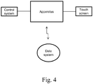
- a mammography apparatus is arranged with a screen (17) or a user interface with a screen (17), which is brought into a functional connection with an information system in which images taken of a patient are recorded.
- a memory of this information system can comprise a memory arranged in connection with the mammography apparatus or a memory separate to that, or both.
- preferable embodiments of the invention provide a new way to guide and calm the patient and to provide information to the patient prior to or during an imaging or biopsy procedures. Thanks to embodiments of the invention, the patient can be provided information on when exactly an exposure will begin and the patient should be still, for example, or how long the operation will still last.
- a mammography apparatus (1) presented in Fig. 1 consists of a substantially vertically standing frame part (10) and an arm structure (11) connected to it.
- An X-ray source (12) is arranged to the upper part of the arm structure (11), within its cover, the x-ray source (12) being arranged to generate a beam which goes through an upper compression plate (14) of the mammography apparatus (when such upper compression plate is connected to the apparatus) and towards a detector (18) placed in a detector housing (13).
- the detector housing (13) or a corresponding structure is typically arranged inside a lower tray structure (15).
- the lower tray structure (15) can be a structure fixed to the apparatus or it can be arranged as removably connected.
- the upper surface of the lower tray structure (15) typically functions as a platform on which a breast is positioned for imaging
- this structure is often also referred to as a lower compression plate.
- a camera or a video camera aligned at the lower tray structure (15) may have been arranged, to be utilized in patient positioning.
- attaching means (16) is arranged to the lower tray structure (15) to enable its releasable connection to the mammography apparatus (1).
- a screen (17) or a user interface with a screen (17), to be described in more detail below, is arranged to the apparatus according to Fig. 1 .
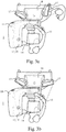
- Figs. 3a-3c show the apparatus according to Fig. 1 as viewed from above and in an oblique imaging position.
- Two screens (17) which may be touch screens, are arranged to the frame part (10) of the apparatus, on the opposite sides of the frame part (10). Attachment of the screen (17) to the frame part (10) of the apparatus may be articulated such that it can be turned to both a first position in which it is aligned away from the lower tray structure (15) of the apparatus ( Fig. 3a ), in connection with which structure the patient is positioned for imaging or operations, and to a second position ( Fig. 3b ) in which it is at least partly aligned towards the lower tray structure (15), i.e. in which it is in such position that the patient has a line of sight to the screen (17).
- FIG. 3 A position is presented in Fig. 3 intermediate to the previous positions which is applicable for consultation, for example, in which position both the operator of the apparatus and the patient have a good view at the screen (17).
- the screen (17) may be just a screen but preferably it is a true traditional touch screen and it may also be some other user interface comprising a screen. Then the first position of the touch screen (17) according to Fig. 3a can be used when operations of the apparatus are controlled or when information is presented on the touch screen which one does not want the patient to see or which the patient needs no to see. Also the position according to Fig. 3b , and especially according to Fig. 3c can be used when controlling operation of the apparatus, but those positions of the screen (17) are especially designed to be used for delivering information to the patient.
- the touch screen (17) is arranged in functional connection with the control system of the apparatus to display imaging parameter values, for example, or the time remaining in the imaging process.
- a functional connection like this is arranged e.g. to a patient data base, whereby one preferable embodiment according to the invention includes a solution in which a touch screen (17) is arranged in a functional connection with an information system in which images of a patient taken earlier have been recorded. It is known as such to use information achievable from such images to facilitate patient positioning when the same object is being re-imaged or when a biopsy is to be started or is going on, which information may include measurement markings or other made to the images relating to findings which have been detected.
- the present invention makes it possible to bring the images to the immediate vicinity of the place where that information is needed.
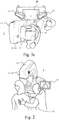
- the arrangement preferably includes a possibility to present images of a patient's breast taken earlier, especially in the same orientation in which the breast is being positioned to the imaging apparatus and in which images, as said, measurement markings or other may have been made relating to findings which have been detected.
- Fig. 2 demonstrates a situation like this.
- the screen or the user interface with a screen connected to a mammography apparatus as presented above can be utilized in patient positioning also if the mammography apparatus is equipped with a camera or a video camera (19) aligned at the imaging station of the apparatus.
- a picture of a breast positioned for imaging may have been taken by such camera (19) in connection with an earlier imaging event (which camera may have in principle been located at some other corresponding apparatus), which picture can then be shown in real time in connection with a new positioning, at the immediate vicinity of the imaging station of the apparatus.
- the present invention provides a possibility for smooth and precise positioning for re-imaging or biopsy. Further, the possibility to guide and inform the patient of various stages of the imaging or biopsy event can help in achieving a successful procedure, by e.g. helping the patient to be still throughout an operation which may take quite a while.
- the invention can also be realized by the patient herself controlling operation of the mammography apparatus from the user interface brought in connection with the imaging station when the patient's breast is being compressed for imaging.
- Such solution is prone to lessen the fears of the patient relating to compressing a breast, taking in consideration that there are studies according to which a patient may allow the mammography apparatus to compress a breast even more when she can control the compressing process herself.
- the solution according to the invention may thus offer the possibility to bring a touch screen (17) arranged to control operation of the mammography apparatus so close to the lower tray structure (15) of the apparatus that the patient can herself control via it e.g. movements of the upper compression plate (14) of the apparatus according to Fig. 1 .
- the articulation of the screen (17) or the user interface with a screen (17) is realized as motorized.
- at least one operation mode can be arranged to the control system of the apparatus according to which the screen (17) automatically turns into a position which has been designated for it in that operation mode.
- the position of the screen (17) can thus be arranged to be changed according to which procedure that operation mode relates to, or according to what the control system displays on the screen.
- the screen (17) can be arranged e.g. to turn towards the patient when the time remaining in the image process is displayed on the screen.
- one or more views may be defined in the control system which are automatically displayed on the screen when it is turned into a position in which it comes to the field of view of the patient, i.e. into a position as aligned towards the lower tray structure (15).
- Exemplary solutions may thus include a mammography apparatus which includes a substantially vertically standing frame part (10) or a frame part (10) attachable to a wall or a ceiling, an arm structure (11) connecting to this frame part (10) and pivotable in relation to a horizontal rotation axis, at a substantially first end of the opposite ends of the arm structure (11) is placed an X-ray source (12), and substantially at a second end an image data receiving means (18), and in connection with said second end of the arm structure (11) is additionally arranged a lower tray structure (15) positioned substantially on top of the image data receiving means (18), a control system and in connection with it a user interface arrangement and at least one screen (17) or user interface with a screen (17).
- a mammography apparatus which includes a substantially vertically standing frame part (10) or a frame part (10) attachable to a wall or a ceiling, an arm structure (11) connecting to this frame part (10) and pivotable in relation to a horizontal rotation axis, at a substantially first end of the opposite ends of the arm structure (1
- Said screen (17) or user interface with a screen (17) is connected to the mammography apparatus (1) and the connection is realized such that the screen (17) or user interface with a screen (17) is aligned or can be aligned at least partly towards the lower tray structure (15).
- a means is arranged to the mammography apparatus (1) to bring said screen (17) or user interface with a screen (17) into a functional connection with an external information system such that patient images recorded earlier in said information system can be displayed on said screen (17) or user interface with a screen (17) connected to structures of the mammography apparatus.
- the arrangement includes a means for showing images on the screen (17) in an orientation corresponding to an orientation in which a breast is positioned on top of said lower tray structure (15) .
- said screens (17) or user interfaces with a screen (17) are arranged to the mammography apparatus (1) at least one on both of its sides, as viewed from said lower tray structure (15).
- the user interface arrangement may include at least one screen (17) or user interface with a screen (17) which is
- the screen (17) or user interface with a screen (17) is attached to the frame part (10) as articulated.
- the articulation can be realized as motorized and said control system to comprise at least one operation mode according to which a view generated by the screen automatically turns into a position determined for it by said operation mode, as dependent on the operation to which the operation mode relates to or on what the control system shows on the screen (17).
- the articulation can also include a structure which enables bringing the screen (17) or the user interface with a screen into a substantial vicinity of the lower tray structure (15), especially within an arm's length or closer.
- One or several views can be defined in the control system to be automatically presented on the screen (17) or a user interface with a screen (17), when it is turned into its said second position.
- This disclosure also relates to a method for using the arrangement according to claim 1 to position a breast into a mammography apparatus, in which method an image taken of the breast is used as an aid for positioning the breast, which image is a patient image recorded earlier in an information system external to the mammography apparatus and said image is presented on a screen (17) or a user interface with a screen (17) attached to the structures of the mammography apparatus.
- This image can be presented in a direction at which there is a line of sight from an imaging station of the mammography apparatus, especially in an orientation corresponding to the orientation in which the breast is being positioned for mammography imaging or for biopsy, in relation to the imaging station of the mammography apparatus.
Landscapes
- Health & Medical Sciences (AREA)
- Life Sciences & Earth Sciences (AREA)
- Engineering & Computer Science (AREA)
- Medical Informatics (AREA)
- Radiology & Medical Imaging (AREA)
- Molecular Biology (AREA)
- Biophysics (AREA)
- Nuclear Medicine, Radiotherapy & Molecular Imaging (AREA)
- Optics & Photonics (AREA)
- Pathology (AREA)
- Physics & Mathematics (AREA)
- Biomedical Technology (AREA)
- Heart & Thoracic Surgery (AREA)
- High Energy & Nuclear Physics (AREA)
- Surgery (AREA)
- Animal Behavior & Ethology (AREA)
- General Health & Medical Sciences (AREA)
- Public Health (AREA)
- Veterinary Medicine (AREA)
- Human Computer Interaction (AREA)
- Dentistry (AREA)
- Oral & Maxillofacial Surgery (AREA)
- Apparatus For Radiation Diagnosis (AREA)
Applications Claiming Priority (3)
| Application Number | Priority Date | Filing Date | Title |
|---|---|---|---|
| FI20130361 | 2013-11-29 | ||
| FI20136259A FI126329B (fi) | 2013-11-29 | 2013-12-13 | Mammografialaitejärjestely |
| PCT/FI2014/050930 WO2015079118A1 (en) | 2013-11-29 | 2014-12-01 | Improvement for mammography examinations |
Publications (3)
| Publication Number | Publication Date |
|---|---|
| EP3073929A1 EP3073929A1 (en) | 2016-10-05 |
| EP3073929A4 EP3073929A4 (en) | 2017-09-13 |
| EP3073929B1 true EP3073929B1 (en) | 2021-06-30 |
Family
ID=53198422
Family Applications (1)
| Application Number | Title | Priority Date | Filing Date |
|---|---|---|---|
| EP14865418.9A Active EP3073929B1 (en) | 2013-11-29 | 2014-12-01 | Improvement for mammography examinations |
Country Status (8)
| Country | Link |
|---|---|
| US (2) | US10575805B2 (ja) |
| EP (1) | EP3073929B1 (ja) |
| JP (2) | JP6758182B2 (ja) |
| KR (1) | KR102373198B1 (ja) |
| CN (1) | CN105934200B (ja) |
| ES (1) | ES2887408T3 (ja) |
| FI (1) | FI126329B (ja) |
| WO (1) | WO2015079118A1 (ja) |
Families Citing this family (7)
| Publication number | Priority date | Publication date | Assignee | Title |
|---|---|---|---|---|
| CA3040736A1 (en) | 2016-11-25 | 2018-05-31 | Hologic, Inc. | Controller for imaging apparatus |
| IT201700122588A1 (it) | 2017-10-27 | 2019-04-27 | Ims Giotto S P A | Apparecchiatura per analisi di campioni prelevati mediante biopsia. |
| DE102018200108A1 (de) | 2018-01-05 | 2019-07-11 | Siemens Healthcare Gmbh | Positionierung eines Untersuchungsobjekts bezüglich eines Röntgengeräts |
| US10863952B2 (en) * | 2018-06-21 | 2020-12-15 | General Electric Company | Apparatus, system and method for controlling medical equipment |
| US11176710B2 (en) * | 2019-12-11 | 2021-11-16 | Scientific Technology Electronics Products | Camera system for an X-ray counter system |
| US11627921B2 (en) | 2020-03-27 | 2023-04-18 | Hologic, Inc. | Systems and methods for visualizing below an opaque compression paddle |
| US11375968B2 (en) | 2020-04-06 | 2022-07-05 | GE Precision Healthcare LLC | Methods and systems for user and/or patient experience improvement in mammography |
Family Cites Families (34)
| Publication number | Priority date | Publication date | Assignee | Title |
|---|---|---|---|---|
| US4674107A (en) | 1985-07-31 | 1987-06-16 | Picker International, Inc. | Display for radiation imaging |
| AU2706500A (en) | 1998-11-25 | 2000-09-21 | Fischer Imaging Corporation | User interface system for mammographic imager |
| JP2001170048A (ja) | 1999-12-15 | 2001-06-26 | Aloka Co Ltd | 超音波診断装置 |
| US6590958B2 (en) * | 2001-11-15 | 2003-07-08 | Ge Medical Systems Global Technology | X-ray positioner having integrated display |
| CN1292709C (zh) | 2002-01-22 | 2007-01-03 | 株式会社东芝 | 具有多个监视器的医学图象诊断设备 |
| US20040102699A1 (en) * | 2002-11-26 | 2004-05-27 | Ge Medical Systems Information Technologies, Inc. | Tool and method to produce orientation marker on a subject of interest |
| US6891920B1 (en) * | 2002-11-29 | 2005-05-10 | Fischer Imaging Corporation | Automated background processing mammographic image data |
| EP1750586A4 (en) * | 2004-04-26 | 2009-07-29 | U Systems Inc | VERSATILE CHEST ULTRASOUND METAL PROCESS |
| JP2007020839A (ja) | 2005-07-15 | 2007-02-01 | Hitachi Medical Corp | 超音波診断装置 |
| JP2007236805A (ja) | 2006-03-10 | 2007-09-20 | Toshiba Corp | 乳房撮影用x線診断装置及び乳房撮影用x線診断装置の制御方法 |
| JP4833785B2 (ja) * | 2006-09-29 | 2011-12-07 | 富士フイルム株式会社 | 放射線撮影装置および放射線撮影方法 |
| US8311305B2 (en) | 2006-10-18 | 2012-11-13 | Kabushiki Kaisha Toshiba | X-ray diagnostic apparatus, image processing apparatus, and method of calculating filter coefficients used for image formation processing in x-ray diagnostic apparatus and the like |
| JP5283882B2 (ja) * | 2006-10-18 | 2013-09-04 | 株式会社東芝 | X線診断装置、画像処理装置及び画像再構成処理に用いられるフィルタ係数の算出プログラム |
| DE102007052650B4 (de) * | 2007-11-05 | 2016-11-03 | Siemens Healthcare Gmbh | Bildgebendes System und Verfahren zum Betrieb eines bildgebenden Systems |
| FI120077B (fi) * | 2007-11-14 | 2009-06-30 | Planmed Oy | Järjestely ja menetelmä digitaalisessa mammografiakuvauksessa |
| DE102007061592B4 (de) * | 2007-12-20 | 2010-03-18 | Siemens Ag | Verfahren zum Positionieren der Brust für eine Biopsie in einer Mammographieeinrichtung und Mammographieeinrichtung zum Durchführen des Verfahrens |
| DE102008011157B4 (de) * | 2008-02-26 | 2010-10-14 | Siemens Aktiengesellschaft | Medizinisches Röntgensystem |
| JP5456988B2 (ja) * | 2008-05-30 | 2014-04-02 | 富士フイルム株式会社 | マンモグラフィ装置及びそれを用いた撮影システム |
| JP2009291336A (ja) | 2008-06-04 | 2009-12-17 | Fujifilm Corp | 放射線画像撮影装置 |
| US8161421B2 (en) * | 2008-07-07 | 2012-04-17 | International Business Machines Corporation | Calibration and verification structures for use in optical proximity correction |
| JP5210080B2 (ja) * | 2008-07-31 | 2013-06-12 | 富士フイルム株式会社 | 医用撮像装置 |
| JP2010167134A (ja) * | 2009-01-23 | 2010-08-05 | Fujifilm Corp | 乳房画像撮影システム |
| JP2010188003A (ja) * | 2009-02-19 | 2010-09-02 | Fujifilm Corp | 画像表示システム及び画像撮影表示システム |
| US10595954B2 (en) * | 2009-10-08 | 2020-03-24 | Hologic, Inc. | Needle breast biopsy system and method for use |
| DE102010011663B4 (de) * | 2010-03-17 | 2020-02-06 | Siemens Healthcare Gmbh | Mammographiegerät |
| US8961011B2 (en) | 2010-04-13 | 2015-02-24 | Carestream Health, Inc. | Mobile radiography unit having multiple monitors |
| FR2962638B1 (fr) | 2010-07-15 | 2013-03-15 | Gen Electric | Systeme d'imagerie deplacable comprenant des moyens d'affichage integres |
| JP2012035068A (ja) * | 2010-07-16 | 2012-02-23 | Fujifilm Corp | 放射線画像処理装置および方法並びにプログラム |
| JP5650467B2 (ja) * | 2010-08-27 | 2015-01-07 | 富士フイルム株式会社 | 放射線画像撮影システム |
| US20120134468A1 (en) * | 2010-11-27 | 2012-05-31 | General Electric Company | System and method for including and correcting subject orientation data in digital radiographic images |
| EP2651308B1 (en) | 2010-12-14 | 2020-03-11 | Hologic, Inc. | System and method for fusing three dimensional image data from a plurality of different imaging systems for use in diagnostic imaging |
| JP2012143548A (ja) * | 2010-12-21 | 2012-08-02 | Fujifilm Corp | 放射線画像取得方法および放射線画像撮影装置 |
| ITBO20110084A1 (it) * | 2011-02-25 | 2012-08-26 | I M S Internaz Medicoscienti Fica S R L | Apparecchiatura per la tomosintesi e la mammografia. |
| US10004470B2 (en) * | 2013-07-18 | 2018-06-26 | General Electric Company | Breast imaging system giving feedback information to the patient and method using thereof |
-
2013
- 2013-12-13 FI FI20136259A patent/FI126329B/fi active IP Right Grant
-
2014
- 2014-12-01 ES ES14865418T patent/ES2887408T3/es active Active
- 2014-12-01 EP EP14865418.9A patent/EP3073929B1/en active Active
- 2014-12-01 WO PCT/FI2014/050930 patent/WO2015079118A1/en active Application Filing
- 2014-12-01 JP JP2016535043A patent/JP6758182B2/ja active Active
- 2014-12-01 CN CN201480065257.3A patent/CN105934200B/zh active Active
- 2014-12-01 KR KR1020167016981A patent/KR102373198B1/ko active IP Right Grant
-
2016
- 2016-05-31 US US15/168,973 patent/US10575805B2/en not_active Expired - Fee Related
-
2020
- 2020-03-02 US US16/806,210 patent/US11006915B2/en active Active
- 2020-06-24 JP JP2020108805A patent/JP7105275B2/ja active Active
Also Published As
| Publication number | Publication date |
|---|---|
| CN105934200A (zh) | 2016-09-07 |
| US20160270751A1 (en) | 2016-09-22 |
| US11006915B2 (en) | 2021-05-18 |
| CN105934200B (zh) | 2021-02-09 |
| JP6758182B2 (ja) | 2020-09-23 |
| JP2016538083A (ja) | 2016-12-08 |
| WO2015079118A1 (en) | 2015-06-04 |
| ES2887408T3 (es) | 2021-12-22 |
| US10575805B2 (en) | 2020-03-03 |
| JP2020168408A (ja) | 2020-10-15 |
| KR20160091382A (ko) | 2016-08-02 |
| EP3073929A1 (en) | 2016-10-05 |
| FI126329B (fi) | 2016-10-14 |
| FI20136259A (fi) | 2015-05-30 |
| JP7105275B2 (ja) | 2022-07-22 |
| EP3073929A4 (en) | 2017-09-13 |
| KR102373198B1 (ko) | 2022-03-11 |
| US20200196970A1 (en) | 2020-06-25 |
Similar Documents
| Publication | Publication Date | Title |
|---|---|---|
| EP3073929B1 (en) | Improvement for mammography examinations | |
| JP6260615B2 (ja) | 診断提供用医用画像システム及び一般撮影用の診断提供用医用画像システムにタルボ撮影装置系を導入する方法 | |
| EP2979638B1 (en) | Radiographic device, radiographic method and radiographic control program | |
| JP4786685B2 (ja) | X線画像表示方法、x線撮影装置、及びx線画像表示装置 | |
| US10271801B2 (en) | Radiation imaging system, image processing device, and image processing program | |
| EP3073923B1 (en) | Mammography apparatus | |
| US10433795B2 (en) | Radiation imaging system, image processing device, radiation imaging method, and image processing program | |
| JP6552809B2 (ja) | 乳房x線撮影装置 | |
| JP4313977B2 (ja) | 検査室の被検体観察装置 | |
| JP2009119281A (ja) | 診断装置 | |
| JP5642927B2 (ja) | 乳房用x線撮影装置 | |
| JP5638466B2 (ja) | 画像生成装置、放射線画像撮影システム、画像生成プログラム、及び画像生成方法 | |
| JP2012157689A (ja) | 放射線画像表示装置および方法 |
Legal Events
| Date | Code | Title | Description |
|---|---|---|---|
| PUAI | Public reference made under article 153(3) epc to a published international application that has entered the european phase |
Free format text: ORIGINAL CODE: 0009012 |
|
| 17P | Request for examination filed |
Effective date: 20160628 |
|
| AK | Designated contracting states |
Kind code of ref document: A1 Designated state(s): AL AT BE BG CH CY CZ DE DK EE ES FI FR GB GR HR HU IE IS IT LI LT LU LV MC MK MT NL NO PL PT RO RS SE SI SK SM TR |
|
| AX | Request for extension of the european patent |
Extension state: BA ME |
|
| DAX | Request for extension of the european patent (deleted) | ||
| A4 | Supplementary search report drawn up and despatched |
Effective date: 20170817 |
|
| RIC1 | Information provided on ipc code assigned before grant |
Ipc: A61B 6/00 20060101ALI20170810BHEP Ipc: A61B 6/04 20060101AFI20170810BHEP |
|
| STAA | Information on the status of an ep patent application or granted ep patent |
Free format text: STATUS: EXAMINATION IS IN PROGRESS |
|
| 17Q | First examination report despatched |
Effective date: 20200608 |
|
| GRAP | Despatch of communication of intention to grant a patent |
Free format text: ORIGINAL CODE: EPIDOSNIGR1 |
|
| STAA | Information on the status of an ep patent application or granted ep patent |
Free format text: STATUS: GRANT OF PATENT IS INTENDED |
|
| INTG | Intention to grant announced |
Effective date: 20210127 |
|
| RIN1 | Information on inventor provided before grant (corrected) |
Inventor name: ASPELUND, LEO Inventor name: LAUKKANEN, TAPIO |
|
| GRAS | Grant fee paid |
Free format text: ORIGINAL CODE: EPIDOSNIGR3 |
|
| GRAA | (expected) grant |
Free format text: ORIGINAL CODE: 0009210 |
|
| STAA | Information on the status of an ep patent application or granted ep patent |
Free format text: STATUS: THE PATENT HAS BEEN GRANTED |
|
| AK | Designated contracting states |
Kind code of ref document: B1 Designated state(s): AL AT BE BG CH CY CZ DE DK EE ES FI FR GB GR HR HU IE IS IT LI LT LU LV MC MK MT NL NO PL PT RO RS SE SI SK SM TR |
|
| REG | Reference to a national code |
Ref country code: CH Ref legal event code: EP |
|
| REG | Reference to a national code |
Ref country code: DE Ref legal event code: R096 Ref document number: 602014078503 Country of ref document: DE Ref country code: AT Ref legal event code: REF Ref document number: 1405599 Country of ref document: AT Kind code of ref document: T Effective date: 20210715 |
|
| REG | Reference to a national code |
Ref country code: IE Ref legal event code: FG4D |
|
| REG | Reference to a national code |
Ref country code: LT Ref legal event code: MG9D |
|
| PG25 | Lapsed in a contracting state [announced via postgrant information from national office to epo] |
Ref country code: FI Free format text: LAPSE BECAUSE OF FAILURE TO SUBMIT A TRANSLATION OF THE DESCRIPTION OR TO PAY THE FEE WITHIN THE PRESCRIBED TIME-LIMIT Effective date: 20210630 Ref country code: BG Free format text: LAPSE BECAUSE OF FAILURE TO SUBMIT A TRANSLATION OF THE DESCRIPTION OR TO PAY THE FEE WITHIN THE PRESCRIBED TIME-LIMIT Effective date: 20210930 Ref country code: HR Free format text: LAPSE BECAUSE OF FAILURE TO SUBMIT A TRANSLATION OF THE DESCRIPTION OR TO PAY THE FEE WITHIN THE PRESCRIBED TIME-LIMIT Effective date: 20210630 |
|
| REG | Reference to a national code |
Ref country code: NL Ref legal event code: MP Effective date: 20210630 |
|
| REG | Reference to a national code |
Ref country code: AT Ref legal event code: MK05 Ref document number: 1405599 Country of ref document: AT Kind code of ref document: T Effective date: 20210630 |
|
| PG25 | Lapsed in a contracting state [announced via postgrant information from national office to epo] |
Ref country code: LV Free format text: LAPSE BECAUSE OF FAILURE TO SUBMIT A TRANSLATION OF THE DESCRIPTION OR TO PAY THE FEE WITHIN THE PRESCRIBED TIME-LIMIT Effective date: 20210630 Ref country code: GR Free format text: LAPSE BECAUSE OF FAILURE TO SUBMIT A TRANSLATION OF THE DESCRIPTION OR TO PAY THE FEE WITHIN THE PRESCRIBED TIME-LIMIT Effective date: 20211001 Ref country code: NO Free format text: LAPSE BECAUSE OF FAILURE TO SUBMIT A TRANSLATION OF THE DESCRIPTION OR TO PAY THE FEE WITHIN THE PRESCRIBED TIME-LIMIT Effective date: 20210930 Ref country code: RS Free format text: LAPSE BECAUSE OF FAILURE TO SUBMIT A TRANSLATION OF THE DESCRIPTION OR TO PAY THE FEE WITHIN THE PRESCRIBED TIME-LIMIT Effective date: 20210630 Ref country code: SE Free format text: LAPSE BECAUSE OF FAILURE TO SUBMIT A TRANSLATION OF THE DESCRIPTION OR TO PAY THE FEE WITHIN THE PRESCRIBED TIME-LIMIT Effective date: 20210630 |
|
| REG | Reference to a national code |
Ref country code: ES Ref legal event code: FG2A Ref document number: 2887408 Country of ref document: ES Kind code of ref document: T3 Effective date: 20211222 |
|
| PG25 | Lapsed in a contracting state [announced via postgrant information from national office to epo] |
Ref country code: SK Free format text: LAPSE BECAUSE OF FAILURE TO SUBMIT A TRANSLATION OF THE DESCRIPTION OR TO PAY THE FEE WITHIN THE PRESCRIBED TIME-LIMIT Effective date: 20210630 Ref country code: SM Free format text: LAPSE BECAUSE OF FAILURE TO SUBMIT A TRANSLATION OF THE DESCRIPTION OR TO PAY THE FEE WITHIN THE PRESCRIBED TIME-LIMIT Effective date: 20210630 Ref country code: EE Free format text: LAPSE BECAUSE OF FAILURE TO SUBMIT A TRANSLATION OF THE DESCRIPTION OR TO PAY THE FEE WITHIN THE PRESCRIBED TIME-LIMIT Effective date: 20210630 Ref country code: CZ Free format text: LAPSE BECAUSE OF FAILURE TO SUBMIT A TRANSLATION OF THE DESCRIPTION OR TO PAY THE FEE WITHIN THE PRESCRIBED TIME-LIMIT Effective date: 20210630 Ref country code: AT Free format text: LAPSE BECAUSE OF FAILURE TO SUBMIT A TRANSLATION OF THE DESCRIPTION OR TO PAY THE FEE WITHIN THE PRESCRIBED TIME-LIMIT Effective date: 20210630 Ref country code: PT Free format text: LAPSE BECAUSE OF FAILURE TO SUBMIT A TRANSLATION OF THE DESCRIPTION OR TO PAY THE FEE WITHIN THE PRESCRIBED TIME-LIMIT Effective date: 20211102 Ref country code: NL Free format text: LAPSE BECAUSE OF FAILURE TO SUBMIT A TRANSLATION OF THE DESCRIPTION OR TO PAY THE FEE WITHIN THE PRESCRIBED TIME-LIMIT Effective date: 20210630 Ref country code: RO Free format text: LAPSE BECAUSE OF FAILURE TO SUBMIT A TRANSLATION OF THE DESCRIPTION OR TO PAY THE FEE WITHIN THE PRESCRIBED TIME-LIMIT Effective date: 20210630 |
|
| PG25 | Lapsed in a contracting state [announced via postgrant information from national office to epo] |
Ref country code: PL Free format text: LAPSE BECAUSE OF FAILURE TO SUBMIT A TRANSLATION OF THE DESCRIPTION OR TO PAY THE FEE WITHIN THE PRESCRIBED TIME-LIMIT Effective date: 20210630 |
|
| REG | Reference to a national code |
Ref country code: DE Ref legal event code: R097 Ref document number: 602014078503 Country of ref document: DE |
|
| PG25 | Lapsed in a contracting state [announced via postgrant information from national office to epo] |
Ref country code: DK Free format text: LAPSE BECAUSE OF FAILURE TO SUBMIT A TRANSLATION OF THE DESCRIPTION OR TO PAY THE FEE WITHIN THE PRESCRIBED TIME-LIMIT Effective date: 20210630 |
|
| PLBE | No opposition filed within time limit |
Free format text: ORIGINAL CODE: 0009261 |
|
| STAA | Information on the status of an ep patent application or granted ep patent |
Free format text: STATUS: NO OPPOSITION FILED WITHIN TIME LIMIT |
|
| PG25 | Lapsed in a contracting state [announced via postgrant information from national office to epo] |
Ref country code: AL Free format text: LAPSE BECAUSE OF FAILURE TO SUBMIT A TRANSLATION OF THE DESCRIPTION OR TO PAY THE FEE WITHIN THE PRESCRIBED TIME-LIMIT Effective date: 20210630 |
|
| 26N | No opposition filed |
Effective date: 20220331 |
|
| PG25 | Lapsed in a contracting state [announced via postgrant information from national office to epo] |
Ref country code: MC Free format text: LAPSE BECAUSE OF FAILURE TO SUBMIT A TRANSLATION OF THE DESCRIPTION OR TO PAY THE FEE WITHIN THE PRESCRIBED TIME-LIMIT Effective date: 20210630 |
|
| REG | Reference to a national code |
Ref country code: CH Ref legal event code: PL |
|
| REG | Reference to a national code |
Ref country code: BE Ref legal event code: MM Effective date: 20211231 |
|
| PG25 | Lapsed in a contracting state [announced via postgrant information from national office to epo] |
Ref country code: LU Free format text: LAPSE BECAUSE OF NON-PAYMENT OF DUE FEES Effective date: 20211201 Ref country code: IE Free format text: LAPSE BECAUSE OF NON-PAYMENT OF DUE FEES Effective date: 20211201 |
|
| PG25 | Lapsed in a contracting state [announced via postgrant information from national office to epo] |
Ref country code: BE Free format text: LAPSE BECAUSE OF NON-PAYMENT OF DUE FEES Effective date: 20211231 |
|
| PG25 | Lapsed in a contracting state [announced via postgrant information from national office to epo] |
Ref country code: LI Free format text: LAPSE BECAUSE OF NON-PAYMENT OF DUE FEES Effective date: 20211231 Ref country code: CH Free format text: LAPSE BECAUSE OF NON-PAYMENT OF DUE FEES Effective date: 20211231 |
|
| PGFP | Annual fee paid to national office [announced via postgrant information from national office to epo] |
Ref country code: IT Payment date: 20221122 Year of fee payment: 9 |
|
| PG25 | Lapsed in a contracting state [announced via postgrant information from national office to epo] |
Ref country code: LT Free format text: LAPSE BECAUSE OF FAILURE TO SUBMIT A TRANSLATION OF THE DESCRIPTION OR TO PAY THE FEE WITHIN THE PRESCRIBED TIME-LIMIT Effective date: 20210630 |
|
| PG25 | Lapsed in a contracting state [announced via postgrant information from national office to epo] |
Ref country code: HU Free format text: LAPSE BECAUSE OF FAILURE TO SUBMIT A TRANSLATION OF THE DESCRIPTION OR TO PAY THE FEE WITHIN THE PRESCRIBED TIME-LIMIT; INVALID AB INITIO Effective date: 20141201 |
|
| PG25 | Lapsed in a contracting state [announced via postgrant information from national office to epo] |
Ref country code: CY Free format text: LAPSE BECAUSE OF FAILURE TO SUBMIT A TRANSLATION OF THE DESCRIPTION OR TO PAY THE FEE WITHIN THE PRESCRIBED TIME-LIMIT Effective date: 20210630 |
|
| PGFP | Annual fee paid to national office [announced via postgrant information from national office to epo] |
Ref country code: GB Payment date: 20231124 Year of fee payment: 10 |
|
| PGFP | Annual fee paid to national office [announced via postgrant information from national office to epo] |
Ref country code: FR Payment date: 20231122 Year of fee payment: 10 Ref country code: DE Payment date: 20231121 Year of fee payment: 10 |
|
| PGFP | Annual fee paid to national office [announced via postgrant information from national office to epo] |
Ref country code: ES Payment date: 20240102 Year of fee payment: 10 |
|
| PG25 | Lapsed in a contracting state [announced via postgrant information from national office to epo] |
Ref country code: MK Free format text: LAPSE BECAUSE OF FAILURE TO SUBMIT A TRANSLATION OF THE DESCRIPTION OR TO PAY THE FEE WITHIN THE PRESCRIBED TIME-LIMIT Effective date: 20210630 |