EP3062884B1 - Zusammensetzungen zur verwendung in der behandlung von retinitis pigmentosa - Google Patents
Zusammensetzungen zur verwendung in der behandlung von retinitis pigmentosa Download PDFInfo
- Publication number
- EP3062884B1 EP3062884B1 EP14857455.1A EP14857455A EP3062884B1 EP 3062884 B1 EP3062884 B1 EP 3062884B1 EP 14857455 A EP14857455 A EP 14857455A EP 3062884 B1 EP3062884 B1 EP 3062884B1
- Authority
- EP
- European Patent Office
- Prior art keywords
- vector
- promoter
- aav
- cells
- cone
- Prior art date
- Legal status (The legal status is an assumption and is not a legal conclusion. Google has not performed a legal analysis and makes no representation as to the accuracy of the status listed.)
- Active
Links
- 238000011282 treatment Methods 0.000 title claims description 47
- 239000000203 mixture Substances 0.000 title claims description 44
- 208000007014 Retinitis pigmentosa Diseases 0.000 title claims description 36
- 239000013598 vector Substances 0.000 claims description 163
- 150000007523 nucleic acids Chemical class 0.000 claims description 130
- 102000039446 nucleic acids Human genes 0.000 claims description 126
- 108020004707 nucleic acids Proteins 0.000 claims description 126
- 102100031701 Nuclear factor erythroid 2-related factor 2 Human genes 0.000 claims description 88
- 101000588302 Homo sapiens Nuclear factor erythroid 2-related factor 2 Proteins 0.000 claims description 87
- 239000003795 chemical substances by application Substances 0.000 claims description 50
- 239000013607 AAV vector Substances 0.000 claims description 36
- 230000002207 retinal effect Effects 0.000 claims description 29
- 230000003612 virological effect Effects 0.000 claims description 26
- 239000013603 viral vector Substances 0.000 claims description 24
- 230000001177 retroviral effect Effects 0.000 claims description 16
- 239000008194 pharmaceutical composition Substances 0.000 claims description 14
- 241000701022 Cytomegalovirus Species 0.000 claims description 12
- 241000702421 Dependoparvovirus Species 0.000 claims description 9
- 241000713666 Lentivirus Species 0.000 claims description 8
- 230000000925 erythroid effect Effects 0.000 claims description 7
- 230000001939 inductive effect Effects 0.000 claims description 7
- 108091004242 G-Protein-Coupled Receptor Kinase 1 Proteins 0.000 claims description 6
- 102000004437 G-Protein-Coupled Receptor Kinase 1 Human genes 0.000 claims description 6
- 102000053171 Glial Fibrillary Acidic Human genes 0.000 claims description 6
- 108700005000 Glial Fibrillary Acidic Proteins 0.000 claims description 6
- 102100038247 Retinol-binding protein 3 Human genes 0.000 claims description 6
- 108090000799 Rhodopsin kinases Proteins 0.000 claims description 6
- 108010048996 interstitial retinol-binding protein Proteins 0.000 claims description 6
- 108050003620 Arrestin-C Proteins 0.000 claims description 5
- 102100026440 Arrestin-C Human genes 0.000 claims description 5
- ROHFNLRQFUQHCH-YFKPBYRVSA-N L-leucine Chemical compound CC(C)C[C@H](N)C(O)=O ROHFNLRQFUQHCH-YFKPBYRVSA-N 0.000 claims description 5
- ROHFNLRQFUQHCH-UHFFFAOYSA-N Leucine Natural products CC(C)CC(N)C(O)=O ROHFNLRQFUQHCH-UHFFFAOYSA-N 0.000 claims description 5
- 230000001537 neural effect Effects 0.000 claims description 5
- 241000991587 Enterovirus C Species 0.000 claims description 4
- 102000003780 Clusterin Human genes 0.000 claims description 3
- 108090000197 Clusterin Proteins 0.000 claims description 3
- 108010003730 Cone Opsins Proteins 0.000 claims description 3
- 241000701024 Human betaherpesvirus 5 Species 0.000 claims description 3
- 241001631646 Papillomaviridae Species 0.000 claims description 3
- 241000125945 Protoparvovirus Species 0.000 claims description 3
- 241000712909 Reticuloendotheliosis virus Species 0.000 claims description 3
- 241000700618 Vaccinia virus Species 0.000 claims description 3
- 102100035071 Vimentin Human genes 0.000 claims description 3
- 108010065472 Vimentin Proteins 0.000 claims description 3
- 230000004069 differentiation Effects 0.000 claims description 3
- 210000005048 vimentin Anatomy 0.000 claims description 3
- 101150092640 HES1 gene Proteins 0.000 claims description 2
- 208000009889 Herpes Simplex Diseases 0.000 claims description 2
- 101150102256 ndrg4 gene Proteins 0.000 claims description 2
- 210000004027 cell Anatomy 0.000 description 202
- 210000001525 retina Anatomy 0.000 description 158
- 108090000623 proteins and genes Proteins 0.000 description 139
- 238000000034 method Methods 0.000 description 93
- 102000004169 proteins and genes Human genes 0.000 description 77
- 230000014509 gene expression Effects 0.000 description 75
- 108010045815 superoxide dismutase 2 Proteins 0.000 description 72
- 102100032891 Superoxide dismutase [Mn], mitochondrial Human genes 0.000 description 71
- 108010053835 Catalase Proteins 0.000 description 68
- 102000016938 Catalase Human genes 0.000 description 68
- 230000000694 effects Effects 0.000 description 54
- 208000037265 diseases, disorders, signs and symptoms Diseases 0.000 description 50
- 241000699670 Mus sp. Species 0.000 description 48
- 241000700605 Viruses Species 0.000 description 41
- 230000000692 anti-sense effect Effects 0.000 description 37
- 108020004414 DNA Proteins 0.000 description 36
- 230000006851 antioxidant defense Effects 0.000 description 35
- 239000013604 expression vector Substances 0.000 description 33
- 239000003963 antioxidant agent Substances 0.000 description 30
- 230000003078 antioxidant effect Effects 0.000 description 28
- -1 PGC1α Proteins 0.000 description 27
- 208000017442 Retinal disease Diseases 0.000 description 26
- 150000001875 compounds Chemical class 0.000 description 26
- 230000002018 overexpression Effects 0.000 description 26
- 201000010099 disease Diseases 0.000 description 25
- 108020004999 messenger RNA Proteins 0.000 description 25
- 208000035475 disorder Diseases 0.000 description 24
- 210000000608 photoreceptor cell Anatomy 0.000 description 24
- 230000004083 survival effect Effects 0.000 description 24
- 108091032973 (ribonucleotides)n+m Proteins 0.000 description 23
- 241000701161 unidentified adenovirus Species 0.000 description 23
- 208000003098 Ganglion Cysts Diseases 0.000 description 22
- 208000005400 Synovial Cyst Diseases 0.000 description 22
- 235000006708 antioxidants Nutrition 0.000 description 22
- 230000036542 oxidative stress Effects 0.000 description 22
- 108091008695 photoreceptors Proteins 0.000 description 22
- 238000001415 gene therapy Methods 0.000 description 21
- HGINCPLSRVDWNT-UHFFFAOYSA-N Acrolein Chemical compound C=CC=O HGINCPLSRVDWNT-UHFFFAOYSA-N 0.000 description 20
- 241001465754 Metazoa Species 0.000 description 19
- 108090000790 Enzymes Proteins 0.000 description 18
- 108010046016 Peanut Agglutinin Proteins 0.000 description 18
- 102100039174 Rod cGMP-specific 3',5'-cyclic phosphodiesterase subunit beta Human genes 0.000 description 18
- 230000003064 anti-oxidating effect Effects 0.000 description 18
- 238000003556 assay Methods 0.000 description 18
- 238000002347 injection Methods 0.000 description 18
- 239000007924 injection Substances 0.000 description 18
- 125000003729 nucleotide group Chemical group 0.000 description 18
- 230000010076 replication Effects 0.000 description 18
- 102000004190 Enzymes Human genes 0.000 description 17
- 230000006870 function Effects 0.000 description 17
- 239000002609 medium Substances 0.000 description 17
- 230000034994 death Effects 0.000 description 16
- 230000001105 regulatory effect Effects 0.000 description 16
- 241000699666 Mus <mouse, genus> Species 0.000 description 15
- 239000002299 complementary DNA Substances 0.000 description 15
- 230000001965 increasing effect Effects 0.000 description 15
- 239000002773 nucleotide Substances 0.000 description 15
- 239000013612 plasmid Substances 0.000 description 15
- 238000002360 preparation method Methods 0.000 description 15
- 238000010186 staining Methods 0.000 description 15
- 108091028043 Nucleic acid sequence Proteins 0.000 description 14
- 239000003937 drug carrier Substances 0.000 description 14
- 210000004408 hybridoma Anatomy 0.000 description 14
- 238000007254 oxidation reaction Methods 0.000 description 14
- 108090000765 processed proteins & peptides Proteins 0.000 description 14
- 230000035899 viability Effects 0.000 description 14
- 206010064930 age-related macular degeneration Diseases 0.000 description 13
- 230000000295 complement effect Effects 0.000 description 13
- 230000003834 intracellular effect Effects 0.000 description 13
- 208000002780 macular degeneration Diseases 0.000 description 13
- 238000012360 testing method Methods 0.000 description 13
- 210000001519 tissue Anatomy 0.000 description 13
- 238000002571 electroretinography Methods 0.000 description 12
- 208000015181 infectious disease Diseases 0.000 description 12
- 230000003647 oxidation Effects 0.000 description 12
- 230000001225 therapeutic effect Effects 0.000 description 12
- 108700028369 Alleles Proteins 0.000 description 11
- 201000004569 Blindness Diseases 0.000 description 11
- 239000003642 reactive oxygen metabolite Substances 0.000 description 11
- 230000004304 visual acuity Effects 0.000 description 11
- 102000040650 (ribonucleotides)n+m Human genes 0.000 description 10
- 241000702423 Adeno-associated virus - 2 Species 0.000 description 10
- 241000725303 Human immunodeficiency virus Species 0.000 description 10
- 238000012228 RNA interference-mediated gene silencing Methods 0.000 description 10
- 201000007737 Retinal degeneration Diseases 0.000 description 10
- 102000040945 Transcription factor Human genes 0.000 description 10
- 108091023040 Transcription factor Proteins 0.000 description 10
- 230000007850 degeneration Effects 0.000 description 10
- 230000009368 gene silencing by RNA Effects 0.000 description 10
- 230000002401 inhibitory effect Effects 0.000 description 10
- 238000003259 recombinant expression Methods 0.000 description 10
- 230000002829 reductive effect Effects 0.000 description 10
- 230000004258 retinal degeneration Effects 0.000 description 10
- 102000053602 DNA Human genes 0.000 description 9
- 230000008901 benefit Effects 0.000 description 9
- 238000004520 electroporation Methods 0.000 description 9
- 230000035772 mutation Effects 0.000 description 9
- 230000004044 response Effects 0.000 description 9
- 210000003994 retinal ganglion cell Anatomy 0.000 description 9
- 208000024891 symptom Diseases 0.000 description 9
- 241001430294 unidentified retrovirus Species 0.000 description 9
- MHAJPDPJQMAIIY-UHFFFAOYSA-N Hydrogen peroxide Chemical compound OO MHAJPDPJQMAIIY-UHFFFAOYSA-N 0.000 description 8
- 206010035226 Plasma cell myeloma Diseases 0.000 description 8
- 108091027967 Small hairpin RNA Proteins 0.000 description 8
- 150000001413 amino acids Chemical group 0.000 description 8
- 201000006754 cone-rod dystrophy Diseases 0.000 description 8
- 230000007812 deficiency Effects 0.000 description 8
- 230000000763 evoking effect Effects 0.000 description 8
- 150000002632 lipids Chemical class 0.000 description 8
- 230000001404 mediated effect Effects 0.000 description 8
- 238000010172 mouse model Methods 0.000 description 8
- 201000000050 myeloid neoplasm Diseases 0.000 description 8
- 210000001328 optic nerve Anatomy 0.000 description 8
- 238000011002 quantification Methods 0.000 description 8
- 239000004055 small Interfering RNA Substances 0.000 description 8
- 208000014644 Brain disease Diseases 0.000 description 7
- 108090000994 Catalytic RNA Proteins 0.000 description 7
- 102000053642 Catalytic RNA Human genes 0.000 description 7
- 208000032274 Encephalopathy Diseases 0.000 description 7
- 206010028980 Neoplasm Diseases 0.000 description 7
- 238000004458 analytical method Methods 0.000 description 7
- 210000004556 brain Anatomy 0.000 description 7
- 201000011510 cancer Diseases 0.000 description 7
- 210000005056 cell body Anatomy 0.000 description 7
- 230000030833 cell death Effects 0.000 description 7
- 230000003247 decreasing effect Effects 0.000 description 7
- 238000004806 packaging method and process Methods 0.000 description 7
- 238000003752 polymerase chain reaction Methods 0.000 description 7
- 108091092562 ribozyme Proteins 0.000 description 7
- 238000012216 screening Methods 0.000 description 7
- 238000010561 standard procedure Methods 0.000 description 7
- 108091026890 Coding region Proteins 0.000 description 6
- LFQSCWFLJHTTHZ-UHFFFAOYSA-N Ethanol Chemical compound CCO LFQSCWFLJHTTHZ-UHFFFAOYSA-N 0.000 description 6
- 241000713800 Feline immunodeficiency virus Species 0.000 description 6
- PEDCQBHIVMGVHV-UHFFFAOYSA-N Glycerine Chemical compound OCC(O)CO PEDCQBHIVMGVHV-UHFFFAOYSA-N 0.000 description 6
- 102000003921 Peroxisome Proliferator-Activated Receptor Gamma Coactivator 1-alpha Human genes 0.000 description 6
- 108090000310 Peroxisome Proliferator-Activated Receptor Gamma Coactivator 1-alpha Proteins 0.000 description 6
- 108090000820 Rhodopsin Proteins 0.000 description 6
- 241000713311 Simian immunodeficiency virus Species 0.000 description 6
- 239000003184 complementary RNA Substances 0.000 description 6
- 230000002950 deficient Effects 0.000 description 6
- 239000003814 drug Substances 0.000 description 6
- 239000003623 enhancer Substances 0.000 description 6
- 230000004438 eyesight Effects 0.000 description 6
- 238000000338 in vitro Methods 0.000 description 6
- 239000003550 marker Substances 0.000 description 6
- 210000003733 optic disk Anatomy 0.000 description 6
- 239000002245 particle Substances 0.000 description 6
- 230000000750 progressive effect Effects 0.000 description 6
- 239000013608 rAAV vector Substances 0.000 description 6
- 210000000964 retinal cone photoreceptor cell Anatomy 0.000 description 6
- 230000004936 stimulating effect Effects 0.000 description 6
- 238000001890 transfection Methods 0.000 description 6
- NCYCYZXNIZJOKI-IOUUIBBYSA-N 11-cis-retinal Chemical compound O=C/C=C(\C)/C=C\C=C(/C)\C=C\C1=C(C)CCCC1(C)C NCYCYZXNIZJOKI-IOUUIBBYSA-N 0.000 description 5
- 108700032225 Antioxidant Response Elements Proteins 0.000 description 5
- 108020005544 Antisense RNA Proteins 0.000 description 5
- 241000283690 Bos taurus Species 0.000 description 5
- 108091033380 Coding strand Proteins 0.000 description 5
- 208000003322 Coinfection Diseases 0.000 description 5
- 102100033039 Glutathione peroxidase 1 Human genes 0.000 description 5
- 241000282412 Homo Species 0.000 description 5
- 101001014936 Homo sapiens Glutathione peroxidase 1 Proteins 0.000 description 5
- 208000012902 Nervous system disease Diseases 0.000 description 5
- 102000004330 Rhodopsin Human genes 0.000 description 5
- 108020004459 Small interfering RNA Proteins 0.000 description 5
- 239000002671 adjuvant Substances 0.000 description 5
- 239000000427 antigen Substances 0.000 description 5
- 102000036639 antigens Human genes 0.000 description 5
- 230000015556 catabolic process Effects 0.000 description 5
- 238000006731 degradation reaction Methods 0.000 description 5
- 239000006185 dispersion Substances 0.000 description 5
- 238000001476 gene delivery Methods 0.000 description 5
- 238000001727 in vivo Methods 0.000 description 5
- 230000037041 intracellular level Effects 0.000 description 5
- 239000002502 liposome Substances 0.000 description 5
- 230000033001 locomotion Effects 0.000 description 5
- 239000007922 nasal spray Substances 0.000 description 5
- 210000004940 nucleus Anatomy 0.000 description 5
- 239000000546 pharmaceutical excipient Substances 0.000 description 5
- 102000004196 processed proteins & peptides Human genes 0.000 description 5
- 150000003254 radicals Chemical class 0.000 description 5
- 101150005399 sod2 gene Proteins 0.000 description 5
- 239000000243 solution Substances 0.000 description 5
- 239000000725 suspension Substances 0.000 description 5
- 238000003786 synthesis reaction Methods 0.000 description 5
- 230000000007 visual effect Effects 0.000 description 5
- 208000030507 AIDS Diseases 0.000 description 4
- 206010012289 Dementia Diseases 0.000 description 4
- 241000713730 Equine infectious anemia virus Species 0.000 description 4
- 101001137074 Homo sapiens Long-wave-sensitive opsin 1 Proteins 0.000 description 4
- 102100034349 Integrase Human genes 0.000 description 4
- 102100035576 Long-wave-sensitive opsin 1 Human genes 0.000 description 4
- 241000829100 Macaca mulatta polyomavirus 1 Species 0.000 description 4
- 206010027476 Metastases Diseases 0.000 description 4
- 108010077850 Nuclear Localization Signals Proteins 0.000 description 4
- 108091034117 Oligonucleotide Proteins 0.000 description 4
- 241000288906 Primates Species 0.000 description 4
- 108010092799 RNA-directed DNA polymerase Proteins 0.000 description 4
- 108020004511 Recombinant DNA Proteins 0.000 description 4
- 102000006382 Ribonucleases Human genes 0.000 description 4
- 108010083644 Ribonucleases Proteins 0.000 description 4
- OUUQCZGPVNCOIJ-UHFFFAOYSA-M Superoxide Chemical compound [O-][O] OUUQCZGPVNCOIJ-UHFFFAOYSA-M 0.000 description 4
- 210000001744 T-lymphocyte Anatomy 0.000 description 4
- 208000010094 Visna Diseases 0.000 description 4
- 239000013543 active substance Substances 0.000 description 4
- 239000000443 aerosol Substances 0.000 description 4
- 108091007433 antigens Proteins 0.000 description 4
- 210000003050 axon Anatomy 0.000 description 4
- 230000001580 bacterial effect Effects 0.000 description 4
- 230000015572 biosynthetic process Effects 0.000 description 4
- 230000037396 body weight Effects 0.000 description 4
- 238000004113 cell culture Methods 0.000 description 4
- 230000008809 cell oxidative stress Effects 0.000 description 4
- 230000001413 cellular effect Effects 0.000 description 4
- 230000001010 compromised effect Effects 0.000 description 4
- 238000012217 deletion Methods 0.000 description 4
- 230000037430 deletion Effects 0.000 description 4
- LOKCTEFSRHRXRJ-UHFFFAOYSA-I dipotassium trisodium dihydrogen phosphate hydrogen phosphate dichloride Chemical compound P(=O)(O)(O)[O-].[K+].P(=O)(O)([O-])[O-].[Na+].[Na+].[Cl-].[K+].[Cl-].[Na+] LOKCTEFSRHRXRJ-UHFFFAOYSA-I 0.000 description 4
- 238000005516 engineering process Methods 0.000 description 4
- 238000002474 experimental method Methods 0.000 description 4
- 230000004927 fusion Effects 0.000 description 4
- 230000002068 genetic effect Effects 0.000 description 4
- FDGQSTZJBFJUBT-UHFFFAOYSA-N hypoxanthine Chemical compound O=C1NC=NC2=C1NC=N2 FDGQSTZJBFJUBT-UHFFFAOYSA-N 0.000 description 4
- 230000002163 immunogen Effects 0.000 description 4
- 238000003364 immunohistochemistry Methods 0.000 description 4
- 239000004615 ingredient Substances 0.000 description 4
- 230000005764 inhibitory process Effects 0.000 description 4
- 210000004698 lymphocyte Anatomy 0.000 description 4
- 238000004519 manufacturing process Methods 0.000 description 4
- 230000009401 metastasis Effects 0.000 description 4
- 210000003470 mitochondria Anatomy 0.000 description 4
- 208000015122 neurodegenerative disease Diseases 0.000 description 4
- 230000004297 night vision Effects 0.000 description 4
- 239000007800 oxidant agent Substances 0.000 description 4
- 230000005043 peripheral vision Effects 0.000 description 4
- 210000002824 peroxisome Anatomy 0.000 description 4
- 238000002823 phage display Methods 0.000 description 4
- 239000002953 phosphate buffered saline Substances 0.000 description 4
- 230000008488 polyadenylation Effects 0.000 description 4
- 239000000843 powder Substances 0.000 description 4
- 230000002265 prevention Effects 0.000 description 4
- 230000008929 regeneration Effects 0.000 description 4
- 238000011069 regeneration method Methods 0.000 description 4
- 210000000880 retinal rod photoreceptor cell Anatomy 0.000 description 4
- 210000001210 retinal vessel Anatomy 0.000 description 4
- 230000008685 targeting Effects 0.000 description 4
- 229940124597 therapeutic agent Drugs 0.000 description 4
- 230000000699 topical effect Effects 0.000 description 4
- 238000013518 transcription Methods 0.000 description 4
- 230000035897 transcription Effects 0.000 description 4
- 230000014616 translation Effects 0.000 description 4
- 230000004393 visual impairment Effects 0.000 description 4
- XLYOFNOQVPJJNP-UHFFFAOYSA-N water Substances O XLYOFNOQVPJJNP-UHFFFAOYSA-N 0.000 description 4
- 238000001262 western blot Methods 0.000 description 4
- 208000036443 AIPL1-related retinopathy Diseases 0.000 description 3
- 241000283707 Capra Species 0.000 description 3
- 206010012689 Diabetic retinopathy Diseases 0.000 description 3
- WSFSSNUMVMOOMR-UHFFFAOYSA-N Formaldehyde Chemical compound O=C WSFSSNUMVMOOMR-UHFFFAOYSA-N 0.000 description 3
- 208000010412 Glaucoma Diseases 0.000 description 3
- 208000026350 Inborn Genetic disease Diseases 0.000 description 3
- 241000124008 Mammalia Species 0.000 description 3
- 102100025912 Melanopsin Human genes 0.000 description 3
- 208000001089 Multiple system atrophy Diseases 0.000 description 3
- 102000014967 Nuclear Respiratory Factors Human genes 0.000 description 3
- 108010078702 Nuclear Respiratory Factors Proteins 0.000 description 3
- 102000019197 Superoxide Dismutase Human genes 0.000 description 3
- 108010012715 Superoxide dismutase Proteins 0.000 description 3
- 241000906446 Theraps Species 0.000 description 3
- 239000004480 active ingredient Substances 0.000 description 3
- 238000010171 animal model Methods 0.000 description 3
- 239000000074 antisense oligonucleotide Substances 0.000 description 3
- 238000012230 antisense oligonucleotides Methods 0.000 description 3
- 239000007864 aqueous solution Substances 0.000 description 3
- QVGXLLKOCUKJST-UHFFFAOYSA-N atomic oxygen Chemical compound [O] QVGXLLKOCUKJST-UHFFFAOYSA-N 0.000 description 3
- 230000003542 behavioural effect Effects 0.000 description 3
- 210000004204 blood vessel Anatomy 0.000 description 3
- 210000000234 capsid Anatomy 0.000 description 3
- 238000006243 chemical reaction Methods 0.000 description 3
- 210000000349 chromosome Anatomy 0.000 description 3
- 238000003776 cleavage reaction Methods 0.000 description 3
- 230000006378 damage Effects 0.000 description 3
- 238000009792 diffusion process Methods 0.000 description 3
- 239000002612 dispersion medium Substances 0.000 description 3
- 239000003995 emulsifying agent Substances 0.000 description 3
- 238000010304 firing Methods 0.000 description 3
- 238000009472 formulation Methods 0.000 description 3
- 238000003197 gene knockdown Methods 0.000 description 3
- 208000016361 genetic disease Diseases 0.000 description 3
- 210000004602 germ cell Anatomy 0.000 description 3
- 230000012010 growth Effects 0.000 description 3
- 238000009396 hybridization Methods 0.000 description 3
- 238000001990 intravenous administration Methods 0.000 description 3
- 238000002955 isolation Methods 0.000 description 3
- 230000007257 malfunction Effects 0.000 description 3
- 210000004962 mammalian cell Anatomy 0.000 description 3
- 239000000463 material Substances 0.000 description 3
- 108010005417 melanopsin Proteins 0.000 description 3
- 238000001823 molecular biology technique Methods 0.000 description 3
- 238000010369 molecular cloning Methods 0.000 description 3
- 210000002569 neuron Anatomy 0.000 description 3
- 239000001301 oxygen Substances 0.000 description 3
- 229910052760 oxygen Inorganic materials 0.000 description 3
- 229920001223 polyethylene glycol Polymers 0.000 description 3
- 230000008569 process Effects 0.000 description 3
- 230000003362 replicative effect Effects 0.000 description 3
- 230000007017 scission Effects 0.000 description 3
- 235000002639 sodium chloride Nutrition 0.000 description 3
- 239000002904 solvent Substances 0.000 description 3
- 210000004988 splenocyte Anatomy 0.000 description 3
- 239000000126 substance Substances 0.000 description 3
- 230000009885 systemic effect Effects 0.000 description 3
- 239000003826 tablet Substances 0.000 description 3
- 238000002560 therapeutic procedure Methods 0.000 description 3
- 231100000331 toxic Toxicity 0.000 description 3
- 230000002588 toxic effect Effects 0.000 description 3
- 238000012546 transfer Methods 0.000 description 3
- 230000009466 transformation Effects 0.000 description 3
- 238000013519 translation Methods 0.000 description 3
- 238000011144 upstream manufacturing Methods 0.000 description 3
- GVJHHUAWPYXKBD-UHFFFAOYSA-N (±)-α-Tocopherol Chemical compound OC1=C(C)C(C)=C2OC(CCCC(C)CCCC(C)CCCC(C)C)(C)CCC2=C1C GVJHHUAWPYXKBD-UHFFFAOYSA-N 0.000 description 2
- RFLVMTUMFYRZCB-UHFFFAOYSA-N 1-methylguanine Chemical compound O=C1N(C)C(N)=NC2=C1N=CN2 RFLVMTUMFYRZCB-UHFFFAOYSA-N 0.000 description 2
- YSAJFXWTVFGPAX-UHFFFAOYSA-N 2-[(2,4-dioxo-1h-pyrimidin-5-yl)oxy]acetic acid Chemical compound OC(=O)COC1=CNC(=O)NC1=O YSAJFXWTVFGPAX-UHFFFAOYSA-N 0.000 description 2
- FZWGECJQACGGTI-UHFFFAOYSA-N 2-amino-7-methyl-1,7-dihydro-6H-purin-6-one Chemical compound NC1=NC(O)=C2N(C)C=NC2=N1 FZWGECJQACGGTI-UHFFFAOYSA-N 0.000 description 2
- FWBHETKCLVMNFS-UHFFFAOYSA-N 4',6-Diamino-2-phenylindol Chemical compound C1=CC(C(=N)N)=CC=C1C1=CC2=CC=C(C(N)=N)C=C2N1 FWBHETKCLVMNFS-UHFFFAOYSA-N 0.000 description 2
- OVONXEQGWXGFJD-UHFFFAOYSA-N 4-sulfanylidene-1h-pyrimidin-2-one Chemical compound SC=1C=CNC(=O)N=1 OVONXEQGWXGFJD-UHFFFAOYSA-N 0.000 description 2
- OIVLITBTBDPEFK-UHFFFAOYSA-N 5,6-dihydrouracil Chemical compound O=C1CCNC(=O)N1 OIVLITBTBDPEFK-UHFFFAOYSA-N 0.000 description 2
- XYJODUBPWNZLML-UHFFFAOYSA-N 5-ethyl-6-phenyl-6h-phenanthridine-3,8-diamine Chemical compound C12=CC(N)=CC=C2C2=CC=C(N)C=C2N(CC)C1C1=CC=CC=C1 XYJODUBPWNZLML-UHFFFAOYSA-N 0.000 description 2
- ZLAQATDNGLKIEV-UHFFFAOYSA-N 5-methyl-2-sulfanylidene-1h-pyrimidin-4-one Chemical compound CC1=CNC(=S)NC1=O ZLAQATDNGLKIEV-UHFFFAOYSA-N 0.000 description 2
- LRFVTYWOQMYALW-UHFFFAOYSA-N 9H-xanthine Chemical compound O=C1NC(=O)NC2=C1NC=N2 LRFVTYWOQMYALW-UHFFFAOYSA-N 0.000 description 2
- 241000580270 Adeno-associated virus - 4 Species 0.000 description 2
- 208000024827 Alzheimer disease Diseases 0.000 description 2
- 102000003916 Arrestin Human genes 0.000 description 2
- 108090000328 Arrestin Proteins 0.000 description 2
- CIWBSHSKHKDKBQ-JLAZNSOCSA-N Ascorbic acid Chemical compound OC[C@H](O)[C@H]1OC(=O)C(O)=C1O CIWBSHSKHKDKBQ-JLAZNSOCSA-N 0.000 description 2
- 206010003591 Ataxia Diseases 0.000 description 2
- 241000713756 Caprine arthritis encephalitis virus Species 0.000 description 2
- 208000014912 Central Nervous System Infections Diseases 0.000 description 2
- 108020004635 Complementary DNA Proteins 0.000 description 2
- 102000005636 Cyclic AMP Response Element-Binding Protein Human genes 0.000 description 2
- 108010045171 Cyclic AMP Response Element-Binding Protein Proteins 0.000 description 2
- 239000003155 DNA primer Substances 0.000 description 2
- 102000004163 DNA-directed RNA polymerases Human genes 0.000 description 2
- 108090000626 DNA-directed RNA polymerases Proteins 0.000 description 2
- 208000009093 Diffuse Neurofibrillary Tangles with Calcification Diseases 0.000 description 2
- 238000002965 ELISA Methods 0.000 description 2
- 241000710188 Encephalomyocarditis virus Species 0.000 description 2
- 241000283086 Equidae Species 0.000 description 2
- 241000282326 Felis catus Species 0.000 description 2
- 101150115464 GPX1 gene Proteins 0.000 description 2
- WQZGKKKJIJFFOK-GASJEMHNSA-N Glucose Natural products OC[C@H]1OC(O)[C@H](O)[C@@H](O)[C@@H]1O WQZGKKKJIJFFOK-GASJEMHNSA-N 0.000 description 2
- 102000006587 Glutathione peroxidase Human genes 0.000 description 2
- 108700016172 Glutathione peroxidases Proteins 0.000 description 2
- DHMQDGOQFOQNFH-UHFFFAOYSA-N Glycine Chemical compound NCC(O)=O DHMQDGOQFOQNFH-UHFFFAOYSA-N 0.000 description 2
- 208000035186 Hemolytic Autoimmune Anemia Diseases 0.000 description 2
- 101001040800 Homo sapiens Integral membrane protein GPR180 Proteins 0.000 description 2
- 101000597428 Homo sapiens Nucleoredoxin-like protein 1 Proteins 0.000 description 2
- 101000829725 Homo sapiens Phospholipid hydroperoxide glutathione peroxidase Proteins 0.000 description 2
- 241000713340 Human immunodeficiency virus 2 Species 0.000 description 2
- UGQMRVRMYYASKQ-UHFFFAOYSA-N Hypoxanthine nucleoside Natural products OC1C(O)C(CO)OC1N1C(NC=NC2=O)=C2N=C1 UGQMRVRMYYASKQ-UHFFFAOYSA-N 0.000 description 2
- 108060003951 Immunoglobulin Proteins 0.000 description 2
- 102100021244 Integral membrane protein GPR180 Human genes 0.000 description 2
- 108020004684 Internal Ribosome Entry Sites Proteins 0.000 description 2
- XEEYBQQBJWHFJM-UHFFFAOYSA-N Iron Chemical compound [Fe] XEEYBQQBJWHFJM-UHFFFAOYSA-N 0.000 description 2
- 101150116862 KEAP1 gene Proteins 0.000 description 2
- 101710128836 Large T antigen Proteins 0.000 description 2
- 108091026898 Leader sequence (mRNA) Proteins 0.000 description 2
- 208000008771 Lymphadenopathy Diseases 0.000 description 2
- 206010025280 Lymphocytosis Diseases 0.000 description 2
- 208000035719 Maculopathy Diseases 0.000 description 2
- 102000015494 Mitochondrial Uncoupling Proteins Human genes 0.000 description 2
- 108010050258 Mitochondrial Uncoupling Proteins Proteins 0.000 description 2
- 101000597433 Mus musculus Nucleoredoxin-like protein 1 Proteins 0.000 description 2
- HYVABZIGRDEKCD-UHFFFAOYSA-N N(6)-dimethylallyladenine Chemical compound CC(C)=CCNC1=NC=NC2=C1N=CN2 HYVABZIGRDEKCD-UHFFFAOYSA-N 0.000 description 2
- 108700019961 Neoplasm Genes Proteins 0.000 description 2
- 102000048850 Neoplasm Genes Human genes 0.000 description 2
- 208000025966 Neurological disease Diseases 0.000 description 2
- 208000001140 Night Blindness Diseases 0.000 description 2
- 102100035399 Nucleoredoxin-like protein 1 Human genes 0.000 description 2
- 102000010175 Opsin Human genes 0.000 description 2
- 108050001704 Opsin Proteins 0.000 description 2
- 206010033546 Pallor Diseases 0.000 description 2
- 208000018737 Parkinson disease Diseases 0.000 description 2
- 241001494479 Pecora Species 0.000 description 2
- 102100023410 Phospholipid hydroperoxide glutathione peroxidase Human genes 0.000 description 2
- 241000709664 Picornaviridae Species 0.000 description 2
- 206010035664 Pneumonia Diseases 0.000 description 2
- 239000002202 Polyethylene glycol Substances 0.000 description 2
- 208000010291 Primary Progressive Nonfluent Aphasia Diseases 0.000 description 2
- 208000012740 Progressive non-fluent aphasia Diseases 0.000 description 2
- 102000009572 RNA Polymerase II Human genes 0.000 description 2
- 108010009460 RNA Polymerase II Proteins 0.000 description 2
- 208000032400 Retinal pigmentation Diseases 0.000 description 2
- 208000009106 Shy-Drager Syndrome Diseases 0.000 description 2
- FAPWRFPIFSIZLT-UHFFFAOYSA-M Sodium chloride Chemical compound [Na+].[Cl-] FAPWRFPIFSIZLT-UHFFFAOYSA-M 0.000 description 2
- 208000027073 Stargardt disease Diseases 0.000 description 2
- 101710172711 Structural protein Proteins 0.000 description 2
- 229930006000 Sucrose Natural products 0.000 description 2
- CZMRCDWAGMRECN-UGDNZRGBSA-N Sucrose Chemical compound O[C@H]1[C@H](O)[C@@H](CO)O[C@@]1(CO)O[C@@H]1[C@H](O)[C@@H](O)[C@H](O)[C@@H](CO)O1 CZMRCDWAGMRECN-UGDNZRGBSA-N 0.000 description 2
- 108091036066 Three prime untranslated region Proteins 0.000 description 2
- IQFYYKKMVGJFEH-XLPZGREQSA-N Thymidine Chemical compound O=C1NC(=O)C(C)=CN1[C@@H]1O[C@H](CO)[C@@H](O)C1 IQFYYKKMVGJFEH-XLPZGREQSA-N 0.000 description 2
- 108700019146 Transgenes Proteins 0.000 description 2
- ISAKRJDGNUQOIC-UHFFFAOYSA-N Uracil Chemical compound O=C1C=CNC(=O)N1 ISAKRJDGNUQOIC-UHFFFAOYSA-N 0.000 description 2
- 241000711975 Vesicular stomatitis virus Species 0.000 description 2
- JLCPHMBAVCMARE-UHFFFAOYSA-N [3-[[3-[[3-[[3-[[3-[[3-[[3-[[3-[[3-[[3-[[3-[[5-(2-amino-6-oxo-1H-purin-9-yl)-3-[[3-[[3-[[3-[[3-[[3-[[5-(2-amino-6-oxo-1H-purin-9-yl)-3-[[5-(2-amino-6-oxo-1H-purin-9-yl)-3-hydroxyoxolan-2-yl]methoxy-hydroxyphosphoryl]oxyoxolan-2-yl]methoxy-hydroxyphosphoryl]oxy-5-(5-methyl-2,4-dioxopyrimidin-1-yl)oxolan-2-yl]methoxy-hydroxyphosphoryl]oxy-5-(6-aminopurin-9-yl)oxolan-2-yl]methoxy-hydroxyphosphoryl]oxy-5-(6-aminopurin-9-yl)oxolan-2-yl]methoxy-hydroxyphosphoryl]oxy-5-(6-aminopurin-9-yl)oxolan-2-yl]methoxy-hydroxyphosphoryl]oxy-5-(6-aminopurin-9-yl)oxolan-2-yl]methoxy-hydroxyphosphoryl]oxyoxolan-2-yl]methoxy-hydroxyphosphoryl]oxy-5-(5-methyl-2,4-dioxopyrimidin-1-yl)oxolan-2-yl]methoxy-hydroxyphosphoryl]oxy-5-(4-amino-2-oxopyrimidin-1-yl)oxolan-2-yl]methoxy-hydroxyphosphoryl]oxy-5-(5-methyl-2,4-dioxopyrimidin-1-yl)oxolan-2-yl]methoxy-hydroxyphosphoryl]oxy-5-(5-methyl-2,4-dioxopyrimidin-1-yl)oxolan-2-yl]methoxy-hydroxyphosphoryl]oxy-5-(6-aminopurin-9-yl)oxolan-2-yl]methoxy-hydroxyphosphoryl]oxy-5-(6-aminopurin-9-yl)oxolan-2-yl]methoxy-hydroxyphosphoryl]oxy-5-(4-amino-2-oxopyrimidin-1-yl)oxolan-2-yl]methoxy-hydroxyphosphoryl]oxy-5-(4-amino-2-oxopyrimidin-1-yl)oxolan-2-yl]methoxy-hydroxyphosphoryl]oxy-5-(4-amino-2-oxopyrimidin-1-yl)oxolan-2-yl]methoxy-hydroxyphosphoryl]oxy-5-(6-aminopurin-9-yl)oxolan-2-yl]methoxy-hydroxyphosphoryl]oxy-5-(4-amino-2-oxopyrimidin-1-yl)oxolan-2-yl]methyl [5-(6-aminopurin-9-yl)-2-(hydroxymethyl)oxolan-3-yl] hydrogen phosphate Polymers Cc1cn(C2CC(OP(O)(=O)OCC3OC(CC3OP(O)(=O)OCC3OC(CC3O)n3cnc4c3nc(N)[nH]c4=O)n3cnc4c3nc(N)[nH]c4=O)C(COP(O)(=O)OC3CC(OC3COP(O)(=O)OC3CC(OC3COP(O)(=O)OC3CC(OC3COP(O)(=O)OC3CC(OC3COP(O)(=O)OC3CC(OC3COP(O)(=O)OC3CC(OC3COP(O)(=O)OC3CC(OC3COP(O)(=O)OC3CC(OC3COP(O)(=O)OC3CC(OC3COP(O)(=O)OC3CC(OC3COP(O)(=O)OC3CC(OC3COP(O)(=O)OC3CC(OC3COP(O)(=O)OC3CC(OC3COP(O)(=O)OC3CC(OC3COP(O)(=O)OC3CC(OC3COP(O)(=O)OC3CC(OC3COP(O)(=O)OC3CC(OC3CO)n3cnc4c(N)ncnc34)n3ccc(N)nc3=O)n3cnc4c(N)ncnc34)n3ccc(N)nc3=O)n3ccc(N)nc3=O)n3ccc(N)nc3=O)n3cnc4c(N)ncnc34)n3cnc4c(N)ncnc34)n3cc(C)c(=O)[nH]c3=O)n3cc(C)c(=O)[nH]c3=O)n3ccc(N)nc3=O)n3cc(C)c(=O)[nH]c3=O)n3cnc4c3nc(N)[nH]c4=O)n3cnc4c(N)ncnc34)n3cnc4c(N)ncnc34)n3cnc4c(N)ncnc34)n3cnc4c(N)ncnc34)O2)c(=O)[nH]c1=O JLCPHMBAVCMARE-UHFFFAOYSA-N 0.000 description 2
- 239000003070 absorption delaying agent Substances 0.000 description 2
- 230000003321 amplification Effects 0.000 description 2
- 206010002026 amyotrophic lateral sclerosis Diseases 0.000 description 2
- 239000003242 anti bacterial agent Substances 0.000 description 2
- 230000000844 anti-bacterial effect Effects 0.000 description 2
- 229940121375 antifungal agent Drugs 0.000 description 2
- 239000003429 antifungal agent Substances 0.000 description 2
- 238000013459 approach Methods 0.000 description 2
- 206010003246 arthritis Diseases 0.000 description 2
- 201000000448 autoimmune hemolytic anemia Diseases 0.000 description 2
- 230000003376 axonal effect Effects 0.000 description 2
- 230000009286 beneficial effect Effects 0.000 description 2
- WQZGKKKJIJFFOK-VFUOTHLCSA-N beta-D-glucose Chemical compound OC[C@H]1O[C@@H](O)[C@H](O)[C@@H](O)[C@@H]1O WQZGKKKJIJFFOK-VFUOTHLCSA-N 0.000 description 2
- 239000011230 binding agent Substances 0.000 description 2
- 244000309464 bull Species 0.000 description 2
- 210000004899 c-terminal region Anatomy 0.000 description 2
- 239000002775 capsule Substances 0.000 description 2
- 125000002915 carbonyl group Chemical group [*:2]C([*:1])=O 0.000 description 2
- 239000000969 carrier Substances 0.000 description 2
- 125000002091 cationic group Chemical group 0.000 description 2
- 230000005779 cell damage Effects 0.000 description 2
- 238000005119 centrifugation Methods 0.000 description 2
- 239000013043 chemical agent Substances 0.000 description 2
- 238000000576 coating method Methods 0.000 description 2
- 230000001332 colony forming effect Effects 0.000 description 2
- 230000004456 color vision Effects 0.000 description 2
- 238000010276 construction Methods 0.000 description 2
- 239000006071 cream Substances 0.000 description 2
- 239000012228 culture supernatant Substances 0.000 description 2
- 210000004748 cultured cell Anatomy 0.000 description 2
- 210000000805 cytoplasm Anatomy 0.000 description 2
- 230000007123 defense Effects 0.000 description 2
- 230000001419 dependent effect Effects 0.000 description 2
- 230000008021 deposition Effects 0.000 description 2
- 238000013461 design Methods 0.000 description 2
- 238000011161 development Methods 0.000 description 2
- 230000018109 developmental process Effects 0.000 description 2
- 239000008121 dextrose Substances 0.000 description 2
- 206010012601 diabetes mellitus Diseases 0.000 description 2
- 230000003467 diminishing effect Effects 0.000 description 2
- 241001493065 dsRNA viruses Species 0.000 description 2
- 206010014599 encephalitis Diseases 0.000 description 2
- 230000007613 environmental effect Effects 0.000 description 2
- 230000002255 enzymatic effect Effects 0.000 description 2
- 210000000981 epithelium Anatomy 0.000 description 2
- 210000003527 eukaryotic cell Anatomy 0.000 description 2
- 239000000284 extract Substances 0.000 description 2
- 239000012530 fluid Substances 0.000 description 2
- 238000002825 functional assay Methods 0.000 description 2
- 239000000499 gel Substances 0.000 description 2
- 230000030279 gene silencing Effects 0.000 description 2
- 238000012226 gene silencing method Methods 0.000 description 2
- 239000011521 glass Substances 0.000 description 2
- 230000036541 health Effects 0.000 description 2
- 239000001257 hydrogen Substances 0.000 description 2
- 229910052739 hydrogen Inorganic materials 0.000 description 2
- 230000002209 hydrophobic effect Effects 0.000 description 2
- 230000000222 hyperoxic effect Effects 0.000 description 2
- 102000018358 immunoglobulin Human genes 0.000 description 2
- 230000016784 immunoglobulin production Effects 0.000 description 2
- 238000013115 immunohistochemical detection Methods 0.000 description 2
- 230000000091 immunopotentiator Effects 0.000 description 2
- 238000002513 implantation Methods 0.000 description 2
- 238000011065 in-situ storage Methods 0.000 description 2
- 238000011534 incubation Methods 0.000 description 2
- 239000007972 injectable composition Substances 0.000 description 2
- 238000003780 insertion Methods 0.000 description 2
- 230000037431 insertion Effects 0.000 description 2
- 230000010354 integration Effects 0.000 description 2
- 230000003993 interaction Effects 0.000 description 2
- 238000007918 intramuscular administration Methods 0.000 description 2
- 238000007912 intraperitoneal administration Methods 0.000 description 2
- 238000010253 intravenous injection Methods 0.000 description 2
- 239000007951 isotonicity adjuster Substances 0.000 description 2
- 238000002386 leaching Methods 0.000 description 2
- 230000004298 light response Effects 0.000 description 2
- 230000003859 lipid peroxidation Effects 0.000 description 2
- 239000006193 liquid solution Substances 0.000 description 2
- 239000006194 liquid suspension Substances 0.000 description 2
- 230000005923 long-lasting effect Effects 0.000 description 2
- 208000018769 loss of vision Diseases 0.000 description 2
- 231100000864 loss of vision Toxicity 0.000 description 2
- 208000018555 lymphatic system disease Diseases 0.000 description 2
- 210000002540 macrophage Anatomy 0.000 description 2
- HQKMJHAJHXVSDF-UHFFFAOYSA-L magnesium stearate Chemical compound [Mg+2].CCCCCCCCCCCCCCCCCC([O-])=O.CCCCCCCCCCCCCCCCCC([O-])=O HQKMJHAJHXVSDF-UHFFFAOYSA-L 0.000 description 2
- OSWPMRLSEDHDFF-UHFFFAOYSA-N methyl salicylate Chemical compound COC(=O)C1=CC=CC=C1O OSWPMRLSEDHDFF-UHFFFAOYSA-N 0.000 description 2
- 210000001616 monocyte Anatomy 0.000 description 2
- 210000005036 nerve Anatomy 0.000 description 2
- 210000000653 nervous system Anatomy 0.000 description 2
- 230000004770 neurodegeneration Effects 0.000 description 2
- 201000001119 neuropathy Diseases 0.000 description 2
- 230000007823 neuropathy Effects 0.000 description 2
- 238000003199 nucleic acid amplification method Methods 0.000 description 2
- 239000002674 ointment Substances 0.000 description 2
- 208000031237 olivopontocerebellar atrophy Diseases 0.000 description 2
- 239000006179 pH buffering agent Substances 0.000 description 2
- FIKAKWIAUPDISJ-UHFFFAOYSA-L paraquat dichloride Chemical compound [Cl-].[Cl-].C1=C[N+](C)=CC=C1C1=CC=[N+](C)C=C1 FIKAKWIAUPDISJ-UHFFFAOYSA-L 0.000 description 2
- 230000036961 partial effect Effects 0.000 description 2
- 208000033808 peripheral neuropathy Diseases 0.000 description 2
- 230000001124 posttranscriptional effect Effects 0.000 description 2
- 238000012545 processing Methods 0.000 description 2
- 239000000047 product Substances 0.000 description 2
- 230000000069 prophylactic effect Effects 0.000 description 2
- 238000000746 purification Methods 0.000 description 2
- 102000005962 receptors Human genes 0.000 description 2
- 108020003175 receptors Proteins 0.000 description 2
- 108010054624 red fluorescent protein Proteins 0.000 description 2
- 230000009467 reduction Effects 0.000 description 2
- 230000022532 regulation of transcription, DNA-dependent Effects 0.000 description 2
- 230000010410 reperfusion Effects 0.000 description 2
- 208000032253 retinal ischemia Diseases 0.000 description 2
- 210000001116 retinal neuron Anatomy 0.000 description 2
- 210000000844 retinal pigment epithelial cell Anatomy 0.000 description 2
- 239000000523 sample Substances 0.000 description 2
- 230000035945 sensitivity Effects 0.000 description 2
- 239000011780 sodium chloride Substances 0.000 description 2
- 210000001082 somatic cell Anatomy 0.000 description 2
- 241000894007 species Species 0.000 description 2
- 239000003381 stabilizer Substances 0.000 description 2
- 230000035882 stress Effects 0.000 description 2
- 238000006467 substitution reaction Methods 0.000 description 2
- 239000005720 sucrose Substances 0.000 description 2
- 238000007910 systemic administration Methods 0.000 description 2
- RWQNBRDOKXIBIV-UHFFFAOYSA-N thymine Chemical compound CC1=CNC(=O)NC1=O RWQNBRDOKXIBIV-UHFFFAOYSA-N 0.000 description 2
- 230000002103 transcriptional effect Effects 0.000 description 2
- 238000013042 tunel staining Methods 0.000 description 2
- OUYCCCASQSFEME-UHFFFAOYSA-N tyrosine Natural products OC(=O)C(N)CC1=CC=C(O)C=C1 OUYCCCASQSFEME-UHFFFAOYSA-N 0.000 description 2
- 125000001493 tyrosinyl group Chemical group [H]OC1=C([H])C([H])=C(C([H])=C1[H])C([H])([H])C([H])(N([H])[H])C(*)=O 0.000 description 2
- 230000003827 upregulation Effects 0.000 description 2
- 239000003981 vehicle Substances 0.000 description 2
- 210000002845 virion Anatomy 0.000 description 2
- 238000009736 wetting Methods 0.000 description 2
- 239000000080 wetting agent Substances 0.000 description 2
- JNYAEWCLZODPBN-JGWLITMVSA-N (2r,3r,4s)-2-[(1r)-1,2-dihydroxyethyl]oxolane-3,4-diol Chemical class OC[C@@H](O)[C@H]1OC[C@H](O)[C@H]1O JNYAEWCLZODPBN-JGWLITMVSA-N 0.000 description 1
- WRIDQFICGBMAFQ-UHFFFAOYSA-N (E)-8-Octadecenoic acid Natural products CCCCCCCCCC=CCCCCCCC(O)=O WRIDQFICGBMAFQ-UHFFFAOYSA-N 0.000 description 1
- WJNGQIYEQLPJMN-IOSLPCCCSA-N 1-methylinosine Chemical compound C1=NC=2C(=O)N(C)C=NC=2N1[C@@H]1O[C@H](CO)[C@@H](O)[C@H]1O WJNGQIYEQLPJMN-IOSLPCCCSA-N 0.000 description 1
- IIZPXYDJLKNOIY-JXPKJXOSSA-N 1-palmitoyl-2-arachidonoyl-sn-glycero-3-phosphocholine Chemical compound CCCCCCCCCCCCCCCC(=O)OC[C@H](COP([O-])(=O)OCC[N+](C)(C)C)OC(=O)CCC\C=C/C\C=C/C\C=C/C\C=C/CCCCC IIZPXYDJLKNOIY-JXPKJXOSSA-N 0.000 description 1
- FPIPGXGPPPQFEQ-UHFFFAOYSA-N 13-cis retinol Natural products OCC=C(C)C=CC=C(C)C=CC1=C(C)CCCC1(C)C FPIPGXGPPPQFEQ-UHFFFAOYSA-N 0.000 description 1
- HLYBTPMYFWWNJN-UHFFFAOYSA-N 2-(2,4-dioxo-1h-pyrimidin-5-yl)-2-hydroxyacetic acid Chemical compound OC(=O)C(O)C1=CNC(=O)NC1=O HLYBTPMYFWWNJN-UHFFFAOYSA-N 0.000 description 1
- SGAKLDIYNFXTCK-UHFFFAOYSA-N 2-[(2,4-dioxo-1h-pyrimidin-5-yl)methylamino]acetic acid Chemical compound OC(=O)CNCC1=CNC(=O)NC1=O SGAKLDIYNFXTCK-UHFFFAOYSA-N 0.000 description 1
- OYIFNHCXNCRBQI-UHFFFAOYSA-N 2-aminoadipic acid Chemical compound OC(=O)C(N)CCCC(O)=O OYIFNHCXNCRBQI-UHFFFAOYSA-N 0.000 description 1
- XMSMHKMPBNTBOD-UHFFFAOYSA-N 2-dimethylamino-6-hydroxypurine Chemical compound N1C(N(C)C)=NC(=O)C2=C1N=CN2 XMSMHKMPBNTBOD-UHFFFAOYSA-N 0.000 description 1
- SMADWRYCYBUIKH-UHFFFAOYSA-N 2-methyl-7h-purin-6-amine Chemical compound CC1=NC(N)=C2NC=NC2=N1 SMADWRYCYBUIKH-UHFFFAOYSA-N 0.000 description 1
- LQJBNNIYVWPHFW-UHFFFAOYSA-N 20:1omega9c fatty acid Natural products CCCCCCCCCCC=CCCCCCCCC(O)=O LQJBNNIYVWPHFW-UHFFFAOYSA-N 0.000 description 1
- KOLPWZCZXAMXKS-UHFFFAOYSA-N 3-methylcytosine Chemical compound CN1C(N)=CC=NC1=O KOLPWZCZXAMXKS-UHFFFAOYSA-N 0.000 description 1
- GJAKJCICANKRFD-UHFFFAOYSA-N 4-acetyl-4-amino-1,3-dihydropyrimidin-2-one Chemical compound CC(=O)C1(N)NC(=O)NC=C1 GJAKJCICANKRFD-UHFFFAOYSA-N 0.000 description 1
- TVZGACDUOSZQKY-LBPRGKRZSA-N 4-aminofolic acid Chemical compound C1=NC2=NC(N)=NC(N)=C2N=C1CNC1=CC=C(C(=O)N[C@@H](CCC(O)=O)C(O)=O)C=C1 TVZGACDUOSZQKY-LBPRGKRZSA-N 0.000 description 1
- 102100033400 4F2 cell-surface antigen heavy chain Human genes 0.000 description 1
- MQJSSLBGAQJNER-UHFFFAOYSA-N 5-(methylaminomethyl)-1h-pyrimidine-2,4-dione Chemical compound CNCC1=CNC(=O)NC1=O MQJSSLBGAQJNER-UHFFFAOYSA-N 0.000 description 1
- WPYRHVXCOQLYLY-UHFFFAOYSA-N 5-[(methoxyamino)methyl]-2-sulfanylidene-1h-pyrimidin-4-one Chemical compound CONCC1=CNC(=S)NC1=O WPYRHVXCOQLYLY-UHFFFAOYSA-N 0.000 description 1
- LQLQRFGHAALLLE-UHFFFAOYSA-N 5-bromouracil Chemical compound BrC1=CNC(=O)NC1=O LQLQRFGHAALLLE-UHFFFAOYSA-N 0.000 description 1
- VKLFQTYNHLDMDP-PNHWDRBUSA-N 5-carboxymethylaminomethyl-2-thiouridine Chemical compound O[C@@H]1[C@H](O)[C@@H](CO)O[C@H]1N1C(=S)NC(=O)C(CNCC(O)=O)=C1 VKLFQTYNHLDMDP-PNHWDRBUSA-N 0.000 description 1
- ZFTBZKVVGZNMJR-UHFFFAOYSA-N 5-chlorouracil Chemical compound ClC1=CNC(=O)NC1=O ZFTBZKVVGZNMJR-UHFFFAOYSA-N 0.000 description 1
- KSNXJLQDQOIRIP-UHFFFAOYSA-N 5-iodouracil Chemical compound IC1=CNC(=O)NC1=O KSNXJLQDQOIRIP-UHFFFAOYSA-N 0.000 description 1
- KELXHQACBIUYSE-UHFFFAOYSA-N 5-methoxy-1h-pyrimidine-2,4-dione Chemical compound COC1=CNC(=O)NC1=O KELXHQACBIUYSE-UHFFFAOYSA-N 0.000 description 1
- LRSASMSXMSNRBT-UHFFFAOYSA-N 5-methylcytosine Chemical compound CC1=CNC(=O)N=C1N LRSASMSXMSNRBT-UHFFFAOYSA-N 0.000 description 1
- DCPSTSVLRXOYGS-UHFFFAOYSA-N 6-amino-1h-pyrimidine-2-thione Chemical compound NC1=CC=NC(S)=N1 DCPSTSVLRXOYGS-UHFFFAOYSA-N 0.000 description 1
- QSBYPNXLFMSGKH-UHFFFAOYSA-N 9-Heptadecensaeure Natural products CCCCCCCC=CCCCCCCCC(O)=O QSBYPNXLFMSGKH-UHFFFAOYSA-N 0.000 description 1
- MSSXOMSJDRHRMC-UHFFFAOYSA-N 9H-purine-2,6-diamine Chemical compound NC1=NC(N)=C2NC=NC2=N1 MSSXOMSJDRHRMC-UHFFFAOYSA-N 0.000 description 1
- 206010065040 AIDS dementia complex Diseases 0.000 description 1
- 241001164825 Adeno-associated virus - 8 Species 0.000 description 1
- 208000011403 Alexander disease Diseases 0.000 description 1
- 101800001761 Alpha-1-microglobulin Proteins 0.000 description 1
- 102000003966 Alpha-1-microglobulin Human genes 0.000 description 1
- 208000003808 Amyloid Neuropathies Diseases 0.000 description 1
- 108020000948 Antisense Oligonucleotides Proteins 0.000 description 1
- 241000416162 Astragalus gummifer Species 0.000 description 1
- 206010003694 Atrophy Diseases 0.000 description 1
- 238000012935 Averaging Methods 0.000 description 1
- 241000894006 Bacteria Species 0.000 description 1
- DWRXFEITVBNRMK-UHFFFAOYSA-N Beta-D-1-Arabinofuranosylthymine Natural products O=C1NC(=O)C(C)=CN1C1C(O)C(O)C(CO)O1 DWRXFEITVBNRMK-UHFFFAOYSA-N 0.000 description 1
- 101100263837 Bovine ephemeral fever virus (strain BB7721) beta gene Proteins 0.000 description 1
- 108090000715 Brain-derived neurotrophic factor Proteins 0.000 description 1
- 102000004219 Brain-derived neurotrophic factor Human genes 0.000 description 1
- 208000010482 CADASIL Diseases 0.000 description 1
- 229940127291 Calcium channel antagonist Drugs 0.000 description 1
- UXVMQQNJUSDDNG-UHFFFAOYSA-L Calcium chloride Chemical compound [Cl-].[Cl-].[Ca+2] UXVMQQNJUSDDNG-UHFFFAOYSA-L 0.000 description 1
- 208000022526 Canavan disease Diseases 0.000 description 1
- 108090000565 Capsid Proteins Proteins 0.000 description 1
- 102000003952 Caspase 3 Human genes 0.000 description 1
- 108090000397 Caspase 3 Proteins 0.000 description 1
- 108030002440 Catalase peroxidases Proteins 0.000 description 1
- 208000002177 Cataract Diseases 0.000 description 1
- 102000000844 Cell Surface Receptors Human genes 0.000 description 1
- 108010001857 Cell Surface Receptors Proteins 0.000 description 1
- 241000282693 Cercopithecidae Species 0.000 description 1
- 208000033221 Cerebral autosomal dominant arteriopathy with subcortical infarcts and leukoencephalopathy Diseases 0.000 description 1
- 208000033935 Cerebral autosomal dominant arteriopathy-subcortical infarcts-leukoencephalopathy Diseases 0.000 description 1
- 102100023321 Ceruloplasmin Human genes 0.000 description 1
- 108010035848 Channelrhodopsins Proteins 0.000 description 1
- 108010005939 Ciliary Neurotrophic Factor Proteins 0.000 description 1
- 102100031614 Ciliary neurotrophic factor Human genes 0.000 description 1
- 208000010200 Cockayne syndrome Diseases 0.000 description 1
- 102000008186 Collagen Human genes 0.000 description 1
- 108010035532 Collagen Proteins 0.000 description 1
- 108020004394 Complementary RNA Proteins 0.000 description 1
- 229920002261 Corn starch Polymers 0.000 description 1
- 230000005778 DNA damage Effects 0.000 description 1
- 231100000277 DNA damage Toxicity 0.000 description 1
- 101710177611 DNA polymerase II large subunit Proteins 0.000 description 1
- 101710184669 DNA polymerase II small subunit Proteins 0.000 description 1
- 241000450599 DNA viruses Species 0.000 description 1
- 208000016192 Demyelinating disease Diseases 0.000 description 1
- 229920002307 Dextran Polymers 0.000 description 1
- 208000005819 Dystonia Musculorum Deformans Diseases 0.000 description 1
- 238000012286 ELISA Assay Methods 0.000 description 1
- 102100038132 Endogenous retrovirus group K member 6 Pro protein Human genes 0.000 description 1
- 241000792859 Enema Species 0.000 description 1
- 101100316840 Enterobacteria phage P4 Beta gene Proteins 0.000 description 1
- 101710126503 Envelope glycoprotein G Proteins 0.000 description 1
- 101710145505 Fiber protein Proteins 0.000 description 1
- 108090000379 Fibroblast growth factor 2 Proteins 0.000 description 1
- 102100024785 Fibroblast growth factor 2 Human genes 0.000 description 1
- GHASVSINZRGABV-UHFFFAOYSA-N Fluorouracil Chemical compound FC1=CNC(=O)NC1=O GHASVSINZRGABV-UHFFFAOYSA-N 0.000 description 1
- 201000011240 Frontotemporal dementia Diseases 0.000 description 1
- 108010010803 Gelatin Proteins 0.000 description 1
- 208000003736 Gerstmann-Straussler-Scheinker Disease Diseases 0.000 description 1
- 206010072075 Gerstmann-Straussler-Scheinker syndrome Diseases 0.000 description 1
- 201000004311 Gilles de la Tourette syndrome Diseases 0.000 description 1
- 239000004471 Glycine Substances 0.000 description 1
- 108010050754 Halorhodopsins Proteins 0.000 description 1
- 102100021519 Hemoglobin subunit beta Human genes 0.000 description 1
- 108091005904 Hemoglobin subunit beta Proteins 0.000 description 1
- 208000002972 Hepatolenticular Degeneration Diseases 0.000 description 1
- 208000026276 Hereditary Central Nervous System Demyelinating disease Diseases 0.000 description 1
- 208000006411 Hereditary Sensory and Motor Neuropathy Diseases 0.000 description 1
- 101000800023 Homo sapiens 4F2 cell-surface antigen heavy chain Proteins 0.000 description 1
- 101000979249 Homo sapiens Neuromodulin Proteins 0.000 description 1
- 101001010792 Homo sapiens Transcriptional regulator ERG Proteins 0.000 description 1
- 241000701109 Human adenovirus 2 Species 0.000 description 1
- 208000023105 Huntington disease Diseases 0.000 description 1
- 102000001706 Immunoglobulin Fab Fragments Human genes 0.000 description 1
- 108010054477 Immunoglobulin Fab Fragments Proteins 0.000 description 1
- 108700005091 Immunoglobulin Genes Proteins 0.000 description 1
- 229930010555 Inosine Natural products 0.000 description 1
- UGQMRVRMYYASKQ-KQYNXXCUSA-N Inosine Chemical compound O[C@@H]1[C@H](O)[C@@H](CO)O[C@H]1N1C2=NC=NC(O)=C2N=C1 UGQMRVRMYYASKQ-KQYNXXCUSA-N 0.000 description 1
- 102000013462 Interleukin-12 Human genes 0.000 description 1
- 108010065805 Interleukin-12 Proteins 0.000 description 1
- 102000004034 Kelch-Like ECH-Associated Protein 1 Human genes 0.000 description 1
- 108090000484 Kelch-Like ECH-Associated Protein 1 Proteins 0.000 description 1
- 201000005725 Kluver-Bucy Syndrome Diseases 0.000 description 1
- OUYCCCASQSFEME-QMMMGPOBSA-N L-tyrosine Chemical compound OC(=O)[C@@H](N)CC1=CC=C(O)C=C1 OUYCCCASQSFEME-QMMMGPOBSA-N 0.000 description 1
- 102000045576 Lactoperoxidases Human genes 0.000 description 1
- 108700037001 Lactoperoxidases Proteins 0.000 description 1
- GUBGYTABKSRVRQ-QKKXKWKRSA-N Lactose Natural products OC[C@H]1O[C@@H](O[C@H]2[C@H](O)[C@@H](O)C(O)O[C@@H]2CO)[C@H](O)[C@@H](O)[C@H]1O GUBGYTABKSRVRQ-QKKXKWKRSA-N 0.000 description 1
- 208000005870 Lafora disease Diseases 0.000 description 1
- 208000014161 Lafora myoclonic epilepsy Diseases 0.000 description 1
- 201000010743 Lambert-Eaton myasthenic syndrome Diseases 0.000 description 1
- 201000003533 Leber congenital amaurosis Diseases 0.000 description 1
- 208000009625 Lesch-Nyhan syndrome Diseases 0.000 description 1
- 208000009829 Lewy Body Disease Diseases 0.000 description 1
- 201000002832 Lewy body dementia Diseases 0.000 description 1
- 208000010415 Low Vision Diseases 0.000 description 1
- 206010025421 Macule Diseases 0.000 description 1
- 102000007307 Maf Transcription Factors Human genes 0.000 description 1
- 108010033714 Maf Transcription Factors Proteins 0.000 description 1
- WAEMQWOKJMHJLA-UHFFFAOYSA-N Manganese(2+) Chemical compound [Mn+2] WAEMQWOKJMHJLA-UHFFFAOYSA-N 0.000 description 1
- 208000008948 Menkes Kinky Hair Syndrome Diseases 0.000 description 1
- 208000012583 Menkes disease Diseases 0.000 description 1
- 206010027294 Menkes' syndrome Diseases 0.000 description 1
- 244000246386 Mentha pulegium Species 0.000 description 1
- 235000016257 Mentha pulegium Nutrition 0.000 description 1
- 235000004357 Mentha x piperita Nutrition 0.000 description 1
- 229920000168 Microcrystalline cellulose Polymers 0.000 description 1
- 102000006404 Mitochondrial Proteins Human genes 0.000 description 1
- 108010058682 Mitochondrial Proteins Proteins 0.000 description 1
- 241001529936 Murinae Species 0.000 description 1
- 101100506445 Mus musculus Helt gene Proteins 0.000 description 1
- 101100186934 Mus musculus Nfe2l2 gene Proteins 0.000 description 1
- 101100409189 Mus musculus Ppargc1a gene Proteins 0.000 description 1
- 206010028424 Myasthenic syndrome Diseases 0.000 description 1
- 208000006550 Mydriasis Diseases 0.000 description 1
- 208000003926 Myelitis Diseases 0.000 description 1
- 208000010316 Myotonia congenita Diseases 0.000 description 1
- 206010068871 Myotonic dystrophy Diseases 0.000 description 1
- SGSSKEDGVONRGC-UHFFFAOYSA-N N(2)-methylguanine Chemical compound O=C1NC(NC)=NC2=C1N=CN2 SGSSKEDGVONRGC-UHFFFAOYSA-N 0.000 description 1
- 108010071382 NF-E2-Related Factor 2 Proteins 0.000 description 1
- 108091061960 Naked DNA Proteins 0.000 description 1
- 208000009905 Neurofibromatoses Diseases 0.000 description 1
- 102100023206 Neuromodulin Human genes 0.000 description 1
- 208000002537 Neuronal Ceroid-Lipofuscinoses Diseases 0.000 description 1
- 108091092724 Noncoding DNA Proteins 0.000 description 1
- 108091005461 Nucleic proteins Proteins 0.000 description 1
- 239000005642 Oleic acid Substances 0.000 description 1
- ZQPPMHVWECSIRJ-UHFFFAOYSA-N Oleic acid Natural products CCCCCCCCC=CCCCCCCCC(O)=O ZQPPMHVWECSIRJ-UHFFFAOYSA-N 0.000 description 1
- 208000005225 Opsoclonus-Myoclonus Syndrome Diseases 0.000 description 1
- 208000030768 Optic nerve injury Diseases 0.000 description 1
- 102000007981 Ornithine carbamoyltransferase Human genes 0.000 description 1
- 101710198224 Ornithine carbamoyltransferase, mitochondrial Proteins 0.000 description 1
- 241000283973 Oryctolagus cuniculus Species 0.000 description 1
- 101710160107 Outer membrane protein A Proteins 0.000 description 1
- 238000012408 PCR amplification Methods 0.000 description 1
- 108010016731 PPAR gamma Proteins 0.000 description 1
- 102100024127 Pantothenate kinase 2, mitochondrial Human genes 0.000 description 1
- 206010033799 Paralysis Diseases 0.000 description 1
- 206010048705 Paraneoplastic cerebellar degeneration Diseases 0.000 description 1
- 208000037273 Pathologic Processes Diseases 0.000 description 1
- 108091005804 Peptidases Proteins 0.000 description 1
- 102000007456 Peroxiredoxin Human genes 0.000 description 1
- 102100038825 Peroxisome proliferator-activated receptor gamma Human genes 0.000 description 1
- 208000000609 Pick Disease of the Brain Diseases 0.000 description 1
- 102100035846 Pigment epithelium-derived factor Human genes 0.000 description 1
- 241000276498 Pollachius virens Species 0.000 description 1
- 229920002732 Polyanhydride Polymers 0.000 description 1
- 229920000954 Polyglycolide Polymers 0.000 description 1
- 208000011185 Polyneuropathy in malignant disease Diseases 0.000 description 1
- 229920001710 Polyorthoester Polymers 0.000 description 1
- 208000010366 Postpoliomyelitis syndrome Diseases 0.000 description 1
- 208000024777 Prion disease Diseases 0.000 description 1
- 239000004365 Protease Substances 0.000 description 1
- 102000004245 Proteasome Endopeptidase Complex Human genes 0.000 description 1
- 108090000708 Proteasome Endopeptidase Complex Proteins 0.000 description 1
- 238000012180 RNAeasy kit Methods 0.000 description 1
- 241000700159 Rattus Species 0.000 description 1
- 206010038997 Retroviral infections Diseases 0.000 description 1
- 208000006289 Rett Syndrome Diseases 0.000 description 1
- 238000011579 SCID mouse model Methods 0.000 description 1
- 102000007562 Serum Albumin Human genes 0.000 description 1
- 108010071390 Serum Albumin Proteins 0.000 description 1
- VYPSYNLAJGMNEJ-UHFFFAOYSA-N Silicium dioxide Chemical compound O=[Si]=O VYPSYNLAJGMNEJ-UHFFFAOYSA-N 0.000 description 1
- 241000700584 Simplexvirus Species 0.000 description 1
- 208000013738 Sleep Initiation and Maintenance disease Diseases 0.000 description 1
- 241000251131 Sphyrna Species 0.000 description 1
- 208000003954 Spinal Muscular Atrophies of Childhood Diseases 0.000 description 1
- 208000010112 Spinocerebellar Degenerations Diseases 0.000 description 1
- 229920002472 Starch Polymers 0.000 description 1
- 108091081024 Start codon Proteins 0.000 description 1
- 208000005716 Subacute Combined Degeneration Diseases 0.000 description 1
- 206010048327 Supranuclear palsy Diseases 0.000 description 1
- 208000036278 TDP-43 proteinopathy Diseases 0.000 description 1
- 208000034799 Tauopathies Diseases 0.000 description 1
- 241000532784 Thelia <leafhopper> Species 0.000 description 1
- RYYWUUFWQRZTIU-UHFFFAOYSA-N Thiophosphoric acid Chemical class OP(O)(S)=O RYYWUUFWQRZTIU-UHFFFAOYSA-N 0.000 description 1
- 208000035954 Thomsen and Becker disease Diseases 0.000 description 1
- 208000035317 Total hypoxanthine-guanine phosphoribosyl transferase deficiency Diseases 0.000 description 1
- 208000000323 Tourette Syndrome Diseases 0.000 description 1
- 208000016620 Tourette disease Diseases 0.000 description 1
- 229920001615 Tragacanth Polymers 0.000 description 1
- 208000026911 Tuberous sclerosis complex Diseases 0.000 description 1
- 102000004243 Tubulin Human genes 0.000 description 1
- 108090000704 Tubulin Proteins 0.000 description 1
- 108010037638 Type 6 Cyclic Nucleotide Phosphodiesterases Proteins 0.000 description 1
- 102000010989 Type 6 Cyclic Nucleotide Phosphodiesterases Human genes 0.000 description 1
- 108090000848 Ubiquitin Proteins 0.000 description 1
- 102000044159 Ubiquitin Human genes 0.000 description 1
- 108091023045 Untranslated Region Proteins 0.000 description 1
- 208000006657 Unverricht-Lundborg syndrome Diseases 0.000 description 1
- 206010047289 Ventricular extrasystoles Diseases 0.000 description 1
- 108010003533 Viral Envelope Proteins Proteins 0.000 description 1
- 108700005077 Viral Genes Proteins 0.000 description 1
- 108010067390 Viral Proteins Proteins 0.000 description 1
- 208000036142 Viral infection Diseases 0.000 description 1
- FPIPGXGPPPQFEQ-BOOMUCAASA-N Vitamin A Natural products OC/C=C(/C)\C=C\C=C(\C)/C=C/C1=C(C)CCCC1(C)C FPIPGXGPPPQFEQ-BOOMUCAASA-N 0.000 description 1
- 229930003427 Vitamin E Natural products 0.000 description 1
- 208000010399 Wasting Syndrome Diseases 0.000 description 1
- 208000027418 Wounds and injury Diseases 0.000 description 1
- HMNZFMSWFCAGGW-XPWSMXQVSA-N [3-[hydroxy(2-hydroxyethoxy)phosphoryl]oxy-2-[(e)-octadec-9-enoyl]oxypropyl] (e)-octadec-9-enoate Chemical compound CCCCCCCC\C=C\CCCCCCCC(=O)OCC(COP(O)(=O)OCCO)OC(=O)CCCCCCC\C=C\CCCCCCCC HMNZFMSWFCAGGW-XPWSMXQVSA-N 0.000 description 1
- 230000002159 abnormal effect Effects 0.000 description 1
- 229960000571 acetazolamide Drugs 0.000 description 1
- BZKPWHYZMXOIDC-UHFFFAOYSA-N acetazolamide Chemical compound CC(=O)NC1=NN=C(S(N)(=O)=O)S1 BZKPWHYZMXOIDC-UHFFFAOYSA-N 0.000 description 1
- 239000002253 acid Substances 0.000 description 1
- 125000000641 acridinyl group Chemical group C1(=CC=CC2=NC3=CC=CC=C3C=C12)* 0.000 description 1
- 230000003213 activating effect Effects 0.000 description 1
- 230000004913 activation Effects 0.000 description 1
- 239000012190 activator Substances 0.000 description 1
- 101150063416 add gene Proteins 0.000 description 1
- 229960000643 adenine Drugs 0.000 description 1
- FPIPGXGPPPQFEQ-OVSJKPMPSA-N all-trans-retinol Chemical compound OC\C=C(/C)\C=C\C=C(/C)\C=C\C1=C(C)CCCC1(C)C FPIPGXGPPPQFEQ-OVSJKPMPSA-N 0.000 description 1
- 229960003896 aminopterin Drugs 0.000 description 1
- 230000001772 anti-angiogenic effect Effects 0.000 description 1
- 210000000628 antibody-producing cell Anatomy 0.000 description 1
- 229940006133 antiglaucoma drug and miotics carbonic anhydrase inhibitors Drugs 0.000 description 1
- 201000007201 aphasia Diseases 0.000 description 1
- 230000006907 apoptotic process Effects 0.000 description 1
- 239000007900 aqueous suspension Substances 0.000 description 1
- 235000010323 ascorbic acid Nutrition 0.000 description 1
- 229960005070 ascorbic acid Drugs 0.000 description 1
- 239000011668 ascorbic acid Substances 0.000 description 1
- 230000037444 atrophy Effects 0.000 description 1
- 230000002238 attenuated effect Effects 0.000 description 1
- 230000004888 barrier function Effects 0.000 description 1
- 230000006399 behavior Effects 0.000 description 1
- DZBUGLKDJFMEHC-UHFFFAOYSA-N benzoquinolinylidene Natural products C1=CC=CC2=CC3=CC=CC=C3N=C21 DZBUGLKDJFMEHC-UHFFFAOYSA-N 0.000 description 1
- IQFYYKKMVGJFEH-UHFFFAOYSA-N beta-L-thymidine Natural products O=C1NC(=O)C(C)=CN1C1OC(CO)C(O)C1 IQFYYKKMVGJFEH-UHFFFAOYSA-N 0.000 description 1
- 239000003833 bile salt Substances 0.000 description 1
- 229940093761 bile salts Drugs 0.000 description 1
- 230000008238 biochemical pathway Effects 0.000 description 1
- 229920000249 biocompatible polymer Polymers 0.000 description 1
- 230000033228 biological regulation Effects 0.000 description 1
- 229960000074 biopharmaceutical Drugs 0.000 description 1
- 210000001775 bruch membrane Anatomy 0.000 description 1
- 239000000872 buffer Substances 0.000 description 1
- 239000006172 buffering agent Substances 0.000 description 1
- DQXBYHZEEUGOBF-UHFFFAOYSA-N but-3-enoic acid;ethene Chemical compound C=C.OC(=O)CC=C DQXBYHZEEUGOBF-UHFFFAOYSA-N 0.000 description 1
- 239000006227 byproduct Substances 0.000 description 1
- 239000000480 calcium channel blocker Substances 0.000 description 1
- 239000001110 calcium chloride Substances 0.000 description 1
- 229910001628 calcium chloride Inorganic materials 0.000 description 1
- 239000001506 calcium phosphate Substances 0.000 description 1
- 229910000389 calcium phosphate Inorganic materials 0.000 description 1
- 235000011010 calcium phosphates Nutrition 0.000 description 1
- 239000003489 carbonate dehydratase inhibitor Substances 0.000 description 1
- 230000003915 cell function Effects 0.000 description 1
- 208000037887 cell injury Diseases 0.000 description 1
- 238000002659 cell therapy Methods 0.000 description 1
- 230000003833 cell viability Effects 0.000 description 1
- 230000004637 cellular stress Effects 0.000 description 1
- 208000016886 cerebral arteriopathy with subcortical infarcts and leukoencephalopathy Diseases 0.000 description 1
- 239000003153 chemical reaction reagent Substances 0.000 description 1
- 208000027746 childhood spinal muscular atrophy Diseases 0.000 description 1
- 230000001684 chronic effect Effects 0.000 description 1
- 230000004087 circulation Effects 0.000 description 1
- 238000010367 cloning Methods 0.000 description 1
- 238000012411 cloning technique Methods 0.000 description 1
- 230000004186 co-expression Effects 0.000 description 1
- 238000000975 co-precipitation Methods 0.000 description 1
- 229940110456 cocoa butter Drugs 0.000 description 1
- 235000019868 cocoa butter Nutrition 0.000 description 1
- 229920001436 collagen Polymers 0.000 description 1
- 229940075614 colloidal silicon dioxide Drugs 0.000 description 1
- 230000000112 colonic effect Effects 0.000 description 1
- 239000003086 colorant Substances 0.000 description 1
- 238000013329 compounding Methods 0.000 description 1
- 210000005130 cone cell inner segment Anatomy 0.000 description 1
- 238000010226 confocal imaging Methods 0.000 description 1
- 238000013270 controlled release Methods 0.000 description 1
- 239000008120 corn starch Substances 0.000 description 1
- 230000002596 correlated effect Effects 0.000 description 1
- 230000000875 corresponding effect Effects 0.000 description 1
- 230000001086 cytosolic effect Effects 0.000 description 1
- 230000007423 decrease Effects 0.000 description 1
- 230000008260 defense mechanism Effects 0.000 description 1
- 230000003412 degenerative effect Effects 0.000 description 1
- 230000003111 delayed effect Effects 0.000 description 1
- 239000000412 dendrimer Substances 0.000 description 1
- 210000001787 dendrite Anatomy 0.000 description 1
- 229920000736 dendritic polymer Polymers 0.000 description 1
- 239000003599 detergent Substances 0.000 description 1
- 230000006866 deterioration Effects 0.000 description 1
- 235000005911 diet Nutrition 0.000 description 1
- 230000037213 diet Effects 0.000 description 1
- HSUGRBWQSSZJOP-RTWAWAEBSA-N diltiazem Chemical compound C1=CC(OC)=CC=C1[C@H]1[C@@H](OC(C)=O)C(=O)N(CCN(C)C)C2=CC=CC=C2S1 HSUGRBWQSSZJOP-RTWAWAEBSA-N 0.000 description 1
- 229960004166 diltiazem Drugs 0.000 description 1
- 229910001882 dioxygen Inorganic materials 0.000 description 1
- 239000002552 dosage form Substances 0.000 description 1
- 230000002222 downregulating effect Effects 0.000 description 1
- 229940079593 drug Drugs 0.000 description 1
- 238000012377 drug delivery Methods 0.000 description 1
- 208000016570 early-onset generalized limb-onset dystonia Diseases 0.000 description 1
- 230000027721 electron transport chain Effects 0.000 description 1
- 230000004406 elevated intraocular pressure Effects 0.000 description 1
- 230000008030 elimination Effects 0.000 description 1
- 238000003379 elimination reaction Methods 0.000 description 1
- 239000007920 enema Substances 0.000 description 1
- 229940079360 enema for constipation Drugs 0.000 description 1
- 230000037149 energy metabolism Effects 0.000 description 1
- 210000002919 epithelial cell Anatomy 0.000 description 1
- 239000005038 ethylene vinyl acetate Substances 0.000 description 1
- 230000005284 excitation Effects 0.000 description 1
- 235000019197 fats Nutrition 0.000 description 1
- 230000002349 favourable effect Effects 0.000 description 1
- 239000000835 fiber Substances 0.000 description 1
- 239000000834 fixative Substances 0.000 description 1
- 239000000796 flavoring agent Substances 0.000 description 1
- NBVXSUQYWXRMNV-UHFFFAOYSA-N fluoromethane Chemical compound FC NBVXSUQYWXRMNV-UHFFFAOYSA-N 0.000 description 1
- 229960002949 fluorouracil Drugs 0.000 description 1
- 235000013355 food flavoring agent Nutrition 0.000 description 1
- 235000003599 food sweetener Nutrition 0.000 description 1
- 239000012634 fragment Substances 0.000 description 1
- IECPWNUMDGFDKC-MZJAQBGESA-N fusidic acid Chemical class O[C@@H]([C@@H]12)C[C@H]3\C(=C(/CCC=C(C)C)C(O)=O)[C@@H](OC(C)=O)C[C@]3(C)[C@@]2(C)CC[C@@H]2[C@]1(C)CC[C@@H](O)[C@H]2C IECPWNUMDGFDKC-MZJAQBGESA-N 0.000 description 1
- 108020001507 fusion proteins Proteins 0.000 description 1
- 102000037865 fusion proteins Human genes 0.000 description 1
- 230000000799 fusogenic effect Effects 0.000 description 1
- WIGCFUFOHFEKBI-UHFFFAOYSA-N gamma-tocopherol Natural products CC(C)CCCC(C)CCCC(C)CCCC1CCC2C(C)C(O)C(C)C(C)C2O1 WIGCFUFOHFEKBI-UHFFFAOYSA-N 0.000 description 1
- 239000008273 gelatin Substances 0.000 description 1
- 229920000159 gelatin Polymers 0.000 description 1
- 239000007903 gelatin capsule Substances 0.000 description 1
- 235000019322 gelatine Nutrition 0.000 description 1
- 235000011852 gelatine desserts Nutrition 0.000 description 1
- 238000003500 gene array Methods 0.000 description 1
- 238000012239 gene modification Methods 0.000 description 1
- 238000012252 genetic analysis Methods 0.000 description 1
- 230000005017 genetic modification Effects 0.000 description 1
- 235000013617 genetically modified food Nutrition 0.000 description 1
- 125000005456 glyceride group Chemical group 0.000 description 1
- 210000004884 grey matter Anatomy 0.000 description 1
- 239000003102 growth factor Substances 0.000 description 1
- 239000001963 growth medium Substances 0.000 description 1
- 210000003128 head Anatomy 0.000 description 1
- 230000035876 healing Effects 0.000 description 1
- 102000035124 heme enzymes Human genes 0.000 description 1
- 108091005655 heme enzymes Proteins 0.000 description 1
- 208000037584 hereditary sensory and autonomic neuropathy Diseases 0.000 description 1
- 238000004128 high performance liquid chromatography Methods 0.000 description 1
- 235000001050 hortel pimenta Nutrition 0.000 description 1
- 210000005260 human cell Anatomy 0.000 description 1
- 230000028996 humoral immune response Effects 0.000 description 1
- 210000003016 hypothalamus Anatomy 0.000 description 1
- 238000005286 illumination Methods 0.000 description 1
- 230000001900 immune effect Effects 0.000 description 1
- 230000028993 immune response Effects 0.000 description 1
- 230000003053 immunization Effects 0.000 description 1
- 238000011532 immunohistochemical staining Methods 0.000 description 1
- 230000003308 immunostimulating effect Effects 0.000 description 1
- 239000007943 implant Substances 0.000 description 1
- 230000001976 improved effect Effects 0.000 description 1
- 239000003701 inert diluent Substances 0.000 description 1
- 230000002458 infectious effect Effects 0.000 description 1
- 238000013383 initial experiment Methods 0.000 description 1
- 230000000977 initiatory effect Effects 0.000 description 1
- 208000014674 injury Diseases 0.000 description 1
- 238000011081 inoculation Methods 0.000 description 1
- 239000002054 inoculum Substances 0.000 description 1
- 229960003786 inosine Drugs 0.000 description 1
- 238000002743 insertional mutagenesis Methods 0.000 description 1
- 206010022437 insomnia Diseases 0.000 description 1
- 229940117681 interleukin-12 Drugs 0.000 description 1
- 238000010255 intramuscular injection Methods 0.000 description 1
- 239000007927 intramuscular injection Substances 0.000 description 1
- 238000005342 ion exchange Methods 0.000 description 1
- 229910052742 iron Inorganic materials 0.000 description 1
- 102000040037 iron/manganese superoxide dismutase family Human genes 0.000 description 1
- 108091069400 iron/manganese superoxide dismutase family Proteins 0.000 description 1
- QXJSBBXBKPUZAA-UHFFFAOYSA-N isooleic acid Natural products CCCCCCCC=CCCCCCCCCC(O)=O QXJSBBXBKPUZAA-UHFFFAOYSA-N 0.000 description 1
- 239000000644 isotonic solution Substances 0.000 description 1
- 238000005304 joining Methods 0.000 description 1
- 230000000366 juvenile effect Effects 0.000 description 1
- 206010023497 kuru Diseases 0.000 description 1
- 239000008101 lactose Substances 0.000 description 1
- 239000000787 lecithin Substances 0.000 description 1
- 229940067606 lecithin Drugs 0.000 description 1
- 235000010445 lecithin Nutrition 0.000 description 1
- 231100000518 lethal Toxicity 0.000 description 1
- 230000001665 lethal effect Effects 0.000 description 1
- 208000010325 limbic encephalitis Diseases 0.000 description 1
- 230000000670 limiting effect Effects 0.000 description 1
- 238000001638 lipofection Methods 0.000 description 1
- 230000004807 localization Effects 0.000 description 1
- 230000007774 longterm Effects 0.000 description 1
- 230000004303 low vision Effects 0.000 description 1
- 239000000314 lubricant Substances 0.000 description 1
- 210000004072 lung Anatomy 0.000 description 1
- 235000019359 magnesium stearate Nutrition 0.000 description 1
- 238000012423 maintenance Methods 0.000 description 1
- 230000014759 maintenance of location Effects 0.000 description 1
- 210000000723 mammalian artificial chromosome Anatomy 0.000 description 1
- 229910001437 manganese ion Inorganic materials 0.000 description 1
- 210000001259 mesencephalon Anatomy 0.000 description 1
- 230000002503 metabolic effect Effects 0.000 description 1
- 239000002207 metabolite Substances 0.000 description 1
- 229960004083 methazolamide Drugs 0.000 description 1
- FLOSMHQXBMRNHR-DAXSKMNVSA-N methazolamide Chemical compound CC(=O)\N=C1/SC(S(N)(=O)=O)=NN1C FLOSMHQXBMRNHR-DAXSKMNVSA-N 0.000 description 1
- IZAGSTRIDUNNOY-UHFFFAOYSA-N methyl 2-[(2,4-dioxo-1h-pyrimidin-5-yl)oxy]acetate Chemical compound COC(=O)COC1=CNC(=O)NC1=O IZAGSTRIDUNNOY-UHFFFAOYSA-N 0.000 description 1
- STZCRXQWRGQSJD-GEEYTBSJSA-M methyl orange Chemical compound [Na+].C1=CC(N(C)C)=CC=C1\N=N\C1=CC=C(S([O-])(=O)=O)C=C1 STZCRXQWRGQSJD-GEEYTBSJSA-M 0.000 description 1
- 229940012189 methyl orange Drugs 0.000 description 1
- 229960001047 methyl salicylate Drugs 0.000 description 1
- 235000019813 microcrystalline cellulose Nutrition 0.000 description 1
- 239000008108 microcrystalline cellulose Substances 0.000 description 1
- 229940016286 microcrystalline cellulose Drugs 0.000 description 1
- 230000008437 mitochondrial biogenesis Effects 0.000 description 1
- 230000002438 mitochondrial effect Effects 0.000 description 1
- 230000002297 mitogenic effect Effects 0.000 description 1
- 230000000394 mitotic effect Effects 0.000 description 1
- 230000004048 modification Effects 0.000 description 1
- 238000012986 modification Methods 0.000 description 1
- 238000009126 molecular therapy Methods 0.000 description 1
- 238000012544 monitoring process Methods 0.000 description 1
- 239000002324 mouth wash Substances 0.000 description 1
- 229940051866 mouthwash Drugs 0.000 description 1
- 201000006417 multiple sclerosis Diseases 0.000 description 1
- 201000000585 muscular atrophy Diseases 0.000 description 1
- XJVXMWNLQRTRGH-UHFFFAOYSA-N n-(3-methylbut-3-enyl)-2-methylsulfanyl-7h-purin-6-amine Chemical compound CSC1=NC(NCCC(C)=C)=C2NC=NC2=N1 XJVXMWNLQRTRGH-UHFFFAOYSA-N 0.000 description 1
- 210000003928 nasal cavity Anatomy 0.000 description 1
- 239000006218 nasal suppository Substances 0.000 description 1
- 230000013738 negative regulation of sequence-specific DNA binding transcription factor activity Effects 0.000 description 1
- 210000004126 nerve fiber Anatomy 0.000 description 1
- 239000002736 nonionic surfactant Substances 0.000 description 1
- 231100000252 nontoxic Toxicity 0.000 description 1
- 230000003000 nontoxic effect Effects 0.000 description 1
- 231100000956 nontoxicity Toxicity 0.000 description 1
- 230000025308 nuclear transport Effects 0.000 description 1
- 230000030648 nucleus localization Effects 0.000 description 1
- 230000004386 ocular blood flow Effects 0.000 description 1
- 239000003921 oil Substances 0.000 description 1
- ZQPPMHVWECSIRJ-KTKRTIGZSA-N oleic acid Chemical compound CCCCCCCC\C=C/CCCCCCCC(O)=O ZQPPMHVWECSIRJ-KTKRTIGZSA-N 0.000 description 1
- 208000001749 optic atrophy Diseases 0.000 description 1
- 230000026269 optomotor response Effects 0.000 description 1
- 210000004205 output neuron Anatomy 0.000 description 1
- 230000001590 oxidative effect Effects 0.000 description 1
- 230000010627 oxidative phosphorylation Effects 0.000 description 1
- 208000021090 palsy Diseases 0.000 description 1
- 208000002593 pantothenate kinase-associated neurodegeneration Diseases 0.000 description 1
- 201000005989 paraneoplastic polyneuropathy Diseases 0.000 description 1
- 208000012111 paraneoplastic syndrome Diseases 0.000 description 1
- 238000007911 parenteral administration Methods 0.000 description 1
- 230000001575 pathological effect Effects 0.000 description 1
- 230000009054 pathological process Effects 0.000 description 1
- 230000007170 pathology Effects 0.000 description 1
- 230000037361 pathway Effects 0.000 description 1
- 238000010647 peptide synthesis reaction Methods 0.000 description 1
- 229940021222 peritoneal dialysis isotonic solution Drugs 0.000 description 1
- 108030002458 peroxiredoxin Proteins 0.000 description 1
- 239000012071 phase Substances 0.000 description 1
- 150000003904 phospholipids Chemical class 0.000 description 1
- 230000026731 phosphorylation Effects 0.000 description 1
- 238000006366 phosphorylation reaction Methods 0.000 description 1
- 230000000649 photocoagulation Effects 0.000 description 1
- 230000008832 photodamage Effects 0.000 description 1
- 238000002428 photodynamic therapy Methods 0.000 description 1
- 230000016732 phototransduction Effects 0.000 description 1
- 230000035790 physiological processes and functions Effects 0.000 description 1
- 108090000102 pigment epithelium-derived factor Proteins 0.000 description 1
- 230000019612 pigmentation Effects 0.000 description 1
- 239000006187 pill Substances 0.000 description 1
- 230000036470 plasma concentration Effects 0.000 description 1
- 229920001983 poloxamer Polymers 0.000 description 1
- 229920001200 poly(ethylene-vinyl acetate) Polymers 0.000 description 1
- 229920000747 poly(lactic acid) Polymers 0.000 description 1
- 239000004633 polyglycolic acid Substances 0.000 description 1
- 239000004626 polylactic acid Substances 0.000 description 1
- 229920000136 polysorbate Polymers 0.000 description 1
- 230000004481 post-translational protein modification Effects 0.000 description 1
- 230000001323 posttranslational effect Effects 0.000 description 1
- 238000001556 precipitation Methods 0.000 description 1
- 239000003755 preservative agent Substances 0.000 description 1
- 238000002203 pretreatment Methods 0.000 description 1
- 239000013615 primer Substances 0.000 description 1
- 239000002987 primer (paints) Substances 0.000 description 1
- 208000037821 progressive disease Diseases 0.000 description 1
- 210000001236 prokaryotic cell Anatomy 0.000 description 1
- 230000002035 prolonged effect Effects 0.000 description 1
- 239000003380 propellant Substances 0.000 description 1
- 230000004952 protein activity Effects 0.000 description 1
- 238000001243 protein synthesis Methods 0.000 description 1
- 239000002516 radical scavenger Substances 0.000 description 1
- 230000008672 reprogramming Effects 0.000 description 1
- 238000011160 research Methods 0.000 description 1
- 210000002345 respiratory system Anatomy 0.000 description 1
- 239000000790 retinal pigment Substances 0.000 description 1
- 210000003583 retinal pigment epithelium Anatomy 0.000 description 1
- 238000012552 review Methods 0.000 description 1
- PYWVYCXTNDRMGF-UHFFFAOYSA-N rhodamine B Chemical compound [Cl-].C=12C=CC(=[N+](CC)CC)C=C2OC2=CC(N(CC)CC)=CC=C2C=1C1=CC=CC=C1C(O)=O PYWVYCXTNDRMGF-UHFFFAOYSA-N 0.000 description 1
- 210000003705 ribosome Anatomy 0.000 description 1
- CVHZOJJKTDOEJC-UHFFFAOYSA-N saccharin Chemical compound C1=CC=C2C(=O)NS(=O)(=O)C2=C1 CVHZOJJKTDOEJC-UHFFFAOYSA-N 0.000 description 1
- 229940081974 saccharin Drugs 0.000 description 1
- 235000019204 saccharin Nutrition 0.000 description 1
- 239000000901 saccharin and its Na,K and Ca salt Substances 0.000 description 1
- 150000003839 salts Chemical class 0.000 description 1
- 208000008864 scrapie Diseases 0.000 description 1
- 230000003248 secreting effect Effects 0.000 description 1
- 210000001044 sensory neuron Anatomy 0.000 description 1
- 230000009528 severe injury Effects 0.000 description 1
- 230000035939 shock Effects 0.000 description 1
- 108010079094 short-wavelength opsin Proteins 0.000 description 1
- 238000002741 site-directed mutagenesis Methods 0.000 description 1
- 239000007787 solid Substances 0.000 description 1
- 238000010532 solid phase synthesis reaction Methods 0.000 description 1
- 238000012409 standard PCR amplification Methods 0.000 description 1
- 239000008107 starch Substances 0.000 description 1
- 235000019698 starch Nutrition 0.000 description 1
- 210000000130 stem cell Anatomy 0.000 description 1
- 238000009168 stem cell therapy Methods 0.000 description 1
- 230000001954 sterilising effect Effects 0.000 description 1
- 238000004659 sterilization and disinfection Methods 0.000 description 1
- 230000000638 stimulation Effects 0.000 description 1
- 238000007920 subcutaneous administration Methods 0.000 description 1
- 235000000346 sugar Nutrition 0.000 description 1
- 150000005846 sugar alcohols Chemical class 0.000 description 1
- 150000008163 sugars Chemical class 0.000 description 1
- 230000008093 supporting effect Effects 0.000 description 1
- 239000000829 suppository Substances 0.000 description 1
- 239000002511 suppository base Substances 0.000 description 1
- 210000000221 suprachiasmatic nucleus Anatomy 0.000 description 1
- 238000001356 surgical procedure Methods 0.000 description 1
- 239000003765 sweetening agent Substances 0.000 description 1
- 230000001360 synchronised effect Effects 0.000 description 1
- 208000011580 syndromic disease Diseases 0.000 description 1
- 230000002194 synthesizing effect Effects 0.000 description 1
- 230000002123 temporal effect Effects 0.000 description 1
- 210000001103 thalamus Anatomy 0.000 description 1
- 231100001274 therapeutic index Toxicity 0.000 description 1
- 229940104230 thymidine Drugs 0.000 description 1
- 238000011200 topical administration Methods 0.000 description 1
- 201000001340 torsion dystonia 1 Diseases 0.000 description 1
- 231100000419 toxicity Toxicity 0.000 description 1
- 230000001988 toxicity Effects 0.000 description 1
- 230000005026 transcription initiation Effects 0.000 description 1
- 230000037317 transdermal delivery Effects 0.000 description 1
- 230000002463 transducing effect Effects 0.000 description 1
- 238000010361 transduction Methods 0.000 description 1
- 230000026683 transduction Effects 0.000 description 1
- 238000003151 transfection method Methods 0.000 description 1
- 239000012096 transfection reagent Substances 0.000 description 1
- 230000001131 transforming effect Effects 0.000 description 1
- 230000009261 transgenic effect Effects 0.000 description 1
- 230000001052 transient effect Effects 0.000 description 1
- 230000009752 translational inhibition Effects 0.000 description 1
- 230000005945 translocation Effects 0.000 description 1
- 238000002054 transplantation Methods 0.000 description 1
- 230000032258 transport Effects 0.000 description 1
- QORWJWZARLRLPR-UHFFFAOYSA-H tricalcium bis(phosphate) Chemical compound [Ca+2].[Ca+2].[Ca+2].[O-]P([O-])([O-])=O.[O-]P([O-])([O-])=O QORWJWZARLRLPR-UHFFFAOYSA-H 0.000 description 1
- 230000010415 tropism Effects 0.000 description 1
- 208000009999 tuberous sclerosis Diseases 0.000 description 1
- 230000007306 turnover Effects 0.000 description 1
- 230000034512 ubiquitination Effects 0.000 description 1
- 238000010798 ubiquitination Methods 0.000 description 1
- 241001515965 unidentified phage Species 0.000 description 1
- 229940035893 uracil Drugs 0.000 description 1
- 238000001291 vacuum drying Methods 0.000 description 1
- 238000009777 vacuum freeze-drying Methods 0.000 description 1
- 230000002792 vascular Effects 0.000 description 1
- 230000009385 viral infection Effects 0.000 description 1
- 210000000605 viral structure Anatomy 0.000 description 1
- 239000004034 viscosity adjusting agent Substances 0.000 description 1
- 210000000857 visual cortex Anatomy 0.000 description 1
- 230000004382 visual function Effects 0.000 description 1
- 239000011782 vitamin Substances 0.000 description 1
- 229940088594 vitamin Drugs 0.000 description 1
- 229930003231 vitamin Natural products 0.000 description 1
- 235000013343 vitamin Nutrition 0.000 description 1
- 235000019155 vitamin A Nutrition 0.000 description 1
- 239000011719 vitamin A Substances 0.000 description 1
- 235000019165 vitamin E Nutrition 0.000 description 1
- 229940046009 vitamin E Drugs 0.000 description 1
- 239000011709 vitamin E Substances 0.000 description 1
- 229940045997 vitamin a Drugs 0.000 description 1
- 210000004127 vitreous body Anatomy 0.000 description 1
- 238000005129 volume perturbation calorimetry Methods 0.000 description 1
- 238000005406 washing Methods 0.000 description 1
- 210000004885 white matter Anatomy 0.000 description 1
- WCNMEQDMUYVWMJ-JPZHCBQBSA-N wybutoxosine Chemical compound C1=NC=2C(=O)N3C(CC([C@H](NC(=O)OC)C(=O)OC)OO)=C(C)N=C3N(C)C=2N1[C@@H]1O[C@H](CO)[C@@H](O)[C@H]1O WCNMEQDMUYVWMJ-JPZHCBQBSA-N 0.000 description 1
- 229940075420 xanthine Drugs 0.000 description 1
Images


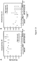
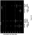


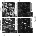


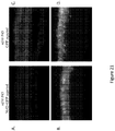


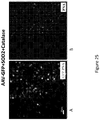

Classifications
-
- A—HUMAN NECESSITIES
- A61—MEDICAL OR VETERINARY SCIENCE; HYGIENE
- A61K—PREPARATIONS FOR MEDICAL, DENTAL OR TOILETRY PURPOSES
- A61K48/00—Medicinal preparations containing genetic material which is inserted into cells of the living body to treat genetic diseases; Gene therapy
- A61K48/005—Medicinal preparations containing genetic material which is inserted into cells of the living body to treat genetic diseases; Gene therapy characterised by an aspect of the 'active' part of the composition delivered, i.e. the nucleic acid delivered
- A61K48/0058—Nucleic acids adapted for tissue specific expression, e.g. having tissue specific promoters as part of a contruct
-
- A—HUMAN NECESSITIES
- A61—MEDICAL OR VETERINARY SCIENCE; HYGIENE
- A61K—PREPARATIONS FOR MEDICAL, DENTAL OR TOILETRY PURPOSES
- A61K45/00—Medicinal preparations containing active ingredients not provided for in groups A61K31/00 - A61K41/00
- A61K45/06—Mixtures of active ingredients without chemical characterisation, e.g. antiphlogistics and cardiaca
-
- A—HUMAN NECESSITIES
- A61—MEDICAL OR VETERINARY SCIENCE; HYGIENE
- A61K—PREPARATIONS FOR MEDICAL, DENTAL OR TOILETRY PURPOSES
- A61K31/00—Medicinal preparations containing organic active ingredients
- A61K31/70—Carbohydrates; Sugars; Derivatives thereof
- A61K31/7088—Compounds having three or more nucleosides or nucleotides
- A61K31/7115—Nucleic acids or oligonucleotides having modified bases, i.e. other than adenine, guanine, cytosine, uracil or thymine
-
- A—HUMAN NECESSITIES
- A61—MEDICAL OR VETERINARY SCIENCE; HYGIENE
- A61K—PREPARATIONS FOR MEDICAL, DENTAL OR TOILETRY PURPOSES
- A61K38/00—Medicinal preparations containing peptides
- A61K38/16—Peptides having more than 20 amino acids; Gastrins; Somatostatins; Melanotropins; Derivatives thereof
- A61K38/17—Peptides having more than 20 amino acids; Gastrins; Somatostatins; Melanotropins; Derivatives thereof from animals; from humans
- A61K38/1703—Peptides having more than 20 amino acids; Gastrins; Somatostatins; Melanotropins; Derivatives thereof from animals; from humans from vertebrates
- A61K38/1709—Peptides having more than 20 amino acids; Gastrins; Somatostatins; Melanotropins; Derivatives thereof from animals; from humans from vertebrates from mammals
-
- A—HUMAN NECESSITIES
- A61—MEDICAL OR VETERINARY SCIENCE; HYGIENE
- A61K—PREPARATIONS FOR MEDICAL, DENTAL OR TOILETRY PURPOSES
- A61K38/00—Medicinal preparations containing peptides
- A61K38/16—Peptides having more than 20 amino acids; Gastrins; Somatostatins; Melanotropins; Derivatives thereof
- A61K38/43—Enzymes; Proenzymes; Derivatives thereof
- A61K38/44—Oxidoreductases (1)
- A61K38/446—Superoxide dismutase (1.15)
-
- A—HUMAN NECESSITIES
- A61—MEDICAL OR VETERINARY SCIENCE; HYGIENE
- A61K—PREPARATIONS FOR MEDICAL, DENTAL OR TOILETRY PURPOSES
- A61K48/00—Medicinal preparations containing genetic material which is inserted into cells of the living body to treat genetic diseases; Gene therapy
- A61K48/005—Medicinal preparations containing genetic material which is inserted into cells of the living body to treat genetic diseases; Gene therapy characterised by an aspect of the 'active' part of the composition delivered, i.e. the nucleic acid delivered
-
- A—HUMAN NECESSITIES
- A61—MEDICAL OR VETERINARY SCIENCE; HYGIENE
- A61K—PREPARATIONS FOR MEDICAL, DENTAL OR TOILETRY PURPOSES
- A61K48/00—Medicinal preparations containing genetic material which is inserted into cells of the living body to treat genetic diseases; Gene therapy
- A61K48/0075—Medicinal preparations containing genetic material which is inserted into cells of the living body to treat genetic diseases; Gene therapy characterised by an aspect of the delivery route, e.g. oral, subcutaneous
-
- A—HUMAN NECESSITIES
- A61—MEDICAL OR VETERINARY SCIENCE; HYGIENE
- A61P—SPECIFIC THERAPEUTIC ACTIVITY OF CHEMICAL COMPOUNDS OR MEDICINAL PREPARATIONS
- A61P27/00—Drugs for disorders of the senses
- A61P27/02—Ophthalmic agents
-
- A—HUMAN NECESSITIES
- A61—MEDICAL OR VETERINARY SCIENCE; HYGIENE
- A61P—SPECIFIC THERAPEUTIC ACTIVITY OF CHEMICAL COMPOUNDS OR MEDICINAL PREPARATIONS
- A61P39/00—General protective or antinoxious agents
- A61P39/06—Free radical scavengers or antioxidants
-
- A—HUMAN NECESSITIES
- A61—MEDICAL OR VETERINARY SCIENCE; HYGIENE
- A61P—SPECIFIC THERAPEUTIC ACTIVITY OF CHEMICAL COMPOUNDS OR MEDICINAL PREPARATIONS
- A61P43/00—Drugs for specific purposes, not provided for in groups A61P1/00-A61P41/00
-
- C—CHEMISTRY; METALLURGY
- C12—BIOCHEMISTRY; BEER; SPIRITS; WINE; VINEGAR; MICROBIOLOGY; ENZYMOLOGY; MUTATION OR GENETIC ENGINEERING
- C12N—MICROORGANISMS OR ENZYMES; COMPOSITIONS THEREOF; PROPAGATING, PRESERVING, OR MAINTAINING MICROORGANISMS; MUTATION OR GENETIC ENGINEERING; CULTURE MEDIA
- C12N2750/00—MICROORGANISMS OR ENZYMES; COMPOSITIONS THEREOF; PROPAGATING, PRESERVING, OR MAINTAINING MICROORGANISMS; MUTATION OR GENETIC ENGINEERING; CULTURE MEDIA ssDNA viruses
- C12N2750/00011—Details
- C12N2750/14011—Parvoviridae
- C12N2750/14111—Dependovirus, e.g. adenoassociated viruses
- C12N2750/14141—Use of virus, viral particle or viral elements as a vector
- C12N2750/14143—Use of virus, viral particle or viral elements as a vector viral genome or elements thereof as genetic vector
-
- C—CHEMISTRY; METALLURGY
- C12—BIOCHEMISTRY; BEER; SPIRITS; WINE; VINEGAR; MICROBIOLOGY; ENZYMOLOGY; MUTATION OR GENETIC ENGINEERING
- C12Y—ENZYMES
- C12Y115/00—Oxidoreductases acting on superoxide as acceptor (1.15)
- C12Y115/01—Oxidoreductases acting on superoxide as acceptor (1.15) with NAD or NADP as acceptor (1.15.1)
- C12Y115/01001—Superoxide dismutase (1.15.1.1)
-
- Y—GENERAL TAGGING OF NEW TECHNOLOGICAL DEVELOPMENTS; GENERAL TAGGING OF CROSS-SECTIONAL TECHNOLOGIES SPANNING OVER SEVERAL SECTIONS OF THE IPC; TECHNICAL SUBJECTS COVERED BY FORMER USPC CROSS-REFERENCE ART COLLECTIONS [XRACs] AND DIGESTS
- Y02—TECHNOLOGIES OR APPLICATIONS FOR MITIGATION OR ADAPTATION AGAINST CLIMATE CHANGE
- Y02A—TECHNOLOGIES FOR ADAPTATION TO CLIMATE CHANGE
- Y02A50/00—TECHNOLOGIES FOR ADAPTATION TO CLIMATE CHANGE in human health protection, e.g. against extreme weather
- Y02A50/30—Against vector-borne diseases, e.g. mosquito-borne, fly-borne, tick-borne or waterborne diseases whose impact is exacerbated by climate change
Definitions
- Cells can be compromised by genetic and environmental factors that lead to their malfunction and death.
- specialized sensory neurons the photoreceptors (rods and cones), as well as retinal ganglion cells (RPG), the output neurons of the retina, are the neuronal cell types that can malfunction and die due to genetic and/or environmental reasons, leading to partial or complete loss of vision.
- RPG retinal ganglion cells
- the retina contains two major types of light-sensitive photoreceptor cells, i.e., rod cells and cone cells.
- Cone cells are responsible for color vision and require brighter light to function, as compared to rod cells.
- cones There are three types of cones, maximally sensitive to long-wavelength, medium-wavelength, and short-wavelength light (often referred to as red, green, and blue, respectively, though the sensitivity peaks are not actually at these colors).
- Cones are mostly concentrated in and near the fovea. Only a small percentage of photoreceptors are cones in the periphery of the retina. Objects are seen most sharply in focus when their images fall on the cone-enriched spot, as when one looks at an object directly.
- Cone cells and rods are connected through intermediate cells in the retina to nerve fibers of the optic nerve. When rods and cones are stimulated by light, the nerves send off impulses through these fibers to the brain.
- Retinitis pigmentosa is a family of inherited retinal degenerations (RD) that is currently incurable and frequently leads to blindness. Affecting roughly 1 in 3,000 individuals, it is the most prevalent form of RD caused by a single disease allele (RetNet, www.sph.uth.tmc.edu/Retnet/). The phenotype is characterized by an initial loss of night vision due to the malfunction and death of rod photoreceptors, followed by a progressive loss of cones ( Madreperla, S.A., et al. (1990) Arch Ophthalmol 108, 358-61 ).
- retinitis pigmentosa is further characterized by, e.g., night blindness, progressive loss of peripheral vision, eventually leading to total blindness, ophthalmoscopic changes consisting of dark mosaic-like retinal pigmentation, attenuation of the retinal vessels, waxy pallor of the optic disc, and in the advanced forms, macular degeneration. Since cones are responsible for color and high acuity vision, it is their loss that leads to a reduction in the quality of life. In many cases, the disease-causing allele is expressed exclusively in rods; nonetheless, cone cell death follows rod cell death. Indeed, to date there is no known form of RD in humans or mice where rods die, and cones survive. In contrast, mutations in cone-specific genes result only in cone death.
- the present invention provides a pharmaceutical composition suitable for intraocular administration, comprising an AAV 2/5 or AAV 2/8 vector comprising a promoter and a nucleic acid molecule encoding nuclear factor erythroid 2-like-2 (Nrf2) for use in the treatment of retinitis pigmentosa.
- a pharmaceutical composition suitable for intraocular administration comprising an AAV 2/5 or AAV 2/8 vector comprising a promoter and a nucleic acid molecule encoding nuclear factor erythroid 2-like-2 (Nrf2) for use in the treatment of retinitis pigmentosa.
- the present invention provides a pharmaceutical composition suitable for intraocular administration, comprising a viral vector comprising a retinal cell-type specific promoter operably linked to a nucleic acid molecule encoding nuclear factor erythroid 2-like 2 (Nrf2), wherein the vector is selected from the group consisting of a retroviral vector, an adenoviral vector, an adenoviral/retroviral chimera vector, an adeno-associated virus (AAV) vector, a herpes simplex viral I or II vector, a parvovirus vector, a reticuloendotheliosis virus vector, a poliovirus vector, a papillomavirus vector, a vaccinia virus vector, and a lentivirus vector; wherein the pharmaceutical composition is for use in treating or preventing retinitis pigmentosa in a subject.
- a viral vector comprising a retinal cell-type specific promoter operably linked to a nucleic acid molecule encoding nuclear
- the vector is an AAV vector.
- the AAV vector is an AAV 2/5 or an AAV 2/8 vector.
- the viral vector is an AAV 2/5 or an AAV 2/8 vector; and the retinal cell-type specific promoter is (i) a rod-specific promoter; or (ii) a cone-specific promoter; or (iii) a rod- and cone-specific promoter.
- the retinal cell-type specific promoter is selected from the group consisting of an Nrl promoter, a Crx promoter, a Rax promoter, a cone opsin promoter, an interphotoreceptor retinoid binding protein (IRBP156) promoter, a rhodopsin kinase (RK) promoter, a neural leucine zipper (NRLL) promoter, a cone arrestin promoter, a Cabp5 promoter, a Cralbp promoter, an Ndrg4promoter, a clusterin promoter, a Hesl promoter, a vimentin promoter, a cluster differentiation (CD44) promoter, and a glial fibrillary acid protein (GFAP) promoter, and combinations thereof.
- an Nrl promoter an Nrl promoter
- Crx promoter a Crx promoter
- Rax promoter Rax promoter
- a cone opsin promoter an interphotoreceptor retinoid binding protein (IRBP156) promote
- the intraocular administration is selected from the group consisting of intravitreal, subretinal, subconjunctival, sub-tenon, periocular, retrobulbar, suprachoroidal, and intrascleral administration.
- compositions further comprise a viscosity inducing agent.
- the promoter is a human cytomegalovirus (CMV) promoter.
- CMV human cytomegalovirus
- the present invention is based, at least in part, on the discovery that increased expression and/or activity of genes encoding antioxidant defense protein(s), in a photoreceptor cell undergoing oxidative stress, can inhibit oxidative stress in the affected cell.
- rods are the major cell type in the outer nuclear layer (ONL)
- cones experience a greatly altered environment following rod death.
- the cone outer segment (OS) collapses, they lose their intimate association with the retinal pigmented epithelium (RPE), and are, thus, exposed to a hyperoxic environment as evidenced by greater oxidation of their nucleic acids, proteins, and lipids.
- RPE retinal pigmented epithelium
- an antioxidant defense protein including antioxidant enzymes and transcription factors that generally up-regulate the anti-oxidation program can serve to inhibit cellular oxidative stress, thus, increasing photoreceptor viability.
- the present disclosure provides methods for inhibiting oxidative stress of a photoreceptor, as well as methods for the treatment and/ or prevention of disorders associated with cellular oxidative stress, for example, retinitis pigmentosa, by increasing the expression and/or activity of at least one antioxidant defense protein.
- an element means one element or more than one element, e.g., a plurality of elements.
- the terms “inhibit oxidative stress” and “inhibition of oxidative stress” refer to preventing as well as ameliorating, decreasing, or reducing oxidative stress.
- antioxidant defense protein includes any protein that inhibits the oxidation of a molecule. More specifically, an “antioxidant defense protein” encompasses any upstream element (e.g., transcription factor) involved in the anti-oxidation program, as well as any downstream element (e.g., antioxidant enzymes) that can be activated as a result of the enhancement of an upstream element.
- upstream element e.g., transcription factor
- downstream element e.g., antioxidant enzymes
- Exemplary antioxidant defense proteins include, superoxide dismutase 2 (SOD2), catalase, peroxisome proliferator-activated receptor gamma coactivator 1-alpha (PGC1 ⁇ ), and nuclear factor erythroid 2-like 2 (Nrf2).
- oxidative stress refers to any imbalance in the normal reduction-oxidation state in a photoreceptor cell that leads to an increase in the level of reactive oxygen species (ROS) resulting in cellular damage including, for example, DNA damage, lipid peroxidation, and oxidation of proteins.
- ROS reactive oxygen species
- antioxidant proteins capable of detoxifying or ameliorating free radicals, i.e., reactive oxygen species (ROS).
- ROS reactive oxygen species
- examples include, but are not limited to, alpha-1-microglobulin, superoxide dismutases, catalases, lactoperoxidases, glutathione peroxidases, and peroxiredoxins.
- the term “reduces the level of ROS” refers to decreasing the level of ROS to physiologically acceptable non-toxic levels, as well as preventing the further generation of ROS.
- Photoreceptor cells i.e., a specialized cell found in the retina.
- the retina is a thin, transparent tissue containing about 120 million separate rod cells (night vision) and 7 million cone cells (day and color vision) as well as millions of other structural supporting and interconnecting cells.
- Photoreceptor cells consist of "rods” and “cones”, which are the photosensitive cells of the retina.
- the rods contain rhodopsin, the rod photopigment, and the cones contain other distinct photopigments, which respond to light and ultimately trigger a neural discharge in the output cells of the retina, the ganglion cells.
- this signal is registered as a visual stimulus in the visual cortex and other target locations in the brain.
- the retinal pigment epithelial (RPE) cells produce, store and transport a variety of factors that are responsible for the normal function and survival of photoreceptors.
- Retinal cells that can also sense light consist of photosensitive ganglion cells. These cells, known as the melanopsin ganglion cells are found in the inner retina, have dendrites and long axons projecting to the protectum (midbrain), the suprachiasmatic nucleus in the hypothalamus, and the lateral geniculate (thalamus).
- the photoreceptor cell may be a rod, or distinct from the optic nerve, or a cone.
- a photosensitive cell may be a melanopsin ganglion cell.
- the term "retinal disorder” refers generally to a disorder of the retina.
- the retinal disorder may be associated with oxidative stress, decreased viability, for example, death, of cone cells, and/ or rod cells.
- a retinal disorder is not associated with blood vessel leakage and/or growth, for example, as is the case with diabetic retinopathy, but, instead is characterized primarily by reduced viability of cone cells and/ or rod cells.
- the retinal disorder may be a genetic disorder, retinitis pigmentosa, age-related macular degeneration, cone-rod dystrophy, or rod-cone dystrophy.
- the retinal disorder is not associated with blood vessel leakage and/or growth, is not associated with diabetes and/or diabetic retinopathy, is not NARP (neuropathy, ataxia, and retinitis pigmentosa), is not a neurological disorder, or is not a disorder that is associated with a compromised optic nerve and/or disorders of the brain.
- the retinal disorder may be associated with a compromised photoreceptor cell, and is not a neurological disorder.
- Retinitis pigmentosa As used herein, the term "retinitis pigmentosa” or "RP” is known in the art and encompasses a disparate group of genetic disorders of rods and cones. Retinitis pigmentosa generally refers to retinal degeneration often characterized by the following manifestations: night blindness, progressive loss of peripheral vision, eventually leading to total blindness; ophthalmoscopic changes consist in dark mosaic-like retinal pigmentation, attenuation of the retinal vessels, waxy pallor of the optic disc, and in the advanced forms, macular degeneration. In some cases there can be a lack of pigmentation. Retinitis pigmentosa can be associated to degenerative opacity of the vitreous body, and cataract. Family history is prominent in retinitis pigmentosa; the pattern of inheritance may be autosomal recessive, autosomal dominant, or X-linked; the autosomal recessive form is the most common and can occur sporadically.
- CRD Cone-Rod Dystrophy
- RCD Reactive-Cone Dystrophy
- age related macular degeneration also referred to as “macular degeneration” or “AMD” refers to the art recognized pathological condition which causes blindness amongst elderly individuals.
- Age related macular degeneration includes both wet and dry forms of ARMD.
- the dry form of ARMD which accounts for about 90 percent of all cases, is also known as atrophic, nonexudative, or drusenoid (age-related) macular degeneration.
- drusen typically accumulate in the retinal pigment epithelium (RPE) tissue beneath/within the Bruch's membrane. Vision loss can then occur when drusen interfere with the function of photoreceptors in the macula.
- RPE retinal pigment epithelium
- the dry form of ARMD results in the gradual loss of vision over many years.
- the dry form of ARMD can lead to the wet form of ARMD.
- the wet form of ARMD also known as exudative or neovascular (age-related) macular degeneration, can progress rapidly and cause severe damage to central vision.
- the macular dystrophies include Stargardt Disease, also known as Stargardt Macular Dystrophy or Fundus Flavimaculatus, which is the most frequently encountered juvenile onset form of macular dystrophy.
- the term "neurological disorder” encompasses neurodegenerative disorders and demyelinating disorders.
- the compositions of this invention are useful for the treatment of neurological disorders including but not limited to Parkinson's Disease, Tauopathies, Alzheimer's Disease, Diffuse Neurofibrillary Tangles with Calcification, Supranuclear Palsy, Progressive, TDP-43 Proteinopathies, Amyotrophic Lateral Sclerosis, Multiple Sclerosis, Frontotemporai Lobar Degeneration, Lewy Body Disease, AIDS Dementia Complex, Aphasia, Primary Progressive, Primary Progressive Nonfluent Aphasia, Dementia, Vascular, CADASIL, Dementia, Multi-lnfarct, Diffuse Neurofibrillary Tangles with Calcification, Frontotemporai Lobar Degeneration, Frontotemporai Dementia, Primary Progressive Nonfluent Aphasia, Kluver-Bucy Syndrome, Pick's Disease, Motor Neuron Disease, Bulbar Palsy, Progressive, Muscular Atrophy, Spinal
- nucleic acid molecule is intended to include DNA molecules (e.g., cDNA or genomic DNA) and RNA molecules (e.g., mRNA) and analogs of the DNA or RNA generated using nucleotide analogs.
- the nucleic acid molecule can be single-stranded or double-stranded, but preferably is double-stranded DNA.
- a nucleic acid molecule used in the methods of the present invention can be isolated using standard molecular biology techniques. Using all or portion of a nucleic acid sequence of interest as a hybridization probe, nucleic acid molecules can be isolated using standard hybridization and cloning techniques (e.g., as described in Sambrook, J., Fritsh, E. F., and Maniatis, T. Molecular Cloning. A Laboratory Manual. 2nd, ed., Cold Spring Harbor Laboratory, Cold Spring Harbor Laboratory Press, Cold Spring Harbor, N.Y., 1989 ).
- an “isolated” nucleic acid molecule is one which is separated from other nucleic acid molecules which are present in the natural source of the nucleic acid.
- the term “isolated” includes nucleic acid molecules which are separated from the chromosome with which the genomic DNA is naturally associated.
- an “isolated” nucleic acid molecule is free of sequences which naturally flank the nucleic acid molecule ( i.e., sequences located at the 5' and 3' ends of the nucleic acid molecule) in the genomic DNA of the organism from which the nucleic acid molecule is derived.
- a nucleic acid molecule for use in the methods disclosed herein can also be isolated by the polymerase chain reaction (PCR) using synthetic oligonucleotide primers designed based upon the sequence of a nucleic acid molecule of interest.
- a nucleic acid molecule used in the methods described herein can be amplified using cDNA, mRNA or, alternatively, genomic DNA as a template and appropriate oligonucleotide primers according to standard PCR amplification techniques.
- oligonucleotides corresponding to nucleotide sequences of interest can be prepared by standard synthetic techniques, e.g., using an automated DNA synthesizer.
- nucleic acids for use in the methods described herein can also be prepared, e.g., by standard recombinant DNA techniques.
- a nucleic acid of the invention can also be chemically synthesized using standard techniques.
- Various methods of chemically synthesizing polydeoxynucleotides are known, including solid-phase synthesis which has been automated in commercially available DNA synthesizers (See e.g., Itakura et al. U.S. Patent No. 4,598,049 ; Caruthers et al. U.S. Patent No. 4,458,066 ; and Itakura U.S. Patent Nos. 4,401,796 and 4,373,071 .
- an "antisense” nucleic acid comprises a nucleotide sequence which is complementary to a “sense” nucleic acid encoding a protein, e.g., complementary to the coding strand of a double-stranded cDNA molecule, complementary to an mRNA sequence or complementary to the coding strand of a gene. Accordingly, an antisense nucleic acid can hydrogen bond to a sense nucleic acid.
- a nucleic acid molecule may be an siRNA molecule, an shRNA molecule, and/or may mediate RNAi.
- RNA interference is a post-transcriptional, targeted gene-silencing technique that uses double-stranded RNA (dsRNA) to degrade messenger RNA (mRNA) containing the same sequence as the dsRNA (Sharp, P.A. and Zamore, P.D. 287. 2431-2432 (2000); Zamore, P.D., et al. Cell 101, 25-33 (2000 ). Tuschl, T. et al. Genes Dev. 13, 3191-3197 (1999 ); Cottrell TR, and Doering TL. 2003. Trends Microbiol. 11:37-43 ; Bushman F.2003. MoI Therapy.
- dsRNA double-stranded RNA
- mRNA messenger RNA
- RNAi 21- or 22-nucleotide-long RNAs
- siRNAs small interfering RNAs
- Kits for synthesis of RNAi are commercially available from, e.g,. New England Biolabs or Ambion.
- One or more of the chemistries described herein for use in antisense RNA can be employed in molecules that mediate RNAi.
- vector refers to a nucleic acid molecule capable of transporting another nucleic acid to which it has been linked.
- plasmid refers to a circular double stranded DNA loop into which additional DNA segments may be ligated.
- viral vector Another type of vector is a viral vector, wherein additional DNA segments may be ligated into the viral genome.
- Certain vectors are capable of autonomous replication in a host cell into which they are introduced (e.g., bacterial vectors having a bacterial origin of replication and episomal mammalian vectors).
- vectors e.g., non-episomal mammalian vectors
- Other vectors are integrated into the genome of a host cell upon introduction into the host cell, and thereby are replicated along with the host genome.
- certain vectors are capable of directing the expression of genes or nucleic acid molecules to which they are operatively linked and are referred to as "expression vectors” or “recombinant expression vectors” or simply "expression vectors".
- Nucleic acid sequences necessary for expression in prokaryotes usually include a promoter, an operator (optional), and a ribosome binding site, often along with other sequences.
- Eukaryotic cells are known to utilize promoters, enhancers, and termination and polyadenylation signals. "Expression vectors" may be used in order to permit pseudotyping of the viral envelope proteins.
- Expression vectors are often in the form of plasmids.
- plasmid and “vector” may be used interchangeably as the plasmid is the most commonly used form of vector.
- the invention is intended to include such other forms of expression vectors, such as viral vectors (e.g., replication defective retroviruses, adenoviruses, adeno-associated viruses, lentiviruses), which serve equivalent functions.
- viral vectors e.g., replication defective retroviruses, adenoviruses, adeno-associated viruses, lentiviruses
- the term "retrovirus” is used in reference to RNA viruses that utilize reverse transcriptase during their replication cycle.
- the retroviral genomic RNA is converted into double-stranded DNA by reverse transcriptase.
- This double-stranded DNA form of the virus is capable of being integrated into the chromosome of the infected cell; once integrated, it is referred to as a "provirus.”
- the provirus serves as a template for RNA polymerase II and directs the expression of RNA molecules which encode the structural proteins and enzymes needed to produce new viral particles.
- LTRs Long terminal repeats
- LTRs contain numerous regulatory signals, including transcriptional control elements, polyadenylation signals, and sequences needed for replication and integration of the viral genome. LTRs may be several hundred base pairs in length.
- AAV vector refers to a vector derived from an adeno-associated virus serotype, including without limitation, AAV-1, AAV-2, AAV-3, AAV-4, AAV-5, or AAVX7.
- rAAV vector refers to a vector that includes AAV nucleotide sequences as well as heterologous nucleotide sequences. rAAV vectors require only the 145 base terminal repeats in cis to generate virus. All other viral sequences are dispensable and may be supplied in trans ( Muzyczka (1992) Curr. Topics Microbiol. Immunol. 158:97 ).
- the rAAV vector genome will only retain the inverted terminal repeat (ITR) sequences so as to maximize the size of the transgene that can be efficiently packaged by the vector.
- ITRs need not be the wild-type nucleotide sequences, and may be altered, e.g., by the insertion, deletion or substitution of nucleotides, as long as the sequences provide for functional rescue, replication and packaging.
- the AAV vector is an AAV2/5 or AAV2/8 vector. Suitable AAV vectors are described in, for example, U.S. Patent No. 7,056,502 and Yan et al. (2002) J. Virology 76(5):2043-2053 .
- lentivirus refers to a group (or genus) of retroviruses that give rise to slowly developing disease.
- HIV human immunodeficiency virus; including but not limited to HIV type 1 and HIV type 2), the etiologic agent of the human acquired immunodeficiency syndrome (AIDS); visna-maedi, which causes encephalitis (visna) or pneumonia (maedi) in sheep; the caprine arthritis-encephalitis virus, which causes immune deficiency, arthritis, and encephalopathy in goats; equine infectious anemia virus (EIAV), which causes autoimmune hemolytic anemia, and encephalopathy in horses; feline immunodeficiency virus (FIV), which causes immune deficiency in cats; bovine immune deficiency virus (BIV), which causes lymphadenopathy, lymphocytosis, and possibly central nervous system infection in cattle; and simian immunodeficiency virus (SIV), which cause immune
- HIV human immunodeficiency virus
- HIV human immuno
- the viruses latently infect monocytes and macrophages, from which they spread to other cells. HIV, FIV, and SIV also readily infect T lymphocytes (i.e., T-cells).
- the lentivirus is not HIV.
- promoter refers to a recognition site of a DNA strand to which the RNA polymerase binds.
- the promoter forms an initiation complex with RNA polymerase to initiate and drive transcriptional activity.
- the complex can be modified by activating sequences termed “enhancers” or inhibitory sequences termed “silencers”.
- transformation refers to introduction of a nucleic acid, e.g., a viral vector, into a recipient cell.
- the term "subject" includes warm-blooded animals, preferably mammals, including humans.
- the subject is a primate.
- the primate is a human.
- modulate are intended to include stimulation (e.g., increasing or upregulating a particular response or activity) and inhibition (e.g., decreasing or downregulating a particular response or activity).
- the term "contacting" i.e., contacting a cell with an agent
- contacting is intended to include incubating the agent and the cell together in vitro (e.g., adding the agent to cells in culture) or administering the agent to a subject such that the agent and cells of the subject are contacted in vivo.
- the term "contacting” is not intended to include exposure of cells to an agent that may occur naturally in a subject (i.e., exposure that may occur as a result of a natural physiological process).
- administering to a subject includes dispensing, delivering or applying a composition of the invention, e.g., capable of inhibiting oxidative stress, to a subject by any suitable route for delivery of the composition to the desired location in the subject, including delivery by intraocular administration or intravenous administration.
- delivery is by the topical, parenteral or oral route, intracerebral injection, intramuscular injection, subcutaneous/intradermal injection, intravenous injection, buccal administration, transdermal delivery and administration by the rectal, colonic, vaginal, intranasal or respiratory tract route.
- the present disclosure provides methods for inhibiting oxidative stress of a photoreceptor cell, which generally comprise contacting a photoreceptor cell with an agent which increases the expression and/or activity of an antioxidant defense protein.
- the present disclosure also provides methods for treating or preventing a retinal disorder, e.g., a retinal disorder associated with oxidative stress of a photoreceptor cell, in a subject.
- the methods generally comprise administering to the subject an agent which increases the expression and/or activity of an antioxidant defense protein.
- the present disclosure provides methods for treating or preventing retinitis pigmentosa in a subject.
- Such methods generally comprise administering to the subject an agent which increases the expression and/or activity of an antioxidant defense protein.
- the present disclosure further provides methods for prolonging the viability of a photoreceptor cell, e.g., a photoreceptor cell compromised by a disorder associated with oxidative stress.
- the methods generally comprise contacting the cell with an agent which increases the expression and/or activity of an antioxidant defense protein.
- a cell may be contacted with or a subject administered a single antioxidant defense genes or a combination of antioxidant defense proteins.
- Suitable antioxidant defense genes include catalase, superoxide dismutase 2 (SOD2), peroxisome proliferator-activated receptor gamma coactivator 1-alpha (PGC1 ⁇ ), nuclear factor erythroid 2-like 2 (nrf2).
- Suitable combinations of antioxidant defense genes for use in the methods of the invention include catalase; SOD2; PGC1 ⁇ ; nrf2; catalase and SOD2; SOD2 and PGC1 ⁇ ; PGC1 ⁇ and nrf2; catalase and PGC1 ⁇ ; catalase and nrf2; SOD2 and nrf2; catalase, SOD2, and PGC1 ⁇ ; SOD2, PGC1 ⁇ , and nrf2; catalase, PGC1 ⁇ , and nrf2; catalase, SOD2, and nrf2; or catalase, SOD2, PGC1 ⁇ , and nrf2.
- the methods described herein can be performed in vitro.
- intracellular levels of an antioxidant defense protein e.g., catalase, SOD2, PGC1 ⁇ , and/or nrf2
- the cell may be a mammalian cell, e.g., a human cell.
- cells can be obtained from a subject by standard methods and incubated ( e.g., cultured) in vitro with an agent which stimulates intracellular levels of an antioxidant defense protein (e.g., catalase, SOD2, PGC1 ⁇ , and/or nrf2).
- an agent which stimulates intracellular levels of an antioxidant defense protein e.g., catalase, SOD2, PGC1 ⁇ , and/or nrf2
- Methods for isolating cells are well known in the art.
- the cells can be re-administered to the same subject, or another subject which is compatible with the donor of the cells.
- the culture For administration of cells to a subject, it may be preferable to first remove residual agents in the culture from the cells before administering them to the subject. This can be done, for example, by gradient centrifugation of the cells or by washing of the tissue. Methods for the ex vivo genetic modification of cells followed by re-administration to a subject are well known in the art and described in, for example, U.S. Patent No. 5,399,346 .
- the disclosure provides methods for modulation of intracellular levels of an antioxidant defense protein (e.g., catalase, SOD2, PGC1 ⁇ , and/or nrf2) in vivo, by administering to a subject a therapeutically effective amount of an agent as described herein.
- an antioxidant defense protein e.g., catalase, SOD2, PGC1 ⁇ , and/or nrf2
- intracellular levels of catalase, SOD2, PGC1 ⁇ , and/or nrf2 can be modulated to treat or prevent a retinal disorder, such as a retinal disorder associated with cellular oxidative stress of a photoreceptor cells.
- the terms “treat,” “treatment” and “treating” include the application or administration of agents, as described herein, to a subject who is suffering from a retinal disorder, e.g., associated with oxidative stress of a photoreceptor cell, or who is susceptible to such conditions with the purpose of curing, healing, alleviating, relieving, altering, remedying, ameliorating, improving or affecting such conditions or at least one symptom of such conditions.
- a retinal disorder e.g., associated with oxidative stress of a photoreceptor cell
- the condition is also “treated” if recurrence of the condition is reduced, slowed, delayed or prevented.
- Subjects suitable for treatment using the regimens of the present disclosure should have or are susceptible to developing retinal disorders associated with oxidative stress.
- subjects may be genetically predisposed to development of the disorders.
- abnormal progression of the following factors including, but not limited to visual acuity, the rate of death of cone and/ or rod cells, night vision, peripheral vision, attenuation of the retinal vessels, and other ophthalmoscopic factors associated with retinal disorders such as retinitis pigmentosa may indicate the existence of or a predisposition to a retinal disorder.
- the agents may be administered as necessary to achieve the desired effect and depend on a variety of factors including, but not limited to, the severity of the condition, age and history of the subject and the nature of the composition, for example, the identity of the genes or the affected biochemical pathway.
- the compositions may be administered at least two, three, four, five or six times a day. Additionally, the therapeutic or preventative regimens may cover a period of at least about 2, 3, 4, 5, 6, 7, 8, 9, 10, 11, 12, 13, 14, 15, 16, 17, 18, 19, 20, 21, 22, 23 or 24 weeks.
- an agent to up-regulate intracellular levels of catalase, SOD2, PGC1 ⁇ , and/or nrf2 can be determined as described herein, e.g., by determining the ability of the agent to modulate cell viability (e.g., modulation of apoptosis), cleavage of LaminA or Caspase 3; production of reactive oxygen species; assessment for oxidation using immunohistochemical detection of acrolein, as well as ELISA for carbonyl adducts in retinal extracts upon exposure of cells to an oxidant (e.g., paraquat); and/or the expression and protein synthesis of photoreceptor specific opsins.
- cell viability e.g., modulation of apoptosis
- cleavage of LaminA or Caspase 3 cleavage of LaminA or Caspase 3
- production of reactive oxygen species e.g., assessment for oxidation using immunohistochemical detection of acrolein
- the methods of the present disclosure may further comprise monitoring the effectiveness of treatment. For example, visual acuity, the rate of death of cone and/ or rod cells, night vision, peripheral vision, attenuation of the retinal vessels, and other ophthalmoscopic changes associated with retinal disorders such as retinitis pigmentosa may be monitored to assess the effectiveness of treatment. Additionally, the rate of death of cells associated with the particular disorder that is the subject of treatment and/or prevention, may be monitored. Alternatively, the viability of such cells may be monitored, for example, as measured by phospholipid production. The assays described in the Examples section below may also be used to monitor the effectiveness of treatment (e.g., electroretinography - ERG).
- An agent for use in the methods of the present disclosure may be a nucleic acid molecule encoding an antioxidant defense protein, e.g., superoxide dismutase 2 (SOD2), catalase, peroxisome proliferator-activated receptor gamma coactivator 1-alpha (PGC1 ⁇ ), and nuclear factor erythroid 2-like 2 (nrf2) (or combinations thereof).
- an antioxidant defense protein e.g., superoxide dismutase 2 (SOD2), catalase, peroxisome proliferator-activated receptor gamma coactivator 1-alpha (PGC1 ⁇ ), and nuclear factor erythroid 2-like 2 (nrf2) (or combinations thereof).
- the nucleic acid molecule may encode superoxide dismutase 2 (SOD2).
- SOD2 is a member of the iron/manganese superoxide dismutase family. It encodes a mitochondrial protein that forms a homotetramer and binds one manganese ion per subunit. This protein binds to the superoxide byproducts of oxidative phosphorylation and converts them to hydrogen peroxide and diatomic oxygen.
- the nucleic acid molecule may encode catalase.
- Catalase is a heme enzyme that is present in the peroxisome of nearly all aerobic cells. Catalase converts the reactive oxygen species hydrogen peroxide to water and oxygen and thereby mitigates the toxic effects of hydrogen peroxide.
- the amino acid sequence of catalase is known and may be found in, for example, GenBank Accession No. GI: 260436906.
- the nucleic acid molecule may encode peroxisome proliferator-activated receptor gamma coactivator 1-alpha (PGC1 ⁇ ).
- PGC1 ⁇ is a transcriptional co-activator that regulates the genes involved in energy metabolism. PGC1 ⁇ interacts with PPARgamma, which permits the interaction of PGC1 ⁇ with multiple transcription factors. PGC1 ⁇ can interact with, and regulate the activities of, cAMP response element binding protein (CREB) and nuclear respiratory factors (NRFs) [Here is correct to keep nuclear respiratory factors]. It provides a direct link between external physiological stimuli and the regulation of mitochondrial biogenesis.
- the amino acid sequence of PGC1 ⁇ is known and may be found in, for example, GenBank Accession No. GI: 116284374.
- the nucleic acid molecule may encode Nuclear factor (erythroid-derived 2)-like 2 (nrf2), a transcription factor which is a member of a small family of basic leucine zipper (bZIP) proteins.
- the encoded transcription factor regulates genes which contain antioxidant response elements (ARE) in their promoters.
- ARE antioxidant response elements
- the viability or survival of photoreceptor cells is short term viability, e.g., about 1 week, about 2 weeks, about 3 weeks, about 4 weeks, about 5 weeks, about 6 weeks, about 7 weeks, about 8 weeks, about 3 years, about 4 years, about 5 years, about 10 years, about 15, years, about 20 years, about 25 years, about 30 years, about 40 years, about 50 years, about 60 years, about 70 years, and about 80 years.
- the disorder may be a retinal disorder including, but not limited to, retinitis pigmentosa, age related macular degeneration, cone rod dystrophy, and rod cone dystrophy.
- the retinal disorder is not associated with blood vessel leakage and/or growth, not associated with diabetes, is not diabetic retinopathy, or is not NARP (neuropathy, ataxia and retinitis pigmentosa).
- the disorder may be a retinal disorder associated with decreased viability of cone and/or rod cells.
- the disorder may be a genetic disorder.
- a retinal disorder such as a retinal disorder associated with oxidative stress of a photoreceptor cell, for example, retinitis pigmentosa
- a retinal disorder such as a retinal disorder associated with oxidative stress of a photoreceptor cell, for example, retinitis pigmentosa
- selecting a subject who is susceptible to the development of the disorder and administering to the subject an effective amount of the nucleic acid molecules, vectors and/or compositions of the present invention, thereby treating or preventing the disorder in the subject.
- the overall strategy to save photoreceptor cells from degeneration by exposure to reactive oxygen species as a result of oxidative stress is to supply them with the genes that will allow them to reduce or eliminate the hyperoxic environment by, e.g., inhibiting and/or removing reactive oxygen species by increasing the expression and/or activity of antioxidant defense protein(s).
- the nucleic acid molecules and/or the vectors of the disclosure are provided in a therapeutically effective amount to elicit the desired effect, e.g., inhibit oxidative stress in a photoreceptor cell.
- the quantity of the nucleic acid molecule, and/or vector to be administered both according to number of treatments and amount, will also depend on factors such as the clinical status, age, and weight of the subject to be treated, and the severity of the disorder. Precise amounts of active ingredient required to be administered depend on the judgment of the gene therapist and will be particular to each individual patient.
- a viral vector comprising the nucleic acid molecules of the invention is administered in titers ranging from about 1x10 5 to about 1x10 9 colony forming units (cfu) per ml, although ranges may vary. Preferred titers will range from about 1x10 6 to about 1x10 8 cfu/ml.
- a therapeutically effective amount of the nucleic acid molecules and/or the vectors of the disclosure may range from about 0.001 to 30 mg/kg body weight, preferably about 0.01 to 25 mg/kg body weight, more preferably about 0.1 to 20 mg/kg body weight, and even more preferably about 1 to 10 mg/kg, 2 to 9 mg/kg, 3 to 8 mg/kg, 4 to 7 mg/kg, or 5 to 6 mg/kg body weight.
- an effective dosage may range from about 0.001 to 30 mg/kg body weight, preferably about 0.01 to 25 mg/kg body weight, more preferably about 0.1 to 20 mg/kg body weight, and even more preferably about 1 to 10 mg/kg, 2 to 9 mg/kg, 3 to 8 mg/kg, 4 to 7 mg/kg, or 5 to 6 mg/kg body weight.
- the skilled artisan will appreciate that certain factors may influence the dosage required to effectively treat a subject, including but not limited to the severity of the disease or disorder, previous treatments, the general health and/or age of the subject, and other diseases present.
- treatment of a subject with a therapeutically effective amount of the nucleic acid molecules and/or the vectors of the disclosure can include a single treatment or, preferably, can include a series of treatments. It will also be appreciated that the effective dosage used for treatment may increase or decrease over the course of a particular treatment. Changes in dosage may result from the results of diagnostic assays as described herein.
- the pharmaceutical compositions can be included in a container, pack, or dispenser together with instructions for administration.
- prophylactic or therapeutic treatment refers to administration to the subject of one or more of the subject compositions. If it is administered prior to clinical manifestation of the unwanted condition (e.g ., disease or other unwanted state of the host animal) then the treatment is prophylactic, i.e., it protects the host against developing the unwanted condition, whereas if administered after manifestation of the unwanted condition, the treatment is therapeutic ( i.e., it is intended to diminish, ameliorate or maintain the existing unwanted condition or side effects therefrom).
- the unwanted condition e.g ., disease or other unwanted state of the host animal
- “Therapeutically effective amount,” as used herein, is intended to include the amount of a nucleic acid molecule and/or the vectors of the disclosure that, when administered to a patient for treating a neurodegenerative disease, is sufficient to effect treatment of the disease (e.g., by diminishing, ameliorating or maintaining the existing disease or one or more symptoms of disease).
- the "therapeutically effective amount” may vary depending on the nucleic acid molecule, peptide, and/or the vector, how the nucleic acid molecule and/or the vectors is administered, the disease and its severity and the history, age, weight, family history, genetic makeup, stage of pathological processes mediated by the neurodegenerative disease expression, the types of preceding or concomitant treatments, if any, and other individual characteristics of the patient to be treated.
- prophylactically effective amount is intended to include the amount of a nucleic acid molecule and/or the vector that, when administered to a subject who does not yet experience or display symptoms of e.g., a retinal disorder, but who may be predisposed to the disease, is sufficient to prevent or ameliorate the disease or one or more symptoms of the disease. Ameliorating the disease includes slowing the course of the disease or reducing the severity of later-developing disease.
- the "prophylactically effective amount” may vary depending on the nucleic acid molecule and/or the vector, how the nucleic acid molecule and/or the vector is administered, the degree of risk of disease, and the history, age, weight, family history, genetic makeup, the types of preceding or concomitant treatments, if any, and other individual characteristics of the patient to be treated.
- a “therapeutically-effective amount” or “prophylactically effective amount” also includes an amount of a nucleic acid molecule and/or the vector that produces some desired local or systemic effect at a reasonable benefit/risk ratio applicable to any treatment.
- a nucleic acid molecule and/or the vector employed in the methods of the present disclosure may be administered in a sufficient amount to produce a reasonable benefit/risk ratio applicable to such treatment.
- Preventing refers to a reduction in risk of acquiring a disease or disorder (i.e., causing at least one of the clinical symptoms of the disease not to develop in a patient that may be exposed to or predisposed to the disease but does not yet experience or display symptoms of the disease).
- treating refers to a beneficial or desired result including, but not limited to, alleviation or amelioration of one or more symptoms, diminishing the extent of infection, stabilized ( i.e., not worsening) state of infection, amelioration or palliation of the infectious state, whether detectable or undetectable.
- Treatment can also mean prolonging survival as compared to expected survival in the absence of treatment.
- An agent e.g., an isolated nucleic acid molecule and/or vector may be administered in combination with an additional therapeutic agent or treatment.
- the compositions and an additional therapeutic agent can be administered in combination in the same composition or the additional therapeutic agent can be administered as part of a separate composition or by another method described herein.
- additional therapeutic agents suitable for use in the methods of the disclosure include those agents known to treat retinal disorders, such as retinitis pigmentosa and age-related macular degeneration and include, for example, fat soluble vitamins (e.g., vitamin A, vitamin E, and ascorbic acid), calcium channel blockers (e.g., diltiazem) carbonic anhydrase inhibitors ( e.g., acetazolamide and methazolamide), anti-angiogenics (e.g., antiVEGF antibodies), growth factors (e.g., rod-derived cone viability factor (RdCVF), BDNF, CNTF, bFGF, and PEDF), antioxidants, other gene therapy agents (e.g., optogenetic gene threrapy, e.g., channelrhodopsin, melanopsin, and halorhodopsin), and compounds that drive photoreceptor regeneration by, e.g., reprogramming Müller cells into photo
- retinal disorders such
- Exemplary treatments for use in combination with the treatment methods of the present disclosure include, for example, retinal and/or retinal pigmented epithelium transplantation, stem cell therapies, retinal prostheses, laser photocoagulation, photodynamic therapy, low vision aid implantation, submacular surgery, and retinal translocation.
- the methods of the disclosure may use stimulatory agents which increase the expression and/or activity of an antioxidant defense protein in a cell.
- stimulatory agents include proteins, nucleic acid molecules, e.g., expression vectors comprising nucleic acid molecules, and chemical agents that stimulate expression and/or activity of a protein which increases the expression and/or activity of an antioxidant defense protein in a cell.
- a preferred stimulatory agent is a nucleic acid molecule encoding a protein of interest.
- a cDNA full length or partial cDNA sequence
- the cDNA can be obtained, for example, by amplification using the polymerase chain reaction (PCR) or by screening an appropriate cDNA library.
- nucleic acid molecules encoding a protein of interest in the form suitable for expression of the protein in a host cell can be prepared using nucleotide sequences based on the nucleic acid sequence of a nucleic acid molecule encoding the protein of interest.
- a stimulatory agent can be present in an inducible construct or in a construct which leads to constitutive expression.
- the nucleic acid molecules of the disclosure may be delivered to cells, e.g., photoreceptor cells, or to subjects, in a vector, e.g., a recombinant expression vector.
- the nucleic acid molecules of the disclosure may be delivered to cells, e.g., photoreceptor cells, or to subjects, in the absence of a vector.
- vector refers to a nucleic acid molecule capable of transporting another nucleic acid to which it has been linked.
- plasmid refers to a circular double stranded DNA loop into which additional DNA segments can be ligated.
- viral vector Another type of vector is a viral vector, wherein additional DNA segments can be ligated into the viral genome.
- Certain vectors are capable of autonomous replication in a host cell into which they are introduced (e.g., bacterial vectors having a bacterial origin of replication and episomal mammalian vectors).
- vectors e.g., non-episomal mammalian vectors
- Other vectors are integrated into the genome of a host cell upon introduction into the host cell, and thereby are replicated along with the host genome.
- certain vectors are capable of directing the expression of genes to which they are operatively linked.
- Such vectors are referred to herein as "expression vectors.”
- expression vectors of utility in recombinant DNA techniques are often in the form of plasmids.
- plasmid and vector can be used interchangeably.
- the disclosure is intended to include such other forms of expression vectors, such as viral vectors (e.g., replication defective retroviruses, adenoviruses and adeno-associated viruses), which serve equivalent functions.
- the recombinant expression vectors of the disclosure comprise a nucleic acid of the disclosure in a form suitable for expression of the nucleic acid in a host cell, which means that the recombinant expression vectors include one or more regulatory sequences, selected on the basis of the host cells to be used for expression, which is operatively linked to the nucleic acid sequence to be expressed.
- "operably linked" is intended to mean that the nucleotide sequence of interest is linked to the regulatory sequence(s) in a manner which allows for expression of the nucleotide sequence (e.g., in an in vitro transcription/translation system or in a host cell when the vector is introduced into the host cell).
- regulatory sequence is intended to include promoters, enhancers and other expression control elements (e.g., polyadenylation signals). Such regulatory sequences are described, for example, in Goeddel; Gene Expression Technology: Methods in Enzymology 185, Academic Press, San Diego, Calif. (1990 ). Regulatory sequences include those which direct constitutive expression of a nucleotide sequence in many types of host cells, those which are constitutively active, those which are inducible, and those which direct expression of the nucleotide sequence only in certain host cells (e.g., tissue-specific regulatory sequences).
- the design of the expression vector can depend on such factors as the choice of the host cell to be transformed, the level of expression of protein desired, and the like.
- the expression vectors of the disclosure can be introduced into host cells to thereby produce proteins or portions thereof, including fusion proteins or portions thereof, encoded by nucleic acids as described herein.
- a nucleic acid molecule encoding an antioxidant defense protein may be expressed in mammalian cells using a mammalian expression vector.
- mammalian expression vectors include pCDM8 ( Seed, B. (1987) Nature 329:840 ) and pMT2PC ( Kaufman et al. (1987) EMBO J. 6:187-195 ).
- the expression vector's control functions are often provided by viral regulatory elements.
- the nucleic acid molecules of the disclosure may be contained within a viral vector and may be delivered to cells, e.g., photoreceptor cells, or to subjects.
- a viral vector is one whose use for gene therapy is well known in the art. Techniques for the formation of vectors or virions are generally described in " Working Toward Human Gene Therapy," Chapter 28 in Recombinant DNA, 2nd Ed., Watson, J. D. et al., eds., New York: Scientific American Books, pp. 567-581 (1992 ). An overview of suitable viral vectors or virions is provided in Wilson, J. M., Clin. Exp. Immunol. 107(Suppl.
- Such vectors may be derived from viruses that contain RNA ( Vile, R. G., et al., Br. Med Bull. 51:12-30 (1995 )) or DNA ( Ali M., et al., Gene Ther. 1:367-384 (1994 )).
- viral vector systems utilized in the gene therapy art and, thus, suitable for use in the present invention, include the following: retroviruses (Vile, R. G., supra; U.S. Pat. Nos. 5,741,486 and 5,763,242 ); adenoviruses ( Brody, S. L., et al., Ann. N.Y. Acad. Sci. 716: 90-101 (1994 ); Heise, C. et al., Nat. Med. 3:639-645 (1997 )); adenoviral/retroviral chimeras ( Bilbao, G., et al., FASEB J. 11:624-634 (1997 ); Feng, M., et al., Nat.
- Extrachromosomal replicating vectors may also be used in the gene therapy methods of the present disclosure. Such vectors are described in, for example, Calos, M. P. (1996) Trends Genet. 12:463-466 .
- Other viruses that can be used as vectors for gene delivery include poliovirus, papillomavirus, vaccinia virus, lentivirus, as well as hybrid or chimeric vectors incorporating favorable aspects of two or more viruses ( Nakanishi, M. (1995) Crit. Rev. Therapeu. Drug Carrier Systems 12:263-310 ; Zhang, J., et al. (1996) Cancer Metastasis Rev. 15:385-401 ; Jacoby, D. R., et al. (1997) Gene Therapy 4:1281-1283 ).
- the term "retrovirus” is used in reference to RNA viruses that utilize reverse transcriptase during their replication cycle.
- the retroviral genomic RNA is converted into double-stranded DNA by reverse transcriptase.
- This double-stranded DNA form of the virus is capable of being integrated into the chromosome of the infected cell; once integrated, it is referred to as a "provirus.”
- the provirus serves as a template for RNA polymerase II and directs the expression of RNA molecules which encode the structural proteins and enzymes needed to produce new viral particles.
- LTRs Long terminal repeats
- LTRs contain numerous regulatory signals, including transcriptional control elements, polyadenylation signals, and sequences needed for replication and integration of the viral genome. LTRs may be several hundred base pairs in length.
- AAV vector refers to a vector derived from an adeno-associated virus serotype, including without limitation, AAV-1, AAV-2, AAV-3, AAV-4, AAV-5, or AAVX7.
- rAAV vector refers to a vector that includes AAV nucleotide sequences as well as heterologous nucleotide sequences. rAAV vectors require only the 145 base terminal repeats in cis to generate virus. All other viral sequences are dispensable and may be supplied in trans ( Muzyczka (1992) Curr. Topics Microbiol. Immunol. 158:97 ).
- the rAAV vector genome will only retain the inverted terminal repeat (ITR) sequences so as to maximize the size of the transgene that can be efficiently packaged by the vector.
- ITRs need not be the wild-type nucleotide sequences, and may be altered, e.g., by the insertion, deletion or substitution of nucleotides, as long as the sequences provide for functional rescue, replication and packaging.
- the AAV vector may be an AAV2/5 or AAV2/8 vector. Suitable AAV vectors are described in, for example, U.S. Patent No. 7,056,502 and Yan et al. (2002) J. Virology 76(5):2043-2053 .
- lentivirus refers to a group (or genus) of retroviruses that give rise to slowly developing disease.
- HIV human immunodeficiency virus; including but not limited to HIV type 1 and HIV type 2), the etiologic agent of the human acquired immunodeficiency syndrome (AIDS); visna-maedi, which causes encephalitis (visna) or pneumonia (maedi) in sheep; the caprine arthritis-encephalitis virus, which causes immune deficiency, arthritis, and encephalopathy in goats; equine infectious anemia virus (EIAV), which causes autoimmune hemolytic anemia, and encephalopathy in horses; feline immunodeficiency virus (FIV), which causes immune deficiency in cats; bovine immune deficiency virus (BIV), which causes lymphadenopathy, lymphocytosis, and possibly central nervous system infection in cattle; and simian immunodeficiency virus (SIV), which cause immune
- HIV human immunodeficiency virus
- HIV human immuno
- viruses Diseases caused by these viruses are characterized by a long incubation period and protracted course. Usually, the viruses latently infect monocytes and macrophages, from which they spread to other cells. HIV, FIV, and SIV also readily infect T lymphocytes ( i.e., T-cells). In one case of the disclosure, the lentivirus is not HIV.
- Ad adenovirus
- Ad-5 Ad-2 or Ad-5 based vector. See, e.g., Muzyczka, Curr. Top. Microbiol. Immunol., 158: 97-123, 1992 ; Ali et al., 1994 Gene Therapy 1: 367-384 ; U.S. Pat. Nos. 4,797,368 , and 5,399,346 .
- Suitable adenovirus vectors derived from the adenovirus strain Ad type 5 dl324 or other strains of adenovirus are well known to those skilled in the art.
- Recombinant adenoviruses are advantageous in that they do not require dividing cells to be effective gene delivery vehicles and can be used to infect a wide variety of cell types.
- introduced adenovirus DNA (and foreign DNA contained therein) is not integrated into the genome of a host cell but remains episomal, thereby avoiding potential problems that can occur as a result of insertional mutagenesis in situations where introduced DNA becomes integrated into the host genome (e.g., retroviral DNA).
- the carrying capacity of the adenovirus genome for foreign DNA is large (up to 8 kilobases) relative to other gene delivery vectors ( Haj-Ahmand et al. J. Virol. 57, 267-273 [1986 ]).
- An adenovirus may be a replication defective adenovirus.
- Most replication-defective adenoviral vectors currently in use have all or parts of the viral E1 and E3 genes deleted but retain as much as 80% of the adenovirus genetic material.
- Adenovirus vectors deleted for all viral coding regions are also described by Kochanek et al. and Chamberlain et al. ( U.S. Pat. No. 5,985,846 and U.S. Pat. No. 6,083,750 ). Such viruses are unable to replicate as viruses in the absence of viral products provided by a second virus, referred to as a "helper" virus.
- An adenoviral vector may be a "gutless" vector.
- Such vectors contain a minimal amount of adenovirus DNA and are incapable of expressing any adenovirus antigens (hence the term "gutless”).
- the gutless replication defective Ad vectors provide the significant advantage of accommodating large inserts of foreign DNA while completely eliminating the problem of expressing adenoviral genes that result in an immunological response to viral proteins when a gutless replication defective Ad vector is used in gene therapy. Methods for producing gutless replication defective Ad vectors have been described, for example, in U.S. Pat. No. 5,981,225 to Kochanek et al. , and U.S. Pat. Nos.
- An adenoviral vector may be a "conditionally replicative adenovirus" ("CRAds"). CRAds are genetically modified to preferentially replicate in specific cells by either (i) replacing viral promoters with tissue specific promoters or (ii) deletion of viral genes important for replication that are compensated for by the target cells only. The skilled artisan would be able to identify epithelial cell specific promoters.
- CRAds conditionally replicative adenovirus
- adenoviral vectors may be used in the methods of the disclosure.
- Ad vectors with recombinant fiber proteins for modified tropism as described in, e.g., van Beusechem et al., 2000 Gene Ther. 7: 1940-1946
- protease pre-treated viral vectors as described in, e.g., Kuriyama et al., 2000 Hum. Gene Ther. 11: 2219-2230
- E2a temperature sensitive mutant Ad vectors as described in, e.g., Engelhardt et al., 1994 Hum. Gene Ther.
- Ad vectors as described in, e.g., Armentano et al., 1997 J. Virol. 71: 2408-2416 ; Chen et al., 1997 Proc. Nat. Acad. Sci. USA 94: 1645-1650 ; Schieder et al., 1998 Nature Genetics 18: 180-183 ).
- the viral vector for use in the methods of the present disclosure may be an AAV vector.
- the viral vector may be an AAV2/5 or AAV2/8 vector.
- Adenoviral vectors suitable for use in the methods disclosed herein may include those that are capable of transducing all retinal cell types upon intravitreal administration, such as an AAV2 variant having a V708I mutation. Additional suitable adenoviral vectors are those that do not generate a humoral immune response against the viral capsid upon administration (see, e.g., Dalkara et al, 2013 Sci Transl Med.
- the vector will include one or more promoters or enhancers, the selection of which will be known to those skilled in the art.
- promoters are derived from polyoma, adenovirus 2, cytomegalovirus and simian virus 40.
- Suitable promoters include, but are not limited to, the retroviral long terminal repeat (LTR), the SV40 promoter, the human cytomegalovirus (CMV) promoter, and other viral and eukaryotic cellular promoters known to the skilled artisan.
- LTR retroviral long terminal repeat
- CMV human cytomegalovirus
- eukaryotic cellular promoters known to the skilled artisan.
- suitable expression systems for both prokaryotic and eukaryotic cells see chapters 16 and 17 of Sambrook, J., Fritsh, E. F., and Maniatis, T. Molecular Cloning: A Laboratory Manual. 2nd, ed., Cold Spring Harbor Laboratory, Cold Spring Harbor Laboratory Press, Cold Spring Harbor, N
- the viral vector may be capable of directing expression of the nucleic acid preferentially in a particular cell type (e.g., tissue-specific regulatory elements are used to express the nucleic acid). Tissue-specific regulatory elements are known in the art.
- a tissue-specific promoter for use in the vectors and methods of the disclosure may be a retinal cell-specific promoter.
- a retinal cell-specific promoter may be a rod-, cone-, and bipolar cell-specific promoter.
- a retinal cell-specific promoter may be a rod- and cone-specific promoter.
- a retinal cell-specific promoter may be a rod-specific promoter.
- a retinal cell-specific promoter may be a cone-specific promoter.
- Suitable retinal cell-specific promoters include, e.g., rhodopsin regulatory sequences, Nr1, Crx, Rax, and the like ( Matsuda and Cepko (2007) Proc. Natl. Acad. Sci. U.S.A. 104:1027 ), opsin promoters, e.g., cone opsin, interphotoreceptor retinoid binding protein promoters (IRBP156), rhodopsin kinase (RK) promoters, neural leucine zipper (NRLL) promoters, (see, e.g., Semple-Rowland, et al. (2010) Molec Vision 16:916 ), or combinations thereof.
- opsin promoters e.g., cone opsin, interphotoreceptor retinoid binding protein promoters (IRBP156), rhodopsin kinase (RK) promoters, neural leucine zipper (NRLL) promoters
- Additional suitable promoters may include cone arrestin, Cabp5, Cralbp, Ndrg4, clusterin, Hes1, vimentin promoters, cluster differentiation (CD44) promoters, and glial fibrillary acid protein (GFAP) promoters.
- cone arrestin Cabp5, Cralbp, Ndrg4, clusterin, Hes1, vimentin promoters, cluster differentiation (CD44) promoters, and glial fibrillary acid protein (GFAP) promoters.
- a virus can be placed in contact with the neuronal cell of interest or alternatively, can be injected into a subject suffering from a neurodegenerative disorder.
- transformation and “transfection” are intended to refer to a variety of art-recognized techniques for introducing foreign nucleic acid (e.g., DNA) into a host cell, including calcium phosphate or calcium chloride co-precipitation, DEAE-dextran-mediated transfection, lipofection ( e.g., using commercially available reagents such as, for example, LIPOFECTIN® (Invitrogen Corp., San Diego, CA), LIPOFECTAMINE® (Invitrogen), FUGENE® (Roche Applied Science, Basel, Switzerland), JETPEITM (Polyplus-transfection Inc., New York, NY), EFFECTENE® (Qiagen, Valencia, CA), DREAMFECTTM (OZ Biosciences, France) and the like), or electroporation (e.g., in vivo electroporation).
- LIPOFECTIN® Invitrogen Corp., San Diego, CA
- LIPOFECTAMINE® Invitrogen
- Suitable methods for transforming or transfecting host cells can be found in Sambrook, et al. (Molecular Cloning: A Laboratory Manual. 2nd, ed., Cold Spring harbor Laboratory, Cold Spring Harbor Laboratory Press, Cold Spring Harbor, N.Y., 1989 ), and other laboratory manuals.
- Gene therapy vectors can be delivered to a subject by, for example, intravenous injection, local administration (see, e.g., U.S. Patent No. 5,328,470 ), stereotactic injection (see, e.g., Chen et al. (1994) Proc. Natl. Acad. Sci. U.S.A. 91:3054 ), or by in vivo electroporation (see, e.g., Matsuda and Cepko (2007) Proc. Natl. Acad. Sci. U.S.A. 104:1027 ).
- nucleic acids and/or gene therapy vectors described herein can be by any suitable method in the art including, for example, injection (e.g., intravitreal or subretinal or subvitreal injection), gene gun, by topical application of the nucleic acid in a gel, oil, or cream, by electroporation, using lipid-based transfection reagents, transcleral delivery, by implantation of scleral plugs or a drug delivery device, or by any other suitable transfection method.
- injection e.g., intravitreal or subretinal or subvitreal injection
- gene gun by topical application of the nucleic acid in a gel, oil, or cream
- electroporation lipid-based transfection reagents
- transcleral delivery by implantation of scleral plugs or a drug delivery device, or by any other suitable transfection method.
- a packaging cell line may be transduced with a retroviral vector carrying the desired nucleic acid molecule to form a producer cell line.
- the packaging cells may be transduced by any means known in the art, including, e.g ., electroporation, CaPO 4 precipitation, or the use of liposomes.
- Examples of packaging cells that may be transfected include, but are not limited to, BOSC23, Bing, PE501, PA317, .PSI.-2, .PSI.-AM, PA12, T19-14X, VT-19-17-H2, .PSI.-CRE, .PSI.-CRIP, GP+E86, GP+envAm12, and DAN cell lines.
- Retroviral vectors may also be packaged with a vesicular stomatitis virus (VSV) envelope glycoprotein G (“pseudotyping"). These vectors are more stable and can be concentrated to 10 9 cfu/ml, allowing them to be injected directly ( Burns, J. C. et al. (1993) Proc. Natl. Acad. Sci. USA 90:8033-8037 ).
- VSV vesicular stomatitis virus
- the producer cells can then be grafted near or into the desired location, for example, intraocularly.
- Direct injection of high titer retroviral producer cells ( Murdoch, B., et al., Gene Ther. 4:744-749 (1997 ); Onodera, M., et al., Hum Gene Ther. 8:1189-1194 (1997 )) allow for efficient in situ infection with the retroviral sequences ( Rainov, N. G., et al., Cancer Gene Ther. 3:99-106 (1996 ); Ram, Z., et al., Cancer Res. 53:83-88 (1993 )).
- Producer cells injected intraocularly do not generally migrate from the site of injection.
- vector producer cell (VPC) dosages range from about 2.5x10 8 , about 1x10 8 , about 1.5x10 8 , about 2x10 8 , about 2.5x10 8 , about 3x10 8 , about 3.5x10 8 , about 4x10 8 , about 4.5x10 8 , about 5x10 8 , about 5.5x10 8 , about 6x10 8 , about 6.5x10 8 , about 7x10 8 , about 7.5x10 8 , about 8x10 8 , about 8.5x10 8 , about 9x10 8 , about 9.5x10 8 , and about 1x10 9 VPCs.
- the exact amount of producer cells will ultimately be determined by the skilled artisan based on numerous factors, including, but not limited to,
- producer cells will ultimately be determined by the skilled artisan based on numerous factors, including, but not limited to, the available injectable volume, clinical status of the patient, and the severity of the disorder.
- the viral genomes of the viral vectors used in the invention should be modified to remove or limit their ability to replicate, however, replication conditional viruses will also be useful in the present invention, as will replicating vectors that are capable of targeting certain cells. (See, e.g., Zhang, J. et al. (1996) Cancer Metastasis Rev. 15:385-401 ).
- the nucleic acid molecules can also be delivered using non-viral methods for gene transfer, preferably those whose use in gene therapy is known in the art ( Nakanishi, M., Crit. Rev. Therapeu. Drug Carrier Systems 12:263-310 (1995 ); Abdallah, B., et al., Biol Cell 85:1-7 (1995 ); Zhang, J., et al., Cancer Metastasis Rev. 15:385-401 (1996 ); Philips, S. C., Biologicals 23:13-16 (1995 ); Lee, R. J. and Huang, L., Crit. Rev. Ther. Drug Carrier Syst. 14:173-206 (1997 )).
- non-viral vectors for gene delivery examples include prokaryotic vectors, cationic liposomes, DNA-protein complexes, non-viral T7 autogene vectors ( Chen, X., et al., Hum. Gene Ther. 9:729-736 (1998 )), fusogenic liposomes, direct injection of nucleic acid ("naked DNA"), particle or receptor-mediated gene transfer, hybrid vectors such as DNA-adenovirus conjugates or other molecular conjugates involving a non-viral and viral component, starburstpolyamidoamine dendrimers ( Kukowska-Latallo, J.
- any suitable virus usable for nucleic acid delivery may be used, including, but not limited to, adenovirus, adeno-associated virus, retroviruses and the like.
- the LIA retrovirus may be used to deliver nucleic acids ( Cepko et al. (1998) Curr. Top. Dev. Biol. 36:51 ; Dyer and Cepko (2001) J. Neurosci. 21:4259 ).
- a single viral vector may be used to carry multiple nucleic acid molecules.
- Two viral vectors may be used each carrying one or more genes of interest. If two viral vectors are used, they can be derived from the same or a different type of virus, and can be administered simultaneously or sequentially ( i.e., without regard for a specific order).
- IRESs are sequences containing 500-600 bp that are typical of the 5' nontransduced regions of picornaviruses, including the polio- and encephalomyocarditis viruses (EMCV). See, e.g., Ghattas, I. R., et al., Molecular and Cellular Biology 11:5848-5859 (1991 ); Morgan, R. A., et al., Nucleic Acids Research 20:1293-1299 (1992 ).
- EMCV polio- and encephalomyocarditis viruses
- a viral sequence the picornavirus 2A sequence
- the viral 2A peptide is 16-20 amino acids and can be employed as a cleavage peptide located between two proteins of interest, where it promotes their cleavage into two separate proteins ( Furler et al. Gene Ther. 8:864-873 (2001 ).
- Another alternative is for the vector to contain multiple genes under the control of distinct promoters.
- the virus can be placed in contact with the neuronal cell of interest or alternatively, can be injected into a subject suffering from a disorder associated with photoreceptor cell oxidative stress.
- the therapeutic nucleic acid molecule or the vector containing the same will be in the form of a pharmaceutical composition containing a pharmaceutically acceptable carrier.
- pharmaceutically acceptable carrier includes any and all solvents, dispersion media, coatings, antibacterial and antifungal agents, isotonic and absorption delaying agents, and the like that are physiologically compatible.
- the carrier may be suitable for intraocular, oral, parenteral, intravenous, intraperitoneal, topical, or intramuscular administration.
- Pharmaceutically acceptable carriers include sterile aqueous solutions or dispersions and sterile powders for the extemporaneous preparation of sterile injectable solutions or dispersion.
- the use of such media and agents for pharmaceutically active substances is well known in the art. Except insofar as any conventional media or agent is incompatible with the gene therapy vector, use thereof in the pharmaceutical compositions of the invention is contemplated. Supplementary active compounds can also be incorporated into the compositions.
- compositions of the present disclosure may be administered in the form of injectable compositions.
- the vector can be prepared as an injectable, either as liquid solutions or suspensions.
- the preparation may also be emulsified. Suitable excipients are, for example, water, saline, dextrose, glycerol, ethanol, or the like and combinations thereof.
- the preparation may contain minor amounts of auxiliary substances such as wetting or emulsifying agents, pH-buffering agents, adjuvants or immunopotentiators.
- the nucleic acid molecules and/or vectors are incorporated in a composition suitable for intraocular administration.
- the compositions may be designed for intravitreal, subconjuctival, sub-tenon, periocular, retrobulbar, suprachoroidal, and/or intrascleral administration, for example, by injection, to effectively treat the retinal disorder.
- a sutured or refillable dome can be placed over the administration site to prevent or to reduce "wash out", leaching and/or diffusion of the active agent in a non-preferred direction.
- Relatively high viscosity compositions may be used to provide effective, and preferably substantially long-lasting delivery of the nucleic acid molecules and/or vectors, for example, by injection to the posterior segment of the eye.
- a viscosity inducing agent can serve to maintain the nucleic acid molecules and/or vectors in a desirable suspension form, thereby preventing deposition of the composition in the bottom surface of the eye.
- Such compositions can be prepared as described in U.S. Patent No. 5,292,724 .
- the nucleic acid molecule is provided in a therapeutically effective amount to elicit the desired effect, e.g., increase the expression and/or activity of an antioxidant defense protein (e.g., catalase, SOD2, PGC1 ⁇ , and/or nrf2).
- an antioxidant defense protein e.g., catalase, SOD2, PGC1 ⁇ , and/or nrf2.
- the quantity of the vector to be administered both according to number of treatments and amount, will also depend on factors such as the clinical status, age, and weight of the subject to be treated, and the severity of the disorder. Precise amounts of active ingredient required to be administered depend on the judgment of the gene therapist and will be particular to each individual patient.
- the viral vector is administered in titers ranging from about 1x10 5 , about 1.5x10 5 , about 2x10 5 , about 2.5x10 5 , about 3x10 5 , about 3.5x10 5 , about 4x10 5 , about 4.5x10 5 , about 5x10 5 , about 5.5x10 5 , about 6x10 5 , about 6.5x10 5 , about 7x10 5 , about 7.5x10 5 , about 8x10 5 , about 8.5x10 5 , about 9x10 5 , about 9.5x10 5 , about 1x10 6 , about 1.5x10 6 , about 2x10 6 , about 2.5x10 6 , about 3x10 6 , about 3.5x10 6 , about 4x10 6 , about 4.5x10 6 , about 5x10 6 , about 5.5x10 6 , about 6x10 6 , about 6.5x10 6 , about 7x10 6 , about 7.5x10 6 , about 8x10 5
- stimulatory agents for increasing the expression and/or activity of an antioxidant defense protein catalase, SOD2, PGC1 ⁇ , and/or nrf2
- an antioxidant defense protein catalase, SOD2, PGC1 ⁇ , and/or nrf2
- the methods of the disclosure may also use agents which inhibit a negative regulator that counteracts the increase in expression and/or activity of an antioxidant defense protein (catalase, SOD2, PGC1 ⁇ , and/or nrf2).
- agents can be, for example, intracellular binding molecules that act to specifically inhibit the expression, processing, post-translational modification, or activity of a negative regulator that counteracts the increase in expression and/or activity of an antioxidant defense protein (catalase, SOD2, PGC1 ⁇ , and/or nrf2).
- intracellular binding molecule is intended to include molecules that act intracellularly to, for example, inhibit the processing expression or activity of a protein by binding to the protein or to a nucleic acid (e.g. an mRNA molecule) that encodes the protein.
- intracellular binding molecules examples include antisense nucleic acids, intracellular antibodies, peptidic compounds, and chemical agents that specifically inhibit the activity of a negative regulator that counteracts the increase in expression and/or activity of an antioxidant defense protein (catalase, SOD2, PGC1 ⁇ , and/or nrf2).
- a negative regulator that counteracts the increase in expression and/or activity of an antioxidant defense protein (catalase, SOD2, PGC1 ⁇ , and/or nrf2).
- Such an agent may be an antisense nucleic acid molecule that is complementary to a gene encoding a negative regulator that counteracts the increase in expression and/or activity of an antioxidant defense protein (catalase, SOD2, PGC1 ⁇ , and/or nrf2), or to a portion of the gene, or a recombinant expression vector encoding the antisense nucleic acid molecule.
- an antioxidant defense protein catalase, SOD2, PGC1 ⁇ , and/or nrf2
- the use of antisense nucleic acids to downregulate the expression of a particular protein in a cell is well known in the art (see e.g. Weintraub, H. et al., Antisense RNA as a molecular tool for genetic analysis, Reviews - Trends in Genetics, Vol. 1(1) 1986 ; Askari, F. K.
- An antisense nucleic acid molecule comprises a nucleotide sequence that is complementary to the coding strand of another nucleic acid molecule (e. g., an mRNA sequence) and accordingly is capable of hydrogen bonding to the coding strand of the other nucleic acid molecule.
- Antisense sequences complementary to a sequence of an mRNA can be complementary to a sequence found in the coding region of the mRNA, the 5' or 3' untranslated region of the mRNA or a region bridging the coding region and an untranslated region ( e.g. at the junction of the 5' untranslated region and the coding region).
- an antisense nucleic acid can be complementary in sequence to a regulatory region of the gene encoding the mRNA, for instance a transcription initiation sequence or regulatory element.
- an antisense nucleic acid is designed so as to be complementary to a region preceding or spanning the initiation codon on the coding strand or in the 3' untranslated region of an mRNA.
- Antisense nucleic acids of the invention can be designed according to the rules of Watson and Crick base pairing.
- the antisense nucleic acid molecule can be complementary to the entire coding region of an mRNA, but more preferably is antisense to only a portion of the coding or noncoding region of an mRNA.
- An antisense oligonucleotide can be, for example, about 5, 10, 15, 20, 25, 30, 35, 40, 45 or 50 nucleotides in length.
- An antisense nucleic acid of the disclosure can be constructed using chemical synthesis and enzymatic ligation reactions using procedures known in the art.
- an antisense nucleic acid e.g. an antisense oligonucleotide
- an antisense nucleic acid e.g. an antisense oligonucleotide
- modified nucleotides which can be used to generate the antisense nucleic acid include 5-fluorouracil, 5-bromouracil, 5-chlorouracil, 5-iodouracil, hypoxanthine, xanthine, 4-acetylcytosine, 5-(carboxyhydroxylmethyl) uracil, 5-carboxymethylaminomethyl-2-thiouridine, 5-carboxymethylaminomethyluracil, dihydrouracil, beta-D- galactosylqueosine, inosine, N6-isopentenyladenine, 1-methylguanine, 1- methylinosine, 2,2- dimethylguanine, 2-methyladenine, 2-methylguanine, 3- methylcytosine, 5- methylcytosine, N6-adenine, 7-methylguanine, 5- methylaminomethyluracil, 5-methoxyaminomethyl-2-thiouracil, beta-D- mannosylqueosine, 5'- me
- an antisense nucleic acid can be produced biologically using an expression vector into which all or a portion of a cDNA has been subcloned in an antisense orientation (i.e., nucleic acid transcribed from the inserted nucleic acid will be of an antisense orientation to a target nucleic acid of interest).
- Regulatory sequences operatively linked to a nucleic acid cloned in the antisense orientation can be chosen which direct the expression of the antisense RNA molecule in a cell of interest, for instance promoters and/or enhancers or other regulatory sequences can be chosen which direct constitutive, tissue specific or inducible expression of antisense RNA.
- the antisense expression vector is prepared according to standard recombinant DNA methods for constructing recombinant expression vectors, except that the cDNA (or portion thereof) is cloned into the vector in the antisense orientation.
- the antisense expression vector can be in the form of, for example, a recombinant plasmid, phagemid or attenuated virus.
- the antisense expression vector can be introduced into cells using a standard transfection technique.
- Antisense nucleic acid molecules are typically administered to a subject or generated in situ such that they hybridize with or bind to cellular mRNA and/or genomic DNA encoding a protein to thereby inhibit expression of the protein, e.g. by inhibiting transcription and/or translation.
- the hybridization can be by conventional nucleotide complementarily to form a stable duplex, or, for example, in the case of an antisense nucleic acid molecule which binds to DNA duplexes, through specific interactions in the major groove of the double helix.
- An example of a route of administration of an antisense nucleic acid molecule of the disclosure includes direct injection at a tissue site.
- an antisense nucleic acid molecule can be modified to target selected cells and then administered systemically.
- an antisense molecule can be modified such that it specifically binds to a receptor or an antigen expressed on a selected cell surface, e.g. by linking the antisense nucleic acid molecule to a peptide or an antibody which binds to a cell surface receptor or antigen.
- the antisense nucleic acid molecule can also be delivered to cells using the vectors described herein.
- vector constructs in which the antisense nucleic acid molecule is placed under the control of a strong pol II or pol III promoter are preferred.
- An antisense nucleic acid molecule that may be used in the methods of the disclosure is an ⁇ -anomeric nucleic acid molecule.
- An ⁇ -anomeric nucleic acid molecule forms specific double-stranded hybrids with complementary RNA in which, contrary to the usual, 8-units, the strands run parallel to each other ( Gaultier et al. (1987) Nucleic Acids. Res. 15:6625-6641 .
- the antisense nucleic acid molecule can also comprise a 2'-o- methylribonucleotide ( Inoue et al. (1987) Nucleic Acids Res. 15:6131 - 6148 or a chimeric RNA-DNA analogue ( Inoue et al. (1987) FEBSLett. 215:327- 330 .
- Ribozymes are catalytic RNA molecules with ribonuclease activity which are capable of cleaving a single- stranded nucleic acid, such as an mRNA, to which they have a complementary region.
- ribozymes e.g. hammerhead ribozymes (described in Haselhoff and Gerlach (1988) Nature 334:585-591 ) can be used to catalytically cleave mRNA transcripts to thereby inhibit translation mRNAs.
- a ribozyme having specificity for an encoding nucleic acid molecule of interest can be designed based upon the nucleotide sequence of the cDNA.
- a derivative of a Tetrahynena L-19 IVS RNA can be constructed in which the nucleotide sequence of the active site is complementary to the nucleotide sequence to be cleaved in, an encoding mRNA of interest. See, e.g. Cech et al. U.S. Patent No. 4,987,071 ; Cech et al. U.S. Patent No. 5,116,742 .
- a mRNA of interest can be used to select a catalytic RNA having a specific ribonuclease activity from a pool of RNA molecules. See, e.g., Bartel, D. and Szostak, J.W. (1993) Science 261:1411 -1418 .
- RNA interference is a post- transcriptional, targeted gene-silencing technique that uses double- stranded RNA (dsRNA) to degrade messenger RNA (mRNA) containing the same sequence as the dsRNA (Sharp, P.A. and Zamore, P.D. 287. 2431-2432 (2000); Zamore et al. Cell 101, 25-33 (2000 ). Tuschl et al. Genes Dev. 13. 3191-3197 (1999 ); Cottrell TR, and Doering TL.
- dsRNA double- stranded RNA
- mRNA messenger RNA
- Antibodies can also be used as agents in the methods of the disclosure.
- An antibody may be an intracellular antibody that inhibits protein activity.
- Such an intracellular antibody is prepared using methods well known in the art which generally involve preparing a recombinant expression vector which encodes the antibody chains in a form such that, upon introduction of the vector into a cell, the antibody chains are expressed as a functional antibody in an intracellular compartment of the cell.
- an intracellular antibody that specifically binds the protein is expressed within the nucleus of the cell.
- Nuclear expression of an intracellular antibody can be accomplished by removing from the antibody light and heavy chain genes those nucleotide sequences that encode the N- terminal hydrophobic leader sequences and adding nucleotide sequences encoding a nuclear localization signal at either the N- or C-terminus of the light and heavy chain genes (see e.g. Biocca et al. (1990) EMBO J. 9:101 -108 ; Mhashilkar et al. (1995) EMBO J. 14:1542- 1551 .
- a preferred nuclear localization signal to be used for nuclear targeting of the intracellular antibody chains is the nuclear localization signal of SV40 Large T antigen (see Biocca et al. (1990) EMBO J. 9:101-108 ; Mhashilkar et al. (1995) EMBO J. 14:1542-1551 .
- antibody light and heavy chain cDNAs encoding antibody chains specific for the target protein of interest is isolated, typically from a hybridoma that secretes a monoclonal antibody specific for the protein.
- Antibodies can be prepared by immunizing a suitable subject, (e.g. rabbit, goat, mouse or other mammal), e.g., with a protein immunogen.
- a suitable subject e.g. rabbit, goat, mouse or other mammal
- An appropriate immunogenic preparation can contain, for example, recombinantly expressed protein or a chemically synthesized peptide.
- the preparation can further include an adjuvant, such as Freund's complete or incomplete adjuvant, or similar immunostimulatory compound.
- Antibody-producing cells can be obtained from the subject and used to prepare monoclonal antibodies by standard techniques, such as the hybridoma technique originally described by Kohler and Milstein (1975, Nature 256:495-497 ; incorporated herein by reference) (see also, Brown et al. (1981) J. Immunol 127:539-46 ; Brown et al. (1980) JBiol Chem 255:4980-83 ; Yehetal. (1976)PNAS76:2927-31 ; Yehetal. (1982)Int.J. Cancer 29:269-75 .
- the technology for producing monoclonal antibody hybridomas is well known (see generally R. H.
- an immortal cell line (typically a myeloma) is fused to lymphocytes (typically splenocytes) from a mammal immunized with a protein immunogen as described above, and the culture supernatants of the resulting hybridoma cells are screened to identify a hybridoma producing a monoclonal antibody that binds specifcally, a protein of interest.
- lymphocytes typically splenocytes
- Any of the many well known protocols used for fusing lymphocytes and immortalized cell lines can be applied for the purpose of generating a monoclonal antibody (see, e.g. G. Galfre et al. (1977) Nature 266:550-52 ; Gefter et al.
- the immortal cell line e.g. a myeloma cell line
- the immortal cell line is derived from the same mammalian species as the lymphocytes.
- murine hybridomas can be made by fusing lymphocytes from a mouse immunized with an immunogenic preparation of the present disclosure with an immortalized mouse cell line.
- Preferred immortal cell lines are mouse myeloma cell lines that are sensitive to culture medium containing hypoxanthine, aminopterin and thymidine ("HAT medium").
- myeloma cell lines can be used as a fusion partner according to standard techniques, e.g., the P3-NS1/1-Ag4-1, P3-x63-Ag8.653 or Sp2/O- Agl4 myeloma lines. These myeloma lines are available from the American Type Culture Collection (ATCC), Rockville, Md. Typically, HAT-sensitive mouse myeloma cells are fused to mouse splenocytes using polyethylene glycol (“PEG").
- PEG polyethylene glycol
- Hybridoma cells resulting from the fusion are then selected using HAT medium, which kills unfused and unproductively fused myeloma cells (unfused splenocytes die after several days because they are not transformed).
- Hybridoma cells producing a monoclonal antibody that specifically binds the protein are identified by screening the hybridoma culture supernatants for such antibodies, e.g. using a standard ELISA assay.
- a monoclonal antibody that binds to a protein can be identified and isolated by screening a recombinant combinatorial immunoglobulin library (e.g. an antibody phage display library) with the protein, or a peptide thereof, to thereby isolate immunoglobulin library members that bind specifically to the protein.
- Kits for generating and screening phage display libraries are commercially available (e.g. the Pharmacia Recombinant Phage Antibody System, Catalog No. 27-9400-01; and the Stratagene SurJZAPTM Phage Display Kit, Catalog No. 240612.
- Examples of methods and compounds particularly amenable for use in generating and screening antibody display libraries can also be found in, for example, Ladner et al U.S. Patent No. 5,223,409 ; Kang et al. International Publication No. WO 92/18619 ; Dower et al. International Publication No. WO 91/17271 ; Winter et al. International Publication WO 92/20791 ; Markland et al. International Publication No. WO 92/15679 ; Breitling et al. International Publication WO 93/01288 ; McCafferty et al. International Publication No. WO 92/01047 ; Garrard et al. International Publication No.
- Ribosomal display can be used to replace bacteriophage as the display platform for identifying antibodies for use in the methods of the disclosure (see, e.g. Hanes et al. 2000. Nat. Biotechnol. 18:1287 ; Wilson et al. 2001. Proc. Natl. Acad. Sci. USA 98:3750 ; Irving et al. 2001 J. Immunol. Methods 248:31 . Cell surface libraries can be screened for antibodies ( Boder et al. 2000. Proc. Natl. Acad. Sci. USA 97: 10701 ; Daugherty et al. 2000 J. Immunol. Methods 243:211 . Such procedures provide alternatives to traditional hybridoma techniques for the isolation and subsequent cloning of monoclonal antibodies.
- An antibody that may be used in the methods of the disclosure is a substantially human antibody generated in transgenic animals (e.g., mice) that are incapable of endogenous immunoglobulin production (see e.g. U.S. Pat. Nos. 6,075,181 , 5,939,598 , 5,591,669 and 5, 589,369 .
- DNAs encoding the light and heavy chains of the monoclonal antibody are isolated by standard molecular biology techniques.
- hybridoma derived antibodies light and heavy chain cDNAs can be obtained, for example, by PCR amplification or cDNA library screening.
- cDNA encoding the light and heavy chains can be recovered from the display package (e.g. phage) isolated during the library screening process.
- Nucleotide sequences of antibody light and heavy chain genes from which PCR primers or cDNA library probes can be prepared are known in the art. For example, many such sequences are disclosed in Kabat, E.A., et al. (1991) Sequences of Proteins of Immunological Interest, Fifth Edition, U. S. Department of Health and Human Services, NIH Publication No. 91-3242 and in the "Vbase" human germline sequence database.
- the antibody light and heavy chain sequences are cloned into a recombinant expression vector using standard methods.
- sequences encoding the hydrophobic leaders of the light and heavy chains are removed and sequences encoding a nuclear localization signal (e.g ,. from SV40 Large T antigen) are linked in-frame to sequences encoding either the amino- or carboxy terminus of both the light and heavy chains.
- the expression vector can encode an intracellular antibody in one of several different forms.
- the vector may encode full-length antibody light and heavy chains such that a full-length antibody is expressed intracellularly.
- the vector may encode a full-length light chain but only the VH/CH1 region of the heavy chain such that a Fab fragment is expressed intracellularly.
- An inhibitory agent for use in the methods of the disclosure is a peptidic compound derived from the amino acid sequence of a negative regulator that counteracts the increase in expression and/or activity of an antioxidant defense protein (catalase, SOD2, PGC1 ⁇ , and/or nrf2).
- an antioxidant defense protein catalase, SOD2, PGC1 ⁇ , and/or nrf2.
- Peptidic compounds useful in the method of the disclosure can be made intracellularly by introducing into the cells an expression vector encoding the peptide.
- Such expression vectors can be made by standard techniques using oligonucleotides that encode the amino acid sequence of the peptidic compound.
- the peptide can be expressed in intracellularly as a fusion with another protein or peptide ( e.g., a GST fusion).
- the peptides can be made by chemical synthesis using standard peptide synthesis techniques.
- Synthesized peptides can then be introduced into cells by a variety of means known in the art for introducing peptides into cells (e.g., liposome and the like).
- Another form of an inhibitory agent which inhibits a negative regulator that counteracts the increase in expression and/or activity of an antioxidant defense protein (catalase, SOD2, PGC1 ⁇ , and/or nrf2) in a cell is a chemical small molecule compound.
- a therapeutic nucleic acid molecule and/or vector containing the same may be in the form of a pharmaceutical composition containing a pharmaceutically acceptable carrier.
- pharmaceutically acceptable carrier includes any and all solvents, dispersion media, coatings, antibacterial and antifungal agents, isotonic and absorption delaying agents, and the like that are physiologically compatible.
- the pharmaceutically acceptable carrier may not be phosphate buffered saline (PBS).
- PBS phosphate buffered saline
- the carrier may be suitable for intraocular, topical, parenteral, oral, intravenous, intraperitoneal, or intramuscular administration.
- Pharmaceutically acceptable carriers include sterile aqueous solutions or dispersions and sterile powders for the extemporaneous preparation of sterile injectable solutions or dispersion.
- compositions of the present disclosure may be administered in the form of injectable compositions.
- the compositions can be prepared as an injectable, either as liquid solutions or suspensions.
- the preparation may also be emulsified. Suitable excipients are, for example, water, saline, dextrose, glycerol, ethanol, or the like and combinations thereof.
- the preparation may contain minor amounts of auxiliary substances such as wetting or emulsifying agents, pH-buffering agents, adjuvants or immunopotentiators.
- the nucleic acid molecules and/or vectors are incorporated in a composition suitable for intraocular administration.
- the compositions may be designed for intravitreal, subretinal, subconjuctival, sub-tenon, periocular, retrobulbar, suprachoroidal, and/or intrascleral administration, for example, by injection, to effectively treat the retinal disorder.
- a sutured or refillable dome can be placed over the administration site to prevent or to reduce "wash out", leaching and/or diffusion of the active agent in a non-preferred direction.
- Relatively high viscosity compositions may be used to provide effective, and preferably substantially long-lasting delivery of the nucleic acid molecules and/or vectors, for example, by injection to the posterior segment of the eye.
- a viscosity inducing agent can serve to maintain the nucleic acid molecules and/or vectors in a desirable suspension form, thereby preventing deposition of the composition in the bottom surface of the eye.
- Such compositions can be prepared as described in U.S. Patent No. 5,292,724 .
- Sterile injectable solutions can be prepared by incorporating the compositions of the invention in the required amount in an appropriate solvent with one or a combination of ingredients enumerated above, as required, followed by filtered sterilization.
- dispersions are prepared by incorporating the active compound into a sterile vehicle which contains a basic dispersion medium and the required other ingredients from those enumerated above.
- the preferred methods of preparation are vacuum drying and freeze-drying which yields a powder of the active ingredient plus any additional desired ingredient from a previously sterile-filtered solution thereof.
- Oral compositions generally include an inert diluent or an edible carrier. They can be enclosed in gelatin capsules or compressed into tablets. For the purpose of oral therapeutic administration, the active compound can be incorporated with excipients and used in the form of tablets, troches, or capsules. Oral compositions can also be prepared using a fluid carrier for use as a mouthwash, wherein the compound in the fluid carrier is applied orally and swished and expectorated or swallowed. Pharmaceutically compatible binding agents, and/or adjuvant materials can be included as part of the composition.
- the tablets, pills, capsules, troches and the like can contain any of the following ingredients, or compounds of a similar nature: A binder such as microcrystalline cellulose, gum tragacanth or gelatin; an excipient such as starch or lactose, a disintegrating agent such as alginic, acid, Primogel, or corn starch; a lubricant such as magnesium stearate or Sterotes; a glidant: such as colloidal silicon dioxide; a sweetening agent such as sucrose or saccharin; or a flavoring agent such as peppermint, methyl salicylate, or orange flavoring.
- a binder such as microcrystalline cellulose, gum tragacanth or gelatin
- an excipient such as starch or lactose, a disintegrating agent such as alginic, acid, Primogel, or corn starch
- a lubricant such as magnesium stearate or Sterotes
- a glidant such as colloidal silicon dioxide
- compositions of the invention may be prepared with carriers that will protect the compound against rapid elimination from the body, such as a controlled release formulation, including implants and microencapsulated delivery systems.
- a controlled release formulation including implants and microencapsulated delivery systems.
- Biodegradable, biocompatible polymers can be used, such as ethylene vinyl acetate, polyanhydrides, polyglycolic acid, collagen, polyorthoesters, and polylactic acid. Methods for preparation of such formulations will be apparent to those skilled in the art.
- the materials can also be obtained commercially from Alza Corporation and Nova Pharmaceuticals, Inc.
- Liposomal suspensions (including liposomes targeted to infected cells with monoclonal antibodies to viral antigens) can also be used as pharmaceutically acceptable carriers. These may be prepared according to methods known to those skilled in the art, for example, as described in U.S. Patent No. 4,522,811 .
- Nasal compositions generally include nasal sprays and inhalants.
- Nasal sprays and inhalants can contain one or more active components and excipients such as preservatives, viscosity modifiers, emulsifiers, buffering agents and the like.
- Nasal sprays may be applied to the nasal cavity for local and/or systemic use.
- Nasal sprays may be dispensed by a non-pressurized dispenser suitable for delivery of a metered dose of the active component.
- Nasal inhalants are intended for delivery to the lungs by oral inhalation for local and/or systemic use.
- Nasal inhalants may be dispensed by a closed container system for delivery of a metered dose of one or more active components.
- Nasal inhalants may be used with an aerosol. This is accomplished by preparing an aqueous aerosol, liposomal preparation or solid particles containing the compound. A non-aqueous (e.g., fluorocarbon propellant) suspension could be used. Sonic nebulizers may be used to minimize exposing the agent to shear, which can result in degradation of the compound.
- a non-aqueous aerosol e.g., fluorocarbon propellant
- Sonic nebulizers may be used to minimize exposing the agent to shear, which can result in degradation of the compound.
- an aqueous aerosol is made by formulating an aqueous solution or suspension of the agent together with conventional pharmaceutically acceptable carriers and stabilizers.
- the carriers and stabilizers vary with the requirements of the particular compound, but typically include nonionic surfactants (Tweens, Pluronics, or polyethylene glycol), innocuous proteins like serum albumin, sorbitan esters, oleic acid, lecithin, amino acids such as glycine, buffers, salts, sugars or sugar alcohols.
- Aerosols generally are prepared from isotonic solutions.
- Systemic administration can also be by transmucosal or transdermal means.
- penetrants appropriate to the barrier to be permeated are used in the formulation.
- penetrants are generally known in the art, and include, for example, for transmucosal administration, detergents, bile salts, and fusidic acid derivatives.
- Transmucosal administration can be accomplished through the use of nasal sprays or suppositories.
- the active compounds are formulated into ointments, salves, gels, or creams as generally known in the art.
- compositions of the disclosure can also be prepared in the form of suppositories (e.g., with conventional suppository bases such as cocoa butter and other glycerides) or retention enemas for rectal delivery.
- suppositories e.g., with conventional suppository bases such as cocoa butter and other glycerides
- retention enemas for rectal delivery.
- Dosage unit form refers to physically discrete units suited as unitary dosages for the subject to be treated; each unit containing a predetermined quantity of active compound calculated to produce the desired therapeutic effect in association with the required pharmaceutical carrier.
- the specification for the dosage unit forms of the invention are dictated by and directly dependent on the unique characteristics of the active compound and the particular therapeutic effect to be achieved, and the limitations inherent in the art of compounding such an active compound for the treatment of individuals.
- Toxicity and therapeutic efficacy of nucleic acid molecules described herein can be determined by standard pharmaceutical procedures in cell cultures or experimental animals, e.g., for determining the LD50 (the dose lethal to 50% of the population) and the ED50 (the dose therapeutically effective in 50% of the population).
- the dose ratio between toxic and therapeutic effects is the therapeutic index and it can be expressed as the ratio LD50/ED50.
- Compounds which exhibit large therapeutic indices are preferred. While compounds that exhibit toxic side effects may be used, care should be taken to design a delivery system that targets such compounds to the site of affected tissue in order to minimize potential damage to uninfected cells and, thereby, reduce side effects.
- Data obtained from cell culture assays and/or animal studies can be used in formulating a range of dosage for use in humans.
- the dosage typically will lie within a range of circulating concentrations that include the ED50 with little or no toxicity.
- the dosage may vary within this range depending upon the dosage form employed and the route of administration utilized.
- the therapeutically effective dose can be estimated initially from cell culture assays.
- a dose may be formulated in animal models to achieve a circulating plasma concentration range that includes the IC50 (i.e., the concentration of the test compound which achieves a half-maximal inhibition of symptoms) as determined in cell culture.
- IC50 i.e., the concentration of the test compound which achieves a half-maximal inhibition of symptoms
- levels in plasma may be measured, for example, by high performance liquid chromatography.
- RP retinitis pigmentosa
- rd1 retinitis pigmentosa
- rd10 rhodopsin knock-out
- mice used in these studies carries the rhodopsin rho null allele (rho) which is described by Lem et al. (PNAS 96(2):736-41, 1999 ). This strain has an intermediate speed of degeneration.
- mutation of the rhodopsin gene is the most common mutation found in autosomal RP in humans.
- P0 neonates were anesthetized on ice. About 0.3 ⁇ l of an AAV virus was introduced into the subretinal space using a pulled glass pipette. Animals were allowed to survive for one week to one year, as several of the animal models for blindness have a slow degeneration. During this time, they were tested for visually guided behavior. Finally, they were euthanized and the histology of their eyes examined.
- the vectors were co-injected with an equal titer of AAV-CMV-GFP to enable tracing of the infection with the use of the GFP.
- a combination of GFP and tdTomato viruses were injected and it was shown that 100% of cones were co-infected, and 85% of all cells in the ONL were co-infected, which is primarily the co-infection rate for rods ( Figure 7 ).
- Injections into the sub-retinal space at P0 were made with 0.3 microliters of virus suspension using a pulled glass pipette and Eppendorf picospritzer. Animals were weaned and carried on standard chow and 12 hours light/12 hours dark until the assays described below were performed.
- the time points for analysis of rdl mice were 8 weeks (W) and 12W. These time points were chosen based upon the previous descriptions of the kinetics of cone death in rdl mice, and are the points where approximately no cones remain in the central 50% of the retina (8W) and no cones within approximately 90% of the retina at 12W.
- WT wild-type retinas
- P0 WT mice were co-infected with an equal ratio of AAV-CMV-GFP and an antioxidation vector, and sacrificed at P21.
- AAV8 is able to express within approximately one week, and most of the infected cells will be rods.
- the CMV promoter is not as strong in rods as it is in cones, but it is strong enough to see GFP expression immunohistochemically (see, e.g., Figure 1E ), as well as on Western blots. Extracts were prepared for protein analysis, and a Western blot was performed. Commercially available antisera was used (see, e.g., Figure 2 ).
- mice were assessed for the function of the anti-oxidation genes by assessment for oxidization using immunohistochemical detection of acrolein on tissue sections. Sections were processed for acrolein staining, as shown in Figure 3 .
- Electroretinography ECG
- Photopic ERG was conducted as per Komeima et al. (PNAS, 103(30):11300-05, 2006 ). Generally, rd10 mice with higher b-wave are correlated with greater cone survival.
- mice were dark-adapted overnight and anesthetized, with both pupils dilated. Rod-dominated responses were elicited in the dark with 10-msec flashes of white light (1.37x10 5 cd/m 2 ) presented at intervals of 1 minute in a Ganzfeld dome. Light-adapted, cone responses were elicited in the presence of a 41-cd/m 2 rod-desensitizing white background with the same flashes (1.37x10 5 cd/m 2 ) presented at 1Hz. ERGs were monitored simultaneously from both eyes, with signal averaging for cone responses.
- Behavioral assays include locomotion tests in light and dark environments (see, Collins et al., J of Neurosci Methods, 60(1-2): 95-98, 1995 ; Legali et al., Nat Neurosci, 11(6):667-75, 2008 ), forced-choice swim test (see, Prusky et al., Vision Res, 40(16):2201-09, 2000 ; Wong and Brown, Genes, Brain & Behav 5(5):389-403, 2006 ), an optomotor test (see, Abdeljalil et al., Vision Res, 45(11):1439-46, 2005 ; Legali et al., Nat Neurosci, 11(6):667-75, 2008 ; Prusky et al., Invest Ophthal and Visual Sci, 45(12):4611-16, 2004 ), and a visual cliff test (see, Nagar et al., Neurosci, 160(2):517-29, 2009 .
- the Optomotor device looks like a rotating drum, with a platform in the center. Awake mice were placed on the platform and allowed to habituate to the chamber. This was assessed by whether they jump off of the platform. It typically takes about 5 minutes to habituate the animal to the device. The investigator watched the animals and once they remain on the platform for 1-2 minutes, testing began. To simulate motion, stripes were projected, flashing on and off, onto the inside surface of the drum. The thickness of the stripe, frequency of flashes, contrast, and color were altered over time to determine the visual performance. A video camera was trained onto the mouse to evaluate its response to the simulated movement. Blind mice did not respond; sighted mice increased their body movements, and also turned their heads as if tracking movement.
- the optomotor test has the advantage that one can measure acuity and sensitivity, which are not easily measured in the other tests.
- the animals were tested for ERG prior to sacrifice.
- Eyes were enucleated, placed in 4% formaldehyde in PBS, and their anterior segments and lens were removed. Fixation continued in this fixative for 30 minutes. The fixed tissues were soaked in 30% sucrose/PBS for 3 hours or overnight, shock frozen, and sectioned along the superior-inferior meridian at 20 ⁇ m thickness. Rhodamine-labeled peanut agglutinin (PNA), red/green opsin, and blue opsin antibodies were used for staining. Stained sections were photographed on a confocal laser scanning microscope. For quantitative comparison, the excitation energy levels and duration of exposure were kept the same for paired samples, and images were taken to preserve the dynamic range of the original signal intensities. Image data were analyzed quantitatively using the ImageJ and Imaris software.
- the retinas were assessed histologically using two types of assays to determine the number of cones.
- a flat mount of the entire retina was generated and scored for the number of bright GFP cells in a consistent area of each retina.
- Flat mount retinas were oriented based upon a mark made by a cauterizing iron at 12 o'clock, and 4 quadrants were marked off.
- One square in each retinal quadrant was chosen for quantification, at a distance of 1.5 mm from the optic nerve head, on a line between the optic nerve head and the periphery, as described by Komeima et al. (PNAS 103(30):11300-05, 2006 ).
- 60X microscope fields were photographed in each quadrant located at these distances, as exemplified in Figure 1F .
- the number of bright GFP+ cells was quantified using Imaris software. Retinal sections were also prepared and stained with anti-cone arrestin and/or PNA binding, along with anti-GFP ( Figure 1B ). Sections were made along the dorsoventral axis, in 50% of the retinas, and along the anterioposterior axis, in 50% of the retinas. Two sections near the optic nerve head were quantified for the number of cells positive for the cone marker throughout the entire section. Confocal imaging and Imaris software was used for this analysis. However, in degenerating retinas, it has been found that very dysmorphic cones often are difficult to quantify for cone arrestin and PNA. The bright GFP from the CMV promoter of the virus has provided a more robust way to score, which is why we also stained with anti-GFP.
- cones show signs of oxidation in RP. For example, cones survive longer in mice that have been supplied with anti-oxidants in the diet, or have been transgenically modified to overexpress an antioxidant gene.
- catalase delivered via an Adenovirus vector to RPE cells, protected adjacent photoreceptors from light damage (see, Rex et al., Human Gene Therapy 15(10):960-67, 2004 ).
- AAV mediated delivery of anti-oxidant enzymes directly to cones might then protect cones, and in addition, even if all cones are not infected, there may be a benefit to nearby uninfected cones.
- Exemplary AAV vectors encoding the antioxidant defense genes, SOD2 and catalase, are depicted in Figure 8 .
- SOD2, GPX4, and catalase have been implicated in photoreceptor viability (see, e.g., Usui et al., Mol Therap 17(5):778-86, 2009 ; Ueta et al., JBC 287(10):7675-82, 2012 ).
- a catalase allele with the modifications per Usai et al. Mol Therap 17(5):778-86, 2009
- replacing the peroxisome targeting sequence with the mitochondrial targeting sequence from ornithine transcarbamylase has been made. This allele was found to be effective, while the normal peroxisome targeted version was not.
- the vectors were packaged in AAV capsid type 8, with a tyrosine mutation in the C-terminal tyrosine, which augments infectivity of the virus preparation. Site-directed mutagenesis was used to create this capsid allele. Production and purification of such vectors were done using an Iodixinol gradient centrifugation, followed by ion exchange column purification. The final titers range from 10 12 -10 14 virus particle (vp)/ml and have excellent infectivity ( Figures 1 and 7 ).
- AAV vectors have been used successfully in patients with, e.g. , Leber's congenital amaurosis. Thus, these vectors were chosen for the delivery of genes to RP retinas.
- the promoters that can be used with AAV must be relatively small, owing to a rather limited capacity of the virus of about 4.7 Kb of DNA.
- CMV although a fairly broadly expressed promoter, is more active in cones than in rods ( Figure 1B, E ). In addition, CMV can drive high level expression in the RPE.
- the viral inoculum is thus free to diffuse from the inoculation site.
- the cones are situated near the scleral side of the subretinal space, and there is not a layer of OS and inner segments (IS) that might interfere with virus diffusion and entry into the cells. They are, thus, readily accessed by the viral particles.
- this injection protocol was found to be so robust, and the CMV-GFP so bright, that the number of bright GFP cells can be used as a proxy for cone survival ( Figure 1F ).
- small hairpin (sh)RNA plasmids were delivered to WT retinas using electroporation at P0 (see, Matsuda and Cepko, PNAS 104(3): 1027-32, 2007 ). This method targets primarily mitotic progenitor cells, which pass the plasmids on to the daughter cells, 80% of which are rods. (Cones are post-mitotic at this time and are not electroporated effectively.) The hairpin plasmids were co-electroporated with a plasmid expressing GFP, under the ubiquitous CAG promoter.
- two sets of vectors were made.
- two anti-oxidation defense genes were expressed from the same AAV vector.
- co-infection with an AAV-CMV-GFP was used.
- AAV-CMV-GFP is very bright in cones and allows quantification
- co-infection rates are high.
- the two anti-oxidation defense genes were expressed from two different AAV vectors, with GFP included in at least one of the vectors. The advantage of having the genes in two different vectors is that it enables testing of various combinations of anti-oxidation genes, and testing of only single gene.
- SOD2 and catalase co-overexpression in rd1 retinas show improved cone survival.
- the cone density in retinas overexpressing SOD2+catalase is higher than that in retinas with GFP overexpression.
- the cone density in retina overexpressing SOD2 + catalase is approximately 1.5-fold higher (196+/-22.3 cones/0.0625mm 2 vs 133+/-60 cones/0.0625mm 2 , p value ⁇ 0.02) ( Figure 11 ).
- cone outer segments, where phototransduction is carried out and which is an important index for cone function were better preserved in rd retina upon SOD2 + catalase co-overexpression ( Figure 12 ).
- cone outer segments are comprised in rd1 retinas at postnatal day 60 ( Figure 12B ), they are present in most remaining cones in retinas with SOD2 and catalase overexpression ( Figure 12C ). Quantification of this phenotype showed a great increase in the percentage of cones with obvious PNA staining in retinas with SOD2 and catalase overexpression ( Figure 12D ).
- mice overexpressing both SOD2 and catalase exhibited better overall photoreceptor function as assessed by optomotor response ( Figure 13 ) and light-evoked ganglion cell activity ( Figure 14 ).
- the injected eyes with AAV vectors encoding SOD2 and catalase have higher visual acuity compared to both the uninjected eyes and the control injected eyes at postnatal days 40 and 50 ( Figure 13 ).
- the difference of left and right eye acuity (right eye acuity-left eye acuity) is statistically significant between the control group and antioxidant AAV treated group (0.0044 cycle/degree vs 0.0834 cycle/degree, P ⁇ 0.02) ( Figure 13B ).
- retinas overexpressing SOD2 and catalase had increased light-evoked activity of ganglion cells ( Figure 14 , left-hand side).
- Light response of surviving cones was assessed by ganglion cell activity, which is the output signal from retina to brain.
- the parameters (wavelength and intensity) of light stimulus were selected to activate cone photoreceptors but not intrinsically photosensitive retinal ganglion cells (ipRGCs).
- the average ganglion cell activity, measured by peak firing rate (spikes/second) was higher in retinas with SOD2 and catalase overexpression than in control retinas (106 spikes/sec vs 38.7 spikes/sec, p ⁇ 0.005) ( Figure 14 ).
- the mice that received antioxidant gene therapy will be further tested through other functional assays, including light-dark box assay, visual cliff assay, and electroretinography.
- a general upregulation of the anti-oxidation program may provide greater cone survival (e.g ., by up-regulation of the expression and/or activity of PGC1 ⁇ and Nrf2).
- the expression of PGC1 ⁇ was examined using immunohistochemistry in WT retinas ( Figure 5 ). The protein was found at a fairly high level in all retinal neurons, with a higher level seen in cones than in rods.
- Nrf2 a basic leucin zipper transcription factor.
- Nrf2 is part of the endogenous cellular stress defense mechanism and also has been shown to regulate transcription of anti-oxidation enzymes.
- Nrf2 is usually kept at low levels in healthy cells by its cytoplasmic binding partner Kelch-like ECH-associated protein 1 (Keap1) protein via ubiquitin-dependent degradation.
- Kelch-like ECH-associated protein 1 (Keap1) protein Upon activation by oxidants, Keap1-mediated Nrf2 turnover is disrupted, and Nrf2 accumulates and translocates to the nucleus, where it modulates the transcription of antioxidant genes with small Maf proteins through the antioxidant response element (ARE).
- the ARE is a cis-acting enhancer found in the 5' region of many antioxidant genes.
- the cDNA for the mouse PGC1 ⁇ allele (2.4 Kb) was inserted into the AAV2/8 vector system, allowing for expression of the PGC1 ⁇ protein under the control of a CMV promoter.
- PGC1 ⁇ expression from AAV-CMV-PGC1 ⁇ was assayed using both Western blots of transfected, cultured cell lines, and by immunohistochemistry on infected retinas upon infection of B16J retinas at P0. To determine PGC1 ⁇ functionality, the expression level of some of its known target genes was assayed using semiquantitative RT/PCR.
- retinal RNA preparations at P21 are obtained using standard methods (RNAeasy kit, Qiagen) and the levels of SOD2, GPX1 and 4, catalase and uncoupling protein (UCP) are compared between control-infected and PGC1 ⁇ -infected retinas.
- Assays for greater oxidation protection are also conducted, as described above, following challenge by paraquat.
- the full-length allele of the mouse Nrf2 gene was obtained from Addgene.
- An AAV2/8 vector expressing the Nrf2 protein driven by the CMV promoter was produced. Its expression was confirmed by Western blot on cultured cell lines transfected with AAV-CMV-Nrf2 plasmid, followed by immunohistochemistry on infected retinas. RT/PCR for several target genes was carried out as described above.
- PGC1 ⁇ protein is expressed in most retinal cell types, with a higher level in cone photoreceptors than rods ( Figure 5 ). As PGC1 ⁇ positively regulates the mitogenic and antioxidant pathways, its expression pattern indicates a higher metabolic demand and a requirement for greater antioxidant capacity of cones. Nrf2, on the other hand, is expressed at a very low level in photoreceptors and other retinal cells in normal retinas ( Figure 6A ). However, during the rod photoreceptor cell death phase of the retinal degeneration, the Nrf2 protein level was elevated as assessed by immunohistochemistry ( Figures 6B-C ). As described before, the Nrf2-Keap1 system functions as a cellular sensor for oxidants.
- Nrf2 levels suggest higher oxidative stress and a greater need to cope with oxidants in cones during rod degeneration.
- This increase of Nrf2 protein level in cones during retinal degeneration was observed in two different rd mouse models with distinct genetic mutations, suggesting that increasing oxidative stress in cones is a general problem for retinal degeneration diseases, including retinitis pigmentosa.
- Nrf2 overexpression of Nrf2 alone, prolonged cone survival in rd1 retinas at P50 ( Figure 19 ).
- the mean cone density in retinas overexpressing Nrf2 was higher than that in retinas with either PGC1 ⁇ alone or with Nrf2 and PGC1 ⁇ .
- treatment of Nrf2 showed that superoxide levels were reduced upon addition of Nrf2 to rd10 mice at P45 ( Figure 21 ), and overexpression of Nrf2 reduced lipid oxidation in cones in rdl mice at P30 ( Figure 22 ).
- mice overexpressing Nrf2 were shown to exhibit better overall photoreceptor function as assessed by optomotor assay ( Figure 23 ), electroretinography ( Figure 24 ) and light-evoked ganglion cell activity ( Figure 14 , right-hand side). Shown by optomotor assay, the injected eyes with AAV vector encoding Nfr2 have higher visual acuity compared to both the uninjected eyes and the control injected eyes at P50 ( Figure 23 ). The ratio of Right eye/Left eye visual acuity of each animal was used to minimize the variation between animals without treatment. The R/L ratio of AAV-CMV-Nrf2 treated retinas was significantly higher than that of the control treated retinas (p ⁇ 0.05). In addition, retinas overexpressing Nrf2 had a substantially better waveform as shown by electroretinography analysis. The ratio of right eye/left eye b-wave amplitude was significantly higher in Nrf2 treated mice than that in the control mice ( Figure 24 ).
- Retinas overexpressing Nrf2 also had increased light-evoked activity of ganglion cells ( Figure 14 ).
- Light response of surviving cones was assessed by ganglion cell activity as described above.
- the average ganglion cell activity was higher in retinas with Nrf2 overexpression than in control retinas ( Figure 14 ).
- Results from these functional assays demonstrate that cone function in mouse models of retinitis pigmentosa was preserved upon Nrf2 treatment.
- the retina is a structure with one white matter tract, the optic nerve, connecting the retinal ganglion cells (RGCs) to their targets within the brain.
- RRCs retinal ganglion cells
- the isolation of these axons from surrounding gray matter provides a unique opportunity to create a pure axonal injury by crushing the nerve to use as a model for glaucoma (see, e.g. , Templeton and Geisert (2012) Mol Vision 18:2147-2152 ).
- Optic nerve crush has advantages over other methods, such as optic nerve transections, for it is relatively mild and does not interrupt ocular blood flow.
- the ONC is particularly useful as a simple synchronous approach for examining ganglion cell injury in a large number of mouse strains. This experimental model produces an insult with the same molecular changes that occur in murine models of glaucoma where there is both an induced and/or intrinsic elevation of intraocular pressure.
- Nrf2 and Sod2 the antioxidant defense proteins, Nrf2 and Sod2, on the survival of RGCs following optic nerve crush.
- Nrf2 and Sod2 significantly promoted RGC survival, as compared to treatment with AAV2-GFP ( Figure 26A ). Accordingly increased expression of one or more antioxidant defense proteins is useful for treating a retinal disorder, such as glaucoma in a subject.
Claims (7)
- Pharmazeutische Zusammensetzung, welche zur intraokularen Verabreichung geeignet ist, welche einen AAV-2/5- oder einen AAV-2/8-Vektor umfasst, der einen Promoter und ein Nukleinsäuremolekül umfasst, das Nuclear-Factor-Erythroid-2-like-2 (Nrf2) kodiert, zur Verwendung bei der Behandlung von Retinitis pigmentosa.
- Pharmazeutische Zusammensetzung, welche zur intraokularen Verabreichung geeignet ist, welche umfasst,
einen viralen Vektor, der einen retinalen zelltypspezifischen Promoter umfasst, der mit einem Nukleinsäuremolekül operativ gekoppelt ist, das Nuclear-Factor-Erythroid-2-like-2 (Nrf2) kodiert,
worin der Vektor ausgewählt ist aus der Gruppe bestehend aus einem retroviralen Vektor, einem adenoviralen Vektor, einem chimären adenoviralen/retroviralen Vektor, einem Adeno-Associated-Virus- (AAV) Vektor, einem Herpes-Simplex-Virus-I- oder -II-Vektor, einem parvoviralen Vektor, einem Retikuloendotheliose-Virus-Vektor, einem Poliovirus-Vektor, einem Papillomavirus-Vektor, einem Vacciniavirus-Vektor, und einem Lentivirus-Vektor;
worin die pharmazeutische Zusammensetzung zur Verwendung bei der Behandlung oder der Vorbeugung von Retinitis pigmentosa in einer Person ist; wahlweise
worin der Vektor ein AAV-Vektor ist; wahlweise
worin der AAV-Vektor ein AAV-2/5- oder ein AAV-2/8-Vektor ist. - Zusammensetzung zur Verwendung nach Anspruch 2, worin der virale Vektor ein AAV-2/5 oder ein AAV-2/8-Vektor ist; und der retinale zelltypspezifische Promoter (i) ein stäbchenspezifischer Promoter; oder (ii) ein zapfenspezifischer Promoter; oder (iii) ein stäbchen- und zapfenspezifischer Promoter ist.
- Zusammensetzung zur Verwendung nach Anspruch 2 oder 3, worin der retinale zelltypspezifische Promotor ausgewählt ist aus der Gruppe bestehend aus einem Nrl-Promoter, einem Crx-Promoter, einem Rax-Promoter, einem Zapfenopsin-Promotor, einem Interphotorezeptor-Retinoid-bindendem-Protein- (IRBP156) Promoter, einem Rhodopsinkinase- (RK) Promoter, einem neuralen Leucin-Zipper- (NRLL) Promoter, einem Zapfenarrestin-Promoter, einem Cabp-5-Promoter, einem Cralbp-Promoter, einem Ndrg-4-Promoter, einem Clusterin-Promoter, einem Hesl-Promoter, einem Vimentin-Promoter, einem Cluster-Differenzierungs- (.CD44) Promoter, und einem glialen fibrillären sauren Protein-(GFAP) Promoter, und Kombinationen davon.
- Zusammensetzung zur Verwendung nach einem der Ansprüche 1 bis 3, worin die intraokulare Verabreichung ausgewählt ist aus der Gruppe bestehend aus intravitrealer, subretinaler, subconjunktivaler, subtenonischer, periokularer, retrobulbärer, suprachoroidaler, und intraskleraler Verabreichung.
- Zusammensetzung zur Verwendung nach einem der Ansprüche 1 bis 3, welche weiter ein Viskositäts-induzierendes Agens umfasst.
- Zusammensetzung zur Verwendung nach Anspruch 1, worin der Promoter ein menschlicher Zytomegalovirus- (CMV) Promoter ist.
Priority Applications (2)
| Application Number | Priority Date | Filing Date | Title |
|---|---|---|---|
| EP20196298.2A EP3777980B1 (de) | 2013-10-29 | 2014-10-29 | Nuclear factor erythroid 2-like 2 (nrf2) zur verwendung in der behandlung der altersbedingten makuladegeneration |
| EP23213942.8A EP4331590A3 (de) | 2013-10-29 | 2014-10-29 | Verfahren und zusammensetzungen zur hemmung von oxidativem stress |
Applications Claiming Priority (2)
| Application Number | Priority Date | Filing Date | Title |
|---|---|---|---|
| US201361896805P | 2013-10-29 | 2013-10-29 | |
| PCT/US2014/062917 WO2015066190A1 (en) | 2013-10-29 | 2014-10-29 | Methods and compositions for inhibting oxidative stress |
Related Child Applications (3)
| Application Number | Title | Priority Date | Filing Date |
|---|---|---|---|
| EP20196298.2A Division EP3777980B1 (de) | 2013-10-29 | 2014-10-29 | Nuclear factor erythroid 2-like 2 (nrf2) zur verwendung in der behandlung der altersbedingten makuladegeneration |
| EP20196298.2A Division-Into EP3777980B1 (de) | 2013-10-29 | 2014-10-29 | Nuclear factor erythroid 2-like 2 (nrf2) zur verwendung in der behandlung der altersbedingten makuladegeneration |
| EP23213942.8A Division EP4331590A3 (de) | 2013-10-29 | 2014-10-29 | Verfahren und zusammensetzungen zur hemmung von oxidativem stress |
Publications (3)
| Publication Number | Publication Date |
|---|---|
| EP3062884A1 EP3062884A1 (de) | 2016-09-07 |
| EP3062884A4 EP3062884A4 (de) | 2017-05-17 |
| EP3062884B1 true EP3062884B1 (de) | 2020-12-02 |
Family
ID=53005074
Family Applications (3)
| Application Number | Title | Priority Date | Filing Date |
|---|---|---|---|
| EP14857455.1A Active EP3062884B1 (de) | 2013-10-29 | 2014-10-29 | Zusammensetzungen zur verwendung in der behandlung von retinitis pigmentosa |
| EP23213942.8A Pending EP4331590A3 (de) | 2013-10-29 | 2014-10-29 | Verfahren und zusammensetzungen zur hemmung von oxidativem stress |
| EP20196298.2A Active EP3777980B1 (de) | 2013-10-29 | 2014-10-29 | Nuclear factor erythroid 2-like 2 (nrf2) zur verwendung in der behandlung der altersbedingten makuladegeneration |
Family Applications After (2)
| Application Number | Title | Priority Date | Filing Date |
|---|---|---|---|
| EP23213942.8A Pending EP4331590A3 (de) | 2013-10-29 | 2014-10-29 | Verfahren und zusammensetzungen zur hemmung von oxidativem stress |
| EP20196298.2A Active EP3777980B1 (de) | 2013-10-29 | 2014-10-29 | Nuclear factor erythroid 2-like 2 (nrf2) zur verwendung in der behandlung der altersbedingten makuladegeneration |
Country Status (4)
| Country | Link |
|---|---|
| US (2) | US10980896B2 (de) |
| EP (3) | EP3062884B1 (de) |
| JP (3) | JP6317440B2 (de) |
| WO (1) | WO2015066190A1 (de) |
Families Citing this family (19)
| Publication number | Priority date | Publication date | Assignee | Title |
|---|---|---|---|---|
| RU2651757C2 (ru) * | 2016-01-19 | 2018-04-23 | Селл энд Джин Терапи Лтд | Линейка биологически активных генно-терапевтических субстанций на основе гена sod2 для коррекции патологических состояний клеток органов и тканей и органов и тканей человека, связанных с оксидативным стрессом, способ получения и использования |
| RU2649820C2 (ru) * | 2016-01-20 | 2018-04-04 | Селл энд Джин Терапи Лтд | Средство для коррекции патологических состояний клеток органов и тканей и/или органов и тканей человека на основе гена col1a2, связанных с количественным снижением белка альфа-2 цепи коллагена i типа |
| RU2651758C2 (ru) * | 2016-01-20 | 2018-04-23 | Селл энд Джин Терапи Лтд | Средство для коррекции патологических состояний клеток органов и тканей и/или органов и тканей человека, на основе гена gpx3, связанных с оксидативным стрессом, способ получения и использования |
| RU2653491C2 (ru) * | 2016-01-20 | 2018-05-08 | Селл энд Джин Терапи Лтд | Линейка биологически активных генно-терапевтических субстанций на основе гена gpx1 для коррекции патологических состояний клеток органов и тканей и органов и тканей человека, связанных с оксидативным стрессом, способ получения и использования |
| US20190049465A1 (en) * | 2016-02-06 | 2019-02-14 | Georgetown University | Compositions and methods for the diagnosis and treatment of age-related macular degeneration |
| RU2651048C1 (ru) * | 2016-11-21 | 2018-04-18 | Общество с ограниченной ответственностью "Аллель Центр Инновационных Биотехнологий" | Средство для лечения состояний человеческого организма, связанных с уменьшением уровня экспрессии гена il-11 и/или уменьшением количества белка интерлейкина-11 на основе генно-терапевтических субстанций с геном il-11, способ получения и использования |
| RU2653487C1 (ru) * | 2016-11-30 | 2018-05-08 | Общество с ограниченной ответственностью "Аллель Центр Инновационных Биотехнологий" | Средство для лечения состояний человеческого организма, связанных с уменьшением уровня экспрессии гена lif и/или уменьшением количества белка фактора, ингибирующего лейкемию, на основе генно-терапевтических субстанций с геном lif, способ получения и использования |
| RU2649814C1 (ru) * | 2017-02-14 | 2018-04-04 | Общество с ограниченной ответственностью "Медсервис" | Средство для лечения состояний человеческого организма, связанных с уменьшением уровня экспрессии гена сат и/или уменьшением активности белка каталазы на основе генно-терапевтических субстанций с геном сат, способ получения и использования |
| JP7105532B2 (ja) * | 2017-05-10 | 2022-07-25 | ロート製薬株式会社 | 後眼部疾患の予防、改善、又は治療用組成物 |
| NZ763018A (en) | 2017-09-20 | 2023-02-24 | 4D Molecular Therapeutics Inc | Adeno-associated virus variant capsids and methods of use thereof |
| CN112236516A (zh) * | 2018-04-03 | 2021-01-15 | 康奈尔大学 | 针对氧化应激的基因疗法 |
| DK3833662T3 (da) | 2018-08-20 | 2024-02-26 | Janssen Pharmaceutica Nv | Inhibitorer af protein-protein-interaktionen mellem KEAP-1-NRF2- |
| CN109055428A (zh) * | 2018-09-19 | 2018-12-21 | 上海市第人民医院 | 一种重组腺相关病毒载体及其制备方法与应用 |
| US20220088222A1 (en) * | 2018-12-20 | 2022-03-24 | President And Fellows Of Harvard College | Compositions and methods for the treatment of degenerative ocular diseases |
| US20220118107A1 (en) * | 2019-02-11 | 2022-04-21 | President And Fellows Of Harvard College | Txnip and ldhb compositions and methods for the treatment of degenerative ocular diseases |
| CN115515613A (zh) | 2020-04-27 | 2022-12-23 | 4D分子治疗有限公司 | 密码子优化的gla基因及其用途 |
| AU2021268622A1 (en) * | 2020-05-04 | 2022-11-24 | Sulfagenix, Inc. | Treatment of viral infections |
| WO2021250081A1 (en) * | 2020-06-09 | 2021-12-16 | Genethon | Nrf2 activator for use in treating dilated cardiomyopathies |
| KR102620997B1 (ko) * | 2022-11-10 | 2024-01-04 | 주식회사 아이두젠 | 노각나무 추출물을 유효성분으로 포함하는 시력 보호용 또는 망막 질환의 개선 및 예방용 조성물 |
Family Cites Families (53)
| Publication number | Priority date | Publication date | Assignee | Title |
|---|---|---|---|---|
| US4458066A (en) | 1980-02-29 | 1984-07-03 | University Patents, Inc. | Process for preparing polynucleotides |
| US4401796A (en) | 1981-04-30 | 1983-08-30 | City Of Hope Research Institute | Solid-phase synthesis of polynucleotides |
| US4373071A (en) | 1981-04-30 | 1983-02-08 | City Of Hope Research Institute | Solid-phase synthesis of polynucleotides |
| US4522811A (en) | 1982-07-08 | 1985-06-11 | Syntex (U.S.A.) Inc. | Serial injection of muramyldipeptides and liposomes enhances the anti-infective activity of muramyldipeptides |
| US4598049A (en) | 1983-08-31 | 1986-07-01 | Systec Inc. | General purpose gene synthesizer |
| US4797368A (en) | 1985-03-15 | 1989-01-10 | The United States Of America As Represented By The Department Of Health And Human Services | Adeno-associated virus as eukaryotic expression vector |
| US5116742A (en) | 1986-12-03 | 1992-05-26 | University Patents, Inc. | RNA ribozyme restriction endoribonucleases and methods |
| US4987071A (en) | 1986-12-03 | 1991-01-22 | University Patents, Inc. | RNA ribozyme polymerases, dephosphorylases, restriction endoribonucleases and methods |
| US5591624A (en) | 1988-03-21 | 1997-01-07 | Chiron Viagene, Inc. | Retroviral packaging cell lines |
| US5716826A (en) | 1988-03-21 | 1998-02-10 | Chiron Viagene, Inc. | Recombinant retroviruses |
| CA1336396C (en) | 1988-07-25 | 1995-07-25 | Kiyoshi Kita | Intraocular anticoagulant including antithrombin iii and method of administration |
| US5223409A (en) | 1988-09-02 | 1993-06-29 | Protein Engineering Corp. | Directed evolution of novel binding proteins |
| US5175384A (en) | 1988-12-05 | 1992-12-29 | Genpharm International | Transgenic mice depleted in mature t-cells and methods for making transgenic mice |
| US5328470A (en) | 1989-03-31 | 1994-07-12 | The Regents Of The University Of Michigan | Treatment of diseases by site-specific instillation of cells or site-specific transformation of cells and kits therefor |
| US5399346A (en) | 1989-06-14 | 1995-03-21 | The United States Of America As Represented By The Department Of Health And Human Services | Gene therapy |
| DE69120146T2 (de) | 1990-01-12 | 1996-12-12 | Cell Genesys Inc | Erzeugung xenogener antikörper |
| US6075181A (en) | 1990-01-12 | 2000-06-13 | Abgenix, Inc. | Human antibodies derived from immunized xenomice |
| US5670488A (en) | 1992-12-03 | 1997-09-23 | Genzyme Corporation | Adenovirus vector for gene therapy |
| US5427908A (en) | 1990-05-01 | 1995-06-27 | Affymax Technologies N.V. | Recombinant library screening methods |
| GB9015198D0 (en) | 1990-07-10 | 1990-08-29 | Brien Caroline J O | Binding substance |
| DK0585287T3 (da) | 1990-07-10 | 2000-04-17 | Cambridge Antibody Tech | Fremgangsmåde til fremstilling af specifikke bindingsparelementer |
| ES2113940T3 (es) | 1990-12-03 | 1998-05-16 | Genentech Inc | Metodo de enriquecimiento para variantes de proteinas con propiedades de union alteradas. |
| DE69233697T2 (de) | 1991-03-01 | 2008-01-24 | Dyax Corp., Cambridge | Verfahren zur Entwicklung von bindenden Mikroproteinen |
| DE69233750D1 (de) | 1991-04-10 | 2009-01-02 | Scripps Research Inst | Bibliotheken heterodimerer Rezeptoren mittels Phagemiden |
| DE4122599C2 (de) | 1991-07-08 | 1993-11-11 | Deutsches Krebsforsch | Phagemid zum Screenen von Antikörpern |
| US5756096A (en) | 1991-07-25 | 1998-05-26 | Idec Pharmaceuticals Corporation | Recombinant antibodies for human therapy |
| IE922437A1 (en) | 1991-07-25 | 1993-01-27 | Idec Pharma Corp | Recombinant antibodies for human therapy |
| WO1993002556A1 (en) | 1991-07-26 | 1993-02-18 | University Of Rochester | Cancer therapy utilizing malignant cells |
| US5529774A (en) | 1991-08-13 | 1996-06-25 | The Regents Of The University Of California | In vivo transfer of the HSV-TK gene implanted retroviral producer cells |
| ATE244763T1 (de) | 1992-02-11 | 2003-07-15 | Cell Genesys Inc | Erzielen von homozygotem durch zielgerichtete genetische ereignisse |
| US5631236A (en) | 1993-08-26 | 1997-05-20 | Baylor College Of Medicine | Gene therapy for solid tumors, using a DNA sequence encoding HSV-Tk or VZV-Tk |
| JPH09505557A (ja) | 1993-09-03 | 1997-06-03 | アメリカ合衆国 | ヒト腫瘍細胞の遺伝子形質転換によるヒト腫瘍の治療 |
| US5763217A (en) | 1993-11-10 | 1998-06-09 | University Of British Columbia | Method of using, process of preparing and composition comprising recombinant herpesvirus vectors |
| US5714353A (en) | 1994-05-24 | 1998-02-03 | Research Corporation Technologies, Inc. | Safe vectors for gene therapy |
| US5688773A (en) | 1994-08-17 | 1997-11-18 | The General Hospital Corporation | Method of selectively destroying neoplastic cells |
| US5763242A (en) | 1995-02-08 | 1998-06-09 | Thomas Jefferson University | Method for increasing transduction efficiency of recombinant retroviral vectors |
| US5756283A (en) | 1995-06-05 | 1998-05-26 | The Trustees Of The University Of Pennsylvania | Method for improved production of recombinant adeno-associated viruses for gene therapy |
| US5985846A (en) | 1995-06-07 | 1999-11-16 | Baylor College Of Medicine | Gene therapy for muscular dystrophy |
| US5811524A (en) | 1995-06-07 | 1998-09-22 | Idec Pharmaceuticals Corporation | Neutralizing high affinity human monoclonal antibodies specific to RSV F-protein and methods for their manufacture and therapeutic use thereof |
| US5994132A (en) | 1996-10-23 | 1999-11-30 | University Of Michigan | Adenovirus vectors |
| US5981225A (en) | 1998-04-16 | 1999-11-09 | Baylor College Of Medicine | Gene transfer vector, recombinant adenovirus particles containing the same, method for producing the same and method of use of the same |
| WO2000054595A1 (en) | 1999-03-15 | 2000-09-21 | John Guy | Recombinant adeno-associated virus transfer of genes affecting demyelination |
| US20020049176A1 (en) * | 1999-11-10 | 2002-04-25 | Anderson Christen M. | Modulation of mitochondrial mass and function for the treatment of diseases and for target and drug discovery |
| WO2001083692A2 (en) | 2000-04-28 | 2001-11-08 | The Trustees Of The University Of Pennsylvania | Recombinant aav vectors with aav5 capsids and aav5 vectors pseudotyped in heterologous capsids |
| WO2005003766A2 (en) | 2003-06-13 | 2005-01-13 | Whitehead Institute For Biomedical Research | Methods of regulating metabolism and mitochondrial function |
| US20080274093A1 (en) * | 2004-12-02 | 2008-11-06 | Johnson Jeffrey A | Method of diminishing the symptoms of neurodegenerative disease |
| CA2614110A1 (en) * | 2005-07-01 | 2007-01-11 | The Johns Hopkins University | Compositions and methods for the treatment or prevention of disorders relating to oxidative stress |
| AU2007324073A1 (en) | 2006-10-23 | 2008-05-29 | John Guy | Suppression of mitochondrial oxidative stress |
| EP2680008A1 (de) | 2007-02-08 | 2014-01-01 | Biogen Idec MA Inc. | Nrf2-screening-Assays sowie entsprechende Verfahren und Zusammensetzungen |
| WO2010005533A2 (en) * | 2008-06-30 | 2010-01-14 | The Johns Hopkins University | Compositions and methods for the treatment of ocular oxidative stress and retinitis pigmentosa |
| MX2011009751A (es) * | 2009-03-16 | 2011-09-29 | Opko Curna Llc | Tratamiento de enfermedades relacionadas con factor nuclear (eritroide derivado 2) tipo 2 (nrf2) por inhibicion del transcrito antisentido natural a nrf2. |
| WO2010121010A2 (en) | 2009-04-16 | 2010-10-21 | President And Fellows Of Harvard College | Methods for inhibiting starvation of a cell |
| WO2012009171A2 (en) * | 2010-07-15 | 2012-01-19 | The Schepens Eye Research Institute, Inc. | Compositions and methods of treatment of corneal endothelium disorders |
-
2014
- 2014-10-29 WO PCT/US2014/062917 patent/WO2015066190A1/en active Application Filing
- 2014-10-29 EP EP14857455.1A patent/EP3062884B1/de active Active
- 2014-10-29 US US15/032,426 patent/US10980896B2/en active Active
- 2014-10-29 JP JP2016526853A patent/JP6317440B2/ja active Active
- 2014-10-29 EP EP23213942.8A patent/EP4331590A3/de active Pending
- 2014-10-29 EP EP20196298.2A patent/EP3777980B1/de active Active
-
2018
- 2018-01-25 JP JP2018010718A patent/JP6440875B2/ja active Active
- 2018-11-20 JP JP2018217337A patent/JP2019048858A/ja active Pending
-
2021
- 2021-03-19 US US17/206,212 patent/US20210275688A1/en active Pending
Non-Patent Citations (1)
| Title |
|---|
| None * |
Also Published As
| Publication number | Publication date |
|---|---|
| US20210275688A1 (en) | 2021-09-09 |
| US20160279265A1 (en) | 2016-09-29 |
| WO2015066190A1 (en) | 2015-05-07 |
| EP3062884A1 (de) | 2016-09-07 |
| JP2016535034A (ja) | 2016-11-10 |
| US10980896B2 (en) | 2021-04-20 |
| EP3062884A4 (de) | 2017-05-17 |
| EP3777980A1 (de) | 2021-02-17 |
| EP3777980B1 (de) | 2023-12-06 |
| EP4331590A3 (de) | 2024-04-17 |
| JP6317440B2 (ja) | 2018-04-25 |
| JP2018062532A (ja) | 2018-04-19 |
| EP4331590A2 (de) | 2024-03-06 |
| JP6440875B2 (ja) | 2018-12-19 |
| JP2019048858A (ja) | 2019-03-28 |
Similar Documents
| Publication | Publication Date | Title |
|---|---|---|
| US20210275688A1 (en) | Methods and compositions for inhibiting oxidative stress | |
| US6503888B1 (en) | AAV-mediated delivery of DNA to cells of the nervous system | |
| Pang et al. | AAV-mediated gene therapy for retinal degeneration in the rd10 mouse containing a recessive PDEβ mutation | |
| US6780409B2 (en) | Glutamic acid decarboxylase (GAD) based delivery system | |
| US8912153B2 (en) | HDAC4 nucleic acid administration to treat retinal disease | |
| Yasuda et al. | Effects of UCH‐L1 on α‐synuclein over‐expression mouse model of Parkinson’s disease | |
| Bouaita et al. | Downregulation of apoptosis-inducing factor in Harlequin mice induces progressive and severe optic atrophy which is durably prevented by AAV2-AIF1 gene therapy | |
| US20110318307A1 (en) | Xiap therapy | |
| US9610363B2 (en) | Methods for inhibiting starvation of a cell | |
| WO2010065723A1 (en) | Methods for prolonging viability of cone cells using modulators of the mammalian target of rapamycine (mtor) pathway | |
| US20230190884A1 (en) | Viral vectors comprising rdh12 coding regions and methods of treating retinal dystrophies | |
| US20020099025A1 (en) | Treatment of neurological disorders | |
| US20160235826A1 (en) | Histone deacetylase compositions and uses thereof | |
| WO1999036558A2 (en) | Viral vectors encoding neurofilament heavy proteins and their use | |
| Tan | Development of gene therapy for the treatment of retinal dystrophies caused by mutations in AIPL1 |
Legal Events
| Date | Code | Title | Description |
|---|---|---|---|
| PUAI | Public reference made under article 153(3) epc to a published international application that has entered the european phase |
Free format text: ORIGINAL CODE: 0009012 |
|
| 17P | Request for examination filed |
Effective date: 20160526 |
|
| AK | Designated contracting states |
Kind code of ref document: A1 Designated state(s): AL AT BE BG CH CY CZ DE DK EE ES FI FR GB GR HR HU IE IS IT LI LT LU LV MC MK MT NL NO PL PT RO RS SE SI SK SM TR |
|
| AX | Request for extension of the european patent |
Extension state: BA ME |
|
| A4 | Supplementary search report drawn up and despatched |
Effective date: 20170418 |
|
| RIC1 | Information provided on ipc code assigned before grant |
Ipc: A61P 39/06 20060101AFI20170410BHEP Ipc: A61K 38/17 20060101ALI20170410BHEP Ipc: A61K 48/00 20060101ALI20170410BHEP Ipc: A61K 38/44 20060101ALI20170410BHEP Ipc: A61K 31/7115 20060101ALI20170410BHEP |
|
| STAA | Information on the status of an ep patent application or granted ep patent |
Free format text: STATUS: EXAMINATION IS IN PROGRESS |
|
| 17Q | First examination report despatched |
Effective date: 20180912 |
|
| GRAP | Despatch of communication of intention to grant a patent |
Free format text: ORIGINAL CODE: EPIDOSNIGR1 |
|
| STAA | Information on the status of an ep patent application or granted ep patent |
Free format text: STATUS: GRANT OF PATENT IS INTENDED |
|
| INTG | Intention to grant announced |
Effective date: 20200527 |
|
| GRAS | Grant fee paid |
Free format text: ORIGINAL CODE: EPIDOSNIGR3 |
|
| GRAA | (expected) grant |
Free format text: ORIGINAL CODE: 0009210 |
|
| STAA | Information on the status of an ep patent application or granted ep patent |
Free format text: STATUS: THE PATENT HAS BEEN GRANTED |
|
| AK | Designated contracting states |
Kind code of ref document: B1 Designated state(s): AL AT BE BG CH CY CZ DE DK EE ES FI FR GB GR HR HU IE IS IT LI LT LU LV MC MK MT NL NO PL PT RO RS SE SI SK SM TR |
|
| AX | Request for extension of the european patent |
Extension state: BA ME |
|
| REG | Reference to a national code |
Ref country code: GB Ref legal event code: FG4D |
|
| REG | Reference to a national code |
Ref country code: AT Ref legal event code: REF Ref document number: 1340243 Country of ref document: AT Kind code of ref document: T Effective date: 20201215 Ref country code: CH Ref legal event code: EP |
|
| REG | Reference to a national code |
Ref country code: IE Ref legal event code: FG4D |
|
| REG | Reference to a national code |
Ref country code: DE Ref legal event code: R096 Ref document number: 602014073043 Country of ref document: DE |
|
| PG25 | Lapsed in a contracting state [announced via postgrant information from national office to epo] |
Ref country code: GR Free format text: LAPSE BECAUSE OF FAILURE TO SUBMIT A TRANSLATION OF THE DESCRIPTION OR TO PAY THE FEE WITHIN THE PRESCRIBED TIME-LIMIT Effective date: 20210303 Ref country code: FI Free format text: LAPSE BECAUSE OF FAILURE TO SUBMIT A TRANSLATION OF THE DESCRIPTION OR TO PAY THE FEE WITHIN THE PRESCRIBED TIME-LIMIT Effective date: 20201202 Ref country code: RS Free format text: LAPSE BECAUSE OF FAILURE TO SUBMIT A TRANSLATION OF THE DESCRIPTION OR TO PAY THE FEE WITHIN THE PRESCRIBED TIME-LIMIT Effective date: 20201202 Ref country code: NO Free format text: LAPSE BECAUSE OF FAILURE TO SUBMIT A TRANSLATION OF THE DESCRIPTION OR TO PAY THE FEE WITHIN THE PRESCRIBED TIME-LIMIT Effective date: 20210302 |
|
| REG | Reference to a national code |
Ref country code: NL Ref legal event code: MP Effective date: 20201202 |
|
| REG | Reference to a national code |
Ref country code: AT Ref legal event code: MK05 Ref document number: 1340243 Country of ref document: AT Kind code of ref document: T Effective date: 20201202 |
|
| PG25 | Lapsed in a contracting state [announced via postgrant information from national office to epo] |
Ref country code: PL Free format text: LAPSE BECAUSE OF FAILURE TO SUBMIT A TRANSLATION OF THE DESCRIPTION OR TO PAY THE FEE WITHIN THE PRESCRIBED TIME-LIMIT Effective date: 20201202 Ref country code: LV Free format text: LAPSE BECAUSE OF FAILURE TO SUBMIT A TRANSLATION OF THE DESCRIPTION OR TO PAY THE FEE WITHIN THE PRESCRIBED TIME-LIMIT Effective date: 20201202 Ref country code: SE Free format text: LAPSE BECAUSE OF FAILURE TO SUBMIT A TRANSLATION OF THE DESCRIPTION OR TO PAY THE FEE WITHIN THE PRESCRIBED TIME-LIMIT Effective date: 20201202 Ref country code: BG Free format text: LAPSE BECAUSE OF FAILURE TO SUBMIT A TRANSLATION OF THE DESCRIPTION OR TO PAY THE FEE WITHIN THE PRESCRIBED TIME-LIMIT Effective date: 20210302 |
|
| PG25 | Lapsed in a contracting state [announced via postgrant information from national office to epo] |
Ref country code: NL Free format text: LAPSE BECAUSE OF FAILURE TO SUBMIT A TRANSLATION OF THE DESCRIPTION OR TO PAY THE FEE WITHIN THE PRESCRIBED TIME-LIMIT Effective date: 20201202 Ref country code: HR Free format text: LAPSE BECAUSE OF FAILURE TO SUBMIT A TRANSLATION OF THE DESCRIPTION OR TO PAY THE FEE WITHIN THE PRESCRIBED TIME-LIMIT Effective date: 20201202 |
|
| REG | Reference to a national code |
Ref country code: LT Ref legal event code: MG9D |
|
| PG25 | Lapsed in a contracting state [announced via postgrant information from national office to epo] |
Ref country code: SK Free format text: LAPSE BECAUSE OF FAILURE TO SUBMIT A TRANSLATION OF THE DESCRIPTION OR TO PAY THE FEE WITHIN THE PRESCRIBED TIME-LIMIT Effective date: 20201202 Ref country code: PT Free format text: LAPSE BECAUSE OF FAILURE TO SUBMIT A TRANSLATION OF THE DESCRIPTION OR TO PAY THE FEE WITHIN THE PRESCRIBED TIME-LIMIT Effective date: 20210405 Ref country code: RO Free format text: LAPSE BECAUSE OF FAILURE TO SUBMIT A TRANSLATION OF THE DESCRIPTION OR TO PAY THE FEE WITHIN THE PRESCRIBED TIME-LIMIT Effective date: 20201202 Ref country code: LT Free format text: LAPSE BECAUSE OF FAILURE TO SUBMIT A TRANSLATION OF THE DESCRIPTION OR TO PAY THE FEE WITHIN THE PRESCRIBED TIME-LIMIT Effective date: 20201202 Ref country code: SM Free format text: LAPSE BECAUSE OF FAILURE TO SUBMIT A TRANSLATION OF THE DESCRIPTION OR TO PAY THE FEE WITHIN THE PRESCRIBED TIME-LIMIT Effective date: 20201202 Ref country code: CZ Free format text: LAPSE BECAUSE OF FAILURE TO SUBMIT A TRANSLATION OF THE DESCRIPTION OR TO PAY THE FEE WITHIN THE PRESCRIBED TIME-LIMIT Effective date: 20201202 Ref country code: EE Free format text: LAPSE BECAUSE OF FAILURE TO SUBMIT A TRANSLATION OF THE DESCRIPTION OR TO PAY THE FEE WITHIN THE PRESCRIBED TIME-LIMIT Effective date: 20201202 |
|
| PG25 | Lapsed in a contracting state [announced via postgrant information from national office to epo] |
Ref country code: AT Free format text: LAPSE BECAUSE OF FAILURE TO SUBMIT A TRANSLATION OF THE DESCRIPTION OR TO PAY THE FEE WITHIN THE PRESCRIBED TIME-LIMIT Effective date: 20201202 |
|
| REG | Reference to a national code |
Ref country code: DE Ref legal event code: R097 Ref document number: 602014073043 Country of ref document: DE |
|
| PG25 | Lapsed in a contracting state [announced via postgrant information from national office to epo] |
Ref country code: IS Free format text: LAPSE BECAUSE OF FAILURE TO SUBMIT A TRANSLATION OF THE DESCRIPTION OR TO PAY THE FEE WITHIN THE PRESCRIBED TIME-LIMIT Effective date: 20210402 |
|
| PLBE | No opposition filed within time limit |
Free format text: ORIGINAL CODE: 0009261 |
|
| STAA | Information on the status of an ep patent application or granted ep patent |
Free format text: STATUS: NO OPPOSITION FILED WITHIN TIME LIMIT |
|
| PG25 | Lapsed in a contracting state [announced via postgrant information from national office to epo] |
Ref country code: IT Free format text: LAPSE BECAUSE OF FAILURE TO SUBMIT A TRANSLATION OF THE DESCRIPTION OR TO PAY THE FEE WITHIN THE PRESCRIBED TIME-LIMIT Effective date: 20201202 Ref country code: AL Free format text: LAPSE BECAUSE OF FAILURE TO SUBMIT A TRANSLATION OF THE DESCRIPTION OR TO PAY THE FEE WITHIN THE PRESCRIBED TIME-LIMIT Effective date: 20201202 |
|
| 26N | No opposition filed |
Effective date: 20210903 |
|
| PG25 | Lapsed in a contracting state [announced via postgrant information from national office to epo] |
Ref country code: DK Free format text: LAPSE BECAUSE OF FAILURE TO SUBMIT A TRANSLATION OF THE DESCRIPTION OR TO PAY THE FEE WITHIN THE PRESCRIBED TIME-LIMIT Effective date: 20201202 Ref country code: ES Free format text: LAPSE BECAUSE OF FAILURE TO SUBMIT A TRANSLATION OF THE DESCRIPTION OR TO PAY THE FEE WITHIN THE PRESCRIBED TIME-LIMIT Effective date: 20201202 Ref country code: SI Free format text: LAPSE BECAUSE OF FAILURE TO SUBMIT A TRANSLATION OF THE DESCRIPTION OR TO PAY THE FEE WITHIN THE PRESCRIBED TIME-LIMIT Effective date: 20201202 |
|
| REG | Reference to a national code |
Ref country code: CH Ref legal event code: PL |
|
| PG25 | Lapsed in a contracting state [announced via postgrant information from national office to epo] |
Ref country code: IS Free format text: LAPSE BECAUSE OF FAILURE TO SUBMIT A TRANSLATION OF THE DESCRIPTION OR TO PAY THE FEE WITHIN THE PRESCRIBED TIME-LIMIT Effective date: 20210402 |
|
| REG | Reference to a national code |
Ref country code: BE Ref legal event code: MM Effective date: 20211031 |
|
| PG25 | Lapsed in a contracting state [announced via postgrant information from national office to epo] |
Ref country code: MC Free format text: LAPSE BECAUSE OF FAILURE TO SUBMIT A TRANSLATION OF THE DESCRIPTION OR TO PAY THE FEE WITHIN THE PRESCRIBED TIME-LIMIT Effective date: 20201202 |
|
| PG25 | Lapsed in a contracting state [announced via postgrant information from national office to epo] |
Ref country code: LU Free format text: LAPSE BECAUSE OF NON-PAYMENT OF DUE FEES Effective date: 20211029 Ref country code: BE Free format text: LAPSE BECAUSE OF NON-PAYMENT OF DUE FEES Effective date: 20211031 |
|
| PG25 | Lapsed in a contracting state [announced via postgrant information from national office to epo] |
Ref country code: LI Free format text: LAPSE BECAUSE OF NON-PAYMENT OF DUE FEES Effective date: 20211031 Ref country code: CH Free format text: LAPSE BECAUSE OF NON-PAYMENT OF DUE FEES Effective date: 20211031 |
|
| PG25 | Lapsed in a contracting state [announced via postgrant information from national office to epo] |
Ref country code: IE Free format text: LAPSE BECAUSE OF NON-PAYMENT OF DUE FEES Effective date: 20211029 |
|
| PG25 | Lapsed in a contracting state [announced via postgrant information from national office to epo] |
Ref country code: HU Free format text: LAPSE BECAUSE OF FAILURE TO SUBMIT A TRANSLATION OF THE DESCRIPTION OR TO PAY THE FEE WITHIN THE PRESCRIBED TIME-LIMIT; INVALID AB INITIO Effective date: 20141029 |
|
| P01 | Opt-out of the competence of the unified patent court (upc) registered |
Effective date: 20230505 |
|
| PG25 | Lapsed in a contracting state [announced via postgrant information from national office to epo] |
Ref country code: CY Free format text: LAPSE BECAUSE OF FAILURE TO SUBMIT A TRANSLATION OF THE DESCRIPTION OR TO PAY THE FEE WITHIN THE PRESCRIBED TIME-LIMIT Effective date: 20201202 |
|
| PGFP | Annual fee paid to national office [announced via postgrant information from national office to epo] |
Ref country code: GB Payment date: 20231027 Year of fee payment: 10 |
|
| PGFP | Annual fee paid to national office [announced via postgrant information from national office to epo] |
Ref country code: FR Payment date: 20231025 Year of fee payment: 10 Ref country code: DE Payment date: 20231027 Year of fee payment: 10 |