EP3036341B1 - Détection de l'amplification d'acide nucléique dans un substrat poreux - Google Patents
Détection de l'amplification d'acide nucléique dans un substrat poreux Download PDFInfo
- Publication number
- EP3036341B1 EP3036341B1 EP14753074.5A EP14753074A EP3036341B1 EP 3036341 B1 EP3036341 B1 EP 3036341B1 EP 14753074 A EP14753074 A EP 14753074A EP 3036341 B1 EP3036341 B1 EP 3036341B1
- Authority
- EP
- European Patent Office
- Prior art keywords
- porous substrate
- amplification
- signal
- sample
- nucleic acid
- Prior art date
- Legal status (The legal status is an assumption and is not a legal conclusion. Google has not performed a legal analysis and makes no representation as to the accuracy of the status listed.)
- Active
Links
- 239000000758 substrate Substances 0.000 title claims description 119
- 230000003321 amplification Effects 0.000 title claims description 101
- 238000003199 nucleic acid amplification method Methods 0.000 title claims description 101
- 150000007523 nucleic acids Chemical class 0.000 title claims description 46
- 108020004707 nucleic acids Proteins 0.000 title claims description 33
- 102000039446 nucleic acids Human genes 0.000 title claims description 33
- 238000001514 detection method Methods 0.000 title description 15
- 239000000523 sample Substances 0.000 claims description 46
- 239000003153 chemical reaction reagent Substances 0.000 claims description 43
- 238000000034 method Methods 0.000 claims description 41
- 238000006243 chemical reaction Methods 0.000 claims description 28
- 238000009792 diffusion process Methods 0.000 claims description 14
- 108091028043 Nucleic acid sequence Proteins 0.000 claims description 13
- 239000007788 liquid Substances 0.000 claims description 10
- 239000013074 reference sample Substances 0.000 claims description 8
- 239000000872 buffer Substances 0.000 claims description 7
- 239000011148 porous material Substances 0.000 claims description 7
- 229920006395 saturated elastomer Polymers 0.000 claims description 7
- 239000007787 solid Substances 0.000 claims description 7
- 229920002678 cellulose Polymers 0.000 claims description 6
- 239000001913 cellulose Substances 0.000 claims description 6
- 230000000295 complement effect Effects 0.000 claims description 6
- 239000000020 Nitrocellulose Substances 0.000 claims description 5
- XPPKVPWEQAFLFU-UHFFFAOYSA-J diphosphate(4-) Chemical compound [O-]P([O-])(=O)OP([O-])([O-])=O XPPKVPWEQAFLFU-UHFFFAOYSA-J 0.000 claims description 5
- 235000011180 diphosphates Nutrition 0.000 claims description 5
- 239000000835 fiber Substances 0.000 claims description 5
- 239000003365 glass fiber Substances 0.000 claims description 5
- 239000012528 membrane Substances 0.000 claims description 5
- 229920001220 nitrocellulos Polymers 0.000 claims description 5
- 229920000642 polymer Polymers 0.000 claims description 5
- 239000010453 quartz Substances 0.000 claims description 5
- VYPSYNLAJGMNEJ-UHFFFAOYSA-N silicon dioxide Inorganic materials O=[Si]=O VYPSYNLAJGMNEJ-UHFFFAOYSA-N 0.000 claims description 5
- 102000016928 DNA-directed DNA polymerase Human genes 0.000 claims description 4
- 108010014303 DNA-directed DNA polymerase Proteins 0.000 claims description 4
- 108090000626 DNA-directed RNA polymerases Proteins 0.000 claims description 4
- 102000004163 DNA-directed RNA polymerases Human genes 0.000 claims description 4
- 238000010438 heat treatment Methods 0.000 claims description 4
- 239000003550 marker Substances 0.000 claims description 3
- 108091093088 Amplicon Proteins 0.000 claims description 2
- 238000001921 nucleic acid quantification Methods 0.000 claims description 2
- 230000036962 time dependent Effects 0.000 claims description 2
- 239000012491 analyte Substances 0.000 claims 2
- 239000000047 product Substances 0.000 description 28
- 238000012360 testing method Methods 0.000 description 16
- 125000003729 nucleotide group Chemical group 0.000 description 14
- 239000002773 nucleotide Substances 0.000 description 13
- 108020004414 DNA Proteins 0.000 description 12
- 239000000975 dye Substances 0.000 description 12
- 239000012472 biological sample Substances 0.000 description 10
- 238000012197 amplification kit Methods 0.000 description 8
- 239000000463 material Substances 0.000 description 7
- 108090000790 Enzymes Proteins 0.000 description 5
- 102000004190 Enzymes Human genes 0.000 description 5
- 230000003287 optical effect Effects 0.000 description 5
- 108090000623 proteins and genes Proteins 0.000 description 5
- 210000001124 body fluid Anatomy 0.000 description 4
- 238000000576 coating method Methods 0.000 description 4
- 238000004891 communication Methods 0.000 description 4
- 238000010586 diagram Methods 0.000 description 4
- 230000005670 electromagnetic radiation Effects 0.000 description 4
- 230000007613 environmental effect Effects 0.000 description 4
- 102000004169 proteins and genes Human genes 0.000 description 4
- 238000011002 quantification Methods 0.000 description 4
- 230000002285 radioactive effect Effects 0.000 description 4
- 230000035484 reaction time Effects 0.000 description 4
- 210000001519 tissue Anatomy 0.000 description 4
- 239000013060 biological fluid Substances 0.000 description 3
- 210000004369 blood Anatomy 0.000 description 3
- 239000008280 blood Substances 0.000 description 3
- 239000007795 chemical reaction product Substances 0.000 description 3
- 239000011248 coating agent Substances 0.000 description 3
- 230000002596 correlated effect Effects 0.000 description 3
- 201000010099 disease Diseases 0.000 description 3
- 208000037265 diseases, disorders, signs and symptoms Diseases 0.000 description 3
- 150000003384 small molecules Chemical class 0.000 description 3
- 239000007858 starting material Substances 0.000 description 3
- 238000003860 storage Methods 0.000 description 3
- 210000002700 urine Anatomy 0.000 description 3
- YBJHBAHKTGYVGT-ZKWXMUAHSA-N (+)-Biotin Chemical group N1C(=O)N[C@@H]2[C@H](CCCCC(=O)O)SC[C@@H]21 YBJHBAHKTGYVGT-ZKWXMUAHSA-N 0.000 description 2
- 108091034117 Oligonucleotide Proteins 0.000 description 2
- 230000001580 bacterial effect Effects 0.000 description 2
- 230000008901 benefit Effects 0.000 description 2
- 238000002405 diagnostic procedure Methods 0.000 description 2
- 230000005284 excitation Effects 0.000 description 2
- 238000002474 experimental method Methods 0.000 description 2
- 239000012530 fluid Substances 0.000 description 2
- 238000009830 intercalation Methods 0.000 description 2
- 238000004519 manufacturing process Methods 0.000 description 2
- 238000005259 measurement Methods 0.000 description 2
- 239000000203 mixture Substances 0.000 description 2
- 210000000056 organ Anatomy 0.000 description 2
- 244000052769 pathogen Species 0.000 description 2
- 230000001717 pathogenic effect Effects 0.000 description 2
- 238000012545 processing Methods 0.000 description 2
- 238000004088 simulation Methods 0.000 description 2
- 238000004611 spectroscopical analysis Methods 0.000 description 2
- 201000008827 tuberculosis Diseases 0.000 description 2
- 241000894006 Bacteria Species 0.000 description 1
- 208000035143 Bacterial infection Diseases 0.000 description 1
- 108010023063 Bacto-peptone Proteins 0.000 description 1
- 102000014914 Carrier Proteins Human genes 0.000 description 1
- BRDJPCFGLMKJRU-UHFFFAOYSA-N DDAO Chemical compound ClC1=C(O)C(Cl)=C2C(C)(C)C3=CC(=O)C=CC3=NC2=C1 BRDJPCFGLMKJRU-UHFFFAOYSA-N 0.000 description 1
- 230000004544 DNA amplification Effects 0.000 description 1
- 238000007397 LAMP assay Methods 0.000 description 1
- 241000124008 Mammalia Species 0.000 description 1
- 108060004795 Methyltransferase Proteins 0.000 description 1
- 241000187479 Mycobacterium tuberculosis Species 0.000 description 1
- 206010028980 Neoplasm Diseases 0.000 description 1
- 102000004160 Phosphoric Monoester Hydrolases Human genes 0.000 description 1
- 108090000608 Phosphoric Monoester Hydrolases Proteins 0.000 description 1
- 208000036142 Viral infection Diseases 0.000 description 1
- JLCPHMBAVCMARE-UHFFFAOYSA-N [3-[[3-[[3-[[3-[[3-[[3-[[3-[[3-[[3-[[3-[[3-[[5-(2-amino-6-oxo-1H-purin-9-yl)-3-[[3-[[3-[[3-[[3-[[3-[[5-(2-amino-6-oxo-1H-purin-9-yl)-3-[[5-(2-amino-6-oxo-1H-purin-9-yl)-3-hydroxyoxolan-2-yl]methoxy-hydroxyphosphoryl]oxyoxolan-2-yl]methoxy-hydroxyphosphoryl]oxy-5-(5-methyl-2,4-dioxopyrimidin-1-yl)oxolan-2-yl]methoxy-hydroxyphosphoryl]oxy-5-(6-aminopurin-9-yl)oxolan-2-yl]methoxy-hydroxyphosphoryl]oxy-5-(6-aminopurin-9-yl)oxolan-2-yl]methoxy-hydroxyphosphoryl]oxy-5-(6-aminopurin-9-yl)oxolan-2-yl]methoxy-hydroxyphosphoryl]oxy-5-(6-aminopurin-9-yl)oxolan-2-yl]methoxy-hydroxyphosphoryl]oxyoxolan-2-yl]methoxy-hydroxyphosphoryl]oxy-5-(5-methyl-2,4-dioxopyrimidin-1-yl)oxolan-2-yl]methoxy-hydroxyphosphoryl]oxy-5-(4-amino-2-oxopyrimidin-1-yl)oxolan-2-yl]methoxy-hydroxyphosphoryl]oxy-5-(5-methyl-2,4-dioxopyrimidin-1-yl)oxolan-2-yl]methoxy-hydroxyphosphoryl]oxy-5-(5-methyl-2,4-dioxopyrimidin-1-yl)oxolan-2-yl]methoxy-hydroxyphosphoryl]oxy-5-(6-aminopurin-9-yl)oxolan-2-yl]methoxy-hydroxyphosphoryl]oxy-5-(6-aminopurin-9-yl)oxolan-2-yl]methoxy-hydroxyphosphoryl]oxy-5-(4-amino-2-oxopyrimidin-1-yl)oxolan-2-yl]methoxy-hydroxyphosphoryl]oxy-5-(4-amino-2-oxopyrimidin-1-yl)oxolan-2-yl]methoxy-hydroxyphosphoryl]oxy-5-(4-amino-2-oxopyrimidin-1-yl)oxolan-2-yl]methoxy-hydroxyphosphoryl]oxy-5-(6-aminopurin-9-yl)oxolan-2-yl]methoxy-hydroxyphosphoryl]oxy-5-(4-amino-2-oxopyrimidin-1-yl)oxolan-2-yl]methyl [5-(6-aminopurin-9-yl)-2-(hydroxymethyl)oxolan-3-yl] hydrogen phosphate Polymers Cc1cn(C2CC(OP(O)(=O)OCC3OC(CC3OP(O)(=O)OCC3OC(CC3O)n3cnc4c3nc(N)[nH]c4=O)n3cnc4c3nc(N)[nH]c4=O)C(COP(O)(=O)OC3CC(OC3COP(O)(=O)OC3CC(OC3COP(O)(=O)OC3CC(OC3COP(O)(=O)OC3CC(OC3COP(O)(=O)OC3CC(OC3COP(O)(=O)OC3CC(OC3COP(O)(=O)OC3CC(OC3COP(O)(=O)OC3CC(OC3COP(O)(=O)OC3CC(OC3COP(O)(=O)OC3CC(OC3COP(O)(=O)OC3CC(OC3COP(O)(=O)OC3CC(OC3COP(O)(=O)OC3CC(OC3COP(O)(=O)OC3CC(OC3COP(O)(=O)OC3CC(OC3COP(O)(=O)OC3CC(OC3COP(O)(=O)OC3CC(OC3CO)n3cnc4c(N)ncnc34)n3ccc(N)nc3=O)n3cnc4c(N)ncnc34)n3ccc(N)nc3=O)n3ccc(N)nc3=O)n3ccc(N)nc3=O)n3cnc4c(N)ncnc34)n3cnc4c(N)ncnc34)n3cc(C)c(=O)[nH]c3=O)n3cc(C)c(=O)[nH]c3=O)n3ccc(N)nc3=O)n3cc(C)c(=O)[nH]c3=O)n3cnc4c3nc(N)[nH]c4=O)n3cnc4c(N)ncnc34)n3cnc4c(N)ncnc34)n3cnc4c(N)ncnc34)n3cnc4c(N)ncnc34)O2)c(=O)[nH]c1=O JLCPHMBAVCMARE-UHFFFAOYSA-N 0.000 description 1
- 230000009471 action Effects 0.000 description 1
- 230000004913 activation Effects 0.000 description 1
- 208000022362 bacterial infectious disease Diseases 0.000 description 1
- 108091008324 binding proteins Proteins 0.000 description 1
- 239000000090 biomarker Substances 0.000 description 1
- 238000001574 biopsy Methods 0.000 description 1
- 230000015572 biosynthetic process Effects 0.000 description 1
- 229960002685 biotin Drugs 0.000 description 1
- 235000020958 biotin Nutrition 0.000 description 1
- 239000011616 biotin Substances 0.000 description 1
- 239000010839 body fluid Substances 0.000 description 1
- 238000007707 calorimetry Methods 0.000 description 1
- 201000011510 cancer Diseases 0.000 description 1
- 239000004202 carbamide Substances 0.000 description 1
- 210000004027 cell Anatomy 0.000 description 1
- 238000005119 centrifugation Methods 0.000 description 1
- 230000008859 change Effects 0.000 description 1
- 150000001875 compounds Chemical class 0.000 description 1
- 239000000356 contaminant Substances 0.000 description 1
- 238000011109 contamination Methods 0.000 description 1
- 230000001419 dependent effect Effects 0.000 description 1
- 238000013461 design Methods 0.000 description 1
- 238000003745 diagnosis Methods 0.000 description 1
- 238000012631 diagnostic technique Methods 0.000 description 1
- 238000006073 displacement reaction Methods 0.000 description 1
- 238000009826 distribution Methods 0.000 description 1
- 230000000694 effects Effects 0.000 description 1
- 230000005684 electric field Effects 0.000 description 1
- 238000000835 electrochemical detection Methods 0.000 description 1
- 238000001704 evaporation Methods 0.000 description 1
- 230000008020 evaporation Effects 0.000 description 1
- 239000000284 extract Substances 0.000 description 1
- 238000001914 filtration Methods 0.000 description 1
- 239000007850 fluorescent dye Substances 0.000 description 1
- 230000006870 function Effects 0.000 description 1
- 238000001502 gel electrophoresis Methods 0.000 description 1
- 238000003205 genotyping method Methods 0.000 description 1
- 238000010348 incorporation Methods 0.000 description 1
- 238000011534 incubation Methods 0.000 description 1
- 208000015181 infectious disease Diseases 0.000 description 1
- 230000003993 interaction Effects 0.000 description 1
- 150000002500 ions Chemical class 0.000 description 1
- 238000011901 isothermal amplification Methods 0.000 description 1
- 239000003446 ligand Substances 0.000 description 1
- 230000001404 mediated effect Effects 0.000 description 1
- 238000002156 mixing Methods 0.000 description 1
- 238000012544 monitoring process Methods 0.000 description 1
- 230000007935 neutral effect Effects 0.000 description 1
- 238000004806 packaging method and process Methods 0.000 description 1
- 239000013610 patient sample Substances 0.000 description 1
- 210000002381 plasma Anatomy 0.000 description 1
- 238000003752 polymerase chain reaction Methods 0.000 description 1
- 108091033319 polynucleotide Proteins 0.000 description 1
- 102000040430 polynucleotide Human genes 0.000 description 1
- 239000002157 polynucleotide Substances 0.000 description 1
- 239000013615 primer Substances 0.000 description 1
- 239000002987 primer (paints) Substances 0.000 description 1
- 108090000765 processed proteins & peptides Proteins 0.000 description 1
- 238000002331 protein detection Methods 0.000 description 1
- 238000003908 quality control method Methods 0.000 description 1
- 238000010791 quenching Methods 0.000 description 1
- 230000000171 quenching effect Effects 0.000 description 1
- 238000012216 screening Methods 0.000 description 1
- 210000002966 serum Anatomy 0.000 description 1
- 238000002791 soaking Methods 0.000 description 1
- 241000894007 species Species 0.000 description 1
- -1 stains Substances 0.000 description 1
- 239000001226 triphosphate Substances 0.000 description 1
- 235000011178 triphosphate Nutrition 0.000 description 1
- UNXRWKVEANCORM-UHFFFAOYSA-N triphosphoric acid Chemical compound OP(O)(=O)OP(O)(=O)OP(O)(O)=O UNXRWKVEANCORM-UHFFFAOYSA-N 0.000 description 1
- 230000009385 viral infection Effects 0.000 description 1
- 238000011179 visual inspection Methods 0.000 description 1
Images


Classifications
-
- C—CHEMISTRY; METALLURGY
- C12—BIOCHEMISTRY; BEER; SPIRITS; WINE; VINEGAR; MICROBIOLOGY; ENZYMOLOGY; MUTATION OR GENETIC ENGINEERING
- C12Q—MEASURING OR TESTING PROCESSES INVOLVING ENZYMES, NUCLEIC ACIDS OR MICROORGANISMS; COMPOSITIONS OR TEST PAPERS THEREFOR; PROCESSES OF PREPARING SUCH COMPOSITIONS; CONDITION-RESPONSIVE CONTROL IN MICROBIOLOGICAL OR ENZYMOLOGICAL PROCESSES
- C12Q1/00—Measuring or testing processes involving enzymes, nucleic acids or microorganisms; Compositions therefor; Processes of preparing such compositions
- C12Q1/68—Measuring or testing processes involving enzymes, nucleic acids or microorganisms; Compositions therefor; Processes of preparing such compositions involving nucleic acids
- C12Q1/6844—Nucleic acid amplification reactions
-
- C—CHEMISTRY; METALLURGY
- C12—BIOCHEMISTRY; BEER; SPIRITS; WINE; VINEGAR; MICROBIOLOGY; ENZYMOLOGY; MUTATION OR GENETIC ENGINEERING
- C12Q—MEASURING OR TESTING PROCESSES INVOLVING ENZYMES, NUCLEIC ACIDS OR MICROORGANISMS; COMPOSITIONS OR TEST PAPERS THEREFOR; PROCESSES OF PREPARING SUCH COMPOSITIONS; CONDITION-RESPONSIVE CONTROL IN MICROBIOLOGICAL OR ENZYMOLOGICAL PROCESSES
- C12Q1/00—Measuring or testing processes involving enzymes, nucleic acids or microorganisms; Compositions therefor; Processes of preparing such compositions
- C12Q1/68—Measuring or testing processes involving enzymes, nucleic acids or microorganisms; Compositions therefor; Processes of preparing such compositions involving nucleic acids
- C12Q1/6813—Hybridisation assays
- C12Q1/6816—Hybridisation assays characterised by the detection means
-
- C—CHEMISTRY; METALLURGY
- C12—BIOCHEMISTRY; BEER; SPIRITS; WINE; VINEGAR; MICROBIOLOGY; ENZYMOLOGY; MUTATION OR GENETIC ENGINEERING
- C12Q—MEASURING OR TESTING PROCESSES INVOLVING ENZYMES, NUCLEIC ACIDS OR MICROORGANISMS; COMPOSITIONS OR TEST PAPERS THEREFOR; PROCESSES OF PREPARING SUCH COMPOSITIONS; CONDITION-RESPONSIVE CONTROL IN MICROBIOLOGICAL OR ENZYMOLOGICAL PROCESSES
- C12Q1/00—Measuring or testing processes involving enzymes, nucleic acids or microorganisms; Compositions therefor; Processes of preparing such compositions
- C12Q1/68—Measuring or testing processes involving enzymes, nucleic acids or microorganisms; Compositions therefor; Processes of preparing such compositions involving nucleic acids
- C12Q1/6844—Nucleic acid amplification reactions
- C12Q1/6851—Quantitative amplification
Definitions
- Caregivers may use diagnostic tests to determine if a patient has a particular clinical condition. Such tests may be performed by testing a patient sample (e.g., blood or urine) for the presence of one or more markers such as proteins or small molecules and, depending on their complexity, the tests may be performed in a dedicated testing laboratory or at the point of care, e.g., in the doctor's office or in the field. However, in certain circumstances, a diagnostic test may be a test for the presence of a particular nucleic acid sequence, either sequences in the patient's own genetic material or sequences associated with pathogenic infection. Relative to proteins or small molecule markers, a given nucleic acid sequence may be present in relatively low concentrations in a given biological sample.
- a diagnostic test may be a test for the presence of a particular nucleic acid sequence, either sequences in the patient's own genetic material or sequences associated with pathogenic infection. Relative to proteins or small molecule markers, a given nucleic acid sequence may be present in relatively low concentrations in a
- nucleic acid testing may be performed via PCR-based amplification techniques, which are relatively complex and are typically performed by skilled technicians using specialized devices.
- WO2004/099438 relates to methods and devices for detecting one or more analytes in a biological sample, in particular relates to improved rapid tests such as 'dipsticks', 'lateral flow' devices and 'flow-through' devices.
- Oligochromatographic devices are described that make use of a peptide- or hapten-coupled oligonucleotide and a reagent specifically recognizing said hapten or peptide and a conjugated probe that hybridizes specifically to a target sequence. It allows to detect specifically the presence of a polynucleotides directly or after molecular amplification steps with the use of a specific internal control and a chromatographic control.
- a method of assessing amplification of nucleic acids includes providing a porous substrate comprising a plurality of amplification reagents distributed throughout the porous substrate; wherein said amplification reagents comprise: a) isothermal nucleic acid amplification reagents; b) DNA polymerase and one or more nucleic acid primers; or c) RNA polymerase, and said porous substrate is formed from a solid porous material selected from paper, cellulose, porous polymer sheets, glass fibers, quartz fibers and nitrocellulose membranes, and wherein said porous substrate is provided wet or saturated with liquid wherein said liquid is an appropriate buffer; applying a sample comprising nucleic acids to a predetermined initial location on the porous substrate; allowing the sample to react with the amplification reagents on the porous substrate for a duration of time to generate amplification products of a nucleic acid sequence of interest, wherein the amplification products move within the porous substrate by reaction
- a method in another embodiment, includes providing a porous substrate comprising a plurality of amplification reagents distributed throughout the porous substrate, wherein the amplification reagents comprise a) isothermal nucleic acid amplification reagents; b) DNA polymerase and one or more nucleic acid primers; or c) RNA polymerase, one or more signal molecules configured to provide a signal during amplification and one or more primers complementary to a target nucleic acid sequence and a sample and wherein said porous substrate is provided wet or saturated with liquid wherein said liquid is an appropriate buffer; comprising a known starting copy number for the target nucleic acid sequence to an initial location on a porous substrate; wherein the amplification products move within the porous substrate by reaction diffusion; and detecting the signal at a plurality of distances and at a plurality of time points to generate calibration data; wherein said porous substrate is formed from a solid porous material selected from paper, cellulose, porous polymer sheets, glass fibers,
- Paper diagnostics are of growing interest to those that design, sell, and use point-of-care platforms. Such diagnostics may be low-cost, portable, and easy to use. For example, paper diagnostics have been used to detect proteins or small molecules. Implementation of paper diagnostic techniques for nucleic acid amplification is more complex. Certain techniques may involve amplification via a paper platform, but a separate end-point detection of the amplified products, e.g., by flowing the amplified material over a capture line. Accordingly, such techniques involve more steps and user effort relative to a paper diagnostics used for protein detection. Further, certain techniques measure change in relative intensity over time, which requires a detector with a wide dynamic range to achieve a large range of quantitation.
- the disclosed embodiments may be used to detect a presence of a nucleic acid sequence of interest in a biological sample.
- the amount of starting sequence in the sample may be quantified based on a detected distance traveled by the amplification products in the porous substrate over time.
- the quantification metrics e.g., velocity vs. time, Vmax
- the present techniques eliminate the steps of removing the amplification products from the substrate before detection.
- amplification and detection via a porous substrate as provided may be faster and more convenient and, thus, may be performed at the point of care or may improve workflow in a dedicated testing laboratory. Further, by only measuring when the signal goes above a threshold, the present techniques can achieve a large dynamic range of quantitation by using a detector with a small dynamic range.
- nucleic acids disclosed herein may include DNA or RNA, and may be in the form of purified nucleic acids, unprocessed biological fluids that include nucleic acids or partially processed biological fluids or samples, bacterial samples, pathogenic samples, environmental samples etc.
- sample or “biological sample” may include samples taken from biological subject, including biological tissue or fluid obtained from a subject.
- Such samples can be, but are not limited to, body fluid (e.g ., blood, blood plasma, serum, or urine), organs, tissues, biopsies, fractions, and cells isolated from, or located in, any biological system, such as mammals.
- Biological samples also may include sections of the biological sample including tissues (e.g., sectional portions of an organ or tissue).
- Biological samples may also include extracts from a biological sample, for example, nucleic acids purified from a biological fluid (e.g., blood or urine).
- Samples may also include unprocessed bodily fluids. For example, in one embodiment, it is envisioned that unprocessed bodily fluids may be applied directly to the porous substrate without any processing.
- the techniques may also be used to assess the presence of nucleic acids of interest in environmental samples, including forensic or security screening samples.
- the techniques may be used to determine if a particular environment (e.g., a filtering system, a piping system) has been contaminated with one or more pathogens.
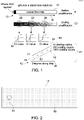
- FIG. 1 is a representation of a quantitative amplification technique used in conjunction with a solid amplification substrate, e.g., a porous substrate.
- a porous substrate 10 may be provided in the form of a lateral flow strip.
- a DNA-containing sample or other nucleic acid sample
- location 12 amplification begins.
- the amplified nucleic acid products flow along arrow 16 and may be detected at a plurality of locations (e.g., locations 22, 24, and 26) at any suitable time point. Based on the magnitude of a detected signal related to an amount of amplified product and the reaction time, the starting copy number may be assessed.
- a starting copy number of a sequence of interest may further be related to particular disease or clinical conditions.
- an estimated starting copy number above a predetermined threshold may be associated with bacterial or viral infection, cancer diagnosis, presence of a biomarker or sequence of interest in forensic or genotyping assessments, etc.
- an estimated starting copy number above a predetermined threshold may be used to determine if an environmental sample is clean or contaminated, e.g., a presence of a non-zero or threshold starting copy number of one or more bacteria types may be indicative of contamination.
- an estimated starting copy number above a predetermined threshold may be used to determine if a food sample is accurately identified and/or is contaminated. For example, a presence of a particular gene (identified by a non-zero or threshold starting copy number) may be used to determine the species associated with a sample.
- the detected amplification signal may be correlated to starting copy number by correlating the detected signal to a cycle threshold vs. distance for a known sample quantity.
- the cycle threshold (Ct) is the number of amplification cycles required for a detected signal to cross a predefined threshold. Or, for isothermal amplification, the amount of time required for a detected signal to cross a predefined threshold. Ct is inversely proportional to the amount of target nucleic acid in the sample.
- Plot 32 is an example of a Ct plot for the time point 22.
- Plot 42 is a plot of the Ct 44 vs. distance along the strip 46. Each of the plotted lines 48, 50, and 52 correlates to a particular starting copy number.
- a sample with an unknown starting copy number may be assessed as provided herein.
- the detected signal may be plotted using the distance on the porous substrate 10 at which the signal crossed a predetermined threshold and based on a fit to data correlating to a reference sample.
- the detected signal may also be plotted using the time at which the signal crossed a predetermined threshold at a predetermined distance on the porous substrate 10 and based on a slope and fit to data correlating to a reference sample. Further, the plot may include detected signal at a number of locations along the porous substrate 10.
- FIG. 2 is a view of implementation of a porous substrate 10 in a rectangular strip form.
- the porous substrate 10 may be implemented in any suitable size and shape to facilitate amplification along the porous substrate 10.
- the porous substrate 10 may be implemented to assess flow along one axis or along multiple axes (e.g., in a star formation).
- the porous substrate 10 may be formed from any appropriate solid porous material, including paper (e.g., Whatman filter paper), cellulose, porous polymer sheets, glass fibers, quartz fibers, nitrocellulose membranes, etc.
- a pore size may be selected based on a predicted size of a desired amplification product.
- the pore size may be 1-3 microns.
- the porous substrate 10 may also be formed from a material that is neutral to negatively charged nucleic acids. That is, the material may be selected such that the amplified nucleic acids do not bind or significantly interact with the material of the porous substrate 10.
- the porosity may be characterized by the fraction of the volume of voids as over the total volume, e.g., between 0.1 and 1, or 0.25 to 1, where a porosity of 1 is an open channel. Accordingly, the porous substrate 10 may include materials with different areas of porosity as well as materials with open channels.
- the porous substrate 10 includes amplification reagents 60 distributed throughout the porous substrate 10.
- an initial location 64 which may be designated by an index or marker printed or otherwise indicated on the porous substrate 10
- the reaction expands outwards, consuming reagents as it progresses.
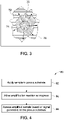
- FIG. 3 is a schematic representation of such reaction progression.
- the sample 70 produces amplified products outward from the application site (e.g. initial location 64) along arrows 72. Over time, a first wavefront 74 forms, and continues to diffuse from the application point.
- DNA products, H+ ions, pyrophosphate and dyes may be released while dNTPs and primers are consumed.
- the amplified product reaches the edges of the porous substrate 10 in many directions in a short time after the reaction starts and after some initial time will only have a net diffusion along the longest axis of the strip.
- the initial location 64 may be positioned in a center of a porous substrate 10, which in turn may extend outwardly from the initial location 64 to form a circle or other shape.
- the porous substrate 10 may be implemented to test multiple different samples on a single substrate.
- the porous substrate 10 may include any number if initial location 64, which may be spaced apart from one another to avoid mixing of multiple reaction fronts.
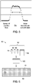
- FIG. 4 is a flow diagram of a method 80 of assessing amplification using a porous substrate 10.
- the amplification reaction begins as the nucleic acids contact the appropriate reagents and, in certain embodiments, when other appropriate environmental conditions are present (e.g., activation temperatures for particular reagents).
- the reaction is allowed to progress for an appropriate amount of time at step 84, and, at one or more selected time points, the amplified sample is assessed based on the position of the signal that is generated by the amplification reaction at step 86.
- There are multiple ways to measure output from the system including time-to-distance and distance-at-time. In the first, distance is set and time is measured. In the second, time is set and distance is measured.
- the distance for a set time of the reaction front and the production of amplified products are related to the initial starting copy number of the sequence of interest, as shown in the plot of FIG. 5 .
- the porous substrate 10 is generally provided wet or saturated with liquid, the movement of the reaction front is not a wicking or capillary action movement across a dry substrate, but instead is a diffusion of amplified product.
- the diffusion rate is primarily driven by the continual production of new amplification products from the old ones and, therefore, the movement of the reaction product through the porous substrate 10 is reaction-diffusion, not simply diffusion. That is, because movement is driven by reaction diffusion, the velocity of the reaction front is independent of the starting concentration.
- the starting concentration determines the "lag time" before the velocity front starts to move.
- the slope of distance over time is not simply proportional to concentration.
- samples with a known starting copy number may be assessed to determine a characteristic reaction time to distance or distance per time for a reaction in the porous substrate 10.
- the amplification reagents are distributed generally evenly throughout the porous substrate and in non-limiting concentrations to permit the reaction to progress without reagent bottlenecks as the number of amplified products increases.
- the porous substrate 10 may be provided with or without the amplification reagents 60 in an amplification kit 90 as shown in FIG. 6 .
- the amplification reagents 60 may include one or more primers 92, i.e., oligonucleotides complementary to a target sequence of interest and/or capable of hybridizing to a target sequence of interest. Sequences of interest may include sequences associated with particular pathogens or particular diseases. Further, the primers may include multiple primers specific complementary to different regions of a single desired sequence.
- the amplification reagents 60 may also include one or more polymerases 94 and nucleotides 96 as well as one or more appropriate buffers 98.
- the amplification reagents 60 may be selected to work in conjunction with a particular amplification technique (e.g., loop-mediated isothermal amplification, strand displacement amplification, helicase dependent amplification, nicking enzyme amplification reaction).
- amplification technique e.g., loop-mediated isothermal amplification, strand displacement amplification, helicase dependent amplification, nicking enzyme amplification reaction.
- loop-mediated amplification 4-6 primers are used that hybridize to 6-8 regions of the target sequence.
- the porous substrate 10 may be provided with the amplification reagents 60 distributed throughout the body of the substrate.
- the porous substrate 10 may be encapsulated in a packaging that is removed prior to use or coating that prevents evaporation of the amplification reagents 60 during storage.
- such a coating may cover the porous substrate with the exception of an opening permitting sample application.
- the opening may be covered by a removable release liner that is removed to permit the user to apply a sample.
- such coatings may be optically transparent or otherwise permeable to the signal generated by the amplification products.
- the porous substrate 10 may be provided with the amplification reagents dried on the porous substrate 10. An end user may wet the porous substrate 10 prior to use by soaking the porous substrate 10 in an appropriate buffer.
- the porous substrate 10 may be provided with separate amplification reagents 60 that the end user may apply to the porous substrate 10.
- the porous substrate 10 may be provided without any encapsulating coating that may interfere with even application of the amplification reagents 60.
- the amplification reagents 60 may be first mixed together in a single solution 100 prior to application on the porous substrate 10 rather than being applied separately.
- the amplification reagents 60 may be provided as separate components or as a premixed solution 100 as part of the amplification kit 90.
- the primers 92 may be provided as part of the amplification kit 90. That is, the amplification kit 90 may be provided with all of the amplification reagents, including primers 92 for a particular target sequence. An end user may purchase the amplification kit 90 that corresponds with a particular disease or clinical condition. In other applications, the primers 92 may be provided separately or provided by the end user.
- the porous substrate 10 may be used to assess any target sequence desired by the end user. To that end, the end user may select an appropriate primer set for application to the porous substrate. In such embodiments, the end user may then apply the desired primers 92 to the porous substrate along with the other amplification reagents 60.
- the amplified products of the reaction may be detected by measuring signals from detectable moieties generated by the amplification.
- detection may involve measuring the concentration of one of the reaction products directly (e.g. amplicons, H+, pyrophosphate, etc) or by measuring something released from the nucleotides upon incorporation into the amplified DNA (etc a dye). This information can be used to track the movement of the reaction/diffusion gradient front.
- the amplification reagents 60 may include one or more appropriate signal molecules that are capable of providing a detectable signal using one or more detection techniques (e.g., spectrometry, calorimetry, spectroscopy, visual inspection, or any other detection method).
- a detectable signal may include a visible signal, an optical signal, an electrical signal, an electrochemical signal, or a radioactive signal.
- DDAO labeled deoxynucleoside tetraphosphates are utilized to perform quantitative measurements of amplification.
- nucleotide substrates are radiolabeled or have attached thereto some other type of label or dye, radioactive isotopes, fluorescent molecules, phosphorescent molecules, enzymes, antibodies, probes, stains, and ligands.
- detection labels that can be incorporated into amplified DNA include nucleotide analogs and nucleotides modified with biotin or with suitable haptens such as digoxygenin. It should be understood that one or more of the provided nucleotides 94 may be labeled. In a specific example, as shown in FIG. 7 , the nucleotides 94 may be labeled with a signal molecule such as a dye. When the nucleotides 94 are incorporated into an amplification product, the dye is released from the nucleotide to yield a dye-labeled tri-phosphate, which in turn is liberated after reaction with a phosphatase to yield liberated dye.
- a signal molecule such as a dye
- the dye may be detected once liberated depending on the characteristics of the dye (e.g. via optical, visible, and/or electrical techniques).
- signal molecules may be assessed via pyrophosphate detection, electrochemical detection of H+, intercalating electrochemical probes, redox active dyes, pyrophosphate, intercalating dyes, or nucleotide dyes.
- signal molecules examples include, for example, a chromophore, a fluorophore, a Raman-active tag, a radioactive label, an enzyme, an enzyme substrate, or combinations thereof.
- the signal molecule and the nucleotides 94 may be present in a single entity (e.g., a target binding protein with a fluorescent label or radiolabel).
- the nucleotides 94 and signal molecules are discrete entities that associate with each other prior to or upon introduction to the porous substrate 10.
- a signal molecule may provide a characteristic signal following interaction with an energy source or a current, such as an electromagnetic radiation source or a fluorescence excitation source.
- Electromagnetic radiation source may be capable of providing electromagnetic energy of any wavelength including visible, infrared, and ultraviolet. Electromagnetic radiation may be in the form of a direct light source or may be emitted by a light emissive compound such as a donor fluorophore.
- a fluorescence excitation source may be capable of making a source fluoresce or may give rise to photonic emissions (that is, electromagnetic radiation, directed electric field, temperature, physical contact, or mechanical disruption).
- Suitable signal generators may provide a signal capable of being detected by a variety of methods including optical measurements (for example, fluorescence), electrochemical signal, electrical conductivity, or radioactivity. Suitable signal molecules may be, for example, light emitting, energy accepting, fluorescing, radioactive, redox active, or quenching.
- FIG. 8 is an example of a porous substrate 10 including a plurality of assessment locations 110 spaced apart along an axis and from the initial location 64.
- the locations 110 may be indicated by pre-printed markers.
- an unfilled location 112 corresponds to amplification below a threshold while a filled location 114 corresponds to levels above a threshold.
- FIG. 9 shows an assessment using two such porous substrates 10, each with a different amount of starting material.
- the test 116 shows three filled marker locations 120 and six unfilled locations 122.
- the higher concentration test 118 after the same amount of time (i.e., time x), eight locations 124 are filled while one location 126 is unfilled.
- the number of filled and unfilled locations at time x may be correlated to a particular starting concentration.
- the amplification kit 90 may be provided with comparison graphs and tables to indicate an estimated concentration at time point x with all possible combinations of filled/unfilled locations 110.
- the porous substrate 10 may provide a rapid and easy to use assessment of not only a presence of a particular target sequence, but also an estimated amount, i.e., a quantitative assessment of nucleic acids in the initial sample. Further, such a technique may also be implemented with automatic detection.
- the detector is binary rather than having to be calibrated for each testing instance.
- the dynamic range of the detector is determined by the number of detection points and their distance apart rather than an inherent range of the detector itself. In this manner, a detector that is even binary in range may used in conjunction with distance and time information to be configured to provide a wide dynamic range.
- FIG. 10 shows a comparison between gradients formed with a porous substrate 10 vs. a solution-based technique.
- Genomic DNA of various concentrations was assessed at various concentrations and at multiple time points.
- a gradient is formed in a paper substrate when comparing the size of the detected spots for a particular concentration over time while the solution-based technique does not form such a gradient. For example, for the 22ng sample, the gradient continues to grow over the time points, indicating that a heterogeneous gradient is formed.
- a desired test may be calibrated by determining in which concentration ranges and at what time points a gradient is formed.
- an experiment was performed with a dried cellulose substrate whose surface was previously blocked with bactopeptone.
- the substrate was saturated with amplification reagent mix for DNA amplification (IsoAmp III Universal tHDA kit from Biohelix).
- the mix included all components (enzymes, buffers, nucleotides, primers, etc) excluding DNA template.
- the primers were designed for mycobacterium tuberculosis (TB) template. Purified TB DNA was added to position 0. Two different DNA amounts were used: 5ng or 500pg.
- the substrate was incubated at 65°C for various times. After 8 or 24 hours of incubation, the substrate was removed from incubator and cut into 2-3mm segments. The fluid was extracted from the segments via centrifugation and analyzed on a 15% TBE-Urea polycrylamide gel using gel electrophoresis. The concentration of amplified DNA in each segment was determined from gel image.
- Table 1 shows a farthest distance at which at least a minimum amount of DNA is detected (e.g., detection above a threshold).
- Table 1 Starting amount of TBDNA Time Farthest distance where at least 0.08ng/ ⁇ L was detected 5ng 8hr 5mm 500pg 8hr 2mm 5ng 24hr 9mm 500pg 24hr 5mm
- FIG. 11 is a plot of predicted threshold value at time (e.g., cycles) vs. distance on a porous substrate 10 from sample application for four different starting copy numbers represented by lines 130, 132, 134, and 136.
- the line of the highest concentration of starting copy, line 136, reaches the threshold sooner than the other samples.
- FIG. 12 is 2D simulation data showing areas above the threshold as a function of time for a sample applied to a porous substrate 10 as provided.
- the sample at 142 is above threshold and, as time progresses (e.g., shown in panels 144, 148, and 152), the area above the threshold (e.g., areas 146, 148, and 154) moves along the porous substrate 10.
- An additional advantage of the diffusion-based technique disclosed herein is that diffusion of the amplified products is related to reaction time for amplification. In contrast, any sample contaminants diffuse independently of amplification and ahead of the reaction front. Accordingly, the disclosed techniques may be used with relatively heterogeneous or complex samples, such as unprocessed bodily fluids.
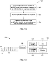
- FIG. 13 is a flow diagram of a method 156 for calibrating a nucleic acid quantification.
- a calibration may be performed by applying a known sample (e.g., with a known concentration or copy number of a target sequence) to an initial location on a porous substrate and the amplification reaction is allowed to progress for at least one time point at step 158.
- a signal at a plurality of distances and at a plurality of time points is detected at step 159.
- a user may determine a concentration of an unknown sample.
- a time to reach a particular distance may also be correlated to a starting copy number.
- the calibration method 156 is performed for multiple concentrations to provide additional calibration data.
- Such calibration data may be stored in a processor-based device so that quantification is performed automatically.
- the techniques disclosed herein may be used in conjunction with a system 160 that includes one or more sensors or detectors 162 for detecting the signal generated by the amplification reaction and coupled to an assessment device 164.
- the detector 162 may be configured to detect one or more signal molecules.
- the detector 162 may be configured to detect optical or electrochemical signals.
- the porous strip 10 may couple to the detector 162 to facilitate detection at one or more locations 110 along the substrate 10.
- the system 160 may also include elements for heating the porous strip to activate amplification.
- the amplification reagents 60 may be configured to react at particular temperatures or using particular temperature cycles. Accordingly, the system 160 may be configured to provide cyclical or isothermal heating to the porous substrate 10.
- the system 160 may also include computer-readable memory circuitry 166, such as magnetic, electronic, or optical storage media, for storing programs and routines executed by the device 164.
- the stored programs or routines may include programs or instructions for performing all or part of the present techniques.
- the system 160 may also include processing circuitry 168 for executing the programs or instructions.
- the device 164 may also include various input/output (I/O) interfaces 172, as well as various network or communication interfaces to allow communication with user interface devices, such as a display 170, that may be used for viewing and inputting quantification information.
- the various network and communication interfaces may allow connection to both local and wide area intranets and storage networks as well as the Internet.
- the various I/O and communication interfaces may utilize wires, lines, or suitable wireless interfaces, as appropriate or desired.
Landscapes
- Chemical & Material Sciences (AREA)
- Life Sciences & Earth Sciences (AREA)
- Organic Chemistry (AREA)
- Engineering & Computer Science (AREA)
- Zoology (AREA)
- Wood Science & Technology (AREA)
- Proteomics, Peptides & Aminoacids (AREA)
- Health & Medical Sciences (AREA)
- Biophysics (AREA)
- General Engineering & Computer Science (AREA)
- Immunology (AREA)
- Microbiology (AREA)
- Molecular Biology (AREA)
- Analytical Chemistry (AREA)
- Physics & Mathematics (AREA)
- Genetics & Genomics (AREA)
- Biochemistry (AREA)
- Bioinformatics & Cheminformatics (AREA)
- Biotechnology (AREA)
- General Health & Medical Sciences (AREA)
- Chemical Kinetics & Catalysis (AREA)
- Apparatus Associated With Microorganisms And Enzymes (AREA)
- Measuring Or Testing Involving Enzymes Or Micro-Organisms (AREA)
Claims (10)
- Procédé d'évaluation d'amplification d'acides nucléiques comprenant :la fourniture d'un substrat poreux comprenant une pluralité de réactifs d'amplification répartis à travers le substrat poreux, dans lequel lesdits réactifs d'amplification comprennent : a) des réactifs d'amplification d'acides nucléiques isotherme ; b) de l'ADN polymérase et une ou plusieurs amorces d'acide nucléique ; ou c) de l'ARN polymérase, et ledit substrat poreux est formé à partir d'une matière poreuse solide sélectionnée parmi le papier, la cellulose, des feuilles polymères poreuses, des fibres de verre, des fibres de quartz et des membranes de nitrocellulose, et dans lequel ledit substrat poreux est fourni humide ou saturé d'un liquide dans lequel ledit liquide est un tampon approprié ;l'application d'un échantillon comprenant des acides nucléiques en un emplacement initial prédéterminé sur le substrat poreux ;le fait de permettre à l'échantillon de réagir avec les réactifs d'amplification sur le substrat poreux pendant une certaine durée pour générer des produits d'amplification d'une séquence d'acides nucléiques d'intérêt, dans lequel les produits d'amplification se déplacent à l'intérieur du substrat poreux par réaction-diffusion et comprennent un ou plusieurs fragments détectables configurés pour fournir un signal lorsque les produits d'amplification sont générés pendant l'amplification ; etla détermination d'une concentration initiale de l'échantillon sur la base d'une progression en fonction du temps du signal à l'intérieur du substrat sur la base d'une correspondance à des données en corrélation avec un échantillon de référence, dans lequel ladite détermination comprenda) le traçage du signal en utilisant la distance sur le substrat poreux à laquelle ledit signal franchit un seuil prédéterminé et sur la base d'une correspondance à des données en corrélation avec l'échantillon de référence ; oub) le traçage du signal en utilisant l'instant où ledit signal franchit un seuil prédéterminé à une distance prédéterminée sur le substrat poreux et sur la base d'une pente et d'une correspondance à des données en corrélation avec l'échantillon de référence.
- Procédé selon la revendication 1, dans lequel le substrat poreux :a) comprend un marqueur imprimé correspondant à l'emplacement initial et une pluralité de marqueurs imprimés correspondant à différentes distances par rapport à l'emplacement initial ;b) est saturé en liquide ;c) présente une porosité non uniforme ; oud) comprend un canal ouvert avec une porosité de un.
- Procédé selon la revendication 1, comprenant la détermination que l'échantillon ne contient pas une quantité seuil de la séquence d'intérêt si le signal détecté n'atteint pas une distance minimale par rapport à l'emplacement initial.
- Procédé selon la revendication 1, dans lequel la détermination de la concentration initiale d'une cible dans l'échantillon comprend la détermination d'un nombre de copies de départ de la séquence d'acides nucléiques d'intérêt qui est complémentaire à au moins une amorce dans les réactifs d'amplification.
- Procédé selon la revendication 1, comprenant la détermination d'une caractéristique clinique d'un patient chez qui l'échantillon a été prélevé sur la base de la concentration initiale de l'échantillon, dans lequel la caractéristique clinique est associée à la présence ou à l'absence de la séquence d'acides nucléiques d'intérêt et dans lequel les réactifs d'amplification comprennent une ou plusieurs amorces complémentaires à la séquence d'acides nucléiques d'intérêt.
- Procédé selon la revendication 1, dans lequel la fourniture du substrat poreux comprend l'application des réactifs d'amplification au substrat poreux dans une seule solution.
- Procédé selon la revendication 1, comprenant le chauffage du substrat poreux pour activer un ou plusieurs des réactifs d'amplification, dans lequel éventuellement le chauffage atteint une température isotherme.
- Procédé selon la revendication 1, dans lequel le signal :a) comprend un signal visible et/ou un signal de fluorescence ;b) est généré par libération d'un colorant ; ou
dc est généré par un ou plusieurs parmi des amplicons, H+, ou du pyrophosphate. - Procédé selon la revendication 1, dans lequel :a) une intensité de signal à une distance définie par rapport à l'emplacement initial est en corrélation avec une quantité de départ d'un analyste cible dans l'échantillon ; oub) la présence ou l'absence d'un signal à une distance définie par rapport à l'emplacement initial est utilisée pour calculer une quantité de départ d'un analyte cible dans l'échantillon.
- Procédé d'étalonnage d'une quantification d'acide nucléique comprenant :la fourniture d'un substrat poreux comprenant une pluralité de réactifs d'amplification répartis à travers le substrat poreux, dans lequel lesdits réactifs d'amplification comprennent : a) des réactifs d'amplification d'acides nucléiques isotherme ; b) de l'ADN polymérase et une ou plusieurs amorces d'acide nucléique ; ou c) de l'ARN polymérase, et dans lequel lesdits réactifs d'amplification comprennent une ou plusieurs molécules signal configurées pour fournir un signal pendant l'amplification et une ou plusieurs amorces complémentaires à une séquence d'acides nucléiques cible ; et dans lequel ledit substrat poreux est fourni humide ou saturé d'un liquide dans lequel ledit liquide est un tampon approprié ; etl'application d'un échantillon connu comprenant un nombre de copies connu de la séquence d'acides nucléiques cible en un emplacement initial sur le substrat poreux ; dans lequel les produits d'amplification se déplacent à l'intérieur du substrat poreux par réaction-diffusion ;
etla détection du signal à une pluralité de distances et à une pluralité d'instants pour générer des données d'étalonnagedans lequel ledit substrat poreux est formé à partir d'une matière poreuse solide sélectionnée parmi le papier, la cellulose, des feuilles polymères poreuses, des fibres de verre, des fibres de quartz et des membranes de nitrocellulose.
Applications Claiming Priority (2)
| Application Number | Priority Date | Filing Date | Title |
|---|---|---|---|
| US13/970,315 US9714447B2 (en) | 2013-08-19 | 2013-08-19 | Detection of nucleic acid amplification in a porous substrate |
| PCT/EP2014/067685 WO2015024948A1 (fr) | 2013-08-19 | 2014-08-19 | Détection de l'amplification d'acide nucléique dans un substrat poreux |
Publications (2)
| Publication Number | Publication Date |
|---|---|
| EP3036341A1 EP3036341A1 (fr) | 2016-06-29 |
| EP3036341B1 true EP3036341B1 (fr) | 2022-06-15 |
Family
ID=51383726
Family Applications (1)
| Application Number | Title | Priority Date | Filing Date |
|---|---|---|---|
| EP14753074.5A Active EP3036341B1 (fr) | 2013-08-19 | 2014-08-19 | Détection de l'amplification d'acide nucléique dans un substrat poreux |
Country Status (4)
| Country | Link |
|---|---|
| US (1) | US9714447B2 (fr) |
| EP (1) | EP3036341B1 (fr) |
| JP (1) | JP6374967B2 (fr) |
| WO (1) | WO2015024948A1 (fr) |
Families Citing this family (4)
| Publication number | Priority date | Publication date | Assignee | Title |
|---|---|---|---|---|
| DE102015115836A1 (de) * | 2015-09-18 | 2017-03-23 | Biotype Diagnostic Gmbh | Bestätigungstest für primäre Nukleinsäure-Amplifikate in einem kontinuierlichen Reaktionsansatz und unmittelbare Auswertung mittels immunchromatographischer Verfahren |
| WO2018069283A1 (fr) * | 2016-10-10 | 2018-04-19 | MAX-PLANCK-Gesellschaft zur Förderung der Wissenschaften e.V. | Procédé de détermination à haute résolution spatiale de l'emplacement d'une molécule individuelle, pouvant être excitée par une lumière d'excitation servant à l'émission de lumière luminescente, dans un échantillon |
| KR102082898B1 (ko) * | 2018-04-18 | 2020-02-28 | 가천대학교 산학협력단 | 병원균 검출을 위한 비색 검출 장치 및 그의 제조방법 |
| US11732315B2 (en) * | 2020-03-12 | 2023-08-22 | New England Biolabs, Inc. | Rapid diagnostic test for lamp |
Family Cites Families (15)
| Publication number | Priority date | Publication date | Assignee | Title |
|---|---|---|---|---|
| US6001568A (en) * | 1992-10-26 | 1999-12-14 | Institut Belka | Solid medium for amplification and expression of nucleic acids as colonies |
| US6153425A (en) * | 1995-07-13 | 2000-11-28 | Xtrana, Inc. | Self-contained device integrating nucleic acid extraction, amplification and detection |
| US5853990A (en) | 1996-07-26 | 1998-12-29 | Edward E. Winger | Real time homogeneous nucleotide assay |
| US6387621B1 (en) | 1999-04-27 | 2002-05-14 | University Of Utah Research Foundation | Automated analysis of real-time nucleic acid amplification |
| AUPQ495700A0 (en) | 2000-01-05 | 2000-02-03 | Johnson & Johnson Research Pty. Limited | Method for concurrent amplification and real time detection of polymorphic nucleic acid sequences |
| AU2003267065A1 (en) | 2002-09-02 | 2004-03-19 | Pamgene B.V. | Novel integrated microarray analysis |
| ES2363501T3 (es) | 2003-05-07 | 2011-08-05 | Coris Bioconcept Sprl | Dispositivo oligocromático de un paso y procedimiento de uso. |
| CN105087777A (zh) | 2004-04-07 | 2015-11-25 | 安克塞斯生物公司 | 核酸检测系统 |
| ES2401879T3 (es) * | 2004-08-31 | 2013-04-25 | Eiken Kagaku Kabushiki Kaisha | Método de análisis de ácidos nucleicos |
| WO2006122311A2 (fr) * | 2005-05-11 | 2006-11-16 | The Trustees Of The University Of Pennsylvania | Puce microfluidique |
| EP1991695A1 (fr) * | 2006-03-01 | 2008-11-19 | Roche Diagnostics GmbH | Substrat pour amplification d'acides nucléiques |
| EP2054526A2 (fr) | 2006-08-14 | 2009-05-06 | Koninklijke Philips Electronics N.V. | Surveillance de procédés enzymatiques à l'aide d'objets magnétisables ou magnétiques comme marqueurs |
| DE102007029772B4 (de) | 2007-06-22 | 2011-12-08 | Aj Innuscreen Gmbh | Verfahren und Schnelltest zum Nachweis spezifischer Nukleinsäuresequenzen |
| DE102007062441A1 (de) * | 2007-12-20 | 2009-06-25 | Aj Innuscreen Gmbh | Mobiles Schnelltestsystem für die Nukleinsäureanalytik |
| ITMI20112177A1 (it) | 2011-11-29 | 2013-05-30 | Genefast S R L | Metodo per rilevare la sintesi e/o l'amplificazione di un acido nucleico |
-
2013
- 2013-08-19 US US13/970,315 patent/US9714447B2/en active Active
-
2014
- 2014-08-19 EP EP14753074.5A patent/EP3036341B1/fr active Active
- 2014-08-19 JP JP2016535465A patent/JP6374967B2/ja active Active
- 2014-08-19 WO PCT/EP2014/067685 patent/WO2015024948A1/fr active Application Filing
Also Published As
| Publication number | Publication date |
|---|---|
| WO2015024948A1 (fr) | 2015-02-26 |
| JP6374967B2 (ja) | 2018-08-15 |
| EP3036341A1 (fr) | 2016-06-29 |
| US9714447B2 (en) | 2017-07-25 |
| JP2016533746A (ja) | 2016-11-04 |
| US20150050654A1 (en) | 2015-02-19 |
Similar Documents
| Publication | Publication Date | Title |
|---|---|---|
| Zheng et al. | Lateral flow test for visual detection of multiple MicroRNAs | |
| Lucena-Aguilar et al. | DNA source selection for downstream applications based on DNA quality indicators analysis | |
| US20210010079A1 (en) | Spatial molecular analysis of tissue | |
| US20130210013A1 (en) | Real time gene expression profiling | |
| TWI527905B (zh) | 透過利用基因檢測技術與微珠微流道結合進行單核苷酸多態性檢測之方法 | |
| ATE307903T1 (de) | Immunologischer nachweis von rna:dna hybriden auf mikroarrays | |
| US10961570B2 (en) | High-throughput and rapid nucleic acids detection method based on capillary microarrays | |
| EP3036341B1 (fr) | Détection de l'amplification d'acide nucléique dans un substrat poreux | |
| US20120183965A1 (en) | Nucleic acid detection | |
| CN104508146A (zh) | 用于快速检测扩增的核苷酸序列的方法和设备 | |
| ATE82773T1 (de) | Diagnostischer test unter verwendung von nucleinsaeure-sonden. | |
| US20170023555A1 (en) | Devices and kits for measuring biological results | |
| Kirchner et al. | mRNA and microRNA purity and integrity: the key to success in expression profiling | |
| CN108291251A (zh) | 用于核酸分析的系统和方法 | |
| US20100069253A1 (en) | Impedance Spectroscopy Measurement of DNA | |
| Zhao et al. | Sensitive and selective label-free alkaline phosphatase detection based on DNA hairpin probe | |
| Shukla et al. | Multiplexed detection and quantitation of extracellular vesicle RNA expression using nanostring | |
| Kim et al. | Microchip-based capillary electrophoretic analysis of telomerase activity for cancer diagnostics | |
| CN102937613B (zh) | 一种基于亚甲基蓝指示剂定量检测pcr的电化学安培检测法 | |
| Hsu et al. | The portable fluorescence detection system matched with PDMS microfluidic biochip for DNA hybridization detection | |
| JP3705499B2 (ja) | ターゲット核酸断片の分析方法及びターゲット核酸断片の分析キット | |
| JP3532907B2 (ja) | ターゲット核酸断片の分析方法及びターゲット核酸断片の分析キット | |
| WO2006031061A1 (fr) | Procede de selection de sonde du gene fret utilisant le criblage par pcr | |
| Lin et al. | Automatic analysis for biochip based on macromolecular compound material | |
| JP5601746B2 (ja) | 同一の試料を用いた二段階核酸検査方法 |
Legal Events
| Date | Code | Title | Description |
|---|---|---|---|
| PUAI | Public reference made under article 153(3) epc to a published international application that has entered the european phase |
Free format text: ORIGINAL CODE: 0009012 |
|
| 17P | Request for examination filed |
Effective date: 20160120 |
|
| AK | Designated contracting states |
Kind code of ref document: A1 Designated state(s): AL AT BE BG CH CY CZ DE DK EE ES FI FR GB GR HR HU IE IS IT LI LT LU LV MC MK MT NL NO PL PT RO RS SE SI SK SM TR |
|
| AX | Request for extension of the european patent |
Extension state: BA ME |
|
| DAX | Request for extension of the european patent (deleted) | ||
| STAA | Information on the status of an ep patent application or granted ep patent |
Free format text: STATUS: EXAMINATION IS IN PROGRESS |
|
| 17Q | First examination report despatched |
Effective date: 20170321 |
|
| RAP1 | Party data changed (applicant data changed or rights of an application transferred) |
Owner name: GLOBAL LIFE SCIENCES SOLUTIONS OPERATIONS UK LTD |
|
| STAA | Information on the status of an ep patent application or granted ep patent |
Free format text: STATUS: EXAMINATION IS IN PROGRESS |
|
| STAA | Information on the status of an ep patent application or granted ep patent |
Free format text: STATUS: EXAMINATION IS IN PROGRESS |
|
| REG | Reference to a national code |
Ref country code: DE Ref legal event code: R079 Ref document number: 602014084028 Country of ref document: DE Free format text: PREVIOUS MAIN CLASS: C12Q0001680000 Ipc: C12Q0001684400 |
|
| GRAP | Despatch of communication of intention to grant a patent |
Free format text: ORIGINAL CODE: EPIDOSNIGR1 |
|
| STAA | Information on the status of an ep patent application or granted ep patent |
Free format text: STATUS: GRANT OF PATENT IS INTENDED |
|
| RIC1 | Information provided on ipc code assigned before grant |
Ipc: C12Q 1/6816 20180101ALI20220224BHEP Ipc: C12Q 1/6851 20180101ALI20220224BHEP Ipc: C12Q 1/6844 20180101AFI20220224BHEP |
|
| INTG | Intention to grant announced |
Effective date: 20220317 |
|
| GRAS | Grant fee paid |
Free format text: ORIGINAL CODE: EPIDOSNIGR3 |
|
| GRAA | (expected) grant |
Free format text: ORIGINAL CODE: 0009210 |
|
| STAA | Information on the status of an ep patent application or granted ep patent |
Free format text: STATUS: THE PATENT HAS BEEN GRANTED |
|
| AK | Designated contracting states |
Kind code of ref document: B1 Designated state(s): AL AT BE BG CH CY CZ DE DK EE ES FI FR GB GR HR HU IE IS IT LI LT LU LV MC MK MT NL NO PL PT RO RS SE SI SK SM TR |
|
| REG | Reference to a national code |
Ref country code: CH Ref legal event code: EP Ref country code: GB Ref legal event code: FG4D |
|
| REG | Reference to a national code |
Ref country code: IE Ref legal event code: FG4D |
|
| REG | Reference to a national code |
Ref country code: DE Ref legal event code: R096 Ref document number: 602014084028 Country of ref document: DE |
|
| REG | Reference to a national code |
Ref country code: AT Ref legal event code: REF Ref document number: 1498446 Country of ref document: AT Kind code of ref document: T Effective date: 20220715 |
|
| REG | Reference to a national code |
Ref country code: LT Ref legal event code: MG9D |
|
| REG | Reference to a national code |
Ref country code: NL Ref legal event code: MP Effective date: 20220615 |
|
| PG25 | Lapsed in a contracting state [announced via postgrant information from national office to epo] |
Ref country code: SE Free format text: LAPSE BECAUSE OF FAILURE TO SUBMIT A TRANSLATION OF THE DESCRIPTION OR TO PAY THE FEE WITHIN THE PRESCRIBED TIME-LIMIT Effective date: 20220615 Ref country code: NO Free format text: LAPSE BECAUSE OF FAILURE TO SUBMIT A TRANSLATION OF THE DESCRIPTION OR TO PAY THE FEE WITHIN THE PRESCRIBED TIME-LIMIT Effective date: 20220915 Ref country code: LT Free format text: LAPSE BECAUSE OF FAILURE TO SUBMIT A TRANSLATION OF THE DESCRIPTION OR TO PAY THE FEE WITHIN THE PRESCRIBED TIME-LIMIT Effective date: 20220615 Ref country code: HR Free format text: LAPSE BECAUSE OF FAILURE TO SUBMIT A TRANSLATION OF THE DESCRIPTION OR TO PAY THE FEE WITHIN THE PRESCRIBED TIME-LIMIT Effective date: 20220615 Ref country code: GR Free format text: LAPSE BECAUSE OF FAILURE TO SUBMIT A TRANSLATION OF THE DESCRIPTION OR TO PAY THE FEE WITHIN THE PRESCRIBED TIME-LIMIT Effective date: 20220916 Ref country code: FI Free format text: LAPSE BECAUSE OF FAILURE TO SUBMIT A TRANSLATION OF THE DESCRIPTION OR TO PAY THE FEE WITHIN THE PRESCRIBED TIME-LIMIT Effective date: 20220615 Ref country code: BG Free format text: LAPSE BECAUSE OF FAILURE TO SUBMIT A TRANSLATION OF THE DESCRIPTION OR TO PAY THE FEE WITHIN THE PRESCRIBED TIME-LIMIT Effective date: 20220915 |
|
| REG | Reference to a national code |
Ref country code: AT Ref legal event code: MK05 Ref document number: 1498446 Country of ref document: AT Kind code of ref document: T Effective date: 20220615 |
|
| PG25 | Lapsed in a contracting state [announced via postgrant information from national office to epo] |
Ref country code: RS Free format text: LAPSE BECAUSE OF FAILURE TO SUBMIT A TRANSLATION OF THE DESCRIPTION OR TO PAY THE FEE WITHIN THE PRESCRIBED TIME-LIMIT Effective date: 20220615 Ref country code: LV Free format text: LAPSE BECAUSE OF FAILURE TO SUBMIT A TRANSLATION OF THE DESCRIPTION OR TO PAY THE FEE WITHIN THE PRESCRIBED TIME-LIMIT Effective date: 20220615 |
|
| PG25 | Lapsed in a contracting state [announced via postgrant information from national office to epo] |
Ref country code: NL Free format text: LAPSE BECAUSE OF FAILURE TO SUBMIT A TRANSLATION OF THE DESCRIPTION OR TO PAY THE FEE WITHIN THE PRESCRIBED TIME-LIMIT Effective date: 20220615 |
|
| PG25 | Lapsed in a contracting state [announced via postgrant information from national office to epo] |
Ref country code: SM Free format text: LAPSE BECAUSE OF FAILURE TO SUBMIT A TRANSLATION OF THE DESCRIPTION OR TO PAY THE FEE WITHIN THE PRESCRIBED TIME-LIMIT Effective date: 20220615 Ref country code: SK Free format text: LAPSE BECAUSE OF FAILURE TO SUBMIT A TRANSLATION OF THE DESCRIPTION OR TO PAY THE FEE WITHIN THE PRESCRIBED TIME-LIMIT Effective date: 20220615 Ref country code: RO Free format text: LAPSE BECAUSE OF FAILURE TO SUBMIT A TRANSLATION OF THE DESCRIPTION OR TO PAY THE FEE WITHIN THE PRESCRIBED TIME-LIMIT Effective date: 20220615 Ref country code: PT Free format text: LAPSE BECAUSE OF FAILURE TO SUBMIT A TRANSLATION OF THE DESCRIPTION OR TO PAY THE FEE WITHIN THE PRESCRIBED TIME-LIMIT Effective date: 20221017 Ref country code: ES Free format text: LAPSE BECAUSE OF FAILURE TO SUBMIT A TRANSLATION OF THE DESCRIPTION OR TO PAY THE FEE WITHIN THE PRESCRIBED TIME-LIMIT Effective date: 20220615 Ref country code: EE Free format text: LAPSE BECAUSE OF FAILURE TO SUBMIT A TRANSLATION OF THE DESCRIPTION OR TO PAY THE FEE WITHIN THE PRESCRIBED TIME-LIMIT Effective date: 20220615 Ref country code: CZ Free format text: LAPSE BECAUSE OF FAILURE TO SUBMIT A TRANSLATION OF THE DESCRIPTION OR TO PAY THE FEE WITHIN THE PRESCRIBED TIME-LIMIT Effective date: 20220615 Ref country code: AT Free format text: LAPSE BECAUSE OF FAILURE TO SUBMIT A TRANSLATION OF THE DESCRIPTION OR TO PAY THE FEE WITHIN THE PRESCRIBED TIME-LIMIT Effective date: 20220615 |
|
| PG25 | Lapsed in a contracting state [announced via postgrant information from national office to epo] |
Ref country code: PL Free format text: LAPSE BECAUSE OF FAILURE TO SUBMIT A TRANSLATION OF THE DESCRIPTION OR TO PAY THE FEE WITHIN THE PRESCRIBED TIME-LIMIT Effective date: 20220615 Ref country code: IS Free format text: LAPSE BECAUSE OF FAILURE TO SUBMIT A TRANSLATION OF THE DESCRIPTION OR TO PAY THE FEE WITHIN THE PRESCRIBED TIME-LIMIT Effective date: 20221015 |
|
| REG | Reference to a national code |
Ref country code: DE Ref legal event code: R097 Ref document number: 602014084028 Country of ref document: DE |
|
| PG25 | Lapsed in a contracting state [announced via postgrant information from national office to epo] |
Ref country code: MC Free format text: LAPSE BECAUSE OF FAILURE TO SUBMIT A TRANSLATION OF THE DESCRIPTION OR TO PAY THE FEE WITHIN THE PRESCRIBED TIME-LIMIT Effective date: 20220615 Ref country code: AL Free format text: LAPSE BECAUSE OF FAILURE TO SUBMIT A TRANSLATION OF THE DESCRIPTION OR TO PAY THE FEE WITHIN THE PRESCRIBED TIME-LIMIT Effective date: 20220615 |
|
| REG | Reference to a national code |
Ref country code: CH Ref legal event code: PL |
|
| PLBE | No opposition filed within time limit |
Free format text: ORIGINAL CODE: 0009261 |
|
| STAA | Information on the status of an ep patent application or granted ep patent |
Free format text: STATUS: NO OPPOSITION FILED WITHIN TIME LIMIT |
|
| PG25 | Lapsed in a contracting state [announced via postgrant information from national office to epo] |
Ref country code: LU Free format text: LAPSE BECAUSE OF NON-PAYMENT OF DUE FEES Effective date: 20220819 Ref country code: LI Free format text: LAPSE BECAUSE OF NON-PAYMENT OF DUE FEES Effective date: 20220831 Ref country code: DK Free format text: LAPSE BECAUSE OF FAILURE TO SUBMIT A TRANSLATION OF THE DESCRIPTION OR TO PAY THE FEE WITHIN THE PRESCRIBED TIME-LIMIT Effective date: 20220615 Ref country code: CH Free format text: LAPSE BECAUSE OF NON-PAYMENT OF DUE FEES Effective date: 20220831 |
|
| REG | Reference to a national code |
Ref country code: BE Ref legal event code: MM Effective date: 20220831 |
|
| 26N | No opposition filed |
Effective date: 20230316 |
|
| PG25 | Lapsed in a contracting state [announced via postgrant information from national office to epo] |
Ref country code: SI Free format text: LAPSE BECAUSE OF FAILURE TO SUBMIT A TRANSLATION OF THE DESCRIPTION OR TO PAY THE FEE WITHIN THE PRESCRIBED TIME-LIMIT Effective date: 20220615 |
|
| P01 | Opt-out of the competence of the unified patent court (upc) registered |
Effective date: 20230526 |
|
| PG25 | Lapsed in a contracting state [announced via postgrant information from national office to epo] |
Ref country code: IE Free format text: LAPSE BECAUSE OF NON-PAYMENT OF DUE FEES Effective date: 20220819 |
|
| PGFP | Annual fee paid to national office [announced via postgrant information from national office to epo] |
Ref country code: FR Payment date: 20230620 Year of fee payment: 10 |
|
| PG25 | Lapsed in a contracting state [announced via postgrant information from national office to epo] |
Ref country code: BE Free format text: LAPSE BECAUSE OF NON-PAYMENT OF DUE FEES Effective date: 20220831 |
|
| PGFP | Annual fee paid to national office [announced via postgrant information from national office to epo] |
Ref country code: GB Payment date: 20230629 Year of fee payment: 10 |
|
| PGFP | Annual fee paid to national office [announced via postgrant information from national office to epo] |
Ref country code: DE Payment date: 20230627 Year of fee payment: 10 |
|
| PG25 | Lapsed in a contracting state [announced via postgrant information from national office to epo] |
Ref country code: IT Free format text: LAPSE BECAUSE OF FAILURE TO SUBMIT A TRANSLATION OF THE DESCRIPTION OR TO PAY THE FEE WITHIN THE PRESCRIBED TIME-LIMIT Effective date: 20220615 |
|
| PG25 | Lapsed in a contracting state [announced via postgrant information from national office to epo] |
Ref country code: HU Free format text: LAPSE BECAUSE OF FAILURE TO SUBMIT A TRANSLATION OF THE DESCRIPTION OR TO PAY THE FEE WITHIN THE PRESCRIBED TIME-LIMIT; INVALID AB INITIO Effective date: 20140819 |
|
| PG25 | Lapsed in a contracting state [announced via postgrant information from national office to epo] |
Ref country code: CY Free format text: LAPSE BECAUSE OF FAILURE TO SUBMIT A TRANSLATION OF THE DESCRIPTION OR TO PAY THE FEE WITHIN THE PRESCRIBED TIME-LIMIT Effective date: 20220615 |