EP2970412B1 - Activation of bioluminescence by structural complementation - Google Patents
Activation of bioluminescence by structural complementation Download PDFInfo
- Publication number
- EP2970412B1 EP2970412B1 EP14769752.8A EP14769752A EP2970412B1 EP 2970412 B1 EP2970412 B1 EP 2970412B1 EP 14769752 A EP14769752 A EP 14769752A EP 2970412 B1 EP2970412 B1 EP 2970412B1
- Authority
- EP
- European Patent Office
- Prior art keywords
- met
- luminescent
- luminescence
- peptide
- polypeptide
- Prior art date
- Legal status (The legal status is an assumption and is not a legal conclusion. Google has not performed a legal analysis and makes no representation as to the accuracy of the status listed.)
- Active
Links
Images


Classifications
-
- A—HUMAN NECESSITIES
- A61—MEDICAL OR VETERINARY SCIENCE; HYGIENE
- A61K—PREPARATIONS FOR MEDICAL, DENTAL OR TOILETRY PURPOSES
- A61K51/00—Preparations containing radioactive substances for use in therapy or testing in vivo
- A61K51/02—Preparations containing radioactive substances for use in therapy or testing in vivo characterised by the carrier, i.e. characterised by the agent or material covalently linked or complexing the radioactive nucleus
- A61K51/04—Organic compounds
- A61K51/08—Peptides, e.g. proteins, carriers being peptides, polyamino acids, proteins
-
- C—CHEMISTRY; METALLURGY
- C07—ORGANIC CHEMISTRY
- C07K—PEPTIDES
- C07K14/00—Peptides having more than 20 amino acids; Gastrins; Somatostatins; Melanotropins; Derivatives thereof
- C07K14/435—Peptides having more than 20 amino acids; Gastrins; Somatostatins; Melanotropins; Derivatives thereof from animals; from humans
- C07K14/43504—Peptides having more than 20 amino acids; Gastrins; Somatostatins; Melanotropins; Derivatives thereof from animals; from humans from invertebrates
- C07K14/43509—Peptides having more than 20 amino acids; Gastrins; Somatostatins; Melanotropins; Derivatives thereof from animals; from humans from invertebrates from crustaceans
-
- C—CHEMISTRY; METALLURGY
- C07—ORGANIC CHEMISTRY
- C07K—PEPTIDES
- C07K19/00—Hybrid peptides, i.e. peptides covalently bound to nucleic acids, or non-covalently bound protein-protein complexes
-
- C—CHEMISTRY; METALLURGY
- C07—ORGANIC CHEMISTRY
- C07K—PEPTIDES
- C07K7/00—Peptides having 5 to 20 amino acids in a fully defined sequence; Derivatives thereof
- C07K7/02—Linear peptides containing at least one abnormal peptide link
-
- C—CHEMISTRY; METALLURGY
- C07—ORGANIC CHEMISTRY
- C07K—PEPTIDES
- C07K7/00—Peptides having 5 to 20 amino acids in a fully defined sequence; Derivatives thereof
- C07K7/04—Linear peptides containing only normal peptide links
- C07K7/08—Linear peptides containing only normal peptide links having 12 to 20 amino acids
-
- C—CHEMISTRY; METALLURGY
- C12—BIOCHEMISTRY; BEER; SPIRITS; WINE; VINEGAR; MICROBIOLOGY; ENZYMOLOGY; MUTATION OR GENETIC ENGINEERING
- C12N—MICROORGANISMS OR ENZYMES; COMPOSITIONS THEREOF; PROPAGATING, PRESERVING, OR MAINTAINING MICROORGANISMS; MUTATION OR GENETIC ENGINEERING; CULTURE MEDIA
- C12N9/00—Enzymes; Proenzymes; Compositions thereof; Processes for preparing, activating, inhibiting, separating or purifying enzymes
- C12N9/0004—Oxidoreductases (1.)
- C12N9/0069—Oxidoreductases (1.) acting on single donors with incorporation of molecular oxygen, i.e. oxygenases (1.13)
-
- C—CHEMISTRY; METALLURGY
- C12—BIOCHEMISTRY; BEER; SPIRITS; WINE; VINEGAR; MICROBIOLOGY; ENZYMOLOGY; MUTATION OR GENETIC ENGINEERING
- C12Q—MEASURING OR TESTING PROCESSES INVOLVING ENZYMES, NUCLEIC ACIDS OR MICROORGANISMS; COMPOSITIONS OR TEST PAPERS THEREFOR; PROCESSES OF PREPARING SUCH COMPOSITIONS; CONDITION-RESPONSIVE CONTROL IN MICROBIOLOGICAL OR ENZYMOLOGICAL PROCESSES
- C12Q1/00—Measuring or testing processes involving enzymes, nucleic acids or microorganisms; Compositions therefor; Processes of preparing such compositions
- C12Q1/66—Measuring or testing processes involving enzymes, nucleic acids or microorganisms; Compositions therefor; Processes of preparing such compositions involving luciferase
-
- G—PHYSICS
- G01—MEASURING; TESTING
- G01N—INVESTIGATING OR ANALYSING MATERIALS BY DETERMINING THEIR CHEMICAL OR PHYSICAL PROPERTIES
- G01N33/00—Investigating or analysing materials by specific methods not covered by groups G01N1/00 - G01N31/00
- G01N33/48—Biological material, e.g. blood, urine; Haemocytometers
- G01N33/50—Chemical analysis of biological material, e.g. blood, urine; Testing involving biospecific ligand binding methods; Immunological testing
- G01N33/53—Immunoassay; Biospecific binding assay; Materials therefor
- G01N33/531—Production of immunochemical test materials
- G01N33/532—Production of labelled immunochemicals
- G01N33/533—Production of labelled immunochemicals with fluorescent label
-
- G—PHYSICS
- G01—MEASURING; TESTING
- G01N—INVESTIGATING OR ANALYSING MATERIALS BY DETERMINING THEIR CHEMICAL OR PHYSICAL PROPERTIES
- G01N33/00—Investigating or analysing materials by specific methods not covered by groups G01N1/00 - G01N31/00
- G01N33/48—Biological material, e.g. blood, urine; Haemocytometers
- G01N33/50—Chemical analysis of biological material, e.g. blood, urine; Testing involving biospecific ligand binding methods; Immunological testing
- G01N33/53—Immunoassay; Biospecific binding assay; Materials therefor
- G01N33/536—Immunoassay; Biospecific binding assay; Materials therefor with immune complex formed in liquid phase
- G01N33/542—Immunoassay; Biospecific binding assay; Materials therefor with immune complex formed in liquid phase with steric inhibition or signal modification, e.g. fluorescent quenching
-
- G—PHYSICS
- G01—MEASURING; TESTING
- G01N—INVESTIGATING OR ANALYSING MATERIALS BY DETERMINING THEIR CHEMICAL OR PHYSICAL PROPERTIES
- G01N33/00—Investigating or analysing materials by specific methods not covered by groups G01N1/00 - G01N31/00
- G01N33/48—Biological material, e.g. blood, urine; Haemocytometers
- G01N33/50—Chemical analysis of biological material, e.g. blood, urine; Testing involving biospecific ligand binding methods; Immunological testing
- G01N33/58—Chemical analysis of biological material, e.g. blood, urine; Testing involving biospecific ligand binding methods; Immunological testing involving labelled substances
- G01N33/581—Chemical analysis of biological material, e.g. blood, urine; Testing involving biospecific ligand binding methods; Immunological testing involving labelled substances with enzyme label (including co-enzymes, co-factors, enzyme inhibitors or substrates)
-
- G—PHYSICS
- G06—COMPUTING OR CALCULATING; COUNTING
- G06Q—INFORMATION AND COMMUNICATION TECHNOLOGY [ICT] SPECIALLY ADAPTED FOR ADMINISTRATIVE, COMMERCIAL, FINANCIAL, MANAGERIAL OR SUPERVISORY PURPOSES; SYSTEMS OR METHODS SPECIALLY ADAPTED FOR ADMINISTRATIVE, COMMERCIAL, FINANCIAL, MANAGERIAL OR SUPERVISORY PURPOSES, NOT OTHERWISE PROVIDED FOR
- G06Q30/00—Commerce
- G06Q30/02—Marketing; Price estimation or determination; Fundraising
- G06Q30/0283—Price estimation or determination
-
- G—PHYSICS
- G06—COMPUTING OR CALCULATING; COUNTING
- G06Q—INFORMATION AND COMMUNICATION TECHNOLOGY [ICT] SPECIALLY ADAPTED FOR ADMINISTRATIVE, COMMERCIAL, FINANCIAL, MANAGERIAL OR SUPERVISORY PURPOSES; SYSTEMS OR METHODS SPECIALLY ADAPTED FOR ADMINISTRATIVE, COMMERCIAL, FINANCIAL, MANAGERIAL OR SUPERVISORY PURPOSES, NOT OTHERWISE PROVIDED FOR
- G06Q30/00—Commerce
- G06Q30/06—Buying, selling or leasing transactions
- G06Q30/0601—Electronic shopping [e-shopping]
- G06Q30/0631—Recommending goods or services
-
- H—ELECTRICITY
- H04—ELECTRIC COMMUNICATION TECHNIQUE
- H04W—WIRELESS COMMUNICATION NETWORKS
- H04W4/00—Services specially adapted for wireless communication networks; Facilities therefor
- H04W4/02—Services making use of location information
-
- H—ELECTRICITY
- H04—ELECTRIC COMMUNICATION TECHNIQUE
- H04W—WIRELESS COMMUNICATION NETWORKS
- H04W4/00—Services specially adapted for wireless communication networks; Facilities therefor
- H04W4/30—Services specially adapted for particular environments, situations or purposes
- H04W4/35—Services specially adapted for particular environments, situations or purposes for the management of goods or merchandise
-
- H—ELECTRICITY
- H04—ELECTRIC COMMUNICATION TECHNIQUE
- H04W—WIRELESS COMMUNICATION NETWORKS
- H04W4/00—Services specially adapted for wireless communication networks; Facilities therefor
- H04W4/30—Services specially adapted for particular environments, situations or purposes
- H04W4/38—Services specially adapted for particular environments, situations or purposes for collecting sensor information
-
- C—CHEMISTRY; METALLURGY
- C07—ORGANIC CHEMISTRY
- C07K—PEPTIDES
- C07K2319/00—Fusion polypeptide
- C07K2319/60—Fusion polypeptide containing spectroscopic/fluorescent detection, e.g. green fluorescent protein [GFP]
Definitions
- non-luminescent e.g., substantially non-luminescent
- bioluminescent activity is conferred upon a non-luminescent polypeptide via structural complementation with another, complementary non-luminescent peptide.
- EP 1156103 A2 describes an Oplophorus luciferase, a photoprotein and a method for producing the recombinant Oplophorus luciferase or the photoprotein.
- WO 2014/093677 A1 describes compositions and methods for detection and analysis of intracellular binding of a bioactive agent to a cellular target.
- US 2012/174242 A1 describes an isolated polynucleotide encoding a modified luciferase polypeptide and substrates.
- the present invention is defined in the claims and provides a non-luminescent pair system for use in detecting and monitoring molecular interactions, e.g., protein-protein, protein-DNA, protein-RNA, RNA-DNA, protein-small molecule, or RNA-small-molecule interactions, said system comprising:
- peptides comprising an amino acid sequence having less than 100% (e.g., 70%... 80%, or more) sequence identity with SEQ ID NO: 2, wherein a detectable bioluminescent signal is produced when the peptide contacts a polypeptide consisting of SEQ ID NO: 440 as comprised in the system defined in the claims.
- peptides comprising an amino acid sequence having less than 100%and greater than 70% (e.g., >70%,>75%, >80%, >85%, >90%, >95%, >98%, >99%) sequence identity with SEQ ID NO: 2, wherein a detectable bioluminescent signal is produced when the peptide contacts a polypeptide consisting of SEQ ID NO: 440 as comprised in the system defined in the claims.
- a detectable bioluminescent signal is produced when the peptide contacts a polypeptide having less than 100% and greater than 70% (e.g., >70%, >75%, >80%, >85%, >90%, >95%, >98%, >99%) sequence identity with SEQ ID NO: 440.
- the detectable bioluminescent signal is produced, or is substantially increased, when the peptide associates with the polypeptide The consisting of SEQ ID NO: 440.
- fusion polypeptides that comprise: (a) an above described peptide, and (b) a first interaction polypeptide that forms a complex with a second interaction polypeptide upon contact of the first interaction polypeptide and the second interaction polypeptide as comprised in the system defined in the claims.
- Bioluminescent complexes are provided that comprise: (a) a first fusion polypeptide described above and (b) a second fusion polypeptide as defined in the claims.
- polypeptides comprising an amino acid sequence having less than 100% sequence identity with SEQ ID NO: 440 as comprised in the system defined in the claims, wherein a detectable bioluminescent signal is produced when the polypeptide contacts a peptide consisting of SEQ ID NO: 2.
- polypeptides comprising an amino acid sequence having less than 100% and greater than 70% (e.g., >70%, >75%, >80%, >85%, >90%, >95%, >98%, >99%) sequence identity with SEQ ID NO: 440 as comprised in the system defined in the claims, wherein a detectable bioluminescent signal is prouced when the polypeptide contacts a peptide consisting of SEQ ID NO: 2.
- a detectable bioluminescent signal is produced when the polypeptide contacts a peptide having less than 100%and greater than 70% (e.g., >70%, >75%, >80%, >85%, >90%, >95%, >98%, >99%) sequence identity with SEQ ID NO: 2.
- the polypeptide can exhibit alteration (e.g., enhancement) of one or more traits compared to a peptide of SEQ ID NO: 440, wherein the traits are selected from: affinity for the peptide consisting of SEQ ID NO: 2, expression, intracellular solubility, intracellular stability, and bioluminescent activity when combined with the peptide consisting of SEQ ID NO: 2.
- the detectable bioluminescent signal is produced when the polypeptide associates with the peptide consisting of SEQ ID NO: 2.
- a fusion polypeptide that comprises: (a) a polypeptide described above and (b) a first interaction polypeptide that forms a complex with a second interaction polypeptide upon contact of the first interaction polypeptide and the second interaction polypeptide as comprised in the system defined in the claims.
- a bioluminescent complex is provided that comprises: (a) a first fusion polypeptide described above; and (b) a second fusion polypeptide as defined in the claims.
- the present invention provides bioluminescent complexes, as defined in the claims.
- the term "substantially” means that the recited characteristic, parameter, and/or value need not be achieved exactly, but that deviations or variations, including for example, tolerances, measurement error, measurement accuracy limitations and other factors known to skill in the art, may occur in amounts that do not preclude the effect the characteristic was intended to provide.
- a characteristic or feature that is substantially absent may be one that is within the noise, beneath background, below the detection capabilities of the assay being used, or a small fraction (e.g., ⁇ 1%, ⁇ 0.1%, ⁇ 0.01 %, ⁇ 0.001%, ⁇ 0.00001%, ⁇ 0.000001%, ⁇ 0.0000001%) of the significant characteristic (e.g., luminescent intensity of a bioluminescent protein or bioluminescent complex).
- bioluminescence refers to production and emission of light by a chemical reaction catalyzed by, or enabled by, an enzyme, protein, protein complex, or other biomolecule (e.g., bioluminescent complex).
- a substrate for a bioluminescent entity e.g., bioluminescent protein or bioluminescent complex
- the substrate subsequently emits light.
- complementary refers to the characteristic of two or more structural elements (e.g., peptide, polypeptide, nucleic acid, small molecule, etc.) of being able to hybridize, dimerize, or otherwise form a complex with each other.
- a "complementary peptide and polypeptide” are capable of coming together to form a complex.
- Complementary elements may require assistance to form a complex (e.g., from interaction elements), for example, to place the elements in the proper conformation for complementarity, to co-localize complementary elements, to lower interaction energy for complementary, etc.
- the term “complex” refers to an assemblage or aggregate of molecules (e.g., peptides, polypeptides, etc.) in direct and/or indirect contact with one another.
- "contact,” or more particularly, “direct contact” means two or more molecules are close enough so that attractive noncovalent interactions, such as Van der Waal forces, hydrogen bonding, ionic and hydrophobic interactions, and the like, dominate the interaction of the molecules.
- a complex of molecules e.g., a peptide and polypeptide
- the term “complex” unless described as otherwise, refers to the assemblage of two or more molecules (e.g., peptides, polypeptides or a combination thereof).
- non-luminescent refers to an entity (e.g., peptide, polypeptide, complex, protein, etc.) that exhibits the characteristic of not emitting a detectable amount of light in the visible spectrum (e.g., in the presence of a substrate).
- an entity may be referred to as non-luminescent if it does not exhibit detectable luminescence in a given assay.
- non-luminescent is synonymous with the term “substantially non-luminescent.
- a non-luminescent polypeptide is substantially non-luminescent, exhibiting, for example, a 10-fold or more (e.g., 100-fold, 200-fold, 500-fold, 1 ⁇ 10 3 -fold, 1 ⁇ 10 4 -fold, 1 ⁇ 10 5 -fold, 1 ⁇ 10 6 -fold, 1 ⁇ 10 7 -fold, etc.) reduction in luminescence compared to a complex of the NLpoly with its non-luminescent complement peptide.
- an entity is "non-luminescent" if any light emission is sufficiently minimal so as not to create interfering background for a particular assay.
- non-luminescent peptide e.g., NLpep
- non-luminescent polypeptide e.g., NLpoly
- a 10-fold or more e.g., 100-fold, 200-fold, 500-fold, 1 ⁇ 10 3 -fold, 1 ⁇ 10 4 -fold, 1 ⁇ 10 5 -fold, 1 ⁇ 10 6 -fold, 1 ⁇ 10 7 -fold, etc.
- a significant signal e.g., luminescent complex
- standard conditions e.g., physiological conditions, assay conditions, etc.
- typical instrumentation e.g., luminometer, etc.
- non-luminescent peptides and polypeptides can assemble, according to the criteria described herein, to form a bioluminescent complex.
- a "non-luminescent element” is a non-luminescent peptide or non-luminescent polypeptide.
- bioluminescent complex refers to the assembled complex of two or more non-luminescent peptides and/or non-luminescent polypeptides. The bioluminescent complex catalyzes or enables the conversion of a substrate for the bioluminescent complex into an unstable form; the substrate subsequently emits light.
- non-luminescent pair two non-luminescent elements that form a bioluminescent complex may be referred to as a "non-luminescent pair.” If a bioluminescent complex is formed by three or more non-luminescent peptides and/or non-luminescent polypeptides, the uncomplexed constituents of the bioluminescent complex may be referred to as a "non-luminescent group.”
- interaction element refers to a moiety that assists in bringing together a pair of non-luminescent elements or a non-luminescent group to form a bioluminescent complex.
- interaction pair a pair of interaction elements (a.k.a. "interaction pair") is attached to a pair of non-luminescent elements (e.g., non-luminescent peptide/polypeptide pair), and the attractive interaction between the two interaction elements facilitates formation of the bioluminescent complex; although not limited to such a mechanism, and an understanding of the mechanism is not required.
- Interaction elements may facilitate formation of the bioluminescent complex by any suitable mechanism (e.g., bringing non-luminescent pair/group into close proximity, placing a non-luminescent pair/group in proper conformation for stable interaction, reducing activation energy for complex formation, combinations thereof, etc.).
- An interaction element may be a protein, polypeptide, peptide, small molecule, cofactor, nucleic acid, lipid, carbohydrate, antibody, etc.
- An interaction pair may be made of two of the same interaction elements (i.e. homopair) or two different interaction elements (i.e. heteropair).
- the interaction elements may be the same type of moiety (e.g., polypeptides) or may be two different types of moieties (e.g., polypeptide and small molecule).
- an interaction pair may be referred to as a "target pair” or a "pair of interest,” and the individual interaction elements are referred to as “target elements” (e.g., “target peptide,” “target polypeptide,” etc.) or “elements of interest” (e.g., "peptide of interest,” “polypeptide or interest,” etc.).
- preexisting protein refers to an amino acid sequence that was in physical existence prior to a certain event or date.
- a "peptide that is not a fragment of a preexisting protein” is a short amino acid chain that is not a fragment or sub-sequence of a protein (e.g., synthetic or naturally-occurring) that was in physical existence prior to the design and/or synthesis of the peptide.
- fragment refers to a peptide or polypeptide that results from dissection or “fragmentation” of a larger whole entity (e.g., protein, polypeptide, enzyme, etc.), or a peptide or polypeptide prepared to have the same sequence as such. Therefore, a fragment is a subsequence of the whole entity (e.g., protein, polypeptide, enzyme, etc.) from which it is made and/or designed.
- a peptide or polypeptide that is not a subsequence of a preexisting whole protein is not a fragment (e.g., not a fragment of a preexisting protein).
- a peptide or polypeptide that is "not a fragment of a preexisting bioluminescent protein” is an amino acid chain that is not a subsequence of a protein (e.g., natural or synthetic) that: (1) was in physical existence prior to design and/or synthesis of the peptide or polypeptide, and (2) exhibits substantial bioluminescent activity.
- subsequence refers to peptide or polypeptide that has 100% sequence identify with another, larger peptide or polypeptide.
- the subsequence is a perfect sequence match for a portion of the larger amino acid chain.
- sequence identity refers to the degree two polymer sequences (e.g., peptide, polypeptide, nucleic acid, etc.) have the same sequential composition of monomer subunits.
- sequence similarity refers to the degree with which two polymer sequences (e.g., peptide, polypeptide, nucleic acid, etc.) have similar polymer sequences.
- similar amino acids are those that share the same biophysical characteristics and can be grouped into the families, e.g., acidic (e.g., aspartate, glutamate), basic (e.g., lysine, arginine, histidine), non-polar (e.g., alanine, valine, leucine, isoleucine, proline, phenylalanine, methionine, tryptophan) and uncharged polar (e.g., glycine, asparagine, glutamine, cysteine, serine, threonine, tyrosine).
- acidic e.g., aspartate, glutamate
- basic e.g., lysine, arginine, histidine
- non-polar e.g., alanine, valine, leucine, isoleucine, proline, phenylalanine, methionine, tryptophan
- uncharged polar e.g.
- the "percent sequence identity” is calculated by: (1) comparing two optimally aligned sequences over a window of comparison (e.g., the length of the longer sequence, the length of the shorter sequence, a specified window), (2) determining the number of positions containing identical (or similar) monomers (e.g., same amino acids occurs in both sequences, similar amino acid occurs in both sequences) to yield the number of matched positions, (3) dividing the number of matched positions by the total number of positions in the comparison window (e.g., the length of the longer sequence, the length of the shorter sequence, a specified window), and (4) multiplying the result by 100 to yield the percent sequence identity or percent sequence similarity.
- a window of comparison e.g., the length of the longer sequence, the length of the shorter sequence, a specified window
- peptides A and B are both 20 amino acids in length and have identical amino acids at all but 1 position, then peptide A and peptide B have 95% sequence identity. If the amino acids at the non-identical position shared the same biophysical characteristics (e.g., both were acidic), then peptide A and peptide B would have 100% sequence similarity.
- peptide C is 20 amino acids in length and peptide D is 15 amino acids in length, and 14 out of 15 amino acids in peptide D are identical to those of a portion of peptide C, then peptides C and D have 70% sequence identity, but peptide D has 93.3% sequence identity to an optimal comparison window of peptide C.
- percent sequence identity or “percent sequence similarity” herein, any gaps in aligned sequences are treated as mismatches at that position.
- physiological conditions encompasses any conditions compatible with living cells, e.g., predominantly aqueous conditions of a temperature, pH, salinity, chemical makeup, etc. that are compatible with living cells.
- sample is used in its broadest sense. In one sense, it is meant to include a specimen or culture obtained from any source, as well as biological and environmental samples.
- Biological samples may be obtained from animals (including humans) and encompass fluids, solids, tissues, and gases.
- Biological samples include blood products, such as plasma, serum and the like.
- Sample may also refer to cell lysates or purified forms of the peptides and/or polypeptides described herein.
- Cell lysates may include cells that have been lysed with a lysing agent or lysates such as rabbit reticulocyte or wheat germ lysates.
- Sample may also include cell-free expression systems.
- Environmental samples include environmental material such as surface matter, soil, water, crystals and industrial samples. Such examples are not however to be construed as limiting the sample types applicable to the present invention.
- peptide and polypeptide refer to polymer compounds of two or more amino acids joined through the main chain by peptide amide bonds (--C(O)NH--).
- peptide typically refers to short amino acid polymers (e.g., chains having fewer than 25 amino acids), whereas the term “polypeptide” typically refers to longer amino acid polymers (e.g., chains having more than 25 amino acids).
- assays can be performed to detect the interaction of two molecules of interest by tethering each one to a separate member of a non-luminescent pair. If the molecules of interest interact (e.g., transiently interact, stably interact, etc.), the non-luminescent pair is brought into close proximity in a suitable conformation and a bioluminescent complex is formed (and bioluminescent signal is produced/detected (in the presence of substrate)). In the absence of an interaction between the molecules of interest (e.g., no complex formation, not even transient interaction, etc.), the non-luminescent pair does not interact in a This sufficient manner, and a bioluminescent signal is not produced or only weakly produced.
- conditions e.g., temperature, pH, etc.
- Different non-luminescent pairs may require different strength, duration and/or stability of the interaction complex to result in bioluminescent complex formation.
- an interaction element is a moiety (e.g., peptide, polypeptide, protein, small molecule, nucleic acid, lipid, carbohydrate, etc.) that is attached to a peptide and/or polypeptide to assemble into the bioluminescent complex.
- the interaction element facilitates the formation of a bioluminescent complex by any suitable mechanism, including: interacting with one or both non-luminescent elements, inducing a conformational change in a non-luminescent element, interacting with another interaction element (e.g., an interaction element attached to the other non-luminescent element), bringing non-luminescent elements into close proximity, orienting non-luminescent elements for proper interaction, etc.
- one interaction element is attached to each member of a non-luminescent pair.
- Favorable interactions between the interaction elements facilitate interactions between the non-luminescent elements.
- the interaction pair may stably interact, transiently interact, form a complex, etc.
- the interaction of the interaction pair facilitates interaction of the non-luminescent elements (and formation of a bioluminescent complex) by any suitable mechanism, including, but not limited to: bringing the non-luminescent pair members into close proximity, properly orienting the non-luminescent pair members from interaction, reducing non-covalent forces acting against non-luminescent pair interaction, etc.
- An interaction pairl comprises any two chemical moieties that facilitate interaction of an associated non-luminescent pair.
- An interaction pair may consist of, for example: two complementary nucleic acids, two polypeptides capable of dimerization (e.g., homodimer, heterodimer, etc.), a protein and ligand, protein and small molecule, an antibody and epitope, a reactive pair of small molecules, etc. Any suitable pair of interacting molecules may find use as an interaction pair.
- An interaction pair can comprise two molecules of interest (e.g., proteins of interest) or target molecules.
- Methods provided herein, as defined in the claims, provide useful assays (e.g., in vitro, in vivo, in situ, whole animal, etc.) for studying the interactions between a pair of target molecules.
- a pair of interaction elements can interact with each other and thereby facilitate formation of the bioluminescent complex, as defined in the claims.
- the presence of a substrate can be necessary to induce the interaction between the interaction elements and facilitate bioluminescent complex formation. Detecting a signal from the bioluminescent complex indicates the presence of the substrate, or conditions that allow for interaction with the interaction elements.
- An interaction element and a non-luminescent element are fused, Typically, a first non-luminescent element and a first interaction element are attached to each other, and a second non-luminescent element and a second interaction element are attached to each other. Attachment of signal and interaction elements may be achieved by any suitable mechanism, chemistry, linker, etc. The interaction and non-luminescent elements are attached through covalent connection For example, the signal and interaction elements can be directly connected or be connected by a linker.
- the interaction element is a peptide or polypeptide, and the signal and interaction elements are contained within a single amino acid chain.
- a single amino acid chain comprises, consists of, or consists essentially of a nonluminescent element and interaction element or a single amino acid chain comprises, consists of, or consists essentially of a non-luminescent element, an interaction element, and optionally one or more an N-terminal sequence, a C-terminal sequence, regulatory elements (e.g., promoter, translational start site, etc.), and a linker sequence.
- the signal and interaction elements are contained within a fusion polypeptide.
- the signal and interaction elements (and any other amino acid segments to be included in the fusion) may be expressed separately; or a fusion protein is expressed that comprises or consist of both the interaction and signal sequences.
- a first fusion protein comprising a first non-luminescent element and first interaction element as well as a second fusion protein comprising a second nonluminescent element and second interaction element can be expressed within the same cells.
- the first and second fusion proteins are purified and/or isolated from the cells, or the interaction of the fusion proteins is assayed within the cells.
- first and second fusion proteins are expressed in separate cells and combined (e.g., following purification and/or isolation, or following fusion of the cells or portions of the cells, or by transfer of a fusion protein from one cell to another, or by secretion of one or more fusion proteins into the extracellular medium) for signal detection.
- One or more fusion proteins can be expressed in cell lysate (e.g., rabbit reticulocyte lysate) or in a cell-free system or one or more fusion proteins can be expressed from the genome of a virus or other cellular pathogen.
- a non-luminescent element and interaction element can be connected by a linker.
- a linker connects the signal and interaction elements while providing a desired amount of space/distance between the elements.
- a linker can allow both the signal and interaction elements to form their respective pairs (e.g., nonluminescent pair and interaction pair) simultaneously.
- a linker can assist the interaction element in facilitating the formation of a non-luminescent pair interaction. For example, when an interaction pair is formed, the linkers that connect each non-luminescent element to their respective interaction elements position the non-luminescent elements at the proper distance and conformation to form a bioluminescent complex.
- An interaction element and non-luminescent element can be held in close proximity (e.g., ⁇ 4 monomer units) by a linker.
- a linker can provide a desired amount of distance (e.g., 1, 2, 3, 4, 5, 6... 10... 20, or more monomer units) between signal and interaction elements (e.g., to prevent undesirable interactions between signal and interaction elements, for steric considerations, to allow proper orientation of non-luminescent element upon formation of interaction complex, to allow propagation of a complex-formation from interaction complex to non-luminescent elements, etc.).
- a linker can provide appropriate attachment chemistry between the signal and interaction elements.
- a linker may also improve the synthetic process of making the signal and interaction element (e.g., allowing them to be synthesized as a single unit, allowing post synthesis connection of the two elements, etc.).
- a linker is any suitable chemical moiety capable of linking, connecting, or tethering a non-luminescent element to an interaction element as defined in the claimed complex and system.
- a linker is a polymer of one or more repeating or non-repeating monomer units (e.g., amino acid).
- a linker when present, is typically an amino acid chain. Any suitable moiety capable of tethering the signal and interaction elements may find use as a linker.
- linker is a single covalent bond.
- the fusion polypeptides comprised in the claimed system and complex are not limited by the types of linkers available.
- the signal and interaction elements are linked, either directly (e.g. linker consists of a single covalent bond) or linked via a suitable linker.
- the fusion polypeptides comprised in the claimed system and complex are not limited to any particular linker group.
- linker groups are contemplated, and suitable linkers could comprise, but are not limited to, alkyl groups, methylene carbon chains, ether, polyether, alkyl amide linker, a peptide linker, a modified peptide linker, a Poly(ethylene glycol) (PEG) linker, a streptavidin-biotin or avidinbiotin linker, polyaminoacids (e.g. polylysine), functionalised PEG, polysaccharides, glycosaminoglycans, dendritic polymers ( WO93/06868 and by Tomalia et al. in Angew. Chem. Int. Ed. Engl.
- linker is cleavable (e.g., enzymatically (e.g., TEV protease site), chemically, photoinduced, etc.
- non-luminescent peptides with less than 100% sequence identity or similarity with SEQ ID NO: 2, as comprised in the system and complex defined in the claims.
- non-luminescent peptides with less than 100%, but more than 70% (e.g., >70%, >75%, >80%, >85%, >90%, >95%, >98%, >99%) sequence identity or similarity with SEQ ID NO: 2, as comprised in the system and complex defined in the claims.
- non-luminescent polypeptides with less than 100% sequence identity or similarity with SEQ ID NO: 440, as comprised in the system and complex defined in the claims.
- non-luminescent polypeptides with less than 100%, but more than 70% (e.g., >70%, >75%, >80%, >85%, >90%, >95%, >98%, >99%) sequence identity or similarity with SEQ ID NO: 440, as comprised in the system and complex defined in the claims.
- Table 1 Peptide sequences SEQ ID NO. PEPTIDE NO. POLY MER SEQUENCE 3 NLpep2 (w/ Met) N.A. ATGGACGTGACCGGCTGGCGGCTGTGCGAACGCATTCTGGCG 4 NLpep2 (w/ Met) A.A. MDVTGWRLCERILA 5 NLpep3 (w/ Met) N.A.
- NLpep94 (w/ Met) A.A.
- MRVTINPVSGWRLFKKISN 189 NLpep95 (w/ Met) N.A. ATGAGCGGCTGGCGGCTGCTGAAGAAGATT 190 NLpep95 (w/ Met) A.A.
- ATGACCGGCTACCGGCTGCTGAAGAAGATT NLpep96 (w/ Met) A.A.
- ATGGTGACCGGCTACCGGCTGTTCGAGAAGGAGAGC 202 NLpep101 (w/ Met) A.A.
- MVTGYRLFEKES 203 NLpep102 (w/ Met) N.A.
- ATGGTGACCGGCTACCGGCTGTTCGAGCAGGAGC 204 NLpep102 (w/ Met) A.A.
- MVTGYRLFEQES 205 NLpep103 (w/ Met) N.A. ATGGTGACCGGCTACCGGCTGTTCGAGCAGGAGCTG 206 NLpep103 (w/ Met) A.A.
- MVTGYRLFEQEL 207 NLpep104 (w/ Met) N.A.
- ATGGTGGAGGGCTACCGGCTGTTCGAGCAGGAGAGC 214 NLpep107 (w/ Met) A.A.
- MVEGYRLFEQES 215 NLpep108 (w/ Met) N.A.
- ATGGTGGAGGGCTACCGGCTGTTCGAGCAGGAGCTG 216 NLpep108 (w/ Met) A.A.
- MVEGYRLFEQEL 217 NLpep109 (w/ Met) N.A.
- ATGATTAGCGGCTGGCGGCTGATGAAGAACATTAGC 218 NLpep109 (w/ Met) A.A.
- MISGWRLMKNIS 219 NLpep110 (w/ Met) N.A.
- GGAGTGACCGGCTGGCGGCTGTGCAAGCGCATTCTGGCG 226 NLpep4 (w/o Met) A.A.
- GVTGWRLCKRILA 227 NLpep5 (w/o Met) N.A.
- GGAGTGACCGGCTGGCGGCTGTGCGAACGCATTAGCGCG 228 NLpep5 (w/o Met) A.A.
- GGAAAGACCGGCTGGCGGCTGTGCAAGCGCATTAGCGCG 256 NLpep19 (w/o Met) A.A.
- GGAAGCACCGGCTGGCGGCTGTGCAAGCGCATTAGCGCG 260 NLpep21 (w/o Met) A.A.
- GGAGTGACCGGCACCCGGCTGTGCAAGCGCATTAGCGCG 268 NLpep25 (w/o Met) A.A.
- GVTGTRLCKRISA 269 NLpep26 (w/o Met) N.A. GGAGTGACCGGCAAGCGGCTGTGCAAGCGCATTAGCGCG 270 NLpep26 (w/o Met) A.A.
- GVTGKRLCKRISA 271 NLpep27 (w/o Met) N.A. GGAGTGACCGGCGTGCGGCTGTGCAAGCGCATTAGCGCG 272 NLpep27 (w/o Met) A.A.
- GVTGVRLCKRISA 273 NLpep28 (w/o Met) N.A.
- GVTGWRLCKRVSA 293 NLpep38 (w/o Met) N.A. GGAGTGACCGGCTGGCGGCTGTGCAAGCGCCAGAGCGCG 294 NLpep38 (w/o Met) A.A.
- GVTGWRLCKRQSA 295 NLpep39 (w/o Met) N.A. GGAGTGACCGGCTGGCGGCTGTGCAAGCGCGAGAGCGCG 296 NLpep39 (w/o Met) A.A.
- GVTGWRLCKRESA 297 NLpep40 (w/o Met) N.A.
- GGAGTGACCGGCTGGCGGCTGAGCAAGCGCATTAGCGCG 328 NLpep55 (w/o Met) A.A.
- GGAGTGACCGGCTGGCGGCTGGCCAAGCGCATTAGCGCG 334 NLpep58 (w/o Met) A.A.
- GVTGWRLAKRISA 335 NLpep59 (w/o Met) N.A. GGAGTGACCGGCTGGCGGCTGCAGAAGCGCATTAGCGCG 336 NLpep59 (w/o Met) A.A.
- GVTGWRLQKRISA 337 NLpep60 (w/o Met) N.A. GGAGTGACCGGCTGGCGGCTGCTGAAGCGCATTAGCGCG 338 NLpep60 (w/o Met) A.A.
- GVTGWRLLKRISA 339 NLpep61 (w/o Met) N.A.
- GGAGTGACCGGCTGGCGGCTGAAGAAGCGCATTAGCGCG 340 NLpep61 (w/o Met) A.A.
- GVTGWRLKKRISA 341 NLpep62 (w/o Met) N.A.
- AACCACACCGGCTGGCGGCTGAACAAGAAGGTGAGCAAC 342 NLpep62 (w/o Met) A.A.
- AACCACACCGGCTACCGGCTGAACAAGAAGGTGAGCAAC 344 NLpep63 (w/o Met) A.A.
- NITGYRLNKKVSN 345 NLpep64 (w/o Met) N.A.
- NLpep64 (w/o Met) A.A.
- NLpep88 (w/o Met) A.A.
- NVSGWGLFKKISN 395 NLpep89 (w/o Met) N.A.
- CCCGTGAGCGGCTGGCGGCTGTTCAAGAAGATTAGCAAC 396
- PVSGWRLFKKISN 397 NLpep90 (w/o Met) N.A.
- NPVSGWRLFKKISN 399 NLpep91 (w/o Met) N.A.
- GTGACCGGCTACCGGCTGTTCGAGAAGGAGAGC 420 NLpep101 (w/o Met) A.A.
- VTGYRLFEKES 421 NLpep102 (w/o Met) N.A.
- GTGACCGGCTACCGGCTGTTCGAGCAGGAGC 422 NLpep102 (w/o Met) A.A.
- VTGYRLFEQES 423 NLpep103 (w/o Met) N.A.
- VTGYRLFEQEL 425 NLpep104 (w/o Met) N.A.
- GTGGAGGGCTACCGGCTGTTCGAGAAGATTAGC 426 NLpep104 (w/o Met) A.A.
- VEGYRLFEKIS 427 NLpep105 (w/o Met) N.A.
- GTGGAGGGCTACCGGCTGTTCGAGCAGATTAGC 428 NLpep105 (w/o Met) A.A.
- VEGYRLFEQIS 429 NLpep106 (w/o Met) N.A.
- GTGGAGGGCTACCGGCTGTTCGAGAAGGAGC 430 NLpep106 (w/o Met) A.A.
- VEGYRLFEKES 431 NLpep107 (w/o Met) N.A.
- NLpep110 (w/o Met) A.A.
- VEGYRLFKKIS 2162 NLpep111 (w/ Met) N.A. ATGGTGACCGGCTACCGGCTGTTCGAGGAGATCAGC 2163 NLpep111 (w/ Met) A.A.
- MVTGYRLFEEIS 2164 NLpep 112 (w/ Met) N.A. ATGGTGACCGGCTACCGGCTGTTCGAGGAGGCCAGC 2165 NLpep112 (w/ Met) A.A. MVTGYRLFEEAS 2166 NLpep 113 (w/ Met) N.A.
- NLpep 116 (w/ Met) A.A.
- MVTGYRLFEEEL 2174 NLpep 117 (w/ Met) N.A.
- ATGGTGGAGGGCTACCGGCTGTTCGAGGAGATCAGC 2175 NLpep 117 (w/ Met) A.A.
- MVEGYRLFEEIS 2176 NLpep 118 (w/ Met) N.A.
- MVEGYRLFEEAS 2178 NLpep 119 (w/ Met) N.A.
- ATGGTGACCGGCTACCGGCTGCTGGAGAAGATCCTG 2203 NLpep131 (w/ Met) A.A.
- MVTGYRLLEKIL 2204 NLpep132 (w/ Met) N.A.
- ATGGTGACCGGCTACCGGCTGAGCGAGAAGATCCTG 2205 NLpep132 (w/ Met) A.A.
- MVTGYRLSEKIL 2206 NLpep 133 (w/ Met) N.A. ATGGTGACCGGCTACCGGCTGATGGAGGAGATCCTG 2207 NLpep133 (w/ Met) A.A.
- MVTGYRLMEEIL 2208 NLpep134(w/ Met) N.A.
- NLpep 146 (w/ Met) A.A. MVTGYRLFKKIS 2234 NLpep147 (w/ Met) A.A. MVSGWRLFKKISA 2235 NLpep148 (w/ Met) A.A. MGVSGWRLFKKIS 2236 NLpep149 (w/ Met) A.A. MSVSGWRLFKKISN 2237 NLpep150 (w/ Met) A.A. MSVSGWRLFKKISA 2238 NLpep151 (w/ Met) A.A. MNSVSGWRLFKKISA 2239 NLpep152 (w/ Met) A.A.
- MNSVSGWRLFKKISN 2240 NLpep153 (w/ Met) A.A.
- MSNVSGWRLFKKIS 2241 NLpep154 (w/ Met) A.A.
- MSGVSGWRLFKKIS 2242 NLpep155 (w/ Met) A.A.
- MNSNVSGWRLFKKIS 2243 NLpep156 (w/ Met) A.A.
- MNSVSGWRLFKKIS 2246 NLpep159 (w/ Met) A.A.
- MSNVSGWRLFKKISN 2247 NLpep160 (w/ Met) A.A.
- VTGYALFEQIL 2355 NLpep166 (w/o Met) A.A.
- VTGYALFEEIL 2356 NLpep167 (w/o Met) N.A.
- GTGTCCGGCTGGGCACTGTTCAAGAAAATTTCC 2357 NLpep167 (w/o Met) A.A.
- VSGWALFKKIS 2358 NLpep168 (w/o Met) A.A.
- VSGWQLFKKIS 2361 NLpep170 (w/o Met) A.A.
- Non-luminescent polypeptides that find use in the present invention include polypeptides with one or more amino acid substitutions, deletions, or additions from SEQ ID NO: 440, as comprised in the system and complex defined in the claims The invention itself is defined in the claims.
- polypeptides comprised in the system and complex of the invention are defined in the claims. Table 2.
- 5P D7 (-151-157) 444 A.A T13I 730 A.A 5A2+V58Q 1016 A.A 5P D7 (-151-157) 445 N.A. G15S 731 N.A. 5A2+V58R 1017 N.A. 5P +F31A 446 A.A G15S 732 A.A 5A2+V58R 1018 A.A 5P +F31A 447 N.A. L18Q 733 N.A. 5A2+V58S 1019 N.A. 5P+F31C 448 A.A L18Q 734 A.A 5A2+V58S 1020 A.A 5P+F31C 449 N.A. Q20K 735 N.A.
- 5A1 (G15A/D19A/G35 A/G51A/G67A) 821 N.A. 5A2+L149D 1107 N.A. 5P+N108I 536 A.A 5A1 (G15A/D19A/G35 A/G51A/G67A) 822 A.A 5A2+L149D 1108 A.A 5P+N108I 537 N.A. 4A1 (G15A/G35A/G67 A/G71A) 823 N.A. 5A2+L149E 1109 N.A.
- 5P+N108S 550 A.A 5A2+A71G 836 A.A 5A2+L149M 1122 A.A 5P+N108S 551 N.A. 5A2+R11A 837 N.A. 5A2+L149N 1123 N.A. 5P+N108T 552 A.A 5A2+R11A 838 A.A 5A2+L149N 1124 A.A 5P+N108T 553 N.A. 5A2+R11C 839 N.A. 5A2+L149P 1125 N.A.
- 5P+T144G 570 A.A 5A2+R11L 856 A.A 5A2+V157A 1142 A.A 5P+T144G 571 N.A. 5A2+R11M 857 N.A. 5A2+V157C 1143 N.A. 5P+T144H 572 A.A 5A2+R11M 858 A.A 5A2+V157C 1144 A.A 5P+T144H 573 N.A. 5A2+R11N 859 N.A. 5A2+V157D 1145 N.A.
- 5P +K123E+N156D 640 A.A 5A2+L18H 926 A.A 3P+N144T 1212 A.A 5P +K123E+N156D 641 N.A. 5A2+L18I 927 N.A. 3E (5A2+R11E+L149 M+V157E) 1213 N.A. 5P +I76V 642 A.A 5A2+L18I 928 A.A 3E (5A2+R11E+L149 M+V157E) 1214 A.A 5P +I76V 643 N.A. 5A2+L18K 929 N.A. 3E+D108N 1215 N.A.
- NLpolyl (5A2+R11N+A15S +L18Q+F31I+V58 A+A67D+M106V+ L149M+V157D) 1227 N.A. 5P+R141H 656 A.A 5A2+L18S 942 A.A NLpolyl (5A2+R11N+A15S +L18Q+F31I+V58 A+A67D+M106V+ L149M+V157D) 1228 A.A 5P+R141H 657 N.A. 5A2+L18T 943 N.A.
- NLpoly2 (5A2+A15S+L18Q +F31I+V58A+A67 D+M106V+L149M +V157D) 1229 N.A. 5P+N33D+V58A 658 A.A 5A2+L18T 944 A.A NLpoly2 (5A2+A15S+L18Q +F31I+V58A+A67 D+M106V+L149M +V157D) 1230 A.A 5P+N33D+V58A 659 N.A. 5A2+L18V 945 N.A.
- NLpoly3 (5A2+R11N+L18Q +F31I+V58A+A67 D+M106V+L149M +V157D) 1231 N.A. 5P+I56N+P157H 660 A.A 5A2+L18V 946 A.A NLpoly3 (5A2+R11N+L18Q +F31I+V58A+A67 D+M106V+L149M +V157D) 1232 A.A 5P+I56N+P157H 661 N.A. 5A2+L18W 947 N.A.
- NLpoly4 (5A2+R11N+A15S +F31I+V58A+A67 D+M106V+L149M +V157D) 1233 N.A. 5P+L46Q+P157H 662 A.A 5A2+L18W 948 A.A NLpoly4 (5A2+R11N+A15S +F31I+V58A+A67 D+M106V+L149M +V157D) 1234 A.A 5P+L46Q+P157H 663 N.A. 5A2+L18Y 949 N.A.
- NLpoly5 (5A2+R11N+A15S +L18Q+V58A+A6 7D+M106V+L149 M+V157D) 1235 N.A. 5P+I59V 664 A.A 5A2+L18Y 950 A.A NLpoly5 (5A2+R11N+A15S +L18Q+V58A+A6 7D+M106V+L149 M+V157D) 1236 A.A 5P+I59V 665 N.A. 5A2+F31A 951 N.A. NLpoly6 (5A2+R11N+A15S +L18Q+F31I+A67 D+M106V+L149M +V157D) 1237 N.A.
- 5P+H93P 680 A.A 5A2+F31K 966 A.A NLpolyl3 (5A2+R11N+A15S +L18Q+M106V+L 149M+V157D) 1252 A.A 5P+H93P 681 N.A. 5A2+F31L 967 N.A. 5P+V 1253 N.A. 5P+I99V 682 A.A 5A2+F31L 968 A.A 5P+V 1254 A.A 5P+I99V 683 N.A. 5A2+F31M 969 N.A. 5P+A 1255 N.A.
- polypeptides and coding nucleic acid sequences of Table 2 all contain N-terminal Met residues (amino acids) or ATG start codons (nucleic acids).
- a non-luminescent peptide or polypeptide and/or an interaction can element can comprise a synthetic peptide, peptide containing one or more non-natural amino acids, peptide mimetic, conjugated synthetic peptide (e.g., conjugated to a functional group (e.g., fluorophore, luminescent substrate, etc.)).
- the present invention provides systems, complexes and methods as defined in the claims, that are useful in a variety of fields including basic research, medical research, molecular diagnostics, etc.
- the reagents and assays described herein are not limited to any particular applications, the following are exemplary assays, kits, fields, experimental set-ups, etc. that can make use of the presently claimed invention.
- the invention itself is defined in the claims.
- Typical applications that make use of the present invention involve the monitoring/detection of protein dimerization (e.g., heterodimers, homodimers), protein-protein interactions, protein-RNA interactions, protein-DNA interactions, nucleic acid hybridization, protein-small molecule interactions, or any other combinations of molecular entities.
- a first entity of interest is attached to a first member of a non-luminescent pair and the second entity of interest is attached to the second member of a non-luminescent pair. If a detectable signal is produced under the particular assay conditions, then interaction of the first and second entities are inferred.
- Such assays are useful for monitoring molecular interactions under any suitable conditions (e.g., in vitro, in vivo, in situ, whole animal, etc.), and find use in, for example, drug discovery, elucidating molecular pathways, studying equilibrium or kinetic aspects of complex assembly, high throughput screening, proximity sensor, etc.
- a non-luminescent pair of known characteristics e.g., spectral characteristics, mutual affinity of pair
- characteristics e.g., spectral characteristics, mutual affinity of pair
- Applications described herein may find use in drug screening and/or drug development. For example, the interaction of a small molecule drug or an entire library of small molecules with a target protein of interest (e.g., therapeutic target) is monitored under one or more relevant conditions (e.g., physiological conditions, disease conditions, etc.). Alternatively, the ability of a small molecule drug or an entire library of small molecules to enhance or inhibit the interactions between two entities (e.g., receptor and ligand, protein-protein, etc.) is assayed. Drug screening applications can be carried out in a high through-put format to allow for the detection of the binding of tens of thousands of different molecules to a target, or to test the effect of those molecules on the binding of other entities.
- a target protein of interest e.g., therapeutic target
- relevant conditions e.g., physiological conditions, disease conditions, etc.
- two entities e.g., receptor and ligand, protein-protein, etc.
- the present invention provides methods for the detection of molecular interactions, as defined in the claims, for example, in living organisms (e.g., bacteria, yeast, eukaryotes, mammals, primates, human, etc.) and/or cells. Fusion proteins comprising signal and interaction (target) polypeptides can be co-expressed in the cell or whole organism, and signal is detected and correlated to the formation of the interaction complex.
- Cells can be transiently and/or stably transformed or transfected with vector(s) coding for non-luminescent element(s), interaction element(s), fusion proteins (e.g., comprising a signal and interaction element), etc.
- Transgenic organisms can be generated that code for the necessary fusion proteins for carrying out the assays described herein or, vectors are injected into whole organisms.
- a transgenic animal or cell e.g., expressing a fusion protein
- a transgenic animal or cell is used to monitor the biodistribution of a small molecule or a biologic tethered (e.g., conjugated or genetically fused) to NLpeptide sequence that would form a complex in the subcellular compartments and/or tissues where it concentrates.
- a peptide (e.g., non-luminescent peptide) portion of a luminescent complex as defined in the claim can be employed as a protein tag (e.g., within cells).
- a polypeptide (e.g., non-luminescent polypeptide) portion of a luminescent complex is applied to cells (e.g., as part of a reagent) to detect/quantify the presence of proteins tagged with the non-luminescent peptide.
- a protein of interest is fused to a high affinity NLpep (e.g., NLpep86).
- Non-luminescent polypeptides used in such a system can be stable enough to exist in a suitable buffer for extended periods of time (e.g., in the presence of the furimazine substrate).
- the non-luminescent polypeptide can have minimal detectable luminescence in the absence of the complementing peptide (e.g., even in the presence of furimazine substrate).
- Optimized buffer conditions can be utilized to meet criteria necessary for protein, tagging.
- High affinity spontaneously polypeptides and peptides are useful in such systems, as defined in the claims, and have utility in, for example, immunoassays, detection of virus particles, the study of protein dynamics in living cells, etc. uch a system provides high sensitivity detection, stability (e.g., particularly under denaturing conditions), and/or a broad dynamic range.
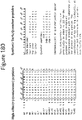
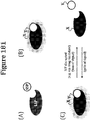
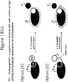
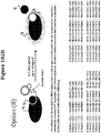
- a peptide of a luminescent pair can be a 'dark peptide,' or one that binds to its complement (e.g., NLpoly) (e.g., with low or high affinity) but produces minimal or no luminescence (See figures 180-182 ).
- a high affinity dark peptide finds use in inverse complementation, or gain of signal assays for measuring inhibitors.
- a low affinity dark peptide can be used to bring down background of NLpoly 11S in a reagent for the detection of a high affinity peptide tag (e.g. NLpep86). Exemplary dark peptides are shown in Figure 180 .
- a peptide of a luminescent pair can be a 'quencher peptide,' or one that contains a quencher moiety (e.g., DAB), and the quencher absorbs the light/energy produced by both a NLpoly in isolation (e.g., the signal produced independent of a complementing NLpep) and a NLpoly-NLpep complex (e.g., the signal produced as a result of complex formation).
- a NLpoly in isolation e.g., the signal produced independent of a complementing NLpep
- a NLpoly-NLpep complex e.g., the signal produced as a result of complex formation
- the strength of the interaction between the non-luminescent pair elements may be altered via mutations to ensure that it is insufficient to produce functionality in the absence of interaction elements that facilitate formation of the bioluminescent complex, as defined in the claims.
- Peptide constructs were generated by one of three methods: annealing 5'-phosphorylated oligonucleotides followed by ligation to pF4Ag-Barnase-HALOTAG vector (Promega Corporation; cut with SgfI and XhoI) or pFN18A (Promega Corporation; cut with SgfI and XbaI), site directed mutagenesis using Quik Change Lightning Multi kit from Agilent or outsourcing the cloning to Gene Dynamics.
- the peptides generated in Example 1 were prepared for analysis by inoculating a single colony of KRX E.coli cells (Promega Corporation) transformed with a plasmid encoding a peptide into 2-5 ml of LB culture and grown at 37°C overnight. The overnight cultures (10 ml) were then diluted into 1L of LB and grown at 37°C for 3 hours. The cultures were then induced by adding 10 ml 20% rhamnose to the 1L culture and induced at 25°C for 18 hours.
- peptides generated in Examples 1-2 contained single mutations to the peptide sequence: GVTGWRLCKRISA (SEQ ID NO: 236). All of the peptides were fused to a HALOTAG protein (Promega Corporation). Peptides identified as "HT-NLpep” indicate that the peptide is located at the C-terminus of the HALOTAG protein. In this case, the gene encoding the peptide includes a stop codon, but does not include a methionine to initiate translation. Peptides identified as "NLpep-HT” indicate that the peptide is at the N-terminus of the HALOTAG protein. In this case, the peptide does include a methionine to initiate translation, but does not include a stop codon.
- the small peptide mutant cultures were assayed for activity.
- the cultures containing the WT 1-156 fragment were pooled, mixed with 10 ml of 2x Lysis Buffer (50mM HEPES pH 7.4, 0.3x Passive Lysis Buffer, and 1mg/ml lysozyme) and incubated at room temperature for 10 minutes.
- 30 ⁇ l of the lysed WT 1-156 culture was then aliquoted into wells of a white, round bottom 96-well assay plate (Costar 3355). To wells of the assay plate, 20 ⁇ l of a peptide culture was added, and the plate incubated at room temperature for 10 minutes.
- NANOGLO Luciferase Assay Reagent Promega Corporation
- Luminescence was measured on a GLOMAX luminometer with 0.5s integrations.
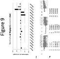
- the results demonstrate various mutations in the peptide (relative to SEQ ID NO: 1) that altered (e.g., increased, decreased) the luminescence following complementation with the wild-type non-luminescent polypeptide.
- the increased luminescence is thought to stem from one (or a combination) of five main factors, any of which are beneficial: affinity between the non-luminescent peptide and non-luminescent polypeptide, expression of the peptide, intracellular solubility, intracellular stability, and bioluminescent activity.
- affinity between the non-luminescent peptide and non-luminescent polypeptide any of which are beneficial: affinity between the non-luminescent peptide and non-luminescent polypeptide, expression of the peptide, intracellular solubility, intracellular stability, and bioluminescent activity.
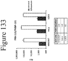
- pF4Ag-NanoLucl-156 WT 1-156
- error-prone PCR was performed using the Diversify PCR Random Mutagenesis Kit from Clontech.
- the resulting PCR product was digested with SgfI and XbaI and ligated to pF4Ag-Barnase (Promega Corporation), a version of the commercially-available pF4A vector (Promega) which contains T7 and CMV promoters and was modified to contain an E. coli ribosome-binding site.
- KRX E. coli cells Promega Corporation
- individual colonies were used to inoculate 200 ⁇ l cultures in clear, flat bottom 96-well plates (Costar 3370).
- Example 4 To determine the luminescence of the non-luminescent polypeptide mutants generated in Example 4, individual colonies of the KRX E.coli cells (Promega Corporation) transformed with a plasmid containing one of the non-luminescent polypeptide mutants from Example 4 was grown according to the procedure used in Example 3. The bacterial cultures were also induced according to the procedure used in Example 3.
- Non-luminescent polypeptides containing glycine to alanine substitutions were generated as described in Example 1.
- Each single mutant colony was inoculated in 200 ⁇ l Minimal Media (1x M9 salts, 0.1mM CaCl 2 , 2mM MgSO 4 , 1mM Thiamine HCl, 1% gelatin, 0.2% glycerol and 1x ampicillin) and incubated with shaking at 37°C for 20 hours. 10 ⁇ l of the culture was then added to 190 ⁇ l of fresh Minimal Media and incubated again with shaking at 37°C for 20 hours. 10 ⁇ l of the second culture was then added to 190 ⁇ l Auto-Induction Media (Minimal Media + 5% glucose + 2% rhamnose) and incubated with shaking at 25°C for 18 hours to allow expression of the non-luminescent polypeptide.
- Minimal Media 1x M9 salts, 0.1mM CaCl 2 , 2mM MgSO 4 , 1mM Thiamine HCl, 1% gelatin, 0.2% glycerol and 1x ampicillin
- NLpep9-HT E.coli clarified lysate 5ml LB was inoculated with a single E.coli colony of NLpep9-HT and incubated at 37°C overnight. 500 ⁇ l of the overnight culture was then diluted in 50mls LB and incubated at 37°C for 3 hours. 500 ⁇ l of 20% rhamnose was added and incubated at 25°C for 18 hours.
- the expression culture was centrifuged at 3000xg for 30 minutes, and the cell pellet resuspended in 5ml peptide lysis buffer (25mM HEPES, pH 7.5, 0.1x Passive Lysis Buffer, 1mg/ml lysozyme, and 0.3U/ ⁇ l RQ1 DNase) and incubated at room temperature for 10 minutes.
- 5ml peptide lysis buffer 25mM HEPES, pH 7.5, 0.1x Passive Lysis Buffer, 1mg/ml lysozyme, and 0.3U/ ⁇ l RQ1 DNase
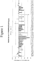
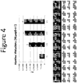
- FIGS 3 and 4 demonstrate the effects of the mutations on luminescence.
- mutations were made in the non-luminescent peptide based on alignment to other fatty acid binding proteins (FABPs) and were chosen based on high probability (frequency in FABPs) to identify a mutation that retains/improves activity (such as NLpep2, 4, and 5) or establish that a mutation is not likely to be tolerated at that position (such as NLpep3).
- NLpepl-5 contain single mutations (See Table 1)
- NLpep6-9 are composite sets of the mutations in NLpep2, 4, and 5 (See Table 1). Mutants were generated as described in Example 1.
- Each mutant colony was inoculated in 200 ⁇ l Minimal Media and incubated with shaking at 37°C for 20 hours. 10 ⁇ l of the culture was then added to 190 ⁇ l of fresh Minimal Media and incubated again with shaking at 37°C for 20 hours. 10 ⁇ l of the second culture was then added to 190 ⁇ l Auto-Induction Media and incubated with shaking at 25°C for 18 hours to allow expression of the non-luminescent peptide mutant.
- NLpep9-HT 1ml of NLpep9-HT was frozen on dry ice for 5 minutes and then thawed in a room temperature water bath for 5 minutes. 60 ⁇ l was then removed for assaying. The freeze-thaw procedure was then repeated another 10 times. After each freeze-thaw cycle, 60 ⁇ l of sample was removed for assaying.
- TMR gel analysis was used to normalize the concentration of the non-luminescent peptide mutants to distinguish mutations that alter the expression from those that alter luminescence (e.g., altered luminescence may stem from altered binding affinity).
- 5ml of LB was inoculated with a single mutant peptide colony and incubated with shaking at 37°C for 20 hours.
- 50 ⁇ l of the overnight culture was diluted into 5ml of fresh LB and incubated with shaking at 37°C for 3 hours.
- 50 ⁇ l of 20% rhamnose was then added and incubated with shaking at 25°C for 18 hours.
- TMR gel analysis 79 ⁇ l of each induced culture was mixed with 10 ⁇ l 10x Fast Break Lysis Buffer (Promega Corporation), 10 ⁇ l of a 1:100 dilution of HALOTAG TMR ligand (Promega Corporation) non-luminescent polypeptide and 10 ⁇ l of RQ1 DNase and incubated at room temperature for 10 minutes. 33.3 ⁇ l of 4x SDS-loading buffer was added, and the samples incubated at 95°C for 5 minutes. 15 ⁇ l of each sample was loaded onto an SDS gel and run according to the manufacturer's directions. The gel was then scanned on a Typhoon.
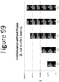
- Each culture was diluted based on the TMR-gel intensity to normalize concentrations. 20 ⁇ l of each diluted culture was then mixed with 30 ⁇ l assay lysis buffer containing non-luminescent polypeptide (1:10 dilution of SEQ ID NO: 2 E.coli clarified lysate) and incubated with shaking at room temperature for 10 minutes. 50 ⁇ l of NANOGLO Luciferase Assay Reagent was added, and the samples incubated at room temperature for 10 minutes. Luminescence was measured on a GLOMAX luminometer with 0.5s integrations (SEE FIG. 9 ).
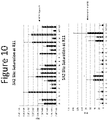
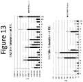
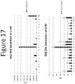
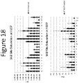
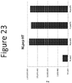
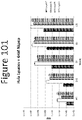
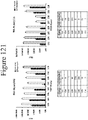
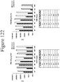
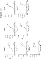
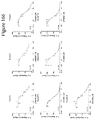
- positions 11, 15, 18, 31, 58, 67, 106, 149, and 157 were identified as sites of interest from screening the library of random mutations in wild-type non-luminescent polypeptide. All 20 amino acids at these positions (built on 5A2 non-luminescent mutant generated in Example 6 (SEQ ID NOS: 539 and 540) to validate with other mutations in the 5A2 mutant) were compared to determine the optimal amino acid at that position. Mutant non-luminescent polypeptides were generated as previously described in Example 1. Single colony of each non-luminescent polypeptide mutant was grown according to the procedure used in Example 6. The bacterial cultures were also induced according to the procedure used in Example 6. Luminescence was assayed and detected according to the procedure used in Example 6 expect NLpep53 E.coli clarified lysate was used at 1:11.85 dilution.
- Figures 10-18 demonstrate the effect of the mutations on the ability to produce luminescence with and without NLpep.
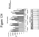
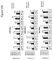
- Figure 24 demonstrates the luminescence of NLpolys containing multiple mutations.
- HEK 293 cells were plated at 100,000 cells/ml into wells of a 24 well plates containing 0.5ml DMEM+10% FBS (50,000/well). The cells were incubated in a 37°C, 5% CO 2 incubator overnight. DNA for expression of each non-luminescent polypeptide mutant was transfected in duplicate. lug plasmid DNA containing a non-luminescent polypeptide mutant was mixed with OptiMEM (Life Technologies) to a final volume of 52ul. 3.3 ⁇ l of Fugene HD (Promega Corporation) was added, and samples incubated for 15 minutes at room temperature. 25 ⁇ l of each sample mixture was added to two wells and incubated overnight in a 37°C, 5% CO 2 incubator overnight. After overnight incubation, the growth media was removed and 0.5ml DMEM (without phenol red) + 0.1% Prionex added. The cells were then frozen on dry ice (for how long) and thawed prior to detecting luminescence.
- Luminescence generated from various non-luminescent polypeptide mutants was measured using either Furimazine or coelenterazine as a substrate as well as with various non-luminescent peptides.
- HEK 293 cells were plated at 15,000 cells/well in 100 ⁇ l DMEM+10% FBS into wells of 96-well plates. The cells were incubated in a 37°C, 5% CO 2 incubator overnight. Transfection complexes were prepared by adding 0.66ug each of plasmid DNA for expression of a non-luminescent polypeptide mutant and a non-luminescent peptide mutant plasmid to a final volume of 31 ⁇ l in OptiMem. 2 ⁇ l Fugene HD was added to each transfection complex and incubated for 15 minutes at room temperature.
- the following example investigates the luminescence and substrate specificity of various non-luminescent polypeptide mutants with NLpep69 and using either Furimazine or coelenterazine as a substrate.
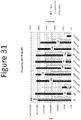
- CHO cells were plated at 20,000 cells/ well in 100 ⁇ l of DMEM+10% FBS into wells of 96-well plates. The cells were incubated in a 37°C, 5% CO2 incubator overnight.
- Transfection complexes were prepared by adding 0.66ug each of plasmid DNA for expression of a non-luminescent polypeptide mutant and a non-luminescent peptide mutant plasmid to a final volume of 31 ⁇ l in OptiMem. 2 ⁇ l Fugene HD was added to each transfection complex and incubated for 15 minutes at room temperature. For each peptide/polypeptide combination, 5 ⁇ l of transfection complex was added to 6 wells of the 96-well plate and grown overnight at 37C in CO 2 incubator.
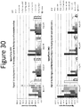
- Figure 31 demonstrates the substrate specificity when NLpolys are coexpressed in mammalian cells with NLpep69.
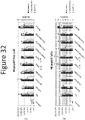
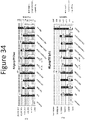
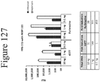
- the following example investigates the luminescence and substrate specificity of various non-luminescent polypeptide mutants with NLpep69, NLpep78 or NLpep79, using either Furimazine or coelenterazine as a substrate and under either lytic or live cell conditions.
- HEK 293 cells were plated at 15,000 cells/well in 100 ⁇ l DMEM+10% FBS into wells of 96-well plates. The cells were incubated in a 37°C, 5% CO2 incubator overnight.
- Transfection complexes were prepared by adding 0.66ug each of plasmid DNA for expression of a non-luminescent polypeptide mutant and a non-luminescent peptide mutant plasmid to a final volume of 31 ⁇ l in OptiMem. 2 ⁇ l Fugene HD was added to each transfection complex and incubated for 15 minutes at room temperature. For each NLpoly-NLpep combination, 5 ⁇ l of transfection complex was added to 6 wells of the 96-well plate and grown overnight at 37C in CO2 incubator.
- Figures 32-34 demonstrate the substrate specificity of NLPolys coexpressed in mammalian cells with NLpep69, 78, or 79 in live-cell and lytic formats.
- Example 7 A single colony of each non-luminescent polypeptide was grown according to the procedure used in Example 7. The bacterial cultures were also induced according to the procedure used in Example 7. Luminescence was assayed and detected according to the procedure used in Example 7 except NLpep78-HT or NLpep79-HT at 1:1,000 dilution was used. Figure 35 demonstrates the luminescence of NLpolys expressed in E. coli and assayed with NLpep78 or 79.
- Example 7 A single colony of each non-luminescent polypeptide was grown according to the procedure used in Example 7. The bacterial cultures were also induced according to the procedure used in Example 7. Luminescence was assayed and detected according to the procedure used in Example 7 except no non-luminescent peptide was added to the assay buffer.
- Figure 36 demonstrates the luminescence of NLpolys expressed in E. coli and assayed in the absence of NLpep.
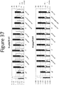
- Example 7 A single colony of each non-luminescent polypeptide was grown according to the procedure used in Example 7. The bacterial cultures were also induced according to the procedure used in Example 7. Luminescence was assayed and detected according to the procedure used in Example except either Furimazine or coelenterazine was mixed with NANOGLO Assay Buffer.
- Figure 37 demonstrates the substrate specificity of NLpolys expressed in E. coli and assayed with NLpep78 or 79.
- CHO and Hela cells (CHO: 100,000 seeded the day prior to transfection; Hela: 50,000 seeded the day prior to transfection) were transfected with 5ng of a non-luminescent polypeptide mutant 5A2 or 5P or with wild-type non-luminescent polypeptide using Fugene HD into wells of a 24-well plate and incubated at 37°C overnight. After the overnight incubation, the media was replaced with DMEM without phenol red, and the cells frozen at -80°C for 30 minutes. The cells were then thawed and transferred to a 1.5ml tube.
- HEK293, Hela or CHO cells were transfected with 5ng 5P NLpoly-firefly luciferase fusion, 5P NLpoly-click beetle luciferase fusion, wild-type 5P-firefly luciferase fusion or wild-type 5P-click beetle luciferase fusion according to the procedure in Example 22. Lysates were also prepared according to Example 22. The cell lysates were then diluted 1:10 DMEM without phenol red, 20 ⁇ l mixed with NLpep78 (diluted 1:100 in DMEM without phenol red; E.coli lysate) and shaken at room temperature for 10 minutes.
- This example demonstrates complementation in live-cells using either wild-type or 5P NLpoly.
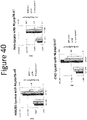
- Hela cells plated into wells of 96-well plated, transfected with 0.5ng of wild-type or 5P non-luminescent polypeptide plasmid DNA using Fugene HD and incubated at 37°C overnight. After the overnight incubation, the cells were then transfected with 0.5ng NLpep78-HT plasmid DNA using Fugene HD and incubated at 37°C for 3 hours. The media was then replaced with CO 2 -independent media+0.1% FBS and 20uM PBI-4377 and luminescence measured at 37°C on a GloMax with 0.5 second integration.
- Figure 41 demonstrates the live-cell complementation between 5P or WT NLpoly and NLpep78.
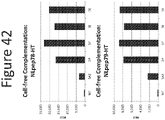
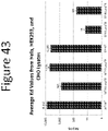
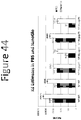
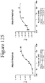
- non-luminescent polypeptide lysates from Hela, HEK293 and CHO cells were prepared as previously described and diluted 1:10 PBS+0.1% Prionex. 4x concentrations of non-luminescent peptide (synthetic) were made in PBS+0.1% Prionex. 20 ⁇ l of the non-luminescent polypeptide lysate was mixed with 20 ⁇ l non-luminescent peptide and shaken at room temperature for 10 minutes. 40 ⁇ l of NanoGlo Luciferase Assay Reagent or PBS+0.1% Prionex with Furimazine was added and shaken at room temperature for 10 minutes.
- Luminescence was detected on a GloMax with 0.5s integration. Kd values were determined using Graphpad Prism, One Site-Specific Binding.
- Figure 43 and 44 demonstrate the dissociation constants measured under various buffer conditions (PBS for complementation then NanoGlo for detection, PBS for complementation and detection, NanoGlo for complementation and detection).
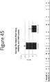
- non-luminescent polypeptide mutant lysates from Hela, HEK293 and CHO cells were prepared as previously described and diluted 1:10 PBS+0.1% Prionex. 4x concentrations of non-luminescent peptide (NLpep) were made in PBS+0.1% Prionex+10mM DTT. 20 ⁇ l of the non-luminescent polypeptide lysate was mixed with 20 ⁇ l non-luminescent peptide and shaken at room temperature for 10 minutes. 40 ⁇ l of NanoGlo Luciferase Assay Reagent was added and shaken at room temperature for 10 minutes. Luminescence was detected on a GloMax with 0.5s integration. Figure 45 demonstrates NLpep C8F mutation significantly improves the binding affinity for 5P.
- non-luminescent polypeptide variants Ile-11 (Ile at residue 11), Val-11, Tyr-11, Glu-11, Glu-157, Pro-157, Asp-157, Ser-157, Met-149, Leu-106, NLpoly11, and NLpoly12 were generated as described below, and their expression analyzed. The additional non-luminescent polypeptide variants were made in the 5A2 non-luminescent polypeptide background.
- Fresh individual colonies (KRX) of each additional non-luminescent polypeptide variants were picked and grown overnight in LB+ampicillin (100ug/ml) at 30°C and then diluted 1:100 in LB+ampicillin and grown at 37°C for 2.5 hours (OD600 ⁇ 0.5).
- Rhamnose was added to a final concentration of 0.2%, and the cells were split in triplicate and grown overnight at 25°C for ⁇ 18 h.
- Cells were lysed using 0.5X Fast Break for 30 minutes at ambient temperature, snap-frozen on dry ice, and stored at -20°C. Upon fast thawing, soluble fractions were prepared by centrifugation at 10K for 15 min at 4°C. Samples were assayed for luminescence on a Tecan Infinite F-500 luminometer.
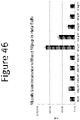
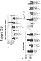
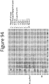
- Figure 49 demonstrates that total lysate and soluble fraction of each non-luminescent polypeptide variant as analyzed by SDS-PAGE.
- the data provides information about expression, solubility and stability of the additional non-luminescent polypeptide variants.
- a majority of the additional non-luminescent polypeptide variants produced more protein (total and soluble) than wild-type, but in many cases, the difference is subtle. Improved expression for NLpoly11 and NLpoly12 was more noticeable.
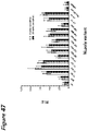
- the background luminescence of the additional non-luminescent polypeptide variants generated in Example 29 was measured by incubating 25 ⁇ l of non-luminescent polypeptide variant lysate with 25 ⁇ l DMEM at room temperature for 10 minutes. 50 ⁇ l NanoGlo Luciferase Assay Reagent was then added, and luminescence measured at 5 and 30 minutes on a Tecan Infinite F500. NLpep53 (Pep 53) alone and DMEM (DMEM) alone were used as controls. Figure 47 demonstrates that a majority of the additional non-luminescent polypeptide variants showed elevated background luminescence.
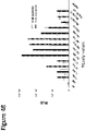
- Luminescence of the additional non-luminescent polypeptide variants generated in Example 28 was measured by incubating 25 ⁇ l of non-luminescent polypeptide variant lysate with 25 ⁇ l NLpep-53 at room temperature for 10 minutes50 ⁇ l NanoGlo Luciferase Assay Reagent was then added, and luminescence measured at 5 and 30 minutes on a Tecan Infinite F500.
- NLpep53 (Pep 53) alone and DMEM (DMEM) alone were used as controls.
- Figure 48 demonstrates that the non-luminescent polypeptide variants Val-11, Glu-11, Glu-157, Pro-157, Asp-157, Ser-157 and Met-149 generated significantly more luminescence than parental 5A2.
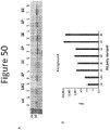
- Figure 50A shows the total lysate and soluble fraction of each non-luminescent polypeptide variant.
- Figure 50B shows the background luminescence of each non-luminescent polypeptide variant.
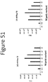
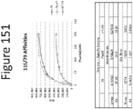
- Figure 51 shows the luminescence generated with each non-luminescent polypeptide variant when complemented with 10 or 100nM NLpep78 (NVSGWRLFKKISN) in LB medium.
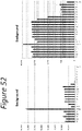
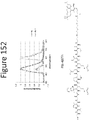
- Background luminescence in E.coli lysates Figure 52
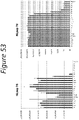
- luminescence generated after complementation with NLpep78 Figure 53 ; NVSGWRLFKKISN
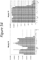
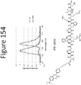
- NLpep79 Figure 54 ; NVTGYRLFKKISN
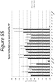
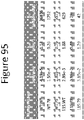
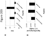
- Figure 55 shows the signal-to-background of the non-luminescent polypeptide 5P variants.
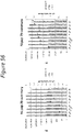
- Figure 56 provides a summary of the luminescent results.
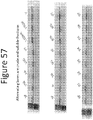
- Figure 57 shows the amount of total lysate and soluble fraction in each non-luminescent polypeptide 5P variant.
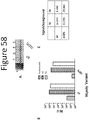
- Figure 58 shows the amount of total lysate and soluble fraction of 5P and I107L (A), luminescence generated by 5P or I107L without non-luminescent peptide or with NLpep78 or NLpep79 (B) and the improved signal-to-background of I107L over 5P (C).
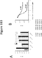
- binding affinity between an elongated non-luminescent polypeptide variant, i.e., containing additional amino acids at the C-terminus, and a deleted non-luminescent peptide, i.e., deleted amino acids at the N-terminus.
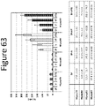
- Lysates of E.coli expressing non-luminescent polypeptide 5P/+V/+VT/+VTG prepared as previously described were diluted 1:2000 in PBS + 0.1% Prionex. 25 ⁇ l of the diluted lysate was incubated with 25 ⁇ l of NLpep78, NLpep80, NLpep81 or NLpep82 (diluted 0-500nM in dilution buffer) for 5 min at room temp. 50 ⁇ l of Furimazine diluted to 1X with NanoGlo Assay Buffer was added to each sample and incubated for 10 minutes at room temperature. Luminescence was measured on a GloMax Multi with 0.5s integration time.
- Figure 63 demonstrates the binding affinity between NLpolys with additional amino acids at the C-terminus with NLpeps with amino acids deleted from the N-terminus.
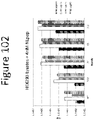
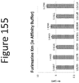
- Non-luminescent polypeptide LB lysates were prepared and diluted 1:100 into PBS+0.1% Prionex. 2X dilutions of synthetic NLpep78 were made in PBS+0.1% Prionex. 25 ⁇ l of the diluted non-luminescent polypeptide lysate was mixed with 25 ⁇ l of each dilution of non-luminescent peptide and incubated 3 minutes at ambient temperature. 50 ⁇ l of NanoGlo Luciferase Assay Reagent was added, incubated for 5 minutes at room temperature, and luminescence measured on a GloMax Multi+. Figure 64 shows the calculated Kd values using one-site specific binding.
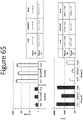
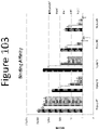
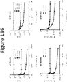
- Lysates of CHO, HEK293T, or HeLa cells expressing NLpoly 5P were diluted 1:1000 in dilution buffer (PBS + 0.1% Prionex.) 25 ⁇ l of diluted lysate was incubated with 25 ⁇ l of NLpep80/87 (diluted 0-5 ⁇ M in dilution buffer) for 5 min at room temp. 50 ⁇ l of furimazine (diluted to 1X with NanoGlo buffer) was added to each well, and the plate was incubated for 10 min at room temp. Luminescence was then read on a GloMax Multi with 0.5s integration time ( Figure 65 ).
- Lysates of E.coli expressing NLpoly 5P were diluted 1:2000 in dilution buffer (PBS + 0.1% Prionex.) 25 ⁇ l of diluted lysate was incubated with 25 ⁇ l of NLpep80/87 (diluted 0-5 ⁇ M in dilution buffer) for 5 min at room temp. 50 ⁇ l of furimazine (diluted to 1X with NanoGlo buffer) was added to each well, and the plate was incubated for 10 min at room temp. Luminescence was then read on a GloMax Multi with 0.5s integration time ( Figure 66 ).
- NLpep-HT E. coli clarified lysates as prepared as previously described in Example 6. The amount of NLpep-HT was quantitated via the HaloTag fusion. Briefly, 10 ⁇ l of clarified lysate was mixed with 10 ⁇ l HaloTag-TMR ligand (diluted 1:100) and 80 ⁇ l water and incubated at room temperature for 10 minutes. 33.3 ⁇ l 4x SDS Loading Buffer was added and incubated at 95°C for 5 minutes.
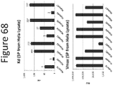
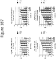
- 5P non-luminescent polypeptide lysate was prepared from Hela cells as previously described and diluted prepared 1:10 in PBS +0.1% Prionex. 4x concentrations (range determined in preliminary titration experiment) of non-luminescent peptide (synthetic peptide; by Peptide 2.0 (Virginia); made at either 5, 10, or 20 mg scale; blocked at the ends by acetylation and amidation, and verified by net peptide content analysis) was prepared in PBS +0.1% Prionex. 20 ⁇ l 5P non-luminescent polypeptide and 20 ⁇ l non-luminescent peptide were mixed and shaken at room temperature for 10 minutes.
- Figure 68 demonstrates the binding affinity and corresponding luminescence between 5P and truncated versions of NLpep78.
- the binding affinity is increased when 1 amino acid is removed from the N-terminus, the C-terminus, or 1 amino acid from each terminus. Removing more than 1 amino acid from either terminus lowers the affinity but does not always lower the Vmax to the same extent.
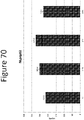
- binding affinity between an elongated non-luminescent polypeptide, i.e., one with 2 extra amino acids on C-terminus, and a truncated non-luminescent peptide, i.e., one with 2 amino acids removed from N-terminus was determined.
- Non-luminescent polypeptide lysate was prepared as previously described and diluted prepared 1:100 in PBS +0.1% Prionex. 2x dilutions of NLpep81 (synthetic peptide; by Peptide 2.0 (Virginia); made at either 5, 10, or 20 mg scale; blocked at the ends by acetylation and amidation, and verified by net peptide content analysis) was prepared in PBS +0.1% Prionex. 25 ⁇ l non-luminescent polypeptide and 25 ⁇ l of each non-luminescent peptide dilution were mixed and shaken at room temperature for 3 minutes. 50 ⁇ l of NanoGlo Luciferase Assay reagent was added and shaken at room temperature for 5 minutes. Luminescence was measured on GloMax with 0.5s integration. Figure 69 shows the calculate Kd values using one-site specific binding.
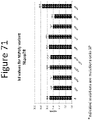
- binding affinity between an elongated non-luminescent polypeptide, i.e., one with 3 extra amino acids on C-terminus, and a truncated non-luminescent peptide, i.e., one with 3 amino acids removed from N-terminus was determined.
- Non-luminescent polypeptide lysate was prepared and diluted prepared 1:100 in PBS +0.1% Prionex. 2x dilutions of NLpep82 (synthetic peptide; by Peptide 2.0 (Virginia); made at either 5, 10, or 20 mg scale; blocked at the ends by acetylation and amidation, and verified by net peptide content analysis) was prepared in PBS +0.1% Prionex. 25 ⁇ l non-luminescent polypeptide and 25 ⁇ l of each non-luminescent peptide dilution were mixed and shaken at room temperature for 3 minutes. 50 ⁇ l of NanoGlo Luciferase Assay reagent was added and shaken at room temperature for 5 minutes. Luminescence was measured on GloMax with 0.5s integration. Figure 70 shows the calculate Kd values derived using one-site specific binding.
- Non-luminescent polypeptide variants were grown in M9 minimal media. Individual colonies were inoculated and grown overnight at 37°C. Samples were diluted 1:20 in M9 minimal media and grown overnight at 37°C. Samples were again diluted 1:20 in M9 induction media and grown overnight at 25°C. Samples were pooled, and 100 ⁇ l of the pooled cells were lysed with 400 ⁇ l of PLB lysis buffer and incubate at room temperature for 10 minutes. The lysates were diluted 1:100 in PBS+0.1% Prionex. 2X dilutions of synthetic NLpep78 were made in PBS+0.1% Prionex.
- NLpoly 5P-B9 (5P with residues 147-157 deleted) and NLpep B9-HT (Met+ residues 147-157 fused to N-terminus of HT7) lysates were prepared.
- Figure 73 demonstrates the feasibility of a ternary system consisting of 2 different NLpeps and a truncated NLpoly. Since all 3 components are non-luminescent without the other 2, this system could be configured such that each NLpep is fused (synthetically or genetic engineering) to a binding moiety and the truncated NLpoly used at high concentrations to produce light only in the presence of an interaction between the binding moieties, or such that each of the 3 components are fused to binding moieties to produce light only in the event of ternary complex formation.
- NLpep88-HT and 5P E. coli clarified lysates were prepared as previously described. Serial dilutions of NLpep88-HT lysate were made in PBS+0.1% Prionex. 20 ⁇ l of 5P lysate and 20 ⁇ l NLpep88-HT lysate were mixed and shaken at room temperature for 10 minutes. 40 ⁇ l of NanoGlo Luciferase Assay Reagent was added and shaken at room temperature for 10 minutes. Luminescence was measured on GloMax with 0.5s integration. Figure 74 demonstrates the importance of the arginine residue at the 6th position of the NLpep. While there is no increase in luminescence above 5P alone at lower concentrations of NLpep88, high concentrations of NLpep increased the luminescence suggesting a catalytically compromised complex and not a lack of interaction between 5P and NLpep88.
- U2OS cells were plated and left to recover overnight at 37°C.
- Cells were then transfected with HaloTag alone DNA construct or the HaloTag-NanoLuc peptide DNA constructs (all under the control of CMV promoter): P1-HT, P78-HT or P79-HT diluted 1:10 with carrier DNA(pSI) using FuGENE HD and incubated for 24 hours at 37°C.
- Cells were then labeled with HaloTag-TMR ligand by the manufacturer's standard rapid labeling protocol and imaged.
- Figure 75 demonstrates that NLpep78 and 79 do not alter the intracellular localization of the HaloTag protein.
- U2OS cells were plated and left to recover overnight at 37°C.
- Cells were either kept as non-transfection controls or transfected with the NanoLuc DNA constructs: FL, NLpoly (wt) or NLpoly(5P) diluted 1:10 with carrier DNA (pSI) using FuGENE HD and incubated for 24 hours at room temperature.
- Cells were fixed and subsequently processed for ICC.
- ICC was done using 1:5000 GS (PRO) primary antibody overnight at 4°C followed by an Alexa488 goat anti-rabbit secondary antibody.
- Figure 76 demonstrates that both NLpoly WT and NLpoly 5P localize uniformly in cells.
- Luminescence (RLU) data for the (+) peptide samples were normalized to the readings for the 5P control, and these results are presented in Figure 78 .
- Luminescence (RLU) data for the (+) peptide samples were also normalized to 5P, but then also normalized to the values in Figure 76 in order to represent signal to background (S/B; Figure 79 ).
- MAGNEHALOTAG beads (Promega Corporation; G728A) were equilibrated as follows: a) 1mL of beads were placed on magnet for ⁇ 30sec, and the buffer removed; b) the beads were removed from magnet, resuspended in 1mL PBS+0.1% Prionex, and shaken for 5min at RT; and c) steps a) and b) were repeated two more times NLpep78-HaloTag ( E. coli clarified lysate) was bound to MAGNEHALOTAG beads by resuspending the beads in 1mL NLpep78-HT clarified lysate, shaking for 1 hr at RT and placing on magnet for ⁇ 30sec.
- NLpep78-HaloTag E. coli clarified lysate
- NLpoly 8S E. coli clarified lysate
- NLpep78 bound-MagneHaloTag beads was bound to the NLpep78 bound-MagneHaloTag beads from the step above by resuspending the beads in 1.5mL 8S lysate, shaking for 1hr at RT and placing on a magnet for ⁇ 30sec.
- the lysate (flow through) was removed and saved for analysis.
- the beads were resuspended in 1mL PBS +0.1% Prionex, shaken for 5 min at RT, placed on magnet for ⁇ 30sec, and PBS (wash) removed. The beads were washed three more times.
- the beads were resuspended in 500uL 1xSDS buffer and shaken for 5min at RT. The beads were then placed on a magnet for ⁇ 30sec; the SDS buffer (elution) removed and saved for analysis. The elution was repeated one more time.
- the samples were then analyzed by gel. 37.5uL of sample (except elutions) was mixed with 12.5uL 4X SDS buffer and incubated at 95°C for 5min. 5uL was loaded onto a Novex 4-20% Tris-Glycine gel and run at ⁇ 180V for ⁇ 50min. The gel was stained with SimplyBlue Safe Stain and imaged on a LAS4000 imager.
- Figure 94 illustrates that the affinity of NLpoly and NLpep is sufficient to allow for purification from an E. coli lysate.
- NLpoly 8S was purified from an E. coli lysate, it is reasonable to expect a protein fused to NLpoly 8S (or other variant described herein) could also be purified in a similar fashion. While in this example the NLpep was immobilized and used to purify NLpoly, it is also reasonable to expect a similar result if NLpoly were immobilized.
- k obs [ NLpep ] k on + k off .
- Figure 95 illustrates the association and dissociation rate constants for the binding between NLpolys and NLpeps.
- NLpoly was diluted into PBS+0.1% Prionex as follows: WT at 1:10 5 , 5P at 1:10 7 , and 11S at 1:10 8 .
- NLpep was diluted into PBS+0.1% Prionex as follows: 30uM for WT NLpoly studies or 3uM for NLpoly 5P and 11S studies.
- 50uL NLpoly/NLpep was incubated at RT for 5min, 50uL NanoGlo + Fz (ranging from 100uM to 1.2uM, 2X) added, and incubated for 10min at RT.
- Luminescence was measured on GloMax ® Multi+ with 0.5sec integration.
- Km was derived using Graphpad Prism, Michaelis-Menton best-fit values.
- Figure 96 illustrates the Km values for various NLpoly/NLpep pairs.
- 11S E. coli clarified lysate
- Synthetic NLpep79 was diluted serially (1:2) from 800nM to 0.39nM (2X). 20uL 11S + 20uL NLpep79 were then mixed and incubated for 5min at RT. 40uL NanoGlo + 5uM or 50uM Fz was added and incubated another 5min at RT. Luminescence was measured on GloMax ® Multi+ with 0.5sec integration. Kd was derived using Graphpad prism, One site-Specific binding value.
- Figure 97 illustrates that saturating concentrations of furimazine increase the affinity between 11S and NLpep79.
- NLpoly 5A2 was diluted into PBS+0.1% Prionex at 1:10 5 .
- NLpep (WT, NLpep 78 or NLpep79) was diluted into PBS+0.1% Prionex to 30uM.
- 50uL NLpoly/NLpep was incubated at RT for 5min.
- 50uL NanoGlo + Fz (ranging from 100uM to 1.2uM, 2X) was added and incubated for 10min at RT.
- Luminescence was measured on GloMax ® Multi+ with 0.5sec integration.
- Km was derived using Graphpad Prism, Michaelis-Menton best-fit values.
- Figure 98 illustrates the Km values for NLpoly5A2 and NLpep WT, 78, and 79.
- E. coli clarified lysate were prepared as described previously for NLpoly WT, 5A2, 5P, 8S and 11S. 50uL of each lysate and 50uL NanoGlo +Fz were mixed and incubated for 5min RT. Luminescence was measured on GloMax ® Multi+ with 0.5sec integration. Figure 99 illustrates that the ability of the NLpoly to produce luminescence in the absence of NLpep gradually increased throughout the evolution process resulting in ⁇ 500 fold higher luminescence for 11S than WT NLpoly.
- a single NLpoly colony of WT, 5A2, 5P, 8S or 11S was inoculated in 200uL minimal media and grown for 20hrs at 37°C on shaker. 10uL of the overnight culture was diluted into 190uL fresh minimal media and grown for 20hrs at 37°C on shaker. 10uL of this overnight culture was diluted into 190uL auto-induction media (previously described) and grown for 18hrs at 25°C on shaker.
- the auto-induced cultures were diluted 50-fold (4uL into 196uL assay lysis buffer), 10uL expression culture added to 40uL of assay lysis buffer containing NLpep (synthetic; 1nM; WT, NLpep78, NL79 or NLpep80) and shaken for 10min at RT. 50uL NanoGlo + Fz was added, and samples shaken for 5 min at RT. Luminescence was measured on a GloMax luminometer with 0.5sec integration.
- Figure 100 illustrates the improvement in luminescence from E. coli-derived NLpoly over the course of the evolution process, an overall ⁇ 10 5 improvement (from NLpolyWT:NLpepWT to NLpoly11S:NLpep80).
- NLpep WT, NLpep78, NLpep79 or NLpep 80 were diluted to 10nM in PBS+0.1% Prionex, and 25ul mixed with 25uL of each of the NLpoly cell lysate.
- Figure 101 illustrates the improvement in luminescence from HeLa-expressed NLpoly over the course of the evolution process, an overall ⁇ 10 5 improvement (from NLpolyWT:NLpepWT to NLpoly 11S:NLpep80).
- NLpep WT, NLpep78, NLpep79 or NLpep 80 were diluted to 10nM in PBS+0.1% Prionex, and 25ul mixed with 25uL of each of the NLpoly cell lysate.
- Figure 102 illustrates the improvement in luminescence from HEK293-expressed NLpoly over the course of the evolution process, an overall ⁇ 10 4 improvement (from NLpolyWT:NLpepWT to NLpoly 11S:NLpep80).
- NLpoly WT, 5A2, 5P, 8S or 11S were diluted into PBS+0.1% Prionex as follows: WT 1:10 4; 5A2 1:105; 5P 1:10 6 ; 8S 1:10 7 ; and 11S 1:10 7 .
- NLpepWT, NLpep78, NLpep79 or NLpep80 were serially into PBS+0.1% Prionex to 4X concentration. 25uL NLpoly and 25uL NLpep were mixed and incubated for 10min at RT. 50uL NanoGlo+100uM Fz was added and incubated for 5min at RT.
- Luminescence was measured on a GloMax Multi+ with 0.5sec integration. Kd was determined using Graphpad Prism, One Site-Specific Binding, Best-fit values.
- Figure 103 illustrates a 10 4 fold improved affinity (starting affinity: NLpolyWT:NLpepWT, Kd ⁇ 10uM) of K d ⁇ 1nM (NLpoly11S:NLpep86 or NLpoly11S:NLpep80) of the variants tested over wild-type.
- Single NLpoly variant colonies were inoculated with 200uL minimal media and grown for 20hrs at 37°C on a shaker. 10uL of the overnight culture were diluted into 190uL fresh minimal media and grown for 20hrs at 37°C on a shaker. 10uL of this overnight culture was then diluted into 190uL auto-induction media (previously described) and grown for 18hrs at 25°C on a shaker. 10uL of this expression culture was mixed with 40uL of assay lysis buffer (previously described) without NLpep or NLpep78-HT (1:3,860 dilution) or NLpep79-HT (1:10,000 dilution) and shaken for10min at RT.
- a single NLpoly variant colony (SEE FIG. 143 ) was inoculated into 5mL LB culture and incubated at 37°C overnight with shaking. The overnight culture was diluted 1:100 into fresh LB and incubated at 37°C for 3hrs with shaking. Rhamnose was added to the cultures to 0.2% and incubated 25°C overnight with shaking. 900ul of these overnight cultures were mixed with 100uL 10X FastBreak Lysis Buffer (Promega Corporation) and incubated for 15min at RT. A 75uL aliquot (total) was removed from each culture and saved for analysis. The remaining culture from each sample were centrifuged at 14,000xrpm in a benchtop microcentrifuge at 4°C for 15min.
- NLpoly variant lysate (SEE FIG. 144 ; prepared as described previously) was diluted 1:10 into PBS +0.1% Prionex. 4x concentrations of NLpep78 (synthetic NLpep78) were made in PBS +0.1% Prionex. 20uL NLpoly variant lysate and 20uL NLpep were mixed and shaken for 10min at RT. 40uL NanoGlo/Fz was added and shaken for 10min at RT. Luminescence was measured on a GloMax ® luminometer with 0.5s integration. Kd determined using Graphpad Prism, One site-specific binding, best-fit values. Figure 144 illustrates dissociation constants of NLpep78 with various NLpolys.
- HEK293T cells (400,000) were reverse-transfected with 1 ⁇ g pF4A Ag FKBP or 1 ⁇ g pF4A Ag FRB vectors expressing N- or C-terminal fusions of NLpoly5P and/or NLpep80/87 using FuGENE HD at a DNA-to-FuGENE HD ratio of 1:4.
- 24-hours post transfection cells were trypsinized and re-plated in opaque 96-well assay plates at a density of 10,000 cells per well. 24-hours after plating, cells were washed with PBS and then incubated with or without 20nM rapamycin for 15, 60 or 120 min in phenol red-free OptiMEMI.
- Example 65 tested all 8 possible combinations of FRB and FKBP fused to NLpoly/NLpep as well as used less total DNA.
- HEK293T cells (400,000) were reverse-transfected with a total of 0.001 ⁇ g pF4A Ag FRB-NLpoly5P and 0.001 ⁇ g pF4A Ag FKBP-NLpep80/NLpep87 using FuGENE HD at a DNA-to-FuGENE ratio of 1 to 8.
- pGEM-3Zf(+) DNA was added to bring total DNA in each transfection to 1 ⁇ g. 24-hours post-transfection, 10,000 cells were re-plated in opaque 96-well assay plates and incubated an additional 24 hours.
- HEK293T cells (400,000) were reverse-transfected with a total of 0.001 ⁇ g pF4A Ag FRB-NLpoly5P or pF4A Ag FKBP-NLpep80/NLpep87 using FuGENE HD at a DNA-to-FuGENE ratio of 1 to 8.
- pGEM-3Zf(+) DNA was added to bring total DNA in each transfection to 1 ⁇ g. 24-hours post-transfection, 10,000 cells were re-plated in opaque 96-well assay plates and incubated an additional 24 hours. Cells were washed with PBS and then incubated in phenol red-free OptiMEMI with 0 or 50nM rapamycin for 2 h.
- FIG. 112 illustrates that the individual components generate a low basal level of luminescence that is not responsive to rapamycin treatment.
- HEK293T (400,000) cells were reverse-transfected with a total of 2, 0.2, 0.02, or 0.002 ⁇ g pF4A Ag FRB-NLpoly5P and pF4A Ag FKBP-NLpep80 using FuGENE HD at a DNA-to-FuGENE ratio of 1 to 4.
- pGEM-3Zf(+) DNA was added to bring total DNA in each transfection to 2 ⁇ g. 24-hours post-transfection, 10,000 cells were re-plated in opaque 96-well assay plates and incubated an additional 24 hours. Cells were washed with PBS and then incubated in phenol red-free OptiMEMI with or without 20nM rapamycin for 2 h.
- HEK293T cells (400,000) were reverse-transfected with a total of 2, 0.2, 0.02, or 0.002 ⁇ g pF4A Ag FRB-NLpoly5P or pF4A Ag FKBP-NLpep80 using FuGENE HD at a DNA-to-FuGENE ratio of 1 to 4.
- pGEM-3Zf(+) DNA was added to bring total DNA in each transfection to 2 ⁇ g. 24-hours post-transfection, 10,000 cells were replated in opaque 96-well assay plates and incubated an additional 24 hours. Cells were washed with PBS and then incubated in phenol red-free OptiMEMI with or without 20nM rapamycin for 2 h.
- HEK293T cells (400,000) were reverse-transfected with a total of 0.2, 0.02, 0.002, or 0.0002 ⁇ g pF4A Ag FRB-NLpoly5P and pF4A Ag FKBP-NLpep80/NLpep87 using FuGENE HD at a DNA-to-FuGENE ratio of 1 to 4.
- pGEM-3Zf(+) DNA was added to bring total DNA in each transfection to 2 ⁇ g. 24-hours post-transfection, 10,000 cells were re-plated in opaque 96-well assay plates and incubated an additional 24 hours. Cells were washed with PBS and then incubated in phenol red-free OptiMEMI with or without 50nM rapamycin for 2 h.
- HEK293T cells (400,000) were reverse-transfected with a total of 0.2, 0.02, 0.002, or 0.0002 ⁇ g pF4A Ag FRB-NLpoly5P or pF4A Ag FKBP-NLpep80/NLpep87 using FuGENE HD at a DNA-to-FuGENE ratio of 1 to 4.
- pGEM-3Zf(+) DNA was added to bring total DNA in each transfection to 2 ⁇ g. 24-hours post-transfection, 10,000 cells were re-plated in opaque 96-well assay plates and incubated an additional 24 hours. Cells were washed with PBS and then incubated in phenol red-free OptiMEMI with or without 50nM rapamycin for 2 h.
- HEK293T cells (400,000) were reverse-transfected with a total of 2, 0.2, 0.02, or 0.002 ⁇ g pF4A Ag FRB-NLpoly5P and pF4A Ag FKBP-NLpep80 or FKBP-NLpep87 using FuGENE HD at a DNA-to-FuGENE ratio of 1 to 4.
- pGEM-3Zf(+) DNA was added to bring total DNA in each transfection to 2 ⁇ g. 24-hours post-transfection, 10,000 cells were re-plated in opaque 96-well assay plates and incubated an additional 24 hours.
- the assay was performed in both a two-day and three-day format.
- 20,000 HEK293T cells were reverse-transfected in opaque 96-well assay plates with a total of 0.1ng pF4A Ag FRB-NLpoly5P or FRB-NLpoly5A2 and pF4A Ag FKBP-NLpep80/87/95/96/97 using FuGENE HD at a DNA-to-FuGENE ratio of 1 to 8.
- pGEM-3Zf(+) DNA was added to bring total DNA in each transfection to 1 ⁇ g.
- 400,000 HEK293T cells were reverse-transfected with a total of 0.002 ⁇ g pF4A Ag FRB-NLpoly5P and pF4A Ag FKBP-NLpep80/87/95/96/97 using FuGENE HD at a DNA-to-FuGENE ratio of 1 to 8.
- pGEM-3Zf(+) DNA was added to bring total DNA in each transfection to 1 ⁇ g.
- 24-hours post-transfection 10,000 cells were re-plated in opaque 96-well assay plates and incubated an additional 24 hours. Cells were washed with PBS and then incubated in phenol red-free OptiMEMI with or without 50nM rapamycin for 2 h.
- HEK293T cells (20,000) were reverse-transfected in opaque 96-well assay plates with a total of 0.1ng pF4A Ag FRB-NLpoly5A2/11S and pF4A Ag FKBP-NLpep 101/104/105/106/107/108/109/110 using FuGENE HD at a DNA-to-FuGENE ratio of 1 to 8.
- pGEM-3Zf(+) DNA was added to bring total DNA in each transfection to 1 ⁇ g. 24 hours-post transfection, cells were washed with PBS and then incubated in phenol red-free OptiMEMI with or without 50nM rapamycin for 2 h.
- Figure 121 illustrates that, of tested combinations, NLpoly11S with NLpep101 showed the greatest rapamycin induction and one of the strongest rapamycin-specific luminescent signals.
- HEK293T cells (20,000) were reverse-transfected in opaque 96-well assay plates with a total of 0.1ng pF4A Ag FRB-NLpoly5A2/11S and pF4A Ag FKBP-NLpep87/96/98/99/100/101/102/103 using FuGENE HD at a DNA-to-FuGENE ratio of 1 to 8.
- pGEM-3Zf(+) DNA was added to bring total DNA in each transfection to 1 ⁇ g. 24 hours-post transfection, cells were washed with PBS and then incubated in phenol red-free OptiMEMI with or without 50nM rapamycin for 2 h.
- Figure 122 illustrates that the NLpoly11S and NLpep 101 combination produces the highest induction while maintaining high levels of specific luminescence.
- HEK293T cells (20,000) were reverse-transfected in opaque 96-well assay plates with a total of 0.01, 0.1, 1, or lOng pF4A Ag FRB-NLpoly11S and pF4A Ag FKBP-NLpep87/101/102/107 using FuGENE HD at a DNA-to-FuGENE ratio of 1 to 8.
- pGEM-3Zf(+) DNA was added to bring total DNA in each transfection to 1 ⁇ g. 24 hours-post transfection, cells were washed with PBS and then incubated in phenol red-free OptiMEMI with or without 50nM rapamycin for 1.5 h.
- Figure 123 illustrates NLpolyl 1S with NLpep101 produces the overall lowest luminescence in untreated samples at all tested DNA levels, and the combination maintains relatively high levels of luminescence in rapamycin-treated samples.
- HEK293T cells (20,000) were reverse-transfected in opaque 96-well assay plates with a total of 0.01, 0.1, 1, or 10ng pF4A Ag FRB-NLpoly5A2 and pF4A Ag FKBP-NLpep87/101/102/107 using FuGENE HD at a DNA-to-FuGENE ratio of 1 to 8.
- pGEM-3Zf(+) DNA was added to bring total DNA in each transfection to 1 ⁇ g. 24 hours-post transfection, cells were washed with PBS and then incubated in phenol red-free OptiMEMI with or without 50nM rapamycin for 1.5 h.
- Figure 124 illustrates that NLpoly5A2 generates higher luminescence in untreated samples than NLpoly11S shown in example 75.
- Rapamycin dose response curve showing luminescence of cells expressing FRB-NLpoly5P and FKBP-NLpep80/87 DNA
- HEK293T cells (400,000) were reverse-transfected with a total of 0.001 ⁇ g pF4A Ag FRB-NLpoly5P and 0.001 ⁇ g pF4A Ag FKBP-NLpep80/NLpep87 using FuGENE HD at a DNA-to-FuGENE ratio of 1 to 8.
- pGEM-3Zf(+) DNA was added to bring total DNA in each transfection to 1 ⁇ g. 24-hours post-transfection, 10,000 cells were re-plated in opaque 96-well assay plates and incubated an additional 24 hours. Cells were washed with PBS and then incubated in phenol red-free OptiMEMI with 0 to 500nM rapamycin for 2 h.
- Rapamycin dose response curve showing luminescence of cells expressing FRB-NLpoly5A2 and FKBP-NLpep87/101 DNA
- HEK293T cells (20,000) were reverse-transfected in opaque 96-well assay plates with a total of 0.1ng pF4A Ag FRB-NLpoly5A2/11S and pF4A Ag FKBP-NLpep87/101 using FuGENE HD at a DNA-to-FuGENE ratio of 1 to 8.
- pGEM-3Zf(+) DNA was added to bring total DNA in each transfection to 1 ⁇ g. 24 hours-post transfection, cells were washed with PBS and then incubated in phenol red-free OptiMEMI with 0 to 1 ⁇ M rapamycin for 1.5 h.
- Figure 126 illustrates a sigmoidal dose response to rapamycin with NLpoly5A2/NLpep 101 and NLpolyl 1 S/NLpep 101 combinations. While combinations with NLpep87 show an increase in luminescence with rapamycin, the collected data points deviate more from the sigmoidal curve.
- HEK293T cells (20,000) were reverse-transfected in opaque 96-well assay plates with a total of 0.1/1/10ng pF4A Ag FRB-NLpoly11S and pF4A Ag FKBP-NLpeplOl using FuGENE HD at a DNA-to-FuGENE ratio of 1 to 8.
- pGEM-3Zf(+) DNA was added to bring total DNA in each transfection to 1 ⁇ g. 24 hours-post transfection, cells were washed with PBS and then incubated in phenol red-free OptiMEMI with 0 or 50nM rapamycin for 1.5 h.
- Figure 127 illustrates a decrease in luminescence and fold induction with the PBI-4377 substrate compared to the furimazine substrate.
- HEK293T cells (20,000) were reverse-transfected in opaque 96-well assay plates with a total of 0.1ng pF4A Ag FRB-NLpolyl 1S/5A2 and pF4A Ag FKBP-NLpep87/101 using FuGENE HD at a DNA-to-FuGENE ratio of 1 to 8.
- pGEM-3Zf(+) DNA was added to bring total DNA in each transfection to 1 ⁇ g. 24 hours-post transfection, cells were washed with PBS and then phenol red-free OptiMEMI with 0 or 50nM rapamycin and 10 ⁇ M furimazine was added either manually or via instrument injection.
- Luminescence was immediately measured on a GloMax Multi with 0.5s integration time.
- Figure 128 and 129 illustrate that, of all combinations tested, NLpoly11S with NLpep101 has the lowest luminescence at time 0, hits a luminescent plateau faster and has the largest dynamic range.
- HEK293T cells (20,000) were reverse-transfected in opaque 96-well assay plates with a total of 0.1ng pF4A Ag FRB-NLpoly11S and pF4A Ag FKBP-NLpeplOl using FuGENE HD at a DNA-to-FuGENE ratio of 1 to 8.
- pGEM-3Zf(+) DNA was added to bring total DNA in each transfection to 1 ⁇ g. 24 hours-post transfection, cells were washed with PBS and then phenol red-free OptiMEMI with 0 or 50nM rapamycin was added for 20 min.
- FIG. 130 illustrates that the rapamycin-specific induction of FRB-NLpolyl 1S and FKBP-NLpep101 can be measured on different instruments.
- HeLa cells (500,000) were reverse transfected with 1 ⁇ g pF4 Ag FRB-NLpoly11S and 1 ⁇ g pF4 Ag FKBP-NLpep101 using FuGENE HD at a DNA to FuGENE ratio of 1 to 4.
- Cells were transfected in 35 mm glass bottom culture dishes (MatTek #p35gc-1.5-14-C). 24 hours post-transfection, cells were washed with PBS and then incubate with 10 ⁇ M furimazine in OptiMEM for 5 min. 50nM rapamycin in OptiMEMI was added to cells and luminescent images were acquired with LV200 at 10s intervals for a total of 20 min.
- Figure 131 illustrates that imaging can detect an increase in cellular luminescence in cells expressing FRB-NLpoly11S and FKBP-NLpep101 following rapamycin treatment.
- HeLa cells (500,000) were reverse transfected with 1 ⁇ g pF4 Ag FRB-NLpoly11S and 1 ⁇ g pF4 Ag FKBP-NLpep101 using FuGENE HD at a DNA to FuGENE ratio of 1 to 4.
- Cells were transfected in 35 mm glass bottom culture dishes (MatTek #p35gc-1.5-14-C). 24 hours post-transfection, cells were washed with PBS and then incubate with 10 ⁇ M furimazine in OptiMEM for 5 min. 50nM rapamycin in OptiMEMI was added to cells, and luminescent images were acquired with LV200 at 10s intervals for a total of 20 min.
- Figure 132 illustrates that signal generated by individual cells can be measured and that the increase in signal by each cell parallels the increase observed in the 96-well plate assay shown in Figures 128 and 129 .
- HEK293T, HeLa, or U2-OS cells (20,000) were reverse-transfected in opaque 96-well assay plates with a total of 0.1ng pF4A Ag FRB-NLpolyl 1S and pF4A Ag FKBP-NLpep 101 using FuGENE HD at a DNA-to-FuGENE ratio of 1 to 8.
- pGEM-3Zf(+) DNA was added to bring total DNA in each transfection to 1 ⁇ g. 24 hours-post transfection, cells were washed with PBS and then phenol red-free OptiMEMI with 0 or 50nM rapamycin was added for 20 min.
- Figure 133 illustrates similar levels of luminescence generated in the absence and presence of rapamycin in three different cells lines transfected with FRB-NLpolyl11S and FKBP-NLpep101.
- HEK293T cells (20,000) were reverse-transfected in opaque 96-well assay plates with a total of 0.1ng pF4A Ag FRB-NLpoly11S and pF4A Ag FKBP-NLpep101 using FuGENE HD at a DNA-to-FuGENE ratio of 1 to 8.
- pGEM-3Zf(+) DNA was added to bring total DNA in each transfection to 1 ⁇ g. 24 hours-post transfection, cells were washed with PBS and then phenol red-free OptiMEMI with 0 or 20nM rapamycin was added for 20 min.
- FK506 inhibitor in OptiMEM was added to cell at final concentration of 5 ⁇ M and incubated for 3 or 5 hours.
- HEK293T cells (20,000) were reverse-transfected in opaque 96-well assay plates with a total of 0.1ng pF4A Ag FRB-NLpolyl11S and pF4A Ag FKBP-NLpep101 using FuGENE HD at a DNA-to-FuGENE ratio of 1 to 8.
- pGEM-3Zf(+) DNA was added to bring total DNA in each transfection to 1 ⁇ g. 24 hours-post transfection, cells were washed with PBS and then phenol red-free OptiMEMI with 0 or 20nM rapamycin was added for 2.5 hours.
- FK506 inhibitor in OptiMEM was added to cell via injector at final concentration of 0, 1 or 10 ⁇ M in OptiMEM with 10 ⁇ M.
- Luminescence was measured every 10 min for 4 hours on a GloMax Multi set to 37oC with 0.5s integration time.
- Figure 135 illustrates that by 200 s, FK506 inhibitor can reduce luminescence close to levels of
- HEK293T cells (20,000) were reverse-transfected in opaque 96-well assay plates with a total of 0.1, 1, or 10ng pF4A Ag V2R-NLpoly11S and pF4A Ag ARRB2-NLpep87/101 using FuGENE HD at a DNA-to-FuGENE ratio of 1 to 8.
- pGEM-3Zf(+) DNA was added to bring total DNA in each transfection to 1 ⁇ g. 24 hours-post transfection, cells were washed with PBS and then phenol red-free OptiMEMI with 0 or 1 ⁇ M AVP and 10 ⁇ M furimazine was added for 25 min. Luminescence was then measured on a GloMax Multi with 0.5s integration time.
- Figure 136 illustrates that V2R-NLpolyl11S with NLpep101 generates the greatest AVP-specific increase in luminescence. Combinations with NLpep87 show no significant response to AVP.
- HEK293T cells (20,000) were reverse-transfected in opaque 96-well assay plates with a total of 0.1 or 1ng pF4A Ag V2R-NLpoly11S or 1ng pF4A Ag V2R-NLpoly5A2 and pF4A Ag ARRB2-NLpep87/101 using FuGENE HD at a DNA-to-FuGENE ratio of 1 to 8.
- pGEM-3Zf(+) DNA was added to bring total DNA in each transfection to 1 ⁇ g.
- Figure 139 illustrates that at 37°C all NLpolyl11S and NLpep101 combinations tested show a time-dependent increase in AVP-induced luminescence that levels out around 200s.
- HEK293T, HeLa, or U2-OS cells (20,000) were reverse-transfected in opaque 96-well assay plates with a total of 1ng pF4A Ag V2R-NLpolyl11S and pF4A Ag ARRB2-NLpep87/101 using FuGENE HD at a DNA-to-FuGENE ratio of 1 to 8.
- pGEM-3Zf(+) DNA was added to bring total DNA in each transfection to 1 ⁇ g. 24 hours-post transfection, cells were washed with PBS and then phenol red-free OptiMEMI with 0 or 1 ⁇ M AVP was added for 20 min. Furimazine in OptiMEM was then added to a final concentration of 10 ⁇ M on cells, and luminescence was measured on a GloMax Multi with 0.5s integration time.
- Figure 140 illustrates similar luminescence levels in three different cell lines expressing V2R-NLpolyl11S and NLpep101-ARRB2 in the presence and absence of AVP.
- HeLa cells (500,000) were reverse transfected with 1 ⁇ g pF4 Ag V2R-NLpoly11S and 1 ⁇ g pF4 Ag ARRB2-NLpep101 using FuGENE HD at a DNA to FuGENE ratio of 1 to 4.
- Cells were transfected in 35 mm glass bottom culture dishes (MatTek #p35gc-1.5-14-C). 24 hours post-transfection, cells were washed with PBS and then incubate with 10 ⁇ M furimazine in OptiMEM for 5 min.
- 1 ⁇ M AVP in OptiMEMI was added to cells, and luminescent images were acquired with LV200 at 15s intervals for a total of 30 min. Instrument was at 37°C, objective was 60X or 150X, gain was 600, and exposure was 1s or 2s.
- Figures 141 and 142 illustrate that imaging can detect the increase in luminescence and formation of punctate in individual cells after treatment with AVP.
- NLpoly 5P E. coli clarified lysate (prepared as described previously) was diluted 1:1,000 into PBS +0.1% Prionex. 4x concentrations of NLpep78-HT (E. coli clarified lysate prepared as described previously) were made in PBS +0.1% Prionex. 20uL NLpoly 5P and 20uL NLpep78 were mixed and shaken for 10min at RT. 40uL NanoGlo/Fz was added and shaken for 10min at RT. Luminescence was measured on GloMax luminometer with 0.5s integration. Kd was determined using Graphpad Prism, One site-specific binding, best-fit values. Figure 80 compares the dissociation constants for an NLpep consisting of either 1 or 2 repeat units of NLpep78.
- NLpoly 5A2 lysate (prepared as described previously after transfecting CHO cells) was diluted 1:10 into PBS +0.1% Prionex. 4x concentrations of NLpep86 (synthetic NLpep) were made in PBS +0.1% Prionex. 20uL NLpoly and 20uL NLpep were mixed and shaken for 10min at RT. 40uL NanoGlo/Fz was added and shaken for 10min at RT. Luminescence was measured on GloMax luminometer with 0.5s integration. Kd was determined using Graphpad Prism, One site-specific binding, best-fit values. Figure 81 illustrates the affinity between NLpoly 5A2 and NLpep86.
- a single colony of various NLpolys were inoculated individually into 200uL minimal media and grown for 20hrs at 37°C on shaker.
- 10uL of overnight culture was diluted into 190uL fresh minimal media and grown for 20hrs at 37°C on shaker.
- 10uL of this overnight culture was diluted into 190uL auto-induction media (previously described) and grow for 18hrs at 25°C on shaker.
- 10uL of the expression culture was mixed with 40uL of assay lysis buffer (previously described) without NLpep or with NLpep78-HT (1:3,860 dilution) or NLpep79-HT (1:10,000 dilution).
- NLpeps were synthesized in array format by New England Peptide (peptides blocked at N-terminus by acetylation and at C-terminus by amidation; peptides in arrays were synthesized at ⁇ 1 mg scale) (Table 6). Each peptide was lyophilized in 3 separate plates. Each well from 1 of the 3 plates of peptides was dissolved in 100uL nanopure water, and the A260 measured and used to calculate the concentration using the extinction coefficient of each peptide. The concentration was then adjusted based on the purity of the peptide, and nanopure water was added to give a final concentration of 750uM.
- Peptides were diluted to 12.66uM (4X) in PBS+0.1% Prionex and then diluted serially 7 times (8 concentrations total) in 0.5 log steps (3.162 fold dilution).
- NLpolys 5P, 8S, 5A2 or 11S were diluted into PBS+0.1% Prionex as follows: 5P 1:2,000; 8S 1:10,000; 11S 1:150,000, 5A2 1:1,000. 25uL each NLpep + 25uL each NLpoly were mixed and incubated for 30min at RT. 50uL NanoGlo+100uM Fz was added and incubated for 30min at RT.
- Luminescence was measure on a GloMax Multi+ with 0.5sec integration.
- Kd/Vmax were determined using Graphpad Prism, One site-specific binding, best-fit values.
- Figures 83-90 illustrate the dissociation constant and Vmax values from NLpolys with the 96 variant NLpeps. The results indicate specific mutations in the NLpeps that exhibit lower binding affinity without loss in Vmax. Table 6.
- a single NLpoly 5A2, 12S, 11S, 12S-75, 12S-107 or 5P-B9 colony was inoculated into 5mL LB culture and incubated at 37°C overnight with shaking.
- the overnight culture was diluted 1:100 into fresh LB and incubated at 37°C for 3hrs with shaking.
- Rhamnose was added to the cultures to 0.2% and incubated 25°C overnight with shaking.
- 900ul of these overnight cultures were mixed with 100uL 10X FastBreak Lysis Buffer (Promega Corporation) and incubated for 15min at RT. A 75uL aliquot (total) was removed from each culture and saved for analysis.
- a single NLpoly colony (listed in Figure 92 ) was inoculated into 5mL LB culture and incubated at 37°C overnight with shaking. The overnight culture was diluted 1:100 into fresh LB and incubated at 37°C for 3hrs with shaking. Rhamnose was added to the cultures to 0.2% and incubated 25°C overnight with shaking. 900ul of these overnight cultures were mixed with 100uL 10X FastBreak Lysis Buffer (Promega Corporation) and incubated for 15min at RT. A 75uL aliquot (total) was removed from each culture and saved for analysis. The remaining culture from each sample was centrifuged at 14,000xrpm in a benchtop microcentrifuge at 4°C for 15min.
- E. coli clarified lysates were prepared for NLpoly 5P or 11S as described previously. The NLpoly lysates were then serially diluted in steps of 10-fold into PBS+0.1% Prionex. 25uL NLpoly and 25uL synthetic NLpep79 (400nM, 4X) were mixed and incubated for 10 min at RT. 50uL NanoGlo + 100uM Fz was added, incubated for10min at RT, luminescence measured on a GloMax Multi+ with 0.5sec integration.
- Figure 93 shows the substrate specificity for 5P and 11S with NLpep79 and demonstrates that 11S has superior specificity for furimazine than 5P.
- a single NLpoly WT, 5A2, 5P, 8S or 11S colony was inoculated into 5mL LB culture and incubated at 37°C overnight with shaking.
- the overnight culture was diluted 1:100 into fresh LB and incubated at 37°C for 3hrs with shaking.
- Rhamnose was added to the cultures to 0.2% and incubated 25°C overnight with shaking.
- 900ul of these overnight cultures were mixed with 100uL 10X FastBreak Lysis Buffer (Promega Corporation) and incubated for 15min at RT.
- a 75uL aliquot (total) was removed from each culture and saved for analysis. The remaining culture from each sample was centrifuged at 14,000xrpm in a benchtop microcentrifuge at 4°C for 15min.
- the non-luminescent peptides can be used to chemically label proteins.
- An NLpep can be synthesized to contain a reactive group, e.g., biotin, succinimidyl ester, maleimide, etc., and attached (e.g., conjugated, linked, labeled, etc.) to a protein, e.g., antibody.
- the NLpep-labeled protein e.g., NLpep-antibody, can then be used in a variety of applications, e.g., ELISA.
- the interaction/binding of the NLpep-labeled protein, e.g., NLpcp-antibody, to its target/binding partner would be detected by adding an NLpoly and NanoGlo ® assay reagent.
- the luminescence generated by the interaction of the NLpep-labeled protein and NLpoly would correlate to the interaction of the NL-labeled protein to its target/binding partner.
- This concept could allow for multiple NLpeps to be attached to a single protein molecule thereby resulting in multiple NLpep-labeled protein/NLpoly interactions leading to signal amplification.
- AMPylation AMP is added to the target protein by a phosphodiester bond using ATP as the donor molecule.
- ADP-ribosylation an ADP-ribose moiety is added to target proteins through a phosphodiester bond using NAD+ as the donor molecule. It has been shown that the N6-position of both ATP and NAD+ can be used to tag linkers without affecting the posttranslational event.
- the target proteins would be modified to contain the chloroalkane-ATP or -NAD+.
- N6-modified ATP/NAD has been used in combination with click-chemistry to develop in-gel fluorescent-based detection systems. Detection of these post-translational modifications by western blotting techniques requires antibodies, which are often not specific or not available.
- An alternative approach could be to combine the properties of HaloTag ® technology and the high luminescence of NanoLuc ® luciferase (NL).
- chloroalkane-ATP for AMPylation
- chloroalkane-NAD+ for ADP-ribosylation
- the blot can be incubated with HaloTag-NLpep.
- HaloTag will bind to the post-translationally-modified proteins.
- the NLpoly and furimazine could be added to the blot to detect the bioluminescence.
- This detection method is an alternative to a chemiluminescent-based approach for detection of western blots.
- a chemiluminescent-based approach could involve incubation HaloTag-protein G fusions (as a primary) in the next step any secondary antibody-linked to HRP could be used followed by ECL reaction.
- PTMs Post translational modifications of proteins are central to all aspects of biological regulation. PTMs amplify the diverse functions of the proteome by covalently adding functional groups to proteins. These modifications include phosphorylation, methylation, acetylation, glycosylation, ubiquitination, nitrosylation, lipidation and influence many aspects of normal cell biology and pathogenesis. More specifically, histone related PTMs are of great importance. Epigenetic covalent modifications of histone proteins have a strong effect on gene transcriptional regulation and cellular activity.
- post translational modification enzymes include but not limited to, Kinases/Phosphatases, Methyltransferases (HMT)/Demethylases (HDMT), Acetyltransferases/Histone Deacetylases, Glycosyltransferases/Glucanases and ADP-Ribosyl Transferases.
- HMT Methyltransferases
- HDMT Demethylases
- Acetyltransferases/Histone Deacetylases Acetyltransferases/Histone Deacetylases
- Glycosyltransferases/Glucanases and ADP-Ribosyl Transferases.
- PTM enzymes Under normal physiological conditions, the regulation of PTM enzymes is tightly regulated. However, under pathological conditions, these enzymes activity can be dysregulated, and the disruption of the intracellular networks governed by these enzymes leads to many diseases including cancer and inflammation.
- non-luminescent peptides and non-luminescent polypeptides (NLpoly) can be used to determine the activity of PTM enzymes by monitoring changes in covalent group transfer (e.g. phosphoryl, acetyl) to a specific peptide substrate linked to an NLpep.
- covalent group transfer e.g. phosphoryl, acetyl
- the NLpep will be linked through peptide synthesis to small PTM enzyme specific peptide and used as a substrate for the PTM enzyme.
- an aminopeptidase can be used to degrade the non-modified peptide (NLpep; control).
- the modified ( acetylated) peptide (NLpep-PTM enzyme substrate) would be degraded at a very slow rate or would not be degraded at all as the aminopeptidase activity is known to be affected by a PTM.
- the NLpoly is added with the NanoGlo ® assay reagent containing Furimazine. Luminescence would be generated from the sample where PTM occurred via the interaction of the NLpep and NLpoly.
- the reaction would be performed under optimal enzyme reaction condition using the histone peptide substrate linked to NLpep and Acetyl-CoA or SAM as the acetyl or methyl group donor.
- a buffer containing aminopeptidase or a mixture of aminopeptidases would be added to degrade specifically all the non-modified substrates.
- a buffer containing a NLpoly and an aminopeptidase inhibitor would be added.
- NanoGlo ® assay reagent would be added, and luminescence detected. Luminescence generated would be proportional to the amount of non-degraded NLpep present, and therefore would correlate with the amount of methylated or acetylated substrates, thereby indicating the amount of methyl or acetyl transferase activity.
- the assay can also be applied to PTM such as phosphorylation, glycosylation, ubiquitination, nitrosylation, and lipidation.
- HDMT Histone Demethylases
- a PTM-specific antibody can be used to create activity interference.
- An NLpep could be linked through peptide synthesis to small methylated peptide and used as a substrate for the hydrolase.
- an anti-methyl antibody can be added to the reaction. This antibody will bind specifically to the methylated peptide (control).
- the peptide product generated by the HDMT will not bind to the antibody.
- an NLpoly can be added. If the antibody interferes with the interaction of NLpep and NLpoly, no luminescence will be generated.
- the concept of aminopeptidase degradation of the non-modified substrate can also be used for a hydrolase assay except it would be a loss of signal assay instead of a gain of signal.
- the reaction would be performed under optimal enzyme reaction condition using a modified (methylated or acetylated) histone peptide substrate linked to an NLpep.
- a buffer containing an antibody capable of recognizing the methyl or acetyl group would be added.
- a buffer containing an NLpoly would be added.
- the NLpoly would interact with NLpep not bound to the antibody.
- NanoGlo ® assay reagent would be added, and luminescence detected.
- the luminescence generated would be proportional to the amount of NLpep not bound to the antibody, and therefore would correlate with the amount of demethylated or deacetylated substrate, thereby indicating the amount of demethylase or deacetylase activity.
- hydrolase assay concepts can also be applied to PTM hydrolases such as phosphatases, glucanases and deubiquitinases.
- the PTM transfer or hydrolysis on the peptide-NLpep would be alone sufficient to reduce or enhance the interaction of NLpep with NLpoly and therefore decrease or increase the luminescence signal without the need of aminopeptidase or antibody.
- the method was used to assay a representative transferase, the Tyrosine Kinase SRC using the following NLpep-SRC substrate peptide: YIYGAFKRRGGVTGWRLCERILA.
- SRC enzyme was titrated in 10 ⁇ l Reaction Buffer A (40mM Tris 7.5, 20mM MgCl2 and O.lmg/ml BSA) in the presence of 150 ⁇ M ATP and 2.5 ⁇ M NLpep-Src substrate and incubated for 1 hour at 23°C.
- Aminopeptidase M (APM) reagent 40mM Tris 7.5, 0.1mg/ml BSA and 50mU APM
- APM Aminopeptidase M
- NLpoly Reagent contained the NLpoly fragment and an Aminopeptidase inhibitor.
- 50 ⁇ l NanoGlo ® assay reagent was added and the luminescence was recorded after 3 minutes on a luminometer.
- RNAs noncoding RNA or mRNA
- the non-luminescent peptide (NLpep) and non-luminescent polypeptide (NLpoly) can be tethered to an RNA binding domain (RBD) with engineered sequence specificity.
- the specificity of the RBD can be changed with precision by changing unique amino acids that confers the base-specificity of the RBD.
- An example of one such RBD is the human pumilio domain (referred here as PUM).
- PUM human pumilio domain
- the RNA recognition code of PUM has been very well established. PUM is composed of eight tandem repeats (each repeat consists of 34 amino acids which folds into tightly packed domains composed of alpha helices). conserveed amino acids from the center of each repeat make specific contacts with individual bases within the RNA recognition sequence (composed of eight bases).
- the sequence specificity of the PUM can be altered precisely by changing the conserved amino acid (by site-directed mutagenesis) involved in base recognition within the RNA recognition sequence.
- PUM domains PUM1 and PUM2
- PUM1 and PUM2 with customized sequence specificities for the target RNA can be tethered to a NLpep and NLpoly (e.g., as a genetic fusion protein via genetic engineering) and can be expressed in mammalian cells.
- PUM1 and PUM2 are designed to recognize 8-nucleotide sequences in the target RNA which are proximal to each other (separated by only few base pairs, determined experimentally).
- Optimal interaction of PUM1 and PUM2 to their target sequence is warranted by introducing a flexible linker (sequence and length of the linker to be determined experimentally) that separates the PUM and the non-luminescent peptide and non-luminescent polypeptide.
- a flexible linker sequence and length of the linker to be determined experimentally
- Binding of the PUM1 and PUM2 to their target sequence will bring the NLpep and NLpoly into close proximity in an orientation that results in a functional complex formation capable of generating bioluminescent signal under our specific assay condition.
- a functional bioluminescent complex would not be generated in the absence of the RNA target due to the unstable interaction of the NLpep and NLpoly pairs that constitutes the complex.
- NLpep-PUM fusion proteins with customized RNA specificity can be expressed and purified from suitable protein expression system (such as E.coli or mammalian protein expression system). Purified components can be added to the biological sample along with suitable substrate and assay components to generate the bioluminescent signal.
- suitable protein expression system such as E.coli or mammalian protein expression system
- RNA oligo-based detection of specific RNA in clinical sample or mammalian cell lysate
- a non-luminescent peptide (NLpep) and non-luminescent polypeptide (NLpoly) can be attached to oligonucleotides complementary to the target RNA with suitable linker (amino acids or nucleotides). Functional assembly of bioluminescent complex occurs only when sequence specific hybridization of DNA oligo to their target RNA brings the NLpep and NLpoly into close proximity in an ideal conformation optimal for the generation of a bioluminescent signal under the assay conditions.
- the detection can also be achieved through a three-component complementation system involving two NLpeps and a third NLpoly. For example, two NLpep-DNA conjugates will be mixed with the target RNA.
- RNAs derived from infectious agents viral RNAs
- specific RNA biomarkers embedded in many disease conditions such as various forms of cancers, liver diseases, and heart diseases
- Biotechnology-derived products including antibodies, peptides and proteins, hold great promises as therapeutics agents.
- biologies are large molecules with secondary and tertiary structures and often contain posttranslational modifications. Internalization, intracellular trafficking, bio-distribution, pharmacokinetics and pharmacodynamics (PK/PD), immunogenicity, etc. of biologies differ significantly from small molecule drugs, and there is a need for new tools to 'track' these antibodies in vivo.
- Conventional chemical labeling with enzyme reporters (HRP, luciferase, etc.) or small fluorescent tags can significantly alter the therapeutic value of the biologies and are not ideal for in vivo imaging using biologies. Radioisotope-labeling for PET-based imaging is also not convenient.
- the NLpolys and NLpeps described herein offer a novel solution for in vivo imaging of biologies.
- the NLpep can be genetically encoded into a biologic therapeutics without any synthetic steps. Genetic encoding allows precise control over amount of peptide per biologic molecule as well as its position, thereby minimizing any perturbation to its therapeutic value.
- a NLpoly along with substrate, e.g., furimazine can be injected into the animal. If the NLpep-biologic and NLpoly interact, luminescence would be generated.
- a transgenic animal expressing NLpoly can be used as a model system.
- This concept fundamentally measures three moieties coming together.
- Two of the NLpolys and/or NLpeps form a complex, and the third moiety, which is either fluorescent or bioluminescent, provides an energy transfer component. If the complex formed is bioluminescent, both bioluminescence and energy transfer (i.e., BRET) can be measured. If the complex formed is fluorescent, the magnitude of energy transfer can be measured if the third component is a bioluminescent molecule.
- This example demonstrates a fluorescent dye attached to a NLpep.
- a fluorescent protein could be fused, e.g., a fusion protein, with a NLpoly or NLpep (created from a genetic construct).
- E. coli clarified lysate of NLpoly WT was prepared as described previously. 40uL NLpoly WT lysate was mixed with 10uL of PBI-4730 (NLpep 1) or PBI-4877 (NLpep1-TMR) and incubated for 10min at RT. 50uL 100uM furimazine in 50mM HEPES pH 7.4 was added and incubated for 30min at RT. Luminescence was measured over 400-700nm on TECAN M1000.
- Figure 147 illustrates very efficient energy transfer from the NLPoly/NLPep complex (donor) to TMR (acceptor), and the corresponding red shift in the wavelength of light being emitted.
- An enzyme that also changes the distance, either through causing a conformational change of the sensor as above or through cleavage of the sensor from the fluorescent moiety, can be measured through a system as described herein.
- a NLpoly or NLpep bound to a fluorescent moiety gives energy transfer when the NLpoly and NLpep interact.
- One example of this is a peptide sensor that has been made wherein the NLpep is conjugated to a fluorescent TOM dye via a DEVD linker (Caspase-3 cleavage site). When exposed to the NLpoly, energy transfer is observed. When exposed to Caspase-3, energy transfer is eliminated, but luminescence at 460nm remains.
- NLpoly 5A2 and NL-HT (NanoLuc fused to HaloTag) were purified. 20uL of 8pM NL-HT was mixed with 20uL of 100nM PBI-3781 (See, e.g., U.S. Pat. App. Ser. No. 13/682,589 and incubated for 10min at RT. 40uL NanoGlo+100uM furimazine was added, and luminescence measured over 300-800nm on TECAN M1000.
- Figure 148 illustrates energy transfer from the NLPoly/NLPep complex (donor) to TOM-dye (acceptor), and the corresponding red shift in the wavelength of light being emitted.
- the energy transfer with an NLpoly and NLpep can also be used to measure three molecules interacting.
- One example would be a GPCR labeled with NLpoly and a GPCR interacting protein with NLpep that forms a bioluminescent complex when they interact. This allows measurement of the binary interaction. If a small molecule GPCR ligand bearing an appropriate fluorescent moiety for energy transfer interacts with this system, energy transfer will occur. Therefore, the binary protein-protein interaction and the ternary drug-protein-protein interaction can be measured in the same experiment. Also, the fluorescent molecule only causes a signal when interacting with a protein pair, which removes any signal from the ligand interacting with an inactive protein ( Figure 149 ).
- the fully protected peptide Boc-GVTGWRLCERILA-resin was synthesized by standard solid phase peptide synthesis using Fmoc techniques, then cleaved from the resin using dichloroacetic acid to liberate the fully protected peptide as a white solid.
- the fully protected peptide H-DEVDGVTGWRLCERILA-resin was synthesized by standard solid phase peptide synthesis using Fmoc techniques. While still on the resin, a solution of 6-TOM ( PBI-3739 ) succidimidyl ester was added and allowed to react with the free N-terminus. The peptide was then cleaved from the resin and fully deprotected using trifluoroacetic acid (TFA) to provide a blue solid. This solid was purified with reverse phase HPLC using a gradient of ACN in 0.1% aqueous TFA to provide PBI 5074 as a blue powder: MS (M+Z/2) calcd 1238.9, found 1238.8.
- TFA trifluoroacetic acid
- Fusions of NLpep78-HaloTag (78-HT) and HaloTag-NLPep78 (HT-78) were quantitated, with a GST-HaloTag ® fusion (GST-HT) as a control, by labeling E. coli lysates with the HaloTag-TMR ® ligand, separated by SDS-PAGE, and scanned on Typhoon. A standard curve was then created using known concentrations of GST-HT standard, and band intensities of 78-HT and HT-78 were used to determine their concentrations.
- E. coli lysates containing NLpoly11S were diluted 1:10 7 into PBS pH 7 + 0.1% Prionex. Serial dilutions of 78-HT, HT-78, and synthetic NLpep78 were made in PBS pH 7 + 0.1% Prionex. 20uL NLpolyl11S and 20uL of one of the NLPep were mixed and incubated at ambient temperature for 5 minutes. 40uL NanoGlo ® reagent (Promega Corporation) + 100uM Fz were added, and the samples incubates at ambient temperature for 5 min. Luminescence was measured on GlomaxMulti+ using 0.5s integration. Data was fit to one-site, specific binding using GraphPad Prism to determine Bmax and Kd.
- Fusions of NLpep79-HaloTag (79-HT) and HaloTag-NLPep79 (HT-79) were quantitated, with a GST-HaloTag ® fusion (GST-HT) as a control, by labeling E. coli lysates with the HaloTag-TMR ® ligand, separated by SDS-PAGE, and scanned on Typhoon. A standard curve was then created using known concentrations of GST-HT standard, and band intensities of 79-HT and HT-79 were used to determine their concentrations.
- E. coli lysates containing NLpoly11S were diluted 1:10 7 into PBS pH 7 + 0.1% Prionex. Serial dilutions of 79-HT, HT-79, and synthetic NLpep79 were made in PBS pH 7 + 0.1% Prionex. 20uL NLpolyl11S and 20uL of one of the NLPep were mixed and incubated at ambient temperature for 5 minutes. 40uL NanoGlo ® reagent (Promega Corporation) + 100uM Fz were added, and the samples incubates at ambient temperature for 5 min. Luminescence was measured on GlomaxMulti+ using 0.5s integration. Data was fit to one-site, specific binding using GraphPad Prism to determine Bmax and Kd.
- NLpoly11S was diluted to 1nM in PBS pH 7 + 0.01% Prionex + 1mM DTT.
- NLPep86 or PBI-4877 was diluted to 40uM in PBS pH 7 + 0.01% Prionex + 1mM DTT.
- 25uL NLpoly11S and 25uL NLPep86 or PBI-4877 were mixed and then incubated at ambient temperature for 10min.
- 50uL buffer (PBS pH 7 + 0.01% Prionex + 1mM DTT) + 100uM Fz was then added.
- Luminescence was measured on Tecan Infinite M1000: 300-800nm, every 5nm, bandwidth 10nm, gain 127, integration 0.5s, z-position 22,000um.
- NLpolyl11S was diluted to 1nM in PBS pH 7 + 0.01% Prionex + 1mM DTT.
- NLPep86 or PBI-5434 was diluted to 40uM in PBS pH 7 + 0.01% Prionex + 1mM DTT.
- 25uL NLpoly11S and 25uL NLPep86 or PBI-5434 were mixed and then incubated at ambient temperature for 10min.
- 50uL buffer (PBS pH 7 + 0.01% Prionex + 1mM DTT) + 100uM Fz was then added.
- Luminescence was measured on Tecan Infinite M1000: 300-800nm, every 5nm, bandwidth 10nm, gain 127, integration 0.5s, z-position 22,000um.
- NLpolyl11S was diluted to 1nM in PBS pH 7 + 0.01% Prionex + 1mM DTT.
- NLPep86 or PBI-5436 was diluted to 40uM in PBS pH 7 + 0.01% Prionex + 1mM DTT.
- 25uL NLpoly11S and 25uL NLPep86 or PBI-5436 were mixed and then incubated at ambient temperature for 10min.
- 50uL buffer (PBS pH 7 + 0.01% Prionex + 1mM DTT) + 100uM Fz was then added.
- Luminescence was measured on Tecan Infinite M1000: 300-800nm, every 5nm, bandwidth 10nm, gain 127, integration 0.5s, z-position 22,000um.
- NLpoly11S was diluted to 40pM in PBS pH 7 + 0.01% Prionex + 1mM DTT +0.005% Tergitol (affinity buffer) or NanoGlo assay reagent (Promega Corporation).
- NLPeps NLpep86, 78, 99, 101, 104, 128 and 114 were diluted to 400uM (NLPep to 1mM) in affinity buffer or NanoGlo assay reagent.
- 300uL NLpoly11S and 300uL of an NLPep were mixed and incubated at ambient temperature for 30min. 50ul was then added to a well of white 96-well plates.
- Fz was added to affinity buffer to 40, 20, 10, 5, 2.5 and 1.25uM, 50uL Fz/affinity buffer added to each well and incubated at ambient temperature for 2min. Luminescence was measured on a Glomax Multi+ with 0.5s integration. GraphPad Prism and 1-site specific binding was used to calculate Kd at each concentration of Fz.
- NLpoly156 and NLpoly11S were diluted to 40pM in affinity buffer (PBS pH 7 + 0.01%prionex + 1mM DTT + 0.005%tergitol).
- Synthetic NLPep1 (WT) was diluted to 560uM for NLpoly156 or 80uM for NLpoly11S in affinity buffer and then serially diluted 3-fold to make 8 concentrations. 50uL was then aliquoted into a well of white 96-well assay plate. Fz was added to affinity buffer to 40, 20, 10, 5, 2.5 and 1.25uM, 50uL Fz/affinity buffer added to each well and incubated at ambient temperature for 2min. Luminescence was measured on a Glomax Multi+ with 0.5s integration. GraphPad Prism and 1-site specific binding was used to calculate Kd at each concentration of NLPep1.
- NLPoly156, NLPoly11S, or NanoLuc ® luciferase were diluted to 40pM in affinity buffer (PBS pH 7 + 0.01%prionex + 1mM DTT + 0.005%tergitol).
- Synthetic NLPep1 (WT) was diluted to 560uM for NLPoly156 or 80uM for NLPoly11S in affinity buffer and then serially diluted 3-fold to make 8 concentrations.
- 350uL NLPep1 (or affinity buffer) and 350uL NLPoly (or Nluc) were mixed and then incubated at ambient temperature for 30min. 50uL was then aliquoted into a well of white 96-well assay plate.
- Fz was added to affinity buffer to 40, 20, 10, 5, 2.5 and 1.25uM, 50uL Fz/affinity buffer added to each well and incubated at ambient temperature for 2min.
- Luminescence was measured on a Glomax Multi+ with 0.5s integration.
- GraphPad Prism and Michaelis-Menton equation was used to calculate Vmax at each concentration of NLPep (input calculated Vmax values at each concentration of NLPep1 into 1-site specific binding to calculate Bmax).
- GraphPad Prism and 1-site specific binding was used to calculate Bmax at each concentration of Fz (input calculated Bmax values at each concentration of Fz into Michaelis-Menton equation to calculate Vmax).
- NLPep86 60nM
- NLPep78 280nM
- NLPep79 800nM
- NLPep99 4uM
- NLPep101 34uM
- NLPep 104 20uM
- NLPep 128 4uM
- NLPep114 4.48mM
- NLPepWT 20uM.
- a transfection mixture of 2ng NLPolyl56, NLPoly11S or NanoLuc ® luciferase (Nluc) DNA, lug pGEM3Zf(+) carrier DNA, 4ul Fugene HD (Promega Corporation) and Phenol red-free OptiMEM to 100ul was made and incubated at RT for 10 minutes.
- the transfection mixture was then transferred to one well of 6 well plate, and 2 ml of HEK293T cells at 400,000 cells/ml (800,000 cells total) was added. The cells were incubated overnight at 37°C.
- the cells were washed with phenol red-free DMEM, 500uL phenol red-free DMEM added to each well, and the cells frozen at -70°C for at least 30 min. The cells were then thawed, 500uL transferred to microcentrifuge tube, and 20ul mixed with 80uL of 1.25x SDS loading buffer and incubated at 95°C for 5min. 10ul was loaded onto 10% Bis-Tris NuPAGE gel with MES running buffer. Protein was transferred to PVDF using iBlot, and the membrane washed in methanol.
- the membrane was then blocked in TBST + 5% BSA for 1hr at ambient temperature, washed 3 times in TBST and then incubated with 10mL TBST + 2uL rabbit anti-Nluc polyclonal antibody + 2uL rabbit anti- ⁇ -actin polyclonal antibody (Abcam #ab8227) at 4°C overnight.
- the membrane was washed 3 times in TBST, incubated with 10mL TBST + 2uL anti-rabbit HRP conjugated antibody for 1hr at ambient temperature, washed again 3 times with TBST and incubated with 12mL ECL Western Blotting Substrate for 1 min. Chemiluminescence was imaged with LAS 4000 Image Quant.
- pFIK-signal-6H-SME, pF1K-signal-6H-SME-11S, pF1K-signal-6H-BLIPY50A, and pF1K-signal-6H-BLIPy50A-114 Promega Flexi vectors for T7 promoter-based expression of recombinant protein in E. coli; the signal refers to the native signal peptide for either SME or BLIP
- Cells were pelleted and resuspended in B-Per lysis reagent (Pierce; 1/50th culture volume) and incubated at ambient temperature for 15min.
- Lysate was then diluted by addition of 1.5x volume 20mM Tris pH 8 + 500mM NaCl and centrifuged at 12,000xg for 10min. The supernatant was transferred to a clean tube, 1mL RQ1 DNase (Promega Corporation) added and centrifuged again at 12,000xg for 10min. Supernatant was purified over HisTALON column Clontech) with 25mM Tris pH 8 and 500mM NaCl loading buffer and eluted with 25mM Tris pH 8, 500mM NaCl and 50mM imidazole.
- Eluted protein was dialyzed into 25mM Tris pH 7.5 and 25mM NaCl and purified over HiTrap Q FF column (GE Healthcare) with 25mM Tris pH 7.5 and 25mM NaCl loading buffer and eluted with 25mM Tris pH 7.5 and 125mM NaCl. Ionic strength was adjusted to final concentration of 150mM NaCl, and concentrated using a VivaSpin concentrator.
- BLIPY50A and BLIPY50A-114 were diluted to 312.5nM in affinity buffer (PBS pH 7 0.01%prionex 0.005%tergitol 1mM DTT), and then serially dilute 1.5-fold.
- SME and SME-11S were diluted to 0.2nM in affinity buffer.
- 11.11uL SME and 88.89uL BLIP were mixed and then incubated at ambient temperature for 2hrs. 90uL of the mixture was transferred to a clear 96-well plate with 10uL of 100uM Nitrocefin (Calbiochem in affinity buffer).
- 90uL of SME-11S/BLIPY50A-114 was transferred to a white 96-well plate with 10uL of 100uM Fz (in affinity buffer).
- Absorbance (nitrocefin) was measured at 486nm every 15sec over 30min, and luminescence (Fz) was measure every 2min over 30min.
- HEK293T cells (20,000) were reverse-transfected into wells of opaque 96-well assay plates with a total of 1ng pF4A Ag FRB-NLpoly11S and pF4A Ag FKBP- NLpep101 or 111-136 plasmid DNA using FuGENE HD at a DNA-to-FuGENE ratio of 1 to 8.
- pGEM-3Zf(+) DNA was added to bring total DNA in each transfection to 1 ⁇ g. Twenty-four hours-post transfection, cells were washed with PBS and then incubated in phenol red-free OptiMEMI with or without 50nM rapamycin for 1.5 h.
- Figure 163 demonstrates that, of tested combinations, NLpoly11S with NLpep114 shows the greatest rapamycin induction and one of the strongest rapamycin-specific luminescent signals.
- HEK293T cells (20,000) were reverse-transfected into wells of opaque 96-well assay plates with a total of 1ng pF4A Ag FRB-NLpoly11S and pF4A Ag FKBP-NLpep114 or 137-143 plasmid DNA using FuGENE HD at a DNA-to-FuGENE ratio of 1 to 8.
- pGEM-3Zf(+) DNA was added to bring total DNA in each transfection to 1 ⁇ g. Twenty-four hours post transfection, cells were washed with PBS and then incubated in phenol red-free OptiMEMI with or without 50nM rapamycin for 1.5 h.
- Figure 164 demonstrates that, of tested combinations, NLpoly11S with NLpep114 shows the greatest rapamycin induction and one of the strongest rapamycin-specific luminescent signals.
- HEK293T cells (20,000) were reverse-transfected into wells of opaque 96-well assay plates with a total of 0.1ng pF4A Ag FRB-NLpoly11S and pF4A Ag FKBP-NLpep78/79/99/101/104/128 plasmid DNA using FuGENE HD at a DNA-to-FuGENE ratio of 1 to 8.
- pGEM-3Zf(+) DNA was added to bring total DNA in each transfection to 1 ⁇ g. Twenty-four hours post transfection, cells were washed with PBS and then incubated in phenol red-free OptiMEMI with 0 to 300nM rapamycin for 2 h.
- Figure 165 shows a sigmoidal dose response to rapamycin for NLpoly11S with NLpep78/79/99/101/104/114/128. Of the combinations plotted, NLpoly11S with NLpep114 shows the greatest dynamic range.
- HEK293T cells (20,000) were reverse-transfected into wells of opaque 96-well assay plates with a total of 0.1ng pF4A Ag FRB-NLpoly11S and pF4A Ag FKBP-NLpep78/79/99/101/104/114/128 plasmid DNA using FuGENE HD at a DNA-to-FuGENE ratio of 1 to 8.
- pGEM-3Zf(+) DNA was added to bring total DNA in each transfection to 1 ⁇ g. Twenty-four hours post transfection, cells were washed with PBS and then phenol red-free OptiMEMI with 10nM rapamycin was added for 2 h.
- FK506 inhibitor in OptiMEM was added to cells at final concentrations of 0 to 50 ⁇ M and incubated for 3h.
- Furimazine in OptiMEM was added to cells for a final concentration of 10 ⁇ M on cells.
- Luminescence was immediately read on a GloMax Multi with 0.5s integration time. Graphpad Prism was used to plot data, fit to a sigmoidal curve, and calculate IC50 values.
- Figure 166 demonstrates dose-dependent decreases in rapamycin-induced signal of FRB-NLpoly11S and FKBP-78/79/99/101/104/114/128 with the rapamycin competitive inhibitor, FK506.
- HEK293T cells (20,000) were reverse-transfected into wells of opaque 96-well assay plates with 1ng pF4A Ag FRB-NLpoly11S and 0.01, 0.1, 1, 10, or 100ng pF4A Ag FKBP-NLpep114 plasmid DNA using FuGENE HD at a DNA-to-FuGENE ratio of 1 to 8.
- HEK293T cells (20,000) were also reverse transfected with 1ng pF4A Ag FKBP-NLpep114 and 0.01, 0.1, 1, 10, or 100ng pF4A Ag FRB-NLpoly11S. In both situations, pGEM-3Zf(+) DNA was added to bring total DNA in each transfection to 1 ⁇ g.
- Figure 167 demonstrates that a DNA ratio of 1:1 generated the greatest rapamycin induction, although a significant induction was observed at all DNA ratios tested.
- HEK293T cells (20,000) were transfected into wells of 96-well plates with vectors expressing combinations of N- and C-terminal fusions of pF4Ag NLpoly11S and pF4Ag NLpep114 with FRB or FKBP.
- NLpoly11S/NLpep114 were separated from their fusion partners with either a 4, 10, or 15 serine/glycine linker.
- 0.1ng NLpoly11S and NLpep114 DNA was transfected per well at a DNA-to-FugeneHD ratio of 1 to 8.
- Figure 168 illustrates a rapamycin-specific increase in RLU regardless of fusion orientation or linker length.
- HEK293T cells (800,000) were transfected into wells of 6-well plates with a total of 20ng pF4A Ag FRB-NLpoly11S and pF4A Ag FKBP-NLpep114 or 750ng pF4A Ag N-Fluc(1-398)-FRB and FKBP-C-Fluc(394-544) using FuGENE HD at a DNA-to-FuGENE ratio of 1 to 4.
- pGEM-3Zf(+) DNA was added to bring total DNA in each transfection to 1 ⁇ g. Twenty-four hours post transfection, 20,000 cells were re-plated into wells of opaque 96-well assay plates and incubated an additional 24 h.
- NLpoly11S/NLpep114-expressing cells were treated with 0-1 ⁇ M rapamycin in phenol red-free OptiMEMI for 3 h and then incubated with 10 ⁇ M furimazine for 5 min before recording luminescence on GloMax Multi.
- Cells expressing N-Fluc(1-398)/C-Fluc(394-544) were incubated with 0-1 ⁇ M rapamycin in phenol red-free for 2 h, followed by an additional 1 h incubation in the presence of 4 mM D-Luciferin, prior to recording luminescence on GloMax Multi.
- NLpoly11S/NLpep114-expressing cells were treated with 0 or 50nM rapamycin in phenol red-free OptiMEMI was added via GloMax Multi injector, and luminescence was immediately measured.
- Cells expressing N-Flu(1-398)/C-Flu(394-544) were treated with 4 mM D-luciferin in phenol red-free OptiMEMI for 1 h followed by addition of 0 or 50nM rapamycin via injector and measurement of luminescence by GloMax Multi. Curves were fit using GraphPad Prism 6 software.
- Figure 169A-B demonstrate that both NLpoly11S/NLpep114 and split firefly complementation systems respond in a rapamycin-dependent manner, generating sigmoidal dose response curves and similar EC50 values.
- the NLpoly11S/NLpep114 system displays faster association kinetics and a higher maximum signal.
- HEK293T cells (800,000) were transfected into wells of 6-well plates with a total of 20ng pF4A Ag FRB-NLpoly11S and pF4A Ag FKBP-NLpep114 or 750ng pF4A Ag N-Fluc(1-398)-FRB and FKBP-C-Fluc(394-544) using FuGENE HD at a DNA-to-FuGENE ratio of 1 to 4.
- pGEM-3Zf(+) DNA was added to bring total DNA in each transfection to 1 ⁇ g. Twenty-four hours post transfection, 20,000 cells were re-plated into wells of opaque 96-well assay plates and incubated an additional 24 h. Cells were then treated with 0 or 20nM rapamycin in phenol red-free OptiMEMI for 3 h.
- Figure 170A-B demonstrates that the NLpoly11S/NLpep114 and split firefly complementation systems show a dose-dependent decrease in light output following treatment with the FK506 inhibitor.
- the loss of signal in the NLpoly11S/NLpep114 system begins at an earlier time point, is more rapid, and is more complete than the split firefly system.
- HEK293T cells (200,000) were transfected with 0 to 30ng of pF4Ag NLpep114-FKBP or pF4Ag FKBP-Fluc(394-544) DNA using FugeneHD at a DNA to Fugene ratio of 1 to 8. Forty-eight hours post-transfection, cells were harvested with 1X SDS gel loading buffer. Samples were separated on a 4-10% Tris-HCl SDS-PAGE gel and transferred to PVDF membrane. The membrane was blocked in 5% BSA in TBST for 1h and then incubated with anti-FKBP (Abcam #ab2918) overnight.
- Figure 171 demonstrates similar expression levels of FKBP-NLpep114 and FKBP-Fluc(394-544) at equal levels of transfected DNA.
- HEK293T cells (20,000) were transfected into wells of a 96-well white assay plate with 10ng of pF4Ag Histone H3.3-NLpep114 and NLpoly11S-NLpoly11S using Fugene HD at a DNA to Fugene ratio of 1 to 8.
- Figure 172A-B demonstrates a dose-dependent decline in luminescence upon treatment with the BRD4 inhibitor IBET-151that occurs within 3 hours of treatment, consistent with literature reports.
- HEK293T cells (20,000) were co-transfected into wells of 96-well assay plates with combinations of pF4Ag NLpoly11S-BRAF, NLpoly11S-CRAF, NLpep114-KRAS, or NLpep114-BRAF using a total of 0.1ng DNA per well and Fugene HD at a ratio of 1 to 4. Twenty-four hours post-transfection, cells were treated with 0 to 10 ⁇ M of the BRAF inhibitor GDC0879 in phenol red-free OptiMEMI for 4h. Furimazine substrate in phenol red-free OptiMEMI was added to 10 ⁇ M, and luminescence was read immediately with GloMax Multi set to 0.5s integration time.
- Figure 173 demonstrates a dose dependent increase of RAS/CRAF, BRAF/BRAF and CRAF/BRAF dimerization in response to BRAF inhibitor GDC0879.
- NLPoly11S was diluted to 40pM in PBS pH 7 + 0.01% Prionex + 1mM DTT +0.005% Tergitol (affinity buffer).
- Figure 174 demonstrates ⁇ 100,000-fold range of affinities using NLPoly11S and various NLPeps.
- Pep 86 is an example of a spontaneously interacting peptide (with LSP 11S), and Pep 114 is shown for reference as a low affinity interacting peptide.
- NLpoly11S HaloTag purification/ E.coli expression; pFN18K
- synthetic peptide NLpep86 obtained from Peptide 2.0
- Figure 176 demonstrates the broad linear range and ability to detect femptamolar concentrations of the high affinity peptide tag (NLpep86). This rivals most sensitive Western Blot (WB)+ Enhanced Chemiluminescence (ECL) kits
- HaloTag ® protein was imaged with HaloTag-TMR ligand (Promega Corporation) on a Typhoon scanner. The samples were transferred to a membrane and PBS pH 7 + 0.1% Prionex + NLpoly11S (E. coli lysate diluted 1:1,000) was used to blot the membrane. NanoGlo/Fz was then added to the membrane and it was imaged on a ImageQuant.
- Figure 177 demonstrates the sensitivity of detecting proteins tagged with a high affinity NLPep using NLpoly11S. Figure 177 also compares the detection using NLPep/NLPoly to the detection using fluorescently labeled HaloTag.
- NanoGlo assay buffer Promega Corporation
- NanoGlo assay buffer+100uM furimazine was used to assay an equal volume of diluted NanoLuc ® luciferase (Promega Corporation).
- HEK293 cells (200,000/ml) were reverse transfected with 10-fold dilutions of DNA (starting with 100ng) from a high affinity peptide, NLpep78, fused to HaloTag ® protein (HT7). 100ul of each transfection was plated in triplicate into wells of a 96-well plate. Twenty-four hours post-transfection, 100ul NanoGlo ® assay buffer containing 100nM NLpoly11S and 100ul furimazine was added and mixed. Luminescence was measured 10 minutes after reagent addition on a GloMax luminometer.
- Panel A indicates that each of the peptide candidates (at 7.5 uM) can inhibit the binding between NLpoly11S and NLpep114, as indicated by less bioluminescence. Note these "dark" peptides do generate some luminescence, thus the increased signal compared to no peptides at all.
- Panel B indicates that with the Lys-162 and Gln-162 peptides the inhibition is dose-dependent.
- NLpeps a.k.a. Dark Peptides
- Nluc NanoLuc ® luciferase residues 1-156 and 157-169 in the context of circularly permuted Nluc (CP Nluc).
- CP NLuc NLuc 157-169---33aa-linker---Nluc 1-156
- Dark peptides VTGWRLCERIL (wt) 1.
- CP Nluc was prepared as a soluble fraction of an E. coli 5x-concentrated lysate (T7-promoter; overnight expression).
- Assay Buffer PBS pH 7/0.01% Prionex/1mM DTT/0.005% Tergitol
- Synthetically-derived dark peptides were prepared across a range of concentrations, also in the Assay Buffer. Reactions were set up using 30 ⁇ L of CP NLuc and 60 ⁇ L of Dark peptide and assayed by adding 90 ⁇ L NanoGlo ® assay reagent (Promega Corporation). Luminescence was measured (5 min) on a Tecan Infinite F500 reader (100 ms integration). Three replicates were used for Dark peptide samples. Two replicates were used for buffer controls (acetic acid from peptide stocks).
- Figure 185 demonstrates a dose-response of the dark peptides with CP Nluc.
- Figure 186 demonstrates a time course of dark peptide (56 ⁇ M peptide) with CP Nluc.
- the medium was then removed from the cells, and the cells washed with 200 ⁇ l DPBS. 50 ⁇ l of 50nM rapamycin was added, and the cells incubated for 1 h at 37°C. 20 ⁇ l of 5mM furimazine in 5 ml phenol red-free OptiMEMI + 50 nM rapamycin was diluted, 50 ⁇ l added directly to the cells and incubated for 5 min in GloMax Multi+. Luminescence was measured on the GloMax.
- Figure 187 demonstrates that the dark peptides, when fused to FKBP, can reduce the background signal of NLpoly11S (i.e., FRB-NLpoly11S).
- Figures 188-190 demonstrate that the dark peptides, when fused to FKBP, can 1) compete with the folding of full length NanoLuc (i.e., FRB-NanoLuc or NanoLuc-FRB) and 2) compete with both low and high affinity peptides (also FKBP fusions) for binding to NLpoly11S (i.e. FRB-NLpoly11S), and as a result reduce the total luminescence being produced and detected in live cells.
- spontaneously interacting NLpeps also enable studying re-assortment of viruses (e.g., influenza).
- Re-assortment of viruses refers to the formation of new "hybrid" viruses from dual infections e.g. H1N1, H5N1, H3N2 (H is hemagglutinin; N is neuraminidase); bird, human, pig, chicken (most common in pigs)
- influenza genome can be readily shuffled in host cells infected with more than one virus.
- reassortment can result in progeny viruses that contain genes from strains that normally infect birds and genes from strains that normally infect humans, leading to the creation of new strains that have never been seen in most hosts.
- progeny viruses that contain genes from strains that normally infect birds and genes from strains that normally infect humans, leading to the creation of new strains that have never been seen in most hosts.
- many different combinations of capsid proteins are possible. Of these subtypes, three subtypes of hemagglutinin (H1, H2, and H3) and two subtypes of neuraminidase (N1 and N2) have caused sustained epidemics in the human population.
- Birds are hosts for all influenza A subtypes and are the reservoir from which new HA subtypes are introduced into humans (Palese, 2004).
- the application of the present system for detecting re-assortment is that the two components of spontaneously interacting NLpeps are be put into different viral particles, or the large component in cells and the small component in a virus, and the presence of both elements (e.g., being present in a cell) is detected by luminescence.
- Nlpep86 was fused to firefly luciferase variants that were also fused to either one or more PEST, CLl or ubiquitin sequences (pBC21, 22, 24-29). Each of these constructs is expected to undergo proteasome-mediated turnover to varying degrees following expression from a mutant CMV promoter (d1CMV).
- constructs pBC21,22, 24-29 and control constructs expressing untagged firefly luciferase or untagged firefly luciferase fused to a PEST sequence were transiently transfected into HELA cells plated at 10,000 cells per well in a 96-well plate using 100 ⁇ Lof DMEM + 10% FBS. The following day, 10 ⁇ L of a transfection mixture (920ul OptiMEM I + 5 ug of the respective construct + 15ul Fugene HD) was added per well and cells were allowed to incubate for 48 hours in a 37 °C incubator containing 5% CO2.
- Protein expression levels were quantified in replicate wells for each construct by detecting firefly luciferase activity or by adding a detection reagent containing NLpoly11S (purified NLpoly11S added to NanoGlo ® ). A good correlation was observed between the NLpep86 and Flue signals in each case, suggesting that NLpep86 detection can be used to monitor fusion protein expression levels for proteins degraded by the proteasome.
- NanoGlo ® NLpep11S reagent 90ul of NLpoly11S in 50ml of NanoGlo ® assay reagent was added to each well and incubate for 3 minutes with shaking. Luminescence was then read on GloMax luminometer (0.5 sec/well).
- a known complementing peptide can be used as a linker between the same or a different complementing NLpep and NLpoly11S (e.g., NLpep78(2X)).
- HEK293T cells (20,000) were transfection with a mixture containing 20 ⁇ L of NLpep78-HaloTag (HT) or NLpep78(2x)-HT DNA, 80 ⁇ L of phenol-free Opimex and 8 ⁇ L of FugeneHD. Cells were grown overnight at 37°C and assayed at 24h using NanoGlo ® assay reagent (Promega Corporation) containing 33nM purified NLpoly11S.
- lysates were prepared as follows: 3ml HEK293T cells that have been diluted to a concentration of 200,000 cells/ml (600,000 cells total) were plated into each well of a 6-well plate and grown overnight at 37°C in a CO 2 incubator. The following day transfection complexes of each DNA were prepared by combining 6.6 ⁇ g of DNA, Opti-MEM ® (Life Technologies 11058-021) to a final volume of 310 ⁇ l and 20 ⁇ l of FuGENE ® HD (Promega E231a).
- the transfection complexes were incubated for 20 minutes and then 150 ⁇ l of each complex added in duplicate to cells.
- the cells were grown overnight 37°C in a CO 2 incubator. The following day, the cells were washed cells with DPBS (Life Technologies 14190-144), and 1ml fresh DPBS added. Cells were frozen to lyse and then thawed for testing. Duplicate transfection reactions lysates were combined.
- HaloTag ® TMR Ligand (Promega Corporation) as follows: HaloTag ® TMR Ligand (Promega G8251) was diluted 1:100 into water to a concentration of 0.05mM;100 ⁇ l of each lysate was mixed with 2 ⁇ l of diluted TMR ligand and incubated for 30 minutes at RT; 20 ⁇ l of SDS loading dye was added, and the samples heated to 95°C for 5 minutes.
- NLpoly11S or NLpep114 alters the intracellular half-live as measured by decay of signal after cycloheximide (CHX) treatment.
- ONE-Glo TM assay reagent was added, incubated at RT for 3 minutes and luminescence measured on Tecan GENios Pro luminometer.
- Figure 193 demonstrates that none of the NLpoly or NLpep components tested interfere with the normal intracellular degradation of a reporter enzyme (FlucP).
- a quenched peptide is provided (e.g., high affinity peptide such as NLpep86) that can only be accessed and refolded into an active luciferase with an NLpoly, e.g., NLpoly11S, upon removal of the quencher moiety by a protease (e.g. caspase)( Figure 194 ).
- NLpoly11S and furimazine are introduced to the assay as a reagent and then samples are measured for bioluminescence.
- ProNLpep is a NLpep with one of the internal amino acids conjugated to a group that prevents the complementation of the peptide to an NLpoly.
- an enzyme that removes the blocking group e.g., caspase 1 in the case of WEHD or a glycosidase in the case of the serine glycoside
- Figure 195 the ability of the NLpep to complement to an NLpoly is restored.
- this results in production of light in proportion to the activity of the enzyme of interest. Because each enzymatic cleavage results in the formation of a luciferase, the sensitivity of this system for assaying small concentrations of enzyme is expected to be high.
- Assays are provided measuring the release of cargo from an antibody.
- An NLpep is attached to an antibody, protein, peptide, or transporter recognition moiety in such a manner that prevents it from associating with a NLpoly to form a luciferase.
- a stimulus such as cellular internalization
- the linker between the antibody, protein, peptide, or transporter recognition moiety and the NLpep is cleaved, due to intracellular reducing potential, and the NLpep is released ( Figure 196 ).
- the NLpep can now complement with an NLpoly to form a luciferase, and the light generated will be proportional to the cleavage of the linker.
- This provides a system to measure the release of a compound from an antibody, which is a surrogate for cytotoxic drug delivery from Antibody Drug Conjugates.
- the linker can be cleaved through any manner known in the art, such as through intracellular proteases or pH sensitivity. Again, because a luciferase is generated through every cleavage, this is expected to be a sensitive method for assaying cleavage.
- ADCC Antibody-dependent cell-mediated cytotoxicity
- binding moieties e.g., A and B
- the binding moieties A and B may comprise many different components, making up several different formats of immunoassays than can be utilized as target specific assays or more generalized reagents to be used in immunoassays.
- the binding moieties will only come into close proximity in the presence of the target, thus bringing the NLpep and NLpoly into close proximity resulting in production of luminescence upon substrate addition.
- Table 7 lists exemplary of binding moieties ( Mie et al. The Analyst. 2012 Mar 7;137(5):1085-9 .;
- the immunoassay system utilizes the NLpoly and NLpep fusions to complex with antibodies prior to addition with the sample containing the target.
- Antibodies bind non-covalently to protein A and G naturally.
- Introduction of a covalent coupling between the antibody and the fusions are introduced in the complex formation step.
- the NLpep/NLpoly-protein A/G/domain fusions binding moieties can be complexed to the antibodies in various formats, for example:
- the binding moieties are target specific antibodies, domains of target specific antibodies, receptor domains that bind target ligands, or a combination of antibodies, antibody domains, and target receptor domains.
- Targets are monitored in samples which include but are not limited to blood, plasma, urine, serum, cell lysates, cells (primary or cell lines), cell culture supernatant, cerebral spinal fluid, bronchial alveolar lavage, tissue biopsy samples, chemical compounds, etc.
- Targets include but are not limited to: proteins, small molecules and compounds, haptens, peptides, hormones, heterodimeric protein-protein interactions, cell surface antigens, interactions between receptors and ligands, proteins in complex, viruses and viral components, bacteria, toxins, synthetic and natural drugs, steroids, catecholamines, eicosanoids, protein phosphorylation events, etc.
- Applications include but are not limited to detection or quantitation of target for clinical disease monitoring, diagnostics, therapeutic drug monitoring, biological research, pharmaceuticals, compound detection and monitoring in the food/beverage/fragrance industry, viral cladc identification, etc.
- Additional applications include high throughput screening of molecules capable of disrupting the interactions of target with its receptor thus resulting in a loss of signal assay.
- NLpep/NLpoly in immunoassays. In some embodiments, these are performed homogeneously and supplied as a kit, as separate diagnostic and research kit components, or as stand-alone reagents customizable to the individual's assay.
- components include: target specific antibody, NLpoly, NLpep-recombinant target fusion, and substrate.
- the NLpoly and NLpep-recombinant target fusion form a luminescent complex when the NLpep is not bound to the target specific antibody.
- the amount of luminescence is directly proportional to the target concentration in the test sample as the target present in the test sample will compete with the NLpep-recombinant target fusion on the antibody (e.g., gain of signal indicates the presence of the target).
- NLpoly 11S may find use in spontaneous complementation assays or systems. Such configurations may include: deletions at the C-term (e.g., to reduce background luminescence), N- and/or C-terminal appendages (e.g., based on whether they are to be purified by His or HaloTag), etc. For example, the appendage left by HaloTag when it's a N-terminal tag is SDNIAI.
- Exemplary configurations include: SDNAIA-11S (HaloTag purification); SDN-11S; SDNAIA-11S, with single del at C-term; SDN-11S, with single del at C-term; SDNAIA-11S, with double del at C-term; SDN-11S, with double del at C-term; SDNAIA-11S, with triple del at C-term; SDN-11S, with triple del at C-term; 6His-AIA-11S; 6His-11S; 6His-AIA-11S with single del at C-term; 6His-11S with single del at C-term; 6His-AIA-11S with double del at C-term; 6His-11S with double del at C-term; 6His-AIA-11S with triple del at C-term; 6His-11S with triple del at C-term; 11S-6His; 11S-6His, minus C-term 11S residue; 11S-6His, minus last two
- NLpeps were synthesized in array format by New England Peptide (peptides blocked at N-terminus by acetylation and at C-terminus by amidation; peptides in arrays were synthesized at ⁇ 2 mg scale) (Table 9). Each peptide was lyophilized in 2 separate plates. Each well from 1 of the plates of peptides was dissolved in 100uL nanopure water, and the A260 measured and used to calculate the concentration using the extinction coefficient of each peptide. The concentration was then adjusted based on the purity of the peptide, and nanopure water was added to give a final concentration of 800uM. Table 9.
- VTGYRLFKKIS array sequences Sequence array2.1 VTGYRLFKKIS array2.2 VTGYRLFKKAS array2.3 VTGYRLFKKES array2.4 VTGYRLFKQIS array2.5 VTGYRLFKQAS array2.6 VTGYRLFKQES array2.7 VTGYRLFKEIS array2.8 VTGYRLFKEAS array2.9 VTGYRLFKEES array2.10 VTGYRLFQKIS array2.11 VTGYRLFQKAS array2.12 VTGYRLFQKES array2.13 VTGYRLFQQIS array2.14 VTGYRLFQQAS array2.15 VTGYRLFQQES array2.16 VTGYRLFQEIS array2.17 VTGYRLFQEAS array2.18 VTGYRLFQEES array2.19 VTGYRLFEKIS array2.20 VTGYRLFEKAS array2.21 VTGYRLFEKES array2.22 VTGYRLFEQIS array2.23 VTGYRLFEQAS array2.24 VTGYRLFEQ
- Peptides were diluted to 400uM (4X) in PBS+0.1% Prionex and then diluted serially 7 times (8 concentrations total) in 0.5 log steps (3.162 fold dilution).
- NLpoly 11S was diluted 1:10 ⁇ 6 into PBS+0.1% Prionex.
- 25uL each NLpep + 25uL NLpoly 11S were mixed and incubated for 30min at RT.
- 50uL NanoGlo+100uM Fz was added and incubated for 30min at RT.
- Luminescence was measured on a GloMax Multi+ with 0.5sec integration. Kd/Bmax were determined using Graphpad Prism, One site-specific binding, best-fit values.
- Table 10 indicates the dissociation constant and Bmax values for NLpoly 11S and the indicated NLPep. The results indicate the affects of mutations on the binding to NLpoly 11S and the ability of the complex to produce luminescence.
- Table 10 Peptide Sequence Bmax Kd Bmax Kd array2.1 VTGYRLFKKIS 134567 0.01334 4936 0.003695 array2.2 VTGYRLFKKAS 103904 0.2411 711.8 0.006084 array2.3 VTGYRLFKKES 55963 0.773 1705 0.06499 array2.4 VTGYRLFKQIS 104275 0.7462 4670 0.09318 array2.5 VTGYRLFKQAS 31031 1.953 436.4 0.05649 array2.6 VTGYRLFKQES 5006 1.348 182 0.1583 array2.7 VTGYRLFKEIS 32026 4.438 1173 0.5196 array2.8 VTGYRLFKEAS 3929 2.568 200.6 0.3566 array2.9 VTGYRLFKEES 1453
- a purified sample of NLpolyl11S was diluted into NanoGlo reagent to give a final concentration of 2 uM.
- Pep86 is a high affinity luminogenic peptide and was used to induce maximum signal for NLpolyl11S.
- Pep86 was prepared at 1 nM in PBS (pH 7.2) for a working solution. Dark peptide and quencher peptides ( Figure 180 ) were dissolved to 1 mM (or lower) in either PBS pH 7.2 or 150 mM NH4HCO3 and added in equal volume to the NanoGlo/NLpolyl11S and then samples were read on a Tecan Infinite F500 reader using a 5 min time point.
- Figure 202A shows that both GWALFKK and Dabcyl-GWALFKK reduce the background luminescence generated by NLpoly11S in the absence of any other luminogenic peptide.
- Figure 202B shows that Pep86 is able to induce luminescence even in the presence of GWALFKK and Dabcyl-GWALFKK.
- FIG 203A shows that VTGWALFEEIL (Trp 11mer) and VTGYALFEEIL (Tyr 11mer) induce luminescence over background (NLpoly11S alone; no peptide control), but that the N-terminal Dabcyl versions of each provide significant quenching of this signal.
- Figure 203B shows that Pep86 is able to induce luminescence even in the presence of the Dabcyl versions of Trp 11mer and Tyr 11mer.
Landscapes
- Health & Medical Sciences (AREA)
- Life Sciences & Earth Sciences (AREA)
- Chemical & Material Sciences (AREA)
- Engineering & Computer Science (AREA)
- Immunology (AREA)
- Molecular Biology (AREA)
- Organic Chemistry (AREA)
- General Health & Medical Sciences (AREA)
- Biochemistry (AREA)
- Medicinal Chemistry (AREA)
- Biomedical Technology (AREA)
- Proteomics, Peptides & Aminoacids (AREA)
- Physics & Mathematics (AREA)
- Genetics & Genomics (AREA)
- Hematology (AREA)
- Urology & Nephrology (AREA)
- Zoology (AREA)
- Microbiology (AREA)
- Biotechnology (AREA)
- Wood Science & Technology (AREA)
- Business, Economics & Management (AREA)
- General Physics & Mathematics (AREA)
- Analytical Chemistry (AREA)
- Biophysics (AREA)
- Bioinformatics & Cheminformatics (AREA)
- Pathology (AREA)
- Food Science & Technology (AREA)
- Cell Biology (AREA)
- Finance (AREA)
- Development Economics (AREA)
- Accounting & Taxation (AREA)
- General Engineering & Computer Science (AREA)
- Signal Processing (AREA)
- Strategic Management (AREA)
- Computer Networks & Wireless Communication (AREA)
- Theoretical Computer Science (AREA)
- General Business, Economics & Management (AREA)
- Marketing (AREA)
- Economics (AREA)
- Public Health (AREA)
Priority Applications (4)
| Application Number | Priority Date | Filing Date | Title |
|---|---|---|---|
| EP22196592.4A EP4169935A1 (en) | 2013-03-15 | 2014-03-13 | Activation of bioluminescence by structural complementation |
| EP20197758.4A EP3783011B1 (en) | 2013-03-15 | 2014-03-13 | Activation of bioluminescence by structural complementation |
| DK20197758.4T DK3783011T5 (da) | 2013-03-15 | 2014-03-13 | Aktivering af bioluminescens via strukturel komplementering |
| EP22182344.6A EP4177261A1 (en) | 2013-03-15 | 2014-03-13 | Activation of bioluminescence by structural complementation |
Applications Claiming Priority (2)
| Application Number | Priority Date | Filing Date | Title |
|---|---|---|---|
| US201361791549P | 2013-03-15 | 2013-03-15 | |
| PCT/US2014/026354 WO2014151736A1 (en) | 2013-03-15 | 2014-03-13 | Activation of bioluminescence by structural complementation |
Related Child Applications (5)
| Application Number | Title | Priority Date | Filing Date |
|---|---|---|---|
| EP20197758.4A Division EP3783011B1 (en) | 2013-03-15 | 2014-03-13 | Activation of bioluminescence by structural complementation |
| EP20197758.4A Division-Into EP3783011B1 (en) | 2013-03-15 | 2014-03-13 | Activation of bioluminescence by structural complementation |
| EP22182344.6A Division EP4177261A1 (en) | 2013-03-15 | 2014-03-13 | Activation of bioluminescence by structural complementation |
| EP22182344.6A Division-Into EP4177261A1 (en) | 2013-03-15 | 2014-03-13 | Activation of bioluminescence by structural complementation |
| EP22196592.4A Division EP4169935A1 (en) | 2013-03-15 | 2014-03-13 | Activation of bioluminescence by structural complementation |
Publications (3)
| Publication Number | Publication Date |
|---|---|
| EP2970412A1 EP2970412A1 (en) | 2016-01-20 |
| EP2970412A4 EP2970412A4 (en) | 2016-07-27 |
| EP2970412B1 true EP2970412B1 (en) | 2022-08-10 |
Family
ID=51581027
Family Applications (4)
| Application Number | Title | Priority Date | Filing Date |
|---|---|---|---|
| EP14769752.8A Active EP2970412B1 (en) | 2013-03-15 | 2014-03-13 | Activation of bioluminescence by structural complementation |
| EP20197758.4A Active EP3783011B1 (en) | 2013-03-15 | 2014-03-13 | Activation of bioluminescence by structural complementation |
| EP22196592.4A Pending EP4169935A1 (en) | 2013-03-15 | 2014-03-13 | Activation of bioluminescence by structural complementation |
| EP22182344.6A Pending EP4177261A1 (en) | 2013-03-15 | 2014-03-13 | Activation of bioluminescence by structural complementation |
Family Applications After (3)
| Application Number | Title | Priority Date | Filing Date |
|---|---|---|---|
| EP20197758.4A Active EP3783011B1 (en) | 2013-03-15 | 2014-03-13 | Activation of bioluminescence by structural complementation |
| EP22196592.4A Pending EP4169935A1 (en) | 2013-03-15 | 2014-03-13 | Activation of bioluminescence by structural complementation |
| EP22182344.6A Pending EP4177261A1 (en) | 2013-03-15 | 2014-03-13 | Activation of bioluminescence by structural complementation |
Country Status (15)
| Country | Link |
|---|---|
| US (11) | US9797890B2 (OSRAM) |
| EP (4) | EP2970412B1 (OSRAM) |
| JP (4) | JP6654557B2 (OSRAM) |
| KR (3) | KR102675860B1 (OSRAM) |
| CN (2) | CN117024548A (OSRAM) |
| AU (3) | AU2014236949C1 (OSRAM) |
| BR (1) | BR112015023394B8 (OSRAM) |
| CA (1) | CA2906063C (OSRAM) |
| DK (2) | DK2970412T5 (OSRAM) |
| ES (2) | ES2926463T3 (OSRAM) |
| IL (3) | IL286816B (OSRAM) |
| PL (2) | PL2970412T3 (OSRAM) |
| PT (2) | PT2970412T (OSRAM) |
| SG (2) | SG10201601929YA (OSRAM) |
| WO (1) | WO2014151736A1 (OSRAM) |
Families Citing this family (36)
| Publication number | Priority date | Publication date | Assignee | Title |
|---|---|---|---|---|
| KR102675860B1 (ko) | 2013-03-15 | 2024-06-20 | 프로메가 코포레이션 | 구조적 보완에 의한 바이오발광성의 활성화 |
| US11365402B2 (en) | 2014-09-12 | 2022-06-21 | Promega Corporation | Internal protein tags |
| US9969991B2 (en) | 2014-09-12 | 2018-05-15 | Promega Corporation | Internal protein tags |
| EP4455668A3 (en) | 2015-02-05 | 2025-02-19 | Promega Corporation | Luciferase-based thermal shift assays |
| US10634680B2 (en) | 2016-04-26 | 2020-04-28 | University Of Utah Research Foundation | Target-binding activated split reporter systems for analyte detection and related components and methods |
| EA039761B1 (ru) | 2016-10-20 | 2022-03-10 | Президент Энд Феллоуз Оф Гарвард Колледж | In vitro и клеточные анализы измерения активности ботулинических нейротоксинов |
| US11579149B2 (en) * | 2017-11-01 | 2023-02-14 | Queen's University At Kingston | Hippo pathway bioluminescent biosensor |
| ES2923526T3 (es) * | 2018-02-26 | 2022-09-28 | Univ Eindhoven Tech | Biosensor bioluminiscente para la detección y cuantificación de biomoléculas |
| EP3802514B1 (en) | 2018-06-01 | 2022-08-03 | Promega Corporation | Inhibitors of oplophorus luciferase-derived bioluminescent complexes |
| CN112567028A (zh) * | 2018-06-12 | 2021-03-26 | 普洛麦格公司 | 多部分萤光素酶 |
| US10962538B2 (en) | 2018-06-14 | 2021-03-30 | The Trustees Of Columbia University In The City Of New York | Assays using arrestin recruitment and unmodified receptors |
| KR20250029277A (ko) | 2018-10-03 | 2025-03-04 | 프로메가 코포레이션 | 코엘렌테라진 및 이의 유사체 및 유도체를 안정화하기 위한 조성물 및 방법 |
| US11175228B2 (en) | 2018-11-28 | 2021-11-16 | Promega Corporation | Reactive peptide labeling |
| EP3891503A2 (en) | 2018-12-04 | 2021-10-13 | Promega Corporation | Broad spectrum gpcr binding agents |
| JP7584433B2 (ja) | 2019-03-20 | 2024-11-15 | プロメガ コーポレイション | 光親和性プローブ |
| JP7645812B2 (ja) * | 2019-04-10 | 2025-03-14 | プロメガ コーポレイション | 生物発光を使用した分析物検出のための組成物及び方法 |
| US11360096B2 (en) | 2019-05-16 | 2022-06-14 | Duke University | Complex BRET technique for measuring biological interactions |
| JP7650829B2 (ja) | 2019-06-24 | 2025-03-25 | プロメガ コーポレイション | 細胞中に生体分子を送達するための修飾ポリアミンポリマー |
| DE102019127894B4 (de) * | 2019-10-16 | 2022-05-12 | Sensor-Instruments Entwicklungs- Und Vertriebs-Gmbh | Produktkennzeichnungssystem und verfahren zur kennzeichnung eines produkts |
| CA3155743A1 (en) * | 2019-11-22 | 2021-05-27 | The Regents Of The University Of California | Split-enzyme system to detect specific dna in living cells |
| JP7713448B2 (ja) | 2019-11-27 | 2025-07-25 | プロメガ コーポレイション | 多分子ルシフェラーゼペプチド及びポリペプチド |
| US12061202B2 (en) | 2019-12-10 | 2024-08-13 | Promega Corporation | Compositions and methods for bioluminescent detection using multifunctional probes |
| WO2021194343A1 (en) | 2020-03-25 | 2021-09-30 | Erasmus University Medical Center Rotterdam | Reporter system for radionuclide imaging |
| WO2021231498A2 (en) * | 2020-05-11 | 2021-11-18 | Chan Zuckerberg Biohub, Inc. | Detection assay for anti-sars-cov-2 antibodies |
| JP7677997B2 (ja) | 2020-05-22 | 2025-05-15 | プロメガ コーポレイション | キナーゼターゲットエンゲージメントの増強 |
| EP4204808A1 (en) | 2020-08-28 | 2023-07-05 | Promega Corporation | Target engagement assay for ras proteins |
| US20240252689A1 (en) * | 2021-05-18 | 2024-08-01 | The Board Of Regents Of The University Of Texas System | Poly-adp ribose (par) tracker optimized split-protein reassembly par detection reagents |
| EP4519290A1 (en) | 2022-05-04 | 2025-03-12 | Promega Corporation | Circularly permuted dehalogenase variants |
| WO2023215505A1 (en) | 2022-05-04 | 2023-11-09 | Promega Corporation | Modified dehalogenase with extended surface loop regions |
| JP2025516354A (ja) | 2022-05-04 | 2025-05-27 | プロメガ コーポレイション | スプリット修飾デハロゲナーゼバリアント |
| US20230357266A1 (en) | 2022-05-04 | 2023-11-09 | Promega Corporation | Photoactivatable compounds and uses thereof |
| WO2024151471A1 (en) * | 2023-01-09 | 2024-07-18 | The United States Of America, As Represented By The Secretary, Department Of Health And Human Services | High-throughput assay based on ligand-biased structural dynamics response |
| CN121057740A (zh) * | 2023-04-21 | 2025-12-02 | 深圳琅技生命科技有限公司 | 一种肽以及含有该肽的发光复合物 |
| US20240368565A1 (en) * | 2023-05-04 | 2024-11-07 | Promega Corporation | Complementation-based tags and reporters for dual-modality labeling |
| WO2025064581A1 (en) * | 2023-09-18 | 2025-03-27 | The Brigham And Women’S Hospital, Inc. | Biosensor platform for the rapid detection of analytes |
| WO2025158412A1 (en) | 2024-01-26 | 2025-07-31 | Biohaven Therapeutics Ltd. | BIFUNCTIONAL DEGRADERS OF IgG4 IMMUNOGLOBULINS |
Family Cites Families (29)
| Publication number | Priority date | Publication date | Assignee | Title |
|---|---|---|---|---|
| US4444879A (en) * | 1981-01-29 | 1984-04-24 | Science Research Center, Inc. | Immunoassay with article having support film and immunological counterpart of analyte |
| US5914095A (en) | 1989-04-07 | 1999-06-22 | Salutar, Inc. | Polychelants containg amide bonds |
| US5756688A (en) | 1992-10-14 | 1998-05-26 | Sterling Winthrop Inc. | MR imaging compositions and methods |
| AU5323794A (en) | 1992-10-14 | 1994-05-09 | Sterling Winthrop Inc. | Chelating polymers |
| GB9503969D0 (en) | 1995-02-28 | 1995-04-19 | Sams Bernard | Incrementing mechanism |
| CA2196496A1 (en) | 1997-01-31 | 1998-07-31 | Stephen William Watson Michnick | Protein fragment complementation assay for the detection of protein-protein interactions |
| US5879739A (en) | 1997-02-20 | 1999-03-09 | Tower Semiconductor Ltd. | Batch process for forming metal plugs in a dielectric layer of a semiconductor wafer |
| JP4613441B2 (ja) * | 2000-04-26 | 2011-01-19 | チッソ株式会社 | 新規ルシフェラーゼおよび発光蛋白質 |
| DE60142761D1 (de) * | 2000-04-26 | 2010-09-23 | Chisso Corp | Oplophorus Luciferase |
| US20040181830A1 (en) | 2001-05-07 | 2004-09-16 | Kovalic David K. | Nucleic acid molecules and other molecules associated with plants and uses thereof for plant improvement |
| US7008642B1 (en) | 2001-02-12 | 2006-03-07 | Advanced Cardiovascular Systems, Inc. | Compositions for achieving a therapeutic effect in an anatomical structure and methods of using the same |
| US7195884B2 (en) | 2002-07-19 | 2007-03-27 | Promega Corp. | Methods and kits for transferases |
| US20040115814A1 (en) * | 2002-09-30 | 2004-06-17 | Protein Design Labs, Inc. | Efficient generation of stable expression cell lines through the use of scorable homeostatic reporter genes |
| US7237811B1 (en) * | 2005-04-20 | 2007-07-03 | Lawrence Barry G | Casement window latch assembly |
| US20090149338A1 (en) | 2005-09-30 | 2009-06-11 | Hughes Thomas E | System for detecting protein-protein interactions |
| US7601517B2 (en) | 2006-01-10 | 2009-10-13 | Stanford University | Split protein self complementing fragments, systems, and methods of use thereof |
| US20130332133A1 (en) | 2006-05-11 | 2013-12-12 | Ramot At Tel Aviv University Ltd. | Classification of Protein Sequences and Uses of Classified Proteins |
| US20090075313A1 (en) | 2007-05-04 | 2009-03-19 | Stanford University | Split protein fragments, split protein systems, methods of making split protein systems, and methods of using split protein systems |
| WO2009058364A1 (en) * | 2007-11-01 | 2009-05-07 | The Arizona Board Of Regents On Behalf Of The University Of Arizona | Cell-free methods for detecting protein-ligand interctions |
| EP2222328A4 (en) * | 2007-11-15 | 2013-06-05 | Endocyte Inc | PROCESS FOR THE ADMINISTRATION OF CONJUGATES |
| WO2010090319A1 (ja) | 2009-02-09 | 2010-08-12 | チッソ株式会社 | セレンテラジン類縁体及びその製造方法 |
| WO2010119721A1 (ja) | 2009-04-17 | 2010-10-21 | 独立行政法人産業技術総合研究所 | 超高輝度で安定な人工生物発光酵素 |
| SG10201901453PA (en) | 2009-05-01 | 2019-03-28 | Promega Corp | Synthetic oplophorus luciferases with enhanced light output |
| WO2011119484A1 (en) * | 2010-03-23 | 2011-09-29 | Iogenetics, Llc | Bioinformatic processes for determination of peptide binding |
| BR112013010855A2 (pt) | 2010-11-02 | 2017-06-27 | Promega Corp | luciferases derivadas de oplophorus, novos substratos de coelenterazina e métodos de uso |
| US20150125438A1 (en) * | 2012-07-20 | 2015-05-07 | Sang Jae Kim | Anti-Inflammatory Peptides and Composition Comprising the Same |
| CA2894435A1 (en) * | 2012-12-12 | 2014-06-19 | Promega Corporation | Recognition of cellular target binding by a bioactive agent using intracellular bioluminescence resonance energy transfer |
| KR102675860B1 (ko) | 2013-03-15 | 2024-06-20 | 프로메가 코포레이션 | 구조적 보완에 의한 바이오발광성의 활성화 |
| US9969991B2 (en) * | 2014-09-12 | 2018-05-15 | Promega Corporation | Internal protein tags |
-
2014
- 2014-03-13 KR KR1020217008951A patent/KR102675860B1/ko active Active
- 2014-03-13 CN CN202310649630.0A patent/CN117024548A/zh active Pending
- 2014-03-13 DK DK14769752.8T patent/DK2970412T5/da active
- 2014-03-13 KR KR1020157024687A patent/KR20150129721A/ko not_active Ceased
- 2014-03-13 PT PT147697528T patent/PT2970412T/pt unknown
- 2014-03-13 US US14/209,610 patent/US9797890B2/en active Active
- 2014-03-13 CN CN201480015213.XA patent/CN105143249A/zh active Pending
- 2014-03-13 PL PL14769752.8T patent/PL2970412T3/pl unknown
- 2014-03-13 EP EP14769752.8A patent/EP2970412B1/en active Active
- 2014-03-13 EP EP20197758.4A patent/EP3783011B1/en active Active
- 2014-03-13 PT PT201977584T patent/PT3783011T/pt unknown
- 2014-03-13 WO PCT/US2014/026354 patent/WO2014151736A1/en not_active Ceased
- 2014-03-13 PL PL20197758.4T patent/PL3783011T3/pl unknown
- 2014-03-13 AU AU2014236949A patent/AU2014236949C1/en active Active
- 2014-03-13 ES ES14769752T patent/ES2926463T3/es active Active
- 2014-03-13 IL IL286816A patent/IL286816B/en unknown
- 2014-03-13 US US14/209,546 patent/US9797889B2/en active Active
- 2014-03-13 ES ES20197758T patent/ES2929362T3/es active Active
- 2014-03-13 SG SG10201601929YA patent/SG10201601929YA/en unknown
- 2014-03-13 CA CA2906063A patent/CA2906063C/en active Active
- 2014-03-13 BR BR112015023394A patent/BR112015023394B8/pt active IP Right Grant
- 2014-03-13 DK DK20197758.4T patent/DK3783011T5/da active
- 2014-03-13 EP EP22196592.4A patent/EP4169935A1/en active Pending
- 2014-03-13 JP JP2016502113A patent/JP6654557B2/ja active Active
- 2014-03-13 KR KR1020237017712A patent/KR20230079494A/ko active Pending
- 2014-03-13 EP EP22182344.6A patent/EP4177261A1/en active Pending
- 2014-03-13 SG SG11201507306VA patent/SG11201507306VA/en unknown
-
2015
- 2015-09-01 IL IL240985A patent/IL240985B/en active IP Right Grant
-
2016
- 2016-03-17 US US15/073,249 patent/US9869670B2/en active Active
-
2017
- 2017-09-27 US US15/717,534 patent/US10107800B2/en active Active
-
2018
- 2018-02-19 US US15/899,219 patent/US10184936B2/en active Active
- 2018-06-29 US US16/023,972 patent/US10288605B2/en active Active
- 2018-07-13 US US16/035,117 patent/US10648971B2/en active Active
- 2018-10-31 AU AU2018256548A patent/AU2018256548B2/en active Active
-
2020
- 2020-01-30 JP JP2020013133A patent/JP7280842B2/ja active Active
- 2020-04-16 IL IL273989A patent/IL273989B/en unknown
- 2020-04-17 US US16/851,841 patent/US11493504B2/en active Active
- 2020-08-04 AU AU2020213288A patent/AU2020213288B2/en active Active
-
2022
- 2022-11-07 US US18/053,273 patent/US12367513B2/en active Active
-
2023
- 2023-01-13 JP JP2023004036A patent/JP7532562B2/ja active Active
-
2024
- 2024-07-31 JP JP2024124929A patent/JP2024167207A/ja active Pending
- 2024-09-19 US US18/890,453 patent/US12366572B2/en active Active
- 2024-09-23 US US18/893,549 patent/US20250045806A1/en active Pending
Also Published As
Similar Documents
| Publication | Publication Date | Title |
|---|---|---|
| EP2970412B1 (en) | Activation of bioluminescence by structural complementation |
Legal Events
| Date | Code | Title | Description |
|---|---|---|---|
| PUAI | Public reference made under article 153(3) epc to a published international application that has entered the european phase |
Free format text: ORIGINAL CODE: 0009012 |
|
| 17P | Request for examination filed |
Effective date: 20150911 |
|
| AK | Designated contracting states |
Kind code of ref document: A1 Designated state(s): AL AT BE BG CH CY CZ DE DK EE ES FI FR GB GR HR HU IE IS IT LI LT LU LV MC MK MT NL NO PL PT RO RS SE SI SK SM TR |
|
| AX | Request for extension of the european patent |
Extension state: BA ME |
|
| DAX | Request for extension of the european patent (deleted) | ||
| A4 | Supplementary search report drawn up and despatched |
Effective date: 20160624 |
|
| RIC1 | Information provided on ipc code assigned before grant |
Ipc: C07K 14/435 20060101AFI20160620BHEP Ipc: G01N 33/50 20060101ALI20160620BHEP |
|
| STAA | Information on the status of an ep patent application or granted ep patent |
Free format text: STATUS: EXAMINATION IS IN PROGRESS |
|
| 17Q | First examination report despatched |
Effective date: 20170314 |
|
| GRAP | Despatch of communication of intention to grant a patent |
Free format text: ORIGINAL CODE: EPIDOSNIGR1 |
|
| STAA | Information on the status of an ep patent application or granted ep patent |
Free format text: STATUS: GRANT OF PATENT IS INTENDED |
|
| INTG | Intention to grant announced |
Effective date: 20220223 |
|
| GRAS | Grant fee paid |
Free format text: ORIGINAL CODE: EPIDOSNIGR3 |
|
| GRAA | (expected) grant |
Free format text: ORIGINAL CODE: 0009210 |
|
| STAA | Information on the status of an ep patent application or granted ep patent |
Free format text: STATUS: THE PATENT HAS BEEN GRANTED |
|
| AK | Designated contracting states |
Kind code of ref document: B1 Designated state(s): AL AT BE BG CH CY CZ DE DK EE ES FI FR GB GR HR HU IE IS IT LI LT LU LV MC MK MT NL NO PL PT RO RS SE SI SK SM TR |
|
| RIN1 | Information on inventor provided before grant (corrected) |
Inventor name: MACHLEIDT, THOMAS Inventor name: DART, MELANIE Inventor name: ROBERS, MATTHEW Inventor name: PULUKKUNAT, DILEEP, K. Inventor name: UNCH, JAMES Inventor name: KIRKLAND, THOMAS Inventor name: MEISENHEIMER, PONCHO Inventor name: GOUELI, SAID Inventor name: MONDAL, SUBHANJAN Inventor name: NATH, NIDHI Inventor name: ZEGZOUTI, HICHAM Inventor name: BINKOWSKI, BROCK, F. Inventor name: SCHWINN, MARIE Inventor name: WOOD, MONIKA Inventor name: WOOD, KEITH Inventor name: HALL, MARY Inventor name: ENCELL, LANCE Inventor name: DIXON, ANDREW, S. |
|
| REG | Reference to a national code |
Ref country code: AT Ref legal event code: REF Ref document number: 1510485 Country of ref document: AT Kind code of ref document: T Effective date: 20220815 Ref country code: CH Ref legal event code: EP |
|
| REG | Reference to a national code |
Ref country code: DK Ref legal event code: T3 Effective date: 20220824 |
|
| REG | Reference to a national code |
Ref country code: IE Ref legal event code: FG4D |
|
| REG | Reference to a national code |
Ref country code: DE Ref legal event code: R096 Ref document number: 602014084576 Country of ref document: DE |
|
| REG | Reference to a national code |
Ref country code: RO Ref legal event code: EPE |
|
| REG | Reference to a national code |
Ref country code: PT Ref legal event code: SC4A Ref document number: 2970412 Country of ref document: PT Date of ref document: 20220913 Kind code of ref document: T Free format text: AVAILABILITY OF NATIONAL TRANSLATION Effective date: 20220906 Ref country code: SE Ref legal event code: TRGR |
|
| REG | Reference to a national code |
Ref country code: FI Ref legal event code: FGE |
|
| REG | Reference to a national code |
Ref country code: NL Ref legal event code: FP |
|
| REG | Reference to a national code |
Ref country code: NO Ref legal event code: T2 Effective date: 20220810 |
|
| REG | Reference to a national code |
Ref country code: ES Ref legal event code: FG2A Ref document number: 2926463 Country of ref document: ES Kind code of ref document: T3 Effective date: 20221026 |
|
| REG | Reference to a national code |
Ref country code: LT Ref legal event code: MG9D |
|
| PG25 | Lapsed in a contracting state [announced via postgrant information from national office to epo] |
Ref country code: RS Free format text: LAPSE BECAUSE OF FAILURE TO SUBMIT A TRANSLATION OF THE DESCRIPTION OR TO PAY THE FEE WITHIN THE PRESCRIBED TIME-LIMIT Effective date: 20220810 Ref country code: LV Free format text: LAPSE BECAUSE OF FAILURE TO SUBMIT A TRANSLATION OF THE DESCRIPTION OR TO PAY THE FEE WITHIN THE PRESCRIBED TIME-LIMIT Effective date: 20220810 Ref country code: LT Free format text: LAPSE BECAUSE OF FAILURE TO SUBMIT A TRANSLATION OF THE DESCRIPTION OR TO PAY THE FEE WITHIN THE PRESCRIBED TIME-LIMIT Effective date: 20220810 |
|
| PG25 | Lapsed in a contracting state [announced via postgrant information from national office to epo] |
Ref country code: IS Free format text: LAPSE BECAUSE OF FAILURE TO SUBMIT A TRANSLATION OF THE DESCRIPTION OR TO PAY THE FEE WITHIN THE PRESCRIBED TIME-LIMIT Effective date: 20221210 Ref country code: HR Free format text: LAPSE BECAUSE OF FAILURE TO SUBMIT A TRANSLATION OF THE DESCRIPTION OR TO PAY THE FEE WITHIN THE PRESCRIBED TIME-LIMIT Effective date: 20220810 Ref country code: GR Free format text: LAPSE BECAUSE OF FAILURE TO SUBMIT A TRANSLATION OF THE DESCRIPTION OR TO PAY THE FEE WITHIN THE PRESCRIBED TIME-LIMIT Effective date: 20221111 |
|
| PG25 | Lapsed in a contracting state [announced via postgrant information from national office to epo] |
Ref country code: SM Free format text: LAPSE BECAUSE OF FAILURE TO SUBMIT A TRANSLATION OF THE DESCRIPTION OR TO PAY THE FEE WITHIN THE PRESCRIBED TIME-LIMIT Effective date: 20220810 |
|
| REG | Reference to a national code |
Ref country code: DE Ref legal event code: R097 Ref document number: 602014084576 Country of ref document: DE |
|
| PG25 | Lapsed in a contracting state [announced via postgrant information from national office to epo] |
Ref country code: SK Free format text: LAPSE BECAUSE OF FAILURE TO SUBMIT A TRANSLATION OF THE DESCRIPTION OR TO PAY THE FEE WITHIN THE PRESCRIBED TIME-LIMIT Effective date: 20220810 Ref country code: EE Free format text: LAPSE BECAUSE OF FAILURE TO SUBMIT A TRANSLATION OF THE DESCRIPTION OR TO PAY THE FEE WITHIN THE PRESCRIBED TIME-LIMIT Effective date: 20220810 |
|
| PLBE | No opposition filed within time limit |
Free format text: ORIGINAL CODE: 0009261 |
|
| STAA | Information on the status of an ep patent application or granted ep patent |
Free format text: STATUS: NO OPPOSITION FILED WITHIN TIME LIMIT |
|
| PG25 | Lapsed in a contracting state [announced via postgrant information from national office to epo] |
Ref country code: AL Free format text: LAPSE BECAUSE OF FAILURE TO SUBMIT A TRANSLATION OF THE DESCRIPTION OR TO PAY THE FEE WITHIN THE PRESCRIBED TIME-LIMIT Effective date: 20220810 |
|
| 26N | No opposition filed |
Effective date: 20230511 |
|
| PG25 | Lapsed in a contracting state [announced via postgrant information from national office to epo] |
Ref country code: SI Free format text: LAPSE BECAUSE OF FAILURE TO SUBMIT A TRANSLATION OF THE DESCRIPTION OR TO PAY THE FEE WITHIN THE PRESCRIBED TIME-LIMIT Effective date: 20220810 |
|
| PG25 | Lapsed in a contracting state [announced via postgrant information from national office to epo] |
Ref country code: MC Free format text: LAPSE BECAUSE OF FAILURE TO SUBMIT A TRANSLATION OF THE DESCRIPTION OR TO PAY THE FEE WITHIN THE PRESCRIBED TIME-LIMIT Effective date: 20220810 |
|
| REG | Reference to a national code |
Ref country code: AT Ref legal event code: UEP Ref document number: 1510485 Country of ref document: AT Kind code of ref document: T Effective date: 20220810 |
|
| REG | Reference to a national code |
Ref country code: DK Ref legal event code: T5 Effective date: 20241010 |
|
| PG25 | Lapsed in a contracting state [announced via postgrant information from national office to epo] |
Ref country code: BG Free format text: LAPSE BECAUSE OF FAILURE TO SUBMIT A TRANSLATION OF THE DESCRIPTION OR TO PAY THE FEE WITHIN THE PRESCRIBED TIME-LIMIT Effective date: 20220810 |
|
| PG25 | Lapsed in a contracting state [announced via postgrant information from national office to epo] |
Ref country code: BG Free format text: LAPSE BECAUSE OF FAILURE TO SUBMIT A TRANSLATION OF THE DESCRIPTION OR TO PAY THE FEE WITHIN THE PRESCRIBED TIME-LIMIT Effective date: 20220810 |
|
| PGFP | Annual fee paid to national office [announced via postgrant information from national office to epo] |
Ref country code: SE Payment date: 20250312 Year of fee payment: 12 |
|
| PGFP | Annual fee paid to national office [announced via postgrant information from national office to epo] |
Ref country code: PT Payment date: 20250224 Year of fee payment: 12 Ref country code: DE Payment date: 20250327 Year of fee payment: 12 |
|
| PGFP | Annual fee paid to national office [announced via postgrant information from national office to epo] |
Ref country code: DK Payment date: 20250325 Year of fee payment: 12 Ref country code: FI Payment date: 20250325 Year of fee payment: 12 Ref country code: RO Payment date: 20250221 Year of fee payment: 12 Ref country code: NL Payment date: 20250327 Year of fee payment: 12 |
|
| PGFP | Annual fee paid to national office [announced via postgrant information from national office to epo] |
Ref country code: LU Payment date: 20250327 Year of fee payment: 12 |
|
| PGFP | Annual fee paid to national office [announced via postgrant information from national office to epo] |
Ref country code: IE Payment date: 20250327 Year of fee payment: 12 |
|
| PGFP | Annual fee paid to national office [announced via postgrant information from national office to epo] |
Ref country code: NO Payment date: 20250327 Year of fee payment: 12 |
|
| PGFP | Annual fee paid to national office [announced via postgrant information from national office to epo] |
Ref country code: BE Payment date: 20250327 Year of fee payment: 12 Ref country code: AT Payment date: 20250219 Year of fee payment: 12 |
|
| PGFP | Annual fee paid to national office [announced via postgrant information from national office to epo] |
Ref country code: PL Payment date: 20250219 Year of fee payment: 12 Ref country code: FR Payment date: 20250325 Year of fee payment: 12 Ref country code: CZ Payment date: 20250224 Year of fee payment: 12 |
|
| PGFP | Annual fee paid to national office [announced via postgrant information from national office to epo] |
Ref country code: IT Payment date: 20250319 Year of fee payment: 12 Ref country code: GB Payment date: 20250327 Year of fee payment: 12 |
|
| PGFP | Annual fee paid to national office [announced via postgrant information from national office to epo] |
Ref country code: ES Payment date: 20250401 Year of fee payment: 12 |
|
| PGFP | Annual fee paid to national office [announced via postgrant information from national office to epo] |
Ref country code: CH Payment date: 20250401 Year of fee payment: 12 |
|
| PG25 | Lapsed in a contracting state [announced via postgrant information from national office to epo] |
Ref country code: CY Free format text: LAPSE BECAUSE OF FAILURE TO SUBMIT A TRANSLATION OF THE DESCRIPTION OR TO PAY THE FEE WITHIN THE PRESCRIBED TIME-LIMIT; INVALID AB INITIO Effective date: 20140313 |
|
| PG25 | Lapsed in a contracting state [announced via postgrant information from national office to epo] |
Ref country code: HU Free format text: LAPSE BECAUSE OF FAILURE TO SUBMIT A TRANSLATION OF THE DESCRIPTION OR TO PAY THE FEE WITHIN THE PRESCRIBED TIME-LIMIT; INVALID AB INITIO Effective date: 20140313 |
|
| PG25 | Lapsed in a contracting state [announced via postgrant information from national office to epo] |
Ref country code: TR Free format text: LAPSE BECAUSE OF FAILURE TO SUBMIT A TRANSLATION OF THE DESCRIPTION OR TO PAY THE FEE WITHIN THE PRESCRIBED TIME-LIMIT Effective date: 20220810 |