EP2913402A1 - Retrovirusdetektion - Google Patents
Retrovirusdetektion Download PDFInfo
- Publication number
- EP2913402A1 EP2913402A1 EP14191299.8A EP14191299A EP2913402A1 EP 2913402 A1 EP2913402 A1 EP 2913402A1 EP 14191299 A EP14191299 A EP 14191299A EP 2913402 A1 EP2913402 A1 EP 2913402A1
- Authority
- EP
- European Patent Office
- Prior art keywords
- xmrv
- env
- mlv
- xrmv
- pol
- Prior art date
- Legal status (The legal status is an assumption and is not a legal conclusion. Google has not performed a legal analysis and makes no representation as to the accuracy of the status listed.)
- Granted
Links
Images


Classifications
-
- C—CHEMISTRY; METALLURGY
- C12—BIOCHEMISTRY; BEER; SPIRITS; WINE; VINEGAR; MICROBIOLOGY; ENZYMOLOGY; MUTATION OR GENETIC ENGINEERING
- C12Q—MEASURING OR TESTING PROCESSES INVOLVING ENZYMES, NUCLEIC ACIDS OR MICROORGANISMS; COMPOSITIONS OR TEST PAPERS THEREFOR; PROCESSES OF PREPARING SUCH COMPOSITIONS; CONDITION-RESPONSIVE CONTROL IN MICROBIOLOGICAL OR ENZYMOLOGICAL PROCESSES
- C12Q1/00—Measuring or testing processes involving enzymes, nucleic acids or microorganisms; Compositions therefor; Processes of preparing such compositions
- C12Q1/70—Measuring or testing processes involving enzymes, nucleic acids or microorganisms; Compositions therefor; Processes of preparing such compositions involving virus or bacteriophage
- C12Q1/701—Specific hybridization probes
- C12Q1/702—Specific hybridization probes for retroviruses
-
- C—CHEMISTRY; METALLURGY
- C12—BIOCHEMISTRY; BEER; SPIRITS; WINE; VINEGAR; MICROBIOLOGY; ENZYMOLOGY; MUTATION OR GENETIC ENGINEERING
- C12Q—MEASURING OR TESTING PROCESSES INVOLVING ENZYMES, NUCLEIC ACIDS OR MICROORGANISMS; COMPOSITIONS OR TEST PAPERS THEREFOR; PROCESSES OF PREPARING SUCH COMPOSITIONS; CONDITION-RESPONSIVE CONTROL IN MICROBIOLOGICAL OR ENZYMOLOGICAL PROCESSES
- C12Q1/00—Measuring or testing processes involving enzymes, nucleic acids or microorganisms; Compositions therefor; Processes of preparing such compositions
- C12Q1/70—Measuring or testing processes involving enzymes, nucleic acids or microorganisms; Compositions therefor; Processes of preparing such compositions involving virus or bacteriophage
- C12Q1/701—Specific hybridization probes
-
- C—CHEMISTRY; METALLURGY
- C12—BIOCHEMISTRY; BEER; SPIRITS; WINE; VINEGAR; MICROBIOLOGY; ENZYMOLOGY; MUTATION OR GENETIC ENGINEERING
- C12N—MICROORGANISMS OR ENZYMES; COMPOSITIONS THEREOF; PROPAGATING, PRESERVING, OR MAINTAINING MICROORGANISMS; MUTATION OR GENETIC ENGINEERING; CULTURE MEDIA
- C12N2740/00—Reverse transcribing RNA viruses
- C12N2740/00011—Details
- C12N2740/10011—Retroviridae
- C12N2740/13011—Gammaretrovirus, e.g. murine leukeamia virus
-
- C—CHEMISTRY; METALLURGY
- C12—BIOCHEMISTRY; BEER; SPIRITS; WINE; VINEGAR; MICROBIOLOGY; ENZYMOLOGY; MUTATION OR GENETIC ENGINEERING
- C12N—MICROORGANISMS OR ENZYMES; COMPOSITIONS THEREOF; PROPAGATING, PRESERVING, OR MAINTAINING MICROORGANISMS; MUTATION OR GENETIC ENGINEERING; CULTURE MEDIA
- C12N2799/00—Uses of viruses
- C12N2799/02—Uses of viruses as vector
- C12N2799/021—Uses of viruses as vector for the expression of a heterologous nucleic acid
- C12N2799/027—Uses of viruses as vector for the expression of a heterologous nucleic acid where the vector is derived from a retrovirus
Definitions
- the invention relates to methods and compositions useful for the detection of retroviruses in a subject or sample.
- Xenotropic murine leukemia virus-related virus is a recently discovered human gammaretrovirus that resembles a xenotropic MLV, but that is distinguishable from xenotropic MLV in the sequence in its envelope ( Urisman et al., PLOS pathogens 2(3):e25, 2006 ; and Dong et al. PNAS 104:1655, 2007 ). All isolates so far examined are highly homologous to each other (>98% sequence identity) and allow the distinction from xenotropic MLV. The reason for this sequence conservation is not currently understood. The original infectious clone is called XMRV VP62 (GenBank accession no. EF185282).
- Fischer et al. found 1 of 105 prostate cancer patients and 1 of 70 control subjects to be XMRV positive in a German study. Hohn et al. screened 589 prostate cancer patients in Germany without detecting a single positive. Van Kuppeveld et al. also failed to detect any DNA or RNA positives in 32 chronic fatigue patients or in 42 matched controls in Holland.
- Recently another paper from Fischer et al. (Emerg Infect Dis. 2010) showed about 10% positivity in RNA derived from sputum of 162 immunosuppressed patients and 2-3% positivity in sputum RNA from 168 normal patients in a German study. The assays did not appear to be different and no explanation was offered for the discrepancies.
- a PCR based diagnostic screening assay for XMRV in human blood has been recently developed (www[.]vipdx.com), using nested PCR and gel detection of the amplification product (Lombardi et al. ), with an estimated sensitivity for the nested DNA PCR around 600 proviral copies/test.
- the report of Lombardi et al. do not show complete concordance of gag and env detection, with positives in gag and negatives for env observed in some subjects. This was attributed to variability in the assay. In all of the assays developed so far great care has been taken to use primers that will differentiate MLV from XMRV, so that only XMRV is detected.
- gene therapy vectors based upon MLV are being used including replication competent MLV-based vectors.
- a replicating retrovirus based on amphotropic MLV and carrying an extra cytosine deaminase gene as a therapeutic agent for cancer including primary brain cancer leading to glioblastoma multiforme (GBM) ( Tai et al., Mol. Ther., 12:842-851 2005 ; http:(//)oba.od.nih.gov/oba/RAC/meetings/Jun2009/976_Aghi.pdf; WO2010036986 ) having been used.
- GBM glioblastoma multiforme
- An exemplary vector is being developed by Tocagen Inc.
- Toca 511 clinical trials.gov trial# NCT01156584.
- 5-fluorocytosine that is converted in situ to 5-fluorouracil, a potent anticancer compound.
- 5-fluorocytosine that is converted in situ to 5-fluorouracil, a potent anticancer compound.
- 5-fluorocytosine that is converted in situ to 5-fluorouracil, a potent anticancer compound.
- the virus is generally only able to replicate in the tumor, this results in a very specific anti-cancer effect.
- efficacy assays for detection of proviral DNA in the blood and MLV RNA in the plasma are needed.
- replication of the virus in the tumor may leak into the periphery and blood stream and so assays that monitor the appearance and levels of viral sequence in the blood as DNA or RNA can be used to determine whether there is an effective treatment and whether there is a need to modify the treatment protocol, for example to readminister the viral vector or to use adjuvants (such as steroids) that will facilitate the viral replication in the tumor.
- adjuvants such as steroids
- the disclosure provides oliogonucleotide primers and probes for amplification and detection of MLV-related polynucleotides in a sample, tissue or subject.
- the disclosure provides primers that can amplify multiple strains of MLV and XMRV and probe that can detect either or both of MLV or XMRV.
- the disclosure provides primers and probes for monitoring subject undergoing treatment with a replication competent retrovirus expressing a heterologous gene such as cytosine deaminase.
- the "companion" diagnostic is used to insure efficacy, expression, spread and long term infection of a vector used in such treatment.
- the disclosure thus provides an isolated oligonucleotide consisting of a sequence selected from the group consisting of: SEQ ID NO:1, 2, 3, 4, 5, 6, 7, 8, 9, 10, 11, 12, 13, 14, 15, 16, 17, 18, 19, 20, 21 or any sequence set forth in Table 1 or 2 and oligonucleotides that are at least 95% identical to any of the foregoing and can hybridize to an MLV-related polynucleotide.
- the primers and probes may differ from the foregoing or listed sequences in table 1 and 2 by 1-10 nucleotides at either the 5' and/or 3' end.
- a primer pair consisting of SEQ ID NO:1 and 2 and sequence that are at least 95% identical to SEQ ID NO:1 and 2 and hybridize to an MLV-related polynucleotide In another embodiment, a primer pair consisting of SEQ ID NO:4 and 5 and sequence that are at least 95% identical to SEQ ID NO:4 and 5 and hybridize to an MLV-related polynucleotide. In yet another embodiment, a primer pair consisting of SEQ ID NO:7 and 8 and sequence that are at least 95% identical to SEQ ID NO:7 and 8 and hybridize to an MLV-related polynucleotide.
- a primer pair consisting of SEQ ID NO: 10 and 11 and sequence that are at least 95% identical to SEQ ID NO: 10 and 11 and hybridize to an MLV-related polynucleotide In yet another embodiment, a primer pair consisting of SEQ ID NO: 13 and 14 and sequence that are at least 95% identical to SEQ ID NO:13 and 14 and hybridize to an MLV-related polynucleotide. In yet another embodiment, a primer pair consisting of SEQ ID NO:16 and 17 and sequence that are at least 95% identical to SEQ ID NO: 16 and 17 and hybridize to an MLV-related polynucleotide.
- a primer pair consisting of SEQ ID NO: 19 and 20 and sequence that are at least 95% identical to SEQ ID NO: 19 and 20 and hybridize to an MLV-related polynucleotide.
- the oligonucleotide comprises a primer chose from regions of homology between XMRV and MLV.
- the disclosure also provides a method of determining viral content in a subject about to undergo or undergoing a retroviral gene delivery therapy using an MLV-related virus, comprising:obtaining a sample from the subject; contacting the sample with one or more primer pairs as set forth above under conditions suitable for nucleic acid amplification to obtain amplified products; contacting the sample with a one or more probes that hydridizes to the amplified product; detecting a hybridized product; indicating that the subject has viral content comprising an MLV-related virus.
- the MLV-related virus is a recombinant retroviral vector used in gene delivery.
- the MLV-related virus is an XMRV virus.
- the method is carried out prior to delivery of a MLV-related retroviral vector for gene delivery.
- the method is carried out following delivery of a MLV-related retroviral vector for gene delivery.
- the method is carried out prior to delivery of a MLV-related retroviral vector for gene delivery.
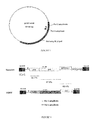
- the MLV-related virus comprises a 5' LTR, gag, pol, env genes, a regulatory domain 3' of the env gene linked to a heterologous polynucleotide to be delivered and a 3' LTR and a promoter for expression in mammalian cells in the 5'LTR.
- the regulatory domain is an internal ribosome entry site (IRES).
- the heterologous polynucleotide encodes a polypeptide having cytosine deaminase activity.
- the method monitors the spread of the MLV-related retroviral vector. In another embodiment, the method is carried out routinely over the course years.
- the disclosure also provides a method for detecting the presence of a viral agent in a sample comprising: measuring the amount of a polynucleotide in a sample using a quantitative polymerase chain reaction or other amplification process comprising oligonucleotide primer/probe combinations selected from the group consisting of: (i) SEQ ID NO: 1, 2 and 3; (ii) SEQ ID NO: 4, 5 and 6; (iii) SEQ ID NO: 7, 8 and 9; (iv) SEQ ID NO: 10, 11 and 12; and (v) primer pairs according to claim 9 and corresponding probes that have at least 95% identity to both XMRV and MLV.
- the polynucleotide is DNA or RNA.
- the quantitating and amplification are performed by quantitative polymerase chain reaction, e.g., RT-qPCR.
- the measuring detects a single copy of a viral agent related nucleic acid.
- the sample can be a mammalian tissue (e.g., blood).
- a viral agent to be detected can be a gene therapy vector.
- the gene therapy vector is a replication-competent vector.
- the method is performed prior to a therapeutic regimen comprising a gene therapy vector treatment.
- the method is performed subsequent to a therapeutic regimen comprising gene therapy vector on a subject.
- the method can be performed to monitor the dosage of a therapeutic regimen comprising a gene therapy vector in a subject.
- the gene therapy vector comprises a replication competent MLV vector.
- the method is performed prior to a therapeutic regimen comprising a gene therapy vector.
- the method is performed subsequent to a therapeutic regimen comprising a gene therapy vector.
- the method is performed to monitor the dosage of a therapeutic regimen comprising a gene therapy vector.
- kits for carrying out any of the foregoing methods and comprising any of the oligonucleotides compositions of the disclosure e.g., SEQ ID NO:1-21, Table 1 and 2).
- the disclosure also provides a method for detecting ⁇ 100 copies of MLV related DNA in a sample extracted from fixed histopathological sections.
- the disclosure also provides a method for detecting ⁇ 100 copies of MLV related RNA in a sample extracted from fixed histopathological sections.
- the disclosure also provides a method that detects both MLV and XMRV and variants thereof. In other embodiment, the method detects only MLV related virus and does not detect XMRV. In certain embodiment, the method detects XMRV gag and MLV gag. In yet other embodiment, the method detects XMRV pol and MLV pol.
- the disclosure provides methods that detects XMRV Env and MLV Env. The disclosure also provides methods for detecting either XMRV or MLV related virus in plasma or serum from a mammalian host.
- the disclosure provides a method of selectively detecting MLV related viruses in humans and which does not detect XMRV comprising primers selected from the group consisting of: SEQ ID NO: 10 and 11; SEQ ID NO:13 and 14; SEQ ID NO:16 and 17; SEQ ID NO: 19 and 20; sequences at least 95% identical to the foregoing; and combination thereof, using the methods described herein.
- the disclosure also provides a method of determining whether a human subject is at risk of having prostate cancer or chronic fatigue syndrome comprising utilizing primer pairs and probes as set forth in SEQ ID NOs: 1, 2, 3, 4, 5, 6, 7, 8, 9, 10, 11, 12, 13 or 14 or primer/probes as set forth in Table 1 or 2, to amplify polynucleotides in a sample from the subject, wherein the presence of an amplified product is indicative of a risk of prostate cancer or chronic fatigue syndrome.
- the disclosure also provides amethod of screening a blood supply or tissue bank for infection by an MLV, MLV-variant or XMRV comprising performing an amplification reaction on the blood supply or tissue bank utilizing primers as set forth in SEQ ID NO:4, 5, 7, 8, 10, 11, 13, 14, or any of the primers in Table 1 or 2, and detecting an amplified product.
- the disclosure is directed to the detection of xenotropic murine leukemia virus related virus (XMRV) or other retroviruses related to murine leukemia viruses that can be present in human tissue blood or serum.
- XMRV xenotropic murine leukemia virus related virus
- the disclosure relates to sensitive reliable quantitative PCR assays for the detection of XMRV provirus in DNA from blood of human and animal subjects and for the sensitive and reliable detection of XMRV RNA (potentially from viral particles) from plasma or serum of human and animal subject by reverse transcription (RT) and polymerase chain reaction.
- the quantitative assay is a TaqMan® assay using the primers and probes constructed based on the genome of the XMRV virus.
- the disclosure constructed PCR based assays that detect MLV related viruses that may or may not be XMRV in human samples, by using MLV specific qPCR primers and probes, that also detect XMRV.
- Such assays are useful in combination with assays for MLV sequences that are not homologous to XMRV to determine if recombination has occurred in patients treated with replication competent retroviruses, who may also be positive for XMRV.
- the disclosure further relates to a diagnostic kit that comprises nucleic acid molecules for the detection of the XMRV and MLV related viruses.
- diagnostic kits that detect the transgene carried by the MLV vector are also disclosed.
- an oligonucleotide includes a plurality of such oligonucleotides and reference to “the polynucleotide” includes reference to one or more polynucleotides known to those skilled in the art, and so forth.
- XMRV or MLV related retroviruses by nucleic acid amplification techniques in human or animal tissues, blood or plasma/serum is of use for determining prostate cancer and risk thereof, chronic fatigue system and risk thereof, contamination of blood supply and tissue donation material and in following the status of subjects undergoing therapy with an MLV derived therapeutic virus comprising a heterologous genetic sequence such as, for example, an engineered retroviral replicatog virus based on amphotropic MLV (e.g., Toca 511).
- an engineered retroviral replicatog virus based on amphotropic MLV e.g., Toca 511).
- the methods and compositions of the disclosure can be used to monitor therapy with an retroviral vector comprising sequences with substantial identity to MLV, in determining if recombination takes place between the therapeutic vector and XMRV or other MLV related natural infections, and for determining if a subject carries XMRV or another MLV related naturally occurring virus.
- Such assays are also useful for screening the blood supply to exclude subjects that are positive for XMRV or other MLV related retroviruses.
- Such assays also can be used to determine levels of MLV related virus over time, and provide information when it would be useful to start administering antiretroviral therapies that are also active against MLV such as, for example, AZT ( Sakuma et al., Virology, 2009 ; Powell et al., J.Virol., 73:8813-8816, 1999 ; G.B.Beck-Engeser, PNAS, 2009 ).
- Such assays when used with histopathology samples can be used to determine the presence or absence of XMRV or other MLV related retroviruses in a patients stored sample or to determine the epidemiology of the XMRV or MLV related virus.
- Such assays can also be used to monitor patients to whom therapeutic vectors based on replicating MLV vectors have been administered. These measurements can be used to track the safety of the therapy over time (e.g., to 15 years and beyond) as high persistent levels (greater than 30,000, 100,000 or 300,000 copies/microgram) of MLV in genomic DNA or greater than 30,000 100,000 or 300,000 RNA copies/ml plasma) or increasing levels of these over time, can be used as a signal to more closely monitor for diseases that could be secondary to a therapy using an gene therapy vector comprising MLV or MLV-related sequences, such as leukemia or to start antiretroviral therapy.
- these measurements can also be used to judge the extent of replication of the MLV or MLV-related vector in a target tissue (i.e., efficacy or susceptibility to successful treatment) because of the possibility of "spill" into the circulatory system.
- efficacy or susceptibility to successful treatment i.e., efficacy or susceptibility to successful treatment
- Other uses of these assays for clinical monitoring will be apparent to those skilled in the art.
- Engineered retroviral vectors that can be monitored include those set forth below:
- RNA transcription based assays for detection of target molecules, such as target nucleic acids comprising viral RNA or DNA, using a plurality of nucleic acid amplification techniques including, for example: NAAT ( J.D. Fox, J.Clin. Virol. 40 Suppl. 1 S15-S23, 2007 ), PCR, RT-PCR, qPCR and RT-qPCR with "touchdown" modifications to improve sensitivity to single copy/assay; RNA transcription based assays (e.g.
- the assays provided by the disclosure may be used in many embodiments to detect sequence-specific nucleic acids. Disclosed herein are different embodiments of assays using amplification (e.g., PCR) and enzymatic degradation of RNA/DNA heteroduplexes.
- amplification e.g., PCR
- enzymatic degradation of RNA/DNA heteroduplexes e.g., RNA/DNA heteroduplexes.
- the disclosure provides a method of identifying MLV-related viral polynucleotides in a subject or sample.
- the disclosure utilizes a combination of primers and probes having identity to conserved regions of MLV-related viruses.
- the primers are used to amplify target polynucleotides in the sample and probes are then used to visualize or detect the amplified products.
- the probe is detectably labeled for detection (e.g., fluorescently labeled, luminescently labeled, enzyme conjugated, radionucleotide labled and the like).
- the primer pairs can be used to amplify MLV-related polynucleotides in a sample such as MLV, XMRV or MLV and XMRV polynucleotides. This is advantageous for the detection of XMRV and naturally occurring variants thereof as well as for detecting MLV and naturally occurring variants thereof (including recombinantly engineered MLV vectors).
- a combination of primers and probes identified herein can be used to identify or detect XMRV in a sample, tissue or subject by using primer pairs having homology to MLV (e.g., primer pairs that share at least 95% sequence identity between and XMRV viral sequence and an MLV viral sequence) and a probe sequence that is specific for XMRV (e.g., the probe only hybridizes to an amplified product under highly stringent conditions).
- primer pairs having homology to MLV e.g., primer pairs that share at least 95% sequence identity between and XMRV viral sequence and an MLV viral sequence
- a probe sequence that is specific for XMRV e.g., the probe only hybridizes to an amplified product under highly stringent conditions.
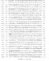
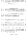
- Primers that share such homology between XMRV and MLV are identified in Figure 5 and Table 2.
- One utility of this general method is to screen blood and tissue supplies for infection.
- XMRV has been suggested to be associated with prostate cancer and chronic fatigue syndrome.
- a subject may be screened prior to undergoing treatment with a recombinant retroviral vector. By identifying subject that may have circulating viral polynucleotides in the tissue a risk of recombination between the inherent viral polynucleotide and the therapeutic viral polynucleotide may be managed.
- a combination of primers and probes identified herein can be used to identify or detect MLV or MLV-related polynucleotides in a sample, tissue or subject by using primer pairs having homology to MLV (e.g., primer pairs that share at least 95% sequence identity between and XMRV viral sequence and an MLV viral sequence) and a probe sequence that is specific for MLV (e.g., the probe only hybridizes to an amplified product under highly stringent conditions).
- primer pairs having homology to MLV e.g., primer pairs that share at least 95% sequence identity between and XMRV viral sequence and an MLV viral sequence
- a probe sequence that is specific for MLV e.g., the probe only hybridizes to an amplified product under highly stringent conditions.
- the methods provide useful diagnostics for monitoring patients after delivery of a replication competent MLV-related viral vector.
- the method can be used to monitor a subject following delivery of the vector on a routine basis (e.g., weekly, monthly, yearly) for as long as a treating physician deems necessary.
- MLV-related virus refers to a retrovirus comprising the general structure of an MLV virus (e.g., LTR-gag-pol-env-LTR) and having at least 60% identity to any of the following sequences set forth in the identified accession numbers (which are incorporated herein by reference in their entirety):
- Any number of different alignment programs can be used to identified regions of identity between any combination of the foregoing MLV-related genomes.
- Other genomes will be readily identified by using a BLAST algorithm or other similar algorithm to identify sequences having homology/identity to the foregoing sequences.
- the disclosure relates to a method of detecting MLV-related viruses including XMRV in a sample comprising contacting the sample with a nucleic acid sequence that hybridizes to all or a portion of XMRV nucleic acid sequence under conditions in which a hybridization complex can occur between the detecting nucleic acid sequence and the XMRV nucleic acid sequence.
- the XMRV specific primers are 95% or more identical to SEQ ID Nos:1 and 2
- the probe is 95% or more identical to SEQ ID NO:3 (XMRV gag).
- the XMRV specific primers are 95% or more identical to SEQ ID NOs:4 and 5 and the probe is 95% or more identical to SEQ ID NO:6 (XMRV env).
- the method uses a combination of primers and probes (e.g., SEQ ID NO:1, 2, 4 and 5 and probes comprising SEQ ID NO:3 and 6).
- the disclosure relates to a method of detecting XMRV or other MLV related nucleic acids in a sample by using primers and probes that are not specific to XMRV but rather are shared between XMRV and other related strains of MLV.
- the MLV/XMRV specific primers are SEQ ID NOs:7 and 8 and the probe is SEQ ID NO:9 (Pol 2 primers and probe; other primers and probes are set forth in Table 1, 2, 3, and 4 below).
- the MLV/XMRV specific primers are SEQ ID NOs:10 and 11 and the probe is SEQ ID NO:12 (pol1 primer and probe).
- sequences can be identified that are common to XMRV and MLV (see the BLAST sequence comparison of two genomes of XMRV and MLV, Figure 5 , where perfect sequence homologies of 20 or more bases are underlined/highlighted). Such homologous sequences (or shorter runs of homology down to 15 bases) can be used to select primers and probes.
- primers and probes can be chosen using programs that compare sequences and suggest common primers and probes. Such programs are usually designed to look for related genes in different species, and can be used to design QPCR reagents for molecules such as MLV and XMRV with significant but incomplete homology.
- An example of such a program is Primaclade http:(//)www.umsl.edu/services/kellogg/primaclade/FAQ.html.
- An oligonucleotide probe or a primer refers to a nucleic acid molecule of between 8 and 2000 nucleotides in length, or is specified to be about 6 and 1000 nucleotides in length. More particularly, the length of these oligonucleotides can range from about 8, 10, 15, 20, or 30 to 100 nucleotides, but will typically be about 10 to 50 (e.g., 15 to 30 nucleotides). The appropriate length for oligonucleotides in assays of the disclosure under a particular set of conditions may be empirically determined by one of skill in the art.
- a primer or probe consisting of at least 95% identity to a reference sequence means that the sequence comprises a sequence that is the same number of oligonucleotides in length, but may differ in nucleotides by 5% from the reference sequence.
- a primer or probe consisting of at least 95% identity to a reference sequence and having from 1-10 additional or deleted nucleotides at the 5' and/or 3' end of the oligonucleotide means that the sequence differs by 1 to up to 20 nucleotides in length from the reference sequence and which also differs 5% or less in identity.
- any primer probe disclosed herein by consist of a reference sequence (e.g., SEQ ID NO: 1-21 or sequences set forth in Table 1 and 2); can consist of a sequence that is 95% of greater in identity to a reference sequence (e.g., SEQ ID NO: 1-21 or sequences set forth in Table 1 and 2); or can consist of a sequence that is at least 95% identical and has an additional 1-20 (e.g., 1, 2, 3, 4, 5, 6, 7, 8, 9 or 10 nucleotides at the 5' and/or 3' end of the reference sequence (e.g., SEQ ID NO: 1-21 or sequence set forth in Table 1 and 2).
- a reference sequence e.g., SEQ ID NO: 1-21 or sequences set forth in Table 1 and 2
- Oligonucleotide primers and probes can be prepared by any suitable method, including direct chemical synthesis and a number of probe systems with derivatized oligonucleotides are available to hybridize to and to detect amplified product, normally by fluorescence change on binding to the probe target.
- the oligonucleotide primers and probes can contain conventional nucleotides, as well as any of a variety of analogs.
- nucleotide refers to a compound comprising a nucleotide base linked to the C-1' carbon of a sugar, such as ribose, arabinose, xylose, and pyranose, and sugar analogs thereof.
- nucleotide also encompasses nucleotide analogs.
- the sugar may be substituted or unsubstituted.
- Substituted ribose sugars include, but are not limited to, those riboses in which one or more of the carbon atoms, for example the 2'-carbon atom, is substituted with one or more of the same or different Cl, F, --R, --OR, --NR 2 or halogen groups, where each R is independently H, C 1 -C 6 alkyl or C 5 -C 14 aryl.
- Exemplary riboses include, but are not limited to, 2'-(C 1 -C 6 )alkoxyribose, 2'-(C 5 -C 14 )aryloxyribose, 2',3'-didehydroribose, 2'-deoxy-3'-haloribose, 2'-deoxy-3'-fluororibose, 2'-deoxy-3'-chlororibose, 2'-deoxy-3'-aminoribose, 2'-deoxy-3'-(C 1 -C 6 )alkylribose, 2'-deoxy-3'-(C 1 -C 6 )alkoxyribose and 2-deoxy-3'-(C 5 -C 14 )aryloxyribose, ribose, 2'-deoxyribose, 2',3'-dideoxyribose, 2'-haloribose, 2'-fluororibos
- Modifications at the 2'- or 3'-position of ribose include, but are not limited to, hydrogen, hydroxy, methoxy, ethoxy, allyloxy, isopropoxy, butoxy, isobutoxy, methoxyethyl, alkoxy, phenoxy, azido, amino, alkylamino, fluoro, chloro and bromo.
- Nucleotides include, but are not limited to, the natural D optical isomer, as well as the L optical isomer forms (see, e.g., Garbesi (1993) Nucl. Acids Res. 21:4159-65 ; Fujimori (1990) J. Amer. Chem. Soc.
- nucleotide base is purine, e.g. A or G
- the ribose sugar is attached to the N 9 -position of the nucleotide base.
- nucleotide base is pyrimidine, e.g.
- the pentose sugar is attached to the N 1 -position of the nucleotide base, except for pseudouridines, in which the pentose sugar is attached to the C 5 position of the uracil nucleotide base (see, e.g., Kornberg and Baker, (1992) DNA Replication, 2nd Ed., Freeman, San Francisco, Calif .).
- the 3' end of the probe can be functionalized with a capture or detectable label to assist in detection of a target polynucleotide or of a polymorphism.
- any of the oligonucleotides or nucleic acids of the disclosure can be labeled by incorporating a detectable label measurable by spectroscopic, photochemical, biochemical, immunochemical, or chemical means.
- labels can comprise radioactive substances (e.g., 32 P, 35 S, 3 H, 125 I), fluorescent dyes (e.g., 5-bromodesoxyuridin, fluorescein, acetylaminofluorene, digoxigenin), molecular quenchers (e.g. blackhole, molecular beacons), biotin, nanoparticles, and others know tothose skilled in the art.
- radioactive substances e.g., 32 P, 35 S, 3 H, 125 I
- fluorescent dyes e.g., 5-bromodesoxyuridin, fluorescein, acetylaminofluorene, digoxigenin
- molecular quenchers e.g. blackhole, molecular beacons
- a probe refers to a molecule which can detectably distinguish changes in gene expression or can distinguish between target molecules differing in structure. Detection can be accomplished in a variety of different ways depending on the type of probe used and the type of target molecule. Thus, for example, detection may be based on discrimination of activity levels of the target molecule, but typically is based on detection of specific binding. Examples of such specific binding include nucleic acid probe hybridization. Thus, for example, probes can include nucleic acid hybridization probes (including primers useful for polynucleotide amplification and/or detection). Thus, in one embodiment, the detection of the presence or absence of the at least one target polynucleotide involves contacting a biological sample with a probe or primer pair.
- an oligonucleotide probe or primer pair where the probe/primers hybridizes with a form of a target polynucleotide in the biological sample containing a complementary sequence, undergoes hybridization using hybridization selective conditions.
- an oligonucleotide probe can include one or more nucleic acid analogs, labels or other substituents or moieties so long as the base-pairing function is retained.
- the disclosure provides methods and systems for identifying and quantifying the amount of a given nucleic acid sequence in a given sample, usually down to a single copy per sample. Furthermore, the methods and systems of the disclosure provide sequence specific detection useful for differentiating/identifying related genomic sequences and provide a detectable signal when the correct target sequence is present. The disclosure provides various embodiments of the invention.
- Methods known in the art can be used to quantitatively measure the amount of a nucleic acid present in a sample. Examples of such methods include quantitative polymerase chain reaction (qPCR), and other NAAT technologies as described above.
- qPCR quantitative polymerase chain reaction
- a method for detecting a specific viral polynucleotide is provided by the disclosure.
- Such a method can include the use of primers, probes, enzymes, and other reagents for the preparation, detection, and quantitation of a viral polynucleotide (e.g., by PCR, by Northern blot and the like).
- the primers listed in SEQ ID NOs: 1-12 are particularly suited for use in profiling using RT-PCR based on a viral polynucleotide.
- Other primers (sense and antisense) and probes are provided in Table 2.
- One of skill in the art can readily identity in Table 2, the combinations of primer/probes useful for detection of an MLV, MLV-related or XMRV viral sequence.
- one method for detection of polynucleotide amplicons is fluorescence spectroscopy, and therefore labels suited to fluorescence spectroscopy are desirable for detecting polynucleotide.
- the amplicon polynucleotide is detected without a hybridization probe but directly with a fluorophore that binds DNA and fluoresces at a wavelength different from that of the free reagent.
- fluorescent label is SYBR Green, though numerous related fluorescent molecules are known including, without limitation, DAPI, Cy3, Cy3.5, Cy5, CyS.5, Cy7, umbelliferone, fluorescein, fluorescein isothiocyanate (FITC), rhodamine, dichlorotriazinylamine fluorescein, dansyl chloride or phycoerythrin.
- an oligonucleotide primer pair is used to amplify a polynucleotide corresponding to the pol gene of a murine retrovirus.
- Primers comprising SEQ ID NOs:7 and 8 (forward and reverse primers, respectively) are used to amplify the region of the pol gene.
- the amplified region can then be detected using a probe (SEQ ID NO:9), which specifically hybridizes to the amplified polynucleotide.
- the probe can be labeled with any number of detectable labels as described herein.
- the foregoing primers (SEQ ID NOs:7, 8 and 10, 11) with the appropriate probes (SEQ ID NOs:9 and 12, respectively) can be used to detect the presence of, for example, Murine Leukemia Virus (MLV) and Xenotropic Murine Retrovirus (XMRV). As described elsewhere herein identifying the presence of MLV and/or XMRV is useful for the determination of a therapeutic gene delivery and cancer treatment regimen.
- MLV Murine Leukemia Virus
- XMRV Xenotropic Murine Retrovirus
- an oligonucleotide primer pair is used to amplify a polynucleotide corresponding to the gag gene of a XMRV.
- Primers comprising SEQ ID NOs:1 and 2 (forward and reverse primers, respectively) are used to amplify the region of the gag gene of XMRV.
- the amplified region can then be detected using a probe (SEQ ID NO:3) which specifically hybridizes to the amplified polynucleotide.
- the probe can be labeled with any number of detectable labels as described herein. This combination of primers and probes is useful for specifically identifying the presence of an XMRV infection or contamination.
- an oligonucleotide primer pair is used to amplify a polynucleotide corresponding to the LTR of an MLV vector.
- Primers comprising SEQ ID NOs:16 and 17 (forward and reverse primers, respectively) are used to amplify the region of the LTR of an MLV vector (e.g., Toca511).
- the amplified region can then be detected using a probe (SEQ ID NO: 18) which specifically hybridizes to the amplified polynucleotide.
- the probe can be labeled with any number of detectable labels as described herein. This combination of primers and probes is useful for specifically identifying the presence of a retroviral vector during gene delivery monitoring.
- an oligonucleotide primer pair is used to amplify a polynucleotide corresponding to a polynucleotide encoding a cytosine deaminase.
- Primers comprising SEQ ID NOs:19 and 20 (forward and reverse primers, respectively) are used to amplify the region of the a cytosine deaminase delivered using an MLV vector (e.g., Toca511).
- MLV vector e.g., Toca511
- the amplified region can then be detected using a probe (SEQ ID NO:21) which specifically hybridizes to the amplified polynucleotide.
- the probe can be labeled with any number of detectable labels as described herein. This combination of primers and probes is useful for specifically identifying the presence of a retroviral vector during gene delivery monitoring.
- the primers (SEQ ID NOs: 1 and 2) and probe (SEQ ID NO:3) can be used to detect the presence of XMRV. As described elsewhere herein identifying the presence of XMRV is useful for the determination of a therapeutic gene delivery, cancer treatment regimen and blood supply screening.
- an oligonucleotide primer pair is used to amplify a polynucleotide corresponding to the env gene of a XMRV.
- Primers comprising SEQ ID NOs:4 and 5 (forward and reverse primers, respectively) are used to amplify the region of the env gene of XMRV.
- the amplified region can then be detected using a probe (SEQ ID NO:6) which specifically hybridizes to the amplified polynucleotide.
- the probe can be labeled with any number of detectable labels as described herein. This combination of primers and probes is useful for specifically identifying the presence of an XMRV infection or contamination.
- primers SEQ ID NOs:4 and 5
- probe SEQ ID NO:6
- identifying the presence of XMRV is useful for the determination of a therapeutic gene delivery, cancer treatment regimen and screening the blood supply.
- any of the oligonucleotide primers and probes of the disclosure can be immobilized on a solid support.
- Solid supports are known to those skilled in the art and include the walls of wells of a reaction tray, test tubes, polystyrene beads, magnetic beads, nitrocellulose strips, membranes, microparticles such as latex particles, glass and the like.
- the solid support is not critical and can be selected by one skilled in the art.
- latex particles, microparticles, magnetic or non-magnetic beads, membranes, plastic tubes, walls of microtiter wells, glass or silicon chips and the like are all suitable examples.
- Suitable methods for immobilizing oligonucleotides on a solid phase include ionic, hydrophobic, covalent interactions and the like.
- the solid support can be chosen for its intrinsic ability to attract and immobilize the capture reagent.
- the oligonucleotide probes or primers of the disclosure can be attached to or immobilized on a solid support individually or in groups of about 2-10,000 distinct oligonucleotides of the disclosure to a single solid support.
- a substrate comprising a plurality of oligonucleotide primers or probes of the disclosure may be used either for detecting or amplifying targeted sequences.
- the oligonucleotide probes and primers of the disclosure can be attached in contiguous regions or at random locations on the solid support.
- the oligonucleotides of the disclosure may be attached in an ordered array wherein each oligonucleotide is attached to a distinct region of the solid support which does not overlap with the attachment site of any other oligonucleotide.
- such oligonucleotide arrays are "addressable" such that distinct locations are recorded and can be accessed as part of an assay procedure.
- oligonucleotide probes can be used in an oligonucleotide chip such as those marketed by Affymetrix and described in U.S. Pat. No. 5,143,854 ; PCT publications WO 90/15070 and 92/10092 , the disclosures of which are incorporated herein by reference.
- arrays can be produced using mechanical synthesis methods or light directed synthesis methods which incorporate a combination of photolithographic methods and solid phase oligonucleotide synthesis.
- an array of oligonucleotides complementary to subsequences of the target gene for example yeast cytosine deaminase (CD) or a version of CD optimized for expression in human cells
- CD yeast cytosine deaminase
- a version of CD optimized for expression in human cells is used to determine the identity of the target, measure its amount and the like.
- Hybridization techniques can also be used to identify the viral polynucleotides in a subject or sample and thereby determine or predict cross reactivity, chances of recombination or a treatment regimen using a gene delivery vector comprising a recombinant MLV vector.
- the hybridization reactions may be carried out in a solid support (e.g., membrane or chip) format, in which, for example, a probe (e.g., SEQ ID NO:3, 6 and/or 9) are immobilized on nitrocellulose or nylon membranes and probed with amplified preparations of nucleic acids obtained, for example, from PCR using primers comprising SEQ ID NO:1, 2, 4, 5, 7, 8, 10, 11, 13 and/or 14 of the disclosure.
- any of the known hybridization formats may be used, including Southern blots, slot blots, "reverse" dot blots, solution hybridization, solid support based sandwich hybridization, bead- based, silicon chip-based and microtiter well-based hybridization formats.
- Hybridization of an oligonucleotide probe to a target polynucleotide may be performed with both entities in solution, or such hybridization may be performed when either the oligonucleotide or the target polynucleotide is covalently or noncovalently affixed to a solid support.
- Attachment to a solid support may be mediated, for example, by antibody-antigen interactions, poly-L-Lysine, streptavidin or avidin-biotin, salt bridges, hydrophobic interactions, chemical linkages, UV cross-linking baking, etc.
- Oligonucleotides may be synthesized directly on the solid support or attached to the solid support subsequent to synthesis.
- the solid support may be treated, coated or derivatized to facilitate the immobilization of the specific oligonucleotide.
- Hybridization assays based on oligonucleotide arrays rely on the differences in hybridization stability of short oligonucleotides to perfectly matched and mismatched target variants.
- Each DNA chip can contain thousands to millions of individual synthetic DNA probes arranged in a grid-like pattern and miniaturized to the size of a dime or smaller.
- Such a chip may comprise oligonucleotides representative of both a wild-type and variant sequences.
- Oligonucleotides of the disclosure can be designed to specifically hybridize to a target region of a polynucleotide.
- specific hybridization means the oligonucleotide forms an anti-parallel double-stranded structure with the target region under certain hybridizing conditions, while failing to form such a structure when incubated with a different target polynucleotide or another region in the polynucleotide or with a polynucleotide lacking the desired locus under the same hybridizing conditions.
- the oligonucleotide specifically hybridizes to the target region under conventional high stringency conditions.
- a nucleic acid molecule such as an oligonucleotide or polynucleotide is said to be a "perfect” or “complete” complement of another nucleic acid molecule if every nucleotide of one of the molecules is complementary to the nucleotide at the corresponding position of the other molecule.
- a nucleic acid molecule is "substantially complementary” to another molecule if it hybridizes to that molecule with sufficient stability to remain in a duplex form under conventional low-stringency conditions. Conventional hybridization conditions are described, for example, in Sambrook et al., Molecular Cloning, A Laboratory Manual, 2nd ed., Cold Spring Harbor Press, Cold Spring Harbor, N. Y.
- an oligonucleotide primer may have a non-complementary fragment at its 5' or 3' end, with the remainder of the primer being complementary to the target region.
- hybridization conditions may be used in the disclosure, including high, moderate and low stringency conditions; see for example Maniatis et al., Molecular Cloning: A Laboratory Manual, 2d Edition, 1989 , and Short Protocols in Molecular Biology, ed. Ausubel , et al., hereby incorporated by reference. Stringent conditions are sequence-dependent and will be different in different circumstances. Longer sequences hybridize specifically at higher temperatures. An extensive guide to the hybridization of nucleic acids is found in Tijssen, Techniques in Biochemistry and Molecular Biology--Hybridization with Nucleic Acid Probes, "Overview of principles of hybridization and the strategy of nucleic acid assays" (1993 ).
- stringent conditions are selected to be about 5-10°C lower than the thermal melting point (T m ) for the specific sequence at a defined ionic strength and pH.
- T m is the temperature (under defined ionic strength, pH and nucleic acid concentration) at which 50% of the probes complementary to the target hybridize to the target sequence at equilibrium (as the target sequences are present in excess, at T m , 50% of the probes are occupied at equilibrium).
- Stringent conditions will be those in which the salt concentration is less than about 1.0 M sodium ion, typically about 0.01 to 1.0 M sodium ion concentration (or other salts) at pH 7.0 to 8.3 and the temperature is at least about 30° C for short probes (e.g., 10 to 50 nucleotides) and at least about 60° C for long probes (e.g., greater than 50 nucleotides).
- Stringent conditions may also be achieved with the addition of helix destabilizing agents such as formamide.
- the hybridization conditions may also vary when a non- ionic backbone, i.e., PNA is used, as is known in the art.
- cross-linking agents may be added after target binding to cross-link, i.e., covalently attach, the two strands of the hybridization complex.
- target polynucleotides can be obtained from samples including, but not limited to, bodily fluids (e.g., blood, urine, serum, lymph, saliva, anal and vaginal secretions, perspiration and semen) of virtually any organism, with mammalian samples common to the methods of the disclosure and human samples being typical.
- bodily fluids e.g., blood, urine, serum, lymph, saliva, anal and vaginal secretions, perspiration and semen
- the sample may comprise individual cells, including primary cells (including bacteria) and cell lines including, but not limited to, tumor cells of all types (particularly melanoma, myeloid leukemia, carcinomas of the lung, breast, ovaries, colon, kidney, prostate, pancreas and testes); cardiomyocytes; endothelial cells; epithelial cells; lymphocytes (T-cell and B cell); mast cells; eosinophils; vascular intimal cells; hepatocytes; leukocytes including mononuclear leukocytes; stem cells such as haemopoetic, neural, skin, lung, kidney, liver and myocyte stem cells; osteoclasts; chondrocytes and other connective tissue cells; keratinocytes; melanocytes; liver cells; kidney cells; and adipocytes.
- primary cells including bacteria
- cell lines including, but not limited to, tumor cells of all types (particularly melanoma, myeloid leukemia, carcinomas of the lung, breast,
- Suitable cells also include known research cells, including, but not limited to, Jurkat T cells, NIH3T3 cells, CHO, Cos, 923, HeLa, SiHa, WI-38, Weri-1, MG-63, and the like (see the ATCC cell line catalog, hereby expressly incorporated by reference).
- a target polynucleotide (e.g., a virus polynucleotide or gene) may be amplified using any oligonucleotide-directed amplification method including, but not limited to, polymerase chain reaction (PCR) ( U.S. Pat. No. 4,965,188 ), ligase chain reaction (LCR) ( Barany et al., Proc. Natl. Acad. Sci. USA 88:189-93 (1991 ); WO 90/01069 ), and oligonucleotide ligation assay (OLA) ( Landegren et al., Science 241:1077-80 (1988 )).
- PCR polymerase chain reaction
- LCR ligase chain reaction
- OLA oligonucleotide ligation assay
- nucleic acid amplification procedures may be used to amplify the target region (s) including transcription-based amplification systems ( U.S. Pat. No. 5,130,238 ; European Patent No. EP 329,822 ; U.S. Pat. No. 5,169,766 ; WO 89/06700 ) and isothermal methods ( Walker et al., Proc. Natl. Acad. Sci. USA 89:392-6 (1992 )).
- Ligase Chain Reaction (LCR) techniques can be used and are particularly useful for detection of single or multiple (e.g., 1, 2, 3, 4, or 5) nucleotide differences between similar polynucleotides. LCR occurs only when the oligonucleotides are correctly base-paired.
- the Ligase Chain Reaction (LCR) which utilizes the thermostable Taq ligase for ligation amplification, is useful for interrogating loci of a gene. LCR differs from PCR because it amplifies the probe molecule rather than producing amplicon through polymerization of nucleotides. Two probes are used per each DNA strand and are ligated together to form a single probe.
- LCR uses both a DNA polymerase enzyme and a DNA ligase enzyme to drive the reaction. Like PCR, LCR requires a thermal cycler to drive the reaction and each cycle results in a doubling of the target nucleic acid molecule. LCR can have greater specificity than PCR. The elevated reaction temperatures permit the ligation reaction to be conducted with high stringency. Where a mismatch occurs, ligation cannot be accomplished. For example, a probe based upon a target polynucleotide is synthesized in two fragments and annealed to the template with possible difference at the boundary of the two primer fragments. A ligase ligates the two primers if they match exactly to the template sequence.
- the two hybridization probes are designed each with a target specific portion.
- the first hybridization probe is designed to be substantially complementary to a first target domain of a target polynucleotide (e.g., a polynucleotide fragment) and the second hybridization probe is substantially complementary to a second target domain of a target polynucleotide (e.g., a polynucleotide fragment).
- each target specific sequence of a hybridization probe is at least about 5 nucleotides long, with sequences of about 15 to 30 being typical and 20 being especially common.
- the first and second target domains are directly adjacent, e.g., they have no intervening nucleotides.
- At least a first hybridization probe is hybridized to the first target domain and a second hybridization probe is hybridized to the second target domain. If perfect complementarity exists at the junction, a ligation structure is formed such that the two probes can be ligated together to form a ligated probe. If this complementarity does not exist (due to mismatch), no ligation structure is formed and the probes are not ligated together to an appreciable degree. This may be done using heat cycling, to allow the ligated probe to be denatured off the target polynucleotide such that it may serve as a template for further reactions. The method may also be done using three hybridization probes or hybridization probes that are separated by one or more nucleotides, if dNTPs and a polymerase are added (this is sometimes referred to as "Genetic Bit" analysis).
- Quantitative PCR and digital PCR can be used to measure the level of a polynucleotide in a sample.
- Digital Polymerase Chain Reaction (digital PCR, dPCR or dePCR) can be used to directly quantify and clonally amplify nucleic acids including DNA, cDNA or RNA.
- Digital PCR amplifies nucleic acids by temperature cycling of a nucleic acid molecule with a DNA polymerase. The reaction is typically carried out in the dispersed phase of an emulsion capturing each individual nucleic acid molecule present in a sample within many separate chambers or regions prior to PCR amplification. A count of chambers containing detectable levels of PCR end-product is a direct measure of the absolute nucleic acids quantity.
- Quantitative polymerase chain reaction is a modification of the polymerase chain reaction and real-time quantitative PCR are useful for measuring the amount of DNA after each cycle of PCR by use of fluorescent markers or other detectable labels.
- Quantitative PCR methods use the addition of a competitor RNA (for reverse-transcriptase PCR) or DNA in serial dilutions or co-amplification of an internal control to ensure that the amplification is stopped while in the exponential growth phase.
- a probe or primer of the disclosure can be associated with a detectable label.
- a signaling component can include any label that can be detected optically, electronically, radioactively and the like.
- a nucleic acid analog may serve as the signaling component.
- label or “detectable label” is meant a moiety that allows detection.
- the detection label is a primary label.
- a primary label is one that can be directly detected, such as a fluorophore.
- labels fall into three classes: a) isotopic labels, which may be radioactive or heavy isotopes; b) magnetic, electrical, thermal labels; and c) colored or luminescent dyes. Common labels include chromophores or phosphors but are typically fluorescent dyes.
- Suitable dyes for use in the disclosure include, but are not limited to; fluorescent lanthamide complexes, including those of Europium and Terbium, fluorescein, rhodamine, tetramethylrhodamine, eosin, erythrosin, coumarin, methyl-coumarins, quantum dots (also referred to as "nanocrystals"), pyrene, Malacite green, stilbene, Lucifer Yellow, Cascade BlueTM, Texas Red, Cy dyes (Cy3, Cy5, and the like), Alexa dyes, phycoerythin, bodipy, and others described in the 6th Edition of the Molecular Probes Handbook by Richard P. Haugland , hereby expressly incorporated by reference.
- fluorescent lanthamide complexes including those of Europium and Terbium, fluorescein, rhodamine, tetramethylrhodamine, eosin, erythrosin, coumarin, methyl-coumarin
- Such a detectable label may be a radioactive label or may be a luminescent, fluorescent of enzyme label.
- Indirect detection processes typically comprise probes covalently labeled with a hapten or ligand such as digoxigenin (DIG) or biotin.
- the target-probe duplex is detected by an antibody- or streptavidin-enzyme complex.
- Enzymes commonly used in DNA diagnostics are horseradish peroxidase and alkaline phosphatase.
- Direct detection methods include the use of fluorophor-labeled oligonucleotides, lanthanide chelate-labeled oligonucleotides or oligonucleotide-enzyme conjugates. Examples of fluorophor labels are fluorescein, rhodamine and phthalocyanine dyes.
- embodiments of the invention include probes having fluorescent dye molecules, fluorescent compounds, or other fluorescent moieties.
- a dye molecule may fluoresce, or be induced to fluoresce upon excitation by application of suitable excitation energy (e.g., electromagnetic energy of suitable wavelength), and may also absorb electromagnetic energy (“quench”) emitted by another dye molecule or fluorescent moiety.
- suitable excitation energy e.g., electromagnetic energy of suitable wavelength
- quench electromagnetic energy
- Any suitable fluorescent dye molecule, compound or moiety may be used in the practice of the invention.
- suitable fluorescent dyes, compounds, and other fluorescent moieties include fluorescein, 6-carboxyfluorescein (6-FAM), 2',4', 1,4,-tetrachlorofluorescein (TET), 2',4',5',7',1,4-hexachlorofluorescein (HEX), 2',7'-dimethoxy-4',5'-dichloro-6-carboxyrhodamine (JOE), 2'-chloro-5'-fluoro-7',8'-fused phenyl-1,4-dichloro-6-carboxyfluorescein (NED) and 2'-chloro-7'-phenyl-1,4-dichloro-6-carboxyfluorescein (VIC), cyanine dyes (e.g., Cy.sup.3, Cy.sup.5, Cy.sup.9, nitrothiazole blue (NTB)), Cys3, FAM.TM., tetramethyl-6
- An oligonucleotide according to the methods of the invention may be labeled at the 5' end or the 3' end of at least one subunit of the probe.
- oligonucleotides may be labeled at both the 5' end and the 3' end.
- at least one subunit of the probe may be labeled internally, having at least one, and, in embodiments, more than one, internal label.
- an oligonucleotide may be labeled at an end and may be labeled internally.
- the oligonucleotides themselves are synthesized using techniques that are also well known in the art.
- Methods for preparing oligonucleotides of specific sequence include, for example, cloning and restriction digest analysis of appropriate sequences and direct chemical synthesis, including, for example, the phosphotriester method described by Narang et al., 1979, Methods in Enzymology, 68:190 , the phosphodiester method disclosed by Brown et al., 1979, Methods in Enzymology, 68:109 , the diethylphosphoramidate method disclosed in Beaucage et al., 1981, Tetrahedron Letters, 22:1859 , and the solid support method disclosed in U.S. Pat. No. 4,458,066 , or by other chemical methods using a commercial automated oligonucleotide synthesizer. Modified linkages also may be included, for example phosphorothioates.
- detection modes contemplated for the disclosed methods include, but are not limited to, spectroscopic techniques, such as fluorescence and UV-Vis spectroscopy, scintillation counting, and mass spectroscopy.
- labels for the purpose of detection and quantitation used in these methods include, but are not limited to, chromophoric labels, scintillation labels, and mass labels.
- the expression levels of polynucleotides and polypeptides measured using these methods may be normalized to a control established for the purpose of the targeted determination.
- Label detection will be based upon the type of label used in the particular assay. Such detection methods are known in the art. For example, radioisotope detection can be performed by autoradiography, scintillation counting or phosphor imaging. For hapten or biotin labels, detection is with an antibody or streptavidin bound to a reporter enzyme such as horseradish peroxidase or alkaline phosphatase, which is then detected by enzymatic means. For fluorophor or lanthanide-chelate labels, fluorescent signals may be measured with spectrofluorimeters with or without time-resolved mode or using automated microtitre plate readers.
- a reporter enzyme such as horseradish peroxidase or alkaline phosphatase
- detection is by color or dye deposition (p-nitropheny phosphate or 5-bromo-4-chloro-3-indolyl phosphate/nitroblue tetrazolium for alkaline phosphatase and 3,3'- diaminobenzidine-NiCh for horseradish peroxidase), fluorescence (e.g., 4-methyl umbelliferyl phosphate for alkaline phosphatase) or chemiluminescence (the alkaline phosphatase dioxetane substrates LumiPhos 530 from Lumigen Inc., Detroit Mich, or AMPPD and CSPD from Tropix, Inc.). Chemiluminescent detection may be carried out with X-ray or polaroid film or by using single photon counting luminometers.
- the methods, compositions, systems and devices disclosed herein find use in the identification and quantization of a target DNA or RNA polynucleotide in a sample, such as in a pool of sequences including one or more target sequences, which may be unrelated polynucleotides.
- Quantization of specific nucleic acid samples may be achieved by comparing the total signal (fluorescent or otherwise) obtained during the assay with a standard curve of known polynucleotide target concentrations.
- Specific examples of applications include the detection of pathogenic viruses through the detection of their biomolecules such as DNA or RNA which are indicative of the presence of said targets.
- Assays having features of the invention may be used to detect and identify the presence of specific DNA sequences and may be used in assays for diagnosis of many types of infection and disease.
- sample preparation kits may be used.
- samples suspected of containing pathogenic DNA may be used.
- Exemplary kits and protocols that can be used include the QIAamp MinElute Virus Spin Kit provided by Qiagen. This kit allows DNA isolation from clinical samples in roughly 1 hour. Other methods for sample preparation are available from suppliers such as Promega.
- Polynucleotides may be prepared from samples using known techniques.
- the sample may be treated to lyse a cell comprising the target polynucleotide, using known lysis buffers, sonication techniques, electroporation, and the like.
- lysis buffers e.g., lysis buffers, sonication techniques, electroporation, and the like.
- electroporation e.g., electroporation, and the like.
- Many methods for cell lysis are common knowledge for those trained in the art.
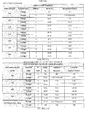
- Ct Cycle number (in qPCR) at which the fluorescence generated within a reaction well exceeds the defined threshold.
- the threshold is arbitrarily defined by the qPCR instrument manufacturer to reflect the point during the reaction at which a sufficient number of amplicons have accumulated.
- gDNA genomic DNA: Deoxyribonucleic acid that has been purified from tissue and/or cultured cells.
- %CV Percentage Coefficient of Variation
- Slope The slope or gradient of a line describes its steepness, incline, or grade.
- An acceptable slope of the linear regression equation for the qPCR should be within the range of -3.00 to -3.7.
- R-squared (R 2 ) Value also known as the Pearson Correlation Coefficient: The correlation of the line, R 2 , is a measure of how well the data fits the model and how well the data fits on a straight line. It is influenced by pipetting accuracy and by the range of the assay. An R 2 of ⁇ 0.94 is acceptable.
- ND Non-detected.
- NTC Non-template control
- Two primer probe sets were designed for detection of all MLV related retroviruses. Amplification curves are shown in Figure 7 .
- One primer probe set was designed for detection of a cytosine deaminase (CD) gene.
- Oligonucleotides for primer probe sets were ordered from IDT (Integrated DNA Technologies, Inc., San Diego, California).
- Example 2 Preparation of genomic DNA from blood and other tissues from mammals including human and canines for PCR testing.
- the XMRV (xenotropic murine leukemia virus-related virus) qPCR assay is performed to quantify DNA.
- Total DNA extraction from the specimens samples is generated by standard means such as the use of commercially available kits (QIAGEN DNA blood mini kit, QIAGEN DNA Tissue kit, Promega DNA Tissue Kit, Promega DNA Cell Kit).
- a quantitation curve is established with 8 non-zero samples comprising of serial dilutions of defined copy number of reference plasmid to generate a Ct value versus copy number correlation. Linear regression analysis generates an equation which is used to calculate the copy number in the sample. Quantitative curves generation are shown for XMRV gag ( Figure 8 ), XMRV env ( Figure 9 ), XMRV pol2 ( Figure 10 ).
- Example 3 Preparation of plasma from humans and dogs for RT-PCR testing.
- RNA samples such as whole blood and plasma
- the assay employs a two-step amplification process with the initial step consisting of the distribution of 2 ⁇ L of experimental sample directly into a cDNA reaction mix. Following completion of the reverse transcriptase (RT) cDNA synthesis, a 2 ⁇ L aliquot is removed, transferred into a qPCR reaction mix and a qPCR protocol is performed.
- RT reverse transcriptase
- a quantitation curve is established with 6 non-zero samples comprising of serial dilutions of defined copy number of reference vector to generate a Ct value versus copy number correlation.
- Linear regression analysis generates an equation which is used to calculate the copy number in the sample.
- Quantitative curves generation are shown in Figure 15 .
- a matrix of primers and probe were made up in various concentration combinations and were used to target the appropriate XMRV plasmid (pUC57 XMRV gag, pET28b XMRV env or pAZ3-emd pol2) containing the gene of interest.
- the choice of optimal primer concentrations were made based on comparisons of Ct value, standard deviation and relative fluorescence units (RFU).
- REU relative fluorescence units
- SYBR Green was used for the primer concentration optimization
- TaqMan was used for the probe concentration optimization assay.
- Annealing temperature optimization was carried out by performing a qPCR annealing temperature gradient ranging from 50°C to 65°C. Plasmids specific for the gene of interest were targeted with the appropriate XMRV primer sets.
- Genomic DNA extracted from human whole blood was spiked with known copy numbers of plasmid DNA containing the gene of interest. Serial log dilutions of the spiked gDNA were made and qPCR was performed. The samples were targeted with XMRV env ( Figure 9 ), XMRV gag ( Figure 8 ) and XMRV pol2 ( Figure 10 ) primer/probe sets in single qPCR reactions.
- Human whole blood gDNA was spiked with known copy numbers of plasmid DNA containing the gene of interest.
- Serial log dilutions of the spiked gDNA were made and a modified version of the qPCR protocol was performed by adding a set of three pre-cycling steps (defined as a one-stage qPCR protocol) to the current qPCR protocol.
- the samples were targeted with XMRV env ( Figure 9 ), XMRV gag ( Figure 8 ) and XMRV pol2 ( Figure 10 ) primer/probe sets in single qPCR reactions.
- Genomic DNA from whole blood from healthy donors were used to spike in known copy numbers of plasmid DNA. Serial dilutions of the gDNA were made to generate 1E3, 1E2, 1E1 and 1E0 copies per reaction. A 0-stage and a 1-stage qPCR protocol were performed. The samples were targeted with XMRV env ( Figure 12 ), XMRV gag ( Figure 11 ) or XMRV pol2 ( Figure 13 ) primer/probe sets in single qPCR reactions.
- 22Rv1 gDNA (positive for XMRV, E.C. Knouf et al. J.Virol 83:78353-7356 2009 ) was spiked into purified human whole blood gDNA (pre and post gDNA extraction) at increasing log dilutions (one human whole blood sample control and one TE sample were spiked pre-extraction with 500 ng of 22Rv1 gDNA to yield a final concentration of ⁇ 2.5 ng/ ⁇ L).
- a 0-stage and a 1-stage qPCR protocol were performed with primers targeting the XMRV gag, XMRV env and XMRV pol sequences.
- Both XMRV gag and XMRV env primer sets are XMRV specific whereas the Pol primer sets detect both MLV and XMRV.
- FIGS 6 and 7 show results obtained by the methods and compositions disclosed above.
- TaqqMan® Gold RT-PCR Kit and TaqMan® PCR universal master mix are obtained from PE Biosystems.
- RNAeasy® mini kit and QIAamp® viral RNA mini kit are obtained from Qiagen.
- Various cell culture materials and biological samples to be tested are obtained from vendors or subjects.
- MLV recombinant isolates comprise the sequences set forth in International Application No. PCT/US09/58512 and published on April 1, 2010 as publication no. WO 2010/036986 .
- Two primer/probe sets for the detection of XMRV were designed as set forth above.
- One forward primer (FP), one reverse primer (RP), and one probe were used for the detection of XMRV gag and XMRV env.
- a third set of primer/probe was used for the detection of XMRV and MLV using the primers above that amplify the pol region of XMRV and MLV.
- the qPCR reaction mixture contains 900 nM primers (both forward and reverse) and 200 nM probe. Concentrations tested to be effective for detection include, 100, 200, 300, 400, 500, 600, 700, 800, 900 nM and any ratio between 1:1, 1:2, 1:3, 1:4 of primer concentrations.
- the activation of Taq polymerase is achieved at 95°C for 5 minutes is followed by forty-four cycles of denaturation at 95°C for 15 seconds and annealing and elongation at 65° C for 30 seconds.
- Example 5 Detection of MLV in formalin fixed paraffin embedded tissue samples.
- Tumors from mice were removed and divided into 2 equal parts.
- One part of the tumor was formalin-fixed, paraffin-embedded and the other part of the tumor was frozen at -80°C.
- the FFPE mouse tumor tissue was cut in half, with one half spiked-in with a known copy number amount of pAZ3-emd and the other half was not spiked-in.
- a known copy number amount of pAZ3-emd was spiked-in to the frozen fresh mouse tissue and pre-processing incubation buffer.
- the FFPE and frozen fresh mouse tissues were incubated at 56°C overnight in a pre-processing incubation buffer containing proteinase K and dithiothreitol (DTT).
- the mouse tissue was processed on the Maxwell 16 instrument to extract out gDNA as per standard procedure.
- the extracted gDNA concentration was quantified on the Nanodrop 1000.
- the extracted DNA was tested for presence of MLV and env2 sequences by qPCR with the results shown in Figure 14 .
- Example 6 XMRV/MLV RT-PCR Assay.
- the XMRV (xenotropic murine leukemia virus-related virus) RT-PCR assay is performed to quantify RNA from biological samples, such as whole blood and plasma, without the need for RNA extraction.
- the assay employs a two-step amplification process with the initial step consisting of the distribution of 2 ⁇ L of experimental sample directly into a cDNA reaction mix. Following completion of the reverse transcriptase (RT) cDNA synthesis, a 2 ⁇ L aliquot is removed, transferred into a qPCR reaction mix and a qPCR protocol is performed. A quantitation curve is established with 7 non-zero samples comprising of serial dilutions of defined copy number of reference vector to generate a Ct value versus copy number correlation. Linear regression analysis generates an equation which is used to calculate the copy number in the sample. ( Figure 15 ).
- a negative matrix sample i. e. whole blood
- a defined quantity of 22Rv1 viral vector see description under 'Reference Standard'. This control is prepared fresh with each run to determine the efficiency of the cDNA generation in the RT step.
- 22Rv1 genomic DNA containing the integrated retroviral vector sequences of XMRV provides the best biophysical mimic of the actual amplification target to be screened in patient tissues.
- a negative matrix sample i. e. whole blood
- This control is prepared fresh with each run to verify that non-specific products are not generated during cDNA synthesis of the qRT step. Confirmation is obtained upon completion of the qPCR procedure. No amplification is expected.
- DNA isolated from non-infected U-87 cell is used as a negative control as it does not contain any endogenous sequences detectable by the XMRV primer sets.
- the 22Rv1 human prostate carcinoma epithelial cell line has been shown to produce high-titer of the human retrovirus XMRV.
- This cell line was bought from ATCC and propagated in RPMI-1640 Medium containing 10% FBS, Sodium Pyruvate and Glutamax. The cell line was passaged four times before obtaining the supernatant containing the viral vector. The supernatant was filtered through a 0.45 ⁇ m filter and stored at -80°C.
- Reference vector 22Rv1 was used to spike PBS for generating a quantitation curve.
- Known copy numbers of vector were serially diluted to generate a Ct value versus copy number correlation.
- Linear regression analysis generates an equation which was used to calculate the copy number in the sample.
- Copy number was determined by a titer analysis which measures the number of copies of the viral genome integrated into the genome of target cells (transduction units, TU). The copy number was measured in TU equivalents.
- Two primer probe sets were designed for detection of all MLV related retroviruses and XMRV.
- Two primer probe sets were designed for detection of amphotropic MLV virus.
- Example 7 Monitoring of GBM patients treated with an MLV vector.
- Gd-MRI gadolinium-enhanced MRI
- the trough 5-FC serum concentration was determined and the dose of 5-FC adjusted in subsequent cycles to maintain the trough concentration in the therapeutic range. If tolerated, these 6-day courses of 5-FC were repeated approximately every 4 weeks ( ⁇ 1 week) until institution of new antineoplastic treatment for tumor progression.
- Tumor response are assessed using the Macdonald criteria.
- a standard dose-escalation algorithm is being followed.
- Three subjects are evaluated at each of up to four dose levels of Toca 511 (2.6 x 10 3 , 9.5 x 10 3 , 2.5 x 10 4 , and the Maximum Feasible Dose [MFD], not to exceed 1 x 10⁵ TU/g). So far three patients at the lowest dose level have been treated.
- Two patients 101 and 102 were monitored using qPCR testing of whole blood DNA (MLVLTR Primer probe set) and RT-qPCR using the MLV env2 primer-probe set.
- saliva an urine were monitored by DNA qPCR, and antibodies to the vector were measured (ref MLV ELISA Application?). These data are shown in Figure 16 .
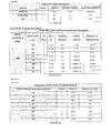
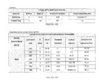
- Table 1 Sequence Definition Pair Rating Product Length Product Tm Sense Primer Anti-sense Primer 5 XMRV position 3 XMRV position XMRV 1581-1778 66.8 178 78.2 AGGTAGGAACCACCTAGTCC AGGGTCATAAGGAGTGTACC 1581 1758 XMRV 1581-1778 63.6 168 78.3 AGGTAGGAACCACCTAGTCC GGAGTGTACCTGCGATAGGC 1581 1748 XMRV 1581-1778 63.5 198 78.9 AGGTAGGAACCACCTAGTCC GTTTCTTGCCCTGGGTCCTC 1581 1778 XMRV 1581-1778 62.5 188 78.8 AGGTAGGAACCACCTAGTCC CTGGGTCCTCAGGGTCATAA 1581 1768 XMRV 1581-1778 60.1 173 78 AGGTAGGAACCACCTAGTCC CATAAGGAGTGTACCTGC
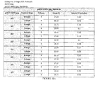
- Table 2 provides additional primer pairs and probes that are useful in the methods and compositions of the disclosure, for example for quantitative PCR and quantitative RT-PCR, and useful for XMRV or XMRV and MLV related virus detection: Table 2: Sequence Rating Probe Sequence Tm GC% Product Length Sense Primer Anti-sense Primer 5' XMRV coordinate 3' XMRV coordinate Detects MLV related and XMRV XMRV gag 82.7 66.7 56 106 1015 1121 no XMRV gag 84.9 67.6 60 114 1015 1128 no XMRV gag 83.5 67 58.3 114 1015 1128 no XMRV gag 82.7 66.7 56 114 1015 1128 no XMRV gag 82.5 66.6 60.9 114 1015 1128 no XMRV gag 82.3 66.5 56 114 1015 1128 no XMRV gag 81.6 66.3 58.3 114 1015 1128 no XMRV gag 84.9 67.6 60 129 1015 1143 no
- RT-PCR assay is performed to quantify RNA from biological samples, such as whole blood and plasma, without the need for RNA extraction.
- the assay employs a two-step amplification process with the initial step consisting of the distribution of 2 ⁇ L of experimental sample directly into a cDNA reaction mix. Following completion of the reverse transcriptase (RT) cDNA synthesis, a 2 ⁇ L aliquot is removed, transferred into a qPCR reaction mix and a qPCR protocol is performed.
- RT reverse transcriptase
- RNA is isolated from cell culture supernatants, whole blood or plasma using QIAamp®viral RNA Mini kit. The RNA is then used for reverse transcription. After reverse transcription, cDNA is subjected to qPCR assay.
Landscapes
- Chemical & Material Sciences (AREA)
- Life Sciences & Earth Sciences (AREA)
- Health & Medical Sciences (AREA)
- Organic Chemistry (AREA)
- Zoology (AREA)
- Immunology (AREA)
- Wood Science & Technology (AREA)
- Engineering & Computer Science (AREA)
- Proteomics, Peptides & Aminoacids (AREA)
- Virology (AREA)
- Molecular Biology (AREA)
- Microbiology (AREA)
- Biophysics (AREA)
- Physics & Mathematics (AREA)
- Analytical Chemistry (AREA)
- Biotechnology (AREA)
- Biochemistry (AREA)
- Bioinformatics & Cheminformatics (AREA)
- General Engineering & Computer Science (AREA)
- General Health & Medical Sciences (AREA)
- Genetics & Genomics (AREA)
- Measuring Or Testing Involving Enzymes Or Micro-Organisms (AREA)
Applications Claiming Priority (4)
| Application Number | Priority Date | Filing Date | Title |
|---|---|---|---|
| US36529710P | 2010-07-16 | 2010-07-16 | |
| US38694110P | 2010-09-27 | 2010-09-27 | |
| US39136010P | 2010-10-08 | 2010-10-08 | |
| EP11807616.5A EP2580325A4 (de) | 2010-07-16 | 2011-07-16 | Retrovirenerkennung |
Related Parent Applications (1)
| Application Number | Title | Priority Date | Filing Date |
|---|---|---|---|
| EP11807616.5A Division EP2580325A4 (de) | 2010-07-16 | 2011-07-16 | Retrovirenerkennung |
Publications (2)
| Publication Number | Publication Date |
|---|---|
| EP2913402A1 true EP2913402A1 (de) | 2015-09-02 |
| EP2913402B1 EP2913402B1 (de) | 2017-10-18 |
Family
ID=45470115
Family Applications (2)
| Application Number | Title | Priority Date | Filing Date |
|---|---|---|---|
| EP14191299.8A Not-in-force EP2913402B1 (de) | 2010-07-16 | 2011-07-16 | Retrovirusdetektion |
| EP11807616.5A Withdrawn EP2580325A4 (de) | 2010-07-16 | 2011-07-16 | Retrovirenerkennung |
Family Applications After (1)
| Application Number | Title | Priority Date | Filing Date |
|---|---|---|---|
| EP11807616.5A Withdrawn EP2580325A4 (de) | 2010-07-16 | 2011-07-16 | Retrovirenerkennung |
Country Status (6)
| Country | Link |
|---|---|
| US (1) | US9663834B2 (de) |
| EP (2) | EP2913402B1 (de) |
| CN (1) | CN103140581A (de) |
| CA (1) | CA2803011C (de) |
| ES (1) | ES2655500T3 (de) |
| WO (1) | WO2012009711A2 (de) |
Families Citing this family (6)
| Publication number | Priority date | Publication date | Assignee | Title |
|---|---|---|---|---|
| JP5923037B2 (ja) | 2009-06-17 | 2016-05-24 | トカジェン インコーポレーテッド | 複製可能レトロウイルスベクターの産生細胞 |
| WO2016172465A1 (en) * | 2015-04-24 | 2016-10-27 | The Johns Hopkins University | Compositions and methods related to characterizing proviral reservoirs |
| EP4286538A3 (de) * | 2020-04-06 | 2024-03-20 | Octant, Inc. | Diagnostischer test für krankheitserreger |
| CN112011649B (zh) * | 2020-10-29 | 2021-01-15 | 苏州良辰生物医药科技有限公司 | 一种用于检测异嗜性小鼠白血病病毒的引物探针、试剂盒及应用 |
| CN113444841B (zh) * | 2021-08-23 | 2022-05-24 | 山东省滨州畜牧兽医研究院 | 一种狐狸逆转录病毒SYBR Green Ⅰ荧光RT-PCR试剂盒及使用方法 |
| CN113444842B (zh) * | 2021-08-23 | 2022-08-02 | 山东省滨州畜牧兽医研究院 | 一种用于狐狸逆转录病毒检测的特异引物组及pcr检测试剂盒的应用 |
Citations (22)
| Publication number | Priority date | Publication date | Assignee | Title |
|---|---|---|---|---|
| US4458066A (en) | 1980-02-29 | 1984-07-03 | University Patents, Inc. | Process for preparing polynucleotides |
| WO1989006700A1 (en) | 1988-01-21 | 1989-07-27 | Genentech, Inc. | Amplification and detection of nucleic acid sequences |
| EP0329822A2 (de) | 1988-02-24 | 1989-08-30 | Cangene Corporation | Verfahren zur Amplifizierung von Nukleinsäuren |
| WO1990001069A1 (en) | 1988-07-20 | 1990-02-08 | Segev Diagnostics, Inc. | Process for amplifying and detecting nucleic acid sequences |
| US4965188A (en) | 1986-08-22 | 1990-10-23 | Cetus Corporation | Process for amplifying, detecting, and/or cloning nucleic acid sequences using a thermostable enzyme |
| WO1990015070A1 (en) | 1989-06-07 | 1990-12-13 | Affymax Technologies N.V. | Very large scale immobilized peptide synthesis |
| WO1992010092A1 (en) | 1990-12-06 | 1992-06-25 | Affymax Technologies N.V. | Very large scale immobilized polymer synthesis |
| US5130238A (en) | 1988-06-24 | 1992-07-14 | Cangene Corporation | Enhanced nucleic acid amplification process |
| US5169766A (en) | 1991-06-14 | 1992-12-08 | Life Technologies, Inc. | Amplification of nucleic acid molecules |
| US5412087A (en) | 1992-04-24 | 1995-05-02 | Affymax Technologies N.V. | Spatially-addressable immobilization of oligonucleotides and other biological polymers on surfaces |
| WO1995011995A1 (en) | 1993-10-26 | 1995-05-04 | Affymax Technologies N.V. | Arrays of nucleic acid probes on biological chips |
| WO1998022489A1 (en) | 1996-11-18 | 1998-05-28 | Takeshi Imanishi | Novel nucleotide analogues |
| WO1998039352A1 (en) | 1997-03-07 | 1998-09-11 | Takeshi Imanishi | Novel bicyclonucleoside and oligonucleotide analogues |
| WO1999014226A2 (en) | 1997-09-12 | 1999-03-25 | Exiqon A/S | Bi- and tri-cyclic nucleoside, nucleotide and oligonucleotide analogues |
| US6015694A (en) * | 1993-09-15 | 2000-01-18 | Chiron Corporation | Method for stimulating an immune response utilizing recombinant alphavirus particles |
| WO2004052297A2 (en) * | 2002-12-06 | 2004-06-24 | Cytogenix, Inc. | TREATMENT OF HSV-RELATED PATHOLOGIES USING ssDNA |
| WO2007130519A2 (en) * | 2006-05-02 | 2007-11-15 | Government Of The Usa, As Represented By The Secretary, Department Of Health And Human Services | Viral nucleic acid microarray and method of use |
| KR20080032562A (ko) * | 2006-10-10 | 2008-04-15 | 주식회사 바이로메드 | 안전성이 향상된 레트로바이러스 벡터 |
| WO2010036986A2 (en) | 2008-09-26 | 2010-04-01 | Tocagen Inc. | Recombinant vectors |
| WO2010075414A2 (en) * | 2008-12-23 | 2010-07-01 | The Cleveland Clinic Foundation | Method for detection of xmrv |
| WO2010148203A2 (en) | 2009-06-17 | 2010-12-23 | Tocagen Inc. | Producer cells for replication competent retroviral vectors |
| WO2012058637A2 (en) * | 2010-10-31 | 2012-05-03 | Tocagen Inc. | Antisera assays for mlv related viruses in humans and other mammals |
Family Cites Families (3)
| Publication number | Priority date | Publication date | Assignee | Title |
|---|---|---|---|---|
| AU2006235266B2 (en) * | 2005-04-07 | 2010-12-23 | The Cleveland Clinic Foundation | Gammaretrovirus associated with cancer |
| US20090211690A1 (en) | 2005-04-08 | 2009-08-27 | Bartholomeusz Daniel A | Rapid Prototyping of Microstructures Using a Cutting Plotter |
| US20100167268A1 (en) * | 2009-07-15 | 2010-07-01 | Mikovits Judy A | Seroconversion assays for detecting xenotropic murine leukemia virus-related virus |
-
2011
- 2011-07-16 EP EP14191299.8A patent/EP2913402B1/de not_active Not-in-force
- 2011-07-16 ES ES14191299.8T patent/ES2655500T3/es active Active
- 2011-07-16 CA CA2803011A patent/CA2803011C/en not_active Expired - Fee Related
- 2011-07-16 US US13/810,403 patent/US9663834B2/en not_active Expired - Fee Related
- 2011-07-16 CN CN2011800446801A patent/CN103140581A/zh active Pending
- 2011-07-16 EP EP11807616.5A patent/EP2580325A4/de not_active Withdrawn
- 2011-07-16 WO PCT/US2011/044296 patent/WO2012009711A2/en not_active Ceased
Patent Citations (24)
| Publication number | Priority date | Publication date | Assignee | Title |
|---|---|---|---|---|
| US4458066A (en) | 1980-02-29 | 1984-07-03 | University Patents, Inc. | Process for preparing polynucleotides |
| US4965188A (en) | 1986-08-22 | 1990-10-23 | Cetus Corporation | Process for amplifying, detecting, and/or cloning nucleic acid sequences using a thermostable enzyme |
| WO1989006700A1 (en) | 1988-01-21 | 1989-07-27 | Genentech, Inc. | Amplification and detection of nucleic acid sequences |
| EP0329822A2 (de) | 1988-02-24 | 1989-08-30 | Cangene Corporation | Verfahren zur Amplifizierung von Nukleinsäuren |
| US5130238A (en) | 1988-06-24 | 1992-07-14 | Cangene Corporation | Enhanced nucleic acid amplification process |
| WO1990001069A1 (en) | 1988-07-20 | 1990-02-08 | Segev Diagnostics, Inc. | Process for amplifying and detecting nucleic acid sequences |
| US5143854A (en) | 1989-06-07 | 1992-09-01 | Affymax Technologies N.V. | Large scale photolithographic solid phase synthesis of polypeptides and receptor binding screening thereof |
| WO1990015070A1 (en) | 1989-06-07 | 1990-12-13 | Affymax Technologies N.V. | Very large scale immobilized peptide synthesis |
| WO1992010092A1 (en) | 1990-12-06 | 1992-06-25 | Affymax Technologies N.V. | Very large scale immobilized polymer synthesis |
| US5169766A (en) | 1991-06-14 | 1992-12-08 | Life Technologies, Inc. | Amplification of nucleic acid molecules |
| US5412087A (en) | 1992-04-24 | 1995-05-02 | Affymax Technologies N.V. | Spatially-addressable immobilization of oligonucleotides and other biological polymers on surfaces |
| US6015694A (en) * | 1993-09-15 | 2000-01-18 | Chiron Corporation | Method for stimulating an immune response utilizing recombinant alphavirus particles |
| WO1995011995A1 (en) | 1993-10-26 | 1995-05-04 | Affymax Technologies N.V. | Arrays of nucleic acid probes on biological chips |
| WO1998022489A1 (en) | 1996-11-18 | 1998-05-28 | Takeshi Imanishi | Novel nucleotide analogues |
| WO1998039352A1 (en) | 1997-03-07 | 1998-09-11 | Takeshi Imanishi | Novel bicyclonucleoside and oligonucleotide analogues |
| WO1999014226A2 (en) | 1997-09-12 | 1999-03-25 | Exiqon A/S | Bi- and tri-cyclic nucleoside, nucleotide and oligonucleotide analogues |
| WO2004052297A2 (en) * | 2002-12-06 | 2004-06-24 | Cytogenix, Inc. | TREATMENT OF HSV-RELATED PATHOLOGIES USING ssDNA |
| WO2007130519A2 (en) * | 2006-05-02 | 2007-11-15 | Government Of The Usa, As Represented By The Secretary, Department Of Health And Human Services | Viral nucleic acid microarray and method of use |
| KR20080032562A (ko) * | 2006-10-10 | 2008-04-15 | 주식회사 바이로메드 | 안전성이 향상된 레트로바이러스 벡터 |
| WO2010036986A2 (en) | 2008-09-26 | 2010-04-01 | Tocagen Inc. | Recombinant vectors |
| WO2010045002A2 (en) | 2008-09-26 | 2010-04-22 | Tocagen Inc. | Gene therapy vectors and cytosine deaminases |
| WO2010075414A2 (en) * | 2008-12-23 | 2010-07-01 | The Cleveland Clinic Foundation | Method for detection of xmrv |
| WO2010148203A2 (en) | 2009-06-17 | 2010-12-23 | Tocagen Inc. | Producer cells for replication competent retroviral vectors |
| WO2012058637A2 (en) * | 2010-10-31 | 2012-05-03 | Tocagen Inc. | Antisera assays for mlv related viruses in humans and other mammals |
Non-Patent Citations (47)
| Title |
|---|
| ANASTASSOPOULOU ET AL., JOURNAL OF VIROLOGICAL METHODS, vol. 91, 2001, pages 67 - 74 |
| ARNOLD R S ET AL: "XMRV Infection in Patients With Prostate Cancer: Novel Serologic Assay and Correlation With PCR and FISH", UROLOGY, BELLE MEAD, NJ, US, vol. 75, no. 4, 1 April 2010 (2010-04-01), pages 755 - 761, XP026990712, ISSN: 0090-4295, [retrieved on 20100401] * |
| AUSUBEL, ET AL.: "Short Protocols in Molecular Biology", WILEY |
| B. DONG ET AL: "From the Cover: An infectious retrovirus susceptible to an IFN antiviral pathway from human prostate tumors", PROCEEDINGS OF THE NATIONAL ACADEMY OF SCIENCES, vol. 104, no. 5, 30 January 2007 (2007-01-30), pages 1655 - 1660, XP055015348, ISSN: 0027-8424, DOI: 10.1073/pnas.0610291104 * |
| BARANY ET AL., PROC. NATL. ACAD. SCI. USA, vol. 88, 1991, pages 189 - 93 |
| BEAUCAGE ET AL., TETRAHEDRON LETTERS, vol. 22, 1981, pages 1859 |
| BROWN ET AL., METHODS IN ENZYMOLOGY, vol. 68, 1979, pages 109 |
| DATABASE Geneseq [online] 10 July 2008 (2008-07-10), "Viral microarray oligonucleotide sequence SEQ ID NO 11.", XP002739251, retrieved from EBI accession no. GSN:AQX99908 Database accession no. AQX99908 * |
| DATABASE Geneseq [online] 22 January 2009 (2009-01-22), "pGEM T easy-3'dLTR3 DNA amplifying PCR primer 3LTR-3, SEQ ID:9.", XP002739250, retrieved from EBI accession no. GSN:ATS37006 Database accession no. ATS37006 * |
| DATABASE Geneseq [online] 22 May 2000 (2000-05-22), "Moloney murine leukaemia virus LTR PCR primer SEQ ID NO:16.", XP002739249, retrieved from EBI accession no. GSN:AAZ92907 Database accession no. AAZ92907 * |
| DATABASE Geneseq [online] 9 September 2004 (2004-09-09), "Oligonucleotide of the invention 3'-E/S/P/P-LINKER.", XP002739252, retrieved from EBI accession no. GSN:ADP67227 Database accession no. ADP67227 * |
| DONG ET AL., PNAS, vol. 104, 2007, pages 1655 |
| E.C. KNOUF ET AL., J.VIROL, vol. 83, 2009, pages 78353 - 7356 |
| ENSERINK ET AL., SCIENCE, vol. 329, 2010, pages 18 - 19 |
| FISCHER ET AL., JOURNAL OF CLINICAL VIROLOGY, vol. 43, 2008, pages 277 - 283 |
| FISCHER N ET AL: "Prevalence of human gammaretrovirus XMRV in sporadic prostate cancer", JOURNAL OF CLINICAL VIROLOGY, ELSEVIER, AMSTERDAM, NL, vol. 43, no. 3, 1 November 2008 (2008-11-01), pages 277 - 283, XP025612273, ISSN: 1386-6532, [retrieved on 20080927], DOI: 10.1016/J.JCV.2008.04.016 * |
| G.B.BECK-ENGESER, PNAS, 2009 |
| GARBESI, NUCL. ACIDS RES., vol. 21, 1993, pages 4159 - 65 |
| GURUNATHAN S; HABIB RE ET AL., VACCINE, vol. 27, 2009, pages 1997 - 2015 |
| HAYMES ET AL.: "Nucleic Acid Hybridization, A Practical Approach", 1985, IRL PRESS |
| HOHN ET AL., RETROVIROLOGY, vol. 6, 2009, pages 92 |
| HOHN OLIVER ET AL: "Lack of evidence for xenotropic murine leukemia virus-related virus(XMRV) in German prostate cancer patients", RETROVIROLOGY, BIOMED CENTRAL LTD., LONDON, GB, vol. 6, no. 1, 16 October 2009 (2009-10-16), pages 92, XP021063734, ISSN: 1742-4690, DOI: 10.1186/1742-4690-6-92 * |
| J. AMER. CHEM. SOC., vol. 112, 1990, pages 7435 |
| J.D. FOX, J.CLIN. VIROL., vol. 40, no. 1, 2007, pages S15 - S23 |
| KORNBERG; BAKER: "DNA Replication, 2nd ed.", 1992, FREEMAN |
| LANDEGREN ET AL., SCIENCE, vol. 241, 1988, pages 1077 - 80 |
| LOGG ET AL., J.VIROL., vol. 75, 2001, pages 6989 - 6998 |
| LOW A.; OKEOMA CM ET AL., VIROLOGY, vol. 385, 2009, pages 455 - 463 |
| MANIATIS ET AL.: "Molecular Cloning: A Laboratory Manual, 2nd ed.", 1989 |
| NARANG ET AL., METHODS IN ENZYMOLOGY, vol. 68, 1979, pages 190 |
| OTTO ERLWEIN ET AL: "Failure to Detect the Novel Retrovirus XMRV in Chronic Fatigue Syndrome", PLOS ONE, vol. 5, no. 1, 6 January 2010 (2010-01-06), pages e8519, XP055072557, DOI: 10.1371/journal.pone.0008519 * |
| PAPROTKA T., SCIENCE, vol. 333, 2011, pages 97 - 101 |
| POWELL ET AL., J.VIROL., vol. 73, 1999, pages 8813 - 8816 |
| R. SCHLABERG ET AL., PNAS, 2009 |
| ROBERT SCHLABERG ET AL: "XMRV is present in malignant prostatic epithelium and is associated with prostate cancer, especially high-grade tumors", PROCEEDINGS OF THE NATIONAL ACADEMY OF SCIENCES, NATIONAL ACADEMY OF SCIENCES, US, vol. 106, no. 38, 22 September 2009 (2009-09-22), pages 16351 - 16356, XP007915265, ISSN: 0027-8424 * |
| ROBINSON MJ. ET AL., RETROVIROLOGY, vol. 7, 2010, pages 108 |
| SAKUMA ET AL., VIROLOGY, 2009 |
| SAMBROOK ET AL.: "Molecular Cloning, A Laboratory Manual, 2nd ed.", 1989, COLD SPRING HARBOR PRESS |
| TAI ET AL., MOL. THER., vol. 12, 2005, pages 842 - 851 |
| TIJSSEN: "Overview of principles of hybridization and the strategy of nucleic acid assays", TECHNIQUES IN BIOCHEMISTRY AND MOLECULAR BIOLOGY--HYBRIDIZATION WITH NUCLEIC ACID PROBES, 1993 |
| URATA, NUCLEIC ACIDS SYMPOSIUM SER. NO. 29, 1993, pages 69 - 70 |
| URISMAN A ET AL: "Identification of a novel Gammaretrovirus in prostate tumors of patients homozygous for R462Q RNASEL variant", PLOS PATHOGENS, PUBLIC LIBRARY OF SCIENCE, US; US NATIONAL LIBRARY OF MEDICINE (NLM), BETHESDA, MD, US, vol. 2, no. 3, 1 March 2006 (2006-03-01), pages E25 - 1, XP002669943, ISSN: 1553-7374, DOI: 10.1371/JOURNAL.PPAT.0020025 * |
| URISMAN ET AL., PLOS PATHOGENS, vol. 2, no. 3, 2006, pages E25 |
| V.C.LOMBARDI ET AL., SCIENCE, 2009 |
| VAN KUPPEVELD ET AL., BMJ, vol. 340, 2010, pages CL018 |
| VINCENT C LOMBARDI ET AL: "Detection of an infectious retrovirus, XMRV, in blood cells of patients with chronic fatigue syndrome", SCIENCE, AMERICAN ASSOCIATION FOR THE ADVANCEMENT OF SCIENCE, US; US NATIONAL LIBRARY OF MEDICINE (NLM), BETHESDA, MD, US, vol. 326, no. 5952, 23 October 2009 (2009-10-23), pages 585 - 589, XP002669944, ISSN: 1095-9203, [retrieved on 20091008], DOI: 10.1126/SCIENCE.1179052 * |
| WALKER ET AL., PROC. NATL. ACAD. SCI. USA, vol. 89, 1992, pages 392 - 6 |
Also Published As
| Publication number | Publication date |
|---|---|
| US9663834B2 (en) | 2017-05-30 |
| CA2803011C (en) | 2019-12-03 |
| ES2655500T3 (es) | 2018-02-20 |
| WO2012009711A2 (en) | 2012-01-19 |
| CN103140581A (zh) | 2013-06-05 |
| EP2913402B1 (de) | 2017-10-18 |
| US20130266931A1 (en) | 2013-10-10 |
| CA2803011A1 (en) | 2012-01-19 |
| EP2580325A4 (de) | 2013-11-20 |
| WO2012009711A3 (en) | 2012-05-31 |
| EP2580325A2 (de) | 2013-04-17 |
Similar Documents
| Publication | Publication Date | Title |
|---|---|---|
| Yapar et al. | Rapid and quantitative detection of Crimean-Congo hemorrhagic fever virus by one-step real-time reverse transcriptase-PCR | |
| US20250382658A1 (en) | Hiv or hcv detection with crispr-cas13a | |
| Jimba et al. | BLV-CoCoMo-qPCR: Quantitation of bovine leukemia virus proviral load using the CoCoMo algorithm | |
| EP2913402B1 (de) | Retrovirusdetektion | |
| US20030077588A1 (en) | Diagnostic detection of nucleic acids | |
| JP5924595B2 (ja) | ウシ白血病ウイルス(blv)検出用キットおよびその利用 | |
| EP4365311A2 (de) | Verfahren zum nachweis von lentivirus | |
| PT1504127E (pt) | Método de amplificação- hibridação para detecção e tipificação do papilomavírus humano | |
| WO2023279042A2 (en) | Compositions and methods for detection of severe acute respiratory syndrome coronavirus 2 variants | |
| EP1220954B1 (de) | Nukelinsäure primer und proben zum nachweis von tumorzellen | |
| CN109971861A (zh) | Ccdc6-ret融合基因检测试剂盒 | |
| JP2007006897A (ja) | 逆転写酵素の高感度検出法 | |
| JP6153515B2 (ja) | Hla−a*24:02を検出する方法、及び検出キット | |
| CN111471800B (zh) | 检测新型冠状病毒的试剂盒及其扩增引物组合物 | |
| US20130171622A1 (en) | Compositions and methods for detecting viral infection using direct-label fluorescence in situ hybridization | |
| KR100440852B1 (ko) | 레트로바이러스에 특이적인 올리고뉴클레오티드 프라이머및 이를 이용한 레트로바이러스 농도 측정방법 | |
| EP1100959B1 (de) | Verfahren und reagentiensatz zur erkennung von antiviralen wirkstoff resistenzen | |
| KR20230056908A (ko) | 실시간 중합효소연쇄반응을 이용한 수두바이러스 검출 및 정량 방법 | |
| US7691572B2 (en) | Method and kit for detecting resistance to antiviral drugs | |
| CA3035808C (en) | Methods of detecting lentivirus | |
| KR20230092799A (ko) | 제2형 중증급성호흡기증후군 코로나바이러스 검출용 고리매개등온증폭 반응용 프라이머 및 프로브 세트 | |
| WO2026072767A1 (en) | Human papillomavirus detection | |
| KR20250024727A (ko) | 재조합효소 중합효소 증폭법 및 CRISPR-Cas12a 기반 인간 거대세포바이러스 진단 방법 | |
| WO2025044471A1 (zh) | 复制型逆转录病毒检测引物及方法 | |
| KR20260044122A (ko) | 위음성 판단이 가능한 hiv-1, hiv-2, hcv 및 hbv 동시 검출 또는 정량용 키트 |
Legal Events
| Date | Code | Title | Description |
|---|---|---|---|
| PUAI | Public reference made under article 153(3) epc to a published international application that has entered the european phase |
Free format text: ORIGINAL CODE: 0009012 |
|
| AC | Divisional application: reference to earlier application |
Ref document number: 2580325 Country of ref document: EP Kind code of ref document: P |
|
| AK | Designated contracting states |
Kind code of ref document: A1 Designated state(s): AL AT BE BG CH CY CZ DE DK EE ES FI FR GB GR HR HU IE IS IT LI LT LU LV MC MK MT NL NO PL PT RO RS SE SI SK SM TR |
|
| 17P | Request for examination filed |
Effective date: 20160211 |
|
| RBV | Designated contracting states (corrected) |
Designated state(s): AL AT BE BG CH CY CZ DE DK EE ES FI FR GB GR HR HU IE IS IT LI LT LU LV MC MK MT NL NO PL PT RO RS SE SI SK SM TR |
|
| 17Q | First examination report despatched |
Effective date: 20160617 |
|
| STAA | Information on the status of an ep patent application or granted ep patent |
Free format text: STATUS: EXAMINATION IS IN PROGRESS |
|
| GRAP | Despatch of communication of intention to grant a patent |
Free format text: ORIGINAL CODE: EPIDOSNIGR1 |
|
| STAA | Information on the status of an ep patent application or granted ep patent |
Free format text: STATUS: GRANT OF PATENT IS INTENDED |
|
| INTG | Intention to grant announced |
Effective date: 20170508 |
|
| GRAS | Grant fee paid |
Free format text: ORIGINAL CODE: EPIDOSNIGR3 |
|
| GRAA | (expected) grant |
Free format text: ORIGINAL CODE: 0009210 |
|
| STAA | Information on the status of an ep patent application or granted ep patent |
Free format text: STATUS: THE PATENT HAS BEEN GRANTED |
|
| AC | Divisional application: reference to earlier application |
Ref document number: 2580325 Country of ref document: EP Kind code of ref document: P |
|
| AK | Designated contracting states |
Kind code of ref document: B1 Designated state(s): AL AT BE BG CH CY CZ DE DK EE ES FI FR GB GR HR HU IE IS IT LI LT LU LV MC MK MT NL NO PL PT RO RS SE SI SK SM TR |
|
| REG | Reference to a national code |
Ref country code: GB Ref legal event code: FG4D |
|
| REG | Reference to a national code |
Ref country code: CH Ref legal event code: EP |
|
| REG | Reference to a national code |
Ref country code: AT Ref legal event code: REF Ref document number: 937988 Country of ref document: AT Kind code of ref document: T Effective date: 20171115 Ref country code: IE Ref legal event code: FG4D |
|
| REG | Reference to a national code |
Ref country code: DE Ref legal event code: R096 Ref document number: 602011042610 Country of ref document: DE |
|
| REG | Reference to a national code |
Ref country code: ES Ref legal event code: FG2A Ref document number: 2655500 Country of ref document: ES Kind code of ref document: T3 Effective date: 20180220 |
|
| REG | Reference to a national code |
Ref country code: NL Ref legal event code: MP Effective date: 20171018 |
|
| REG | Reference to a national code |
Ref country code: LT Ref legal event code: MG4D |
|
| REG | Reference to a national code |
Ref country code: AT Ref legal event code: MK05 Ref document number: 937988 Country of ref document: AT Kind code of ref document: T Effective date: 20171018 |
|
| PG25 | Lapsed in a contracting state [announced via postgrant information from national office to epo] |
Ref country code: NL Free format text: LAPSE BECAUSE OF FAILURE TO SUBMIT A TRANSLATION OF THE DESCRIPTION OR TO PAY THE FEE WITHIN THE PRESCRIBED TIME-LIMIT Effective date: 20171018 |
|
| PG25 | Lapsed in a contracting state [announced via postgrant information from national office to epo] |
Ref country code: FI Free format text: LAPSE BECAUSE OF FAILURE TO SUBMIT A TRANSLATION OF THE DESCRIPTION OR TO PAY THE FEE WITHIN THE PRESCRIBED TIME-LIMIT Effective date: 20171018 Ref country code: LT Free format text: LAPSE BECAUSE OF FAILURE TO SUBMIT A TRANSLATION OF THE DESCRIPTION OR TO PAY THE FEE WITHIN THE PRESCRIBED TIME-LIMIT Effective date: 20171018 Ref country code: NO Free format text: LAPSE BECAUSE OF FAILURE TO SUBMIT A TRANSLATION OF THE DESCRIPTION OR TO PAY THE FEE WITHIN THE PRESCRIBED TIME-LIMIT Effective date: 20180118 Ref country code: SE Free format text: LAPSE BECAUSE OF FAILURE TO SUBMIT A TRANSLATION OF THE DESCRIPTION OR TO PAY THE FEE WITHIN THE PRESCRIBED TIME-LIMIT Effective date: 20171018 |
|
| PG25 | Lapsed in a contracting state [announced via postgrant information from national office to epo] |
Ref country code: LV Free format text: LAPSE BECAUSE OF FAILURE TO SUBMIT A TRANSLATION OF THE DESCRIPTION OR TO PAY THE FEE WITHIN THE PRESCRIBED TIME-LIMIT Effective date: 20171018 Ref country code: RS Free format text: LAPSE BECAUSE OF FAILURE TO SUBMIT A TRANSLATION OF THE DESCRIPTION OR TO PAY THE FEE WITHIN THE PRESCRIBED TIME-LIMIT Effective date: 20171018 Ref country code: IS Free format text: LAPSE BECAUSE OF FAILURE TO SUBMIT A TRANSLATION OF THE DESCRIPTION OR TO PAY THE FEE WITHIN THE PRESCRIBED TIME-LIMIT Effective date: 20180218 Ref country code: HR Free format text: LAPSE BECAUSE OF FAILURE TO SUBMIT A TRANSLATION OF THE DESCRIPTION OR TO PAY THE FEE WITHIN THE PRESCRIBED TIME-LIMIT Effective date: 20171018 Ref country code: BG Free format text: LAPSE BECAUSE OF FAILURE TO SUBMIT A TRANSLATION OF THE DESCRIPTION OR TO PAY THE FEE WITHIN THE PRESCRIBED TIME-LIMIT Effective date: 20180118 Ref country code: GR Free format text: LAPSE BECAUSE OF FAILURE TO SUBMIT A TRANSLATION OF THE DESCRIPTION OR TO PAY THE FEE WITHIN THE PRESCRIBED TIME-LIMIT Effective date: 20180119 Ref country code: AT Free format text: LAPSE BECAUSE OF FAILURE TO SUBMIT A TRANSLATION OF THE DESCRIPTION OR TO PAY THE FEE WITHIN THE PRESCRIBED TIME-LIMIT Effective date: 20171018 |
|
| REG | Reference to a national code |
Ref country code: DE Ref legal event code: R097 Ref document number: 602011042610 Country of ref document: DE |
|
| REG | Reference to a national code |
Ref country code: FR Ref legal event code: PLFP Year of fee payment: 8 |
|
| PG25 | Lapsed in a contracting state [announced via postgrant information from national office to epo] |
Ref country code: EE Free format text: LAPSE BECAUSE OF FAILURE TO SUBMIT A TRANSLATION OF THE DESCRIPTION OR TO PAY THE FEE WITHIN THE PRESCRIBED TIME-LIMIT Effective date: 20171018 Ref country code: DK Free format text: LAPSE BECAUSE OF FAILURE TO SUBMIT A TRANSLATION OF THE DESCRIPTION OR TO PAY THE FEE WITHIN THE PRESCRIBED TIME-LIMIT Effective date: 20171018 Ref country code: SK Free format text: LAPSE BECAUSE OF FAILURE TO SUBMIT A TRANSLATION OF THE DESCRIPTION OR TO PAY THE FEE WITHIN THE PRESCRIBED TIME-LIMIT Effective date: 20171018 Ref country code: CZ Free format text: LAPSE BECAUSE OF FAILURE TO SUBMIT A TRANSLATION OF THE DESCRIPTION OR TO PAY THE FEE WITHIN THE PRESCRIBED TIME-LIMIT Effective date: 20171018 |
|
| PLBE | No opposition filed within time limit |
Free format text: ORIGINAL CODE: 0009261 |
|
| STAA | Information on the status of an ep patent application or granted ep patent |
Free format text: STATUS: NO OPPOSITION FILED WITHIN TIME LIMIT |
|
| PG25 | Lapsed in a contracting state [announced via postgrant information from national office to epo] |
Ref country code: PL Free format text: LAPSE BECAUSE OF FAILURE TO SUBMIT A TRANSLATION OF THE DESCRIPTION OR TO PAY THE FEE WITHIN THE PRESCRIBED TIME-LIMIT Effective date: 20171018 Ref country code: RO Free format text: LAPSE BECAUSE OF FAILURE TO SUBMIT A TRANSLATION OF THE DESCRIPTION OR TO PAY THE FEE WITHIN THE PRESCRIBED TIME-LIMIT Effective date: 20171018 Ref country code: SM Free format text: LAPSE BECAUSE OF FAILURE TO SUBMIT A TRANSLATION OF THE DESCRIPTION OR TO PAY THE FEE WITHIN THE PRESCRIBED TIME-LIMIT Effective date: 20171018 |
|
| 26N | No opposition filed |
Effective date: 20180719 |
|
| PG25 | Lapsed in a contracting state [announced via postgrant information from national office to epo] |
Ref country code: SI Free format text: LAPSE BECAUSE OF FAILURE TO SUBMIT A TRANSLATION OF THE DESCRIPTION OR TO PAY THE FEE WITHIN THE PRESCRIBED TIME-LIMIT Effective date: 20171018 |
|
| REG | Reference to a national code |
Ref country code: CH Ref legal event code: PL |
|
| PG25 | Lapsed in a contracting state [announced via postgrant information from national office to epo] |
Ref country code: MC Free format text: LAPSE BECAUSE OF FAILURE TO SUBMIT A TRANSLATION OF THE DESCRIPTION OR TO PAY THE FEE WITHIN THE PRESCRIBED TIME-LIMIT Effective date: 20171018 Ref country code: LU Free format text: LAPSE BECAUSE OF NON-PAYMENT OF DUE FEES Effective date: 20180716 |
|
| REG | Reference to a national code |
Ref country code: BE Ref legal event code: MM Effective date: 20180731 |
|
| REG | Reference to a national code |
Ref country code: IE Ref legal event code: MM4A |
|
| PG25 | Lapsed in a contracting state [announced via postgrant information from national office to epo] |
Ref country code: IE Free format text: LAPSE BECAUSE OF NON-PAYMENT OF DUE FEES Effective date: 20180716 Ref country code: LI Free format text: LAPSE BECAUSE OF NON-PAYMENT OF DUE FEES Effective date: 20180731 Ref country code: CH Free format text: LAPSE BECAUSE OF NON-PAYMENT OF DUE FEES Effective date: 20180731 |
|
| PG25 | Lapsed in a contracting state [announced via postgrant information from national office to epo] |
Ref country code: BE Free format text: LAPSE BECAUSE OF NON-PAYMENT OF DUE FEES Effective date: 20180731 |
|
| PGFP | Annual fee paid to national office [announced via postgrant information from national office to epo] |
Ref country code: DE Payment date: 20190729 Year of fee payment: 9 Ref country code: FR Payment date: 20190725 Year of fee payment: 9 Ref country code: ES Payment date: 20190801 Year of fee payment: 9 Ref country code: IT Payment date: 20190726 Year of fee payment: 9 |
|
| PGFP | Annual fee paid to national office [announced via postgrant information from national office to epo] |
Ref country code: GB Payment date: 20190729 Year of fee payment: 9 |
|
| PG25 | Lapsed in a contracting state [announced via postgrant information from national office to epo] |
Ref country code: MT Free format text: LAPSE BECAUSE OF NON-PAYMENT OF DUE FEES Effective date: 20180716 |
|
| PG25 | Lapsed in a contracting state [announced via postgrant information from national office to epo] |
Ref country code: TR Free format text: LAPSE BECAUSE OF FAILURE TO SUBMIT A TRANSLATION OF THE DESCRIPTION OR TO PAY THE FEE WITHIN THE PRESCRIBED TIME-LIMIT Effective date: 20171018 |
|
| PG25 | Lapsed in a contracting state [announced via postgrant information from national office to epo] |
Ref country code: PT Free format text: LAPSE BECAUSE OF FAILURE TO SUBMIT A TRANSLATION OF THE DESCRIPTION OR TO PAY THE FEE WITHIN THE PRESCRIBED TIME-LIMIT Effective date: 20171018 |
|
| PG25 | Lapsed in a contracting state [announced via postgrant information from national office to epo] |
Ref country code: MK Free format text: LAPSE BECAUSE OF NON-PAYMENT OF DUE FEES Effective date: 20171018 Ref country code: HU Free format text: LAPSE BECAUSE OF FAILURE TO SUBMIT A TRANSLATION OF THE DESCRIPTION OR TO PAY THE FEE WITHIN THE PRESCRIBED TIME-LIMIT; INVALID AB INITIO Effective date: 20110716 Ref country code: CY Free format text: LAPSE BECAUSE OF FAILURE TO SUBMIT A TRANSLATION OF THE DESCRIPTION OR TO PAY THE FEE WITHIN THE PRESCRIBED TIME-LIMIT Effective date: 20171018 |
|
| PG25 | Lapsed in a contracting state [announced via postgrant information from national office to epo] |
Ref country code: AL Free format text: LAPSE BECAUSE OF FAILURE TO SUBMIT A TRANSLATION OF THE DESCRIPTION OR TO PAY THE FEE WITHIN THE PRESCRIBED TIME-LIMIT Effective date: 20171018 |
|
| REG | Reference to a national code |
Ref country code: DE Ref legal event code: R119 Ref document number: 602011042610 Country of ref document: DE |
|
| GBPC | Gb: european patent ceased through non-payment of renewal fee |
Effective date: 20200716 |
|
| PG25 | Lapsed in a contracting state [announced via postgrant information from national office to epo] |
Ref country code: FR Free format text: LAPSE BECAUSE OF NON-PAYMENT OF DUE FEES Effective date: 20200731 Ref country code: GB Free format text: LAPSE BECAUSE OF NON-PAYMENT OF DUE FEES Effective date: 20200716 |
|
| PG25 | Lapsed in a contracting state [announced via postgrant information from national office to epo] |
Ref country code: DE Free format text: LAPSE BECAUSE OF NON-PAYMENT OF DUE FEES Effective date: 20210202 |
|
| PG25 | Lapsed in a contracting state [announced via postgrant information from national office to epo] |
Ref country code: IT Free format text: LAPSE BECAUSE OF NON-PAYMENT OF DUE FEES Effective date: 20200716 |
|
| REG | Reference to a national code |
Ref country code: ES Ref legal event code: FD2A Effective date: 20211229 |
|
| PG25 | Lapsed in a contracting state [announced via postgrant information from national office to epo] |
Ref country code: ES Free format text: LAPSE BECAUSE OF NON-PAYMENT OF DUE FEES Effective date: 20200717 |