EP2812350B1 - R-spondin-translokationen und verfahren damit - Google Patents
R-spondin-translokationen und verfahren damit Download PDFInfo
- Publication number
- EP2812350B1 EP2812350B1 EP13706133.9A EP13706133A EP2812350B1 EP 2812350 B1 EP2812350 B1 EP 2812350B1 EP 13706133 A EP13706133 A EP 13706133A EP 2812350 B1 EP2812350 B1 EP 2812350B1
- Authority
- EP
- European Patent Office
- Prior art keywords
- antibody
- translocation
- spondin
- cancer
- antagonist
- Prior art date
- Legal status (The legal status is an assumption and is not a legal conclusion. Google has not performed a legal analysis and makes no representation as to the accuracy of the status listed.)
- Active
Links
- 238000000034 method Methods 0.000 title claims description 194
- 230000005945 translocation Effects 0.000 title claims description 180
- 239000005557 antagonist Substances 0.000 claims description 290
- 206010028980 Neoplasm Diseases 0.000 claims description 175
- 201000011510 cancer Diseases 0.000 claims description 155
- 108090000623 proteins and genes Proteins 0.000 claims description 122
- 101000825960 Homo sapiens R-spondin-3 Proteins 0.000 claims description 120
- 102100022766 R-spondin-3 Human genes 0.000 claims description 118
- 238000011282 treatment Methods 0.000 claims description 47
- 238000011319 anticancer therapy Methods 0.000 claims description 39
- 102000004169 proteins and genes Human genes 0.000 claims description 34
- 239000003814 drug Substances 0.000 claims description 23
- 206010009944 Colon cancer Diseases 0.000 claims description 21
- 230000008901 benefit Effects 0.000 claims description 19
- 229940124597 therapeutic agent Drugs 0.000 claims description 12
- 208000001333 Colorectal Neoplasms Diseases 0.000 claims description 10
- 208000029742 colonic neoplasm Diseases 0.000 claims description 10
- 230000002759 chromosomal effect Effects 0.000 claims description 5
- 208000015634 Rectal Neoplasms Diseases 0.000 claims description 4
- 206010038038 rectal cancer Diseases 0.000 claims description 4
- 201000001275 rectum cancer Diseases 0.000 claims description 4
- 101000591201 Homo sapiens Receptor-type tyrosine-protein phosphatase kappa Proteins 0.000 claims 3
- 102100034089 Receptor-type tyrosine-protein phosphatase kappa Human genes 0.000 claims 3
- 108090000765 processed proteins & peptides Proteins 0.000 description 257
- 102000004196 processed proteins & peptides Human genes 0.000 description 254
- 229920001184 polypeptide Polymers 0.000 description 251
- 230000004927 fusion Effects 0.000 description 239
- 102000013814 Wnt Human genes 0.000 description 207
- 108050003627 Wnt Proteins 0.000 description 207
- 230000037361 pathway Effects 0.000 description 203
- 210000004027 cell Anatomy 0.000 description 157
- 230000027455 binding Effects 0.000 description 129
- 239000000090 biomarker Substances 0.000 description 123
- 102000040430 polynucleotide Human genes 0.000 description 102
- 108091033319 polynucleotide Proteins 0.000 description 102
- 239000002157 polynucleotide Substances 0.000 description 102
- 210000001519 tissue Anatomy 0.000 description 91
- 101000825949 Homo sapiens R-spondin-2 Proteins 0.000 description 87
- 102100022763 R-spondin-2 Human genes 0.000 description 86
- 241000282414 Homo sapiens Species 0.000 description 85
- 239000000523 sample Substances 0.000 description 84
- 230000008707 rearrangement Effects 0.000 description 81
- 230000014509 gene expression Effects 0.000 description 71
- 125000003275 alpha amino acid group Chemical group 0.000 description 51
- 150000007523 nucleic acids Chemical group 0.000 description 50
- 230000011664 signaling Effects 0.000 description 49
- 239000000427 antigen Substances 0.000 description 46
- 108091007433 antigens Proteins 0.000 description 44
- 102000036639 antigens Human genes 0.000 description 44
- 102000039446 nucleic acids Human genes 0.000 description 44
- 108020004707 nucleic acids Proteins 0.000 description 44
- 101000825954 Homo sapiens R-spondin-1 Proteins 0.000 description 35
- 150000003384 small molecules Chemical class 0.000 description 35
- 101000825962 Homo sapiens R-spondin-4 Proteins 0.000 description 34
- 208000037265 diseases, disorders, signs and symptoms Diseases 0.000 description 34
- 102100022762 R-spondin-1 Human genes 0.000 description 33
- 102100022759 R-spondin-4 Human genes 0.000 description 32
- 235000018102 proteins Nutrition 0.000 description 32
- YHPKGSLWSUCJQK-UHFFFAOYSA-N 2-[[2-[[2-[[2-[[2-[[2-[[5-amino-2-[[2-[(2,4-diamino-4-oxobutanoyl)amino]-4-methylpentanoyl]amino]-5-oxopentanoyl]amino]-4-methylpentanoyl]amino]-4-methylpentanoyl]amino]-4-methylsulfanylbutanoyl]amino]-3-carboxypropanoyl]amino]-5-(diaminomethylideneamino) Chemical compound NC(N)=NCCCC(C(=O)NC(C(C)C)C(O)=O)NC(=O)C(CC(O)=O)NC(=O)C(CCSC)NC(=O)C(CC(C)C)NC(=O)C(CC(C)C)NC(=O)C(CCC(N)=O)NC(=O)C(CC(C)C)NC(=O)C(N)CC(N)=O YHPKGSLWSUCJQK-UHFFFAOYSA-N 0.000 description 30
- -1 and/or their analogs Substances 0.000 description 30
- 238000006467 substitution reaction Methods 0.000 description 30
- 230000035772 mutation Effects 0.000 description 28
- 238000003556 assay Methods 0.000 description 27
- 239000003795 chemical substances by application Substances 0.000 description 22
- 201000010099 disease Diseases 0.000 description 22
- 102000008394 Immunoglobulin Fragments Human genes 0.000 description 21
- 108010021625 Immunoglobulin Fragments Proteins 0.000 description 21
- 239000013074 reference sample Substances 0.000 description 20
- 230000004044 response Effects 0.000 description 20
- 239000012634 fragment Substances 0.000 description 19
- 108020004999 messenger RNA Proteins 0.000 description 19
- 235000001014 amino acid Nutrition 0.000 description 18
- 125000003729 nucleotide group Chemical group 0.000 description 18
- 239000013598 vector Substances 0.000 description 18
- AOJJSUZBOXZQNB-TZSSRYMLSA-N Doxorubicin Chemical compound O([C@H]1C[C@@](O)(CC=2C(O)=C3C(=O)C=4C=CC=C(C=4C(=O)C3=C(O)C=21)OC)C(=O)CO)[C@H]1C[C@H](N)[C@H](O)[C@H](C)O1 AOJJSUZBOXZQNB-TZSSRYMLSA-N 0.000 description 17
- 239000013068 control sample Substances 0.000 description 17
- 238000002823 phage display Methods 0.000 description 17
- 108091032973 (ribonucleotides)n+m Proteins 0.000 description 16
- 229940127089 cytotoxic agent Drugs 0.000 description 16
- 230000003993 interaction Effects 0.000 description 16
- 239000000047 product Substances 0.000 description 16
- 125000000539 amino acid group Chemical group 0.000 description 15
- 239000002246 antineoplastic agent Substances 0.000 description 15
- 230000006870 function Effects 0.000 description 15
- 238000004519 manufacturing process Methods 0.000 description 15
- 230000001404 mediated effect Effects 0.000 description 15
- 239000000203 mixture Substances 0.000 description 15
- 229940002612 prodrug Drugs 0.000 description 15
- 239000000651 prodrug Substances 0.000 description 15
- 108091007491 NSP3 Papain-like protease domains Proteins 0.000 description 14
- 230000000295 complement effect Effects 0.000 description 14
- 230000000694 effects Effects 0.000 description 14
- 239000002773 nucleotide Substances 0.000 description 14
- 108020004414 DNA Proteins 0.000 description 13
- 108060003951 Immunoglobulin Proteins 0.000 description 12
- 230000004075 alteration Effects 0.000 description 12
- 230000010056 antibody-dependent cellular cytotoxicity Effects 0.000 description 12
- 230000004071 biological effect Effects 0.000 description 12
- 208000035475 disorder Diseases 0.000 description 12
- 102000018358 immunoglobulin Human genes 0.000 description 12
- 230000004048 modification Effects 0.000 description 12
- 238000012986 modification Methods 0.000 description 12
- 230000002829 reductive effect Effects 0.000 description 12
- 238000012216 screening Methods 0.000 description 12
- 238000005516 engineering process Methods 0.000 description 11
- 230000001976 improved effect Effects 0.000 description 11
- 230000001965 increasing effect Effects 0.000 description 11
- 102000004190 Enzymes Human genes 0.000 description 10
- 108090000790 Enzymes Proteins 0.000 description 10
- FAPWRFPIFSIZLT-UHFFFAOYSA-M Sodium chloride Chemical compound [Na+].[Cl-] FAPWRFPIFSIZLT-UHFFFAOYSA-M 0.000 description 10
- 229940024606 amino acid Drugs 0.000 description 10
- 150000001413 amino acids Chemical class 0.000 description 10
- 238000004458 analytical method Methods 0.000 description 10
- 230000000692 anti-sense effect Effects 0.000 description 10
- 229940088598 enzyme Drugs 0.000 description 10
- 230000002401 inhibitory effect Effects 0.000 description 10
- 238000003752 polymerase chain reaction Methods 0.000 description 10
- 230000002285 radioactive effect Effects 0.000 description 10
- 102100039520 39S ribosomal protein L33, mitochondrial Human genes 0.000 description 9
- 101000670355 Homo sapiens 39S ribosomal protein L33, mitochondrial Proteins 0.000 description 9
- 241001465754 Metazoa Species 0.000 description 9
- 230000015572 biosynthetic process Effects 0.000 description 9
- 230000004663 cell proliferation Effects 0.000 description 9
- 230000004540 complement-dependent cytotoxicity Effects 0.000 description 9
- 230000003247 decreasing effect Effects 0.000 description 9
- 229940079593 drug Drugs 0.000 description 9
- 230000012010 growth Effects 0.000 description 9
- 239000000243 solution Substances 0.000 description 9
- 239000000126 substance Substances 0.000 description 9
- 238000002560 therapeutic procedure Methods 0.000 description 9
- ZHNUHDYFZUAESO-UHFFFAOYSA-N Formamide Chemical compound NC=O ZHNUHDYFZUAESO-UHFFFAOYSA-N 0.000 description 8
- 102100030708 GTPase KRas Human genes 0.000 description 8
- 101000584612 Homo sapiens GTPase KRas Proteins 0.000 description 8
- 210000004369 blood Anatomy 0.000 description 8
- 239000008280 blood Substances 0.000 description 8
- 238000006243 chemical reaction Methods 0.000 description 8
- 239000002254 cytotoxic agent Substances 0.000 description 8
- 238000009396 hybridization Methods 0.000 description 8
- 229940127121 immunoconjugate Drugs 0.000 description 8
- 238000001727 in vivo Methods 0.000 description 8
- 125000005647 linker group Chemical group 0.000 description 8
- 235000000346 sugar Nutrition 0.000 description 8
- 108700012359 toxins Proteins 0.000 description 8
- 102000004127 Cytokines Human genes 0.000 description 7
- 108090000695 Cytokines Proteins 0.000 description 7
- ROHFNLRQFUQHCH-YFKPBYRVSA-N L-leucine Chemical compound CC(C)C[C@H](N)C(O)=O ROHFNLRQFUQHCH-YFKPBYRVSA-N 0.000 description 7
- ZDZOTLJHXYCWBA-VCVYQWHSSA-N N-debenzoyl-N-(tert-butoxycarbonyl)-10-deacetyltaxol Chemical compound O([C@H]1[C@H]2[C@@](C([C@H](O)C3=C(C)[C@@H](OC(=O)[C@H](O)[C@@H](NC(=O)OC(C)(C)C)C=4C=CC=CC=4)C[C@]1(O)C3(C)C)=O)(C)[C@@H](O)C[C@H]1OC[C@]12OC(=O)C)C(=O)C1=CC=CC=C1 ZDZOTLJHXYCWBA-VCVYQWHSSA-N 0.000 description 7
- 230000004156 Wnt signaling pathway Effects 0.000 description 7
- 210000000349 chromosome Anatomy 0.000 description 7
- 231100000599 cytotoxic agent Toxicity 0.000 description 7
- 238000001514 detection method Methods 0.000 description 7
- 229960004679 doxorubicin Drugs 0.000 description 7
- 239000012636 effector Substances 0.000 description 7
- 108020001507 fusion proteins Proteins 0.000 description 7
- 102000037865 fusion proteins Human genes 0.000 description 7
- 210000004408 hybridoma Anatomy 0.000 description 7
- 150000002482 oligosaccharides Chemical class 0.000 description 7
- 238000012552 review Methods 0.000 description 7
- 210000002966 serum Anatomy 0.000 description 7
- 238000010186 staining Methods 0.000 description 7
- 239000003053 toxin Substances 0.000 description 7
- 231100000765 toxin Toxicity 0.000 description 7
- 102100034540 Adenomatous polyposis coli protein Human genes 0.000 description 6
- SHZGCJCMOBCMKK-UHFFFAOYSA-N D-mannomethylose Natural products CC1OC(O)C(O)C(O)C1O SHZGCJCMOBCMKK-UHFFFAOYSA-N 0.000 description 6
- 108700039887 Essential Genes Proteins 0.000 description 6
- PNNNRSAQSRJVSB-SLPGGIOYSA-N Fucose Natural products C[C@H](O)[C@@H](O)[C@H](O)[C@H](O)C=O PNNNRSAQSRJVSB-SLPGGIOYSA-N 0.000 description 6
- 101000924577 Homo sapiens Adenomatous polyposis coli protein Proteins 0.000 description 6
- SHZGCJCMOBCMKK-DHVFOXMCSA-N L-fucopyranose Chemical compound C[C@@H]1OC(O)[C@@H](O)[C@H](O)[C@@H]1O SHZGCJCMOBCMKK-DHVFOXMCSA-N 0.000 description 6
- FBOZXECLQNJBKD-ZDUSSCGKSA-N L-methotrexate Chemical compound C=1N=C2N=C(N)N=C(N)C2=NC=1CN(C)C1=CC=C(C(=O)N[C@@H](CCC(O)=O)C(O)=O)C=C1 FBOZXECLQNJBKD-ZDUSSCGKSA-N 0.000 description 6
- KDXKERNSBIXSRK-UHFFFAOYSA-N Lysine Natural products NCCCCC(N)C(O)=O KDXKERNSBIXSRK-UHFFFAOYSA-N 0.000 description 6
- 241000124008 Mammalia Species 0.000 description 6
- 108091028043 Nucleic acid sequence Proteins 0.000 description 6
- 108091034117 Oligonucleotide Proteins 0.000 description 6
- 108010029485 Protein Isoforms Proteins 0.000 description 6
- 102000001708 Protein Isoforms Human genes 0.000 description 6
- 239000000556 agonist Substances 0.000 description 6
- 230000003321 amplification Effects 0.000 description 6
- 230000004611 cancer cell death Effects 0.000 description 6
- XUJNEKJLAYXESH-UHFFFAOYSA-N cysteine Natural products SCC(N)C(O)=O XUJNEKJLAYXESH-UHFFFAOYSA-N 0.000 description 6
- 235000018417 cysteine Nutrition 0.000 description 6
- 230000007423 decrease Effects 0.000 description 6
- 238000003745 diagnosis Methods 0.000 description 6
- 238000010494 dissociation reaction Methods 0.000 description 6
- 230000005593 dissociations Effects 0.000 description 6
- 230000013595 glycosylation Effects 0.000 description 6
- 238000006206 glycosylation reaction Methods 0.000 description 6
- 239000003112 inhibitor Substances 0.000 description 6
- 238000003780 insertion Methods 0.000 description 6
- 230000037431 insertion Effects 0.000 description 6
- 230000004060 metabolic process Effects 0.000 description 6
- 229960000485 methotrexate Drugs 0.000 description 6
- 238000003199 nucleic acid amplification method Methods 0.000 description 6
- 229920001542 oligosaccharide Polymers 0.000 description 6
- 210000002381 plasma Anatomy 0.000 description 6
- 229920000642 polymer Polymers 0.000 description 6
- 238000003757 reverse transcription PCR Methods 0.000 description 6
- 239000000758 substrate Substances 0.000 description 6
- RCINICONZNJXQF-MZXODVADSA-N taxol Chemical compound O([C@@H]1[C@@]2(C[C@@H](C(C)=C(C2(C)C)[C@H](C([C@]2(C)[C@@H](O)C[C@H]3OC[C@]3([C@H]21)OC(C)=O)=O)OC(=O)C)OC(=O)[C@H](O)[C@@H](NC(=O)C=1C=CC=CC=1)C=1C=CC=CC=1)O)C(=O)C1=CC=CC=C1 RCINICONZNJXQF-MZXODVADSA-N 0.000 description 6
- 238000012360 testing method Methods 0.000 description 6
- 230000001225 therapeutic effect Effects 0.000 description 6
- 230000014616 translation Effects 0.000 description 6
- 210000004881 tumor cell Anatomy 0.000 description 6
- 229960005486 vaccine Drugs 0.000 description 6
- STQGQHZAVUOBTE-UHFFFAOYSA-N 7-Cyan-hept-2t-en-4,6-diinsaeure Natural products C1=2C(O)=C3C(=O)C=4C(OC)=CC=CC=4C(=O)C3=C(O)C=2CC(O)(C(C)=O)CC1OC1CC(N)C(O)C(C)O1 STQGQHZAVUOBTE-UHFFFAOYSA-N 0.000 description 5
- 241000196324 Embryophyta Species 0.000 description 5
- LYCAIKOWRPUZTN-UHFFFAOYSA-N Ethylene glycol Chemical compound OCCO LYCAIKOWRPUZTN-UHFFFAOYSA-N 0.000 description 5
- 101000869050 Homo sapiens Caveolae-associated protein 2 Proteins 0.000 description 5
- 101001063456 Homo sapiens Leucine-rich repeat-containing G-protein coupled receptor 5 Proteins 0.000 description 5
- 102100026120 IgG receptor FcRn large subunit p51 Human genes 0.000 description 5
- CKLJMWTZIZZHCS-REOHCLBHSA-N L-aspartic acid Chemical compound OC(=O)[C@@H](N)CC(O)=O CKLJMWTZIZZHCS-REOHCLBHSA-N 0.000 description 5
- 102100031036 Leucine-rich repeat-containing G-protein coupled receptor 5 Human genes 0.000 description 5
- 229930012538 Paclitaxel Natural products 0.000 description 5
- 108010076504 Protein Sorting Signals Proteins 0.000 description 5
- 238000011529 RT qPCR Methods 0.000 description 5
- NKANXQFJJICGDU-QPLCGJKRSA-N Tamoxifen Chemical compound C=1C=CC=CC=1C(/CC)=C(C=1C=CC(OCCN(C)C)=CC=1)/C1=CC=CC=C1 NKANXQFJJICGDU-QPLCGJKRSA-N 0.000 description 5
- 108010078814 Tumor Suppressor Protein p53 Proteins 0.000 description 5
- 102000015098 Tumor Suppressor Protein p53 Human genes 0.000 description 5
- 241000251539 Vertebrata <Metazoa> Species 0.000 description 5
- 239000002253 acid Substances 0.000 description 5
- 238000013459 approach Methods 0.000 description 5
- 239000012472 biological sample Substances 0.000 description 5
- 230000009702 cancer cell proliferation Effects 0.000 description 5
- 230000022131 cell cycle Effects 0.000 description 5
- 230000025084 cell cycle arrest Effects 0.000 description 5
- 239000003153 chemical reaction reagent Substances 0.000 description 5
- 150000001875 compounds Chemical class 0.000 description 5
- STQGQHZAVUOBTE-VGBVRHCVSA-N daunorubicin Chemical compound O([C@H]1C[C@@](O)(CC=2C(O)=C3C(=O)C=4C=CC=C(C=4C(=O)C3=C(O)C=21)OC)C(C)=O)[C@H]1C[C@H](N)[C@H](O)[C@H](C)O1 STQGQHZAVUOBTE-VGBVRHCVSA-N 0.000 description 5
- 238000012217 deletion Methods 0.000 description 5
- 230000037430 deletion Effects 0.000 description 5
- 238000009826 distribution Methods 0.000 description 5
- 229960003668 docetaxel Drugs 0.000 description 5
- 230000002255 enzymatic effect Effects 0.000 description 5
- 102000015694 estrogen receptors Human genes 0.000 description 5
- 108010038795 estrogen receptors Proteins 0.000 description 5
- 238000009472 formulation Methods 0.000 description 5
- 238000000338 in vitro Methods 0.000 description 5
- 208000014018 liver neoplasm Diseases 0.000 description 5
- 229960001592 paclitaxel Drugs 0.000 description 5
- 230000009467 reduction Effects 0.000 description 5
- 239000011780 sodium chloride Substances 0.000 description 5
- 239000001509 sodium citrate Substances 0.000 description 5
- 239000007787 solid Substances 0.000 description 5
- 230000000392 somatic effect Effects 0.000 description 5
- 206010041823 squamous cell carcinoma Diseases 0.000 description 5
- 208000024891 symptom Diseases 0.000 description 5
- 238000013519 translation Methods 0.000 description 5
- 229960004528 vincristine Drugs 0.000 description 5
- OGWKCGZFUXNPDA-XQKSVPLYSA-N vincristine Chemical compound C([N@]1C[C@@H](C[C@]2(C(=O)OC)C=3C(=CC4=C([C@]56[C@H]([C@@]([C@H](OC(C)=O)[C@]7(CC)C=CCN([C@H]67)CC5)(O)C(=O)OC)N4C=O)C=3)OC)C[C@@](C1)(O)CC)CC1=C2NC2=CC=CC=C12 OGWKCGZFUXNPDA-XQKSVPLYSA-N 0.000 description 5
- OGWKCGZFUXNPDA-UHFFFAOYSA-N vincristine Natural products C1C(CC)(O)CC(CC2(C(=O)OC)C=3C(=CC4=C(C56C(C(C(OC(C)=O)C7(CC)C=CCN(C67)CC5)(O)C(=O)OC)N4C=O)C=3)OC)CN1CCC1=C2NC2=CC=CC=C12 OGWKCGZFUXNPDA-UHFFFAOYSA-N 0.000 description 5
- UGGWPQSBPIFKDZ-KOTLKJBCSA-N vindesine Chemical compound C([C@@H](C[C@]1(C(=O)OC)C=2C(=CC3=C([C@]45[C@H]([C@@]([C@H](O)[C@]6(CC)C=CCN([C@H]56)CC4)(O)C(N)=O)N3C)C=2)OC)C[C@@](C2)(O)CC)N2CCC2=C1N=C1[C]2C=CC=C1 UGGWPQSBPIFKDZ-KOTLKJBCSA-N 0.000 description 5
- 229960004355 vindesine Drugs 0.000 description 5
- IJGRMHOSHXDMSA-UHFFFAOYSA-N Atomic nitrogen Chemical compound N#N IJGRMHOSHXDMSA-UHFFFAOYSA-N 0.000 description 4
- 201000009030 Carcinoma Diseases 0.000 description 4
- 241000282693 Cercopithecidae Species 0.000 description 4
- 108010087819 Fc receptors Proteins 0.000 description 4
- 102000009109 Fc receptors Human genes 0.000 description 4
- GHASVSINZRGABV-UHFFFAOYSA-N Fluorouracil Chemical compound FC1=CNC(=O)NC1=O GHASVSINZRGABV-UHFFFAOYSA-N 0.000 description 4
- 101001076292 Homo sapiens Insulin-like growth factor II Proteins 0.000 description 4
- 101000653369 Homo sapiens Methylcytosine dioxygenase TET3 Proteins 0.000 description 4
- 101000984753 Homo sapiens Serine/threonine-protein kinase B-raf Proteins 0.000 description 4
- 108010001336 Horseradish Peroxidase Proteins 0.000 description 4
- 101710177940 IgG receptor FcRn large subunit p51 Proteins 0.000 description 4
- 102100025947 Insulin-like growth factor II Human genes 0.000 description 4
- DCXYFEDJOCDNAF-REOHCLBHSA-N L-asparagine Chemical compound OC(=O)[C@@H](N)CC(N)=O DCXYFEDJOCDNAF-REOHCLBHSA-N 0.000 description 4
- AGPKZVBTJJNPAG-WHFBIAKZSA-N L-isoleucine Chemical compound CC[C@H](C)[C@H](N)C(O)=O AGPKZVBTJJNPAG-WHFBIAKZSA-N 0.000 description 4
- LRQKBLKVPFOOQJ-YFKPBYRVSA-N L-norleucine Chemical compound CCCC[C@H]([NH3+])C([O-])=O LRQKBLKVPFOOQJ-YFKPBYRVSA-N 0.000 description 4
- 102100030812 Methylcytosine dioxygenase TET3 Human genes 0.000 description 4
- 241000699666 Mus <mouse, genus> Species 0.000 description 4
- NWIBSHFKIJFRCO-WUDYKRTCSA-N Mytomycin Chemical compound C1N2C(C(C(C)=C(N)C3=O)=O)=C3[C@@H](COC(N)=O)[C@@]2(OC)[C@@H]2[C@H]1N2 NWIBSHFKIJFRCO-WUDYKRTCSA-N 0.000 description 4
- OVRNDRQMDRJTHS-UHFFFAOYSA-N N-acelyl-D-glucosamine Natural products CC(=O)NC1C(O)OC(CO)C(O)C1O OVRNDRQMDRJTHS-UHFFFAOYSA-N 0.000 description 4
- MBLBDJOUHNCFQT-LXGUWJNJSA-N N-acetylglucosamine Natural products CC(=O)N[C@@H](C=O)[C@@H](O)[C@H](O)[C@H](O)CO MBLBDJOUHNCFQT-LXGUWJNJSA-N 0.000 description 4
- 108091093018 PVT1 Proteins 0.000 description 4
- 239000002202 Polyethylene glycol Substances 0.000 description 4
- 238000003559 RNA-seq method Methods 0.000 description 4
- 102100027103 Serine/threonine-protein kinase B-raf Human genes 0.000 description 4
- 102000019361 Syndecan Human genes 0.000 description 4
- 108050006774 Syndecan Proteins 0.000 description 4
- 102100037220 Syndecan-4 Human genes 0.000 description 4
- 108010055215 Syndecan-4 Proteins 0.000 description 4
- 230000004913 activation Effects 0.000 description 4
- 239000003242 anti bacterial agent Substances 0.000 description 4
- 229940088710 antibiotic agent Drugs 0.000 description 4
- 239000003886 aromatase inhibitor Substances 0.000 description 4
- 229940046844 aromatase inhibitors Drugs 0.000 description 4
- 150000001720 carbohydrates Chemical class 0.000 description 4
- 230000001413 cellular effect Effects 0.000 description 4
- 238000010367 cloning Methods 0.000 description 4
- 239000002299 complementary DNA Substances 0.000 description 4
- 239000000562 conjugate Substances 0.000 description 4
- 231100000433 cytotoxic Toxicity 0.000 description 4
- 230000001472 cytotoxic effect Effects 0.000 description 4
- 229960002949 fluorouracil Drugs 0.000 description 4
- 206010073071 hepatocellular carcinoma Diseases 0.000 description 4
- 230000005764 inhibitory process Effects 0.000 description 4
- 238000002347 injection Methods 0.000 description 4
- 239000007924 injection Substances 0.000 description 4
- 210000003292 kidney cell Anatomy 0.000 description 4
- 239000003446 ligand Substances 0.000 description 4
- 125000001360 methionine group Chemical group N[C@@H](CCSC)C(=O)* 0.000 description 4
- 238000002493 microarray Methods 0.000 description 4
- DIOQZVSQGTUSAI-UHFFFAOYSA-N n-butylhexane Natural products CCCCCCCCCC DIOQZVSQGTUSAI-UHFFFAOYSA-N 0.000 description 4
- 230000001575 pathological effect Effects 0.000 description 4
- 239000002953 phosphate buffered saline Substances 0.000 description 4
- 229920001223 polyethylene glycol Polymers 0.000 description 4
- 238000002360 preparation method Methods 0.000 description 4
- 230000008569 process Effects 0.000 description 4
- 238000000159 protein binding assay Methods 0.000 description 4
- HXCHCVDVKSCDHU-PJKCJEBCSA-N s-[(2r,3s,4s,6s)-6-[[(2r,3s,4s,5r,6r)-5-[(2s,4s,5s)-5-(ethylamino)-4-methoxyoxan-2-yl]oxy-4-hydroxy-6-[[(2s,5z,9r,13e)-9-hydroxy-12-(methoxycarbonylamino)-13-[2-(methyltrisulfanyl)ethylidene]-11-oxo-2-bicyclo[7.3.1]trideca-1(12),5-dien-3,7-diynyl]oxy]-2-m Chemical compound C1[C@H](OC)[C@@H](NCC)CO[C@H]1O[C@H]1[C@H](O[C@@H]2C\3=C(NC(=O)OC)C(=O)C[C@@](C/3=C/CSSSC)(O)C#C\C=C/C#C2)O[C@H](C)[C@@H](NO[C@@H]2O[C@H](C)[C@@H](SC(=O)C=3C(=C(OC)C(O[C@H]4[C@@H]([C@H](OC)[C@@H](O)[C@H](C)O4)O)=C(I)C=3C)OC)[C@@H](O)C2)[C@@H]1O HXCHCVDVKSCDHU-PJKCJEBCSA-N 0.000 description 4
- 230000028327 secretion Effects 0.000 description 4
- BNRNXUUZRGQAQC-UHFFFAOYSA-N sildenafil Chemical compound CCCC1=NN(C)C(C(N2)=O)=C1N=C2C(C(=CC=1)OCC)=CC=1S(=O)(=O)N1CCN(C)CC1 BNRNXUUZRGQAQC-UHFFFAOYSA-N 0.000 description 4
- NLJMYIDDQXHKNR-UHFFFAOYSA-K sodium citrate Chemical compound O.O.[Na+].[Na+].[Na+].[O-]C(=O)CC(O)(CC([O-])=O)C([O-])=O NLJMYIDDQXHKNR-UHFFFAOYSA-K 0.000 description 4
- WYWHKKSPHMUBEB-UHFFFAOYSA-N tioguanine Chemical compound N1C(N)=NC(=S)C2=C1N=CN2 WYWHKKSPHMUBEB-UHFFFAOYSA-N 0.000 description 4
- 230000009261 transgenic effect Effects 0.000 description 4
- 238000011144 upstream manufacturing Methods 0.000 description 4
- JXLYSJRDGCGARV-CFWMRBGOSA-N vinblastine Chemical compound C([C@H](C[C@]1(C(=O)OC)C=2C(=CC3=C([C@]45[C@H]([C@@]([C@H](OC(C)=O)[C@]6(CC)C=CCN([C@H]56)CC4)(O)C(=O)OC)N3C)C=2)OC)C[C@@](C2)(O)CC)N2CCC2=C1NC1=CC=CC=C21 JXLYSJRDGCGARV-CFWMRBGOSA-N 0.000 description 4
- 238000005406 washing Methods 0.000 description 4
- NDMPLJNOPCLANR-UHFFFAOYSA-N 3,4-dihydroxy-15-(4-hydroxy-18-methoxycarbonyl-5,18-seco-ibogamin-18-yl)-16-methoxy-1-methyl-6,7-didehydro-aspidospermidine-3-carboxylic acid methyl ester Natural products C1C(CC)(O)CC(CC2(C(=O)OC)C=3C(=CC4=C(C56C(C(C(O)C7(CC)C=CCN(C67)CC5)(O)C(=O)OC)N4C)C=3)OC)CN1CCC1=C2NC2=CC=CC=C12 NDMPLJNOPCLANR-UHFFFAOYSA-N 0.000 description 3
- 102000002260 Alkaline Phosphatase Human genes 0.000 description 3
- 108020004774 Alkaline Phosphatase Proteins 0.000 description 3
- 241000894006 Bacteria Species 0.000 description 3
- 102000015735 Beta-catenin Human genes 0.000 description 3
- 108060000903 Beta-catenin Proteins 0.000 description 3
- 206010005003 Bladder cancer Diseases 0.000 description 3
- 206010006187 Breast cancer Diseases 0.000 description 3
- 108010077544 Chromatin Proteins 0.000 description 3
- UHDGCWIWMRVCDJ-CCXZUQQUSA-N Cytarabine Chemical compound O=C1N=C(N)C=CN1[C@H]1[C@@H](O)[C@H](O)[C@@H](CO)O1 UHDGCWIWMRVCDJ-CCXZUQQUSA-N 0.000 description 3
- 238000002965 ELISA Methods 0.000 description 3
- 102100039466 Eukaryotic translation initiation factor 5B Human genes 0.000 description 3
- PEDCQBHIVMGVHV-UHFFFAOYSA-N Glycerine Chemical compound OCC(O)CO PEDCQBHIVMGVHV-UHFFFAOYSA-N 0.000 description 3
- 241000282412 Homo Species 0.000 description 3
- 101001036496 Homo sapiens Eukaryotic translation initiation factor 5B Proteins 0.000 description 3
- 101001039199 Homo sapiens Low-density lipoprotein receptor-related protein 6 Proteins 0.000 description 3
- 101000653360 Homo sapiens Methylcytosine dioxygenase TET1 Proteins 0.000 description 3
- 101000653374 Homo sapiens Methylcytosine dioxygenase TET2 Proteins 0.000 description 3
- 101000868154 Homo sapiens Son of sevenless homolog 2 Proteins 0.000 description 3
- 101001041525 Homo sapiens Transcription factor 12 Proteins 0.000 description 3
- 102000013691 Interleukin-17 Human genes 0.000 description 3
- 108050003558 Interleukin-17 Proteins 0.000 description 3
- 208000008839 Kidney Neoplasms Diseases 0.000 description 3
- QNAYBMKLOCPYGJ-REOHCLBHSA-N L-alanine Chemical compound C[C@H](N)C(O)=O QNAYBMKLOCPYGJ-REOHCLBHSA-N 0.000 description 3
- WHUUTDBJXJRKMK-VKHMYHEASA-N L-glutamic acid Chemical compound OC(=O)[C@@H](N)CCC(O)=O WHUUTDBJXJRKMK-VKHMYHEASA-N 0.000 description 3
- OUYCCCASQSFEME-QMMMGPOBSA-N L-tyrosine Chemical compound OC(=O)[C@@H](N)CC1=CC=C(O)C=C1 OUYCCCASQSFEME-QMMMGPOBSA-N 0.000 description 3
- 102100040704 Low-density lipoprotein receptor-related protein 6 Human genes 0.000 description 3
- 206010027476 Metastases Diseases 0.000 description 3
- 102100030819 Methylcytosine dioxygenase TET1 Human genes 0.000 description 3
- 102100030803 Methylcytosine dioxygenase TET2 Human genes 0.000 description 3
- 102000029749 Microtubule Human genes 0.000 description 3
- 108091022875 Microtubule Proteins 0.000 description 3
- 102100025725 Mothers against decapentaplegic homolog 4 Human genes 0.000 description 3
- 101710143112 Mothers against decapentaplegic homolog 4 Proteins 0.000 description 3
- OVRNDRQMDRJTHS-RTRLPJTCSA-N N-acetyl-D-glucosamine Chemical compound CC(=O)N[C@H]1C(O)O[C@H](CO)[C@@H](O)[C@@H]1O OVRNDRQMDRJTHS-RTRLPJTCSA-N 0.000 description 3
- 108010029751 Notch2 Receptor Proteins 0.000 description 3
- 102000001756 Notch2 Receptor Human genes 0.000 description 3
- 229920001213 Polysorbate 20 Polymers 0.000 description 3
- 206010036790 Productive cough Diseases 0.000 description 3
- DNIAPMSPPWPWGF-UHFFFAOYSA-N Propylene glycol Chemical compound CC(O)CO DNIAPMSPPWPWGF-UHFFFAOYSA-N 0.000 description 3
- 241000700159 Rattus Species 0.000 description 3
- 206010038389 Renal cancer Diseases 0.000 description 3
- IIDJRNMFWXDHID-UHFFFAOYSA-N Risedronic acid Chemical compound OP(=O)(O)C(P(O)(O)=O)(O)CC1=CC=CN=C1 IIDJRNMFWXDHID-UHFFFAOYSA-N 0.000 description 3
- 206010039491 Sarcoma Diseases 0.000 description 3
- 108010003723 Single-Domain Antibodies Proteins 0.000 description 3
- 206010041067 Small cell lung cancer Diseases 0.000 description 3
- DBMJMQXJHONAFJ-UHFFFAOYSA-M Sodium laurylsulphate Chemical compound [Na+].CCCCCCCCCCCCOS([O-])(=O)=O DBMJMQXJHONAFJ-UHFFFAOYSA-M 0.000 description 3
- 102100032930 Son of sevenless homolog 2 Human genes 0.000 description 3
- 229940123237 Taxane Drugs 0.000 description 3
- FOCVUCIESVLUNU-UHFFFAOYSA-N Thiotepa Chemical compound C1CN1P(N1CC1)(=S)N1CC1 FOCVUCIESVLUNU-UHFFFAOYSA-N 0.000 description 3
- IQFYYKKMVGJFEH-XLPZGREQSA-N Thymidine Chemical compound O=C1NC(=O)C(C)=CN1[C@@H]1O[C@H](CO)[C@@H](O)C1 IQFYYKKMVGJFEH-XLPZGREQSA-N 0.000 description 3
- 208000024770 Thyroid neoplasm Diseases 0.000 description 3
- 102100021123 Transcription factor 12 Human genes 0.000 description 3
- 208000007097 Urinary Bladder Neoplasms Diseases 0.000 description 3
- JXLYSJRDGCGARV-WWYNWVTFSA-N Vinblastine Natural products O=C(O[C@H]1[C@](O)(C(=O)OC)[C@@H]2N(C)c3c(cc(c(OC)c3)[C@]3(C(=O)OC)c4[nH]c5c(c4CCN4C[C@](O)(CC)C[C@H](C3)C4)cccc5)[C@@]32[C@H]2[C@@]1(CC)C=CCN2CC3)C JXLYSJRDGCGARV-WWYNWVTFSA-N 0.000 description 3
- 102000052547 Wnt-1 Human genes 0.000 description 3
- 102000052549 Wnt-3 Human genes 0.000 description 3
- JLCPHMBAVCMARE-UHFFFAOYSA-N [3-[[3-[[3-[[3-[[3-[[3-[[3-[[3-[[3-[[3-[[3-[[5-(2-amino-6-oxo-1H-purin-9-yl)-3-[[3-[[3-[[3-[[3-[[3-[[5-(2-amino-6-oxo-1H-purin-9-yl)-3-[[5-(2-amino-6-oxo-1H-purin-9-yl)-3-hydroxyoxolan-2-yl]methoxy-hydroxyphosphoryl]oxyoxolan-2-yl]methoxy-hydroxyphosphoryl]oxy-5-(5-methyl-2,4-dioxopyrimidin-1-yl)oxolan-2-yl]methoxy-hydroxyphosphoryl]oxy-5-(6-aminopurin-9-yl)oxolan-2-yl]methoxy-hydroxyphosphoryl]oxy-5-(6-aminopurin-9-yl)oxolan-2-yl]methoxy-hydroxyphosphoryl]oxy-5-(6-aminopurin-9-yl)oxolan-2-yl]methoxy-hydroxyphosphoryl]oxy-5-(6-aminopurin-9-yl)oxolan-2-yl]methoxy-hydroxyphosphoryl]oxyoxolan-2-yl]methoxy-hydroxyphosphoryl]oxy-5-(5-methyl-2,4-dioxopyrimidin-1-yl)oxolan-2-yl]methoxy-hydroxyphosphoryl]oxy-5-(4-amino-2-oxopyrimidin-1-yl)oxolan-2-yl]methoxy-hydroxyphosphoryl]oxy-5-(5-methyl-2,4-dioxopyrimidin-1-yl)oxolan-2-yl]methoxy-hydroxyphosphoryl]oxy-5-(5-methyl-2,4-dioxopyrimidin-1-yl)oxolan-2-yl]methoxy-hydroxyphosphoryl]oxy-5-(6-aminopurin-9-yl)oxolan-2-yl]methoxy-hydroxyphosphoryl]oxy-5-(6-aminopurin-9-yl)oxolan-2-yl]methoxy-hydroxyphosphoryl]oxy-5-(4-amino-2-oxopyrimidin-1-yl)oxolan-2-yl]methoxy-hydroxyphosphoryl]oxy-5-(4-amino-2-oxopyrimidin-1-yl)oxolan-2-yl]methoxy-hydroxyphosphoryl]oxy-5-(4-amino-2-oxopyrimidin-1-yl)oxolan-2-yl]methoxy-hydroxyphosphoryl]oxy-5-(6-aminopurin-9-yl)oxolan-2-yl]methoxy-hydroxyphosphoryl]oxy-5-(4-amino-2-oxopyrimidin-1-yl)oxolan-2-yl]methyl [5-(6-aminopurin-9-yl)-2-(hydroxymethyl)oxolan-3-yl] hydrogen phosphate Polymers Cc1cn(C2CC(OP(O)(=O)OCC3OC(CC3OP(O)(=O)OCC3OC(CC3O)n3cnc4c3nc(N)[nH]c4=O)n3cnc4c3nc(N)[nH]c4=O)C(COP(O)(=O)OC3CC(OC3COP(O)(=O)OC3CC(OC3COP(O)(=O)OC3CC(OC3COP(O)(=O)OC3CC(OC3COP(O)(=O)OC3CC(OC3COP(O)(=O)OC3CC(OC3COP(O)(=O)OC3CC(OC3COP(O)(=O)OC3CC(OC3COP(O)(=O)OC3CC(OC3COP(O)(=O)OC3CC(OC3COP(O)(=O)OC3CC(OC3COP(O)(=O)OC3CC(OC3COP(O)(=O)OC3CC(OC3COP(O)(=O)OC3CC(OC3COP(O)(=O)OC3CC(OC3COP(O)(=O)OC3CC(OC3COP(O)(=O)OC3CC(OC3CO)n3cnc4c(N)ncnc34)n3ccc(N)nc3=O)n3cnc4c(N)ncnc34)n3ccc(N)nc3=O)n3ccc(N)nc3=O)n3ccc(N)nc3=O)n3cnc4c(N)ncnc34)n3cnc4c(N)ncnc34)n3cc(C)c(=O)[nH]c3=O)n3cc(C)c(=O)[nH]c3=O)n3ccc(N)nc3=O)n3cc(C)c(=O)[nH]c3=O)n3cnc4c3nc(N)[nH]c4=O)n3cnc4c(N)ncnc34)n3cnc4c(N)ncnc34)n3cnc4c(N)ncnc34)n3cnc4c(N)ncnc34)O2)c(=O)[nH]c1=O JLCPHMBAVCMARE-UHFFFAOYSA-N 0.000 description 3
- 230000009824 affinity maturation Effects 0.000 description 3
- 235000004279 alanine Nutrition 0.000 description 3
- 229940049595 antibody-drug conjugate Drugs 0.000 description 3
- 125000003118 aryl group Chemical group 0.000 description 3
- 230000001580 bacterial effect Effects 0.000 description 3
- 210000004899 c-terminal region Anatomy 0.000 description 3
- 229930195731 calicheamicin Natural products 0.000 description 3
- 235000014633 carbohydrates Nutrition 0.000 description 3
- 210000004978 chinese hamster ovary cell Anatomy 0.000 description 3
- 210000003483 chromatin Anatomy 0.000 description 3
- 239000003593 chromogenic compound Substances 0.000 description 3
- 238000002648 combination therapy Methods 0.000 description 3
- 238000004590 computer program Methods 0.000 description 3
- 229920001577 copolymer Polymers 0.000 description 3
- 238000002784 cytotoxicity assay Methods 0.000 description 3
- 231100000263 cytotoxicity test Toxicity 0.000 description 3
- 230000006378 damage Effects 0.000 description 3
- 229960000975 daunorubicin Drugs 0.000 description 3
- 230000034994 death Effects 0.000 description 3
- 231100000517 death Toxicity 0.000 description 3
- CYQFCXCEBYINGO-IAGOWNOFSA-N delta1-THC Chemical compound C1=C(C)CC[C@H]2C(C)(C)OC3=CC(CCCCC)=CC(O)=C3[C@@H]21 CYQFCXCEBYINGO-IAGOWNOFSA-N 0.000 description 3
- 238000011161 development Methods 0.000 description 3
- 230000018109 developmental process Effects 0.000 description 3
- VJJPUSNTGOMMGY-MRVIYFEKSA-N etoposide Chemical compound COC1=C(O)C(OC)=CC([C@@H]2C3=CC=4OCOC=4C=C3[C@@H](O[C@H]3[C@@H]([C@@H](O)[C@@H]4O[C@H](C)OC[C@H]4O3)O)[C@@H]3[C@@H]2C(OC3)=O)=C1 VJJPUSNTGOMMGY-MRVIYFEKSA-N 0.000 description 3
- 235000019688 fish Nutrition 0.000 description 3
- 238000002509 fluorescent in situ hybridization Methods 0.000 description 3
- 230000033581 fucosylation Effects 0.000 description 3
- 229940088597 hormone Drugs 0.000 description 3
- 239000005556 hormone Substances 0.000 description 3
- 230000001900 immune effect Effects 0.000 description 3
- 230000005847 immunogenicity Effects 0.000 description 3
- 230000006872 improvement Effects 0.000 description 3
- 238000007901 in situ hybridization Methods 0.000 description 3
- 238000011534 incubation Methods 0.000 description 3
- GURKHSYORGJETM-WAQYZQTGSA-N irinotecan hydrochloride (anhydrous) Chemical compound Cl.C1=C2C(CC)=C3CN(C(C4=C([C@@](C(=O)OC4)(O)CC)C=4)=O)C=4C3=NC2=CC=C1OC(=O)N(CC1)CCC1N1CCCCC1 GURKHSYORGJETM-WAQYZQTGSA-N 0.000 description 3
- 210000003734 kidney Anatomy 0.000 description 3
- 201000010982 kidney cancer Diseases 0.000 description 3
- 208000032839 leukemia Diseases 0.000 description 3
- 201000007270 liver cancer Diseases 0.000 description 3
- 238000003468 luciferase reporter gene assay Methods 0.000 description 3
- 238000007403 mPCR Methods 0.000 description 3
- GLVAUDGFNGKCSF-UHFFFAOYSA-N mercaptopurine Chemical compound S=C1NC=NC2=C1NC=N2 GLVAUDGFNGKCSF-UHFFFAOYSA-N 0.000 description 3
- 229910052751 metal Inorganic materials 0.000 description 3
- 239000002184 metal Substances 0.000 description 3
- 150000002739 metals Chemical class 0.000 description 3
- 210000004688 microtubule Anatomy 0.000 description 3
- KKZJGLLVHKMTCM-UHFFFAOYSA-N mitoxantrone Chemical compound O=C1C2=C(O)C=CC(O)=C2C(=O)C2=C1C(NCCNCCO)=CC=C2NCCNCCO KKZJGLLVHKMTCM-UHFFFAOYSA-N 0.000 description 3
- 208000002154 non-small cell lung carcinoma Diseases 0.000 description 3
- 238000010606 normalization Methods 0.000 description 3
- 210000000056 organ Anatomy 0.000 description 3
- 238000010647 peptide synthesis reaction Methods 0.000 description 3
- 239000008194 pharmaceutical composition Substances 0.000 description 3
- 239000000256 polyoxyethylene sorbitan monolaurate Substances 0.000 description 3
- 235000010486 polyoxyethylene sorbitan monolaurate Nutrition 0.000 description 3
- 230000004481 post-translational protein modification Effects 0.000 description 3
- 230000000069 prophylactic effect Effects 0.000 description 3
- 238000003127 radioimmunoassay Methods 0.000 description 3
- 238000001959 radiotherapy Methods 0.000 description 3
- GZUITABIAKMVPG-UHFFFAOYSA-N raloxifene Chemical compound C1=CC(O)=CC=C1C1=C(C(=O)C=2C=CC(OCCN3CCCCC3)=CC=2)C2=CC=C(O)C=C2S1 GZUITABIAKMVPG-UHFFFAOYSA-N 0.000 description 3
- 102000005962 receptors Human genes 0.000 description 3
- 108020003175 receptors Proteins 0.000 description 3
- 150000003839 salts Chemical class 0.000 description 3
- QFJCIRLUMZQUOT-HPLJOQBZSA-N sirolimus Chemical compound C1C[C@@H](O)[C@H](OC)C[C@@H]1C[C@@H](C)[C@H]1OC(=O)[C@@H]2CCCCN2C(=O)C(=O)[C@](O)(O2)[C@H](C)CC[C@H]2C[C@H](OC)/C(C)=C/C=C/C=C/[C@@H](C)C[C@@H](C)C(=O)[C@H](OC)[C@H](O)/C(C)=C/[C@@H](C)C(=O)C1 QFJCIRLUMZQUOT-HPLJOQBZSA-N 0.000 description 3
- 208000000587 small cell lung carcinoma Diseases 0.000 description 3
- 210000003802 sputum Anatomy 0.000 description 3
- 208000024794 sputum Diseases 0.000 description 3
- 208000017572 squamous cell neoplasm Diseases 0.000 description 3
- JJAHTWIKCUJRDK-UHFFFAOYSA-N succinimidyl 4-(N-maleimidomethyl)cyclohexane-1-carboxylate Chemical compound C1CC(CN2C(C=CC2=O)=O)CCC1C(=O)ON1C(=O)CCC1=O JJAHTWIKCUJRDK-UHFFFAOYSA-N 0.000 description 3
- 150000008163 sugars Chemical class 0.000 description 3
- 238000003786 synthesis reaction Methods 0.000 description 3
- 201000002510 thyroid cancer Diseases 0.000 description 3
- 208000029729 tumor suppressor gene on chromosome 11 Diseases 0.000 description 3
- 201000005112 urinary bladder cancer Diseases 0.000 description 3
- 229960003048 vinblastine Drugs 0.000 description 3
- 238000001262 western blot Methods 0.000 description 3
- XRASPMIURGNCCH-UHFFFAOYSA-N zoledronic acid Chemical compound OP(=O)(O)C(P(O)(O)=O)(O)CN1C=CN=C1 XRASPMIURGNCCH-UHFFFAOYSA-N 0.000 description 3
- JWDFQMWEFLOOED-UHFFFAOYSA-N (2,5-dioxopyrrolidin-1-yl) 3-(pyridin-2-yldisulfanyl)propanoate Chemical compound O=C1CCC(=O)N1OC(=O)CCSSC1=CC=CC=N1 JWDFQMWEFLOOED-UHFFFAOYSA-N 0.000 description 2
- FDKXTQMXEQVLRF-ZHACJKMWSA-N (E)-dacarbazine Chemical compound CN(C)\N=N\c1[nH]cnc1C(N)=O FDKXTQMXEQVLRF-ZHACJKMWSA-N 0.000 description 2
- WNXJIVFYUVYPPR-UHFFFAOYSA-N 1,3-dioxolane Chemical compound C1COCO1 WNXJIVFYUVYPPR-UHFFFAOYSA-N 0.000 description 2
- DGHHQBMTXTWTJV-BQAIUKQQSA-N 119413-54-6 Chemical compound Cl.C1=C(O)C(CN(C)C)=C2C=C(CN3C4=CC5=C(C3=O)COC(=O)[C@]5(O)CC)C4=NC2=C1 DGHHQBMTXTWTJV-BQAIUKQQSA-N 0.000 description 2
- HZAXFHJVJLSVMW-UHFFFAOYSA-N 2-Aminoethan-1-ol Chemical compound NCCO HZAXFHJVJLSVMW-UHFFFAOYSA-N 0.000 description 2
- LSBDFXRDZJMBSC-UHFFFAOYSA-N 2-phenylacetamide Chemical class NC(=O)CC1=CC=CC=C1 LSBDFXRDZJMBSC-UHFFFAOYSA-N 0.000 description 2
- VPFUWHKTPYPNGT-UHFFFAOYSA-N 3-(3,4-dihydroxyphenyl)-1-(5-hydroxy-2,2-dimethylchromen-6-yl)propan-1-one Chemical compound OC1=C2C=CC(C)(C)OC2=CC=C1C(=O)CCC1=CC=C(O)C(O)=C1 VPFUWHKTPYPNGT-UHFFFAOYSA-N 0.000 description 2
- FPQQSJJWHUJYPU-UHFFFAOYSA-N 3-(dimethylamino)propyliminomethylidene-ethylazanium;chloride Chemical compound Cl.CCN=C=NCCCN(C)C FPQQSJJWHUJYPU-UHFFFAOYSA-N 0.000 description 2
- AOJJSUZBOXZQNB-VTZDEGQISA-N 4'-epidoxorubicin Chemical compound O([C@H]1C[C@@](O)(CC=2C(O)=C3C(=O)C=4C=CC=C(C=4C(=O)C3=C(O)C=21)OC)C(=O)CO)[C@H]1C[C@H](N)[C@@H](O)[C@H](C)O1 AOJJSUZBOXZQNB-VTZDEGQISA-N 0.000 description 2
- VVIAGPKUTFNRDU-UHFFFAOYSA-N 6S-folinic acid Natural products C1NC=2NC(N)=NC(=O)C=2N(C=O)C1CNC1=CC=C(C(=O)NC(CCC(O)=O)C(O)=O)C=C1 VVIAGPKUTFNRDU-UHFFFAOYSA-N 0.000 description 2
- 102000000872 ATM Human genes 0.000 description 2
- 102100036799 Adhesion G-protein coupled receptor V1 Human genes 0.000 description 2
- OGSPWJRAVKPPFI-UHFFFAOYSA-N Alendronic Acid Chemical compound NCCCC(O)(P(O)(O)=O)P(O)(O)=O OGSPWJRAVKPPFI-UHFFFAOYSA-N 0.000 description 2
- BFYIZQONLCFLEV-DAELLWKTSA-N Aromasine Chemical compound O=C1C=C[C@]2(C)[C@H]3CC[C@](C)(C(CC4)=O)[C@@H]4[C@@H]3CC(=C)C2=C1 BFYIZQONLCFLEV-DAELLWKTSA-N 0.000 description 2
- 108010004586 Ataxia Telangiectasia Mutated Proteins Proteins 0.000 description 2
- 241000972773 Aulopiformes Species 0.000 description 2
- MLDQJTXFUGDVEO-UHFFFAOYSA-N BAY-43-9006 Chemical compound C1=NC(C(=O)NC)=CC(OC=2C=CC(NC(=O)NC=3C=C(C(Cl)=CC=3)C(F)(F)F)=CC=2)=C1 MLDQJTXFUGDVEO-UHFFFAOYSA-N 0.000 description 2
- 102100025985 BMP/retinoic acid-inducible neural-specific protein 3 Human genes 0.000 description 2
- 102100037674 Bis(5'-adenosyl)-triphosphatase Human genes 0.000 description 2
- 108010006654 Bleomycin Proteins 0.000 description 2
- 108091003079 Bovine Serum Albumin Proteins 0.000 description 2
- 208000026310 Breast neoplasm Diseases 0.000 description 2
- 102100031173 CCN family member 4 Human genes 0.000 description 2
- 102100025215 CCN family member 5 Human genes 0.000 description 2
- 102100040750 CUB and sushi domain-containing protein 1 Human genes 0.000 description 2
- 101100156752 Caenorhabditis elegans cwn-1 gene Proteins 0.000 description 2
- GAGWJHPBXLXJQN-UORFTKCHSA-N Capecitabine Chemical compound C1=C(F)C(NC(=O)OCCCCC)=NC(=O)N1[C@H]1[C@H](O)[C@H](O)[C@@H](C)O1 GAGWJHPBXLXJQN-UORFTKCHSA-N 0.000 description 2
- 102000014914 Carrier Proteins Human genes 0.000 description 2
- 102100028914 Catenin beta-1 Human genes 0.000 description 2
- 102100025175 Cellular communication network factor 6 Human genes 0.000 description 2
- 206010008342 Cervix carcinoma Diseases 0.000 description 2
- 208000037051 Chromosomal Instability Diseases 0.000 description 2
- 108091026890 Coding region Proteins 0.000 description 2
- 108020004635 Complementary DNA Proteins 0.000 description 2
- 241000699800 Cricetinae Species 0.000 description 2
- 241000699802 Cricetulus griseus Species 0.000 description 2
- CMSMOCZEIVJLDB-UHFFFAOYSA-N Cyclophosphamide Chemical compound ClCCN(CCCl)P1(=O)NCCCO1 CMSMOCZEIVJLDB-UHFFFAOYSA-N 0.000 description 2
- WQZGKKKJIJFFOK-QTVWNMPRSA-N D-mannopyranose Chemical compound OC[C@H]1OC(O)[C@@H](O)[C@@H](O)[C@@H]1O WQZGKKKJIJFFOK-QTVWNMPRSA-N 0.000 description 2
- 108010092160 Dactinomycin Proteins 0.000 description 2
- WEAHRLBPCANXCN-UHFFFAOYSA-N Daunomycin Natural products CCC1(O)CC(OC2CC(N)C(O)C(C)O2)c3cc4C(=O)c5c(OC)cccc5C(=O)c4c(O)c3C1 WEAHRLBPCANXCN-UHFFFAOYSA-N 0.000 description 2
- 229920002307 Dextran Polymers 0.000 description 2
- 206010061818 Disease progression Diseases 0.000 description 2
- 102100023332 Dual specificity mitogen-activated protein kinase kinase 7 Human genes 0.000 description 2
- 102100031648 Dynein axonemal heavy chain 5 Human genes 0.000 description 2
- 102100024108 Dystrophin Human genes 0.000 description 2
- 102100038913 E1A-binding protein p400 Human genes 0.000 description 2
- 102100026245 E3 ubiquitin-protein ligase RNF43 Human genes 0.000 description 2
- 102100037460 E3 ubiquitin-protein ligase Topors Human genes 0.000 description 2
- 102000001301 EGF receptor Human genes 0.000 description 2
- 108060006698 EGF receptor Proteins 0.000 description 2
- 206010014733 Endometrial cancer Diseases 0.000 description 2
- 206010014759 Endometrial neoplasm Diseases 0.000 description 2
- 108010055211 EphA1 Receptor Proteins 0.000 description 2
- 108010055323 EphB4 Receptor Proteins 0.000 description 2
- 102100030322 Ephrin type-A receptor 1 Human genes 0.000 description 2
- 102100031983 Ephrin type-B receptor 4 Human genes 0.000 description 2
- 102100023733 Ephrin-B3 Human genes 0.000 description 2
- 108010044085 Ephrin-B3 Proteins 0.000 description 2
- HTIJFSOGRVMCQR-UHFFFAOYSA-N Epirubicin Natural products COc1cccc2C(=O)c3c(O)c4CC(O)(CC(OC5CC(N)C(=O)C(C)O5)c4c(O)c3C(=O)c12)C(=O)CO HTIJFSOGRVMCQR-UHFFFAOYSA-N 0.000 description 2
- 241000402754 Erythranthe moschata Species 0.000 description 2
- 241000588724 Escherichia coli Species 0.000 description 2
- VWUXBMIQPBEWFH-WCCTWKNTSA-N Fulvestrant Chemical compound OC1=CC=C2[C@H]3CC[C@](C)([C@H](CC4)O)[C@@H]4[C@@H]3[C@H](CCCCCCCCCS(=O)CCCC(F)(F)C(F)(F)F)CC2=C1 VWUXBMIQPBEWFH-WCCTWKNTSA-N 0.000 description 2
- 241000233866 Fungi Species 0.000 description 2
- 102100033045 G-protein coupled receptor 4 Human genes 0.000 description 2
- 206010017993 Gastrointestinal neoplasms Diseases 0.000 description 2
- 108700039691 Genetic Promoter Regions Proteins 0.000 description 2
- 102100029283 Hepatocyte nuclear factor 3-alpha Human genes 0.000 description 2
- 241000238631 Hexapoda Species 0.000 description 2
- 102100022103 Histone-lysine N-methyltransferase 2A Human genes 0.000 description 2
- 101000928167 Homo sapiens Adhesion G-protein coupled receptor V1 Proteins 0.000 description 2
- 101000933354 Homo sapiens BMP/retinoic acid-inducible neural-specific protein 3 Proteins 0.000 description 2
- 101000777560 Homo sapiens CCN family member 4 Proteins 0.000 description 2
- 101000934220 Homo sapiens CCN family member 5 Proteins 0.000 description 2
- 101000892017 Homo sapiens CUB and sushi domain-containing protein 1 Proteins 0.000 description 2
- 101000916173 Homo sapiens Catenin beta-1 Proteins 0.000 description 2
- 101000934310 Homo sapiens Cellular communication network factor 6 Proteins 0.000 description 2
- 101000624594 Homo sapiens Dual specificity mitogen-activated protein kinase kinase 7 Proteins 0.000 description 2
- 101000866368 Homo sapiens Dynein axonemal heavy chain 5 Proteins 0.000 description 2
- 101001053946 Homo sapiens Dystrophin Proteins 0.000 description 2
- 101000882371 Homo sapiens E1A-binding protein p400 Proteins 0.000 description 2
- 101000692702 Homo sapiens E3 ubiquitin-protein ligase RNF43 Proteins 0.000 description 2
- 101000662670 Homo sapiens E3 ubiquitin-protein ligase Topors Proteins 0.000 description 2
- 101000802406 Homo sapiens E3 ubiquitin-protein ligase ZNRF3 Proteins 0.000 description 2
- 101000871138 Homo sapiens G-protein coupled receptor 4 Proteins 0.000 description 2
- 101001062353 Homo sapiens Hepatocyte nuclear factor 3-alpha Proteins 0.000 description 2
- 101001045846 Homo sapiens Histone-lysine N-methyltransferase 2A Proteins 0.000 description 2
- 101000852489 Homo sapiens Inositol 1,4,5-triphosphate receptor associated 1 Proteins 0.000 description 2
- 101001050575 Homo sapiens Kinesin-like protein KIF2B Proteins 0.000 description 2
- 101001063463 Homo sapiens Leucine-rich repeat-containing G-protein coupled receptor 4 Proteins 0.000 description 2
- 101000692954 Homo sapiens Lysine-specific demethylase PHF2 Proteins 0.000 description 2
- 101001059982 Homo sapiens Mitogen-activated protein kinase kinase kinase kinase 5 Proteins 0.000 description 2
- 101001108436 Homo sapiens Neurexin-1 Proteins 0.000 description 2
- 101001108433 Homo sapiens Neurexin-1-beta Proteins 0.000 description 2
- 101001137085 Homo sapiens Olfactory receptor 2W3 Proteins 0.000 description 2
- 101001051767 Homo sapiens Protein kinase C beta type Proteins 0.000 description 2
- 101100531974 Homo sapiens RSPO2 gene Proteins 0.000 description 2
- 101000580043 Homo sapiens Ras-specific guanine nucleotide-releasing factor 2 Proteins 0.000 description 2
- 101000831949 Homo sapiens Receptor for retinol uptake STRA6 Proteins 0.000 description 2
- 101000738765 Homo sapiens Receptor-type tyrosine-protein phosphatase N2 Proteins 0.000 description 2
- 101000581153 Homo sapiens Rho GTPase-activating protein 10 Proteins 0.000 description 2
- 101001092004 Homo sapiens Rho GTPase-activating protein 21 Proteins 0.000 description 2
- 101000733264 Homo sapiens Rho guanine nucleotide exchange factor 33 Proteins 0.000 description 2
- 101000754911 Homo sapiens Serine/threonine-protein kinase RIO3 Proteins 0.000 description 2
- 101000654356 Homo sapiens Sodium channel protein type 10 subunit alpha Proteins 0.000 description 2
- 101000596771 Homo sapiens Transcription factor 7-like 2 Proteins 0.000 description 2
- 101000598055 Homo sapiens Transmembrane protease serine 11A Proteins 0.000 description 2
- 101000606129 Homo sapiens Tyrosine-protein kinase receptor TYRO3 Proteins 0.000 description 2
- 101000753253 Homo sapiens Tyrosine-protein kinase receptor Tie-1 Proteins 0.000 description 2
- 101000772560 Homo sapiens Zinc finger transcription factor Trps1 Proteins 0.000 description 2
- 102100036344 Inositol 1,4,5-triphosphate receptor associated 1 Human genes 0.000 description 2
- XEEYBQQBJWHFJM-UHFFFAOYSA-N Iron Chemical compound [Fe] XEEYBQQBJWHFJM-UHFFFAOYSA-N 0.000 description 2
- 102100023427 Kinesin-like protein KIF2B Human genes 0.000 description 2
- 239000002147 L01XE04 - Sunitinib Substances 0.000 description 2
- 102100031035 Leucine-rich repeat-containing G-protein coupled receptor 4 Human genes 0.000 description 2
- 108010000817 Leuprolide Proteins 0.000 description 2
- 102100035135 Limbin Human genes 0.000 description 2
- 108050003065 Limbin Proteins 0.000 description 2
- 206010058467 Lung neoplasm malignant Diseases 0.000 description 2
- 206010025323 Lymphomas Diseases 0.000 description 2
- 102100026395 Lysine-specific demethylase PHF2 Human genes 0.000 description 2
- 230000027311 M phase Effects 0.000 description 2
- 101150078127 MUSK gene Proteins 0.000 description 2
- 208000032818 Microsatellite Instability Diseases 0.000 description 2
- 102100028195 Mitogen-activated protein kinase kinase kinase kinase 5 Human genes 0.000 description 2
- 241000699670 Mus sp. Species 0.000 description 2
- 102100038168 Muscle, skeletal receptor tyrosine-protein kinase Human genes 0.000 description 2
- 238000005481 NMR spectroscopy Methods 0.000 description 2
- 102100021582 Neurexin-1-beta Human genes 0.000 description 2
- 238000000636 Northern blotting Methods 0.000 description 2
- 102100035575 Olfactory receptor 2W3 Human genes 0.000 description 2
- 241000283973 Oryctolagus cuniculus Species 0.000 description 2
- 206010033128 Ovarian cancer Diseases 0.000 description 2
- 206010061535 Ovarian neoplasm Diseases 0.000 description 2
- 102000004316 Oxidoreductases Human genes 0.000 description 2
- 108090000854 Oxidoreductases Proteins 0.000 description 2
- 238000010222 PCR analysis Methods 0.000 description 2
- 229910019142 PO4 Inorganic materials 0.000 description 2
- 206010061902 Pancreatic neoplasm Diseases 0.000 description 2
- 108010067902 Peptide Library Proteins 0.000 description 2
- 206010035226 Plasma cell myeloma Diseases 0.000 description 2
- 239000004372 Polyvinyl alcohol Substances 0.000 description 2
- 241000288906 Primates Species 0.000 description 2
- NBBJYMSMWIIQGU-UHFFFAOYSA-N Propionic aldehyde Chemical compound CCC=O NBBJYMSMWIIQGU-UHFFFAOYSA-N 0.000 description 2
- 206010060862 Prostate cancer Diseases 0.000 description 2
- 208000000236 Prostatic Neoplasms Diseases 0.000 description 2
- 102100024923 Protein kinase C beta type Human genes 0.000 description 2
- 239000013614 RNA sample Substances 0.000 description 2
- 102100027555 Ras-specific guanine nucleotide-releasing factor 2 Human genes 0.000 description 2
- 102100024235 Receptor for retinol uptake STRA6 Human genes 0.000 description 2
- 101710100969 Receptor tyrosine-protein kinase erbB-3 Proteins 0.000 description 2
- 102100029986 Receptor tyrosine-protein kinase erbB-3 Human genes 0.000 description 2
- 102100037404 Receptor-type tyrosine-protein phosphatase N2 Human genes 0.000 description 2
- 108020004511 Recombinant DNA Proteins 0.000 description 2
- 102100035753 Rho GTPase-activating protein 21 Human genes 0.000 description 2
- 102100033205 Rho guanine nucleotide exchange factor 33 Human genes 0.000 description 2
- 108010039491 Ricin Proteins 0.000 description 2
- 241000283984 Rodentia Species 0.000 description 2
- 230000018199 S phase Effects 0.000 description 2
- 101001053942 Saccharolobus solfataricus (strain ATCC 35092 / DSM 1617 / JCM 11322 / P2) Diphosphomevalonate decarboxylase Proteins 0.000 description 2
- 240000004808 Saccharomyces cerevisiae Species 0.000 description 2
- 206010061934 Salivary gland cancer Diseases 0.000 description 2
- 102100022109 Serine/threonine-protein kinase RIO3 Human genes 0.000 description 2
- CDBYLPFSWZWCQE-UHFFFAOYSA-L Sodium Carbonate Chemical compound [Na+].[Na+].[O-]C([O-])=O CDBYLPFSWZWCQE-UHFFFAOYSA-L 0.000 description 2
- 102100031374 Sodium channel protein type 10 subunit alpha Human genes 0.000 description 2
- 108091081024 Start codon Proteins 0.000 description 2
- 208000005718 Stomach Neoplasms Diseases 0.000 description 2
- CYQFCXCEBYINGO-UHFFFAOYSA-N THC Natural products C1=C(C)CCC2C(C)(C)OC3=CC(CCCCC)=CC(O)=C3C21 CYQFCXCEBYINGO-UHFFFAOYSA-N 0.000 description 2
- NAVMQTYZDKMPEU-UHFFFAOYSA-N Targretin Chemical compound CC1=CC(C(CCC2(C)C)(C)C)=C2C=C1C(=C)C1=CC=C(C(O)=O)C=C1 NAVMQTYZDKMPEU-UHFFFAOYSA-N 0.000 description 2
- 241001116498 Taxus baccata Species 0.000 description 2
- DKJJVAGXPKPDRL-UHFFFAOYSA-N Tiludronic acid Chemical compound OP(O)(=O)C(P(O)(O)=O)SC1=CC=C(Cl)C=C1 DKJJVAGXPKPDRL-UHFFFAOYSA-N 0.000 description 2
- 102100035101 Transcription factor 7-like 2 Human genes 0.000 description 2
- 102100037022 Transmembrane protease serine 11A Human genes 0.000 description 2
- 102100039127 Tyrosine-protein kinase receptor TYRO3 Human genes 0.000 description 2
- 102100022007 Tyrosine-protein kinase receptor Tie-1 Human genes 0.000 description 2
- 208000006105 Uterine Cervical Neoplasms Diseases 0.000 description 2
- 241000863480 Vinca Species 0.000 description 2
- 206010047741 Vulval cancer Diseases 0.000 description 2
- 108700020987 Wnt-1 Proteins 0.000 description 2
- 108700020985 Wnt-3 Proteins 0.000 description 2
- 102000006083 ZNRF3 Human genes 0.000 description 2
- 102100030619 Zinc finger transcription factor Trps1 Human genes 0.000 description 2
- IBXPAFBDJCXCDW-MHFPCNPESA-A [Na+].[Na+].[Na+].[Na+].[Na+].[Na+].[Na+].[Na+].[Na+].[Na+].[Na+].[Na+].[Na+].[Na+].[Na+].[Na+].[Na+].Cc1cn([C@H]2C[C@H](O)[C@@H](COP([S-])(=O)O[C@H]3C[C@@H](O[C@@H]3COP([O-])(=S)O[C@H]3C[C@@H](O[C@@H]3COP([O-])(=S)O[C@H]3C[C@@H](O[C@@H]3COP([O-])(=S)O[C@H]3C[C@@H](O[C@@H]3COP([O-])(=S)O[C@H]3C[C@@H](O[C@@H]3COP([O-])(=S)O[C@H]3C[C@@H](O[C@@H]3COP([O-])(=S)O[C@H]3C[C@@H](O[C@@H]3COP([O-])(=S)O[C@H]3C[C@@H](O[C@@H]3COP([O-])(=S)O[C@H]3C[C@@H](O[C@@H]3COP([O-])(=S)O[C@H]3C[C@@H](O[C@@H]3COP([O-])(=S)O[C@H]3C[C@@H](O[C@@H]3COP([O-])(=S)O[C@H]3C[C@@H](O[C@@H]3COP([O-])(=S)O[C@H]3C[C@@H](O[C@@H]3COP([O-])(=S)O[C@H]3C[C@@H](O[C@@H]3COP([O-])(=S)O[C@H]3C[C@@H](O[C@@H]3COP([O-])(=S)O[C@H]3C[C@@H](O[C@@H]3COP([O-])(=S)O[C@H]3C[C@@H](O[C@@H]3CO)n3cc(C)c(=O)[nH]c3=O)n3ccc(N)nc3=O)n3cc(C)c(=O)[nH]c3=O)n3ccc(N)nc3=O)n3ccc(N)nc3=O)n3ccc(N)nc3=O)n3cnc4c(N)ncnc34)n3cnc4c3nc(N)[nH]c4=O)n3ccc(N)nc3=O)n3cnc4c3nc(N)[nH]c4=O)n3cc(C)c(=O)[nH]c3=O)n3cnc4c3nc(N)[nH]c4=O)n3ccc(N)nc3=O)n3cnc4c3nc(N)[nH]c4=O)n3ccc(N)nc3=O)n3ccc(N)nc3=O)n3cnc4c(N)ncnc34)O2)c(=O)[nH]c1=O Chemical compound [Na+].[Na+].[Na+].[Na+].[Na+].[Na+].[Na+].[Na+].[Na+].[Na+].[Na+].[Na+].[Na+].[Na+].[Na+].[Na+].[Na+].Cc1cn([C@H]2C[C@H](O)[C@@H](COP([S-])(=O)O[C@H]3C[C@@H](O[C@@H]3COP([O-])(=S)O[C@H]3C[C@@H](O[C@@H]3COP([O-])(=S)O[C@H]3C[C@@H](O[C@@H]3COP([O-])(=S)O[C@H]3C[C@@H](O[C@@H]3COP([O-])(=S)O[C@H]3C[C@@H](O[C@@H]3COP([O-])(=S)O[C@H]3C[C@@H](O[C@@H]3COP([O-])(=S)O[C@H]3C[C@@H](O[C@@H]3COP([O-])(=S)O[C@H]3C[C@@H](O[C@@H]3COP([O-])(=S)O[C@H]3C[C@@H](O[C@@H]3COP([O-])(=S)O[C@H]3C[C@@H](O[C@@H]3COP([O-])(=S)O[C@H]3C[C@@H](O[C@@H]3COP([O-])(=S)O[C@H]3C[C@@H](O[C@@H]3COP([O-])(=S)O[C@H]3C[C@@H](O[C@@H]3COP([O-])(=S)O[C@H]3C[C@@H](O[C@@H]3COP([O-])(=S)O[C@H]3C[C@@H](O[C@@H]3COP([O-])(=S)O[C@H]3C[C@@H](O[C@@H]3COP([O-])(=S)O[C@H]3C[C@@H](O[C@@H]3CO)n3cc(C)c(=O)[nH]c3=O)n3ccc(N)nc3=O)n3cc(C)c(=O)[nH]c3=O)n3ccc(N)nc3=O)n3ccc(N)nc3=O)n3ccc(N)nc3=O)n3cnc4c(N)ncnc34)n3cnc4c3nc(N)[nH]c4=O)n3ccc(N)nc3=O)n3cnc4c3nc(N)[nH]c4=O)n3cc(C)c(=O)[nH]c3=O)n3cnc4c3nc(N)[nH]c4=O)n3ccc(N)nc3=O)n3cnc4c3nc(N)[nH]c4=O)n3ccc(N)nc3=O)n3ccc(N)nc3=O)n3cnc4c(N)ncnc34)O2)c(=O)[nH]c1=O IBXPAFBDJCXCDW-MHFPCNPESA-A 0.000 description 2
- 150000007513 acids Chemical class 0.000 description 2
- DZBUGLKDJFMEHC-UHFFFAOYSA-N acridine Chemical compound C1=CC=CC2=CC3=CC=CC=C3N=C21 DZBUGLKDJFMEHC-UHFFFAOYSA-N 0.000 description 2
- RJURFGZVJUQBHK-UHFFFAOYSA-N actinomycin D Natural products CC1OC(=O)C(C(C)C)N(C)C(=O)CN(C)C(=O)C2CCCN2C(=O)C(C(C)C)NC(=O)C1NC(=O)C1=C(N)C(=O)C(C)=C2OC(C(C)=CC=C3C(=O)NC4C(=O)NC(C(N5CCCC5C(=O)N(C)CC(=O)N(C)C(C(C)C)C(=O)OC4C)=O)C(C)C)=C3N=C21 RJURFGZVJUQBHK-UHFFFAOYSA-N 0.000 description 2
- 239000004480 active ingredient Substances 0.000 description 2
- 239000011543 agarose gel Substances 0.000 description 2
- 238000012867 alanine scanning Methods 0.000 description 2
- 150000001299 aldehydes Chemical class 0.000 description 2
- 229940045714 alkyl sulfonate alkylating agent Drugs 0.000 description 2
- 150000008052 alkyl sulfonates Chemical class 0.000 description 2
- 239000002168 alkylating agent Substances 0.000 description 2
- WQZGKKKJIJFFOK-PHYPRBDBSA-N alpha-D-galactose Chemical compound OC[C@H]1O[C@H](O)[C@H](O)[C@@H](O)[C@H]1O WQZGKKKJIJFFOK-PHYPRBDBSA-N 0.000 description 2
- 229960003437 aminoglutethimide Drugs 0.000 description 2
- ROBVIMPUHSLWNV-UHFFFAOYSA-N aminoglutethimide Chemical compound C=1C=C(N)C=CC=1C1(CC)CCC(=O)NC1=O ROBVIMPUHSLWNV-UHFFFAOYSA-N 0.000 description 2
- 210000004381 amniotic fluid Anatomy 0.000 description 2
- YBBLVLTVTVSKRW-UHFFFAOYSA-N anastrozole Chemical compound N#CC(C)(C)C1=CC(C(C)(C#N)C)=CC(CN2N=CN=C2)=C1 YBBLVLTVTVSKRW-UHFFFAOYSA-N 0.000 description 2
- 239000003098 androgen Substances 0.000 description 2
- 229940030486 androgens Drugs 0.000 description 2
- 229940046836 anti-estrogen Drugs 0.000 description 2
- 230000001833 anti-estrogenic effect Effects 0.000 description 2
- 239000000611 antibody drug conjugate Substances 0.000 description 2
- 229940041181 antineoplastic drug Drugs 0.000 description 2
- 125000004429 atom Chemical group 0.000 description 2
- 150000001541 aziridines Chemical class 0.000 description 2
- 108010005774 beta-Galactosidase Proteins 0.000 description 2
- QZPQTZZNNJUOLS-UHFFFAOYSA-N beta-lapachone Chemical compound C12=CC=CC=C2C(=O)C(=O)C2=C1OC(C)(C)CC2 QZPQTZZNNJUOLS-UHFFFAOYSA-N 0.000 description 2
- 230000001588 bifunctional effect Effects 0.000 description 2
- 238000001574 biopsy Methods 0.000 description 2
- 108010005713 bis(5'-adenosyl)triphosphatase Proteins 0.000 description 2
- 201000000053 blastoma Diseases 0.000 description 2
- OYVAGSVQBOHSSS-UAPAGMARSA-O bleomycin A2 Chemical class N([C@H](C(=O)N[C@H](C)[C@@H](O)[C@H](C)C(=O)N[C@@H]([C@H](O)C)C(=O)NCCC=1SC=C(N=1)C=1SC=C(N=1)C(=O)NCCC[S+](C)C)[C@@H](O[C@H]1[C@H]([C@@H](O)[C@H](O)[C@H](CO)O1)O[C@@H]1[C@H]([C@@H](OC(N)=O)[C@H](O)[C@@H](CO)O1)O)C=1N=CNC=1)C(=O)C1=NC([C@H](CC(N)=O)NC[C@H](N)C(N)=O)=NC(N)=C1C OYVAGSVQBOHSSS-UAPAGMARSA-O 0.000 description 2
- 230000000903 blocking effect Effects 0.000 description 2
- GXJABQQUPOEUTA-RDJZCZTQSA-N bortezomib Chemical compound C([C@@H](C(=O)N[C@@H](CC(C)C)B(O)O)NC(=O)C=1N=CC=NC=1)C1=CC=CC=C1 GXJABQQUPOEUTA-RDJZCZTQSA-N 0.000 description 2
- 229940098773 bovine serum albumin Drugs 0.000 description 2
- 239000000872 buffer Substances 0.000 description 2
- 150000004657 carbamic acid derivatives Chemical class 0.000 description 2
- 229960000590 celecoxib Drugs 0.000 description 2
- RZEKVGVHFLEQIL-UHFFFAOYSA-N celecoxib Chemical compound C1=CC(C)=CC=C1C1=CC(C(F)(F)F)=NN1C1=CC=C(S(N)(=O)=O)C=C1 RZEKVGVHFLEQIL-UHFFFAOYSA-N 0.000 description 2
- 238000004113 cell culture Methods 0.000 description 2
- 230000030833 cell death Effects 0.000 description 2
- 230000003915 cell function Effects 0.000 description 2
- 210000001175 cerebrospinal fluid Anatomy 0.000 description 2
- 201000010881 cervical cancer Diseases 0.000 description 2
- 239000002738 chelating agent Substances 0.000 description 2
- 229940044683 chemotherapy drug Drugs 0.000 description 2
- 229960004630 chlorambucil Drugs 0.000 description 2
- JCKYGMPEJWAADB-UHFFFAOYSA-N chlorambucil Chemical compound OC(=O)CCCC1=CC=C(N(CCCl)CCCl)C=C1 JCKYGMPEJWAADB-UHFFFAOYSA-N 0.000 description 2
- 229960004316 cisplatin Drugs 0.000 description 2
- DQLATGHUWYMOKM-UHFFFAOYSA-L cisplatin Chemical compound N[Pt](N)(Cl)Cl DQLATGHUWYMOKM-UHFFFAOYSA-L 0.000 description 2
- ACSIXWWBWUQEHA-UHFFFAOYSA-N clodronic acid Chemical compound OP(O)(=O)C(Cl)(Cl)P(O)(O)=O ACSIXWWBWUQEHA-UHFFFAOYSA-N 0.000 description 2
- 238000012875 competitive assay Methods 0.000 description 2
- 230000021615 conjugation Effects 0.000 description 2
- 229940111134 coxibs Drugs 0.000 description 2
- COFJBSXICYYSKG-OAUVCNBTSA-N cph2u7dndy Chemical compound OS(O)(=O)=O.C([C@H](C[C@]1(C(=O)OC)C=2C(=CC3=C([C@]45[C@H]([C@@]([C@H](O)[C@]6(CC)C=CCN([C@H]56)CC4)(O)C(N)=O)N3C)C=2)OC)C[C@@](C2)(O)CC)N2CCC2=C1NC1=CC=CC=C21 COFJBSXICYYSKG-OAUVCNBTSA-N 0.000 description 2
- 239000013078 crystal Substances 0.000 description 2
- 210000004748 cultured cell Anatomy 0.000 description 2
- 238000012258 culturing Methods 0.000 description 2
- 239000003255 cyclooxygenase 2 inhibitor Substances 0.000 description 2
- 125000000151 cysteine group Chemical group N[C@@H](CS)C(=O)* 0.000 description 2
- OPTASPLRGRRNAP-UHFFFAOYSA-N cytosine Chemical compound NC=1C=CNC(=O)N=1 OPTASPLRGRRNAP-UHFFFAOYSA-N 0.000 description 2
- 229960003901 dacarbazine Drugs 0.000 description 2
- 230000002950 deficient Effects 0.000 description 2
- 230000001419 dependent effect Effects 0.000 description 2
- 238000001212 derivatisation Methods 0.000 description 2
- 229960002086 dextran Drugs 0.000 description 2
- 229960000633 dextran sulfate Drugs 0.000 description 2
- 239000000539 dimer Substances 0.000 description 2
- 230000003292 diminished effect Effects 0.000 description 2
- 230000005750 disease progression Effects 0.000 description 2
- AMRJKAQTDDKMCE-UHFFFAOYSA-N dolastatin Chemical compound CC(C)C(N(C)C)C(=O)NC(C(C)C)C(=O)N(C)C(C(C)C)C(OC)CC(=O)N1CCCC1C(OC)C(C)C(=O)NC(C=1SC=CN=1)CC1=CC=CC=C1 AMRJKAQTDDKMCE-UHFFFAOYSA-N 0.000 description 2
- 229930188854 dolastatin Natural products 0.000 description 2
- 239000003937 drug carrier Substances 0.000 description 2
- 238000012377 drug delivery Methods 0.000 description 2
- VLCYCQAOQCDTCN-UHFFFAOYSA-N eflornithine Chemical compound NCCCC(N)(C(F)F)C(O)=O VLCYCQAOQCDTCN-UHFFFAOYSA-N 0.000 description 2
- 201000008184 embryoma Diseases 0.000 description 2
- 201000003914 endometrial carcinoma Diseases 0.000 description 2
- 230000002357 endometrial effect Effects 0.000 description 2
- 229960001904 epirubicin Drugs 0.000 description 2
- 229930013356 epothilone Natural products 0.000 description 2
- 150000003883 epothilone derivatives Chemical class 0.000 description 2
- 150000002148 esters Chemical class 0.000 description 2
- 239000000328 estrogen antagonist Substances 0.000 description 2
- 229960005420 etoposide Drugs 0.000 description 2
- 238000011156 evaluation Methods 0.000 description 2
- 238000002474 experimental method Methods 0.000 description 2
- 239000013604 expression vector Substances 0.000 description 2
- 239000000284 extract Substances 0.000 description 2
- 239000012530 fluid Substances 0.000 description 2
- 238000001943 fluorescence-activated cell sorting Methods 0.000 description 2
- 239000007850 fluorescent dye Substances 0.000 description 2
- OVBPIULPVIDEAO-LBPRGKRZSA-N folic acid Chemical compound C=1N=C2NC(N)=NC(=O)C2=NC=1CNC1=CC=C(C(=O)N[C@@H](CCC(O)=O)C(O)=O)C=C1 OVBPIULPVIDEAO-LBPRGKRZSA-N 0.000 description 2
- VVIAGPKUTFNRDU-ABLWVSNPSA-N folinic acid Chemical compound C1NC=2NC(N)=NC(=O)C=2N(C=O)C1CNC1=CC=C(C(=O)N[C@@H](CCC(O)=O)C(O)=O)C=C1 VVIAGPKUTFNRDU-ABLWVSNPSA-N 0.000 description 2
- 235000008191 folinic acid Nutrition 0.000 description 2
- 239000011672 folinic acid Substances 0.000 description 2
- 230000002538 fungal effect Effects 0.000 description 2
- CHPZKNULDCNCBW-UHFFFAOYSA-N gallium nitrate Chemical compound [Ga+3].[O-][N+]([O-])=O.[O-][N+]([O-])=O.[O-][N+]([O-])=O CHPZKNULDCNCBW-UHFFFAOYSA-N 0.000 description 2
- 206010017758 gastric cancer Diseases 0.000 description 2
- 238000011223 gene expression profiling Methods 0.000 description 2
- 201000010536 head and neck cancer Diseases 0.000 description 2
- 208000014829 head and neck neoplasm Diseases 0.000 description 2
- 230000036541 health Effects 0.000 description 2
- 230000002440 hepatic effect Effects 0.000 description 2
- 229920001519 homopolymer Polymers 0.000 description 2
- 102000050526 human RSPO1 Human genes 0.000 description 2
- 102000054144 human RSPO3 Human genes 0.000 description 2
- 102000048108 human RSPO4 Human genes 0.000 description 2
- 210000005260 human cell Anatomy 0.000 description 2
- 230000002209 hydrophobic effect Effects 0.000 description 2
- 125000002887 hydroxy group Chemical group [H]O* 0.000 description 2
- 229960001101 ifosfamide Drugs 0.000 description 2
- HOMGKSMUEGBAAB-UHFFFAOYSA-N ifosfamide Chemical compound ClCCNP1(=O)OCCCN1CCCl HOMGKSMUEGBAAB-UHFFFAOYSA-N 0.000 description 2
- 230000002163 immunogen Effects 0.000 description 2
- 238000011532 immunohistochemical staining Methods 0.000 description 2
- 238000001802 infusion Methods 0.000 description 2
- 238000001155 isoelectric focusing Methods 0.000 description 2
- 230000003902 lesion Effects 0.000 description 2
- HPJKCIUCZWXJDR-UHFFFAOYSA-N letrozole Chemical compound C1=CC(C#N)=CC=C1C(N1N=CN=C1)C1=CC=C(C#N)C=C1 HPJKCIUCZWXJDR-UHFFFAOYSA-N 0.000 description 2
- 229960001691 leucovorin Drugs 0.000 description 2
- GFIJNRVAKGFPGQ-LIJARHBVSA-N leuprolide Chemical compound CCNC(=O)[C@@H]1CCCN1C(=O)[C@H](CCCNC(N)=N)NC(=O)[C@H](CC(C)C)NC(=O)[C@@H](CC(C)C)NC(=O)[C@@H](NC(=O)[C@H](CO)NC(=O)[C@H](CC=1C2=CC=CC=C2NC=1)NC(=O)[C@H](CC=1N=CNC=1)NC(=O)[C@H]1NC(=O)CC1)CC1=CC=C(O)C=C1 GFIJNRVAKGFPGQ-LIJARHBVSA-N 0.000 description 2
- 229960004338 leuprorelin Drugs 0.000 description 2
- 210000005229 liver cell Anatomy 0.000 description 2
- 210000004072 lung Anatomy 0.000 description 2
- 201000005249 lung adenocarcinoma Diseases 0.000 description 2
- 201000005202 lung cancer Diseases 0.000 description 2
- 208000020816 lung neoplasm Diseases 0.000 description 2
- 238000002595 magnetic resonance imaging Methods 0.000 description 2
- 208000015486 malignant pancreatic neoplasm Diseases 0.000 description 2
- 210000004962 mammalian cell Anatomy 0.000 description 2
- 230000035800 maturation Effects 0.000 description 2
- HAWPXGHAZFHHAD-UHFFFAOYSA-N mechlorethamine Chemical compound ClCCN(C)CCCl HAWPXGHAZFHHAD-UHFFFAOYSA-N 0.000 description 2
- 229960004961 mechlorethamine Drugs 0.000 description 2
- 229960004296 megestrol acetate Drugs 0.000 description 2
- RQZAXGRLVPAYTJ-GQFGMJRRSA-N megestrol acetate Chemical compound C1=C(C)C2=CC(=O)CC[C@]2(C)[C@@H]2[C@@H]1[C@@H]1CC[C@@](C(C)=O)(OC(=O)C)[C@@]1(C)CC2 RQZAXGRLVPAYTJ-GQFGMJRRSA-N 0.000 description 2
- SGDBTWWWUNNDEQ-LBPRGKRZSA-N melphalan Chemical compound OC(=O)[C@@H](N)CC1=CC=C(N(CCCl)CCCl)C=C1 SGDBTWWWUNNDEQ-LBPRGKRZSA-N 0.000 description 2
- 229960001924 melphalan Drugs 0.000 description 2
- 238000002844 melting Methods 0.000 description 2
- 230000008018 melting Effects 0.000 description 2
- 229960001428 mercaptopurine Drugs 0.000 description 2
- 230000009401 metastasis Effects 0.000 description 2
- 206010061289 metastatic neoplasm Diseases 0.000 description 2
- 238000010208 microarray analysis Methods 0.000 description 2
- 230000033607 mismatch repair Effects 0.000 description 2
- 229960004857 mitomycin Drugs 0.000 description 2
- 229960001156 mitoxantrone Drugs 0.000 description 2
- 230000000877 morphologic effect Effects 0.000 description 2
- 238000002703 mutagenesis Methods 0.000 description 2
- 231100000350 mutagenesis Toxicity 0.000 description 2
- 201000000050 myeloid neoplasm Diseases 0.000 description 2
- UPSFMJHZUCSEHU-JYGUBCOQSA-N n-[(2s,3r,4r,5s,6r)-2-[(2r,3s,4r,5r,6s)-5-acetamido-4-hydroxy-2-(hydroxymethyl)-6-(4-methyl-2-oxochromen-7-yl)oxyoxan-3-yl]oxy-4,5-dihydroxy-6-(hydroxymethyl)oxan-3-yl]acetamide Chemical compound CC(=O)N[C@@H]1[C@@H](O)[C@H](O)[C@@H](CO)O[C@H]1O[C@H]1[C@H](O)[C@@H](NC(C)=O)[C@H](OC=2C=C3OC(=O)C=C(C)C3=CC=2)O[C@@H]1CO UPSFMJHZUCSEHU-JYGUBCOQSA-N 0.000 description 2
- LBWFXVZLPYTWQI-IPOVEDGCSA-N n-[2-(diethylamino)ethyl]-5-[(z)-(5-fluoro-2-oxo-1h-indol-3-ylidene)methyl]-2,4-dimethyl-1h-pyrrole-3-carboxamide;(2s)-2-hydroxybutanedioic acid Chemical compound OC(=O)[C@@H](O)CC(O)=O.CCN(CC)CCNC(=O)C1=C(C)NC(\C=C/2C3=CC(F)=CC=C3NC\2=O)=C1C LBWFXVZLPYTWQI-IPOVEDGCSA-N 0.000 description 2
- QZGIWPZCWHMVQL-UIYAJPBUSA-N neocarzinostatin chromophore Chemical compound O1[C@H](C)[C@H](O)[C@H](O)[C@@H](NC)[C@H]1O[C@@H]1C/2=C/C#C[C@H]3O[C@@]3([C@@H]3OC(=O)OC3)C#CC\2=C[C@H]1OC(=O)C1=C(O)C=CC2=C(C)C=C(OC)C=C12 QZGIWPZCWHMVQL-UIYAJPBUSA-N 0.000 description 2
- 230000007935 neutral effect Effects 0.000 description 2
- 229910052757 nitrogen Inorganic materials 0.000 description 2
- 239000002777 nucleoside Substances 0.000 description 2
- 210000001672 ovary Anatomy 0.000 description 2
- DWAFYCQODLXJNR-BNTLRKBRSA-L oxaliplatin Chemical compound O1C(=O)C(=O)O[Pt]11N[C@@H]2CCCC[C@H]2N1 DWAFYCQODLXJNR-BNTLRKBRSA-L 0.000 description 2
- 229960001756 oxaliplatin Drugs 0.000 description 2
- WRUUGTRCQOWXEG-UHFFFAOYSA-N pamidronate Chemical compound NCCC(O)(P(O)(O)=O)P(O)(O)=O WRUUGTRCQOWXEG-UHFFFAOYSA-N 0.000 description 2
- 201000002528 pancreatic cancer Diseases 0.000 description 2
- 208000008443 pancreatic carcinoma Diseases 0.000 description 2
- 239000002245 particle Substances 0.000 description 2
- 230000007170 pathology Effects 0.000 description 2
- 210000003819 peripheral blood mononuclear cell Anatomy 0.000 description 2
- 201000002628 peritoneum cancer Diseases 0.000 description 2
- 102000013415 peroxidase activity proteins Human genes 0.000 description 2
- 108040007629 peroxidase activity proteins Proteins 0.000 description 2
- NBIIXXVUZAFLBC-UHFFFAOYSA-K phosphate Chemical compound [O-]P([O-])([O-])=O NBIIXXVUZAFLBC-UHFFFAOYSA-K 0.000 description 2
- 239000010452 phosphate Substances 0.000 description 2
- BASFCYQUMIYNBI-UHFFFAOYSA-N platinum Chemical compound [Pt] BASFCYQUMIYNBI-UHFFFAOYSA-N 0.000 description 2
- 229940068977 polysorbate 20 Drugs 0.000 description 2
- 229920002451 polyvinyl alcohol Polymers 0.000 description 2
- 239000001267 polyvinylpyrrolidone Substances 0.000 description 2
- 229920000036 polyvinylpyrrolidone Polymers 0.000 description 2
- 235000013855 polyvinylpyrrolidone Nutrition 0.000 description 2
- 239000003755 preservative agent Substances 0.000 description 2
- 238000012545 processing Methods 0.000 description 2
- 230000001902 propagating effect Effects 0.000 description 2
- 125000006239 protecting group Chemical group 0.000 description 2
- ZCCUUQDIBDJBTK-UHFFFAOYSA-N psoralen Chemical compound C1=C2OC(=O)C=CC2=CC2=C1OC=C2 ZCCUUQDIBDJBTK-UHFFFAOYSA-N 0.000 description 2
- 230000005180 public health Effects 0.000 description 2
- RXWNCPJZOCPEPQ-NVWDDTSBSA-N puromycin Chemical compound C1=CC(OC)=CC=C1C[C@H](N)C(=O)N[C@H]1[C@@H](O)[C@H](N2C3=NC=NC(=C3N=C2)N(C)C)O[C@@H]1CO RXWNCPJZOCPEPQ-NVWDDTSBSA-N 0.000 description 2
- 229960004622 raloxifene Drugs 0.000 description 2
- ZAHRKKWIAAJSAO-UHFFFAOYSA-N rapamycin Natural products COCC(O)C(=C/C(C)C(=O)CC(OC(=O)C1CCCCN1C(=O)C(=O)C2(O)OC(CC(OC)C(=CC=CC=CC(C)CC(C)C(=O)C)C)CCC2C)C(C)CC3CCC(O)C(C3)OC)C ZAHRKKWIAAJSAO-UHFFFAOYSA-N 0.000 description 2
- 102000016914 ras Proteins Human genes 0.000 description 2
- 102000027426 receptor tyrosine kinases Human genes 0.000 description 2
- 108091008598 receptor tyrosine kinases Proteins 0.000 description 2
- 238000010188 recombinant method Methods 0.000 description 2
- 230000000717 retained effect Effects 0.000 description 2
- 235000002020 sage Nutrition 0.000 description 2
- 201000003804 salivary gland carcinoma Diseases 0.000 description 2
- 235000019515 salmon Nutrition 0.000 description 2
- 229940095743 selective estrogen receptor modulator Drugs 0.000 description 2
- 239000000333 selective estrogen receptor modulator Substances 0.000 description 2
- 238000013207 serial dilution Methods 0.000 description 2
- 230000019491 signal transduction Effects 0.000 description 2
- 229960002930 sirolimus Drugs 0.000 description 2
- 239000001488 sodium phosphate Substances 0.000 description 2
- 229910000162 sodium phosphate Inorganic materials 0.000 description 2
- 239000007790 solid phase Substances 0.000 description 2
- 241000894007 species Species 0.000 description 2
- 230000009870 specific binding Effects 0.000 description 2
- 238000001228 spectrum Methods 0.000 description 2
- 201000011549 stomach cancer Diseases 0.000 description 2
- PVYJZLYGTZKPJE-UHFFFAOYSA-N streptonigrin Chemical compound C=1C=C2C(=O)C(OC)=C(N)C(=O)C2=NC=1C(C=1N)=NC(C(O)=O)=C(C)C=1C1=CC=C(OC)C(OC)=C1O PVYJZLYGTZKPJE-UHFFFAOYSA-N 0.000 description 2
- 208000035458 subtype of a disease Diseases 0.000 description 2
- 238000002198 surface plasmon resonance spectroscopy Methods 0.000 description 2
- 230000000946 synaptic effect Effects 0.000 description 2
- 229960001603 tamoxifen Drugs 0.000 description 2
- XOAAWQZATWQOTB-UHFFFAOYSA-N taurine Chemical compound NCCS(O)(=O)=O XOAAWQZATWQOTB-UHFFFAOYSA-N 0.000 description 2
- RCINICONZNJXQF-XAZOAEDWSA-N taxol® Chemical compound O([C@@H]1[C@@]2(CC(C(C)=C(C2(C)C)[C@H](C([C@]2(C)[C@@H](O)C[C@H]3OC[C@]3(C21)OC(C)=O)=O)OC(=O)C)OC(=O)[C@H](O)[C@@H](NC(=O)C=1C=CC=CC=1)C=1C=CC=CC=1)O)C(=O)C1=CC=CC=C1 RCINICONZNJXQF-XAZOAEDWSA-N 0.000 description 2
- 229940063683 taxotere Drugs 0.000 description 2
- 229960001196 thiotepa Drugs 0.000 description 2
- 229960003087 tioguanine Drugs 0.000 description 2
- PLHJCIYEEKOWNM-HHHXNRCGSA-N tipifarnib Chemical compound CN1C=NC=C1[C@](N)(C=1C=C2C(C=3C=C(Cl)C=CC=3)=CC(=O)N(C)C2=CC=1)C1=CC=C(Cl)C=C1 PLHJCIYEEKOWNM-HHHXNRCGSA-N 0.000 description 2
- 231100000331 toxic Toxicity 0.000 description 2
- 230000002588 toxic effect Effects 0.000 description 2
- 238000013518 transcription Methods 0.000 description 2
- 230000035897 transcription Effects 0.000 description 2
- 238000001890 transfection Methods 0.000 description 2
- 238000012546 transfer Methods 0.000 description 2
- 229930013292 trichothecene Natural products 0.000 description 2
- RYFMWSXOAZQYPI-UHFFFAOYSA-K trisodium phosphate Chemical compound [Na+].[Na+].[Na+].[O-]P([O-])([O-])=O RYFMWSXOAZQYPI-UHFFFAOYSA-K 0.000 description 2
- 241001515965 unidentified phage Species 0.000 description 2
- 210000002700 urine Anatomy 0.000 description 2
- 206010046766 uterine cancer Diseases 0.000 description 2
- 208000012991 uterine carcinoma Diseases 0.000 description 2
- 201000005102 vulva cancer Diseases 0.000 description 2
- 229920003169 water-soluble polymer Polymers 0.000 description 2
- 229960004276 zoledronic acid Drugs 0.000 description 2
- DNXHEGUUPJUMQT-UHFFFAOYSA-N (+)-estrone Natural products OC1=CC=C2C3CCC(C)(C(CC4)=O)C4C3CCC2=C1 DNXHEGUUPJUMQT-UHFFFAOYSA-N 0.000 description 1
- NNJPGOLRFBJNIW-HNNXBMFYSA-N (-)-demecolcine Chemical compound C1=C(OC)C(=O)C=C2[C@@H](NC)CCC3=CC(OC)=C(OC)C(OC)=C3C2=C1 NNJPGOLRFBJNIW-HNNXBMFYSA-N 0.000 description 1
- JKHVDAUOODACDU-UHFFFAOYSA-N (2,5-dioxopyrrolidin-1-yl) 3-(2,5-dioxopyrrol-1-yl)propanoate Chemical compound O=C1CCC(=O)N1OC(=O)CCN1C(=O)C=CC1=O JKHVDAUOODACDU-UHFFFAOYSA-N 0.000 description 1
- PVGATNRYUYNBHO-UHFFFAOYSA-N (2,5-dioxopyrrolidin-1-yl) 4-(2,5-dioxopyrrol-1-yl)butanoate Chemical compound O=C1CCC(=O)N1OC(=O)CCCN1C(=O)C=CC1=O PVGATNRYUYNBHO-UHFFFAOYSA-N 0.000 description 1
- BQWBEDSJTMWJAE-UHFFFAOYSA-N (2,5-dioxopyrrolidin-1-yl) 4-[(2-iodoacetyl)amino]benzoate Chemical compound C1=CC(NC(=O)CI)=CC=C1C(=O)ON1C(=O)CCC1=O BQWBEDSJTMWJAE-UHFFFAOYSA-N 0.000 description 1
- PMJWDPGOWBRILU-UHFFFAOYSA-N (2,5-dioxopyrrolidin-1-yl) 4-[4-(2,5-dioxopyrrol-1-yl)phenyl]butanoate Chemical compound O=C1CCC(=O)N1OC(=O)CCCC(C=C1)=CC=C1N1C(=O)C=CC1=O PMJWDPGOWBRILU-UHFFFAOYSA-N 0.000 description 1
- VLARLSIGSPVYHX-UHFFFAOYSA-N (2,5-dioxopyrrolidin-1-yl) 6-(2,5-dioxopyrrol-1-yl)hexanoate Chemical compound O=C1CCC(=O)N1OC(=O)CCCCCN1C(=O)C=CC1=O VLARLSIGSPVYHX-UHFFFAOYSA-N 0.000 description 1
- WCMOHMXWOOBVMZ-UHFFFAOYSA-N (2,5-dioxopyrrolidin-1-yl) 6-[3-(2,5-dioxopyrrol-1-yl)propanoylamino]hexanoate Chemical compound O=C1CCC(=O)N1OC(=O)CCCCCNC(=O)CCN1C(=O)C=CC1=O WCMOHMXWOOBVMZ-UHFFFAOYSA-N 0.000 description 1
- IHVODYOQUSEYJJ-UHFFFAOYSA-N (2,5-dioxopyrrolidin-1-yl) 6-[[4-[(2,5-dioxopyrrol-1-yl)methyl]cyclohexanecarbonyl]amino]hexanoate Chemical compound O=C1CCC(=O)N1OC(=O)CCCCCNC(=O)C(CC1)CCC1CN1C(=O)C=CC1=O IHVODYOQUSEYJJ-UHFFFAOYSA-N 0.000 description 1
- WDQLRUYAYXDIFW-RWKIJVEZSA-N (2r,3r,4s,5r,6r)-4-[(2s,3r,4s,5r,6r)-3,5-dihydroxy-4-[(2r,3r,4s,5s,6r)-3,4,5-trihydroxy-6-(hydroxymethyl)oxan-2-yl]oxy-6-[[(2r,3r,4s,5s,6r)-3,4,5-trihydroxy-6-(hydroxymethyl)oxan-2-yl]oxymethyl]oxan-2-yl]oxy-6-(hydroxymethyl)oxane-2,3,5-triol Chemical compound O[C@@H]1[C@@H](CO)O[C@@H](O)[C@H](O)[C@H]1O[C@H]1[C@H](O)[C@@H](O[C@H]2[C@@H]([C@@H](O)[C@H](O)[C@@H](CO)O2)O)[C@H](O)[C@@H](CO[C@H]2[C@@H]([C@@H](O)[C@H](O)[C@@H](CO)O2)O)O1 WDQLRUYAYXDIFW-RWKIJVEZSA-N 0.000 description 1
- RIWLPSIAFBLILR-WVNGMBSFSA-N (2s)-1-[(2s)-2-[[(2s,3s)-2-[[(2s)-2-[[(2s,3r)-2-[[(2r,3s)-2-[[(2s)-2-[[2-[[2-[acetyl(methyl)amino]acetyl]amino]acetyl]amino]-3-methylbutanoyl]amino]-3-methylpentanoyl]amino]-3-hydroxybutanoyl]amino]pentanoyl]amino]-3-methylpentanoyl]amino]-5-(diaminomethy Chemical compound CC(=O)N(C)CC(=O)NCC(=O)N[C@@H](C(C)C)C(=O)N[C@H]([C@@H](C)CC)C(=O)N[C@@H]([C@@H](C)O)C(=O)N[C@@H](CCC)C(=O)N[C@@H]([C@@H](C)CC)C(=O)N[C@@H](CCCN=C(N)N)C(=O)N1CCC[C@H]1C(=O)NCC RIWLPSIAFBLILR-WVNGMBSFSA-N 0.000 description 1
- MFRNYXJJRJQHNW-DEMKXPNLSA-N (2s)-2-[[(2r,3r)-3-methoxy-3-[(2s)-1-[(3r,4s,5s)-3-methoxy-5-methyl-4-[methyl-[(2s)-3-methyl-2-[[(2s)-3-methyl-2-(methylamino)butanoyl]amino]butanoyl]amino]heptanoyl]pyrrolidin-2-yl]-2-methylpropanoyl]amino]-3-phenylpropanoic acid Chemical compound CN[C@@H](C(C)C)C(=O)N[C@@H](C(C)C)C(=O)N(C)[C@@H]([C@@H](C)CC)[C@H](OC)CC(=O)N1CCC[C@H]1[C@H](OC)[C@@H](C)C(=O)N[C@H](C(O)=O)CC1=CC=CC=C1 MFRNYXJJRJQHNW-DEMKXPNLSA-N 0.000 description 1
- YXTKHLHCVFUPPT-YYFJYKOTSA-N (2s)-2-[[4-[(2-amino-5-formyl-4-oxo-1,6,7,8-tetrahydropteridin-6-yl)methylamino]benzoyl]amino]pentanedioic acid;(1r,2r)-1,2-dimethanidylcyclohexane;5-fluoro-1h-pyrimidine-2,4-dione;oxalic acid;platinum(2+) Chemical compound [Pt+2].OC(=O)C(O)=O.[CH2-][C@@H]1CCCC[C@H]1[CH2-].FC1=CNC(=O)NC1=O.C1NC=2NC(N)=NC(=O)C=2N(C=O)C1CNC1=CC=C(C(=O)N[C@@H](CCC(O)=O)C(O)=O)C=C1 YXTKHLHCVFUPPT-YYFJYKOTSA-N 0.000 description 1
- FLWWDYNPWOSLEO-HQVZTVAUSA-N (2s)-2-[[4-[1-(2-amino-4-oxo-1h-pteridin-6-yl)ethyl-methylamino]benzoyl]amino]pentanedioic acid Chemical compound C=1N=C2NC(N)=NC(=O)C2=NC=1C(C)N(C)C1=CC=C(C(=O)N[C@@H](CCC(O)=O)C(O)=O)C=C1 FLWWDYNPWOSLEO-HQVZTVAUSA-N 0.000 description 1
- HSINOMROUCMIEA-FGVHQWLLSA-N (2s,4r)-4-[(3r,5s,6r,7r,8s,9s,10s,13r,14s,17r)-6-ethyl-3,7-dihydroxy-10,13-dimethyl-2,3,4,5,6,7,8,9,11,12,14,15,16,17-tetradecahydro-1h-cyclopenta[a]phenanthren-17-yl]-2-methylpentanoic acid Chemical compound C([C@@]12C)C[C@@H](O)C[C@H]1[C@@H](CC)[C@@H](O)[C@@H]1[C@@H]2CC[C@]2(C)[C@@H]([C@H](C)C[C@H](C)C(O)=O)CC[C@H]21 HSINOMROUCMIEA-FGVHQWLLSA-N 0.000 description 1
- CGMTUJFWROPELF-YPAAEMCBSA-N (3E,5S)-5-[(2S)-butan-2-yl]-3-(1-hydroxyethylidene)pyrrolidine-2,4-dione Chemical compound CC[C@H](C)[C@@H]1NC(=O)\C(=C(/C)O)C1=O CGMTUJFWROPELF-YPAAEMCBSA-N 0.000 description 1
- TVIRNGFXQVMMGB-OFWIHYRESA-N (3s,6r,10r,13e,16s)-16-[(2r,3r,4s)-4-chloro-3-hydroxy-4-phenylbutan-2-yl]-10-[(3-chloro-4-methoxyphenyl)methyl]-6-methyl-3-(2-methylpropyl)-1,4-dioxa-8,11-diazacyclohexadec-13-ene-2,5,9,12-tetrone Chemical compound C1=C(Cl)C(OC)=CC=C1C[C@@H]1C(=O)NC[C@@H](C)C(=O)O[C@@H](CC(C)C)C(=O)O[C@H]([C@H](C)[C@@H](O)[C@@H](Cl)C=2C=CC=CC=2)C/C=C/C(=O)N1 TVIRNGFXQVMMGB-OFWIHYRESA-N 0.000 description 1
- XRBSKUSTLXISAB-XVVDYKMHSA-N (5r,6r,7r,8r)-8-hydroxy-7-(hydroxymethyl)-5-(3,4,5-trimethoxyphenyl)-5,6,7,8-tetrahydrobenzo[f][1,3]benzodioxole-6-carboxylic acid Chemical compound COC1=C(OC)C(OC)=CC([C@@H]2C3=CC=4OCOC=4C=C3[C@H](O)[C@@H](CO)[C@@H]2C(O)=O)=C1 XRBSKUSTLXISAB-XVVDYKMHSA-N 0.000 description 1
- IEUUDEWWMRQUDS-UHFFFAOYSA-N (6-azaniumylidene-1,6-dimethoxyhexylidene)azanium;dichloride Chemical compound Cl.Cl.COC(=N)CCCCC(=N)OC IEUUDEWWMRQUDS-UHFFFAOYSA-N 0.000 description 1
- XRBSKUSTLXISAB-UHFFFAOYSA-N (7R,7'R,8R,8'R)-form-Podophyllic acid Natural products COC1=C(OC)C(OC)=CC(C2C3=CC=4OCOC=4C=C3C(O)C(CO)C2C(O)=O)=C1 XRBSKUSTLXISAB-UHFFFAOYSA-N 0.000 description 1
- AESVUZLWRXEGEX-DKCAWCKPSA-N (7S,9R)-7-[(2S,4R,5R,6R)-4-amino-5-hydroxy-6-methyloxan-2-yl]oxy-6,9,11-trihydroxy-9-(2-hydroxyacetyl)-4-methoxy-8,10-dihydro-7H-tetracene-5,12-dione iron(3+) Chemical compound [Fe+3].COc1cccc2C(=O)c3c(O)c4C[C@@](O)(C[C@H](O[C@@H]5C[C@@H](N)[C@@H](O)[C@@H](C)O5)c4c(O)c3C(=O)c12)C(=O)CO AESVUZLWRXEGEX-DKCAWCKPSA-N 0.000 description 1
- JXVAMODRWBNUSF-KZQKBALLSA-N (7s,9r,10r)-7-[(2r,4s,5s,6s)-5-[[(2s,4as,5as,7s,9s,9ar,10ar)-2,9-dimethyl-3-oxo-4,4a,5a,6,7,9,9a,10a-octahydrodipyrano[4,2-a:4',3'-e][1,4]dioxin-7-yl]oxy]-4-(dimethylamino)-6-methyloxan-2-yl]oxy-10-[(2s,4s,5s,6s)-4-(dimethylamino)-5-hydroxy-6-methyloxan-2 Chemical compound O([C@@H]1C2=C(O)C=3C(=O)C4=CC=CC(O)=C4C(=O)C=3C(O)=C2[C@@H](O[C@@H]2O[C@@H](C)[C@@H](O[C@@H]3O[C@@H](C)[C@H]4O[C@@H]5O[C@@H](C)C(=O)C[C@@H]5O[C@H]4C3)[C@H](C2)N(C)C)C[C@]1(O)CC)[C@H]1C[C@H](N(C)C)[C@H](O)[C@H](C)O1 JXVAMODRWBNUSF-KZQKBALLSA-N 0.000 description 1
- INAUWOVKEZHHDM-PEDBPRJASA-N (7s,9s)-6,9,11-trihydroxy-9-(2-hydroxyacetyl)-7-[(2r,4s,5s,6s)-5-hydroxy-6-methyl-4-morpholin-4-yloxan-2-yl]oxy-4-methoxy-8,10-dihydro-7h-tetracene-5,12-dione;hydrochloride Chemical compound Cl.N1([C@H]2C[C@@H](O[C@@H](C)[C@H]2O)O[C@H]2C[C@@](O)(CC=3C(O)=C4C(=O)C=5C=CC=C(C=5C(=O)C4=C(O)C=32)OC)C(=O)CO)CCOCC1 INAUWOVKEZHHDM-PEDBPRJASA-N 0.000 description 1
- RCFNNLSZHVHCEK-IMHLAKCZSA-N (7s,9s)-7-(4-amino-6-methyloxan-2-yl)oxy-6,9,11-trihydroxy-9-(2-hydroxyacetyl)-4-methoxy-8,10-dihydro-7h-tetracene-5,12-dione;hydrochloride Chemical compound [Cl-].O([C@H]1C[C@@](O)(CC=2C(O)=C3C(=O)C=4C=CC=C(C=4C(=O)C3=C(O)C=21)OC)C(=O)CO)C1CC([NH3+])CC(C)O1 RCFNNLSZHVHCEK-IMHLAKCZSA-N 0.000 description 1
- NOPNWHSMQOXAEI-PUCKCBAPSA-N (7s,9s)-7-[(2r,4s,5s,6s)-4-(2,3-dihydropyrrol-1-yl)-5-hydroxy-6-methyloxan-2-yl]oxy-6,9,11-trihydroxy-9-(2-hydroxyacetyl)-4-methoxy-8,10-dihydro-7h-tetracene-5,12-dione Chemical compound N1([C@H]2C[C@@H](O[C@@H](C)[C@H]2O)O[C@H]2C[C@@](O)(CC=3C(O)=C4C(=O)C=5C=CC=C(C=5C(=O)C4=C(O)C=32)OC)C(=O)CO)CCC=C1 NOPNWHSMQOXAEI-PUCKCBAPSA-N 0.000 description 1
- FPVKHBSQESCIEP-UHFFFAOYSA-N (8S)-3-(2-deoxy-beta-D-erythro-pentofuranosyl)-3,6,7,8-tetrahydroimidazo[4,5-d][1,3]diazepin-8-ol Natural products C1C(O)C(CO)OC1N1C(NC=NCC2O)=C2N=C1 FPVKHBSQESCIEP-UHFFFAOYSA-N 0.000 description 1
- IEXUMDBQLIVNHZ-YOUGDJEHSA-N (8s,11r,13r,14s,17s)-11-[4-(dimethylamino)phenyl]-17-hydroxy-17-(3-hydroxypropyl)-13-methyl-1,2,6,7,8,11,12,14,15,16-decahydrocyclopenta[a]phenanthren-3-one Chemical compound C1=CC(N(C)C)=CC=C1[C@@H]1C2=C3CCC(=O)C=C3CC[C@H]2[C@H](CC[C@]2(O)CCCO)[C@@]2(C)C1 IEXUMDBQLIVNHZ-YOUGDJEHSA-N 0.000 description 1
- LKJPYSCBVHEWIU-KRWDZBQOSA-N (R)-bicalutamide Chemical compound C([C@@](O)(C)C(=O)NC=1C=C(C(C#N)=CC=1)C(F)(F)F)S(=O)(=O)C1=CC=C(F)C=C1 LKJPYSCBVHEWIU-KRWDZBQOSA-N 0.000 description 1
- AGNGYMCLFWQVGX-AGFFZDDWSA-N (e)-1-[(2s)-2-amino-2-carboxyethoxy]-2-diazonioethenolate Chemical compound OC(=O)[C@@H](N)CO\C([O-])=C\[N+]#N AGNGYMCLFWQVGX-AGFFZDDWSA-N 0.000 description 1
- FONKWHRXTPJODV-DNQXCXABSA-N 1,3-bis[2-[(8s)-8-(chloromethyl)-4-hydroxy-1-methyl-7,8-dihydro-3h-pyrrolo[3,2-e]indole-6-carbonyl]-1h-indol-5-yl]urea Chemical compound C1([C@H](CCl)CN2C(=O)C=3NC4=CC=C(C=C4C=3)NC(=O)NC=3C=C4C=C(NC4=CC=3)C(=O)N3C4=CC(O)=C5NC=C(C5=C4[C@H](CCl)C3)C)=C2C=C(O)C2=C1C(C)=CN2 FONKWHRXTPJODV-DNQXCXABSA-N 0.000 description 1
- VILFTWLXLYIEMV-UHFFFAOYSA-N 1,5-difluoro-2,4-dinitrobenzene Chemical compound [O-][N+](=O)C1=CC([N+]([O-])=O)=C(F)C=C1F VILFTWLXLYIEMV-UHFFFAOYSA-N 0.000 description 1
- MYBLAOJMRYYKMS-RTRLPJTCSA-N 1-(2-chloroethyl)-1-nitroso-3-[(3r,4r,5s,6r)-2,4,5-trihydroxy-6-(hydroxymethyl)oxan-3-yl]urea Chemical compound OC[C@H]1OC(O)[C@H](NC(=O)N(CCCl)N=O)[C@@H](O)[C@@H]1O MYBLAOJMRYYKMS-RTRLPJTCSA-N 0.000 description 1
- DIYPCWKHSODVAP-UHFFFAOYSA-N 1-[3-(2,5-dioxopyrrol-1-yl)benzoyl]oxy-2,5-dioxopyrrolidine-3-sulfonic acid Chemical compound O=C1C(S(=O)(=O)O)CC(=O)N1OC(=O)C1=CC=CC(N2C(C=CC2=O)=O)=C1 DIYPCWKHSODVAP-UHFFFAOYSA-N 0.000 description 1
- CULQNACJHGHAER-UHFFFAOYSA-N 1-[4-[(2-iodoacetyl)amino]benzoyl]oxy-2,5-dioxopyrrolidine-3-sulfonic acid Chemical compound O=C1C(S(=O)(=O)O)CC(=O)N1OC(=O)C1=CC=C(NC(=O)CI)C=C1 CULQNACJHGHAER-UHFFFAOYSA-N 0.000 description 1
- NFGXHKASABOEEW-UHFFFAOYSA-N 1-methylethyl 11-methoxy-3,7,11-trimethyl-2,4-dodecadienoate Chemical compound COC(C)(C)CCCC(C)CC=CC(C)=CC(=O)OC(C)C NFGXHKASABOEEW-UHFFFAOYSA-N 0.000 description 1
- MQLACMBJVPINKE-UHFFFAOYSA-N 10-[(3-hydroxy-4-methoxyphenyl)methylidene]anthracen-9-one Chemical compound C1=C(O)C(OC)=CC=C1C=C1C2=CC=CC=C2C(=O)C2=CC=CC=C21 MQLACMBJVPINKE-UHFFFAOYSA-N 0.000 description 1
- PNDPGZBMCMUPRI-HVTJNCQCSA-N 10043-66-0 Chemical compound [131I][131I] PNDPGZBMCMUPRI-HVTJNCQCSA-N 0.000 description 1
- VGONTNSXDCQUGY-RRKCRQDMSA-N 2'-deoxyinosine Chemical compound C1[C@H](O)[C@@H](CO)O[C@H]1N1C(N=CNC2=O)=C2N=C1 VGONTNSXDCQUGY-RRKCRQDMSA-N 0.000 description 1
- YBBNVCVOACOHIG-UHFFFAOYSA-N 2,2-diamino-1,4-bis(4-azidophenyl)-3-butylbutane-1,4-dione Chemical compound C=1C=C(N=[N+]=[N-])C=CC=1C(=O)C(N)(N)C(CCCC)C(=O)C1=CC=C(N=[N+]=[N-])C=C1 YBBNVCVOACOHIG-UHFFFAOYSA-N 0.000 description 1
- BTOTXLJHDSNXMW-POYBYMJQSA-N 2,3-dideoxyuridine Chemical compound O1[C@H](CO)CC[C@@H]1N1C(=O)NC(=O)C=C1 BTOTXLJHDSNXMW-POYBYMJQSA-N 0.000 description 1
- KGLPWQKSKUVKMJ-UHFFFAOYSA-N 2,3-dihydrophthalazine-1,4-dione Chemical class C1=CC=C2C(=O)NNC(=O)C2=C1 KGLPWQKSKUVKMJ-UHFFFAOYSA-N 0.000 description 1
- BOMZMNZEXMAQQW-UHFFFAOYSA-N 2,5,11-trimethyl-6h-pyrido[4,3-b]carbazol-2-ium-9-ol;acetate Chemical compound CC([O-])=O.C[N+]1=CC=C2C(C)=C(NC=3C4=CC(O)=CC=3)C4=C(C)C2=C1 BOMZMNZEXMAQQW-UHFFFAOYSA-N 0.000 description 1
- HIXDQWDOVZUNNA-UHFFFAOYSA-N 2-(3,4-dimethoxyphenyl)-5-hydroxy-7-methoxychromen-4-one Chemical compound C=1C(OC)=CC(O)=C(C(C=2)=O)C=1OC=2C1=CC=C(OC)C(OC)=C1 HIXDQWDOVZUNNA-UHFFFAOYSA-N 0.000 description 1
- FZDFGHZZPBUTGP-UHFFFAOYSA-N 2-[[2-[bis(carboxymethyl)amino]-3-(4-isothiocyanatophenyl)propyl]-[2-[bis(carboxymethyl)amino]propyl]amino]acetic acid Chemical compound OC(=O)CN(CC(O)=O)C(C)CN(CC(O)=O)CC(N(CC(O)=O)CC(O)=O)CC1=CC=C(N=C=S)C=C1 FZDFGHZZPBUTGP-UHFFFAOYSA-N 0.000 description 1
- FBUTXZSKZCQABC-UHFFFAOYSA-N 2-amino-1-methyl-7h-purine-6-thione Chemical compound S=C1N(C)C(N)=NC2=C1NC=N2 FBUTXZSKZCQABC-UHFFFAOYSA-N 0.000 description 1
- QCXJFISCRQIYID-IAEPZHFASA-N 2-amino-1-n-[(3s,6s,7r,10s,16s)-3-[(2s)-butan-2-yl]-7,11,14-trimethyl-2,5,9,12,15-pentaoxo-10-propan-2-yl-8-oxa-1,4,11,14-tetrazabicyclo[14.3.0]nonadecan-6-yl]-4,6-dimethyl-3-oxo-9-n-[(3s,6s,7r,10s,16s)-7,11,14-trimethyl-2,5,9,12,15-pentaoxo-3,10-di(propa Chemical compound C[C@H]1OC(=O)[C@H](C(C)C)N(C)C(=O)CN(C)C(=O)[C@@H]2CCCN2C(=O)[C@H](C(C)C)NC(=O)[C@H]1NC(=O)C1=C(N=C2C(C(=O)N[C@@H]3C(=O)N[C@H](C(N4CCC[C@H]4C(=O)N(C)CC(=O)N(C)[C@@H](C(C)C)C(=O)O[C@@H]3C)=O)[C@@H](C)CC)=C(N)C(=O)C(C)=C2O2)C2=C(C)C=C1 QCXJFISCRQIYID-IAEPZHFASA-N 0.000 description 1
- VNBAOSVONFJBKP-UHFFFAOYSA-N 2-chloro-n,n-bis(2-chloroethyl)propan-1-amine;hydrochloride Chemical compound Cl.CC(Cl)CN(CCCl)CCCl VNBAOSVONFJBKP-UHFFFAOYSA-N 0.000 description 1
- AOPRXJXHLWYPQR-UHFFFAOYSA-N 2-phenoxyacetamide Chemical class NC(=O)COC1=CC=CC=C1 AOPRXJXHLWYPQR-UHFFFAOYSA-N 0.000 description 1
- YIMDLWDNDGKDTJ-QLKYHASDSA-N 3'-deamino-3'-(3-cyanomorpholin-4-yl)doxorubicin Chemical compound N1([C@H]2C[C@@H](O[C@@H](C)[C@H]2O)O[C@H]2C[C@@](O)(CC=3C(O)=C4C(=O)C=5C=CC=C(C=5C(=O)C4=C(O)C=32)OC)C(=O)CO)CCOCC1C#N YIMDLWDNDGKDTJ-QLKYHASDSA-N 0.000 description 1
- PWMYMKOUNYTVQN-UHFFFAOYSA-N 3-(8,8-diethyl-2-aza-8-germaspiro[4.5]decan-2-yl)-n,n-dimethylpropan-1-amine Chemical compound C1C[Ge](CC)(CC)CCC11CN(CCCN(C)C)CC1 PWMYMKOUNYTVQN-UHFFFAOYSA-N 0.000 description 1
- QGJZLNKBHJESQX-UHFFFAOYSA-N 3-Epi-Betulin-Saeure Natural products C1CC(O)C(C)(C)C2CCC3(C)C4(C)CCC5(C(O)=O)CCC(C(=C)C)C5C4CCC3C21C QGJZLNKBHJESQX-UHFFFAOYSA-N 0.000 description 1
- HVCOBJNICQPDBP-UHFFFAOYSA-N 3-[3-[3,5-dihydroxy-6-methyl-4-(3,4,5-trihydroxy-6-methyloxan-2-yl)oxyoxan-2-yl]oxydecanoyloxy]decanoic acid;hydrate Chemical compound O.OC1C(OC(CC(=O)OC(CCCCCCC)CC(O)=O)CCCCCCC)OC(C)C(O)C1OC1C(O)C(O)C(O)C(C)O1 HVCOBJNICQPDBP-UHFFFAOYSA-N 0.000 description 1
- CLOUCVRNYSHRCF-UHFFFAOYSA-N 3beta-Hydroxy-20(29)-Lupen-3,27-oic acid Natural products C1CC(O)C(C)(C)C2CCC3(C)C4(C(O)=O)CCC5(C)CCC(C(=C)C)C5C4CCC3C21C CLOUCVRNYSHRCF-UHFFFAOYSA-N 0.000 description 1
- VXGRJERITKFWPL-UHFFFAOYSA-N 4',5'-Dihydropsoralen Natural products C1=C2OC(=O)C=CC2=CC2=C1OCC2 VXGRJERITKFWPL-UHFFFAOYSA-N 0.000 description 1
- CLPFFLWZZBQMAO-UHFFFAOYSA-N 4-(5,6,7,8-tetrahydroimidazo[1,5-a]pyridin-5-yl)benzonitrile Chemical compound C1=CC(C#N)=CC=C1C1N2C=NC=C2CCC1 CLPFFLWZZBQMAO-UHFFFAOYSA-N 0.000 description 1
- AKJHMTWEGVYYSE-AIRMAKDCSA-N 4-HPR Chemical compound C=1C=C(O)C=CC=1NC(=O)/C=C(\C)/C=C/C=C(C)C=CC1=C(C)CCCC1(C)C AKJHMTWEGVYYSE-AIRMAKDCSA-N 0.000 description 1
- XZKIHKMTEMTJQX-UHFFFAOYSA-N 4-Nitrophenyl Phosphate Chemical compound OP(O)(=O)OC1=CC=C([N+]([O-])=O)C=C1 XZKIHKMTEMTJQX-UHFFFAOYSA-N 0.000 description 1
- DODQJNMQWMSYGS-QPLCGJKRSA-N 4-[(z)-1-[4-[2-(dimethylamino)ethoxy]phenyl]-1-phenylbut-1-en-2-yl]phenol Chemical compound C=1C=C(O)C=CC=1C(/CC)=C(C=1C=CC(OCCN(C)C)=CC=1)/C1=CC=CC=C1 DODQJNMQWMSYGS-QPLCGJKRSA-N 0.000 description 1
- ZMRMMAOBSFSXLN-UHFFFAOYSA-N 4-[4-(2,5-dioxopyrrol-1-yl)phenyl]butanehydrazide Chemical compound C1=CC(CCCC(=O)NN)=CC=C1N1C(=O)C=CC1=O ZMRMMAOBSFSXLN-UHFFFAOYSA-N 0.000 description 1
- TVZGACDUOSZQKY-LBPRGKRZSA-N 4-aminofolic acid Chemical compound C1=NC2=NC(N)=NC(N)=C2N=C1CNC1=CC=C(C(=O)N[C@@H](CCC(O)=O)C(O)=O)C=C1 TVZGACDUOSZQKY-LBPRGKRZSA-N 0.000 description 1
- IDPUKCWIGUEADI-UHFFFAOYSA-N 5-[bis(2-chloroethyl)amino]uracil Chemical compound ClCCN(CCCl)C1=CNC(=O)NC1=O IDPUKCWIGUEADI-UHFFFAOYSA-N 0.000 description 1
- NMUSYJAQQFHJEW-KVTDHHQDSA-N 5-azacytidine Chemical compound O=C1N=C(N)N=CN1[C@H]1[C@H](O)[C@H](O)[C@@H](CO)O1 NMUSYJAQQFHJEW-KVTDHHQDSA-N 0.000 description 1
- SQDAZGGFXASXDW-UHFFFAOYSA-N 5-bromo-2-(trifluoromethoxy)pyridine Chemical compound FC(F)(F)OC1=CC=C(Br)C=N1 SQDAZGGFXASXDW-UHFFFAOYSA-N 0.000 description 1
- FHIDNBAQOFJWCA-UAKXSSHOSA-N 5-fluorouridine Chemical compound O[C@@H]1[C@H](O)[C@@H](CO)O[C@H]1N1C(=O)NC(=O)C(F)=C1 FHIDNBAQOFJWCA-UAKXSSHOSA-N 0.000 description 1
- 102000040125 5-hydroxytryptamine receptor family Human genes 0.000 description 1
- 108091032151 5-hydroxytryptamine receptor family Proteins 0.000 description 1
- WYXSYVWAUAUWLD-SHUUEZRQSA-N 6-azauridine Chemical compound O[C@@H]1[C@H](O)[C@@H](CO)O[C@H]1N1C(=O)NC(=O)C=N1 WYXSYVWAUAUWLD-SHUUEZRQSA-N 0.000 description 1
- 229960005538 6-diazo-5-oxo-L-norleucine Drugs 0.000 description 1
- YCWQAMGASJSUIP-YFKPBYRVSA-N 6-diazo-5-oxo-L-norleucine Chemical compound OC(=O)[C@@H](N)CCC(=O)C=[N+]=[N-] YCWQAMGASJSUIP-YFKPBYRVSA-N 0.000 description 1
- 108010029731 6-phosphogluconolactonase Proteins 0.000 description 1
- CJIJXIFQYOPWTF-UHFFFAOYSA-N 7-hydroxycoumarin Natural products O1C(=O)C=CC2=CC(O)=CC=C21 CJIJXIFQYOPWTF-UHFFFAOYSA-N 0.000 description 1
- ZGXJTSGNIOSYLO-UHFFFAOYSA-N 88755TAZ87 Chemical compound NCC(=O)CCC(O)=O ZGXJTSGNIOSYLO-UHFFFAOYSA-N 0.000 description 1
- FUXVKZWTXQUGMW-FQEVSTJZSA-N 9-Aminocamptothecin Chemical compound C1=CC(N)=C2C=C(CN3C4=CC5=C(C3=O)COC(=O)[C@]5(O)CC)C4=NC2=C1 FUXVKZWTXQUGMW-FQEVSTJZSA-N 0.000 description 1
- HDZZVAMISRMYHH-UHFFFAOYSA-N 9beta-Ribofuranosyl-7-deazaadenin Natural products C1=CC=2C(N)=NC=NC=2N1C1OC(CO)C(O)C1O HDZZVAMISRMYHH-UHFFFAOYSA-N 0.000 description 1
- 108010066676 Abrin Proteins 0.000 description 1
- 206010069754 Acquired gene mutation Diseases 0.000 description 1
- 108091080272 Actin family Proteins 0.000 description 1
- 102000042089 Actin family Human genes 0.000 description 1
- WQVFQXXBNHHPLX-ZKWXMUAHSA-N Ala-Ala-His Chemical compound C[C@H](N)C(=O)N[C@@H](C)C(=O)N[C@@H](Cc1cnc[nH]1)C(O)=O WQVFQXXBNHHPLX-ZKWXMUAHSA-N 0.000 description 1
- YYSWCHMLFJLLBJ-ZLUOBGJFSA-N Ala-Ala-Ser Chemical compound C[C@H](N)C(=O)N[C@@H](C)C(=O)N[C@@H](CO)C(O)=O YYSWCHMLFJLLBJ-ZLUOBGJFSA-N 0.000 description 1
- YYAVDNKUWLAFCV-ACZMJKKPSA-N Ala-Ser-Gln Chemical compound [H]N[C@@H](C)C(=O)N[C@@H](CO)C(=O)N[C@@H](CCC(N)=O)C(O)=O YYAVDNKUWLAFCV-ACZMJKKPSA-N 0.000 description 1
- 102100021266 Alpha-(1,6)-fucosyltransferase Human genes 0.000 description 1
- CEIZFXOZIQNICU-UHFFFAOYSA-N Alternaria alternata Crofton-weed toxin Natural products CCC(C)C1NC(=O)C(C(C)=O)=C1O CEIZFXOZIQNICU-UHFFFAOYSA-N 0.000 description 1
- QGZKDVFQNNGYKY-OUBTZVSYSA-N Ammonia-15N Chemical compound [15NH3] QGZKDVFQNNGYKY-OUBTZVSYSA-N 0.000 description 1
- 206010061424 Anal cancer Diseases 0.000 description 1
- 108020000948 Antisense Oligonucleotides Proteins 0.000 description 1
- 108020005544 Antisense RNA Proteins 0.000 description 1
- 101100404726 Arabidopsis thaliana NHX7 gene Proteins 0.000 description 1
- PTVGLOCPAVYPFG-CIUDSAMLSA-N Arg-Gln-Asp Chemical compound [H]N[C@@H](CCCNC(N)=N)C(=O)N[C@@H](CCC(N)=O)C(=O)N[C@@H](CC(O)=O)C(O)=O PTVGLOCPAVYPFG-CIUDSAMLSA-N 0.000 description 1
- PTNFNTOBUDWHNZ-GUBZILKMSA-N Asn-Arg-Met Chemical compound [H]N[C@@H](CC(N)=O)C(=O)N[C@@H](CCCNC(N)=N)C(=O)N[C@@H](CCSC)C(O)=O PTNFNTOBUDWHNZ-GUBZILKMSA-N 0.000 description 1
- MECFLTFREHAZLH-ACZMJKKPSA-N Asn-Glu-Cys Chemical compound C(CC(=O)O)[C@@H](C(=O)N[C@@H](CS)C(=O)O)NC(=O)[C@H](CC(=O)N)N MECFLTFREHAZLH-ACZMJKKPSA-N 0.000 description 1
- KHCNTVRVAYCPQE-CIUDSAMLSA-N Asn-Lys-Asn Chemical compound [H]N[C@@H](CC(N)=O)C(=O)N[C@@H](CCCCN)C(=O)N[C@@H](CC(N)=O)C(O)=O KHCNTVRVAYCPQE-CIUDSAMLSA-N 0.000 description 1
- 101000669426 Aspergillus restrictus Ribonuclease mitogillin Proteins 0.000 description 1
- 201000001320 Atherosclerosis Diseases 0.000 description 1
- NOWKCMXCCJGMRR-UHFFFAOYSA-N Aziridine Chemical class C1CN1 NOWKCMXCCJGMRR-UHFFFAOYSA-N 0.000 description 1
- 108091008875 B cell receptors Proteins 0.000 description 1
- 102000006942 B-Cell Maturation Antigen Human genes 0.000 description 1
- 108010008014 B-Cell Maturation Antigen Proteins 0.000 description 1
- 101710165244 B-cell CLL/lymphoma 9 protein Proteins 0.000 description 1
- 102100032481 B-cell CLL/lymphoma 9 protein Human genes 0.000 description 1
- 230000003844 B-cell-activation Effects 0.000 description 1
- 239000012664 BCL-2-inhibitor Substances 0.000 description 1
- 108090000363 Bacterial Luciferases Proteins 0.000 description 1
- 229940123711 Bcl2 inhibitor Drugs 0.000 description 1
- VGGGPCQERPFHOB-MCIONIFRSA-N Bestatin Chemical compound CC(C)C[C@H](C(O)=O)NC(=O)[C@@H](O)[C@H](N)CC1=CC=CC=C1 VGGGPCQERPFHOB-MCIONIFRSA-N 0.000 description 1
- DWRXFEITVBNRMK-UHFFFAOYSA-N Beta-D-1-Arabinofuranosylthymine Natural products O=C1NC(=O)C(C)=CN1C1C(O)C(O)C(CO)O1 DWRXFEITVBNRMK-UHFFFAOYSA-N 0.000 description 1
- 102100026189 Beta-galactosidase Human genes 0.000 description 1
- DIZWSDNSTNAYHK-XGWVBXMLSA-N Betulinic acid Natural products CC(=C)[C@@H]1C[C@H]([C@H]2CC[C@]3(C)[C@H](CC[C@@H]4[C@@]5(C)CC[C@H](O)C(C)(C)[C@@H]5CC[C@@]34C)[C@@H]12)C(=O)O DIZWSDNSTNAYHK-XGWVBXMLSA-N 0.000 description 1
- 229940122361 Bisphosphonate Drugs 0.000 description 1
- ZOXJGFHDIHLPTG-UHFFFAOYSA-N Boron Chemical compound [B] ZOXJGFHDIHLPTG-UHFFFAOYSA-N 0.000 description 1
- 241000283690 Bos taurus Species 0.000 description 1
- 206010055113 Breast cancer metastatic Diseases 0.000 description 1
- MBABCNBNDNGODA-LTGLSHGVSA-N Bullatacin Natural products O=C1C(C[C@H](O)CCCCCCCCCC[C@@H](O)[C@@H]2O[C@@H]([C@@H]3O[C@H]([C@@H](O)CCCCCCCCCC)CC3)CC2)=C[C@H](C)O1 MBABCNBNDNGODA-LTGLSHGVSA-N 0.000 description 1
- KGGVWMAPBXIMEM-ZRTAFWODSA-N Bullatacinone Chemical compound O1[C@@H]([C@@H](O)CCCCCCCCCC)CC[C@@H]1[C@@H]1O[C@@H]([C@H](O)CCCCCCCCCC[C@H]2OC(=O)[C@H](CC(C)=O)C2)CC1 KGGVWMAPBXIMEM-ZRTAFWODSA-N 0.000 description 1
- KGGVWMAPBXIMEM-JQFCFGFHSA-N Bullatacinone Natural products O=C(C[C@H]1C(=O)O[C@H](CCCCCCCCCC[C@H](O)[C@@H]2O[C@@H]([C@@H]3O[C@@H]([C@@H](O)CCCCCCCCCC)CC3)CC2)C1)C KGGVWMAPBXIMEM-JQFCFGFHSA-N 0.000 description 1
- 108010037003 Buserelin Proteins 0.000 description 1
- COVZYZSDYWQREU-UHFFFAOYSA-N Busulfan Chemical compound CS(=O)(=O)OCCCCOS(C)(=O)=O COVZYZSDYWQREU-UHFFFAOYSA-N 0.000 description 1
- 102100024167 C-C chemokine receptor type 3 Human genes 0.000 description 1
- 101710149862 C-C chemokine receptor type 3 Proteins 0.000 description 1
- 125000001433 C-terminal amino-acid group Chemical group 0.000 description 1
- QCMYYKRYFNMIEC-UHFFFAOYSA-N COP(O)=O Chemical class COP(O)=O QCMYYKRYFNMIEC-UHFFFAOYSA-N 0.000 description 1
- 101000741929 Caenorhabditis elegans Serine/threonine-protein phosphatase 2A catalytic subunit Proteins 0.000 description 1
- KLWPJMFMVPTNCC-UHFFFAOYSA-N Camptothecin Natural products CCC1(O)C(=O)OCC2=C1C=C3C4Nc5ccccc5C=C4CN3C2=O KLWPJMFMVPTNCC-UHFFFAOYSA-N 0.000 description 1
- 241000282465 Canis Species 0.000 description 1
- 241000282472 Canis lupus familiaris Species 0.000 description 1
- 101710158575 Cap-specific mRNA (nucleoside-2'-O-)-methyltransferase Proteins 0.000 description 1
- GAGWJHPBXLXJQN-UHFFFAOYSA-N Capecitabine Natural products C1=C(F)C(NC(=O)OCCCCC)=NC(=O)N1C1C(O)C(O)C(C)O1 GAGWJHPBXLXJQN-UHFFFAOYSA-N 0.000 description 1
- 101710132601 Capsid protein Proteins 0.000 description 1
- OKTJSMMVPCPJKN-UHFFFAOYSA-N Carbon Chemical compound [C] OKTJSMMVPCPJKN-UHFFFAOYSA-N 0.000 description 1
- OKTJSMMVPCPJKN-OUBTZVSYSA-N Carbon-13 Chemical compound [13C] OKTJSMMVPCPJKN-OUBTZVSYSA-N 0.000 description 1
- OKTJSMMVPCPJKN-NJFSPNSNSA-N Carbon-14 Chemical compound [14C] OKTJSMMVPCPJKN-NJFSPNSNSA-N 0.000 description 1
- SHHKQEUPHAENFK-UHFFFAOYSA-N Carboquone Chemical compound O=C1C(C)=C(N2CC2)C(=O)C(C(COC(N)=O)OC)=C1N1CC1 SHHKQEUPHAENFK-UHFFFAOYSA-N 0.000 description 1
- 229920002134 Carboxymethyl cellulose Polymers 0.000 description 1
- 208000005623 Carcinogenesis Diseases 0.000 description 1
- AOCCBINRVIKJHY-UHFFFAOYSA-N Carmofur Chemical compound CCCCCCNC(=O)N1C=C(F)C(=O)NC1=O AOCCBINRVIKJHY-UHFFFAOYSA-N 0.000 description 1
- DLGOEMSEDOSKAD-UHFFFAOYSA-N Carmustine Chemical compound ClCCNC(=O)N(N=O)CCCl DLGOEMSEDOSKAD-UHFFFAOYSA-N 0.000 description 1
- 108010078791 Carrier Proteins Proteins 0.000 description 1
- 102100034357 Casein kinase I isoform alpha Human genes 0.000 description 1
- 108090000994 Catalytic RNA Proteins 0.000 description 1
- 102000053642 Catalytic RNA Human genes 0.000 description 1
- 102000000844 Cell Surface Receptors Human genes 0.000 description 1
- 108010001857 Cell Surface Receptors Proteins 0.000 description 1
- 102000019034 Chemokines Human genes 0.000 description 1
- 108010012236 Chemokines Proteins 0.000 description 1
- JWBOIMRXGHLCPP-UHFFFAOYSA-N Chloditan Chemical compound C=1C=CC=C(Cl)C=1C(C(Cl)Cl)C1=CC=C(Cl)C=C1 JWBOIMRXGHLCPP-UHFFFAOYSA-N 0.000 description 1
- XCDXSSFOJZZGQC-UHFFFAOYSA-N Chlornaphazine Chemical compound C1=CC=CC2=CC(N(CCCl)CCCl)=CC=C21 XCDXSSFOJZZGQC-UHFFFAOYSA-N 0.000 description 1
- 241000282552 Chlorocebus aethiops Species 0.000 description 1
- 229920001287 Chondroitin sulfate Polymers 0.000 description 1
- 208000006545 Chronic Obstructive Pulmonary Disease Diseases 0.000 description 1
- 102100040836 Claudin-1 Human genes 0.000 description 1
- 101710094648 Coat protein Proteins 0.000 description 1
- 108020004705 Codon Proteins 0.000 description 1
- 206010048832 Colon adenoma Diseases 0.000 description 1
- 108010047041 Complementarity Determining Regions Proteins 0.000 description 1
- 108091035707 Consensus sequence Proteins 0.000 description 1
- 239000004971 Cross linker Substances 0.000 description 1
- 108700032819 Croton tiglium crotin II Proteins 0.000 description 1
- 229930188224 Cryptophycin Natural products 0.000 description 1
- 102000005636 Cyclic AMP Response Element-Binding Protein Human genes 0.000 description 1
- 108010045171 Cyclic AMP Response Element-Binding Protein Proteins 0.000 description 1
- 102000008130 Cyclic AMP-Dependent Protein Kinases Human genes 0.000 description 1
- 108010049894 Cyclic AMP-Dependent Protein Kinases Proteins 0.000 description 1
- 102100037579 D-3-phosphoglycerate dehydrogenase Human genes 0.000 description 1
- IGXWBGJHJZYPQS-SSDOTTSWSA-N D-Luciferin Chemical compound OC(=O)[C@H]1CSC(C=2SC3=CC=C(O)C=C3N=2)=N1 IGXWBGJHJZYPQS-SSDOTTSWSA-N 0.000 description 1
- AHLPHDHHMVZTML-SCSAIBSYSA-N D-Ornithine Chemical compound NCCC[C@@H](N)C(O)=O AHLPHDHHMVZTML-SCSAIBSYSA-N 0.000 description 1
- ODKSFYDXXFIFQN-SCSAIBSYSA-N D-arginine Chemical compound OC(=O)[C@H](N)CCCNC(N)=N ODKSFYDXXFIFQN-SCSAIBSYSA-N 0.000 description 1
- 229930028154 D-arginine Natural products 0.000 description 1
- HMFHBZSHGGEWLO-SOOFDHNKSA-N D-ribofuranose Chemical compound OC[C@H]1OC(O)[C@H](O)[C@@H]1O HMFHBZSHGGEWLO-SOOFDHNKSA-N 0.000 description 1
- 239000012624 DNA alkylating agent Substances 0.000 description 1
- 229940124087 DNA topoisomerase II inhibitor Drugs 0.000 description 1
- 230000004568 DNA-binding Effects 0.000 description 1
- 108010014303 DNA-directed DNA polymerase Proteins 0.000 description 1
- 102000016928 DNA-directed DNA polymerase Human genes 0.000 description 1
- 108090000626 DNA-directed RNA polymerases Proteins 0.000 description 1
- 102000004163 DNA-directed RNA polymerases Human genes 0.000 description 1
- 101100317380 Danio rerio wnt4a gene Proteins 0.000 description 1
- CYCGRDQQIOGCKX-UHFFFAOYSA-N Dehydro-luciferin Natural products OC(=O)C1=CSC(C=2SC3=CC(O)=CC=C3N=2)=N1 CYCGRDQQIOGCKX-UHFFFAOYSA-N 0.000 description 1
- XXGMIHXASFDFSM-UHFFFAOYSA-N Delta9-tetrahydrocannabinol Natural products CCCCCc1cc2OC(C)(C)C3CCC(=CC3c2c(O)c1O)C XXGMIHXASFDFSM-UHFFFAOYSA-N 0.000 description 1
- NNJPGOLRFBJNIW-UHFFFAOYSA-N Demecolcine Natural products C1=C(OC)C(=O)C=C2C(NC)CCC3=CC(OC)=C(OC)C(OC)=C3C2=C1 NNJPGOLRFBJNIW-UHFFFAOYSA-N 0.000 description 1
- 108010002156 Depsipeptides Proteins 0.000 description 1
- AUGQEEXBDZWUJY-ZLJUKNTDSA-N Diacetoxyscirpenol Chemical compound C([C@]12[C@]3(C)[C@H](OC(C)=O)[C@@H](O)[C@H]1O[C@@H]1C=C(C)CC[C@@]13COC(=O)C)O2 AUGQEEXBDZWUJY-ZLJUKNTDSA-N 0.000 description 1
- AUGQEEXBDZWUJY-UHFFFAOYSA-N Diacetoxyscirpenol Natural products CC(=O)OCC12CCC(C)=CC1OC1C(O)C(OC(C)=O)C2(C)C11CO1 AUGQEEXBDZWUJY-UHFFFAOYSA-N 0.000 description 1
- BWGNESOTFCXPMA-UHFFFAOYSA-N Dihydrogen disulfide Chemical compound SS BWGNESOTFCXPMA-UHFFFAOYSA-N 0.000 description 1
- 102000016607 Diphtheria Toxin Human genes 0.000 description 1
- 108010053187 Diphtheria Toxin Proteins 0.000 description 1
- 108050007016 Dishevelled Proteins 0.000 description 1
- 102000017944 Dishevelled Human genes 0.000 description 1
- 102000015554 Dopamine receptor Human genes 0.000 description 1
- 108050004812 Dopamine receptor Proteins 0.000 description 1
- ZQZFYGIXNQKOAV-OCEACIFDSA-N Droloxifene Chemical compound C=1C=CC=CC=1C(/CC)=C(C=1C=C(O)C=CC=1)\C1=CC=C(OCCN(C)C)C=C1 ZQZFYGIXNQKOAV-OCEACIFDSA-N 0.000 description 1
- CYQFCXCEBYINGO-DLBZAZTESA-N Dronabinol Natural products C1=C(C)CC[C@H]2C(C)(C)OC3=CC(CCCCC)=CC(O)=C3[C@H]21 CYQFCXCEBYINGO-DLBZAZTESA-N 0.000 description 1
- 229930193152 Dynemicin Natural products 0.000 description 1
- KCXVZYZYPLLWCC-UHFFFAOYSA-N EDTA Chemical compound OC(=O)CN(CC(O)=O)CCN(CC(O)=O)CC(O)=O KCXVZYZYPLLWCC-UHFFFAOYSA-N 0.000 description 1
- 102100039578 ETS translocation variant 4 Human genes 0.000 description 1
- 102400000686 Endothelin-1 Human genes 0.000 description 1
- 101800004490 Endothelin-1 Proteins 0.000 description 1
- AFMYMMXSQGUCBK-UHFFFAOYSA-N Endynamicin A Natural products C1#CC=CC#CC2NC(C=3C(=O)C4=C(O)C=CC(O)=C4C(=O)C=3C(O)=C3)=C3C34OC32C(C)C(C(O)=O)=C(OC)C41 AFMYMMXSQGUCBK-UHFFFAOYSA-N 0.000 description 1
- SAMRUMKYXPVKPA-VFKOLLTISA-N Enocitabine Chemical compound O=C1N=C(NC(=O)CCCCCCCCCCCCCCCCCCCCC)C=CN1[C@H]1[C@@H](O)[C@H](O)[C@@H](CO)O1 SAMRUMKYXPVKPA-VFKOLLTISA-N 0.000 description 1
- OBMLHUPNRURLOK-XGRAFVIBSA-N Epitiostanol Chemical compound C1[C@@H]2S[C@@H]2C[C@]2(C)[C@H]3CC[C@](C)([C@H](CC4)O)[C@@H]4[C@@H]3CC[C@H]21 OBMLHUPNRURLOK-XGRAFVIBSA-N 0.000 description 1
- 241000283086 Equidae Species 0.000 description 1
- 208000000461 Esophageal Neoplasms Diseases 0.000 description 1
- 229930189413 Esperamicin Natural products 0.000 description 1
- JOYRKODLDBILNP-UHFFFAOYSA-N Ethyl urethane Chemical compound CCOC(N)=O JOYRKODLDBILNP-UHFFFAOYSA-N 0.000 description 1
- DBVJJBKOTRCVKF-UHFFFAOYSA-N Etidronic acid Chemical compound OP(=O)(O)C(O)(C)P(O)(O)=O DBVJJBKOTRCVKF-UHFFFAOYSA-N 0.000 description 1
- 229910052693 Europium Inorganic materials 0.000 description 1
- 108700024394 Exon Proteins 0.000 description 1
- 101710082714 Exotoxin A Proteins 0.000 description 1
- 206010073306 Exposure to radiation Diseases 0.000 description 1
- 206010015866 Extravasation Diseases 0.000 description 1
- 101710105178 F-box/WD repeat-containing protein 7 Proteins 0.000 description 1
- 102100028138 F-box/WD repeat-containing protein 7 Human genes 0.000 description 1
- 241000282326 Felis catus Species 0.000 description 1
- 229920001917 Ficoll Polymers 0.000 description 1
- 241000724791 Filamentous phage Species 0.000 description 1
- 108090000331 Firefly luciferases Proteins 0.000 description 1
- BJGNCJDXODQBOB-UHFFFAOYSA-N Fivefly Luciferin Natural products OC(=O)C1CSC(C=2SC3=CC(O)=CC=C3N=2)=N1 BJGNCJDXODQBOB-UHFFFAOYSA-N 0.000 description 1
- PXGOKWXKJXAPGV-UHFFFAOYSA-N Fluorine Chemical compound FF PXGOKWXKJXAPGV-UHFFFAOYSA-N 0.000 description 1
- 102100027570 Forkhead box protein Q1 Human genes 0.000 description 1
- 102000018152 Frizzled domains Human genes 0.000 description 1
- 108050007261 Frizzled domains Proteins 0.000 description 1
- 102000003688 G-Protein-Coupled Receptors Human genes 0.000 description 1
- 108090000045 G-Protein-Coupled Receptors Proteins 0.000 description 1
- 230000037057 G1 phase arrest Effects 0.000 description 1
- 229910052688 Gadolinium Inorganic materials 0.000 description 1
- 108010015133 Galactose oxidase Proteins 0.000 description 1
- 108700004714 Gelonium multiflorum GEL Proteins 0.000 description 1
- 208000032612 Glial tumor Diseases 0.000 description 1
- 206010018338 Glioma Diseases 0.000 description 1
- WQWMZOIPXWSZNE-WDSKDSINSA-N Gln-Asp-Gly Chemical compound [H]N[C@@H](CCC(N)=O)C(=O)N[C@@H](CC(O)=O)C(=O)NCC(O)=O WQWMZOIPXWSZNE-WDSKDSINSA-N 0.000 description 1
- YYOBUPFZLKQUAX-FXQIFTODSA-N Glu-Asn-Glu Chemical compound [H]N[C@@H](CCC(O)=O)C(=O)N[C@@H](CC(N)=O)C(=O)N[C@@H](CCC(O)=O)C(O)=O YYOBUPFZLKQUAX-FXQIFTODSA-N 0.000 description 1
- 108010073178 Glucan 1,4-alpha-Glucosidase Proteins 0.000 description 1
- 102100022624 Glucoamylase Human genes 0.000 description 1
- 239000004366 Glucose oxidase Substances 0.000 description 1
- 108010015776 Glucose oxidase Proteins 0.000 description 1
- 102100035172 Glucose-6-phosphate 1-dehydrogenase Human genes 0.000 description 1
- 108010018962 Glucosephosphate Dehydrogenase Proteins 0.000 description 1
- 108010027915 Glutamate Receptors Proteins 0.000 description 1
- 102000018899 Glutamate Receptors Human genes 0.000 description 1
- 102100038942 Glutamate receptor ionotropic, NMDA 3A Human genes 0.000 description 1
- SXRSQZLOMIGNAQ-UHFFFAOYSA-N Glutaraldehyde Chemical compound O=CCCCC=O SXRSQZLOMIGNAQ-UHFFFAOYSA-N 0.000 description 1
- DHMQDGOQFOQNFH-UHFFFAOYSA-N Glycine Chemical compound NCC(O)=O DHMQDGOQFOQNFH-UHFFFAOYSA-N 0.000 description 1
- 102000019058 Glycogen Synthase Kinase 3 beta Human genes 0.000 description 1
- 108010051975 Glycogen Synthase Kinase 3 beta Proteins 0.000 description 1
- 229930186217 Glycolipid Natural products 0.000 description 1
- 102000003886 Glycoproteins Human genes 0.000 description 1
- 108090000288 Glycoproteins Proteins 0.000 description 1
- 102100021181 Golgi phosphoprotein 3 Human genes 0.000 description 1
- BLCLNMBMMGCOAS-URPVMXJPSA-N Goserelin Chemical compound C([C@@H](C(=O)N[C@H](COC(C)(C)C)C(=O)N[C@@H](CC(C)C)C(=O)N[C@@H](CCCN=C(N)N)C(=O)N1[C@@H](CCC1)C(=O)NNC(N)=O)NC(=O)[C@H](CO)NC(=O)[C@H](CC=1C2=CC=CC=C2NC=1)NC(=O)[C@H](CC=1NC=NC=1)NC(=O)[C@H]1NC(=O)CC1)C1=CC=C(O)C=C1 BLCLNMBMMGCOAS-URPVMXJPSA-N 0.000 description 1
- 108010069236 Goserelin Proteins 0.000 description 1
- 244000041633 Grewia tenax Species 0.000 description 1
- 235000005612 Grewia tenax Nutrition 0.000 description 1
- 206010019668 Hepatic fibrosis Diseases 0.000 description 1
- 108090000246 Histone acetyltransferases Proteins 0.000 description 1
- 102000003893 Histone acetyltransferases Human genes 0.000 description 1
- 208000017604 Hodgkin disease Diseases 0.000 description 1
- 208000021519 Hodgkin lymphoma Diseases 0.000 description 1
- 208000010747 Hodgkins lymphoma Diseases 0.000 description 1
- 101000819490 Homo sapiens Alpha-(1,6)-fucosyltransferase Proteins 0.000 description 1
- 101000994700 Homo sapiens Casein kinase I isoform alpha Proteins 0.000 description 1
- 101000749331 Homo sapiens Claudin-1 Proteins 0.000 description 1
- 101000813747 Homo sapiens ETS translocation variant 4 Proteins 0.000 description 1
- 101000935587 Homo sapiens Flavin reductase (NADPH) Proteins 0.000 description 1
- 101000861406 Homo sapiens Forkhead box protein Q1 Proteins 0.000 description 1
- 101000603180 Homo sapiens Glutamate receptor ionotropic, NMDA 3A Proteins 0.000 description 1
- 101000878605 Homo sapiens Low affinity immunoglobulin epsilon Fc receptor Proteins 0.000 description 1
- 101001043594 Homo sapiens Low-density lipoprotein receptor-related protein 5 Proteins 0.000 description 1
- 101000974340 Homo sapiens Nuclear receptor corepressor 1 Proteins 0.000 description 1
- 101000651906 Homo sapiens Paired amphipathic helix protein Sin3a Proteins 0.000 description 1
- 101000605639 Homo sapiens Phosphatidylinositol 4,5-bisphosphate 3-kinase catalytic subunit alpha isoform Proteins 0.000 description 1
- 101001056567 Homo sapiens Protein Jumonji Proteins 0.000 description 1
- 101000969825 Homo sapiens Protein MROH8 Proteins 0.000 description 1
- 101000814371 Homo sapiens Protein Wnt-10a Proteins 0.000 description 1
- 101000770799 Homo sapiens Protein Wnt-10b Proteins 0.000 description 1
- 101000781981 Homo sapiens Protein Wnt-11 Proteins 0.000 description 1
- 101000781950 Homo sapiens Protein Wnt-16 Proteins 0.000 description 1
- 101000804728 Homo sapiens Protein Wnt-2b Proteins 0.000 description 1
- 101000804792 Homo sapiens Protein Wnt-5a Proteins 0.000 description 1
- 101000804804 Homo sapiens Protein Wnt-5b Proteins 0.000 description 1
- 101000855002 Homo sapiens Protein Wnt-6 Proteins 0.000 description 1
- 101000855004 Homo sapiens Protein Wnt-7a Proteins 0.000 description 1
- 101000814380 Homo sapiens Protein Wnt-7b Proteins 0.000 description 1
- 101000814350 Homo sapiens Protein Wnt-8a Proteins 0.000 description 1
- 101000650149 Homo sapiens Protein Wnt-8b Proteins 0.000 description 1
- 101000650117 Homo sapiens Protein Wnt-9a Proteins 0.000 description 1
- 101000650119 Homo sapiens Protein Wnt-9b Proteins 0.000 description 1
- 101000781955 Homo sapiens Proto-oncogene Wnt-1 Proteins 0.000 description 1
- 101000954762 Homo sapiens Proto-oncogene Wnt-3 Proteins 0.000 description 1
- 101001061942 Homo sapiens Ras-related protein Rab-40C Proteins 0.000 description 1
- 101000702544 Homo sapiens SWI/SNF-related matrix-associated actin-dependent regulator of chromatin subfamily A member 5 Proteins 0.000 description 1
- 101000610605 Homo sapiens Tumor necrosis factor receptor superfamily member 10A Proteins 0.000 description 1
- 101000851018 Homo sapiens Vascular endothelial growth factor receptor 1 Proteins 0.000 description 1
- VSNHCAURESNICA-UHFFFAOYSA-N Hydroxyurea Chemical compound NC(=O)NO VSNHCAURESNICA-UHFFFAOYSA-N 0.000 description 1
- XQFRJNBWHJMXHO-RRKCRQDMSA-N IDUR Chemical compound C1[C@H](O)[C@@H](CO)O[C@H]1N1C(=O)NC(=O)C(I)=C1 XQFRJNBWHJMXHO-RRKCRQDMSA-N 0.000 description 1
- MPBVHIBUJCELCL-UHFFFAOYSA-N Ibandronate Chemical compound CCCCCN(C)CCC(O)(P(O)(O)=O)P(O)(O)=O MPBVHIBUJCELCL-UHFFFAOYSA-N 0.000 description 1
- XDXDZDZNSLXDNA-TZNDIEGXSA-N Idarubicin Chemical compound C1[C@H](N)[C@H](O)[C@H](C)O[C@H]1O[C@@H]1C2=C(O)C(C(=O)C3=CC=CC=C3C3=O)=C3C(O)=C2C[C@@](O)(C(C)=O)C1 XDXDZDZNSLXDNA-TZNDIEGXSA-N 0.000 description 1
- XDXDZDZNSLXDNA-UHFFFAOYSA-N Idarubicin Natural products C1C(N)C(O)C(C)OC1OC1C2=C(O)C(C(=O)C3=CC=CC=C3C3=O)=C3C(O)=C2CC(O)(C(C)=O)C1 XDXDZDZNSLXDNA-UHFFFAOYSA-N 0.000 description 1
- JJKOTMDDZAJTGQ-DQSJHHFOSA-N Idoxifene Chemical compound C=1C=CC=CC=1C(/CC)=C(C=1C=CC(OCCN2CCCC2)=CC=1)/C1=CC=C(I)C=C1 JJKOTMDDZAJTGQ-DQSJHHFOSA-N 0.000 description 1
- IOVUXUSIGXCREV-DKIMLUQUSA-N Ile-Leu-Phe Chemical compound CC[C@H](C)[C@H](N)C(=O)N[C@@H](CC(C)C)C(=O)N[C@H](C(O)=O)CC1=CC=CC=C1 IOVUXUSIGXCREV-DKIMLUQUSA-N 0.000 description 1
- 108010054477 Immunoglobulin Fab Fragments Proteins 0.000 description 1
- 102000001706 Immunoglobulin Fab Fragments Human genes 0.000 description 1
- 102000006496 Immunoglobulin Heavy Chains Human genes 0.000 description 1
- 108010019476 Immunoglobulin Heavy Chains Proteins 0.000 description 1
- 108010041012 Integrin alpha4 Proteins 0.000 description 1
- 102000014150 Interferons Human genes 0.000 description 1
- 108010050904 Interferons Proteins 0.000 description 1
- 108010002352 Interleukin-1 Proteins 0.000 description 1
- ZCYVEMRRCGMTRW-AHCXROLUSA-N Iodine-123 Chemical compound [123I] ZCYVEMRRCGMTRW-AHCXROLUSA-N 0.000 description 1
- 206010069755 K-ras gene mutation Diseases 0.000 description 1
- 229920000288 Keratan sulfate Polymers 0.000 description 1
- 239000005511 L01XE05 - Sorafenib Substances 0.000 description 1
- 102100038609 Lactoperoxidase Human genes 0.000 description 1
- 108010023244 Lactoperoxidase Proteins 0.000 description 1
- 108091026898 Leader sequence (mRNA) Proteins 0.000 description 1
- 229920001491 Lentinan Polymers 0.000 description 1
- ROHFNLRQFUQHCH-UHFFFAOYSA-N Leucine Natural products CC(C)CC(N)C(O)=O ROHFNLRQFUQHCH-UHFFFAOYSA-N 0.000 description 1
- 102000037126 Leucine-rich repeat-containing G-protein-coupled receptors Human genes 0.000 description 1
- 108091006332 Leucine-rich repeat-containing G-protein-coupled receptors Proteins 0.000 description 1
- MEPSBMMZQBMKHM-UHFFFAOYSA-N Lomatiol Natural products CC(=C/CC1=C(O)C(=O)c2ccccc2C1=O)CO MEPSBMMZQBMKHM-UHFFFAOYSA-N 0.000 description 1
- GQYIWUVLTXOXAJ-UHFFFAOYSA-N Lomustine Chemical compound ClCCN(N=O)C(=O)NC1CCCCC1 GQYIWUVLTXOXAJ-UHFFFAOYSA-N 0.000 description 1
- 102100038007 Low affinity immunoglobulin epsilon Fc receptor Human genes 0.000 description 1
- 102100021926 Low-density lipoprotein receptor-related protein 5 Human genes 0.000 description 1
- 108060001084 Luciferase Proteins 0.000 description 1
- DDWFXDSYGUXRAY-UHFFFAOYSA-N Luciferin Natural products CCc1c(C)c(CC2NC(=O)C(=C2C=C)C)[nH]c1Cc3[nH]c4C(=C5/NC(CC(=O)O)C(C)C5CC(=O)O)CC(=O)c4c3C DDWFXDSYGUXRAY-UHFFFAOYSA-N 0.000 description 1
- 208000030289 Lymphoproliferative disease Diseases 0.000 description 1
- 239000004472 Lysine Substances 0.000 description 1
- 101710125418 Major capsid protein Proteins 0.000 description 1
- 108010026217 Malate Dehydrogenase Proteins 0.000 description 1
- PWHULOQIROXLJO-UHFFFAOYSA-N Manganese Chemical compound [Mn] PWHULOQIROXLJO-UHFFFAOYSA-N 0.000 description 1
- VJRAUFKOOPNFIQ-UHFFFAOYSA-N Marcellomycin Natural products C12=C(O)C=3C(=O)C4=C(O)C=CC(O)=C4C(=O)C=3C=C2C(C(=O)OC)C(CC)(O)CC1OC(OC1C)CC(N(C)C)C1OC(OC1C)CC(O)C1OC1CC(O)C(O)C(C)O1 VJRAUFKOOPNFIQ-UHFFFAOYSA-N 0.000 description 1
- 229930126263 Maytansine Natural products 0.000 description 1
- 208000000172 Medulloblastoma Diseases 0.000 description 1
- IVDYZAAPOLNZKG-KWHRADDSSA-N Mepitiostane Chemical compound O([C@@H]1[C@]2(CC[C@@H]3[C@@]4(C)C[C@H]5S[C@H]5C[C@@H]4CC[C@H]3[C@@H]2CC1)C)C1(OC)CCCC1 IVDYZAAPOLNZKG-KWHRADDSSA-N 0.000 description 1
- 206010027406 Mesothelioma Diseases 0.000 description 1
- 108090000157 Metallothionein Proteins 0.000 description 1
- 108091092878 Microsatellite Proteins 0.000 description 1
- VFKZTMPDYBFSTM-KVTDHHQDSA-N Mitobronitol Chemical compound BrC[C@@H](O)[C@@H](O)[C@H](O)[C@H](O)CBr VFKZTMPDYBFSTM-KVTDHHQDSA-N 0.000 description 1
- 229930192392 Mitomycin Natural products 0.000 description 1
- HRHKSTOGXBBQCB-UHFFFAOYSA-N Mitomycin E Natural products O=C1C(N)=C(C)C(=O)C2=C1C(COC(N)=O)C1(OC)C3N(C)C3CN12 HRHKSTOGXBBQCB-UHFFFAOYSA-N 0.000 description 1
- 244000302512 Momordica charantia Species 0.000 description 1
- 235000009811 Momordica charantia Nutrition 0.000 description 1
- 102000016943 Muramidase Human genes 0.000 description 1
- 108010014251 Muramidase Proteins 0.000 description 1
- 241000699660 Mus musculus Species 0.000 description 1
- 108010062010 N-Acetylmuramoyl-L-alanine Amidase Proteins 0.000 description 1
- NQTADLQHYWFPDB-UHFFFAOYSA-N N-Hydroxysuccinimide Chemical compound ON1C(=O)CCC1=O NQTADLQHYWFPDB-UHFFFAOYSA-N 0.000 description 1
- OVBPIULPVIDEAO-UHFFFAOYSA-N N-Pteroyl-L-glutaminsaeure Natural products C=1N=C2NC(N)=NC(=O)C2=NC=1CNC1=CC=C(C(=O)NC(CCC(O)=O)C(O)=O)C=C1 OVBPIULPVIDEAO-UHFFFAOYSA-N 0.000 description 1
- OVRNDRQMDRJTHS-FMDGEEDCSA-N N-acetyl-beta-D-glucosamine Chemical compound CC(=O)N[C@H]1[C@H](O)O[C@H](CO)[C@@H](O)[C@@H]1O OVRNDRQMDRJTHS-FMDGEEDCSA-N 0.000 description 1
- WTBIAPVQQBCLFP-UHFFFAOYSA-N N.N.N.CC(O)=O.CC(O)=O.CC(O)=O.CC(O)=O.CC(O)=O Chemical compound N.N.N.CC(O)=O.CC(O)=O.CC(O)=O.CC(O)=O.CC(O)=O WTBIAPVQQBCLFP-UHFFFAOYSA-N 0.000 description 1
- 108010072915 NAc-Sar-Gly-Val-(d-allo-Ile)-Thr-Nva-Ile-Arg-ProNEt Proteins 0.000 description 1
- SYNHCENRCUAUNM-UHFFFAOYSA-N Nitrogen mustard N-oxide hydrochloride Chemical compound Cl.ClCC[N+]([O-])(C)CCCl SYNHCENRCUAUNM-UHFFFAOYSA-N 0.000 description 1
- KGTDRFCXGRULNK-UHFFFAOYSA-N Nogalamycin Natural products COC1C(OC)(C)C(OC)C(C)OC1OC1C2=C(O)C(C(=O)C3=C(O)C=C4C5(C)OC(C(C(C5O)N(C)C)O)OC4=C3C3=O)=C3C=C2C(C(=O)OC)C(C)(O)C1 KGTDRFCXGRULNK-UHFFFAOYSA-N 0.000 description 1
- 208000015914 Non-Hodgkin lymphomas Diseases 0.000 description 1
- 102100022935 Nuclear receptor corepressor 1 Human genes 0.000 description 1
- 101710163270 Nuclease Proteins 0.000 description 1
- 101710141454 Nucleoprotein Proteins 0.000 description 1
- 241000238413 Octopus Species 0.000 description 1
- 206010030155 Oesophageal carcinoma Diseases 0.000 description 1
- 108020005187 Oligonucleotide Probes Proteins 0.000 description 1
- 102000015636 Oligopeptides Human genes 0.000 description 1
- 108010038807 Oligopeptides Proteins 0.000 description 1
- 229930187135 Olivomycin Natural products 0.000 description 1
- 108700020796 Oncogene Proteins 0.000 description 1
- 102000043276 Oncogene Human genes 0.000 description 1
- 102100027334 Paired amphipathic helix protein Sin3a Human genes 0.000 description 1
- 241000609499 Palicourea Species 0.000 description 1
- VREZDOWOLGNDPW-ALTGWBOUSA-N Pancratistatin Chemical compound C1=C2[C@H]3[C@@H](O)[C@H](O)[C@@H](O)[C@@H](O)[C@@H]3NC(=O)C2=C(O)C2=C1OCO2 VREZDOWOLGNDPW-ALTGWBOUSA-N 0.000 description 1
- VREZDOWOLGNDPW-MYVCAWNPSA-N Pancratistatin Natural products O=C1N[C@H]2[C@H](O)[C@H](O)[C@H](O)[C@H](O)[C@@H]2c2c1c(O)c1OCOc1c2 VREZDOWOLGNDPW-MYVCAWNPSA-N 0.000 description 1
- 241001494479 Pecora Species 0.000 description 1
- 108010057150 Peplomycin Proteins 0.000 description 1
- 108091005804 Peptidases Proteins 0.000 description 1
- 102000035195 Peptidases Human genes 0.000 description 1
- 206010057249 Phagocytosis Diseases 0.000 description 1
- WEMYTDDMDBLPMI-DKIMLUQUSA-N Phe-Ile-Lys Chemical compound CC[C@H](C)[C@@H](C(=O)N[C@@H](CCCCN)C(=O)O)NC(=O)[C@H](CC1=CC=CC=C1)N WEMYTDDMDBLPMI-DKIMLUQUSA-N 0.000 description 1
- KIQUCMUULDXTAZ-HJOGWXRNSA-N Phe-Tyr-Tyr Chemical compound N[C@@H](Cc1ccccc1)C(=O)N[C@@H](Cc1ccc(O)cc1)C(=O)N[C@@H](Cc1ccc(O)cc1)C(O)=O KIQUCMUULDXTAZ-HJOGWXRNSA-N 0.000 description 1
- 102100038332 Phosphatidylinositol 4,5-bisphosphate 3-kinase catalytic subunit alpha isoform Human genes 0.000 description 1
- 108700019535 Phosphoprotein Phosphatases Proteins 0.000 description 1
- 102000045595 Phosphoprotein Phosphatases Human genes 0.000 description 1
- ABLZXFCXXLZCGV-UHFFFAOYSA-N Phosphorous acid Chemical group OP(O)=O ABLZXFCXXLZCGV-UHFFFAOYSA-N 0.000 description 1
- 108091000080 Phosphotransferase Proteins 0.000 description 1
- 108010053210 Phycocyanin Proteins 0.000 description 1
- 101100413173 Phytolacca americana PAP2 gene Proteins 0.000 description 1
- KMSKQZKKOZQFFG-HSUXVGOQSA-N Pirarubicin Chemical compound O([C@H]1[C@@H](N)C[C@@H](O[C@H]1C)O[C@H]1C[C@@](O)(CC=2C(O)=C3C(=O)C=4C=CC=C(C=4C(=O)C3=C(O)C=21)OC)C(=O)CO)[C@H]1CCCCO1 KMSKQZKKOZQFFG-HSUXVGOQSA-N 0.000 description 1
- 108010038512 Platelet-Derived Growth Factor Proteins 0.000 description 1
- 102000010780 Platelet-Derived Growth Factor Human genes 0.000 description 1
- 108010051742 Platelet-Derived Growth Factor beta Receptor Proteins 0.000 description 1
- 102100026547 Platelet-derived growth factor receptor beta Human genes 0.000 description 1
- HFVNWDWLWUCIHC-GUPDPFMOSA-N Prednimustine Chemical compound O=C([C@@]1(O)CC[C@H]2[C@H]3[C@@H]([C@]4(C=CC(=O)C=C4CC3)C)[C@@H](O)C[C@@]21C)COC(=O)CCCC1=CC=C(N(CCCl)CCCl)C=C1 HFVNWDWLWUCIHC-GUPDPFMOSA-N 0.000 description 1
- 101710083689 Probable capsid protein Proteins 0.000 description 1
- 102100025733 Protein Jumonji Human genes 0.000 description 1
- 102100021342 Protein MROH8 Human genes 0.000 description 1
- 102000009516 Protein Serine-Threonine Kinases Human genes 0.000 description 1
- 108010009341 Protein Serine-Threonine Kinases Proteins 0.000 description 1
- 102100039461 Protein Wnt-10a Human genes 0.000 description 1
- 102100029062 Protein Wnt-10b Human genes 0.000 description 1
- 102100036567 Protein Wnt-11 Human genes 0.000 description 1
- 102100036587 Protein Wnt-16 Human genes 0.000 description 1
- 102100035289 Protein Wnt-2b Human genes 0.000 description 1
- 102100035331 Protein Wnt-5b Human genes 0.000 description 1
- 102100020732 Protein Wnt-6 Human genes 0.000 description 1
- 102100020729 Protein Wnt-7a Human genes 0.000 description 1
- 102100039470 Protein Wnt-7b Human genes 0.000 description 1
- 102100039453 Protein Wnt-8a Human genes 0.000 description 1
- 102100027542 Protein Wnt-8b Human genes 0.000 description 1
- 102100027503 Protein Wnt-9a Human genes 0.000 description 1
- 102100027502 Protein Wnt-9b Human genes 0.000 description 1
- 102100024924 Protein kinase C alpha type Human genes 0.000 description 1
- 101710109947 Protein kinase C alpha type Proteins 0.000 description 1
- 241000589517 Pseudomonas aeruginosa Species 0.000 description 1
- 201000004681 Psoriasis Diseases 0.000 description 1
- 241001228590 Pygopus Species 0.000 description 1
- 102100029539 Ras-related protein Rab-40C Human genes 0.000 description 1
- 241000700157 Rattus norvegicus Species 0.000 description 1
- 201000000582 Retinoblastoma Diseases 0.000 description 1
- OWPCHSCAPHNHAV-UHFFFAOYSA-N Rhizoxin Natural products C1C(O)C2(C)OC2C=CC(C)C(OC(=O)C2)CC2CC2OC2C(=O)OC1C(C)C(OC)C(C)=CC=CC(C)=CC1=COC(C)=N1 OWPCHSCAPHNHAV-UHFFFAOYSA-N 0.000 description 1
- 108091028664 Ribonucleotide Proteins 0.000 description 1
- PYMYPHUHKUWMLA-LMVFSUKVSA-N Ribose Natural products OC[C@@H](O)[C@@H](O)[C@@H](O)C=O PYMYPHUHKUWMLA-LMVFSUKVSA-N 0.000 description 1
- NSFWWJIQIKBZMJ-YKNYLIOZSA-N Roridin A Chemical compound C([C@]12[C@]3(C)[C@H]4C[C@H]1O[C@@H]1C=C(C)CC[C@@]13COC(=O)[C@@H](O)[C@H](C)CCO[C@H](\C=C\C=C/C(=O)O4)[C@H](O)C)O2 NSFWWJIQIKBZMJ-YKNYLIOZSA-N 0.000 description 1
- 241000714474 Rous sarcoma virus Species 0.000 description 1
- CIEYTVIYYGTCCI-UHFFFAOYSA-N SJ000286565 Natural products C1=CC=C2C(=O)C(CC=C(C)C)=C(O)C(=O)C2=C1 CIEYTVIYYGTCCI-UHFFFAOYSA-N 0.000 description 1
- 108700022176 SOS1 Proteins 0.000 description 1
- 102100031028 SWI/SNF-related matrix-associated actin-dependent regulator of chromatin subfamily A member 5 Human genes 0.000 description 1
- 101100197320 Saccharomyces cerevisiae (strain ATCC 204508 / S288c) RPL35A gene Proteins 0.000 description 1
- QMCDMHWAKMUGJE-IHRRRGAJSA-N Ser-Phe-Val Chemical compound [H]N[C@@H](CO)C(=O)N[C@@H](CC1=CC=CC=C1)C(=O)N[C@@H](C(C)C)C(O)=O QMCDMHWAKMUGJE-IHRRRGAJSA-N 0.000 description 1
- DKGRNFUXVTYRAS-UBHSHLNASA-N Ser-Ser-Trp Chemical compound [H]N[C@@H](CO)C(=O)N[C@@H](CO)C(=O)N[C@@H](CC1=CNC2=C1C=CC=C2)C(O)=O DKGRNFUXVTYRAS-UBHSHLNASA-N 0.000 description 1
- 229920000519 Sizofiran Polymers 0.000 description 1
- VMHLLURERBWHNL-UHFFFAOYSA-M Sodium acetate Chemical compound [Na+].CC([O-])=O VMHLLURERBWHNL-UHFFFAOYSA-M 0.000 description 1
- 102100032929 Son of sevenless homolog 1 Human genes 0.000 description 1
- 101150100839 Sos1 gene Proteins 0.000 description 1
- 238000002105 Southern blotting Methods 0.000 description 1
- 241000256251 Spodoptera frugiperda Species 0.000 description 1
- 101000582398 Staphylococcus aureus Replication initiation protein Proteins 0.000 description 1
- QAOWNCQODCNURD-UHFFFAOYSA-L Sulfate Chemical compound [O-]S([O-])(=O)=O QAOWNCQODCNURD-UHFFFAOYSA-L 0.000 description 1
- BXFOFFBJRFZBQZ-QYWOHJEZSA-N T-2 toxin Chemical compound C([C@@]12[C@]3(C)[C@H](OC(C)=O)[C@@H](O)[C@H]1O[C@H]1[C@]3(COC(C)=O)C[C@@H](C(=C1)C)OC(=O)CC(C)C)O2 BXFOFFBJRFZBQZ-QYWOHJEZSA-N 0.000 description 1
- WFWLQNSHRPWKFK-UHFFFAOYSA-N Tegafur Chemical compound O=C1NC(=O)C(F)=CN1C1OCCC1 WFWLQNSHRPWKFK-UHFFFAOYSA-N 0.000 description 1
- CBPNZQVSJQDFBE-FUXHJELOSA-N Temsirolimus Chemical compound C1C[C@@H](OC(=O)C(C)(CO)CO)[C@H](OC)C[C@@H]1C[C@@H](C)[C@H]1OC(=O)[C@@H]2CCCCN2C(=O)C(=O)[C@](O)(O2)[C@H](C)CC[C@H]2C[C@H](OC)/C(C)=C/C=C/C=C/[C@@H](C)C[C@@H](C)C(=O)[C@H](OC)[C@H](O)/C(C)=C/[C@@H](C)C(=O)C1 CBPNZQVSJQDFBE-FUXHJELOSA-N 0.000 description 1
- CGMTUJFWROPELF-UHFFFAOYSA-N Tenuazonic acid Natural products CCC(C)C1NC(=O)C(=C(C)/O)C1=O CGMTUJFWROPELF-UHFFFAOYSA-N 0.000 description 1
- 208000024313 Testicular Neoplasms Diseases 0.000 description 1
- 206010057644 Testis cancer Diseases 0.000 description 1
- RYYWUUFWQRZTIU-UHFFFAOYSA-N Thiophosphoric acid Chemical class OP(O)(S)=O RYYWUUFWQRZTIU-UHFFFAOYSA-N 0.000 description 1
- COYHRQWNJDJCNA-NUJDXYNKSA-N Thr-Thr-Thr Chemical compound C[C@@H](O)[C@H](N)C(=O)N[C@@H]([C@@H](C)O)C(=O)N[C@@H]([C@@H](C)O)C(O)=O COYHRQWNJDJCNA-NUJDXYNKSA-N 0.000 description 1
- 101710183280 Topoisomerase Proteins 0.000 description 1
- 239000000317 Topoisomerase II Inhibitor Substances 0.000 description 1
- IWEQQRMGNVVKQW-OQKDUQJOSA-N Toremifene citrate Chemical compound OC(=O)CC(O)(C(O)=O)CC(O)=O.C1=CC(OCCN(C)C)=CC=C1C(\C=1C=CC=CC=1)=C(\CCCl)C1=CC=CC=C1 IWEQQRMGNVVKQW-OQKDUQJOSA-N 0.000 description 1
- 102000040945 Transcription factor Human genes 0.000 description 1
- 108091023040 Transcription factor Proteins 0.000 description 1
- 108020004566 Transfer RNA Proteins 0.000 description 1
- UMILHIMHKXVDGH-UHFFFAOYSA-N Triethylene glycol diglycidyl ether Chemical compound C1OC1COCCOCCOCCOCC1CO1 UMILHIMHKXVDGH-UHFFFAOYSA-N 0.000 description 1
- FYAMXEPQQLNQDM-UHFFFAOYSA-N Tris(1-aziridinyl)phosphine oxide Chemical compound C1CN1P(N1CC1)(=O)N1CC1 FYAMXEPQQLNQDM-UHFFFAOYSA-N 0.000 description 1
- 108090000704 Tubulin Proteins 0.000 description 1
- 102000004243 Tubulin Human genes 0.000 description 1
- 102100040113 Tumor necrosis factor receptor superfamily member 10A Human genes 0.000 description 1
- KHPLUFDSWGDRHD-SLFFLAALSA-N Tyr-Tyr-Pro Chemical compound C1C[C@@H](N(C1)C(=O)[C@H](CC2=CC=C(C=C2)O)NC(=O)[C@H](CC3=CC=C(C=C3)O)N)C(=O)O KHPLUFDSWGDRHD-SLFFLAALSA-N 0.000 description 1
- 102000006275 Ubiquitin-Protein Ligases Human genes 0.000 description 1
- 108010083111 Ubiquitin-Protein Ligases Proteins 0.000 description 1
- 108010092464 Urate Oxidase Proteins 0.000 description 1
- 108010046334 Urease Proteins 0.000 description 1
- 101150117115 V gene Proteins 0.000 description 1
- 244000000188 Vaccinium ovalifolium Species 0.000 description 1
- 108010053100 Vascular Endothelial Growth Factor Receptor-3 Proteins 0.000 description 1
- 102100033178 Vascular endothelial growth factor receptor 1 Human genes 0.000 description 1
- 102100033179 Vascular endothelial growth factor receptor 3 Human genes 0.000 description 1
- 240000001866 Vernicia fordii Species 0.000 description 1
- 208000014070 Vestibular schwannoma Diseases 0.000 description 1
- 229940122803 Vinca alkaloid Drugs 0.000 description 1
- 241000700605 Viruses Species 0.000 description 1
- 101150010310 WNT-4 gene Proteins 0.000 description 1
- 101150019524 WNT2 gene Proteins 0.000 description 1
- 102100038258 Wnt inhibitory factor 1 Human genes 0.000 description 1
- 101710194167 Wnt inhibitory factor 1 Proteins 0.000 description 1
- 230000031712 Wnt receptor signaling pathway, planar cell polarity pathway Effects 0.000 description 1
- 108700020986 Wnt-2 Proteins 0.000 description 1
- 102000052556 Wnt-2 Human genes 0.000 description 1
- 102000052548 Wnt-4 Human genes 0.000 description 1
- 108700020984 Wnt-4 Proteins 0.000 description 1
- 102000043366 Wnt-5a Human genes 0.000 description 1
- 102000044880 Wnt3A Human genes 0.000 description 1
- 108700013515 Wnt3A Proteins 0.000 description 1
- 102100033220 Xanthine oxidase Human genes 0.000 description 1
- 108010093894 Xanthine oxidase Proteins 0.000 description 1
- 101100485099 Xenopus laevis wnt2b-b gene Proteins 0.000 description 1
- ZYVSOIYQKUDENJ-ASUJBHBQSA-N [(2R,3R,4R,6R)-6-[[(6S,7S)-6-[(2S,4R,5R,6R)-4-[(2R,4R,5R,6R)-4-[(2S,4S,5S,6S)-5-acetyloxy-4-hydroxy-4,6-dimethyloxan-2-yl]oxy-5-hydroxy-6-methyloxan-2-yl]oxy-5-hydroxy-6-methyloxan-2-yl]oxy-7-[(3S,4R)-3,4-dihydroxy-1-methoxy-2-oxopentyl]-4,10-dihydroxy-3-methyl-5-oxo-7,8-dihydro-6H-anthracen-2-yl]oxy]-4-[(2R,4R,5R,6R)-4-hydroxy-5-methoxy-6-methyloxan-2-yl]oxy-2-methyloxan-3-yl] acetate Chemical class COC([C@@H]1Cc2cc3cc(O[C@@H]4C[C@@H](O[C@@H]5C[C@@H](O)[C@@H](OC)[C@@H](C)O5)[C@H](OC(C)=O)[C@@H](C)O4)c(C)c(O)c3c(O)c2C(=O)[C@H]1O[C@H]1C[C@@H](O[C@@H]2C[C@@H](O[C@H]3C[C@](C)(O)[C@@H](OC(C)=O)[C@H](C)O3)[C@H](O)[C@@H](C)O2)[C@H](O)[C@@H](C)O1)C(=O)[C@@H](O)[C@@H](C)O ZYVSOIYQKUDENJ-ASUJBHBQSA-N 0.000 description 1
- SPJCRMJCFSJKDE-ZWBUGVOYSA-N [(3s,8s,9s,10r,13r,14s,17r)-10,13-dimethyl-17-[(2r)-6-methylheptan-2-yl]-2,3,4,7,8,9,11,12,14,15,16,17-dodecahydro-1h-cyclopenta[a]phenanthren-3-yl] 2-[4-[bis(2-chloroethyl)amino]phenyl]acetate Chemical compound O([C@@H]1CC2=CC[C@H]3[C@@H]4CC[C@@H]([C@]4(CC[C@@H]3[C@@]2(C)CC1)C)[C@H](C)CCCC(C)C)C(=O)CC1=CC=C(N(CCCl)CCCl)C=C1 SPJCRMJCFSJKDE-ZWBUGVOYSA-N 0.000 description 1
- IFJUINDAXYAPTO-UUBSBJJBSA-N [(8r,9s,13s,14s,17s)-17-[2-[4-[4-[bis(2-chloroethyl)amino]phenyl]butanoyloxy]acetyl]oxy-13-methyl-6,7,8,9,11,12,14,15,16,17-decahydrocyclopenta[a]phenanthren-3-yl] benzoate Chemical compound C([C@@H]1[C@@H](C2=CC=3)CC[C@]4([C@H]1CC[C@@H]4OC(=O)COC(=O)CCCC=1C=CC(=CC=1)N(CCCl)CCCl)C)CC2=CC=3OC(=O)C1=CC=CC=C1 IFJUINDAXYAPTO-UUBSBJJBSA-N 0.000 description 1
- IHGLINDYFMDHJG-UHFFFAOYSA-N [2-(4-methoxyphenyl)-3,4-dihydronaphthalen-1-yl]-[4-(2-pyrrolidin-1-ylethoxy)phenyl]methanone Chemical compound C1=CC(OC)=CC=C1C(CCC1=CC=CC=C11)=C1C(=O)C(C=C1)=CC=C1OCCN1CCCC1 IHGLINDYFMDHJG-UHFFFAOYSA-N 0.000 description 1
- XZSRRNFBEIOBDA-CFNBKWCHSA-N [2-[(2s,4s)-4-[(2r,4s,5s,6s)-4-amino-5-hydroxy-6-methyloxan-2-yl]oxy-2,5,12-trihydroxy-7-methoxy-6,11-dioxo-3,4-dihydro-1h-tetracen-2-yl]-2-oxoethyl] 2,2-diethoxyacetate Chemical compound O([C@H]1C[C@](CC2=C(O)C=3C(=O)C4=CC=CC(OC)=C4C(=O)C=3C(O)=C21)(O)C(=O)COC(=O)C(OCC)OCC)[C@H]1C[C@H](N)[C@H](O)[C@H](C)O1 XZSRRNFBEIOBDA-CFNBKWCHSA-N 0.000 description 1
- IEDXPSOJFSVCKU-HOKPPMCLSA-N [4-[[(2S)-5-(carbamoylamino)-2-[[(2S)-2-[6-(2,5-dioxopyrrolidin-1-yl)hexanoylamino]-3-methylbutanoyl]amino]pentanoyl]amino]phenyl]methyl N-[(2S)-1-[[(2S)-1-[[(3R,4S,5S)-1-[(2S)-2-[(1R,2R)-3-[[(1S,2R)-1-hydroxy-1-phenylpropan-2-yl]amino]-1-methoxy-2-methyl-3-oxopropyl]pyrrolidin-1-yl]-3-methoxy-5-methyl-1-oxoheptan-4-yl]-methylamino]-3-methyl-1-oxobutan-2-yl]amino]-3-methyl-1-oxobutan-2-yl]-N-methylcarbamate Chemical compound CC[C@H](C)[C@@H]([C@@H](CC(=O)N1CCC[C@H]1[C@H](OC)[C@@H](C)C(=O)N[C@H](C)[C@@H](O)c1ccccc1)OC)N(C)C(=O)[C@@H](NC(=O)[C@H](C(C)C)N(C)C(=O)OCc1ccc(NC(=O)[C@H](CCCNC(N)=O)NC(=O)[C@@H](NC(=O)CCCCCN2C(=O)CCC2=O)C(C)C)cc1)C(C)C IEDXPSOJFSVCKU-HOKPPMCLSA-N 0.000 description 1
- AIWRTTMUVOZGPW-HSPKUQOVSA-N abarelix Chemical compound C([C@@H](C(=O)N[C@H](CC(N)=O)C(=O)N[C@@H](CC(C)C)C(=O)N[C@@H](CCCCNC(C)C)C(=O)N1[C@@H](CCC1)C(=O)N[C@H](C)C(N)=O)N(C)C(=O)[C@H](CO)NC(=O)[C@@H](CC=1C=NC=CC=1)NC(=O)[C@@H](CC=1C=CC(Cl)=CC=1)NC(=O)[C@@H](CC=1C=C2C=CC=CC2=CC=1)NC(C)=O)C1=CC=C(O)C=C1 AIWRTTMUVOZGPW-HSPKUQOVSA-N 0.000 description 1
- 108010023617 abarelix Proteins 0.000 description 1
- 229960002184 abarelix Drugs 0.000 description 1
- 230000001594 aberrant effect Effects 0.000 description 1
- ZOZKYEHVNDEUCO-XUTVFYLZSA-N aceglatone Chemical compound O1C(=O)[C@H](OC(C)=O)[C@@H]2OC(=O)[C@@H](OC(=O)C)[C@@H]21 ZOZKYEHVNDEUCO-XUTVFYLZSA-N 0.000 description 1
- 229950002684 aceglatone Drugs 0.000 description 1
- 150000001241 acetals Chemical class 0.000 description 1
- 230000002378 acidificating effect Effects 0.000 description 1
- 208000004064 acoustic neuroma Diseases 0.000 description 1
- 229930183665 actinomycin Natural products 0.000 description 1
- RJURFGZVJUQBHK-IIXSONLDSA-N actinomycin D Chemical compound C[C@H]1OC(=O)[C@H](C(C)C)N(C)C(=O)CN(C)C(=O)[C@@H]2CCCN2C(=O)[C@@H](C(C)C)NC(=O)[C@H]1NC(=O)C1=C(N)C(=O)C(C)=C2OC(C(C)=CC=C3C(=O)N[C@@H]4C(=O)N[C@@H](C(N5CCC[C@H]5C(=O)N(C)CC(=O)N(C)[C@@H](C(C)C)C(=O)O[C@@H]4C)=O)C(C)C)=C3N=C21 RJURFGZVJUQBHK-IIXSONLDSA-N 0.000 description 1
- 239000013543 active substance Substances 0.000 description 1
- 229940037127 actonel Drugs 0.000 description 1
- 125000002015 acyclic group Chemical group 0.000 description 1
- 208000009956 adenocarcinoma Diseases 0.000 description 1
- 239000002671 adjuvant Substances 0.000 description 1
- 229950004955 adozelesin Drugs 0.000 description 1
- BYRVKDUQDLJUBX-JJCDCTGGSA-N adozelesin Chemical compound C1=CC=C2OC(C(=O)NC=3C=C4C=C(NC4=CC=3)C(=O)N3C[C@H]4C[C@]44C5=C(C(C=C43)=O)NC=C5C)=CC2=C1 BYRVKDUQDLJUBX-JJCDCTGGSA-N 0.000 description 1
- 229940009456 adriamycin Drugs 0.000 description 1
- 239000003463 adsorbent Substances 0.000 description 1
- 238000001261 affinity purification Methods 0.000 description 1
- 150000001298 alcohols Chemical class 0.000 description 1
- 229940062527 alendronate Drugs 0.000 description 1
- 150000001336 alkenes Chemical class 0.000 description 1
- 125000003342 alkenyl group Chemical group 0.000 description 1
- 150000001345 alkine derivatives Chemical class 0.000 description 1
- 125000000217 alkyl group Chemical group 0.000 description 1
- 150000001350 alkyl halides Chemical class 0.000 description 1
- 229940100198 alkylating agent Drugs 0.000 description 1
- SHGAZHPCJJPHSC-YCNIQYBTSA-N all-trans-retinoic acid Chemical compound OC(=O)\C=C(/C)\C=C\C=C(/C)\C=C\C1=C(C)CCCC1(C)C SHGAZHPCJJPHSC-YCNIQYBTSA-N 0.000 description 1
- 230000000172 allergic effect Effects 0.000 description 1
- HMFHBZSHGGEWLO-UHFFFAOYSA-N alpha-D-Furanose-Ribose Natural products OCC1OC(O)C(O)C1O HMFHBZSHGGEWLO-UHFFFAOYSA-N 0.000 description 1
- 108010001818 alpha-sarcin Proteins 0.000 description 1
- 229960000473 altretamine Drugs 0.000 description 1
- 229940059260 amidate Drugs 0.000 description 1
- 150000001408 amides Chemical class 0.000 description 1
- 150000001412 amines Chemical group 0.000 description 1
- 150000001414 amino alcohols Chemical class 0.000 description 1
- 229960002749 aminolevulinic acid Drugs 0.000 description 1
- 229960003896 aminopterin Drugs 0.000 description 1
- 229960001220 amsacrine Drugs 0.000 description 1
- XCPGHVQEEXUHNC-UHFFFAOYSA-N amsacrine Chemical compound COC1=CC(NS(C)(=O)=O)=CC=C1NC1=C(C=CC=C2)C2=NC2=CC=CC=C12 XCPGHVQEEXUHNC-UHFFFAOYSA-N 0.000 description 1
- 206010002026 amyotrophic lateral sclerosis Diseases 0.000 description 1
- 201000007538 anal carcinoma Diseases 0.000 description 1
- 229960002932 anastrozole Drugs 0.000 description 1
- BBDAGFIXKZCXAH-CCXZUQQUSA-N ancitabine Chemical compound N=C1C=CN2[C@@H]3O[C@H](CO)[C@@H](O)[C@@H]3OC2=N1 BBDAGFIXKZCXAH-CCXZUQQUSA-N 0.000 description 1
- 229950000242 ancitabine Drugs 0.000 description 1
- 239000004037 angiogenesis inhibitor Substances 0.000 description 1
- 150000001448 anilines Chemical class 0.000 description 1
- 238000010171 animal model Methods 0.000 description 1
- 238000000137 annealing Methods 0.000 description 1
- 229940045799 anthracyclines and related substance Drugs 0.000 description 1
- 230000002280 anti-androgenic effect Effects 0.000 description 1
- 238000011122 anti-angiogenic therapy Methods 0.000 description 1
- 230000000340 anti-metabolite Effects 0.000 description 1
- 230000000259 anti-tumor effect Effects 0.000 description 1
- 239000000051 antiandrogen Substances 0.000 description 1
- 229940030495 antiandrogen sex hormone and modulator of the genital system Drugs 0.000 description 1
- 230000009833 antibody interaction Effects 0.000 description 1
- 239000003146 anticoagulant agent Substances 0.000 description 1
- 229940127219 anticoagulant drug Drugs 0.000 description 1
- 238000010913 antigen-directed enzyme pro-drug therapy Methods 0.000 description 1
- 230000000890 antigenic effect Effects 0.000 description 1
- 239000013059 antihormonal agent Substances 0.000 description 1
- 229940100197 antimetabolite Drugs 0.000 description 1
- 239000002256 antimetabolite Substances 0.000 description 1
- 229940045687 antimetabolites folic acid analogs Drugs 0.000 description 1
- 229940045719 antineoplastic alkylating agent nitrosoureas Drugs 0.000 description 1
- 229940045720 antineoplastic alkylating drug epoxides Drugs 0.000 description 1
- 239000000074 antisense oligonucleotide Substances 0.000 description 1
- 238000012230 antisense oligonucleotides Methods 0.000 description 1
- 230000001640 apoptogenic effect Effects 0.000 description 1
- PYMYPHUHKUWMLA-WDCZJNDASA-N arabinose Chemical compound OC[C@@H](O)[C@@H](O)[C@H](O)C=O PYMYPHUHKUWMLA-WDCZJNDASA-N 0.000 description 1
- PYMYPHUHKUWMLA-UHFFFAOYSA-N arabinose Natural products OCC(O)C(O)C(O)C=O PYMYPHUHKUWMLA-UHFFFAOYSA-N 0.000 description 1
- 150000008209 arabinosides Chemical class 0.000 description 1
- 229940078010 arimidex Drugs 0.000 description 1
- 229940087620 aromasin Drugs 0.000 description 1
- 150000001491 aromatic compounds Chemical class 0.000 description 1
- 206010003246 arthritis Diseases 0.000 description 1
- 150000001502 aryl halides Chemical class 0.000 description 1
- 125000005228 aryl sulfonate group Chemical group 0.000 description 1
- 210000003567 ascitic fluid Anatomy 0.000 description 1
- 125000000613 asparagine group Chemical group N[C@@H](CC(N)=O)C(=O)* 0.000 description 1
- 229940009098 aspartate Drugs 0.000 description 1
- 238000002820 assay format Methods 0.000 description 1
- 208000010668 atopic eczema Diseases 0.000 description 1
- 108010044540 auristatin Proteins 0.000 description 1
- 230000003376 axonal effect Effects 0.000 description 1
- 229960002756 azacitidine Drugs 0.000 description 1
- VSRXQHXAPYXROS-UHFFFAOYSA-N azanide;cyclobutane-1,1-dicarboxylic acid;platinum(2+) Chemical compound [NH2-].[NH2-].[Pt+2].OC(=O)C1(C(O)=O)CCC1 VSRXQHXAPYXROS-UHFFFAOYSA-N 0.000 description 1
- 229950011321 azaserine Drugs 0.000 description 1
- 210000003719 b-lymphocyte Anatomy 0.000 description 1
- 230000009286 beneficial effect Effects 0.000 description 1
- WPYMKLBDIGXBTP-UHFFFAOYSA-M benzoate Chemical compound [O-]C(=O)C1=CC=CC=C1 WPYMKLBDIGXBTP-UHFFFAOYSA-M 0.000 description 1
- SRBFZHDQGSBBOR-UHFFFAOYSA-N beta-D-Pyranose-Lyxose Natural products OC1COC(O)C(O)C1O SRBFZHDQGSBBOR-UHFFFAOYSA-N 0.000 description 1
- WQZGKKKJIJFFOK-FPRJBGLDSA-N beta-D-galactose Chemical compound OC[C@H]1O[C@@H](O)[C@H](O)[C@@H](O)[C@H]1O WQZGKKKJIJFFOK-FPRJBGLDSA-N 0.000 description 1
- 102000005936 beta-Galactosidase Human genes 0.000 description 1
- IQFYYKKMVGJFEH-UHFFFAOYSA-N beta-L-thymidine Natural products O=C1NC(=O)C(C)=CN1C1OC(CO)C(O)C1 IQFYYKKMVGJFEH-UHFFFAOYSA-N 0.000 description 1
- SQVRNKJHWKZAKO-UHFFFAOYSA-N beta-N-Acetyl-D-neuraminic acid Natural products CC(=O)NC1C(O)CC(O)(C(O)=O)OC1C(O)C(O)CO SQVRNKJHWKZAKO-UHFFFAOYSA-N 0.000 description 1
- QGJZLNKBHJESQX-FZFNOLFKSA-N betulinic acid Chemical compound C1C[C@H](O)C(C)(C)[C@@H]2CC[C@@]3(C)[C@]4(C)CC[C@@]5(C(O)=O)CC[C@@H](C(=C)C)[C@@H]5[C@H]4CC[C@@H]3[C@]21C QGJZLNKBHJESQX-FZFNOLFKSA-N 0.000 description 1
- 229960002938 bexarotene Drugs 0.000 description 1
- 229960000997 bicalutamide Drugs 0.000 description 1
- 239000003613 bile acid Substances 0.000 description 1
- 210000003445 biliary tract Anatomy 0.000 description 1
- 238000013357 binding ELISA Methods 0.000 description 1
- 108091008324 binding proteins Proteins 0.000 description 1
- 230000000975 bioactive effect Effects 0.000 description 1
- 230000003115 biocidal effect Effects 0.000 description 1
- 229950008548 bisantrene Drugs 0.000 description 1
- 150000004663 bisphosphonates Chemical class 0.000 description 1
- 229950006844 bizelesin Drugs 0.000 description 1
- 229960001561 bleomycin Drugs 0.000 description 1
- 210000001772 blood platelet Anatomy 0.000 description 1
- 210000001124 body fluid Anatomy 0.000 description 1
- 229910052796 boron Inorganic materials 0.000 description 1
- 229960001467 bortezomib Drugs 0.000 description 1
- 238000013276 bronchoscopy Methods 0.000 description 1
- 230000001680 brushing effect Effects 0.000 description 1
- 229960005520 bryostatin Drugs 0.000 description 1
- MJQUEDHRCUIRLF-TVIXENOKSA-N bryostatin 1 Chemical compound C([C@@H]1CC(/[C@@H]([C@@](C(C)(C)/C=C/2)(O)O1)OC(=O)/C=C/C=C/CCC)=C\C(=O)OC)[C@H]([C@@H](C)O)OC(=O)C[C@H](O)C[C@@H](O1)C[C@H](OC(C)=O)C(C)(C)[C@]1(O)C[C@@H]1C\C(=C\C(=O)OC)C[C@H]\2O1 MJQUEDHRCUIRLF-TVIXENOKSA-N 0.000 description 1
- MUIWQCKLQMOUAT-AKUNNTHJSA-N bryostatin 20 Natural products COC(=O)C=C1C[C@@]2(C)C[C@]3(O)O[C@](C)(C[C@@H](O)CC(=O)O[C@](C)(C[C@@]4(C)O[C@](O)(CC5=CC(=O)O[C@]45C)C(C)(C)C=C[C@@](C)(C1)O2)[C@@H](C)O)C[C@H](OC(=O)C(C)(C)C)C3(C)C MUIWQCKLQMOUAT-AKUNNTHJSA-N 0.000 description 1
- MBABCNBNDNGODA-LUVUIASKSA-N bullatacin Chemical compound O1[C@@H]([C@@H](O)CCCCCCCCCC)CC[C@@H]1[C@@H]1O[C@@H]([C@H](O)CCCCCCCCCC[C@@H](O)CC=2C(O[C@@H](C)C=2)=O)CC1 MBABCNBNDNGODA-LUVUIASKSA-N 0.000 description 1
- CUWODFFVMXJOKD-UVLQAERKSA-N buserelin Chemical compound CCNC(=O)[C@@H]1CCCN1C(=O)[C@H](CCCN=C(N)N)NC(=O)[C@H](CC(C)C)NC(=O)[C@@H](COC(C)(C)C)NC(=O)[C@@H](NC(=O)[C@H](CO)NC(=O)[C@H](CC=1C2=CC=CC=C2NC=1)NC(=O)[C@H](CC=1NC=NC=1)NC(=O)[C@H]1NC(=O)CC1)CC1=CC=C(O)C=C1 CUWODFFVMXJOKD-UVLQAERKSA-N 0.000 description 1
- 229960002719 buserelin Drugs 0.000 description 1
- 229960002092 busulfan Drugs 0.000 description 1
- ZTQSAGDEMFDKMZ-UHFFFAOYSA-N butyric aldehyde Natural products CCCC=O ZTQSAGDEMFDKMZ-UHFFFAOYSA-N 0.000 description 1
- 230000011496 cAMP-mediated signaling Effects 0.000 description 1
- 238000010804 cDNA synthesis Methods 0.000 description 1
- 108700002839 cactinomycin Proteins 0.000 description 1
- 229950009908 cactinomycin Drugs 0.000 description 1
- 230000028956 calcium-mediated signaling Effects 0.000 description 1
- 238000004422 calculation algorithm Methods 0.000 description 1
- 238000004364 calculation method Methods 0.000 description 1
- 229950009823 calusterone Drugs 0.000 description 1
- IVFYLRMMHVYGJH-PVPPCFLZSA-N calusterone Chemical compound C1C[C@]2(C)[C@](O)(C)CC[C@H]2[C@@H]2[C@@H](C)CC3=CC(=O)CC[C@]3(C)[C@H]21 IVFYLRMMHVYGJH-PVPPCFLZSA-N 0.000 description 1
- 229940088954 camptosar Drugs 0.000 description 1
- VSJKWCGYPAHWDS-FQEVSTJZSA-N camptothecin Chemical compound C1=CC=C2C=C(CN3C4=CC5=C(C3=O)COC(=O)[C@]5(O)CC)C4=NC2=C1 VSJKWCGYPAHWDS-FQEVSTJZSA-N 0.000 description 1
- 229940127093 camptothecin Drugs 0.000 description 1
- 230000036952 cancer formation Effects 0.000 description 1
- 230000005907 cancer growth Effects 0.000 description 1
- 229960004117 capecitabine Drugs 0.000 description 1
- 238000005251 capillar electrophoresis Methods 0.000 description 1
- 235000013877 carbamide Nutrition 0.000 description 1
- 125000002837 carbocyclic group Chemical group 0.000 description 1
- 125000004432 carbon atom Chemical group C* 0.000 description 1
- 239000002041 carbon nanotube Substances 0.000 description 1
- 229910021393 carbon nanotube Inorganic materials 0.000 description 1
- 150000004649 carbonic acid derivatives Chemical class 0.000 description 1
- 229960004562 carboplatin Drugs 0.000 description 1
- 229960002115 carboquone Drugs 0.000 description 1
- 239000001768 carboxy methyl cellulose Substances 0.000 description 1
- 235000010948 carboxy methyl cellulose Nutrition 0.000 description 1
- 150000001735 carboxylic acids Chemical class 0.000 description 1
- 239000008112 carboxymethyl-cellulose Substances 0.000 description 1
- 231100000504 carcinogenesis Toxicity 0.000 description 1
- 208000002458 carcinoid tumor Diseases 0.000 description 1
- 230000000747 cardiac effect Effects 0.000 description 1
- XREUEWVEMYWFFA-CSKJXFQVSA-N carminomycin Chemical compound C1[C@H](N)[C@H](O)[C@H](C)O[C@H]1O[C@@H]1C2=C(O)C(C(=O)C3=C(O)C=CC=C3C3=O)=C3C(O)=C2C[C@@](O)(C(C)=O)C1 XREUEWVEMYWFFA-CSKJXFQVSA-N 0.000 description 1
- 229930188550 carminomycin Natural products 0.000 description 1
- XREUEWVEMYWFFA-UHFFFAOYSA-N carminomycin I Natural products C1C(N)C(O)C(C)OC1OC1C2=C(O)C(C(=O)C3=C(O)C=CC=C3C3=O)=C3C(O)=C2CC(O)(C(C)=O)C1 XREUEWVEMYWFFA-UHFFFAOYSA-N 0.000 description 1
- 229960003261 carmofur Drugs 0.000 description 1
- 229960005243 carmustine Drugs 0.000 description 1
- 229950001725 carubicin Drugs 0.000 description 1
- BBZDXMBRAFTCAA-AREMUKBSSA-N carzelesin Chemical compound C1=2NC=C(C)C=2C([C@H](CCl)CN2C(=O)C=3NC4=CC=C(C=C4C=3)NC(=O)C3=CC4=CC=C(C=C4O3)N(CC)CC)=C2C=C1OC(=O)NC1=CC=CC=C1 BBZDXMBRAFTCAA-AREMUKBSSA-N 0.000 description 1
- 229950007509 carzelesin Drugs 0.000 description 1
- 108010047060 carzinophilin Proteins 0.000 description 1
- 230000015556 catabolic process Effects 0.000 description 1
- 230000020411 cell activation Effects 0.000 description 1
- 239000006143 cell culture medium Substances 0.000 description 1
- 230000006369 cell cycle progression Effects 0.000 description 1
- 230000010261 cell growth Effects 0.000 description 1
- 239000013592 cell lysate Substances 0.000 description 1
- 208000019065 cervical carcinoma Diseases 0.000 description 1
- 239000013043 chemical agent Substances 0.000 description 1
- 238000002512 chemotherapy Methods 0.000 description 1
- 150000001805 chlorine compounds Chemical class 0.000 description 1
- 229950008249 chlornaphazine Drugs 0.000 description 1
- 229960001480 chlorozotocin Drugs 0.000 description 1
- 229940059329 chondroitin sulfate Drugs 0.000 description 1
- 230000001684 chronic effect Effects 0.000 description 1
- 230000027288 circadian rhythm Effects 0.000 description 1
- 229960002286 clodronic acid Drugs 0.000 description 1
- HJKBJIYDJLVSAO-UHFFFAOYSA-L clodronic acid disodium salt Chemical compound [Na+].[Na+].OP([O-])(=O)C(Cl)(Cl)P(O)([O-])=O HJKBJIYDJLVSAO-UHFFFAOYSA-L 0.000 description 1
- 230000004186 co-expression Effects 0.000 description 1
- 238000007398 colorimetric assay Methods 0.000 description 1
- 238000006258 combinatorial reaction Methods 0.000 description 1
- 230000000052 comparative effect Effects 0.000 description 1
- 230000024203 complement activation Effects 0.000 description 1
- 230000004154 complement system Effects 0.000 description 1
- 239000003184 complementary RNA Substances 0.000 description 1
- 239000000470 constituent Substances 0.000 description 1
- 238000007796 conventional method Methods 0.000 description 1
- 239000007822 coupling agent Substances 0.000 description 1
- 238000004132 cross linking Methods 0.000 description 1
- 230000005574 cross-species transmission Effects 0.000 description 1
- PSNOPSMXOBPNNV-VVCTWANISA-N cryptophycin 1 Chemical compound C1=C(Cl)C(OC)=CC=C1C[C@@H]1C(=O)NC[C@@H](C)C(=O)O[C@@H](CC(C)C)C(=O)O[C@H]([C@H](C)[C@@H]2[C@H](O2)C=2C=CC=CC=2)C/C=C/C(=O)N1 PSNOPSMXOBPNNV-VVCTWANISA-N 0.000 description 1
- 108010089438 cryptophycin 1 Proteins 0.000 description 1
- 108010090203 cryptophycin 8 Proteins 0.000 description 1
- PSNOPSMXOBPNNV-UHFFFAOYSA-N cryptophycin-327 Natural products C1=C(Cl)C(OC)=CC=C1CC1C(=O)NCC(C)C(=O)OC(CC(C)C)C(=O)OC(C(C)C2C(O2)C=2C=CC=CC=2)CC=CC(=O)N1 PSNOPSMXOBPNNV-UHFFFAOYSA-N 0.000 description 1
- 125000000392 cycloalkenyl group Chemical group 0.000 description 1
- 125000000753 cycloalkyl group Chemical group 0.000 description 1
- 229960004397 cyclophosphamide Drugs 0.000 description 1
- 229960000684 cytarabine Drugs 0.000 description 1
- 229940104302 cytosine Drugs 0.000 description 1
- 230000001086 cytosolic effect Effects 0.000 description 1
- 229960000640 dactinomycin Drugs 0.000 description 1
- 125000001295 dansyl group Chemical group [H]C1=C([H])C(N(C([H])([H])[H])C([H])([H])[H])=C2C([H])=C([H])C([H])=C(C2=C1[H])S(*)(=O)=O 0.000 description 1
- 230000007547 defect Effects 0.000 description 1
- 238000006731 degradation reaction Methods 0.000 description 1
- 230000002939 deleterious effect Effects 0.000 description 1
- 229960005052 demecolcine Drugs 0.000 description 1
- 239000005547 deoxyribonucleotide Substances 0.000 description 1
- 125000002637 deoxyribonucleotide group Chemical group 0.000 description 1
- VGONTNSXDCQUGY-UHFFFAOYSA-N desoxyinosine Natural products C1C(O)C(CO)OC1N1C(NC=NC2=O)=C2N=C1 VGONTNSXDCQUGY-UHFFFAOYSA-N 0.000 description 1
- 239000003599 detergent Substances 0.000 description 1
- 229950003913 detorubicin Drugs 0.000 description 1
- 230000001627 detrimental effect Effects 0.000 description 1
- 239000000032 diagnostic agent Substances 0.000 description 1
- 229940039227 diagnostic agent Drugs 0.000 description 1
- 238000010586 diagram Methods 0.000 description 1
- 229930191339 dianthin Natural products 0.000 description 1
- WVYXNIXAMZOZFK-UHFFFAOYSA-N diaziquone Chemical compound O=C1C(NC(=O)OCC)=C(N2CC2)C(=O)C(NC(=O)OCC)=C1N1CC1 WVYXNIXAMZOZFK-UHFFFAOYSA-N 0.000 description 1
- 229950002389 diaziquone Drugs 0.000 description 1
- 150000008049 diazo compounds Chemical class 0.000 description 1
- RGLYKWWBQGJZGM-ISLYRVAYSA-N diethylstilbestrol Chemical compound C=1C=C(O)C=CC=1C(/CC)=C(\CC)C1=CC=C(O)C=C1 RGLYKWWBQGJZGM-ISLYRVAYSA-N 0.000 description 1
- 229960000452 diethylstilbestrol Drugs 0.000 description 1
- 230000029087 digestion Effects 0.000 description 1
- PZXJOHSZQAEJFE-UHFFFAOYSA-N dihydrobetulinic acid Natural products C1CC(O)C(C)(C)C2CCC3(C)C4(C)CCC5(C(O)=O)CCC(C(C)C)C5C4CCC3C21C PZXJOHSZQAEJFE-UHFFFAOYSA-N 0.000 description 1
- 125000005442 diisocyanate group Chemical group 0.000 description 1
- 238000006471 dimerization reaction Methods 0.000 description 1
- 150000002009 diols Chemical class 0.000 description 1
- 206010013023 diphtheria Diseases 0.000 description 1
- ZWIBGKZDAWNIFC-UHFFFAOYSA-N disuccinimidyl suberate Chemical compound O=C1CCC(=O)N1OC(=O)CCCCCCC(=O)ON1C(=O)CCC1=O ZWIBGKZDAWNIFC-UHFFFAOYSA-N 0.000 description 1
- 150000002019 disulfides Chemical class 0.000 description 1
- NAGJZTKCGNOGPW-UHFFFAOYSA-N dithiophosphoric acid Chemical class OP(O)(S)=S NAGJZTKCGNOGPW-UHFFFAOYSA-N 0.000 description 1
- VSJKWCGYPAHWDS-UHFFFAOYSA-N dl-camptothecin Natural products C1=CC=C2C=C(CN3C4=CC5=C(C3=O)COC(=O)C5(O)CC)C4=NC2=C1 VSJKWCGYPAHWDS-UHFFFAOYSA-N 0.000 description 1
- 239000003534 dna topoisomerase inhibitor Substances 0.000 description 1
- 230000003828 downregulation Effects 0.000 description 1
- ZWAOHEXOSAUJHY-ZIYNGMLESA-N doxifluridine Chemical compound O[C@@H]1[C@H](O)[C@@H](C)O[C@H]1N1C(=O)NC(=O)C(F)=C1 ZWAOHEXOSAUJHY-ZIYNGMLESA-N 0.000 description 1
- 229950005454 doxifluridine Drugs 0.000 description 1
- 229940115080 doxil Drugs 0.000 description 1
- 229950004203 droloxifene Drugs 0.000 description 1
- NOTIQUSPUUHHEH-UXOVVSIBSA-N dromostanolone propionate Chemical compound C([C@@H]1CC2)C(=O)[C@H](C)C[C@]1(C)[C@@H]1[C@@H]2[C@@H]2CC[C@H](OC(=O)CC)[C@@]2(C)CC1 NOTIQUSPUUHHEH-UXOVVSIBSA-N 0.000 description 1
- 229960004242 dronabinol Drugs 0.000 description 1
- 229950004683 drostanolone propionate Drugs 0.000 description 1
- 230000009977 dual effect Effects 0.000 description 1
- 229960005501 duocarmycin Drugs 0.000 description 1
- VQNATVDKACXKTF-XELLLNAOSA-N duocarmycin Chemical compound COC1=C(OC)C(OC)=C2NC(C(=O)N3C4=CC(=O)C5=C([C@@]64C[C@@H]6C3)C=C(N5)C(=O)OC)=CC2=C1 VQNATVDKACXKTF-XELLLNAOSA-N 0.000 description 1
- 229930184221 duocarmycin Natural products 0.000 description 1
- AFMYMMXSQGUCBK-AKMKHHNQSA-N dynemicin a Chemical compound C1#C\C=C/C#C[C@@H]2NC(C=3C(=O)C4=C(O)C=CC(O)=C4C(=O)C=3C(O)=C3)=C3[C@@]34O[C@]32[C@@H](C)C(C(O)=O)=C(OC)[C@H]41 AFMYMMXSQGUCBK-AKMKHHNQSA-N 0.000 description 1
- 238000013399 early diagnosis Methods 0.000 description 1
- FSIRXIHZBIXHKT-MHTVFEQDSA-N edatrexate Chemical compound C=1N=C2N=C(N)N=C(N)C2=NC=1CC(CC)C1=CC=C(C(=O)N[C@@H](CCC(O)=O)C(O)=O)C=C1 FSIRXIHZBIXHKT-MHTVFEQDSA-N 0.000 description 1
- 229950006700 edatrexate Drugs 0.000 description 1
- 229960002759 eflornithine Drugs 0.000 description 1
- 229940121647 egfr inhibitor Drugs 0.000 description 1
- 150000002066 eicosanoids Chemical class 0.000 description 1
- XOPYFXBZMVTEJF-PDACKIITSA-N eleutherobin Chemical compound C(/[C@H]1[C@H](C(=CC[C@@H]1C(C)C)C)C[C@@H]([C@@]1(C)O[C@@]2(C=C1)OC)OC(=O)\C=C\C=1N=CN(C)C=1)=C2\CO[C@@H]1OC[C@@H](O)[C@@H](O)[C@@H]1OC(C)=O XOPYFXBZMVTEJF-PDACKIITSA-N 0.000 description 1
- XOPYFXBZMVTEJF-UHFFFAOYSA-N eleutherobin Natural products C1=CC2(OC)OC1(C)C(OC(=O)C=CC=1N=CN(C)C=1)CC(C(=CCC1C(C)C)C)C1C=C2COC1OCC(O)C(O)C1OC(C)=O XOPYFXBZMVTEJF-UHFFFAOYSA-N 0.000 description 1
- 229950000549 elliptinium acetate Drugs 0.000 description 1
- 229940120655 eloxatin Drugs 0.000 description 1
- 210000001671 embryonic stem cell Anatomy 0.000 description 1
- 150000002081 enamines Chemical class 0.000 description 1
- 230000002124 endocrine Effects 0.000 description 1
- 210000002889 endothelial cell Anatomy 0.000 description 1
- JOZGNYDSEBIJDH-UHFFFAOYSA-N eniluracil Chemical compound O=C1NC=C(C#C)C(=O)N1 JOZGNYDSEBIJDH-UHFFFAOYSA-N 0.000 description 1
- 229950010213 eniluracil Drugs 0.000 description 1
- 229950011487 enocitabine Drugs 0.000 description 1
- 108010028531 enomycin Proteins 0.000 description 1
- 238000006911 enzymatic reaction Methods 0.000 description 1
- 210000003979 eosinophil Anatomy 0.000 description 1
- 230000001973 epigenetic effect Effects 0.000 description 1
- YJGVMLPVUAXIQN-UHFFFAOYSA-N epipodophyllotoxin Natural products COC1=C(OC)C(OC)=CC(C2C3=CC=4OCOC=4C=C3C(O)C3C2C(OC3)=O)=C1 YJGVMLPVUAXIQN-UHFFFAOYSA-N 0.000 description 1
- 229950002973 epitiostanol Drugs 0.000 description 1
- 150000002118 epoxides Chemical class 0.000 description 1
- 201000004101 esophageal cancer Diseases 0.000 description 1
- ITSGNOIFAJAQHJ-BMFNZSJVSA-N esorubicin Chemical compound O([C@H]1C[C@@](O)(CC=2C(O)=C3C(=O)C=4C=CC=C(C=4C(=O)C3=C(O)C=21)OC)C(=O)CO)[C@H]1C[C@H](N)C[C@H](C)O1 ITSGNOIFAJAQHJ-BMFNZSJVSA-N 0.000 description 1
- 229950002017 esorubicin Drugs 0.000 description 1
- LJQQFQHBKUKHIS-WJHRIEJJSA-N esperamicin Chemical compound O1CC(NC(C)C)C(OC)CC1OC1C(O)C(NOC2OC(C)C(SC)C(O)C2)C(C)OC1OC1C(\C2=C/CSSSC)=C(NC(=O)OC)C(=O)C(OC3OC(C)C(O)C(OC(=O)C=4C(=CC(OC)=C(OC)C=4)NC(=O)C(=C)OC)C3)C2(O)C#C\C=C/C#C1 LJQQFQHBKUKHIS-WJHRIEJJSA-N 0.000 description 1
- 229960001842 estramustine Drugs 0.000 description 1
- FRPJXPJMRWBBIH-RBRWEJTLSA-N estramustine Chemical compound ClCCN(CCCl)C(=O)OC1=CC=C2[C@H]3CC[C@](C)([C@H](CC4)O)[C@@H]4[C@@H]3CCC2=C1 FRPJXPJMRWBBIH-RBRWEJTLSA-N 0.000 description 1
- 229940011871 estrogen Drugs 0.000 description 1
- 239000000262 estrogen Substances 0.000 description 1
- JKKFKPJIXZFSSB-CBZIJGRNSA-N estrone 3-sulfate Chemical compound OS(=O)(=O)OC1=CC=C2[C@H]3CC[C@](C)(C(CC4)=O)[C@@H]4[C@@H]3CCC2=C1 JKKFKPJIXZFSSB-CBZIJGRNSA-N 0.000 description 1
- RTZKZFJDLAIYFH-UHFFFAOYSA-N ether Substances CCOCC RTZKZFJDLAIYFH-UHFFFAOYSA-N 0.000 description 1
- 150000002170 ethers Chemical class 0.000 description 1
- QCYAXXZCQKMTMO-QFIPXVFZSA-N ethyl (2s)-2-[(2-bromo-3-oxospiro[3.5]non-1-en-1-yl)amino]-3-[4-(2,7-naphthyridin-1-ylamino)phenyl]propanoate Chemical compound N([C@@H](CC=1C=CC(NC=2C3=CN=CC=C3C=CN=2)=CC=1)C(=O)OCC)C1=C(Br)C(=O)C11CCCCC1 QCYAXXZCQKMTMO-QFIPXVFZSA-N 0.000 description 1
- QSRLNKCNOLVZIR-KRWDZBQOSA-N ethyl (2s)-2-[[2-[4-[bis(2-chloroethyl)amino]phenyl]acetyl]amino]-4-methylsulfanylbutanoate Chemical compound CCOC(=O)[C@H](CCSC)NC(=O)CC1=CC=C(N(CCCl)CCCl)C=C1 QSRLNKCNOLVZIR-KRWDZBQOSA-N 0.000 description 1
- 229940009626 etidronate Drugs 0.000 description 1
- 229960005237 etoglucid Drugs 0.000 description 1
- NPUKDXXFDDZOKR-LLVKDONJSA-N etomidate Chemical compound CCOC(=O)C1=CN=CN1[C@H](C)C1=CC=CC=C1 NPUKDXXFDDZOKR-LLVKDONJSA-N 0.000 description 1
- 229960004945 etoricoxib Drugs 0.000 description 1
- MNJVRJDLRVPLFE-UHFFFAOYSA-N etoricoxib Chemical compound C1=NC(C)=CC=C1C1=NC=C(Cl)C=C1C1=CC=C(S(C)(=O)=O)C=C1 MNJVRJDLRVPLFE-UHFFFAOYSA-N 0.000 description 1
- 210000003527 eukaryotic cell Anatomy 0.000 description 1
- OGPBJKLSAFTDLK-UHFFFAOYSA-N europium atom Chemical compound [Eu] OGPBJKLSAFTDLK-UHFFFAOYSA-N 0.000 description 1
- 229940085363 evista Drugs 0.000 description 1
- 230000005284 excitation Effects 0.000 description 1
- 229960000255 exemestane Drugs 0.000 description 1
- 238000010195 expression analysis Methods 0.000 description 1
- 210000003722 extracellular fluid Anatomy 0.000 description 1
- 230000036251 extravasation Effects 0.000 description 1
- 229950011548 fadrozole Drugs 0.000 description 1
- 229940043168 fareston Drugs 0.000 description 1
- 229940087861 faslodex Drugs 0.000 description 1
- 230000009123 feedback regulation Effects 0.000 description 1
- 229940087476 femara Drugs 0.000 description 1
- 229950003662 fenretinide Drugs 0.000 description 1
- 210000003754 fetus Anatomy 0.000 description 1
- 210000002950 fibroblast Anatomy 0.000 description 1
- 239000000834 fixative Substances 0.000 description 1
- 238000000684 flow cytometry Methods 0.000 description 1
- ODKNJVUHOIMIIZ-RRKCRQDMSA-N floxuridine Chemical compound C1[C@H](O)[C@@H](CO)O[C@H]1N1C(=O)NC(=O)C(F)=C1 ODKNJVUHOIMIIZ-RRKCRQDMSA-N 0.000 description 1
- 229960000961 floxuridine Drugs 0.000 description 1
- XRECTZIEBJDKEO-UHFFFAOYSA-N flucytosine Chemical compound NC1=NC(=O)NC=C1F XRECTZIEBJDKEO-UHFFFAOYSA-N 0.000 description 1
- 229960004413 flucytosine Drugs 0.000 description 1
- 229960000390 fludarabine Drugs 0.000 description 1
- GIUYCYHIANZCFB-FJFJXFQQSA-N fludarabine phosphate Chemical compound C1=NC=2C(N)=NC(F)=NC=2N1[C@@H]1O[C@H](COP(O)(O)=O)[C@@H](O)[C@@H]1O GIUYCYHIANZCFB-FJFJXFQQSA-N 0.000 description 1
- GNBHRKFJIUUOQI-UHFFFAOYSA-N fluorescein Chemical compound O1C(=O)C2=CC=CC=C2C21C1=CC=C(O)C=C1OC1=CC(O)=CC=C21 GNBHRKFJIUUOQI-UHFFFAOYSA-N 0.000 description 1
- 229910052731 fluorine Inorganic materials 0.000 description 1
- 150000002222 fluorine compounds Chemical class 0.000 description 1
- YLRFCQOZQXIBAB-RBZZARIASA-N fluoxymesterone Chemical compound C1CC2=CC(=O)CC[C@]2(C)[C@]2(F)[C@@H]1[C@@H]1CC[C@](C)(O)[C@@]1(C)C[C@@H]2O YLRFCQOZQXIBAB-RBZZARIASA-N 0.000 description 1
- 229960001751 fluoxymesterone Drugs 0.000 description 1
- 229960002074 flutamide Drugs 0.000 description 1
- MKXKFYHWDHIYRV-UHFFFAOYSA-N flutamide Chemical compound CC(C)C(=O)NC1=CC=C([N+]([O-])=O)C(C(F)(F)F)=C1 MKXKFYHWDHIYRV-UHFFFAOYSA-N 0.000 description 1
- 235000019152 folic acid Nutrition 0.000 description 1
- 239000011724 folic acid Substances 0.000 description 1
- 229960000304 folic acid Drugs 0.000 description 1
- 150000002224 folic acids Chemical class 0.000 description 1
- 210000001733 follicular fluid Anatomy 0.000 description 1
- 229960004421 formestane Drugs 0.000 description 1
- OSVMTWJCGUFAOD-KZQROQTASA-N formestane Chemical compound O=C1CC[C@]2(C)[C@H]3CC[C@](C)(C(CC4)=O)[C@@H]4[C@@H]3CCC2=C1O OSVMTWJCGUFAOD-KZQROQTASA-N 0.000 description 1
- 229940001490 fosamax Drugs 0.000 description 1
- 229960004783 fotemustine Drugs 0.000 description 1
- YAKWPXVTIGTRJH-UHFFFAOYSA-N fotemustine Chemical compound CCOP(=O)(OCC)C(C)NC(=O)N(CCCl)N=O YAKWPXVTIGTRJH-UHFFFAOYSA-N 0.000 description 1
- 229960002258 fulvestrant Drugs 0.000 description 1
- 230000005714 functional activity Effects 0.000 description 1
- UIWYJDYFSGRHKR-UHFFFAOYSA-N gadolinium atom Chemical compound [Gd] UIWYJDYFSGRHKR-UHFFFAOYSA-N 0.000 description 1
- 229930182830 galactose Natural products 0.000 description 1
- 229940044658 gallium nitrate Drugs 0.000 description 1
- 230000002496 gastric effect Effects 0.000 description 1
- 208000015419 gastrin-producing neuroendocrine tumor Diseases 0.000 description 1
- 201000000052 gastrinoma Diseases 0.000 description 1
- 239000000499 gel Substances 0.000 description 1
- SDUQYLNIPVEERB-QPPQHZFASA-N gemcitabine Chemical compound O=C1N=C(N)C=CN1[C@H]1C(F)(F)[C@H](O)[C@@H](CO)O1 SDUQYLNIPVEERB-QPPQHZFASA-N 0.000 description 1
- 229960005277 gemcitabine Drugs 0.000 description 1
- 229940020967 gemzar Drugs 0.000 description 1
- 238000001415 gene therapy Methods 0.000 description 1
- 238000007429 general method Methods 0.000 description 1
- 210000004602 germ cell Anatomy 0.000 description 1
- 208000005017 glioblastoma Diseases 0.000 description 1
- 229940116332 glucose oxidase Drugs 0.000 description 1
- 235000019420 glucose oxidase Nutrition 0.000 description 1
- 150000004676 glycans Chemical class 0.000 description 1
- 229930182470 glycoside Natural products 0.000 description 1
- 150000002339 glycosphingolipids Chemical class 0.000 description 1
- PCHJSUWPFVWCPO-UHFFFAOYSA-N gold Chemical compound [Au] PCHJSUWPFVWCPO-UHFFFAOYSA-N 0.000 description 1
- 229960002913 goserelin Drugs 0.000 description 1
- 230000009422 growth inhibiting effect Effects 0.000 description 1
- 230000009036 growth inhibition Effects 0.000 description 1
- 210000003958 hematopoietic stem cell Anatomy 0.000 description 1
- 210000004024 hepatic stellate cell Anatomy 0.000 description 1
- 150000002391 heterocyclic compounds Chemical class 0.000 description 1
- 125000000623 heterocyclic group Chemical group 0.000 description 1
- UUVWYPNAQBNQJQ-UHFFFAOYSA-N hexamethylmelamine Chemical compound CN(C)C1=NC(N(C)C)=NC(N(C)C)=N1 UUVWYPNAQBNQJQ-UHFFFAOYSA-N 0.000 description 1
- 229940088013 hycamtin Drugs 0.000 description 1
- 229940042795 hydrazides for tuberculosis treatment Drugs 0.000 description 1
- 150000007857 hydrazones Chemical class 0.000 description 1
- 229910052739 hydrogen Inorganic materials 0.000 description 1
- 239000001257 hydrogen Substances 0.000 description 1
- 125000004435 hydrogen atom Chemical class [H]* 0.000 description 1
- WGCNASOHLSPBMP-UHFFFAOYSA-N hydroxyacetaldehyde Natural products OCC=O WGCNASOHLSPBMP-UHFFFAOYSA-N 0.000 description 1
- 229960001330 hydroxycarbamide Drugs 0.000 description 1
- KNOSIOWNDGUGFJ-UHFFFAOYSA-N hydroxysesamone Natural products C1=CC(O)=C2C(=O)C(CC=C(C)C)=C(O)C(=O)C2=C1O KNOSIOWNDGUGFJ-UHFFFAOYSA-N 0.000 description 1
- VVIUBCNYACGLLV-UHFFFAOYSA-N hypotaurine Chemical compound [NH3+]CCS([O-])=O VVIUBCNYACGLLV-UHFFFAOYSA-N 0.000 description 1
- 229940015872 ibandronate Drugs 0.000 description 1
- 229960000908 idarubicin Drugs 0.000 description 1
- 229950002248 idoxifene Drugs 0.000 description 1
- 238000003384 imaging method Methods 0.000 description 1
- 229960003685 imatinib mesylate Drugs 0.000 description 1
- YLMAHDNUQAMNNX-UHFFFAOYSA-N imatinib methanesulfonate Chemical compound CS(O)(=O)=O.C1CN(C)CCN1CC1=CC=C(C(=O)NC=2C=C(NC=3N=C(C=CN=3)C=3C=NC=CC=3)C(C)=CC=2)C=C1 YLMAHDNUQAMNNX-UHFFFAOYSA-N 0.000 description 1
- 150000002463 imidates Chemical class 0.000 description 1
- 230000036039 immunity Effects 0.000 description 1
- 238000002649 immunization Methods 0.000 description 1
- 230000003053 immunization Effects 0.000 description 1
- 238000003018 immunoassay Methods 0.000 description 1
- 238000003119 immunoblot Methods 0.000 description 1
- 230000016784 immunoglobulin production Effects 0.000 description 1
- 229940072221 immunoglobulins Drugs 0.000 description 1
- 230000002055 immunohistochemical effect Effects 0.000 description 1
- 238000013388 immunohistochemistry analysis Methods 0.000 description 1
- 238000001114 immunoprecipitation Methods 0.000 description 1
- 230000002637 immunotoxin Effects 0.000 description 1
- 229940051026 immunotoxin Drugs 0.000 description 1
- 239000002596 immunotoxin Substances 0.000 description 1
- 231100000608 immunotoxin Toxicity 0.000 description 1
- DBIGHPPNXATHOF-UHFFFAOYSA-N improsulfan Chemical compound CS(=O)(=O)OCCCNCCCOS(C)(=O)=O DBIGHPPNXATHOF-UHFFFAOYSA-N 0.000 description 1
- 229950008097 improsulfan Drugs 0.000 description 1
- 238000000099 in vitro assay Methods 0.000 description 1
- 230000000415 inactivating effect Effects 0.000 description 1
- APFVFJFRJDLVQX-AHCXROLUSA-N indium-111 Chemical compound [111In] APFVFJFRJDLVQX-AHCXROLUSA-N 0.000 description 1
- 229940055742 indium-111 Drugs 0.000 description 1
- 230000001939 inductive effect Effects 0.000 description 1
- 208000015181 infectious disease Diseases 0.000 description 1
- 230000008595 infiltration Effects 0.000 description 1
- 238000001764 infiltration Methods 0.000 description 1
- 230000002757 inflammatory effect Effects 0.000 description 1
- 239000004615 ingredient Substances 0.000 description 1
- 238000002743 insertional mutagenesis Methods 0.000 description 1
- 229940125798 integrin inhibitor Drugs 0.000 description 1
- 239000000138 intercalating agent Substances 0.000 description 1
- 230000002452 interceptive effect Effects 0.000 description 1
- 230000009319 interchromosomal translocation Effects 0.000 description 1
- 229940047124 interferons Drugs 0.000 description 1
- 238000001361 intraarterial administration Methods 0.000 description 1
- 230000009320 intrachromosomal translocation Effects 0.000 description 1
- 238000007918 intramuscular administration Methods 0.000 description 1
- 238000007912 intraperitoneal administration Methods 0.000 description 1
- 238000001990 intravenous administration Methods 0.000 description 1
- 238000010253 intravenous injection Methods 0.000 description 1
- 238000004255 ion exchange chromatography Methods 0.000 description 1
- 229960004768 irinotecan Drugs 0.000 description 1
- 229910052742 iron Inorganic materials 0.000 description 1
- 239000012948 isocyanate Substances 0.000 description 1
- 150000002513 isocyanates Chemical class 0.000 description 1
- KXCLCNHUUKTANI-RBIYJLQWSA-N keratan Chemical compound CC(=O)N[C@@H]1[C@@H](O)C[C@@H](COS(O)(=O)=O)O[C@H]1O[C@@H]1[C@@H](O)[C@H](O[C@@H]2[C@H](O[C@@H](O[C@H]3[C@H]([C@@H](COS(O)(=O)=O)O[C@@H](O)[C@@H]3O)O)[C@H](NC(C)=O)[C@H]2O)COS(O)(=O)=O)O[C@H](COS(O)(=O)=O)[C@@H]1O KXCLCNHUUKTANI-RBIYJLQWSA-N 0.000 description 1
- 150000002576 ketones Chemical class 0.000 description 1
- 229940043355 kinase inhibitor Drugs 0.000 description 1
- 229940057428 lactoperoxidase Drugs 0.000 description 1
- CWPGNVFCJOPXFB-UHFFFAOYSA-N lapachol Chemical compound C1=CC=C2C(=O)C(=O)C(CC=C(C)C)=C(O)C2=C1 CWPGNVFCJOPXFB-UHFFFAOYSA-N 0.000 description 1
- SIUGQQMOYSVTAT-UHFFFAOYSA-N lapachol Natural products CC(=CCC1C(O)C(=O)c2ccccc2C1=O)C SIUGQQMOYSVTAT-UHFFFAOYSA-N 0.000 description 1
- DXOJIXGRFSHVKA-BZVZGCBYSA-N larotaxel Chemical compound O([C@@H]1[C@@]2(C[C@@H](C(C)=C(C2(C)C)[C@H](C([C@@]23[C@H]1[C@@]1(CO[C@@H]1C[C@@H]2C3)OC(C)=O)=O)OC(=O)C)OC(=O)[C@H](O)[C@@H](NC(=O)OC(C)(C)C)C=1C=CC=CC=1)O)C(=O)C1=CC=CC=C1 DXOJIXGRFSHVKA-BZVZGCBYSA-N 0.000 description 1
- 229950005692 larotaxel Drugs 0.000 description 1
- 229940115286 lentinan Drugs 0.000 description 1
- 229960003881 letrozole Drugs 0.000 description 1
- 210000000265 leukocyte Anatomy 0.000 description 1
- RGLRXNKKBLIBQS-XNHQSDQCSA-N leuprolide acetate Chemical compound CC(O)=O.CCNC(=O)[C@@H]1CCCN1C(=O)[C@H](CCCNC(N)=N)NC(=O)[C@H](CC(C)C)NC(=O)[C@@H](CC(C)C)NC(=O)[C@@H](NC(=O)[C@H](CO)NC(=O)[C@H](CC=1C2=CC=CC=C2NC=1)NC(=O)[C@H](CC=1N=CNC=1)NC(=O)[C@H]1NC(=O)CC1)CC1=CC=C(O)C=C1 RGLRXNKKBLIBQS-XNHQSDQCSA-N 0.000 description 1
- 206010024627 liposarcoma Diseases 0.000 description 1
- 239000002502 liposome Substances 0.000 description 1
- 238000011068 loading method Methods 0.000 description 1
- 229960002247 lomustine Drugs 0.000 description 1
- DHMTURDWPRKSOA-RUZDIDTESA-N lonafarnib Chemical compound C1CN(C(=O)N)CCC1CC(=O)N1CCC([C@@H]2C3=C(Br)C=C(Cl)C=C3CCC3=CC(Br)=CN=C32)CC1 DHMTURDWPRKSOA-RUZDIDTESA-N 0.000 description 1
- 229950001750 lonafarnib Drugs 0.000 description 1
- 230000020796 long term synaptic depression Effects 0.000 description 1
- 230000027928 long-term synaptic potentiation Effects 0.000 description 1
- YROQEQPFUCPDCP-UHFFFAOYSA-N losoxantrone Chemical compound OCCNCCN1N=C2C3=CC=CC(O)=C3C(=O)C3=C2C1=CC=C3NCCNCCO YROQEQPFUCPDCP-UHFFFAOYSA-N 0.000 description 1
- 229950008745 losoxantrone Drugs 0.000 description 1
- 210000005265 lung cell Anatomy 0.000 description 1
- 108010078259 luprolide acetate gel depot Proteins 0.000 description 1
- 229940087857 lupron Drugs 0.000 description 1
- RVFGKBWWUQOIOU-NDEPHWFRSA-N lurtotecan Chemical compound O=C([C@]1(O)CC)OCC(C(N2CC3=4)=O)=C1C=C2C3=NC1=CC=2OCCOC=2C=C1C=4CN1CCN(C)CC1 RVFGKBWWUQOIOU-NDEPHWFRSA-N 0.000 description 1
- 229950002654 lurtotecan Drugs 0.000 description 1
- 210000004880 lymph fluid Anatomy 0.000 description 1
- 210000004698 lymphocyte Anatomy 0.000 description 1
- 239000006166 lysate Substances 0.000 description 1
- 125000003588 lysine group Chemical group [H]N([H])C([H])([H])C([H])([H])C([H])([H])C([H])([H])C([H])(N([H])[H])C(*)=O 0.000 description 1
- 239000004325 lysozyme Substances 0.000 description 1
- 229960000274 lysozyme Drugs 0.000 description 1
- 235000010335 lysozyme Nutrition 0.000 description 1
- 150000002671 lyxoses Chemical class 0.000 description 1
- 210000002540 macrophage Anatomy 0.000 description 1
- 238000012423 maintenance Methods 0.000 description 1
- 230000036210 malignancy Effects 0.000 description 1
- 229910052748 manganese Inorganic materials 0.000 description 1
- 239000011572 manganese Substances 0.000 description 1
- MQXVYODZCMMZEM-ZYUZMQFOSA-N mannomustine Chemical compound ClCCNC[C@@H](O)[C@@H](O)[C@H](O)[C@H](O)CNCCCl MQXVYODZCMMZEM-ZYUZMQFOSA-N 0.000 description 1
- 229950008612 mannomustine Drugs 0.000 description 1
- 238000013507 mapping Methods 0.000 description 1
- 229940099262 marinol Drugs 0.000 description 1
- 230000008774 maternal effect Effects 0.000 description 1
- 238000001840 matrix-assisted laser desorption--ionisation time-of-flight mass spectrometry Methods 0.000 description 1
- WKPWGQKGSOKKOO-RSFHAFMBSA-N maytansine Chemical compound CO[C@@H]([C@@]1(O)C[C@](OC(=O)N1)([C@H]([C@@H]1O[C@@]1(C)[C@@H](OC(=O)[C@H](C)N(C)C(C)=O)CC(=O)N1C)C)[H])\C=C\C=C(C)\CC2=CC(OC)=C(Cl)C1=C2 WKPWGQKGSOKKOO-RSFHAFMBSA-N 0.000 description 1
- 238000005259 measurement Methods 0.000 description 1
- PSGAAPLEWMOORI-PEINSRQWSA-N medroxyprogesterone acetate Chemical compound C([C@@]12C)CC(=O)C=C1[C@@H](C)C[C@@H]1[C@@H]2CC[C@]2(C)[C@@](OC(C)=O)(C(C)=O)CC[C@H]21 PSGAAPLEWMOORI-PEINSRQWSA-N 0.000 description 1
- 229960002985 medroxyprogesterone acetate Drugs 0.000 description 1
- 201000001441 melanoma Diseases 0.000 description 1
- 206010027191 meningioma Diseases 0.000 description 1
- 229950009246 mepitiostane Drugs 0.000 description 1
- 230000001394 metastastic effect Effects 0.000 description 1
- TWXDDNPPQUTEOV-FVGYRXGTSA-N methamphetamine hydrochloride Chemical compound Cl.CN[C@@H](C)CC1=CC=CC=C1 TWXDDNPPQUTEOV-FVGYRXGTSA-N 0.000 description 1
- VJRAUFKOOPNFIQ-TVEKBUMESA-N methyl (1r,2r,4s)-4-[(2r,4s,5s,6s)-5-[(2s,4s,5s,6s)-5-[(2s,4s,5s,6s)-4,5-dihydroxy-6-methyloxan-2-yl]oxy-4-hydroxy-6-methyloxan-2-yl]oxy-4-(dimethylamino)-6-methyloxan-2-yl]oxy-2-ethyl-2,5,7,10-tetrahydroxy-6,11-dioxo-3,4-dihydro-1h-tetracene-1-carboxylat Chemical compound O([C@H]1[C@@H](O)C[C@@H](O[C@H]1C)O[C@H]1[C@H](C[C@@H](O[C@H]1C)O[C@H]1C[C@]([C@@H](C2=CC=3C(=O)C4=C(O)C=CC(O)=C4C(=O)C=3C(O)=C21)C(=O)OC)(O)CC)N(C)C)[C@H]1C[C@H](O)[C@H](O)[C@H](C)O1 VJRAUFKOOPNFIQ-TVEKBUMESA-N 0.000 description 1
- QRMNENFZDDYDEF-GOSISDBHSA-N methyl (8s)-8-(bromomethyl)-2-methyl-4-(4-methylpiperazine-1-carbonyl)oxy-6-(5,6,7-trimethoxy-1h-indole-2-carbonyl)-7,8-dihydro-3h-pyrrolo[3,2-e]indole-1-carboxylate Chemical compound C1([C@H](CBr)CN(C1=C1)C(=O)C=2NC3=C(OC)C(OC)=C(OC)C=C3C=2)=C2C(C(=O)OC)=C(C)NC2=C1OC(=O)N1CCN(C)CC1 QRMNENFZDDYDEF-GOSISDBHSA-N 0.000 description 1
- 125000002496 methyl group Chemical group [H]C([H])([H])* 0.000 description 1
- HRHKSTOGXBBQCB-VFWICMBZSA-N methylmitomycin Chemical compound O=C1C(N)=C(C)C(=O)C2=C1[C@@H](COC(N)=O)[C@@]1(OC)[C@H]3N(C)[C@H]3CN12 HRHKSTOGXBBQCB-VFWICMBZSA-N 0.000 description 1
- HPNSFSBZBAHARI-UHFFFAOYSA-N micophenolic acid Natural products OC1=C(CC=C(C)CCC(O)=O)C(OC)=C(C)C2=C1C(=O)OC2 HPNSFSBZBAHARI-UHFFFAOYSA-N 0.000 description 1
- 238000012775 microarray technology Methods 0.000 description 1
- 108010029942 microperoxidase Proteins 0.000 description 1
- 235000013336 milk Nutrition 0.000 description 1
- 210000004080 milk Anatomy 0.000 description 1
- 239000008267 milk Substances 0.000 description 1
- 229960005485 mitobronitol Drugs 0.000 description 1
- 229960003539 mitoguazone Drugs 0.000 description 1
- MXWHMTNPTTVWDM-NXOFHUPFSA-N mitoguazone Chemical compound NC(N)=N\N=C(/C)\C=N\N=C(N)N MXWHMTNPTTVWDM-NXOFHUPFSA-N 0.000 description 1
- VFKZTMPDYBFSTM-GUCUJZIJSA-N mitolactol Chemical compound BrC[C@H](O)[C@@H](O)[C@@H](O)[C@H](O)CBr VFKZTMPDYBFSTM-GUCUJZIJSA-N 0.000 description 1
- 229950010913 mitolactol Drugs 0.000 description 1
- 229960000350 mitotane Drugs 0.000 description 1
- 108010010621 modeccin Proteins 0.000 description 1
- 239000003607 modifier Substances 0.000 description 1
- 230000009149 molecular binding Effects 0.000 description 1
- 238000010369 molecular cloning Methods 0.000 description 1
- 210000001616 monocyte Anatomy 0.000 description 1
- 238000010172 mouse model Methods 0.000 description 1
- 210000003097 mucus Anatomy 0.000 description 1
- 238000011512 multiplexed immunoassay Methods 0.000 description 1
- HPNSFSBZBAHARI-RUDMXATFSA-N mycophenolic acid Chemical compound OC1=C(C\C=C(/C)CCC(O)=O)C(OC)=C(C)C2=C1C(=O)OC2 HPNSFSBZBAHARI-RUDMXATFSA-N 0.000 description 1
- 229960000951 mycophenolic acid Drugs 0.000 description 1
- NJSMWLQOCQIOPE-OCHFTUDZSA-N n-[(e)-[10-[(e)-(4,5-dihydro-1h-imidazol-2-ylhydrazinylidene)methyl]anthracen-9-yl]methylideneamino]-4,5-dihydro-1h-imidazol-2-amine Chemical compound N1CCN=C1N\N=C\C(C1=CC=CC=C11)=C(C=CC=C2)C2=C1\C=N\NC1=NCCN1 NJSMWLQOCQIOPE-OCHFTUDZSA-N 0.000 description 1
- 229950006780 n-acetylglucosamine Drugs 0.000 description 1
- 239000002105 nanoparticle Substances 0.000 description 1
- 210000000822 natural killer cell Anatomy 0.000 description 1
- 229930014626 natural product Natural products 0.000 description 1
- 229940086322 navelbine Drugs 0.000 description 1
- 230000027498 negative regulation of mitosis Effects 0.000 description 1
- 108010068617 neonatal Fc receptor Proteins 0.000 description 1
- MQYXUWHLBZFQQO-UHFFFAOYSA-N nepehinol Natural products C1CC(O)C(C)(C)C2CCC3(C)C4(C)CCC5(C)CCC(C(=C)C)C5C4CCC3C21C MQYXUWHLBZFQQO-UHFFFAOYSA-N 0.000 description 1
- 208000004296 neuralgia Diseases 0.000 description 1
- 208000007538 neurilemmoma Diseases 0.000 description 1
- 201000011519 neuroendocrine tumor Diseases 0.000 description 1
- 210000002569 neuron Anatomy 0.000 description 1
- 208000021722 neuropathic pain Diseases 0.000 description 1
- 230000003472 neutralizing effect Effects 0.000 description 1
- XWXYUMMDTVBTOU-UHFFFAOYSA-N nilutamide Chemical compound O=C1C(C)(C)NC(=O)N1C1=CC=C([N+]([O-])=O)C(C(F)(F)F)=C1 XWXYUMMDTVBTOU-UHFFFAOYSA-N 0.000 description 1
- 229960002653 nilutamide Drugs 0.000 description 1
- 229960001420 nimustine Drugs 0.000 description 1
- VFEDRRNHLBGPNN-UHFFFAOYSA-N nimustine Chemical compound CC1=NC=C(CNC(=O)N(CCCl)N=O)C(N)=N1 VFEDRRNHLBGPNN-UHFFFAOYSA-N 0.000 description 1
- KGTDRFCXGRULNK-JYOBTZKQSA-N nogalamycin Chemical compound CO[C@@H]1[C@@](OC)(C)[C@@H](OC)[C@H](C)O[C@H]1O[C@@H]1C2=C(O)C(C(=O)C3=C(O)C=C4[C@@]5(C)O[C@H]([C@H]([C@@H]([C@H]5O)N(C)C)O)OC4=C3C3=O)=C3C=C2[C@@H](C(=O)OC)[C@@](C)(O)C1 KGTDRFCXGRULNK-JYOBTZKQSA-N 0.000 description 1
- 229950009266 nogalamycin Drugs 0.000 description 1
- 229940085033 nolvadex Drugs 0.000 description 1
- 108091027963 non-coding RNA Proteins 0.000 description 1
- 102000042567 non-coding RNA Human genes 0.000 description 1
- 230000036963 noncompetitive effect Effects 0.000 description 1
- 231100000252 nontoxic Toxicity 0.000 description 1
- 230000003000 nontoxic effect Effects 0.000 description 1
- 230000025308 nuclear transport Effects 0.000 description 1
- 230000001293 nucleolytic effect Effects 0.000 description 1
- 150000003833 nucleoside derivatives Chemical class 0.000 description 1
- 235000015097 nutrients Nutrition 0.000 description 1
- 229960000435 oblimersen Drugs 0.000 description 1
- 239000002751 oligonucleotide probe Substances 0.000 description 1
- CZDBNBLGZNWKMC-MWQNXGTOSA-N olivomycin Chemical class O([C@@H]1C[C@@H](O[C@H](C)[C@@H]1O)OC=1C=C2C=C3C[C@H]([C@@H](C(=O)C3=C(O)C2=C(O)C=1)O[C@H]1O[C@@H](C)[C@H](O)[C@@H](OC2O[C@@H](C)[C@H](O)[C@@H](O)C2)C1)[C@H](OC)C(=O)[C@@H](O)[C@@H](C)O)[C@H]1C[C@H](O)[C@H](OC)[C@H](C)O1 CZDBNBLGZNWKMC-MWQNXGTOSA-N 0.000 description 1
- 229950011093 onapristone Drugs 0.000 description 1
- BWKDAMBGCPRVPI-ZQRPHVBESA-N ortataxel Chemical compound O([C@@H]1[C@]23OC(=O)O[C@H]2[C@@H](C(=C([C@@H](OC(C)=O)C(=O)[C@]2(C)[C@@H](O)C[C@H]4OC[C@]4([C@H]21)OC(C)=O)C3(C)C)C)OC(=O)[C@H](O)[C@@H](NC(=O)OC(C)(C)C)CC(C)C)C(=O)C1=CC=CC=C1 BWKDAMBGCPRVPI-ZQRPHVBESA-N 0.000 description 1
- 229950001094 ortataxel Drugs 0.000 description 1
- 150000002917 oxazolidines Chemical class 0.000 description 1
- 150000002918 oxazolines Chemical class 0.000 description 1
- 230000001590 oxidative effect Effects 0.000 description 1
- 150000002923 oximes Chemical class 0.000 description 1
- 230000008050 pain signaling Effects 0.000 description 1
- 229940046231 pamidronate Drugs 0.000 description 1
- VREZDOWOLGNDPW-UHFFFAOYSA-N pancratistatine Natural products C1=C2C3C(O)C(O)C(O)C(O)C3NC(=O)C2=C(O)C2=C1OCO2 VREZDOWOLGNDPW-UHFFFAOYSA-N 0.000 description 1
- 201000002530 pancreatic endocrine carcinoma Diseases 0.000 description 1
- 230000008506 pathogenesis Effects 0.000 description 1
- 230000009428 pathway alteration Effects 0.000 description 1
- 229940046159 pegylated liposomal doxorubicin Drugs 0.000 description 1
- 208000030940 penile carcinoma Diseases 0.000 description 1
- 201000008174 penis carcinoma Diseases 0.000 description 1
- 229960002340 pentostatin Drugs 0.000 description 1
- FPVKHBSQESCIEP-JQCXWYLXSA-N pentostatin Chemical compound C1[C@H](O)[C@@H](CO)O[C@H]1N1C(N=CNC[C@H]2O)=C2N=C1 FPVKHBSQESCIEP-JQCXWYLXSA-N 0.000 description 1
- QIMGFXOHTOXMQP-GFAGFCTOSA-N peplomycin Chemical compound N([C@H](C(=O)N[C@H](C)[C@@H](O)[C@H](C)C(=O)N[C@@H]([C@H](O)C)C(=O)NCCC=1SC=C(N=1)C=1SC=C(N=1)C(=O)NCCCN[C@@H](C)C=1C=CC=CC=1)[C@@H](O[C@H]1[C@H]([C@@H](O)[C@H](O)[C@H](CO)O1)O[C@@H]1[C@H]([C@@H](OC(N)=O)[C@H](O)[C@@H](CO)O1)O)C=1NC=NC=1)C(=O)C1=NC([C@H](CC(N)=O)NC[C@H](N)C(N)=O)=NC(N)=C1C QIMGFXOHTOXMQP-GFAGFCTOSA-N 0.000 description 1
- 229950003180 peplomycin Drugs 0.000 description 1
- 229950010632 perifosine Drugs 0.000 description 1
- SZFPYBIJACMNJV-UHFFFAOYSA-N perifosine Chemical compound CCCCCCCCCCCCCCCCCCOP([O-])(=O)OC1CC[N+](C)(C)CC1 SZFPYBIJACMNJV-UHFFFAOYSA-N 0.000 description 1
- 230000002093 peripheral effect Effects 0.000 description 1
- 230000008782 phagocytosis Effects 0.000 description 1
- 239000000546 pharmaceutical excipient Substances 0.000 description 1
- 108010076042 phenomycin Proteins 0.000 description 1
- 125000002467 phosphate group Chemical group [H]OP(=O)(O[H])O[*] 0.000 description 1
- 150000004713 phosphodiesters Chemical class 0.000 description 1
- 102000020233 phosphotransferase Human genes 0.000 description 1
- 239000003757 phosphotransferase inhibitor Substances 0.000 description 1
- 230000016732 phototransduction Effects 0.000 description 1
- 230000004962 physiological condition Effects 0.000 description 1
- 229960000952 pipobroman Drugs 0.000 description 1
- NJBFOOCLYDNZJN-UHFFFAOYSA-N pipobroman Chemical compound BrCCC(=O)N1CCN(C(=O)CCBr)CC1 NJBFOOCLYDNZJN-UHFFFAOYSA-N 0.000 description 1
- NUKCGLDCWQXYOQ-UHFFFAOYSA-N piposulfan Chemical compound CS(=O)(=O)OCCC(=O)N1CCN(C(=O)CCOS(C)(=O)=O)CC1 NUKCGLDCWQXYOQ-UHFFFAOYSA-N 0.000 description 1
- 229950001100 piposulfan Drugs 0.000 description 1
- 229960001221 pirarubicin Drugs 0.000 description 1
- PEZPMAYDXJQYRV-UHFFFAOYSA-N pixantrone Chemical compound O=C1C2=CN=CC=C2C(=O)C2=C1C(NCCN)=CC=C2NCCN PEZPMAYDXJQYRV-UHFFFAOYSA-N 0.000 description 1
- 229960004403 pixantrone Drugs 0.000 description 1
- 239000013612 plasmid Substances 0.000 description 1
- 229910052697 platinum Inorganic materials 0.000 description 1
- 210000004910 pleural fluid Anatomy 0.000 description 1
- 229960001237 podophyllotoxin Drugs 0.000 description 1
- YJGVMLPVUAXIQN-XVVDYKMHSA-N podophyllotoxin Chemical compound COC1=C(OC)C(OC)=CC([C@@H]2C3=CC=4OCOC=4C=C3[C@H](O)[C@@H]3[C@@H]2C(OC3)=O)=C1 YJGVMLPVUAXIQN-XVVDYKMHSA-N 0.000 description 1
- YVCVYCSAAZQOJI-UHFFFAOYSA-N podophyllotoxin Natural products COC1=C(O)C(OC)=CC(C2C3=CC=4OCOC=4C=C3C(O)C3C2C(OC3)=O)=C1 YVCVYCSAAZQOJI-UHFFFAOYSA-N 0.000 description 1
- 229920000191 poly(N-vinyl pyrrolidone) Polymers 0.000 description 1
- 229920001308 poly(aminoacid) Polymers 0.000 description 1
- 229920001583 poly(oxyethylated polyols) Polymers 0.000 description 1
- 108010054442 polyalanine Proteins 0.000 description 1
- 238000006116 polymerization reaction Methods 0.000 description 1
- 229920001282 polysaccharide Polymers 0.000 description 1
- 239000005017 polysaccharide Substances 0.000 description 1
- 229950004406 porfiromycin Drugs 0.000 description 1
- 239000013641 positive control Substances 0.000 description 1
- 210000004044 posterior horn cell Anatomy 0.000 description 1
- 230000001323 posttranslational effect Effects 0.000 description 1
- 230000003389 potentiating effect Effects 0.000 description 1
- 239000002243 precursor Substances 0.000 description 1
- 229960004694 prednimustine Drugs 0.000 description 1
- OIGNJSKKLXVSLS-VWUMJDOOSA-N prednisolone Chemical compound O=C1C=C[C@]2(C)[C@H]3[C@@H](O)C[C@](C)([C@@](CC4)(O)C(=O)CO)[C@@H]4[C@@H]3CCC2=C1 OIGNJSKKLXVSLS-VWUMJDOOSA-N 0.000 description 1
- 229960005205 prednisolone Drugs 0.000 description 1
- 229960004618 prednisone Drugs 0.000 description 1
- XOFYZVNMUHMLCC-ZPOLXVRWSA-N prednisone Chemical compound O=C1C=C[C@]2(C)[C@H]3C(=O)C[C@](C)([C@@](CC4)(O)C(=O)CO)[C@@H]4[C@@H]3CCC2=C1 XOFYZVNMUHMLCC-ZPOLXVRWSA-N 0.000 description 1
- 230000035935 pregnancy Effects 0.000 description 1
- 229940063238 premarin Drugs 0.000 description 1
- 230000002335 preservative effect Effects 0.000 description 1
- 230000002265 prevention Effects 0.000 description 1
- 230000003449 preventive effect Effects 0.000 description 1
- 150000003141 primary amines Chemical class 0.000 description 1
- CPTBDICYNRMXFX-UHFFFAOYSA-N procarbazine Chemical compound CNNCC1=CC=C(C(=O)NC(C)C)C=C1 CPTBDICYNRMXFX-UHFFFAOYSA-N 0.000 description 1
- 229960000624 procarbazine Drugs 0.000 description 1
- 230000003623 progesteronic effect Effects 0.000 description 1
- 238000004393 prognosis Methods 0.000 description 1
- 210000001236 prokaryotic cell Anatomy 0.000 description 1
- 230000035755 proliferation Effects 0.000 description 1
- 238000011321 prophylaxis Methods 0.000 description 1
- 235000019833 protease Nutrition 0.000 description 1
- 239000003528 protein farnesyltransferase inhibitor Substances 0.000 description 1
- 238000001243 protein synthesis Methods 0.000 description 1
- 231100000654 protein toxin Toxicity 0.000 description 1
- 230000017854 proteolysis Effects 0.000 description 1
- 230000002797 proteolythic effect Effects 0.000 description 1
- WOLQREOUPKZMEX-UHFFFAOYSA-N pteroyltriglutamic acid Chemical compound C=1N=C2NC(N)=NC(=O)C2=NC=1CNC1=CC=C(C(=O)NC(CCC(=O)NC(CCC(=O)NC(CCC(O)=O)C(O)=O)C(O)=O)C(O)=O)C=C1 WOLQREOUPKZMEX-UHFFFAOYSA-N 0.000 description 1
- 150000003212 purines Chemical class 0.000 description 1
- 229950010131 puromycin Drugs 0.000 description 1
- 150000003230 pyrimidines Chemical class 0.000 description 1
- 238000010791 quenching Methods 0.000 description 1
- 230000000171 quenching effect Effects 0.000 description 1
- UOWVMDUEMSNCAV-WYENRQIDSA-N rachelmycin Chemical compound C1([C@]23C[C@@H]2CN1C(=O)C=1NC=2C(OC)=C(O)C4=C(C=2C=1)CCN4C(=O)C1=CC=2C=4CCN(C=4C(O)=C(C=2N1)OC)C(N)=O)=CC(=O)C1=C3C(C)=CN1 UOWVMDUEMSNCAV-WYENRQIDSA-N 0.000 description 1
- 230000005855 radiation Effects 0.000 description 1
- 238000007420 radioactive assay Methods 0.000 description 1
- 229920005604 random copolymer Polymers 0.000 description 1
- 229940099538 rapamune Drugs 0.000 description 1
- 229910052761 rare earth metal Inorganic materials 0.000 description 1
- 150000002910 rare earth metals Chemical class 0.000 description 1
- 108010014186 ras Proteins Proteins 0.000 description 1
- BMKDZUISNHGIBY-UHFFFAOYSA-N razoxane Chemical compound C1C(=O)NC(=O)CN1C(C)CN1CC(=O)NC(=O)C1 BMKDZUISNHGIBY-UHFFFAOYSA-N 0.000 description 1
- 229960000460 razoxane Drugs 0.000 description 1
- 230000000306 recurrent effect Effects 0.000 description 1
- 230000022983 regulation of cell cycle Effects 0.000 description 1
- 230000001105 regulatory effect Effects 0.000 description 1
- 230000010076 replication Effects 0.000 description 1
- 238000011160 research Methods 0.000 description 1
- 238000002271 resection Methods 0.000 description 1
- 208000023504 respiratory system disease Diseases 0.000 description 1
- 229930002330 retinoic acid Natural products 0.000 description 1
- 238000004007 reversed phase HPLC Methods 0.000 description 1
- 230000002441 reversible effect Effects 0.000 description 1
- 206010039073 rheumatoid arthritis Diseases 0.000 description 1
- OWPCHSCAPHNHAV-LMONGJCWSA-N rhizoxin Chemical compound C/C([C@H](OC)[C@@H](C)[C@@H]1C[C@H](O)[C@]2(C)O[C@@H]2/C=C/[C@@H](C)[C@]2([H])OC(=O)C[C@@](C2)(C[C@@H]2O[C@H]2C(=O)O1)[H])=C\C=C\C(\C)=C\C1=COC(C)=N1 OWPCHSCAPHNHAV-LMONGJCWSA-N 0.000 description 1
- PYWVYCXTNDRMGF-UHFFFAOYSA-N rhodamine B Chemical compound [Cl-].C=12C=CC(=[N+](CC)CC)C=C2OC2=CC(N(CC)CC)=CC=C2C=1C1=CC=CC=C1C(O)=O PYWVYCXTNDRMGF-UHFFFAOYSA-N 0.000 description 1
- 239000002336 ribonucleotide Substances 0.000 description 1
- 125000002652 ribonucleotide group Chemical group 0.000 description 1
- 108020004418 ribosomal RNA Proteins 0.000 description 1
- 108091092562 ribozyme Proteins 0.000 description 1
- 229940089617 risedronate Drugs 0.000 description 1
- 229950004892 rodorubicin Drugs 0.000 description 1
- MBABCNBNDNGODA-WPZDJQSSSA-N rolliniastatin 1 Natural products O1[C@@H]([C@@H](O)CCCCCCCCCC)CC[C@H]1[C@H]1O[C@@H]([C@H](O)CCCCCCCCCC[C@@H](O)CC=2C(O[C@@H](C)C=2)=O)CC1 MBABCNBNDNGODA-WPZDJQSSSA-N 0.000 description 1
- IMUQLZLGWJSVMV-UOBFQKKOSA-N roridin A Natural products CC(O)C1OCCC(C)C(O)C(=O)OCC2CC(=CC3OC4CC(OC(=O)C=C/C=C/1)C(C)(C23)C45CO5)C IMUQLZLGWJSVMV-UOBFQKKOSA-N 0.000 description 1
- 102220295613 rs200968478 Human genes 0.000 description 1
- VHXNKPBCCMUMSW-FQEVSTJZSA-N rubitecan Chemical compound C1=CC([N+]([O-])=O)=C2C=C(CN3C4=CC5=C(C3=O)COC(=O)[C@]5(O)CC)C4=NC2=C1 VHXNKPBCCMUMSW-FQEVSTJZSA-N 0.000 description 1
- 210000003296 saliva Anatomy 0.000 description 1
- 229930182947 sarcodictyin Natural products 0.000 description 1
- 206010039667 schwannoma Diseases 0.000 description 1
- 150000003335 secondary amines Chemical class 0.000 description 1
- 150000003341 sedoheptuloses Chemical class 0.000 description 1
- 210000000582 semen Anatomy 0.000 description 1
- 150000007659 semicarbazones Chemical class 0.000 description 1
- 230000035945 sensitivity Effects 0.000 description 1
- 238000012163 sequencing technique Methods 0.000 description 1
- 238000003196 serial analysis of gene expression Methods 0.000 description 1
- 210000000717 sertoli cell Anatomy 0.000 description 1
- SQVRNKJHWKZAKO-OQPLDHBCSA-N sialic acid Chemical compound CC(=O)N[C@@H]1[C@@H](O)C[C@@](O)(C(O)=O)OC1[C@H](O)[C@H](O)CO SQVRNKJHWKZAKO-OQPLDHBCSA-N 0.000 description 1
- 229960003310 sildenafil Drugs 0.000 description 1
- 238000002741 site-directed mutagenesis Methods 0.000 description 1
- 229950001403 sizofiran Drugs 0.000 description 1
- 229940112726 skelid Drugs 0.000 description 1
- 239000001632 sodium acetate Substances 0.000 description 1
- 235000017281 sodium acetate Nutrition 0.000 description 1
- 229910000029 sodium carbonate Inorganic materials 0.000 description 1
- FQENQNTWSFEDLI-UHFFFAOYSA-J sodium diphosphate Chemical compound [Na+].[Na+].[Na+].[Na+].[O-]P([O-])(=O)OP([O-])([O-])=O FQENQNTWSFEDLI-UHFFFAOYSA-J 0.000 description 1
- 238000002415 sodium dodecyl sulfate polyacrylamide gel electrophoresis Methods 0.000 description 1
- 239000012064 sodium phosphate buffer Substances 0.000 description 1
- 229940048086 sodium pyrophosphate Drugs 0.000 description 1
- MKNJJMHQBYVHRS-UHFFFAOYSA-M sodium;1-[11-(2,5-dioxopyrrol-1-yl)undecanoyloxy]-2,5-dioxopyrrolidine-3-sulfonate Chemical compound [Na+].O=C1C(S(=O)(=O)[O-])CC(=O)N1OC(=O)CCCCCCCCCCN1C(=O)C=CC1=O MKNJJMHQBYVHRS-UHFFFAOYSA-M 0.000 description 1
- ULARYIUTHAWJMU-UHFFFAOYSA-M sodium;1-[4-(2,5-dioxopyrrol-1-yl)butanoyloxy]-2,5-dioxopyrrolidine-3-sulfonate Chemical compound [Na+].O=C1C(S(=O)(=O)[O-])CC(=O)N1OC(=O)CCCN1C(=O)C=CC1=O ULARYIUTHAWJMU-UHFFFAOYSA-M 0.000 description 1
- VUFNRPJNRFOTGK-UHFFFAOYSA-M sodium;1-[4-[(2,5-dioxopyrrol-1-yl)methyl]cyclohexanecarbonyl]oxy-2,5-dioxopyrrolidine-3-sulfonate Chemical compound [Na+].O=C1C(S(=O)(=O)[O-])CC(=O)N1OC(=O)C1CCC(CN2C(C=CC2=O)=O)CC1 VUFNRPJNRFOTGK-UHFFFAOYSA-M 0.000 description 1
- MIDXXTLMKGZDPV-UHFFFAOYSA-M sodium;1-[6-(2,5-dioxopyrrol-1-yl)hexanoyloxy]-2,5-dioxopyrrolidine-3-sulfonate Chemical compound [Na+].O=C1C(S(=O)(=O)[O-])CC(=O)N1OC(=O)CCCCCN1C(=O)C=CC1=O MIDXXTLMKGZDPV-UHFFFAOYSA-M 0.000 description 1
- 230000037439 somatic mutation Effects 0.000 description 1
- 230000009322 somatic translocation Effects 0.000 description 1
- 229960003787 sorafenib Drugs 0.000 description 1
- 229950006315 spirogermanium Drugs 0.000 description 1
- ICXJVZHDZFXYQC-UHFFFAOYSA-N spongistatin 1 Natural products OC1C(O2)(O)CC(O)C(C)C2CCCC=CC(O2)CC(O)CC2(O2)CC(OC)CC2CC(=O)C(C)C(OC(C)=O)C(C)C(=C)CC(O2)CC(C)(O)CC2(O2)CC(OC(C)=O)CC2CC(=O)OC2C(O)C(CC(=C)CC(O)C=CC(Cl)=C)OC1C2C ICXJVZHDZFXYQC-UHFFFAOYSA-N 0.000 description 1
- 239000003381 stabilizer Substances 0.000 description 1
- 238000010561 standard procedure Methods 0.000 description 1
- 210000000130 stem cell Anatomy 0.000 description 1
- 230000003637 steroidlike Effects 0.000 description 1
- 150000003431 steroids Chemical class 0.000 description 1
- 229960001052 streptozocin Drugs 0.000 description 1
- ZSJLQEPLLKMAKR-GKHCUFPYSA-N streptozocin Chemical compound O=NN(C)C(=O)N[C@H]1[C@@H](O)O[C@H](CO)[C@@H](O)[C@@H]1O ZSJLQEPLLKMAKR-GKHCUFPYSA-N 0.000 description 1
- 238000007920 subcutaneous administration Methods 0.000 description 1
- 238000010254 subcutaneous injection Methods 0.000 description 1
- 239000007929 subcutaneous injection Substances 0.000 description 1
- 229940124530 sulfonamide Drugs 0.000 description 1
- 150000003456 sulfonamides Chemical class 0.000 description 1
- YBBRCQOCSYXUOC-UHFFFAOYSA-N sulfuryl dichloride Chemical class ClS(Cl)(=O)=O YBBRCQOCSYXUOC-UHFFFAOYSA-N 0.000 description 1
- WINHZLLDWRZWRT-ATVHPVEESA-N sunitinib Chemical compound CCN(CC)CCNC(=O)C1=C(C)NC(\C=C/2C3=CC(F)=CC=C3NC\2=O)=C1C WINHZLLDWRZWRT-ATVHPVEESA-N 0.000 description 1
- 229960001796 sunitinib Drugs 0.000 description 1
- 239000006228 supernatant Substances 0.000 description 1
- 230000001629 suppression Effects 0.000 description 1
- 239000004094 surface-active agent Substances 0.000 description 1
- 230000004083 survival effect Effects 0.000 description 1
- 239000000725 suspension Substances 0.000 description 1
- 230000002459 sustained effect Effects 0.000 description 1
- 229940034785 sutent Drugs 0.000 description 1
- 210000001179 synovial fluid Anatomy 0.000 description 1
- 210000002437 synoviocyte Anatomy 0.000 description 1
- 230000008685 targeting Effects 0.000 description 1
- 229940099419 targretin Drugs 0.000 description 1
- 229960003080 taurine Drugs 0.000 description 1
- DKPFODGZWDEEBT-QFIAKTPHSA-N taxane Chemical class C([C@]1(C)CCC[C@@H](C)[C@H]1C1)C[C@H]2[C@H](C)CC[C@@H]1C2(C)C DKPFODGZWDEEBT-QFIAKTPHSA-N 0.000 description 1
- 210000001138 tear Anatomy 0.000 description 1
- 229960001674 tegafur Drugs 0.000 description 1
- WFWLQNSHRPWKFK-ZCFIWIBFSA-N tegafur Chemical compound O=C1NC(=O)C(F)=CN1[C@@H]1OCCC1 WFWLQNSHRPWKFK-ZCFIWIBFSA-N 0.000 description 1
- 229960000235 temsirolimus Drugs 0.000 description 1
- NRUKOCRGYNPUPR-QBPJDGROSA-N teniposide Chemical compound COC1=C(O)C(OC)=CC([C@@H]2C3=CC=4OCOC=4C=C3[C@@H](O[C@H]3[C@@H]([C@@H](O)[C@@H]4O[C@@H](OC[C@H]4O3)C=3SC=CC=3)O)[C@@H]3[C@@H]2C(OC3)=O)=C1 NRUKOCRGYNPUPR-QBPJDGROSA-N 0.000 description 1
- 229960001278 teniposide Drugs 0.000 description 1
- 150000003512 tertiary amines Chemical class 0.000 description 1
- MODVSQKJJIBWPZ-VLLPJHQWSA-N tesetaxel Chemical compound O([C@H]1[C@@H]2[C@]3(OC(C)=O)CO[C@@H]3CC[C@@]2(C)[C@H]2[C@@H](C3=C(C)[C@@H](OC(=O)[C@H](O)[C@@H](NC(=O)OC(C)(C)C)C=4C(=CC=CN=4)F)C[C@]1(O)C3(C)C)O[C@H](O2)CN(C)C)C(=O)C1=CC=CC=C1 MODVSQKJJIBWPZ-VLLPJHQWSA-N 0.000 description 1
- 229950009016 tesetaxel Drugs 0.000 description 1
- 201000003120 testicular cancer Diseases 0.000 description 1
- 229960005353 testolactone Drugs 0.000 description 1
- BPEWUONYVDABNZ-DZBHQSCQSA-N testolactone Chemical compound O=C1C=C[C@]2(C)[C@H]3CC[C@](C)(OC(=O)CC4)[C@@H]4[C@@H]3CCC2=C1 BPEWUONYVDABNZ-DZBHQSCQSA-N 0.000 description 1
- 235000019818 tetrasodium diphosphate Nutrition 0.000 description 1
- 239000001577 tetrasodium phosphonato phosphate Substances 0.000 description 1
- MPLHNVLQVRSVEE-UHFFFAOYSA-N texas red Chemical compound [O-]S(=O)(=O)C1=CC(S(Cl)(=O)=O)=CC=C1C(C1=CC=2CCCN3CCCC(C=23)=C1O1)=C2C1=C(CCC1)C3=[N+]1CCCC3=C2 MPLHNVLQVRSVEE-UHFFFAOYSA-N 0.000 description 1
- 238000011285 therapeutic regimen Methods 0.000 description 1
- 150000003548 thiazolidines Chemical class 0.000 description 1
- 150000003549 thiazolines Chemical class 0.000 description 1
- 150000003555 thioacetals Chemical class 0.000 description 1
- 150000003568 thioethers Chemical class 0.000 description 1
- 125000003396 thiol group Chemical group [H]S* 0.000 description 1
- CNHYKKNIIGEXAY-UHFFFAOYSA-N thiolan-2-imine Chemical compound N=C1CCCS1 CNHYKKNIIGEXAY-UHFFFAOYSA-N 0.000 description 1
- 150000003573 thiols Chemical class 0.000 description 1
- RYYWUUFWQRZTIU-UHFFFAOYSA-K thiophosphate Chemical compound [O-]P([O-])([O-])=S RYYWUUFWQRZTIU-UHFFFAOYSA-K 0.000 description 1
- 229940104230 thymidine Drugs 0.000 description 1
- YFTWHEBLORWGNI-UHFFFAOYSA-N tiamiprine Chemical compound CN1C=NC([N+]([O-])=O)=C1SC1=NC(N)=NC2=C1NC=N2 YFTWHEBLORWGNI-UHFFFAOYSA-N 0.000 description 1
- 229950011457 tiamiprine Drugs 0.000 description 1
- 229940019375 tiludronate Drugs 0.000 description 1
- 229950009158 tipifarnib Drugs 0.000 description 1
- 239000003104 tissue culture media Substances 0.000 description 1
- 238000004448 titration Methods 0.000 description 1
- RUELTTOHQODFPA-UHFFFAOYSA-N toluene 2,6-diisocyanate Chemical compound CC1=C(N=C=O)C=CC=C1N=C=O RUELTTOHQODFPA-UHFFFAOYSA-N 0.000 description 1
- 229940044693 topoisomerase inhibitor Drugs 0.000 description 1
- 229960000303 topotecan Drugs 0.000 description 1
- XFCLJVABOIYOMF-QPLCGJKRSA-N toremifene Chemical compound C1=CC(OCCN(C)C)=CC=C1C(\C=1C=CC=CC=1)=C(\CCCl)C1=CC=CC=C1 XFCLJVABOIYOMF-QPLCGJKRSA-N 0.000 description 1
- 229960005026 toremifene Drugs 0.000 description 1
- 108091008023 transcriptional regulators Proteins 0.000 description 1
- 238000011830 transgenic mouse model Methods 0.000 description 1
- 238000011269 treatment regimen Methods 0.000 description 1
- IUCJMVBFZDHPDX-UHFFFAOYSA-N tretamine Chemical compound C1CN1C1=NC(N2CC2)=NC(N2CC2)=N1 IUCJMVBFZDHPDX-UHFFFAOYSA-N 0.000 description 1
- 229950001353 tretamine Drugs 0.000 description 1
- 229960001727 tretinoin Drugs 0.000 description 1
- PXSOHRWMIRDKMP-UHFFFAOYSA-N triaziquone Chemical compound O=C1C(N2CC2)=C(N2CC2)C(=O)C=C1N1CC1 PXSOHRWMIRDKMP-UHFFFAOYSA-N 0.000 description 1
- 229960004560 triaziquone Drugs 0.000 description 1
- LZAJKCZTKKKZNT-PMNGPLLRSA-N trichothecene Chemical compound C12([C@@]3(CC[C@H]2OC2C=C(CCC23C)C)C)CO1 LZAJKCZTKKKZNT-PMNGPLLRSA-N 0.000 description 1
- 150000003327 trichothecene derivatives Chemical class 0.000 description 1
- 229960001670 trilostane Drugs 0.000 description 1
- KVJXBPDAXMEYOA-CXANFOAXSA-N trilostane Chemical compound OC1=C(C#N)C[C@]2(C)[C@H]3CC[C@](C)([C@H](CC4)O)[C@@H]4[C@@H]3CC[C@@]32O[C@@H]31 KVJXBPDAXMEYOA-CXANFOAXSA-N 0.000 description 1
- NOYPYLRCIDNJJB-UHFFFAOYSA-N trimetrexate Chemical compound COC1=C(OC)C(OC)=CC(NCC=2C(=C3C(N)=NC(N)=NC3=CC=2)C)=C1 NOYPYLRCIDNJJB-UHFFFAOYSA-N 0.000 description 1
- 229960001099 trimetrexate Drugs 0.000 description 1
- 229950000212 trioxifene Drugs 0.000 description 1
- HRXKRNGNAMMEHJ-UHFFFAOYSA-K trisodium citrate Chemical compound [Na+].[Na+].[Na+].[O-]C(=O)CC(O)(CC([O-])=O)C([O-])=O HRXKRNGNAMMEHJ-UHFFFAOYSA-K 0.000 description 1
- 229940038773 trisodium citrate Drugs 0.000 description 1
- 229960000875 trofosfamide Drugs 0.000 description 1
- UMKFEPPTGMDVMI-UHFFFAOYSA-N trofosfamide Chemical compound ClCCN(CCCl)P1(=O)OCCCN1CCCl UMKFEPPTGMDVMI-UHFFFAOYSA-N 0.000 description 1
- 229950010147 troxacitabine Drugs 0.000 description 1
- RXRGZNYSEHTMHC-BQBZGAKWSA-N troxacitabine Chemical compound O=C1N=C(N)C=CN1[C@H]1O[C@@H](CO)OC1 RXRGZNYSEHTMHC-BQBZGAKWSA-N 0.000 description 1
- HDZZVAMISRMYHH-LITAXDCLSA-N tubercidin Chemical compound C1=CC=2C(N)=NC=NC=2N1[C@@H]1O[C@@H](CO)[C@H](O)[C@H]1O HDZZVAMISRMYHH-LITAXDCLSA-N 0.000 description 1
- 230000007306 turnover Effects 0.000 description 1
- OUYCCCASQSFEME-UHFFFAOYSA-N tyrosine Natural products OC(=O)C(N)CC1=CC=C(O)C=C1 OUYCCCASQSFEME-UHFFFAOYSA-N 0.000 description 1
- 229940121358 tyrosine kinase inhibitor Drugs 0.000 description 1
- 239000005483 tyrosine kinase inhibitor Substances 0.000 description 1
- 229950009811 ubenimex Drugs 0.000 description 1
- ORHBXUUXSCNDEV-UHFFFAOYSA-N umbelliferone Chemical compound C1=CC(=O)OC2=CC(O)=CC=C21 ORHBXUUXSCNDEV-UHFFFAOYSA-N 0.000 description 1
- HFTAFOQKODTIJY-UHFFFAOYSA-N umbelliferone Natural products Cc1cc2C=CC(=O)Oc2cc1OCC=CC(C)(C)O HFTAFOQKODTIJY-UHFFFAOYSA-N 0.000 description 1
- 229960001055 uracil mustard Drugs 0.000 description 1
- 150000003672 ureas Chemical class 0.000 description 1
- 238000010200 validation analysis Methods 0.000 description 1
- 229940099039 velcade Drugs 0.000 description 1
- 229940094720 viagra Drugs 0.000 description 1
- GBABOYUKABKIAF-IELIFDKJSA-N vinorelbine Chemical compound C1N(CC=2C3=CC=CC=C3NC=22)CC(CC)=C[C@H]1C[C@]2(C(=O)OC)C1=CC([C@]23[C@H]([C@@]([C@H](OC(C)=O)[C@]4(CC)C=CCN([C@H]34)CC2)(O)C(=O)OC)N2C)=C2C=C1OC GBABOYUKABKIAF-IELIFDKJSA-N 0.000 description 1
- 229960002066 vinorelbine Drugs 0.000 description 1
- CILBMBUYJCWATM-PYGJLNRPSA-N vinorelbine ditartrate Chemical compound OC(=O)[C@H](O)[C@@H](O)C(O)=O.OC(=O)[C@H](O)[C@@H](O)C(O)=O.C1N(CC=2C3=CC=CC=C3NC=22)CC(CC)=C[C@H]1C[C@]2(C(=O)OC)C1=CC([C@]23[C@H]([C@@]([C@H](OC(C)=O)[C@]4(CC)C=CCN([C@H]34)CC2)(O)C(=O)OC)N2C)=C2C=C1OC CILBMBUYJCWATM-PYGJLNRPSA-N 0.000 description 1
- 230000003612 virological effect Effects 0.000 description 1
- 238000012800 visualization Methods 0.000 description 1
- 229960001771 vorozole Drugs 0.000 description 1
- XLMPPFTZALNBFS-INIZCTEOSA-N vorozole Chemical compound C1([C@@H](C2=CC=C3N=NN(C3=C2)C)N2N=CN=C2)=CC=C(Cl)C=C1 XLMPPFTZALNBFS-INIZCTEOSA-N 0.000 description 1
- XLYOFNOQVPJJNP-UHFFFAOYSA-N water Substances O XLYOFNOQVPJJNP-UHFFFAOYSA-N 0.000 description 1
- XLYOFNOQVPJJNP-OUBTZVSYSA-N water-17o Chemical compound [17OH2] XLYOFNOQVPJJNP-OUBTZVSYSA-N 0.000 description 1
- 238000012070 whole genome sequencing analysis Methods 0.000 description 1
- 101150068520 wnt3a gene Proteins 0.000 description 1
- 239000002023 wood Substances 0.000 description 1
- 229940053867 xeloda Drugs 0.000 description 1
- 150000003742 xyloses Chemical class 0.000 description 1
- 229950009268 zinostatin Drugs 0.000 description 1
- 229940002005 zometa Drugs 0.000 description 1
- FBTUMDXHSRTGRV-ALTNURHMSA-N zorubicin Chemical compound O([C@H]1C[C@@](O)(CC=2C(O)=C3C(=O)C=4C=CC=C(C=4C(=O)C3=C(O)C=21)OC)C(\C)=N\NC(=O)C=1C=CC=CC=1)[C@H]1C[C@H](N)[C@H](O)[C@H](C)O1 FBTUMDXHSRTGRV-ALTNURHMSA-N 0.000 description 1
- 229960000641 zorubicin Drugs 0.000 description 1
- 150000003952 β-lactams Chemical class 0.000 description 1
Images
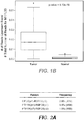
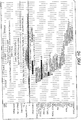
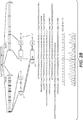


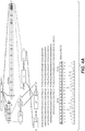

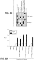


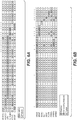
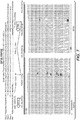
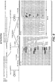

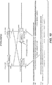
Classifications
-
- C—CHEMISTRY; METALLURGY
- C07—ORGANIC CHEMISTRY
- C07K—PEPTIDES
- C07K14/00—Peptides having more than 20 amino acids; Gastrins; Somatostatins; Melanotropins; Derivatives thereof
- C07K14/415—Peptides having more than 20 amino acids; Gastrins; Somatostatins; Melanotropins; Derivatives thereof from plants
-
- A—HUMAN NECESSITIES
- A61—MEDICAL OR VETERINARY SCIENCE; HYGIENE
- A61K—PREPARATIONS FOR MEDICAL, DENTAL OR TOILETRY PURPOSES
- A61K39/00—Medicinal preparations containing antigens or antibodies
- A61K39/395—Antibodies; Immunoglobulins; Immune serum, e.g. antilymphocytic serum
- A61K39/39533—Antibodies; Immunoglobulins; Immune serum, e.g. antilymphocytic serum against materials from animals
- A61K39/39558—Antibodies; Immunoglobulins; Immune serum, e.g. antilymphocytic serum against materials from animals against tumor tissues, cells, antigens
-
- A—HUMAN NECESSITIES
- A61—MEDICAL OR VETERINARY SCIENCE; HYGIENE
- A61K—PREPARATIONS FOR MEDICAL, DENTAL OR TOILETRY PURPOSES
- A61K45/00—Medicinal preparations containing active ingredients not provided for in groups A61K31/00 - A61K41/00
- A61K45/06—Mixtures of active ingredients without chemical characterisation, e.g. antiphlogistics and cardiaca
-
- A—HUMAN NECESSITIES
- A61—MEDICAL OR VETERINARY SCIENCE; HYGIENE
- A61P—SPECIFIC THERAPEUTIC ACTIVITY OF CHEMICAL COMPOUNDS OR MEDICINAL PREPARATIONS
- A61P1/00—Drugs for disorders of the alimentary tract or the digestive system
-
- A—HUMAN NECESSITIES
- A61—MEDICAL OR VETERINARY SCIENCE; HYGIENE
- A61P—SPECIFIC THERAPEUTIC ACTIVITY OF CHEMICAL COMPOUNDS OR MEDICINAL PREPARATIONS
- A61P35/00—Antineoplastic agents
-
- A—HUMAN NECESSITIES
- A61—MEDICAL OR VETERINARY SCIENCE; HYGIENE
- A61P—SPECIFIC THERAPEUTIC ACTIVITY OF CHEMICAL COMPOUNDS OR MEDICINAL PREPARATIONS
- A61P43/00—Drugs for specific purposes, not provided for in groups A61P1/00-A61P41/00
-
- C—CHEMISTRY; METALLURGY
- C12—BIOCHEMISTRY; BEER; SPIRITS; WINE; VINEGAR; MICROBIOLOGY; ENZYMOLOGY; MUTATION OR GENETIC ENGINEERING
- C12Q—MEASURING OR TESTING PROCESSES INVOLVING ENZYMES, NUCLEIC ACIDS OR MICROORGANISMS; COMPOSITIONS OR TEST PAPERS THEREFOR; PROCESSES OF PREPARING SUCH COMPOSITIONS; CONDITION-RESPONSIVE CONTROL IN MICROBIOLOGICAL OR ENZYMOLOGICAL PROCESSES
- C12Q1/00—Measuring or testing processes involving enzymes, nucleic acids or microorganisms; Compositions therefor; Processes of preparing such compositions
- C12Q1/68—Measuring or testing processes involving enzymes, nucleic acids or microorganisms; Compositions therefor; Processes of preparing such compositions involving nucleic acids
- C12Q1/6876—Nucleic acid products used in the analysis of nucleic acids, e.g. primers or probes
- C12Q1/6883—Nucleic acid products used in the analysis of nucleic acids, e.g. primers or probes for diseases caused by alterations of genetic material
- C12Q1/6886—Nucleic acid products used in the analysis of nucleic acids, e.g. primers or probes for diseases caused by alterations of genetic material for cancer
-
- C—CHEMISTRY; METALLURGY
- C12—BIOCHEMISTRY; BEER; SPIRITS; WINE; VINEGAR; MICROBIOLOGY; ENZYMOLOGY; MUTATION OR GENETIC ENGINEERING
- C12Q—MEASURING OR TESTING PROCESSES INVOLVING ENZYMES, NUCLEIC ACIDS OR MICROORGANISMS; COMPOSITIONS OR TEST PAPERS THEREFOR; PROCESSES OF PREPARING SUCH COMPOSITIONS; CONDITION-RESPONSIVE CONTROL IN MICROBIOLOGICAL OR ENZYMOLOGICAL PROCESSES
- C12Q2600/00—Oligonucleotides characterized by their use
- C12Q2600/106—Pharmacogenomics, i.e. genetic variability in individual responses to drugs and drug metabolism
Definitions
- therapies related to the treatment of pathological conditions such as cancer.
- CRC Colorectal cancer
- Genomic studies have identified acquisition of mutations in genes like APC , KRAS, and TP53 during CRC progression ( Fearon, E. R., Annu. Rev. Pathol. 6:479-507 (2011 )). Sequencing colon cancer protein-coding exons and whole genomes in a small number of samples have identified several additional mutations and chromosomal structural variants that likely contribute to oncogenesis ( Wood, L. D. et al., Science 318:1108-1113 (2007 ); Timmermann, B. et al., PloS One 5:e15661 (2010 )). However, recent insertional mutagenesis screens in mouse models of colon cancer suggested involvement of additional genes and pathways in CRC development ( Starr, T. K. et al., Science 323:1747-1750 (2009 ); March, H. N. et al., Nat. Genet. 43:1202-1209 (2011 )).
- R-spondin-translocation antagonists Provided herein are R-spondin-translocation antagonists and methods of using the same.
- the invention relates to the embodiments as defined in the claims.
- One aspect of the invention relates to an antibody which binds to R-spondin 3 (RSPO3) for use in treating a cancer cell, wherein the cancer cell comprises an RSPO3 translocation.
- RSPO3 R-spondin 3
- Also comprised by the present invention is an antibody which binds to RSPO3 for use in treating cancer, wherein the cancer comprising an RSPO3 translocation.
- the invention also relates to a method of identifying an individual with cancer who is more or less likely to exhibit benefit from treatment with an anti-cancer therapy comprising an antibody which binds to RSPO3, the method comprising: determining presence or absence of an RSPO3 translocation in a sample obtained from the individual, wherein presence of the RSPO3 translocation in the sample indicates that the individual is more likely to exhibit benefit from treatment with the anti-cancer therapy comprising the antibody which binds to RSPO3 or absence of the RSPO3 translocation indicates that the individual is less likely to exhibit benefit from treatment with the anti-cancer therapy comprising the antibody which binds to RSPO3.
- wnt pathway antagonists are also provided herein.
- Furher provided herein are methods of inhibiting cell proliferation of a cancer cell comprising contacting the cancer cell with an effective amount of an R-spondin-translocation antagonist. Further provided herein are methods of treating cancer in an individual comprising administering to the individual an effective amount of an R-spondin-translocation antagonist. In some embodiments of any of the methods, the cancer or cancer cell comprises an R-spondin translocation.
- kits for treating cancer in an individual comprising administering to the individual an effective amount of a wnt pathway antagonist, wherein treatment is based upon the individual having cancer comprising an R-spondin translocation.
- methods of treating a cancer cell wherein the cancer cell comprises an R-spondin translocation, and wherein the method comprises providing an effective amount of a wnt pathway antagonist.
- methods of treating cancer in an individual provided that the individual has been found to have cancer comprising an R-spondin translocation, the treatment comprising administering to the individual an effective amount of a wnt pathway antagonist.
- a sample obtained from the individual comprises an R-spondin translocation, and administering an effective amount of an anti-cancer therapy comprising a wnt pathway antagonist to the individual, whereby the cancer is treated.
- kits for treating cancer comprising: (a) selecting an individual having cancer, wherein the cancer comprising an R-spondin translocation; and (b) administering to the individual thus selected an effective amount of a wnt pathway antagonist, whereby the cancer is treated.
- the method comprising: determining presence or absence of an R-spondin translocation in a sample obtained from the individual, wherein presence of the R-spondin translocation in the sample indicates that the individual is more likely to exhibit benefit from treatment with the anti-cancer therapy comprising the wnt pathway antagonist or absence of the R-spondin translocation indicates that the individual is less likely to exhibit benefit from treatment with the anti-cancer therapy comprising the wnt pathway antagonist.
- the method further comprises administering an effective amount of the anti-cancer therapy comprising a wnt pathway antagonist.
- the method comprising determining an R-spondin translocation, whereby presence of the R-spondin translocation indicates that the individual is more likely to respond effectively to treatment with the wnt pathway antagonist and absence of the R-spondin translocation indicates that the individual is less likely to respond effectively to treatment with the wnt pathway antagonist.
- the method further comprises administering an effective amount of the anti-cancer therapy comprising a wnt pathway antagonist.
- the method further comprises administering an effective amount of the anti-cancer therapy comprising a wnt pathway antagonist.
- the R-spondin translocation is a RSPO1 translocation, RSPO2 translocation, RSPO3 translocation and/or RSPO4 translocation.
- the R-spondin translocation is a RSPO2 translocation.
- the RSPO2 translocation comprises EIF3E and RSPO2.
- the RSPO2 translocation comprises EIF3E exon 1 and RSPO2 exon 2.
- the RSPO2 translocation comprises EIF3E exon 1 and RSPO2 exon 3.
- the RSPO2 translocation comprises SEQ ID NO:71
- the R-spondin translocation is a RSPO3 translocation.
- the RSPO3 translocation comprises PTPRK and RSPO3.
- the RSPO3 translocation comprises PTPRK exon 1 and RSPO3 exon 2.
- the RSPO3 translocation comprises PTPRK exon 7 and RSPO3 exon 2.
- the RSPO3 translocation comprises SEQ ID NO:72 and/or SEQ ID NO:73.
- the R-spondin translocation is detected at the chromosomal level (e.g., FISH), DNA level, RNA level (e.g ., RSPO1-translocation fusion transcript), and/or protein level (e.g ., RSPO1-translocation fusion polypeptide).
- chromosomal level e.g., FISH
- DNA level e.g., DNA level
- RNA level e.g ., RSPO1-translocation fusion transcript
- protein level e.g ., RSPO1-translocation fusion polypeptide
- the cancer is colorectal cancer. In some embodiments, the cancer is a colon cancer or rectal cancer.
- R-spondin-translocation antagonists wherein the R-spondin-translocation antagonist is an antibody, binding polypeptide, small molecule, or polynucleotide.
- the R-spondin-translocation antagonist binds a RSPO1-translocation fusion polypeptide and/or polynucleotide, RSPO2-translocation fusion polypeptide and/or polynucleotide, RSPO3-translocation fusion polypeptide and/or polynucleotide and/or RSPO4-translocation fusion polypeptide and/or polynucleotide.
- the R-spondin-translocation antagonist binds a RSPO2-translocation fusion polypeptide and/or polynucleotide.
- the RSPO2-translocation fusion polypeptide and/or polynucleotide comprises EIF3E and RSPO2.
- the RSPO2-translocation fusion polypeptide and/or polynucleotide comprises EIF3E exon 1 and RSPO2 exon 2.
- the RSPO2-translocation fusion polypeptide and/or polynucleotide comprises EIF3E exon 1 and RSPO2 exon 3.
- the RSPO2-translocation fusion polypeptide and/or polynucleotide comprises SEQ ID NO:71.
- the R-spondin-translocation fusion polypeptide and/or polynucleotide is a RSPO3-translocation fusion polypeptide and/or polynucleotide.
- the RSPO3-translocation fusion polypeptide and/or polynucleotide comprises PTPRK and RSPO3.
- the RSPO3-translocation fusion polypeptide and/or polynucleotide comprises PTPRK exon 1 and RSPO3 exon 2.
- the RSPO3-translocation fusion polypeptide and/or polynucleotide comprises PTPRK exon 7 and RSPO3 exon 2. In some embodiments, the RSPO3-translocation fusion polypeptide and/or polynucleotide comprises SEQ ID NO:72 and/or SEQ ID NO:73.
- R-spondin and RSPO refer herein to a native R-spondin from any vertebrate source, including mammals such as primates ( e.g., humans) and rodents ( e.g., mice and rats), unless otherwise indicated.
- the term encompasses "full-length,” unprocessed R-spondin as well as any form of R-spondin that results from processing in the cell.
- the term also encompasses naturally occurring variants of R-spondin, e.g ., splice variants or allelic variants.
- R-spondin is a family of four proteins, R-spondin 1 (RSPO1), R-spondin 2 (RSPO2), R-spondin 3 (RSPO3), and R-spondin 4 (RSPO4).
- the R-spondin is RSPO1.
- the sequence of an exemplary human RSPO1 nucleic acid sequence is SEQ ID NO:1 or an exemplary human RSPO1 is amino acid sequence of SEQ ID NO:2.
- the R-spondin is RSPO2.
- the sequence of an exemplary human RSPO2 nucleic acid sequence is SEQ ID NO:3 or an exemplary human RSPO2 is amino acid sequence of SEQ ID NO:4.
- the R-spondin is RSPO3.
- sequence of an exemplary human RSPO3 nucleic acid sequence is SEQ ID NO:5 or an exemplary human RSPO3 is amino acid sequence of SEQ ID NO:6.
- the R-spondin is RSPO4.
- sequence of an exemplary human RSPO4 nucleic acid sequence is SEQ ID NO:7 or an exemplary human RSPO4 is amino acid sequence of SEQ ID NO:8.
- R-Spondin variant means an R-spondin polypeptide or polynucleotide, generally being or encoding an active R-Spondin polypeptide, as defined herein having at least about 80% amino acid sequence identity with any of the R-Spondin as disclosed herein.
- R-Spondin variants include, for instance, R-Spondin wherein one or more nucleic acid or amino acid residues are added or deleted.
- an R-spondin variant will have at least about 80% sequence identity, alternatively at least about 81%, 82%, 83%, 84%, 85%, 86%, 87%, 88%, 89%, 90%, 91%, 92%, 93%, 94%, 95%, 96%, 97%, 98%, or 99% sequence identity, to R-Spondin as disclosed herein.
- R-Spondin variant are at least about 10 residues in length, alternatively at least about 20, 30, 40, 50, 60, 70, 80, 90, 100, 110, 120, 130, 140, 150, 160, 170, 180, 190, 200, 210, 220, 230, 240, 250, 260, 270, 280, 290, 300, 310, 320, 330, 340, 350, 360, 370, 380, 390, 400, 410, 420, 430, 440, 450, 460, 470, 480, 490, 500, 510, 520, 530, 540, 550, 560, 570, 580, 590, 600 in length, or more.
- R-Spondin variant will have or encode a sequence having no more than one conservative amino acid substitution as compared to R-Spondin, alternatively no more than 2, 3, 4, 5, 6, 7, 8, 9, or 10 conservative amino acid substitution as compared to R-Spondin.
- R-spondin translocation and "RSPO translocation” refer herein to an R-spondin wherein a portion of a broken chromosome including, for example, R-spondin, variant, or fragment thereof or a second gene, variant, or fragment thereof, reattaches in a different chromosome location, for example, a chromosome location different from R-spondin native location or a chromosome location in and/or around the R-spondin native location which is different from the second gene's native location.
- the R-spondin translocation may be a RSPO1 translocation, RSPO2 translocation, RSPO3 translocation, and/or RSPO4 translocation.
- R-spondin-translocation fusion polynucleotide and "RSPO-translocation fusion polynucleotide” refer herein to the nucleic acid sequence of an R-spondin translocation gene product or fusion polynucleotide.
- the R-spondin-translocation fusion polynucleotide may be a RSPO1-translocation fusion polynucleotide, RSPO2-translocation fusion polynucleotide, RSPO3-translocation fusion polynucleotide, and/or RSPO4-translocation fusion polynucleotide.
- R-spondin-translocation fusion polypeptide and "RSPO-translocation fusion polypeptide” refer herein to the amino acid sequence of an R-spondin translocation gene product or fusion polynucleotide.
- the R-spondin-translocation fusion polypeptide may be a RSPO1-translocation fusion polypeptide, RSPO2-translocation fusion polypeptide, RSPO3-translocation fusion polypeptide, and/or RSPO4-translocation fusion polypeptide.
- R-spondin-translocation antagonist is any molecule that partially or fully blocks, inhibits, or neutralizes a biological activity mediated by an R-spondin-translocation fusion polypeptide. In some embodiments such antagonist binds to R-spondin-translocation fusion polypeptide. According to one embodiment, the antagonist is a polypeptide. According to another embodiment, the antagonist is an anti-R-spondin-translocation antibody. According to another embodiment, the antagonist is a small molecule antagonist. According to another embodiment, the antagonist is a polynucleotide antagonist.
- the R-spondin translocation may be a RSPO1-translocation antagonist, RSPO2-translocation antagonist, RSPO3-translocation antagonist, and/or RSPO4-translocation antagonist.
- wnt pathway antagonist is any molecule that partially or fully blocks, inhibits, or neutralizes a biological activity mediated by the wnt pathway (e.g ., wnt pathway polypeptide). In some embodiments such antagonist binds to a wnt pathway polypeptide.
- the antagonist is a polypeptide.
- the antagonist is an antibody antagonist.
- the antagonist is a small molecule antagonist.
- the antagonist is a polynucleotide antagonist.
- Polynucleotide or “nucleic acid” as used interchangeably herein, refers to polymers of nucleotides of any length, and include DNA and RNA.
- the nucleotides can be deoxyribonucleotides, ribonucleotides, modified nucleotides or bases, and/or their analogs, or any substrate that can be incorporated into a polymer by DNA or RNA polymerase or by a synthetic reaction.
- a polynucleotide may comprise modified nucleotides, such as methylated nucleotides and their analogs.
- a sequence of nucleotides may be interrupted by non-nucleotide components.
- a polynucleotide may comprise modification(s) made after synthesis, such as conjugation to a label.
- modifications include, for example, "caps,” substitution of one or more of the naturally occurring nucleotides with an analog, internucleotide modifications such as, for example, those with uncharged linkages (e.g ., methyl phosphonates, phosphotriesters, phosphoamidates, carbamates, etc.) and with charged linkages (e.g.
- phosphorothioates those containing pendant moieties, such as, for example, proteins (e.g ., nucleases, toxins, antibodies, signal peptides, ply-L-lysine, etc.), those with intercalators (e.g ., acridine, psoralen, etc.), those containing chelators (e.g ., metals, radioactive metals, boron, oxidative metals, etc.), those containing alkylators, those with modified linkages ( e.g., alpha anomeric nucleic acids, etc.), as well as unmodified forms of the polynucleotides(s).
- proteins e.g ., nucleases, toxins, antibodies, signal peptides, ply-L-lysine, etc.
- intercalators e.g ., acridine, psoralen, etc.
- chelators e.g ., metals, radioactive metals
- any of the hydroxyl groups ordinarily present in the sugars may be replaced, for example, by phosphonate groups, phosphate groups, protected by standard protecting groups, or activated to prepare additional linkages to additional nucleotides, or may be conjugated to solid or semi-solid supports.
- the 5' and 3' terminal OH can be phosphorylated or substituted with amines or organic capping group moieties of from 1 to 20 carbon atoms.
- Other hydroxyls may also be derivatized to standard protecting groups.
- Polynucleotides can also contain analogous forms of ribose or deoxyribose sugars that are generally known in the art, including, for example, 2'-O-methyl-, 2'-O-allyl-, 2'-fluoro- or 2'-azido-ribose, carbocyclic sugar analogs, ⁇ -anomeric sugars, epimeric sugars such as arabinose, xyloses or lyxoses, pyranose sugars, furanose sugars, sedoheptuloses, acyclic analogs, and basic nucleoside analogs such as methyl riboside.
- One or more phosphodiester linkages may be replaced by alternative linking groups.
- linking groups include, but are not limited to, embodiments wherein phosphate is replaced by P(O)S ("thioate”), P(S)S ("dithioate”), (O)NR 2 ("amidate"), P(O)R, P(O)OR', CO, or CH2 ("formacetal"), in which each R or R' is independently H or substituted or unsubstituted alkyl (1-20 C) optionally containing an ether (-O-) linkage, aryl, alkenyl, cycloalkyl, cycloalkenyl or araldyl. Not all linkages in a polynucleotide need be identical. The preceding description applies to all polynucleotides referred to herein, including RNA and DNA.
- Oligonucleotide refers to generally single-stranded, synthetic polynucleotides that are generally, but not necessarily, less than about 200 nucleotides in length.
- oligonucleotide and polynucleotide are not mutually exclusive. The description above for polynucleotides is equally and fully applicable to oligonucleotides.
- primer refers to a single stranded polynucleotide that is capable of hybridizing to a nucleic acid and following polymerization of a complementary nucleic acid, generally by providing a free 3'-OH group.
- small molecule refers to any molecule with a molecular weight of about 2000 Daltons or less, preferably of about 500 Daltons or less.
- host cell refers to cells into which exogenous nucleic acid has been introduced, including the progeny of such cells.
- Host cells include “transformants” and “transformed cells,” which include the primary transformed cell and progeny derived therefrom without regard to the number of passages. Progeny may not be completely identical in nucleic acid content to a parent cell, but may contain mutations. Mutant progeny that have the same function or biological activity as screened or selected for in the originally transformed cell are included herein.
- vector refers to a nucleic acid molecule capable of propagating another nucleic acid to which it is linked.
- the term includes the vector as a self-replicating nucleic acid structure as well as the vector incorporated into the genome of a host cell into which it has been introduced.
- Certain vectors are capable of directing the expression of nucleic acids to which they are operatively linked. Such vectors are referred to herein as "expression vectors.”
- an “isolated” antibody is one which has been separated from a component of its natural environment.
- an antibody is purified to greater than 95% or 99% purity as determined by, for example, electrophoretic (e.g ., SDS-PAGE, isoelectric focusing (IEF), capillary electrophoresis) or chromatographic (e.g ., ion exchange or reverse phase HPLC).
- electrophoretic e.g ., SDS-PAGE, isoelectric focusing (IEF), capillary electrophoresis
- chromatographic e.g ., ion exchange or reverse phase HPLC
- nucleic acid refers to a nucleic acid molecule that has been separated from a component of its natural environment.
- An isolated nucleic acid includes a nucleic acid molecule contained in cells that ordinarily contain the nucleic acid molecule, but the nucleic acid molecule is present extrachromosomally or at a chromosomal location that is different from its natural chromosomal location.
- antibody herein is used in the broadest sense and encompasses various antibody structures, including but not limited to monoclonal antibodies, polyclonal antibodies, multispecific antibodies (e.g ., bispecific antibodies), and antibody fragments so long as they exhibit the desired antigen-binding activity.
- antibody fragment refers to a molecule other than an intact antibody that comprises a portion of an intact antibody that binds the antigen to which the intact antibody binds.
- antibody fragments include but are not limited to Fv, Fab, Fab', Fab'-SH, F(ab') 2 ; diabodies; linear antibodies; single-chain antibody molecules ( e.g ., scFv); and multispecific antibodies formed from antibody fragments.
- an "antibody that binds to the same epitope” as a reference antibody refers to an antibody that blocks binding of the reference antibody to its antigen in a competition assay by 50% or more, and conversely, the reference antibody blocks binding of the antibody to its antigen in a competition assay by 50% or more.
- An exemplary competition assay is provided herein.
- full length antibody “intact antibody,” and “whole antibody” are used herein interchangeably to refer to an antibody having a structure substantially similar to a native antibody structure or having heavy chains that contain an Fc region as defined herein.
- the term "monoclonal antibody” as used herein refers to an antibody obtained from a population of substantially homogeneous antibodies, i.e ., the individual antibodies comprising the population are identical and/or bind the same epitope, except for possible variant antibodies, e.g ., containing naturally occurring mutations or arising during production of a monoclonal antibody preparation, such variants generally being present in minor amounts.
- polyclonal antibody preparations typically include different antibodies directed against different determinants (epitopes)
- each monoclonal antibody of a monoclonal antibody preparation is directed against a single determinant on an antigen.
- the modifier "monoclonal” indicates the character of the antibody as being obtained from a substantially homogeneous population of antibodies, and is not to be construed as requiring production of the antibody by any particular method.
- the monoclonal antibodies to be used in accordance with the present disclosure may be made by a variety of techniques, including but not limited to the hybridoma method, recombinant DNA methods, phage-display methods, and methods utilizing transgenic animals containing all or part of the human immunoglobulin loci, such methods and other exemplary methods for making monoclonal antibodies being described herein.
- Native antibodies refer to naturally occurring immunoglobulin molecules with varying structures.
- native IgG antibodies are heterotetrameric glycoproteins of about 150,000 Daltons, composed of two identical light chains and two identical heavy chains that are disulfide-bonded. From N- to C-terminus, each heavy chain has a variable region (VH), also called a variable heavy domain or a heavy chain variable domain, followed by three constant domains (CH1, CH2, and CH3).
- VH variable region
- VL variable region
- the light chain of an antibody may be assigned to one of two types, called kappa ( ⁇ ) and lambda ( ⁇ ), based on the amino acid sequence of its constant domain.
- chimeric antibody refers to an antibody in which a portion of the heavy and/or light chain is derived from a particular source or species, while the remainder of the heavy and/or light chain is derived from a different source or species.
- a "human antibody” is one which possesses an amino acid sequence which corresponds to that of an antibody produced by a human or a human cell or derived from a non-human source that utilizes human antibody repertoires or other human antibody-encoding sequences. This definition of a human antibody specifically excludes a humanized antibody comprising non-human antigen-binding residues.
- a “humanized” antibody refers to a chimeric antibody comprising amino acid residues from non-human HVRs and amino acid residues from human FRs.
- a humanized antibody will comprise substantially all of at least one, and typically two, variable domains, in which all or substantially all of the HVRs (e.g ., CDRs) correspond to those of a non-human antibody, and all or substantially all of the FRs correspond to those of a human antibody.
- a humanized antibody optionally may comprise at least a portion of an antibody constant region derived from a human antibody.
- a "humanized form" of an antibody, e.g ., a non-human antibody refers to an antibody that has undergone humanization.
- the "class" of an antibody refers to the type of constant domain or constant region possessed by its heavy chain.
- the heavy chain constant domains that correspond to the different classes of immunoglobulins are called ⁇ , ⁇ , ⁇ , ⁇ , and ⁇ , respectively.
- Antibody effector functions refer to those biological activities attributable to the Fc region of an antibody, which vary with the antibody isotype. Examples of antibody effector functions include: C1q binding and complement dependent cytotoxicity (CDC); Fc receptor binding; antibody-dependent cell-mediated cytotoxicity (ADCC); phagocytosis; down regulation of cell surface receptors ( e.g., B cell receptor); and B cell activation.
- Fc region herein is used to define a C-terminal region of an immunoglobulin heavy chain that contains at least a portion of the constant region.
- the term includes native sequence Fc regions and variant Fc regions.
- a human IgG heavy chain Fc region extends from Cys226, or from Pro230, to the carboxyl-terminus of the heavy chain.
- the C-terminal lysine (Lys447) of the Fc region may or may not be present.
- numbering of amino acid residues in the Fc region or constant region is according to the EU numbering system, also called the EU index, as described in Kabat et al., Sequences of Proteins of Immunological Interest, 5th Ed. Public Health Service, National Institutes of Health, Bethesda, MD, 1991 .
- FR Framework or "FR” refers to variable domain residues other than hypervariable region (HVR) residues.
- the FR of a variable domain generally consists of four FR domains: FR1, FR2, FR3, and FR4. Accordingly, the HVR and FR sequences generally appear in the following sequence in VH (or VL): FR1-H1(L1)-FR2-H2(L2)-FR3-H3(L3)-FR4.
- a "human consensus framework” is a framework which represents the most commonly occurring amino acid residues in a selection of human immunoglobulin VL or VH framework sequences.
- the selection of human immunoglobulin VL or VH sequences is from a subgroup of variable domain sequences.
- the subgroup of sequences is a subgroup as in Kabat et al., Sequences of Proteins of Immunological Interest, Fifth Edition, NIH Publication 91-3242, Bethesda MD (1991), vols. 1-3 .
- the subgroup is subgroup kappa I as in Kabat et al ., supra.
- the subgroup is subgroup III as in Kabat et al ., supra.
- acceptor human framework for the purposes herein is a framework comprising the amino acid sequence of a light chain variable domain (VL) framework or a heavy chain variable domain (VH) framework derived from a human immunoglobulin framework or a human consensus framework, as defined below.
- An acceptor human framework "derived from” a human immunoglobulin framework or a human consensus framework may comprise the same amino acid sequence thereof, or it may contain amino acid sequence changes. In some embodiments, the number of amino acid changes are 10 or less, 9 or less, 8 or less, 7 or less, 6 or less, 5 or less, 4 or less, 3 or less, or 2 or less.
- the VL acceptor human framework is identical in sequence to the VL human immunoglobulin framework sequence or human consensus framework sequence.
- variable region refers to the domain of an antibody heavy or light chain that is involved in binding the antibody to antigen.
- the variable domains of the heavy chain and light chain (VH and VL, respectively) of a native antibody generally have similar structures, with each domain comprising four conserved framework regions (FRs) and three hypervariable regions (HVRs).
- FRs conserved framework regions
- HVRs hypervariable regions
- antibodies that bind a particular antigen may be isolated using a VH or VL domain from an antibody that binds the antigen to screen a library of complementary VL or VH domains, respectively. See, e.g., Portolano et al., J. Immunol. 150:880-887 (1993 ); Clarkson et al., Nature 352:624-628 (1991 ).
- hypervariable region refers to each of the regions of an antibody variable domain which are hypervariable in sequence and/or form structurally defined loops ("hypervariable loops").
- native four-chain antibodies comprise six HVRs; three in the VH (H1, H2, H3), and three in the VL (L1, L2, L3).
- HVRs generally comprise amino acid residues from the hypervariable loops and/or from the "complementarity determining regions" (CDRs), the latter being of highest sequence variability and/or involved in antigen recognition.
- CDRs complementarity determining regions
- Exemplary hypervariable loops occur at amino acid residues 26-32 (L1), 50-52 (L2), 91-96 (L3), 26-32 (H1), 53-55 (H2), and 96-101 (H3).
- Exemplary CDRs CDR-L1, CDR-L2, CDR-L3, CDR-H1, CDR-H2, and CDR-H3 occur at amino acid residues 24-34 of L1, 50-56 of L2, 89-97 of L3, 31-35B of H1, 50-65 of H2, and 95-102 of H3.
- CDRs generally comprise the amino acid residues that form the hypervariable loops.
- CDRs also comprise "specificity determining residues,” or "SDRs,” which are residues that contact antigen. SDRs are contained within regions of the CDRs called abbreviated-CDRs, or a-CDRs.
- Exemplary a-CDRs (a-CDR-L1, a-CDR-L2, a-CDR-L3, a-CDR-H1, a-CDR-H2, and a-CDR-H3) occur at amino acid residues 31-34 of L1, 50-55 of L2, 89-96 of L3, 31-35B of H1, 50-58 of H2, and 95-102 of H3.
- HVR residues and other residues in the variable domain e.g ., FR residues
- FR residues are numbered herein according to Kabat et al ., supra.
- Binding affinity refers to the strength of the sum total of noncovalent interactions between a single binding site of a molecule (e.g., an antibody) and its binding partner (e.g., an antigen). Unless indicated otherwise, as used herein, "binding affinity” refers to intrinsic binding affinity which reflects a 1:1 interaction between members of a binding pair ( e.g ., antibody and antigen).
- the affinity of a molecule X for its partner Y can generally be represented by the dissociation constant (Kd). Affinity can be measured by common methods known in the art, including those described herein. Specific illustrative and exemplary embodiments for measuring binding affinity are described in the following.
- an “affinity matured” antibody refers to an antibody with one or more alterations in one or more hypervariable regions (HVRs), compared to a parent antibody which does not possess such alterations, such alterations resulting in an improvement in the affinity of the antibody for antigen.
- HVRs hypervariable regions
- anti-R-spondin-translocation antibody and "an antibody that binds to R-spondin-translocation fusion polypeptide” refer to an antibody that is capable of binding R-spondin-translocation fusion polypeptide with sufficient affinity such that the antibody is useful as a diagnostic and/or therapeutic agent in targeting R-spondin translocation.
- the extent of binding of an anti-R-spondin translocation antibody to an unrelated, non-R-spondin-translocation fusion polypeptide, and/or nontranslocated-R-spondin polypeptide is less than about 10% of the binding of the antibody to R-spondin-translocation fusion polypeptides measured, e.g ., by a radioimmunoassay (RIA).
- RIA radioimmunoassay
- an antibody that binds to R-spondin-translocation fusion polypeptide has a dissociation constant (Kd) of ⁇ 1 ⁇ M, ⁇ 100 nM, ⁇ 10 nM, ⁇ 1 nM, ⁇ 0.1 nM, ⁇ 0.01 nM, or ⁇ 0.001 nM ( e.g., 10 -8 M or less, e.g., from 10 -8 M to 10 -13 M, e.g., from 10 -9 M to 10 -13 M).
- Kd dissociation constant
- an anti- R-spondin translocation antibody binds to an epitope of R-spondin translocation that is unique among R-spondin translocations.
- blocking antibody or an “antagonist” antibody is one which inhibits or reduces biological activity of the antigen it binds.
- Preferred blocking antibodies or antagonist antibodies substantially or completely inhibit the biological activity of the antigen.
- naked antibody refers to an antibody that is not conjugated to a heterologous moiety (e.g., a cytotoxic moiety) or radiolabel.
- the naked antibody may be present in a pharmaceutical formulation.
- an “immunoconjugate” is an antibody conjugated to one or more heterologous molecule(s), including but not limited to a cytotoxic agent.
- Percent (%) amino acid sequence identity with respect to a reference polypeptide sequence is defined as the percentage of amino acid residues in a candidate sequence that are identical with the amino acid residues in the reference polypeptide sequence, after aligning the sequences and introducing gaps, if necessary, to achieve the maximum percent sequence identity, and not considering any conservative substitutions as part of the sequence identity. Alignment for purposes of determining percent amino acid sequence identity can be achieved in various ways that are within the skill in the art, for instance, using publicly available computer software such as BLAST, BLAST-2, ALIGN or Megalign (DNASTAR) software. Those skilled in the art can determine appropriate parameters for aligning sequences, including any algorithms needed to achieve maximal alignment over the full length of the sequences being compared.
- % amino acid sequence identity values are generated using the sequence comparison computer program ALIGN-2.
- the ALIGN-2 sequence comparison computer program was authored by Genentech, Inc., and the source code has been filed with user documentation in the U.S. Copyright Office, Washington D.C., 20559, where it is registered under U.S. Copyright Registration No. TXU510087.
- the ALIGN-2 program is publicly available from Genentech, Inc., South San Francisco, California, or may be compiled from the source code.
- the ALIGN-2 program should be compiled for use on a UNIX operating system, including digital UNIX V4.0D. All sequence comparison parameters are set by the ALIGN-2 program and do not vary.
- the % amino acid sequence identity of a given amino acid sequence A to, with, or against a given amino acid sequence B is calculated as follows: 100 times the fraction X/Y where X is the number of amino acid residues scored as identical matches by the sequence alignment program ALIGN-2 in that program's alignment of A and B, and where Y is the total number of amino acid residues in B.
- detection includes any means of detecting, including direct and indirect detection.
- biomarker refers to an indicator, e.g ., predictive, diagnostic, and/or prognostic, which can be detected in a sample.
- the biomarker may serve as an indicator of a particular subtype of a disease or disorder (e.g ., cancer) characterized by certain, molecular, pathological, histological, and/or clinical features.
- the biomarker is a gene.
- the biomarker is a variation (e.g ., mutation and/or polymorphism) of a gene.
- the biomarkers is a translocation.
- Biomarkers include, but are not limited to, polynucleotides (e.g ., DNA, and/or RNA), polypeptides, polypeptide and polynucleotide modifications (e.g ., posttranslational modifications), carbohydrates, and/or glycolipid-based molecular markers.
- the "presence,” “amount,” or “level” of a biomarker associated with an increased clinical benefit to an individual is a detectable level in a biological sample. These can be measured by methods known to one skilled in the art and also disclosed herein. The expression level or amount of biomarker assessed can be used to determine the response to the treatment.
- level of expression or “expression level” in general are used interchangeably and generally refer to the amount of a biomarker in a biological sample. “Expression” generally refers to the process by which information (e.g ., gene-encoded and/or epigenetic) is converted into the structures present and operating in the cell. Therefore, as used herein, “expression” may refer to transcription into a polynucleotide, translation into a polypeptide, or even polynucleotide and/or polypeptide modifications ( e.g ., posttranslational modification of a polypeptide).
- Fragments of the transcribed polynucleotide, the translated polypeptide, or polynucleotide and/or polypeptide modifications shall also be regarded as expressed whether they originate from a transcript generated by alternative splicing or a degraded transcript, or from a post-translational processing of the polypeptide, e.g ., by proteolysis.
- "Expressed genes” include those that are transcribed into a polynucleotide as mRNA and then translated into a polypeptide, and also those that are transcribed into RNA but not translated into a polypeptide (for example, transfer and ribosomal RNAs).
- Elevated expression refers to an increased expression or increased levels of a biomarker in an individual relative to a control, such as an individual or individuals who are not suffering from the disease or disorder (e.g ., cancer) or an internal control (e.g ., housekeeping biomarker).
- a control such as an individual or individuals who are not suffering from the disease or disorder (e.g ., cancer) or an internal control (e.g ., housekeeping biomarker).
- Reduced expression refers to a decrease expression or decreased levels of a biomarker in an individual relative to a control, such as an individual or individuals who are not suffering from the disease or disorder (e.g ., cancer) or an internal control (e.g ., housekeeping biomarker).
- a control such as an individual or individuals who are not suffering from the disease or disorder (e.g ., cancer) or an internal control (e.g ., housekeeping biomarker).
- housekeeping biomarker refers to a biomarker or group of biomarkers (e.g ., polynucleotides and/or polypeptides) which are typically similarly present in all cell types.
- the housekeeping biomarker is a "housekeeping gene.”
- a "housekeeping gene” refers herein to a gene or group of genes which encode proteins whose activities are essential for the maintenance of cell function and which are typically similarly present in all cell types.
- Amplification generally refers to the process of producing multiple copies of a desired sequence.
- Multiple copies mean at least two copies.
- a “copy” does not necessarily mean perfect sequence complementarity or identity to the template sequence.
- copies can include nucleotide analogs such as deoxyinosine, intentional sequence alterations (such as sequence alterations introduced through a primer comprising a sequence that is hybridizable, but not complementary, to the template), and/or sequence errors that occur during amplification.
- multiplex-PCR refers to a single PCR reaction carried out on nucleic acid obtained from a single source (e.g., an individual) using more than one primer set for the purpose of amplifying two or more DNA sequences in a single reaction.
- “Stringency” of hybridization reactions is readily determinable by one of ordinary skill in the art, and generally is an empirical calculation dependent upon probe length, washing temperature, and salt concentration. In general, longer probes require higher temperatures for proper annealing, while shorter probes need lower temperatures. Hybridization generally depends on the ability of denatured DNA to reanneal when complementary strands are present in an environment below their melting temperature. The higher the degree of desired homology between the probe and hybridizable sequence, the higher the relative temperature which can be used. As a result, it follows that higher relative temperatures would tend to make the reaction conditions more stringent, while lower temperatures less so. For additional details and explanation of stringency of hybridization reactions, see Ausubel et al., Current Protocols in Molecular Biology, Wiley Interscience Publishers, (1995 ).
- “Stringent conditions” or “high stringency conditions”, as defined herein, can be identified by those that: (1) employ low ionic strength and high temperature for washing, for example 0.015 M sodium chloride/0.0015 M sodium citrate/0.1% sodium dodecyl sulfate at 50°C; (2) employ during hybridization a denaturing agent, such as formamide, for example, 50% (v/v) formamide with 0.1% bovine serum albumin/0.1% Ficoll/0.1% polyvinylpyrrolidone/50mM sodium phosphate buffer at pH 6.5 with 750 mM sodium chloride, 75 mM sodium citrate at 42°C; or (3) overnight hybridization in a solution that employs 50% formamide, 5 x SSC (0.75 M NaCl, 0.075 M sodium citrate), 50 mM sodium phosphate (pH 6.8), 0.1% sodium pyrophosphate, 5 x Denhardt's solution, sonicated salmon sperm DNA (50 ⁇ g/ml), 0.1% SDS
- Modely stringent conditions can be identified as described by Sambrook et al., Molecular Cloning: A Laboratory Manual, New York: Cold Spring Harbor Press, 1989 , and include the use of washing solution and hybridization conditions (e.g ., temperature, ionic strength and %SDS) less stringent that those described above.
- washing solution and hybridization conditions e.g ., temperature, ionic strength and %SDS
- moderately stringent conditions is overnight incubation at 37°C in a solution comprising: 20% formamide, 5 x SSC (150 mM NaCl, 15 mM trisodium citrate), 50 mM sodium phosphate (pH 7.6), 5 x Denhardt's solution, 10% dextran sulfate, and 20 mg/ml denatured sheared salmon sperm DNA, followed by washing the filters in 1 x SSC at about 37-50°C.
- the skilled artisan will recognize how to adjust the temperature, ionic strength, etc. as necessary to accommodate factors such as probe length and the like.
- diagnosis is used herein to refer to the identification or classification of a molecular or pathological state, disease or condition (e.g ., cancer).
- diagnosis may refer to identification of a particular type of cancer.
- Diagnosis may also refer to the classification of a particular subtype of cancer, e.g., by histopathological criteria, or by molecular features (e.g., a subtype characterized by expression of one or a combination of biomarkers (e.g ., particular genes or proteins encoded by said genes)).
- a method of aiding diagnosis of a disease or condition can comprise detecting certain biomarkers in a biological sample from an individual.
- sample refers to a composition that is obtained or derived from a subject and/or individual of interest that contains a cellular and/or other molecular entity that is to be characterized and/or identified, for example based on physical, biochemical, chemical and/or physiological characteristics.
- disease sample and variations thereof refers to any sample obtained from a subject of interest that would be expected or is known to contain the cellular and/or molecular entity that is to be characterized.
- Samples include, but are not limited to, primary or cultured cells or cell lines, cell supernatants, cell lysates, platelets, serum, plasma, vitreous fluid, lymph fluid, synovial fluid, follicular fluid, seminal fluid, amniotic fluid, milk, whole blood, blood-derived cells, urine, cerebro-spinal fluid, saliva, sputum, tears, perspiration, mucus, tumor lysates, and tissue culture medium, tissue extracts such as homogenized tissue, tumor tissue, cellular extracts, and combinations thereof.
- tissue sample or “cell sample” is meant a collection of similar cells obtained from a tissue of a subject or individual.
- the source of the tissue or cell sample may be solid tissue as from a fresh, frozen and/or preserved organ, tissue sample, biopsy, and/or aspirate; blood or any blood constituents such as plasma; bodily fluids such as cerebral spinal fluid, amniotic fluid, peritoneal fluid, or interstitial fluid; cells from any time in gestation or development of the subject.
- the tissue sample may also be primary or cultured cells or cell lines.
- the tissue or cell sample is obtained from a disease tissue/organ.
- the tissue sample may contain compounds which are not naturally intermixed with the tissue in nature such as preservatives, anticoagulants, buffers, fixatives, nutrients, antibiotics, or the like.
- a “reference sample”, “reference cell”, “reference tissue”, “control sample”, “control cell”, or “control tissue”, as used herein, refers to a sample, cell, tissue, standard, or level that is used for comparison purposes.
- a reference sample, reference cell, reference tissue, control sample, control cell, or control tissue is obtained from a healthy and/or non-diseased part of the body (e.g ., tissue or cells) of the same subject or individual.
- healthy and/or non-diseased cells or tissue adjacent to the diseased cells or tissue e.g., cells or tissue adjacent to a tumor.
- a reference sample is obtained from an untreated tissue and/or cell of the body of the same subject or individual.
- a reference sample, reference cell, reference tissue, control sample, control cell, or control tissue is obtained from a healthy and/or non-diseased part of the body (e.g ., tissues or cells) of an individual who is not the subject or individual.
- a reference sample, reference cell, reference tissue, control sample, control cell, or control tissue is obtained from an untreated tissue and/or cell of the body of an individual who is not the subject or individual.
- a "section" of a tissue sample is meant a single part or piece of a tissue sample, e.g., a thin slice of tissue or cells cut from a tissue sample. It is understood that multiple sections of tissue samples may be taken and subjected to analysis, provided that it is understood that the same section of tissue sample may be analyzed at both morphological and molecular levels, or analyzed with respect to both polypeptides and polynucleotides.
- correlate or “correlating” is meant comparing, in any way, the performance and/or results of a first analysis or protocol with the performance and/or results of a second analysis or protocol. For example, one may use the results of a first analysis or protocol in carrying out a second protocols and/or one may use the results of a first analysis or protocol to determine whether a second analysis or protocol should be performed. With respect to the embodiment of polynucleotide analysis or protocol, one may use the results of the polynucleotide expression analysis or protocol to determine whether a specific therapeutic regimen should be performed.
- “Individual response” or “response” can be assessed using any endpoint indicating a benefit to the individual, including, without limitation, (1) inhibition, to some extent, of disease progression (e.g., cancer progression), including slowing down and complete arrest; (2) a reduction in tumor size; (3) inhibition ( i.e., reduction, slowing down or complete stopping) of cancer cell infiltration into adjacent peripheral organs and/or tissues; (4) inhibition ( i.e. reduction, slowing down or complete stopping) of metasisis; (5) relief, to some extent, of one or more symptoms associated with the disease or disorder (e.g ., cancer); (6) increase in the length of progression free survival; and/or (9) decreased mortality at a given point of time following treatment.
- disease progression e.g., cancer progression
- inhibition i.e., reduction, slowing down or complete stopping
- metasisis i.e. reduction, slowing down or complete stopping
- substantially similar refers to a sufficiently high degree of similarity between two numeric values (generally one associated with a molecule and the other associated with a reference/comparator molecule) such that one of skill in the art would consider the difference between the two values to not be of statistical significance within the context of the biological characteristic measured by said values (e.g ., Kd values).
- the difference between said two values may be, for example, less than about 20%, less than about 10%, and/or less than about 5% as a function of the reference/comparator value.
- substantially normal refers to substantially similar to a reference (e.g ., normal reference).
- substantially different refers to a sufficiently high degree of difference between two numeric values (generally one associated with a molecule and the other associated with a reference/comparator molecule) such that one of skill in the art would consider the difference between the two values to be of statistical significance within the context of the biological characteristic measured by said values (e.g ., Kd values).
- the difference between said two values may be, for example, greater than about 10%, greater than about 20%, greater than about 30%, greater than about 40%, and/or greater than about 50% as a function of the value for the reference/comparator molecule.
- label when used herein refers to a detectable compound or composition.
- the label is typically conjugated or fused directly or indirectly to a reagent, such as a polynucleotide probe or an antibody, and facilitates detection of the reagent to which it is conjugated or fused.
- the label may itself be detectable (e.g ., radioisotope labels or fluorescent labels) or, in the case of an enzymatic label, may catalyze chemical alteration of a substrate compound or composition which results in a detectable product.
- an “effective amount” of an agent refers to an amount effective, at dosages and for periods of time necessary, to achieve the desired therapeutic or prophylactic result.
- a “therapeutically effective amount” of a substance/molecule of the disclosure, agonist or antagonist may vary according to factors such as the disease state, age, sex, and weight of the individual, and the ability of the substance/molecule, agonist or antagonist to elicit a desired response in the individual.
- a therapeutically effective amount is also one in which any toxic or detrimental effects of the substance/molecule, agonist or antagonist are outweighed by the therapeutically beneficial effects.
- a “prophylactically effective amount” refers to an amount effective, at dosages and for periods of time necessary, to achieve the desired prophylactic result. Typically but not necessarily, since a prophylactic dose is used in subjects prior to or at an earlier stage of disease, the prophylactically effective amount will be less than the therapeutically effective amount.
- pharmaceutical formulation refers to a preparation which is in such form as to permit the biological activity of an active ingredient contained therein to be effective, and which contains no additional components which are unacceptably toxic to a subject to which the formulation would be administered.
- a “pharmaceutically acceptable carrier” refers to an ingredient in a pharmaceutical formulation, other than an active ingredient, which is nontoxic to a subject.
- a pharmaceutically acceptable carrier includes, but is not limited to, a buffer, excipient, stabilizer, or preservative.
- treatment refers to clinical intervention in an attempt to alter the natural course of the individual being treated, and can be performed either for prophylaxis or during the course of clinical pathology. Desirable effects of treatment include, but are not limited to, preventing occurrence or recurrence of disease, alleviation of symptoms, diminishment of any direct or indirect pathological consequences of the disease, preventing metastasis, decreasing the rate of disease progression, amelioration or palliation of the disease state, and remission or improved prognosis.
- antibodies provided herein are used to delay development of a disease or to slow the progression of a disease.
- cancer and “cancerous” refer to or describe the physiological condition in mammals that is typically characterized by unregulated cell growth/proliferation.
- examples of cancer include, but are not limited to, carcinoma, lymphoma (e.g ., Hodgkin's and non-Hodgkin's lymphoma), blastoma, sarcoma, and leukemia.
- cancers include squamous cell cancer, small-cell lung cancer, non-small cell lung cancer, adenocarcinoma of the lung, squamous carcinoma of the lung, cancer of the peritoneum, hepatocellular cancer, gastrointestinal cancer, pancreatic cancer, glioma, cervical cancer, ovarian cancer, liver cancer, bladder cancer, hepatoma, breast cancer, colon cancer, colorectal cancer, endometrial or uterine carcinoma, salivary gland carcinoma, kidney cancer, liver cancer, prostate cancer, vulval cancer, thyroid cancer, hepatic carcinoma, leukemia and other lymphoproliferative disorders, and various types of head and neck cancer.
- anti-cancer therapy refers to a therapy useful in treating cancer.
- anti-cancer therapeutic agents include, but are limited to, e.g ., chemotherapeutic agents, growth inhibitory agents, cytotoxic agents, agents used in radiation therapy, anti-angiogenesis agents, apoptotic agents, anti-tubulin agents, and other agents to treat cancer , anti-CD20 antibodies, platelet derived growth factor inhibitors (e.g., GleevecTM (Imatinib Mesylate)), a COX-2 inhibitor (e.g., celecoxib), interferons, cytokines, antagonists (e.g ., neutralizing antibodies) that bind to one or more of the following targets PDGFR-beta, BlyS, APRIL, BCMA receptor(s), TRAIL/Apo2, and other bioactive and organic chemical agents, etc. Combinations thereof are also included in the disclosure.
- cytotoxic agent refers to a substance that inhibits or prevents a cellular function and/or causes cell death or destruction.
- Cytotoxic agents include, but are not limited to, radioactive isotopes (e.g ., At 211 , I 131 , I 125 , Y 90 , Re 186 , Re 188 , Sm 153 , Bi 212 , P 32 , Pb 212 and radioactive isotopes of Lu); chemotherapeutic agents or drugs (e.g., methotrexate, adriamicin, vinca alkaloids (vincristine, vinblastine, etoposide), doxorubicin, melphalan, mitomycin C, chlorambucil, daunorubicin or other intercalating agents); growth inhibitory agents; enzymes and fragments thereof such as nucleolytic enzymes; antibiotics; toxins such as small molecule toxins or enzymatically active toxins of bacterial,
- chemotherapeutic agent refers to a chemical compound useful in the treatment of cancer.
- examples of chemotherapeutic agents include alkylating agents such as thiotepa and cyclosphosphamide (CYTOXAN®); alkyl sulfonates such as busulfan, improsulfan and piposulfan; aziridines such as benzodopa, carboquone, meturedopa, and uredopa; ethylenimines and methylamelamines including altretamine, triethylenemelamine, triethylenephosphoramide, triethylenethiophosphoramide and trimethylomelamine; acetogenins (especially bullatacin and bullatacinone); delta-9-tetrahydrocannabinol (dronabinol, MARINOL®); beta-lapachone; lapachol; colchicines; betulinic acid; a camptothecin (including the synthetic analogue topotecan (HYCAMTIN®
- calicheamicin especially calicheamicin gamma1I and calicheamicin omegaI1 ( see, e.g., Nicolaou et al., Angew. Chem Intl. Ed. Engl., 33: 183-186 (1994 )); CDP323, an oral alpha-4 integrin inhibitor; dynemicin, including dynemicin A; an esperamicin; as well as neocarzinostatin chromophore and related chromoprotein enediyne antibiotic chromophores), aclacinomysins, actinomycin, authramycin, azaserine, bleomycins, cactinomycin, carabicin, carminomycin, carzinophilin, chromomycins, dactinomycin, daunorubicin, detorubicin, 6-diazo-5-oxo-L-norleucine, doxor
- Chemotherapeutic agents as defined herein include “anti-hormonal agents” or “endocrine therapeutics” which act to regulate, reduce, block, or inhibit the effects of hormones that can promote the growth of cancer. They may be hormones themselves, including, but not limited to: anti-estrogens with mixed agonist/antagonist profile, including, tamoxifen (NOLVADEX®), 4-hydroxytamoxifen, toremifene (FARESTON®), idoxifene, droloxifene, raloxifene (EVISTA®), trioxifene, keoxifene, and selective estrogen receptor modulators (SERMs) such as SERM3; pure anti-estrogens without agonist properties, such as fulvestrant (FASLODEX®), and EM800 (such agents may block estrogen receptor (ER) dimerization, inhibit DNA binding, increase ER turnover, and/or suppress ER levels); aromatase inhibitors, including steroidal aromatase inhibitors such as forme
- prodrug refers to a precursor or derivative form of a pharmaceutically active substance that is less cytotoxic to tumor cells compared to the parent drug and is capable of being enzymatically activated or converted into the more active parent form. See, e.g., Wilman, "Prodrugs in Cancer Chemotherapy” Biochemical Society Transactions, 14, pp. 375-382, 615th Meeting Harbor (1986 ) and Stella et al., “Prodrugs: A Chemical Approach to Targeted Drug Delivery,” Directed Drug Delivery, Borchardt et al., (ed.), pp. 247-267, Humana Press (1985 ).
- the prodrugs of this disclosure include, but are not limited to, phosphate-containing prodrugs, thiophosphate-containing prodrugs, sulfate-containing prodrugs, peptide-containing prodrugs, D-amino acid-modified prodrugs, glycosylated prodrugs, ⁇ -lactam-containing prodrugs, optionally substituted phenoxyacetamide-containing prodrugs or optionally substituted phenylacetamide-containing prodrugs, 5-fluorocytosine and other 5-fluorouridine prodrugs which can be converted into the more active cytotoxic free drug.
- cytotoxic drugs that can be derivatized into a prodrug form for use in this disclosure include, but are not limited to, those chemotherapeutic agents described above.
- a “growth inhibitory agent” when used herein refers to a compound or composition which inhibits growth of a cell (e.g., a cell whose growth is dependent upon a wnt pathway gene and/or R-spondin translocation expression either in vitro or in vivo ) .
- growth inhibitory agents include agents that block cell cycle progression (at a place other than S phase), such as agents that induce G1 arrest and M-phase arrest.
- Classical M-phase blockers include the vincas (vincristine and vinblastine), taxanes, and topoisomerase II inhibitors such as doxorubicin, epirubicin, daunorubicin, etoposide, and bleomycin.
- DNA alkylating agents such as tamoxifen, prednisone, dacarbazine, mechlorethamine, cisplatin, methotrexate, 5-fluorouracil, and ara-C.
- DNA alkylating agents such as tamoxifen, prednisone, dacarbazine, mechlorethamine, cisplatin, methotrexate, 5-fluorouracil, and ara-C.
- DNA alkylating agents such as tamoxifen, prednisone, dacarbazine, mechlorethamine, cisplatin, methotrexate, 5-fluorouracil, and ara-C.
- Docetaxel (TAXOTERE®, Rhone-Poulenc Rorer), derived from the European yew, is a semisynthetic analogue of paclitaxel (TAXOL®, Bristol-Myers Squibb). Paclitaxel and docetaxel promote the assembly of microtubules from tubulin dimers and stabilize microtubules by preventing depolymerization, which results in the inhibition of mitosis in cells.
- radiation therapy is meant the use of directed gamma rays or beta rays to induce sufficient damage to a cell so as to limit its ability to function normally or to destroy the cell altogether. It will be appreciated that there will be many ways known in the art to determine the dosage and duration of treatment. Typical treatments are given as a one time administration and typical dosages range from 10 to 200 units (Grays) per day.
- mammals include, but are not limited to, domesticated animals (e.g., cows, sheep, cats, dogs, and horses), primates (e.g., humans and non-human primates such as monkeys), rabbits, and rodents (e.g ., mice and rats).
- domesticated animals e.g., cows, sheep, cats, dogs, and horses
- primates e.g., humans and non-human primates such as monkeys
- rabbits e.g ., mice and rats
- rodents e.g ., mice and rats
- concurrent administration includes a dosing regimen when the administration of one or more agent(s) continues after discontinuing the administration of one or more other agent(s).
- Reduce or “inhibit” is meant the ability to cause an overall decrease of 20%, 30%, 40%, 50%, 60%, 70%, 75%, 80%, 85%, 90%, 95%, or greater.
- Reduce or inhibit can refer to the symptoms of the disorder being treated, the presence or size of metastases, or the size of the primary tumor.
- package insert is used to refer to instructions customarily included in commercial packages of therapeutic products, that contain information about the indications, usage, dosage, administration, combination therapy, contraindications and/or warnings concerning the use of such therapeutic products.
- An "article of manufacture” is any manufacture (e.g ., a package or container) or kit comprising at least one reagent, e.g., a medicament for treatment of a disease or disorder (e.g., cancer), or a probe for specifically detecting a biomarker described herein.
- the manufacture or kit is promoted, distributed, or sold as a unit for performing the methods described herein.
- a "target audience” is a group of people or an institution to whom or to which a particular medicament is being promoted or intended to be promoted, as by marketing or advertising, especially for particular uses, treatments, or indications, such as individuals, populations, readers of newspapers, medical literature, and magazines, television or internet viewers, radio or internet listeners, physicians, drug companies, etc.
- provided herein are methods utilizing a wnt pathway antagonist.
- methods utilizing an R-spondin-translocation antagonist For example, provided herein are methods of inhibiting cell proliferation of a cancer cell comprising contacting the cancer cell with an effective amount of an R-spondin-translocation antagonist.
- methods of treating cancer in an individual comprising administering to the individual an effective amount of an R-spondin-translocation antagonist.
- the cancer or cancer comprises an R-spondin translocation.
- the anti-cancer therapy comprises a wnt pathway antagonist.
- the wnt pathway antagonist is an R-spondin antagonist (e.g ., RSPO1, RSPO2, RSPO3, and/or RSPO4 antagonist).
- the wnt pathway antagonist is an R-spondin-translocation antagonist.
- the R-spondin antagonist and/or R-spondin translocation antagonist is an isolated antibody that binds R-spondin (e.g ., RSPO1, RSPO2, RSPO3, and/or RSPO4).
- wnt pathway antagonist is an R-spondin antagonist (e.g ., RSPO1, RSPO2, RSPO3, and/or RSPO4 antagonist).
- the wnt pathway antagonist is an R-spondin-translocation antagonist.
- the R-spondin antagonist and/or R-spondin translocation antagonist is an isolated antibody that binds R-spondin (e.g ., RSPO1, RSPO2, RSPO3, and/or RSPO4).
- methods for treating cancer in an individual comprising: determining that a sample obtained from the individual comprises one or more biomarkers, and administering an effective amount of an anti-cancer therapy comprising a wnt pathway antagonist to the individual, whereby the cancer is treated.
- methods for treating cancer in an individual comprising: determining that a sample obtained from the individual comprises an R-spondin translocation, and administering an effective amount of an anti-cancer therapy comprising a wnt pathway antagonist to the individual, whereby the cancer is treated.
- the wnt pathway antagonist is an R-spondin antagonist (e.g ., RSPO1, RSPO2, RSPO3, and/or RSPO4 antagonist).
- the wnt pathway antagonist is an R-spondin-translocation antagonist.
- the R-spondin antagonist and/or R-spondin translocation antagonist is an isolated antibody that binds R-spondin ( e.g ., RSPO1, RSPO2, RSPO3, and/or RSPO4).
- wnt pathway antagonist is an R-spondin antagonist (e.g ., RSPO1, RSPO2, RSPO3, and/or RSPO4 antagonist).
- the wnt pathway antagonist is an R-spondin-translocation antagonist.
- the R-spondin antagonist and/or R-spondin translocation antagonist is an isolated antibody that binds R-spondin (e.g ., RSPO1, RSPO2, RSPO3, and/or RSPO4).
- the method comprising: determining presence or absence of one or more biomarkers in a sample obtained from the individual, wherein presence of the one or more biomarkers in the sample indicates that the individual is more likely to exhibit benefit from treatment with the anti-cancer therapy or absence of the one or more biomarkers indicates that the individual is less likely to exhibit benefit from treatment with the anti-cancer therapy.
- the anti-cancer therapy comprises a wnt pathway antagonist.
- methods of identifying an individual with cancer who is more or less likely to exhibit benefit from treatment with an anti-cancer therapy comprising a wnt pathway antagonist comprising: determining presence or absence of an R-spondin translocation in a sample obtained from the individual, wherein presence of the R-spondin translocation in the sample indicates that the individual is more likely to exhibit benefit from treatment with the anti-cancer therapy comprising the wnt pathway antagonist or absence of the R-spondin translocation indicates that the individual is less likely to exhibit benefit from treatment with the anti-cancer therapy comprising the wnt pathway antagonist.
- the method further comprises administering an effective amount of a wnt pathway antagonist.
- the wnt pathway antagonist is an R-spondin antagonist (e.g ., RSPO1, RSPO2, RSPO3, and/or RSPO4 antagonist).
- the wnt pathway antagonist is an R-spondin-translocation antagonist.
- the R-spondin antagonist and/or R-spondin translocation antagonist is an isolated antibody that binds R-spondin ( e.g ., RSPO1, RSPO2, RSPO3, and/or RSPO4).
- determining one or more biomarkers whereby presence of the one or more biomarkers indicates that the individual is more likely to respond effectively to treatment with the wnt pathway antagonist and absence of the one or more biomarkers indicates that the individual is less likely to respond effectively to treatment with the wnt pathway antagonist.
- methods for predicting whether an individual with cancer is more or less likely to respond effectively to treatment with an anti-cancer therapy comprising a wnt pathway antagonist comprising determining an R-spondin translocation, whereby presence of the R-spondin translocation indicates that the individual is more likely to respond effectively to treatment with the wnt pathway antagonist and absence of the R-spondin translocation indicates that the individual is less likely to respond effectively to treatment with the wnt pathway antagonist.
- the method further comprises administering an effective amount of a wnt pathway antagonist.
- the wnt pathway antagonist is an R-spondin antagonist (e.g., RSPO1, RSPO2, RSPO3, and/or RSPO4 antagonist).
- the wnt pathway antagonist is an R-spondin-translocation antagonist.
- the R-spondin antagonist and/or R-spondin translocation antagonist is an isolated antibody that binds R-spondin ( e.g ., RSPO1, RSPO2, RSPO3, and/or RSPO4).
- a wnt pathway antagonist comprising detecting in a sample obtained from the individual presence or absence of one or more biomarkers, wherein presence of the one or more biomarkers is predictive of response of the individual to the anti-cancer therapy comprising the wnt pathway antagonist and absence of the one or more biomarkers is predictive of lack of response of the individual to the anti-cancer therapy comprising the wnt pathway antagonist.
- methods of predicting the response or lack of response of an individual with cancer to an anti-cancer therapy comprising a wnt pathway antagonist comprising detecting in a sample obtained from the individual presence or absence of an R-spondin translocation, wherein presence of the R-spondin translocation is predictive of response of the individual to the anti-cancer therapy comprising the wnt pathway antagonist and absence of the R-spondin translocation is predictive of lack of response of the individual to the anti-cancer therapy comprising the wnt pathway antagonist.
- the method further comprises administering an effective amount of a wnt pathway antagonist.
- the wnt pathway antagonist is an R-spondin antagonist (e.g ., RSPO1, RSPO2, RSPO3, and/or RSPO4 antagonist).
- the wnt pathway antagonist is an R-spondin-translocation antagonist.
- the R-spondin antagonist and/or R-spondin translocation antagonist is an isolated antibody that binds R-spondin ( e.g ., RSPO1, RSPO2, RSPO3, and/or RSPO4).
- the one or more biomarkers comprise one or more genes listed in Table 2.
- the presence of one or more biomarkers comprises the presence of a variation (e.g ., polymorphism or mutation) of one or more genes listed in Table 2 (e.g., a variation ( e.g., polymorphism or mutation) in Table 2).
- the one or more biomarkers comprise one or more genes listed in Table 3.
- the presence of one or more biomarkers comprises the presence of a variation (e.g., polymorphism or mutation) of one or more genes listed in Table 3 ( e.g., a variation ( e.g., polymorphism or mutation) in Table 3).
- the one or more biomarkers comprise one or more genes listed in Table 4.
- the presence of one or more biomarkers comprises the presence of a variation ( e.g ., polymorphism or mutation) of one or more genes listed in Table 4 ( e.g., a variation ( e.g ., polymorphism or mutation) in Table 4).
- the one or more biomarkers comprise one or more genes listed in Table 5.
- the presence of one or more biomarkers comprises the presence of a variation (e.g ., polymorphism or mutation) of one or more genes listed in Table 5 (e.g., a variation ( e.g., polymorphism or mutation) in Table 5).
- the variation e.g., polymorphism or mutation
- the variation is a somatic variation ( e.g., polymorphism or mutation).
- the one or more biomarkers comprise one or more genes selected from the group consisting of KRAS, TP53, APC, PIK3CA, SMAD4, FBXW7, CSMD1 , NRXN1 , DNAH5, MRVI1 , TRPS1 , DMD, KIF2B, ATM, FAM5C, EVC2, OR2W3 , SIN3A, SMARCA5, NCOR1 , JARID2, TCF12 , TCF7L2, PHF2, SOS2, RASGRF2, ARHGAP10 , ARHGEF33, Rab40c , TET2, TET3, EP400 , MLL, TMPRSS11A , ERBB3, EPHB4, EFNB3, EPHA1 , TYRO3 , TIE1 , FLT, RIOK3 , PRKCB, MUSK, MAP2K7, MAP4K5, PTPRN2, GPR4, GPR98, TOPORS, and SCN
- the one or more biomarkers comprise one or more genes selected from the group consisting of CSMD1 , NRXN1 , DNAH5, MRVI1, TRPS1 , DMD, KIF2B, ATM, FAM5C, EVC2, OR2W3 , TMPRSS11A , and SCN10A.
- the one or more biomarkers comprise RAB40C, TCF12, C20orf132 , GRIN3A , and/or SOS2.
- the one or more biomarkers comprise ETV4, GRIND2D , FOXQ1 , and/or CLDN1.
- the one or more biomarkers comprise MRPL33.
- the one or more biomarkers comprise one or more transcriptional regulators (e.g., TCF12, TCF7L2 and/or PHF2)
- the one or more biomarkers comprise one or more Ras/Rho related regulators (e.g., SOS1 ( e.g. , R547W, T614M R854*, G1129V), SOS2 ( e.g., R225*, R854C, and Q1296H) RASGRF2 , ARHGAP10 , ARHGEF33 and/or Rab40c ( e.g. , G251S)).
- SOS1 e.g. , R547W, T614M R854*, G1129V
- SOS2 e.g., R225*, R854C, and Q1296H
- RASGRF2 RASGRF2
- ARHGAP10 ARHGAP10
- ARHGEF33 e.g. , G251S
- the one or more biomarkers comprise one or more chromatin modifying enzymes (e.g. , TET1 , TET2 , TET3 , EP400 and/or MLL ).
- the one or more chromatin modifying enzymes are TET1 and/or TET3.
- the one or more chromatin modifying enzymes are TET1 ( e.g. , R81H, E417A, K540T, K792T, S879L, S1012*, Q1322*, C1482Y, A1896V, and A2129V), TET2 (e.g.
- the one or more biomarkers comprise one or more receptor tyrosine kinases (e.g. , ERBB3 , EPHB4 , EFNB3 , EPHA1 , TYRO3 , TIE1 and FLT4 ). In some embodiments, the one or more biomarkers comprise one or more kinases (e.g.
- the one or more biomarkers comprise one or more protein phosphatase (e.g., PTPRN2 ). In some embodiments, the one or more biomarkers comprise one or more GPRCs ( e.g. , GPR4 and/or GPR98 ). In some embodiments, the one or more biomarkers comprise one or more E3-ligase ( e.g. , TOPORS ).
- the presence of the one or more biomarkers comprise presence of a variation (e.g ., polymorphism or mutation) of the one or more biomarkers listed in Table 2, 3, 4, and/or 5 (e.g., a variation ( e.g ., polymorphism or mutation) in Table 2, 3, 4, and/or 5).
- the variation e.g ., polymorphism or mutation
- the variation comprise a somatic variation (e.g ., polymorphism or mutation).
- the one or more biomarkers comprise one or more RSPO (e.g., RSPO1, RSPO2, RSPO3, and/or RSPO4).
- presence of the one or more biomarkers is indicated by the presence of elevated expression levels ( e.g ., compared to reference) of one or more RSPO (e.g., RSPO1, RSPO2, RSPO3, and/or RSPO4).
- the one or more biomarkers comprises RSPO1.
- the one or more biomarkers comprises RSPO2.
- the one or more biomarkers comprises RSPO3.
- the one or more biomarkers comprises RSPO4.
- the one or more biomarkers comprise one or more genes listed in Table 6. In some embodiments, presence of the one or more biomarkers is indicated by the presence of elevated expression levels ( e.g ., compared to reference) of one or more genes listed in Table 6. In some embodiments, the one or more biomarkers comprise FOXA1 , CLND1 , and/or IGF2. In some embodiments, presence of the one or more biomarkers is indicated by presence of elevated expression levels ( e.g., compared to reference) of FOXA1, CLND1 , and/or IGF2.
- the one or more biomarkers comprise a differentially expressed signaling pathway including, but not limited to, Calcium Signaling, cAMP-mediated signaling, Glutamate Receptor Signaling, Amyotrophic Lateral Sclerosis Signaling, Nitrogen Metabolism, Axonal Guidance Signaling, Role of IL-17A in Psoriasis, Serotonin Receptor Signaling, Airway Pathology in Chronic Obstructive Pulmonary Disease, Protein Kinase A Signaling, Bladder Cancer Signaling, HIF1 ⁇ Signaling, Cardiac ⁇ -adrenergic Signaling, Synaptic Long Term Potentiation, Atherosclerosis Signaling, Circadian Rhythm Signaling, CREB Signaling in Neurons, G-Protein Coupled Receptor Signaling, Leukocyte Extravasation Signaling, Complement System, Eicosanoid Signaling, Tyrosine Metabolism, Cysteine Metabolism, Synaptic Long Term Depression, Role of IL-17A in Arth
- the one or more biomarkers comprise one or more genes listed in Table 7. In some embodiments, presence of the one or more biomarkers is indicated by the presence of elevated gene copy number ( e.g ., compared to reference) of one or more genes listed in Table 7. In some embodiments, the one or more biomarkers comprise IGF2, KRAS, and/or MYC. In some embodiments, presence of the one or more biomarkers is indicated by the presence of elevated gene copy number ( e.g., compared to reference) of IGF2, KRAS, and/or MYC.
- presence of the one or more biomarkers is indicated by the presence of reduced gene copy number ( e.g ., compared to reference) of one or more genes listed in Table 7.
- the one or more biomarkers comprise FHIT, APC, and/or SMAD4.
- presence of the one or more biomarkers is indicated by the presence of reduced gene copy number ( e.g., compared to reference) of FHIT, APC, and/or SMAD4.
- presence of the one or more biomarkers is indicated by the presence of elevated copy number ( e.g ., compared to reference) of chromosome 20q.
- presence of the one or more biomarkers is indicated by the presence of reduced copy number ( e.g ., compared to reference) of chromosome 18q.
- the one or more biomarkers comprise one or more genes listed in Table 9.
- presence of the one or more biomarkers is indicated by the presence of a variation (e.g ., polymorphism or mutation) of one or more genes listed in Table 9 (e.g., a variation ( e.g ., polymorphism or mutation) in Table 9) and/or alternative splicing ( e.g ., compared to reference) of one or more genes listed in Table 9.
- the one or more biomarkers comprise TP53, NOTCH2, MRPL33, and/or EIF5B.
- the one or more biomarkers is MRPL33.
- presence of the one or more biomarkers is indicated by the presence of a variation (e.g ., polymorphism or mutation) of TP53 , NOTCH2 , MRPL33 , and/or EIF5B (e.g., a variation ( e.g., polymorphism or mutation) in Table 9) and/or alternative splicing ( e.g., compared to reference) of TP53 , NOTCH2 , MRPL33 , and/or EIF5B.
- a variation e.g ., polymorphism or mutation
- EIF5B e.g., a variation (e.g., polymorphism or mutation) in Table 9
- alternative splicing e.g., compared to reference
- the one or more biomarkers comprise a translocation (e.g ., rearrangement and/or fusion) of one or more genes listed in Table 10.
- the presence of one or more biomarkers comprises the presence of a translocation (e.g., rearrangement and/or fusion) of one or more genes listed in Table 10 (e.g., a translocation (e.g., rearrangement and/or fusion) in Table 10).
- the translocation (e.g., rearrangement and/or fusion) is a PVT1 translocation ( e.g., rearrangement and/or fusion).
- the PVT1 translocation (e.g ., rearrangement and/or fusion) comprises PVT1 and MYC.
- the RSPO2 translocation (e.g., rearrangement and/or fusion) comprises PVT1 and IncDNA.
- the translocation (e.g., rearrangement and/or fusion) is an R-spondin translocation ( e.g. , rearrangement and/or fusion).
- the R-spondin translocation e.g. , rearrangement and/or fusion
- the R-spondin translocation is a RSPO1 translocation ( e.g ., rearrangement and/or fusion).
- the R-spondin translocation is a RSPO2 translocation (e.g. , rearrangement and/or fusion).
- the RSPO2 translocation e.g., rearrangement and/or fusion
- the RSPO2 translocation comprises EIF3E and RSPO2.
- the RSPO2 translocation e.g. , rearrangement and/or fusion
- the RSPO2 translocation comprises EIF3E exon 1 and RSPO2 exon 2.
- the RSPO2 translocation e.g., rearrangement and/or fusion
- the RSPO2 translocation (e.g ., rearrangement and/or fusion) comprises SEQ ID NO:71. In some embodiments, the RSPO2 translocation (e.g ., rearrangement and/or fusion) is detectable by primers which include SEQ ID NO:12, 41, and/or 42. In some embodiments, the RSPO2 translocation ( e.g., rearrangement and/or fusion) is driven by the EIF3E promoter. In some embodiments, the RSPO2 translocation ( e.g ., rearrangement and/or fusion) is driven by the RSPO2 promoter.
- the R-spondin translocation is a RSPO3 translocation (e.g ., rearrangement and/or fusion).
- the RSPO3 translocation e.g., rearrangement and/or fusion
- the RSPO3 translocation comprises PTPRK and RSPO3.
- the RSPO3 translocation e.g., rearrangement and/or fusion
- the RSPO3 translocation e.g., rearrangement and/or fusion
- the RSPO3 translocation (e.g ., rearrangement and/or fusion) comprises SEQ ID NO:72 and/or SEQ ID NO:73. In some embodiments, the RSPO3 translocation (e.g ., rearrangement and/or fusion) is detectable by primers which include SEQ ID NO: 13, 14, 43, and/or 44. In some embodiments, the RSPO3 translocation ( e.g ., rearrangement and/or fusion) is driven by the PTPRK promoter. In some embodiments, the RSPO3 translocation (e.g., rearrangement and/or fusion) is driven by the RSPO3 promoter.
- the RSPO3 translocation (e.g ., rearrangement and/or fusion) comprises the PTPRK secretion signal sequence (and/or does not comprise the RSPO3 secretion signal sequence).
- the R-spondin translocation (e.g ., rearrangement and/or fusion) is a RSPO4 translocation ( e.g ., rearrangement and/or fusion).
- the R-spondin translocation e.g ., rearrangement and/or fusion
- results in elevated expression levels of R-spondin e.g ., compared to a reference without the R-spondin translocation).
- the R-spondin translocation results in elevated activity and/or activation of R-spondin ( e.g ., compared to a reference without the R-spondin translocation).
- the presence of one or more biomarkers comprises an R-spondin translocation (e.g ., rearrangement and/or fusion), such as a translocation ( e.g ., rearrangement and/or fusion) in Table 10, and KRAS and/or BRAF.
- the presence of one or more biomarkers is presence of an R-spondin translocation (e.g ., rearrangement and/or fusion), such as a translocation (e.g., rearrangement and/or fusion) in Table 10, and a variation (e.g., polymorphism or mutation) KRAS and/or BRAF.
- R-spondin translocation e.g ., rearrangement and/or fusion
- a translocation e.g., rearrangement and/or fusion
- a variation e.g., polymorphism or mutation
- the presence of one or more biomarkers is presence of an R-spondin translocation (e.g., rearrangement and/or fusion), such as a translocation ( e.g., rearrangement and/or fusion) in Table 10, and the absence of one or more biomarkers is absence of a variation ( e.g., polymorphism or mutation) CTNNB1 and/or APC.
- an R-spondin translocation e.g., rearrangement and/or fusion
- a translocation e.g., rearrangement and/or fusion
- a variation e.g., polymorphism or mutation
- the translocation e.g., rearrangement and/or fusion
- the translocation is a somatic translocation (e.g., rearrangement and/or fusion).
- the translocation e.g ., rearrangement and/or fusion
- the translocation is an intra-chromosomal translocation ( e.g ., rearrangement and/or fusion).
- the translocation e.g., rearrangement and/or fusion
- is an inter-chromosomal translocation e.g., rearrangement and/or fusion.
- the translocation (e.g ., rearrangement and/or fusion) is an inversion. In some embodiments, the translocation (e.g ., rearrangement and/or fusion) is a deletion. In some embodiments, the translocation ( e.g ., rearrangement and/or fusion) is a functional translocation fusion polynucleotide ( e.g ., functional R-spondin-translocation fusion polynucleotide) and/or functional translocation fusion polypeptide ( e.g ., functional R-spondin-translocation fusion polypeptide).
- a functional translocation fusion polynucleotide e.g ., functional R-spondin-translocation fusion polynucleotide
- functional translocation fusion polypeptide e.g ., functional R-spondin-translocation fusion polypeptide
- the functional translocation fusion polypeptide activates a pathway known to be modulated by one of the tranlocated genes (e.g ., wnt signaling pathway).
- the pathway is canonical wnt signaling pathway.
- the pathway is noncanonical wnt signaling pathway.
- the Methods of determining pathway activation are known in the art and include luciferase reporter assays as described herein.
- cancers and cancer cells include, but are not limited to, carcinoma, lymphoma, blastoma (including medulloblastoma and retinoblastoma), sarcoma (including liposarcoma and synovial cell sarcoma), neuroendocrine tumors (including carcinoid tumors, gastrinoma, and islet cell cancer), mesothelioma, schwannoma (including acoustic neuroma), meningioma, adenocarcinoma, melanoma, and leukemia or lymphoid malignancies.
- carcinoma lymphoma
- blastoma including medulloblastoma and retinoblastoma
- sarcoma including liposarcoma and synovial cell sarcoma
- neuroendocrine tumors including carcinoid tumors, gastrinoma, and islet cell cancer
- mesothelioma including schwannoma (including
- cancers include squamous cell cancer (e.g., epithelial squamous cell cancer), lung cancer including small-cell lung cancer (SCLC), non-small cell lung cancer (NSCLC), adenocarcinoma of the lung and squamous carcinoma of the lung, cancer of the peritoneum, hepatocellular cancer, gastric or stomach cancer including gastrointestinal cancer, pancreatic cancer, glioblastoma, cervical cancer, ovarian cancer, liver cancer, bladder cancer, hepatoma, breast cancer (including metastatic breast cancer), colon cancer, rectal cancer, colorectal cancer, endometrial or uterine carcinoma, salivary gland carcinoma, kidney or renal cancer, prostate cancer, vulval cancer, thyroid cancer, hepatic carcinoma, anal carcinoma, penile carcinoma, testicular cancer, esophageal cancer, tumors of the biliary tract, as well as head and neck cancer.
- the cancer is colorectal cancer.
- the cancer is colorectal cancer.
- Presence and/or expression levels/amount of a biomarker can be determined qualitatively and/or quantitatively based on any suitable criterion known in the art, including but not limited to DNA, mRNA, cDNA, proteins, protein fragments and/or gene copy number.
- presence and/or expression levels/amount of a biomarker in a first sample is increased as compared to presence/absence and/or expression levels/amount in a second sample.
- presence/absence and/or expression levels/amount of a biomarker in a first sample is decreased as compared to presence and/or expression levels/amount in a second sample.
- the second sample is a reference sample, reference cell, reference tissue, control sample, control cell, or control tissue. Additional disclosures for determining presence/absence and/or expression levels/amount of a gene are described herein.
- elevated expression refers to an overall increase of about any of 10%, 20%, 30%, 40%, 50%, 60%, 70%, 80%, 90%, 95%, 96%, 97%, 98%, 99% or greater, in the level of biomarker (e.g., protein or nucleic acid (e.g., gene or mRNA)), detected by standard art known methods such as those described herein, as compared to a reference sample, reference cell, reference tissue, control sample, control cell, or control tissue.
- biomarker e.g., protein or nucleic acid (e.g., gene or mRNA)
- the elevated expression refers to the increase in expression level/amount of a biomarker in the sample wherein the increase is at least about any of 1.5X, 1.75X, 2X, 3X, 4X, 5X, 6X, 7X, 8X, 9X, 10X, 25X, 50X, 75X, or 100X the expression level/amount of the respective biomarker in a reference sample, reference cell, reference tissue, control sample, control cell, or control tissue.
- elevated expression refers to an overall increase of greater than about 1.5 fold, about 1.75 fold, about 2 fold, about 2.25 fold, about 2.5 fold, about 2.75 fold, about 3.0 fold, or about 3.25 fold as compared to a reference sample, reference cell, reference tissue, control sample, control cell, control tissue, or internal control ( e.g ., housekeeping gene).
- reduced expression refers to an overall reduction of about any of 10%, 20%, 30%, 40%, 50%, 60%, 70%, 80%, 90%, 95%, 96%, 97%, 98%, 99% or greater, in the level of biomarker (e.g., protein or nucleic acid (e.g., gene or mRNA)), detected by standard art known methods such as those described herein, as compared to a reference sample, reference cell, reference tissue, control sample, control cell, or control tissue.
- biomarker e.g., protein or nucleic acid (e.g., gene or mRNA)
- reduced expression refers to the decrease in expression level/amount of a biomarker in the sample wherein the decrease is at least about any of 0.9X, 0.8X, 0.7X, 0.6X, 0.5X, 0.4X, 0.3X, 0.2X, 0.1X, 0.05X, or 0.01X the expression level/amount of the respective biomarker in a reference sample, reference cell, reference tissue, control sample, control cell, or control tissue.
- Presence and/or expression level/amount of various biomarkers in a sample can be analyzed by a number of methodologies, many of which are known in the art and understood by the skilled artisan, including, but not limited to, immunohistochemical ("IHC"), Western blot analysis, immunoprecipitation, molecular binding assays, ELISA, ELIFA, fluorescence activated cell sorting (“FACS”), MassARRAY, proteomics, quantitative blood based assays (as for example Serum ELISA), biochemical enzymatic activity assays, in situ hybridization, Southern analysis, Northern analysis, whole genome sequencing, polymerase chain reaction (“PCR”) including quantitative real time PCR (“qRT-PCR”) and other amplification type detection methods, such as, for example, branched DNA, SISBA, TMA and the like), RNA-Seq, FISH, microarray analysis, gene expression profiling, and/or serial analysis of gene expression (“SAGE”), as well as any one of the wide variety of assays that can be
- Typical protocols for evaluating the status of genes and gene products are found, for example in Ausubel et al., eds., 1995, Current Protocols In Molecular Biology , Units 2 (Northern Blotting), 4 (Southern Blotting), 15 (Immunoblotting) and 18 (PCR Analysis). Multiplexed immunoassays such as those available from Rules Based Medicine or Meso Scale Discovery (“MSD”) may also be used.
- MSD Meso Scale Discovery
- presence and/or expression level/amount of a biomarker is determined using a method comprising: (a) performing gene expression profiling, PCR (such as rtPCR), RNA-seq, microarray analysis, SAGE, MassARRAY technique, or FISH on a sample (such as a subject cancer sample); and b) determining presence and/or expression level/amount of a biomarker in the sample.
- the microarray method comprises the use of a microarray chip having one or more nucleic acid molecules that can hybridize under stringent conditions to a nucleic acid molecule encoding a gene mentioned above or having one or more polypeptides (such as peptides or antibodies) that can bind to one or more of the proteins encoded by the genes mentioned above.
- the PCR method is qRT-PCR.
- the PCR method is multiplex-PCR.
- gene expression is measured by microarray.
- gene expression is measured by qRT-PCR.
- expression is measured by multiplex-PCR.
- Methods for the evaluation of mRNAs in cells include, for example, hybridization assays using complementary DNA probes (such as in situ hybridization using labeled riboprobes specific for the one or more genes, Northern blot and related techniques) and various nucleic acid amplification assays (such as RT-PCR using complementary primers specific for one or more of the genes, and other amplification type detection methods, such as, for example, branched DNA, SISBA, TMA and the like).
- complementary DNA probes such as in situ hybridization using labeled riboprobes specific for the one or more genes, Northern blot and related techniques
- nucleic acid amplification assays such as RT-PCR using complementary primers specific for one or more of the genes, and other amplification type detection methods, such as, for example, branched DNA, SISBA, TMA and the like.
- Samples from mammals can be conveniently assayed for mRNAs using Northern, dot blot or PCR analysis.
- such methods can include one or more steps that allow one to determine the levels of target mRNA in a biological sample (e.g., by simultaneously examining the levels a comparative control mRNA sequence of a "housekeeping" gene such as an actin family member).
- the sequence of the amplified target cDNA can be determined.
- Optional methods of the disclosure include protocols which examine or detect mRNAs, such as target mRNAs, in a tissue or cell sample by microarray technologies.
- mRNAs such as target mRNAs
- test and control mRNA samples from test and control tissue samples are reverse transcribed and labeled to generate cDNA probes.
- the probes are then hybridized to an array of nucleic acids immobilized on a solid support.
- the array is configured such that the sequence and position of each member of the array is known. For example, a selection of genes whose expression correlates with increased or reduced clinical benefit of anti-angiogenic therapy may be arrayed on a solid support. Hybridization of a labeled probe with a particular array member indicates that the sample from which the probe was derived expresses that gene.
- presence and/or expression level/amount is measured by observing protein expression levels of an aforementioned gene.
- the method comprises contacting the biological sample with antibodies to a biomarker (e.g ., anti-R-spondin translocation antibodies) described herein under conditions permissive for binding of the biomarker, and detecting whether a complex is formed between the antibodies and biomarker.
- a biomarker e.g ., anti-R-spondin translocation antibodies
- Such method may be an in vitro or in vivo method.
- an antibody is used to select subjects eligible for therapy with wnt pathway antagonist, in particular R-spondin-translocation antagonist, e.g., a biomarker for selection of individuals.
- the presence and/or expression level/amount of biomarker proteins in a sample is examined using IHC and staining protocols.
- IHC staining of tissue sections has been shown to be a reliable method of determining or detecting presence of proteins in a sample.
- expression level of biomarker is determined using a method comprising: (a) performing IHC analysis of a sample (such as a subject cancer sample) with an antibody; and b) determining expression level of a biomarker in the sample.
- IHC staining intensity is determined relative to a reference value.
- IHC may be performed in combination with additional techniques such as morphological staining and/or fluorescence in-situ hybridization.
- Two general methods of IHC are available; direct and indirect assays.
- binding of antibody to the target antigen is determined directly.
- This direct assay uses a labeled reagent, such as a fluorescent tag or an enzyme-labeled primary antibody, which can be visualized without further antibody interaction.
- a labeled primary antibody binds to the antigen and then a labeled secondary antibody binds to the primary antibody.
- a chromogenic or fluorogenic substrate is added to provide visualization of the antigen. Signal amplification occurs because several secondary antibodies may react with different epitopes on the primary antibody.
- the primary and/or secondary antibody used for IHC typically will be labeled with a detectable moiety.
- Numerous labels are available which can be generally grouped into the following categories: (a) Radioisotopes, such as 35S, 14C, 125I, 3H, and 131I; (b) colloidal gold particles; (c) fluorescent labels including, but are not limited to, rare earth chelates (europium chelates), Texas Red, rhodamine, fluorescein, dansyl, Lissamine, umbelliferone, phycocrytherin, phycocyanin, or commercially available fluorophores such SPECTRUM ORANGE7 and SPECTRUM GREEN7 and/or derivatives of any one or more of the above; (d) various enzyme-substrate labels are available and U.S.
- Patent No. 4,275,149 provides a review of some of these.
- Examples of enzymatic labels include luciferases (e.g ., firefly luciferase and bacterial luciferase; U.S. Patent No. 4,737,456 ), luciferin, 2,3-dihydrophthalazinediones, malate dehydrogenase, urease, peroxidase such as horseradish peroxidase (HRPO), alkaline phosphatase, ⁇ -galactosidase, glucoamylase, lysozyme, saccharide oxidases (e.g ., glucose oxidase, galactose oxidase, and glucose-6-phosphate dehydrogenase), heterocyclic oxidases (such as uricase and xanthine oxidase), lactoperoxidase, microperoxidase, and the like.
- luciferases
- enzyme-substrate combinations include, for example, horseradish peroxidase (HRPO) with hydrogen peroxidase as a substrate; alkaline phosphatase (AP) with para-Nitrophenyl phosphate as chromogenic substrate; and ⁇ -D-galactosidase ( ⁇ -D-Gal) with a chromogenic substrate (e.g., p-nitrophenyl- ⁇ -D-galactosidase) or fluorogenic substrate (e.g., 4-methylumbelliferyl- ⁇ -D-galactosidase).
- HRPO horseradish peroxidase
- AP alkaline phosphatase
- ⁇ -D-galactosidase ⁇ -D-Gal
- a chromogenic substrate e.g., p-nitrophenyl- ⁇ -D-galactosidase
- fluorogenic substrate e.g., 4-methylumbelliferyl- ⁇ -D-gal
- Specimens thus prepared may be mounted and coverslipped. Slide evaluation is then determined, e.g ., using a microscope, and staining intensity criteria, routinely used in the art, may be employed.
- a staining pattern score of about 1+ or higher is diagnostic and/or prognostic.
- a staining pattern score of about 2+ or higher in an IHC assay is diagnostic and/or prognostic.
- a staining pattern score of about 3 or higher is diagnostic and/or prognostic.
- staining is generally determined or assessed in tumor cell and/or tissue (as opposed to stromal or surrounding tissue that may be present in the sample).
- the sample may be contacted with an antibody specific for said biomarker (e.g ., anti-R-spondin translocation antibody) under conditions sufficient for an antibody-biomarker complex to form, and then detecting said complex.
- an antibody specific for said biomarker e.g ., anti-R-spondin translocation antibody
- the presence of the biomarker may be detected in a number of ways, such as by Western blotting and ELISA procedures for assaying a wide variety of tissues and samples, including plasma or serum.
- a wide range of immunoassay techniques using such an assay format are available, see, e.g., U.S. Pat. Nos. 4,016,043 , 4,424,279 and 4,018,653 .
- Presence and/or expression level/amount of a selected biomarker in a tissue or cell sample may also be examined by way of functional or activity-based assays.
- the biomarker is an enzyme
- the samples are normalized for both differences in the amount of the biomarker assayed and variability in the quality of the samples used, and variability between assay runs.
- normalization may be accomplished by detecting and incorporating the expression of certain normalizing biomarkers, including well known housekeeping genes, such as ACTB.
- normalization can be based on the mean or median signal of all of the assayed genes or a large subset thereof (global normalization approach).
- measured normalized amount of a subject tumor mRNA or protein is compared to the amount found in a reference set. Normalized expression levels for each mRNA or protein per tested tumor per subject can be expressed as a percentage of the expression level measured in the reference set. The presence and/or expression level/amount measured in a particular subject sample to be analyzed will fall at some percentile within this range, which can be determined by methods well known in the art.
- relative expression level of a gene is determined as follows:
- RNA is a comprehensive mix of RNA from various tissue sources (e.g ., reference RNA #636538 from Clontech, Mountain View, CA). Identical reference RNA is included in each qRT-PCR run, allowing comparison of results between different experimental runs.
- the sample is a clinical sample. In another embodiment, the sample is used in a diagnostic assay. In some embodiments, the sample is obtained from a primary or metastatic tumor. Tissue biopsy is often used to obtain a representative piece of tumor tissue. Alternatively, tumor cells can be obtained indirectly in the form of tissues or fluids that are known or thought to contain the tumor cells of interest. For instance, samples of lung cancer lesions may be obtained by resection, bronchoscopy, fine needle aspiration, bronchial brushings, or from sputum, pleural fluid or blood. Genes or gene products can be detected from cancer or tumor tissue or from other body samples such as urine, sputum, serum or plasma.
- Cancer cells may be sloughed off from cancer lesions and appear in such body samples. By screening such body samples, a simple early diagnosis can be achieved for these cancers. In addition, the progress of therapy can be monitored more easily by testing such body samples for target genes or gene products.
- a reference sample, reference cell, reference tissue, control sample, control cell, or control tissue is a single sample or combined multiple samples from the same subject or individual that are obtained at one or more different time points than when the test sample is obtained.
- a reference sample, reference cell, reference tissue, control sample, control cell, or control tissue is obtained at an earlier time point from the same subject or individual than when the test sample is obtained.
- Such reference sample, reference cell, reference tissue, control sample, control cell, or control tissue may be useful if the reference sample is obtained during initial diagnosis of cancer and the test sample is later obtained when the cancer becomes metastatic.
- a reference sample, reference cell, reference tissue, control sample, control cell, or control tissue is a combined multiple samples from one or more healthy individuals who are not the subject or individual.
- a reference sample, reference cell, reference tissue, control sample, control cell, or control tissue is a combined multiple samples from one or more individuals with a disease or disorder (e.g ., cancer) who are not the subject or individual.
- a reference sample, reference cell, reference tissue, control sample, control cell, or control tissue is pooled RNA samples from normal tissues or pooled plasma or serum samples from one or more individuals who are not the subject or individual.
- a reference sample, reference cell, reference tissue, control sample, control cell, or control tissue is pooled RNA samples from tumor tissues or pooled plasma or serum samples from one or more individuals with a disease or disorder (e.g ., cancer) who are not the subject or individual.
- a disease or disorder e.g ., cancer
- the wnt pathway antagonist is an R-spondin antagonist (e.g ., RSPO1, RSPO2, RSPO3, and/or RSPO4 antagonist).
- the R-spondin antagonist in particular R-spondin-translocation antagonist is an antibody, binding polypeptide, binding small molecule, or polynucleotide.
- the R-spondin antagonist in particular R-spondin-translocation antagonist is an antibody.
- the antibody is a monoclonal antibody.
- the antibody is a human, humanized, or chimeric antibody.
- the antibody is an antibody fragment and the antibody fragment binds wnt pathway polypeptide in particular R-spondin antagonist and/or R-spondin-translocation fusion polypeptide.
- the individual according to any of the above embodiments may be a human.
- the method comprises administering to an individual having such cancer an effective amount of a wnt pathway antagonist in particular R-spondin-translocation antagonist. In one such embodiment, the method further comprises administering to the individual an effective amount of at least one additional therapeutic agent, as described below. In some embodiments, the individual may be a human.
- the wnt pathway antagonist, in particular R-spondin-translocation antagonist, described herein can be used either alone or in combination with other agents in a therapy.
- a wnt pathway antagonist, in particular R-spondin-translocation antagonist, described herein may be co-administered with at least one additional therapeutic agent including another wnt pathway antagonist.
- an additional therapeutic agent is a chemotherapeutic agent.
- combination therapies noted above encompass combined administration (where two or more therapeutic agents are included in the same or separate formulations), and separate administration, in which case, administration of the wnt pathway antagonist, in particular R-spondin-translocation antagonist, can occur prior to, simultaneously, and/or following, administration of the additional therapeutic agent and/or adjuvant.
- Wnt pathway antagonist, in particular R-spondin-translocation antagonist can also be used in combination with radiation therapy.
- a wnt pathway antagonist in particular R-spondin-translocation antagonist (e.g., an antibody, binding polypeptide, and/or small molecule) described herein (and any additional therapeutic agent) can be administered by any suitable means, including parenteral, intrapulmonary, and intranasal, and, if desired for local treatment, intralesional administration.
- Parenteral infusions include intramuscular, intravenous, intraarterial, intraperitoneal, or subcutaneous administration. Dosing can be by any suitable route, e.g., by injections, such as intravenous or subcutaneous injections, depending in part on whether the administration is brief or chronic.
- Various dosing schedules including but not limited to single or multiple administrations over various time-points, bolus administration, and pulse infusion are contemplated herein.
- Wnt pathway antagonist in particular R-spondin antagonist (e.g ., an antibody, binding polypeptide, and/or small molecule) described herein may be formulated, dosed, and administered in a fashion consistent with good medical practice. Factors for consideration in this context include the particular disorder being treated, the particular mammal being treated, the clinical condition of the individual, the cause of the disorder, the site of delivery of the agent, the method of administration, the scheduling of administration, and other factors known to medical practitioners.
- the wnt pathway antagonist, in particular R-spondin antagonist need not be, but is optionally formulated with one or more agents currently used to prevent or treat the disorder in question.
- the effective amount of such other agents depends on the amount of the wnt pathway antagonist, in particular R-spondin antagonist, present in the formulation, the type of disorder or treatment, and other factors discussed above. These are generally used in the same dosages and with administration routes as described herein, or about from 1 to 99% of the dosages described herein, or in any dosage and by any route that is empirically/clinically determined to be appropriate.
- a wnt pathway antagonist in particular R-spondin antagonist, described herein (when used alone or in combination with one or more other additional therapeutic agents) will depend on the type of disease to be treated, the severity and course of the disease, whether the wnt pathway antagonist, in particular R-spondin antagonist, is administered for preventive or therapeutic purposes, previous therapy, the subject's clinical history and response to the wnt pathway antagonist, and the discretion of the attending physician.
- the wnt pathway antagonist, in particular R-spondin antagonist is suitably administered to the individual at one time or over a series of treatments.
- One typical daily dosage might range from about 1 ⁇ g/kg to 100 mg/kg or more, depending on the factors mentioned above.
- the treatment would generally be sustained until a desired suppression of disease symptoms occurs.
- Such doses may be administered intermittently, e.g ., every week or every three weeks ( e.g ., such that the individual receives from about two to about twenty, or e.g., about six doses of the wnt pathway antagonist).
- An initial higher loading dose, followed by one or more lower doses may be administered.
- An exemplary dosing regimen comprises administering. However, other dosage regimens may be useful. The progress of this therapy is easily monitored by conventional techniques and assays.
- any of the above formulations or therapeutic methods may be carried out using an immunoconjugate of the disclosure in place of or in addition to the wnt pathway antagonist, in particular R-spondin antagonist.
- the wnt pathway antagonists useful in the methods described herein.
- the wnt pathway antagonists are an antibody, binding polypeptide, binding small molecule, and/or polynucleotide.
- the wnt pathway antagonists are canonical wnt pathway antagonists.
- the wnt pathway antagonists are non-canonical wnt pathway antagonists.
- the wnt pathway antagonists are R-spondin antagonists. In some embodiments, the R-spondin antagonists are R-spondin-translocation antagonists. In some embodiments, the R-spondin antagonist inhibits LPR6 mediated wnt signaling. In some embodiments, the R-spondin antagonist inhibits and/or blocks the interaction of R-spondin and LRP6. In some embodiments, the R-spondin antagonist inhibits LGR5 mediated wnt signaling. In some embodiments, the R-spondin antagonist inhibits and/or blocks the interaction of R-spondin and LGR5. In some embodiments, the R-spondin antagonist inhibits KRM mediated wnt signaling.
- the R-spondin antagonist inhibits and/or blocks the interaction of R-spondin and KRM. In some embodiments, the R-spondin antagonist inhibits syndecan (e.g ., syndecan 4) mediated wnt signaling. In some embodiments, the R-spondin antagonist inhibits and/or blocks the interaction of R-spondin and syndecan ( e.g ., syndecan 4).
- R-spondin antagonists include, but are not limited to, those described in WO 2008/046649 , WO 2008/020942 , WO 2007/013666 , WO 2005/040418 , WO 2009/005809 , US 8,088,374 , US 7,541,431 , WO 2011/076932 , and/or US 2009/0074782 .
- a wnt signaling pathway component or wnt pathway polypeptide is a component that transduces a signal originating from an interaction between a Wnt protein and an Fz receptor.
- the wnt signaling pathway is complex, and involves extensive feedback regulation.
- Example of wnt signaling pathway components include Wnt ( e.g., WNT1, WNT2, WNT2B, WNT3, WNT3A, WNT4, WNT5A, WNT5B, WNT6, WNT7A, WNT7B, WNT8A, WNT8B, WNT9A, WNT9B, WNT10A, WNT10B, WNT11, WNT16), Frizzled (e.g.
- RSPO e.g. , RSPO1, RSPO2, RSPO3, and/or RSPO4
- LGR e.g ., LGR5
- WTX WTX
- WISP e.g ., WISP1, WISP2, and/or WISP3
- ⁇ TrCp STRA6, the membrane associated proteins LRP ( e.g., LRP5 and/or LRP6), Axin, and Dishevelled, the extracellular Wnt interactive proteins sFRP, WIF-1, the LRP inactivating proteins Dkk and Krn, the cytoplasmic protein ⁇ -catenin, members of the ⁇ -catenin "degradation complex" APC, GSK3 ⁇ , CKI ⁇ and PP2A, the nuclear transport proteins APC, pygopus and bcl9/legless, and the transcription factors TCF/LEF, Groucho and various histone acetylases such as CBP/p300 and Brg-1.
- an antibody that binds to a wnt pathway polypeptide.
- an antibody is humanized.
- an anti-wnt pathway antibody according to any of the above embodiments is a monoclonal antibody, including a chimeric, humanized or human antibody.
- an anti-wnt pathway antibody is an antibody fragment, e.g ., an Fv, Fab, Fab', scFv, diabody, or F(ab') 2 fragment.
- the antibody is a full length antibody, e.g ., an intact IgG1" antibody or other antibody class or isotype as defined herein.
- the anti-wnt pathway antibody is an anti-LRP6 antibody.
- anti-LRP6 antibodies include, but are not limited to, the anti-LRP6 antibodies described in U.S. Patent Application No. 2011/0256127 .
- the anti-LRP6 antibody inhibits signaling induced by a first Wnt isoform and potentiates signaling induced by a second Wnt isoform.
- the first Wnt isoform is selected from the group consisting of Wnt3 and Wnt3a and the second Wnt isoform is selected from the group consisting of Wnt 1, 2, 2b, 4, 6, 7a, 7b, 8a, 9a, 9b, 10a, and 10b.
- the first Wnt isoform is selected from the group consisting of Wnt 1, 2, 2b, 6, 8a, 9a, 9b, and 10b and the second Wnt isoform is selected from the group consisting of Wnt3 and Wnt3a.
- the anti-wnt pathway antibody is an anti-Frizzled antibody.
- anti-Frizzled antibodies include, but are not limited to, the anti-Frizzled antibodies described in U.S. Patent No. 7,947,277 .
- the anti-wnt pathway antibody is an anti-STRA6 antibody.
- anti-STRA6 antibodies include, but are not limited to, the anti-STRA6 antibodies described in U.S. Patent No. 7,173,115 , 7,741,439 , and/or 7,855,278 .
- the anti-wnt pathway antibody is an anti-SlOO-like cytokine polypeptide antibody.
- the anti-S100-like cytokine polypeptide antibody is an anti-S100-A14 antibody.
- anti-S100-like cytokine polypeptide antibodies include, but are not limited to, the anti-S100-like cytokine polypeptide antibodies described in U.S. Patent No. 7,566,536 and/or 7,005,499 .
- the anti-wnt pathway antibody is an anti-R-spondin antibody.
- the R-spondin is RSPO1.
- the R-spondin is RSPO2.
- the R-spondin is RSPO3.
- the R-spondin is RSPO4.
- the R-spondin antagonist inhibits LPR6 mediated wnt signaling.
- the R-spondin antagonist inhibits and/or blocks the interaction of R-spondin and LRP6.
- the R-spondin antagonist inhibits LGR5 mediated wnt signaling.
- the R-spondin antagonist inhibits and/or blocks the interaction of R-spondin and LGR5. In some embodiments, the R-spondin antagonist inhibits LGR4 mediated wnt signaling. In some embodiments, the R-spondin antagonist inhibits and/or blocks the interaction of R-spondin and LGR4. In some embodiments, the R-spondin antagonist inhibits ZNRF3 and/or RNF43 mediated wnt signaling. In some embodiments, the R-spondin antagonist inhibits and/or blocks the interaction of R-spondin and ZNRF3 and/or RNF43. In some embodiments, the R-spondin antagonist inhibits KRM mediated wnt signaling.
- the R-spondin antagonist inhibits and/or blocks the interaction of R-spondin and KRM. In some embodiments, the R-spondin antagonist inhibits syndecan (e.g ., syndecan 4) mediated wnt signaling. In some embodiments, the R-spondin antagonist inhibits and/or blocks the interaction of R-spondin and syndecan (e.g ., syndecan 4).
- R-spondin antibodies include, but are not limited to, any antibody disclosed in US 2009/0074782 , US 8088374 , US 8,158,757 , US8,1587,58 and/or US Biological R9417-50C.
- the anti-R-spondin antibody binds to an R-spondin-translocation fusion polypeptide.
- the antibodies that bind to an R-spondin-translocation fusion polypeptide specifically bind an R-spondin-translocation fusion polypeptide, but do not substantially bind wild-type R-spondin and/or a second gene of the translocation.
- the R-spondin-translocation fusion polypeptide is RSPO1-translocation fusion polypeptide.
- the R-spondin-translocation fusion polypeptide is RSPO2-translocation fusion polypeptide.
- the R-spondin-translocation fusion polypeptide is RSPO3-translocation fusion polypeptide. In some embodiments, the R-spondin-translocation fusion polypeptide is RSPO4-translocation fusion polypeptide. In some embodiments, the RSPO2-translocation fusion polypeptide comprises EIF3E and RSPO2. In some embodiments, the RSPO2 -translocation fusion polypeptide comprises EIF3E exon 1 and RSPO2 exon 2. In some embodiments, the RSPO2-translocation fusion polypeptide comprises EIF3E exon 1 and RSPO2 exon 3. In some embodiments, the RSPO2-translocation fusion polypeptide comprises SEQ ID NO:71.
- the RSPO3-translocation fusion polypeptide comprises PTPRK and RSPO3. In some embodiments, the RSPO3-translocation fusion polypeptide comprises PTPRK exon 1 and RSPO3 exon 2. In some embodiments, the RSPO3-translocation fusion polypeptide comprises PTPRK exon 7 and RSPO3 exon 2. In some embodiments, the RSPO3-translocation fusion polypeptide comprises SEQ ID NO:72 and/or SEQ ID NO:73.
- an anti-wnt pathway antibody in particular, an anti-R-spondin-translocation antibody, according to any of the above embodiments may incorporate any of the features, singly or in combination, as described in Sections below:
- an antibody provided herein has a dissociation constant (Kd) of ⁇ 1 ⁇ M.
- Kd is measured by a radiolabeled antigen binding assay (RIA) performed with the Fab version of an antibody of interest and its antigen as described by the following assay.
- Solution binding affinity of Fabs for antigen is measured by equilibrating Fab with a minimal concentration of ( 125 I)-labeled antigen in the presence of a titration series of unlabeled antigen, then capturing bound antigen with an anti-Fab antibody-coated plate (see, e.g., Chen et al., J. Mol. Biol. 293:865-881(1999 )).
- MICROTITER® multi-well plates (Thermo Scientific) are coated overnight with 5 ⁇ g/ml of a capturing anti-Fab antibody (Cappel Labs) in 50 mM sodium carbonate (pH 9.6), and subsequently blocked with 2% (w/v) bovine serum albumin in PBS for two to five hours at room temperature (approximately 23°C).
- a non-adsorbent plate (Nunc #269620)
- 100 pM or 26 pM [ 125 I]-antigen are mixed with serial dilutions of a Fab of interest (e.g ., consistent with assessment of the anti-VEGF antibody, Fab-12, in Presta et al., Cancer Res.
- the Fab of interest is then incubated overnight; however, the incubation may continue for a longer period (e.g., about 65 hours) to ensure that equilibrium is reached. Thereafter, the mixtures are transferred to the capture plate for incubation at room temperature ( e.g ., for one hour). The solution is then removed and the plate washed eight times with 0.1% polysorbate 20 (TWEEN-20®) in PBS. When the plates have dried, 150 ⁇ l/well of scintillant (MICROSCINT-20TM; Packard) is added, and the plates are counted on a TOPCOUNTTM gamma counter (Packard) for ten minutes. Concentrations of each Fab that give less than or equal to 20% of maximal binding are chosen for use in competitive binding assays.
- Kd is measured using surface plasmon resonance assays using a BIACORE®-2000 or a BIACORE®-3000 (BIAcore, Inc., Piscataway, NJ) at 25°C with immobilized antigen CM5 chips at ⁇ 10 response units (RU).
- CM5 carboxymethylated dextran biosensor chips
- EDC N -ethyl- N' -(3-dimethylaminopropyl)-carbodiimide hydrochloride
- NHS N -hydroxysuccinimide
- Antigen is diluted with 10 mM sodium acetate, pH 4.8, to 5 ⁇ g/ml ( ⁇ 0.2 ⁇ M) before injection at a flow rate of 5 ⁇ l/minute to achieve approximately 10 response units (RU) of coupled protein. Following the injection of antigen, 1 M ethanolamine is injected to block unreacted groups. For kinetics measurements, two-fold serial dilutions of Fab (0.78 nM to 500 nM) are injected in PBS with 0.05% polysorbate 20 (TWEEN-20TM) surfactant (PBST) at 25°C at a flow rate of approximately 25 ⁇ l/min.
- TWEEN-20TM polysorbate 20
- association rates (k on ) and dissociation rates (k off ) are calculated using a simple one-to-one Langmuir binding model (BIACORE® Evaluation Software version 3.2) by simultaneously fitting the association and dissociation sensorgrams.
- the equilibrium dissociation constant (Kd) is calculated as the ratio k off /k on . See, e.g., Chen et al., J. Mol. Biol. 293:865-881 (1999 ).
- an antibody provided herein is an antibody fragment.
- Antibody fragments include, but are not limited to, Fab, Fab', Fab'-SH, F(ab') 2 , Fv, and scFv fragments, and other fragments described below.
- Fab fragment antigen
- Fab' fragment antigen binding domain
- Fab'-SH fragment antigen binding domain antigen binding domain antigen binding domain antigen binding domain antigen binding domain antigen binding domains
- Fv fragment antigen binding domain antigen binding
- scFv fragments see, e.g., Pluckthün, in The Pharmacology of Monoclonal Antibodies, vol. 113, Rosenburg and Moore eds., (Springer-Verlag, New York), pp. 269-315 (1994 ); see also WO 93/16185 ; and U.S.
- Patent Nos. 5,571,894 and 5,587,458 For discussion of Fab and F(ab') 2 fragments comprising salvage receptor binding epitope residues and having increased in vivo half-life, see U.S. Patent No. 5,869,046 .
- Diabodies are antibody fragments with two antigen-binding sites that may be bivalent or bispecific. See, for example, EP 404,097 ; WO 1993/01161 ; Hudson et al., Nat. Med. 9:129-134 (2003 ); and Hollinger et al., Proc. Natl. Acad. Sci. USA 90: 6444-6448 (1993 ). Triabodies and tetrabodies are also described in Hudson et al., Nat. Med. 9:129-134 (2003 ).
- Single-domain antibodies are antibody fragments comprising all or a portion of the heavy chain variable domain or all or a portion of the light chain variable domain of an antibody.
- a single-domain antibody is a human single-domain antibody (Domantis, Inc., Waltham, MA; see, e.g., U.S. Patent No. 6,248,516 B1 ).
- Antibody fragments can be made by various techniques, including but not limited to proteolytic digestion of an intact antibody as well as production by recombinant host cells (e.g., E. coli or phage), as described herein.
- recombinant host cells e.g., E. coli or phage
- an antibody provided herein is a chimeric antibody.
- Certain chimeric antibodies are described, e.g., in U.S. Patent No. 4,816,567 ; and Morrison et al., Proc. Natl. Acad. Sci. USA, 81:6851-6855 (1984 )).
- a chimeric antibody comprises a non-human variable region (e.g., a variable region derived from a mouse, rat, hamster, rabbit, or non-human primate, such as a monkey) and a human constant region.
- a chimeric antibody is a "class switched" antibody in which the class or subclass has been changed from that of the parent antibody. Chimeric antibodies include antigen-binding fragments thereof.
- a chimeric antibody is a humanized antibody.
- a non-human antibody is humanized to reduce immunogenicity to humans, while retaining the specificity and affinity of the parental non-human antibody.
- a humanized antibody comprises one or more variable domains in which HVRs, e.g ., CDRs, (or portions thereof) are derived from a non-human antibody, and FRs (or portions thereof) are derived from human antibody sequences.
- HVRs e.g ., CDRs, (or portions thereof) are derived from a non-human antibody
- FRs or portions thereof
- a humanized antibody optionally will also comprise at least a portion of a human constant region.
- some FR residues in a humanized antibody are substituted with corresponding residues from a non-human antibody (e.g., the antibody from which the HVR residues are derived), e.g., to restore or improve antibody specificity or affinity.
- a non-human antibody e.g., the antibody from which the HVR residues are derived
- Human framework regions that may be used for humanization include but are not limited to: framework regions selected using the "best-fit" method ( see, e.g., Sims et al., J. Immunol. 151:2296 (1993 )); framework regions derived from the consensus sequence of human antibodies of a particular subgroup of light or heavy chain variable regions ( see, e.g., Carter et al., Proc. Natl. Acad. Sci. USA, 89:4285 (1992 ); and Presta et al., J. Immunol., 151:2623 (1993 )); human mature (somatically mutated) framework regions or human germline framework regions (see, e.g., Almagro and Fransson, Front. Biosci.
- an antibody provided herein is a human antibody.
- Human antibodies can be produced using various techniques known in the art. Human antibodies are described generally in van Dijk and van de Winkel, Curr. Opin. Pharmacol. 5: 368-74 (2001 ) and Lonberg, Curr. Opin. Immunol. 20:450-459 (2008 ).
- Human antibodies may be prepared by administering an immunogen to a transgenic animal that has been modified to produce intact human antibodies or intact antibodies with human variable regions in response to antigenic challenge.
- Such animals typically contain all or a portion of the human immunoglobulin loci, which replace the endogenous immunoglobulin loci, or which are present extrachromosomally or integrated randomly into the animal's chromosomes. In such transgenic mice, the endogenous immunoglobulin loci have generally been inactivated.
- Human antibodies can also be made by hybridoma-based methods. Human myeloma and mouse-human heteromyeloma cell lines for the production of human monoclonal antibodies have been described. ( See, e.g., Kozbor J. Immunol., 133: 3001 (1984 ); and Boerner et al., J. Immunol., 147: 86 (1991 ).) Human antibodies generated via human B-cell hybridoma technology are also described in Li et al., Proc. Natl. Acad. Sci. USA, 103:3557-3562 (2006 ). Additional methods include those described, for example, in U.S. Patent No.
- Human antibodies may also be generated by isolating Fv clone variable domain sequences selected from human-derived phage display libraries. Such variable domain sequences may then be combined with a desired human constant domain. Techniques for selecting human antibodies from antibody libraries are described below.
- Antibodies of the disclosure may be isolated by screening combinatorial libraries for antibodies with the desired activity or activities. For example, a variety of methods are known in the art for generating phage display libraries and screening such libraries for antibodies possessing the desired binding characteristics. Such methods are reviewed, e.g., in Hoogenboom et al., in METHODS IN MOL. BIOL. 178:1-37 (O'Brien et al., ed., Human Press, Totowa, NJ, 2001 ) and further described, e.g., in the McCafferty et al., Nature 348:552-554 ; Clackson et al., Nature 352: 624-628 (1991 ); Marks et al., J. Mol.
- repertoires of VH and VL genes are separately cloned by polymerase chain reaction (PCR) and recombined randomly in phage libraries, which can then be screened for antigen-binding phage as described in Winter et al., Ann. Rev. Immunol., 12: 433-455 (1994 ).
- Phage typically display antibody fragments, either as single-chain Fv (scFv) fragments or as Fab fragments.
- scFv single-chain Fv
- Libraries from immunized sources provide high-affinity antibodies to the immunogen without the requirement of constructing hybridomas.
- naive repertoire can be cloned (e.g., from human) to provide a single source of antibodies to a wide range of non-self and also self antigens without any immunization as described by Griffiths et al., EMBO J, 12: 725-734 (1993 ).
- naive libraries can also be made synthetically by cloning unrearranged V-gene segments from stem cells, and using PCR primers containing random sequence to encode the highly variable CDR3 regions and to accomplish rearrangement in vitro, as described by Hoogenboom and Winter, J. Mol. Biol., 227: 381-388 (1992 ).
- Patent publications describing human antibody phage libraries include, for example: US Patent No.
- Antibodies or antibody fragments isolated from human antibody libraries are considered human antibodies or human antibody fragments herein.
- an antibody provided herein is a multispecific antibody, e.g ., a bispecific antibody.
- Multispecific antibodies are monoclonal antibodies that have binding specificities for at least two different sites.
- one of the binding specificities is for wnt pathway polypeptide such as an R-spondin-translocation fusion polypeptide and the other is for any other antigen.
- bispecific antibodies may bind to two different epitopes of wnt pathway polypeptide such as an R-spondin-translocation fusion polypeptide.
- Bispecific antibodies may also be used to localize cytotoxic agents to cells which express wnt pathway polypeptide such as an R-spondin-translocation fusion polypeptide.
- Bispecific antibodies can be prepared as full length antibodies or antibody fragments.
- Multispecific antibodies include, but are not limited to, recombinant co-expression of two immunoglobulin heavy chain-light chain pairs having different specificities ( see Milstein and Cuello, Nature 305: 537 (1983 )), WO 93/08829 , and Traunecker et al., EMBO J. 10: 3655 (1991 )), and "knob-in-hole” engineering ( see, e.g., U.S. Patent No. 5,731,168 ). Multi-specific antibodies may also be made by engineering electrostatic steering effects for making antibody Fc-heterodimeric molecules ( WO 2009/089004A1 ); cross-linking two or more antibodies or fragments ( see, e.g., US Patent No.
- the antibody or fragment herein also includes a "Dual Acting FAb” or “DAF” comprising an antigen binding site that binds to a wnt pathway polypeptide such as an R-spondin-translocation fusion polypeptide as well as another, different antigen ( see, US 2008/0069820 , for example).
- a wnt pathway polypeptide such as an R-spondin-translocation fusion polypeptide
- another, different antigen see, US 2008/0069820 , for example.
- an antibody provided herein is altered to increase or decrease the extent to which the antibody is glycosylated.
- Addition or deletion of glycosylation sites to an antibody may be conveniently accomplished by altering the amino acid sequence such that one or more glycosylation sites is created or removed.
- the carbohydrate attached thereto may be altered.
- Native antibodies produced by mammalian cells typically comprise a branched, biantennary oligosaccharide that is generally attached by an N-linkage to Asn297 of the CH2 domain of the Fc region. See, e.g., Wright et al., TIBTECH 15:26-32 (1997 ).
- the oligosaccharide may include various carbohydrates, e.g ., mannose, N-acetyl glucosamine (GlcNAc), galactose, and sialic acid, as well as a fucose attached to a GlcNAc in the "stem" of the biantennary oligosaccharide structure.
- modifications of the oligosaccharide in an antibody of the disclosure may be made in order to create antibody variants with certain improved properties.
- antibody variants having a carbohydrate structure that lacks fucose attached (directly or indirectly) to an Fc region.
- the amount of fucose in such antibody may be from 1% to 80%, from 1% to 65%, from 5% to 65% or from 20% to 40%.
- the amount of fucose is determined by calculating the average amount of fucose within the sugar chain at Asn297, relative to the sum of all glycostructures attached to Asn 297 ( e.g ., complex, hybrid and high mannose structures) as measured by MALDI-TOF mass spectrometry, as described in WO 2008/077546 , for example.
- Asn297 refers to the asparagine residue located at about position 297 in the Fc region (Eu numbering of Fc region residues); however, Asn297 may also be located about ⁇ 3 amino acids upstream or downstream of position 297, i.e. , between positions 294 and 300, due to minor sequence variations in antibodies. Such fucosylation variants may have improved ADCC function. See, e.g., US Patent Publication Nos. US 2003/0157108 (Presta, L. ); US 2004/0093621 (Kyowa Hakko Kogyo Co., Ltd).
- Examples of publications related to "defucosylated” or “fucose-deficient” antibody variants include: US 2003/0157108 ; WO 2000/61739 ; WO 2001/29246 ; US 2003/0115614 ; US 2002/0164328 ; US 2004/0093621 ; US 2004/0132140 ; US 2004/0110704 ; US 2004/0110282 ; US 2004/0109865 ; WO 2003/085119 ; WO 2003/084570 ; WO 2005/035586 ; WO 2005/035778 ; WO2005/053742 ; WO2002/031140 ; Okazaki et al., J. Mol. Biol.
- Examples of cell lines capable of producing defucosylated antibodies include Lec13 CHO cells deficient in protein fucosylation ( Ripka et al., Arch. Biochem. Biophys. 249:533-545 (1986 ); US 2003/0157108, Presta, L ; and WO 2004/056312, Adams et al ., especially at Example 11), and knockout cell lines, such as alpha-1,6-fucosyltransferase gene, FUT8, knockout CHO cells ( see, e.g., Yamane-Ohnuki et al., Biotech. Bioeng. 87: 614 (2004 ); Kanda, Y. et al., Biotechnol. Bioeng., 94(4):680-688 (2006 ); and WO2003/085107 ).
- Antibodies variants are further provided with bisected oligosaccharides, e.g., in which a biantennary oligosaccharide attached to the Fc region of the antibody is bisected by GlcNAc. Such antibody variants may have reduced fucosylation and/or improved ADCC function. Examples of such antibody variants are described, e.g., in WO 2003/011878 (Jean-Mairet et al .); US Patent No. 6,602,684 (Umana et al .); and US 2005/0123546 (Umana et al .). Antibody variants with at least one galactose residue in the oligosaccharide attached to the Fc region are also provided.
- Such antibody variants may have improved CDC function.
- Such antibody variants are described, e.g., in WO 1997/30087 (Patel et al .); WO 1998/58964 (Raju, S. ); and WO 1999/22764 (Raju, S .).
- one or more amino acid modifications may be introduced into the Fc region of an antibody provided herein, thereby generating an Fc region variant.
- the Fc region variant may comprise a human Fc region sequence (e.g., a human IgG1, IgG2, IgG3 or IgG4 Fc region) comprising an amino acid modification (e.g ., a substitution) at one or more amino acid positions.
- the disclosure contemplates an antibody variant that possesses some but not all effector functions, which make it a desirable candidate for applications in which the half life of the antibody in vivo is important yet certain effector functions (such as complement and ADCC) are unnecessary or deleterious.
- In vitro and/or in vivo cytotoxicity assays can be conducted to confirm the reduction/depletion of CDC and/or ADCC activities.
- Fc receptor (FcR) binding assays can be conducted to ensure that the antibody lacks FcyR binding (hence likely lacking ADCC activity), but retains FcRn binding ability.
- NK cells express Fc(RIII only, whereas monocytes express Fc(RI, Fc(RII and Fc(RIII.
- FcR expression on hematopoietic cells is summarized in Table 3 on page 464 of Ravetch and Kinet, Annu. Rev. Immunol. 9:457-492 (1991 ).
- Nonlimiting examples of in vitro assays to assess ADCC activity of a molecule of interest is described in U.S. Patent No. 5,500,362 ( see, e.g., Hellstrom, I. et al., Proc. Nat'l Acad. Sci. USA 83:7059-7063 (1986 )) and Hellstrom, I et al., Proc.
- non-radioactive assays methods may be employed (see, for example, ACTITM non-radioactive cytotoxicity assay for flow cytometry (CellTechnology, Inc. Mountain View, CA; and CytoTox 96® non-radioactive cytotoxicity assay (Promega, Madison, WI).
- Useful effector cells for such assays include peripheral blood mononuclear cells (PBMC) and Natural Killer (NK) cells.
- ADCC activity of the molecule of interest may be assessed in vivo, e.g., in a animal model such as that disclosed in Clynes et al., Proc. Natl. Acad. Sci. USA 95:652-656 (1998 ).
- C1q binding assays may also be carried out to confirm that the antibody is unable to bind C1q and hence lacks CDC activity. See, e.g., C1q and C3c binding ELISA in WO 2006/029879 and WO 2005/100402 .
- a CDC assay may be performed (see, for example, Gazzano-Santoro et al., J. Immunol.
- FcRn binding and in vivo clearance/half life determinations can also be performed using methods known in the art (see, e.g., Petkova, S.B. et al., Int'l. Immunol. 18(12):1759-1769 (2006 )).
- Antibodies with reduced effector function include those with substitution of one or more of Fc region residues 238, 265, 269, 270, 297, 327 and 329 ( U.S. Patent No. 6,737,056 ).
- Fc mutants include Fc mutants with substitutions at two or more of amino acid positions 265, 269, 270, 297 and 327, including the so-called "DANA" Fc mutant with substitution of residues 265 and 297 to alanine ( US Patent No. 7,332,581 ).
- an antibody variant comprises an Fc region with one or more amino acid substitutions which improve ADCC, e.g ., substitutions at positions 298, 333, and/or 334 of the Fc region (EU numbering of residues).
- alterations are made in the Fc region that result in altered ( i.e., either improved or diminished) C1q binding and/or Complement Dependent Cytotoxicity (CDC), e.g., as described in US Patent No. 6,194,551 , WO 99/51642 , and Idusogie et al., J. Immunol. 164: 4178-4184 (2000 ).
- CDC Complement Dependent Cytotoxicity
- Antibodies with increased half lives and improved binding to the neonatal Fc receptor (FcRn), which is responsible for the transfer of maternal IgGs to the fetus are described in US2005/0014934A1 (Hinton et al .). Those antibodies comprise an Fc region with one or more substitutions therein which improve binding of the Fc region to FcRn.
- Such Fc variants include those with substitutions at one or more of Fc region residues: 238, 256, 265, 272, 286, 303, 305, 307, 311, 312, 317, 340, 356, 360, 362, 376, 378, 380, 382, 413, 424 or 434, e.g., substitution of Fc region residue 434 ( US Patent No. 7,371,826 ). See also Duncan & Winter, Nature 322:738-40 (1988 ); U.S. Patent No. 5,648,260 ; U.S. Patent No. 5,624,821 ; and WO 94/29351 concerning other examples of Fc region variants.
- cysteine engineered antibodies e.g ., "thioMAbs”
- one or more residues of an antibody are substituted with cysteine residues.
- the substituted residues occur at accessible sites of the antibody.
- reactive thiol groups are thereby positioned at accessible sites of the antibody and may be used to conjugate the antibody to other moieties, such as drug moieties or linker-drug moieties, to create an immunoconjugate, as described further herein.
- any one or more of the following residues may be substituted with cysteine: V205 (Kabat numbering) of the light chain; A118 (EU numbering) of the heavy chain; and S400 (EU numbering) of the heavy chain Fc region.
- Cysteine engineered antibodies may be generated as described, e.g., in U.S. Patent No. 7,521,541 .
- immunoconjugates comprising an anti-wnt pathway antibody such as an R-spondin-translocation fusion polypeptide herein conjugated to one or more cytotoxic agents, such as chemotherapeutic agents or drugs, growth inhibitory agents, toxins (e.g ., protein toxins, enzymatically active toxins of bacterial, fungal, plant, or animal origin, or fragments thereof), or radioactive isotopes.
- cytotoxic agents such as chemotherapeutic agents or drugs, growth inhibitory agents, toxins (e.g ., protein toxins, enzymatically active toxins of bacterial, fungal, plant, or animal origin, or fragments thereof), or radioactive isotopes.
- an immunoconjugate is an antibody-drug conjugate (ADC) in which an antibody is conjugated to one or more drugs, including but not limited to a maytansinoid (see U.S. Patent Nos. 5,208,020 , 5,416,064 and European Patent EP 0 425 235 B1 ); an auristatin such as monomethylauristatin drug moieties DE and DF (MMAE and MMAF) ( see U.S. Patent Nos. 5,635,483 and 5,780,588 , and 7,498,298 ); a dolastatin; a calicheamicin or derivative thereof ( see U.S. Patent Nos.
- ADC antibody-drug conjugate
- drugs including but not limited to a maytansinoid (see U.S. Patent Nos. 5,208,020 , 5,416,064 and European Patent EP 0 425 235 B1 ); an auristatin such as monomethylauristatin drug moieties DE and DF (MMA
- an immunoconjugate comprises an antibody as described herein conjugated to an enzymatically active toxin or fragment thereof, including but not limited to diphtheria A chain, nonbinding active fragments of diphtheria toxin, exotoxin A chain (from Pseudomonas aeruginosa), ricin A chain, abrin A chain, modeccin A chain, alpha-sarcin, Aleurites fordii proteins, dianthin proteins, Phytolaca americana proteins (PAPI, PAPII, and PAP-S), momordica charantia inhibitor, curcin, crotin, sapaonaria officinalis inhibitor, gelonin, mitogellin, restrictocin, phenomycin, enomycin, and the tricothecenes.
- an enzymatically active toxin or fragment thereof including but not limited to diphtheria A chain, nonbinding active fragments of diphtheria toxin, exotoxin A chain (
- an immunoconjugate comprises an antibody as described herein conjugated to a radioactive atom to form a radioconjugate.
- a variety of radioactive isotopes are available for the production of radioconjugates. Examples include At 211 , I 131 , I 125 , Y 90 , Re 186 , Re 188 , Sm 153 , Bi 212 , P 32 , Pb 212 and radioactive isotopes of Lu.
- the radioconjugate When used for detection, it may comprise a radioactive atom for scintigraphic studies, for example Tc 99 or I 123 , or a spin label for nuclear magnetic resonance (NMR) imaging (also known as magnetic resonance imaging, MRI), such as iodine-123 again, iodine-131, indium-111, fluorine-19, carbon-13, nitrogen-15, oxygen-17, gadolinium, manganese or iron.
- NMR nuclear magnetic resonance
- Conjugates of an antibody and cytotoxic agent may be made using a variety of bifunctional protein coupling agents such as N-succinimidyl-3-(2-pyridyldithio) propionate (SPDP), succinimidyl-4-(N-maleimidomethyl) cyclohexane-1-carboxylate (SMCC), iminothiolane (IT), bifunctional derivatives of imidoesters (such as dimethyl adipimidate HCl), active esters (such as disuccinimidyl suberate), aldehydes (such as glutaraldehyde), bis-azido compounds (such as bis (p-azidobenzoyl) hexanediamine), bis-diazonium derivatives (such as bis-(p-diazoniumbenzoyl)-ethylenediamine), diisocyanates (such as toluene 2,6-diisocyanate), and bis-active fluorine compounds (such as
- a ricin immunotoxin can be prepared as described in Vitetta et al., Science 238:1098 (1987 ).
- Carbon-14-labeled 1-isothiocyanatobenzyl-3-methyldiethylene triaminepentaacetic acid (MX-DTPA) is an exemplary chelating agent for conjugation of radionucleotide to the antibody. See WO94/11026 .
- the linker may be a "cleavable linker" facilitating release of a cytotoxic drug in the cell.
- an acid-labile linker, peptidase-sensitive linker, photolabile linker, dimethyl linker or disulfide-containing linker Chari el al., Cancer Res. 52:127-131 (1992 ); U.S. Patent No. 5,208,020 ) may be used.
- the immunuoconjugates or ADCs herein expressly contemplate, but are not limited to such conjugates prepared with cross-linker reagents including, but not limited to, BMPS, EMCS, GMBS, HBVS, LC-SMCC, MBS, MPBH, SBAP, SIA, SIAB, SMCC, SMPB, SMPH, sulfo-EMCS, sulfo-GMBS, sulfo-KMUS, sulfo-MBS, sulfo-SIAB, sulfo-SMCC, and sulfo-SMPB, and SVSB (succinimidyl-(4-vinylsulfone)benzoate) which are commercially available ( e.g ., from Pierce Biotechnology, Inc., Rockford, IL., U.S.A).
- cross-linker reagents including, but not limited to, BMPS, EMCS, GMBS, HBVS,
- Wnt pathway binding polypeptide antagonists for use as a wnt pathway antagonist in any of the methods described herein.
- Wnt pathway binding polypeptide antagonists are polypeptides that bind, preferably specifically, to a wnt pathway polypeptide.
- the wnt pathway binding polypeptide antagonist is a chimeric polypeptide.
- the wnt pathway binding polypeptide antagonist comprises (a) a Frizzled domain component, and (b) a Fc domain.
- any wnt pathway antagonists described in U.S. Patent No. 7,947,277 are examples of any wnt pathway antagonists described in U.S. Patent No. 7,947,277 .
- the wnt pathway binding polypeptide antagonist is a polypeptide that binds specifically to Dvl PDZ, wherein said polypeptide comprises a C-terminal region comprising a sequence with Gly at position -2, Trp or Tyr at position -1, Phe or Leu at position 0, and a hydrophobic or aromatic residue at position -3, wherein amino acid numbering is based on the C-terminal residue being in position 0.
- position -6 is Trp.
- position -1 is Trp.
- the polypeptide inhibits Dvl PDZ interaction with its endogenous binding partner.
- the polypeptide inhibits endogenous Dvl-mediated Wnt signaling.
- a polypeptide comprising a C-terminus consisting of KWYGWL (SEQ ID NO: 80).
- the polypeptide comprises the amino acid sequence X 1 -X 2 -W-X 3 -D-X 4 -P, and wherein X 1 is L or V, X 2 is L, X 3 is S or T, and X 4 is I, F or L.
- the polypeptide comprises the amino acid sequence GEIVLWSDIPG (SEQ ID NO:81).
- the polypeptide is any polypeptide described in U.S. Patent No. 7,977,064 and/or 7,695,928 .
- the binding polypeptide binds WISP.
- the WISP is WISP1, WISP2, and/or WISP3.
- the polypeptide is any polypeptide described in U.S. Patent No. 6,387,657 , 7,455,834 , 7,732,567 , 7,687,460 , and/or 7,101,850 and/or U.S. Patent Application No. 2006/0292150 .
- the binding polypeptide binds a S100-like cytokine polypeptide.
- the S100-like cytokine polypeptide is a S100-A14 polypeptide.
- the polypeptide is any polypeptide described in U.S. Patent No. 7,566,536 and/or 7,005,499 .
- the wnt pathway binding polypeptide antagonist is a polypeptide that binds specifically to STRA6.
- the polypeptide is any polypeptide described in U.S. Patent No. 7,173,115 , 7,741,439 , and/or 7,855,278 .
- the binding polypeptide binds R-spondin polypeptide.
- the R-spondin polypeptide is RSPO1 polypeptide.
- the R-spondin polypeptide is RSPO2 polypeptide.
- the R-spondin polypeptide is RSPO3 polypeptide.
- the R-spondin polypeptide is RSPO4 polypeptide.
- the wnt pathway binding polypeptide antagonists bind to an R-spondin-translocation fusion polypeptide.
- the binding polypeptide specifically bind an R-spondin-translocation fusion polypeptide, but do not substantially bind wild-type R-spondin and/or a second gene of the translocation.
- the R-spondin-translocation fusion polypeptide is RSPO1-translocation fusion polypeptide.
- the R-spondin-translocation fusion polypeptide is RSPO2-translocation fusion polypeptide.
- the R-spondin-translocation fusion polypeptide is RSPO3-translocation fusion polypeptide. In some embodiments, the R-spondin-translocation fusion polypeptide is RSPO4-translocation fusion polypeptide. In some embodiments, the RSPO2-translocation fusion polypeptide comprises EIF3E and RSPO2. In some embodiments, the RSPO2 -translocation fusion polypeptide comprises EIF3E exon 1 and RSPO2 exon 2. In some embodiments, the RSPO2-translocation fusion polypeptide comprises EIF3E exon 1 and RSPO2 exon 3. In some embodiments, the RSPO2-translocation fusion polypeptide comprises SEQ ID NO:71.
- the RSPO3-translocation fusion polypeptide comprises PTPRK and RSPO3. In some embodiments, the RSPO3-translocation fusion polypeptide comprises PTPRK exon 1 and RSPO3 exon 2. In some embodiments, the RSPO3-translocation fusion polypeptide comprises PTPRK exon 7 and RSPO3 exon 2. In some embodiments, the RSPO3-translocation fusion polypeptide comprises SEQ ID NO:72 and/or SEQ ID NO:73.
- Binding polypeptides may be chemically synthesized using known polypeptide synthesis methodology or may be prepared and purified using recombinant technology. Binding polypeptides are usually at least about 5 amino acids in length, alternatively at least about 6, 7, 8, 9, 10, 11, 12, 13, 14, 15, 16, 17, 18, 19, 20, 21, 22, 23, 24, 25, 26, 27, 28, 29, 30, 31, 32, 33, 34, 35, 36, 37, 38, 39, 40, 41, 42, 43, 44, 45, 46, 47, 48, 49, 50, 51, 52, 53, 54, 55, 56, 57, 58, 59, 60, 61, 62, 63, 64, 65, 66, 67, 68, 69, 70, 71, 72, 73, 74, 75, 76, 77, 78, 79, 80, 81, 82, 83, 84, 85, 86, 87, 88, 89, 90, 91, 92, 93, 94, 95, 96, 97, 98, 99,
- Binding polypeptides may be identified without undue experimentation using well known techniques.
- techniques for screening polypeptide libraries for binding polypeptides that are capable of specifically binding to a polypeptide target are well known in the art (see, e.g., U.S. Patent Nos. 5,556,762 , 5,750,373 , 4,708,871 , 4,833,092 , 5,223,409 , 5,403,484 , 5,571,689 , 5,663,143 ; PCT Publication Nos. WO 84/03506 and WO84/03564 ; Geysen et al., Proc. Natl. Acad. Sci.
- bacteriophage (phage) display is one well known technique which allows one to screen large polypeptide libraries to identify member(s) of those libraries which are capable of specifically binding to a target polypeptide, wnt pathway polypeptide.
- Phage display is a technique by which variant polypeptides are displayed as fusion proteins to the coat protein on the surface of bacteriophage particles ( Scott, J.K. and Smith, G. P. (1990) Science, 249: 386 ).
- the utility of phage display lies in the fact that large libraries of selectively randomized protein variants (or randomly cloned cDNAs) can be rapidly and efficiently sorted for those sequences that bind to a target molecule with high affinity. Display of peptide ( Cwirla, S.
- Sorting phage libraries of random mutants requires a strategy for constructing and propagating a large number of variants, a procedure for affinity purification using the target receptor, and a means of evaluating the results of binding enrichments.
- U.S. Patent Nos. 5,223,409 , 5,403,484 , 5,571,689 , and 5,663,143 are examples of U.S. Patent Nos. 5,223,409 , 5,403,484 , 5,571,689 , and 5,663,143 .
- WO 97/35196 describes a method of isolating an affinity ligand in which a phage display library is contacted with one solution in which the ligand will bind to a target molecule and a second solution in which the affinity ligand will not bind to the target molecule, to selectively isolate binding ligands.
- WO 97/46251 describes a method of biopanning a random phage display library with an affinity purified antibody and then isolating binding phage, followed by a micropanning process using microplate wells to isolate high affinity binding phage.
- Staphylococcus aureus protein A as an affinity tag has also been reported ( Li et al., (1998) Mol Biotech., 9:187 ).
- WO 97/47314 describes the use of substrate subtraction libraries to distinguish enzyme specificities using a combinatorial library which may be a phage display library.
- a method for selecting enzymes suitable for use in detergents using phage display is described in WO 97/09446 . Additional methods of selecting specific binding proteins are described in U.S. Patent Nos. 5,498,538 , 5,432,018 , and WO 98/15833 .
- wnt pathway small molecule antagonists for use as a wnt pathway antagonist in any of the methods described herein.
- the wnt pathway antagonist is a canonical wnt pathway antagonist.
- the wnt pathway antagonist is a non-canonical wnt pathway antagonist.
- the wnt pathway small molecule antagonist is an R-spondin small molecule antagonist (e.g ., RSPO1, 2, 3, and/or 4 small molecule antagonist).
- the R-spondin small molecule antagonist is RSPO1-translocation small molecule antagonist.
- the R-spondin small molecule antagonist is RSPO2-translocation small molecule antagonist.
- the R-spondin small molecule antagonist is RSPO3-translocation antagonist.
- the R-spondin small molecule antagonist is RSPO4-translocation small molecule antagonist.
- the small molecule binds to an R-spondin-translocation fusion polypeptide. In some embodiments, small molecule specifically binds an R-spondin-translocation fusion polypeptide, but do not substantially bind wild-type R-spondin and/or a second gene of the translocation. In some embodiments, the R-spondin-translocation fusion polypeptide is RSPO1-translocation fusion polypeptide. In some embodiments, the R-spondin-translocation fusion polypeptide is RSPO2-translocation fusion polypeptide. In some embodiments, the R-spondin-translocation fusion polypeptide is RSPO3-translocation fusion polypeptide.
- the R-spondin-translocation fusion polypeptide is RSPO4-translocation fusion polypeptide.
- the RSPO2-translocation fusion polypeptide comprises EIF3E and RSPO2.
- the RSPO2 -translocation fusion polypeptide comprises EIF3E exon 1 and RSPO2 exon 2.
- the RSPO2-translocation fusion polypeptide comprises EIF3E exon 1 and RSPO2 exon 3.
- the RSPO2-translocation fusion polypeptide comprises SEQ ID NO:71.
- the RSPO3-translocation fusion polypeptide comprises PTPRK and RSPO3.
- the RSPO3-translocation fusion polypeptide comprises PTPRK exon 1 and RSPO3 exon 2. In some embodiments, the RSPO3-translocation fusion polypeptide comprises PTPRK exon 7 and RSPO3 exon 2. In some embodiments, the RSPO3-translocation fusion polypeptide comprises SEQ ID NO:72 and/or SEQ ID NO:73.
- Small molecules are preferably organic molecules other than binding polypeptides or antibodies as defined herein that bind, preferably specifically, to wnt pathway polypeptide as described herein.
- Organic small molecules may be identified and chemically synthesized using known methodology (see, e.g., PCT Publication Nos. WO00/00823 and WO00/39585 ).
- Organic small molecules are usually less than about 2000 Daltons in size, alternatively less than about 1500, 750, 500, 250 or 200 Daltons in size, wherein such organic small molecules that are capable of binding, preferably specifically, to a polypeptide as described herein may be identified without undue experimentation using well known techniques.
- Organic small molecules may be, for example, aldehydes, ketones, oximes, hydrazones, semicarbazones, carbazides, primary amines, secondary amines, tertiary amines, N-substituted hydrazines, hydrazides, alcohols, ethers, thiols, thioethers, disulfides, carboxylic acids, esters, amides, ureas, carbamates, carbonates, ketals, thioketals, acetals, thioacetals, aryl halides, aryl sulfonates, alkyl halides, alkyl sulfonates, aromatic compounds, heterocyclic compounds, anilines, alkenes, alkynes, diols, amino alcohols, oxazolidines, oxazolines, thiazolidines, thiazolines, enamines, sulfonamides, epoxide
- wnt pathway polynucleotide antagonists for use as a wnt pathway antagonist in any of the methods described herein.
- the polynucleotide may be an antisense nucleic acid and/or a ribozyme.
- the antisense nucleic acids comprise a sequence complementary to at least a portion of an RNA transcript of a wnt pathway gene. However, absolute complementarity, although preferred, is not required.
- the wnt pathway antagonist is a canonical wnt pathway antagonist.
- the wnt pathway antagonist is a non-canonical wnt pathway antagonist.
- wnt pathway polynucleotide is R-spondin.
- the R-spondin is RSPO1. In some embodiments, the R-spondin is RSPO2. In some embodiments, the R-spondin is RSPO3 . In some embodiments, the R-spondin is RSPO4.
- polynucleotide antagonists include those described in WO 2005/040418 such as TCCCATTTGCAAGGGTTGT (SEQ ID NO: 82) and/or AGCTGACTGTGATACCTGT(SEQ ID NO: 83).
- the polynucleotide binds to an R-spondin-translocation fusion polynucleotide.
- polynucleotide specifically binds an R-spondin-translocation fusion polynucleotide, but do not substantially bind wild-type R-spondin and/or a second gene of the translocation.
- the R-spondin-translocation fusion polynucleotide is RSPO1-translocation fusion polynucleotide.
- the R-spondin-translocation fusion polynucleotide is RSPO2-translocation fusion polynucleotide.
- the R-spondin-translocation fusion polynucleotide is RSPO3-translocation fusion polynucleotide. In some embodiments, the R-spondin-translocation fusion polynucleotide is RSPO4-translocation fusion polynucleotide. In some embodiments, the RSPO2-translocation fusion polynucleotide comprises EIF3E and RSPO2. In some embodiments, the RSPO2 -translocation fusion polynucleotide comprises EIF3E exon 1 and RSPO2 exon 2. In some embodiments, the RSPO2-translocation fusion polynucleotide comprises EIF3E exon 1 and RSPO2 exon 3.
- the RSPO2-translocation fusion polynucleotide comprises SEQ ID NO:71. In some embodiments, the RSPO3-translocation fusion polynucleotide comprises PTPRK and RSPO3. In some embodiments, the RSPO3-translocation fusion polynucleotide comprises PTPRK exon 1 and RSPO3 exon 2. In some embodiments, the RSPO3-translocation fusion polynucleotide comprises PTPRK exon 7 and RSPO3 exon 2. In some embodiments, the RSPO3-translocation fusion polynucleotide comprises SEQ ID NO:72 and/or SEQ ID NO:73.
- a sequence "complementary to at least a portion of an RNA,” referred to herein, means a sequence having sufficient complementarity to be able to hybridize with the RNA, forming a stable duplex; in the case of double stranded wnt pathway antisense nucleic acids, a single strand of the duplex DNA may thus be tested, or triplex formation may be assayed.
- the ability to hybridize will depend on both the degree of complementarity and the length of the antisense nucleic acid. Generally, the larger the hybridizing nucleic acid, the more base mismatches with an wnt pathway RNA it may contain and still form a stable duplex (or triplex as the case may be).
- One skilled in the art can ascertain a tolerable degree of mismatch by use of standard procedures to determine the melting point of the hybridized complex.
- Polynucleotides that are complementary to the 5' end of the message should work most efficiently at inhibiting translation.
- sequences complementary to the 3' untranslated sequences of mRNAs have been shown to be effective at inhibiting translation of mRNAs as well. See generally, Wagner, R., 1994, Nature 372:333-335 .
- oligonucleotides complementary to either the 5'- or 3'-non-translated, noncoding regions of the wnt pathway gene could be used in an antisense approach to inhibit translation of endogenous wnt pathway mRNA.
- Polynucleotides complementary to the 5' untranslated region of the mRNA should include the complement of the AUG start codon.
- Antisense polynucleotides complementary to mRNA coding regions are less efficient inhibitors of translation but could be used in accordance with the disclosure. Whether designed to hybridize to the 5'-, 3'- or coding region of wnt pathway mRNA, antisense nucleic acids should be at least six nucleotides in length, and are preferably oligonucleotides ranging from 6 to about 50 nucleotides in length. In specific aspects the oligonucleotide is at least 10 nucleotides, at least 17 nucleotides, at least 25 nucleotides or at least 50 nucleotides.
- the wnt pathway antisense nucleic acid of the disclosure is produced intracellularly by transcription from an exogenous sequence.
- a vector or a portion thereof is transcribed, producing an antisense nucleic acid (RNA) of the wnt pathway gene.
- RNA antisense nucleic acid
- Such a vector would contain a sequence encoding the wnt pathway antisense nucleic acid.
- Such a vector can remain episomal or become chromosomally integrated, as long as it can be transcribed to produce the desired antisense RNA.
- Such vectors can be constructed by recombinant DNA technology methods standard in the art. Vectors can be plasmid, viral, or others know in the art, used for replication and expression in vertebrate cells.
- Expression of the sequence encoding wnt pathway, or fragments thereof, can be by any promoter known in the art to act in vertebrate, preferably human cells. Such promoters can be inducible or constitutive. Such promoters include, but are not limited to, the SV40 early promoter region ( Bernoist and Chambon, Nature 29:304-310 (1981 ), the promoter contained in the 3' long terminal repeat of Rous sarcoma virus ( Yamamoto et al., Cell 22:787-797 (1980 ), the herpes thymidine promoter ( Wagner et al., Proc. Natl. Acad. Sci. U.S.A. 78:1441-1445 (1981 ), the regulatory sequences of the metallothionein gene ( Brinster et al., Nature 296:39-42 (1982 )), etc.
- amino acid sequence variants of the antibodies and/or the binding polypeptides provided herein are contemplated. For example, it may be desirable to improve the binding affinity and/or other biological properties of the antibody and/or binding polypeptide.
- Amino acid sequence variants of an antibody and/or binding polypeptides may be prepared by introducing appropriate modifications into the nucleotide sequence encoding the antibody and/or binding polypeptide, or by peptide synthesis. Such modifications include, for example, deletions from, and/or insertions into and/or substitutions of residues within the amino acid sequences of the antibody and/or binding polypeptide. Any combination of deletion, insertion, and substitution can be made to arrive at the final construct, provided that the final construct possesses the desired characteristics, e.g ., target-binding.
- antibody variants and/or binding polypeptide variants having one or more amino acid substitutions are provided.
- Sites of interest for substitutional mutagenesis include the HVRs and FRs.
- Conservative substitutions are shown in Table 1 under the heading of "conservative substitutions.” More substantial changes are provided in Table 1 under the heading of "exemplary substitutions," and as further described below in reference to amino acid side chain classes.
- Amino acid substitutions may be introduced into an antibody and/or binding polypeptide of interest and the products screened for a desired activity, e.g ., retained/improved antigen binding, decreased immunogenicity, or improved ADCC or CDC.
- Amino acids may be grouped according to common side-chain properties:
- Non-conservative substitutions will entail exchanging a member of one of these classes for another class.
- substitutional variant involves substituting one or more hypervariable region residues of a parent antibody (e.g ., a humanized or human antibody).
- a parent antibody e.g ., a humanized or human antibody.
- the resulting variant(s) selected for further study will have modifications (e.g ., improvements) in certain biological properties (e.g ., increased affinity, reduced immunogenicity) relative to the parent antibody and/or will have substantially retained certain biological properties of the parent antibody.
- An exemplary substitutional variant is an affinity matured antibody, which may be conveniently generated, e.g ., using phage display-based affinity maturation techniques such as those described herein. Briefly, one or more HVR residues are mutated and the variant antibodies displayed on phage and screened for a particular biological activity (e.g ., binding affinity).
- Alterations may be made in HVRs, e.g., to improve antibody affinity. Such alterations may be made in HVR "hotspots," i.e ., residues encoded by codons that undergo mutation at high frequency during the somatic maturation process ( see, e.g., Chowdhury, Methods Mol. Biol. 207:179-196 (2008 )), and/or SDRs (a-CDRs), with the resulting variant VH or VL being tested for binding affinity.
- HVR "hotspots,” i.e ., residues encoded by codons that undergo mutation at high frequency during the somatic maturation process see, e.g., Chowdhury, Methods Mol. Biol. 207:179-196 (2008 )
- SDRs a-CDRs
- affinity maturation diversity is introduced into the variable genes chosen for maturation by any of a variety of methods (e.g ., error-prone PCR, chain shuffling, or oligonucleotide-directed mutagenesis).
- a secondary library is then created. The library is then screened to identify any antibody variants with the desired affinity.
- Another method to introduce diversity involves HVR-directed approaches, in which several HVR residues ( e.g., 4-6 residues at a time) are randomized.
- HVR residues involved in antigen binding may be specifically identified, e.g ., using alanine scanning mutagenesis or modeling.
- CDR-H3 and CDR-L3 in particular are often targeted.
- substitutions, insertions, or deletions may occur within one or more HVRs so long as such alterations do not substantially reduce the ability of the antibody to bind antigen.
- conservative alterations e.g ., conservative substitutions as provided herein
- Such alterations may be outside of HVR "hotspots" or SDRs.
- each HVR either is unaltered, or contains no more than one, two or three amino acid substitutions.
- a useful method for identification of residues or regions of the antibody and/or the binding polypeptide that may be targeted for mutagenesis is called "alanine scanning mutagenesis" as described by Cunningham and Wells (1989) Science, 244:1081-1085 .
- a residue or group of target residues e.g ., charged residues such as arg, asp, his, lys, and glu
- a neutral or negatively charged amino acid e.g ., alanine or poly alanine
- Further substitutions may be introduced at the amino acid locations demonstrating functional sensitivity to the initial substitutions.
- a crystal structure of an antigen-antibody complex to identify contact points between the antibody and antigen. Such contact residues and neighboring residues may be targeted or eliminated as candidates for substitution.
- Variants may be screened to determine whether they contain the desired properties.
- Amino acid sequence insertions include amino- and/or carboxyl-terminal fusions ranging in length from one residue to polypeptides containing a hundred or more residues, as well as intrasequence insertions of single or multiple amino acid residues.
- terminal insertions include an antibody with an N-terminal methionyl residue.
- Other insertional variants of the antibody molecule include the fusion to the N- or C-terminus of the antibody to an enzyme (e.g., for ADEPT) or a polypeptide which increases the serum half-life of the antibody.
- an antibody and/or binding polypeptide provided herein may be further modified to contain additional nonproteinaceous moieties that are known in the art and readily available.
- the moieties suitable for derivatization of the antibody and/or binding polypeptide include but are not limited to water soluble polymers.
- Non-limiting examples of water soluble polymers include, but are not limited to, polyethylene glycol (PEG), copolymers of ethylene glycol/propylene glycol, carboxymethylcellulose, dextran, polyvinyl alcohol, polyvinyl pyrrolidone, poly-1, 3-dioxolane, poly-1,3,6-trioxane, ethylene/maleic anhydride copolymer, polyaminoacids (either homopolymers or random copolymers), and dextran or poly(n-vinyl pyrrolidone)polyethylene glycol, propropylene glycol homopolymers, prolypropylene oxide/ethylene oxide co-polymers, polyoxyethylated polyols ( e.g., glycerol), polyvinyl alcohol, and mixtures thereof.
- PEG polyethylene glycol
- copolymers of ethylene glycol/propylene glycol carboxymethylcellulose
- dextran polyvinyl alcohol
- Polyethylene glycol propionaldehyde may have advantages in manufacturing due to its stability in water.
- the polymer may be of any molecular weight, and may be branched or unbranched.
- the number of polymers attached to the antibody and/or binding polypeptide may vary, and if more than one polymer is attached, they can be the same or different molecules. In general, the number and/or type of polymers used for derivatization can be determined based on considerations including, but not limited to, the particular properties or functions of the antibody and/or binding polypeptide to be improved, whether the antibody derivative and/or binding polypeptide derivative will be used in a therapy under defined conditions, etc.
- conjugates of an antibody and/or binding polypeptide to nonproteinaceous moiety that may be selectively heated by exposure to radiation.
- the nonproteinaceous moiety is a carbon nanotube ( Kam et al., Proc. Natl. Acad. Sci. USA 102: 11600-11605 (2005 )).
- the radiation may be of any wavelength, and includes, but is not limited to, wavelengths that do not harm ordinary cells, but which heat the nonproteinaceous moiety to a temperature at which cells proximal to the antibody and/or binding polypeptide-nonproteinaceous moiety are killed.
- Antibodies and/or binding polypeptides may be produced using recombinant methods and compositions, e.g ., as described in U.S. Patent No. 4,816,567 .
- isolated nucleic acid encoding an anti- wnt pathway antibody may encode an amino acid sequence comprising the VL and/or an amino acid sequence comprising the VH of the antibody ( e.g ., the light and/or heavy chains of the antibody).
- one or more vectors e.g ., expression vectors
- a host cell comprising such nucleic acid is provided.
- a host cell comprises ( e.g ., has been transformed with): (1) a vector comprising a nucleic acid that encodes an amino acid sequence comprising the VL of the antibody and an amino acid sequence comprising the VH of the antibody, or (2) a first vector comprising a nucleic acid that encodes an amino acid sequence comprising the VL of the antibody and a second vector comprising a nucleic acid that encodes an amino acid sequence comprising the VH of the antibody.
- the host cell is eukaryotic, e.g ., a Chinese Hamster Ovary (CHO) cell or lymphoid cell (e.g., Y0, NS0, Sp20 cell).
- a method of making an antibody such as an anti-wnt pathway antibody and/or binding polypeptide comprises culturing a host cell comprising a nucleic acid encoding the antibody and/or binding polypeptide, as provided above, under conditions suitable for expression of the antibody and/or binding polypeptide, and optionally recovering the antibody and/or polypeptide from the host cell (or host cell culture medium).
- nucleic acid encoding the antibody and/or the binding polypeptide is isolated and inserted into one or more vectors for further cloning and/or expression in a host cell.
- nucleic acid may be readily isolated and sequenced using conventional procedures (e.g ., by using oligonucleotide probes that are capable of binding specifically to genes encoding the heavy and light chains of the antibody).
- Suitable host cells for cloning or expression of vectors include prokaryotic or eukaryotic cells described herein.
- antibodies may be produced in bacteria, in particular when glycosylation and Fc effector function are not needed.
- For expression of antibody fragments and polypeptides in bacteria see, e.g., U.S. Patent Nos. 5,648,237 , 5,789,199 , and 5,840,523 . ( See also Charlton, METHODS IN MOL. BIOL., Vol. 248 (B.K.C. Lo, ed., Humana Press, Totowa, NJ, 2003), pp. 245-254 , describing expression of antibody fragments in E. coli .)
- the antibody may be isolated from the bacterial cell paste in a soluble fraction and can be further purified.
- eukaryotic microbes such as filamentous fungi or yeast are suitable cloning or expression hosts for vectors, including fungi and yeast strains whose glycosylation pathways have been "humanized,” resulting in the production of an antibody with a partially or fully human glycosylation pattern. See Gerngross, Nat. Biotech. 22:1409-1414 (2004 ), and Li et al., Nat. Biotech. 24:210-215 (2006 ).
- Suitable host cells for the expression of glycosylated antibody and/or glycosylated binding polypeptides are also derived from multicellular organisms (invertebrates and vertebrates). Examples of invertebrate cells include plant and insect cells. Numerous baculoviral strains have been identified which may be used in conjunction with insect cells, particularly for transfection of Spodoptera frugiperda cells.
- Plant cell cultures can also be utilized as hosts. See, e.g., US Patent Nos. 5,959,177 , 6,040,498 , 6,420,548 , 7,125,978 , and 6,417,429 (describing PLANTIBODIESTM technology for producing antibodies in transgenic plants).
- Vertebrate cells may also be used as hosts.
- mammalian cell lines that are adapted to grow in suspension may be useful.
- Other examples of useful mammalian host cell lines are monkey kidney CV1 line transformed by SV40 (COS-7); human embryonic kidney line (293 or 293 cells as described, e.g., in Graham et al., J. Gen Virol. 36:59 (1977 )); baby hamster kidney cells (BHK); mouse sertoli cells (TM4 cells as described, e.g., in Mather, Biol. Reprod.
- monkey kidney cells (CV1); African green monkey kidney cells (VERO-76); human cervical carcinoma cells (HELA); canine kidney cells (MDCK; buffalo rat liver cells (BRL 3A); human lung cells (W138); human liver cells (Hep G2); mouse mammary tumor (MMT 060562); TRI cells, as described, e.g., in Mather et al., Annals N.Y. Acad. Sci. 383:44-68 (1982 ); MRC 5 cells; and FS4 cells.
- Other useful mammalian host cell lines include Chinese hamster ovary (CHO) cells, including DHFR - CHO cells ( Urlaub et al., Proc. Natl. Acad. Sci.
- myeloma cell lines such as Y0, NS0 and Sp2/0.
- myeloma cell lines such as Y0, NS0 and Sp2/0.
- In vitro protein synthesis may be performed using manual techniques or by automation. Automated synthesis may be accomplished, for instance, using an Applied Biosystems Peptide Synthesizer (Foster City, CA) using manufacturer's instructions. Various portions of the antibody and/or binding polypeptide may be chemically synthesized separately and combined using chemical or enzymatic methods to produce the desired antibody and/or binding polypeptide.
- wnt pathway antagonists such as antibodies, binding polypeptides, and/or small molecules
- Additional wnt pathway antagonists such as anti-wnt pathway antibodies, binding polypeptides, small molecules, and/or polynucleotides provided herein may be identified, screened for, or characterized for their physical/chemical properties and/or biological activities by various assays known in the art.
- a wnt pathway antagonist which inhibits wnt pathway signaling, induces cancer cell cycle arrest, inhibits cancer cell proliferation, and/or promotes cancer cell death
- said method comprising: (a) contacting (i) a cancer cell, cancer tissue, and/or cancer sample, wherein the cancer cell, cancer tissue, and/or cancer comprises one or more biomarkers, and (ii) a reference cancer cell, reference cancer tissue, and/or reference cancer sample with a wnt pathway candidate antagonist, (b) determining the level of wnt pathway signaling, distribution of cell cycle stage, level of cell proliferation, and/or level of cancer cell death, whereby decreased level of wnt pathway signaling, a difference in distribution of cell cycle stage, decreased level of cell proliferation, and/or increased level of cancer cell death between the cancer cell, cancer tissue, and/or cancer sample, wherein the cancer cell, cancer tissue, and/or cancer comprises one or more biomarkers, and reference cancer cell, reference cancer tissue, and/or
- a wnt pathway antagonist which inhibits wnt pathway signaling, induces cancer cell cycle arrest, inhibits cancer cell proliferation, and/or promotes cancer cell death
- said method comprising: (a) contacting a cancer cell, cancer tissue, and/or cancer sample, wherein the cancer cell, cancer tissue, and/or cancer comprises one or more biomarkers with a wnt pathway candidate antagonist, (b) determining the level of wnt pathway signaling, distribution of cell cycle stage, level of cell proliferation, and/or level of cancer cell death to the cancer cell, cancer tissue, and/or cancer sample in the absence of the wnt pathway candidate antagonist, whereby decreased level of wnt pathway signaling, a difference in distribution of cell cycle stage, decreased level of cell proliferation, and/or increased level of cancer cell death between the cancer cell, cancer tissue, and/or cancer sample in the presence of the wnt pathway candidate antagonist and the cancer cell, cancer tissue, and/or cancer sample in the absence of the wnt
- the one or more biomarkers is a translocation (e.g ., rearrangement and/or fusion) of one or more genes listed in Table 9.
- the translocation e.g ., rearrangement and/or fusion
- the R-spondin translocation e.g ., rearrangement and/or fusion
- the R-spondin translocation is a RSPO1 translocation ( e.g ., rearrangement and/or fusion).
- the R-spondin translocation is a RSPO2 translocation (e.g., rearrangement and/or fusion).
- the RSPO2 translocation e.g., rearrangement and/or fusion
- the RSPO2 translocation comprises EIF3E and RSPO2.
- the RSPO2 translocation e.g., rearrangement and/or fusion
- the RSPO2 translocation comprises EIF3E exon 1 and RSPO2 exon 2.
- the RSPO2 translocation e.g., rearrangement and/or fusion
- the RSPO2 translocation (e.g ., rearrangement and/or fusion) comprises SEQ ID NO:71
- the RSPO2 translocation (e.g ., rearrangement and/or fusion) is detectable by primers which include SEQ ID NO:12, 41, and/or 42.
- the RSPO2 translocation (e.g., rearrangement and/or fusion) is driven by the EIF3E promoter.
- the RSPO2 translocation (e.g., rearrangement and/or fusion) is driven by the RSPO2 promoter.
- the R-spondin translocation is a RSPO3 translocation (e.g., rearrangement and/or fusion).
- the RSPO3 translocation e.g., rearrangement and/or fusion
- the RSPO3 translocation comprises PTPRK and RSPO3.
- the RSPO3 translocation e.g., rearrangement and/or fusion
- the RSPO3 translocation e.g ., rearrangement and/or fusion
- the RSPO3 translocation (e.g ., rearrangement and/or fusion) comprises SEQ ID NO:72 and/or SEQ ID NO:73. In some embodiments, the RSPO3 translocation (e.g ., rearrangement and/or fusion) is detectable by primers which include SEQ ID NO:13, 14, 43, and/or 44. In some embodiments, the RSPO3 translocation ( e.g., rearrangement and/or fusion) is driven by the PTPRK promoter. In some embodiments, the RSPO3 translocation ( e.g ., rearrangement and/or fusion) is driven by the RSPO3 promoter.
- the RSPO3 translocation (e.g., rearrangement and/or fusion) comprises the PTPRK secretion signal sequence (and/or does not comprise the RSPO3 secretion signal sequence).
- the R-spondin translocation (e.g ., rearrangement and/or fusion) is a RSPO4 translocation (e.g., rearrangement and/or fusion).
- the R-spondin translocation (e.g., rearrangement and/or fusion) results in elevated expression levels of R-spondin ( e.g., compared to a reference without the R-spondin translocation.
- the one or more biomarkers is an R-spondin translocation (e.g ., rearrangement and/or fusion) and KRAS and/or BRAF.
- the presence of one or more biomarkers is presence of an R-spondin translocation (e . g ., rearrangement and/or fusion) and a variation ( e.g ., polymorphism or mutation) KRAS and/or BRAF .
- the presence of one or more biomarkers is presence of an R-spondin translocation (e.g ., rearrangement and/or fusion) and the absence of one or more biomarkers is absence of a variation (e.g., polymorphism or mutation) CTNNB1 and/or APC.
- an R-spondin translocation e.g ., rearrangement and/or fusion
- a variation e.g., polymorphism or mutation
- the levels of wnt pathway signaling are determined using a luciferase reporter assay as described in the Examples.
- the wnt pathway antagonist inhibits wnt pathway signaling by reducing the level of wnt pathway signaling by about any of 10, 20, 30, 40, 50, 60, 70, 80, 90, or 100%.
- a wnt pathway antagonist described herein may be assessed by methods known in the art, e.g., using cells which express wnt pathway either endogenously or following transfection with the respective gene(s).
- appropriate tumor cell lines, and wnt pathway polypeptide-transfected cells may be treated with a wnt pathway antagonist described herein at various concentrations for a few days ( e.g., 2-7) days and stained with crystal violet or MTT or analyzed by some other colorimetric assay.
- Another method of measuring proliferation would be by comparing 3 H-thymidine uptake by the cells treated in the presence or absence an antibody, binding polypeptide, small molecule, and/or polynucleotides of the disclosure.
- cancer cell cycle arrest is arrest in G1.
- the wnt pathway antagonist will inhibit cancer cell proliferation of the cancer cell, cancer tissue, or cancer sample in vitro or in vivo by about 25-100% compared to the untreated cancer cell, cancer tissue, or cancer sample, more preferably, by about 30-100%, and even more preferably by about 50-100% or about 70-100%.
- growth inhibition can be measured at a wnt pathway antagonist concentration of about 0.5 to about 30 ⁇ g/ml or about 0.5 nM to about 200 nM in cell culture, where the growth inhibition is determined 1-10 days after exposure of the tumor cells to the wnt pathway candidate antagonist.
- the wnt pathway antagonist is growth inhibitory in vivo if administration of the wnt pathway candidate antagonist at about 1 ⁇ g/kg to about 100 mg/kg body weight results in reduction in tumor size or reduction of tumor cell proliferation within about 5 days to 3 months from the first administration of the wnt pathway candidate antagonist, preferably within about 5 to 30 days.
- a wnt pathway antagonists which induces cancer cell death loss of membrane integrity as indicated by, e.g ., propidium iodide (PI), trypan blue or 7AAD uptake may be assessed relative to a reference.
- a PI uptake assay can be performed in the absence of complement and immune effector cells.
- wnt pathway-expressing tumor cells are incubated with medium alone or medium containing the appropriate a wnt pathway antagonist. The cells are incubated for a 3-day time period. Following each treatment, cells are washed and aliquoted into 35 mm strainer-capped 12 x 75 tubes (1 ml per tube, 3 tubes per treatment group) for removal of cell clumps. Tubes then receive PI (10 ⁇ g/ml).
- Samples may be analyzed using a FACSCAN® flow cytometer and FACSCONVERT® CellQuest software (Becton Dickinson).
- Those wnt pathway antagonists that induce statistically significant levels of cell death as determined by PI uptake may be selected as cell death-inducing antibodies, binding polypeptides, small molecules, and/or polynucleotides.
- a routine cross-blocking assay such as that described in Antibodies, A Laboratory Manual, Cold Spring Harbor Laboratory, Ed Harlow and David Lane (1988 ), can be performed. This assay can be used to determine if a candidate wnt pathway antagonist binds the same site or epitope as a known antibody.
- epitope mapping can be performed by methods known in the art.
- the antibody and/or binding polypeptide sequence can be mutagenized such as by alanine scanning, to identify contact residues. The mutant antibody is initially tested for binding with polyclonal antibody and/or binding polypeptide to ensure proper folding.
- peptides corresponding to different regions of a polypeptide can be used in competition assays with the candidate antibodies and/or polypeptides or with a candidate antibody and/or binding polypeptide and an antibody with a characterized or known epitope.
- the wnt pathway candidate antagonist is an antibody, binding polypeptide, small molecule, or polynucleotide. In some embodiments, the wnt pathway candidate antagonist is an antibody. In some embodiments, the wnt pathway antagonist (e.g ., R-spondin-translocation antagonist) antagonist is a small molecule.
- a wnt pathway antagonist is tested for its antigen binding activity, e.g ., by known methods such as ELISA, Western blot, etc.
- compositions of a wnt pathway antagonist as described herein are prepared by mixing such antibody having the desired degree of purity with one or more optional pharmaceutically acceptable carriers (REMINGTON'S PHARMA. SCI. 16th edition, Osol, A. Ed. (1980 )), in the form of lyophilized formulations or aqueous solutions.
- the wnt pathway antagonist is a small molecule, an antibody, binding polypeptide, and/or polynucleotide.
- Pharmaceutically acceptable carriers are generally nontoxic to recipients at the dosages and concentrations employed, and include, but are not limited to: buffers such as phosphate, citrate, and other organic acids; antioxidants including ascorbic acid and methionine; preservatives (such as octadecyldimethylbenzyl ammonium chloride; hexamethonium chloride; benzalkonium chloride; benzethonium chloride; phenol, butyl or benzyl alcohol; alkyl parabens such as methyl or propyl paraben; catechol; resorcinol; cyclohexanol; 3-pentanol; and m-cresol); low molecular weight (less than about 10 residues) polypeptides; proteins, such as serum albumin, gelatin, or immunoglobulins; hydrophilic polymers such as polyvinylpyrrolidone; amino acids such as glycine, glutamine, asparagine, histidine, arg
- Exemplary pharmaceutically acceptable carriers herein further include interstitial drug dispersion agents such as soluble neutral-active hyaluronidase glycoproteins (sHASEGP), for example, human soluble PH-20 hyaluronidase glycoproteins, such as rHuPH20 (HYLENEX®, Baxter International, Inc.).
- sHASEGP soluble neutral-active hyaluronidase glycoproteins
- rHuPH20 HYLENEX®, Baxter International, Inc.
- Certain exemplary sHASEGPs and methods of use, including rHuPH20 are described in US Patent Publication Nos. 2005/0260186 and 2006/0104968 .
- a sHASEGP is combined with one or more additional glycosaminoglycanases such as chondroitinases.
- Exemplary lyophilized formulations are described in US Patent No. 6,267,958 .
- Aqueous antibody formulations include those described in US Patent No. 6,171,586 and WO 2006/044908 , the latter formulations including a histidine-acetate buffer.
- the formulation herein may also contain more than one active ingredients as necessary for the particular indication being treated, preferably those with complementary activities that do not adversely affect each other.
- active ingredients are suitably present in combination in amounts that are effective for the purpose intended.
- Active ingredients may be entrapped in microcapsules prepared, for example, by coacervation techniques or by interfacial polymerization, for example, hydroxymethylcellulose or gelatin-microcapsules and poly-(methylmethacylate) microcapsules, respectively, in colloidal drug delivery systems (for example, liposomes, albumin microspheres, microemulsions, nano-particles and nanocapsules) or in macroemulsions.
- colloidal drug delivery systems for example, liposomes, albumin microspheres, microemulsions, nano-particles and nanocapsules
- macroemulsions for example, REMINGTON'S PHARMA. SCI. 16th edition, Osol, A. Ed. (1980 ).
- Sustained-release preparations may be prepared. Suitable examples of sustained-release preparations include semipermeable matrices of solid hydrophobic polymers containing the wnt pathway antagonist, which matrices are in the form of shaped articles, e.g ., films, or microcapsules.
- the formulations to be used for in vivo administration are generally sterile. Sterility may be readily accomplished, e.g ., by filtration through sterile filtration membranes.
- an article of manufacture containing materials useful for the treatment, prevention and/or diagnosis of the disorders described above comprises a container and a label or package insert on or associated with the container.
- Suitable containers include, for example, bottles, vials, syringes, IV solution bags, etc.
- the containers may be formed from a variety of materials such as glass or plastic.
- the container holds a composition which is by itself or combined with another composition effective for treating, preventing and/or diagnosing the condition and may have a sterile access port (for example the container may be an intravenous solution bag or a vial having a stopper pierceable by a hypodermic injection needle).
- At least one active agent in the composition is a wnt pathway antagonist (e.g., R-spondin antagonist, e.g., R-spondin-translocation antagonist) described herein.
- the label or package insert indicates that the composition is used for treating the condition of choice.
- the article of manufacture may comprise (a) a first container with a composition contained therein, wherein the composition comprises a wnt pathway antagonist (e.g., R-spondin antagonist, e.g., R-spondin-translocation antagonist); and (b) a second container with a composition contained therein, wherein the composition comprises a further cytotoxic or otherwise therapeutic agent.
- the article of manufacture comprises a container, a label on said container, and a composition contained within said container; wherein the composition includes one or more reagents (e.g ., primary antibodies that bind to one or more biomarkers or probes and/or primers to one or more of the biomarkers described herein), the label on the container indicating that the composition can be used to evaluate the presence of one or more biomarkers in a sample, and instructions for using the reagents for evaluating the presence of one or more biomarkers in a sample.
- the article of manufacture can further comprise a set of instructions and materials for preparing the sample and utilizing the reagents.
- the article of manufacture may include reagents such as both a primary and secondary antibody, wherein the secondary antibody is conjugated to a label, e.g., an enzymatic label.
- the article of manufacture one or more probes and/or primers to one or more of the biomarkers described herein.
- the one or more biomarkers comprises a translocation (e.g ., rearrangement and/or fusion) of one or more genes listed in Table 9.
- the translocation e.g ., rearrangement and/or fusion
- the R-spondin translocation e.g ., rearrangement and/or fusion
- the R-spondin translocation is a RSPO1 translocation ( e.g ., rearrangement and/or fusion).
- the R-spondin translocation is a RSPO2 translocation (e.g ., rearrangement and/or fusion).
- the RSPO2 translocation e.g ., rearrangement and/or fusion
- the RSPO2 translocation comprises EIF3E and RSPO2.
- the RSPO2 translocation e.g., rearrangement and/or fusion
- the RSPO2 translocation comprises EIF3E exon 1 and RSPO2 exon 2.
- the RSPO2 translocation e.g., rearrangement and/or fusion
- the RSPO2 translocation (e.g., rearrangement and/or fusion) comprises SEQ ID NO:71. In some embodiments, the RSPO2 translocation (e.g ., rearrangement and/or fusion) is detectable by primers which include SEQ ID NO:12, 41, and/or 42. In some embodiments, the RSPO2 translocation ( e.g., rearrangement and/or fusion) is driven by the EIF3E promoter. In some embodiments, the RSPO2 translocation ( e.g ., rearrangement and/or fusion) is driven by the RSPO2 promoter.
- the R-spondin translocation is a RSPO3 translocation (e.g ., rearrangement and/or fusion).
- the RSPO3 translocation e.g., rearrangement and/or fusion
- the RSPO3 translocation comprises PTPRK and RSPO3.
- the RSPO3 translocation e.g., rearrangement and/or fusion
- the RSPO3 translocation e.g., rearrangement and/or fusion
- the RSPO3 translocation (e.g., rearrangement and/or fusion) comprises SEQ ID NO:72 and/or SEQ ID NO:73. In some embodiments, the RSPO3 translocation (e.g ., rearrangement and/or fusion) is detectable by primers which include SEQ ID NO:13, 14, 43, and/or 44. In some embodiments, the RSPO3 translocation ( e.g ., rearrangement and/or fusion) is driven by the PTPRK promoter. In some embodiments, the RSPO3 translocation ( e.g ., rearrangement and/or fusion) is driven by the RSPO3 promoter.
- the RSPO3 translocation (e.g., rearrangement and/or fusion) comprises the PTPRK secretion signal sequence (and/or does not comprise the RSPO3 secretion signal sequence).
- the R-spondin translocation (e.g ., rearrangement and/or fusion) is a RSPO4 translocation ( e.g ., rearrangement and/or fusion).
- the R-spondin translocation e.g ., rearrangement and/or fusion
- results in elevated expression levels of R-spondin e.g ., compared to a reference without the R-spondin translocation.
- the one or more biomarkers is an R-spondin translocation (e.g ., rearrangement and/or fusion) and KRAS and/or BRAF .
- the presence of one or more biomarkers is presence of an R-spondin translocation (e.g., rearrangement and/or fusion) and a variation (e.g., polymorphism or mutation) KRAS and/or BRAF .
- the presence of one or more biomarkers is presence of an R-spondin translocation (e.g ., rearrangement and/or fusion) and the absence of one or more biomarkers is absence of a variation (e.g., polymorphism or mutation) CTNNB1 and/or APC.
- an R-spondin translocation e.g ., rearrangement and/or fusion
- a variation e.g., polymorphism or mutation
- the articles of manufacture comprise primers.
- the primers are any of SEQ ID NO:12, 13, 14, 41, 42, 43, and/or 44.
- the wnt pathway antagonist (e.g ., R-spondin-translocation antagonist) is an antibody, binding polypeptide, small molecule, or polynucleotide. In some embodiments, the wnt pathway antagonist (e.g ., R-spondin-translocation antagonist) is a small molecule. In some embodiments, the wnt pathway antagonist (e.g ., R-spondin-translocation antagonist) is an antibody. In some embodiments, the antibody is a monoclonal antibody. In some embodiments, the antibody is a human, humanized, or chimeric antibody. In some embodiments, the antibody is an antibody fragment and the antibody fragment binds wnt pathway polypeptide ( e.g ., R-spondin-translocation fusion polypeptide).
- the article of manufacture in this embodiment of the disclosure may further comprise a package insert indicating that the compositions can be used to treat a particular condition.
- the article of manufacture may further comprise a second (or third) container comprising a pharmaceutically-acceptable buffer, such as bacteriostatic water for injection (BWFI), phosphate-buffered saline, Ringer's solution and dextrose solution.
- BWFI bacteriostatic water for injection
- phosphate-buffered saline such as bacteriostatic water for injection (BWFI), phosphate-buffered saline, Ringer's solution and dextrose solution.
- BWFI bacteriostatic water for injection
- phosphate-buffered saline such as bacteriostatic water for injection (BWFI), phosphate-buffered saline, Ringer's solution and dextrose solution.
- BWFI bacteriostatic water for injection
- phosphate-buffered saline such as bac
- buffers e.g ., block buffer, wash buffer, substrate buffer, etc
- other reagents such as substrate (e.g ., chromogen) which is chemically altered by an enzymatic label, epitope retrieval solution, control samples (positive and/or negative controls), control slide(s) etc.
- any of the above articles of manufacture may include an immunoconjugate described herein in place of or in addition to a wnt pathway antagonist.
- Tumor DNA and RNA were extracted using Qiagen AllPrep DNA/RNA kit (Qiagen, CA). Tumor samples were assessed for microsatellite instability using an MSI detection kit (Promega, WI).
- Exome capture was performed using SeqCap EZ human exome library v2.0 (Nimblegen, WI) consisting of 2.1 million empirically optimized long oligonucleotides that target ⁇ 30,000 coding genes ( ⁇ 300,000 exons, total size 36.5 Mb).
- the library was capable of capturing a total of 44.1 Mb of the genome, including genes and exons represented in RefSeq (Jan 2010), CCDS (Sept 2009) and miRBase (v.14, Sept 2009).
- Exome capture libraries generated were sequenced on HiSeq 2000 (Illumina, CA). One lane of 2x75 bp paired-end data was collected for each sample.
- RNA from 68 colon tumor and matched normal sample pairs was used to generate RNA-seq libraries using TruSeq RNA Sample Preparation kit (Illumina, CA). RNA-seq libraries were multiplex (two per lane) and sequenced on HiSeq 2000 as per manufacturer's recommendation (Illumina, CA). ⁇ 30 million 2 x 75bp paired-end sequencing reads per sample were generated.
- Sequencing reads were mapped to UCSC human genome (GRCh37/hg19) using BWA software set to default parameters. Li, H. & Durbin, R. Bioinformatics 25, 1754-1760 (2009 ). Local realignment, duplicate marking and raw variant calling were performed as described previously. DePristo, M. A. et al., Nat. Genet. 43, 491-498 (2011 ). Known germline variations represented in dbSNP Build 131 ( Sherry, S. T. et al., Nucleic Acids Res 29, 308-311 (2001 )), but not represented in COSMIC ( Forbes, S. A. et al., Nucleic Acids Res.
- Sequenom validated variants were additionally included using the following algorithm. Reads were mapped to UCSC human genome (GRCh37/hg19) using GSNAP. Wu, T. D. & Nacu, S. Bioinformatics 26, 873-881 (2010 ). Variants seen at least twice at a given position and greater than 10% allele frequency were selected. These variants were additionally filtered for significant biases in strand and position using Fisher's exact test. In addition variants that did not have adequate coverage in the adjacent normal as determined as at least a 1% chance of being missed using a beta-binomial distribution at a normal allele frequency of 12.5% were excluded.
- Tumor and matched normal DNA was whole genome amplified and using the REPLI-g Whole Genome Amplification Midi Kit (Qiagen, CA) and cleaned up as per manufacturer's recommendations and used. Variants found as expected in the tumor but absent in the normal were designated somatic. Those that were present in both tumor and normal were classified as germline. Variants that could not be validated in tumor or normal were designated as failed.
- primers for PCR were designed that will generate an amplicon of -300 bp that contained the indel region.
- the region was PCR amplified in both tumor and matched normal sample using Phusion (NEB, MA) as per manufacturer's instructions. The PCR fragments were then purified on a gel an isolated the relevant bands and Sanger sequenced them. The sequencing trace files were analyzed using Mutation Surveyor (SoftGenetics, PA). Indels that were present in the tumor and absent in the normal were designated somatic and are reported in Table 3.
- Mutational significance of genes was evaluated using a previously described method. Briefly this method can identify genes that have statistically significant more protein-altering mutations than what would be expected based on a calculated background mutation rate.
- the background mutation rate was calculated for six different nucleotide mutation categories (A,C,G,T,CG1,CG2) in which there was sufficient coverage ( ⁇ 10x) in both the tumor and matched normal sample.
- a nonsynonymous to synonymous ratio, r i was calculated using a simulation of mutating all protein coding nucleotides and seeing if the resulting change would result in a synonymous or nonsynonymous change.
- the background mutation rate, ⁇ i was determined by multiplying the number of synonymous somatic variants by r i and normalizing by the total number of protein-coding nucleotides.
- the number of expected mutations for a given gene was determined as the number of protein-coding bases multiplied by ⁇ i and integrated across all mutation categories.
- a p-value was calculated using a Poisson probability function given the expected and observed number of mutations for each gene. P values were corrected for multiple testing using the Benjamini Hochberg method and the resulting q-values were converted to q-scores by taking the negative log10 of the q-values. Given that different mutation rates existed for the MSI and MSS samples, qscores were calculated separately for each with the two hypermutated samples being removed completely.
- Paired-end DNA-Seq reads were aligned to GRCh37 using BWA. Further processing of the alignments to obtain mutation calls was similar to the exome sequencing analysis using the GATK pipeline. Copy-number was calculated by computing the number of reads in 10 kb non-overlapping bins and taking the ratio tumor/normal of these counts. Chromosomal breakpoints were predicted using breakdancer. Chen, K. et al., Nat. Methods 6, 677-681 (2009 ). Genome plots were created using Circos ( Krzywinski, M. et al., Genome Res. 19, 1639-1459 (2009 )).
- RNA-Seq reads were aligned to the human genome version GRCh37 using GSNAP ( Wu, T.D. &Nacu, S. Bioinformatics 26, 873-881 (2010 ).
- Expression counts per gene were obtained by counting the number of reads aligning concordant and uniquely to each gene locus as defined by CCDS. The gene counts were then normalized for library size and subsequently variance stabilized using the DESeq Bioconductor software package. Anders, S. & Huber, W. Genome Biology 11, R106 (2010 ). Differential gene expression was computed by pairwise t-tests on the variance stabilized counts followed by correction for multiple testing using the Benjamini & Hochberg method.
- Illumina HumanOmni2.5_4v1 arrays were used to assay 74 colon tumors and matched normals for genotype, DNA copy and LOH at -2.5 million SNP positions. These samples all passed our quality control metrics for sample identity and data quality ( see below). A subset of 2295239 high-quality SNPs was selected for all analyses.
- the PICNIC Greenman, C. D. et al., Biostatistics 11, 164-175 (2010 )
- the PICNIC Greenman, C. D. et al., Biostatistics 11, 164-175 (2010 )
- Modification included replacement of the segment initialization component with the CBS algorithm ( Venkatraman, E. S. & Olshen, A. B. Bioinformatics 23, 657-663 (2007 )), and adjustment of the prior distribution for background raw copy number signal (abjusted mean of 0.7393 and a standard deviation of 0.05).
- HMM hidden Markov model
- a Bayesiaan model to estimate cluster centroids for each SNP.
- SNP k and genotype g observed data in normal sample were modeled as following a bivariate Gaussian distribution.
- Cluster centers for the three diploid genotypes were modeled jointly by a 6-dimensional Gaussian distribution with mean treated as a hyperparameter and set empirically based on a training set of 156 normal samples.
- Cluster center and within-genotype covariance matrices were modeled as inverse Wishart with scale matrix hyperparameters also set empirically and with degrees of freedom manually tuned to provide satisfactory results for a wide range of probe behavior and minor allele frequencies.
- Sample identity was verified using genotype concordance between all samples. Pairs of tumors from the same patient were expected to have > 90% concordance and all other pairs were expected to have ⁇ 80% concordance. Samples failing those criteria were excluded from all analyses. Following modified PICNIC, the quality of the overall HMM fit was assessed by measuring the root mean squared error (RMSE) between the raw and HMM-fitted value for each SNP. Samples with and RMSE >1.5 were excluded from all analyses. Finally to account for two commonly observed artifacts, fitted copy number values were set to "NA" for singletons with fitted copy number 0 or when the observed and fitted means differed by more tha 2 for regions of inferred copy gain.
- RMSE root mean squared error
- Putative fusions were identified using a computational pipeline developed called GSTRUCT-fusions.
- the pipeline was based on a generate-and-test strategy that is fundamentally similar to methodology reported previously for finding readthrough fusions.
- Paired-end reads were aligned using our alignment program GSNAP.
- GSNAP has the ability to detect splices representing translocations, inversions, and other distant fusions within a single read end.
- the other set of candidate fusions derived from unpaired unique alignments, where each end of the paired-end read aligned uniquely to a different chromosome, and also from paired, but discordant unique alignments, where each end aligned uniquely to the same chromosome, but with an apparent genomic distance that exceeded 200,000 bp or with genomic orientations that suggested an inversion or scrambling event.
- Candidate fusions were then filtered against known transcripts from RefSeq, aligned to the genome using GMAP. Wu, T. D. & Watanabe, C. K. Bioinformatics 21, 1859-1875 (2005 ). Both fragments flanking a distant splice, or both ends of an unpaired or discordant paired-end alignment, were required to map to known exon regions. This filtering step eliminated approximately 90% of the candidates.
- Candidate inversions and deletions were further eliminated that suggested rearrangements of the same gene, as well as apparent readthrough fusion events involving adjacent genes in the genome, which our previous research indicated were likely to have a transcriptional rather than genomic origin.
- genomic index from the artificial exon-exon junctions and controls using the GMAP_BUILD program included as part of the GMAP and GSNAP package.
- This genomic index and the GSNAP program with splice detection turned off were used to realign the original read ends that were not concordant to the genome. Reads were extracted that aligned to an intergenic junction corresponding to a candidate fusion, but not to a control intragenic junction.
- each candidate fusion had at least one read with an overhang of 20 bp.
- Each candidate fusion was also required to have at least 10 supporting reads.
- the two component genes were aligned against each other using GMAP and eliminated the fusion if the alignment had any region containing 60 matches in a window of 75 bp.
- the exon-exon junction were also aligned against each of the component genes using GMAP and eliminated the fusion if the alignment had coverage greater than 90% of the junction and identity greater than 95%.
- RNA reverse transcription
- cDNA Reverse Transcription kit (Life Technologies, CA) following manufacturer's instructions.
- 50 ng of cDNA was amplified in a 25 ⁇ l reaction containing 400 pM of each primer, 300 ⁇ M of each deoxynucleoside triphosphates and 2.5 units of LongAmp Taq DNA polymerase (New England Biolabs, MA).
- PCR was performed with an initial denaturation at 95°C for 3 minutes followed by 35 cycles of 95°C for 10 seconds, 56°C for 1 minute and 68°C for 30 seconds and a final extension step at 68°C for 10 minutes. 3 ⁇ l of PCR product was run on 1.2% agarose gel to identify samples containing gene fusion. Specific PCR products were purified with either a QIAquick PCR Purification kit or Gel Extraction kit (Qiagen, CA). The purified DNA was either sequenced directly with PCR primers specific to each fusion or cloned into TOPO cloning vector pCR2.1 (Life Technologies, CA) prior to Sanger sequencing. The clones were sequenced using Sanger sequencing on a ABI3730xl (Life Technologies, CA) as per manufacturer instructions. The Sanger sequencing trace files were analyzed using Sequencher (Gene Cordes Corp., MI).
- PTPRK(e7)-RSPO3(e2) was generated using standard PCR and cloning strategies.
- HEK 293T human embryonic kidney cells
- DMEM fetal bovine serum
- HEK 293T cells were plated at a density of 50,000 cells/ml in 90 ⁇ l of media containing 2.5% FBS per well of a 96-well plate. After 24 hours, cells were transfected using Figure 6 according to manufacturer's instructions (Roche, CA) with the following DNA per well: 0.04 ⁇ g TOPbrite Firefly reporter ( Nature Chem. Biol. 5, 217 - 219 (2009 )), 0.02 ⁇ g pRL SV40-Renilla (Promega, WI) and 0.01 ⁇ g of the appropriate R-spondin or control constructs.
- Cells were stimulated with 25 ⁇ l of either fresh or conditioned media containing 10% FBS with or without rmWnt3a (20-100 ng/ml (final), R&D Systems, MN). Following 24 hours stimulation, 50 ⁇ l of media was removed and replaced with Dual-Glo luciferase detection reagents (Promega, WI) according to manufacturer's instructions. An Envision Luminometer (Perkin-Elmer, MA) was used to detect luminescence. To control for transfection efficiency, Firefly luciferase levels were normalized to Renilla luciferase levels to generate the measure of relative luciferase units (RLU). Experimental data was presented as mean ⁇ SD from three independent wells.
- FLAG tagged proteins were immunoprecipitated from the media using anti-FLAG-M2 antibody coupled beads (Sigma, MO), boiled in SDS-PAGE loading buffer, resolved on a 4-20% SDS-PAGE (Invitrogen, Carlsbad, CA) and transferred onto a nitrocellulose membrane.
- RSPO and other FLAG tagged proteins expressed in cells were detected from cell lysates using western blot as described before (Bijay p85 paper).
- immunoprecipitated proteins and proteins from cell lysates were detected by Western blot using FLAG-HRP-conjugated antibody and chemiluminescences Super signal West Dura chemiluminescence detection substrate (Thermo Fisher Scientific, IL).
- Identifying and understanding changes in cancer genomes is essential for the development of targeted therapeutics.
- a systematically analysis of over 70 pairs of primary human colon cancers was undertaken by applying next generation sequencing to characterize their exomes, transcirptomes and copy number alterations.
- 36,303 protein altering somatic changes were identified that include several new recurrent mutations in Wnt pathway genes like TCF12 and TCF7L2 , chromatin remodeling proteins such as TET2 and TET3 and receptor tyrosine kinases including ERBB3.
- the analysis for significant cancer genes identified 18 candidates, including cell cycle checkpoint kinase ATM.
- the copy number and RNA-seq data analysis identified amplifications and corresponding overexpression of IGF2 in a subset of colon tumors.
- RNA-seq data multiple fusion transcripts were identified including recurrent gene fusions of the R-spondin genes RSPO2 and RSPO3 , occurring in 10% of the samples.
- the RSPO fusion proteins were demonstrated to be biologically active and potentiate Wnt signaling.
- the RSPO fusions are mutually exclusive with APC mutations indicating that they likely play a role in activating Wnt signaling and tumorigenesis.
- the R-spondin gene fusions and several other gene mutations identified in these examples provide new opportunities for therapeutic intervention in colon cancer.
- Exons were captured using Nimblegen SeqCap EZ human exome library v2.0 and sequenced on HiSeq 2000 (Illumina, CA) to generate 75 bp paired-end sequencing reads.
- the targeted regions had a mean coverage of 179x with 97.4% bases covered at ⁇ 10 times.
- 95,075 somatic mutations in the 72 colon tumor samples analyzed were identified of which 36,303 were protein-altering.
- Two MSS samples showed an unusually large number of mutations (24,830 and 5,780 mutations of which 9,479 and 2,332 were protein-altering mutations respectively). These were designated as hypermutated samples and were not considered for calculating the background mutation rate.
- the MSS whole genome analyzed showed 17,651 mutations compared to the 97,968 mutations observed in the MSI whole genome.
- the average whole genome mutation rate was 6.2/Mb and 34.5/Mb for the MSS and MSI genome respectively.
- a mutation rate of 4.0-9.8/Mb was previously reported for MSS CRC genomes ( Bass, A. J. et al., Nat. Genet. 43:964-968 (2011 )).
- the mutation analysis identified protein altering somatic single nucleotide variants in 12,956 genes including 3,257 in the MSS samples, 9,851 in the MSI samples and 6,891 in the two hyper mutated samples.
- human kinases including RTKs, G-protein coupled receptors, and nuclear hormone receptors.
- Polyphen Ramensky, V. et al., Nucleic Acids Res 30:3894-3900 (2002 )
- mCluster Yue, P. et al., Hum. Mutat. 31:264-271 (2010 ) was applied and 36.7% of the mutations were found likely to have a functional consequence, in contrast to 12% for germline variants from the normal samples, based on at least two of the three methods (Table 2).
- the significantly mutated MSS colon cancer genes included previously reported genes including KRAS, APC, TP53, SMAD4, FBXW7, and PIK3CA and several new genes including the cell cycle checkpoint gene ATM. Genes like KRAS and TP53 were among the top mutated MSI colon cancer genes, however, none of the genes achieved statistical significance due to the limited number of MSI samples analyzed.
- the mutated genes were compared against 399 candidate colon cancer genes identified in screens involving mouse models of cancer ( Starr, T. K. et al., Science 323, 1747-1750 (2009 ); March, H. N. et al., Nat. Genet. 43, 1202-1209 (2011 )). Of the 399 genes mutations were found in 327. When the data sets were analyzed via an alternative method, of the 432 genes, mutations were found in 356. The frequently mutated genes in the data set that overlapped with mouse colon cancer model hits included KRAS , APC , SMAD4 , FBXW7 and EP400.
- genes involved in chromatin remodeling like SIN3A , SMARCA5 and NCOR1 and histone modifying enzyme JARID2 found in the mouse CRC screen ( Starr, T. K. et al., Science 323, 1747-1750 (2009 ); March, H. N. et al., Nat. Genet. 43, 1202-1209 (2011 )) were also mutated in our exome screen. Further, TCF12 , identified in the mouse colon cancer model screen, was mutated in 5 (Q179*, G444*, and R603W/Q) of our samples (7%) and contained a hotspot mutation at R603 (3 of 5 mutations; R603W/Q).
- Mutational hotspots where the same position in a gene was mutated across independent samples, are indicative of functionally relevant driver cancer gene.
- 270 genes were identified with hotspot mutation (Table 4). Seventy of these genes were not previously reported in COSMIC. Comparison of our mutations with those reported in COSMIC identified an additional 245 hotspot mutations in 166 genes (Table 5). Utilizing an alternative data analysis method, 274 genes were identified with hotspot mutations with forty of these genes not previously in COSMI and an additional 435 hotspot mutations in 361 genes.
- Genes with novel hotspot mutations include transcriptional regulators ( TCF12 , TCF7L2 and PHF2 ), Ras/Rho related regulators ( SOS1 ( e.g ., R547W, T614M R854*, G1129V), SOS2 ( e.g ., R225*, R854C,and Q1296H), RASGRF2, ARHGAP10 , ARHGEF33 and Rab40c ( e .
- chromatin modifying enzymes TET2 , TET3 , EP400 and MLL
- glutamate receptors GRIN3A and GRM8
- receptor tyrosine kinases ERBB3 , EPHB4, EFNB3, EPHA1 , TYRO3 , TIE1 and FLT4
- other kinases RIOK3 , PRKCB , MUSK , MAP2K7 and MAP4K5
- protein phosphatase PTPRN2
- GPRCs GPR4 and GPR98
- E3-ligase TOPORS
- TET2 and TET3 both of which encode methylcytosine dioxygenase involved in DNA methylation ( Mohr, F. et al., Exp. Hematol. 39:272-281 (2011 )). While mutations in TET2 have been reported in myeloid cancers, thus far mutations in TET3 or TET1 have not been reported in solid tumors, especially, in CRC ( Mohr, F. et al., Exp. Hematol. 39:272-281 (2011 )). All the three family members TET1 ( e .
- TET2 e . g ., K108T, T118I, S289L, F373L, K1056N, Y1169*, A1497V, and V1857M
- TET3 e . g ., T165M, A874T, M977V, G1398R, and R1576Q/W
- RNA-seq data was used to compute differentially expressed genes between tumor and normal samples (Table 6).
- the top differentially overexpressed genes include FOXQ1 and CLND1 which have both been implicated in tumorigenesis ( Kaneda, H. et al., Cancer Res. 70:2053-2063 (2010 )).
- IGF2 upregulation was identified in 12% (8/68) of the colon tumors examined.
- a majority (7/8) of the tumors with IGF2 overexpression also showed focal amplification of the IGF2 locus as measured by Illumina 2.5M array.
- RNA-seq data can be exploited to examine splicing patterns.
- 112 genes were found with canonical splice site mutations that show evidence for splicing defects based on RNA-seq data.
- the affected genes include TP53 , NOTCH2 and EIF5B (Table 9).
- RNA-seq data was also used to analyze tumor specific expression of certain exons in gene coding regions. Two novel tumor specific exons upstream of the first 5'annotated exon of a mitochondrial large subunit MRPL33 gene were identified ( Figure 1 ). Analysis of this genomic region identified transcription factor binding sites 5' of these novel exons, further supporting our observation.
- RNA-seq data was next used to identify intra- and inter-chromosomal rearrangements such as gene fusions that occur in cancer genomes ( Ozsolak, F. & Milos, P. M. Nature Rev Genet. 12:87-98 (2011 )).
- 36 somatic gene fusions including two recurrent ones, were indentified in the analyzed CRC transcriptomes. The somatic nature of the fusions was established by confirming it presence in the tumors and absence in corresponding matched normal using RT-PCR. Further, all fusions reported in these examples were Sanger sequenced and validated (Table 10). The majority of predicted somatic fusions identified were intra-chromosomal (89%; 32/36).
- CTCTACACCCCCAAGTGCAT (SEQ ID NO:16) TGACACCATAATGGATTCCTG (SEQ ID NO:46) 164 TUBGCP3 PDS5B inversion 13:113200013-13:33327470 AACAGGAGACCCGTACATGC (SEQ ID NO:17) AAAGGGCACAGATTGCCATA (SEQ ID NO:47) 221 ARHGEF18 NCRNA00157 translocation 19:7460133-21:19212970 CCAGCTGCTAGCTACTGTGGA (SEQ ID NO:18) ACTAGGTGGTCCAGGGTGTG (SEQ ID NO:48) 186 NT5C2 ASAH2 deletion 10:104899163-10:51978390 TGAACCGAAGTTTAGCAATGG (SEQ ID NO:19) TGCTCAAGCAGGTAAGATGC (SEQ ID NO:49) 156 NRBP2 VPS28 intrachrom.
- R-spondin family members RSPO2 (3%; 2/68) and RSPO3 (8%; 5/68; Figure 2A ) found in MSS CRC samples.
- R-spondins are secreted proteins known to potentiate canonical Wnt signaling ( Yoon, J. K. & Lee, J. S. Cell Signal. 24(2):369-77 (2012 )), potentially by binding to the LGR family of GPCRs ( Carmon, K. S. et al., Proceedings of the National Academy of Sciences of the United States of America 108:11452-11457 (2011 ); de Lau, W. et al., Nature 476:293-297 (2011 ); Glinka, A.
- the recurrent RSPO2 fusion identified in two tumor samples involves EIF3E (eukaryotic translation initiation factor 3) exon 1 and RSPO2 exon 2 ( Figure 2B ).
- EIF3E eukaryotic translation initiation factor 3
- RSPO2 exon 2 Figure 2B
- This fusion transcript was expected to produce a functional RSPO2 protein driven by EIF3E promoter ( Figure 2D ).
- a second RSPO2 fusion detected in the same samples involves EIF3E exon 1 and RSPO2 exon 3 (Table 10). However, this EIF3E(e1)-RSPO2(e3) was not expected to produce a functional protein.
- RSPO3 translocations were observed in 5 of 68 tumors and they involve PTPRK (protein tyrosine kinase receptor kappa) as its 5' partner.
- WGS reads from the 5 tumors expressing the RSPO3 fusions showed rearrangements involving a simple (3 samples) or a complex (2 samples) inversion that places RSPO3 in proximity to PTPRK on the same strand as PTPRK on chromosome 6q.
- Two different RSPO3 fusion variants were identified consisting either of exon 1 (e1) or exon 7 (e7) of PTPRK and exon 2 (e2) of RSPO3 ( Figure 3 and Figure 4 ).
- the RSPO3 fusions likely arise from a deletion-inversion event at the chromosomal level as normally PTPRK and RSPO3 are 850 Kb apart on opposing strands on chromosome 6q.
- PTPRK contains a much stronger secretion signal sequence compared to RSPO3 and potentially leads to more efficient secretion of the fusion variants identified.
- RNA-seq data showed that the mRNA expression of RSPO2 and RSPO3 in colon tumor samples containing the fusions was elevated compared to their matched normal samples and tumor samples lacking R-spondin fusions ( Figure 2E ). Further, all the RSPO positive fusion tumors expressed the potential R-spondin receptors LGR4/5/623-25, though LGR6 expression was lower compared to LGR4/5.
- RSPO gene fusions were analyzed in the context of mutations and other alterations that occur in components of cellular signaling pathways including the Wnt signaling cascade ( Figure 6B ).
- the RSPO2 and RSPO3 fusions were mutually exclusive between themselves, besides being mutually exclusive with APC mutations ( Figure 5E ), except for one sample that had a single copy deletion in the APC coding region ( Figure 5E ).
- the RSPO gene fusions were mutually exclusive with CTNNB1 , another Wnt pathway gene that was mutated in CRC. Further, all of the samples with RSPO gene fusions also carried mutation in KRAS or BRAF ( Figure 6A ).
- APC mutant samples had RAS pathway gene mutations, indicating that the RSPO gene fusions are likely to play the same role as APC mutations by promoting Wnt signaling during colon tumor development.
- tumors with RSPO gene fusions were shown to exhibit a WNT expression signature similar to that of APC mutant tumors indicating that R-Spondins can activate the WNT pathway in colon tumors in the absence of downstream WNT mutations.
- R-spondins provide attractive targets for antibody based therapy in colon cancer patients that harbor them. Besides directly targeting R-spondins, other therapeutic strategies that block Wnt signaling will likely be effective against tumors positive for R-spondin fusions.
Claims (12)
- Antikörper, der an R-Spondin 3 (RSPO3) bindet, zur Verwendung beim Behandeln einer Krebszelle, wobei die Krebszelle eine RSPO3-Translokation umfasst.
- Antikörper, der an R-Spondin 3 (RSPO3) bindet, zur Verwendung beim Behandeln von Krebs, wobei der Krebs eine RSPO3-Translokation umfasst.
- Antikörper zur Verwendung nach der Definition in Anspruch 1 oder 2, wobei die RSPO3-Translokation PTPRK und RSPO3 umfasst.
- Antikörper zur Verwendung nach der Definition in Anspruch 1 oder 2, wobei die RSPO3-Translokation das PTPRK-Exon 1 und das RSPO3-Exon 2 umfasst.
- Antikörper zur Verwendung nach der Definition in Anspruch 1 oder 2, wobei die RSPO3-Translokation das PTPRK-Exon 7 und das RSPO3-Exon 2 umfasst.
- Antikörper zur Verwendung nach der Definition in Anspruch 1 oder 2, wobei die RSPO3-Translokation SEQ ID NO:72 und/oder SEQ ID NO:73 umfasst.
- Antikörper zur Verwendung nach der Definition in einem der Ansprüche 1-6, wobei die R-Spondin-Translokation auf chromosomaler Ebene, DNS-Ebene, RNS-Ebene und/oder Proteinebene nachgewiesen wird.
- Antikörper zur Verwendung nach der Definition in einem der Ansprüche 1-7, wobei der Krebs oder die Krebszelle Dickdarmkrebs, Kolonkrebs oder Rektumkrebs ist.
- Antikörper zur Verwendung nach der Definition in einem der Ansprüche 1-8, wobei der Antikörper ein isolierter monoklonaler Antikörper ist, der RSPO3 bindet.
- Antikörper zur Verwendung nach der Definition in einem der Ansprüche 1-9, wobei der Antikörper ein R-Spondin-Translokationsantagonist ist.
- Antikörper zur Verwendung nach der Definition in einem der Ansprüche 1-10, wobei ein zusätzliches therapeutisches Mittel zu verabreichen ist.
- Verfahren zum Identifizieren eines Individuums mit Krebs, das mehr oder weniger wahrscheinlich von einer Behandlung mit einer Antikrebstherapie umfassend einen Antikörper, der an RSPO3 bindet, profitieren wird, wobei das Verfahren Folgendes umfasst: das Bestimmen des Vorhandenseins oder des Nichtvorhandenseins einer RSPO3-Translokation in einer Probe, die von dem Individuum erhalten worden ist, wobei das Vorhandensein der RSPO3-Translokation in der Probe darauf hinweist, dass das Individuum mit größerer Wahrscheinlichkeit von einer Behandlung mit der Antikrebstherapie umfassend den Antikörper, der an RSPO3 bindet, profitieren wird, oder das Nichtvorhandensein der RSPO3-Translokation in der Probe darauf hinweist, dass das Individuum mit geringerer Wahrscheinlichkeit von einer Behandlung mit der Antikrebstherapie umfassend den Antikörper, der an RSPO3 bindet, profitieren wird.
Applications Claiming Priority (3)
| Application Number | Priority Date | Filing Date | Title |
|---|---|---|---|
| US201261597746P | 2012-02-11 | 2012-02-11 | |
| US201261674763P | 2012-07-23 | 2012-07-23 | |
| PCT/US2013/025525 WO2013120056A1 (en) | 2012-02-11 | 2013-02-11 | R-spondin translocations and methods using the same |
Related Child Applications (1)
| Application Number | Title | Priority Date | Filing Date |
|---|---|---|---|
| EP19158106.5 Division-Into | 2019-02-19 |
Publications (2)
| Publication Number | Publication Date |
|---|---|
| EP2812350A1 EP2812350A1 (de) | 2014-12-17 |
| EP2812350B1 true EP2812350B1 (de) | 2019-04-03 |
Family
ID=47750060
Family Applications (1)
| Application Number | Title | Priority Date | Filing Date |
|---|---|---|---|
| EP13706133.9A Active EP2812350B1 (de) | 2012-02-11 | 2013-02-11 | R-spondin-translokationen und verfahren damit |
Country Status (16)
| Country | Link |
|---|---|
| US (2) | US20130209473A1 (de) |
| EP (1) | EP2812350B1 (de) |
| JP (2) | JP6545959B2 (de) |
| KR (1) | KR102148303B1 (de) |
| CN (2) | CN104718220A (de) |
| AR (1) | AR089978A1 (de) |
| AU (1) | AU2013216753B2 (de) |
| BR (1) | BR112014019741A2 (de) |
| CA (1) | CA2862316A1 (de) |
| HK (1) | HK1211037A1 (de) |
| IL (2) | IL233680B (de) |
| MX (1) | MX366804B (de) |
| RU (1) | RU2014133069A (de) |
| SG (1) | SG11201404703WA (de) |
| WO (1) | WO2013120056A1 (de) |
| ZA (1) | ZA201406620B (de) |
Families Citing this family (25)
| Publication number | Priority date | Publication date | Assignee | Title |
|---|---|---|---|---|
| SI2173379T1 (sl) | 2007-07-02 | 2015-12-31 | Oncomed Pharmaceuticals, Inc. | Sestavki in postopki za zdravljenje in diagnosticiranje raka |
| MX349198B (es) | 2011-07-15 | 2017-07-18 | Oncomed Pharmaceuticals Inc * | Agentes que se unen a las proteinas de r-espondinas (rspo) y usos de los mismos. |
| NZ704269A (en) | 2012-07-13 | 2016-05-27 | Oncomed Pharm Inc | Rspo3 binding agents and uses thereof |
| EP3366769B1 (de) | 2012-10-16 | 2021-02-17 | DNA Polymerase Technology, Inc. | Hemmungsresistente polymerasen |
| KR20160070136A (ko) | 2013-10-18 | 2016-06-17 | 제넨테크, 인크. | 항-rspo2 및/또는 항-rspo3 항체 및 그의 용도 |
| WO2015195949A2 (en) | 2014-06-18 | 2015-12-23 | Clear Gene, Inc. | Methods, compositions, and devices for rapid analysis of biological markers |
| CN106488775A (zh) | 2014-07-11 | 2017-03-08 | 基因泰克公司 | Notch途径抑制 |
| AU2015317986A1 (en) | 2014-09-16 | 2017-03-09 | Oncomed Pharmaceuticals, Inc. | Treatment of fibrotic diseases |
| IL251178B (en) * | 2014-09-16 | 2022-07-01 | Genzyme Corp | Adeno-associated viral vector for the treatment of glaucoma myocilin (myoc) |
| US9777052B2 (en) | 2014-12-02 | 2017-10-03 | The Board Of Regents Of The University Of Oklahoma | R-spondin variants, compositions, and methods of use |
| US11091745B2 (en) | 2015-05-12 | 2021-08-17 | Dna Polymerase Technology, Inc. | Mutant polymerases and uses thereof |
| CN107708699A (zh) * | 2015-05-26 | 2018-02-16 | 源生公司 | 肿瘤生物标志物及其应用 |
| WO2017095918A2 (en) * | 2015-12-01 | 2017-06-08 | Oncomed Pharmaceuticals, Inc. | Methods for treating cancer using rspo3 antagonists |
| CA3008273A1 (en) | 2015-12-18 | 2017-06-22 | Clear Gene, Inc. | Methods, compositions, kits and devices for rapid analysis of biological markers |
| US20170319688A1 (en) | 2016-04-14 | 2017-11-09 | Genentech, Inc. | Anti-rspo3 antibodies and methods of use |
| WO2017197352A1 (en) * | 2016-05-13 | 2017-11-16 | Children's Hospital Medical Center | Simplification of a septic shock endotyping strategy for clinical application |
| US11104953B2 (en) | 2016-05-13 | 2021-08-31 | Children's Hospital Medical Center | Septic shock endotyping strategy and mortality risk for clinical application |
| SG11201900616UA (en) * | 2016-08-02 | 2019-02-27 | Visterra Inc | Engineered polypeptides and uses thereof |
| WO2018039185A1 (en) * | 2016-08-23 | 2018-03-01 | Mayo Foundation For Medical Education And Research | Methods and materials for treating estrogen receptor positive breast cancer |
| US20190262371A1 (en) * | 2016-10-20 | 2019-08-29 | The Regents Of The University Of California | Methods and composition for the treatment of rna viral infections |
| JP7419068B2 (ja) * | 2016-12-21 | 2024-01-22 | エージェンシー フォー サイエンス,テクノロジー アンド リサーチ | 悪性腫瘍を特定するためのキット及びその使用 |
| EP4043542A4 (de) * | 2019-10-08 | 2022-11-23 | The University of Tokyo | Analysevorrichtung, analyseverfahren sowie analyseprogramm |
| WO2021132574A1 (ja) | 2019-12-27 | 2021-07-01 | 日本放送協会 | 送信装置及び受信装置 |
| CN113957075B (zh) * | 2021-11-22 | 2023-04-07 | 百世诺(北京)医学检验实验室有限公司 | 突变的遗传性心律失常基因及其应用 |
| CN114107482A (zh) * | 2021-11-29 | 2022-03-01 | 百世诺(北京)医学检验实验室有限公司 | 肺动脉高压突变基因及其应用 |
Family Cites Families (146)
| Publication number | Priority date | Publication date | Assignee | Title |
|---|---|---|---|---|
| US4018653A (en) | 1971-10-29 | 1977-04-19 | U.S. Packaging Corporation | Instrument for the detection of Neisseria gonorrhoeae without culture |
| US4016043A (en) | 1975-09-04 | 1977-04-05 | Akzona Incorporated | Enzymatic immunological method for the determination of antigens and antibodies |
| US4275149A (en) | 1978-11-24 | 1981-06-23 | Syva Company | Macromolecular environment control in specific receptor assays |
| US4318980A (en) | 1978-04-10 | 1982-03-09 | Miles Laboratories, Inc. | Heterogenous specific binding assay employing a cycling reactant as label |
| US4424279A (en) | 1982-08-12 | 1984-01-03 | Quidel | Rapid plunger immunoassay method and apparatus |
| WO1984003506A1 (en) | 1983-03-08 | 1984-09-13 | Commw Serum Lab Commission | Antigenically active amino acid sequences |
| NZ207394A (en) | 1983-03-08 | 1987-03-06 | Commw Serum Lab Commission | Detecting or determining sequence of amino acids |
| US4708871A (en) | 1983-03-08 | 1987-11-24 | Commonwealth Serum Laboratories Commission | Antigenically active amino acid sequences |
| US4816567A (en) | 1983-04-08 | 1989-03-28 | Genentech, Inc. | Recombinant immunoglobin preparations |
| DE3546807C2 (de) | 1985-03-30 | 1991-03-28 | Marc Genf/Geneve Ch Ballivet | |
| NZ215865A (en) | 1985-04-22 | 1988-10-28 | Commw Serum Lab Commission | Method of determining the active site of a receptor-binding analogue |
| US4737456A (en) | 1985-05-09 | 1988-04-12 | Syntex (U.S.A.) Inc. | Reducing interference in ligand-receptor binding assays |
| US4676980A (en) | 1985-09-23 | 1987-06-30 | The United States Of America As Represented By The Secretary Of The Department Of Health And Human Services | Target specific cross-linked heteroantibodies |
| US6548640B1 (en) | 1986-03-27 | 2003-04-15 | Btg International Limited | Altered antibodies |
| US5763192A (en) | 1986-11-20 | 1998-06-09 | Ixsys, Incorporated | Process for obtaining DNA, RNA, peptides, polypeptides, or protein, by recombinant DNA technique |
| IL85035A0 (en) | 1987-01-08 | 1988-06-30 | Int Genetic Eng | Polynucleotide molecule,a chimeric antibody with specificity for human b cell surface antigen,a process for the preparation and methods utilizing the same |
| EP0307434B2 (de) | 1987-03-18 | 1998-07-29 | Scotgen Biopharmaceuticals, Inc. | Geänderte antikörper |
| US5606040A (en) | 1987-10-30 | 1997-02-25 | American Cyanamid Company | Antitumor and antibacterial substituted disulfide derivatives prepared from compounds possessing a methyl-trithio group |
| US5770701A (en) | 1987-10-30 | 1998-06-23 | American Cyanamid Company | Process for preparing targeted forms of methyltrithio antitumor agents |
| US5266684A (en) | 1988-05-02 | 1993-11-30 | The Reagents Of The University Of California | Peptide mixtures |
| US5571689A (en) | 1988-06-16 | 1996-11-05 | Washington University | Method of N-acylating peptide and proteins with diheteroatom substituted analogs of myristic acid |
| US5663143A (en) | 1988-09-02 | 1997-09-02 | Dyax Corp. | Engineered human-derived kunitz domains that inhibit human neutrophil elastase |
| US5223409A (en) | 1988-09-02 | 1993-06-29 | Protein Engineering Corp. | Directed evolution of novel binding proteins |
| AU634186B2 (en) | 1988-11-11 | 1993-02-18 | Medical Research Council | Single domain ligands, receptors comprising said ligands, methods for their production, and use of said ligands and receptors |
| DE3920358A1 (de) | 1989-06-22 | 1991-01-17 | Behringwerke Ag | Bispezifische und oligospezifische, mono- und oligovalente antikoerperkonstrukte, ihre herstellung und verwendung |
| CA2026147C (en) | 1989-10-25 | 2006-02-07 | Ravi J. Chari | Cytotoxic agents comprising maytansinoids and their therapeutic use |
| US5208020A (en) | 1989-10-25 | 1993-05-04 | Immunogen Inc. | Cytotoxic agents comprising maytansinoids and their therapeutic use |
| US5959177A (en) | 1989-10-27 | 1999-09-28 | The Scripps Research Institute | Transgenic plants expressing assembled secretory antibodies |
| US6075181A (en) | 1990-01-12 | 2000-06-13 | Abgenix, Inc. | Human antibodies derived from immunized xenomice |
| US6150584A (en) | 1990-01-12 | 2000-11-21 | Abgenix, Inc. | Human antibodies derived from immunized xenomice |
| US5498538A (en) | 1990-02-15 | 1996-03-12 | The University Of North Carolina At Chapel Hill | Totally synthetic affinity reagents |
| US5427908A (en) | 1990-05-01 | 1995-06-27 | Affymax Technologies N.V. | Recombinant library screening methods |
| US5723286A (en) | 1990-06-20 | 1998-03-03 | Affymax Technologies N.V. | Peptide library and screening systems |
| US5770429A (en) | 1990-08-29 | 1998-06-23 | Genpharm International, Inc. | Transgenic non-human animals capable of producing heterologous antibodies |
| US5698426A (en) | 1990-09-28 | 1997-12-16 | Ixsys, Incorporated | Surface expression libraries of heteromeric receptors |
| US5770434A (en) | 1990-09-28 | 1998-06-23 | Ixsys Incorporated | Soluble peptides having constrained, secondary conformation in solution and method of making same |
| DE69130831T2 (de) | 1990-11-21 | 1999-09-16 | Iterex Pharma Lp | Synthese äquimolarer mischungen vielzähliger oligomere, speziell oligopeptidmischungen |
| EP0564531B1 (de) | 1990-12-03 | 1998-03-25 | Genentech, Inc. | Verfahren zur anreicherung von proteinvarianten mit geänderten bindungseigenschaften |
| US5571894A (en) | 1991-02-05 | 1996-11-05 | Ciba-Geigy Corporation | Recombinant antibodies specific for a growth factor receptor |
| LU91067I2 (fr) | 1991-06-14 | 2004-04-02 | Genentech Inc | Trastuzumab et ses variantes et dérivés immuno chimiques y compris les immotoxines |
| GB9114948D0 (en) | 1991-07-11 | 1991-08-28 | Pfizer Ltd | Process for preparing sertraline intermediates |
| US7018809B1 (en) | 1991-09-19 | 2006-03-28 | Genentech, Inc. | Expression of functional antibody fragments |
| US5587458A (en) | 1991-10-07 | 1996-12-24 | Aronex Pharmaceuticals, Inc. | Anti-erbB-2 antibodies, combinations thereof, and therapeutic and diagnostic uses thereof |
| US5270170A (en) | 1991-10-16 | 1993-12-14 | Affymax Technologies N.V. | Peptide library and screening method |
| WO1993008829A1 (en) | 1991-11-04 | 1993-05-13 | The Regents Of The University Of California | Compositions that mediate killing of hiv-infected cells |
| ATE503496T1 (de) | 1992-02-06 | 2011-04-15 | Novartis Vaccines & Diagnostic | Biosynthetisches bindeprotein für tumormarker |
| ES2091684T3 (es) | 1992-11-13 | 1996-11-01 | Idec Pharma Corp | Aplicacion terapeutica de anticuerpos quimericos y radiomarcados contra el antigeno de diferenciacion restringida de los linfocitos b humanos para el tratamiento del linfoma de las celulas b. |
| US5635483A (en) | 1992-12-03 | 1997-06-03 | Arizona Board Of Regents Acting On Behalf Of Arizona State University | Tumor inhibiting tetrapeptide bearing modified phenethyl amides |
| US5780588A (en) | 1993-01-26 | 1998-07-14 | Arizona Board Of Regents | Elucidation and synthesis of selected pentapeptides |
| AU691811B2 (en) | 1993-06-16 | 1998-05-28 | Celltech Therapeutics Limited | Antibodies |
| US5773001A (en) | 1994-06-03 | 1998-06-30 | American Cyanamid Company | Conjugates of methyltrithio antitumor agents and intermediates for their synthesis |
| AU7104294A (en) | 1994-06-10 | 1996-01-05 | Symbiotech, Inc. | Method of detecting compounds utilizing genetically modified lambdoid bacteriophage |
| US5627024A (en) | 1994-08-05 | 1997-05-06 | The Scripps Research Institute | Lambdoid bacteriophage vectors for expression and display of foreign proteins |
| US5789199A (en) | 1994-11-03 | 1998-08-04 | Genentech, Inc. | Process for bacterial production of polypeptides |
| US5731168A (en) | 1995-03-01 | 1998-03-24 | Genentech, Inc. | Method for making heteromultimeric polypeptides |
| US5840523A (en) | 1995-03-01 | 1998-11-24 | Genetech, Inc. | Methods and compositions for secretion of heterologous polypeptides |
| US5869046A (en) | 1995-04-14 | 1999-02-09 | Genentech, Inc. | Altered polypeptides with increased half-life |
| US5714586A (en) | 1995-06-07 | 1998-02-03 | American Cyanamid Company | Methods for the preparation of monomeric calicheamicin derivative/carrier conjugates |
| US5712374A (en) | 1995-06-07 | 1998-01-27 | American Cyanamid Company | Method for the preparation of substantiallly monomeric calicheamicin derivative/carrier conjugates |
| US6267958B1 (en) | 1995-07-27 | 2001-07-31 | Genentech, Inc. | Protein formulation |
| DE69628026T2 (de) | 1995-09-07 | 2004-04-08 | Novozymes A/S | Nachweis der aktivität von enzymen für detergenzien durch phagen. |
| GB9603256D0 (en) | 1996-02-16 | 1996-04-17 | Wellcome Found | Antibodies |
| WO1997035196A1 (en) | 1996-03-20 | 1997-09-25 | Dyax Corp. | Engineering affinity ligands for macromolecules |
| AU734638B2 (en) | 1996-06-06 | 2001-06-21 | Lajolla Pharmaceutical Company | aPL immunoreactive peptides, conjugates thereof and methods of treatment for aPL antibody-mediated pathologies |
| CA2257873A1 (en) | 1996-06-10 | 1997-12-18 | The Scripps Research Institute | Use of substrate subtraction libraries to distinguish enzyme specificities |
| US5766905A (en) | 1996-06-14 | 1998-06-16 | Associated Universities Inc. | Cytoplasmic bacteriophage display system |
| WO1998014277A1 (en) | 1996-10-04 | 1998-04-09 | Whatman, Inc. | Device and method for simultaneous multiple chemical syntheses |
| ATE230850T1 (de) | 1996-10-08 | 2003-01-15 | Bisys B V U | Verfahren und mittel zur auswahl von peptiden und proteinen mit spezifischer affinität zu einem zielmolekül |
| IL129772A0 (en) | 1996-11-06 | 2000-02-29 | Genentech Inc | Constrained helical peptides and methods of making same |
| IL119586A (en) | 1996-11-07 | 2001-09-13 | Univ Ramot | Discontinuous library of a single biological unit and a method for its preparation |
| IL119587A (en) | 1996-11-07 | 2000-12-06 | Univ Ramot | Method of preparing and for obtaining bimolecular interactions |
| US6171586B1 (en) | 1997-06-13 | 2001-01-09 | Genentech, Inc. | Antibody formulation |
| EP0994903B1 (de) | 1997-06-24 | 2005-05-25 | Genentech, Inc. | Galactosylierte glykoproteine enthaltende zusammensetzungen und verfahren zur deren herstellung |
| US6040498A (en) | 1998-08-11 | 2000-03-21 | North Caroline State University | Genetically engineered duckweed |
| US6387657B1 (en) | 1997-10-29 | 2002-05-14 | Genentech, Inc. | WISP polypeptides and nucleic acids encoding same |
| WO1999022764A1 (en) | 1997-10-31 | 1999-05-14 | Genentech, Inc. | Methods and compositions comprising glycoprotein glycoforms |
| US6610833B1 (en) | 1997-11-24 | 2003-08-26 | The Institute For Human Genetics And Biochemistry | Monoclonal human natural antibodies |
| BR9813365A (pt) | 1997-12-05 | 2004-06-15 | Scripps Research Inst | Método para produção e humanização de um anticorpo monoclonal de rato |
| US6194551B1 (en) | 1998-04-02 | 2001-02-27 | Genentech, Inc. | Polypeptide variants |
| PT1068241E (pt) | 1998-04-02 | 2007-11-19 | Genentech Inc | Variantes de anticorpos e respectivos fragmentos |
| DK1071700T3 (da) | 1998-04-20 | 2010-06-07 | Glycart Biotechnology Ag | Glykosylerings-modifikation af antistoffer til forbedring af antistofafhængig cellulær cytotoksicitet |
| US6335155B1 (en) | 1998-06-26 | 2002-01-01 | Sunesis Pharmaceuticals, Inc. | Methods for rapidly identifying small organic molecule ligands for binding to biological target molecules |
| CA2355215A1 (en) | 1998-12-28 | 2000-07-06 | Jim Wells | Identifying small organic molecule ligands for binding |
| EP2364997A3 (de) | 1999-01-15 | 2012-07-04 | Genentech, Inc. | Polypeptidvarianten mit veränderter Effektorfunktion |
| US6737056B1 (en) | 1999-01-15 | 2004-05-18 | Genentech, Inc. | Polypeptide variants with altered effector function |
| ES2420835T3 (es) | 1999-04-09 | 2013-08-27 | Kyowa Hakko Kirin Co., Ltd. | Procedimiento para controlar la actividad de las moléculas inmunofuncionales |
| US7125978B1 (en) | 1999-10-04 | 2006-10-24 | Medicago Inc. | Promoter for regulating expression of foreign genes |
| ES2248127T3 (es) | 1999-10-04 | 2006-03-16 | Medicago Inc. | Metodo para regular la transcripcion de genes foraneos en presencia de nigtrogeno. |
| EP1229125A4 (de) | 1999-10-19 | 2005-06-01 | Kyowa Hakko Kogyo Kk | Verfahren zur herstellung eines polypeptids |
| US7005499B1 (en) | 1999-11-18 | 2006-02-28 | Genentech, Inc. | Wnt-regulated cytokine-like polypeptide and nucleic acids encoding same |
| IL149809A0 (en) | 1999-12-15 | 2002-11-10 | Genentech Inc | Shotgun scanning, a combinatorial method for mapping functional protein epitopes |
| ATE344801T1 (de) | 1999-12-29 | 2006-11-15 | Immunogen Inc | Doxorubicin- und daunorubicin-enthaltende, zytotoxische mittel und deren therapeutische anwendung |
| ATE424457T1 (de) | 2000-01-13 | 2009-03-15 | Genentech Inc | Menschliche stra6 polypeptide |
| LT2857516T (lt) | 2000-04-11 | 2017-09-11 | Genentech, Inc. | Multivalentiniai antikūnai ir jų panaudojimas |
| US7064191B2 (en) | 2000-10-06 | 2006-06-20 | Kyowa Hakko Kogyo Co., Ltd. | Process for purifying antibody |
| BR0114475A (pt) | 2000-10-06 | 2003-12-23 | Kyowa Hakko Kogyo Kk | Célula para a produção de composição de anticorpo |
| US6946292B2 (en) | 2000-10-06 | 2005-09-20 | Kyowa Hakko Kogyo Co., Ltd. | Cells producing antibody compositions with increased antibody dependent cytotoxic activity |
| IL155279A0 (en) | 2000-10-16 | 2003-11-23 | Genentech Inc | Methods of treatment using wisp polypeptides |
| US6596541B2 (en) | 2000-10-31 | 2003-07-22 | Regeneron Pharmaceuticals, Inc. | Methods of modifying eukaryotic cells |
| IL155977A0 (en) | 2000-11-30 | 2003-12-23 | Medarex Inc | Transgenic transchromosomal rodents for making human antibodies |
| NZ592087A (en) | 2001-08-03 | 2012-11-30 | Roche Glycart Ag | Antibody glycosylation variants having increased antibody-dependent cellular cytotoxicity |
| JP2005532253A (ja) | 2001-10-25 | 2005-10-27 | ジェネンテック・インコーポレーテッド | 糖タンパク質組成物 |
| US20040093621A1 (en) | 2001-12-25 | 2004-05-13 | Kyowa Hakko Kogyo Co., Ltd | Antibody composition which specifically binds to CD20 |
| EP1500698B1 (de) | 2002-04-09 | 2011-03-30 | Kyowa Hakko Kirin Co., Ltd. | Zelle mit erniedrigter oder deletierter aktivität eines am gdp-fucosetransport beteiligten proteins |
| US20040259150A1 (en) | 2002-04-09 | 2004-12-23 | Kyowa Hakko Kogyo Co., Ltd. | Method of enhancing of binding activity of antibody composition to Fcgamma receptor IIIa |
| AU2003236019A1 (en) | 2002-04-09 | 2003-10-20 | Kyowa Hakko Kirin Co., Ltd. | Drug containing antibody composition appropriate for patient suffering from Fc Gamma RIIIa polymorphism |
| EP1498485A4 (de) | 2002-04-09 | 2006-09-06 | Kyowa Hakko Kogyo Kk | Zellen mit modifiziertem genom |
| AU2003236015A1 (en) | 2002-04-09 | 2003-10-20 | Kyowa Hakko Kirin Co., Ltd. | Process for producing antibody composition |
| JPWO2003084569A1 (ja) | 2002-04-09 | 2005-08-11 | 協和醗酵工業株式会社 | 抗体組成物含有医薬 |
| AU2003239966B9 (en) | 2002-06-03 | 2010-08-26 | Genentech, Inc. | Synthetic antibody phage libraries |
| US7455834B2 (en) | 2002-06-29 | 2008-11-25 | Genentech, Inc. | Methods and compositions for modulating and detecting WISP activity |
| US7361740B2 (en) | 2002-10-15 | 2008-04-22 | Pdl Biopharma, Inc. | Alteration of FcRn binding affinities or serum half-lives of antibodies by mutagenesis |
| JP4351674B2 (ja) | 2002-12-16 | 2009-10-28 | ジェネンテック・インコーポレーテッド | 免疫グロブリン変異体とその使用法およびその使用 |
| EP1585767A2 (de) | 2003-01-16 | 2005-10-19 | Genentech, Inc. | Phagenbibliothek von synthetischen antikörpern |
| US7871607B2 (en) | 2003-03-05 | 2011-01-18 | Halozyme, Inc. | Soluble glycosaminoglycanases and methods of preparing and using soluble glycosaminoglycanases |
| US20060104968A1 (en) | 2003-03-05 | 2006-05-18 | Halozyme, Inc. | Soluble glycosaminoglycanases and methods of preparing and using soluble glycosaminogly ycanases |
| WO2005025603A2 (en) | 2003-09-11 | 2005-03-24 | Genentech, Inc. | Methods of using wisp antagonists |
| AU2004279742A1 (en) | 2003-10-08 | 2005-04-21 | Kyowa Hakko Kirin Co., Ltd. | Fused protein composition |
| JPWO2005035778A1 (ja) | 2003-10-09 | 2006-12-21 | 協和醗酵工業株式会社 | α1,6−フコシルトランスフェラーゼの機能を抑制するRNAを用いた抗体組成物の製造法 |
| EP2157192B1 (de) | 2003-10-10 | 2013-08-28 | Deutsches Krebsforschungszentrum | Zusammensetzungen zur Diagnose und Therapie für mit aberranter Expression von Futrins (R-Spondins) assoziierten Krankheiten |
| WO2005044859A2 (en) | 2003-11-05 | 2005-05-19 | Glycart Biotechnology Ag | Cd20 antibodies with increased fc receptor binding affinity and effector function |
| BR122018071808B8 (pt) | 2003-11-06 | 2020-06-30 | Seattle Genetics Inc | conjugado |
| WO2005053742A1 (ja) | 2003-12-04 | 2005-06-16 | Kyowa Hakko Kogyo Co., Ltd. | 抗体組成物を含有する医薬 |
| AU2005230848B9 (en) | 2004-03-31 | 2011-06-02 | Genentech, Inc. | Humanized anti-TGF-beta antibodies |
| US7785903B2 (en) | 2004-04-09 | 2010-08-31 | Genentech, Inc. | Variable domain library and uses |
| EP2067789A1 (de) | 2004-04-13 | 2009-06-10 | F. Hoffmann-La Roche Ag | Antikörper gegen P-Selectin |
| TWI309240B (en) | 2004-09-17 | 2009-05-01 | Hoffmann La Roche | Anti-ox40l antibodies |
| RU2412947C2 (ru) | 2004-09-23 | 2011-02-27 | Дженентек, Инк. | Антитела, сконструированные на основе цистеинов, и их конъюгаты |
| JO3000B1 (ar) | 2004-10-20 | 2016-09-05 | Genentech Inc | مركبات أجسام مضادة . |
| EP2083088A3 (de) * | 2005-04-07 | 2009-10-14 | Novartis Vaccines and Diagnostics, Inc. | Gene in Verbindung mit Krebs |
| EP1917022A2 (de) | 2005-07-26 | 2008-05-07 | Kirin Pharma Kabushiki Kaisha | Antitumorale mittel mit r-spondinen |
| US7541431B2 (en) | 2005-09-07 | 2009-06-02 | Maine Medical Center | Cristin/R-spondin ligands active in the Wnt signaling pathway and methods, compositions and kits relating thereto |
| ES2577292T3 (es) | 2005-11-07 | 2016-07-14 | Genentech, Inc. | Polipéptidos de unión con secuencias hipervariables de VH/VL diversificadas y consenso |
| EP1973951A2 (de) | 2005-12-02 | 2008-10-01 | Genentech, Inc. | Bindende polypeptide mit eingeschränkten diversitätssequenzen |
| CA2648322C (en) | 2006-04-10 | 2017-11-28 | Genentech, Inc. | Disheveled pdz modulators |
| WO2007134050A2 (en) | 2006-05-09 | 2007-11-22 | Genentech, Inc. | Binding polypeptides with optimized scaffolds |
| JP2008044926A (ja) | 2006-08-14 | 2008-02-28 | Trustees Of Columbia Univ In The City Of New York | Wnt信号伝達に関係した分泌タンパク質 |
| EP2059533B1 (de) | 2006-08-30 | 2012-11-14 | Genentech, Inc. | Multispezifische antikörper |
| PE20081217A1 (es) | 2006-09-08 | 2008-09-24 | Genentech Inc | Antagonistas del gen wnt y su uso en el diagnostico y tratamiento de trastornos mediados por el wnt |
| EP2081586B2 (de) | 2006-10-20 | 2018-10-31 | Deutsches Krebsforschungszentrum, Stiftung des öffentlichen Rechts | Rspondine als modulatoren der angiogenese und vaskulogenese |
| US20080226635A1 (en) | 2006-12-22 | 2008-09-18 | Hans Koll | Antibodies against insulin-like growth factor I receptor and uses thereof |
| CN100592373C (zh) | 2007-05-25 | 2010-02-24 | 群康科技(深圳)有限公司 | 液晶显示面板驱动装置及其驱动方法 |
| SI2173379T1 (sl) | 2007-07-02 | 2015-12-31 | Oncomed Pharmaceuticals, Inc. | Sestavki in postopki za zdravljenje in diagnosticiranje raka |
| CA2709847C (en) | 2008-01-07 | 2018-07-10 | Amgen Inc. | Method for making antibody fc-heterodimeric molecules using electrostatic steering effects |
| ES2573643T3 (es) | 2009-12-23 | 2016-06-09 | Deutsches Krebsforschungszentrum | Receptores de Rspo2 y Rspo3 |
| KR101899835B1 (ko) | 2010-03-24 | 2018-09-19 | 제넨테크, 인크. | 항-lrp6 항체 |
-
2013
- 2013-02-11 RU RU2014133069A patent/RU2014133069A/ru not_active Application Discontinuation
- 2013-02-11 AU AU2013216753A patent/AU2013216753B2/en active Active
- 2013-02-11 BR BR112014019741-5A patent/BR112014019741A2/pt not_active Application Discontinuation
- 2013-02-11 US US13/764,631 patent/US20130209473A1/en not_active Abandoned
- 2013-02-11 EP EP13706133.9A patent/EP2812350B1/de active Active
- 2013-02-11 KR KR1020147024749A patent/KR102148303B1/ko active IP Right Grant
- 2013-02-11 MX MX2014009512A patent/MX366804B/es active IP Right Grant
- 2013-02-11 CA CA2862316A patent/CA2862316A1/en not_active Abandoned
- 2013-02-11 CN CN201380019639.8A patent/CN104718220A/zh active Pending
- 2013-02-11 WO PCT/US2013/025525 patent/WO2013120056A1/en active Application Filing
- 2013-02-11 CN CN202110410263.XA patent/CN113398268A/zh active Pending
- 2013-02-11 JP JP2014556773A patent/JP6545959B2/ja active Active
- 2013-02-11 SG SG11201404703WA patent/SG11201404703WA/en unknown
- 2013-02-13 AR ARP130100439A patent/AR089978A1/es unknown
-
2014
- 2014-07-17 IL IL233680A patent/IL233680B/en active IP Right Grant
- 2014-09-10 ZA ZA2014/06620A patent/ZA201406620B/en unknown
-
2015
- 2015-11-30 HK HK15111759.1A patent/HK1211037A1/xx unknown
-
2017
- 2017-09-14 JP JP2017176424A patent/JP2018021068A/ja active Pending
-
2019
- 2019-06-20 IL IL267560A patent/IL267560A/en unknown
-
2020
- 2020-06-08 US US16/895,395 patent/US20210025008A1/en not_active Abandoned
Non-Patent Citations (1)
| Title |
|---|
| None * |
Also Published As
| Publication number | Publication date |
|---|---|
| AU2013216753A1 (en) | 2014-08-14 |
| IL233680B (en) | 2020-02-27 |
| KR20140130461A (ko) | 2014-11-10 |
| US20210025008A1 (en) | 2021-01-28 |
| IL233680A0 (en) | 2014-09-30 |
| HK1211037A1 (en) | 2016-05-13 |
| WO2013120056A1 (en) | 2013-08-15 |
| JP2015513529A (ja) | 2015-05-14 |
| MX2014009512A (es) | 2014-09-12 |
| JP6545959B2 (ja) | 2019-07-17 |
| IL267560A (en) | 2019-08-29 |
| CA2862316A1 (en) | 2013-08-15 |
| AR089978A1 (es) | 2014-10-01 |
| US20130209473A1 (en) | 2013-08-15 |
| MX366804B (es) | 2019-07-25 |
| AU2013216753B2 (en) | 2017-09-21 |
| ZA201406620B (en) | 2016-08-31 |
| EP2812350A1 (de) | 2014-12-17 |
| RU2014133069A (ru) | 2016-04-10 |
| JP2018021068A (ja) | 2018-02-08 |
| SG11201404703WA (en) | 2014-09-26 |
| CN104718220A (zh) | 2015-06-17 |
| BR112014019741A2 (pt) | 2020-12-22 |
| KR102148303B1 (ko) | 2020-08-26 |
| CN113398268A (zh) | 2021-09-17 |
Similar Documents
| Publication | Publication Date | Title |
|---|---|---|
| EP2812350B1 (de) | R-spondin-translokationen und verfahren damit | |
| US20220363764A1 (en) | Biomarkers and methods of treating pd-1 and pd-l1 related conditions | |
| US20220146517A1 (en) | Biomarkers and methods of treating pd-1 and pd-l1 related conditions | |
| EP2804630B1 (de) | Verfahren zur verwendung von fgf19-modulatoren | |
| JP6254087B2 (ja) | 癌を治療するためのscd1アンタゴニスト | |
| US20180194844A1 (en) | Methods of treating fgfr3 related conditions | |
| US20200138944A1 (en) | Mit biomarkers and methods using the same | |
| WO2013122609A1 (en) | Methods of using cdk8 antagonists | |
| US20130217014A1 (en) | Methods of using cdk8 antagonists | |
| US20230392210A1 (en) | Methods and compositions for cancer immunotherapy |
Legal Events
| Date | Code | Title | Description |
|---|---|---|---|
| PUAI | Public reference made under article 153(3) epc to a published international application that has entered the european phase |
Free format text: ORIGINAL CODE: 0009012 |
|
| 17P | Request for examination filed |
Effective date: 20140911 |
|
| AK | Designated contracting states |
Kind code of ref document: A1 Designated state(s): AL AT BE BG CH CY CZ DE DK EE ES FI FR GB GR HR HU IE IS IT LI LT LU LV MC MK MT NL NO PL PT RO RS SE SI SK SM TR |
|
| AX | Request for extension of the european patent |
Extension state: BA ME |
|
| DAX | Request for extension of the european patent (deleted) | ||
| 17Q | First examination report despatched |
Effective date: 20160613 |
|
| STAA | Information on the status of an ep patent application or granted ep patent |
Free format text: STATUS: EXAMINATION IS IN PROGRESS |
|
| GRAP | Despatch of communication of intention to grant a patent |
Free format text: ORIGINAL CODE: EPIDOSNIGR1 |
|
| STAA | Information on the status of an ep patent application or granted ep patent |
Free format text: STATUS: GRANT OF PATENT IS INTENDED |
|
| INTG | Intention to grant announced |
Effective date: 20181017 |
|
| GRAS | Grant fee paid |
Free format text: ORIGINAL CODE: EPIDOSNIGR3 |
|
| GRAA | (expected) grant |
Free format text: ORIGINAL CODE: 0009210 |
|
| STAA | Information on the status of an ep patent application or granted ep patent |
Free format text: STATUS: THE PATENT HAS BEEN GRANTED |
|
| AK | Designated contracting states |
Kind code of ref document: B1 Designated state(s): AL AT BE BG CH CY CZ DE DK EE ES FI FR GB GR HR HU IE IS IT LI LT LU LV MC MK MT NL NO PL PT RO RS SE SI SK SM TR |
|
| REG | Reference to a national code |
Ref country code: GB Ref legal event code: FG4D |
|
| REG | Reference to a national code |
Ref country code: CH Ref legal event code: EP Ref country code: AT Ref legal event code: REF Ref document number: 1115604 Country of ref document: AT Kind code of ref document: T Effective date: 20190415 |
|
| REG | Reference to a national code |
Ref country code: DE Ref legal event code: R096 Ref document number: 602013053248 Country of ref document: DE |
|
| REG | Reference to a national code |
Ref country code: IE Ref legal event code: FG4D |
|
| REG | Reference to a national code |
Ref country code: NL Ref legal event code: MP Effective date: 20190403 |
|
| REG | Reference to a national code |
Ref country code: LT Ref legal event code: MG4D |
|
| REG | Reference to a national code |
Ref country code: AT Ref legal event code: MK05 Ref document number: 1115604 Country of ref document: AT Kind code of ref document: T Effective date: 20190403 |
|
| PG25 | Lapsed in a contracting state [announced via postgrant information from national office to epo] |
Ref country code: NL Free format text: LAPSE BECAUSE OF FAILURE TO SUBMIT A TRANSLATION OF THE DESCRIPTION OR TO PAY THE FEE WITHIN THE PRESCRIBED TIME-LIMIT Effective date: 20190403 |
|
| PG25 | Lapsed in a contracting state [announced via postgrant information from national office to epo] |
Ref country code: NO Free format text: LAPSE BECAUSE OF FAILURE TO SUBMIT A TRANSLATION OF THE DESCRIPTION OR TO PAY THE FEE WITHIN THE PRESCRIBED TIME-LIMIT Effective date: 20190703 Ref country code: HR Free format text: LAPSE BECAUSE OF FAILURE TO SUBMIT A TRANSLATION OF THE DESCRIPTION OR TO PAY THE FEE WITHIN THE PRESCRIBED TIME-LIMIT Effective date: 20190403 Ref country code: CZ Free format text: LAPSE BECAUSE OF FAILURE TO SUBMIT A TRANSLATION OF THE DESCRIPTION OR TO PAY THE FEE WITHIN THE PRESCRIBED TIME-LIMIT Effective date: 20190403 Ref country code: LT Free format text: LAPSE BECAUSE OF FAILURE TO SUBMIT A TRANSLATION OF THE DESCRIPTION OR TO PAY THE FEE WITHIN THE PRESCRIBED TIME-LIMIT Effective date: 20190403 Ref country code: ES Free format text: LAPSE BECAUSE OF FAILURE TO SUBMIT A TRANSLATION OF THE DESCRIPTION OR TO PAY THE FEE WITHIN THE PRESCRIBED TIME-LIMIT Effective date: 20190403 Ref country code: AL Free format text: LAPSE BECAUSE OF FAILURE TO SUBMIT A TRANSLATION OF THE DESCRIPTION OR TO PAY THE FEE WITHIN THE PRESCRIBED TIME-LIMIT Effective date: 20190403 Ref country code: PT Free format text: LAPSE BECAUSE OF FAILURE TO SUBMIT A TRANSLATION OF THE DESCRIPTION OR TO PAY THE FEE WITHIN THE PRESCRIBED TIME-LIMIT Effective date: 20190803 Ref country code: SE Free format text: LAPSE BECAUSE OF FAILURE TO SUBMIT A TRANSLATION OF THE DESCRIPTION OR TO PAY THE FEE WITHIN THE PRESCRIBED TIME-LIMIT Effective date: 20190403 Ref country code: FI Free format text: LAPSE BECAUSE OF FAILURE TO SUBMIT A TRANSLATION OF THE DESCRIPTION OR TO PAY THE FEE WITHIN THE PRESCRIBED TIME-LIMIT Effective date: 20190403 |
|
| PG25 | Lapsed in a contracting state [announced via postgrant information from national office to epo] |
Ref country code: LV Free format text: LAPSE BECAUSE OF FAILURE TO SUBMIT A TRANSLATION OF THE DESCRIPTION OR TO PAY THE FEE WITHIN THE PRESCRIBED TIME-LIMIT Effective date: 20190403 Ref country code: GR Free format text: LAPSE BECAUSE OF FAILURE TO SUBMIT A TRANSLATION OF THE DESCRIPTION OR TO PAY THE FEE WITHIN THE PRESCRIBED TIME-LIMIT Effective date: 20190704 Ref country code: RS Free format text: LAPSE BECAUSE OF FAILURE TO SUBMIT A TRANSLATION OF THE DESCRIPTION OR TO PAY THE FEE WITHIN THE PRESCRIBED TIME-LIMIT Effective date: 20190403 Ref country code: PL Free format text: LAPSE BECAUSE OF FAILURE TO SUBMIT A TRANSLATION OF THE DESCRIPTION OR TO PAY THE FEE WITHIN THE PRESCRIBED TIME-LIMIT Effective date: 20190403 Ref country code: BG Free format text: LAPSE BECAUSE OF FAILURE TO SUBMIT A TRANSLATION OF THE DESCRIPTION OR TO PAY THE FEE WITHIN THE PRESCRIBED TIME-LIMIT Effective date: 20190703 |
|
| PG25 | Lapsed in a contracting state [announced via postgrant information from national office to epo] |
Ref country code: AT Free format text: LAPSE BECAUSE OF FAILURE TO SUBMIT A TRANSLATION OF THE DESCRIPTION OR TO PAY THE FEE WITHIN THE PRESCRIBED TIME-LIMIT Effective date: 20190403 Ref country code: IS Free format text: LAPSE BECAUSE OF FAILURE TO SUBMIT A TRANSLATION OF THE DESCRIPTION OR TO PAY THE FEE WITHIN THE PRESCRIBED TIME-LIMIT Effective date: 20190803 |
|
| REG | Reference to a national code |
Ref country code: DE Ref legal event code: R097 Ref document number: 602013053248 Country of ref document: DE |
|
| PG25 | Lapsed in a contracting state [announced via postgrant information from national office to epo] |
Ref country code: SK Free format text: LAPSE BECAUSE OF FAILURE TO SUBMIT A TRANSLATION OF THE DESCRIPTION OR TO PAY THE FEE WITHIN THE PRESCRIBED TIME-LIMIT Effective date: 20190403 Ref country code: RO Free format text: LAPSE BECAUSE OF FAILURE TO SUBMIT A TRANSLATION OF THE DESCRIPTION OR TO PAY THE FEE WITHIN THE PRESCRIBED TIME-LIMIT Effective date: 20190403 Ref country code: EE Free format text: LAPSE BECAUSE OF FAILURE TO SUBMIT A TRANSLATION OF THE DESCRIPTION OR TO PAY THE FEE WITHIN THE PRESCRIBED TIME-LIMIT Effective date: 20190403 Ref country code: DK Free format text: LAPSE BECAUSE OF FAILURE TO SUBMIT A TRANSLATION OF THE DESCRIPTION OR TO PAY THE FEE WITHIN THE PRESCRIBED TIME-LIMIT Effective date: 20190403 |
|
| PLBE | No opposition filed within time limit |
Free format text: ORIGINAL CODE: 0009261 |
|
| STAA | Information on the status of an ep patent application or granted ep patent |
Free format text: STATUS: NO OPPOSITION FILED WITHIN TIME LIMIT |
|
| PG25 | Lapsed in a contracting state [announced via postgrant information from national office to epo] |
Ref country code: IT Free format text: LAPSE BECAUSE OF FAILURE TO SUBMIT A TRANSLATION OF THE DESCRIPTION OR TO PAY THE FEE WITHIN THE PRESCRIBED TIME-LIMIT Effective date: 20190403 Ref country code: SM Free format text: LAPSE BECAUSE OF FAILURE TO SUBMIT A TRANSLATION OF THE DESCRIPTION OR TO PAY THE FEE WITHIN THE PRESCRIBED TIME-LIMIT Effective date: 20190403 |
|
| 26N | No opposition filed |
Effective date: 20200106 |
|
| PG25 | Lapsed in a contracting state [announced via postgrant information from national office to epo] |
Ref country code: TR Free format text: LAPSE BECAUSE OF FAILURE TO SUBMIT A TRANSLATION OF THE DESCRIPTION OR TO PAY THE FEE WITHIN THE PRESCRIBED TIME-LIMIT Effective date: 20190403 |
|
| PG25 | Lapsed in a contracting state [announced via postgrant information from national office to epo] |
Ref country code: SI Free format text: LAPSE BECAUSE OF FAILURE TO SUBMIT A TRANSLATION OF THE DESCRIPTION OR TO PAY THE FEE WITHIN THE PRESCRIBED TIME-LIMIT Effective date: 20190403 |
|
| REG | Reference to a national code |
Ref country code: BE Ref legal event code: MM Effective date: 20200229 |
|
| PG25 | Lapsed in a contracting state [announced via postgrant information from national office to epo] |
Ref country code: LU Free format text: LAPSE BECAUSE OF NON-PAYMENT OF DUE FEES Effective date: 20200211 Ref country code: MC Free format text: LAPSE BECAUSE OF FAILURE TO SUBMIT A TRANSLATION OF THE DESCRIPTION OR TO PAY THE FEE WITHIN THE PRESCRIBED TIME-LIMIT Effective date: 20190403 |
|
| PG25 | Lapsed in a contracting state [announced via postgrant information from national office to epo] |
Ref country code: IE Free format text: LAPSE BECAUSE OF NON-PAYMENT OF DUE FEES Effective date: 20200211 |
|
| PG25 | Lapsed in a contracting state [announced via postgrant information from national office to epo] |
Ref country code: BE Free format text: LAPSE BECAUSE OF NON-PAYMENT OF DUE FEES Effective date: 20200229 |
|
| PG25 | Lapsed in a contracting state [announced via postgrant information from national office to epo] |
Ref country code: MT Free format text: LAPSE BECAUSE OF FAILURE TO SUBMIT A TRANSLATION OF THE DESCRIPTION OR TO PAY THE FEE WITHIN THE PRESCRIBED TIME-LIMIT Effective date: 20190403 Ref country code: CY Free format text: LAPSE BECAUSE OF FAILURE TO SUBMIT A TRANSLATION OF THE DESCRIPTION OR TO PAY THE FEE WITHIN THE PRESCRIBED TIME-LIMIT Effective date: 20190403 |
|
| PG25 | Lapsed in a contracting state [announced via postgrant information from national office to epo] |
Ref country code: MK Free format text: LAPSE BECAUSE OF FAILURE TO SUBMIT A TRANSLATION OF THE DESCRIPTION OR TO PAY THE FEE WITHIN THE PRESCRIBED TIME-LIMIT Effective date: 20190403 |
|
| PGFP | Annual fee paid to national office [announced via postgrant information from national office to epo] |
Ref country code: FR Payment date: 20230109 Year of fee payment: 11 Ref country code: CH Payment date: 20230307 Year of fee payment: 11 |
|
| PGFP | Annual fee paid to national office [announced via postgrant information from national office to epo] |
Ref country code: GB Payment date: 20230111 Year of fee payment: 11 Ref country code: DE Payment date: 20230111 Year of fee payment: 11 |
|
| REG | Reference to a national code |
Ref country code: DE Ref legal event code: R082 Ref document number: 602013053248 Country of ref document: DE Representative=s name: KRAUS & LEDERER PARTGMBB, DE |