EP2715310B1 - Procédé de détection et de quantification d'une molécule cible dans un tissu - Google Patents
Procédé de détection et de quantification d'une molécule cible dans un tissu Download PDFInfo
- Publication number
- EP2715310B1 EP2715310B1 EP12731087.8A EP12731087A EP2715310B1 EP 2715310 B1 EP2715310 B1 EP 2715310B1 EP 12731087 A EP12731087 A EP 12731087A EP 2715310 B1 EP2715310 B1 EP 2715310B1
- Authority
- EP
- European Patent Office
- Prior art keywords
- tissue
- molecule
- ground
- standard
- interest
- Prior art date
- Legal status (The legal status is an assumption and is not a legal conclusion. Google has not performed a legal analysis and makes no representation as to the accuracy of the status listed.)
- Active
Links
Images

Classifications
-
- G—PHYSICS
- G01—MEASURING; TESTING
- G01N—INVESTIGATING OR ANALYSING MATERIALS BY DETERMINING THEIR CHEMICAL OR PHYSICAL PROPERTIES
- G01N33/00—Investigating or analysing materials by specific methods not covered by groups G01N1/00 - G01N31/00
- G01N33/48—Biological material, e.g. blood, urine; Haemocytometers
- G01N33/50—Chemical analysis of biological material, e.g. blood, urine; Testing involving biospecific ligand binding methods; Immunological testing
- G01N33/68—Chemical analysis of biological material, e.g. blood, urine; Testing involving biospecific ligand binding methods; Immunological testing involving proteins, peptides or amino acids
-
- G—PHYSICS
- G01—MEASURING; TESTING
- G01N—INVESTIGATING OR ANALYSING MATERIALS BY DETERMINING THEIR CHEMICAL OR PHYSICAL PROPERTIES
- G01N33/00—Investigating or analysing materials by specific methods not covered by groups G01N1/00 - G01N31/00
- G01N33/48—Biological material, e.g. blood, urine; Haemocytometers
- G01N33/50—Chemical analysis of biological material, e.g. blood, urine; Testing involving biospecific ligand binding methods; Immunological testing
- G01N33/5005—Chemical analysis of biological material, e.g. blood, urine; Testing involving biospecific ligand binding methods; Immunological testing involving human or animal cells
- G01N33/5091—Chemical analysis of biological material, e.g. blood, urine; Testing involving biospecific ligand binding methods; Immunological testing involving human or animal cells for testing the pathological state of an organism
-
- G—PHYSICS
- G01—MEASURING; TESTING
- G01N—INVESTIGATING OR ANALYSING MATERIALS BY DETERMINING THEIR CHEMICAL OR PHYSICAL PROPERTIES
- G01N1/00—Sampling; Preparing specimens for investigation
- G01N1/28—Preparing specimens for investigation including physical details of (bio-)chemical methods covered elsewhere, e.g. G01N33/50, C12Q
- G01N1/38—Diluting, dispersing or mixing samples
-
- G—PHYSICS
- G01—MEASURING; TESTING
- G01N—INVESTIGATING OR ANALYSING MATERIALS BY DETERMINING THEIR CHEMICAL OR PHYSICAL PROPERTIES
- G01N33/00—Investigating or analysing materials by specific methods not covered by groups G01N1/00 - G01N31/00
- G01N33/48—Biological material, e.g. blood, urine; Haemocytometers
- G01N33/483—Physical analysis of biological material
- G01N33/4833—Physical analysis of biological material of solid biological material, e.g. tissue samples, cell cultures
-
- G—PHYSICS
- G01—MEASURING; TESTING
- G01N—INVESTIGATING OR ANALYSING MATERIALS BY DETERMINING THEIR CHEMICAL OR PHYSICAL PROPERTIES
- G01N33/00—Investigating or analysing materials by specific methods not covered by groups G01N1/00 - G01N31/00
- G01N33/48—Biological material, e.g. blood, urine; Haemocytometers
- G01N33/50—Chemical analysis of biological material, e.g. blood, urine; Testing involving biospecific ligand binding methods; Immunological testing
- G01N33/68—Chemical analysis of biological material, e.g. blood, urine; Testing involving biospecific ligand binding methods; Immunological testing involving proteins, peptides or amino acids
- G01N33/6803—General methods of protein analysis not limited to specific proteins or families of proteins
- G01N33/6848—Methods of protein analysis involving mass spectrometry
- G01N33/6851—Methods of protein analysis involving laser desorption ionisation mass spectrometry
-
- G—PHYSICS
- G01—MEASURING; TESTING
- G01N—INVESTIGATING OR ANALYSING MATERIALS BY DETERMINING THEIR CHEMICAL OR PHYSICAL PROPERTIES
- G01N1/00—Sampling; Preparing specimens for investigation
- G01N1/28—Preparing specimens for investigation including physical details of (bio-)chemical methods covered elsewhere, e.g. G01N33/50, C12Q
- G01N1/286—Preparing specimens for investigation including physical details of (bio-)chemical methods covered elsewhere, e.g. G01N33/50, C12Q involving mechanical work, e.g. chopping, disintegrating, compacting, homogenising
- G01N2001/2866—Grinding or homogeneising
-
- G—PHYSICS
- G01—MEASURING; TESTING
- G01N—INVESTIGATING OR ANALYSING MATERIALS BY DETERMINING THEIR CHEMICAL OR PHYSICAL PROPERTIES
- G01N1/00—Sampling; Preparing specimens for investigation
- G01N1/28—Preparing specimens for investigation including physical details of (bio-)chemical methods covered elsewhere, e.g. G01N33/50, C12Q
- G01N2001/2893—Preparing calibration standards
Definitions
- the invention relates to the use of a range of dilution of a molecule for a tissue for the quantification of said molecule in said tissue, for example by mass spectrometry.
- the dilution range according to the invention takes into account the specific behavior of the molecule to be quantified in the tissue of interest. More particularly, the invention provides a method allowing the detection and/or the quantification of a target molecule directly at the surface of a tissue, using imaging by mass spectrometry, and in particular MALDI imaging.
- the invention also relates to a method for validating a detection and/or quantification method, as well as a method for controlling the quality and/or the reproducibility of a detection and/or quantification method.
- the invention finds applications in any field where the detection and/or the quantification of a molecule in a tissue is useful/necessary.
- the invention finds, for example, applications in the pharmaceutical field for studying the distribution and pharmacokinetics of a drug in various biological tissues.
- the invention finds applications in the biomedical field, for example for detecting, identifying and tracing a given biomarker, in a pathological tissue at a given instant.
- the invention also finds applications in the field of agrochemistry, for example for evaluating the toxicity and the degradation of a molecule such as a phytosanitary product in plants.
- mass spectrometry is a technique widely known and used in chemical and biochemical analysis, to detect and identify molecules of interest in a sample.
- mass spectrometry is a technique widely known and used in chemical and biochemical analysis, to detect and identify molecules of interest in a sample.
- molecular imaging by mass spectrometry such as MALDI imaging
- MALDI imaging due to its very high sensitivity, makes it possible to simultaneously visualize the distribution of a very large number of different molecules directly on the surface. fabrics.
- this technology makes it possible, for example, to compare the distribution of a molecule in different organs at different treatment times.
- a given molecule at a given concentration does not emit a signal of the same intensity depending on the tissue in which it is detected.
- two different molecules at an identical concentration in a given tissue show a different signal intensity.
- TEC tissue extinction coefficient
- tissue effect or ion overpressure effect
- the TEC is representative of the loss or gain in signal intensity of a given molecule depending on the tissue and/or its location in the tissue, relative to the signal of said molecule on an inert analysis support.
- the TEC is dependent on several factors, and in particular on the nature of the tissue (animal or vegetable), the chemical environment, the chemical treatment or not of the tissue, etc. The existence of this TEC makes it difficult to interpret the intensity of the signal emitted by a molecule in a tissue during an analysis, in particular in mass spectrometry.
- a method making it possible to prepare a dilution range, or standard range, of a given molecule for a given tissue is disclosed.
- the standard range is prepared using a section of a reconstruction of the tissue of interest from a fragmented tissue.
- the inventors have discovered that a given molecule behaves in an equivalent manner in such a reconstituted tissue and in the corresponding intact tissue.
- the mass spectrum of the molecule in the reconstituted tissue is substantially identical to the mass spectrum of the same molecule, at the same concentration, in the corresponding intact tissue.
- the intensity of the fluorescent signal emitted by the labeled molecule in the reconstituted tissue is substantially identical to the intensity of the fluorescent signal emitted by the same labeled molecule, at the same concentration, in the corresponding intact tissue.
- a standard range according to the invention for a quantitative analysis of a given molecule in a given tissue, it is not necessary to take account of the TEC.
- the signal obtained during the analysis of said molecule in said tissue can be directly correlated with the signals of the corresponding standard range.
- the invention also proposes a method for detecting and/or quantifying a target molecule in a tissue, whatever the mode of detection and in particular by ionization, radioactivity or fluorescence.
- the method according to the invention uses mass spectrometry, or imaging by mass spectrometry, allowing automated acquisition of a signal linked to the mass spectrum of the molecule, directly on a section of the tissue, in order to reconstructing images of the distribution and quantity of said target molecule in the tissue.
- a dilution range of the target molecule, or of a molecule having similar physico-chemical characteristics is used, produced using a section of a tissue reconstituted from a tissue identical to the tissue to be analyzed. Accurate quantification of the molecule in the tissue is then possible by direct comparison with the results of the dilution range.
- the invention also proposes a method for validating a method for detecting and/or quantifying a target molecule in a tissue using a tissue section.
- the validation method according to the invention makes it possible to verify, for a given tissue and a given molecule, that the analysis results obtained for said molecule directly on a section of said tissue will be representative of said tissue, independently of the tissue effect.
- the invention also provides a method for controlling the quality and/or the reproducibility of a method for detecting and/or quantifying a target molecule in a tissue using a tissue section.
- a method for controlling the quality and/or the reproducibility of a method for detecting and/or quantifying a target molecule in a tissue using a tissue section makes it possible to monitor experimental variabilities and/or to take them into account, in particular by normalizing the values obtained for each analysis with respect to this quality control in order, for example, to make the relative quantification of the compound(s) of interest between different analyses.
- tissue is generally meant a set of cells of the same origin and grouped together in a functional set to contribute to the same function.
- a tissue can mean an organ or part of an organ.
- the standard molecule can be a peptide, a polypeptide, a protein, an amino acid, a nucleic acid, a lipid, a metabolite, a small molecule such as a drug etc. More generally, the standard molecule can be any pharmaceutically, biologically or otherwise active molecule. Depending on the analysis to be carried out in step (v), the standard molecule may be unlabeled or labeled, and in particular radiolabeled or labeled with a fluorescent molecule, such as GFP (“Green Fluorescent Protein”).
- GFP Green Fluorescent Protein
- the fabric can be fragmented so as to form a more or less fine ground material, in particular liquid, semi-liquid, pasty, etc.
- the shredded tissue may not be homogeneous and for example include tissue fragments of different sizes. It is possible, if necessary, to add a homogenization solution to the ground material obtained, and in particular an aqueous solution, so as to to dilute said ground material and make it less compact.
- the homogenization solution can be added upstream, downstream or during the grinding stage.
- the grinding can be carried out by any means, in particular manual or mechanical.
- the shredded tissue is obtained by shearing using a mixer, or by crushing using a pestle.
- the tissue used in step (i) is advantageously an organ, such as a kidney, a liver etc.
- An entire organ or only a portion of said organ is then used indiscriminately to produce the standard range.
- the tissue can be ground before being subdivided into at least as many samples of ground tissue as there are dilution points in the standard range to be produced. It is otherwise possible to carry out the division of said tissue before grinding, each sample being individually ground. It is also possible to use one or more different organs, whole or not, for each of the dilution points of the standard range, the organs used for the realization of the same range being each time of identical nature and origin. .
- a new rat liver is used for each concentration.
- Preparations of the standard molecule at at least two different concentrations are made.
- the standard molecule is suspended in a solubilization solution (aqueous or containing a solvent), at different known concentrations. The same quantity of solution is then added to each of the samples of ground tissue, each having a different and known concentration of said standard molecule.
- the standard molecule is suspended in a solubilization solution, different quantities of said solution being added to each of the samples of ground tissue, so that each sample of ground tissue from the step (ii) has a different and known concentration of said standard molecule.
- the dilution range includes between 2 and 10 dilution points.
- a homogenization step is provided before step (iii) of conditioning, in order to obtain a homogeneous distribution of the standard molecule in the sample of ground tissue.
- This homogenization step can for example be done using a mixer such as a Vortex.
- steps (i), (ii) and, where appropriate, the homogenization step are carried out successively; it is of course possible to carry out all or part of these steps simultaneously.
- the tissue is ground with the solution containing the standard molecule.
- the conditioning step (iii) consists of a treatment and/or shaping of the sample of shredded fabric, to allow it to be cut in order to obtain at least one section of said shredded material.
- the packaging includes a step of freezing the sample of ground tissue.
- the sample of ground tissue is for example placed in a mould, the shape of which can be arbitrary, the assembly then being frozen by any appropriate technique.
- said mold is immersed in liquid nitrogen, in order to cause instantaneous freezing of the entire mass, then maintained at -18° C. until the cutting step.
- the conditioning step consists of coating said sample of ground tissue, in particular in paraffin, resin or gelatin.
- coating is meant a coating which completely surrounds the sample of ground tissue.
- the coating step can in particular be done using a mould.
- the freezing step can be followed by a coating step.
- the choice of packaging may in particular depend on the state of the sample of ground tissue.
- the shredded tissue from step (ii) is liquid, it will advantageously be chosen to freeze the sample of shredded tissue to allow subsequent cutting.
- the shredded material consists of a coarse mince of the fabric, or of a compact paste, an embedding in paraffin or the like may suffice to allow it to be cut.
- the mold has different compartments, to allow physical separation between the different samples.
- the partitions delimiting the compartments are removable and/or in a material capable of being cut under the same conditions as the samples of ground tissue.
- the samples of shredded tissue are placed in successive increasing (or decreasing) concentrations in the successive compartments.
- the samples of ground tissue can either be maintained under the packaging conditions, for example frozen, or be stored under conditions guaranteeing the maintenance of their integrity (for example at a temperature of approximately +5°C), in their molds or unmolded, until their cutting and/or their use in step (v) of analysis.
- the sample of tissue shredded from step (iii) has a density identical to that of a corresponding intact tissue.
- the sample of ground tissue from step (iii) has a tissue extinction coefficient (TEC) identical to that of a corresponding healthy tissue.
- density is meant the density of the tissue or of the tissue shredded sample.
- intact fabric we mean a fabric identical to the fabric used to produce the samples of shredded fabric, which retains the inherent qualities/properties of said fabric.
- the analysis step (v) is carried out on sections of the tissue shredded samples, previously made from the conditioned shredded samples. Any technology allowing molecular analysis on a tissue section can be used for step (v), such as mass spectrometry, fluorescence analysis, autoradiography etc.
- a section means a section of a tissue (reconstituted or intact). Also, all the dimensions of sections compatible with molecular and in particular quantitative analysis, for example in a spectrometer or a microscope, can be used.
- the sections have a thickness comprised between 2 ⁇ m and 50 ⁇ m, and in particular around 20 ⁇ m. Thickness refers to the dimension extending perpendicular to the plane of the section.
- the sections can be processed before the analysis step. For example, it is possible to proceed with desiccation of the section obtained from a sample of ground tissue previously frozen or coated. In the case of a section taken from a sample of pre-frozen ground tissue, it is possible to cryo-dry the said section. For example, the section is placed at -18°C overnight before the analysis step.
- Tissue sections can be made from samples of ground tissue still frozen, or from said samples once thawed.
- the same packaging and possible subsequent treatment(s) are applied to all the samples of ground tissue.
- all the cuts used for the production of a given standard range are of substantially identical thickness.
- the sections of samples of ground tissue are deposited on a support, such as a slide, allowing them to be introduced into a spectrometer, a microscope, or the like.
- a support such as a slide
- all the sections used for the production of a given standard range are arranged on identical supports for the conduct of the analysis.
- analysis conditions we mean in particular the adjustments and settings of the apparatus used for the analysis, and the conditions for preparing the samples, such as any washing steps, the deposition of a matrix in the case of a spectrophotometric analysis, etc.
- the same signal associated with the mass spectrum of the target molecule for all the dilution points, to plot the calibration curve.
- the signal can in particular be the intensity of the peak, the area of the peak or the signal/noise ratio of said mass spectrum.
- the intensity of the radioactivity emitted by the radiolabeled standard molecule for example with carbon 14, is used.
- the intensity of the emission is used. light of the standard molecule labeled with a fluorescent molecule.
- At least two different standard ranges are produced at the same time.
- step (ii) at least two different standard molecules are added in step (ii) to each of the samples of ground tissue, the analysis in step (v) being carried out for each of the standard molecules so as to obtain the signal representative of the quantity in the standard molecule of each of the standard molecules in the tissue, for each of the concentrations.
- Each standard molecule is added to a given sample of ground tissue at a given concentration, which may be different from the concentration of the other molecule in said sample of ground tissue.
- the dilution range can be used to calculate the smallest quantity of standard molecule detectable in the tissue with a given level of confidence, or LOD ("Limit of detection"). For this, the smallest detectable signal in the detection limit, i.e. having a signal/noise value greater than or equal to 3, is identified on the dilution range.
- LOD level of confidence
- the standard range can be used to calculate the smallest quantifiable quantity of standard molecule in the tissue with a given level of confidence, or LOQ (“Limit of quantification”).
- LOQ Limit of quantification
- the dilution range can also be used to evaluate the influence of the thickness of the sections and/or of the nature of the matrix and/or of the mode of deposition of said matrix on the detection and/or quantification of the molecule. It is thus possible to determine the optimum cutting thickness and/or the most appropriate matrix depending on the tissue and/or the molecule.
- the dilution ranges obtained according to the method can also be used to quantify a target molecule in a tissue during an analysis of said tissue.
- the subject of the invention is therefore a use of a dilution range of a standard molecule in a reconstituted tissue to quantify a target molecule in a tissue of interest identical to the reconstituted tissue of the dilution range.
- the dilution range used is obtained according to steps (i) to (v) described above using a tissue identical to the tissue of interest to produce the ground tissue.
- the tissue means at least a part of at least one biological tissue (of plant or animal origin).
- the section of the tissue sample can consist of a section of a whole animal or of a portion of an animal, comprising at least one organ or at least one organ portion.
- the analysis on a section of a whole animal can make it possible to compare, on the same sample, the distribution of one or more target molecules in different tissues of said animal.
- a dilution range of said target molecule specific to the tissue in question is used each time.
- Step a) of analysis of the target molecule can be done by any method of analysis, such as mass spectrometry, and in particular by using direct mass spectrometry (MS) or in tandem (MS n , MRM, SRM ), fluorescence analysis, autoradiography etc.
- MS mass spectrometry
- MS n , MRM, SRM direct mass spectrometry
- fluorescence analysis fluorescence analysis
- autoradiography autoradiography
- the method according to the invention can advantageously be used with mass spectrometry.
- different ionization sources MALDI (Matrix Assisted Laser Desorption-Ionization), LDI (Laser Desorption-Ionization), LESA (Liquid extraction Surface Analysis), LAESI (Laser Ablation Electrospray Ionization) , DESI (Desorption-Ionization by Electrospray), NanoDESI, etc, combined with different types of analyzers, TOF (Time of Flight), Orbitrap, FTICR (Cyclotron Resonance of Transformed Ions of Fourier), etc.
- MALDI Microx Assisted Laser Desorption-Ionization
- LDI Laser Desorption-Ionization
- LESA Liquid extraction Surface Analysis
- LAESI Laser Ablation Electrospray Ionization
- DESI Desorption-Ionization by Electrospray
- NanoDESI NanoDESI
- This imaging technique makes it possible to quantify the target molecule directly on the ion density map obtained for the tissue sample, corresponding to the spatial distribution of the target molecule in said tissue sample.
- the signal obtained on said ion density map can in fact be transferred to the corresponding dilution range.
- Certain mass spectrometry techniques such as MALDI or ME-SIMS, require a section of the tissue sample to be analyzed to be covered beforehand with a matrix comprising small organic molecules that absorb in the UV. This matrix allows the desorption and ionization of the molecules present on the sample.
- the method according to the invention can be used regardless of the matrix chosen.
- These matrices come in solid (crystallization on the sample) or liquid form and are said to be ionic or not.
- the choice of the matrix is made according to the range of mass analyzed. They are most often prepared extemporaneously in a solvent-aqueous solution mixture.
- the experimental parameters such as the mass range and/or the laser intensity, are advantageously set so as to optimize the detection of the target molecule in terms of intensity, sensitivity and resolution.
- the signal representative of the quantity of target molecule in said section is then acquired.
- the analysis consists of acquiring the mass spectrum
- different spectral characteristics can be used as the signal in step b), and in particular the intensity of the peaks of the mass spectrum, the signal-to-noise ratio ( S/N), peak area, etc.
- the spectral characteristic used to quantify the target molecule is the same as the spectral characteristic used for the preparation of the standard range.
- the characteristic of the signal taken into account to quantify the target molecule is the same as the characteristic of the signal used for the preparation of the standard range.
- the method according to the invention uses imaging by mass spectrometry of the MALDI type
- a step is provided for evaluating the homogeneity of the deposition of matrix on the sample.
- the signal corresponding to the matrix used can provide information as to the quality/uniformity of the deposition of said matrix.
- Matrix defects on the surface of the sample can then be correlated with the non-detection or the loss of signal intensity of the target molecule in the sample under consideration.
- the evaluation of the homogeneity of the matrix can be done according to qualitative criteria, by observing under an optical microscope the homogeneity of the deposit on the surface of the sample, and/or according to quantitative criteria, by following the variations of the signal relative to the matrix itself on the sample.
- the matrix is considered as a specific molecule whose signal is detected in the same way as the signal of the target molecule, at the time of the analysis of a sample.
- the signal from the template molecule is then compared to its reference signal.
- the matrix reference signal corresponds in this case to the signal emitted by the matrix on a reference matrix deposit, that is to say on a sample and on an analysis support used specifically for measuring the signal of matrix reference.
- the signal of the target molecule is validated and normalized to take account of the variation in the quality of the deposition of the matrix which may affect the spectral characteristics of the latter.
- the standard range used can be produced using a standard molecule identical to the target molecule. It is also possible to use a standard molecule different from the target molecule, and having similar physico-chemical properties. It is also possible to use the target molecule labeled with an isotope as a standard molecule for the preparation of the dilution range.
- the target molecule can be labelled.
- the target molecule in the case of a fluorescence analysis, the target molecule is labeled with a fluorescent molecule.
- the target molecule in the case of an analysis by autoradiography, the target molecule is radiolabelled.
- the method according to the invention can be used for the analysis of all kinds of molecules, such as for example peptides, polypeptides, proteins, amino acids, nucleic acids, lipids, metabolites, etc., and, in general, for any pharmaceutically or otherwise active molecule, and in particular, in the case of an analysis by mass spectrometry, for any molecule which can be ionized.
- molecules such as for example peptides, polypeptides, proteins, amino acids, nucleic acids, lipids, metabolites, etc.
- the target molecule is a protein of high molecular weight
- an enzymatic pretreatment of the target molecule in order to cleave it into several peptides.
- Detection and quantification are then carried out for at least one of the peptides resulting from said enzymatic digestion, representative of said protein.
- trypsin can be used to cleave the target protein into several previously identified peptides.
- the tissue it is possible to treat the tissue to be analyzed with at least one solvent and/or at least one detergent before the detection step, so as to eliminate molecules responsible for background noise.
- at least one solvent and/or at least one detergent For example, washing with chloroform removes certain classes of lipids. Washing with ethanol allows better detection of molecules with low masses. These two washes advantageously make it possible to remove certain molecules, in particular lipids, thus promoting the detection of new ions directly on the tissue sample.
- the method according to the invention can also be used to quantify at least two different target molecules on the same tissue and/or on the same tissue sample comprising several tissues, simultaneously or not. Different dilution ranges are then used, each specific to the target molecule under consideration and to the tissue under consideration.
- the same target molecule can be detected and quantified simultaneously on different tissues, by analyzing by mass spectrometry, or the like, a tissue sample comprising several tissues.
- a tissue sample comprising several tissues.
- the range of dilution achieved for said target molecule in the corresponding tissue/organ is used.
- Such a method makes it possible to verify, in particular upstream of a detection and/or quantification method, that a given reconstituted tissue, that is to say a ground material of conditioned tissue, is representative of the corresponding intact tissue.
- This method makes it possible to evaluate the sensitivity of the method of detection and/or quantification according to the invention for a given molecule and a given sample, in particular by means of the LOD.
- step (xi) is carried out on at least three samples of ground tissue.
- step (xiv) advantageously makes it possible to evaluate the reproducibility of the detection and/or of the quantification of a given molecule in a given tissue, by calculating the coefficient of variation RSD (“relative standard deviation ”), given in decimal or percentage.
- RSD relative standard deviation
- a section of doped tissue, and in particular of organ, blood, plasma, serum, urine, plant tissue, etc., containing one or more compounds can be used as quality control for each analysis carried out in the as part of a detection and/or quantification study, and/or for monitoring the performance of an analytical instrument.
- the tissue of interest means the tissue in which the detection and/or quantification must take place.
- Step (xxiii) is advantageously carried out by depositing on the same support, and in particular on the same slide, the section of the tissue of interest to be analyzed and the section of the sample of ground material of doped/conditioned tissue.
- the same conditions of preparation of the support for example the matrix used, the quality of its deposit
- the same conditions of preparation of the support for example the matrix used, the quality of its deposit
- the variation of the signal obtained in step (xxiv) can be normalized, for example by using the corresponding weighting average, to take account of the variations of the parameters such as the matrix effect.
- Such a method can for example be used for the relative quantification of a molecule by simple comparison of the signal from one cut to another, for monitoring the evolution of the bioavailability of a molecule over time, etc.
- the normalization of the source data that is to say the correlations between a signal of a target molecule in a tissue and the corresponding concentration from a dilution range of a standard molecule in a identical reconstituted tissue, with a view to quantification, can be carried out by means of a computer program integrating all or some of these factors.
- This computer program, or data reprocessing software can advantageously weight these values by the effect of the matrix or of the standard (isotopic or not) during the processing of the image in the case of imaging by mass spectrometry .
- a computer program according to the invention can compare the intensity of the target molecule on the tissue of interest with the intensity of the standard molecule on the reconstituted tissue, so as to arrive at a ratio making it possible to carry out a relative quantification of the compound of interest between different tissues by comparison of the ratios obtained for each of them.
- the invention discloses a computer-readable data carrier comprising instructions executable by the computer, such as for example the reading of raw data resulting from the analysis by mass spectrometry, and/or the determination of the calibration curve, and/or the normalization of the raw data by the latter in order to obtain a quantitative value of the target molecule.
- these instructions that can be executed by the computer are adapted to allow a computer system to execute at least step b) of the quantification method according to the invention and/or step (xxiv) of the method for controlling the quality/reproducibility of a detection/quantification method, and where appropriate the weighting step using the results according to step (xxiv), as well as step (xxxiv) and/or step (xxxv) of the relative quantification method according to the invention.
- the data medium advantageously comprises at least one standard range of at least one target molecule in at least two different tissues.
- the database includes the dilution range of at least one target molecule in the different tissues of said sample.
- the data medium can also comprise a database of the reference signal of at least one matrix used in imaging by mass spectrometry.
- a database of the reference signal of at least one matrix used in imaging by mass spectrometry can also comprise a database of the reference signal of at least one matrix used in imaging by mass spectrometry.
- Propranolol from Sigma-Aldrich was used to produce a propranolol solution at 1.10 ⁇ 10 mol/ ⁇ l.
- mice of the Swiss type weighing 25-40 g from Charles River (France) were used.
- One of the particular sub-volumes of the propranolol solution is added to each of the ground tissue samples.
- Table 1 below shows all the specific data for the six samples of ground kidney.
- Table 1 Preparation of ground kidney samples used for the production of a representative dilution range of propranolol in the kidney.
- Kidney ground sample number Homogenate weight (g) Volume of propranolol ( ⁇ l) of a 1.10 -10 mol/ ⁇ l propranolol solution added per sample
- Final propranolol concentration in sample ( ⁇ g/g) 1 0.7 61.7 2.8 2 0.4 66.4 5.6 3 0.7 246.8 11.2 4 0.4 265.8 22.4 5 0.4 637.8 44.8 6 0.5 0.0 0.0
- Each tube containing a sample of ground kidney is vortexed for 1 hour at room temperature, so as to distribute the propranolol homogeneously in said samples.
- An aluminum mold 100 is used, such as that shown in Figure 1A , provided with 7 transverse partitions 101 so as to provide six compartments 102 extending successively in a larger dimension of said mold (only three compartments are provided in the mold of the Figure 1A ).
- the mold is placed in liquid nitrogen so as to rapidly freeze the ground kidneys.
- the mold is then placed for 1 hour at -80°C.
- the block 200 of reconstituted or packaged tissues (i.e. the frozen kidney ground samples) is then removed from the mold ( figure 1B ).
- the reconstituted tissues 201 are cut using a cryostat cooled to ⁇ 22° C. to a thickness of 20 ⁇ m. Depending on the needs, all the fabrics 201 of the range are kept on the same cut, separated from each other by the partitions 101, or the portion corresponding to the desired reconstituted fabric(s) is separated from the rest of the cut.
- TEC molecular extinction coefficient
- Ten sections of reconstituted tissue No. 6 are individualized and each placed on a support (ITO2 slide).
- Ten 20 ⁇ m tissue sections are also made from an intact kidney taken from another control mouse, maintained at ambient temperature. Each section is placed on a support (ITO3 blade).
- the media are simultaneously scanned in an HP Scanjet G4010 scanner at 2400dpi and the image is saved in jpeg format.
- the average density of the reconstituted kidney sections and of the intact kidney sections is then calculated, as explained in table 2 below. More precisely, the average value of the surface, of the height, of the volume and of the weight of the reconstituted kidney is calculated from the values obtained for each of the ten sections of the reconstituted control tissue. Similarly, the mean value of the surface area, height, volume and weight of the intact kidney is calculated from the values obtained for each of the ten corresponding sections.
- the MALDI DHB matrix (40mg/mL methanol/TFA 0.1% 1/1) containing 10pmol of propranolol is sprayed using a robotic system (SunCollect ) on the two batches of ten ITO2 and ITO3 slides used for the density calculation.
- Each tissue section is then analyzed using MALDI imaging using an Autoflex speed LRF (Bruker, Daltonics).
- the propranolol intensities (m/z 260.2) of each batch of sections are averaged over the tissue section and on the support.
- the reconstituted kidney therefore has the same density and the same tissue extinction coefficient as an intact kidney.
- Table 2 Comparison of densities and TECs in a reconstituted kidney and an intact kidney.
- Reconstituted kidney section Integral kidney cut pixels 121637 229374.2 Area ( mm2 ) 12.1637 22.93742 Height (mm) 0.02 0.02 Volume ( mm3 ) 0.243274 0.4587484 Weight (mg)/cut 0.17 0.305 Density (mg/mm3) 0.7 0.66 TEC 1.9361 1.922125 Standard Deviation 0.162580886 0.408923827 % TEC error 8.4 21.3
- Propranolol therefore behaves identically in a reconstituted kidney and in an intact kidney.
- the use of a reconstituted tissue for the preparation of a range of dilutions of a molecule according to the invention can therefore subsequently make it possible to reliably quantify said molecule in a corresponding intact tissue.
- EXAMPLE 2 PREPARATION OF A DILUTION RANGE OF PROPRANOLOL FOR THE KIDNEY
- MALDI DHB matrix (40 mg/mL methanol/TFA 0.1% 1/1) is sprayed onto the tissue section using a robotic system (SunCollect).
- Each portion of the section representing a sample of shredded kidney is then analyzed by mass spectrometry imaging using an Autoflex speed LRF (Bruker, Daltonics).
- ROI regions of interest
- the intensities of the peaks of the mass spectrum of propranolol are recovered for each ROI, for each of the samples of ground tissue. And for each sample of ground tissue, an average intensity (average ROI) is calculated from the intensity obtained for all the corresponding ROIs.
- Table 3 Average intensities obtained by MALDI imaging for each point of the propranolol dilution range (m/z 260.2) in a reconstituted kidney Kidney ground sample number
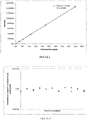
- a calibration line can then be drawn.
- This calibration line can be used later, during a method for quantification of propranolol in a kidney. It suffices to correlate the intensity of the peak of the mass spectrum obtained for propranolol in the kidney analyzed with a concentration on said calibration line.
- a calibration line is drawn representing the concentration of propranolol in the tissue ( ⁇ g/g) as a function of the intensity of the peak of the mass spectrum ( figure 2 ).
- EXAMPLE 3 QUANTIFICATION OF PROPRANOLOL IN A KIDNEY OF TREATED MOUSE
- Three sections of tissue 20 ⁇ m thick are made from the kidney and are each placed on a support (ITO blade).
- MALDI DHB matrix (40mg/mL methanol/TFA 0.1% 1/1) is sprayed using a robotic system (SunCollect) on the sections.
- the propranolol concentration in the kidney is quantified using the equation of the calibration line ( picture 2 ), by plotting the mean intensity value obtained on said straight line.
- Propranolol is here at a concentration of 5.9 ⁇ g/g of tissue.
- a dilution range or a dilution point of one or more molecules of interest in reconstituted tissues is used in order to evaluate the detection of the molecule (s) using a chosen analytical method.
- a dilution range is produced from nine different precise sub-volumes of the propranolol solution, which are each added to a sample of ground kidney, so as to obtain a concentration in the samples of 0, 0.3, 0.7, 1.4, 2.8, 5.6, 11.2, 22.4 and 44.8 ⁇ g/g.
- a section containing the succession of samples of ground kidney material, and therefore presenting the 9 successive concentrations of propanolol (in the order presented in Table 4) is deposited on a support (ITO slide).
- the optimal thickness is fixed at 20 ⁇ m and the most efficient MALDI matrix is DHB at 40 mg/mL methanol/TFA 0.1% 1/1.
- Each portion of the section representing a sample of shredded kidney is then analyzed by imaging by mass spectrometry or by direct analysis using an Autoflex speed LRF (Bruker, Daltonics).
- ROI regions of interest
- the detection limit of the compound in the chosen medium which is here 0.7 ⁇ g/g.
- This detection limit is fixed in this example as being the lowest concentration for which the compound is detected with a signal/noise ratio greater than 3. Note that it is also possible to determine the quantification limit.
- a section of ground kidney material comprising a known concentration of propranolol (5.6 ⁇ g/g) is deposited on the same support (ITO slide) as the sample of interest. It should be noted that this reconstituted kidney homogenate is used in the context of all the analyzes whose reproducibility is to be assessed.
- the value of the mean intensity of propranolol is therefore obtained for each analysis and it is thus possible to monitor the reproducibility of the analyses.
- a section of reconstituted kidney doped with propranolol at 5.6 ⁇ g/g was added.
- the picture 3 shows the variability obtained in the context of 10 constitutive analyzes within the same study.
Landscapes
- Health & Medical Sciences (AREA)
- Life Sciences & Earth Sciences (AREA)
- Engineering & Computer Science (AREA)
- Physics & Mathematics (AREA)
- Molecular Biology (AREA)
- Chemical & Material Sciences (AREA)
- Immunology (AREA)
- Biomedical Technology (AREA)
- Hematology (AREA)
- Urology & Nephrology (AREA)
- Biochemistry (AREA)
- Analytical Chemistry (AREA)
- General Health & Medical Sciences (AREA)
- General Physics & Mathematics (AREA)
- Pathology (AREA)
- Food Science & Technology (AREA)
- Medicinal Chemistry (AREA)
- Bioinformatics & Computational Biology (AREA)
- Bioinformatics & Cheminformatics (AREA)
- Biotechnology (AREA)
- Cell Biology (AREA)
- Microbiology (AREA)
- Optics & Photonics (AREA)
- Spectroscopy & Molecular Physics (AREA)
- Biophysics (AREA)
- Proteomics, Peptides & Aminoacids (AREA)
- Tropical Medicine & Parasitology (AREA)
- Physiology (AREA)
- Other Investigation Or Analysis Of Materials By Electrical Means (AREA)
- Investigating Or Analysing Biological Materials (AREA)
- Sampling And Sample Adjustment (AREA)
Applications Claiming Priority (2)
| Application Number | Priority Date | Filing Date | Title |
|---|---|---|---|
| FR1154731A FR2976076B1 (fr) | 2011-05-31 | 2011-05-31 | Procede de detection et de quantification d'une molecule cible dans un tissu |
| PCT/FR2012/051205 WO2012164221A1 (fr) | 2011-05-31 | 2012-05-29 | Procede de detection et de quantification d'une molecule cible dans un tissu |
Publications (2)
| Publication Number | Publication Date |
|---|---|
| EP2715310A1 EP2715310A1 (fr) | 2014-04-09 |
| EP2715310B1 true EP2715310B1 (fr) | 2022-04-27 |
Family
ID=46420402
Family Applications (1)
| Application Number | Title | Priority Date | Filing Date |
|---|---|---|---|
| EP12731087.8A Active EP2715310B1 (fr) | 2011-05-31 | 2012-05-29 | Procédé de détection et de quantification d'une molécule cible dans un tissu |
Country Status (12)
| Country | Link |
|---|---|
| US (1) | US10132796B2 (OSRAM) |
| EP (1) | EP2715310B1 (OSRAM) |
| JP (1) | JP6170911B2 (OSRAM) |
| KR (1) | KR102004350B1 (OSRAM) |
| CN (1) | CN103688151B (OSRAM) |
| AU (1) | AU2012264458B2 (OSRAM) |
| CA (1) | CA2837153C (OSRAM) |
| DK (1) | DK2715310T3 (OSRAM) |
| ES (1) | ES2922478T3 (OSRAM) |
| FR (1) | FR2976076B1 (OSRAM) |
| IN (1) | IN2013MN02451A (OSRAM) |
| WO (1) | WO2012164221A1 (OSRAM) |
Families Citing this family (8)
| Publication number | Priority date | Publication date | Assignee | Title |
|---|---|---|---|---|
| WO2014128309A1 (en) | 2013-02-25 | 2014-08-28 | Imabiotech | Method to evaluate the tissue targeting of a molecule of interest |
| CN103336128A (zh) * | 2013-06-08 | 2013-10-02 | 天津药物研究院 | 一种检测小分子多肽类药物组织分布的方法 |
| KR102696044B1 (ko) * | 2015-11-06 | 2024-08-16 | 벤타나 메디컬 시스템즈, 인코포레이티드 | 대표 진단법 |
| KR101996666B1 (ko) * | 2016-05-02 | 2019-07-04 | 주식회사 엘지화학 | 정량 분석 방법 |
| US11289316B2 (en) | 2018-05-30 | 2022-03-29 | Shimadzu Corporation | Spectrum data processing device and analyzer |
| EP4111174A4 (en) * | 2020-02-27 | 2024-03-27 | Nicoya Lifesciences, Inc. | SYSTEMS AND METHODS FOR CHARACTERIZING AN ASSAY FROM REGIONS OF INTEREST USING OPTICAL REACTIONS |
| WO2021175842A1 (en) | 2020-03-02 | 2021-09-10 | Universiteit Maastricht | Quality control standards for mass spectrometry imaging |
| CN115326915A (zh) * | 2022-08-01 | 2022-11-11 | 中央民族大学 | 一种植物组织微阵列maldi-msi高通量分析方法 |
Family Cites Families (12)
| Publication number | Priority date | Publication date | Assignee | Title |
|---|---|---|---|---|
| IL149171A0 (en) * | 1999-11-16 | 2002-11-10 | Genentech Inc | Elisa for vegf |
| KR20050070034A (ko) * | 2002-10-04 | 2005-07-05 | 미쯔비시 웰 파마 가부시키가이샤 | 질량 분석용 플레이트, 그 제조 방법 및 그 용도 |
| US20040096907A1 (en) * | 2002-11-06 | 2004-05-20 | Bernd Bohrmann | Quantification of beta amyloid |
| JP2005181011A (ja) * | 2003-12-17 | 2005-07-07 | Yoshio Yamauchi | タンパク質解析方法 |
| JP4547173B2 (ja) | 2004-03-17 | 2010-09-22 | シスメックス株式会社 | 糖尿病診療支援システム |
| CA2611266C (en) | 2005-06-07 | 2015-11-24 | Centre National De La Recherche Scientifique (Cnrs) | Use of conjugates with linkers cleavable by photodissociation or fragmentation for mass spectrometry analysis of tissue sections |
| EP2163900A1 (en) | 2008-09-04 | 2010-03-17 | Commissariat A L'energie Atomique | New method of imaging by mass spectrometry and new mass tag associated trityl derivatives |
| WO2010041459A1 (ja) * | 2008-10-08 | 2010-04-15 | 積水メディカル株式会社 | 薬物代謝物の定量分析方法及び分析装置 |
| WO2011073740A1 (en) | 2009-12-15 | 2011-06-23 | Centre National De La Recherche Scientifique (Cnrs) | Matrices for mass spectrometry imaging |
| FR2973112B1 (fr) | 2011-03-21 | 2018-05-25 | Imabiotech | Procede de detection et de quantification d'une molecule cible dans un echantillon |
| EP2767832A1 (en) | 2013-02-18 | 2014-08-20 | Imabiotech | Photo or chemolabile conjugates for molecules detection |
| WO2014128309A1 (en) | 2013-02-25 | 2014-08-28 | Imabiotech | Method to evaluate the tissue targeting of a molecule of interest |
-
2011
- 2011-05-31 FR FR1154731A patent/FR2976076B1/fr active Active
-
2012
- 2012-05-29 ES ES12731087T patent/ES2922478T3/es active Active
- 2012-05-29 WO PCT/FR2012/051205 patent/WO2012164221A1/fr not_active Ceased
- 2012-05-29 EP EP12731087.8A patent/EP2715310B1/fr active Active
- 2012-05-29 US US14/123,128 patent/US10132796B2/en active Active
- 2012-05-29 CA CA2837153A patent/CA2837153C/fr active Active
- 2012-05-29 CN CN201280026907.4A patent/CN103688151B/zh active Active
- 2012-05-29 KR KR1020137035169A patent/KR102004350B1/ko active Active
- 2012-05-29 JP JP2014513236A patent/JP6170911B2/ja active Active
- 2012-05-29 AU AU2012264458A patent/AU2012264458B2/en active Active
- 2012-05-29 IN IN2451MUN2013 patent/IN2013MN02451A/en unknown
- 2012-05-29 DK DK12731087.8T patent/DK2715310T3/da active
Also Published As
| Publication number | Publication date |
|---|---|
| US10132796B2 (en) | 2018-11-20 |
| KR20140051187A (ko) | 2014-04-30 |
| JP6170911B2 (ja) | 2017-07-26 |
| ES2922478T3 (es) | 2022-09-15 |
| CN103688151B (zh) | 2016-05-18 |
| WO2012164221A1 (fr) | 2012-12-06 |
| AU2012264458A1 (en) | 2014-01-09 |
| US20140106391A1 (en) | 2014-04-17 |
| AU2012264458B2 (en) | 2015-09-24 |
| JP2014520259A (ja) | 2014-08-21 |
| EP2715310A1 (fr) | 2014-04-09 |
| IN2013MN02451A (OSRAM) | 2015-04-17 |
| FR2976076B1 (fr) | 2015-02-27 |
| FR2976076A1 (fr) | 2012-12-07 |
| KR102004350B1 (ko) | 2019-07-26 |
| CA2837153A1 (fr) | 2012-12-06 |
| CN103688151A (zh) | 2014-03-26 |
| DK2715310T3 (da) | 2022-07-25 |
| CA2837153C (fr) | 2019-06-25 |
Similar Documents
| Publication | Publication Date | Title |
|---|---|---|
| EP2715310B1 (fr) | Procédé de détection et de quantification d'une molécule cible dans un tissu | |
| Chughtai et al. | Mass spectrometric imaging for biomedical tissue analysis | |
| Massonnet et al. | A concise tutorial review of TOF-SIMS based molecular and cellular imaging | |
| FR2973112A1 (fr) | Procede de detection et de quantification d'une molecule cible dans un echantillon | |
| Schuhmann et al. | Intensity-independent noise filtering in FT MS and FT MS/MS spectra for shotgun lipidomics | |
| EP2124192A1 (en) | Method for the analysis of tissue sections | |
| Patti et al. | Detection of carbohydrates and steroids by cation-enhanced nanostructure-initiator mass spectrometry (NIMS) for biofluid analysis and tissue imaging | |
| DE112011106145B4 (de) | Massenspektrometrische Quantifizierung von Analyten unter Verwendung eines Universalreporters | |
| JP6580103B2 (ja) | コエンザイムq10を検出するための方法及びキット | |
| Nimesh et al. | Current status and future perspectives of mass spectrometry imaging | |
| Holbrook et al. | Quantitative mass spectrometry imaging: therapeutics & biomolecules | |
| Crecelius et al. | MALDI mass spectrometric imaging meets “omics”: recent advances in the fruitful marriage | |
| Liu et al. | Mass spectrometry imaging for biomedical applications | |
| Lukowski et al. | An optimized approach and inflation media for obtaining complimentary mass spectrometry-based omics data from human lung tissue | |
| Ngai et al. | Mini review: Highlight of recent advances and applications of MALDI mass spectrometry imaging in 2024 | |
| Gulin et al. | A novel approach for 3D reconstruction of mice full-grown oocytes by time-of-flight secondary ion mass spectrometry | |
| González-Rodríguez et al. | Mass fingerprinting for high-throughput analyses of food: authentication and quality control | |
| Okyem et al. | Spatially mapping neuropeptide isomers via MALDI trapped ion mobility MS imaging | |
| CN121499177A (zh) | 一种生物组织冰冻切片解吸电喷雾电离质谱成像的样本预处理方法 | |
| Caponigro et al. | Mass Spectrometry Imaging (MSI) | |
| Xie | Multiscale biochemical mapping of the brain through data-driven and machine learning enabled-mass spectrometry | |
| Castro | High-throughput matrix-assisted laser desorption/ionization mass spectrometry for single-cell and single-organelle measurements | |
| CN118425120A (zh) | 一种自动化全定量拉曼分析方法 | |
| Chughtai | Multimodal Imaging of Hypoxia in Breast Cancer |
Legal Events
| Date | Code | Title | Description |
|---|---|---|---|
| PUAI | Public reference made under article 153(3) epc to a published international application that has entered the european phase |
Free format text: ORIGINAL CODE: 0009012 |
|
| 17P | Request for examination filed |
Effective date: 20131129 |
|
| AK | Designated contracting states |
Kind code of ref document: A1 Designated state(s): AL AT BE BG CH CY CZ DE DK EE ES FI FR GB GR HR HU IE IS IT LI LT LU LV MC MK MT NL NO PL PT RO RS SE SI SK SM TR |
|
| DAX | Request for extension of the european patent (deleted) | ||
| 17Q | First examination report despatched |
Effective date: 20150422 |
|
| STAA | Information on the status of an ep patent application or granted ep patent |
Free format text: STATUS: EXAMINATION IS IN PROGRESS |
|
| GRAP | Despatch of communication of intention to grant a patent |
Free format text: ORIGINAL CODE: EPIDOSNIGR1 |
|
| STAA | Information on the status of an ep patent application or granted ep patent |
Free format text: STATUS: GRANT OF PATENT IS INTENDED |
|
| INTG | Intention to grant announced |
Effective date: 20211202 |
|
| RIN1 | Information on inventor provided before grant (corrected) |
Inventor name: BONNEL, DAVID Inventor name: STAUBER, JONATHAN |
|
| GRAS | Grant fee paid |
Free format text: ORIGINAL CODE: EPIDOSNIGR3 |
|
| GRAA | (expected) grant |
Free format text: ORIGINAL CODE: 0009210 |
|
| STAA | Information on the status of an ep patent application or granted ep patent |
Free format text: STATUS: THE PATENT HAS BEEN GRANTED |
|
| AK | Designated contracting states |
Kind code of ref document: B1 Designated state(s): AL AT BE BG CH CY CZ DE DK EE ES FI FR GB GR HR HU IE IS IT LI LT LU LV MC MK MT NL NO PL PT RO RS SE SI SK SM TR |
|
| REG | Reference to a national code |
Ref country code: GB Ref legal event code: FG4D Free format text: NOT ENGLISH |
|
| REG | Reference to a national code |
Ref country code: CH Ref legal event code: EP |
|
| REG | Reference to a national code |
Ref country code: DE Ref legal event code: R096 Ref document number: 602012078100 Country of ref document: DE |
|
| REG | Reference to a national code |
Ref country code: AT Ref legal event code: REF Ref document number: 1487288 Country of ref document: AT Kind code of ref document: T Effective date: 20220515 |
|
| REG | Reference to a national code |
Ref country code: IE Ref legal event code: FG4D Free format text: LANGUAGE OF EP DOCUMENT: FRENCH |
|
| REG | Reference to a national code |
Ref country code: DK Ref legal event code: T3 Effective date: 20220722 |
|
| REG | Reference to a national code |
Ref country code: NL Ref legal event code: FP |
|
| RAP4 | Party data changed (patent owner data changed or rights of a patent transferred) |
Owner name: IMABIOTECH |
|
| REG | Reference to a national code |
Ref country code: SE Ref legal event code: TRGR |
|
| REG | Reference to a national code |
Ref country code: DE Ref legal event code: R081 Ref document number: 602012078100 Country of ref document: DE Owner name: IMABIOTECH, FR Free format text: FORMER OWNER: IMABIOTECH, VILLENEUVE D'ASCQ, FR |
|
| REG | Reference to a national code |
Ref country code: LT Ref legal event code: MG9D |
|
| REG | Reference to a national code |
Ref country code: AT Ref legal event code: MK05 Ref document number: 1487288 Country of ref document: AT Kind code of ref document: T Effective date: 20220427 Ref country code: ES Ref legal event code: FG2A Ref document number: 2922478 Country of ref document: ES Kind code of ref document: T3 Effective date: 20220915 |
|
| PG25 | Lapsed in a contracting state [announced via postgrant information from national office to epo] |
Ref country code: PT Free format text: LAPSE BECAUSE OF FAILURE TO SUBMIT A TRANSLATION OF THE DESCRIPTION OR TO PAY THE FEE WITHIN THE PRESCRIBED TIME-LIMIT Effective date: 20220829 Ref country code: NO Free format text: LAPSE BECAUSE OF FAILURE TO SUBMIT A TRANSLATION OF THE DESCRIPTION OR TO PAY THE FEE WITHIN THE PRESCRIBED TIME-LIMIT Effective date: 20220727 Ref country code: LT Free format text: LAPSE BECAUSE OF FAILURE TO SUBMIT A TRANSLATION OF THE DESCRIPTION OR TO PAY THE FEE WITHIN THE PRESCRIBED TIME-LIMIT Effective date: 20220427 Ref country code: HR Free format text: LAPSE BECAUSE OF FAILURE TO SUBMIT A TRANSLATION OF THE DESCRIPTION OR TO PAY THE FEE WITHIN THE PRESCRIBED TIME-LIMIT Effective date: 20220427 Ref country code: GR Free format text: LAPSE BECAUSE OF FAILURE TO SUBMIT A TRANSLATION OF THE DESCRIPTION OR TO PAY THE FEE WITHIN THE PRESCRIBED TIME-LIMIT Effective date: 20220728 Ref country code: FI Free format text: LAPSE BECAUSE OF FAILURE TO SUBMIT A TRANSLATION OF THE DESCRIPTION OR TO PAY THE FEE WITHIN THE PRESCRIBED TIME-LIMIT Effective date: 20220427 Ref country code: BG Free format text: LAPSE BECAUSE OF FAILURE TO SUBMIT A TRANSLATION OF THE DESCRIPTION OR TO PAY THE FEE WITHIN THE PRESCRIBED TIME-LIMIT Effective date: 20220727 Ref country code: AT Free format text: LAPSE BECAUSE OF FAILURE TO SUBMIT A TRANSLATION OF THE DESCRIPTION OR TO PAY THE FEE WITHIN THE PRESCRIBED TIME-LIMIT Effective date: 20220427 |
|
| PG25 | Lapsed in a contracting state [announced via postgrant information from national office to epo] |
Ref country code: RS Free format text: LAPSE BECAUSE OF FAILURE TO SUBMIT A TRANSLATION OF THE DESCRIPTION OR TO PAY THE FEE WITHIN THE PRESCRIBED TIME-LIMIT Effective date: 20220427 Ref country code: PL Free format text: LAPSE BECAUSE OF FAILURE TO SUBMIT A TRANSLATION OF THE DESCRIPTION OR TO PAY THE FEE WITHIN THE PRESCRIBED TIME-LIMIT Effective date: 20220427 Ref country code: LV Free format text: LAPSE BECAUSE OF FAILURE TO SUBMIT A TRANSLATION OF THE DESCRIPTION OR TO PAY THE FEE WITHIN THE PRESCRIBED TIME-LIMIT Effective date: 20220427 Ref country code: IS Free format text: LAPSE BECAUSE OF FAILURE TO SUBMIT A TRANSLATION OF THE DESCRIPTION OR TO PAY THE FEE WITHIN THE PRESCRIBED TIME-LIMIT Effective date: 20220827 |
|
| REG | Reference to a national code |
Ref country code: DE Ref legal event code: R097 Ref document number: 602012078100 Country of ref document: DE |
|
| PG25 | Lapsed in a contracting state [announced via postgrant information from national office to epo] |
Ref country code: SM Free format text: LAPSE BECAUSE OF FAILURE TO SUBMIT A TRANSLATION OF THE DESCRIPTION OR TO PAY THE FEE WITHIN THE PRESCRIBED TIME-LIMIT Effective date: 20220427 Ref country code: SK Free format text: LAPSE BECAUSE OF FAILURE TO SUBMIT A TRANSLATION OF THE DESCRIPTION OR TO PAY THE FEE WITHIN THE PRESCRIBED TIME-LIMIT Effective date: 20220427 Ref country code: RO Free format text: LAPSE BECAUSE OF FAILURE TO SUBMIT A TRANSLATION OF THE DESCRIPTION OR TO PAY THE FEE WITHIN THE PRESCRIBED TIME-LIMIT Effective date: 20220427 Ref country code: MC Free format text: LAPSE BECAUSE OF FAILURE TO SUBMIT A TRANSLATION OF THE DESCRIPTION OR TO PAY THE FEE WITHIN THE PRESCRIBED TIME-LIMIT Effective date: 20220427 Ref country code: LU Free format text: LAPSE BECAUSE OF NON-PAYMENT OF DUE FEES Effective date: 20220529 Ref country code: EE Free format text: LAPSE BECAUSE OF FAILURE TO SUBMIT A TRANSLATION OF THE DESCRIPTION OR TO PAY THE FEE WITHIN THE PRESCRIBED TIME-LIMIT Effective date: 20220427 Ref country code: CZ Free format text: LAPSE BECAUSE OF FAILURE TO SUBMIT A TRANSLATION OF THE DESCRIPTION OR TO PAY THE FEE WITHIN THE PRESCRIBED TIME-LIMIT Effective date: 20220427 |
|
| PLBE | No opposition filed within time limit |
Free format text: ORIGINAL CODE: 0009261 |
|
| STAA | Information on the status of an ep patent application or granted ep patent |
Free format text: STATUS: NO OPPOSITION FILED WITHIN TIME LIMIT |
|
| PG25 | Lapsed in a contracting state [announced via postgrant information from national office to epo] |
Ref country code: AL Free format text: LAPSE BECAUSE OF FAILURE TO SUBMIT A TRANSLATION OF THE DESCRIPTION OR TO PAY THE FEE WITHIN THE PRESCRIBED TIME-LIMIT Effective date: 20220427 |
|
| 26N | No opposition filed |
Effective date: 20230130 |
|
| PG25 | Lapsed in a contracting state [announced via postgrant information from national office to epo] |
Ref country code: IE Free format text: LAPSE BECAUSE OF NON-PAYMENT OF DUE FEES Effective date: 20220529 |
|
| PG25 | Lapsed in a contracting state [announced via postgrant information from national office to epo] |
Ref country code: SI Free format text: LAPSE BECAUSE OF FAILURE TO SUBMIT A TRANSLATION OF THE DESCRIPTION OR TO PAY THE FEE WITHIN THE PRESCRIBED TIME-LIMIT Effective date: 20220427 |
|
| P01 | Opt-out of the competence of the unified patent court (upc) registered |
Effective date: 20230411 |
|
| PG25 | Lapsed in a contracting state [announced via postgrant information from national office to epo] |
Ref country code: HU Free format text: LAPSE BECAUSE OF FAILURE TO SUBMIT A TRANSLATION OF THE DESCRIPTION OR TO PAY THE FEE WITHIN THE PRESCRIBED TIME-LIMIT; INVALID AB INITIO Effective date: 20120529 |
|
| PG25 | Lapsed in a contracting state [announced via postgrant information from national office to epo] |
Ref country code: MK Free format text: LAPSE BECAUSE OF FAILURE TO SUBMIT A TRANSLATION OF THE DESCRIPTION OR TO PAY THE FEE WITHIN THE PRESCRIBED TIME-LIMIT Effective date: 20220427 Ref country code: CY Free format text: LAPSE BECAUSE OF FAILURE TO SUBMIT A TRANSLATION OF THE DESCRIPTION OR TO PAY THE FEE WITHIN THE PRESCRIBED TIME-LIMIT Effective date: 20220427 |
|
| PG25 | Lapsed in a contracting state [announced via postgrant information from national office to epo] |
Ref country code: TR Free format text: LAPSE BECAUSE OF FAILURE TO SUBMIT A TRANSLATION OF THE DESCRIPTION OR TO PAY THE FEE WITHIN THE PRESCRIBED TIME-LIMIT Effective date: 20220427 |
|
| PG25 | Lapsed in a contracting state [announced via postgrant information from national office to epo] |
Ref country code: MT Free format text: LAPSE BECAUSE OF FAILURE TO SUBMIT A TRANSLATION OF THE DESCRIPTION OR TO PAY THE FEE WITHIN THE PRESCRIBED TIME-LIMIT Effective date: 20220427 |
|
| PG25 | Lapsed in a contracting state [announced via postgrant information from national office to epo] |
Ref country code: BG Free format text: LAPSE BECAUSE OF FAILURE TO SUBMIT A TRANSLATION OF THE DESCRIPTION OR TO PAY THE FEE WITHIN THE PRESCRIBED TIME-LIMIT Effective date: 20220427 |
|
| PG25 | Lapsed in a contracting state [announced via postgrant information from national office to epo] |
Ref country code: BG Free format text: LAPSE BECAUSE OF FAILURE TO SUBMIT A TRANSLATION OF THE DESCRIPTION OR TO PAY THE FEE WITHIN THE PRESCRIBED TIME-LIMIT Effective date: 20220427 |
|
| PGFP | Annual fee paid to national office [announced via postgrant information from national office to epo] |
Ref country code: NL Payment date: 20250526 Year of fee payment: 14 |
|
| PGFP | Annual fee paid to national office [announced via postgrant information from national office to epo] |
Ref country code: DE Payment date: 20250528 Year of fee payment: 14 |
|
| PGFP | Annual fee paid to national office [announced via postgrant information from national office to epo] |
Ref country code: ES Payment date: 20250612 Year of fee payment: 14 Ref country code: GB Payment date: 20250520 Year of fee payment: 14 Ref country code: DK Payment date: 20250526 Year of fee payment: 14 |
|
| PGFP | Annual fee paid to national office [announced via postgrant information from national office to epo] |
Ref country code: IT Payment date: 20250522 Year of fee payment: 14 Ref country code: BE Payment date: 20250526 Year of fee payment: 14 |
|
| PGFP | Annual fee paid to national office [announced via postgrant information from national office to epo] |
Ref country code: FR Payment date: 20250519 Year of fee payment: 14 |
|
| PGFP | Annual fee paid to national office [announced via postgrant information from national office to epo] |
Ref country code: CH Payment date: 20250601 Year of fee payment: 14 |
|
| PGFP | Annual fee paid to national office [announced via postgrant information from national office to epo] |
Ref country code: SE Payment date: 20250526 Year of fee payment: 14 |