EP2582862B1 - Methods and systems for generating, validating and using monoclonal antibodies - Google Patents
Methods and systems for generating, validating and using monoclonal antibodies Download PDFInfo
- Publication number
- EP2582862B1 EP2582862B1 EP11796472.6A EP11796472A EP2582862B1 EP 2582862 B1 EP2582862 B1 EP 2582862B1 EP 11796472 A EP11796472 A EP 11796472A EP 2582862 B1 EP2582862 B1 EP 2582862B1
- Authority
- EP
- European Patent Office
- Prior art keywords
- antibodies
- antibody
- antigens
- library
- array
- Prior art date
- Legal status (The legal status is an assumption and is not a legal conclusion. Google has not performed a legal analysis and makes no representation as to the accuracy of the status listed.)
- Active
Links
Images


Classifications
-
- C—CHEMISTRY; METALLURGY
- C07—ORGANIC CHEMISTRY
- C07K—PEPTIDES
- C07K1/00—General methods for the preparation of peptides, i.e. processes for the organic chemical preparation of peptides or proteins of any length
- C07K1/04—General methods for the preparation of peptides, i.e. processes for the organic chemical preparation of peptides or proteins of any length on carriers
- C07K1/047—Simultaneous synthesis of different peptide species; Peptide libraries
-
- C—CHEMISTRY; METALLURGY
- C07—ORGANIC CHEMISTRY
- C07K—PEPTIDES
- C07K16/00—Immunoglobulins [IG], e.g. monoclonal or polyclonal antibodies
-
- G—PHYSICS
- G01—MEASURING; TESTING
- G01N—INVESTIGATING OR ANALYSING MATERIALS BY DETERMINING THEIR CHEMICAL OR PHYSICAL PROPERTIES
- G01N33/00—Investigating or analysing materials by specific methods not covered by groups G01N1/00 - G01N31/00
- G01N33/48—Biological material, e.g. blood, urine; Haemocytometers
- G01N33/50—Chemical analysis of biological material, e.g. blood, urine; Testing involving biospecific ligand binding methods; Immunological testing
- G01N33/68—Chemical analysis of biological material, e.g. blood, urine; Testing involving biospecific ligand binding methods; Immunological testing involving proteins, peptides or amino acids
- G01N33/6803—General methods of protein analysis not limited to specific proteins or families of proteins
- G01N33/6845—Methods of identifying protein-protein interactions in protein mixtures
-
- G—PHYSICS
- G01—MEASURING; TESTING
- G01N—INVESTIGATING OR ANALYSING MATERIALS BY DETERMINING THEIR CHEMICAL OR PHYSICAL PROPERTIES
- G01N33/00—Investigating or analysing materials by specific methods not covered by groups G01N1/00 - G01N31/00
- G01N33/48—Biological material, e.g. blood, urine; Haemocytometers
- G01N33/50—Chemical analysis of biological material, e.g. blood, urine; Testing involving biospecific ligand binding methods; Immunological testing
- G01N33/68—Chemical analysis of biological material, e.g. blood, urine; Testing involving biospecific ligand binding methods; Immunological testing involving proteins, peptides or amino acids
- G01N33/6854—Immunoglobulins
-
- C—CHEMISTRY; METALLURGY
- C07—ORGANIC CHEMISTRY
- C07K—PEPTIDES
- C07K2317/00—Immunoglobulins specific features
- C07K2317/30—Immunoglobulins specific features characterized by aspects of specificity or valency
- C07K2317/33—Crossreactivity, e.g. for species or epitope, or lack of said crossreactivity
Definitions
- the detection or binding of an epitope, antigen or proteins is a large component of the research products industry, serving both academia and the pharmaceutical industry, as well as in the diagnostic and therapeutic industries.
- the use of antibodies to detect epitope, antigens or proteins can be used to identify new biomarkers and perform many assays.
- antibodies are also widely used in diagnostic applications, such as for clinical medicine (e.g. , ELISA and radioimmunoassay systems).
- the production of monospecific antibodies has been described (see, for example, Nilsson et al. Proteomics. 2005 Nov; 5(17): 4327-37 ).
- Analysis of cells and tissues in pathology laboratories includes the use of antibodies on tissue sections and in flow cytometry analyses. Antibodies are also useful as therapeutics.
- the production of antibodies can be costly and time-consuming, thus methods for the high throughput production of antibodies, in particular, highly specific antibodies, that is more cost-effective and less time-consuming is desirable.
- the present disclosure meets these needs, and provides related advantages.
- the present invention generally relates to libraries of antibodies, including methods and systems to produce, generate, characterize and utilize antibodies.
- the antibodies can be highly specific.
- the antibodies are monoclonal antibodies.
- the library can comprise a plurality of different antibodies, where the antibodies are produced by the same platform.
- the library can comprise a plurality of different antibodies, wherein within the plurality or a subset of the plurality, each antibody is a monospecific antibody; binds a native form of its target protein; is a monoclonal antibody; is an immunoprecipitating antibody; is an IgG antibody or antibody of IgG isotype; has a binding affinity for its target that is similar to that of another antibody of the plurality of antibodies (for example, within at least 1, 2, 3, 4, 5, 6, 7, 8, 9, 10, 11, 12, 13, 14, 15, 16, 17, 18, 19, or 20%); have a binding affinity of at least 10 -7 M (K D ) (for example, such as at least 10 -8 M, 10 -9 M, 10 -10 M, 10 -11 M, 10 -12 M, 10 -13 M, 10 -14 M, 10 -15 M, or 10- 16 M) for its target; or any combination thereof.
- K D binding affinity of at least 10 -7 M (K D ) (for example, such as at least 10 -8 M, 10 -9 M, 10 -10 M,
- the libraries of antibodies can be produced with high reproducibility.
- the first and second library comprises the same set of different antibodies and each antibody of the first library has a binding affinity that is within at least 1, 2, 3, 4, 5, 6, 7, 8, 9, 10, 11, 12, 13,14,15, 16,17,18, 19, or 20% of the binding affinity of the same antibody of the second library.
- Another aspect of the present invention is a library of antibodies comprising a plurality of different antibodies, wherein at least 10%, such as at least 20%, 30%, 40%, 50%, 60%, 70%, 80%, 90%, 95%, 99% or 100%, of said plurality is a monospecific antibody.
- Each monospecific antibody of the plurality can bind a native form of its target protein; be a monoclonal antibody; be an immunoprecipitating antibody; be an IgG antibody or antibody of IgG isotype; have a binding affinity for its target that is within at least 20%, such as within at least 1, 2, 3, 4, 5, 6, 7, 8, 9, 10, 11, 12, 13, 14, 15, 16, 17, 18, or 19%, of the binding affinity of another antibody of the plurality of antibodies; have a binding affinity of at least 10 -7 M (K D ), such as at least 10 -8 M, 10 -9 M, 10 -10 M, 10 -11 M, 10 -12 M, 10 -13 M, 10 -14 M, 10 -15 M, or 10 -16 M, for its target; or any combination thereof.
- the monospecific antibodies that comprise at least 10%, 20%, 30%, 40%, 50%, 60%, 70%, 80%, 90%, 95%, 99% or 100% of the library can comprise at least 50, 75, 100, 125, 150, 175, 200, 225, 250, 275, 300, 325, 350, 375, 400, 425, 450, 475, 500, 525, 550, 575, 600, 625, 650, 675, 700, 725, 750, 775, 800, 825, 850, 875, 900, or 1000 different antibodies, wherein the antibodies or a subset of the antibodies can bind a native form of its target protein; be a monoclonal antibody; be an immunoprecipitating antibody; be an IgG antibody or antibody of IgG isotype; have a binding affinity for its target that is within at least 20%, such as within at least 1, 2, 3, 4, 5, 6, 7, 8, 9, 10, 11, 12, 13, 14, 15, 16, 17, 18, or 19%, of the binding affinity of another antibody of the at least 10% plurality of monospecific
- the monospecific antibodies that comprise at least 10%, 20%, 30%, 40%, 50%, 60%, 70%, 80%, 90%, 95%, 99% or 100% of the plurality of different antibodies can bind at least 0.5%, 1%, 2%, 3%, 4%, 5%, 6%, 7%, 8%, 9%, 10%, 11%, 12%, 13%, 14%, 15%, 16%, 17%, 18%, 19%, 20%, 25%, 30%, 35%, 40%, 45%, 50%, 60%, 70%, 80%, 90%, or 100% of the human proteome, wherein the antibodies or a subset of the antibodies can bind a native form of its target protein; be a monoclonal antibody; be an immunoprecipitating antibody; be an IgG antibody or antibody of IgG isotype; have a binding affinity for its target that is within at least 20%, such as within at least 1, 2, 3, 4, 5, 6, 7, 8, 9, 10, 11, 12, 13, 14, 15, 16, 17,18, or 19%, of the binding affinity of another antibody of the at least 10% plurality of mono
- the monospecific antibodies that comprise at least 10%, 20%, 30%, 40%, 50%, 60%, 70%, 80%, 90%, 95%, 99% or 100% of the plurality of different antibodies can bind at least 0.5%, 1%, 2%, 3%, 4%, 5%, 6%, 7%, 8%, 9%, 10%, 11%, 12%, 13%, 14%, 15%, 16%, 17%, 18%, 19%, 20%, 25%, 30%, 35%, 40%, 45%, 50%, 60%, 70%, 80%, 90%, or 100% of the human proteins listed in Table 5, wherein the antibodies or a subset of the antibodies can bind a native form of its target protein; be a monoclonal antibody; be an immunoprecipitating antibody; be an IgG antibody or antibody of IgG isotype; have a binding affinity for its target that is within at least 20%, such as within at least 1, 2, 3, 4, 5, 6, 7, 8, 9, 10, 11, 12, 13, 14, 15, 16, 17, 18, or 19%, of the binding affinity of another antibody of the plurality of antibodies; have
- Also provided herein are methods of producing a library of antibodies comprising: (a) immunizing an animal with a plurality of antigens; (b) isolating antibody-generating cells from said animal; (c) isolating a plurality of antibodies from said antibody-generating cells; (d) screening said plurality of antibodies of step c) with a human proteome array, wherein the human proteome array can comprise at least 0.5%, 1%, 2%, 3%, 4%, 5%, 6%, 7%, 8%, 9%, 10%, 11%, 12%, 13%, 14%, 15%, 16%, 17%, 18%, 19%, 20%, 25%, 30%, 35%, 40%, 45%, 50%, 60%, 70%, 80%, 90%, or 100% of the human proteome or at least 0.5%, 1%, 2%, 3%, 4%, 5%, 6%, 7%, 8%, 9%, 10%, 11%, 12%, 13%, 14%, 15%, 16%, 17%, 18%, 19%, 20%
- the antibody selected in step (e) is added to a library, wherein the library can comprise a plurality of different antibodies, where at least 10%, 20%, 30%, 40%, 50%, 60%, 70%, 80%, 90%, 95%, 99% or 100% of the plurality of different antibodies can be produced from the same platform, be a monospecific antibody; bind a native form of its target protein; be a monoclonal antibody; be an immunoprecipitating antibody; be an IgG antibody or antibody of IgG isotype; have a binding affinity for its target that is within at least 20%, such as within at least 1, 2, 3, 4, 5, 6, 7, 8, 9, 10, 11, 12, 13, 14, 15, 16, 17, 18, or 19%, of the binding affinity of another antibody of the plurality of antibodies produced by the same platform; have a binding affinity of at least 10 -7 M (K D ), such as at least 10- 8 M, 10 -9 M, 10 -10 M, 10 -11 M, 10 -12 M, 10 -13 M,10 -14 M, 10 -15 M, or 10 -16
- the method can produce a library of antibodies that can comprise at least 50, 75, 100, 125, 150, 175, 200, 225, 250, 275, 300, 325, 350, 375, 400, 425, 450, 475, 500, 525, 550, 575, 600, 625, 650, 675, 700, 725, 750, 775, 800, 825, 850, 875, 900, or 1000 different antibodies, wherein the antibodies or a subset of the antibodies can be produced from the same platform, be a monospecific antibody; bind a native form of its target protein; not bind a denatured form of its target protein; be a monoclonal antibody; be an immunoprecipitating antibody; be an IgG antibody or antibody of IgG isotype; have a binding affinity for its target that is within at least 20%, such as within at least 1, 2, 3, 4, 5, 6, 7, 8, 9, 10, 11, 12, 13, 14, 15, 16, 17, 18, or 19%, of the binding affinity of another antibody of the plurality of antibodies produced by the
- the method can produce a library of antibodies that can bind at least 0.5%, 1%, 2%, 3%, 4%, 5%, 6%, 7%, 8%, 9%, 10%, 11%, 12%, 13%, 14%, 15%, 16%, 17%, 18%, 19%, 20%, 25%, 30%, 35%, 40%, 45%, 50%, 60%, 70%, 80%, 90%, or 100% of the human proteome, wherein the antibodies or a subset of the antibodies can be produced from the same platform, be a monospecific antibody; bind a native form of its target protein; be a monoclonal antibody; be an immunoprecipitating antibody; be an IgG antibody or antibody of IgG isotype; have a binding affinity for its target that is within at least 20%, such as within at least 1, 2, 3, 4, 5, 6, 7, 8, 9, 10, 11, 12, 13, 14, 15, 16, 17, 18, or 19%, of the binding affinity of another antibody of the plurality of antibodies produced by the same platform; have a binding affinity of at least 10 -7 M (K D
- the method can produce a library of antibodies that can bind at least 0.5%, 1%, 2%, 3%, 4%, 5%, 6%, 7%, 8%, 9%, 10%, 11%, 12%, 13%, 14%, 15%, 16%, 17%, 18%, 19%, 20%, 25%, 30%, 35%, 40%, 45%, 50%, 60%, 70%, 80%, 90%, or 100% of the human proteins listed in Table 5, wherein the antibodies or a subset of the antibodies can be produced from the same platform, be a monospecific antibody; bind a native form of its target protein; be a monoclonal antibody; be an immunoprecipitating antibody; be an IgG antibody or antibody of IgG isotype; have a binding affinity for its target that is within at least 20%, such as within at least 1, 2, 3, 4, 5, 6, 7, 8, 9, 10, 11, 12, 13, 14, 15, 16, 17, 18, or 19%, of the binding affinity of another antibody of the plurality of antibodies produced by the same platform; have a binding affinity of at least 10- 7 M (K
- the methods of producing the library of antibodies can further comprise pre-screening the plurality of antibodies from the antibody-generating cells prior to step c), such as by performing immunocytochemistry or determining binding of antibodies from said antibody-generating cells with a mixture comprising one or more target antigens, such as native proteins.
- the mixture can comprise a crude lysate, one or more cells, one or more proteins, one or peptides, or one or more nucleic acids or a biological sample
- the biological sample can be, but is not limited to, a cell mixture, a tissue, blood, sera, plasma, urine, cerebrospinal fluid (CSF), sputum, saliva, bone marrow, synovial fluid, aqueous humor, amniotic fluid, cerumen, breast milk, broncheoalveolar lavage fluid, semen, prostatic fluid, Cowper's fluid, pre-ejaculatory fluid, female ejaculate, sweat, tears, cyst fluid, pleural fluid, peritoneal fluid, pericardial fluid, lymph, chyme, chyle, bile, interstitial fluid, menses, pus, sebum, vaginal secretion, mucosal secretion, stool water, pancreatic juice, lavage fluid from sinus cavities, bronchopulmonary aspirate, blastocyl cavity
- the methods of producing the library of antibodies can comprise immunizing an animal with a plurality of antigens, wherein the plurality of antigens can comprise a crude lysate, one or more cells, one or more proteins, one or more peptides, or one or more nucleic acids.
- the plurality of antigens can also comprise a biological sample, wherein the biological sample can be, but is not limited to, a mixture of cells, a tissue, blood, sera, plasma, urine, cerebrospinal fluid (CSF), sputum, saliva, bone marrow, synovial fluid, aqueous humor, amniotic fluid, cerumen, breast milk, broncheoalveolar lavage fluid, semen, prostatic fluid, Cowper's fluid, pre-ejaculatory fluid, female ejaculate, sweat, tears, cyst fluid, pleural fluid, peritoneal fluid, pericardial fluid, lymph, chyme, chyle, bile, interstitial fluid, menses, pus, sebum, vaginal secretion, mucosal secretion, stool water, pancreatic juice, lavage fluid from sinus cavities, bronchopulmonary aspirate, blastocyl cavity fluid, or umbilical cord blood.
- CSF cerebrospinal fluid
- the plurality of antigens comprises at least 11,000 different antigens, such as at least 12,000, 13,000, 14,000, 15,000, 16,000, 17,000, 18,000, 19,000, or 20,000 different antigens.
- the plurality of antigens comprises at least 0.5% of the human proteome, such as at least 1%, 2%, 3%, 4%, 5%, 6%, 7%, 8%, 9%, 10%, 11%, 12%, 13%, 14%, 15%, 16%, 17%, 18%, 19%, 20%, 25%, 30%, 35%, 40%, 45%, 50%, 60%, 70%, 80%, 90%, or 100%, of the human proteome.
- the antibody-generating cells in producing the library of antibodies can be B-cells.
- the method of producing a library of antibodies can further comprise immobilizing the antibody to a substrate, wherein the substrate can be planar or a particle, comprise a solid or porous material, or any combination thereof.
- the antibody can be reversibly or irreversibly immobilized to the substrate.
- Also provided herein are methods of identifying an antibody monospecific for a human protein comprising: (a) contacting a plurality of antibodies with a human proteome array; (b) determining binding between said plurality of antibodies and the targets present on the human proteome array; and (c) identifying an antibody as monospecific when said antibody is monospecific for a single target on the proteome array, wherein the human proteome array can comprise at least 0.5%, 1%, 2%, 3%, 4%, 5%, 6%, 7%, 8%, 9%, 10%, 11%, 12%, 13%, 14%, 15%, 16%, 17%, 18%, 19%, 20%, 25%, 30%, 35%, 40%, 45%, 50%, 60%, 70%, 80%, 90%, or 100% of the human proteome or at least 0.5%, 1%, 2%, 3%, 4%, 5%, 6%, 7%, 8%, 9%, 10%, 11%, 12%, 13%, 14%, 15%, 16%, 17%, 18%, 19%, 20%
- the present invention generally relates to libraries of antibodies, including methods and systems to produce, generate, characterize, and utilize antibodies.
- the antibodies can be highly specific.
- the library can comprise of a plurality of different antibodies, where the antibodies are produced by the same platform.
- the library can comprise plurality of different antibodies, wherein within the plurality or a subset of the plurality, each antibody is a monospecific antibody; binds a native form of its target protein; is a monoclonal antibody; is an immunoprecipitating antibody; is an IgG antibody or antibody of IgG isotype; has a binding affinity for its target that is similar to that of another antibody of the plurality of antibodies, have a binding affinity of at least 10 -7 M (K D ) for its target; or any combination thereof.
- the library of antibodies can comprise a plurality of different antibodies produced by the same platform.
- the library of antibodies comprises a plurality of different antibodies wherein at least 10% of said plurality is produced by the same platform.
- the library can comprise a plurality of different antibodies wherein at least 20%, 30%, 40%, 50%, 60%, 70%, 80%, 90%, 95%, 99% or 100% of said plurality are antibodies produced by the same platform.
- Antibodies are considered to be produced by the same platform when a first antibody and second antibody is produced from the same protocol.
- the plurality of antibodies can be produced by the platform as depicted in FIG. 1A and/or B.
- an animal in step 102, can be immunized with a plurality of antigens.
- the animal can be a non-human animal, such as a bovine, avian, canine, equine, feline, ovine, porcine, or primate animal.
- the animal can be a mammal, such as a mouse, rat, rabbit, cat, dog monkey, or goat.
- the plurality of antigens can comprise purified or non-purified samples, such as purified or non-purified cells, proteins, peptides or nucleic acids.
- the plurality of antigens can comprise non-purified samples, such as a crude lysate or unpurified cell, protein, peptide, or nucleic acid samples.
- the plurality of antigens can comprise a biological sample.
- the plurality of antigens can comprise a single cell or multiple cells, proteins, representative peptides, or tissues from an organism.
- the organism can be a human or non-human.
- the non-human organism can be a mammal, such as a mouse, rat, rabbit, cat, dog, monkey, or goat.
- the biological sample can be a tissue, blood, sera, plasma, urine, cerebrospinal fluid (CSF), sputum, saliva, bone marrow, synovial fluid, aqueous humor, amniotic fluid, cerumen, breast milk, broncheoalveolar lavage fluid, semen, prostatic fluid, Cowper's fluid, pre-ejaculatory fluid, female ejaculate, sweat, tears, cyst fluid, pleural fluid, peritoneal fluid, pericardial fluid, lymph, chyme, chyle, bile, interstitial fluid, menses, pus, sebum, vaginal secretion, mucosal secretion, stool water, pancreatic juice, lavage fluid from sinus cavities, bronchopulmonary aspirate, blastocyl cavity fluid, or umbilical cord blood.
- the biological sample can be substantially depleted of a common serum protein, such as, but not limited to, albumin or IgG. Depletion can comprise filtration, fractionation, or affinity
- the biological sample can comprise a cell, such as a stem cell, undifferentiated cell, differentiated cell, or cell from a diseased subject or subject with a specific condition.
- the disease or condition can be a cancer, inflammatory disease, immune disease, autoimmune disease, cardiovascular disease, neurological disease, infectious disease, metabolic disease, or perinatal condition.
- the cancer can be breast cancer, ovarian cancer, lung cancer, colon cancer, colorectal cancer, prostate cancer, melanoma, pancreatic cancer, brain cancer hematological malignancy, hepatocellular carcinoma, cervical cancer, endometrial cancer, head and neck cancer, esophageal cancer, gastrointestinal stromal tumor (GIST), renal cell carcinoma (RCC) or gastric cancer.
- GIST gastrointestinal stromal tumor
- RRCC renal cell carcinoma
- the plurality of antigens can also comprise samples produced in bacteria (such as, but not limited to, E. coli ) , yeast, mammalian, or insect cells, such as proteins being overexpressed by the organisms.
- bacteria such as, but not limited to, E. coli
- yeast such as, but not limited to, yeast, mammalian, or insect cells, such as proteins being overexpressed by the organisms.
- the antigens, purified or non-purified, such as in the form of an overexpressing cell or cell mixture, expressing the antigens of interest, can be used to immunize the animals
- the plurality of antigens can comprise at least 11,000 different antigens, such as at least 12,000, 13,000, 14,000, 15,000, 16,000, 17,000, 18,000, 19,000, or 20,000 different antigens.
- antibody-generating cells such as lymphoid cells
- the antibody-generating cell can be used to generate a hybridoma.
- the antibody-generating cell can be a B-cell.
- the B-cell can be fused to a myeloma cell to create a hybidoma. Examples of myeloma cells include, but are not limited to, NS-1, P3U1, SP2/0, AP-1 and the like cells.
- the antibody-producing cells such as hybridoma cells
- the antibody-producing cells can then be used to generate clonal lines.
- hybridoma cells can be placed on semisolid media to rapidly generate clonal lines.
- Fluorescently-tagged antigens or isotype-specific probes can be used to isolate the clones of interest for characterization and expansion.
- the clones of interest are probed against a library of antigens, to detect the antigen each antibody recognizes and at the same time gain information as to which antigens it does not react to (eliminate false positives).
- the library of antigens can represent at least 0.5%, 1%, 2%, 3%, 4%, 5%, 6%, 7%, 8%, 9%, 10%, 11%, 12%, 13%, 14%, 15%, 16%, 17%, 18%, 19%, 20%, 25%, 30%, 35%, 40%, 45%, 50%, 60%, 70%, 80%, 90%, or 100% of an organism's proteome.
- the library of antigens can represent a substantial portion or an entire organism's proteome, such as a bacterial, viral, fungal proteome.
- the library of antigens can represent a substantial portion or an entire proteome of an insect or mammal, such as a mouse, rat, rabbit, cat, dog, monkey, goat, or human.
- the library of antigens can comprise at least 11,000, 12,000, 13,000, 14,000, 15,000, 16,000, 17,000, 18,000, 19,000, or 20,000 different antigens.
- the library of antigens can comprise at least 0.5%, 1%, 2%, 3%, 4%, 5%, 6%, 7%, 8%, 9%, 10%, 11%, 12%, 13%, 14%, 15%, 16%, 17%, 18%, 19%, 20%, 25%, 30%, 35%, 40%, 45%, 50%, 60%, 70%, 80%, 90%, or 100% of the human proteins listed in Table 5.
- the library of antigens can also comprise a fusion protein library that represents an organism's proteome in unpurified form, such as a collection of overexpressing cells (E.coli, yeast, mammalian, insect, or other organism).
- Probing can be performed by arraying the cells overexpressing an antigen, such as a fusion protein, or cells of different cell lines (such as cancer cell lines), and testing the hybridoma supernatants against the the arrayed cells.
- the arrayed cells can be permeabilized.
- Probing can also be performed by fluorescence flow cytometry and the target identified by PCR or sequence analysis of recombinant DNA in the cell.
- antibodies that have the desired profile can be highly specific monoclonal antibodies that recognize only one target and do not cross react with the other targets in the proteome library of that organism.
- the cell lines that secrete these monoclonal antibodies can be expanded.
- the antibodies produced by the same platform or protocol can be used to produce or form a library of antibodies.
- Antibodies can also be produced by the same platform when the antibodies are produced by the same method or protocol, such as described further below, in methods of producing a library.
- the library of antibodies can comprise a plurality of different antibodies, each antibody having a particular binding specificity for its target.
- the library of antibodies can comprise a plurality of different antibodies, wherein the antibodies are monospecific.
- the library of antibodies comprises a plurality of different antibodies wherein at least 10%, 20%, 30%, 40%, 50%, 60%, 70%, 80%, 90%, 95%, 99% or 100% of the plurality of antibodies is monospecific.
- An antibody is monospecific if the antibody has an A value of greater than 6 and an S value of greater than 3 for a protein or antigen when incubated on array comprising the antigens listed on Table 5.
- the number of standard deviations above mean signal intensity across the entire array for an antibody is a value termed A.
- the difference between the top signal and the second-highest signal on the array when the signal intensities are rank ordered for the antibody against the entire array is the S value.
- the A and S value can be calculated by combining individual supernatants from antibody-producing cells or hybridomas into sets of 12x12 two-dimensional pools, and these pools are incubated on an array (such as a human proteome microarrays), and the antibodies are labeled (such as by a Cy5-coupled anti-IgG secondary antibody, or any other method of detecting an antibody).
- an array such as a human proteome microarrays
- the antibodies are labeled (such as by a Cy5-coupled anti-IgG secondary antibody, or any other method of detecting an antibody).
- the signal intensity for each spot (representing antibody binding to a protein or antigen on the array) as the ratio of foreground to background signal.
- the number of standard deviations above mean signal intensity across the entire array is a value termed A.
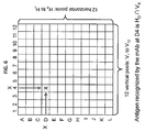
- Duplicate spots for each duplicate pair of proteins or antigens for which A >3 are flagged and results deconvoluted to identify proteins or antigens that are present at the intersection of a single horizontal and single vertical pool, and thus recognized by an individual monoclonal antibody.
- Each candidate highly specific monoclonal antibody ie. A >3 is tested individually against the entire array, and A measured for each spotted protein. The signal intensities are then rank ordered and the difference between the top signal and the second-highest signal on the array is calculated, giving the S value.
- dMAbs dispecific monoclonal antibodies bind intensely to two different proteins on the array and have A>6 and S ⁇ 3.
- the mMAbs can have an A value of greater than 6, 7, 8, 9, 10, 20, 30, 40, 50, 60, 70, 80, 90, or 100 and/or an S value of greater than 3, 4, 5, 6, 7, 8, 9, 10, 20, 30, 40, 50, 60, 70, 80, 90, or 100.
- the array for determining the monospecificity of an antibody is comprises an organism's proteome library, such as at least 0.5%, 1%, 2%, 3%, 4%, 5%, 6%, 7%, 8%, 9%, 10%, 11%, 12%, 13%, 14%, 15%, 16%, 17%, 18%, 19%, 20%, 25%, 30%, 35%, 40%, 45%, 50%, 60%, 70%, 80%, 90%, or 100% of an organism's proteome.
- the library of antigens can represent a substantial portion or an entire organism's proteome, such as a bacterial, viral, fungal proteome.
- the library of antigens can represent a substantial portion or an entire proteome of an insect or mammal, such as a mouse, rat, rabbit, cat, dog, monkey, goat, or human.
- an organism's proteome library can comprise at least 11,000, 12,000, 13,000, 14,000, 15,000, 16,000, 17,000, 18,000, 19,000, or 20,000 different antigens.
- the organism's proteome library can comprise at least 0.5%, 1%, 2%, 3%, 4%, 5%, 6%, 7%, 8%, 9%, 10%, 11%, 12%, 13%, 14%, 15%, 16%, 17%, 18%. 19%, 20%, 25%, 30%, 35%, 40%, 45%, 50%, 60%, 70%, 80%, 90%, or 100% of the human proteins listed in Table 5.
- the library of antibodies can comprise a plurality of different antibodies have a particular binding affinity its target.
- the library can comprise a plurality of different antibodies wherein at least 10%, 20%, 30%, 40%, 50%, 60%, 70%, 80%, 90%, 95%, 99% or 100% of the plurality has a particular binding affinity.
- the plurality of antibodies can have a binding affinity as determined by its dissociation constant (K D ), of at least 10 -7 M, such as at least 10 -8 M, 10 -9 M, 10 -10 M, 10 -11 M, 10 -12 M, 10 -13 M, 10 -14 M, 10 -15 M, or 10 -16 M, for its target protein.
- K D dissociation constant
- the plurality of antibodies can have a binding affinity as measured by its association rate constant (k on ), wherein the plurality of antibodies have a binding affinity of at least 10 4 M -1 s -1 , at least 5 X 10 4 M -1 s -1 , at least 10 5 M -1 s -1 , at least 5 X 10 5 M -1 s -1 at least 10 6 M -1 s -1 , at least 5 X 10 6 M -1 s -1 , at least 10 7 M -1 s -1 , at least 5 X 10 7 M -1 s - , or at least 10 8 M -1 s -1 .
- the plurality of antibodies can have a binding affinity as measured by its dissociation rate constant (k off ), wherein the plurality of antibodies have a binding affinity of less than 10 3 M -1 s -1 , less than 5 X 10 3 M -1 s -1 , less than 10 4 M -1 s -1 , less than 5 X 10 4 M -1 s -1 , less than 10 5 M -1 s -1 , less than 5 X 10 5 M -1 s -1 , less than 10 6 M -1 s -1 , less than 5 X 10 6 M -1 s -1 , less than 10 7 M -1 s -1 , less than 5 X 10 7 M -1 s - , or less than 10 8 M -1 s -1 .
- binding affinity can be determined by surface plasmon resonance, chromatography or any other methods known in the art. Binding affinity can also be determined by optically, such as by using real-time and/or label-free methods of detecting biomolecule interactions. In one embodiment, oblique-incidence reflectivity different (OIRD) is used.
- OIRD oblique-incidence reflectivity different
- the library of antibodies can comprise a plurality of different antibodies that binds native form of its target protein.
- the library can comprise a plurality of different antibodies wherein at least 10%, 20%, 30%, 40%, 50%, 60%, 70%, 80%, 90%, 95%, 99% or 100% of the plurality binds a native form of its target protein.
- the plurality of antibodies that binds a native form of its target protein does not bind a denatured form of its target protein under the same binding conditions.
- the library of antibodies can comprise a plurality of different antibodies, wherein one or more antibodies of the library have a binding affinity for its target that is similar to the binding affinity of another antibody of the plurality.
- the library of antibodies can comprise a first antibody and a second antibody, wherein the first antibody has a binding affinity for a first protein that similar to the binding affinity of the second antibody to a second protein.
- One antibody of the library can have a binding affinity for its target that is within at least 1%, 2%, 3%, 4%, 5%, 6%, 7%, 8%, 9%, 10%, 11%, 12%, 13%, 14%, 15%, 16%, 17%, 18%, 19%, or 20% of the binding affinity of one or more other antibodies of the library.
- the first antibody of the library can have a binding affinity for its target that is within at least 20% of the binding affinity of one or more other antibodies of the library.
- the library of antibodies can comprise a plurality of different antibodies, wherein at least 10%, 20%, 30%, 40%, 50%, 60%, 70%, 80%, 90%, 95%, 99% or 100% of the different antibodies have a binding affinity for its target proteins that is within at least 20% of the binding affinity of the rest of the plurality of different antibodies to their respective target protein.
- a library of antibodies comprising a plurality of different antibodies that binds a portion of an organism's proteome.
- the plurality of different antibodies can bind at least 0.5%, 1 %, 2%, 3%, 4%, 5%, 6%, 7%, 8%, 9%, 10%, 11%, 12%, 13%, 14%, 15%, 16%, 17%, 18%, 19%, 20%, 25%, 30%, 35%, 40%, 45%, 50%, 60%, 70%, 80%, 90%, or 100% of an organism's proteome.
- the plurality of different antibodies can bind at least 11,000, 12,000, 13,000, 14,000, 15,000, 16,000, 17,000, 18,000, 19,000, or 20,000 proteins of an organism.
- the proteome can be a bacterial, viral, fungal proteome.
- the proteome can be of an insect or mammal, such as a mouse, rat, rabbit, cat, dog, monkey, goat, or human. In some embodiments, the proteome is a human proteome.
- the plurality of different antibodies can bind at least 0.5% of the human proteome.
- the plurality of different antibodies can bind at least 1%, 2%, 3%, 4%, 5%, 6%, 7%, 8%, 9%, 10%, 11%, 12%, 13%, 14%, 15%, 16%, 17%, 18%, 19%, 20%, 25%, 30%, 35%, 40%, 45%, 50%, 60%, 70%, 80%, 90%, or 100% of the human proteome.
- the plurality of different antibodies can bind at least 0.5%, 1%, 2%, 3%, 4%, 5%, 6%, 7%, 8%, 9%, 10%, 11%, 12%, 13%, 14%, 15%, 16%, 17%, 18%, 19%, 20%, 25%, 30%, 35%, 40%, 45%, 50%, 60%, 70%, 80%, 90%, or 100% of the human proteins listed in Table 5.
- the library of antibodies can also comprise a plurality of different antibodies, wherein the antibodies are IgG antibodies (e.g. antibodies of IgG isotype).
- a library of antibodies can comprise a plurality of different antibodies, wherein at least 10%, 20%, 30%, 40%, 50%, 60%, 70%, 80%, 90%, 95%, 99% or 100% of the antibodies is an IgG antibody or antibody of IgG isotype.
- the library of antibodies can also comprise a plurality of different antibodies, wherein the antibodies are immunoprecipitating antibodies.
- a library of antibodies can comprise a plurality of different antibodies, wherein at least 10%, 20%, 30%, 40%, 50%, 60%, 70%, 80%, 90%, 95%, 99% or 100% of the antibodies is an immunoprecipicating antibody.
- An immunoprecipicating antibody is an antibody that can immunoprecipitate a target protein from a cell homogenate as compared to a no antibody negative control and an anti-V5 antibody positive control. Detection of the imnmnoprecipitated target can be by Western blot.
- the immunopreiciptation can be carried out with cleared cell lysate and approximately 2 ⁇ g of antibody, followed by incubation for 2 hours at 4°C, then addition of a substrate to bind any antibody-protein complex (such as protein-G Dynabead), and then incubation for an additional 2 hours at 4°C. After incubation the substrate is washed, such as with ice cold TBST twice, the substrate transferred to a new reaction vessel, washed again with ice cold TBST, before being subjected to SDS-PAGE and Western blot analysis.
- a substrate such as protein-G Dynabead
- the present invention relates to a library of antibodies comprising a plurality of antibodies that are specific to antigens of a common pathway.
- Antigens belong to a common pathway when they share one or more attributes in common in a gene ontology, a collection that assigns defined characteristics to a set of genes and their products.
- the ontology administered by the Gene Ontology ("GO") Consortium is particularly useful in this regard.
- Antigens belonging to common pathways can be identified by searching a gene ontology, such as GO, for genes sharing one or more attributes.
- the common attribute could be, for example, a common structural feature, a common location, a common biological process or a common molecular function.
- antigens in a common pathway are the expression product of genes involved in the same biological process or molecular function as annotated by a gene ontology.
- genes involved in the response to DNA damage are genes involved in the response to DNA damage.
- gene products of transcription factors such as of a particular tissue, cell type or organ.
- gene products of transcription factors of the brain are genes involved in the expression product of genes involved in the same biological process or molecular function as annotated by a gene ontology.
- antigens in a common pathway are gene products of genes which are all bound by the same transcription factor protein, complex of transcription factor proteins, other nucleic acid binding proteins, or other molecule. These interactions may occur in a living cell (in vivo) or in a solution of purified molecules (in vitro). For instance, all of the gene products of the genes bound by the hypoxia inducible transcription factor protein.
- antigens in a common pathway are the gene products of genes whose transcript levels or proteins levels change upon treatment or exposure to the same stimulus and are thus coregulated. For example all of the antigens that are induced or repressed upon treatment to UV radiation.
- antigens in a common pathway are antigens that contain similar sequence features. These features may be a DNA sequence motif, collection of DNA sequence motifs, or enrichment of higher order sequence features that are distinguishable from a background model of random genomic sequences.
- a sequence motif is a string of 2 or more nucleic acid bases (A, T, C, or G).
- a DNA sequence motif can either be defined by a consensus sequence or a probability matrix where the identity of each base at each position of a motif is defined as a probability.
- antigens in a common pathway could be the gene products of genes whose sequences, transcripts or proteins are connected via metabolic transformations and/or physical protein-protein, protein-DNA and protein-compound interactions. Enzymes catalyze these reactions, and often require dietary minerals, vitamins and other cofactors in order to function properly. Because of the many chemicals that may be involved, pathways can be quite elaborate.
- the members of the pathway share a common structural or functional attribute.
- the proteins could share a common sequence motif, such as a zinc finger or a transmembrane region.
- the antigens in a common pathway belong to the same signal transduction pathway.
- in biology signal transduction refers to any process by which a cell converts one kind of signal or stimulus into another, most often involving ordered sequences of biochemical reactions inside the cell that are carried out by enzymes, activated by second messengers resulting in what is thought of as a signal transduction pathway.
- signal transduction involves the binding of extracellular signaling molecules (or ligands) to cell-surface receptors that face outwards from the plasma membrane and trigger events inside the cell.
- intracellular signaling cascades can be triggered through cell-substratum interactions, as in the case of integrins which bind ligands found within the extracellular matrix.
- Steroids represent another example of extracellular signaling molecules that may cross the plasma membrane due to their lipophilic or hydrophobic nature. Many steroids, but not all, have receptors within the cytoplasm and usually act by stimulating the binding of their receptors to the promoter region of steroid responsive genes. Within multicellular organisms there are a diverse number of small molecules and polypeptides that serve to coordinate a cell's individual biological activity within the context of the organism as a whole. Examples of these molecules include hormones (e.g. melatonin), growth factors (e.g. epidermal growth factor), extra-cellular matrix components (e.g. fibronectin), cytokines (e.g. interferon-gamma), chemokines (e.g. RANTES), neurotransmitters (e.g. acetylcholine), and neurotrophins (e.g. nerve growth factor).
- hormones e.g. melatonin
- growth factors e.g. epidermal growth factor
- extra-cellular matrix components e
- Environmental stimuli may also be molecular in nature or more physical, such as, light striking cells in the retina of the eye, odorants binding to odorant receptors in the nasal epithelium, bitter and sweet tastes stimulating taste receptors in the taste buds, UV light altering DNA in a cell, and hypoxia activating a series of events in cells.
- Certain microbial molecules e.g. viral nucleotides, bacterial lipopolysaccharides, or protein antigens are able to elicit an immune system response against invading pathogens, mediated via signal transduction processes.
- Gene activation leads to further cellular effects, since the protein products of many of the responding genes include enzymes and transcription factors themselves. Transcription factors produced as a result of a signal transduction cascade can in turn activate yet more genes. Therefore an initial stimulus can trigger the expression of an entire cohort of genes, and this in turn can lead to the activation of any number of complex physiological events. These events include, for example, the increased uptake of glucose from the blood stream stimulated by insulin and the migration of neutrophils to sites of infection stimulated by bacterial products.
- the invention provides methods and compositions including a library comprising a plurality of antibodies that are specific to antigens that are part of an oncology pathway.
- Antigens in an oncology pathway are those gene products of genes involve in the development of hyperplasia, neoplasia and/or cancer. Examples of oncology pathways include, but are not limited to, hypoxia, DNA damage, apoptosis, cell cycle, and p53 pathway.
- the invention provides methods and compositions including a library comprising a plurality of antibodies that are specific to antigens that that are part of a membrane pathway.
- membrane pathways include, but are not limited to, transport proteins, G-coupled receptors, ion channels, cell adhesion proteins and receptors pathways.
- the invention provides methods and compositions including a library comprising a plurality of antibodies that are specific to antigens that are part of a nuclear receptor pathway.
- antigens in a nuclear receptor pathway include, but are not limited to, gene products that are regulated by the glucocorticoid receptor protein, estrogen receptor protein, peroxisome proliferator-activated receptor protein, androgen receptor protein and transporter protein pathways, including ABC and SLC transporters.
- the invention provides methods and compositions including a library comprising a plurality of antibodies that are specific to antigens that are part of a neuronal pathway.
- antigens in a neuronal pathway include, but not limited to, gene products of genes expressed in neurons such as neurotransmitters and cell adhesion proteins.
- the invention provides methods and compositions including a library comprising a plurality of antibodies that are specific to antigens that are part of a vascular pathway.
- antigens in a vascular pathway include, but not limited to, antigens involved in angiogenesis, lipid metabolism, and inflammation.
- the invention provides methods and compositions including a library comprising a plurality of antibodies that specific to antigens that are part of a signaling pathway.
- antigens in a signaling pathway include, but are not limited to, gene products involved in cell-to-cell signaling, hormones, hormone receptors, cAMP response, and cytokines.
- the invention provides methods and compositions including a library comprising a plurality of antibodies that specific to antigens that are part of an enzymatic pathway.
- antigens in a enzymatic pathway include, but are not limited to, gene products of genes involved in glycolysis, anaerobic respiration, Krebs cycle / Citric acid cycle, Oxidative phosphorylation, fatty acid oxidation ( ⁇ -oxidation), gluconeogenesis, HMG-CoA reductase pathway, pentose phosphate pathway, porphyrin synthesis (or heme synthesis) pathway, urea cycle, photosynthesis (plants, algae, cyanobacteria) and chemosynthesis (some bacteria).
- the present invention also provides a library comprising a plurality of antibodies specific to a plurality of antigens in which at least 5%, 10%, 20%, 30%, 40%, 50%, 60%, 70%, 80%, 90%, 99% or 100% of the antiges are part of a common pathway.
- the invention provides a library comprising a plurality of antibodies specific to a plurality of antigens in which at least 5%, 10%, 20%, 30%, 40%, 50%, 60%, 70%, 80%, 90%, 99% or 100% of the antigens are part of an oncology pathway.
- the invention provides a library comprising a plurality of antibodies specific to a plurality of antigens in which at least 5%, 10%, 20%, 30%, 40%, 50%, 60%, 70%, 80%, 90%, 99% or 100% of the antigens are part of a hypoxia pathway.
- the invention provides a library comprising a plurality of antibodies specific to a plurality of antigens in which at least 5%, 10%, 20%, 30%, 40%, 50%, 60%, 70%, 80%, 90%, 99% or 100% of the antigens are part of a DNA-damage pathway.
- the invention provides a library comprising a plurality of antibodies specific to a plurality of antigens in which at least 5%, 10%, 20%, 30%, 40%, 50%, 60%, 70%, 80%, 90%, 99% or 100% of the antigens are part of an apoptosis pathway.
- the invention provides a library comprising a plurality of antibodies specific to a plurality of antigens in which at least 5%, 10%, 20%, 30%, 40%, 50%, 60%, 70%, 80%, 90%, 99% or 100% of the antigens are part of a cell cycle pathway.
- the invention provides a library comprising a plurality of antibodies specific to a plurality of antigens in which at least 5%, 10%, 20%, 30%, 40%, 50%, 60%, 70%, 80%, 90%, 99% or 100% of the antigens are part of a p53 pathway.
- the inventions provides a library comprising a plurality of antibodies specific to a plurality of antigens in which at least 5%, 10%, 20%, 30%, 40%, 50%, 60%, 70%, 80%, 90%, 99% or 100% of the antigens are differently selected from the group consisting of hypoxia pathway, DNA-damage pathway, apoptosis pathway, cell cycle pathway, and p53 pathway.
- the invention provides a library comprising a plurality of antibodies specific to a plurality of antigens in which at least 5%, 10%, 20%, 30%, 40%, 50%, 60%, 70%, 80%, 90%, 99% or 100% of the antigens are part of a membrane bound pathway.
- the invention provides a library comprising a plurality of antibodies specific to a plurality of antigens in which at least 5%, 10%, 20%, 30%, 40%, 50%, 60%, 70%, 80%, 90%, 99% or 100% of the antigens are part of a nuclear receptor pathway.
- the invention provides a library comprising a plurality of antibodies specific to a plurality of antigens in which at least 5%, 10%, 20%, 30%, 40%, 50%, 60%, 70%, 80%, 90%, 99% or 100% of the antigens are part of a glucocorticoid receptor pathway.
- the invention provides a library comprising a plurality of antibodies specific to a plurality of antigens in which at least 5%, 10%, 20%, 30%, 40%, 50%, 60%, 70%, 80%, 90%, 99% or 100% of the antigens are part of a peroxisome proliferator-activated receptor pathway.
- the invention provides a library comprising a plurality of antibodies specific to a plurality of antigens in which at least 5%, 10%, 20%, 30%, 40%, 50%, 60%, 70%, 80%, 90%, 99% or 100% of the antigens are part of an estrogen receptor pathway.
- the invention provides a library comprising a plurality of antibodies specific to a plurality of antigens in which at least 5%, 10%, 20%, 30%, 40%, 50%, 60%, 70%, 80%, 90%, 99% or 100% of the antigens are part of an androgen receptor pathway.
- the invention provides a library comprising a plurality of antibodies specific to a plurality of antigens in which at least 5%, 10%, 20%, 30%, 40%, 50%, 60%, 70%, 80%, 90%, 99% or 100% of the antigens are part of a cytochrome P450 receptor pathway.
- the invention provides a library comprising a plurality of antibodies specific to a plurality of antigens in which at least 5%, 10%, 20%, 30%, 40%, 50%, 60%, 70%, 80%, 90%, 99% or 100% of the antigens are part of a transporter receptor pathway.
- the invention provides a library comprising a plurality of antibodies specific to a plurality of antigens in which at least 5%, 10%, 20%, 30%, 40%, 50%, 60%, 70%, 80%, 90%, 99% or 100% of the antigens are differently selected from the group consisting of glucocorticoid receptor pathway, peroxisome proliferator-activated receptor pathway, estrogen receptor pathway, androgen receptor pathway, cytochrome P450 pathway, and transporter pathways
- the invention provides a library comprising a plurality of antibodies specific to a plurality of antigens in which at least 5%, 10%, 20%, 30%, 40%, 50%, 60%, 70%, 80%, 90%, 99% or 100% of the antigens are part of a vascular pathway.
- the invention provides a library comprising a plurality of antibodies specific to a plurality of antigens in which at least 5%, 10%, 20%, 30%, 40%, 50%, 60%, 70%, 80%, 90%, 99% or 100% of the antigens are part of a neuronal pathway.
- the invention provides a library comprising a plurality of antibodies specific to a plurality of antigens in which at least 5%, 10%, 20%, 30%, 40%, 50%, 60%, 70%, 80%, 90%, 99% or 100% of the antigens are part of a transcription factor pathway.
- the invention provides a library comprising a plurality of antibodies specific to a plurality of antigens in which at least 5%, 10%, 20%, 30%, 40%, 50%, 60%, 70%, 80%, 90%, 99% or 100% of the antigens are part of a signaling pathway.
- the present invention also provides a library comprising a plurality of antibodies specific to a plurality of antigens in which the library represents at least 5%, 10%, 20%, 30%, 40%, 50%, 60%, 70%, 80%, 90%, 99% or 100% of all the antigens that are part of a common pathway in the genome.
- the invention provides a library comprising a plurality of antibodies specific to a plurality of antigens in which the library represents at least 5%, 10%, 20%, 30%, 40%, 50%, 60%, 70%, 80%, 90%, 99% or 100% of all the antigens that are part of an oncology pathway in the genome.
- the invention provides a library comprising a plurality of antibodies specific to a plurality of antigens in which the library represents at least 5%, 10%, 20%, 30%, 40%, 50%, 60%, 70%, 80%, 90%, 99% or 100% of all the antigens that are part of a hypoxia pathway in the genome.
- the invention provides a library comprising a plurality of antibodies specific to a plurality of antigens in which the library represents at least 5%, 10%, 20%, 30%, 40%, 50%, 60%, 70%, 80%, 90%, 99% or 100% of all the antigens that are part of a DNA-damage pathway in the genome.
- the invention provides a library comprising a plurality of antibodies specific to a plurality of antigens in which the library represents at least 5%, 10%, 20%, 30%, 40%, 50%, 60%, 70%, 80%, 90%, 99% or 100% of all the antigens that are part of an apoptosis pathway in the genome.
- the invention provides a library comprising a plurality of antibodies specific to a plurality of antigens in which the library represents at least 5%, 10%, 20%, 30%, 40%, 50%, 60%, 70%, 80%, 90%, 99% or 100% of all the antigens are part of a cell cycle pathway in the genome.
- the invention provides a library comprising a plurality of antibodies specific to a plurality of antigens in which the library represents at least 5%, 10%, 20%, 30%, 40%, 50%, 60%, 70%, 80%, 90%, 99% or 100% of all the antigens that are part of a p53 pathway in the genome.
- the invention provides a library comprising a plurality of antibodies specific to a plurality of antigens in which the library represents at least 5%, 10%, 20%, 30%, 40%, 50%, 60%, 70%, 80%, 90%, 99% or 100% of all the antigens that are part of a membrane bound pathway in the genome.
- the invention provides a library comprising a plurality of antibodies specific to a plurality of antigens in which the library represents at least 5%, 10%, 20%, 30%, 40%, 50%, 60%, 70%, 80%, 90%, 99% or 100% of all the antigens are part of a nuclear receptor pathway in the genome.
- the invention provides a library comprising a plurality of antibodies specific to a plurality of antigens in which the library represents at least 5%, 10%, 20%, 30%, 40%, 50%, 60%, 70%, 80%, 90%, 99% or 100% of all the antigens that are part of a glucocorticoid receptor pathway in the genome.
- the invention provides a library comprising a plurality of antibodies specific to a plurality of antigens in which the library represents at least 5%, 10%, 20%, 30%, 40%, 50%, 60%, 70%, 80%, 90%, 99% or 100% of all the antigens that are part of a peroxisome proliferator-activated receptor pathway in the genome.
- the invention provides a library comprising a plurality of antibodies specific to a plurality of antigens in which the library represents at least 5%, 10%, 20%, 30%, 40%, 50%, 60%, 70%, 80%, 90%, 99% or 100% of all the antigens that are part of a estrogen receptor pathway in the genome.
- the invention provides a library comprising a plurality of antibodies specific to a plurality of antigens in which the library represents at least 5%, 10%, 20%, 30%, 40%, 50%, 60%, 70%, 80%, 90%, 99% or 100% of all the antigens that are part of an androgen receptor pathway in the genome.
- the invention provides a library comprising a plurality of antibodies specific to a plurality of antigens in which the library represents at least 5%, 10%, 20%, 30%, 40%, 50%, 60%, 70%, 80%, 90%, 99% or 100% of all the antigens that are part of a cytochrome P450 receptor pathway in the genome.
- the invention provides a library comprising a plurality of antibodies specific to a plurality of antigens in which the library represents at least 5%, 10%, 20%, 30%, 40%, 50%, 60%, 70%, 80%, 90%, 99% or 100% of all the antigens that are part of a transporter receptor pathway in the genome.
- the present invention also provides a library comprising a plurality of antibodies specific to a plurality of antigens in which the library represents at least 5%, 10%, 20%, 30%, 40%, 50%, 60%, 70%, 80%, 90%, 99% or 100% of all the antigens that are part of a neuronal pathway in the genome.
- the present invention also provides a library comprising a plurality of antibodies specific to a plurality of antigens in which the library represents at least 5%, 10%, 20%, 30%, 40%, 50%, 60%, 70%, 80%, 90%, 99% or 100% of all the antigens that are part of a signaling pathway in the genome.
- the present invention also provides a library comprising a plurality of antibodies specific to a plurality of antigens in which the library represents at least 5%, 10%, 20%, 30%, 40%, 50%, 60%, 70%, 80%, 90%, 99% or 100% of all the antigens that are part of a vascular pathway in the genome.
- the present invention also provides a library comprising a plurality of antibodies specific to a plurality of antigens in which the library represents at least 5%, 10%, 20%, 30%, 40%, 50%, 60%, 70%, 80%, 90%, 99% or 100% of all the antigens that are part of a transcription factor pathway in the genome.
- the method comprises (a) immunizing an animal with a plurality of antigens; (b) isolating antibody-generating cells from the animal; (c) isolating a plurality of antibodies from the antibody-generating cells; (d) screening the plurality of antibodies of step c) with a protein array (such as a human proteome array); (e) selecting an antibody that is monospecific for for a single target on the proteome array; and (f) adding the antibody from (e) to the library.
- the method can further comprise pre-screening the plurality of antibodies from the antibody-generating cells prior to step c).
- the animal can be a non-human animal, such as a bovine, avian, canine, equine, feline, ovine, porcine, or primate animal.
- the animal can be a mammal, such as a mouse, rat, rabbit, cat, dog monkey, or goat.
- the animal can be immunized with a plurality of antigens that can comprise purified or non-purified samples, such as purified or non-purified cells, proteins, peptides or nucleic acids.
- the plurality of antigens can comprise non-purified samples, such as a crude lysate or unpurified cell samples, protein samples, peptide samples, or nucleic acid samples.
- the plurality of antigens can comprise a biological sample.
- the plurality of antigens can comprise a single cell or multiple cells, proteins, representative peptides, or tissues from an organism, such as a bovine, avian, canine, equine, feline, ovine, porcine, or primate animal.
- the organism can be a human or non-human.
- the non-human organism can be a mammal, such as a mouse, rat, rabbit, cat, dog, monkey, or goat.
- the biological sample can be a tissue, blood, sera, plasma, urine, cerebrospinal fluid (CSF), sputum, saliva, bone marrow, synovial fluid, aqueous humor, amniotic fluid, cerumen, breast milk, broncheoalveolar lavage fluid, semen, prostatic fluid, Cowper's fluid, pre-ejaculatory fluid, female ejaculate, sweat, tears, cyst fluid, pleural fluid, peritoneal fluid, pericardial fluid, lymph, chyme, chyle, bile, interstitial fluid, menses, pus, sebum, vaginal secretion, mucosal secretion, stool water, pancreatic juice, lavage fluid from sinus cavities, bronchopulmonary aspirate, blastocyl cavity fluid, or umbilical cord blood.
- the biological sample can be substantially depleted of a common serum protein, such as, but not limited to, albumin or IgG. Depletion can comprise filtration, fractionation, or affinity
- the biological sample can comprise a cell, such as a stem cell, undifferentiated cell, differentiated cell, or cell from a diseased subject or subject with a specific condition.
- the disease or condition can be a cancer, inflammatory disease, immune disease, autoimmune disease, cardiovascular disease, neurological disease, infectious disease, metabolic disease, or perinatal condition.
- the cancer can be breast cancer, ovarian cancer, lung cancer, colon cancer, colorectal cancer, prostate cancer, melanoma, pancreatic cancer, brain cancer hematological malignancy, hepatocellular carcinoma, cervical cancer, endometrial cancer, head and neck cancer, esophageal cancer, gastrointestinal stromal tumor (GIST), renal cell carcinoma (RCC) or gastric cancer.
- GIST gastrointestinal stromal tumor
- RRCC renal cell carcinoma
- the plurality of antigens can also comprise samples produced in bacteria (such as, but not limited to, E. coli ) , yeast, mammalian, or insect cells, such as proteins being overexpressed by the organisms.
- bacteria such as, but not limited to, E. coli
- yeast such as, but not limited to, yeast, mammalian, or insect cells, such as proteins being overexpressed by the organisms.
- the antigens, purified or non-purified, such as in the form of an overexpressing cell or cell mixture, expressing the antigens of interest, can be used to immunize the animals
- the plurality of antigens can comprise at least 11,000 different antigens, such as at least 12,000, 13,000, 14,000, 15,000, 16,000, 17,000, 18,000, 19,000, or 20,000 different antigens. In some embodiments, said plurality of antigens comprises at least 0.5% of the human proteome.
- the plurality of antigens can comprise at least 0.5%, 1%, 2%, 3%, 4%, 5%, 6%, 7%, 8%, 9%, 10%, 11%, 12%, 13%, 14%, 15%, 16%, 17%, 18%, 19%, 20%, 25%, 30%, 35%, 40%, 45%, 50%, 60%, 70%, 80%, 90%, or 100% of an organism's proteome.
- the plurality of antigens can comprise at least 0.5%, 1%, 2%, 3%, 4%, 5%, 6%, 7%, 8%, 9%, 10%, 11%, 12%, 13%, 14%, 15%, 16%, 17%, 18%, 19%, 20%, 25%, 30%, 35%, 40%, 45%, 50%, 60%, 70%, 80%, 90%, or 100% of the human proteome.
- the plurality of antigens can comprise at least 0.5%, 1%, 2%, 3%. 4%, 5%, 6%.
- the library of antigens can also comprise a fusion protein library that represents an organism's proteome in unpurified form, such as a collection of overexpressing cells (E.coli, yeast, mammalian, insect, or other organism).
- a fusion protein library that represents an organism's proteome in unpurified form, such as a collection of overexpressing cells (E.coli, yeast, mammalian, insect, or other organism).
- the antibody-generating cells from the animal can be lymphoid cells, such as B-cells.
- the antibody-generating cell can be used to generate a hybridoma, for example, fused to an immortal cell such as a myeloma cell, to create a hybidoma.
- a pre-screening step is performed, wherein a plurality of antibodies from the antibody-generating cells are screened prior to isolating a plurality of antibodies from the antibody-generating cells.
- Pre-screening can be performed by using the serum or supernatant of the antibody-producing cells to determine binding of antibodies from said antibody-generating cells with a mixture comprising one or more target antigens, such as native antigens or proteins.
- the pre-screening can be performed using immunohistochemistry, immunocytochemistry, ELISA, chromatography, or any other suitable methods known in the art to determine binding.
- Pre-screening can be used to select the antibody-generating cells (which can include antibody-secreting cells that have been fused to immortalized cells, such as hybridomas), for further screening, such as by a proteome array as described herein.
- the mixture can comprise a crude lysate, cell, protein, peptide, or nucleic acid.
- the mixture can comprise a biological sample, such as a tissue, blood, sera, plasma, urine, cerebrospinal fluid (CSF), sputum, saliva, bone marrow, synovial fluid, aqueous humor, amniotic fluid, cerumen, breast milk, broncheoalveolar lavage fluid, semen, prostatic fluid, Cowper's fluid, pre-ejaculatory fluid, female ejaculate, sweat, tears, cyst fluid, pleural fluid, peritoneal fluid, pericardial fluid, lymph, chyme, chyle, bile, interstitial fluid, menses, pus, sebum, vaginal secretion, mucosal secretion, stool water, pancreatic juice, lavage fluid from sinus cavities, bronchopulmonary aspirate, blastocyl cavity fluid, or umbilical cord blood.
- CSF cerebrospinal fluid
- the biological sample can be substantially depleted of a common serum protein, such as, but not limited to, albumin or IgG. Depletion can comprise filtration, fractionation, or affinity purification.
- the biological sample can comprise a cell, such as a stem cell, undifferentiated cell, differentiated cell, or cell from a diseased subject or subject with a specific condition
- a plurality of antibodies from the antibody-generating cells can be isolated, with or without a previous pre-screening step, before being subjected to a screening with an entire, or portion of, a proteome of an organism.
- the isolated antibodies can be screened with at least 0.5%, 1%, 2%, 3%, 4%, 5%, 6%, 7%, 8%,9%,10%,11%,12%,13%,14%,15%,16%,17%,18%,19%,20%,25%, 30%, 35%, 40%, 45%, 50%, 60%, 70%1,80%, 90%, or 100% of an organism's proteome.
- the isolated antibodies can be screened with at least 11,000, 12,000, 13,000, 14,000, 15,000, 16,000, 17,000, 18,000, 19,000, or 20,000 proteins of an organism.
- the proteome can be a bacterial, viral, fungal proteome.
- the proteome can be of an insect or mammal, such as a mouse, rat, rabbit, cat, dog, monkey, goat, or human. In some embodiments, the proteome is of a human.
- the isolated antibodies can be screened with at least 0.5% of the human proteome.
- the isolated antibodies is screened with at least 1%, 2%, 3%, 4%, 5%, 6%, 7%, 8%, 9%, 10%, 11%, 12%, 13%, 14%, 15%, 16%, 17%, 18%, 19%, 20%, 25%, 30%, 35%, 40%, 45%, 50%, 60%, 70%, 80%, 90%, or 100% of the human proteome.
- the isolated antibodies can be screened with at least 0.5%, 1%, 2%, 3%, 4%, 5%, 6%, 7%, 8%, 9%, 10%, 11%, 12%, 13%, 14%, 15%, 16%, 17%, 18%, 19%, 20%, 25%, 30%, 35%, 40%, 45%, 50%, 60%, 70%, 80%, 90%, or 100% of the human proteins listed in Table 5.
- the proteome, or portion thereof, that is used to screen the antibodies can be present on an array.
- an antibody can be selected based on its binding profile. For example, an antibody can be selected if it binds to a single target of the proteome, such as to a single target of a human proteome array, for a library described herein.
- the method can be used to perform high-through production of antibodies.
- antigens to immunize mice can be produced in a high-throughput manner. This can be done by: 1) directed production of recombinant proteins from a gene expression library, where proteins of interest can be expressed as needed or through the use of whole heterologous cells, tissues, or crude or enriched cell extracts from the target species (an undirected approach), or whole mammalian cells engineered to express a heterologous protein (a directed approach).
- the extracts for injection can include subcellular components, such as cell and intracellular membranes, organelles, particles, or protein complexes and may be prepared using differential centrifugation or other well-known methods.
- both soluble and membrane proteins can be chromatographed in either their native or denatured state(s), by size-exclusion, ion-exchange, hydrophobic or affinity chromatography or by using individually collected small fractions to immunize with dozens to hundreds rather than thousands of proteins.
- simple ammonium sulfate fractionation of extracts will provide a wide variety of antigen subsets. Specific techniques to target specific classes of proteins can be used.
- proteins involved in SNO signaling may be purified by Immobilized Metal Affinity Chromatography (IMAC) and eluted metal-free proteins may then be used as antigens for immunization, or phosphoproteins may be bound to Ti O2 affinity matrices and thereby greatly enriched.
- IMAC Immobilized Metal Affinity Chromatography
- Native proteins isolated or obtained directly from living or fixed cells can be preferred sources of immunogens.
- the high-throughput method can also comprise a short timescale immunization to produce antibody-secreting lymphoid cells.
- animals can be immunized with multiple antigens and adjuvant preparations by way of the rear footpads.
- Popliteal lymph nodes are collected from 7 to 21 days post-immunization. Lymph nodes in immunized animals are directly revealed by injection with Evans blue at the footpad.
- the high-throughput method can also comprise generating the antibodies in GST-tolerant mice. Since many antigens are expressed as GST fusions, a line of GST-expressing mice (mice that actively express GST from S. japonicum ) can be used to increase the yield of specific antibodies raised against the non-GST components of the fusion proteins and eliminate generation of anti-GST antibody.
- a high-throughput method for producing antibodies can also comprise creation of cell fusions/hybridomas for antibody production.
- Primed lymphoid cells and myeloma cells are fused and fusion products are either plated onto semisolid medium (methylcellulose) containing fluorescently tagged antibodies that recognize the desired subclass of immunoglobulin molecules, or into semisolid media containing fluorescently tagged antigens that will mark colonies secreting the desired antibodies.
- semisolid medium methylcellulose
- fluorescently tagged antibodies that recognize the desired subclass of immunoglobulin molecules
- semisolid media containing fluorescently tagged antigens that will mark colonies secreting the desired antibodies.
- a combination of the above methods may also be used. Fluorescently marked colonies are rescued from semisolid medium to liquid medium, using an inverted fluorescence microscope in combination with hand-picking using Drummond microcapillary and Drummond WireTrol devices, followed by outgrowth of the individual clones.
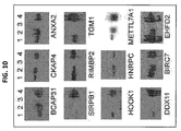
- the responsible antigen can be identified by protein microarray deconvolution. Single-step deconvolution can be performed.
- hybridomas when whole cells or cell lysates are used to generate hybridomas, a two-dimensional pooling strategy is employed to reveal identity of antigens that each monoclonal antibody would recognize.
- Supernatants of hybridomas are arrayed in two-dimensional grids and pooled both horizontally and vertically in 3-by-3 to 100-by-100 pool sizes.
- Three-dimensional pooling strategies can also be performed in which hybridomas are arrayed in plate sets to generate plate pools and horizontal and vertical pools in 3-by-3-by-3 to 100-by-100-by-100 format. The resulting pools are hybridized individually to the protein microarrays and scored using microarray analysis software.
- the shared positives (hits) of each horizontal and vertical pair or those at each 3-way intersection of plate, horizontal and vertical pools are identified as the antigens recognized by the monoclonal antibody at the intersection of the same pairs or trios.
- the identified antigens are validated by probing the microarrays with the corresponding antibodies.
- the pooling strategy can reduce the cost of characterizing or producing antibodies because the cost of the arrays is high, and use of a e.g. 10 by 10 pool reduces the number of arrays needed to analyze 100 clones from 100 to 20; for a 20 X 20 pool it reduces the number of arrays needed from 400 to 40.
- the 3-dimensional pools can screen a pool of 4096 hybridomas, while a 16-by-16-by-16 3-D strategy requires only 48 arrays. Using the 10-by-10 strategy, 820 arrays would can be used.
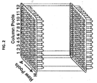
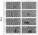
- a 3-D pooling strategy (12-by-8-by-12) based on 96-well plates is depicted in FIG. 2 .
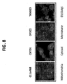
- Characterization of the monoclonal antibodies generated in the high-throughput method can be performed using whole proteome microarrays.
- the microarrays can be used to determine the specific antigens to which a given monoclonal antibody binds.
- Critical quality information about monoclonal antibody affinity, potential cross reactivity or lack of crossreactivity with other antigens are all provided by an array analysis.
- Production of monoclonal antibody in the high-throughput method can be from fusion clones.
- Monoclonal antibodies are produced in vitro or in vivo in the desired quantities. These can be purified using various well-established methods.
- monoclonal antibodies of high quality can be selected and used to produce antibody arrays.
- the antibodies are selected and their concentrations normalized to a similar titer. They can be arrayed in a multiwell format (e.g. 96- 384- or 1562-well) with proper positive (e.g., diluted human IgG) and negative (e.g., human IgM and BSA) controls to fabricate antibody microarrays using a microarrays robot (e.g., Nanoprint, Array It, Inc.).
- the arrangement of the monoclonal antibodies in different microarray configurations may be customized to facilitate a range of proteome-wide studies.
- a method of identifying an antibody for a target comprises contacting a target with a library of antibodies, determining binding between the target and the plurality of antibodies; and identifying an antibody for the target when the target binds to an antibody of the library.
- a method of identifying a target comprises contacting a target with a library of antibodies, determining binding between the target and the plurality of antibodies; and identifying the target antibody when the target binds to an antibody of the library.
- the library of antibodies can be attached to a substrate such that the target is contacted with an array comprising the library of antibodies.
- the library can comprise of a plurality of different antibodies, such as described above.
- the library of antibodies can comprise antibodies that are produced by the same platform.
- the library can comprise a plurality of different antibodies, wherein within the plurality or a subset of the plurality (such as at least 10%, 20%, 30%, 40%, 50%, 60%, 70%, 80%, 90%, 95%, 99% or 100% of the plurality), the antibodies are produced by the same platform, are monospecific, bind a native form of its target protein; are monoclonal; are immunoprecipitating antibodies; are IgG antibodies (e.g.
- antibodies of IgG isotype have a binding affinity for its target that is similar to that of another antibody of the plurality of antibodies (for example, within at least 1, 2, 3, 4, 5, 6, 7, 8, 9, 10, 11, 12, 13, 14, 15, 16, 17, 18, 19, or 20%); each antibody is a have a binding affinity of at least 10 -7 M (K D ) (for example, such as at least 10 -8 M, 10 -9 M, 10 -10 M, 10 -11 M, 10 -12 M, 10 -13 M, 10 -14 M, 10 -15 M, or 10 -16 M) for its target; or any combination thereof.
- the library may comprise at least 50, 75, 100, 125, 150, 175, 200, 225, 250, 275, 300, 325, 350, 375, 400, 425, 450, 475, 500, 525, 550, 575, 600, 625, 650, 675, 700, 725, 750, 775, 800, 825, 850, 875, 900, or 1000 different monoclonal antibodies, bind at least 0.5%, 1%, 2%, 3%, 4%, 5%, 6%, 7%, 8%, 9%, 10%, 11%, 12%, 13%, 14%, 15%, 16%, 17%, 18%, 19%, 20%, 25%, 30%, 35%, 40%, 45%, 50%, 60%, 70%, 80%, 90%, or 100% of an organism's proteome (e.g.
- a human proteome bind at least 0.5%, 1%, 2%, 3%, 4%, 5%, 6%, 7%, 8%, 9%, 10%, 11%, 12%, 13%, 14%, 15%, 16%, 17%, 18%, 19%, 20%, 25%, 30%, 35%, 40%, 45%, 50%, 60%, 70%, 80%, 90%, or 100% of the human proteins listed in Table 5, or any combination thereof.
- Also provided herein is a method of identifying an antibody monospecific for a protein, such as a human protein, comprising: contacting a plurality of antibodies with a proteome array, such as a human proteome array; determining binding between the plurality of antibodies and the targets present on the proteome array; and identifying an antibody as monospecific.
- the array can comprise a plurality of antigens or proteins that comprise at least 0.5%, 1%, 2%, 3%, 4%, 5%, 6%, 7%, 8%, 9%, 10%, 11%, 12%, 13%, 14%, 15%, 16%, 17%, 18%, 19%, 20%, 25%, 30%, 35%, 40%, 45%, 50%, 60%, 70%, 80%, 90%, or 100% of an organism's proteome.
- the proteome array can comprise at least 11,000, 12,000, 13,000, 14,000, 15,000, 16,000, 17,000, 18,000, 19,000, or 20,000 proteins of an organism.
- the organism can be a bacterium, virus, or fungus.
- the organism can be of an insect or mammal, such as a mouse, rat, rabbit, cat, dog, monkey, goat, or human.
- the proteome can comprise at least 0.5%, 1%, 2%, 3%, 4%, 5%, 6%, 7%, 8%, 9%, 10%, 11%, 12%, 13%, 14%, 15%, 16%, 17%, 18%, 19%, 20%, 25%, 30%, 35%, 40%, 45%, 50%, 60%, 70%, 80%, 90%, or 100% of the human proteome.
- the proteome array can comprise at least 0.5%, 1%, 2%, 3%, 4%, 5%, 6%, 7%, 8%, 9%, 10%, 11%, 12%, 13%, 14%, 15%, 16%, 17%, 18%, 19%, 20%, 25%, 30%, 35%, 40%, 45%, 50%, 60%, 70%, 80%, 90%, or 100% of the human proteins listed in Table 5.
- Binding can be detected by techniques known in the art (e.g., radioimmunoassay, ELISA (enzyme-linked immunosorbant assay), "sandwich” immunoassays, immunoradiometric assays, gel diffusion precipitation reactions, immunodiffusion assays, in situ immunoassays (e.g., using colloidal gold, enzyme or radioisotope labels, for example), Western blots, precipitation reactions, agglutination assays (e.g., gel agglutination assays, hemagglutination assays, etc.), complement fixation assays, immunofluorescence assays, protein A assays, and immunoelectrophoresis assays.
- radioimmunoassay e.g., ELISA (enzyme-linked immunosorbant assay), "sandwich” immunoassays, immunoradiometric assays, gel diffusion precipitation reactions, immunodiffusion assays, in
- Antibody binding can be detected by detecting a label on the primary antibody.
- the primary antibody is detected by detecting binding of a secondary antibody or reagent to the primary antibody.
- the secondary antibody can be labeled.
- an automated detection assay or high-throughput system is utilized.
- an antibody/antigen reaction is made measurable by immobilization of the antibody and subsequent direct or indirect colorimetric, fluorescent, luminescent or radioactive detection of bound, labeled antigens.
- the antigen can be labeled by biotin or other labels, which will allow downstream detection.
- the immobilized antibodies will generally bind to a single antigenic determinant present.
- the antigenic determinant can be labeled, such as through labeling of the biomarker comprising the antigenic determinant.
- the specificity of this reaction will permit quantification in the ELISA measurements.
- the ELISA reaction can be used in a high throughput format to screen all hybridoma supernatants via the following steps. Screening assays built on other principles than an ELISA can be deployed (e.g., antibody microarrays, high-throughput screening based on MALDI/MS and/or multi-channel capillary electrophoresis). ELISA or microarray data are evaluated, e.g., by published methods. The goal of the data analysis process is the selection of hybridoma supernatants that show the best collection with an important clinical parameter and are specific to one of the analyte groups.
- An array (or microarray) comprises a library of antibodies and a substrate.
- each antibody is immobilized on a substrate.
- the antibody may be reversibly or irreversibly immobilized on the substrate.
- the substrate can be planar or a particle and can comprise a solid or porous material.
- the substrate may be either organic or inorganic, biological or non-biological, or any combination of these materials.
- the substrate is transparent or translucent.
- the portion of the surface of the substrate on which the patches reside is preferably flat and firm or semi-firm.
- Numerous materials are suitable for use as a substrate.
- the substrate can comprise silicon, silica, glass, or a polymer.
- the substrate can comprise a material selected from a group consisting of silicon, silica, quartz, glass, controlled pore glass, carbon, alumina, titanium dioxide, germanium, silicon nitride, zeolites, and gallium arsenide.
- Many metals such as gold, platinum, aluminum, copper, titanium, and their alloys are also options for substrates of the array.
- many ceramics and polymers may also be used as substrates.
- Polymers which may be used as substrates include, but are not limited to, the following: polystyrene; poly(tetra)fluorethylene; (poly)vinylidenedifluoride; polycarbonate; polymethylmethacrylate; polyvinylethylene; polyethyleneimine; poly(etherether)ketone; polyoxymethylene (POM); polyvinylphenol; polylactides; polymethacrylimide (PMI); polyalkenesulfone (PAS); polyhydroxyethylmethacrylate; polydimethylsiloxane; polyacrylamide; polyimide; co-block-polymers; and Eupergit®. Photoresists, polymerized Langmuir-Blodgett films, and LIGA structures may also serve as substrates.
- a microarray of distinct antibodies is bound on a glass slide coated with a polycationic polymer.
- a substrate may be formed and intended for use in detecting binding of target molecule to one or more distinct antibodies.
- the substrate includes a glass substrate having formed on its surface, a coating of a polycationic polymer, preferably a cationic polypeptide, such as poly-lysine or poly-arginine. Formed on the polycationic coating is a microarray of distinct biopolymers, each localized at known selected array regions, such as regions.
- the slide may be coated by placing a uniform-thickness film of a polycationic polymer, e.g., poly-1-lysine, on the surface of a slide and drying the film to form a dried coating.
- a polycationic polymer e.g., poly-1-lysine
- the amount of polycationic polymer added is sufficient to form at least a monolayer of polymers on the glass surface.
- the polymer film is bound to surface via electrostatic binding between negative silyl-OH groups on the surface and charged amine groups in the polymers.
- Poly-1-lysine coated glass slides may be obtained commercially, e.g., from Sigma Chemical Co. (St. Louis, Mo.).
- a suitable microarray substrate is also made through chemical derivatization of glass.
- Silane compounds with appropriate leaving groups on a terminal Si will covalently bond to glass surfaces.
- a derivatization molecule can be designed to confer the desired chemistry to the surface of the glass substrate.
- An example of such a bifunctional reagent is amino-propyl-tri(ethoxy)silane, which reacts with glass surfaces at the tri(ethoxy)silane portion of the molecule while leaving the amino portion of the molecule free.
- Surfaces having terminal amino groups are suitable for adsorption of biopolymers in the same manner as poly-lysine coated slides. The identity of the terminal surface group can be modified by further chemical reaction.
- reaction of the terminal amine in the above example with glutaraldehyde results in a terminal aldehyde group.
- Further layers of modification may be applied to achieve the desired reactivity before spotting the microarray, such as by application of a Protein A or Protein G solution to the silynated glass.
- Additional surfaces that bind polypeptides are nitrocellulose-coated glass slides, available commercially from Schleicher and Schuell, and protein-binding plastics such as polystyrene.
- the spotted antibodies may be attached by either adsorption or covalent bonding. Adsorption occurs through electrostatic, hydrophobic, Van der Waals, or hydrogen-bonding interactions between the spotted polypeptide and the array substrate. Simple application of the polypeptide solution to the surface in an aqueous environment is sufficient to adsorb the polypeptide. Covalent attachment is achieved by reaction of functional groups on the polypeptide with a chemically activated surface. For example, if the surface has been activated with a highly reactive electrophilic group such as an aldehyde or succinimide group, unmodified polypeptides react at amine groups, as at lysine residues or the terminal amine, to form a covalent bond.
- a highly reactive electrophilic group such as an aldehyde or succinimide group
- microarray defined volumes of distinct biopolymers are deposited on the polymer-coated slide using any suitable method known in the art. According to an important feature of the substrate, the deposited antibodies remain bound to the coated slide surface non-covalently when an aqueous sample is applied to the substrate under conditions that allow binding of labeled ligands in the sample to cognate binding partners in the substrate array.
- each microarray contains at least 50, 60, 70, 80, 90, 100, 150, 200, 250, 300, 350, 400, 450, 500, 600, 700, 800, 900, or 1000 distinct antibodies per surface area of less than about 1 cm 2 .
- the microarray contains 50, 60, 70, 80, 90, 100, 150, 200, 250, 300, 350 or 400 regions in an area of about 16 mm 2 , or 2.5 x 10 3 regions/cm 2 .
- the antibodies in each microarray region are present in a defined amount between about 0.1 femtomoles and 100 nanomoles.
- the biopolymers have lengths of at least about 50 units, e.g. amino acids, nucleotides, etc., i.e., substantially longer than polymers which can be formed in high-density arrays by various in situ synthesis schemes.
- Microarrays can be used in medical diagnostics, drug discovery, molecular biology, immunology and toxicology.
- Microarrays of immobilized antibodies prepared in accordance with the invention can be used for large scale binding assays in numerous diagnostic and screening applications.
- the multiplexed measurement of quantitative variation in levels of large numbers of targets e.g. proteins
- targets e.g. proteins
- One embodiment involves the separation, identification and characterization of proteins present in a biological sample. For example, by comparison of disease and control samples, it is possible to identify "disease specific proteins". These proteins may be used as targets for drug development or as molecular markers of disease.
- Antibody arrays can be used to monitor the expression levels of proteins in a sample where such samples may include biopsy of a tissue of interest, cultured cells, microbial cell populations, biological fluids, including blood, plasma, lymph, synovial fluid, cerebrospinal fluid, cell lysates, culture supernatants, amniotic fluid, etc., and derivatives thereof. Of particular interest are clinical samples of biological fluids, including blood and derivatives thereof, cerebrospinal fluid, urine, saliva, lymph, synovial fluids, etc. Such measurements may be quantitative, semi-quantitative, or qualitative. Where the assay is to be quantitative or semi-quantitative, it will preferably comprise a competition-type format, for example between labeled and unlabeled samples, or between samples that are differentially labeled.
- Assays to detect the presence of target molecules to the immobilized polypeptides may be performed as follows, although the methods need not be limited to those set forth herein and include any suitable method known in the art.
- Samples, fractions or aliquots thereof are added to a microarray comprising the antibodies.
- Samples may comprise a wide variety of biological fluids or extracts as described above.
- a series of standards, containing known concentrations of control ligand(s) is assayed in parallel with the samples or aliquots thereof to serve as controls.
- the incubation time should be sufficient for target molecules to bind the polypeptides. Generally, from about 0.1 to 3 hr, usually 1 hr, but could be as long as one day or longer.
- the insoluble support is generally washed of non-bound components.
- a dilute non-ionic detergent medium at an appropriate pH, generally 7-8, is used as a wash medium. From one to six washes may be employed, with sufficient volume to thoroughly wash non-specifically bound proteins present in the sample.
- the target itself may be labeled with a detectable label, and the amount of bound label directly measured.
- the labeled sample may be mixed with a differentially labeled, or unlabeled sample in a competition assay.
- the sample itself is not labeled, but a second stage labeled reagent is added in order to quantitate the amount of ligand present.
- radiolabels such as 3 H or 125 I, fluorescers, dyes, beads, chemilumninescers, colloidal particles, and the like.
- Suitable fluorescent dyes are known in the art, including fluorescein isothiocyanate (FITC); rhodamine and rhodamine derivatives; Texas Red; phycoerythrin; allophycocyanin; 6-carboxyfluorescein (6-FAM); 2',7'-dimethoxy-4',5'-dichloro-6-carboxyfluorescein (JOE); 6-carboxy-X-rhodamine (ROX); 6-carboxy-2',4',7',4,7-hexachlorofluorescein (HEX); 5-carboxyfluorescein (5-FAM); N,N,N',N'-tetramethyl-6-carboxyrhodamine (TAMRA); sulfonated r
- the label can be a covalently bound enzyme capable of providing a detectable product signal after addition of suitable substrate.
- suitable enzymes for use in conjugates include horseradish peroxidase, alkaline phosphatase, malate dehydrogenase and the like. Where not commercially available, such antibody-enzyme conjugates are readily produced by techniques known to those skilled in the art.
- the second stage binding reagent may be any compound that binds the target molecules with sufficient specificity such that it can be distinguished from other components present.
- second stage binding reagents are antibodies specific for the ligand, either monoclonal or polyclonal sera, e.g. mouse anti-human antibodies, etc.
- the ligand may be labeled with an agent such as biotin, digoxigenin, etc., where the second stage reagent will comprise avidin, streptavidin, anti-digoxigenin antibodies, etc. as appropriate for the label.
- an agent such as biotin, digoxigenin, etc.
- the second stage reagent will comprise avidin, streptavidin, anti-digoxigenin antibodies, etc. as appropriate for the label.
- Microarrays can be scanned to detect binding of the ligands, e.g. by using a scanning laser microscope, by fluorimetry, a modified ELISA plate reader, etc.
- a scanning laser microscope may perform a separate scan, using the appropriate excitation line, for each of the fluorophores used.
- the digital images generated from the scan are then combined for subsequent analysis.
- the ratio of the fluorescent signal with one label is compared to the fluorescent signal from the other label DNA, and the relative abundance determined.
- the microarray and methods of detecting target molecules may be used for a number of screening, investigative and diagnostic assays.
- an array of antibodies is bound to total protein from an organism to monitor protein expression for research or diagnostic purposes. Labeling total protein from a normal cell with one color fluorophore and total protein from a diseased cell with another color fluorophore and simultaneously binding the two samples to the same array allows for differential protein expression to be measured as the ratio of the two fluorophore intensities. This two-color experiment can be used to monitor expression in different tissue types, disease states, response to drugs, or response to environmental factors.
- measurements may be made from cultured cells.
- Such cells may be experimentally manipulated by the addition of pharmacologically active agents that act on a target or pathway of interest. This application is important for elucidation of biological function or discovery of therapeutic targets.
- target molecules e.g. proteins
- This application is important for the discovery and diagnosis of clinically useful markers that correlate with a particular diagnosis or prognosis. For example, by monitoring a range of antibody or T cell receptor specificities in parallel, one may determine the levels and kinetics of antibodies during the course of autoimmune disease, during infection, through graft rejection, etc.
- novel protein markers associated with a disease of interest may be developed through comparisons of normal and diseased blood sample, or by comparing clinical samples at different stages of disease.
- Information on the protein expression in a genome of an organism can have a wide variety of applications, including but not limited to diagnosis and treatment of diseases in a personalized manner (also known as "personalized medicine") by association with phenotype such as onset, development of disease, disease resistance, disease susceptibility or drug response. Identification and characterization of the proteins relevant to biological pathways in a genome of an organism in terms of cell- or tissue-specificity can also aid in the design of transgenic expression constructs for therapy with enhanced therapeutic efficacy and reduced side effects. Identification and characterization of protein expression in terms of cell- or tissue-specificity can also aid in the development of function markers for diagnosis, prevention and treatment of diseases.
- Disease includes but is not limited to any condition, trait or characteristic of an organism that it is desirable to change. For example, the condition may be physical, physiological or psychological and may be symptomatic or asymptomatic.
- the antibody arrays are used to detect post-translational modifications in proteins, which is important in studying signaling pathways and cellular regulation.
- Post-translational modifications can be detected using antibodies specific for a particular state of a protein, such as phosphorylated, glycosylated, farnesylated, etc.
- the detection of these interactions between ligands and polypeptides can lead to a medical diagnosis.
- the identity of a pathogenic microorganism can be established unambiguously by binding a sample of the unknown pathogen to an array containing many types of antibodies specific for known pathogenic antigens.
- a kit can comprise a library of antibodies.
- the library of antibodies is arrayed in a support, e.g., 96 or 384 wells.
- a kit comprises a microarray of antibodies.
- a kit may further include: reporter assay substrates; reagents for induction or repression of a particular biological pathway (cytokines or other purified proteins, small molecules, cDNAs, siRNAs, etc.), and/or data analysis software.
- kits can comprise reagents and instructions for performing methods of the present invention, or for performing tests or assays utilizing any of the compositions, libraries, arrays, or assemblies of articles of the present invention.
- a kits may further comprise buffers, enzymes, adaptors, labels, secondary antibodies and instructions necessary for use of the kits, optionally including troubleshooting information.
- a kit may comprise a library of antibodies, such as described herein, and a library of antigens, such as a proteome of an organism.
- the kit may comprise at least 0.5%, 1%, 2%, 3%, 4%, 5%, 6%, 7%, 8%, 9%, 10%, 11%, 12%, 13%, 14%, 15%, 16%, 17%, 18%, 19%, 20%, 25%, 30%, 35%, 40%, 45%, 50%, 60%, 70%, 80%, 90%, or 100% of an organism's proteome.
- the library of antigens can represent a substantial portion or an entire organism's proteome, such as a bacterial, viral, fungal proteome.
- the library of antigens can represent a substantial portion or an entire proteome of an insect or manmmal, such as a mouse, rat, rabbit, cat, dog, monkey, goat, or human.
- an organism's proteome library can comprise at least 11,000, 12,000, 13,000, 14,000, 15,000, 16,000, 17,000, 18,000, 19,000, or 20,000 different antigens.
- the organism's proteome library can comprise at least 0.5%, 1%, 2%, 3%, 4%, 5%, 6%, 7%, 8%, 9%, 10%, 11%, 12%, 13%, 14%, 15%, 16%, 17%, 18%, 19%, 20%, 25%, 30%, 35%, 40%, 45%, 50%, 60%, 70%, 80%, 90%, or 100% of the human proteins listed in Table 5.
- Example 1 Subcloning ⁇ 17,000 Full-Length Human ORFs into an Expression Vectors .
- a Gateway® ORF collection of ⁇ 16,000 unique ORFs (Invitrogen, CA) was obtained along with an additional 1,000 full-length human ORFs were subcloned.
- a Gateway® -compatible expression vector for yeast (pEGH-A) was constructed that upon galactose induction produces N-terminal 6xHis6::glutathione-S-transferase (GST) fusion proteins in yeast ( FIG. 3 ).
- GST glycose
- all ⁇ 17,000 human ORFs was subcloned at a success rate of 99% as determined by restriction digestion. Specifically, three replicates of the collection were made, from one of which the Entry DNAs were extracted, and the quality of the plasmid DNA was checked on agarose gels.
- the resulting recombinants were then transformed into bacteria and single colonies were selected on Amp-containing plates. For each LR recombination, four single colonies were picked to generate glycerol stocks, two of which were further processed to extract plasmid DNAs in a 96-well format.
- the extracted plasmid DNAs were digested with a restriction enzyme to release the inserts, and run on agarose gels to examine the vector and insert sizes as an indicator of successful subcloning ( FIG . 3 ). Each restriction event was scored based on expected insert sizes and the success rate was determined as 99.3%.
- Example 2 Design and fabrication of a 5000 human antigen microarray.
- Example 3 Fabrication of a Human Antigen Protein Chip.
- Example 4 Antibody quality profiling on an antigen microarray
- Monoclonal antibodies against human proteins expressed in liver was performed using traditional methods ( Hu, S., et al., A protein chip approach for high-throughput antigen identification and characterization. Proteomics, 2007. 7(13): p. 2151-61 ). Large amounts of monoclonal were purified from ascites and diluted 10,000-fold in PBS containing 1% BSA. To evaluate the quality (e.g., affinity and specificity) of the mAbs, a human liver protein chip was used. On the basis of a literature/database search, 1,058 full-length cDNAs that are known to be expressed in human liver were cloned into an expression vector that expresses these proteins as N-terminal GST fusion proteins.
- the chips were blocked with 1% BSA in PBS buffer at room temperature (RT) for a minimum of 1 hr with gentle shaking in a homemade, humidified chamber. Meanwhile, the monoclonal antibodies purified from ascites were diluted 20,000-fold in phosphate-buffered saline (PBS) buffer. After blocking, the diluted mAbs were added to different chips and incubated at RT for 30 min with gentle shaking. The chips were then washed three times in PBST (1% Tween 20) buffer for 10 min at 42° C with shaking.
- PBST 1% Tween 20
- Anti-mouse IgG antibodies labeled with Cy3 (Jackson Laboratories, USA, 11,000 dilution in PBS) were added to the chips and incubated in the dark at RT for 1 hr. The chips were then washed 3 times with pre-warmed PBS+0.1% Triton for 10 min each at 42° C with gentle shaking. After rinsing twice in filter-sterilized, double-deionized water, chips were spun to dryness. To visualize the binding profiles, we scanned the chips with a microarray scanner and further analyzed the binding signals with the GenePix software. As shown in FIG.
- Antigen recognized by monospecific mAb Number of all commercially-available Abs to antigen (mAbs to antigen) ZG16B 0 CAPZA3 16 (7) C1orf123 0 LOC284361 0 COASY 37 (17) CA1 64 (19) TES 11 (3) LSM6 7 (3) PRKCQ 7 (5) C19orf47 0 SGCG 3 (2)
- Example 6 Monoclonal antibodies against human brain-expressed transcription factors.
- Human proteins are expressed from a proprietary expression vector in E. coli. As described previously, the human proteins are expressed and purified as N-terminus-tagged GST: His6 fusion proteins. As some human proteins are much easier to express and purify in large quantities than others from the bacterial host, a large-scale screening to determine which human chromosomal proteins fall in this category is performed. An extremely high-throughput protein purification protocol that allows purification of >4,000 individual proteins from E. coli within ten hours can be performed as depicted in FIG . 7 ( Chen, C.S., et al., A proteome chip approach reveals new DNA damage recognition activities in Escherichia coli. Nat Methods, 2008. 5(1): p. 69-74 .).
- the proteins of the ⁇ 200 candidates that are easier to purify will be identified. Approximately 30% of the strains should yield 2 mg protein per 250 mL of culture, 60% should yield 0.5 mg protein/250 mL culture, and 40% should yield ⁇ 0.1 mg protein/250 mL culture.
- the "easier" higher yield proteins from larger bacterial culture is purified.
- the strains are activated in smaller overnight culture and inoculated into 250 mL media to a final OD 600 of ⁇ 0.1 the next morning.
- IPTG isopropyl ⁇ -D-thiogalactoside
- the cells are collected by centrifugation and resuspended at 4°C in lysis buffer containing 50 mM NaH 2 PO 4 , pH 8, with 300 mM NaCl, 20 mM imidazole, CelLytic B (Sigma), lysozyme at 1 mg/ml, benzonase at 50 units/ml, proteinase inhibitor cocktail (Sigma), and 1 mM PMSF. After incubation on ice for 30 min, the cell debris is removed by centrifugation and pre-washed Ni-NTA Superflow (QIAGEN) is added to the cell lysates.
- lysis buffer containing 50 mM NaH 2 PO 4 , pH 8, with 300 mM NaCl, 20 mM imidazole, CelLytic B (Sigma), lysozyme at 1 mg/ml, benzonase at 50 units/ml, proteinase inhibitor cocktail (Sigma), and 1 mM PMSF.
- the nickel resin is collected and washed three times with wash buffer I (50 mM NaH 2 PO 4 with 300 mM NaCl, 10% glycerol, 20 mM imidazole, and 0.01% Triton X-100, pH 8) and three times with wash buffer II (50 mM NaH 2 PO 4 with 150 mM NaCl, 25% glycerol, 20 mM imidazole, and 0.01% Triton X-100, pH 8) in a cold room.
- wash buffer I 50 mM NaH 2 PO 4 with 300 mM NaCl, 10% glycerol, 20 mM imidazole, and 0.01% Triton X-100, pH 8
- wash buffer II 50 mM NaH 2 PO 4 with 150 mM NaCl, 25% glycerol, 20 mM imidazole, and 0.01% Triton X-100, pH 8
- the purified proteins are eluted by 3C protease cle
- the culture volume is increased to 1 L to reach the 2 mg yield.
- the proteins are purified from a yeast culture using the GST affinity tag.
- the expression of the human proteins can be induced in yeast by adding galactose.
- the proteins are synthesized using a commercially available in vitro transcription/translation system (e.g., the Roche rabbit reticulocyte system). No additional subcloning is required as a T7 promoter is already been built into the vector system being used. Taken together, at least 85% of the human proteins are expected to yield ⁇ 2 mg. This amount of antigen is more than sufficient for immunization, monoclonal antibody production and screening purposes.
- RIMMS Rapid immunizations at multiple sites
- IP intraperitoneal
- Hybridomas are generated by fusing two cell types, immune B cells and a culture-stable myeloma cell line. This is carried out in the presence of a fusogenic compound such as PEG.
- the desired hybrid cell products can be selected from among the unfused cells by taking advantage of the presence of two metabolic routes of pyrimidine/purine synthesis, the de novo and scavenging pathways.
- the myeloma cell lines commonly used are deficient in the salvage pathway, as they have been selected for resistance to 8-azaguanine or 6-thioguanine and are thus hypoxanthine-guanine phosphoribosyl transferase (HGPRT) deficient.
- HGPRT hypoxanthine-guanine phosphoribosyl transferase
- Fusion reactions are thus plated in HAT medium to eliminate unfused immune cells and myeloma cells but is permissive for hybridoma outgrowth.
- the myeloma cell lines that is used is X63-Ag8.653, NSO/1 or Sp2/0-Ag-14.
- Screening to identify the desired hybridomas is performed by analyzing aliquots of each microtiter well culture supernatant from the dispersed cell fusion reaction.
- An ELISA type of assay in which antigen is immobilized onto a 96 well polyvinyl plate and then incubated with supernatants from all of the colony-containing wells is devised for each individual antigen.
- the cells in the culture wells producing antigen-binding antibody are harvested and replated in diluted form for isolation of clonal colonies. This limiting dilution cloning step takes up to approximately 2 weeks of incubation for individual cells to grow into assayable colonies.
- Commonly built into the ELISA assay is a second screen, which reveals the class of antibody being secreted by the hybridoma.
- the two final steps in the isolation of monoclonal antibodies involve the establishment of the clonality of the hybridoma lines, and then the expansion of those lines to produce useful amounts of antibody.
- the cell line is cloned to eliminate the presence of contaminating hybridomas that may be producing unrelated antibodies. This is typically carried out by limiting dilution plating of the cells into microtiter wells, followed by a rescreening of the supernatant with an antigen-specific ELISA.
- Limiting dilution cloning can be bypassed if the original fusion reaction is plated in semi-solid medium (methylcellulose) in Petri dishes ( Davis, J.M., et al., A simple, single-step technique for selecting and cloning hybridomas for the production of monoclonal antibodies. J Immunol Methods, 1982. 50(2): p. 161-71 ), since the cells grow out as defined clones from the beginning. Clones that are expressing antibodies to the antigen can be identified by including antigen in the semisolid medium, in effect carrying out an Ouchterlony reaction that will precipitate antigen:antibody complexes in the vicinity of the clone. Clones are then picked with capillaries into 12 well plates as the first step of expansion.
- semi-solid medium methylcellulose
- Small scale cultures from either type of cloning process are then expanded using culture vessels of increasing volume.
- Useful amounts of antibody are harvested by maintaining the hybridomas in large volume culture systems, (e.g., large stationary flasks, spinner flasks, hollow fiber reactors), which typically yield 20-100 mg/1, or by injection of the cells into mice for ascites production, which can yield 5-10 mg/ml, but substantially smaller total volumes.
- large volume culture systems e.g., large stationary flasks, spinner flasks, hollow fiber reactors
- the ⁇ 17,000 human antigens is first purified as N-terminal GST-His6 fusion proteins from yeast cells, using an established high-throughput protocol (discussed above).
- the induced yeast cells are collected and stored at -80°C.
- the purified human antigen proteins and control proteins including dilution series of BSA and GST-His6 as negative controls and dilution series of histones, human IgG and IgM, and EBNA1 as positive controls, is spotted in duplicate on FullMoon slides (FullMoon Biosystems, USA) using two microarrayers, ChipWriter Pro (Bio-Rad) and NanoPrint (TeleChem, USA), at a throughput of 150 chips/day.
- ChipWriter Pro Bio-Rad
- NanoPrint TeleChem, USA
- mAbs purified from ascites is diluted 1000-fold in PBS as a working stock.
- the chips are blocked with 1% BSA in PBS buffer at room temperature (RT) for a minimum of 1 hr with gentle shaking in a homemade, humidified chamber.
- RT room temperature
- Properly diluted mAbs in PBS buffer is incubated on chips at RT for 30 min with gentle shaking.
- the chips are then subjected to 3-10 min washes in PBST (1% Tween 20) buffer at 42 °C with shaking.
- Anti-mouse IgG antibodies labeled with Cy-5 (Jackson Laboratories, USA, 1:1,000 dilution in PBS) are added to the chips and incubated in the dark at RT for 1 hr. After the same washing step, the chips are briefly rinsed in filter-sterilized, double-deionized water, and spun to dryness. To visualize the binding profiles, the chips are scanned with a microarray scanner and the binding signals with the GenePix software is are analyzed. As a negative control experiment, Cy5-labeled secondary antibodies is probed to the chips to identify non-specific binding activities. About 150 or so proteins (e.g., MGMT and PCBP1) showed binding activities to Cy5-labeled secondary antibody against mouse IgG when tested against the 17,000 protein microarrays. The non-specific interactors are excluded from further analysis.
- Cy-5 Jackson Laboratories, USA, 1:1,000 dilution in PBS
- Antibodies determined to be monospecific by the analysis of pooled supernatants is then subject to a round of secondary screening using the protein microarrays, to further characterize both their affinity and specificity. Antibodies that recognize no more than three different proteins on the array will also be set aside for further analysis in this manner, although they will be given lower priority than the monospecific mAbs, as mAbs with highly selective (though not necessarily monospecific) protein recognition properties may also be useful in a range of applications. For these experiments, one human proteome chip is probed with each mAb at 10,000-fold dilution in the course of secondary screening.
- a mAb can specifically recognize its corresponding target protein, meaning no obvious binding signals to any other proteins (excluding those non-specific ones as described above) on the chips is observed, the mAb is probed to a chip again at 50,000-fold dilution to help define an optimal dilution. If no binding signals is observed, the titer is increased to 100-fold and the chip is probed again. If binding signals occur, a 1000-fold dilution is added. Those showing no binding signals at 100-fold dilution are considered as failures. Therefore, for a typical antigen, 6 chips per antigen (test 3 mAbs per antigen, using 2 chips for each) is used for this second round of microarray-based screening.
- the primary tool for evaluation of specificity is reaction with a single protein on the 17,000 antigen microarray as outlined above.
- Antibodies that show high specificity on the antigen microarray-based screen is tested further via a battery of tests that is consolidated into a testing workflow referred to as an application testing pipeline.
- the immunoblotting test checks the specificity of each antibody against the cognate antigen, and against at least three human cell extracts from diverse tissue culture cell types. These are tested with and without a mixed-in cognate antigen (since the antigen is GST tagged, it can be distinguished from the native antigen).
- NDN PWS/AS Rabbit polyclonal 17 0 Miller, N.L., R. Wevriclc, and P.L. Mellon, Hum Mol Genet, 2009.18(2). p. 248-60 ; Kurita, M., et al., J Neurosci, 2006. 26(46): p. 12003-13 NEURO SZ No 18 0 Ho, B.C., et al., Schizophr Res, 2008. 106(2-3): p. 192-9 ; Yang, P, et al., Neuropsychobiology, 2008. 57(1-2): p.
- BFLS Borjenson-Forssman-Lehmann Syndrome
- MD mood disorder
- NI none identified
- NSMR nonsyndromic mental retardation
- PWS/AS Prader-Willi Syndrome/Angelman Syndrome
- RS Rett Syndrome
- SZ schizophrenia
- WBS Williams-Beuren Syndrome. * indicates ChIP grade antibody is not commercially available.
- the well-characterized SH-SY5Y neuroblastoma cell line is used for most antigens of interest. In other cases, however, the mRNA corresponding to the antigen of interest is significantly more highly expressed, for example in a different readily cultured neuroblastoma or nonneuronal cell line. These are, indicated in Table 3.
- Table 3 Cell lines to be used for immunohistochemical and/or chromatin immunoprecipitation (ChIP)-based analysis of antibodies generated. Target genes to be analyzed for ChIP are also listed when known. Gene Cell line to be used Reference Target gene(s) Reference Arnt2 SH-SY5Y Peddada, S., D.H. Yasui, and J.M. LaSalle, Hum Mol Genet, 2006.
- these other cell lines are used for immunocytochemical analysis, and for chromatin immunoprecipitation, as described later.
- test panel is used: Antigen alone, line 1-3 extracts, line 1-3 extracts + antigen mixed in at 1 ⁇ Gm/mL.
- IP immunoprecipitation
- IHC immunohistochemistry
- indirect immunofluorescence-based detection is conducted to screen neuroblastoma cells grown on poly-D-lysine coated coverslips. As a negative control, a mouse fibroblast cell line (3T3) is screened.
- ChIP chromatin immunoprecipitation
- each antibody is used to immunoprecipitate a same amount of formaldehyde-crosslinked, and sonication-sheared chromatin from cultured cells (5 x 10 6 cells).
- Specific genomic regions enriched in the IP samples versus input controls will be detected and quantified using real-time qPCR with gene specific primers.
- Non-specific mouse IgG will be used as a negative control.
- the IP/Input ratios in various genomic regions of a gene will be used to compare different antibodies made to a same protein for their specificity and effectiveness.
- the cell lines that express the target antigens at high levels, as indicated in Table 3, is employed for this analysis.
- genomic regions for amplification and analysis Several criteria is taken into account when selecting genomic regions for amplification and analysis. In the case of monoclonals directed against antigens for which ChIP-qualified polyclonal antibodies are already available (as indicated in Table 2), genomic regions and cell types in which express the protein in question and where the target genes of that transcription factor are already known are chosen, so that the effectiveness of the new reagents can be gauged. In the case of a number of these antigens, ChIP analysis of SH-SY5Y cells has already been conducted using polyclonal antibodies, and in these cases genomic regions already shown to be bound by these factors in this cell type is analyzed. For other proteins, ChIP analysis has been conducted in other cell or tissue types, although usually with an antibody that is not commercially available.
- Table 3 lists both known and proposed direct target genes for the antibodies to be generated.
- previously tested primer sets are used.
- bioinformatic tools such as TRANSFACdb is used to identify consensus binding sites for these factors, and then design primer pairs that flank evolutionarily conserved sequences corresponding to sites that are found in putative regulatory regions for these transcripts, or have been characterized using high-throughput protein microarray-based analysis of protein-DNA interaction.
- Monospecific MAb ICC validation To further characterize the mMAbs generated using the shotgun approach, high resolution immunocytochemistry (ICC) was carried out to assess the ability of the mMAbs to bind to proteins in fixed cells. Permeabilized HeLa, HepG2, or HCT-116 cells were fixed in paraformaldehyde and blocked in 3% BSA + 3% FBS. Each mMAb was diluted to 10 ⁇ g/mL and incubated overnight at 4 1 / 4 C. DyLightTM conjugated Goat anti-Mouse IgG antibody (Rockland) was used as the secondary antibody. Stained cells were observed and images taken using a fluorescent microscope. Five simple categories, including nucleus, mitochondria, ER/Golgi, cytosol, and plasma membrane, were used to classify subcellular localization of a given antigen detected by the mMAbs.
- HeLa cells plated at ⁇ 0.6 x 10 4 cells/well in 6-well tissue culture plates were transfected with the corresponding ORF expression constructs. Two days after transfection, cells were lysed and a portion of each lysate was then subjected to immunoblot analysis using the mMAbs (1:1000 dilution).
- ⁇ -EFHD2 recognized two bands in cells transfected with V5-tagged EFHD2 but only one in vector-transfected cells ( FIG. 9 , Lanes 1, 2, upper left panel).
- FIG. 9 Lanes 1, 2, upper left panel.
- the faster migrating bands disappeared in both cells, indicating that ⁇ -EFHD2 faithfully detected both V5-tagged and native EFHD2 in cells.
- 30 (71%) were confirmed as correctly detecting the corresponding proteins, by comparing the mMAb with the V5 blots ( FIG. 9 , Lanes 1, 2 in upper and lower panels).
- IP immunoprecipitation
- RNA-binding protein HNRPC RNA-binding protein-binding protein
- hnRNPs heterogeneous nuclear ribonucleoproteins
- hnRNAs heterogeneous nuclear RNAs
- hnRNP proteins have distinct nucleic acid binding properties, such as forming the 40S hnRNP particle
- PBM protein binding microarray
- ICC staining with this antibody in HeLa cells showed a clear cut nuclear localization of HNRPC, consistent with its known subcellular localization (i.e., nucleoplasm and pronucleus). Since this mMAb could detect native HNRPC in HeLa cells, testing to determine whether it is useful for IP analysis was performed, before testing if it could specifically immunoprecipitate HNRPC after crosslink to DNA.
- ChIP chromatin immunoprecipitation
- Example 8 Platform for production and characterization of monospecific monoclonal antibodies (mMAbs):
- a protein microarray comprised of 17,263 unique full-length human proteins comprising over 70% of the annotated human proteome was constructed. To express and purify these proteins, the Invitrogen Ultimate ORF collection along with 400 additional ORFs was subcloned into a yeast expression vector which allows galactose-dependent overexpression of N-terminal GST- and 6x-His tagged recombinant proteins. The quality of the purified proteins was determined by both GST immunobloting and silver staining of a random subset of recombinant proteins. Based on these results, approximately 85% of all proteins could be purified at high yield to at least 80% purity. A microarray comprised of these recombinant proteins was fabricated and tested its quality by probing with anti-GST antibody. Over 85% of spotted proteins produced significant signals above background.
- mAbs monoclonal antibodies
- ICC immunocytochemistry
- mice were immunized with a variety of different cancer cell lines. Live human cells were injected directly into the footpad, and three weeks later, politeal lymph nodes were collected and hybridomas generated. To enrich for useful mAbs, a high-throughput ICC prescreening step against one or more cell lines was conducted using supernatants from !2200 hybridomas. ICC-positive hybridomas were then grown further and isotyped, and IgG isotype-positive supernatants selected for microarray analysis.
- each candidate highly specific mAb was then tested individually against the entire array, and A measured for each spotted protein. The signal intensities were then rank ordered and the difference between the top signal and the second-highest signal on the array calculated, which we expressed as S.
- S the difference between the top signal and the second-highest signal on the array calculated.
- a number of mAbs were obtained where A>6 and S>3 were identified, which we termed monospecific mAbs (mMAbs). In many cases, mMAbs showed extremely high selectivity (i.e. S>100).
- a number of mAbs bound intensely to two different proteins on the array (A>6, S ⁇ 3), which were termedMAbs).
Landscapes
- Life Sciences & Earth Sciences (AREA)
- Health & Medical Sciences (AREA)
- Chemical & Material Sciences (AREA)
- Molecular Biology (AREA)
- Engineering & Computer Science (AREA)
- Immunology (AREA)
- Biomedical Technology (AREA)
- Urology & Nephrology (AREA)
- Hematology (AREA)
- General Health & Medical Sciences (AREA)
- Biochemistry (AREA)
- Medicinal Chemistry (AREA)
- Proteomics, Peptides & Aminoacids (AREA)
- Organic Chemistry (AREA)
- Physics & Mathematics (AREA)
- Biophysics (AREA)
- Analytical Chemistry (AREA)
- Microbiology (AREA)
- Food Science & Technology (AREA)
- Cell Biology (AREA)
- Biotechnology (AREA)
- General Physics & Mathematics (AREA)
- Pathology (AREA)
- Genetics & Genomics (AREA)
- Bioinformatics & Computational Biology (AREA)
- Bioinformatics & Cheminformatics (AREA)
- Peptides Or Proteins (AREA)
- Preparation Of Compounds By Using Micro-Organisms (AREA)
Applications Claiming Priority (2)
| Application Number | Priority Date | Filing Date | Title |
|---|---|---|---|
| US35532910P | 2010-06-16 | 2010-06-16 | |
| PCT/US2011/040789 WO2011159959A2 (en) | 2010-06-16 | 2011-06-16 | Methods and systems for generating, validating and using monoclonal antibodies |
Publications (3)
| Publication Number | Publication Date |
|---|---|
| EP2582862A2 EP2582862A2 (en) | 2013-04-24 |
| EP2582862A4 EP2582862A4 (en) | 2014-01-15 |
| EP2582862B1 true EP2582862B1 (en) | 2016-04-13 |
Family
ID=45348882
Family Applications (1)
| Application Number | Title | Priority Date | Filing Date |
|---|---|---|---|
| EP11796472.6A Active EP2582862B1 (en) | 2010-06-16 | 2011-06-16 | Methods and systems for generating, validating and using monoclonal antibodies |
Country Status (6)
| Country | Link |
|---|---|
| US (3) | US20130288910A1 (enExample) |
| EP (1) | EP2582862B1 (enExample) |
| JP (2) | JP2013533865A (enExample) |
| CN (1) | CN103168119B (enExample) |
| CA (1) | CA2802868A1 (enExample) |
| WO (1) | WO2011159959A2 (enExample) |
Families Citing this family (6)
| Publication number | Priority date | Publication date | Assignee | Title |
|---|---|---|---|---|
| US20140051586A1 (en) * | 2011-01-25 | 2014-02-20 | Ignacio Pino | High-throughput methods to produce, validate and characterize mmabs |
| US20140307931A1 (en) * | 2013-04-15 | 2014-10-16 | Massachusetts Institute Of Technology | Fully automated system and method for image segmentation and quality control of protein microarrays |
| WO2014205344A2 (en) * | 2013-06-21 | 2014-12-24 | The Johns Hopkins University | Virion display array for profiling functions and interactions of human membrane proteins |
| US20190391147A1 (en) * | 2016-04-07 | 2019-12-26 | Rowan University | Methods and kits for detecting myo/nog cells in human adipose tissue and methods of use thereof |
| CN109470852A (zh) * | 2018-12-07 | 2019-03-15 | 华南农业大学 | 一种母乳多克隆抗体免疫亲和柱及其制备方法 |
| WO2025101487A1 (en) * | 2023-11-06 | 2025-05-15 | Bruker Cellular Analysis, Inc. | Affinity assay for microfluidic devices |
Family Cites Families (13)
| Publication number | Priority date | Publication date | Assignee | Title |
|---|---|---|---|---|
| DE4134297A1 (de) * | 1991-10-17 | 1993-04-22 | Behringwerke Ag | Monoclonale antikoerper gegen mycoplasma pneumoniae, diese produzierende hybridome, verfahren zu deren herstellung sowie deren verwendung |
| CA2319828A1 (en) * | 1998-01-29 | 1999-08-05 | Miller, Samuel | High density arrays for proteome analysis and methods and compositions therefor |
| US6794132B2 (en) * | 1999-10-02 | 2004-09-21 | Biosite, Inc. | Human antibodies |
| JP4555089B2 (ja) * | 2002-11-15 | 2010-09-29 | モーフオテク・インコーポレーテツド | invitro免疫感作により創製されたハイブリドーマからの抗体の高生産量の生成方法 |
| US20060223131A1 (en) * | 2004-09-15 | 2006-10-05 | Barry Schweitzer | Protein arrays and methods of use thereof |
| ATE553385T1 (de) * | 2004-10-23 | 2012-04-15 | Biosystems Technology Corp | Plattformtechnologie zur expressionsprofilierung |
| CA2589782A1 (en) * | 2004-11-30 | 2006-06-08 | Veridex Llc | Lung cancer prognostics |
| WO2006124644A2 (en) * | 2005-05-12 | 2006-11-23 | Board Of Regents, The University Of Texas System | Protein and antibody profiling using small molecule microarrays |
| CN1786298A (zh) * | 2005-10-28 | 2006-06-14 | 上海博昂抗体生物科技有限公司 | 抗细胞或组织蛋白质单克隆抗体库的制备方法 |
| EP1921142A1 (en) * | 2006-11-07 | 2008-05-14 | Cytos Biotechnology AG | Selection of human monoclonal antibodies by eukaryotic cell display |
| WO2009108170A2 (en) * | 2007-11-16 | 2009-09-03 | Invitrogen Corporation | Compositions and methods for determining immune status |
| CN102164957A (zh) * | 2008-07-25 | 2011-08-24 | 特罗科隆科学有限公司 | 利用抗体库阵列(ara)发现靶标特异性抗体的方法和组合物 |
| EP2480892A1 (en) * | 2009-09-25 | 2012-08-01 | Origene Technologies, Inc. | Protein arrays and uses thereof |
-
2011
- 2011-06-16 WO PCT/US2011/040789 patent/WO2011159959A2/en not_active Ceased
- 2011-06-16 JP JP2013515529A patent/JP2013533865A/ja not_active Withdrawn
- 2011-06-16 CA CA2802868A patent/CA2802868A1/en not_active Abandoned
- 2011-06-16 EP EP11796472.6A patent/EP2582862B1/en active Active
- 2011-06-16 CN CN201180039055.8A patent/CN103168119B/zh active Active
- 2011-06-16 US US13/704,592 patent/US20130288910A1/en not_active Abandoned
-
2016
- 2016-10-03 JP JP2016195881A patent/JP2016222725A/ja active Pending
-
2017
- 2017-06-23 US US15/632,261 patent/US20180030086A1/en not_active Abandoned
-
2022
- 2022-06-06 US US17/833,001 patent/US20220363712A1/en not_active Abandoned
Also Published As
| Publication number | Publication date |
|---|---|
| US20180030086A1 (en) | 2018-02-01 |
| CN103168119B (zh) | 2016-05-11 |
| CN103168119A (zh) | 2013-06-19 |
| EP2582862A2 (en) | 2013-04-24 |
| JP2016222725A (ja) | 2016-12-28 |
| HK1182745A1 (zh) | 2013-12-06 |
| WO2011159959A3 (en) | 2013-03-28 |
| JP2013533865A (ja) | 2013-08-29 |
| WO2011159959A2 (en) | 2011-12-22 |
| US20130288910A1 (en) | 2013-10-31 |
| EP2582862A4 (en) | 2014-01-15 |
| CA2802868A1 (en) | 2011-12-22 |
| US20220363712A1 (en) | 2022-11-17 |
Similar Documents
| Publication | Publication Date | Title |
|---|---|---|
| US20220363712A1 (en) | Methods and systems for generating, validating and using monoclonal antibodies | |
| US9146241B2 (en) | Proteomic identification of antibodies | |
| US11279762B2 (en) | Idiotypic antibodies against anti-PD-L1 antibodies and uses thereof | |
| US11866785B2 (en) | Tumor specific antibodies and T-cell receptors and methods of identifying the same | |
| US11421347B2 (en) | Methods for labelling, analyzing, detecting and measuring protein-protein interactions | |
| US20140051586A1 (en) | High-throughput methods to produce, validate and characterize mmabs | |
| EP4490200A2 (en) | Lysine-free ubiquibody variants for long-lived intracellular protein silencing | |
| JP2007502837A (ja) | 小型エピトープ抗体を使用するサンプルの複雑さを低減するための方法 | |
| CN107383199B (zh) | 一种s-腺苷蛋氨酸合成酶的单克隆抗体及其应用 | |
| US20230228763A1 (en) | Process for direct readout of immunoglobulins | |
| Le Bihan et al. | de Novo Sequencing of Antibodies for Identification of Neutralizing Antibodies in Human Plasma Post SARS-CoV-2 Vaccination | |
| HK1182745B (en) | Methods and systems for generating, validating and using monoclonal antibodies | |
| JP2016508129A (ja) | バイオマーカー方法および組成物 | |
| Pan et al. | Preparation of anti-idiotypic antibodies and their applications in mycotoxin detection: a review | |
| CN117500826A (zh) | 抗泛素化抗体及其使用方法 | |
| HK40106503A (zh) | 抗泛素化抗体及其使用方法 | |
| HK40085879A (en) | Methods for producing antibodies | |
| Boeke et al. | Rapid Identification of Monospecific Monoclonal Antibodies Using a Human Proteome Microarray | |
| Saragozza | High-throughput antibody validation platforms | |
| HK40013368A (en) | Idiotypic antibodies against anti-pd-l1 antibodies and uses thereof |
Legal Events
| Date | Code | Title | Description |
|---|---|---|---|
| PUAI | Public reference made under article 153(3) epc to a published international application that has entered the european phase |
Free format text: ORIGINAL CODE: 0009012 |
|
| 17P | Request for examination filed |
Effective date: 20130108 |
|
| AK | Designated contracting states |
Kind code of ref document: A2 Designated state(s): AL AT BE BG CH CY CZ DE DK EE ES FI FR GB GR HR HU IE IS IT LI LT LU LV MC MK MT NL NO PL PT RO RS SE SI SK SM TR |
|
| R17D | Deferred search report published (corrected) |
Effective date: 20130328 |
|
| RIN1 | Information on inventor provided before grant (corrected) |
Inventor name: BOEKE, JEF, D. Inventor name: EICHINGER, DANIEL, J. Inventor name: PINO, IGNACIO Inventor name: ZHU, HENG Inventor name: BLACKSHAW, SETH |
|
| A4 | Supplementary search report drawn up and despatched |
Effective date: 20131216 |
|
| RIC1 | Information provided on ipc code assigned before grant |
Ipc: C40B 30/04 20060101ALI20131209BHEP Ipc: G01N 33/68 20060101ALI20131209BHEP Ipc: C07K 16/00 20060101ALI20131209BHEP Ipc: C40B 40/10 20060101AFI20131209BHEP |
|
| DAX | Request for extension of the european patent (deleted) | ||
| 17Q | First examination report despatched |
Effective date: 20141209 |
|
| GRAP | Despatch of communication of intention to grant a patent |
Free format text: ORIGINAL CODE: EPIDOSNIGR1 |
|
| RIN1 | Information on inventor provided before grant (corrected) |
Inventor name: BLACKSHAW, SETH Inventor name: PINO, IGNACIO Inventor name: BOEKE, JEF, D. Inventor name: EICHINGER, DANIEL, J. Inventor name: ZHU, HENG |
|
| RAP1 | Party data changed (applicant data changed or rights of an application transferred) |
Owner name: JOHNS HOPKINS UNIVERSITY Owner name: CDI LABORATORIES |
|
| INTG | Intention to grant announced |
Effective date: 20151008 |
|
| GRAS | Grant fee paid |
Free format text: ORIGINAL CODE: EPIDOSNIGR3 |
|
| GRAA | (expected) grant |
Free format text: ORIGINAL CODE: 0009210 |
|
| RAP1 | Party data changed (applicant data changed or rights of an application transferred) |
Owner name: JOHNS HOPKINS UNIVERSITY Owner name: CDI LABORATORIES INC. |
|
| AK | Designated contracting states |
Kind code of ref document: B1 Designated state(s): AL AT BE BG CH CY CZ DE DK EE ES FI FR GB GR HR HU IE IS IT LI LT LU LV MC MK MT NL NO PL PT RO RS SE SI SK SM TR |
|
| REG | Reference to a national code |
Ref country code: GB Ref legal event code: FG4D |
|
| REG | Reference to a national code |
Ref country code: AT Ref legal event code: REF Ref document number: 790269 Country of ref document: AT Kind code of ref document: T Effective date: 20160415 Ref country code: CH Ref legal event code: EP |
|
| REG | Reference to a national code |
Ref country code: IE Ref legal event code: FG4D |
|
| REG | Reference to a national code |
Ref country code: DE Ref legal event code: R096 Ref document number: 602011025415 Country of ref document: DE Ref country code: NL Ref legal event code: FP |
|
| REG | Reference to a national code |
Ref country code: FR Ref legal event code: PLFP Year of fee payment: 6 |
|
| REG | Reference to a national code |
Ref country code: SE Ref legal event code: TRGR |
|
| RAP2 | Party data changed (patent owner data changed or rights of a patent transferred) |
Owner name: CDI LABORATORIES INC. Owner name: JOHNS HOPKINS UNIVERSITY |
|
| REG | Reference to a national code |
Ref country code: LT Ref legal event code: MG4D |
|
| REG | Reference to a national code |
Ref country code: SE Ref legal event code: RPOT |
|
| REG | Reference to a national code |
Ref country code: AT Ref legal event code: MK05 Ref document number: 790269 Country of ref document: AT Kind code of ref document: T Effective date: 20160413 |
|
| PG25 | Lapsed in a contracting state [announced via postgrant information from national office to epo] |
Ref country code: FI Free format text: LAPSE BECAUSE OF FAILURE TO SUBMIT A TRANSLATION OF THE DESCRIPTION OR TO PAY THE FEE WITHIN THE PRESCRIBED TIME-LIMIT Effective date: 20160413 Ref country code: PL Free format text: LAPSE BECAUSE OF FAILURE TO SUBMIT A TRANSLATION OF THE DESCRIPTION OR TO PAY THE FEE WITHIN THE PRESCRIBED TIME-LIMIT Effective date: 20160413 Ref country code: LT Free format text: LAPSE BECAUSE OF FAILURE TO SUBMIT A TRANSLATION OF THE DESCRIPTION OR TO PAY THE FEE WITHIN THE PRESCRIBED TIME-LIMIT Effective date: 20160413 Ref country code: NO Free format text: LAPSE BECAUSE OF FAILURE TO SUBMIT A TRANSLATION OF THE DESCRIPTION OR TO PAY THE FEE WITHIN THE PRESCRIBED TIME-LIMIT Effective date: 20160713 |
|
| PG25 | Lapsed in a contracting state [announced via postgrant information from national office to epo] |
Ref country code: GR Free format text: LAPSE BECAUSE OF FAILURE TO SUBMIT A TRANSLATION OF THE DESCRIPTION OR TO PAY THE FEE WITHIN THE PRESCRIBED TIME-LIMIT Effective date: 20160714 Ref country code: RS Free format text: LAPSE BECAUSE OF FAILURE TO SUBMIT A TRANSLATION OF THE DESCRIPTION OR TO PAY THE FEE WITHIN THE PRESCRIBED TIME-LIMIT Effective date: 20160413 Ref country code: LV Free format text: LAPSE BECAUSE OF FAILURE TO SUBMIT A TRANSLATION OF THE DESCRIPTION OR TO PAY THE FEE WITHIN THE PRESCRIBED TIME-LIMIT Effective date: 20160413 Ref country code: HR Free format text: LAPSE BECAUSE OF FAILURE TO SUBMIT A TRANSLATION OF THE DESCRIPTION OR TO PAY THE FEE WITHIN THE PRESCRIBED TIME-LIMIT Effective date: 20160413 Ref country code: AT Free format text: LAPSE BECAUSE OF FAILURE TO SUBMIT A TRANSLATION OF THE DESCRIPTION OR TO PAY THE FEE WITHIN THE PRESCRIBED TIME-LIMIT Effective date: 20160413 Ref country code: PT Free format text: LAPSE BECAUSE OF FAILURE TO SUBMIT A TRANSLATION OF THE DESCRIPTION OR TO PAY THE FEE WITHIN THE PRESCRIBED TIME-LIMIT Effective date: 20160816 |
|
| PG25 | Lapsed in a contracting state [announced via postgrant information from national office to epo] |
Ref country code: BE Free format text: LAPSE BECAUSE OF FAILURE TO SUBMIT A TRANSLATION OF THE DESCRIPTION OR TO PAY THE FEE WITHIN THE PRESCRIBED TIME-LIMIT Effective date: 20160413 |
|
| REG | Reference to a national code |
Ref country code: DE Ref legal event code: R097 Ref document number: 602011025415 Country of ref document: DE |
|
| PG25 | Lapsed in a contracting state [announced via postgrant information from national office to epo] |
Ref country code: EE Free format text: LAPSE BECAUSE OF FAILURE TO SUBMIT A TRANSLATION OF THE DESCRIPTION OR TO PAY THE FEE WITHIN THE PRESCRIBED TIME-LIMIT Effective date: 20160413 Ref country code: DK Free format text: LAPSE BECAUSE OF FAILURE TO SUBMIT A TRANSLATION OF THE DESCRIPTION OR TO PAY THE FEE WITHIN THE PRESCRIBED TIME-LIMIT Effective date: 20160413 Ref country code: CZ Free format text: LAPSE BECAUSE OF FAILURE TO SUBMIT A TRANSLATION OF THE DESCRIPTION OR TO PAY THE FEE WITHIN THE PRESCRIBED TIME-LIMIT Effective date: 20160413 Ref country code: RO Free format text: LAPSE BECAUSE OF FAILURE TO SUBMIT A TRANSLATION OF THE DESCRIPTION OR TO PAY THE FEE WITHIN THE PRESCRIBED TIME-LIMIT Effective date: 20160413 Ref country code: SK Free format text: LAPSE BECAUSE OF FAILURE TO SUBMIT A TRANSLATION OF THE DESCRIPTION OR TO PAY THE FEE WITHIN THE PRESCRIBED TIME-LIMIT Effective date: 20160413 Ref country code: MC Free format text: LAPSE BECAUSE OF FAILURE TO SUBMIT A TRANSLATION OF THE DESCRIPTION OR TO PAY THE FEE WITHIN THE PRESCRIBED TIME-LIMIT Effective date: 20160413 |
|
| REG | Reference to a national code |
Ref country code: CH Ref legal event code: PL |
|
| PLBE | No opposition filed within time limit |
Free format text: ORIGINAL CODE: 0009261 |
|
| STAA | Information on the status of an ep patent application or granted ep patent |
Free format text: STATUS: NO OPPOSITION FILED WITHIN TIME LIMIT |
|
| PG25 | Lapsed in a contracting state [announced via postgrant information from national office to epo] |
Ref country code: SM Free format text: LAPSE BECAUSE OF FAILURE TO SUBMIT A TRANSLATION OF THE DESCRIPTION OR TO PAY THE FEE WITHIN THE PRESCRIBED TIME-LIMIT Effective date: 20160413 |
|
| 26N | No opposition filed |
Effective date: 20170116 |
|
| PG25 | Lapsed in a contracting state [announced via postgrant information from national office to epo] |
Ref country code: CH Free format text: LAPSE BECAUSE OF NON-PAYMENT OF DUE FEES Effective date: 20160630 Ref country code: LI Free format text: LAPSE BECAUSE OF NON-PAYMENT OF DUE FEES Effective date: 20160630 |
|
| PG25 | Lapsed in a contracting state [announced via postgrant information from national office to epo] |
Ref country code: SI Free format text: LAPSE BECAUSE OF FAILURE TO SUBMIT A TRANSLATION OF THE DESCRIPTION OR TO PAY THE FEE WITHIN THE PRESCRIBED TIME-LIMIT Effective date: 20160413 |
|
| REG | Reference to a national code |
Ref country code: FR Ref legal event code: PLFP Year of fee payment: 7 |
|
| REG | Reference to a national code |
Ref country code: GB Ref legal event code: S117 Free format text: CORRECTIONS ALLOWED; REQUEST FOR CORRECTION UNDER SECTION 117 FILED ON 22 JUNE 2017 ALLOWED ON 29 JUNE 2017 Ref country code: GB Ref legal event code: S117 Free format text: REQUEST FILED; REQUEST FOR CORRECTION UNDER SECTION 117 FILED ON 22 JUNE 2017 |
|
| REG | Reference to a national code |
Ref country code: DE Ref legal event code: R082 Ref document number: 602011025415 Country of ref document: DE Representative=s name: VENNER SHIPLEY LLP, GB Ref country code: DE Ref legal event code: R082 Ref document number: 602011025415 Country of ref document: DE Representative=s name: VENNER SHIPLEY LLP, DE Ref country code: DE Ref legal event code: R081 Ref document number: 602011025415 Country of ref document: DE Owner name: CDI LABORATORIES INC., MAYAGUEZ, US Free format text: FORMER OWNERS: CDI LABORATORIES INC., MAYAGUEZ, P.R., US; JOHNS HOPKINS UNIVERSITY, BALTIMORE, MD., US Ref country code: DE Ref legal event code: R081 Ref document number: 602011025415 Country of ref document: DE Owner name: THE JOHNS HOPKINS UNIVERSITY, BALTIMORE, US Free format text: FORMER OWNERS: CDI LABORATORIES INC., MAYAGUEZ, P.R., US; JOHNS HOPKINS UNIVERSITY, BALTIMORE, MD., US |
|
| REG | Reference to a national code |
Ref country code: FR Ref legal event code: RM Effective date: 20171120 |
|
| PG25 | Lapsed in a contracting state [announced via postgrant information from national office to epo] |
Ref country code: CY Free format text: LAPSE BECAUSE OF FAILURE TO SUBMIT A TRANSLATION OF THE DESCRIPTION OR TO PAY THE FEE WITHIN THE PRESCRIBED TIME-LIMIT Effective date: 20160413 Ref country code: ES Free format text: LAPSE BECAUSE OF FAILURE TO SUBMIT A TRANSLATION OF THE DESCRIPTION OR TO PAY THE FEE WITHIN THE PRESCRIBED TIME-LIMIT Effective date: 20160413 Ref country code: HU Free format text: LAPSE BECAUSE OF FAILURE TO SUBMIT A TRANSLATION OF THE DESCRIPTION OR TO PAY THE FEE WITHIN THE PRESCRIBED TIME-LIMIT; INVALID AB INITIO Effective date: 20110616 |
|
| REG | Reference to a national code |
Ref country code: FR Ref legal event code: PLFP Year of fee payment: 8 |
|
| PG25 | Lapsed in a contracting state [announced via postgrant information from national office to epo] |
Ref country code: IS Free format text: LAPSE BECAUSE OF FAILURE TO SUBMIT A TRANSLATION OF THE DESCRIPTION OR TO PAY THE FEE WITHIN THE PRESCRIBED TIME-LIMIT Effective date: 20160413 Ref country code: MK Free format text: LAPSE BECAUSE OF FAILURE TO SUBMIT A TRANSLATION OF THE DESCRIPTION OR TO PAY THE FEE WITHIN THE PRESCRIBED TIME-LIMIT Effective date: 20160413 Ref country code: TR Free format text: LAPSE BECAUSE OF FAILURE TO SUBMIT A TRANSLATION OF THE DESCRIPTION OR TO PAY THE FEE WITHIN THE PRESCRIBED TIME-LIMIT Effective date: 20160413 Ref country code: MT Free format text: LAPSE BECAUSE OF NON-PAYMENT OF DUE FEES Effective date: 20160630 Ref country code: LU Free format text: LAPSE BECAUSE OF NON-PAYMENT OF DUE FEES Effective date: 20160616 |
|
| PG25 | Lapsed in a contracting state [announced via postgrant information from national office to epo] |
Ref country code: BG Free format text: LAPSE BECAUSE OF FAILURE TO SUBMIT A TRANSLATION OF THE DESCRIPTION OR TO PAY THE FEE WITHIN THE PRESCRIBED TIME-LIMIT Effective date: 20160413 |
|
| PG25 | Lapsed in a contracting state [announced via postgrant information from national office to epo] |
Ref country code: AL Free format text: LAPSE BECAUSE OF FAILURE TO SUBMIT A TRANSLATION OF THE DESCRIPTION OR TO PAY THE FEE WITHIN THE PRESCRIBED TIME-LIMIT Effective date: 20160413 |
|
| REG | Reference to a national code |
Ref country code: NL Ref legal event code: HC Owner name: CDI LABORATORIES INC.; US Free format text: DETAILS ASSIGNMENT: CHANGE OF OWNER(S), CHANGE OF OWNER(S) NAME; FORMER OWNER NAME: JOHNS HOPKINS UNIVERSITY Effective date: 20181011 |
|
| REG | Reference to a national code |
Ref country code: DE Ref legal event code: R082 Ref document number: 602011025415 Country of ref document: DE Representative=s name: VENNER SHIPLEY GERMANY LLP, DE Ref country code: DE Ref legal event code: R082 Ref document number: 602011025415 Country of ref document: DE Representative=s name: VENNER SHIPLEY LLP, DE |
|
| PGFP | Annual fee paid to national office [announced via postgrant information from national office to epo] |
Ref country code: FR Payment date: 20210625 Year of fee payment: 11 Ref country code: IT Payment date: 20210621 Year of fee payment: 11 Ref country code: NL Payment date: 20210626 Year of fee payment: 11 |
|
| PGFP | Annual fee paid to national office [announced via postgrant information from national office to epo] |
Ref country code: IE Payment date: 20210628 Year of fee payment: 11 |
|
| REG | Reference to a national code |
Ref country code: NL Ref legal event code: MM Effective date: 20220701 |
|
| PG25 | Lapsed in a contracting state [announced via postgrant information from national office to epo] |
Ref country code: NL Free format text: LAPSE BECAUSE OF NON-PAYMENT OF DUE FEES Effective date: 20220701 |
|
| PG25 | Lapsed in a contracting state [announced via postgrant information from national office to epo] |
Ref country code: IE Free format text: LAPSE BECAUSE OF NON-PAYMENT OF DUE FEES Effective date: 20220616 Ref country code: FR Free format text: LAPSE BECAUSE OF NON-PAYMENT OF DUE FEES Effective date: 20220630 |
|
| PG25 | Lapsed in a contracting state [announced via postgrant information from national office to epo] |
Ref country code: IT Free format text: LAPSE BECAUSE OF NON-PAYMENT OF DUE FEES Effective date: 20220616 |
|
| PGFP | Annual fee paid to national office [announced via postgrant information from national office to epo] |
Ref country code: DE Payment date: 20250627 Year of fee payment: 15 |
|
| PGFP | Annual fee paid to national office [announced via postgrant information from national office to epo] |
Ref country code: GB Payment date: 20250627 Year of fee payment: 15 |
|
| PGFP | Annual fee paid to national office [announced via postgrant information from national office to epo] |
Ref country code: SE Payment date: 20250627 Year of fee payment: 15 |