EP2473529B1 - Clec14a inhibitors - Google Patents
Clec14a inhibitors Download PDFInfo
- Publication number
- EP2473529B1 EP2473529B1 EP10757617.5A EP10757617A EP2473529B1 EP 2473529 B1 EP2473529 B1 EP 2473529B1 EP 10757617 A EP10757617 A EP 10757617A EP 2473529 B1 EP2473529 B1 EP 2473529B1
- Authority
- EP
- European Patent Office
- Prior art keywords
- clec14a
- tumour
- antibody
- compound
- inhibitor
- Prior art date
- Legal status (The legal status is an assumption and is not a legal conclusion. Google has not performed a legal analysis and makes no representation as to the accuracy of the status listed.)
- Active
Links
Images


Classifications
-
- C—CHEMISTRY; METALLURGY
- C07—ORGANIC CHEMISTRY
- C07K—PEPTIDES
- C07K16/00—Immunoglobulins [IG], e.g. monoclonal or polyclonal antibodies
- C07K16/18—Immunoglobulins [IG], e.g. monoclonal or polyclonal antibodies against material from animals or humans
- C07K16/28—Immunoglobulins [IG], e.g. monoclonal or polyclonal antibodies against material from animals or humans against receptors, cell surface antigens or cell surface determinants
- C07K16/2851—Immunoglobulins [IG], e.g. monoclonal or polyclonal antibodies against material from animals or humans against receptors, cell surface antigens or cell surface determinants against the lectin superfamily, e.g. CD23, CD72
-
- A—HUMAN NECESSITIES
- A61—MEDICAL OR VETERINARY SCIENCE; HYGIENE
- A61P—SPECIFIC THERAPEUTIC ACTIVITY OF CHEMICAL COMPOUNDS OR MEDICINAL PREPARATIONS
- A61P15/00—Drugs for genital or sexual disorders; Contraceptives
- A61P15/08—Drugs for genital or sexual disorders; Contraceptives for gonadal disorders or for enhancing fertility, e.g. inducers of ovulation or of spermatogenesis
-
- A—HUMAN NECESSITIES
- A61—MEDICAL OR VETERINARY SCIENCE; HYGIENE
- A61P—SPECIFIC THERAPEUTIC ACTIVITY OF CHEMICAL COMPOUNDS OR MEDICINAL PREPARATIONS
- A61P17/00—Drugs for dermatological disorders
- A61P17/06—Antipsoriatics
-
- A—HUMAN NECESSITIES
- A61—MEDICAL OR VETERINARY SCIENCE; HYGIENE
- A61P—SPECIFIC THERAPEUTIC ACTIVITY OF CHEMICAL COMPOUNDS OR MEDICINAL PREPARATIONS
- A61P19/00—Drugs for skeletal disorders
- A61P19/02—Drugs for skeletal disorders for joint disorders, e.g. arthritis, arthrosis
-
- A—HUMAN NECESSITIES
- A61—MEDICAL OR VETERINARY SCIENCE; HYGIENE
- A61P—SPECIFIC THERAPEUTIC ACTIVITY OF CHEMICAL COMPOUNDS OR MEDICINAL PREPARATIONS
- A61P27/00—Drugs for disorders of the senses
- A61P27/02—Ophthalmic agents
-
- A—HUMAN NECESSITIES
- A61—MEDICAL OR VETERINARY SCIENCE; HYGIENE
- A61P—SPECIFIC THERAPEUTIC ACTIVITY OF CHEMICAL COMPOUNDS OR MEDICINAL PREPARATIONS
- A61P29/00—Non-central analgesic, antipyretic or antiinflammatory agents, e.g. antirheumatic agents; Non-steroidal antiinflammatory drugs [NSAID]
-
- A—HUMAN NECESSITIES
- A61—MEDICAL OR VETERINARY SCIENCE; HYGIENE
- A61P—SPECIFIC THERAPEUTIC ACTIVITY OF CHEMICAL COMPOUNDS OR MEDICINAL PREPARATIONS
- A61P3/00—Drugs for disorders of the metabolism
- A61P3/04—Anorexiants; Antiobesity agents
-
- A—HUMAN NECESSITIES
- A61—MEDICAL OR VETERINARY SCIENCE; HYGIENE
- A61P—SPECIFIC THERAPEUTIC ACTIVITY OF CHEMICAL COMPOUNDS OR MEDICINAL PREPARATIONS
- A61P35/00—Antineoplastic agents
-
- A—HUMAN NECESSITIES
- A61—MEDICAL OR VETERINARY SCIENCE; HYGIENE
- A61P—SPECIFIC THERAPEUTIC ACTIVITY OF CHEMICAL COMPOUNDS OR MEDICINAL PREPARATIONS
- A61P43/00—Drugs for specific purposes, not provided for in groups A61P1/00-A61P41/00
-
- A—HUMAN NECESSITIES
- A61—MEDICAL OR VETERINARY SCIENCE; HYGIENE
- A61P—SPECIFIC THERAPEUTIC ACTIVITY OF CHEMICAL COMPOUNDS OR MEDICINAL PREPARATIONS
- A61P9/00—Drugs for disorders of the cardiovascular system
-
- A—HUMAN NECESSITIES
- A61—MEDICAL OR VETERINARY SCIENCE; HYGIENE
- A61K—PREPARATIONS FOR MEDICAL, DENTAL OR TOILETRY PURPOSES
- A61K39/00—Medicinal preparations containing antigens or antibodies
- A61K2039/505—Medicinal preparations containing antigens or antibodies comprising antibodies
-
- C—CHEMISTRY; METALLURGY
- C07—ORGANIC CHEMISTRY
- C07K—PEPTIDES
- C07K2317/00—Immunoglobulins specific features
- C07K2317/70—Immunoglobulins specific features characterized by effect upon binding to a cell or to an antigen
- C07K2317/76—Antagonist effect on antigen, e.g. neutralization or inhibition of binding
Definitions
- the present invention discloses tumour endothelium specific genes and polypeptides, the use of antibodies that bind these polypeptides for imaging and targeting tumour vasculature, and the use of inhibitors of these tumour endothelium specific genes/polypeptides for inhibiting angiogenesis in solid tumours.
- the present invention relates to CLEC14A, to the use of antibodies that bind CLEC14A for imaging and targeting tumour neovasculature, and to the use of inhibitors of CLEC14A for inhibiting angiogenesis in solid tumours.
- the endothelium plays a central role in many physiological and pathological processes and it is known to be an exceptionally active transcriptional site. Approximately 1,000 distinct genes are expressed in an endothelial cell, although many of them are not endothelial cell specific. In contrast red blood cells were found to express 8, platelets 22 and smooth muscle 127 separate genes ( Adams et al (1995) Nature 377 (6547 Suppl): 3-174 ). Known endothelial specific genes attract much attention from both basic research and the clinical community. For example, the endothelial-specific tyrosine kinases Tie, TIE2/TEK, KDR, and flt1 are crucial players in the regulation of vascular integrity, endothelium-mediated inflammatory processes and angiogenesis.
- Ho et al used data mining and micro-array expression analysis to identify endothelial specific genes, and identified 64 genes that are either specific for endothelial cells or at least 3-fold preferentially expressed in endothelial cells, none of which were CLEC14A ( Ho et al (2003) "Identification of endothelial cell genes by combined database mining and microarray analysis.” Physiol Genomics. 13: 249-262 ).
- Wallgard et al analysed publicly available micro-array expression data and identified a core set of 58 genes with broad, endothelial-specific expression in the microvasculature. This set includes most known and currently used endothelial markers, as well as genes that had not previously been linked to endothelial function, none of which were CLEC14A ( Wallgard et al (2008) "Identification of a core set of 58 gene transcripts with broad and specific expression in the microvasculature.” Arterioscler Thromb. Vasc. Biol. 28: 1469-1476 ).
- Endothelial cells form a single cell layer that lines all blood vessels and regulates exchanges between the blood stream and the surrounding tissues. New blood vessels develop from the walls of existing small vessels by the outgrowth of endothelial cells in the process called angiogenesis. Endothelial cells even have the capacity to form hollow capillary tubes when isolated in culture. Once the vascular system is fully developed, endothelial cells of blood vessels normally remain quiescent with no new vessel formation, with the exception of the formation of new blood vessels in natural wound healing.
- tumours attract a new blood supply by secreting factors that stimulate nearby endothelial cells to construct new capillary sprouts.
- Angiogenesis plays a major role in the progression of solid tumours and is widely recognised as a rate-limiting process in the growth of solid tumours. Tumours that fail to attract a blood supply are severely limited in their growth. Thus the ability to inhibit inappropriate or undesirable angiogenesis may be useful in the treatment of solid tumours.
- tumour angiogenesis involves the degradation of the basement membrane by activated tissue or circulating endothelial precursors, proliferation and migration of endothelial cells, interaction with the extracellular matrix, morphological differentiation, cell adherence and vascular tube formation.
- tumour angiogenesis is thus a target for anti-tumour therapies, employing either angiogenesis inhibitors alone or in combination with standard cancer treatments.
- targeting anti-tumour agents to the site of angiogenesis depends upon the identification of specific markers of tumour angiogenesis. It is now accepted that the growth of solid tumours is dependent on their capacity to acquire a blood supply, and much effort has been directed towards the development of anti-angiogenic agents that disrupt this process. It has also become apparent that targeted destruction of the established tumour vasculature is another avenue for exciting therapeutic opportunities, and the discovery of widely expressed tumour endothelial markers promises much clinical benefit ( Neri & Bicknell (2005) "Tumour vascular targeting.” Nat Rev Cancer 5(6): 436-446 ).
- tumour endothelial markers In a screen for tumour-specific endothelial markers that might be candidates for anti-angiogenic tumour therapy, St Croix et al (2000) identified 79 genes that were differentially expressed between endothelial cells derived from tumour endothelium and normal colonic mucosa, none of which were CLEC14A ( St Croix et al (2000) "Genes expressed in human tumor endothelium.” Science 289: 1197-202 ). The expression of 33 of these genes was elevated at least 10-fold in tumour endothelial cells, including 11 known and 14 as-then uncharacterised genes.
- TEMs tumour endothelial markers
- TEMs Tumour Endothelial Markers
- CLEC14A has a high degree of tumour endothelial specificity.
- CLEC14A is expressed specifically in human endothelial cells, and in angiogenic tissues during development in a zebrafish model.
- siRNA technology we have also shown that the down-regulation of CLEC14A using siRNA technology and the inhibition of CLEC14A using anti-CLEC14A antibodies reduced endothelial cell migration, which is an essential component of angiogenesis.
- CLEC14A expression is down-regulated in response to shear stress, which is often associated with down-regulation of pro-angiogenesis genes.
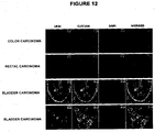
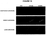
- CLEC14A is not expressed in normal adult tissues, but is expressed in neo-angiogenic vessels of cancers including colon, rectal, ovarian, liver, bladder, prostate, breast, kidney, pancreas, stomach, oesophagus, lung and thyroid cancer.
- CLEC14A genuinely encodes a TEM. Therefore, we now consider that the CLEC14A gene/polypeptide will be valuable as a marker of the tumour endothelium; that antibodies that selectively bind the CLEC14A polypeptide can be used to image and target the tumour neovasculature; and that inhibitors of the CLEC14A gene/polypeptide would be therapeutically useful in the inhibition of tumour neoangiogenesis in solid tumours.
- WO 02/079492 (Eos Biotechnology, Inc) lists many hundreds of ESTs whose expression was said to vary over time in angiogenic tissue. One of these ESTs was an EST encoding a 166 residue fragment of CLEC14A (referred to therein as Pkey 105729, Accession No. Hs46612, Unigene No. Hs293815, Unigene Title HSPC285). However, WO 02/079492 provides no data on the expression of this EST during angiogenesis; it does not even state whether expression was supposed to have increased or decreased or whether it was restricted to tumour endothelial cells.
- PRO269 (Clone DNA38260-1180) is equivalent to CLEC14A.
- multiple potential roles have been ascribed to PRO269.
- PRO269 was said to have use as an antithrombotic agent due to its homology with thrombomodulin, and was said to have the ability to stimulate lymphocyte proliferation, to induce c-fos in cortical neurons, and to affect glucose uptake.
- PRO269 was said to be over-expressed in lung and rectal tumours, but not in breast, colon, cervical, prostate or liver tumours, in comparison to a 'universal' epithelial control sample which was prepared by pooling non-cancerous human tissues of epithelial origin, including liver, kidney, and lung tissues.
- WO 03/101283 (Incyte Genomics, Inc) describes 170 transcripts whose expression was found to be differentially regulated in lung tumours.
- a transcript equivalent to CLEC14A referred to therein as Incyte ID 2264002CB1, Genbank Accession No. g15209752 was found to be between 4-16-fold down-regulated in 21 of 39 distinct lung tumour samples. This appears to contradict the findings in US 2003/0194775 regarding CLEC14A expression in lung tumours.
- CLEC14A was one of 34 human atherosclerosis-associated genes due to its co-expression with other known atherosclerotic genes ( WO 01/04264 ).
- a first aspect of the invention thus provides a method of inhibiting tumour angiogenesis in an individual in need thereof, the method comprising administering to the individual an inhibitor of CLEC14A.
- This aspect of the invention includes the use of an inhibitor of CLEC14A in the preparation of a medicament for inhibiting tumour angiogenesis in an individual.
- the invention further includes an inhibitor of CLEC14A for use in inhibiting tumour angiogenesis in an individual.
- the individual has a solid tumour, which can be treated by inhibiting tumour angiogenesis, i.e. the solid tumour is associated with new blood vessel production.
- tumour is to be understood as referring to all forms of neoplastic cell growth, including, but not limited to, tumours of the breast, ovary, liver, bladder, prostate, kidney, pancreas, stomach, oesophagus, lung and thyroid.
- the tumour is associated with undesirable neovasculature formation and the inhibitor of CLEC14A reduces this to a useful extent.
- the reduction of undesirable neovasculature formation may halt the progression of the tumour and can lead to a clinically useful reduction of tumour size and growth.
- the inhibition of tumour angiogenesis can be used to treat the tumour, for example, to prevent the (further) growth of the tumour, to prevent the spread of the tumour (metastasis), or to reduce the size of the tumour.
- CLEC14A is specifically expressed in the vascular endothelium of a range of solid tumours including tumours of the colon, rectum, ovary, liver, bladder, prostate, breast, kidney, pancreas, stomach, oesophagus, lung and thyroid.
- the individual has a solid tumour selected from colon, rectal, ovarian, liver, bladder, prostate, breast, kidney, pancreas, stomach, oesophagus, lung and thyroid tumours.
- the individual has a solid tumour other than a lung cancer and/or a rectal cancer.
- the methods and medicaments of the invention are used to treat humans, in which case the inhibitor of CLEC14A is an inhibitor of human CLEC14A. It is appreciated, however, that when the methods and medicaments of the invention are for treatment of non-human mammals, it is preferred if the inhibitor is specific for the CLEC14A gene/polypeptide from the other species.
- CLEC14A C-type lectin domain family 14, member A
- CLEC14A encodes a 490 amino acid residue polypeptide with a predicted MW of 51kDa.
- CLEC14A polypeptide we include the meaning of a gene product of human CLEC14A, including naturally occurring variants thereof.
- Human CLEC14A polypeptide includes the amino acid sequence found in Genbank Accession No NP_778230 and naturally occurring variants thereof.
- the CLEC14A polypeptide sequence from NP_778230 is shown in Figure 1 (SEQ ID NO: 1).
- a cDNA sequence corresponding to a human CLEC14A mRNA is found in Genbank Accession No NM_175060 and shown in Figure 1 (SEQ ID NO: 2).
- the coding region of this cDNA from NM_175060 is from nucleotide 348 to nucleotide 1820, and this is also shown in Figure 1 (SEQ ID NO: 3).
- CLEC14A is a type I transmembrane protein with a signal peptide at residues 1-21.
- the mature human polypeptide is 469 amino acids in length (amino acid residues 22-490), and contains a 375 residue extracellular region (residues 22-396), a transmembrane region (residues 397-425), and a cytoplasmic region (residues 426-490).
- the extracellular region contains a C-type lectin like domain (residues 32-175) and an EGF-like region (residues 245-287).
- an inhibitor of CLEC14A we include both inhibitors of the CLEC14A polypeptide and of the CLEC14A gene/cDNA.
- Suitable inhibitors of CLEC14A include antibodies that selectively bind to CLEC14A.
- Other suitable inhibitors of CLEC14A include siRNA, antisense polynucleotides and ribozyme molecules that are specific for polynucleotides encoding the CLEC14A polypeptide, and which prevent its expression.
- polynucleotide inhibitors of CLEC14A may be administered directly, or may be administered in the form of a polynucleotide that encodes the inhibitor.
- an inhibitor of CLEC14A which is a polynucleotide we include the meanings of administering the inhibitor directly, or administering a polynucleotide that encodes the inhibitor, typically in the form of a vector.
- a medicament or a composition comprising an inhibitor of CLEC14A which is a polynucleotide we include the meanings that the medicament or composition comprises the inhibitor itself, or comprises a polynucleotide that encodes the inhibitor.
- Suitable antibodies which bind to CLEC14A, or to specified portions thereof, can be made by the skilled person using technology long-established in the art. Methods of preparation of monoclonal antibodies and antibody fragments are well known in the art and include hybridoma technology ( Kohler & Milstein (1975) "Continuous cultures of fused cells secreting antibody of predefined specificity. Nature 256: 495-497 ); antibody phage display ( Winter et al (1994) "Making antibodies by phage display technology.” Annu. Rev. Immunol. 12: 433-455 ); ribosome display ( Schaffitzel et al (1999) "Ribosome display: an in vitro method for selection and evolution of antibodies from libraries.” J. Immunol.
- Antibodies that are especially active at inhibiting tumour angiogenesis are preferred for anti-cancer therapeutic agents, and they can be selected for this activity using methods well known in the art and described below.
- an antibody that selectively binds the CLEC14A polypeptide we mean that the antibody molecule binds CLEC14A with a greater affinity than for an irrelevant polypeptide, such as human serum albumin (HSA).
- an irrelevant polypeptide such as human serum albumin (HSA).
- the antibody binds the CLEC14A with at least 5, or at least 10 or at least 50 times greater affinity than for the irrelevant polypeptide.
- the antibody molecule binds the CLEC14A with at least 100, or at least 1,000, or at least 10,000 times greater affinity than for the irrelevant polypeptide.
- Such binding may be determined by methods well known in the art, such as one of the Biacore ® systems.
- the antibody that selectively binds the CLEC14A polypeptide does not bind a related polypeptide, such as thrombomodulin, or that the antibody molecule binds CLEC14A with a greater affinity than for the related polypeptide such as thrombomodulin.
- the antibody binds the CLEC14A with at least 5, or at least 10 or at least 50 times greater affinity than for the related polypeptide. More preferably, the antibody molecule binds the CLEC14A with at least 100, or at least 1,000, or at least 10,000 times greater affinity than for the related polypeptide. Such binding may be determined by methods well known in the art, such as one of the Biacore ® systems.
- the antibodies have an affinity for CLEC14A of at least 10 -7 M and more preferably 10 -8 M, although antibodies with higher affinities, e.g. 10 -9 M, or higher, may be even more preferred.
- the antibody that selectively binds CLEC14A polypeptide binds to the mature peptide (residues 22-490) and not to the signal peptide (residues 1-21).
- the antibody that selectively binds CLEC14A binds to the extracellular region of CLEC14A (residues 22-396).
- the antibody may bind to the C-type lectin like domain (residues 32-175) or may bind to the EGF-like region (residues 245-287).
- an antibody that selectively binds a specific portion of CLEC14A we mean that not only does the antibody selectively bind to the target as described above, the antibody molecule also binds the specified portion of the CLEC14A with a greater affinity than for any other portion of it.
- the antibody binds the specified portion with at least 2, or at least 5, or at least 10 or at least 50 times greater affinity than for any other epitope on CLEC14A. More preferably, the antibody molecule binds the specified portion with at least 100, or at least 1,000, or at least 10,000 times greater affinity than for than for any other epitope on the CLEC14A.
- Such binding may be determined by methods well known in the art, such as one of the Biacore ® systems.
- the antibodies have an affinity for their target epitope on the CLEC14A of at least 10 -7 M and more preferably 10 -8 M, although antibodies with higher affinities, e.g. 10 -9 M, or higher, may be even more preferred.
- the antibody selectively binds the particular specified epitope within the CLEC14A and does not bind any other epitopes within it.
- the antibody when the antibody is administered to an individual, the antibody binds to the target CLEC14A or to the specified portion thereof with a greater affinity than for any other molecule in the individual.
- the antibody binds to (a specified portion of) the CLEC14A with at least 2, or at least 5, or at least 10 or at least 50 times greater affinity than for any other molecule in the individual.
- the agent binds the CLEC14A (at the specific domain) with at least 100, or at least 1,000, or at least 10,000 times greater affinity than any other molecule in the individual.
- the antibody molecule selectively binds the CLEC14A without significantly binding other polypeptides in the body.
- antibody or "antibody molecule” as used herein includes but is not limited to polyclonal, monoclonal, chimaeric, single chain, Fab fragments and fragments produced by a Fab expression library. Such fragments include fragments of whole antibodies which retain their binding activity for a target substance, Fv, F(ab') and F(ab')2 fragments, as well as single chain antibodies (scFv), fusion proteins and other synthetic proteins which comprise the antigen-binding site of the antibody.
- the term also includes antibody-like molecules which may be produced using phage-display techniques or other random selection techniques for molecules which bind to the specified polypeptide or to particular regions of it.
- the term antibody includes all molecules which contain a structure, preferably a peptide structure, which is part of the recognition site (i.e. the part of the antibody that binds or combines with the epitope or antigen) of a natural antibody.
- the antibodies and fragments thereof may be humanised antibodies, which are now well known in the art.
- ScFv molecules we mean molecules wherein the V H and V L partner domains are linked via a flexible oligopeptide.
- Engineered antibodies, such as ScFv antibodies can be made using the techniques and approaches long known in the art. The advantages of using antibody fragments, rather than whole antibodies, are several-fold. The smaller size of the fragments may lead to improved pharmacological properties, such as better penetration to the target site. Effector functions of whole antibodies, such as complement binding, are removed.
- Fab, Fv, ScFv and dAb antibody fragments can all be expressed in and secreted from E. coli, thus allowing the facile production of large amounts of the fragments.
- Whole antibodies, and F(ab') 2 fragments are "bivalent". By “bivalent” we mean that the antibodies and F(ab') 2 fragments have two antigen combining sites. In contrast, Fab, Fv, ScFv and dAb fragments are monovalent, having only one antigen combining site.
- CLEC14A may be a glycoprotein.
- the antibody that binds to CLEC14A may bind to any combination of the protein or carbohydrate components of CLEC14A.
- Antibodies may be produced by standard techniques, for example by immunisation with the appropriate (glyco)polypeptide or portion(s) thereof, or by using a phage display library.
- polyclonal antibodies are desired, a selected mammal (e.g., mouse, rabbit, goat, horse, etc) is immunised with an immunogenic polypeptide bearing a desired epitope(s), optionally haptenised to another polypeptide.
- various adjuvants may be used to increase immunological response.
- adjuvants include, but are not limited to, Freund's, mineral gels such as aluminium hydroxide, and surface active substances such as lysolecithin, pluronic polyols, polyanions, peptides, oil emulsions, keyhole limpet hemocyanin, and dinitrophenol.
- Serum from the immunised animal is collected and treated according to known procedures. If serum containing polyclonal antibodies to the desired epitope contains antibodies to other antigens, the polyclonal antibodies can be purified by immunoaffinity chromatography. Techniques for producing and processing polyclonal antisera are well known in the art.
- Anti-CLEC14A polyclonal antibodies are commercially available, for example from Sigma-Aldrich (Catalogue No. SAB1400831), R&D Systems (Catalogue Nos. AF4968 and BAF 4968), Abcam (Product code ab73087) and Novus Biologicals (Catalogue No. H00161198-B01).
- Monoclonal antibodies directed against entire polypeptides or particular epitopes thereof can also be readily produced by one skilled in the art.
- the general methodology for making monoclonal antibodies by hybridomas is well known.
- Immortal antibody-producing cell lines can be created by cell fusion, and also by other techniques such as direct transformation of B lymphocytes with oncogenic DNA, or transfection with Epstein-Barr virus.
- Panels of monoclonal antibodies produced against the polypeptides listed above can be screened for various properties; i.e., for isotype and epitope affinity.
- Monoclonal antibodies may be prepared using any of the well known techniques which provides for the production of antibody molecules by continuous cell lines in culture.
- the antibody is a monoclonal antibody.
- the monoclonal antibody is a human monoclonal antibody or a humanised monoclonal antibody, which are suitable for administration to humans without engendering an immune response by the human against the administered immunoglobulin.
- Suitably prepared non-human antibodies can be "humanised” in known ways, for example by inserting the CDR regions of mouse antibodies into the framework of human antibodies. Humanised antibodies can be made using the techniques and approaches described in Verhoeyen et al (1988) Science, 239, 1534-1536 , and in Kettleborough et al, (1991) Protein Engineering, 14(7), 773-783 .
- Fv framework residues of the human immunoglobulin are replaced by corresponding non-human residues.
- the humanised antibody will contain variable domains in which all or most of the CDR regions correspond to those of a non-human immunoglobulin, and framework regions which are substantially or completely those of a human immunoglobulin consensus sequence.
- Completely human antibodies may be produced using recombinant technologies. Typically large libraries comprising billions of different antibodies are used. In contrast to the previous technologies employing chimerisation or humanisation of e.g. murine antibodies this technology does not rely on immunisation of animals to generate the specific antibody. Instead the recombinant libraries comprise a huge number of pre-made antibody variants wherein it is likely that the library will have at least one antibody specific for any antigen. Thus, using such libraries, an existing antibody having the desired binding characteristics can be identified. In order to find the good binder in a library in an efficient manner, various systems where phenotype i.e. the antibody or antibody fragment is linked to its genotype i.e. the encoding gene have been devised.
- phage display system where antibody fragments are expressed, displayed, as fusions with phage coat proteins on the surface of filamentous phage particles, while simultaneously carrying the genetic information encoding the displayed molecule
- Phage displaying antibody fragments specific for a particular antigen may be selected through binding to the antigen in question. Isolated phage may then be amplified and the gene encoding the selected antibody variable domains may optionally be transferred to other antibody formats, such as e.g. full-length immunoglobulin, and expressed in high amounts using appropriate vectors and host cells well known in the art.
- the "human" antibodies can be made by immunising transgenic mice which contain, in essence, human immunoglobulin genes ( Vaughan et al (1998) Nature Biotechnol. 16, 535-539 ).
- the antibody when the antibody is for administration to a non-human individual, the antibody may have been specifically designed/produced for the intended recipient species.
- the format of displayed antibody specificities on phage particles may differ.
- the most commonly used formats are Fab ( Griffiths et al, 1994. EMBO J, 13: 3245-3260 ) and single chain (scFv) ( Hoogenboom et al, 1992, J Mol Biol. 227: 381-388 ) both comprising the variable antigen binding domains of antibodies.
- the single chain format is composed of a variable heavy domain (V H ) linked to a variable light domain (V L ) via a flexible linker ( US 4,946,778 ).
- the antibody Before use as a therapeutic agent, the antibody may be transferred to a soluble format e.g. Fab or scFv and analysed as such. In later steps the antibody fragment identified to have desirable characteristics may be transferred into yet other formats such as full-length antibodies.
- WO 98/32845 and Soderlind et al (2000) Nature BioTechnol. 18: 852-856 describe technology for the generation of variability in antibody libraries.
- Antibody fragments derived from this library all have the same framework regions and only differ in their CDRs. Since the framework regions are of germline sequence the immunogenicity of antibodies derived from the library, or similar libraries produced using the same technology, are expected to be particularly low (Soderlind et al, 2000). This property is of great value for therapeutic antibodies, reducing the risk that the patient forms antibodies to the administered antibody, thereby reducing risks for allergic reactions, the occurrence of blocking antibodies, and allowing a long plasma half-life of the antibody.
- antibodies we also include heavy-chain antibodies structurally derived from camelidae antibodies, such as Nanobodies ® (Ablynx). These are antibody-derived therapeutic proteins that contain the structural and functional properties of naturally-occurring heavy-chain antibodies.
- the Nanobody ® technology was developed following the discovery that camelidae (camels and llamas) possess fully functional antibodies that lack light chains.
- These heavy-chain antibodies contain a single variable domain (VHH) and two constant domains (C H 2 and C H 3).
- VHH domain variable domain
- C H 2 and C H 3 constant domains
- the cloned and isolated VHH domain is a perfectly stable polypeptide harbouring the full antigen-binding capacity of the original heavy-chain antibody.
- RNA interference is the process of sequence-specific post-transcriptional gene silencing in animals initiated by double-stranded (dsRNA) that is homologous in sequence to the silenced gene.
- the mediators of sequence-specific mRNA degradation are typically 21- and 22-nucleotide small interfering RNAs (siRNAs) which, in vivo, may be generated by ribonuclease III cleavage from longer dsRNAs.
- 21-nucleotide siRNA duplexes have been shown to specifically suppress expression of both endogenous and heterologous genes ( Elbashir et al (2001) Nature 411: 494-498 ).
- the siRNA has to be comprised of two complementary 21mers as described below since longer double-stranded (ds) RNAs will activate PKR (dsRNA-dependent protein kinase) and inhibit overall protein synthesis.
- Duplex siRNA molecules selective for a polynucleotide encoding the CLEC14A polypeptide can readily be designed by reference to its cDNA sequence. For example, they can be designed by reference to the CLEC14A cDNA sequences in the Genbank Accession No. M_175060 and as listed in Figure 1 .
- the first 21-mer sequence that begins with an AA dinucleotide which is at least 120 nucleotides downstream from the initiator methionine codon is selected.
- the second RNA sequence should be perfectly complementary to the first 19 residues of the first, with an additional UU dinucleotide at its 3' end.
- anti-CLEC14A siRNAs are given below.
- anti-CLEC14A siRNAs are commercially available, for example from Applied Biosystems (siRNA ID Nos. s46248, s46249, s46250, 129879, 129880, 129881) and from Invitrogen (Oligo ID Nos. HSS136238, HSS175925 and HSS175926).
- siRNAs may be introduced into cells in the patient using any suitable method, such as those described herein.
- the RNA is protected from the extracellular environment, for example by being contained within a suitable carrier or vehicle.
- Liposome-mediated transfer e.g. the oligofectamine method, may be used.
- Antisense nucleic acid molecules selective for a polynucleotide encoding the CLEC14A polypeptide can readily be designed by reference to its cDNA or gene sequence, as is known in the art.
- Antisense nucleic acids such as oligonucleotides, are single-stranded nucleic acids, which can specifically bind to a complementary nucleic acid sequence. By binding to the appropriate target sequence, an RNA-RNA, a DNA-DNA, or RNA-DNA, duplex is formed. These nucleic acids are often termed "antisense" because they are complementary to the sense or coding strand of the gene. Recently, formation of a triple helix has proven possible where the oligonucleotide is bound to a DNA duplex.
- oligonucleotides could recognise sequences in the major groove of the DNA double helix. A triple helix was formed thereby. This suggests that it is possible to synthesise a sequence-specific molecules which specifically bind double-stranded DNA via recognition of major groove hydrogen binding sites.
- the above oligonucleotides can inhibit the function of the target nucleic acid. This could, for example, be a result of blocking the transcription, processing, poly(A) addition, replication, translation, or promoting inhibitory mechanisms of the cells, such as promoting RNA degradations.
- Antisense oligonucleotides are prepared in the laboratory and then introduced into cells, for example by microinjection or uptake from the cell culture medium into the cells, or they are expressed in cells after transfection with plasmids or retroviruses or other vectors carrying an antisense gene.
- Antisense oligonucleotides were first discovered to inhibit viral replication or expression in cell culture for Rous sarcoma virus, vesicular stomatitis virus, herpes simplex virus type 1, simian virus and influenza virus. Since then, inhibition of mRNA translation by antisense oligonucleotides has been studied extensively in cell-free systems including rabbit reticulocyte lysates and wheat germ extracts.
- antisense oligonucleotides are 15 to 35 bases in length.
- 20-mer oligonucleotides have been shown to inhibit the expression of the epidermal growth factor receptor mRNA ( Witters et al., Breast Cancer Res Treat 53:41-50 (1999 )) and 25-mer oligonucleotides have been shown to decrease the expression of adrenocorticotropic hormone by greater than 90% ( Frankel et al., J Neurosurg 91:261-7 (1999 )).
- Antisense polynucleotides may be administered systemically. Alternatively, and preferably, the inherent binding specificity of polynucleotides characteristic of base pairing is enhanced by limiting the availability of the polynucleotide to its intended locus in vivo, permitting lower dosages to be used and minimising systemic effects. Thus, polynucleotides may be applied locally to the solid tumour to achieve the desired effect. The concentration of the polynucleotides at the desired locus is much higher than if the polynucleotides were administered systemically, and the therapeutic effect can be achieved using a significantly lower total amount. The local high concentration of polynucleotides enhances penetration of the targeted cells and effectively blocks translation of the target nucleic acid sequences.
- antisense agents may also include larger molecules which bind to polynucleotides (mRNA or genes) encoding the CLEC14A polypeptide and substantially prevent expression of the protein.
- antisense molecules which are substantially complementary to the respective mRNA are also envisaged.
- the molecules may be expressed from any suitable genetic construct and delivered to the patient.
- the genetic construct which expresses the antisense molecule comprises at least a portion of the CLEC14A cDNA or gene operatively linked to a promoter which can express the antisense molecule in the cell.
- the genetic construct is adapted for delivery to a human cell.
- Ribozymes are RNA or RNA-protein complexes that cleave nucleic acids in a site-specific fashion. Ribozymes have specific catalytic domains that possess endonuclease activity. For example, a large number of ribozymes accelerate phosphoester transfer reactions with a high degree of specificity, often cleaving only one of several phosphoesters in an oligonucleotide substrate. This specificity has been attributed to the requirement that the substrate bind via specific base-pairing interactions to the internal guide sequence ("IGS") of the ribozyme prior to chemical reaction.
- IGS internal guide sequence
- Ribozyme catalysis has primarily been observed as part of sequence-specific cleavage/ligation reactions involving nucleic acids.
- US 5,354,855 reports that certain ribozymes can act as endonucleases with a sequence specificity greater than that of known ribonucleases and approaching that of the DNA restriction enzymes.
- sequence-specific ribozyme-mediated inhibition of gene expression may be particularly suited to therapeutic applications, and ribozymes specific for a polynucleotide encoding the CLEC14A polypeptide may be designed by reference to the cDNA sequences listed in the Genbank Accession No. NM_175060 and as listed in Figure 1 .
- polynucleotide inhibitors such as siRNA molecules, antisense molecules and ribozymes
- agents that inhibit transcription of the genes encoding any of the above listed polypeptides can also be designed, for example using an engineered transcription repressor described in Isalan et al (Nat Biotechnol, 19(7): 656-60 (2001 )) and in Urnov (Biochem Pharmacol, 64 (5-6): 919 (2002 )). Additionally, they an be selected, for example using the screening methods described in later aspects of this disclosure.
- inhibitor of CLEC14A will typically be formulated for administration to an individual as a pharmaceutical composition, i.e. together with a pharmaceutically acceptable carrier, diluent or excipient.
- the formulation is sterile and pyrogen free.
- Suitable pharmaceutical carriers, diluents and excipients are well known in the art of pharmacy.
- the carrier(s) must be “acceptable” in the sense of being compatible with the inhibitor and not deleterious to the recipients thereof.
- the carriers will be water or saline which will be sterile and pyrogen free; however, other acceptable carriers may be used.
- the pharmaceutical compositions or formulations of the invention are for parenteral administration, more particularly for intravenous administration.
- the pharmaceutical composition is suitable for intravenous administration to a patient, for example by injection.
- Formulations suitable for parenteral administration include aqueous and non-aqueous sterile injection solutions which may contain anti-oxidants, buffers, bacteriostats and solutes which render the formulation isotonic with the blood of the intended recipient; and aqueous and non-aqueous sterile suspensions which may include suspending agents and thickening agents.
- the pharmaceutical composition is suitable for topical administration to a patient.
- the formulation is a unit dosage containing a daily dose or unit, daily sub-dose or an appropriate fraction thereof, of the active ingredient.
- the inhibitor may be administered orally or by any parenteral route, in the form of a pharmaceutical formulation comprising the active ingredient, optionally in the form of a non-toxic organic, or inorganic, acid, or base, addition salt, in a pharmaceutically acceptable dosage form.
- a pharmaceutical formulation comprising the active ingredient, optionally in the form of a non-toxic organic, or inorganic, acid, or base, addition salt, in a pharmaceutically acceptable dosage form.
- the compositions may be administered at varying doses.
- the inhibitor will generally be administered in admixture with a suitable pharmaceutical excipient, diluent or carrier selected with regard to the intended route of administration and standard pharmaceutical practice.
- the inhibitor may be administered orally, buccally or sublingually in the form of tablets, capsules, ovules, elixirs, solutions or suspensions, which may contain flavouring or colouring agents, for immediate-, delayed- or controlled-release applications.
- the inhibitor may also be administered via intracavernosal injection.
- Suitable tablets may contain excipients such as microcrystalline cellulose, lactose, sodium citrate, calcium carbonate, dibasic calcium phosphate and glycine, disintegrants such as starch (preferably corn, potato or tapioca starch), sodium starch glycolate, croscarmellose sodium and certain complex silicates, and granulation binders such as polyvinylpyrrolidone, hydroxypropylmethylcellulose (HPMC), hydroxy-propylcellulose (HPC), sucrose, gelatin and acacia. Additionally, lubricating agents such as magnesium stearate, stearic acid, glyceryl behenate and talc may be included.
- excipients such as microcrystalline cellulose, lactose, sodium citrate, calcium carbonate, dibasic calcium phosphate and glycine
- disintegrants such as starch (preferably corn, potato or tapioca starch), sodium starch glycolate, croscarmellose sodium and certain complex silicates
- Solid compositions of a similar type may also be employed as fillers in gelatin capsules.
- Preferred excipients in this regard include lactose, starch, a cellulose, milk sugar or high molecular weight polyethylene glycols.
- the compounds of the invention may be combined with various sweetening or flavouring agents, colouring matter or dyes, with emulsifying and/or suspending agents and with diluents such as water, ethanol, propylene glycol and glycerin, and combinations thereof.
- the inhibitor can also be administered parenterally, for example, intravenously, intra-arterially, intraperitoneally, intrathecally, intraventricularly, intrasternally, intracranially, intra-muscularly or subcutaneously, or they may be administered by infusion techniques. They are best used in the form of a sterile aqueous solution which may contain other substances, for example, enough salts or glucose to make the solution isotonic with blood.
- aqueous solutions should be suitably buffered (preferably to a pH of from 3 to 9), if necessary.
- suitable parenteral formulations under sterile conditions is readily accomplished by standard pharmaceutical techniques well-known to those skilled in the art.
- the formulations may be presented in unit-dose or multi-dose containers, for example sealed ampoules and vials, and may be stored in a freeze-dried (lyophilised) condition requiring only the addition of the sterile liquid carrier, for example water for injections, immediately prior to use.
- sterile liquid carrier for example water for injections, immediately prior to use.
- Extemporaneous injection solutions and suspensions may be prepared from sterile powders, granules and tablets of the kind previously described.
- the daily dosage level of an inhibitor will usually be from 1 to 1,000 mg per adult (i.e. from about 0.015 to 15 mg/kg), administered in single or divided doses.
- the tablets or capsules of the inhibitor may contain from 1 mg to 1,000 mg of active agent for administration singly or two or more at a time, as appropriate.
- the physician in any event will determine the actual dosage which will be most suitable for any individual patient and it will vary with the age, weight and response of the particular patient.
- the above dosages are exemplary of the average case. There can, of course, be individual instances where higher or lower dosage ranges are merited and such are within the scope of this invention.
- the inhibitor can also be administered intranasally or by inhalation and are conveniently delivered in the form of a dry powder inhaler or an aerosol spray presentation from a pressurised container, pump, spray or nebuliser with the use of a suitable propellant, e.g. dichlorodifluoromethane, trichlorofluoromethane, dichlorotetrafluoro-ethane, a hydrofluoroalkane such as 1,1,1,2-tetrafluoroethane (HFA 134A3 or 1,1,1,2,3,3,3-heptafluoropropane (HFA 227EA3), carbon dioxide or other suitable gas.
- a suitable propellant e.g. dichlorodifluoromethane, trichlorofluoromethane, dichlorotetrafluoro-ethane, a hydrofluoroalkane such as 1,1,1,2-tetrafluoroethane (HFA 134A3 or 1,1,1,2,
- the dosage unit may be determined by providing a valve to deliver a metered amount.
- the pressurised container, pump, spray or nebuliser may contain a solution or suspension of the active compound, e.g. using a mixture of ethanol and the propellant as the solvent, which may additionally contain a lubricant, e.g. sorbitan trioleate.
- Capsules and cartridges (made, for example, from gelatin) for use in an inhaler or insufflator may be formulated to contain a powder mix of a antibody and a suitable powder base such as lactose or starch. Such formulations may be particularly useful for treating solid tumours of the lung, such as, for example, small cell lung carcinoma, non-small cell lung carcinoma, pleuropulmonary blastoma or carcinoid tumour.
- Aerosol or dry powder formulations are preferably arranged so that each metered dose or "puff" contains at least 1 mg of the inhibitor for delivery to the patient. It will be appreciated that the overall daily dose with an aerosol will vary from patient to patient, and may be administered in a single dose or, more usually, in divided doses throughout the day.
- the inhibitor can be administered in the form of a suppository or pessary, particularly for treating or targeting colon, rectal or prostate tumours.
- the inhibitor may also be administered by the ocular route.
- the inhibitor can be formulated as, e.g., micronised suspensions in isotonic, pH adjusted, sterile saline, or, preferably, as solutions in isotonic, pH adjusted, sterile saline, optionally in combination with a preservative such as a benzylalkonium chloride.
- a preservative such as a benzylalkonium chloride.
- they may be formulated in an ointment such as petrolatum.
- Such formulations may be particularly useful for treating solid tumours of the eye, such as retinoblastoma, medulloepithelioma, uveal melanoma, rhabdomyosarcoma, intraocular lymphoma, or orbital lymphoma.
- the inhibitor may be applied topically in the form of a lotion, solution, cream, ointment or dusting powder, or may be transdermally administered, for example, by the use of a skin patch.
- the inhibitor can be formulated as a suitable ointment containing the active compound suspended or dissolved in, for example, a mixture with one or more of the following: mineral oil, liquid petrolatum, white petrolatum, propylene glycol, polyoxyethylene polyoxypropylene compound, emulsifying wax and water.
- ком ⁇ онентs can be formulated as a suitable lotion or cream, suspended or dissolved in, for example, a mixture of one or more of the following: mineral oil, sorbitan monostearate, a polyethylene glycol, liquid paraffin, polysorbate 60, cetyl esters wax, cetearyl alcohol, 2-octyldodecanol, benzyl alcohol and water.
- suitable lotion or cream suspended or dissolved in, for example, a mixture of one or more of the following: mineral oil, sorbitan monostearate, a polyethylene glycol, liquid paraffin, polysorbate 60, cetyl esters wax, cetearyl alcohol, 2-octyldodecanol, benzyl alcohol and water.
- Such formulations may be particularly useful for treating solid tumours of the skin, such as, for example, basal cell cancer, squamous cell cancer or melanoma.
- the inhibitors can also be delivered by electroincorporation (EI).
- EI occurs when small particles of up to 30 microns in diameter on the surface of the skin experience electrical pulses identical or similar to those used in electroporation. In EI, these particles are driven through the stratum corneum and into deeper layers of the skin.
- the particles can be loaded or coated with inhibitor or can simply act as "bullets" that generate pores in the skin through which the inhibitor can enter.
- Formulations suitable for topical administration in the mouth include lozenges comprising the active ingredient in a flavoured basis, usually sucrose and acacia or tragacanth; pastilles comprising the active ingredient in an inert basis such as gelatin and glycerin, or sucrose and acacia; and mouth-washes comprising the active ingredient in a suitable liquid carrier.
- Such formulations may be particularly useful for treating solid tumours of the mouth and throat.
- the inhibitor when it is a polypeptide, such as an anti-CLEC14A antibody, it may be delivered using an injectable sustained-release drug delivery system. These are designed specifically to reduce the frequency of injections.
- An example of such a system is Nutropin Depot which encapsulates recombinant human growth hormone (rhGH) in biodegradable microspheres that, once injected, release rhGH slowly over a sustained period.
- the antibody can be administered by a surgically implanted device that releases the drug directly to the required site, for example, into the eye to treat ocular tumours.
- a surgically implanted device that releases the drug directly to the required site, for example, into the eye to treat ocular tumours.
- ReGel injectable system that is thermo-sensitive. Below body temperature, ReGel is an injectable liquid while at body temperature it immediately forms a gel reservoir that slowly erodes and dissolves into known, safe, biodegradable polymers. The active drug is delivered over time as the biopolymers dissolve.
- Polypeptide pharmaceuticals such as antibodies can also be delivered orally.
- the process employs a natural process for oral uptake of vitamin B 12 in the body to co-deliver proteins and peptides. By riding the vitamin B 12 uptake system, the protein or peptide can move through the intestinal wall.
- Complexes are synthesised between vitamin B 12 analogues and the drug that retain both significant affinity for intrinsic factor (IF) in the vitamin B 12 portion of the complex and significant bioactivity of the drug portion of the complex.
- IF intrinsic factor
- Polynucleotides may be administered by any effective method, for example, parenterally (e.g. intravenously, subcutaneously, intramuscularly) or by oral, nasal or other means which permit the polynucleotides to access and circulate in the patient's bloodstream.
- Polynucleotides administered systemically preferably are given in addition to locally administered polynucleotides, but also have utility in the absence of local administration.
- a dosage in the range of from about 0.1 to about 10 grams per administration to an adult human generally will be effective for this purpose.
- the polynucleotide may be administered as a suitable genetic construct as is described below and delivered to the patient where it is expressed.
- the polynucleotide in the genetic construct is operatively linked to a promoter which can express the compound in the cell.
- the genetic constructs of the invention can be prepared using methods well known in the art, for example in Sambrook et al (2001).
- genetic constructs for delivery of polynucleotides can be DNA or RNA, it is preferred if they are DNA.
- the genetic construct is adapted for delivery to a human cell.
- the constructs of the invention may be introduced into cells by any convenient method, for example methods involving retroviruses, so that the construct is inserted into the genome of the cell.
- any convenient method for example methods involving retroviruses, so that the construct is inserted into the genome of the cell.
- retroviral DNA constructs comprising a polynucleotide as described above may be made using methods well known in the art.
- DMEM Dulbecco's modified Eagle's medium
- FCS foetal calf serum
- Transfection of the cell line is conveniently by calcium phosphate co-precipitation, and stable transformants are selected by addition of G418 to a final concentration of 1 mg/mt (assuming the retroviral construct contains a neo R gene).
- Independent colonies are isolated and expanded and the culture supernatant removed, filtered through a 0.45 ⁇ m pore-size filter and stored at -70°C.
- retroviral supernatant for example, it is convenient to inject directly retroviral supernatant to which 10 ⁇ g/ml Polybrene has been added.
- tumours exceeding 10 mm in diameter it is appropriate to inject between 0.1 ml and 1 ml of retroviral supernatant; preferably 0.5 ml.
- cells which produce retroviruses may be injected.
- the retrovirus-producing cells so introduced are engineered to actively produce retroviral vector particles so that continuous productions of the vector occurred within the tumour mass in situ.
- Targeted retroviruses are also available for use in the invention; for example, sequences conferring specific binding affinities may be engineered into pre-existing viral env genes (see Miller & Vile (1995) Faseb J. 9, 190-199 , for a review of this and other targeted vectors for gene therapy).
- adenoviruses carrying external DNA via an antibody-polylysine bridge see Curiel (1993) Prog. Med. Virol. 40, 1-18
- transferrin-polycation conjugates as carriers Wagner et al (1990) Proc. Natl. Acad. Sci. USA 87, 3410-3414 ).
- a polycation-antibody complex is formed with the DNA construct or other genetic construct of the invention, wherein the antibody is specific for either wild-type adenovirus or a variant adenovirus in which a new epitope has been introduced which binds the antibody.
- the polycation moiety binds the DNA via electrostatic interactions with the phosphate backbone.
- the adenovirus because it contains unaltered fibre and penton proteins, is internalised into the cell and carries into the cell with it the DNA construct of the invention. It is preferred if the polycation is polylysine.
- a high-efficiency nucleic acid delivery system that uses receptor-mediated endocytosis to carry DNA macromolecules into cells is employed. This is accomplished by conjugating the iron-transport protein transferrin to polycations that bind nucleic acids. Human transferrin, or the chicken homologue conalbumin, or combinations thereof is covalently linked to the small DNA-binding protein protamine or to polylysines of various sizes through a disulphide linkage. These modified transferrin molecules maintain their ability to bind their cognate receptor and to mediate efficient iron transport into the cell.
- the transferrin-polycation molecules form electrophoretically stable complexes with DNA constructs or other genetic constructs of the invention independent of nucleic acid size (from short oligonucleotides to DNA of 21 kilobase pairs).
- complexes of transferrin-polycation and the DNA constructs or other genetic constructs of the invention are supplied to the tumour cells, a high level of expression from the construct in the cells is expected.
- High-efficiency receptor-mediated delivery of the DNA constructs or other genetic constructs of the invention using the endosome-disruption activity of defective or chemically inactivated adenovirus particles produced by the methods of Cotten et al (1992) Proc. Natl. Acad. Sci. USA 89, 6094-6098 may also be used.
- This approach appears to rely on the fact that adenoviruses are adapted to allow release of their DNA from an endosome without passage through the lysosome, and in the presence of, for example transferrin linked to the DNA construct or other genetic construct of the invention, the construct is taken up by the cell by the same route as the adenovirus particle.
- This approach has the advantages that there is no need to use complex retroviral constructs; there is no permanent modification of the genome as occurs with retroviral infection; and the targeted expression system is coupled with a targeted delivery system, thus reducing toxicity to other cell types.
- naked DNA and DNA complexed with cationic and neutral lipids may also be useful in introducing the DNA of the invention into cells of the individual to be treated.
- Non-viral approaches to gene therapy are described in Ledley (1995, Human Gene Therapy 6, 1129-1144 ) .
- tissue-specific promoters in the vectors encoding a polynucleotide inhibitor, this is not essential. This is because the targeted genes are only expressed, or selectively expressed, in the tumour endothelium. Accordingly, expression of CLEC14A-specific inhibitors such as siRNA, antisense molecules and ribozymes in the body at locations other than the solid tumour would be expected to have no effect since CLEC14A is not expressed or is expressed at a comparatively low level. Moreover, the risk of inappropriate expression of these inhibitors, in a cell that may express the target polypeptide at a low lever, is minuscule compared to the therapeutic benefit to a patient suffering from a solid tumour.
- CLEC14A-specific inhibitors such as siRNA, antisense molecules and ribozymes
- Targeted delivery systems are also known, such as the modified adenovirus system described in WO 94/10323 , wherein, typically, the DNA is carried within the adenovirus, or adenovirus-like, particle.
- Michael et al (1995) Gene Therapy 2: 660-668 describes modification of adenovirus to add a cell-selective moiety into a fibre protein.
- Mutant adenoviruses which replicate selectively in p53-deficient human tumour cells such as those described in Bischoff et al (1996) Science 274: 373-376 are also useful for delivering genetic constructs to a cell.
- Other suitable viruses, viral vectors or virus-like particles include lentivirus and lentiviral vectors, HSV, adeno-assisted virus (AAV) and AAV-based vectors, vaccinia and parvovirus.
- Methods of delivering polynucleotides to a patient are well known to a person of skill in the art and include the use of immunoliposomes, viral vectors (including vaccinia, modified vaccinia, adenovirus and adeno-associated viral (AAV) vectors), and by direct delivery of DNA, e.g. using a gene-gun and electroporation.
- methods of delivering polynucleotides to a target tissue of a patient for treatment are also well known in the art.
- US 6,503,242 describes an implanted catheter apparatus for delivering therapeutic agents directly to the hippocampus.
- Methods of targeting and delivering agents to the brain can be used for the treatment of solid tumours of the brain.
- therapeutic agents including vectors can be distributed throughout a wide region of the CNS by injection into the cerebrospinal fluid, e.g., by lumbar puncture (See e.g., Kapadia et al (1996) Neurosurg 10: 585-587 ).
- precise delivery of the therapeutic agent into specific sites of the brain can be conducted using stereotactic microinjection techniques.
- the subject being treated can be placed within a stereotactic frame base (MRI-compatible) and then imaged using high resolution MRI to determine the three-dimensional positioning of the particular region to be treated.
- the MRI images can then be transferred to a computer having the appropriate stereotactic software, and a number of images are used to determine a target site and trajectory for microinjection of the therapeutic agent.
- the software translates the trajectory into three-dimensional coordinates that are precisely registered for the stereotactic frame.
- the skull will be exposed, burr holes will be drilled above the entry site, and the stereotactic apparatus used to position the needle and ensure implantation at a predetermined depth.
- the therapeutic agent can be delivered to regions of the CNS such as the hippocampus, cells of the spinal cord, brainstem, (medulla, pons, and midbrain), cerebellum, diencephalon (thalamus, hypothalamus), telencephalon (corpus stratium, cerebral cortex, or within the cortex, the occipital, temporal, parietal or frontal lobes), or combinations, thereof.
- the therapeutic agent is delivered using other delivery methods suitable for localised delivery, such as localised permeation of the blood-brain barrier.
- US 2005/0025746 describes delivery systems for localised delivery of an adeno-associated virus vector (AAV) vector encoding a therapeutic agent to a specific region of the brain.
- AAV adeno-associated virus vector
- a therapeutic agent for the treatment of a solid tumour of, for example, the brain is encoded by a polynucleotide
- Central nervous system (CNS) specific promoters such as, neuron-specific promoters (e.g., the neurofilament promoter (Byme and Ruddle (1989) Proc. Natl. Acad. Sci. USA 86: 5473-5477 ) and glial specific promoters ( Morii et at (1991) Biochem. Biophys Res. Commun: 175: 185-191 ) are preferably used for directing expression of a polynucleotide preferentially in cells of the CNS.
- the promoter is tissue specific and is essentially not active outside the central nervous system, or the activity of the promoter is higher in the central nervous system than in other cells or tissues.
- the promoter may be specific for the spinal cord, brainstem, (medulla, pons, and midbrain), cerebellum, diencephalon (thalamus, hypothalamus), telencephalon (corpus stratium, cerebral cortex, or within the cortex, the occipital, temporal, parietal or frontal lobes), or combinations, thereof.
- the promoter may be specific for particular cell types, such as neurons or glial cells in the CNS.
- glial cells If it is active in glial cells, it may be specific for astrocytes, oligodendrocytes, ependymat cells, Schwann cells, or microglia. If it is active in neurons, it may be specific for particular types of neurons, e.g., motor neurons, sensory neurons, or interneurons.
- the promoter may be specific for cells in particular regions of the brain, for example, the cortex, stratium, nigra and hippocampus.
- Suitable neuronal specific promoters include, but are not limited to, neuron specific enolase (NSE; Arlington et al (1991) Genomics 10: 157-165 ); GenBank Accession No: X51956), and human neurofilament light chain promoter (NEFL; Rogaev et al (1992) Hum. Mol. Genet. 1: 781 ); GenBank Accession No: L04147).
- Glial specific promoters include, but are not limited to, glial fibrillary acidic protein (GFAP) promoter (Morii et al (1991); GenBank Accession No: M65210), S100 promoter (Morii et al (1991); GenBank Accession No: M65210) and glutamine synthase promoter ( Van den et al (1991) Biochem. Biophys. Acta. 2: 249-251 ); GenBank Accession No: X59834).
- the gene is flanked upstream (i.e., 5') by the neuron specific enolase (NSE) promoter.
- NSE neuron specific enolase
- the gene of interest is flanked upstream (i.e., 5') by the elongation factor 1 alpha (EF) promoter.
- EF elongation factor 1 alpha
- a hippocampus specific promoter that might be used is the hippocampus specific glucocorticoid receptor (GR) gene promoter.
- Svensson et al. (1999) describes the delivery of recombinant genes to cardiomyocytes by intramyocardial injection or intracoronary infusion of cardiotropic vectors, such as recombinant adeno-associated virus vectors, resulting in transgene expression in murine cardiomyocytes in vivo ( Svensson et al (1999) "Efficient and stable transduction of cardiomyocytes after intramyocardial injection or intracoronary perfusion with recombinant adeno-associated virus vectors.” Circulation. 99: 201-5 ).
- polynucleotide inhibitor it may be desirable to be able to temporally regulate expression of the polynucleotide inhibitor in the cell, although this is not essential for the reasons given above.
- expression of the polynucleotide is directly or indirectly (see below) under the control of a promoter that may be regulated, for example by the concentration of a small molecule that may be administered to the patient when it is desired to activate or, more likely, repress (depending upon whether the small molecule effects activation or repression of the said promoter) expression of the antibody from the polynucleotide.
- the expression construct is stable, i.e., capable of expressing the inhibitor (in the presence of any necessary regulatory molecules), in the cell for a period of at least one week, one, two, three, four, five, six, eight months or one or more years.
- the polynucleotide may be operatively linked to a regulatable promoter.
- regulatable promoters include those referred to in the following papers: Rivera et al (1999) Proc Natl Acad Sci USA 96(15), 8657-62 (control by rapamycin, an orally bioavailable drug, using two separate adenovirus or adeno-associated virus (AAV) vectors, one encoding an inducible human growth hormone (hGH) target gene, and the other a bipartite rapamycin-regulated transcription factor); Magari et al (1997) J Clin Invest 100(11), 2865-72 (control by rapamycin); Bueler (1999) Biol Chem 380(6), 613-22 (review of adeno-associated viral vectors); Bohl et al (1998) Blood 92(5), 1512-7 (control by doxycycline in adeno-associated vector); Abruzzese et al (1996) J Mol Med 74(7), 379-92 (review of induction factors, e.g.. hormones, growth factors,
- the inhibitor is typically administered as a suitably acceptable formulation in accordance with normal veterinary practice and the veterinary surgeon will determine the dosing regimen and route of administration which will be most appropriate for a particular animal.
- angiogenesis inhibitor anti-VEGF monoclonal antibody bevacizumab improves the clinical outcome for a number of solid tumours when administered in combination with standard chemotherapy.
- Combinations that have been used include bevacizumab in combination with irinotecan, fluorouracil, and leucovorin; bevacizumab in combination with FOLFOX4 (a regimen of oxaliplatin, 5-fluorouracil and leucovorin); bevacizumab in combination with paclitaxel; and bevacizumab in combination with paclitaxel and carboplatin.
- inhibitors of CLEC14A described above may be clinically effective in the absence of any other anti-cancer compound, it may be advantageous to administer these inhibitors in conjunction with a further anticancer agent.
- the method may also comprising administering to the individual at least one further anticancer agent.
- the method may comprise administering to the individual a pharmaceutical composition containing the inhibitor of CLEC14A and the further anticancer agent.
- the inhibitor of CLEC14A and the further anticancer agent may be administered separately, for instance by separate routes of administration.
- the inhibitor of CLEC14A and the at least one further anticancer agent can be administered sequentially or (substantially) simultaneously. They may be administered within the same pharmaceutical formulation or medicament or they may be formulated and administered separately.
- the medicament containing the inhibitor of CLEC14A may also comprise at least one further anticancer agent.
- the individual to be treated may be one who is administered at least one further anticancer agent. It is appreciated that the individual may be administered the further anticancer agent at the same time as the medicament containing the inhibitor of CLEC14A, although the individual may have been (or will be) administered the further anticancer agent before (or after) receiving the medicament containing the inhibitor of CLEC14A.
- the further anticancer agent may be selected from alkylating agents including nitrogen mustards such as mechlorethamine (HN 2 ), cyclophosphamide, ifosfamide, melphalan (L-sarcolysin) and chlorambucil; ethylenimines and methylmelamines such as hexamethylmelamine, thiotepa; alkyl sulphonates such as busulphan; nitrosoureas such as carmustine (BCNU), lomustine (CCNU), semustine (methyl-CCNU) and streptozocin (streptozotocin); and triazenes such as decarbazine (DTIC; dimethyltriazenoimidazole-carboxamide); antimetabolites including folic acid analogues such as methotrexate (amethopterin); pyrimidine analogues such as fluorouracil (5-fluorouracil; 5-FU), floxuridine (fluoro
- the clinically used anticancer agents are typically grouped by mechanism of action: Alkylating agents, Topoisomerase I inhibitors, Topoisomerase II inhibitors, RNA/DNA antimetabolites, DNA antimetabolites and Antimitotic agents.
- Alkylating agents Topoisomerase I inhibitors
- Topoisomerase II inhibitors Topoisomerase II inhibitors
- RNA/DNA antimetabolites DNA antimetabolites
- Antimitotic agents The US NIH/National Cancer Institute website lists 122 compounds (http://dtp.nci.nih.gov/docs/cancer/ searches/standard_mechanism.html), all of which may be used in conjunction with an inhibitor of CLEC14A.
- Alkylating agents including Asaley, AZQ, BCNU, Busulfan, carboxyphthalatoplatinum, CBDCA, CCNU, CHIP, chlorambucil, chlorozotocin, cis -platinum, clomesone, cyanomorpholino-doxorubicin, cyclodisone, dianhydrogalactitol, fluorodopan, hepsulfam, hycanthone, melphalan, methyl CCNU, mitomycin C, mitozolamide, nitrogen mustard, PCNU, piperazine, piperazinedione, pipobroman, porfiromycin, spirohydantoin mustard, teroxirone, tetraplatin, picoplatin (SP-4-3) (cis-aminedichloro(2-methylpyridine)Pt(II)), thio-tepa, triethylenemelamine, uracil nitrogen mustard, Yoshi-864; anitmitotic agents including allocol
- the at least one further anticancer agent is selected from cisplatin; carboplatin; picoplatin; 5-flurouracil; paclitaxel; mitomycin C; doxorubicin; gemcitabine; tomudex; pemetrexed; methotrexate; irinotecan, fluorouracil and leucovorin; oxaliplatin, 5-fluorouracil and leucovorin; and paclitaxel and carboplatin.
- the inhibitor of CLEC14A is used in combination with that further anticancer agent to treat that specific tumour type.
- tumour endothelial markers are specific for tumour endothelial markers. Due to their accessibility and to the therapeutic options that they allow (for example, intraluminal blood coagulation or recruitment of immune cells), vascular markers selectively expressed on tumour blood vessels are ideally suited for ligand-based tumour-targeting strategies, allowing for the imaging of tumour neovasculature and for targeting cytotoxic agents to the tumour neovasculature.
- a second aspect of the invention provides a method of targeting a cytotoxic agent to tumour neovasculature in the body of an individual, the method comprising administering to the individual a compound comprising (i) an antibody that selectively binds the CLEC14A polypeptide and (ii) a cytotoxic moiety.
- This aspect of the invention also provides the use of a compound comprising (i) an antibody that selectively binds the CLEC14A polypeptide and (ii) a cytotoxic moiety in the preparation of a medicament for targeting a cytotoxic agent to tumour neovasculature in the body of an individual.
- a compound comprising (i) an antibody that selectively binds the CLEC14A polypeptide and (ii) a cytotoxic moiety in the preparation of a medicament for targeting a cytotoxic agent to tumour neovasculature in the body of an individual.
- This aspect further provides such a compound for use in targeting a cytotoxic agent to tumour neovasculature in the body of an individual.
- the cytotoxic moiety is selected from a directly cytotoxic chemotherapeutic agent, a directly cytotoxic polypeptide, a moiety which is able to convert a prodrug into a cytotoxic drug, a radiosensitizer, a directly cytotoxic nucleic acid, a nucleic acid molecule that encodes a directly or indirectly cytotoxic polypeptide or a radioactive atom.
- cytotoxic moieties as well as methods of making the conjugates comprising the antibody and the cytotoxic moiety, are provided in our earlier publications WO 02/36771 and WO 2004/046191 .
- the cytotoxic moiety may be directly or indirectly toxic to cells in neovasculature or cells which are in close proximity to and associated with neovasculature.
- directly cytotoxic we include the meaning that the moiety is one which on its own is cytotoxic.
- indirectly cytotoxic we include the meaning that the moiety is one which, although is not itself cytotoxic, can induce cytotoxicity, for example by its action on a further molecule or by further action on it.
- the cytotoxic moiety is a cytotoxic chemotherapeutic agent.
- Cytotoxic chemotherapeutic agents are well known in the art. Cytotoxic chemotherapeutic agents, such as anticancer agents, include those listed herein.
- cytotoxic moieties such as cytotoxic chemotherapeutic agents
- cytotoxic chemotherapeutic agents have previously been attached to antibodies and other targeting agents, and so compounds of the invention comprising these agents may readily be made by the person skilled in the art.
- carbodiimide conjugation Bauminger & Wilchek (1980) Methods Enzymol. 70, 151-159
- Other methods for conjugating a cytotoxic moiety to an antibody can also be used. For example, sodium periodate oxidation followed by reductive alkylation of appropriate reactants can be used, as can glutaraldehyde cross-linking.
- the cytotoxic moiety may be a cytotoxic peptide or polypeptide moiety by which we include any moiety which leads to cell death.
- Cytotoxic peptide and polypeptide moieties are well known in the art and include, for example, ricin, abrin, Pseudomonas exotoxin, tissue factor and the like. Methods for linking them to targeting moieties such as antibodies are also known in the art. The use of ricin as a cytotoxic agent is described in Burrows & Thorpe (1993) Proc. Natl. Acad. Sci.
- cytokines such as TNF ⁇ , INF ⁇ and IL-2, may also be useful as cytotoxic agents.
- radioactive atoms may also be cytotoxic if delivered in sufficient doses.
- the cytotoxic moiety may comprise a radioactive atom which, in use, delivers a sufficient quantity of radioactivity to the target site so as to be cytotoxic.
- Suitable radioactive atoms include phosphorus-32, iodine-125, iodine-131, indium-111, rhenium-186, rhenium-188 or yttrium-90, or any other isotope which emits enough energy to destroy neighbouring cells, organelles or nucleic acid.
- the isotopes and density of radioactive atoms in the compound of the invention are such that a dose of more than 4000 cGy (preferably at least 6000, 8000 or 10000 cGy) is delivered to the target site and, preferably, to the cells at the target site and their organelles, particularly the nucleus.
- the radioactive atom may be attached to the antibody in known ways.
- EDTA or another chelating agent may be attached to the antibody and used to attach 111 In or 80 Y.
- Tyrosine residues may be labelled with 125 I or 131 I.
- the cytotoxic moiety may be a radiosensitizer.
- Radiosensitizers include fluoropyrimidines, thymidine analogues, hydroxyurea, gemcitabine, fludarabine, nicotinamide, halogenated pyrimidines, 3-aminobenzamide, 3-aminobenzodiamide, etanixadole, pimonidazole and misonidazole (see, for example, McGinn et al (1996) J. Natl. Cancer Inst. 88, 1193-11203 ; Shewach & Lawrence (1996) Invest. New Drugs 14, 257-263 ; Horsman (1995) Acta Oncol.
- the cytotoxic moiety may be a procoagulant factor, such as the extracellular domain of tissue factor ( Rippmann et al (2000) "Fusion of the tissue factor extracellular domain to a tumour stroma specific single-chain fragment variable antibody results in an antigen-specific coagulation-promoting molecule.” Biochem J. 349: 805-12 ; Huang et al (1997) “Tumor infarction in mice by antibody-directed targeting of tissue factor to tumor vasculature.” Science. 275(5299): 547-550 .
- a procoagulant factor such as the extracellular domain of tissue factor
- the cytotoxic moiety may be an indirectly cytotoxic polypeptide.
- the indirectly cytotoxic polypeptide is a polypeptide which has enzymatic activity and can convert a relatively non-toxic prodrug into a cytotoxic drug.
- ADEPT Antibody-Directed Enzyme Prodrug Therapy
- the system requires that the targeting moiety locates the enzymatic portion to the desired site in the body of the patient (e.g.
- the object of the approach is to maximise the concentration of drug at the desired site and to minimise the concentration of drug in normal tissues ( Senter et al (1988) "Anti-tumor effects of antibody-alkaline phosphatase conjugates in combination with etoposide phosphate" Proc. Natl. Acad. Sci. USA 85, 4842-4846 ; Bagshawe (1987) Br. J.
- the prodrug is relatively non-toxic compared to the cytotoxic drug. Typically, it has less than 10% of the toxicity, preferably less than 1% of the toxicity as measured in a suitable in vitro cytotoxicity test
- the further moiety may be one which becomes cytotoxic, or releases a cytotoxic moiety, upon irradiation.
- the boron-10 isotope when appropriately irradiated, releases a particles which are cytotoxic ( US 4,348,376 ; Primus et al (1996) Bioconjug. Chem. 7: 532-535 ).
- the cytotoxic moiety may be one which is useful in photodynamic therapy such as photofrin (see, for example, Dougherty et al (1998) J. Natl. Cancer Inst. 90, 889-905 ).
- Preferences for the individual to be treated, the types of solid tumour, the routes of administration, the antibody, and so on, are as defined above with respect to the first aspect of the invention.
- a third aspect of the invention provides a method of inhibiting tumour angiogenesis in an individual, the method comprising administering to the individual a compound comprising (i) an antibody that selectively binds the CLEC14A polypeptide and (ii) a cytotoxic moiety, as defined above with respect to the second aspect of the invention.
- This third aspect also provides the use of a compound comprising (i) an antibody that selectively binds the CLEC14A polypeptide and (ii) a cytotoxic moiety in the preparation of a medicament for inhibiting tumour angiogenesis in an individual.
- This aspect further provides such a compound for use in inhibiting tumour angiogenesis in an individual. Preferences for the individual to be treated, the types of solid tumour, the routes of administration, the antibody, and so on, are as defined above with respect to the first aspect of the invention.
- the method may comprise administering to the individual a further anticancer agent. Preferences for the further anticancer agent to be administered are as described above.
- the compound comprising (i) an antibody that selectively binds the CLEC14A polypeptide and (ii) a cytotoxic moiety and the further anticancer agent may be administered in the form of a pharmaceutical composition containing both of these components.
- the compound and the further anticancer agent may be administered separately, for instance by separate routes of administration.
- the compound and the at least one further anticancer agent can be administered sequentially or (substantially) simultaneously. They may be administered within the same pharmaceutical formulation or medicament or they may be formulated and administered separately.
- Antibodies that selectively bind to the CLEC14A polypeptide, when attached to a detectable moiety, may be useful in imaging, for example vascular imaging of tumours. Methods and compounds useful in vascular imaging of tumours are described in our earlier publication WO 02/36771 .
- a compound comprising an anti-CLEC14A antibody as defined above and a detectable moiety can be used, in combination with an appropriate detection method, to detect the location of the compound in the individual, and hence to identify the sites and extent of tumour angiogenesis in the individual.
- a fourth aspect of the invention provides a method of imaging tumour neovasculature in the body of an individual the method comprising administering to the individual a compound comprising (i) an antibody that selectively binds the CLEC14A polypeptide and (ii) a detectable moiety, and imaging the detectable moiety in the body.
- the method may further comprise the step of detecting the location of the compound in the individual.
- a “detectable moiety” we include the meaning that the moiety is one which, when located at the target site following administration of the compound of the invention into a patient, may be detected, typically non-invasively from outside the body, and the site of the target located.
- the compounds of this aspect of the invention are useful in imaging and diagnosis, especially in the imaging and diagnosis of neovasculature of solid tumours.
- the detectable moiety is or comprises a magnetic nano-particle, a radionuclide or a fluorophore.
- the detectable moiety may be a radioactive atom which is useful in imaging.
- Suitable radioactive atoms include technetium-99m or iodine-123 for scintigraphic studies.
- Other readily detectable moieties include, for example, spin labels for magnetic resonance imaging (MRI) such as iodine-123 again, iodine-131, indium-111, fluorine-19, carbon-13, nitrogen-15, oxygen-17, gadolinium, manganese or iron.
- MRI magnetic resonance imaging
- the compound of the invention must have sufficient of the appropriate atomic isotopes in order for the molecule to be detectable.
- the radio- or other label may be incorporated in the compound in known ways.
- the antibody may be biosynthesised or synthesised by chemical amino acid synthesis using suitable amino acid precursors involving, for example, fluorine-19 in place of hydrogen.
- Labels such as 99m Tc, 123 I, 186 Rh, 188 Rh and 111 In can, for example, be attached via cysteine residues in the antibody.
- Yttrium-90 can be attached via a lysine residue.
- the IODOGEN method Fraker et al (1978) Biochem. Biophys. Res. Comm. 80, 49-57
- the reference Monoclonal Antibodies in Immunoscintigraphy", J.F. Chatal, CRC Press, 1989 ) describes other methods in detail.
- the individual has a solid tumour, preferably such as those described above with respect to the first aspect of the invention, and the neovasculature of the solid tumour is imaged.
- the localisation of the antibody at a particular organ in the body indicates that the individual may have or may be developing a solid tumour at that organ.
- This method may be useful, for example, in determining the size of a previously diagnosed solid tumour, determining the effectiveness of a therapy against the solid tumour, or determining the extent of metastasis of the tumour.
- Methods for imaging a detectable moiety in the body are well known in the art, and include PET (positron emission tomography).
- this aspect of the invention provides a method of detecting, diagnosing or prognosing a solid tumour in an individual, the method comprising:
- Preferences for the antibody, the compound and the detectable moiety are as described above.
- an inhibitor of CLEC14A has never previously been suggested to be an inhibitor of angiogenesis. It is also appreciated that inhibition of angiogenesis may be useful in treating angiogenic diseases or conditions other than tumours.
- a method of inhibiting angiogenesis in an individual in need thereof comprising administering an inhibitor of CLEC14A to the individual.
- inhibition of angiogenesis may also be useful in combating any disease or condition involving unwanted, undesirable or inappropriate angiogenesis.
- diseases include psoriasis, menorrhagia, endometriosis, arthritis (both inflammatory and rheumatoid), macular degeneration, Paget's disease, retinopathy and its vascular complications (including proliferative and of prematurity, and diabetic retinopathy), benign vascular proliferations, fibroses, obesity and inflammation.
- this aspect of the invention includes a method of treating these diseases or conditions by administering an inhibitor of CLEC14A to an individual in need thereof.
- the invention also provides the use of an inhibitor of CLEC14A in the preparation of a medicament for treating these diseases or conditions.
- These disease and conditions are associated with undesirable neovasculature formation and typically the inhibitor of CLEC14A reduces this to a useful extent.
- inhibiting angiogenesis we include the meaning of reducing the rate or level of angiogenesis.
- the reduction can be a low level reduction of about 10%, or about 20%, or about 30%, or about 40% of the rate or level of angiogenesis.
- the reduction is a medium level reduction of about 50%, or about 60%, or about 70%, or about 80% reduction of the rate or level of angiogenesis.
- the reduction is a high level reduction of about 90%, or about 95%, or about 99%, or about 99.9%, or about 99.99% of the rate or level of angiogenesis.
- inhibition can also include the elimination of angiogenesis or its reduction to an undetectable level.
- the therapy may be on humans or animals.
- the methods of the inventions are used to treat humans.
- Preferences for the inhibitor of CLEC14A, formulations and routes of administration etc are as defined above.
- a sixth aspect of the invention provides an ex vivo method of inhibiting angiogenesis, the method comprising administering an inhibitor of CLEC14A to tissue or cells ex vivo.
- this ex vivo method of inhibiting angiogenesis is carried out in the context of an angiogenesis assay or in a model of tumour angiogenesis, such as those described below.
- the cells may be established tumour cell lines or tumour cells that have been removed from an individual.
- the tissue or cells are preferably mammalian tissue or cells, and most preferably are human tissue or cells.
- the tissue or cells comprise tumour endothelium, or are a model of tumour endothelium.
- Preferences for the inhibitor of CLEC14A are as described above with respect to the first aspect of the invention.
- Suitable angiogenesis assays include assays for endothelial cell proliferation, migration and invasion, and include the BD BioCoatTM Angiogenesis System for Endothelial Cell Invasion which is available as Catalogue Nos. 354141 and 354142 from BD Biosciences, Bedford, MA, USA.
- Suitable models of tumour angiogenesis include assays for migration of tumour endothelial cells, including bFGF- and VEGF-induced migration, proliferation of tumour endothelial cells, and invasion of tumour endothelial cells and aortic ring assays.
- a seventh aspect discloses a method of identifying an agent that may be useful in the treatment of a solid tumour, or a lead compound for the identification of an agent that may be useful in the treatment of a solid tumour, the method comprising:
- the method further comprises the preceding step of determining whether the candidate compound selectively binds to the CLEC14A polypeptide, or a fragment thereof.
- CLEC14A polypeptide By a CLEC14A polypeptide we include a polypeptide having the sequence listed in Figure 1 (SEQ ID NO: 1), and naturally-occurring variants thereof. It is appreciated that for the binding assay, it is not necessary to use a polypeptide having 100% sequence identity to the CLEC14A polypeptide sequence listed in Figure 1 (whether over the full-length polypeptide or the fragment thereof).
- polypeptide having at least 80%, more preferably at least 85%, still more preferably at least 90%, yet more preferably at least 95%, or at least 96%, or at least 97%, or at least 98%, or at least 99% sequence identity with the CLEC14A sequence listed in Figure 1 (whether over the full-length polypeptide or the fragment thereof). It is preferred if the variant polypeptide has a consecutive region of at least 20 amino acid residues, more preferably at least 50 residues, of the sequence of the CLEC14A polypeptide listed in Figure 1 . Such variants may be made, for example, using the methods of recombinant DNA technology, protein engineering and site-directed mutagenesis which are well known in the art.
- the percent sequence identity between two polypeptides may be determined using suitable computer programs, for example the GAP program of the University of Wisconsin Genetic Computing Group and it will be appreciated that percent identity is calculated in relation to polypeptides whose sequence has been aligned optimally.
- the alignment may alternatively be carried out using the Clustal W program ( Thompson et al., (1994) Nucleic Acids Res 22, 4673-80 ).
- the parameters used may be as follows: Fast pairwise alignment parameters: K-tuple(word) size; 1, window size; 5, gap penalty; 3, number of top diagonals; 5. Scoring method: x percent. Multiple alignment parameters: gap open penalty; 10, gap extension penalty; 0.05. Scoring matrix: BLOSUM.
- the fragment is at least 20 amino acid residues in length, and may be between 20 and 50 residues or between 50 and 100 residues or between 100 and 150 residues or between 150 and 200 residues in length, or more. It is preferred that the fragment is a fragment of, or that the fragment contains, the extracellular domain of the mature CLEC14A polypeptide.
- the candidate compound may be an antibody that selectively binds the CLEC14A polypeptide, or a fragment thereof. Suitable antibodies are described above.
- the candidate compound may be a peptide.
- Suitable peptides that bind to the CLEC14A polypeptide, or a fragment thereof, may be identified by methods such as phage display of peptide libraries ( Scott & Smith (1990) "Searching for peptide ligands with an epitope library.” Science 249: 386-390 ; Felici et al (1995) "Peptide and protein display on the surface of filamentous bacteriophage.” Biotechnol. Annu. Rev. 1: 149-183 ); and Collins et al (2001) "Cosmix-plexing: a novel recombinatorial approach for evolutionary selection from combinatorial libraries.” J. Biotechnol.
- the dissociation constants of peptides are typically in the micromolar range, although avidity can be improved by multimerization ( Terskikh et al (1997) "Peptabody”: a new type of high avidity binding protein. Proc. Natl Acad. Sci. USA 94, 1663-1668 ; and Wrighton et al (1997) "Increased potency of an erythropoietin peptide mimetic through covalent dimerization. Nature Biotechnol. 15, 1261-1265 ).
- the primary ligands of C-type lectins are carbohydrates, even though binding of other proteins, lipids or inorganic compounds has been shown.
- the candidate compound may be a carbohydrate, or a molecule containing carbohydrate moieties such as a glycoprotein or gycolipid. It is appreciated that carbohydrate recognition and binding by C-type lectins is calcium dependant. Thus, in this embodiment, the method is carried out in the presence of calcium ions.
- the candidate compound may be an aptamer, i.e. a single-stranded DNA molecule that folds into a specific ligand-binding structure.
- Suitable aptamers that bind to the CLEC14A polypeptide, or a fragment thereof, may be identified by methods such as in vitro selection and amplification ( Ellington & Szostak (1992) "Selection in vitro of single stranded DNA molecules that fold into specific ligand binding structures.” Nature 355: 850-852 ; and Daniels et al (2003) "A tenascin-C aptamer identified by tumor cell SELEX: systematic evolution of ligands by exponential enrichment.” Proc. Natl Acad. Sci.
- the aptamer may be a nuclease-stable 'Spiegelmer' ( Helmling et al (2004) "Inhibition of ghrelin action in vitro and in vivo by an RNA-Spiegelmer.” Proc. Natl Acad. Sci. USA 101: 13174-13179 ), Aptamers typically have dissociation constants in the micromolar to the subnanomolar range.
- the candidate compound may be a small organic molecule.
- Suitable small molecule that bind to the CLEC14A polypeptide, or a fragment thereof may be identified by methods such as screening large libraries of compounds ( Beck-Sickinger & Weber (2001) Combinational Strategies in Biology and Chemistry (John Wiley & Sons, Chichester, Hampshire ); by structure-activity relationship by nuclear magnetic resonance ( Shuker et al (1996) "Discovering high-affinity ligands for proteins: SAR by NMR. Science 274: 1531-1534 ); encoded self-assembling chemical libraries Melkko et al (2004) “Encoded self-assembling chemical libraries.” Nature Biotechnol.
- small organic molecules will have a dissociation constant for the polypeptide in the nanomolar range, particularly for antigens with cavities.
- the benefits of most small organic molecule binders include their ease of manufacture, lack of immunogenicity, tissue distribution properties, chemical modification strategies and oral bioavailability.
- the capability of a candidate compound to bind to or interact with the CLEC14A polypeptide or fragment thereof may be measured by any method of detecting/measuring a protein/protein interaction or other compound/protein interaction, as discussed further below. Suitable methods include methods such as, for example, yeast two-hybrid interactions, co-purification, ELISA, co-immunoprecipitation and surface plasmon resonance methods.
- the candidate compound may be considered capable of binding to the polypeptide or fragment thereof if an interaction may be detected between the candidate compound and the polypeptide or fragment thereof by ELISA, co-immunoprecipitation or surface plasmon resonance methods or by a yeast two-hybrid interaction or copurification method.
- the interaction can be detected using a surface plasmon resonance method.
- Surface plasmon resonance methods are well known to those skilled in the art. Techniques are described in, for example, O'Shannessy DJ (1994) "Determination of kinetic rate and equilibrium binding constants for macromolecular interactions: a critique of the surface plasmon resonance literature" Curr Opin Biotechnol. 5(1):65-71 ; Fivash et al (1998) "BIAcore for macromolecular interaction.” Curr Opin Biotechnol. 9(1):97-101 ; Malmqvist (1999) "BIACORE: an affinity biosensor system for characterization of biomolecular interactions.” Biochem Soc Trans. 27(2):335-40 .
- screening assays which are capable of high throughput operation are particularly preferred.
- Examples may include cell based assays and protein-protein binding assays.
- An SPA-based (Scintillation Proximity Assay; Amersham International) system may be used.
- an assay for identifying a compound capable of modulating the activity of a protein kinase may be performed as follows. Beads comprising scintillant and a substrate polypeptide that may be phosphorylated may be prepared. The beads may be mixed with a sample comprising the protein kinase and 32 P-ATP or 33 P-ATP and with the test compound. Conveniently this is done in a multi-well (e.g., 96 or 384) format.
- the plate is then counted using a suitable scintillation counter, using known parameters for 32 P or 33 P SPA assays. Only 32 P or 33 P that is in proximity to the scintillant, i.e., only that bound to the polypeptide, is detected. Variants of such an assay, for example in which the polypeptide is immobilised on the scintillant beads via binding to an antibody or antibody fragment, may also be used.
- FRET Fluorescence Energy Resonance Transfer
- a further method of identifying a compound that is capable of binding to the CLEC14A polypeptide or fragment thereof is one where the polypeptide is exposed to the compound and any binding of the compound to the said polypeptide is detected and/or measured.
- the binding constant for the binding of the compound to the CLEC14A polypeptide may be determined.
- Suitable methods for detecting and/or measuring (quantifying) the binding of a compound to a polypeptide are well known to those skilled in the art and may be performed, for example, using a method capable of high throughput operation, for example a chip-based method.
- Technology, called VLSIPSTM has enabled the production of extremely small chips that contain hundreds of thousands or more of different molecular probes.
- biological chips or arrays have probes arranged in arrays, each probe assigned a specific location.
- Biological chips have been produced in which each location has a scale of, for example, ten microns. The chips can be used to determine whether target molecules interact with any of the probes on the chip. After exposing the array to target molecules under selected test conditions, scanning devices can examine each location in the array and determine whether a target molecule has interacted with the probe at that location.
- identification of a candidate compound that binds to the CLEC14A polypeptide or fragment thereof may be an initial step in the drug screening pathway, and the identified compounds may be further selected e.g. for the ability to inhibit angiogenesis.
- inhibiting angiogenesis we include the meaning of reducing the rate or level of angiogenesis.
- the reduction can be a low level reduction of about 10%, or about 20%, or about 30%, or about 40% of the rate or level of angiogenesis.
- the reduction is a medium level reduction of about 50%, or about 60%, or about 70%, or about 80% reduction of the rate or level of angiogenesis.
- the reduction is a high level reduction of about 90%, or about 95%, for about 99%, or about 99.9%, or about 99.99% of the rate or level of angiogenesis.
- inhibition can also include the elimination of angiogenesis or its reduction to an undetectable level.
- Assays for endothelial cell proliferation, migration and invasion are also useful as angiogenesis assays.
- Suitable assays for endothelial cell proliferation and migration are known to a person of skill in the art and are described herein.
- Suitable assays for endothelial cell invasion are also known to a person of skill in the art and include the BD BioCoatTM Angiogenesis System for Endothelial Cell Invasion which is available as Catalogue Nos. 354141 and 354142 from BD Biosciences, Bedford, MA, USA.
- a candidate compound that selectively binds to the CLEC14A - polypeptide may inhibit migration of tumour endothelial cells, including bFGF- and VEGF-induced migration, inhibit proliferation of tumour endothelial cells, or invasion of tumour endothelial cells.
- candidate compounds that show inhibitory activity in the HUVEC migration assay, or that show anti-proliferative activity, or that show anti-invasive activity in an assay such as the BD BioCoatTM Angiogenesis System for Endothelial Cell Invasion (BD Biosciences, Bedford, MA, USA), may be therapeutically useful in combating solid tumours in which tumour endothelial cell migration, proliferation or invasion contributes to the angiogenesis of neovasculature and hence the pathology of solid tumours.
- these methods may be a drug screening methods, a term well known to those skilled in the art, and the candidate compound may be a drug-like compound or lead compound for the development of a drug-like compound.
- a drug-like compound is well known to those skilled in the art, and may include the meaning of a compound that has characteristics that may make it suitable for use in medicine, for example as the active ingredient in a medicament.
- a drug-like compound may be a molecule that may be synthesised by the techniques of organic chemistry, less preferably by techniques of molecular biology or biochemistry, and is preferably a small molecule, which may be of less than 5000 Daltons and which may be water-soluble.
- a drug-like compound may additionally exhibit features of selective interaction with a particular protein or proteins and be bioavailable and/or able to penetrate target cellular membranes or the blood:brain barrier, but it will be appreciated that these features are not essential.
- lead compound is similarly well known to those skilled in the art, and may include the meaning that the compound, whilst not itself suitable for use as a drug (for example because it is only weakly potent against its intended target, non-selective in its action, unstable, poorly soluble, difficult to synthesise or has poor bioavailability) may provide a starting-point for the design of other compounds that may have more desirable characteristics.
- the identified compound is modified, and the modified compound is tested for the ability to inhibit angiogenesis.
- Suitable assays for the inhibition of angiogenesis are described above.
- the screening methods can be used to identify agents that may be useful in combating solid tumours.
- the screening methods preferably also comprise the further step of testing the identified compound or the modified compound for efficacy in an animal model of cancer, particularly a solid tumour.
- Suitable models are known in the art and include Lewis lung carcinoma subcutaneous implants in mice (homograft in Black 57 mice) or HT29 xenografts subcutaneous implants in nude mice.
- the disclosure may comprise the further step of synthesising an/or purifying the identified compound or the modified compound.
- the invention may further comprise the step of formulating the compound into a pharmaceutically acceptable composition.
- Compounds may also be subjected to other tests, for example toxicology or metabolism tests, as is well known to those skilled in the art.
- the disclosure includes a method for preparing an anticancer compound that may be useful in the treatment of a solid tumour, the method comprising identifying a compound using the screening methods described above and synthesising, purifying and/or formulating the identified compound.
- the disclosure also includes a method of making a pharmaceutical composition comprising the step of mixing the compound identified using the methods described above with a pharmaceutically acceptable carrier.
- HUVECs Human umbilical vein endothelial cells
- HASMC Human aortic smooth muscle cells
- HBE human bronchial epithelial cells
- MRC5 TCS Cell Works- Human lung fibroblasts
- PBMCs peripheral blood mononuclear cells
- primer and probe set was:
- Quantitative PCR reactions were performed on the Rotor-Gene RG3000 thermal cycler (Corbett Research). A reaction mix was prepared in triplicate for each primary cell type and 5 ng of cDNA was applied in each reaction. The fold change was calculated using the ⁇ CT method.
- the zebrafish orthologue of human CLEC14A (NM_199786.1, zgc: 66439) cDNA was amplified from a 24 hour post fertilisation (hpf) cDNA preparation using zClec14A primers:
- RNA from various stages of zebrafish development were prepared using Trizol reagent (Invitrogen).
- cDNA was prepared using Superscript III reverse transcriptase (Invitrogen) and random hexamers (Fermentas).
- ZClec14A was amplified using the following conditions (T m 55°C, 26cycles) and zClec14A primers:
- HUVECs were grown in glass micro-well chambers (Nunc) fixed in ice-cold methanol, washed with PBST blocked in 10% FCS 3% BSA in PBST. Cells were then stained with CLEC14A antibody following the same protocol used for paraffin embedded sections or co-stained with 5 ⁇ g/ml mouse monoclonal IgG antibody against human VE-cadherin, kindly donated by Professor Maria Grazia Lampugnani, Firc Institute for Molecular Oncology, Milan. Sections staining were analyzed with a 510 laser scanning confocal microscope (Cart Zeiss).
- Full length CLEC14A in PCMV-sport6 vector was purchased from MGC. The sequence was subcloned in a TOPO vector (Invitrogen) and then in pEGFP-N1 vector (Invitrogen).10 5 CHO cells were plated and cultured in DMEM (Invitrogen) containing 10% FCS and 4mM glutamine. 48 hours after seeding, sub-confluent cells were transfected with pEGFP-N1 containing full length CLEC14A using Fugene6 (ROCHE) at the ratio of 6:1. The transfection mixture was added to the cell culture for 24h and then replaced with serum free DMEM. Cells were analyzed 72 hours post transfection using an Axioscope 2 Plus live fluorescent microscope system (Zeiss) and images were acquired with an Axiocom colour camera (Zeiss).
- Protein was isolated using NP40 lysis buffer and quantified using the BioRad Dc protein assay following the manufacturer's instructions (BioRad). Equal amounts of protein in each lane were loaded and separated via SDS PAGE. Proteins were transferred onto nitrocellulose membranes and probed using 0.2 ng/ml of sheep CLEC14A IgG antihuman polyclonal antibody (R&D System) and 0.2 ⁇ g/ml of HRP conjugated rabbit polyclonal anti-sheep secondary antibody (Abcam). The same blot was probed with 1 ng/ml of mouse monoclonal anti-tubulin primary antibody and 1 ⁇ g/ml of goat polyclonal anti-mouse HRP conjugated secondary antibody.
- HUVECs 2.5 x 10 5 HUVEC were seeded into 6 well plates the day before transfection. At 48 hours after transfection a scratch was made with 20 ⁇ l pipette tip. Chemokinetic migration of HUVECs was assessed by acquiring images of wound closure at time zero 4, 8, 12 and 24 hours with a Leica DM 1000 light microscope and a USB 2.0 2M Xli camera. The open area of the wound was highlighted and calculated with cell IQ analyzer software.
- a scratch with a 20 ⁇ l pipette tip was made in confluent HUVECs.
- New medium containing 5, 10 and 20 ⁇ g/ml of CLEC14A antisera was replaced.
- Chemokinetic migration of HUVECs was assessed by acquiring images of wound closure at time zero 4, 8, 12 and 24 hours with a Leica DM 1000 light microscope and USB 2.0 2M Xli camera. The open area of the wound was highlighted and calculated with cell IQ analyser software.
- a scratch with a 10 ⁇ l pipette tip was made in confluent HUVECs or cells of the mouse endothelial cell line, SEND.
- New medium containing 1 ⁇ g/ml or 10 ⁇ g/ml of a monoclonal CLEC14A antibody raised in mice against the extracellular domain of CLEC14A was applied.
- Chemokinetic migration of HUVECs or SEND cells was assessed by acquiring images of wound closure at time zero, 4, 6, 12 hours with a Leica DM 1000 light microscope and USB 2.0 2M Xli camera. The open area of the wound was quantitated using Image J software.
- HUVEC Primary cultures of HUVEC were dissociated with trypsin/EDTA (Sigma) and seeded into rectangular glass capillaries (microslides; internal width 3mm, depth 0.3mm) which had been coated with collagen/gelatin. Seeding was at a density that yielded confluent monolayers within 24 hours. After seeding, microslides were placed into specially constructed glass dishes, and attached to glass tubing which had been fused into the wall. Silicon rubber tubing (Tygon R1000; Fisher, Loughborough, UK) was connected to each external arm. The dish contained culture medium and was placed in a humidified CO 2 incubator (Nuaire DH; Triple Red, Thame, Oxfordshire, UK). The tubing was passed through a port in the incubator wall.
- a humidified CO 2 incubator Nuaire DH; Triple Red, Thame, Oxfordshire, UK
- the tubing from two adjacent arms was connected and placed into a multichannel, 8-roller pump (model 502S; Watson Marlow Ltd.) forming a continuous flow loop.
- the pump and external tubing were enclosed in a perspex box, thermostatically regulated at 37°C.
- the tubing from a separate microslide in each dish was connected to a separate pump.
- HUVEC HUVEC were cultured under static conditions for 24 hours and then exposed to shear stress of 2.0 Pa for 24 hours. Paired static and flow microslides in each dish were exposed to identical recirculated medium for the same periods.
- Sections were probed with 10 ⁇ g/ml of sheep IgG primary polyclonal antibody against the extracellular domain of human CLEC14A (R&D system) and 15 ⁇ g/ml of FITC conjugated rabbit IgG secondary anti-sheep polyclonal antibody (Zymax).
- Vessel endothelial cells were stained with 20 ⁇ g/ml of Ulex europeaus agglutinin I (UEAI) conjugated with rhodamine (Vector labs). Slides were permanently mounted with prolong gold anti-fade reagent with DAPI (Invitrogen) to counterstain cell nuclei. Section staining was analysed using a 510 laser scanning confocal microscope (Carl Zeiss).
- CM murine CLEC14A-Fc
- CH human CLEC14A-Fc
- AP adjuvant protein
- the fusion protocol was as follows:
- HAT-resistant hybridoma clones from each mouse were obtained. All of the clone supernatants were tested twice with 4 days interval by ELISA against three absorbed antigens (CM, CH and Fc). Testing resulted in 5 clones (all subclass IgG1) that reacted with both CM and CH and did not react with Fc. All positives were cloned 2-4 times by the limiting dilution method, propagated in culture flasks and injected into mice for ascites.
- CTR-3 Three clones were derived as a result of immunisation with CLEC14a human (CH), one clone (CRT-3) was the result of immunisation with CLEC14a human-AP (CH-AP), and one clone (CRT-2) was the result of immunisation with CLEC14a mouse-AP (CM-AP).
- Figure 2 is a graph showing the relative expression of CLEC14A in HUVECs and other primary cells. CLEC14A was expressed specifically in endothelial cells. This confirms our previous finding that CLEC14A was endothelial-specific (Herbert et al, 2008).
- Figure 3 illustrates the results of in situ hybridisation of CLEC14A orthologue in 24 hour post-fertilisation zebrafish embryo.
- the zebrafish CLEC14A orthologue which is conserved between zebrafish and human, is expressed in the dorsal aorta and in intersomitic vessels, showing that CLEC14A is expressed only at sites of vasculogenesis and angiogenesis in the zebrafish embryo model.
- Figure 4 shows live fluorescent imaging of a full length CLEC14A-GFP fusion in CHO cells (which do not normally express CLEC14A).
- the CLEC14A-GFP fusion localises at the membrane in filopodia and microspikes, with the same pattern observed in HUVECs.
- Figure 5 shows confocal imaging of CLEC14A expression and co-localisation with VE-Cadherin in confluent HUVECs.
- CLEC14A is expressed at the cell junctions and co-localises with VE-Cadherin, at cell-cell contact, in protruding microspikes and filopodia.
- CLEC14A is a cell-surface expressed protein, making it an ideal molecule for targeting.
- FIG. 6 is a Western blot of CLEC14A siRNA knockdown in HUVECs.
- CLEC14A was expressed at very high level in HUVECs, showing two bands - one at about 60 kDa (presumably the unglycosylated form of CLEC14A) and one at about 100 kDa (presumably the glycosylated form).
- Transfection of 10nM siRNA duplexes specific for CLEC14A into HUVECs decreased the intensity of both bands by approximately 80%, demonstrating that both bands are specific for CLEC14A.
- Figure 7 shows confocal imaging of CLEC14A siRNA knockdown in HUVECs.
- Figure 8A is a light microscopy image showing that siRNA knockdown of CLEC14A inhibits endothelial cell migration in a HUVEC scratch wound healing assay.
- Figure 9A is a light microscopy image showing that anti-CLEC14A polyclonal antibodies inhibit endothelial cell migration in a HUVEC scratch wound healing assay.
- Figures 8B and 9B are a graphical representation of the results from 8A and 9A, respectively. Similar results were obtained in scratch wound healing assays using monoclonal antibodies.
- FIG 10 illustrates the results from real-time PCR of HUVECs under shear stress and static conditions.
- ROBO4 is a known tumour endothelial marker, that is highly specific for endothelial cells, and the inhibition of which inhibits angiogenesis.
- Both ROBO4 and CLEC14A mRNAs are significantly down-regulated under shear stress (2Pa), and down-regulation under shear stress is a phenomenon that is associated with pro-angiogenesis genes.
- FIG. 11-14 are immunofluorescence images of CLEC14A expression in human ovarian, bladder, liver, breast, colon, rectal, oesophagus, kidney, lung, prostate, stomach, pancreatic and thyroid tumour tissues. Endothelial specificity of CLEC14A expression was confirmed by co-localisation with Ulex europeaus agglutinin I (UEAI) which binds specific fucose residues on endothelial cells. CLEC14A expression was seen in the blood vessels in all tumour tissues analysed.
- UEAI Ulex europeaus agglutinin I
- Example 2 Treatment of a solid tumour in an animal model
- a mouse model of a solid tumour e.g. either a Lewis lung carcinoma subcutaneous homograft implant in Black 57 mice or an HT29 subcutaneous xenograft implant in nude mice
- a solid tumour e.g. either a Lewis lung carcinoma subcutaneous homograft implant in Black 57 mice or an HT29 subcutaneous xenograft implant in nude mice
- the infusions are administered weekly for a time of 2 to 4 months.
- the tumour regresses in the animal model compared to the controls.
Landscapes
- Health & Medical Sciences (AREA)
- Chemical & Material Sciences (AREA)
- Organic Chemistry (AREA)
- Medicinal Chemistry (AREA)
- General Health & Medical Sciences (AREA)
- Life Sciences & Earth Sciences (AREA)
- Animal Behavior & Ethology (AREA)
- Public Health (AREA)
- Chemical Kinetics & Catalysis (AREA)
- Nuclear Medicine, Radiotherapy & Molecular Imaging (AREA)
- Veterinary Medicine (AREA)
- Pharmacology & Pharmacy (AREA)
- General Chemical & Material Sciences (AREA)
- Bioinformatics & Cheminformatics (AREA)
- Engineering & Computer Science (AREA)
- Immunology (AREA)
- Proteomics, Peptides & Aminoacids (AREA)
- Molecular Biology (AREA)
- Genetics & Genomics (AREA)
- Biophysics (AREA)
- Biochemistry (AREA)
- Reproductive Health (AREA)
- Rheumatology (AREA)
- Pain & Pain Management (AREA)
- Orthopedic Medicine & Surgery (AREA)
- Obesity (AREA)
- Hematology (AREA)
- Diabetes (AREA)
- Child & Adolescent Psychology (AREA)
- Heart & Thoracic Surgery (AREA)
- Gynecology & Obstetrics (AREA)
- Pregnancy & Childbirth (AREA)
- Cardiology (AREA)
- Endocrinology (AREA)
- Ophthalmology & Optometry (AREA)
- Physical Education & Sports Medicine (AREA)
- Dermatology (AREA)
- Medicines That Contain Protein Lipid Enzymes And Other Medicines (AREA)
- Medicines Containing Antibodies Or Antigens For Use As Internal Diagnostic Agents (AREA)
- Pharmaceuticals Containing Other Organic And Inorganic Compounds (AREA)
Applications Claiming Priority (2)
| Application Number | Priority Date | Filing Date | Title |
|---|---|---|---|
| US23958409P | 2009-09-03 | 2009-09-03 | |
| PCT/GB2010/001689 WO2011027132A1 (en) | 2009-09-03 | 2010-09-03 | Clec14a inhibitors |
Publications (2)
| Publication Number | Publication Date |
|---|---|
| EP2473529A1 EP2473529A1 (en) | 2012-07-11 |
| EP2473529B1 true EP2473529B1 (en) | 2016-02-10 |
Family
ID=43333027
Family Applications (1)
| Application Number | Title | Priority Date | Filing Date |
|---|---|---|---|
| EP10757617.5A Active EP2473529B1 (en) | 2009-09-03 | 2010-09-03 | Clec14a inhibitors |
Country Status (8)
| Country | Link |
|---|---|
| US (1) | US9255148B2 (https=) |
| EP (1) | EP2473529B1 (https=) |
| JP (1) | JP5962996B2 (https=) |
| AU (1) | AU2010290989B2 (https=) |
| CA (1) | CA2810119C (https=) |
| ES (1) | ES2567030T3 (https=) |
| HU (1) | HUE028878T2 (https=) |
| WO (1) | WO2011027132A1 (https=) |
Families Citing this family (7)
| Publication number | Priority date | Publication date | Assignee | Title |
|---|---|---|---|---|
| WO2013187556A1 (en) * | 2012-06-14 | 2013-12-19 | Scripps Korea Antibody Institute | Novel antibody specific for clec14a and uses thereof |
| GB201501004D0 (en) * | 2015-01-21 | 2015-03-04 | Cancer Rec Tech Ltd | Inhibitors |
| CA3015162A1 (en) * | 2016-03-15 | 2017-09-21 | Cancer Research Technology Limited | Antibodies and related molecules and uses thereof |
| JP2019515656A (ja) * | 2016-03-15 | 2019-06-13 | キャンサー・リサーチ・テクノロジー・リミテッドCancer Research Technology Limited | キメラ抗原受容体 |
| KR102201992B1 (ko) * | 2016-09-08 | 2021-01-12 | 주식회사 우리기술 | clec14a에 특이적으로 결합하는 탈당화 항체 및 그 용도 |
| CN113336830B (zh) * | 2017-03-17 | 2023-07-07 | 浙江孚诺医药股份有限公司 | 肿瘤靶向多肽、制备方法及其应用 |
| GB202219154D0 (en) | 2022-12-19 | 2023-02-01 | Metacurum Biotech Ab | Antibodies and uses thereof |
Family Cites Families (31)
| Publication number | Priority date | Publication date | Assignee | Title |
|---|---|---|---|---|
| US4348376A (en) | 1980-03-03 | 1982-09-07 | Goldenberg Milton David | Tumor localization and therapy with labeled anti-CEA antibody |
| US4946778A (en) | 1987-09-21 | 1990-08-07 | Genex Corporation | Single polypeptide chain binding molecules |
| US4987071A (en) | 1986-12-03 | 1991-01-22 | University Patents, Inc. | RNA ribozyme polymerases, dephosphorylases, restriction endoribonucleases and methods |
| US6005079A (en) | 1992-08-21 | 1999-12-21 | Vrije Universiteit Brussels | Immunoglobulins devoid of light chains |
| US6765087B1 (en) | 1992-08-21 | 2004-07-20 | Vrije Universiteit Brussel | Immunoglobulins devoid of light chains |
| DK1621554T4 (da) | 1992-08-21 | 2012-12-17 | Univ Bruxelles | Immunoglobuliner blottet for lette kæder |
| GB9223084D0 (en) | 1992-11-04 | 1992-12-16 | Imp Cancer Res Tech | Compounds to target cells |
| AU3374795A (en) | 1994-08-29 | 1996-03-22 | Prizm Pharmaceuticals, Inc. | Conjugates of vascular endothelial growth factor with targeted agents |
| EP0739981A1 (en) | 1995-04-25 | 1996-10-30 | Vrije Universiteit Brussel | Variable fragments of immunoglobulins - use for therapeutic or veterinary purposes |
| US5716081A (en) | 1996-03-11 | 1998-02-10 | Automotive Products (Usa), Inc. | Spring clip for quick connect coupling |
| US5846220A (en) | 1996-04-30 | 1998-12-08 | Medtronic, Inc. | Therapeutic method for treatment of Alzheimer's disease |
| ES2294799T3 (es) | 1996-06-27 | 2008-04-01 | Vlaams Interuniversitair Instituut Voor Biotechnologie Vzw. | Moleculas de anticuerpos que interactuan especificamente con el sitio activo o hendidura de una molecula diana. |
| WO2002000690A2 (en) | 2000-06-23 | 2002-01-03 | Genentech, Inc. | Compositions and methods for the diagnosis and treatment of disorders involving angiogenesis |
| GB9701425D0 (en) | 1997-01-24 | 1997-03-12 | Bioinvent Int Ab | A method for in vitro molecular evolution of protein function |
| US20020137890A1 (en) | 1997-03-31 | 2002-09-26 | Genentech, Inc. | Secreted and transmembrane polypeptides and nucleic acids encoding the same |
| US20030092002A1 (en) | 1997-09-17 | 2003-05-15 | Genentech, Inc. | Secreted and transmembrane polypeptides and nucleic acids encoding the same |
| US6225118B1 (en) | 1997-10-01 | 2001-05-01 | Biocure Limited | Multicellular in vitro assay of angiogenesis |
| US6894148B2 (en) | 1997-11-12 | 2005-05-17 | Genentech, Inc. | Secreted and transmembrane polypeptides and nucleic acids encoding the same |
| US20020015950A1 (en) | 1999-07-07 | 2002-02-07 | Karen Anne Jones | Atherosclerosis-associated genes |
| PT1626058E (pt) * | 1999-03-08 | 2008-02-15 | Genentech Inc | Composições e métodos para o diagnóstico de tumores |
| KR100510795B1 (en) | 1999-08-31 | 2005-08-30 | Compositions and Methods for the Treatment of Tumor | |
| US7695959B2 (en) | 2000-05-23 | 2010-04-13 | Neurologix, Inc. | Glutamic acid decarboxylase (GAD) based delivery systems |
| ES2296829T3 (es) | 2000-11-06 | 2008-05-01 | Cancer Research Technology Limited | Imaginologia, diagnostico y tratamiento de la enfermedad. |
| CA2438030A1 (en) * | 2001-02-14 | 2002-10-10 | Protein Design Labs | Methods of diagnosis of angiogenesis, compositions and methods of screening for angiogenesis modulators |
| US20040033495A1 (en) * | 2001-08-03 | 2004-02-19 | Eos Biotechnology, Inc. | Methods of diagnosis of angiogenesis, compositions and methods of screening for angiogenesis modulators |
| WO2003101283A2 (en) * | 2002-06-04 | 2003-12-11 | Incyte Corporation | Diagnostics markers for lung cancer |
| ATE462727T1 (de) | 2002-11-20 | 2010-04-15 | Cancer Rec Tech Ltd | An menschliches roundabout bindende antikörper, polypeptide und ihre verwendung zur hemmung der angiogenese |
| US20060148695A1 (en) | 2005-01-03 | 2006-07-06 | National Cheng Kung University | Novel angiogenic composition and use thereof |
| US8293710B2 (en) | 2005-01-03 | 2012-10-23 | Blue Blood Biotech Corp. | Method for treating wounds using an EGF-like domain of thrombomodulin |
| AU2006223579A1 (en) * | 2005-03-11 | 2006-09-21 | Genentech, Inc. | Gene disruptions, compositions and methods relating thereto |
| BRPI0613227A2 (pt) * | 2005-06-09 | 2011-01-04 | Chiang Chuan Chi | uma composição para cicatrização de ferida e uso desta |
-
2010
- 2010-09-03 WO PCT/GB2010/001689 patent/WO2011027132A1/en not_active Ceased
- 2010-09-03 EP EP10757617.5A patent/EP2473529B1/en active Active
- 2010-09-03 AU AU2010290989A patent/AU2010290989B2/en not_active Ceased
- 2010-09-03 US US13/393,921 patent/US9255148B2/en active Active
- 2010-09-03 HU HUE10757617A patent/HUE028878T2/en unknown
- 2010-09-03 CA CA2810119A patent/CA2810119C/en not_active Expired - Fee Related
- 2010-09-03 JP JP2012527386A patent/JP5962996B2/ja not_active Expired - Fee Related
- 2010-09-03 ES ES10757617.5T patent/ES2567030T3/es active Active
Also Published As
| Publication number | Publication date |
|---|---|
| WO2011027132A1 (en) | 2011-03-10 |
| HUE028878T2 (en) | 2017-01-30 |
| JP2013503842A (ja) | 2013-02-04 |
| CA2810119C (en) | 2018-10-23 |
| US9255148B2 (en) | 2016-02-09 |
| JP5962996B2 (ja) | 2016-08-03 |
| US20120276000A1 (en) | 2012-11-01 |
| AU2010290989B2 (en) | 2016-05-19 |
| ES2567030T3 (es) | 2016-04-19 |
| CA2810119A1 (en) | 2011-03-10 |
| EP2473529A1 (en) | 2012-07-11 |
| AU2010290989A1 (en) | 2012-03-15 |
Similar Documents
| Publication | Publication Date | Title |
|---|---|---|
| JP5750433B2 (ja) | 診断および治療のための腫瘍関連抗原の同定 | |
| EP2473529B1 (en) | Clec14a inhibitors | |
| US9150647B2 (en) | Biological inhibitors of ROR1 capable of inducing cell death | |
| EP3209696B1 (en) | Methods and compositions for diagnosis and treatment of glioblastoma | |
| UA128387C2 (uk) | Антитіло до lag3 | |
| KR101886890B1 (ko) | 항인간 ccr7 항체, 하이브리도마, 핵산, 벡터, 세포, 의약 조성물 및 항체 고정화 담체 | |
| JP6628290B2 (ja) | 阻害剤 | |
| WO2009044158A2 (en) | Inhibitors and uses | |
| US20210363255A1 (en) | Methods and compositions for blocking interaction between non-glycosylated pd-1 polypeptides | |
| US20140377288A1 (en) | Compositions and methods related to dna damage repair | |
| JP2004537282A (ja) | 脳腫瘍の処置及び可視化における生体分子標的の使用法 | |
| WO2016092301A1 (en) | Vascular targeting | |
| US20240034790A1 (en) | Antibody specific for cd47 and uses thereof | |
| TW202535920A (zh) | 程序性死亡配體1(pd-l1)嵌合抗原受體及其應用 | |
| KR101753457B1 (ko) | 암 줄기 유사세포 검출용 바이오마커 gpr50 및 이의 용도 |
Legal Events
| Date | Code | Title | Description |
|---|---|---|---|
| PUAI | Public reference made under article 153(3) epc to a published international application that has entered the european phase |
Free format text: ORIGINAL CODE: 0009012 |
|
| 17P | Request for examination filed |
Effective date: 20120403 |
|
| AK | Designated contracting states |
Kind code of ref document: A1 Designated state(s): AL AT BE BG CH CY CZ DE DK EE ES FI FR GB GR HR HU IE IS IT LI LT LU LV MC MK MT NL NO PL PT RO SE SI SK SM TR |
|
| RIN1 | Information on inventor provided before grant (corrected) |
Inventor name: ZHUANG, XIAODONG Inventor name: BICKNELL, ROY Inventor name: MURA, MANUELA |
|
| DAX | Request for extension of the european patent (deleted) | ||
| 17Q | First examination report despatched |
Effective date: 20140704 |
|
| GRAP | Despatch of communication of intention to grant a patent |
Free format text: ORIGINAL CODE: EPIDOSNIGR1 |
|
| INTG | Intention to grant announced |
Effective date: 20150227 |
|
| RIN1 | Information on inventor provided before grant (corrected) |
Inventor name: MURA, MANUELA Inventor name: BICKNELL, ROY Inventor name: ZHUANG, XIAODONG |
|
| GRAP | Despatch of communication of intention to grant a patent |
Free format text: ORIGINAL CODE: EPIDOSNIGR1 |
|
| INTG | Intention to grant announced |
Effective date: 20150728 |
|
| GRAS | Grant fee paid |
Free format text: ORIGINAL CODE: EPIDOSNIGR3 |
|
| RAP1 | Party data changed (applicant data changed or rights of an application transferred) |
Owner name: CANCER RESEARCH TECHNOLOGY LIMITED |
|
| RIN1 | Information on inventor provided before grant (corrected) |
Inventor name: MURA, MANUELA Inventor name: BICKNELL, ROY Inventor name: ZHUANG, XIAODONG |
|
| GRAA | (expected) grant |
Free format text: ORIGINAL CODE: 0009210 |
|
| AK | Designated contracting states |
Kind code of ref document: B1 Designated state(s): AL AT BE BG CH CY CZ DE DK EE ES FI FR GB GR HR HU IE IS IT LI LT LU LV MC MK MT NL NO PL PT RO SE SI SK SM TR |
|
| REG | Reference to a national code |
Ref country code: GB Ref legal event code: FG4D |
|
| REG | Reference to a national code |
Ref country code: AT Ref legal event code: REF Ref document number: 774626 Country of ref document: AT Kind code of ref document: T Effective date: 20160215 Ref country code: CH Ref legal event code: EP |
|
| REG | Reference to a national code |
Ref country code: IE Ref legal event code: FG4D |
|
| REG | Reference to a national code |
Ref country code: DE Ref legal event code: R096 Ref document number: 602010030565 Country of ref document: DE |
|
| REG | Reference to a national code |
Ref country code: CH Ref legal event code: NV Representative=s name: ISLER AND PEDRAZZINI AG, CH |
|
| REG | Reference to a national code |
Ref country code: ES Ref legal event code: FG2A Ref document number: 2567030 Country of ref document: ES Kind code of ref document: T3 Effective date: 20160419 |
|
| REG | Reference to a national code |
Ref country code: SE Ref legal event code: TRGR |
|
| REG | Reference to a national code |
Ref country code: NL Ref legal event code: MP Effective date: 20160210 |
|
| REG | Reference to a national code |
Ref country code: AT Ref legal event code: MK05 Ref document number: 774626 Country of ref document: AT Kind code of ref document: T Effective date: 20160210 |
|
| REG | Reference to a national code |
Ref country code: FR Ref legal event code: PLFP Year of fee payment: 7 |
|
| PG25 | Lapsed in a contracting state [announced via postgrant information from national office to epo] |
Ref country code: HR Free format text: LAPSE BECAUSE OF FAILURE TO SUBMIT A TRANSLATION OF THE DESCRIPTION OR TO PAY THE FEE WITHIN THE PRESCRIBED TIME-LIMIT Effective date: 20160210 Ref country code: NO Free format text: LAPSE BECAUSE OF FAILURE TO SUBMIT A TRANSLATION OF THE DESCRIPTION OR TO PAY THE FEE WITHIN THE PRESCRIBED TIME-LIMIT Effective date: 20160510 Ref country code: GR Free format text: LAPSE BECAUSE OF FAILURE TO SUBMIT A TRANSLATION OF THE DESCRIPTION OR TO PAY THE FEE WITHIN THE PRESCRIBED TIME-LIMIT Effective date: 20160511 Ref country code: FI Free format text: LAPSE BECAUSE OF FAILURE TO SUBMIT A TRANSLATION OF THE DESCRIPTION OR TO PAY THE FEE WITHIN THE PRESCRIBED TIME-LIMIT Effective date: 20160210 |
|
| PG25 | Lapsed in a contracting state [announced via postgrant information from national office to epo] |
Ref country code: AT Free format text: LAPSE BECAUSE OF FAILURE TO SUBMIT A TRANSLATION OF THE DESCRIPTION OR TO PAY THE FEE WITHIN THE PRESCRIBED TIME-LIMIT Effective date: 20160210 Ref country code: LV Free format text: LAPSE BECAUSE OF FAILURE TO SUBMIT A TRANSLATION OF THE DESCRIPTION OR TO PAY THE FEE WITHIN THE PRESCRIBED TIME-LIMIT Effective date: 20160210 Ref country code: PT Free format text: LAPSE BECAUSE OF FAILURE TO SUBMIT A TRANSLATION OF THE DESCRIPTION OR TO PAY THE FEE WITHIN THE PRESCRIBED TIME-LIMIT Effective date: 20160613 Ref country code: IS Free format text: LAPSE BECAUSE OF FAILURE TO SUBMIT A TRANSLATION OF THE DESCRIPTION OR TO PAY THE FEE WITHIN THE PRESCRIBED TIME-LIMIT Effective date: 20160610 Ref country code: NL Free format text: LAPSE BECAUSE OF FAILURE TO SUBMIT A TRANSLATION OF THE DESCRIPTION OR TO PAY THE FEE WITHIN THE PRESCRIBED TIME-LIMIT Effective date: 20160210 Ref country code: PL Free format text: LAPSE BECAUSE OF FAILURE TO SUBMIT A TRANSLATION OF THE DESCRIPTION OR TO PAY THE FEE WITHIN THE PRESCRIBED TIME-LIMIT Effective date: 20160210 |
|
| PG25 | Lapsed in a contracting state [announced via postgrant information from national office to epo] |
Ref country code: EE Free format text: LAPSE BECAUSE OF FAILURE TO SUBMIT A TRANSLATION OF THE DESCRIPTION OR TO PAY THE FEE WITHIN THE PRESCRIBED TIME-LIMIT Effective date: 20160210 Ref country code: DK Free format text: LAPSE BECAUSE OF FAILURE TO SUBMIT A TRANSLATION OF THE DESCRIPTION OR TO PAY THE FEE WITHIN THE PRESCRIBED TIME-LIMIT Effective date: 20160210 |
|
| REG | Reference to a national code |
Ref country code: DE Ref legal event code: R097 Ref document number: 602010030565 Country of ref document: DE |
|
| PG25 | Lapsed in a contracting state [announced via postgrant information from national office to epo] |
Ref country code: RO Free format text: LAPSE BECAUSE OF FAILURE TO SUBMIT A TRANSLATION OF THE DESCRIPTION OR TO PAY THE FEE WITHIN THE PRESCRIBED TIME-LIMIT Effective date: 20160210 Ref country code: SM Free format text: LAPSE BECAUSE OF FAILURE TO SUBMIT A TRANSLATION OF THE DESCRIPTION OR TO PAY THE FEE WITHIN THE PRESCRIBED TIME-LIMIT Effective date: 20160210 Ref country code: CZ Free format text: LAPSE BECAUSE OF FAILURE TO SUBMIT A TRANSLATION OF THE DESCRIPTION OR TO PAY THE FEE WITHIN THE PRESCRIBED TIME-LIMIT Effective date: 20160210 Ref country code: SK Free format text: LAPSE BECAUSE OF FAILURE TO SUBMIT A TRANSLATION OF THE DESCRIPTION OR TO PAY THE FEE WITHIN THE PRESCRIBED TIME-LIMIT Effective date: 20160210 |
|
| PLBE | No opposition filed within time limit |
Free format text: ORIGINAL CODE: 0009261 |
|
| STAA | Information on the status of an ep patent application or granted ep patent |
Free format text: STATUS: NO OPPOSITION FILED WITHIN TIME LIMIT |
|
| PG25 | Lapsed in a contracting state [announced via postgrant information from national office to epo] |
Ref country code: BE Free format text: LAPSE BECAUSE OF FAILURE TO SUBMIT A TRANSLATION OF THE DESCRIPTION OR TO PAY THE FEE WITHIN THE PRESCRIBED TIME-LIMIT Effective date: 20160210 |
|
| 26N | No opposition filed |
Effective date: 20161111 |
|
| REG | Reference to a national code |
Ref country code: HU Ref legal event code: AG4A Ref document number: E028878 Country of ref document: HU |
|
| PG25 | Lapsed in a contracting state [announced via postgrant information from national office to epo] |
Ref country code: SI Free format text: LAPSE BECAUSE OF FAILURE TO SUBMIT A TRANSLATION OF THE DESCRIPTION OR TO PAY THE FEE WITHIN THE PRESCRIBED TIME-LIMIT Effective date: 20160210 Ref country code: BG Free format text: LAPSE BECAUSE OF FAILURE TO SUBMIT A TRANSLATION OF THE DESCRIPTION OR TO PAY THE FEE WITHIN THE PRESCRIBED TIME-LIMIT Effective date: 20160510 |
|
| PG25 | Lapsed in a contracting state [announced via postgrant information from national office to epo] |
Ref country code: MC Free format text: LAPSE BECAUSE OF FAILURE TO SUBMIT A TRANSLATION OF THE DESCRIPTION OR TO PAY THE FEE WITHIN THE PRESCRIBED TIME-LIMIT Effective date: 20160210 |
|
| REG | Reference to a national code |
Ref country code: FR Ref legal event code: PLFP Year of fee payment: 8 |
|
| PG25 | Lapsed in a contracting state [announced via postgrant information from national office to epo] |
Ref country code: LU Free format text: LAPSE BECAUSE OF NON-PAYMENT OF DUE FEES Effective date: 20160903 |
|
| PG25 | Lapsed in a contracting state [announced via postgrant information from national office to epo] |
Ref country code: CY Free format text: LAPSE BECAUSE OF FAILURE TO SUBMIT A TRANSLATION OF THE DESCRIPTION OR TO PAY THE FEE WITHIN THE PRESCRIBED TIME-LIMIT Effective date: 20160210 |
|
| PG25 | Lapsed in a contracting state [announced via postgrant information from national office to epo] |
Ref country code: MK Free format text: LAPSE BECAUSE OF FAILURE TO SUBMIT A TRANSLATION OF THE DESCRIPTION OR TO PAY THE FEE WITHIN THE PRESCRIBED TIME-LIMIT Effective date: 20160210 Ref country code: TR Free format text: LAPSE BECAUSE OF FAILURE TO SUBMIT A TRANSLATION OF THE DESCRIPTION OR TO PAY THE FEE WITHIN THE PRESCRIBED TIME-LIMIT Effective date: 20160210 Ref country code: MT Free format text: LAPSE BECAUSE OF NON-PAYMENT OF DUE FEES Effective date: 20160930 |
|
| REG | Reference to a national code |
Ref country code: DE Ref legal event code: R082 Ref document number: 602010030565 Country of ref document: DE Representative=s name: FLACH BAUER & PARTNER PATENTANWAELTE MBB, DE Ref country code: DE Ref legal event code: R082 Ref document number: 602010030565 Country of ref document: DE Representative=s name: FLACH BAUER STAHL PATENTANWAELTE PARTNERSCHAFT, DE |
|
| REG | Reference to a national code |
Ref country code: FR Ref legal event code: PLFP Year of fee payment: 9 |
|
| PG25 | Lapsed in a contracting state [announced via postgrant information from national office to epo] |
Ref country code: AL Free format text: LAPSE BECAUSE OF FAILURE TO SUBMIT A TRANSLATION OF THE DESCRIPTION OR TO PAY THE FEE WITHIN THE PRESCRIBED TIME-LIMIT Effective date: 20160210 |
|
| REG | Reference to a national code |
Ref country code: CH Ref legal event code: PLI Free format text: EXCLUSIVE LICENCE Name of requester: CHIMERIC THERAPEUTICS LIMITED, GB |
|
| PGFP | Annual fee paid to national office [announced via postgrant information from national office to epo] |
Ref country code: LT Payment date: 20220228 Year of fee payment: 12 Ref country code: IE Payment date: 20220228 Year of fee payment: 12 Ref country code: HU Payment date: 20220228 Year of fee payment: 12 Ref country code: CH Payment date: 20220325 Year of fee payment: 12 |
|
| PGFP | Annual fee paid to national office [announced via postgrant information from national office to epo] |
Ref country code: SE Payment date: 20220325 Year of fee payment: 12 Ref country code: ES Payment date: 20220302 Year of fee payment: 12 |
|
| PGFP | Annual fee paid to national office [announced via postgrant information from national office to epo] |
Ref country code: IT Payment date: 20220330 Year of fee payment: 12 |
|
| REG | Reference to a national code |
Ref country code: LT Ref legal event code: MM4D Effective date: 20220903 |
|
| PG25 | Lapsed in a contracting state [announced via postgrant information from national office to epo] |
Ref country code: LT Free format text: LAPSE BECAUSE OF NON-PAYMENT OF DUE FEES Effective date: 20220903 |
|
| REG | Reference to a national code |
Ref country code: CH Ref legal event code: PL |
|
| REG | Reference to a national code |
Ref country code: SE Ref legal event code: EUG |
|
| P01 | Opt-out of the competence of the unified patent court (upc) registered |
Effective date: 20230511 |
|
| PG25 | Lapsed in a contracting state [announced via postgrant information from national office to epo] |
Ref country code: LI Free format text: LAPSE BECAUSE OF NON-PAYMENT OF DUE FEES Effective date: 20220930 Ref country code: IE Free format text: LAPSE BECAUSE OF NON-PAYMENT OF DUE FEES Effective date: 20220903 Ref country code: HU Free format text: LAPSE BECAUSE OF NON-PAYMENT OF DUE FEES Effective date: 20220904 Ref country code: CH Free format text: LAPSE BECAUSE OF NON-PAYMENT OF DUE FEES Effective date: 20220930 |
|
| PG25 | Lapsed in a contracting state [announced via postgrant information from national office to epo] |
Ref country code: SE Free format text: LAPSE BECAUSE OF NON-PAYMENT OF DUE FEES Effective date: 20220904 |
|
| REG | Reference to a national code |
Ref country code: ES Ref legal event code: FD2A Effective date: 20231027 |
|
| PG25 | Lapsed in a contracting state [announced via postgrant information from national office to epo] |
Ref country code: IT Free format text: LAPSE BECAUSE OF NON-PAYMENT OF DUE FEES Effective date: 20220903 |
|
| PG25 | Lapsed in a contracting state [announced via postgrant information from national office to epo] |
Ref country code: ES Free format text: LAPSE BECAUSE OF NON-PAYMENT OF DUE FEES Effective date: 20220904 |
|
| PG25 | Lapsed in a contracting state [announced via postgrant information from national office to epo] |
Ref country code: ES Free format text: LAPSE BECAUSE OF NON-PAYMENT OF DUE FEES Effective date: 20220904 |
|
| PGFP | Annual fee paid to national office [announced via postgrant information from national office to epo] |
Ref country code: DE Payment date: 20250919 Year of fee payment: 16 |
|
| PGFP | Annual fee paid to national office [announced via postgrant information from national office to epo] |
Ref country code: GB Payment date: 20250828 Year of fee payment: 16 |
|
| PGFP | Annual fee paid to national office [announced via postgrant information from national office to epo] |
Ref country code: FR Payment date: 20250926 Year of fee payment: 16 |