EP2471908A1 - Verfahren zur kultivierung von hepatozyten - Google Patents
Verfahren zur kultivierung von hepatozyten Download PDFInfo
- Publication number
- EP2471908A1 EP2471908A1 EP10811643A EP10811643A EP2471908A1 EP 2471908 A1 EP2471908 A1 EP 2471908A1 EP 10811643 A EP10811643 A EP 10811643A EP 10811643 A EP10811643 A EP 10811643A EP 2471908 A1 EP2471908 A1 EP 2471908A1
- Authority
- EP
- European Patent Office
- Prior art keywords
- hepatocytes
- gas
- collagen
- permeable membrane
- extracellular matrix
- Prior art date
- Legal status (The legal status is an assumption and is not a legal conclusion. Google has not performed a legal analysis and makes no representation as to the accuracy of the status listed.)
- Granted
Links
Images


Classifications
-
- C—CHEMISTRY; METALLURGY
- C12—BIOCHEMISTRY; BEER; SPIRITS; WINE; VINEGAR; MICROBIOLOGY; ENZYMOLOGY; MUTATION OR GENETIC ENGINEERING
- C12M—APPARATUS FOR ENZYMOLOGY OR MICROBIOLOGY; APPARATUS FOR CULTURING MICROORGANISMS FOR PRODUCING BIOMASS, FOR GROWING CELLS OR FOR OBTAINING FERMENTATION OR METABOLIC PRODUCTS, i.e. BIOREACTORS OR FERMENTERS
- C12M23/00—Constructional details, e.g. recesses, hinges
- C12M23/20—Material Coatings
-
- C—CHEMISTRY; METALLURGY
- C12—BIOCHEMISTRY; BEER; SPIRITS; WINE; VINEGAR; MICROBIOLOGY; ENZYMOLOGY; MUTATION OR GENETIC ENGINEERING
- C12M—APPARATUS FOR ENZYMOLOGY OR MICROBIOLOGY; APPARATUS FOR CULTURING MICROORGANISMS FOR PRODUCING BIOMASS, FOR GROWING CELLS OR FOR OBTAINING FERMENTATION OR METABOLIC PRODUCTS, i.e. BIOREACTORS OR FERMENTERS
- C12M23/00—Constructional details, e.g. recesses, hinges
- C12M23/24—Gas permeable parts
-
- C—CHEMISTRY; METALLURGY
- C12—BIOCHEMISTRY; BEER; SPIRITS; WINE; VINEGAR; MICROBIOLOGY; ENZYMOLOGY; MUTATION OR GENETIC ENGINEERING
- C12M—APPARATUS FOR ENZYMOLOGY OR MICROBIOLOGY; APPARATUS FOR CULTURING MICROORGANISMS FOR PRODUCING BIOMASS, FOR GROWING CELLS OR FOR OBTAINING FERMENTATION OR METABOLIC PRODUCTS, i.e. BIOREACTORS OR FERMENTERS
- C12M25/00—Means for supporting, enclosing or fixing the microorganisms, e.g. immunocoatings
- C12M25/14—Scaffolds; Matrices
-
- C—CHEMISTRY; METALLURGY
- C12—BIOCHEMISTRY; BEER; SPIRITS; WINE; VINEGAR; MICROBIOLOGY; ENZYMOLOGY; MUTATION OR GENETIC ENGINEERING
- C12M—APPARATUS FOR ENZYMOLOGY OR MICROBIOLOGY; APPARATUS FOR CULTURING MICROORGANISMS FOR PRODUCING BIOMASS, FOR GROWING CELLS OR FOR OBTAINING FERMENTATION OR METABOLIC PRODUCTS, i.e. BIOREACTORS OR FERMENTERS
- C12M41/00—Means for regulation, monitoring, measurement or control, e.g. flow regulation
- C12M41/46—Means for regulation, monitoring, measurement or control, e.g. flow regulation of cellular or enzymatic activity or functionality, e.g. cell viability
-
- C—CHEMISTRY; METALLURGY
- C12—BIOCHEMISTRY; BEER; SPIRITS; WINE; VINEGAR; MICROBIOLOGY; ENZYMOLOGY; MUTATION OR GENETIC ENGINEERING
- C12N—MICROORGANISMS OR ENZYMES; COMPOSITIONS THEREOF; PROPAGATING, PRESERVING, OR MAINTAINING MICROORGANISMS; MUTATION OR GENETIC ENGINEERING; CULTURE MEDIA
- C12N5/00—Undifferentiated human, animal or plant cells, e.g. cell lines; Tissues; Cultivation or maintenance thereof; Culture media therefor
- C12N5/06—Animal cells or tissues; Human cells or tissues
- C12N5/0602—Vertebrate cells
- C12N5/067—Hepatocytes
- C12N5/0671—Three-dimensional culture, tissue culture or organ culture; Encapsulated cells
-
- G—PHYSICS
- G01—MEASURING; TESTING
- G01N—INVESTIGATING OR ANALYSING MATERIALS BY DETERMINING THEIR CHEMICAL OR PHYSICAL PROPERTIES
- G01N33/00—Investigating or analysing materials by specific methods not covered by groups G01N1/00 - G01N31/00
- G01N33/48—Biological material, e.g. blood, urine; Haemocytometers
- G01N33/50—Chemical analysis of biological material, e.g. blood, urine; Testing involving biospecific ligand binding methods; Immunological testing
- G01N33/5005—Chemical analysis of biological material, e.g. blood, urine; Testing involving biospecific ligand binding methods; Immunological testing involving human or animal cells
- G01N33/5008—Chemical analysis of biological material, e.g. blood, urine; Testing involving biospecific ligand binding methods; Immunological testing involving human or animal cells for testing or evaluating the effect of chemical or biological compounds, e.g. drugs, cosmetics
- G01N33/5044—Chemical analysis of biological material, e.g. blood, urine; Testing involving biospecific ligand binding methods; Immunological testing involving human or animal cells for testing or evaluating the effect of chemical or biological compounds, e.g. drugs, cosmetics involving specific cell types
-
- C—CHEMISTRY; METALLURGY
- C12—BIOCHEMISTRY; BEER; SPIRITS; WINE; VINEGAR; MICROBIOLOGY; ENZYMOLOGY; MUTATION OR GENETIC ENGINEERING
- C12N—MICROORGANISMS OR ENZYMES; COMPOSITIONS THEREOF; PROPAGATING, PRESERVING, OR MAINTAINING MICROORGANISMS; MUTATION OR GENETIC ENGINEERING; CULTURE MEDIA
- C12N2503/00—Use of cells in diagnostics
-
- C—CHEMISTRY; METALLURGY
- C12—BIOCHEMISTRY; BEER; SPIRITS; WINE; VINEGAR; MICROBIOLOGY; ENZYMOLOGY; MUTATION OR GENETIC ENGINEERING
- C12N—MICROORGANISMS OR ENZYMES; COMPOSITIONS THEREOF; PROPAGATING, PRESERVING, OR MAINTAINING MICROORGANISMS; MUTATION OR GENETIC ENGINEERING; CULTURE MEDIA
- C12N2533/00—Supports or coatings for cell culture, characterised by material
- C12N2533/30—Synthetic polymers
-
- C—CHEMISTRY; METALLURGY
- C12—BIOCHEMISTRY; BEER; SPIRITS; WINE; VINEGAR; MICROBIOLOGY; ENZYMOLOGY; MUTATION OR GENETIC ENGINEERING
- C12N—MICROORGANISMS OR ENZYMES; COMPOSITIONS THEREOF; PROPAGATING, PRESERVING, OR MAINTAINING MICROORGANISMS; MUTATION OR GENETIC ENGINEERING; CULTURE MEDIA
- C12N2533/00—Supports or coatings for cell culture, characterised by material
- C12N2533/50—Proteins
- C12N2533/54—Collagen; Gelatin
Definitions
- the present invention relates to a method for culturing hepatocytes by which the polarity of the hepatocytes can be efficiently induced to allow formation of a bile canaliculus, a method for producing cultured hepatocytes forming a bile canaliculus by the method, a method for using the cultured hepatocytes, and a device using the cultured hepatocytes.
- Patent Document 1 describes that it is thought that, by binding a binding protein or an adhesion protein to a microscale substrate and culturing hepatocytes thereon, formation of a bile canaliculus is promoted.
- Patent Document 2 describes that, by culturing small hepatocytes on a polycarbonate porous sheet covered with collagen, a bile canaliculus-like structure was formed on Day 30 of the culture.
- preparation of hepatocytes having the polarity from the primary cultured cells takes several weeks to several months.
- Non-patent Document 1 LeCluyse et al., Am J Physiol Cell Physiol, 1994, vol.266, pp.1764-1774
- formation of a bile canaliculus and the bile component excretion activity begins to be detected about 3 or 4 days after deposition of a collagen gel.
- the collagen gel sandwich method is used, several days are required before the bile component excretion activity is obtained.
- metabolites cannot be continuously analyzed in such cases since a bile duct having an outlet, as is the case in a living body, is required for the continuous analysis.
- the number of bile canaliculi is too small and the activity is too low, to be used in drug screening.
- Non-patent Document 2 Tissue Engineering vol.13 Number 8 2007 2105-2117
- Non-patent Document 3 Acta Biomaterialia 5 2009 613-620
- adhesion of the cells can be maintained for 3 days by coating the surface of the PDMS with PEM or by forming thin pillars having a diameter of 1 to 3 ⁇ m.
- the adhesion cannot be maintained for a long time and hence those cultured cells are problematic in view of stable use in a test.
- the present invention was made in view of the above problems and aims to provide a cell culture method for induction of the polarity in hepatocytes quickly to allow formation of bile canaliculi, and a method for culturing hepatocytes by which the formed polarity (bile canaliculi) can be maintained for a longer time. Further, by this, the present invention also aims to provide methods that enable stable and highly sensitive drug delivery test, such as a method by the cell culture for producing cultured hepatocytes forming canaliculi, a method for using the cultured hepatocytes, and a device using the cultured hepatocytes.
- the present invention provides the followings in order to solve the above problems.
- the present invention is characterized in that hepatocytes embedded in an extracellular matrix are disposed on a gas-permeable membrane, and the hepatocytes are cultured while being supplied with oxygen from the gas-permeable membrane side.
- the polarity can be efficiently induced in the hepatocytes.
- a polarity-inducing signal having a high activity can be stably achieved between the hepatocytes and cells in the vicinity, the extracellular matrix or the like, leading to induction of bile canaliculi in a broader area and maintenance of the polarity for a long period.
- long bile canaliculi linked to hepatocytes can be maintained for a long period.
- a drug delivery test can be carried out in a short period of time at a high sensitivity.
- the "embedded" state means that the vicinity of the hepatocytes is surrounded by at least one layer of an extracellular matrix. As long as bile canaliculi can be efficiently formed, the extracellular matrix may be either continuous or discontinuous.
- hepatocytes embedded in an extracellular matrix are disposed on a gas-permeable membrane and the hepatocytes are cultured while being supplied with oxygen from the gas-permeable membrane side.
- hepatocytes are allowed to adhere to the collagen-coated side of a gas-permeable membrane whose surface is coated with collagen, and the hepatocytes in the state of being embedded in an extracellular matrix are cultured while being supplied with oxygen from the gas-permeable membrane side.
- the gas-permeable membrane to be used in the present invention is not limited as long as it allows permeation of oxygen gas, and the membrane is preferably porous and highly hydrophobic.
- the gas-permeable membrane include polydimethylsiloxane (PDMS), fluorocarbon, polytetrafluoroethylene (tetrafluoride) and polyurethane, and derivatives and analogs thereof.
- a method for preparing a PDMS membrane is described in a Non-patent Document ( M Nishikawa et al. Biotechnology and Bioengineering, vol.99, pp. 1472-1481 ).
- the method for preparing a PDMS membrane is not limited thereto, and the membrane may be prepared by a commonly-known method for preparing a membrane.
- Examples of the commonly-known method which can be used for the preparation include a method by coating with using a bar coater (the bar coat method) and a method by coating with a gap coater (the gap coating method).
- preparation of the membrane can be carried out in the same manner.
- a culture plate having a culture surface on which a fluorocarbon membrane is placed such as Lumox (manufactured by In vitro systems and services) may be purchased and used as appropriate.
- the gas-permeable membrane is preferably as thin as possible in view of the gas permeability, and a thickness of 50 ⁇ m to 2.0 mm is appropriate.
- the optimum thickness of the membrane varies depending on the durability of the material and the use of the membrane, and hence is not limited to the above-described range.
- the whole culturing device may be constituted by the gas-permeable membrane, but the embodiment of the device is not restricted thereto as long as at least the portion where hepatocytes are disposed is constituted by the gas-permeable membrane.
- the embodiment may be modified as appropriate depending on the embodiment of the culturing device.
- the collagen covering the gas-permeable membrane may be one prepared by a known method, or a commercially available collagen solution (e.g., rat tail collagen manufactured by Becton, Dickinson and Company) may be used to cover the membrane at a thickness which allows permeation of oxygen.
- a known method may be used as the method to cover the gas-permeable membrane with the collagen. Examples of the method include a method wherein oxygen plasma treatment is carried out to make the collagen adsorb to the gas-permeable membrane, and a method wherein a chemically reactive functional group is used to form a covalent bond.
- the method of binding of collagen to the PDMS membrane using covalent bonding may be carried out according to, for example, the method described in a Non-patent Document ( M. Nishikawa et al. Biotechnology and Bioengineering, 2008, vol.99, pp1472-1481 ).
- the method by covering the gas-permeable membrane with collagen by covalent bonding is preferred since this method enables efficient preparation of hepatocytes stably forming a bile canaliculus for a long period.
- the efficiency of formation of a bile canaliculus in a short period is the same between the cases of covalent bonding and bonding by adsorption, either of these may be used for short-term measurement. As long as a bile canaliculus necessary for the test can be formed, an optimum bonding may be appropriately selected depending on the culture conditions of the hepatocytes.
- the hepatocytes which can be cultured may be derived from any animal, and examples of the animal include human, monkey, dog, cat, cow, pig, miniature pig, hamster, ferret, rabbit, rat and mouse. Isolation of hepatocytes from the animal may be carried out according to a known method.
- the origin of the hepatocytes may be any of a fetus, neonate or adult. Further, hepatocytes whose differentiation was induced from embryonic stem (ES) cells or induced pluripotent stem (iPS) cells, or from umbilical cord blood, bone marrow, fat or blood-derived tissue stem cells may also be used. Induction of the hepatocytes from these cells may be carried out according to a known method.
- ES embryonic stem
- iPS induced pluripotent stem
- the cell density at which the cells are to be disposed is not restricted as long as the cells can be normally grown at the density.
- the cells are preferably plated at a cell density of 0.1 to 12.0 ⁇ 10 5 cells/cm 2 , and the cells may also be plated such that 2 to 3 layers are formed by the cells.
- a preferred cell density is appropriately set depending on the culture conditions, culture instruments to be used, and the like.
- extracellular matrix in which hepatocytes are to be embedded examples include those which may be used in the known collagen gel sandwich method.
- collagen I fibronectin, laminin, vitronectin, gelatin, elastin, proteoglycan, glucosaminoglycan, chondroitin sulfate, dermatan sulfate, heparan sulfate, heparin, keratan sulfate, Matrigel (trademark, Becton, Dickinson and Company), growth factor (basic FGF, EGF, IGF-1, PDGF, NGF, TGF- ⁇ or the like), or a mixture thereof may be selected appropriately to be used as the extracellular matrix.
- the extracellular matrix may be prepared according to the method described in (Non-patent Document 1: LeCluyse et al., Am J Physiol Cell Physiol, 1994, vol.266, pp.1764-1774 ).
- the thickness of the extracellular matrix layer in which the hepatocytes are to be embedded may be appropriately selected in view of the nutrition and the permeability of the layer to the test compound, and the thickness is preferably 10 to 100 ⁇ m.
- the hepatocytes may also be covered using a sheet of an extracellular matrix.
- a sheet of an extracellular matrix Such an embodiment is also included in the "embedding".
- the thickness of the sheet may be appropriately selected in view of the nutrition and the permeability of the sheet to the test compound, and the thickness is preferably 10 to 100 ⁇ m.
- an extracellular matrix-like substance composed of a non-biological component may be used to cover the hepatocytes.
- Such an embodiment is also included in the "embedding".
- a semipermeable membrane such as the collagen-coated cellulose membrane described in Non-patent Document 4 ( TISSUE ENGINEERING, Volume 12, 2006, 2181-2191 ), the porous silicon nitride membrane described in Non-patent Document 5 ( Biomaterials, volume 29,2008, 3993-4002 ) or the polyethylene terephthalate membrane described in Non-patent Document 6 ( Biomaterials, volume 29,2008, 290-301 ) can exert a similar effect as the effect obtained with a collagen gel, by inducing formation of a bile canaliculus via a physical signal, and by depositing a substance which can supply a culture medium component and/or the like on the hepatocytes.
- the semipermeable membrane composed of a non-biological component include porous membranes of regenerated cellulose (cellophane), acetylcellulose, polyacrylonitrile, Teflon (registered trademark), polyester-polymer alloy or polysulfone.
- the extracellular matrix-like substance composed of the above-described non-biological component may be used in combination with the extracellular matrix.
- FIG. 1 Using Fig. 1 , a simple method of the present invention for culturing hepatocytes will now be described.
- a gas-permeable membrane is covered with collagen, and hepatocytes are seeded thereon, followed by embedding adhered and expanded hepatocytes in an extracellular matrix gel such as a collagen gel, which hepatocytes are then cultured while being supplied with oxygen from the gas-permeable membrane side.
- the side of the gas-permeable membrane which is not covered with cells is preferably in contact with a gas phase in order to allow oxygen supply.
- the gas phase across the gas-permeable membrane may be the air having an oxygen concentration of 1 to 20%.
- An oxygen concentration between 5% and 13% is most appropriate, which concentration corresponds to that in the liver in a living body.
- the oxygen concentration can be easily controlled by setting of a multigas incubator (e.g., MCO-5M, manufactured by SANYO Electric Co., Ltd.).
- the oxygen may be supplied through an artificial blood vessel composed of an artificial material or vascular cells placed across the gas-permeable membrane.
- the culture conditions may be those according to a known method for culturing hepatocytes, and examples of the culture medium which may be used include Dulbecco's modified Eagle's medium and Williams E medium supplied with serum, insulin/transferrin/selenium salt and dexamethasone.
- the culture is carried out as in a common cell culture, under the conditions of 37°C and 5% CO 2 . In cases where the culture is carried out with special cells or under special conditions, the temperature and the CO 2 concentration may be modified as appropriate.
- the hepatocytes can be cultured two-dimensionally or three-dimensionally, and the number of bile canaliculi can be controlled.
- the culture method of the present invention causes polarization of the membrane of hepatocytes, and a bile canaliculus is formed along the gap between the hepatocytes, while the basement membrane is formed in the other portions.
- the linked bile canaliculus formed by the bile canaliculus membrane usually has a width of 1 to 2 ⁇ m and a length of not less than 100 ⁇ m. Along the bile canaliculus network, wide portions having a width of about 5 ⁇ m may be formed.
- organic anion transporters and sodium/taurocholate cotransporters for incorporation of compounds are expressed.
- Representative examples thereof include OATP (Organic anion transporting polypeptide) 1a1 OATP1b2, OATP1b3, OAT2, OATP4 and OATP8, and their existence can be confirmed by cell antibody staining using antibodies specific thereto.
- ABSC ATP binding cassette
- MRP2 Multidrug-Resistance Protein 2
- MDR1 Multidrug-Resistance 1
- BCRP breast cancer resistance protein
- a metabolic property of a compound can be tested by adding the compound to the hepatocytes and analyzing its metabolite excreted into the bile canaliculus.
- the compound testing device of the present invention is a compound testing device using cultured hepatocytes, which device has a main body section, a compound-supplying section for supplying a compound to the main body section, and a collection section for collecting the compound or a metabolite thereof from the main body section, which main body section comprises a gas-permeable membrane whose surface is coated with collagen, hepatocytes adhering to the collagen-coated surface of the permeable membrane, and an extracellular matrix embedding the hepatocytes.
- the gas-permeable membrane may be subjected to microfabrication.
- the gas-permeable membrane may be subjected to microfabrication to form a groove, hollow or partition wall on a surface of the gas-permeable membrane.
- the array direction and area of adherence of the cells, and the array direction and area of bile canaliculi formed by the cells can be controlled.
- a groove or hollow (recess) with an extracellular matrix such as a collagen gel on the gas-permeable membrane by forming a groove or hollow (recess) with an extracellular matrix such as a collagen gel on the gas-permeable membrane and placing the cells in the groove or recess, the array direction and area of adherence of the cells can be restricted, and, by controlling the bile canaliculus formed thereby, a metabolite excreted into the bile canaliculus can be continuously collected. Placement of hepatocytes in such a manner in a groove of an extracellular matrix having the groove is also included in the "embedding". Further, by covering the hepatocytes placed in the groove with an extracellular matrix, formation of a bile canaliculus can be promoted.
- an extracellular matrix such as a collagen gel
- the shape of the groove or hollow is not restricted as long as hepatocytes forming a bile canaliculus can be prepared therewith, and the width of the groove or hollow is preferably one that allows arrangement of about 2 rows of hepatocytes, in order to reproduce the arrangement of hepatocytes in a living body. That is, the width is preferably not less than 20 ⁇ m and not more than 70 ⁇ m, more preferably not less than 30 ⁇ m and not more than 50 ⁇ m, furthermore preferably about 30 ⁇ m. Further, the distance between the bottom surface of the groove or hollow and the gas-permeable membrane is preferably small, but the distance is not restricted as long as oxygen can be sufficiently supplied from the gas-permeable membrane.
- the height of the lateral face is preferably not less than 10 ⁇ m and not more than 1 mm, more preferably not less than 50 ⁇ m and not more than 500 ⁇ m, furthermore preferably about 100 ⁇ m.
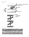
- a gas-permeable membrane 11 in a culturing device A on a gas-permeable membrane 11 in a culturing device A, 2 partition walls 11', which are similarly composed of gas-permeable membranes, are placed, and hepatocytes are cultured in the space surrounded by the gas-permeable membrane 11 and the 2 partition walls 11' .
- the gas-permeable membrane 11 has a collagen coating layer 12, and hepatocytes 13 are allowed to adhere to the layer and embedded in a collagen matrix 15.
- the cells are cultured by supplying oxygen using an oxygen-supplying device which is not shown placed outside the gas-permeable membrane 11 and the 2 partition walls 11', and adding a culture medium 16.
- an oxygen-supplying device which is not shown placed outside the gas-permeable membrane 11 and the 2 partition walls 11', and adding a culture medium 16.
- a bile canaliculus 14 is formed on the surface where the hepatocytes 13 are in contact with each other.
- the size of this partition wall is not restricted as long as adherence of hepatocytes to each other can be prevented, and, more particularly, the size is preferably not less than 1 ⁇ m.
- Fig. 18 an example of the method for culturing hepatocytes which forms a bile canaliculus in a groove or hollow will now be described.
- the extracellular matrix shown in Fig. 18(A) such as a collagen gel on which a groove is formed is placed.
- a collagen solution at a concentration of 0.1 to 30 mg/mL, preferably 0.3 to 3 mg/mL, more preferably about 2.0 mg/mL
- a PDMS mold having a protruded portion is placed thereon and left to stand until gelation, to prepare a collagen gel having a recess.
- the width of the formed groove is preferably not less than 20 ⁇ m and not more than 70 ⁇ m, more preferably not less than 30 ⁇ m and not more than 50 ⁇ m, furthermore preferably about 30 ⁇ m; the height is not preferably less than 10 ⁇ m and not more than 1 mm, more preferably not less than 50 ⁇ m and not more than 500 ⁇ m, furthermore preferably about 100 ⁇ m; and the length may be changed as appropriate for use.
- the material of the mold having a protruded portion is not restricted as long as the mold does not destroy the shape of the collagen gel, and preferred examples of the material include PDMS in view of the weight and ease of handling.
- hepatocytes suspended in a culture medium are seeded on the gel having a recess prepared as described above, and the cells are washed several times with the culture medium, to arrange the hepatocytes only in the recess, followed by culture of the cells.
- a collagen solution before gelation (at a concentration of 0.1 to 30 mg/mL, preferably 0.3 to 3 mg/mL, more preferably about 2.0 mg/mL) or a Matrigel solution (at a concentration of 5 to 5000 ⁇ g/mL, preferably 50 to 500 ⁇ g/mL, more preferably about 150 ⁇ g/mL) is deposited on the cells, and culture is carried out for additional 2 to 9 days. Formation of a bile canaliculus can be observed from Day 2, but a better bile canaliculus can be prepared by long-term culture. An optimum state for an evaluation test of a drug, or the like may be appropriately selected.
- the gas-permeable membrane may form a tubular passage having a cylindrical shape, cubic shape or the like. That is, the culture method of the present invention also includes an embodiment wherein the gas-permeable membrane is placed into a tubular shape with the collagen-coated side facing inward, and hepatocytes embedded in an extracellular matrix are cultured inside the tubular space.
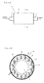
- Fig. 3A is a cross-sectional view taken along the a-a line in Fig 3A .
- the compound test device B of the present invention is a device having a main body section 2, a compound-supplying section 1 for supplying a compound to the main body section, and a collection section 3 for collecting the compound or a metabolite thereof from the main body section, which main body section 2 has: a tubular body 28 prepared by placing a gas-permeable membrane 21, whose surface is coated with collagen, into a tubular shape such that the collagen-coated side 22 faces inward; hepatocytes 23 adhering to the collagen-coated side 22 inside the tubular body 28; and an extracellular matrix 25 embedding the hepatocytes 23.
- the main body section 2 further has a passage 27 forming a space defined by a semipermeable membrane 26 in the axial direction of the tubular body 28. From this passage, the feed can be supplied to the hepatocytes embedded in the extracellular matrix through the semipermeable membrane 26.
- the material of the semipermeable membrane is not restricted as long as the material allows permeation of the compound and culture medium components, and examples of the material include porous membranes of regenerated cellulose (cellophane), acetylcellulose, collagen-coated cellulose, polyacrylonitrile, Teflon (registered trademark), porous silicon nitride, polyethylene terephthalate, polyester-polymer alloy and polysulfone.
- the polarity can be induced to form a bile canaliculus, by placing a semipermeable membrane composed of such a material such that the semipermeable membrane covers the hepatocytes, without separately adding an extracellular matrix.
- a culture medium for culturing and maintaining hepatocytes is also supplied from the compound-supplying section 1, and, when the culture medium passes through the passage 27, culture medium components are supplied to the hepatocytes via the semipermeable membrane 26.
- the culture medium-supplying section for supplying the culture medium may be provided separately from the compound-supplying section.
- the shape of the passage 27 is not restricted to a cylindrical shape, and may be another shape, including a branched shape. Further, the diameter of the passage is not restricted. A plurality of passages may be provided, and a passage through which the culture medium passes and a passage through which the compound passes may be separately provided. Further, the passage 27 and the semipermeable membrane 26 are not indispensable, and the whole content of the tubular body formed by the gas-permeable membrane may be hepatocytes and an extracellular matrix embedding the hepatocytes. In such a case, the culture medium and the compound may be allowed to pass through the extracellular matrix enclosed inside the tubular body.
- hepatocytes 23 are arranged along the inner wall (collagen-coated side 22) of the gas-permeable membrane 21.
- an extracellular matrix 25 is provided, and, further inside the extracellular matrix, a space (passage 27) separated by a semipermeable membrane 26, which is to be used for addition of a culture medium and a compound, is provided.
- Culturing of the hepatocytes with supply of oxygen from the outside of the gas-permeable membrane 21 causes induction of bile canaliculi 24 by the extracellular matrix. Since the phase in the outside of the gas-permeable membrane 21 is a gas phase, the concentration of oxygen that is in contact with the hepatocytes can be controlled.
- a metabolic property of a compound can be evaluated. That is, by supplying a test compound from the culture medium-supplying section 1 into the passage 27, hepatocytes are exposed to the compound via the semipermeable membrane 26. The compound metabolized by the hepatocytes are collected in the collection section 3 and analyzed.
- the collection section to collect the compound or a metabolite thereof is not restricted as long as the section is a member with which a liquid such as a culture medium containing a compound or a metabolite can be collected from the passage or the extracellular matrix, and the collection section may have: a tubular body connected to the main body section, which tubular body has been prepared by placing a gas-permeable membrane, whose surface is coated with collagen, into a tubular shape such that the collagen-coated side faces inward; hepatocytes adhering to the collagen-coated surface inside the tubular body; an extracellular matrix embedding the hepatocytes; and a passage formed by bile canaliculi of the hepatocytes.
- bile ducts prepared by the hepatocyte population cultured by the method of the present invention may be assembled to form a passage (cavity), through which a metabolite can flow.
- a hepatocyte culture device of such an embodiment of the present invention will now be described by reference to Fig. 4A, and Fig. 4B , which is a cross-sectional view taken along the b-b line in Fig. 4A .
- the cross-sectional view taken along the a-a line is the same as Fig. 3B .
- a main body section 102 is formed such that its diameter gradually decreases, and connected to a collection section 103.
- hepatocytes are introduced into the inside of a gas-permeable membrane such that a bile canaliculus cavity is formed at the center. From the formed bile canaliculi, passages composed of ultrafine tubes or bile duct epithelial cells are provided, by which metabolites excreted into the bile canaliculi are discharged without delay, and collected into a reservoir for analysis.
- a compound test device can be suitably used for a drug (compound) delivery test such as the one described later.
- the hepatocytes that formed the bile canaliculi prepared by the method of the present invention can be used for a drug delivery test or a high-throughput screening of a drug candidate substance.
- the drug delivery test include a test to study the amounts and the rates of incorporation of a drug into hepatocytes and the following excretion of the drug into bile.
- Further examples of the drug delivery test include a test to confirm whether a certain Compound A inhibits or promotes delivery of Compound B.
- Examples of the method of the drug delivery test include the method described in a Non-patent Document ( Liu X et al., Am J Physiol, 1999, vol. 277, pp. G12-21 ).
- the method of the high-throughput screening include the following method, wherein a very small amount of a compound can be analyzed using an ultrafine passage and a highly sensitive detector, which analysis is automatically or semi-automatically carried out.
- the high-throughput screening is possible by constructing a micropassage device having very small compartments separated from each other or having ultrafine passages each of which may be a gas-permeable passage such as the one shown in Fig.
- compartments or passages contain cultured hepatocytes that are embedded in an extracellular matrix and forming bile canaliculi
- micropassage device has, in each compartment, an inlet for exposure of a test compound and an opening for collection of a metabolite that has been metabolized by the hepatocytes and excreted into the bile canaliculi.
- the inlet of the micropassage device is connected to a pump for collecting and sending a compound solution from a compound library, and the outlet is connected to a passage through which the metabolite is sent to a high-performance liquid chromatography or LC/MS/MS to obtain results of quantification or composition analysis of the metabolite, which pump and passage may be operated by a computer or the like.
- the method was carried out according to the method described in a Non-patent Document ( M. Nishikawa et al. Biotechnology and Bioengineering, 2008, vol.99, pp.1472-1481 ).
- the base compound and the curing agent of Silpot 184 manufactured by Dow Corning Toray Co., Ltd.
- the base compound and the curing agent of Silpot 184 were mixed together at a ratio of 10:2 to prepare a mixture, and 30 g of this mixture was thinly spread on a plastic container having a size of 258 mm ⁇ 174 mm ⁇ 45mm, followed by curing the mixture at 80°C for 2 hours to prepare a PDMS membrane having a thickness of 1 mm.
- the ratio between the base compound and the curing agent is not restricted to 10:2, and the ratio is usually 10:1 to 10:2 in the preparation.
- the PDMS membrane was sandwiched between a polycarbonate frame having 24 holes and an SUS plate having a thickness of 1.5 mm having holes at the same positions as those on the frame, and the frame and plate were immobilized with screws, to prepare a PDMS membrane 24-well culturing device.
- the device was subjected to oxygen plasma treatment (for 5 seconds), and 1% acetic acid, 2% aminosilane (manufactured by Shin-Etsu Silicone) was added to each well to allow reaction to proceed at room temperature for 45 minutes, followed by heating of the reaction solution at 80°C for 90 minutes.
- the collagen gel sandwich method was carried out according to Non-patent Document 1 ( LeCluyse et al., Am J Physiol, 1994, vol. 266, pp. 1764-1774 ). Hepatic parenchymal cells were plated on the above prepared culturing device at 2 ⁇ 10 5 cells/well, and the culture medium was replaced 4 hours later. Twenty four hours after the plating, 20 ⁇ L of a 1.7 mg/ml collagen solution (manufactured by Becton, Dickinson and Company) was deposited on the cells, and gelation was allowed to proceed at 37°C for 1 hour, followed by adding 500 ⁇ L of a serum-free medium and culturing the cells at 37°C at 5% CO 2 . The culture medium was replaced once every 2 days.
- Non-patent Document 1 LeCluyse et al., Am J Physiol, 1994, vol. 266, pp. 1764-1774 .
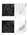
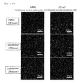
- Hepatic cells have a property that, when 5-(and-6)-carboxy-2',7'-dichlorofluorescein (CDCF) was administered, they incorporate this substance into the cytoplasm and metabolize the substance to convert the substance to a fluorescent substance fluorescein, followed by excreting the fluorescein to into a bile canaliculus. Using this property, the process of formation of bile canaliculi and the area of the bile canaliculi were investigated. Fluorescein diacetate was added to the culture medium at a concentration of 5 ⁇ M, and the resulting mixture was left to stand at 37°C for 15 minutes followed by washing the cells with a cooled culture medium.
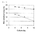
- Fluorescence of the fluorescein was observed under the microscope equipped with a fluorescence observing device. As shown in Fig. 5 , the hepatocytes cultured on the PDMS membrane began to be stained on Day 2 with the fluorescent dye at their tubular structures. This indicates that bile canaliculi were formed.
- the hepatocytes cultured on a conventional polystyrene culturing device (trade name, BioCoat Collagen I-coated 24-well; manufactured by Becton, Dickinson and Company) hardly showed tubular structures on Day 2, and, since the cells did not have the excretion activity, most of the cells were stained in their inside.
- Comparison of the area of bile canaliculi with time showed that, from Day 2 to Day 10, the area of formation of bile canaliculi was larger in the case of culture on the PDMS membrane than in the case of culture on the conventional polystyrene.
- a 24-well culturing device having a PDMS membrane to which collagen is covalently bound was prepared in the same manner as in Example 1.
- a 24-well culturing device having a PDMS membrane to which collagen was bound by adsorption was prepared by adding a small quantity of a 1.7 mg/mL collagen solution (manufactured by Becton, Dickinson and Company) to each well of the PDMS membrane 24-well culturing device subjected to the aminosilane treatment step and then to the oxygen plasma treatment in Example 1, to cover the well, and leaving the resultant to stand for 18 hours at room temperature, followed by washing the resultant with PBS. Both of these devices were used on the same day in the following experiment.
- a collagen gel sandwich hepatocyte culture was prepared.
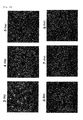
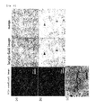
- formation of bile canaliculi was studied using fluorescein diacetate (Fig. 8 )
- formation of bile canaliculi was found on Day 4 of the culture (3 days after deposition of the collagen gel) both for the culturing device to which collagen was bound by adsorption (a) and for the culturing device to which collagen was covalently bound (b).
- detachment of the cells was more remarkable in the culturing device to which collagen was bound by adsorption (c), as compared to the culturing device to which collagen was covalently bound (d), and the bile canaliculi have disappeared in the former case.
- covalently binding collagen preparation of hepatocytes forming bile canaliculi more stably for a longer period is possible.
- Example 2 This study was carried out in the same manner as in Example 1 except for the extracellular matrix component to be deposited.
- the extracellular matrix component On the culturing device prepared as described above, 2 ⁇ 10 5 cells/well of hepatic parenchymal cells were seeded, and the culture medium was replaced 4 hours later. Twenty four hours after the plating, 20 ⁇ L of a 1.7 mg/ml collagen solution (manufactured by Becton, Dickinson and Company) was deposited on the cells, and gelation was allowed to proceed at 37°C for 1 hour, to provide a collagen gel sandwich group.
- a 1.7 mg/ml collagen solution manufactured by Becton, Dickinson and Company
- Matrigel manufactured by Becton, Dickinson and Company 50-fold diluted (corresponds to a concentration of 150 ⁇ g/mL) with a serum-free medium was added to the cells to provide a Matrigel sandwich group. Cells to which none of these was added were provided as an untreated group.
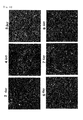
- bile canaliculi When formation of bile canaliculi was studied using fluorescein diacetate ( FIG. 9 ), formation of bile canaliculi was found on Day 4 of the culture (3 days after deposition of the extracellular matrix component) both for the collagen gel sandwich group (a) and for the Matrigel sandwich group (b), which bile canaliculi were maintained until Day 7 of the culture. In the untreated group, no bile canaliculus was formed at all, and remarkable detachment of the cells was observed from Day 3 of the culture, and most of the cells were detached by Day 5 (c). From these results, it was revealed that bile canaliculi can be formed on a PDMS membrane either by deposition of collagen or by deposition of Matrigel.
- the activity of a drug transporter was measured by incorporation of 5-(and-6)-carboxy-2',7'-dichlorofluorescein (CDCF) as follows according to a Non-patent Document ( Liu X et al., Am J Physiol, 1999, vol.277, pp.12-21 ). Further, hepatocytes cultured in a conventional polystyrene culturing device were used for comparison.
- Ca/Mg(+) HBSS was prepared before use by mixing 50 mL of HBSS (Invitrogen, 14175-079), 500 ⁇ L of 14 g/L CaCl 2 and 500 ⁇ L of 10 g/L MgCl 2 /6H 2 O together.
- HBSS Invitrogen, 14175-079
- 500 ⁇ L of 14 g/L CaCl 2 500 ⁇ L of 10 g/L MgCl 2 /6H 2 O together.
- a 5 ⁇ M CDCF solution was prepared before use by using 1 mM CDCF (in dimethyl sulfoxide: Molecular Probes, C-369) and Ca/Mg(+) HBSS, and the prepared solution was incubated in a water bath at 37°C.
- Ca/Mg(-) HBSS was prepared by adding 500 ⁇ L of 100 mM EGTA to 50 mL of HBSS (Invitrogen, 14175-079). Further, 0.5% Triton X-100/PBS was prepared by adding Triton X-100 to PBS buffer at a concentration of 0.5%.
- Two hepatocyte cultures were prepared 4 days after the beginning of the culture (3 days after the deposition of the gel). Subsequently, each culture was separately washed twice with 0.5 mL of warm Ca/Mg(+) HBSS or warm Ca/Mg(-) HBSS buffer. Thereafter, each culture was separately left to stand in 0.5 mL of warm Ca/Mg(+) HBSS or warm Ca/Mg(-) HBSS buffer at 37°C for 10 minutes, and the liquid was then removed. Subsequently, 0.5 mL of warm Ca/Mg(+) HBSS buffer supplemented with 5 ⁇ M CDCF was added to the both cultures, and the resultants were incubated for 5 minutes, followed by removal of the CDCF solution.
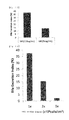
- the Bile Excretion Index was calculated according to the following equation based on the fluorescent brightness per protein amount (Accumulation).
- BEI Accumulation Ca 2 / Mg 2 + + - Accumulation Ca 2 / Mg 2 + - Accumulation Ca 2 + / Mg 2 + +
- the BEI value was about 40%
- the BEI value was about 20%. From the above results, it was revealed that use of a gas-permeable membrane enables hepatocytes to form a bile canaliculus structure having a higher MRP excretion activity as compared to use of a polystyrene substrate, and hence allows a highly sensitive evaluation of a compound (that is, accurate evaluation of a small amount of a compound).
- hepatocytes that were cultured on the PDMS membrane culturing device prepared in Example 1 and forming bile canaliculi localization of the MRP2 protein was investigated according to a conventional method by cell antibody staining. Further, hepatocytes cultured in a conventional polystyrene culturing device were used for comparison. Although the MRP2 protein was detected between cells in both the PDMS culturing device and the conventional polystyrene culturing device, expression of the MRP2 protein was observed in a broader area in the PDMS culturing device ( Fig. 11 ). Since MRP2 is a major transporter involved in excretion of bile, it can be assumed that the bile canaliculi prepared with the PDMS culturing device have a higher bile excretion activity.
- hepatocytes prepared from rats were seeded.
- the culture medium was replaced, 2 hours after the seeding, with William's Medium E (containing 5 ⁇ g/mL insulin, 5 ⁇ g/mL transferrin, 5 ⁇ g/mL selenious acid and 1 ⁇ M dexamethasone) supplemented with Matrigel.
- MG concentration of Matrigel
- 2 ⁇ 10 5 cells/well of the hepatocytes were seeded on the 24-well culturing device having a PDMS membrane to which collagen was bound by adsorption, and, when the culture medium was replaced 2 hours after the seeding, the concentration of Matrigel in the culture medium was set to 50 and 150 ⁇ g/mL.
- BEI was measured according to the method described in Example 4. As a result, as shown in Fig. 12 , a higher BEI was observed in the case of 150 ⁇ g/mL Matrigel than in the case of 50 ⁇ g/mL Matrigel.
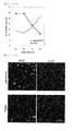
- the hepatocytes were plated on the 24-well culturing device having a PDMS membrane to which collagen was bound by adsorption at plating densities of 2 ⁇ 10 5 , 4 ⁇ 10 5 and 6 ⁇ 10 5 per well, and cultured for 48 hours with 150 ⁇ g/mL Matrigel, followed by measuring BEI according to the method described in Example 4.
- the highest BEI was observed at the plating density of 2 ⁇ 10 5 followed by the plating densities of 4 ⁇ 10 5 and 6 ⁇ 10 5 in that order.
- 14B shows fluorescence micrographs of portions where CDCF accumulated, which micrographs were taken 48 hours and 96 hours after the seeding of hepatocytes on the PDMS membrane 24-well culturing device and on the collagen-coated polystyrene 24-well plate. It can be confirmed also from these images that formation of active bile canaliculi occurred earlier in hepatocytes on the PDMS membrane than in hepatocytes cultured by the conventional method. Based on these results, when a PDMS membrane 24-well culturing device was used and Matrigel was deposited to induce the polarity, the polarity was formed by 24 hours after the plating, leading to appearance of functional bile canaliculi. It was proved that this occurs 72 hours earlier than achievement of an equivalent BEI value by a conventional method.
- hepatocytes forming bile canaliculi which were prepared in Example 6, localization of MRP2 and a basement membrane marker CD 147 were investigated according to a conventional method by cell antibody staining. Further, the results were compared with those obtained with hepatocytes cultured on a collagen-coated polystyrene 24-well plate. As shown in Fig. 15 , when the cells were cultured on the PDMS membrane, the MRP2 protein and the CD147 protein were detected between the cells after 48 hours of culture. On the other hand, on the conventional polystyrene, expression of CD 147 was observed, but localization of the MRP2 protein was hardly observed.
- the area of expression of the MRP2 protein was broader in hepatocytes on the PDMS membrane even in comparison with that in the cells already having the polarity on polystyrene after 120 hours of culture.
- the localization pattern of CD 147 was almost the same between the both. From these results, it was shown that hepatocytes cultured on a PDMS membrane cause localization of MRP2 molecules earlier than hepatocytes cultured on polystyrene, and the expression level of MRP2 is higher in the case of a PDMS membrane than in the case of polystyrene.
- Fig. 16A The ultrafine structure of the hepatocytes after 48 hours of culture, which were prepared in Example 6, was observed with a transmission electron microscope (JEM1400, manufactured by JEOL).
- JEM1400 transmitted electron microscope
- Fig. 16B shows a magnified photograph showing a portion having tight junctions
- Fig. 16C shows bile canaliculi having microvilli (MV) on the wall of the cavities.
- Hepatocytes prepared from rats were seeded on a 24-well culture plate Lumox (manufactured by In vitro systems and services), which has a culturing area where a gas-permeable fluorocarbon membrane is placed, at a density of 1.0 ⁇ 10 5 cells or 2.0 ⁇ 10 5 cells per well.
- the culture medium was replaced, 2 hours after the seeding, with William's Medium E (containing 5 ⁇ g/mL insulin, 5 ⁇ g/mL transferrin, 5 ⁇ g/mL selenious acid and 1 ⁇ M dexamethasone) supplemented with 150 ⁇ g/mL Matrigel.
- William's Medium E containing 5 ⁇ g/mL insulin, 5 ⁇ g/mL transferrin, 5 ⁇ g/mL selenious acid and 1 ⁇ M dexamethasone
- BEI was measured 48 hours after the seeding of hepatocytes, according to the method described in Example 4, to analyze the extent of polarity formation.
- As a control for comparison cells cultured on a collagen-coated polystyrene 24-well plate were used. As shown in Fig. 17 , a higher BEI was observed for Lumox as compared to the polystyrene (PS) 24-well plate. A higher BEI was given at the cell plating density of 1.0 ⁇ 10 5 than at the plating density of 2.0 ⁇ 10 5
- a PDMS mold having a protruded portion having a width of 30 ⁇ m, height of 100 ⁇ m and length of 10 mm was placed on a collagen solution (at a concentration of 2.1 mg/mL) before gelation, and left to stand at 37°C for 60 minutes to allow gelation, to prepare a collagen gel having a recess.
- a collagen solution at a concentration of 2.1 mg/mL
- rat hepatocytes suspended in a culture medium were plated on the gel having the prepared recess, and the cells were washed twice with the culture medium, to arrange the hepatocytes only in the recess, followed by culture of the cells.
- a collagen solution (at a concentration of 2.1 mg/mL) was deposited on the cells, and additional culture was then carried out for 9 days.
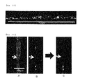
- a photograph of the thus arranged hepatocytes on Day 9 is shown in Fig. 19 . It appeared that the cells were forming continuous bile canaliculi.
- CDCF was added to the culture medium in the same manner as described in Example 1 to allow accumulation of fluorescein as a metabolite in the above prepared continuous bile canaliculi, and fluorescence from the fluorescein was observed.
- formation of bile canaliculi could be observed in a part of the cells, and, on Day 6, it could be observed that those bile canaliculi were continuous.
- On Day 9 it could be observed that the bile canaliculi continued even longer.
- a result obtained on Day 9 is shown in Fig. 20 . Since continuous fluorescence was observed, it could be confirmed that a continuous bile canaliculus having a length of not less than 1 mm can be prepared.
- fluorescein was allowed to accumulate in a prepared continuous bile canaliculus, and one end of the bile canaliculus ( Fig. 21A ) was opened with a thin glass tube. As a result, it was revealed that fluorescein that had accumulated in the bile canaliculus before the opening ( Fig. 21B ) disappeared about 60 seconds after the opening ( Fig. 21C ). By this, the continuity of the bile canaliculuscould be demonstrated, and possible use of the bile canaliculus for analysis of bile acid by continuous collection was also demonstrated. Further, in the above experiment, the results were not influenced by whether or not the collagen coating treatment of the fluorocarbon oxygen-permeable membrane was carried out.
- Cultured hepatocytes obtained by the culture method of the present invention can be used for, for example, an assay using hepatocytes for screening of a candidate compound for a pharmaceutical agent.
- the method contributes to improvement of the accuracy and efficiency of analysis of incorporation, metabolism and excretion of a candidate compound for a pharmaceutical agent in hepatocytes, leading to a highly efficient drug discovery process.
Landscapes
- Health & Medical Sciences (AREA)
- Life Sciences & Earth Sciences (AREA)
- Engineering & Computer Science (AREA)
- Chemical & Material Sciences (AREA)
- Bioinformatics & Cheminformatics (AREA)
- Biomedical Technology (AREA)
- Organic Chemistry (AREA)
- Wood Science & Technology (AREA)
- Zoology (AREA)
- Biotechnology (AREA)
- Microbiology (AREA)
- Biochemistry (AREA)
- General Health & Medical Sciences (AREA)
- Genetics & Genomics (AREA)
- Immunology (AREA)
- General Engineering & Computer Science (AREA)
- Sustainable Development (AREA)
- Cell Biology (AREA)
- Hematology (AREA)
- Analytical Chemistry (AREA)
- Clinical Laboratory Science (AREA)
- Urology & Nephrology (AREA)
- Molecular Biology (AREA)
- Toxicology (AREA)
- Tropical Medicine & Parasitology (AREA)
- Gastroenterology & Hepatology (AREA)
- Food Science & Technology (AREA)
- Medicinal Chemistry (AREA)
- Physics & Mathematics (AREA)
- General Physics & Mathematics (AREA)
- Pathology (AREA)
- Apparatus Associated With Microorganisms And Enzymes (AREA)
- Micro-Organisms Or Cultivation Processes Thereof (AREA)
- Measuring Or Testing Involving Enzymes Or Micro-Organisms (AREA)
Applications Claiming Priority (3)
| Application Number | Priority Date | Filing Date | Title |
|---|---|---|---|
| JP2009195869 | 2009-08-26 | ||
| JP2010073486 | 2010-03-26 | ||
| PCT/JP2010/062707 WO2011024592A1 (ja) | 2009-08-26 | 2010-07-28 | 肝細胞の培養方法 |
Publications (3)
| Publication Number | Publication Date |
|---|---|
| EP2471908A1 true EP2471908A1 (de) | 2012-07-04 |
| EP2471908A4 EP2471908A4 (de) | 2013-11-06 |
| EP2471908B1 EP2471908B1 (de) | 2016-10-05 |
Family
ID=43627702
Family Applications (1)
| Application Number | Title | Priority Date | Filing Date |
|---|---|---|---|
| EP10811643.5A Not-in-force EP2471908B1 (de) | 2009-08-26 | 2010-07-28 | Verfahren zur kultivierung von hepatozyten |
Country Status (4)
| Country | Link |
|---|---|
| US (1) | US8906644B2 (de) |
| EP (1) | EP2471908B1 (de) |
| JP (1) | JP5818001B2 (de) |
| WO (1) | WO2011024592A1 (de) |
Cited By (1)
| Publication number | Priority date | Publication date | Assignee | Title |
|---|---|---|---|---|
| CN105378053A (zh) * | 2013-11-29 | 2016-03-02 | 株式会社日立高新技术 | 成分分析装置、药效分析装置和分析方法 |
Families Citing this family (14)
| Publication number | Priority date | Publication date | Assignee | Title |
|---|---|---|---|---|
| JP2013215152A (ja) * | 2012-04-11 | 2013-10-24 | Vessel Inc | 3次元細胞培養プレートとその製造方法 |
| KR101448377B1 (ko) | 2013-03-19 | 2014-10-07 | 경희대학교 산학협력단 | 세포 기능 평가용 양방향 동시측정 시스템 |
| JP6176703B2 (ja) * | 2013-03-28 | 2017-08-09 | 株式会社Lsiメディエンス | 肝細胞培養用基板及び肝細胞培養方法 |
| JP2014223061A (ja) * | 2013-04-24 | 2014-12-04 | 株式会社Lsiメディエンス | 肝細胞培養方法 |
| US9512406B2 (en) | 2013-12-20 | 2016-12-06 | The J. David Gladstone Institute, a testamentary trust established under the Will of J. David Gladstone | Generating hepatocytes |
| WO2016147975A1 (ja) * | 2015-03-13 | 2016-09-22 | 国立研究開発法人医薬基盤・健康・栄養研究所 | 小腸上皮様細胞 |
| US20180120298A1 (en) * | 2015-03-27 | 2018-05-03 | National Agriculture and Food Reserach Organization | Hepatocyte culture device capable of enhancing accumulation and excretion of hepatic metabolite in and into bile canaliculus-like structure, and method for evaluating candidate compound sensitive to excretion into bile or blood and hepatic metabolite of said candidate compound using said hepatocyte culture device |
| JP6282362B2 (ja) * | 2017-01-31 | 2018-02-21 | 株式会社日立ハイテクノロジーズ | 成分分析装置、薬効分析装置、及び分析方法 |
| JP2019024399A (ja) * | 2017-07-28 | 2019-02-21 | 株式会社日立製作所 | 酸素供給機構 |
| JP6582070B2 (ja) * | 2018-01-22 | 2019-09-25 | 株式会社日立ハイテクノロジーズ | 分析方法 |
| JP7136509B2 (ja) * | 2019-12-27 | 2022-09-13 | 学校法人高崎健康福祉大学 | 肝細胞培養膜、それを備えた薬物輸送能評価キット、及び薬物輸送能評価方法 |
| WO2022158609A1 (ja) * | 2021-01-25 | 2022-07-28 | ニッタ株式会社 | 細胞培養容器 |
| JP7348997B1 (ja) | 2022-07-27 | 2023-09-21 | 浜松ホトニクス株式会社 | 毛細胆管領域の特定又は評価の方法、装置及びプログラム |
| JP2024057646A (ja) * | 2022-10-13 | 2024-04-25 | 国立大学法人 東京大学 | デバイス、システム、方法、および薬剤評価方法 |
Family Cites Families (6)
| Publication number | Priority date | Publication date | Assignee | Title |
|---|---|---|---|---|
| EP1200110B1 (de) * | 1999-07-22 | 2014-04-23 | Organogenesis Inc. | In vivo induktion zur erhöhten funktion isolierter hepatozyten |
| JPWO2005047496A1 (ja) | 2003-11-14 | 2007-05-31 | 学校法人慶應義塾 | 細胞培養法、細胞の三次元培養法、三次元組織、人工臓器、及び組織移植方法 |
| US7393687B2 (en) * | 2004-07-16 | 2008-07-01 | William Marsh Rice University | Biomimetic 3-dimensional scaffold with metabolic stream separation for bioartificial liver device |
| JP2006254722A (ja) * | 2005-03-15 | 2006-09-28 | Toray Ind Inc | 細胞足場材料 |
| CN101223268A (zh) | 2005-05-18 | 2008-07-16 | 康奈尔研究基金会(有限公司) | 具有生物屏障的基于药代动力学的培养系统 |
| JP4863655B2 (ja) * | 2005-06-17 | 2012-01-25 | 国立大学法人 東京医科歯科大学 | 細胞含有シート |
-
2010
- 2010-07-28 US US13/392,226 patent/US8906644B2/en not_active Expired - Fee Related
- 2010-07-28 WO PCT/JP2010/062707 patent/WO2011024592A1/ja not_active Ceased
- 2010-07-28 JP JP2011528714A patent/JP5818001B2/ja not_active Expired - Fee Related
- 2010-07-28 EP EP10811643.5A patent/EP2471908B1/de not_active Not-in-force
Cited By (5)
| Publication number | Priority date | Publication date | Assignee | Title |
|---|---|---|---|---|
| CN105378053A (zh) * | 2013-11-29 | 2016-03-02 | 株式会社日立高新技术 | 成分分析装置、药效分析装置和分析方法 |
| CN105378053B (zh) * | 2013-11-29 | 2017-10-20 | 株式会社日立高新技术 | 成分分析装置、药效分析装置和分析方法 |
| US9880153B2 (en) | 2013-11-29 | 2018-01-30 | Hitachi High-Technologies Corporation | Componential analyzer, drug efficacy analyzer, and analysis method |
| US10684276B2 (en) | 2013-11-29 | 2020-06-16 | Hitachi High-Tech Corporation | Componential analyzer, drug efficacy analyzer, and analysis method |
| US11789013B2 (en) | 2013-11-29 | 2023-10-17 | Hitachi High-Tech Corporation | Componential analyzer, drug efficacy analyzer, and analysis method |
Also Published As
| Publication number | Publication date |
|---|---|
| EP2471908A4 (de) | 2013-11-06 |
| EP2471908B1 (de) | 2016-10-05 |
| JP5818001B2 (ja) | 2015-11-18 |
| US20120183989A1 (en) | 2012-07-19 |
| WO2011024592A1 (ja) | 2011-03-03 |
| US8906644B2 (en) | 2014-12-09 |
| JPWO2011024592A1 (ja) | 2013-01-24 |
Similar Documents
| Publication | Publication Date | Title |
|---|---|---|
| EP2471908B1 (de) | Verfahren zur kultivierung von hepatozyten | |
| Gijzen et al. | Culture and analysis of kidney tubuloids and perfused tubuloid cells-on-a-chip | |
| EP3383413B1 (de) | Vorrichtungen und verfahren zur simulation einer funktion eines lebergewebes | |
| Nieskens et al. | Kidney-on-a-chip technology for renal proximal tubule tissue reconstruction | |
| Soldatow et al. | In vitro models for liver toxicity testing | |
| KR102621919B1 (ko) | iPSC-유래 세포의 장기 등가물로의 분화를 확립하는 신규한 다중 장기 칩 | |
| US10633623B2 (en) | Artificial placenta and methods of preparation | |
| Ahmed et al. | Human liver microtissue spheroids in hollow fiber membrane bioreactor | |
| Beißner et al. | Organ on chip | |
| EP2383332B1 (de) | Verfahren zur kultivierung von hepatozyten tierischen ursprungs | |
| Aufderheide et al. | A new computer-controlled air–liquid interface cultivation system for the generation of differentiated cell cultures of the airway epithelium | |
| Vis et al. | Osteogenesis and osteoclastogenesis on a chip: Engineering a self-assembling 3D coculture | |
| Van Ness et al. | Microphysiological systems in absorption, distribution, metabolism, and elimination sciences | |
| Kim et al. | Effect of shear stress on the proximal tubule-on-a-chip for multi-organ microphysiological system | |
| US20210340572A1 (en) | High-content imaging of microfluidic devices | |
| Drabbe et al. | Retinal organoid chip: engineering a physiomimetic oxygen gradient for optimizing long term culture of human retinal organoids | |
| JP2013226112A (ja) | 肝細胞の培養方法 | |
| JP2014223061A (ja) | 肝細胞培養方法 | |
| Boutaud et al. | 2D and 3D human induced pluripotent stem cell-based models to dissect primary cilium involvement during neocortical development | |
| Mueller et al. | 3D hepatic in vitro models as tools for toxicity studies | |
| Kaisar et al. | In vitro BBB models: Working with static platforms and microfluidic systems | |
| US20180044640A1 (en) | Contractile cellular construct for cell culture | |
| Comley | Progress made in applying 3D cell culture technologies | |
| Henkens et al. | Rat hepatocyte cultures: conventional monolayer and cocultures with rat liver epithelial cells | |
| JP6176703B2 (ja) | 肝細胞培養用基板及び肝細胞培養方法 |
Legal Events
| Date | Code | Title | Description |
|---|---|---|---|
| PUAI | Public reference made under article 153(3) epc to a published international application that has entered the european phase |
Free format text: ORIGINAL CODE: 0009012 |
|
| 17P | Request for examination filed |
Effective date: 20120322 |
|
| AK | Designated contracting states |
Kind code of ref document: A1 Designated state(s): AL AT BE BG CH CY CZ DE DK EE ES FI FR GB GR HR HU IE IS IT LI LT LU LV MC MK MT NL NO PL PT RO SE SI SK SM TR |
|
| DAX | Request for extension of the european patent (deleted) | ||
| REG | Reference to a national code |
Ref country code: DE Ref legal event code: R079 Ref document number: 602010037023 Country of ref document: DE Free format text: PREVIOUS MAIN CLASS: C12N0005077000 Ipc: C12N0005071000 |
|
| RIC1 | Information provided on ipc code assigned before grant |
Ipc: C12Q 1/02 20060101ALI20130926BHEP Ipc: C12N 5/071 20100101AFI20130926BHEP Ipc: C12M 1/34 20060101ALI20130926BHEP |
|
| A4 | Supplementary search report drawn up and despatched |
Effective date: 20131004 |
|
| RAP1 | Party data changed (applicant data changed or rights of an application transferred) |
Owner name: THE UNIVERSITY OF TOKYO Owner name: LSI MEDIENCE CORPORATION |
|
| GRAP | Despatch of communication of intention to grant a patent |
Free format text: ORIGINAL CODE: EPIDOSNIGR1 |
|
| INTG | Intention to grant announced |
Effective date: 20160422 |
|
| GRAS | Grant fee paid |
Free format text: ORIGINAL CODE: EPIDOSNIGR3 |
|
| GRAA | (expected) grant |
Free format text: ORIGINAL CODE: 0009210 |
|
| AK | Designated contracting states |
Kind code of ref document: B1 Designated state(s): AL AT BE BG CH CY CZ DE DK EE ES FI FR GB GR HR HU IE IS IT LI LT LU LV MC MK MT NL NO PL PT RO SE SI SK SM TR |
|
| REG | Reference to a national code |
Ref country code: GB Ref legal event code: FG4D |
|
| RIN1 | Information on inventor provided before grant (corrected) |
Inventor name: TSUDA, YUKIKO Inventor name: SAKAI, YASUYUKI Inventor name: FUJII, TERUO Inventor name: TAKEUCHI, SHOJI Inventor name: MATSUI, HITOSHI |
|
| REG | Reference to a national code |
Ref country code: CH Ref legal event code: EP |
|
| REG | Reference to a national code |
Ref country code: AT Ref legal event code: REF Ref document number: 834687 Country of ref document: AT Kind code of ref document: T Effective date: 20161015 |
|
| REG | Reference to a national code |
Ref country code: IE Ref legal event code: FG4D |
|
| REG | Reference to a national code |
Ref country code: DE Ref legal event code: R096 Ref document number: 602010037023 Country of ref document: DE |
|
| REG | Reference to a national code |
Ref country code: CH Ref legal event code: NV Representative=s name: AMMANN PATENTANWAELTE AG BERN, CH |
|
| REG | Reference to a national code |
Ref country code: NL Ref legal event code: MP Effective date: 20161005 |
|
| REG | Reference to a national code |
Ref country code: LT Ref legal event code: MG4D |
|
| PG25 | Lapsed in a contracting state [announced via postgrant information from national office to epo] |
Ref country code: LV Free format text: LAPSE BECAUSE OF FAILURE TO SUBMIT A TRANSLATION OF THE DESCRIPTION OR TO PAY THE FEE WITHIN THE PRESCRIBED TIME-LIMIT Effective date: 20161005 |
|
| REG | Reference to a national code |
Ref country code: AT Ref legal event code: MK05 Ref document number: 834687 Country of ref document: AT Kind code of ref document: T Effective date: 20161005 |
|
| PG25 | Lapsed in a contracting state [announced via postgrant information from national office to epo] |
Ref country code: LT Free format text: LAPSE BECAUSE OF FAILURE TO SUBMIT A TRANSLATION OF THE DESCRIPTION OR TO PAY THE FEE WITHIN THE PRESCRIBED TIME-LIMIT Effective date: 20161005 Ref country code: SE Free format text: LAPSE BECAUSE OF FAILURE TO SUBMIT A TRANSLATION OF THE DESCRIPTION OR TO PAY THE FEE WITHIN THE PRESCRIBED TIME-LIMIT Effective date: 20161005 Ref country code: NO Free format text: LAPSE BECAUSE OF FAILURE TO SUBMIT A TRANSLATION OF THE DESCRIPTION OR TO PAY THE FEE WITHIN THE PRESCRIBED TIME-LIMIT Effective date: 20170105 Ref country code: GR Free format text: LAPSE BECAUSE OF FAILURE TO SUBMIT A TRANSLATION OF THE DESCRIPTION OR TO PAY THE FEE WITHIN THE PRESCRIBED TIME-LIMIT Effective date: 20170106 |
|
| PG25 | Lapsed in a contracting state [announced via postgrant information from national office to epo] |
Ref country code: AT Free format text: LAPSE BECAUSE OF FAILURE TO SUBMIT A TRANSLATION OF THE DESCRIPTION OR TO PAY THE FEE WITHIN THE PRESCRIBED TIME-LIMIT Effective date: 20161005 Ref country code: BE Free format text: LAPSE BECAUSE OF FAILURE TO SUBMIT A TRANSLATION OF THE DESCRIPTION OR TO PAY THE FEE WITHIN THE PRESCRIBED TIME-LIMIT Effective date: 20161005 Ref country code: ES Free format text: LAPSE BECAUSE OF FAILURE TO SUBMIT A TRANSLATION OF THE DESCRIPTION OR TO PAY THE FEE WITHIN THE PRESCRIBED TIME-LIMIT Effective date: 20161005 Ref country code: HR Free format text: LAPSE BECAUSE OF FAILURE TO SUBMIT A TRANSLATION OF THE DESCRIPTION OR TO PAY THE FEE WITHIN THE PRESCRIBED TIME-LIMIT Effective date: 20161005 Ref country code: IS Free format text: LAPSE BECAUSE OF FAILURE TO SUBMIT A TRANSLATION OF THE DESCRIPTION OR TO PAY THE FEE WITHIN THE PRESCRIBED TIME-LIMIT Effective date: 20170205 Ref country code: NL Free format text: LAPSE BECAUSE OF FAILURE TO SUBMIT A TRANSLATION OF THE DESCRIPTION OR TO PAY THE FEE WITHIN THE PRESCRIBED TIME-LIMIT Effective date: 20161005 Ref country code: PT Free format text: LAPSE BECAUSE OF FAILURE TO SUBMIT A TRANSLATION OF THE DESCRIPTION OR TO PAY THE FEE WITHIN THE PRESCRIBED TIME-LIMIT Effective date: 20170206 Ref country code: FI Free format text: LAPSE BECAUSE OF FAILURE TO SUBMIT A TRANSLATION OF THE DESCRIPTION OR TO PAY THE FEE WITHIN THE PRESCRIBED TIME-LIMIT Effective date: 20161005 Ref country code: PL Free format text: LAPSE BECAUSE OF FAILURE TO SUBMIT A TRANSLATION OF THE DESCRIPTION OR TO PAY THE FEE WITHIN THE PRESCRIBED TIME-LIMIT Effective date: 20161005 |
|
| REG | Reference to a national code |
Ref country code: DE Ref legal event code: R097 Ref document number: 602010037023 Country of ref document: DE |
|
| REG | Reference to a national code |
Ref country code: FR Ref legal event code: PLFP Year of fee payment: 8 |
|
| PG25 | Lapsed in a contracting state [announced via postgrant information from national office to epo] |
Ref country code: DK Free format text: LAPSE BECAUSE OF FAILURE TO SUBMIT A TRANSLATION OF THE DESCRIPTION OR TO PAY THE FEE WITHIN THE PRESCRIBED TIME-LIMIT Effective date: 20161005 Ref country code: SK Free format text: LAPSE BECAUSE OF FAILURE TO SUBMIT A TRANSLATION OF THE DESCRIPTION OR TO PAY THE FEE WITHIN THE PRESCRIBED TIME-LIMIT Effective date: 20161005 Ref country code: RO Free format text: LAPSE BECAUSE OF FAILURE TO SUBMIT A TRANSLATION OF THE DESCRIPTION OR TO PAY THE FEE WITHIN THE PRESCRIBED TIME-LIMIT Effective date: 20161005 Ref country code: CZ Free format text: LAPSE BECAUSE OF FAILURE TO SUBMIT A TRANSLATION OF THE DESCRIPTION OR TO PAY THE FEE WITHIN THE PRESCRIBED TIME-LIMIT Effective date: 20161005 Ref country code: EE Free format text: LAPSE BECAUSE OF FAILURE TO SUBMIT A TRANSLATION OF THE DESCRIPTION OR TO PAY THE FEE WITHIN THE PRESCRIBED TIME-LIMIT Effective date: 20161005 |
|
| PGFP | Annual fee paid to national office [announced via postgrant information from national office to epo] |
Ref country code: GB Payment date: 20170619 Year of fee payment: 8 |
|
| PLBE | No opposition filed within time limit |
Free format text: ORIGINAL CODE: 0009261 |
|
| STAA | Information on the status of an ep patent application or granted ep patent |
Free format text: STATUS: NO OPPOSITION FILED WITHIN TIME LIMIT |
|
| PG25 | Lapsed in a contracting state [announced via postgrant information from national office to epo] |
Ref country code: SM Free format text: LAPSE BECAUSE OF FAILURE TO SUBMIT A TRANSLATION OF THE DESCRIPTION OR TO PAY THE FEE WITHIN THE PRESCRIBED TIME-LIMIT Effective date: 20161005 Ref country code: IT Free format text: LAPSE BECAUSE OF FAILURE TO SUBMIT A TRANSLATION OF THE DESCRIPTION OR TO PAY THE FEE WITHIN THE PRESCRIBED TIME-LIMIT Effective date: 20161005 Ref country code: BG Free format text: LAPSE BECAUSE OF FAILURE TO SUBMIT A TRANSLATION OF THE DESCRIPTION OR TO PAY THE FEE WITHIN THE PRESCRIBED TIME-LIMIT Effective date: 20170105 |
|
| 26N | No opposition filed |
Effective date: 20170706 |
|
| PGFP | Annual fee paid to national office [announced via postgrant information from national office to epo] |
Ref country code: CH Payment date: 20170719 Year of fee payment: 8 Ref country code: DE Payment date: 20170724 Year of fee payment: 8 Ref country code: FR Payment date: 20170724 Year of fee payment: 8 |
|
| PG25 | Lapsed in a contracting state [announced via postgrant information from national office to epo] |
Ref country code: SI Free format text: LAPSE BECAUSE OF FAILURE TO SUBMIT A TRANSLATION OF THE DESCRIPTION OR TO PAY THE FEE WITHIN THE PRESCRIBED TIME-LIMIT Effective date: 20161005 |
|
| REG | Reference to a national code |
Ref country code: IE Ref legal event code: MM4A |
|
| PG25 | Lapsed in a contracting state [announced via postgrant information from national office to epo] |
Ref country code: IE Free format text: LAPSE BECAUSE OF NON-PAYMENT OF DUE FEES Effective date: 20170728 |
|
| PG25 | Lapsed in a contracting state [announced via postgrant information from national office to epo] |
Ref country code: LU Free format text: LAPSE BECAUSE OF NON-PAYMENT OF DUE FEES Effective date: 20170728 |
|
| PG25 | Lapsed in a contracting state [announced via postgrant information from national office to epo] |
Ref country code: MT Free format text: LAPSE BECAUSE OF NON-PAYMENT OF DUE FEES Effective date: 20170728 |
|
| REG | Reference to a national code |
Ref country code: DE Ref legal event code: R119 Ref document number: 602010037023 Country of ref document: DE |
|
| REG | Reference to a national code |
Ref country code: CH Ref legal event code: PL |
|
| GBPC | Gb: european patent ceased through non-payment of renewal fee |
Effective date: 20180728 |
|
| PG25 | Lapsed in a contracting state [announced via postgrant information from national office to epo] |
Ref country code: FR Free format text: LAPSE BECAUSE OF NON-PAYMENT OF DUE FEES Effective date: 20180731 Ref country code: CH Free format text: LAPSE BECAUSE OF NON-PAYMENT OF DUE FEES Effective date: 20180731 Ref country code: LI Free format text: LAPSE BECAUSE OF NON-PAYMENT OF DUE FEES Effective date: 20180731 Ref country code: DE Free format text: LAPSE BECAUSE OF NON-PAYMENT OF DUE FEES Effective date: 20190201 Ref country code: GB Free format text: LAPSE BECAUSE OF NON-PAYMENT OF DUE FEES Effective date: 20180728 |
|
| PG25 | Lapsed in a contracting state [announced via postgrant information from national office to epo] |
Ref country code: HU Free format text: LAPSE BECAUSE OF FAILURE TO SUBMIT A TRANSLATION OF THE DESCRIPTION OR TO PAY THE FEE WITHIN THE PRESCRIBED TIME-LIMIT; INVALID AB INITIO Effective date: 20100728 Ref country code: MC Free format text: LAPSE BECAUSE OF FAILURE TO SUBMIT A TRANSLATION OF THE DESCRIPTION OR TO PAY THE FEE WITHIN THE PRESCRIBED TIME-LIMIT Effective date: 20161005 |
|
| PG25 | Lapsed in a contracting state [announced via postgrant information from national office to epo] |
Ref country code: CY Free format text: LAPSE BECAUSE OF NON-PAYMENT OF DUE FEES Effective date: 20161005 |
|
| PG25 | Lapsed in a contracting state [announced via postgrant information from national office to epo] |
Ref country code: MK Free format text: LAPSE BECAUSE OF FAILURE TO SUBMIT A TRANSLATION OF THE DESCRIPTION OR TO PAY THE FEE WITHIN THE PRESCRIBED TIME-LIMIT Effective date: 20161005 |
|
| PG25 | Lapsed in a contracting state [announced via postgrant information from national office to epo] |
Ref country code: TR Free format text: LAPSE BECAUSE OF FAILURE TO SUBMIT A TRANSLATION OF THE DESCRIPTION OR TO PAY THE FEE WITHIN THE PRESCRIBED TIME-LIMIT Effective date: 20161005 |
|
| PG25 | Lapsed in a contracting state [announced via postgrant information from national office to epo] |
Ref country code: AL Free format text: LAPSE BECAUSE OF FAILURE TO SUBMIT A TRANSLATION OF THE DESCRIPTION OR TO PAY THE FEE WITHIN THE PRESCRIBED TIME-LIMIT Effective date: 20161005 |