EP2412825B1 - Leukämiestammzellenmarker - Google Patents
Leukämiestammzellenmarker Download PDFInfo
- Publication number
- EP2412825B1 EP2412825B1 EP10756142.5A EP10756142A EP2412825B1 EP 2412825 B1 EP2412825 B1 EP 2412825B1 EP 10756142 A EP10756142 A EP 10756142A EP 2412825 B1 EP2412825 B1 EP 2412825B1
- Authority
- EP
- European Patent Office
- Prior art keywords
- genes
- dna
- artificial
- primer
- cells
- Prior art date
- Legal status (The legal status is an assumption and is not a legal conclusion. Google has not performed a legal analysis and makes no representation as to the accuracy of the status listed.)
- Not-in-force
Links
Images


Classifications
-
- A—HUMAN NECESSITIES
- A61—MEDICAL OR VETERINARY SCIENCE; HYGIENE
- A61K—PREPARATIONS FOR MEDICAL, DENTAL OR TOILETRY PURPOSES
- A61K31/00—Medicinal preparations containing organic active ingredients
- A61K31/70—Carbohydrates; Sugars; Derivatives thereof
- A61K31/7088—Compounds having three or more nucleosides or nucleotides
- A61K31/713—Double-stranded nucleic acids or oligonucleotides
-
- G—PHYSICS
- G01—MEASURING; TESTING
- G01N—INVESTIGATING OR ANALYSING MATERIALS BY DETERMINING THEIR CHEMICAL OR PHYSICAL PROPERTIES
- G01N33/00—Investigating or analysing materials by specific methods not covered by groups G01N1/00 - G01N31/00
- G01N33/48—Biological material, e.g. blood, urine; Haemocytometers
- G01N33/50—Chemical analysis of biological material, e.g. blood, urine; Testing involving biospecific ligand binding methods; Immunological testing
- G01N33/53—Immunoassay; Biospecific binding assay; Materials therefor
- G01N33/574—Immunoassay; Biospecific binding assay; Materials therefor for cancer
- G01N33/57407—Specifically defined cancers
- G01N33/57426—Specifically defined cancers leukemia
-
- A—HUMAN NECESSITIES
- A61—MEDICAL OR VETERINARY SCIENCE; HYGIENE
- A61K—PREPARATIONS FOR MEDICAL, DENTAL OR TOILETRY PURPOSES
- A61K35/00—Medicinal preparations containing materials or reaction products thereof with undetermined constitution
- A61K35/12—Materials from mammals; Compositions comprising non-specified tissues or cells; Compositions comprising non-embryonic stem cells; Genetically modified cells
- A61K35/28—Bone marrow; Haematopoietic stem cells; Mesenchymal stem cells of any origin, e.g. adipose-derived stem cells
-
- A—HUMAN NECESSITIES
- A61—MEDICAL OR VETERINARY SCIENCE; HYGIENE
- A61P—SPECIFIC THERAPEUTIC ACTIVITY OF CHEMICAL COMPOUNDS OR MEDICINAL PREPARATIONS
- A61P35/00—Antineoplastic agents
- A61P35/02—Antineoplastic agents specific for leukemia
-
- A—HUMAN NECESSITIES
- A61—MEDICAL OR VETERINARY SCIENCE; HYGIENE
- A61P—SPECIFIC THERAPEUTIC ACTIVITY OF CHEMICAL COMPOUNDS OR MEDICINAL PREPARATIONS
- A61P43/00—Drugs for specific purposes, not provided for in groups A61P1/00-A61P41/00
-
- C—CHEMISTRY; METALLURGY
- C12—BIOCHEMISTRY; BEER; SPIRITS; WINE; VINEGAR; MICROBIOLOGY; ENZYMOLOGY; MUTATION OR GENETIC ENGINEERING
- C12Q—MEASURING OR TESTING PROCESSES INVOLVING ENZYMES, NUCLEIC ACIDS OR MICROORGANISMS; COMPOSITIONS OR TEST PAPERS THEREFOR; PROCESSES OF PREPARING SUCH COMPOSITIONS; CONDITION-RESPONSIVE CONTROL IN MICROBIOLOGICAL OR ENZYMOLOGICAL PROCESSES
- C12Q1/00—Measuring or testing processes involving enzymes, nucleic acids or microorganisms; Compositions therefor; Processes of preparing such compositions
- C12Q1/68—Measuring or testing processes involving enzymes, nucleic acids or microorganisms; Compositions therefor; Processes of preparing such compositions involving nucleic acids
- C12Q1/6876—Nucleic acid products used in the analysis of nucleic acids, e.g. primers or probes
- C12Q1/6883—Nucleic acid products used in the analysis of nucleic acids, e.g. primers or probes for diseases caused by alterations of genetic material
- C12Q1/6886—Nucleic acid products used in the analysis of nucleic acids, e.g. primers or probes for diseases caused by alterations of genetic material for cancer
-
- C—CHEMISTRY; METALLURGY
- C12—BIOCHEMISTRY; BEER; SPIRITS; WINE; VINEGAR; MICROBIOLOGY; ENZYMOLOGY; MUTATION OR GENETIC ENGINEERING
- C12Q—MEASURING OR TESTING PROCESSES INVOLVING ENZYMES, NUCLEIC ACIDS OR MICROORGANISMS; COMPOSITIONS OR TEST PAPERS THEREFOR; PROCESSES OF PREPARING SUCH COMPOSITIONS; CONDITION-RESPONSIVE CONTROL IN MICROBIOLOGICAL OR ENZYMOLOGICAL PROCESSES
- C12Q2600/00—Oligonucleotides characterized by their use
- C12Q2600/158—Expression markers
Definitions
- the present invention relates to leukemic stem cell markers and the field of treatment of acute myeloid leukemia.
- AML Acute myeloid leukemia
- LSCs rare leukemic stem cells
- the present inventors have succeeded in the development of an animal model capable of reproducing features of human, rather than mouse, AML, particularly AML of individual patients, rather than a cell line, and permitting long-term assessment (Non-patent Document 5, Patent Application PCT/JP2008/068892 ).
- the present inventors further identified using a neonatal NOD/SCID/IL2rg KO mouse model, which is one of the most sensitive human stem cell assays, that CD34+CD38-AML cells meet all criteria for cancer stem cells recommended by the American Association for Cancer Research (Non-patent Document 6).
- CD34+CD38- AML cells self-renew, produce non-stem leukemia cells, and have the exclusive capability of causing AML in living organisms.
- AML stem cells are present predominantly in the endosteal region of the bone marrow; when human AML transplantation recipient mice were treated with chemotherapeutic agents, the great majority of chemotherapy-resistant AML cells were found in osteoblast niches.
- AML stem cells (not CD34+CD38+ and CD34-AML cells) are stationary and hence exhibit resistance to cell cycle-dependent chemotherapeutic agents.
- a problem to be solved is to find a molecular target that is specific for human leukemic stem cells (LSCs) and provide a therapeutic means that will lead to radical treatment of acute myeloid leukemia (AML) and the like.
- LSCs human leukemic stem cells
- AML acute myeloid leukemia
- the present inventors found sets of genes differentially expressed between LSCs and non-stem cells, and proposed the possibility that these genes serve as therapeutic targets for AML ( Ishikawa F. et al., Nature Biotechnol 25:1315-1321, 2007 and PCT/JP2008/068892 ), but were unable to rule out the possibility that the genes are at the same time differentially expressed in normal hematopoietic stem cells (HSCs) as well.
- HSCs normal hematopoietic stem cells
- the present inventors succeeded in developing a mouse model enabling reproduction of human AML (mice generated by transplanting a fraction containing leukemic stem cells derived from a human AML patient to NOD/SCID/IL2rg null mice), transplanting a small number of bone marrow cells derived from an AML patient, and reconstructing the pathology of AML in the animal model.
- the present inventors then prepared LSCs derived from an AML patient and those from an AML transplantation recipient mouse, as well as bone marrow samples and cord blood samples (HSCs are contained) derived from healthy donors, conducted a comprehensive analysis, and have developed the present invention.
- the present invention provides the following.
- the present invention has been developed as a result of succeeding in analyzing the comprehensive expression profiling of leukemic stem cells (LSCs) derived from human primary AML, and identifying LSC-specific targets for separating LSCs from HSCs. Therefore, the leukemic stem cell markers found in the present invention make it possible not only to distinguish between non-stem cells and LSCs, but also to distinguish between normal hematopoietic stem cells (HSCs) and LSCs, which have been thought to be difficult to distinguish from each other.
- HSCs normal hematopoietic stem cells
- a therapeutic agent that acts specifically on LSCs that are the source of onset or recurrence of AML can be provided.
- the presence or absence of LSCs in a collected biological sample or in a body can be determined with a leukemic stem cell marker found in the present invention as an index, whereby recurrences or the initial onset of acute myeloid leukemia can also be predicted.
- the initial onset of leukemia refers to a state in which leukemia has developed for the first time, or is likely to develop
- a recurrence of leukemia refers to a state in which leukemia has developed again, or is likely to develop, after treatment or remission of initial-onset leukemia.
- the tissue where leukemia recurs or is likely to recur is not limited to initial-onset tissue, and may be another tissue. Therefore, the concept of recurrence is understood to include infiltration and metastasis.
- Treatment of leukemia encompasses all treatments, including administration of anticancer agents, radiotherapy, and bone marrow transplantation.
- leukemic stem cells may be a CD34+ cell fraction derived from the bone marrow, with preference given to CD34+CD38- cell fraction.
- the crude substance containing LSC can be recovered from the bone marrow of a test subject or patient by a conventional method, cell fractions containing the LSC can be obtained by flow cytometry and the like using CD34 and CD38 cell surface marker molecules. Note that separation of LSC from HSC is difficult. Furthermore, it is also possible to further sort LSCs with another cell surface marker molecule selected from among leukemic stem cell markers found by the present invention, as an index.
- the present invention provides a test method for predicting the initial onset or a recurrence of acute myeloid leukemia.
- the test method of the present invention comprises,
- Leukemic stem cell marker genes are leukemic stem cell-specific markers sorted from a set of genes expressed differentially in the CD34+CD38- cell fraction than in the CD34+CD38+ cell fraction by the present inventors on the basis of their unique viewpoint, and comprise 2 to 218 genes selected from among the following leukemic stem cell marker genes (hereinafter sometimes simply abbreviated as "marker genes” or “markers”) (1).
- the marker genes (1) preferably consist of 3 or more, 5 or more, 10 or more, 15 or more, 20 or more, or 25 or more, genes.
- the marker genes used in a method of the invention are set out in the claims.
- cell membrane- or extracellularly-localized genes consisting of ADFP, ALOX5AP, AZU1, C3AR1, CACNB4, CALCRL, CCL4, CCL5, CD33, CD36, CD3D, CD86, CD9, CD93, CD96, CD97, CFD, CHI3L1, CLEC12A, CLECL1, COCH, CST7, CXCL1, DOK2, EMR2, FCER1G, FCGR2A, FUCA2, GPR109B, GPR160, GPR34, GPR84, HAVCR2, HBEGF, HCST, HGF, HLA-DOB, HOMER3, IFI30, IL13RA1, IL2RA, IL2RG, IL3RA, INHBA, ITGB2, LGALS1, LRG1, LY86, MAMDC2, MGAT4A, P2RY14, P2RY5, PLAUR, PPBP, PRG2, PRSS21, PTH2R, PTX3, REEP5, RNASE2, RXFP1, S
- the individual genes that constitute the aforementioned leukemic stem cell marker genes are publicly known, and the base sequences and amino acid sequences thereof are also known.
- symbol names, gene IDs, location chromosomes, characteristics and the like are shown in Table 1.
- IL2RA also called CD25 has the gene ID 3559, is located on chromosome 10, and encodes interleukin 2 receptor alpha.
- the IL2RA protein is a transmembranous receptor localized on the cell membrane.
- the marker genes (2) consist of 2 to 58 genes, more preferably consist of 3 or more, 5 or more, 10 or more, 15 or more, 20 or more, or 25 or more, genes.
- the following marker genes (3), out of the marker genes (2) be used as an index.
- the marker genes (3) are more preferable because normally 5 times or higher differential expression is observed in LSCs than in HSCs.
- the marker genes (3) consist of 2 to 35 genes, more preferably consist of 3 or more, 5 or more, 10 or more, 15 or more, 20 or more, or 25 or more, genes.
- cell membrane- or extracellularly-localized genes consisting of ADFP, ALOX5AP, CACNB4, CCL5, CD33, CD3D, CD93, CD97, CLEC12A, DOK2, FCER1G, FCGR2A, FUCA2, GPR34, GPR84, HCST, HGF, HOMER3, IL2RA, IL2RG, IL3RA, ITGB2, LGALS1, LRG1, LY86, MGAT4A, P2RY5, PRSS21, PTH2R, RNASE2, SLC43A3, SUCNR1, TIMP1, TNF, TNFRSF4, TNFSF13B, TYROBP and VNN1; cell cycle-related genes consisting of ZWINT, NEK6 and TXNL4B; an apoptosis-related gene consisting of BIK; signaling-related genes consisting of AK5, ARHGAP18, FYB, HCK, LPXN, PDE9A, PDK1, PRKCD, RAB20, R
- cell membrane- or extracellularly-localized genes consisting of ALOX5AP, CACNB4, CCL5, CD33, CD3D, CD93, CD97, CLEC12A, DOK2, FCGR2A, GPR84, HCST, HOMER3, ITGB2, LGALS1, LRG1, PTH2R, RNASE2, TNF, TNFSF13B, TYROBP and VNN1; a cell cycle-related gene consisting of NEK6; an apoptosis-related gene consisting of BIK; signaling-related genes consisting of AK5, FYB, HCK, LPXN, PDE9A, PDK1, PRKCD and RAB20; a transcription factor gene consisting of WT1; and other genes consisting of CTSC and NCF4.
- the subject in the test method of the present invention is not particularly limited, as far as it is a mammal, including a human, a human suspected of suffering the initial onset or a recurrence of leukemia is preferred.
- the biological sample to be measured by the test method of the present invention is not particularly limited, as far as it can be collected from a mammal, preferably from a human; examples include humoral samples such as blood, bone marrow fluid, and lymph fluid, and solid samples such as lymph nodes, blood vessels, bone marrow, brain, spleen, and skin.
- RNA can be isolated from the biological sample by a conventional method. Ordinary methods for RNA extraction are well known in the relevant technical field, and are disclosed in standard textbooks of molecular biology, including Ausubel et al., Current Protocols of Molecular Biology, John Wiley and Sons (1997 ) and the like. Specifically, isolation of RNA can be achieved using purification kits, buffer solution sets, and proteases obtained from their manufacturers, such as Qiagen, as directed by the manufacturers.
- the method of measuring the expression level of a marker gene for a transcription product as an analyte is not particularly limited; available methods include Northern blotting and in situ hybridization ( Parker & Barnes, Methods in Molecular Biology 106: 247-283 (1999 )); RNase protection assay ( Hod, Biotechniques 13: 852-854 (1992 )); reverse transcription polymerase chain reaction (RT-PCR) ( Weis et al., Trends in Genetics 8: 263-264 (1992 )); realtime quantitative RT-PCR ( Held et al., Genome Research 6: 986-994 (1996 )); microarray analysis and the like.
- RT-PCR reverse transcription polymerase chain reaction
- Microarray analysis can be performed using the Affymetrix GeneChip technique, the microarray technique of Agilent Technologies or the microarray technique of Incyte with a commercially available apparatus, as directed by the manufacturer. Details of realtime quantitative RT-PCR are described in Examples below. Examples of the base sequences of primers and probes that are suitably used for realtime quantitative RT-PCR are listed in Table 3 and the sequence listing.
- protein can be isolated from the biological sample according to a conventional method. Ordinary methods for protein extraction are well known in the relevant technical field, and are disclosed in standard textbooks of molecular biology, including Ausubel et al., Current Protocols of Molecular Biology, John Wiley and Sons (1997 ) and the like. Isolation of protein can be achieved using purification kits, buffer solution sets, and protease inhibitors obtained from their manufacturers, as directed by the manufacturers.
- the method of measuring the expression level of a marker gene for a translation product as an analyte is not particularly limited; available methods include the immunohistochemical method, the proteomics method and the like.
- the immunohistochemical method comprises detecting the expression using an antibody specific for each marker gene product. Protocols and kits for the immunohistochemical method are well known in the relevant technical field, and are commercially available.
- the proteomics method comprises examining overall changes in protein expression in a certain sample.
- the proteomics method generally comprises the following steps: (1) separation of various proteins in the sample by 2-D gel electrophoresis (2-D PAGE), (2) identification of the various proteins recovered from this gel by, for example, mass analysis or N-terminal sequencing, and (3) data analysis using bioinfbrmatics.
- the proteomics method is a useful method for supplementing other gene expression profiling methods, and can be used alone, or in combination with another method, to detect products of marker genes of the present invention.
- a cell surface marker is the target, a measuring method using flow cytometry is possible.
- the results of measurements of the expression levels of 2 to 218 kinds of marker genes in a biological sample show that the expression levels of 2 kinds or more thereof are significantly higher than reference values (gene expression differs about 2 fold or more, preferably about 4 fold or more, more preferably about 6 fold or more, most preferably about 10 fold or more), the possible presence of a leukemic stem cell in the sample or the subject's body is suggested.
- useful reference values include comparator values such as mean expression levels for healthy persons and mean levels for the subject before onset.
- the suggestion of the possible presence of leukemic stem cell leads to prediction of the initial onset or a recurrence of leukemia in the subject. It is preferable that the presence or absence of the initial onset or a recurrence of leukemia be checked by another test.
- test method of the present invention when the results of measurements of the expression levels of 2 to 218 kinds of marker genes in a biological sample show that the expression levels of 2 kinds or more thereof are significantly higher than reference values (gene expression differs about 2 fold or more, preferably about 4 fold or more, more preferably about 6 fold or more, most preferably about 10 fold or more), the possible presence of a leukemic stem cell in the sample or the body of the source from which the sample has been collected is suggested.
- useful reference values include comparator values such as mean expression levels for healthy persons and mean expression level for the subject before onset. In this case, the suggestion of the possible presence of a leukemic stem cell leads to prediction that the treatment is not completely effective on the cancer in the leukemia patient.
- the expression levels of the aforementioned 2 kinds or more are significantly lower (for example, substantially zero), it can be predicted that leukemic stem cells are absent in the sample. In this case, it is thought that the treatment of leukemia eliminated leukemic stem cells and is effective. Furthermore, it is preferable that the test method be combined with other examinations to achieve multi-angle confirmation of a therapeutic effect on leukemia.
- the test method of the present invention it is possible to detect leukemic stem cells in a living organism before leukemia occurs initially or recurs, and predict the onset. Alternatively, it is also possible to detect the onset of leukemia in the initial stage and lead to early treatment of cancer patients. Furthermore, it is also possible to evaluate the therapeutic effect on leukemia patients with the presence or absence of leukemic stem cells as an index.
- a therapeutic agent for acute myeloid leukemia that targets leukemic stem cells, comprising as an active ingredient a substance capable of suppressing the expression of a leukemic stem cell marker gene or a substance capable of suppressing the activity of a translation product of the gene.
- Molecular targets for the therapeutic agent are the above-described leukemic stem cell marker genes, and any marker may be selected according to the purpose of treatment.
- the therapeutic agent targets stem cells out of leukemic stem cells, that are present in bone marrow niches, are in the stationary phase of cell cycle, and are resistant to anticancer agents, it is recommended that a substance capable of suppressing the expression of genes selected from the group consisting of AK5, BIK, DOK2, FCGR2A, IL2RA, LRG1, SUCNR1 and WT1 (hereinafter also referred to as marker genes (4)) or a substance capable of suppressing the activity of a translation product of the gene be selected.
- At least one active ingredient is contained in the therapeutic agent, and it is preferable that two or more be combined according to the purpose of treatment. Two or more active ingredients may be contained in a single pharmaceutical preparation, or may be contained in separate pharmaceutical preparations.
- Substances capable of suppressing the expression of a leukemic stem cell marker gene include, for example, antisense nucleic acids, RNAi-inducible nucleic acids and the like.
- Substances capable of suppressing the activity of a translation product of a leukemic stem cell marker gene include, for example, aptamers, antibodies and the like.
- the substance may be an inhibitory substance that acts directly or indirectly on each marker.
- the kind of the antisense nucleic acid may be DNA or RNA, or a DNA/RNA chimera.
- Other important factors for the designing of antisense nucleic acids include increases in water-solubility and cell membrane permeability and the like; these can also be cleared by choosing appropriate dosage forms such as those using liposome or microspheres.
- the length of the antisense nucleic acid is not particularly limited, as far as the antisense nucleic acid is capable of specifically hybridizing with the transcription product; the antisense nucleic acid may be a sequence comprising about 15 nucleotides for the shortest, or comprising a sequence complementary to the entire sequence of the transcription product for the longest. Taking into account the issues of the ease of synthesis, antigenicity and the like, oligonucleotides consisting of, for example, about 15 or more nucleotides, preferably about 15 to about 100 nucleotides, more preferably about 18 to about 50 nucleotides, can be mentioned as examples. Furthermore, the antisense nucleic acid may be one that not only hybridizes with the transcription product to inhibit the translation, but also is capable of binding to a double-stranded DNA to form a triple strand (triplex) to inhibit the transcription into mRNA.
- RNAi-inducible nucleic acid refers to a polynucleotide, preferably an RNA, capable of inducing the RNA interference (RNAi) effect when introduced into cells.
- the RNAi effect refers to the phenomenon in which a double-stranded RNA comprising the same nucleic acid sequence as that of mRNA, or a partial sequence thereof, suppresses the expression of the mRNA.
- the double-stranded structure may be configured by different strands, or may be a double strand conferred by a stem-loop structure of one RNA.
- RNAi-inducing nucleic acids include siRNAs, miRNAs and the like, with preference given to siRNAs.
- the siRNA is not particularly limited, as far as it can induce an RNAi, and the siRNA can be, for example, 19 to 27 bases long, preferably 21 to 25 bases long.
- An aptamer refers to a polynucleotide having a binding activity (or inhibitory activity) on a specified target molecule.
- An aptamer is an RNA, a DNA, a modified nucleotide or a mixture thereof.
- the aptamer can be in a linear or circular form.
- the length of the aptamer is not particularly limited, and is normally about 16 to about 200 nucleotides; for example, the length is about 100 nucleotides or less, preferably about 50 nucleotides or less, and more preferably about 40 nucleotides or less.
- the length of the aptamer may be, for example, about 18, about 20, about 25 or about 30, nucleotides or more.
- the aptamer for increasing the bindability, stability, drug delivering quality and the like, may be one wherein a sugar residue (e.g., ribose) of each nucleotide is modified.
- a sugar residue e.g., ribose
- portions of the sugar residue where it is modified include ones wherein the oxygen atom at the 2'-position, 3'-position and/or 4'-position of the sugar residue is replaced with another atom and the like.
- types of modifications include fluorination, O-alkylation, O-allylation, S-alkylation, S-allylation and amination (see, e.g., Sproat et al., (1991) Nucle. Acid. Res. 19, 733-738 ; Cotton et al., (1991) Nucl. Acid.
- the aptamer may also be one wherein a purine or pyrimidine is altered. Examples of such alterations include alteration of the 5-position pyrimidine, alteration of the 8-position purine, alteration with an exocyclic amine, substitution with 4-thiouridine, and substitution with 5-bromo or 5-iodo-uracil.
- the phosphate group contained in the aptamer of the present invention may be altered to make it resistant to nucleases and hydrolysis. For example, the phosphate group may be substituted with a thioate, a dithioate or an amidate.
- An aptamer can be prepared according to available reports (for example, Ellington et al., (1990) Nature, 346, 818-822 ; Tuerk et al., (1990) Science, 249, 505-510 ).
- the antibody may be a polyclonal antibody (antiserum) or a monoclonal antibody, and can be prepared by a commonly known immunological technique.
- the monoclonal antibody may be of any isotype, IgG, IgM, IgA, IgD, IgE, or the like, IgG or IgM is preferable.
- the polyclonal antibody can be acquired by subcutaneously or intraperitoneally administering the above-described antigen (as required, may be prepared as a complex crosslinked to a carrier protein such as bovine serum albumin or KLH ( K eyhole L impet H emocyanin)), along with a commercially available adjuvant (for example, Freund's complete or incomplete adjuvant), to an animal about 2 to 4 times at intervals of 2 to 3 weeks (the antibody titer of partially drawn serum has been determined by a known antigen-antibody reaction and its elevation has been confirmed in advance), collecting whole blood about 3 to 10 days after final immunization, and purifying the antiserum.
- Animals to receive the antigen include mammals such as rats, mice, rabbits, goat, guinea pigs, and hamsters.
- the monoclonal antibody can also be prepared by cell fusion.
- the above-described antigen along with a commercially available adjuvant, is subcutaneously or intraperitoneally administered to a mouse 2 to 4 times, and 3 days after final administration, the spleen or lymph nodes are collected, and leukocytes are collected.
- leukocytes and myeloma cells for example, NS-1, P3X63Ag8 and the like
- This cell fusion may be performed by the PEG method or the voltage pulse method.
- a hybridoma that produces the desired monoclonal antibody can be selected by detecting an antibody that binds specifically to the antigen in the culture supernatant, using a widely known EIA or RIA method and the like. Cultivation of the hybridoma that produces the monoclonal antibody can be performed in vitro, or in vivo such as in ascitic fluid of a mouse or rat, preferably a mouse, and the antibody can be acquired from the culture supernatant of the hybridoma or the ascitic fluid of the animal.
- the antibody may be a chimeric antibody, a humanized antibody or a human antibody.
- a chimeric antibody means a monoclonal antibody derived from immunoglobulins of animal species having mutually different variable regions and constant regions.
- the chimeric antibody can be a mouse/human chimeric monoclonal antibody whose variable region is a variable region derived from a mouse immunoglobulin, and whose constant region is a constant region derived from a human immunoglobulin.
- the constant region derived from a human immunoglobulin has an amino acid sequence unique depending on the isotype, IgG, IgM, IgA, IgD, IgE or the like, and the constant region of a recombinant chimeric monoclonal antibody in the present invention may be the constant region of a human immunoglobulin belonging to any isotype.
- the constant region of human IgG is preferable.
- a chimeric antibody can be prepared by a method known per se.
- a mouse/human chimeric monoclonal antibody can be prepared according to available reports (e.g., Jikken Igaku (extra issue), Vol. 6, No.10, 1988 and JP-B-HEI-3-73280 ).
- a mouse/human chimeric monoclonal antibody can be prepared by inserting the C H gene acquired from the DNA that encodes a human immunoglobulin (C gene that encodes H chain constant region) downstream of the active V H gene acquired from the DNA that encodes a mouse monoclonal antibody, isolated from a hybridoma that produces the mouse monoclonal antibody (rearranged VDJ gene that encodes H chain variable region), and inserting the C L gene acquired from the DNA that encodes a human immunoglobulin (C gene that encodes L chain constant region) downstream of the active V L gene acquired from the DNA that encodes a mouse monoclonal antibody, isolated from the hybridoma (rearranged VJ gene that encodes L chain variable region), into one or separate expression vectors in a way that allows the expression of each gene, transforming a host cell with the expression vector, and culturing the transformant cell.
- C H gene acquired from the DNA that encodes a human immunoglobulin (C gene that encodes H chain constant region) downstream of the active V
- a humanized antibody means a monoclonal antibody prepared by a gene engineering technique, for example, a human type monoclonal antibody wherein some or all of the complementarity-determining regions of the ultra-variable region thereof are derived from a mouse monoclonal antibody, and the framework region of the variable region thereof and the constant region thereof are derived from a human immunoglobulin.
- the complementarity-determining regions of the ultra-variable region are three regions that are present in the ultra-variable region in the variable region of the antibody, and that complementarily bind directly to the antigen (complementarity-determining regions; CDR1, CDR2, CDR3), and the framework regions of the variable region are four relatively highly conserved regions interposing the front and back of the three complementarity-determining regions (frameworks; FR1, FR2, FR3, FR4).
- a humanized antibody means, for example, a monoclonal antibody wherein all regions other than some or all of the complementarity-determining regions of the ultra-variable region of a mouse monoclonal antibody are replaced with corresponding regions of a human immunoglobulin.
- a humanized antibody can be prepared by a method known per se.
- a recombinant humanized antibody derived from a mouse monoclonal antibody can be prepared according to available reports (e.g., Japanese Patent Application Kohyo Publication No. HEI-4-506458 and JP-A-SHO-62-296890 ).
- mice H chain CDR gene and at least one mouse L chain CDR gene corresponding to the mouse H chain CDR gene are isolated, and from a human immunoglobulin gene, the human H chain gene that encodes all regions other than the human H chain CDR corresponding to the mouse H chain CDR and the human L chain gene that encodes all regions other than the human L chain CDR corresponding to the mouse L chain CDR are isolated.
- the mouse H chain CDR gene and human H chain gene isolated are introduced into an appropriate expression vector expressibly; likewise, the mouse L chain CDR gene and the human L chain gene are introduced into another appropriate expression vector expressively.
- mouse H chain CDR gene/human H chain gene and the mouse L chain CDR gene/human L chain gene can be introduced into the same expression vector expressively.
- transforming a host cell with the expression vector thus prepared it is possible to obtain a cell that produces a humanized antibody, and by culturing the cell, the desired humanized antibody can be obtained from the culture supernatant.
- a human antibody means an antibody wherein all regions comprising the variable regions and constant regions of the H chain and L chain constituting an immunoglobulin are derived from the gene that encodes a human immunoglobulin.
- a human antibody can be prepared by a method known per se.
- a human antibody can be produced by immunologically sensitizing with an antigen a transgenic animal prepared by incorporating at least a human immunoglobulin gene into a gene locus of a non-human mammal such as a mouse, in the same way as the above-described method of preparing a polyclonal antibody or a monoclonal antibody.
- a transgenic mouse that produces a human antibody can be prepared according to available reports ( Nature Genetics, Vol.15, p.146-156, 1997 ; Nature Genetics, Vol.7, p.13-21, 1994 ; Japanese Patent Application Kohyo Publication No. HEI-4-504365 ; International Patent Application Publication WO94/25585 ; Nature, Vol.368, p.856-859, 1994 ; and Japanese Patent Application Kohyo Publication No. HEI-6-500233 ).
- the antibody may be a part of the above-mentioned antibody (e.g., monoclonal antibody).
- the antibody may be a fragment such as F(ab') 2 , Fab', Fab, Fv and the like, a conjugate molecule prepared by genetic engineering such as scFv, scFv-Fc, minibody, diabody and the like, or a derivative thereof, which is modified by a molecule and the like having a protein stabilizing action such as polyethylene glycol (PEG) and the like, and the like.
- PEG polyethylene glycol
- the above-described antibody may be in the form of an immunoconjugate bound with various anticancer substances and the like by a conventional method.
- the antibody functions as a drug delivery system for delivering an anticancer agent to LSCs.
- Anticancer substances to be combined include, but are not limited to, cisplatin, carboplatin, cyclophosphamide, melphalan, carmusulin, methotrexate, 5-fluorouracil, cytarabine (AraC), mercaptopurine, daunorubicin, idarubicin, mitoxantrone, thioguanine, azacitidine, amsacrine, doxorubicin, tretinoin, allopurinol, prednisone (prednisolone), epirubicin, vinblastine, vincristine, dactinomycin (actinomycin), mitomycin C, taxol, L-asparaginase,
- the agent of the present invention can comprise, in addition to a substance capable of suppressing the expression of a leukemic stem cell marker gene or the activity of a translation product of the gene, an optionally chosen carrier, for example, a pharmaceutically acceptable carrier.
- pharmaceutically acceptable carriers include, but are not limited to, excipients such as sucrose, starch, mannitol, sorbitol, lactose, glucose, cellulose, talc, calcium phosphate and calcium carbonate; binders such as cellulose, methylcellulose, hydroxypropylcellulose, polypropylpyrrolidone, gelatin, acacia, polyethylene glycol, sucrose and starch; disintegrants such as starch, carboxymethylcellulose, hydroxypropyl starch, sodium-glycol-starch, sodium hydrogen carbonate, calcium phosphate and calcium citrate; lubricants such as magnesium stearate, Aerosil, talc and sodium lauryl sulfate; flavoring agents such as citric acid, ment
- Preparations suitable for oral administration are liquids prepared by dissolving an effective amount of a substance in a diluent such as water or physiological saline, capsules, sachets or tablets containing an effective amount of a substance in the form of solids or granules, suspensions prepared by suspending an effective amount of a substance in an appropriate dispersant, emulsions prepared by dispersing and emulsifying a solution of an effective amount of a substance in an appropriate dispersant, or powders, granules and the like.
- a diluent such as water or physiological saline
- capsules, sachets or tablets containing an effective amount of a substance in the form of solids or granules
- suspensions prepared by suspending an effective amount of a substance in an appropriate dispersant
- emulsions prepared by dispersing and emulsifying a solution of an effective amount of a substance in an appropriate dispersant, or powders, granul
- Preparations suitable for parenteral administration are aqueous and non-aqueous isotonic sterile injectable liquids, which may contain an antioxidant, a buffer solution, a bacteriostatic agent, an isotonizing agent and the like.
- Aqueous and non-aqueous sterile suspensions can also be mentioned, which may contain a suspending agent, a solubilizer, a thickening agent, a stabilizer, an antiseptic and the like.
- These preparations can be enclosed in containers such as ampoules and vials for unit dosage or a plurality of dosages. It is also possible to freeze-dry the active ingredient and a pharmaceutically acceptable carrier, and store the preparation in a state that may be dissolved or suspended in an appropriate sterile vehicle just before use.
- the dosage of the agent varies depending on the activity and choice of active ingredient, the mode of administration (e.g., oral, parenteral), the seriousness of disease, the animal species which is the subject of administration, the drug tolerance, body weight and age of the subject of administration, and the like, and cannot be generalized, it is normally about 0.001 mg to about 5.0 g as the amount of active ingredient per day for an adult.
- the subject of administration of the agent is not particularly limited, as far as it is an animal species having a hematopoietic tissue (bone marrow), and possibly contracting acute myeloid leukemia, and it is preferably a mammal, more preferably a human.
- the present invention also provides a method for producing a sample containing hematopoietic cells for autologous transplantation or allogeneic transplantation for a patient with acute myeloid leukemia.
- the production method of the present invention comprises,
- the leukemic stem cell marker genes are as mentioned above; for the purpose of purging, however, it is preferred to target at least one kind of cell surface marker gene selected from among the following set of genes:
- the inner cylinder of the puncture needle is removed, a 10 mL syringe containing 5000 units of heparin is attached, and the required amount of bone marrow fluid is quickly aspirated. On average, 10 mL to 20 mL of bone marrow fluid is aspirated.
- the puncture needle is removed, and astriction is performed for about 10 minutes.
- the bone marrow fluid acquired is centrifuged at 1,000 ⁇ g, and bone marrow cells are recovered, after which the bone marrow cells are washed with PBS (Phosphate Buffered Saline). After the washing step is repeated several times, a sample containing hematopoietic cells can be obtained.
- PBS Phosphate Buffered Saline
- peripheral blood collection is performed from a vein. Specifically, the skin surface of the portion for peripheral blood collection is disinfected. The inner cylinder of the injection needle is removed, a 10 mL syringe containing 5000 units of heparin is attached, and the required amount of peripheral blood is quickly aspirated. On average, 10 mL to 20 mL of peripheral blood is aspirated. The injection needle is removed, and astriction is performed for about 10 minutes. The peripheral blood acquired is centrifuged at 1,000 ⁇ g, and peripheral blood cells are recovered, after which the peripheral blood cells are washed with PBS (Phosphate Buffered Saline). After the washing step is repeated several times, a sample containing hematopoietic cells can be obtained.
- PBS Phosphate Buffered Saline
- the substances that recognize a translation product of the marker genes for use in this step include antibodies described above, with particular preference given to antibodies against at least one kind of cell surface marker selected from among ADFP, ALOX5AP, CACNB4, CD33, CD3D, CD93, CD97, CLEC12A, DOK2, FCER1G, FCGR2A, GPR34, GPR84, HCST, HOMER3, IL2RA, IL2RG, IL3RA, ITGB2, LY86, P2RY5, PTH2R, SUCNR1, TNFRSF4, TYROBP and VNN1.
- the antibodies are fluorescently labeled, and preferable fluorescent dyes used for the labeling are fluorescent substances commonly used for flow cytometry.
- fluorescent dyes include FITC (fluorescein isothiocyanate), PE (phycoerythrin), PerCP (peridinin-chlorophyll-protein), PerCP-Cy5.5, PE-Cy5, PE-Cy7, PE-TR (PE-Texas Red), APC (allophycocyanin), APC-Cy7 and the like.
- Conditions for the contacting are not particularly limited, as far as a contact between the above-mentioned cell surface marker (antigen) and the antibody can be achieved.
- cell sorting can easily be accomplished by combining with flow cytometry.
- the sample in contact with a fluorescently labeled antibody is set to a flow cytometer, and the cells bound to the antibody are sorted; leukemic stem cells can be separated from the sample.
- the thus-obtained LSC-purged sample can be used for the treatment of AML patients, without the fear of recurrences, as the LSCs have been efficiently eliminated, whereas HSCs have been concentrated escaping elimination.
- BMMNCs bone marrow mononuclear cells derived from healthy donors were obtained from Cambrex (Walkerville, MD). BMMNCs and CBMNCs (cord blood mononuclear cells) derived from AML patients were isolated using density gradient centrifugation.
- BMMNC cells from AML patients were labeled with fluorescent dye-coupled mouse anti-hCD3, anti-hCD4, anti-hCD8, anti-hCD34 and anti-hCD38 monoclonal antibodies (BD Biosciences, San Jose, CA), and recipient BMMNC cells were labeled with mouse anti-hCD45, anti-hCD34 and anti-hCD38 monoclonal antibodies (BD Biosciences); the cells were sorted using FACSAria (BD Biosciences). Doublets were eliminated via analyzing FSC/SSC height and FSC/SSC width. After the sorting, the purity of hCD34+hCD38- and hCD34+ cells was higher than 98%.
- BMMNCs of AML patients, recipient peripheral blood or recipient BM was labeled with the above-described fluorescent dye-coupled mouse anti-hCD3, anti-hCD4, anti-hCD8, anti-hCD34 and anti-hCD38 monoclonal antibodies or mouse anti-hCD45, anti-hCD34 and anti-hCD38 monoclonal antibodies.
- Biotinylated cRNAs were synthesized using Two-Cycle Target Labeling Kit (Affymetrix) for Human Genome U133 plus 2.0 GeneChip (Affymetrix).
- Affymetrix Two-Cycle Target Labeling Kit
- a first round of cDNA synthesis and cRNA amplification were performed using MessageAmp Premier RNA Amplification Kit (Applied Biosystems), and a subsequent second round of cDNA synthesis, biotinylation and fragmentation were performed using WT cDNA Synthesis and Terminal Labeling kits (Affymetrix).
- Hybridization, washing, staining and scanning were performed according to the manufacturers' instruction.
- the microarray data for each platform was separately analyzed using Bioconductor package (http://www.bioconductor.org/).
- the signal intensities of probe sets on the microarray platforms were normalized with GC-RMA program ( Zhijin et al., J. Am. Stat. Assoc., 99, 909-917, 2004 ).
- the normalized data was analyzed with RankProd program ( Hong et al., Bioinformatics, 22, 2825-2827, 2006 ) to select genes differentially expressed between LSCs and HSCs with the cutoff p value of 0.01 and the false-positive estimation of 0.05%.
- the gene When a significantly higher level of expression was observed in LSC than in HSC commonly in both the microarray platforms, the gene was selected as a significant candidate LSC marker gene ( Fig. 5 , Table 1).
- the gene IL2RA which gave a high hit rate for Human Gene 1.0ST GeneChip, and provided favorable results in the protein level analysis, was also selected as a candidate marker gene, since it is expressed in stem cells resistant to anticancer drugs as described below (Table 1).
- the localization and the biological function of the candidates were annotated based on information from Ingenuity Pathway Analysis Database (Ingenuity Systems) and Gene Ontology Annotation Database (http://www.ebi.ac.uk/GOA/).
- RNA from HSCs or LSCs was subjected to cDNA amplification using WT-Ovation RNA Amplification System (Nugen).
- the cDNA products were diluted 1:7.5 in TE, and 1 ⁇ l of the dilution products was used per 25 ⁇ l of qPCR reaction.
- the sequences of doubly-labeled fluorescent probes and gene specific primers were listed in Table 3.
- PCR reactions were performed using LightCycler 480 (Roche Applied Science) with Platinum Quantitative PCR SuperMix-UDG (Invitrogen). The abundance of the respective transcripts was calculated by the standard curve method ( Methods, 25, 386-401, 2001 ). When any of Kruskal-Wallis, Wilcoxon-Mann-Whitney and Student's t-test in Kaleida Graph software package showed P ⁇ 0.05, it was determined there is a significant difference in the expression level between LSC and HSC.
- NOD.Cg-Prkdc scid Il2rg tmlWjl /Sz mice were developed at The Jackson Laboratory (Bar Harbor, ME) by backcrossing a complete null mutation at the Il2rg locus onto the NOD.Cg-Prkdc scid (NOD/SCID) strain ( Shultz, L.D. et al. Multiple defects in innate and adaptive immunologic function in NOD/LtSz-scid mice. J Immunol 154, 180-191 (1995 )). Mice were bred and maintained under defined flora with irradiated food and acidified water at the animal facility of RIKEN and at The Jackson Laboratory according to guidelines established by the Institutional Animal Committees at the respective institutions.
- NOD/SCID/IL2rg null mice received 150 cGy of total body irradiation using a 137 Cs-source irradiator, followed by intravenous injection of AML cells within two hours.
- the recipients were subjected to blood sampling from retro-orbital every 3 - 4 weeks, and human AML transplantation chimerism in peripheral blood was assessed.
- Para-formaldehyde-fixed decalcified paraffin-embedded sections were prepared from a femoral bone of a primary AML transplantation recipient.
- the primary antibodies used for labeling were a mouse anti-human CD45 monoclonal antibody (DAKO, Denmark) and a rabbit anti-CD32 monoclonal antibody (Abcam, UK).
- Laser scanning confocal imaging was obtained using Zeiss LSM Exciter and LSM 710 (Carl Zeiss).
- Para-formaldehyde-fixed decalcified paraffin-embedded sections were prepared from a femoral bone of a recipient of transplantation of primary AML treated with an anticancer agent, and stained with antibodies against DAPI (nuclear staining: blue); various markers (FCGR2A, AK5, DOK2, LRG1, BIK, IL2RA, WT1, SUCNR1: red); and stationary cell markers (green: CD34 (FCGR2A, AK5, DOK2, LRG1, BIK) or Ki67 (IL2RA, WT1, SUCNR1). Laser scanning confocal imaging was obtained using Zeiss LSM Exciter and LSM 710 (Carl Zeiss) ( FIG. 7 ).
- Table 2 List of genes whose transcription product is expressed in larger amounts in AML CD34+CD38- LSCs than in normal CD34+CD38- HSCs
- Table 3 List of primers, probes and PCR products used in qRT-PCR
- the list includes 57 genes, the mRNA levels of which were significantly (P ⁇ 0.05; according to Kruskal-Wallis, Wilcoxon-Mann-Whitney or Student's t-test) higher in LSCs than in HSCs.
- the columns in Table 2 indicates, in the order from the left column, Entrez Gene ID (Column A), HUGO Gene Symbol (Column B), localization (Column C), molecular function (Column D), biological process (Column E), P values from each statistical test (Columns F-H), ratio of median values of the mRNA levels (Column I), and the number of LSC samples showing a higher expression level than the mRNA levels for the HSC samples (Column J).
- the present inventors previously reported that LSCs derived from bone marrow (BM) of AML patient origin and LSCs derived from BM of a mouse receiving transplantation of AML patient BM have similar transcription profiles ( Nature Biotechnology, 2007 , ibid). Based on this finding, the present inventors performed a comprehensive transcriptome analysis to compare LSCs and normal hematopoietic stem cells (HSCs), using two array platforms: Human Genome U133 plus 2.0 GeneChips (BM derived from 16 AML patients and BM derived from 5 AML transplantation recipient mice were compared with BM derived from 2 healthy donors and cord blood (CB) derived from 5 healthy donors) and Human Gene 1.0ST GeneChips (BM derived from 1 AML patient and BM derived from 5 AML transplantation recipient mice were compared with CB from 1 healthy donor and BM from 4).
- BM bone marrow
- CB cord blood
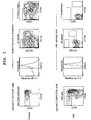
- CD34+CD38- HSCs were also purified from normal BM and CB samples ( FIG. 1 ).
- the onset of AML by LSCs and the lack of reconstitution of normal immunity were confirmed, and long-time transplantation and multi-lineage (T/B/bone marrow) differentiation of HSCs were confirmed ( FIG. 1 ).
- CD34+CD38+ cells or CD34- cells derived from the AML transplantation recipient mice did not come from the HSCs, but retained the nature of the LSCs.
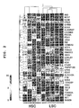
- genes that exhibit a significantly higher (p value ⁇ 0.01, percentage of false positivity ⁇ 0.05) array signal in LSCs than in HSCs on both the two microarray platforms were extracted using RankProd ( Bioinformatics 22, 2825, 2006 ) mounted on the Bioconductor package. A total of 217 gene candidates met the criteria ( FIG. 5 , Table 1); further, IL2R was added to make a total of 218 gene candidates.
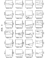
- the mRNA contents concerning 57 genes out of the 121 genes were statistically higher in LSCs than in HSCs. Of the 57 genes, 35 genes were identified as excellent LSC markers. The reason was that 1) the median expression levels of these genes were 5 times or higher in LSCs, and that 2) their mRNA contents were higher in all LSC samples tested than in each HSC population tested ( FIG. 2 ).
- FCGR2A(CD32) exhibited a strong correlation with LSCs in a significant ratio of the AML patients tested, and this was selected for further functional analysis.
- the great majority (>80%) of AML stem cells expressed this antigen ( FIG. 3 ).
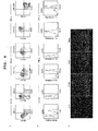
- in vivo NOD/SCID/IL2rg KO transplantation assay was performed using purified LSCs derived from three patients with AML.
- purified CD34+CD38-CD32+ and CD34+CD38-CD32- cells were transplanted to sub-lethally irradiated recipients, AML developed exclusively from the CD32+ fraction ( FIG. 4 ).
- any LSC-targeting treatment is thought to be best used along with a commonly used chemotherapeutic agent that is effective in removing non-LSC AML cells, it is important to confirm that the target molecule is continuously expressed even after chemotherapy. Accordingly, the present inventors examined whether the expression of CD32 was maintained after chemotherapy, and confirmed the expression of CD32 in BM, spleens and peripheral blood (PB) of AML transplantation recipient mice after AraC treatment ( FIG. 6 ). Also, CD32-expressing cells were found by immunofluorescent labeling in both the membrane region and central region of bone marrow ( FIG. 4 ).
- CD32 in normal human HSCs was assessed.
- the frequency of CD32+ cells was 9.8% +/- SD ( FIG. 4A ).
- CD32+ cells were detected exclusively in the CD34+CD38-CD133- fraction ( FIG. 4a ). It was found by heterologous transplantation assay that not the CD34+CD38-CD32+ fraction but the CD34+CD38-CD133+CD32- fraction contains HSCs ( FIG. 4B ).
- CD34+CD38-CD32+ cells have the capability of producing bone marrow-series and erythrocyte-series hematopoietic colonies.
- CD34+CD38-CD32- cells have the capability of producing bone marrow-series and erythrocyte-series hematopoietic colonies.
- the lack of the capability of in vivo long-term hematopoiesis reconstitution in CD32+ normal HSCs suggests the possibility that therapeutic agents targeting CD32 expression cells may help avoid important adverse reactions related to the normal hematopoietic and immune systems without affecting HSCs.
- the present inventors first confirmed by neonatal NOD/SCID/IL2rg KO mouse transplantation assay that in AML patient samples lacking the expression of CD32 by LSCs, CD34+CD38- cells possess the LSC function ( FIG. 1 ), and then examined the expression of ITGB2(CD18), CD93, (as well as CD25, CD132, OX41, and CD97), CD33, CD3D and TNF ⁇ by flow cytometry. Combination of the antigens CD32, ITGB2, CD93, 97 and 33 enabled good separation of LSCs from normal HSCs in 31 patients out of 47 patients.
- the list of LSC-specific genes identified using the two sets of microarrays and quantitative PCR includes genes that are expressed preferentially in bone marrow progeny, but their expression is limited in HSCs.
- FCGR2A, HCK and NCF4 are highly expressed in mature bone marrow cells and mediate the phagocytosis by immunoconjugates and subsequent superoxide production ( Prot Natl Acad Sci USA, 97, 1725; 2000 ; J Exp Med 191, 669, 2000 ; Nat Cell Biol, 3, 679, 2001 ; J Biol Chem 279, 1415, 2004 ).
- CD3D which is a constituent of the CD3 conjugate, transmits in mature T lymphocytes a T cell receptor signal via the ITAM motif thereof. Therefore, at least a particular ratio of AMLs can develop via abnormal regulation of differentiation in the stem cell stage.
- CD33 is a well recognized immunological marker of AML cells, and serves as a target for antibody pharmaceuticals such as gemtuzumab ozogamicin ( Leukemia 19: 176, 2005 ).
- CD97 has been reported to be accumulated in colorectal cancers that infiltrate lymphatic vessels ( Am J Pathol 161, 1657, 2002 ). Overexpression of these molecules in LSCs suggests that a therapeutic method that targets these molecules may be effective not only on LSCs, but also on mature AML cells.
- the candidate genes were classified into the following sets:
- the gene set 1 includes candidates that specifically target LSCs and do not affect HSCs, for example, promising candidates for the development of therapeutic agents such as antibody pharmaceuticals based on the lack of the aforementioned candidates in normal HSCs.
- the genes included in the gene set 2 (the most promising candidate is CD33) encode biomarkers having high applicability to ex vivo purging of LSCs for separating LSCs from HSCs and the like against the background of autologous transplantation of hematopoietic stem cells.
- WT1 has been shown to be expressed in a wide variety of tumors, including leukemia. However, whether this molecule is expressed at the level of stem cells, which exhibit recurrences and anticancer agent resistance, has been unknown. The present inventors found that this molecule is expressed in leukemic stem cells that are present in niches and are in the stationary phase of cell cycle, and have shown that the molecule is of significance as a target molecule for killing leukemic stem cells, which have been unable to be killed by conventional chemotherapy and radiotherapy.
- peripheral blood was collected from 47 patients with AML in various stages, samples containing hematopoietic cells were prepared, and FCGR2A (CD32a), FCGR2B(CD32b), IL2RA(CD25), ITGB2(CD18) and CD93 positivity rates in leukemic stem cells contained in the samples were examined.
- FCGR2A CD32a
- FCGR2B CD32b
- IL2RA CD25
- ITGB2(CD18) and CD93 positivity rates in leukemic stem cells contained in the samples were examined. The results are shown in Table 4.
- a therapeutic agent that acts specifically on LSCs that are the source of onset or recurrence of AML can be provided. It is possible to specifically remove LSCs from bone marrow cells of a patient or a donor using a cell sorter such as FACS, with a leukemic stem cell marker found in the present invention as an index. This will increase effectiveness of purging for autologous or allogeneic bone marrow transplantation, and enable to significantly prevent recurrences or the initial onset of acute myeloid leukemia.
- the presence or absence of LSCs in a collected biological sample or in a living organism can be determined with a leukemic stem cell marker found in the present invention as an index, whereby recurrences or the initial onset of acute myeloid leukemia can also be predicted.
Landscapes
- Health & Medical Sciences (AREA)
- Life Sciences & Earth Sciences (AREA)
- Chemical & Material Sciences (AREA)
- Immunology (AREA)
- Engineering & Computer Science (AREA)
- General Health & Medical Sciences (AREA)
- Organic Chemistry (AREA)
- Medicinal Chemistry (AREA)
- Proteomics, Peptides & Aminoacids (AREA)
- Molecular Biology (AREA)
- Hematology (AREA)
- Zoology (AREA)
- Pharmacology & Pharmacy (AREA)
- Animal Behavior & Ethology (AREA)
- Public Health (AREA)
- Veterinary Medicine (AREA)
- Pathology (AREA)
- Analytical Chemistry (AREA)
- Biochemistry (AREA)
- Biotechnology (AREA)
- Cell Biology (AREA)
- Genetics & Genomics (AREA)
- Oncology (AREA)
- Biomedical Technology (AREA)
- Wood Science & Technology (AREA)
- Developmental Biology & Embryology (AREA)
- Epidemiology (AREA)
- Physics & Mathematics (AREA)
- Microbiology (AREA)
- Hospice & Palliative Care (AREA)
- Bioinformatics & Cheminformatics (AREA)
- Urology & Nephrology (AREA)
- Biophysics (AREA)
- General Engineering & Computer Science (AREA)
- Nuclear Medicine, Radiotherapy & Molecular Imaging (AREA)
- General Chemical & Material Sciences (AREA)
- Virology (AREA)
- Chemical Kinetics & Catalysis (AREA)
- Food Science & Technology (AREA)
- General Physics & Mathematics (AREA)
Claims (3)
- Testverfahren zur Vorhersage des ersten Einsetzens oder eines Rezidivs von akuter myeloider Leukämie, umfassend:(1) einen Schritt des Messens des Expressionsniveaus von Leukämie-Stammzell-Markergenen in einer biologischen Probe, die einem Probanden entnommen wurde, bezüglich eines Transkriptionsprodukts oder Translationsprodukts der Gene als Analyt; und(2) einen Schritt des Vergleichens der in dem Messschritt erhaltenen Expressionsniveaus mit einem Referenzwert;wobei der Referenzwert für jeden der Marker der mittlere Expressionswert des Markers in CD34-positiven, CD38-negativen hämatopoetischen Stammzellen für gesunde Personen oder der mittlere Expressionswert des Markers in CD34-positiven, CD38-negativen hämatopoetischen Stammzellen für den Probanden vor dem Einsetzen ist;
wobei die Leukämie-Stammzell-Markergene ausgewählt sind aus:(i) 3 bis 35 Genen, wobei eines der Gene FCGR2A ist und die anderen Gene aus der Gruppe ausgewählt sind, die aus folgenden besteht: in der Zellmembran oder extrazellulär lokalisierten Genen, die aus ALOX5AP, CACNB4, CCL5, CD33, CD3D, CD93, CD97, CLEC12A, DOK2, GPR84, HCST, HOMER3, ITGB2, LGALS1, LRG1, PTH2R, RNASE2, TNF, TNFSF13B, TYROBP und VNN1 bestehen; einem mit dem Zellzyklus zusammenhängenden Gen, das aus NEK6 besteht; einem mit Apoptose zusammenhängenden Gen, das aus BIK besteht; mit der Signalgebung zusammenhängenden Genen, die aus AK5, FYB, HCK, LPXN, PDE9A, PDK1, PRKCD und RAB20 bestehen; einem Transkriptionsfaktorgen, das aus WT1 besteht; und anderen Genen, die aus CTSC und NCF4 bestehen;(ii) 6 Genen, die aus FCGR2A, ITGB2, CD93, CD33, CD3D und TNF bestehen;(iii) 4 Genen, die aus FCGR2A, IL2RA, ITGB2 und CD93 bestehen; oder(iv) 8 Genen, die aus FCGR2A, AK5, DOK2, LRG1, BIK, IL2RA, WT1 und SUCNR1 bestehen;
und wobei dann, wenn die Expression von zwei oder mehr Leukämie-Stammzell-Markergenen in dem Probanden signifikant höher ist als der Referenzwert, eine mögliche Anwesenheit einer Leukämie-Stammzelle in der entnommenen biologischen Probe oder dem Körper des Probanden zu vermuten ist. - Testverfahren gemäß Anspruch 1, wobei die Leukämie-Stammzell-Markergene folgende sind:(i') 5 bis 35 Gene, wobei eines der Gene FCGR2A ist und die anderen Gene aus der Gruppe ausgewählt sind, die aus folgenden besteht: in der Zellmembran oder extrazellulär lokalisierten Genen, die aus ALOX5AP, CACNB4, CCL5, CD33, CD3D, CD93, CD97, CLEC12A, DOK2, GPR84, HCST, HOMER3, ITGB2, LGALS1, LRG1, PTH2R, RNASE2, TNF, TNFSF13B, TYROBP und VNN1 bestehen; einem mit dem Zellzyklus zusammenhängenden Gen, das aus NEK6 besteht; einem mit Apoptose zusammenhängenden Gen, das aus BIK besteht; mit der Signalgebung zusammenhängenden Genen, die aus AK5, FYB, HCK, LPXN, PDE9A, PDK1, PRKCD und RAB20 bestehen; einem Transkriptionsfaktorgen, das aus WT1 besteht; und anderen Genen, die aus CTSC und NCF4 bestehen.
- Herstellungsverfahren für eine Probe, die hämatopoetische Zellen für die autologe Transplantation oder allogene Transplantation bei einem Patienten mit akuter myeloider Leukämie enthält, umfassend:a) einen Schritt des Entnehmens einer Probe, die hämatopoetische Zellen enthält, von dem Patienten oder einem Spender;b) einen Schritt des In-Kontakt-Bringens der entnommenen Probe mit Antikörpern, die ein Translationsprodukt von Leukämie-Stammzell-Markergenen erkennen, welche ausgewählt sind aus:(i) 3 bis 35 Genen, wobei eines der Gene FCGR2A ist und die anderen Gene aus der Gruppe ausgewählt sind, die aus folgenden besteht: in der Zellmembran oder extrazellulär lokalisierten Genen, die aus ALOX5AP, CACNB4, CCL5, CD33, CD3D, CD93, CD97, CLEC12A, DOK2, GPR84, HCST, HOMER3, ITGB2, LGALS1, LRG1, PTH2R, RNASE2, TNF, TNFSF13B, TYROBP und VNN1 bestehen; einem mit dem Zellzyklus zusammenhängenden Gen, das aus NEK6 besteht; einem mit Apoptose zusammenhängenden Gen, das aus BIK besteht; mit der Signalgebung zusammenhängenden Genen, die aus AK5, FYB, HCK, LPXN, PDE9A, PDK1, PRKCD und RAB20 bestehen; einem Transkriptionsfaktorgen, das aus WT1 besteht; und anderen Genen, die aus CTSC und NCF4 bestehen;(ii) 6 Genen, die aus FCGR2A, ITGB2, CD93, CD33, CD3D und TNF bestehen;(iii) 4 Genen, die aus FCGR2A, IL2RA, ITGB2 und CD93 bestehen; oder(iv) 8 Genen, die aus FCGR2A, AK5, DOK2, LRG1, BIK, IL2RA, WT1 und SUCNR1 bestehen; undc) einen Schritt des Aussortierens von Zellen, an die die Antikörper gebunden haben, und Erhalten der Probe, aus der Leukämie-Stammzellen entfernt wurden.
Applications Claiming Priority (2)
| Application Number | Priority Date | Filing Date | Title |
|---|---|---|---|
| JP2009072400 | 2009-03-24 | ||
| PCT/JP2010/055131 WO2010110346A1 (ja) | 2009-03-24 | 2010-03-24 | 白血病幹細胞マーカー |
Publications (4)
| Publication Number | Publication Date |
|---|---|
| EP2412825A1 EP2412825A1 (de) | 2012-02-01 |
| EP2412825A4 EP2412825A4 (de) | 2013-10-23 |
| EP2412825B1 true EP2412825B1 (de) | 2017-09-20 |
| EP2412825B8 EP2412825B8 (de) | 2018-01-10 |
Family
ID=42781039
Family Applications (1)
| Application Number | Title | Priority Date | Filing Date |
|---|---|---|---|
| EP10756142.5A Not-in-force EP2412825B8 (de) | 2009-03-24 | 2010-03-24 | Leukämiestammzellenmarker |
Country Status (4)
| Country | Link |
|---|---|
| US (2) | US20120070450A1 (de) |
| EP (1) | EP2412825B8 (de) |
| JP (2) | JPWO2010110346A1 (de) |
| WO (1) | WO2010110346A1 (de) |
Families Citing this family (42)
| Publication number | Priority date | Publication date | Assignee | Title |
|---|---|---|---|---|
| GB201107118D0 (en) | 2011-04-27 | 2011-06-08 | Imp Innovations Ltd | Method of diagnosis and prognosis |
| US9334306B2 (en) | 2011-07-09 | 2016-05-10 | The Regents Of The University Of California | Leukemia stem cell targeting ligands and methods of use |
| WO2013048345A1 (en) * | 2011-09-28 | 2013-04-04 | Agency For Science, Technology And Research | Methods and pharmaceutical compositions for treating cancer |
| CN105142662B (zh) * | 2012-01-05 | 2017-07-07 | 德国癌症研究中心 | Idh1 r132h突变阳性癌症治疗或诊断的手段与方法 |
| WO2013133450A1 (ja) * | 2012-03-06 | 2013-09-12 | 株式会社バイオマトリックス研究所 | がん治療用医薬組成物 |
| WO2013139479A1 (en) * | 2012-03-21 | 2013-09-26 | Institut Gustave Roussy (Igr) | New diagnostic markers of specific chronic myelomonocytic leukemia (cmml) |
| EP2878601B1 (de) | 2012-07-27 | 2018-03-28 | Riken | Mittel zur behandlung oder bekämpfung des wiederauftretens akuter myelogener leukämie |
| KR101470125B1 (ko) * | 2013-02-15 | 2014-12-08 | 순천향대학교 산학협력단 | 알엔에이즈 2를 포함하는 아스피린에 의해 악화된 호흡기 질환 진단용 마커 조성물 및 진단용 키트 |
| EP3744736A1 (de) | 2013-02-20 | 2020-12-02 | Novartis AG | Effektives targeting primärer humaner leukämie mit durch den chimären anti-cd123-antigen-rezeptor veränderten t-zellen |
| BR112015023510A2 (pt) * | 2013-03-15 | 2017-10-10 | Fundacio Inst De Recerca Biomedica Irb Barcelona | método para o diagnóstico, prognóstico e tratamento de câncer metastático |
| JP6683986B2 (ja) * | 2013-07-10 | 2020-04-22 | 国立大学法人 東京大学 | がん幹細胞分子マーカー |
| EP3049442A4 (de) | 2013-09-26 | 2017-06-28 | Costim Pharmaceuticals Inc. | Verfahren zur behandlung von blutkrebs |
| CN105934252B (zh) | 2013-10-10 | 2020-11-10 | 贝思以色列女会吏医学中心公司 | Tm4sf1结合蛋白及其使用方法 |
| JOP20200094A1 (ar) | 2014-01-24 | 2017-06-16 | Dana Farber Cancer Inst Inc | جزيئات جسم مضاد لـ pd-1 واستخداماتها |
| JOP20200096A1 (ar) | 2014-01-31 | 2017-06-16 | Children’S Medical Center Corp | جزيئات جسم مضاد لـ tim-3 واستخداماتها |
| EP3138923B1 (de) | 2014-04-28 | 2020-01-15 | The Catholic University Of Korea Industry-Academic | Verfahren zur prognose eines rückfalls von akuter myeolischer leukämie |
| WO2015167210A1 (ko) * | 2014-04-28 | 2015-11-05 | 가톨릭대학교 산학협력단 | 급성 골수성 백혈병 재발의 예후 예측 방법 |
| ES2791199T3 (es) * | 2014-06-25 | 2020-11-03 | Tel Hashomer Medical Res Infrastructure & Services Ltd | Identificación de marcadores de células madre cancerosas y uso de los mismos para diagnóstico y tratamiento |
| ES2791248T3 (es) | 2014-08-19 | 2020-11-03 | Novartis Ag | Receptor antigénico quimérico (CAR) anti-CD123 para su uso en el tratamiento del cáncer |
| CA2960824A1 (en) | 2014-09-13 | 2016-03-17 | Novartis Ag | Combination therapies of alk inhibitors |
| US10793642B2 (en) | 2014-12-11 | 2020-10-06 | Inbiomotion S.L. | Binding members for human c-MAF |
| AU2016256911B2 (en) | 2015-05-07 | 2022-03-31 | Agenus Inc. | Anti-OX40 antibodies and methods of use thereof |
| WO2016201365A2 (en) * | 2015-06-12 | 2016-12-15 | Visani Giuseppe | Methods for treating cancers |
| CN108430516A (zh) * | 2015-11-19 | 2018-08-21 | 艾伯维施特姆森特克斯有限责任公司 | 新颖抗emr2抗体和使用方法 |
| US11447557B2 (en) | 2015-12-02 | 2022-09-20 | Agenus Inc. | Antibodies and methods of use thereof |
| MY196837A (en) | 2015-12-11 | 2023-05-03 | Immatics Biotechnologies Gmbh | Novel peptides and combination of peptides for use in immunotherapy against various cancers |
| JP7075125B2 (ja) * | 2016-05-25 | 2022-05-25 | イマティクス バイオテクノロジーズ ゲーエムベーハー | 標的としてのおよび胆嚢がんおよび胆管がんおよびその他のがんに対する免疫療法で使用するための新規ペプチド、ペプチド組み合わせ |
| NZ749355A (en) | 2016-05-27 | 2023-04-28 | Agenus Inc | Anti-tim-3 antibodies and methods of use thereof |
| MA46770A (fr) | 2016-11-09 | 2019-09-18 | Agenus Inc | Anticorps anti-ox40, anticorps anti-gitr, et leurs procédés d'utilisation |
| KR101970764B1 (ko) | 2017-05-19 | 2019-04-22 | 아주대학교산학협력단 | 조혈모세포의 항상성 유지에 관여하는 cotl1 단백질 및 이의 용도 |
| WO2019084434A1 (en) * | 2017-10-26 | 2019-05-02 | National University Of Singapore | NEW APPROACH FOR UNIVERSAL MONITORING OF MINIMUM RESIDUAL DISEASE IN ACUTE MYELOID LEUKEMIA |
| CN107828740B (zh) * | 2017-11-29 | 2020-06-09 | 武汉大学 | Homer3单克隆抗体及其应用 |
| GB201720077D0 (en) * | 2017-12-01 | 2018-01-17 | Univ Oxford Innovation Ltd | Leukaemic stem cell |
| JP2021505175A (ja) | 2017-12-11 | 2021-02-18 | ロシュ イノベーション センター コペンハーゲン エーエス | Fndc3bの発現を調節するためのオリゴヌクレオチド |
| CN109187987B (zh) * | 2018-08-23 | 2021-05-11 | 中国人民解放军第三0九医院 | Ms4a3蛋白作为标志物在诊断活动性结核病中的应用 |
| JP2022512860A (ja) * | 2018-11-06 | 2022-02-07 | アンスティチュ ナショナル ドゥ ラ サンテ エ ドゥ ラ ルシェルシュ メディカル | 白血病幹細胞を根絶することによる急性骨髄性白血病の治療のための方法および医薬組成物 |
| AR117327A1 (es) | 2018-12-20 | 2021-07-28 | 23Andme Inc | Anticuerpos anti-cd96 y métodos de uso de estos |
| KR102176419B1 (ko) * | 2019-01-28 | 2020-11-09 | 고려대학교 산학협력단 | 엑소좀 유래 면역표현형을 이용한 급성 골수성 백혈병의 치료 반응성 예측방법 |
| CN110132930B (zh) * | 2019-06-12 | 2023-11-14 | 东北师范大学 | 激光诱导叶绿素荧光的多角度激发探测装置及其分析方法 |
| WO2021159479A1 (zh) * | 2020-02-14 | 2021-08-19 | 深圳华大智造科技股份有限公司 | 基于图像分析液滴的方法、计算机装置及存储介质 |
| CN113209300A (zh) * | 2021-05-17 | 2021-08-06 | 北京大学人民医院 | Gilt在作为提高急性髓系白血病患者对化疗药敏感性的作用靶点中的应用 |
| CN113293202B (zh) * | 2021-07-02 | 2022-02-22 | 广东莱恩医药研究院有限公司 | 一种定量检测机体中mRNA含量的实时荧光定量PCR试剂盒及检测方法和应用 |
Family Cites Families (14)
| Publication number | Priority date | Publication date | Assignee | Title |
|---|---|---|---|---|
| JPS6147500A (ja) | 1984-08-15 | 1986-03-07 | Res Dev Corp Of Japan | キメラモノクロ−ナル抗体及びその製造法 |
| GB8607679D0 (en) | 1986-03-27 | 1986-04-30 | Winter G P | Recombinant dna product |
| GB8928874D0 (en) | 1989-12-21 | 1990-02-28 | Celltech Ltd | Humanised antibodies |
| DE69120146T2 (de) | 1990-01-12 | 1996-12-12 | Cell Genesys Inc | Erzeugung xenogener antikörper |
| DK0814159T3 (da) | 1990-08-29 | 2005-10-24 | Genpharm Int | Transgene, ikke-humane dyr, der er i stand til at danne heterologe antistoffer |
| CA2161351C (en) | 1993-04-26 | 2010-12-21 | Nils Lonberg | Transgenic non-human animals capable of producing heterologous antibodies |
| US6828151B2 (en) * | 2001-12-04 | 2004-12-07 | Isis Pharmaceuticals, Inc. | Antisense modulation of hematopoietic cell protein tyrosine kinase expression |
| GB2343679A (en) * | 1998-11-16 | 2000-05-17 | Alison Miriam Davies | Autologous transplantation and method for making cells dormant |
| CA2434690A1 (en) * | 2001-01-16 | 2002-09-06 | Curagen Corporation | Proteins, polynucleotides encoding them and methods of using the same |
| US20070105118A1 (en) * | 2003-11-04 | 2007-05-10 | Martin Dugas | Method for distinguishing aml subtypes with recurring genetic aberrations |
| US8142994B2 (en) * | 2004-02-23 | 2012-03-27 | Erasmus University Medical Center Rotterdam | Classification, diagnosis and prognosis of acute myeloid leukemia by gene expression profiling |
| JP2008068892A (ja) | 2006-09-13 | 2008-03-27 | Tsubakimoto Chain Co | 創薬用試料保管システム |
| WO2009051238A1 (ja) * | 2007-10-18 | 2009-04-23 | Riken | ヒト白血病幹細胞および白血病非幹細胞が増幅されたマウスおよびその製造方法 |
| WO2009091547A1 (en) * | 2008-01-15 | 2009-07-23 | The Board Of Trustees Of The Leland Stanford Junior University | Markers of acute myeloid leukemia stem cells |
-
2010
- 2010-03-24 EP EP10756142.5A patent/EP2412825B8/de not_active Not-in-force
- 2010-03-24 JP JP2011506104A patent/JPWO2010110346A1/ja active Pending
- 2010-03-24 WO PCT/JP2010/055131 patent/WO2010110346A1/ja active Application Filing
- 2010-03-24 US US13/258,993 patent/US20120070450A1/en not_active Abandoned
-
2014
- 2014-03-20 US US14/220,842 patent/US20140274788A1/en not_active Abandoned
-
2015
- 2015-07-07 JP JP2015135949A patent/JP6247253B2/ja not_active Expired - Fee Related
Non-Patent Citations (1)
| Title |
|---|
| None * |
Also Published As
| Publication number | Publication date |
|---|---|
| WO2010110346A1 (ja) | 2010-09-30 |
| EP2412825A1 (de) | 2012-02-01 |
| JP2015231375A (ja) | 2015-12-24 |
| US20120070450A1 (en) | 2012-03-22 |
| EP2412825A4 (de) | 2013-10-23 |
| JP6247253B2 (ja) | 2017-12-13 |
| US20140274788A1 (en) | 2014-09-18 |
| EP2412825B8 (de) | 2018-01-10 |
| JPWO2010110346A1 (ja) | 2012-10-04 |
Similar Documents
| Publication | Publication Date | Title |
|---|---|---|
| EP2412825B1 (de) | Leukämiestammzellenmarker | |
| KR102246710B1 (ko) | 파르네실전달효소 억제제를 이용하여 암환자를 치료하는 방법 | |
| JP2020126067A5 (de) | ||
| Ngo et al. | Antibody therapy targeting CD47 and CD271 effectively suppresses melanoma metastasis in patient-derived xenografts | |
| JP6860476B2 (ja) | 前立腺がん治療に関する方法及び組成物 | |
| WO2019070755A1 (en) | METHODS AND COMPOSITIONS FOR DETECTING AND MODULATING A GENETIC SIGNATURE OF IMMUNOTHERAPY RESISTANCE IN CANCER | |
| AU2013296233B2 (en) | Biomarker associated with risk of melanoma reoccurrence | |
| US20070092519A1 (en) | Method for diagnosing chronic myeloid leukemia | |
| KR20210100656A (ko) | 암 면역요법을 위한 진단 방법 및 조성물 | |
| US20200123258A1 (en) | Targeting b cells to enhance response to immune checkpoint blockade | |
| US20140323342A1 (en) | Methods and Compositions for the Treatment and Diagnosis of Bladder Cancer | |
| US20150147271A1 (en) | Targets for diagnosis, prognosis and therapy of acute myeloid leukemia and myelodysplastic syndromes | |
| JP2010178650A (ja) | 固形癌の再発予測のための試験方法および再発予防剤 | |
| US20180251851A1 (en) | Ectopic lymphoid structures as targets for liver cancer detection, risk prediction and therapy | |
| US20200299783A1 (en) | Molecular signature for selecting lymphoma patients for treatment with ibrutinib | |
| WO2018011166A2 (en) | Methods for quantifying the population of myeloid dendritic cells in a tissue sample | |
| US20180105882A1 (en) | New biomarker for outcome in aml | |
| EP3529621B1 (de) | Verfahren zur bestimmung der prognose von krebs | |
| JP2020530015A (ja) | ファルネシルトランスフェラーゼ阻害剤を用いてがんを治療する方法 | |
| EP3255433A1 (de) | Verfahren unter verwendung von blm als marker des multiplen myeloms | |
| KR102042332B1 (ko) | 간암의 재발 및 예후 예측용 tcirg1 마커 및 이의 용도 | |
| WO2017055320A1 (en) | Methods for quantifying the population of cytotoxic lymphocytes in a tissue sample | |
| KR20230010426A (ko) | 혈액 종양 질환 특이적 마커 및 이의 용도 | |
| WO2022187374A1 (en) | Methods of treating red blood cell disorders | |
| Li et al. | CHROMOSOME ABNORMALITIES IN MANTLE CELL LEUKEMIA: A STUDY OF 96 CASES |
Legal Events
| Date | Code | Title | Description |
|---|---|---|---|
| PUAI | Public reference made under article 153(3) epc to a published international application that has entered the european phase |
Free format text: ORIGINAL CODE: 0009012 |
|
| 17P | Request for examination filed |
Effective date: 20111020 |
|
| AK | Designated contracting states |
Kind code of ref document: A1 Designated state(s): AT BE BG CH CY CZ DE DK EE ES FI FR GB GR HR HU IE IS IT LI LT LU LV MC MK MT NL NO PL PT RO SE SI SK SM TR |
|
| DAX | Request for extension of the european patent (deleted) | ||
| RIC1 | Information provided on ipc code assigned before grant |
Ipc: C12Q 1/68 20060101AFI20130909BHEP Ipc: A61K 35/28 20060101ALI20130909BHEP Ipc: A61K 31/713 20060101ALI20130909BHEP |
|
| A4 | Supplementary search report drawn up and despatched |
Effective date: 20130925 |
|
| RIC1 | Information provided on ipc code assigned before grant |
Ipc: C12Q 1/68 20060101AFI20130918BHEP Ipc: A61K 35/28 20060101ALI20130918BHEP Ipc: A61K 31/713 20060101ALI20130918BHEP |
|
| 17Q | First examination report despatched |
Effective date: 20150422 |
|
| STAA | Information on the status of an ep patent application or granted ep patent |
Free format text: STATUS: EXAMINATION IS IN PROGRESS |
|
| GRAP | Despatch of communication of intention to grant a patent |
Free format text: ORIGINAL CODE: EPIDOSNIGR1 |
|
| STAA | Information on the status of an ep patent application or granted ep patent |
Free format text: STATUS: GRANT OF PATENT IS INTENDED |
|
| INTG | Intention to grant announced |
Effective date: 20170331 |
|
| GRAS | Grant fee paid |
Free format text: ORIGINAL CODE: EPIDOSNIGR3 |
|
| GRAA | (expected) grant |
Free format text: ORIGINAL CODE: 0009210 |
|
| STAA | Information on the status of an ep patent application or granted ep patent |
Free format text: STATUS: THE PATENT HAS BEEN GRANTED |
|
| AK | Designated contracting states |
Kind code of ref document: B1 Designated state(s): AT BE BG CH CY CZ DE DK EE ES FI FR GB GR HR HU IE IS IT LI LT LU LV MC MK MT NL NO PL PT RO SE SI SK SM TR |
|
| REG | Reference to a national code |
Ref country code: GB Ref legal event code: FG4D |
|
| REG | Reference to a national code |
Ref country code: CH Ref legal event code: EP |
|
| RIN2 | Information on inventor provided after grant (corrected) |
Inventor name: HIJIKATA, ATSUSHI Inventor name: OZAWA, HIDETOSHI Inventor name: OHARA, OSAMU Inventor name: ISHIKAWA, FUMIHIKO Inventor name: KITAMURA, HIROSHI Inventor name: SHULTZ, LEONARD D. Inventor name: SAITO, YORIKO |
|
| REG | Reference to a national code |
Ref country code: AT Ref legal event code: REF Ref document number: 930161 Country of ref document: AT Kind code of ref document: T Effective date: 20171015 |
|
| REG | Reference to a national code |
Ref country code: IE Ref legal event code: FG4D |
|
| RAP2 | Party data changed (patent owner data changed or rights of a patent transferred) |
Owner name: RIKEN |
|
| REG | Reference to a national code |
Ref country code: DE Ref legal event code: R096 Ref document number: 602010045380 Country of ref document: DE |
|
| RAP2 | Party data changed (patent owner data changed or rights of a patent transferred) |
Owner name: RIKEN Owner name: THE JACKSON LABORATORY |
|
| REG | Reference to a national code |
Ref country code: NL Ref legal event code: MP Effective date: 20170920 |
|
| PG25 | Lapsed in a contracting state [announced via postgrant information from national office to epo] |
Ref country code: SE Free format text: LAPSE BECAUSE OF FAILURE TO SUBMIT A TRANSLATION OF THE DESCRIPTION OR TO PAY THE FEE WITHIN THE PRESCRIBED TIME-LIMIT Effective date: 20170920 Ref country code: NO Free format text: LAPSE BECAUSE OF FAILURE TO SUBMIT A TRANSLATION OF THE DESCRIPTION OR TO PAY THE FEE WITHIN THE PRESCRIBED TIME-LIMIT Effective date: 20171220 Ref country code: FI Free format text: LAPSE BECAUSE OF FAILURE TO SUBMIT A TRANSLATION OF THE DESCRIPTION OR TO PAY THE FEE WITHIN THE PRESCRIBED TIME-LIMIT Effective date: 20170920 Ref country code: LT Free format text: LAPSE BECAUSE OF FAILURE TO SUBMIT A TRANSLATION OF THE DESCRIPTION OR TO PAY THE FEE WITHIN THE PRESCRIBED TIME-LIMIT Effective date: 20170920 Ref country code: HR Free format text: LAPSE BECAUSE OF FAILURE TO SUBMIT A TRANSLATION OF THE DESCRIPTION OR TO PAY THE FEE WITHIN THE PRESCRIBED TIME-LIMIT Effective date: 20170920 |
|
| REG | Reference to a national code |
Ref country code: LT Ref legal event code: MG4D |
|
| REG | Reference to a national code |
Ref country code: AT Ref legal event code: MK05 Ref document number: 930161 Country of ref document: AT Kind code of ref document: T Effective date: 20170920 |
|
| PG25 | Lapsed in a contracting state [announced via postgrant information from national office to epo] |
Ref country code: BG Free format text: LAPSE BECAUSE OF FAILURE TO SUBMIT A TRANSLATION OF THE DESCRIPTION OR TO PAY THE FEE WITHIN THE PRESCRIBED TIME-LIMIT Effective date: 20171220 Ref country code: GR Free format text: LAPSE BECAUSE OF FAILURE TO SUBMIT A TRANSLATION OF THE DESCRIPTION OR TO PAY THE FEE WITHIN THE PRESCRIBED TIME-LIMIT Effective date: 20171221 Ref country code: LV Free format text: LAPSE BECAUSE OF FAILURE TO SUBMIT A TRANSLATION OF THE DESCRIPTION OR TO PAY THE FEE WITHIN THE PRESCRIBED TIME-LIMIT Effective date: 20170920 |
|
| REG | Reference to a national code |
Ref country code: FR Ref legal event code: PLFP Year of fee payment: 9 |
|
| PG25 | Lapsed in a contracting state [announced via postgrant information from national office to epo] |
Ref country code: NL Free format text: LAPSE BECAUSE OF FAILURE TO SUBMIT A TRANSLATION OF THE DESCRIPTION OR TO PAY THE FEE WITHIN THE PRESCRIBED TIME-LIMIT Effective date: 20170920 |
|
| PG25 | Lapsed in a contracting state [announced via postgrant information from national office to epo] |
Ref country code: CZ Free format text: LAPSE BECAUSE OF FAILURE TO SUBMIT A TRANSLATION OF THE DESCRIPTION OR TO PAY THE FEE WITHIN THE PRESCRIBED TIME-LIMIT Effective date: 20170920 Ref country code: ES Free format text: LAPSE BECAUSE OF FAILURE TO SUBMIT A TRANSLATION OF THE DESCRIPTION OR TO PAY THE FEE WITHIN THE PRESCRIBED TIME-LIMIT Effective date: 20170920 Ref country code: PL Free format text: LAPSE BECAUSE OF FAILURE TO SUBMIT A TRANSLATION OF THE DESCRIPTION OR TO PAY THE FEE WITHIN THE PRESCRIBED TIME-LIMIT Effective date: 20170920 Ref country code: RO Free format text: LAPSE BECAUSE OF FAILURE TO SUBMIT A TRANSLATION OF THE DESCRIPTION OR TO PAY THE FEE WITHIN THE PRESCRIBED TIME-LIMIT Effective date: 20170920 |
|
| PGFP | Annual fee paid to national office [announced via postgrant information from national office to epo] |
Ref country code: GB Payment date: 20180321 Year of fee payment: 9 |
|
| PG25 | Lapsed in a contracting state [announced via postgrant information from national office to epo] |
Ref country code: IT Free format text: LAPSE BECAUSE OF FAILURE TO SUBMIT A TRANSLATION OF THE DESCRIPTION OR TO PAY THE FEE WITHIN THE PRESCRIBED TIME-LIMIT Effective date: 20170920 Ref country code: SK Free format text: LAPSE BECAUSE OF FAILURE TO SUBMIT A TRANSLATION OF THE DESCRIPTION OR TO PAY THE FEE WITHIN THE PRESCRIBED TIME-LIMIT Effective date: 20170920 Ref country code: AT Free format text: LAPSE BECAUSE OF FAILURE TO SUBMIT A TRANSLATION OF THE DESCRIPTION OR TO PAY THE FEE WITHIN THE PRESCRIBED TIME-LIMIT Effective date: 20170920 Ref country code: SM Free format text: LAPSE BECAUSE OF FAILURE TO SUBMIT A TRANSLATION OF THE DESCRIPTION OR TO PAY THE FEE WITHIN THE PRESCRIBED TIME-LIMIT Effective date: 20170920 Ref country code: IS Free format text: LAPSE BECAUSE OF FAILURE TO SUBMIT A TRANSLATION OF THE DESCRIPTION OR TO PAY THE FEE WITHIN THE PRESCRIBED TIME-LIMIT Effective date: 20180120 Ref country code: EE Free format text: LAPSE BECAUSE OF FAILURE TO SUBMIT A TRANSLATION OF THE DESCRIPTION OR TO PAY THE FEE WITHIN THE PRESCRIBED TIME-LIMIT Effective date: 20170920 |
|
| PGFP | Annual fee paid to national office [announced via postgrant information from national office to epo] |
Ref country code: FR Payment date: 20180312 Year of fee payment: 9 |
|
| REG | Reference to a national code |
Ref country code: DE Ref legal event code: R097 Ref document number: 602010045380 Country of ref document: DE |
|
| PLBE | No opposition filed within time limit |
Free format text: ORIGINAL CODE: 0009261 |
|
| STAA | Information on the status of an ep patent application or granted ep patent |
Free format text: STATUS: NO OPPOSITION FILED WITHIN TIME LIMIT |
|
| PG25 | Lapsed in a contracting state [announced via postgrant information from national office to epo] |
Ref country code: DK Free format text: LAPSE BECAUSE OF FAILURE TO SUBMIT A TRANSLATION OF THE DESCRIPTION OR TO PAY THE FEE WITHIN THE PRESCRIBED TIME-LIMIT Effective date: 20170920 |
|
| PGFP | Annual fee paid to national office [announced via postgrant information from national office to epo] |
Ref country code: DE Payment date: 20180403 Year of fee payment: 9 |
|
| 26N | No opposition filed |
Effective date: 20180621 |
|
| REG | Reference to a national code |
Ref country code: CH Ref legal event code: PL |
|
| PG25 | Lapsed in a contracting state [announced via postgrant information from national office to epo] |
Ref country code: MC Free format text: LAPSE BECAUSE OF FAILURE TO SUBMIT A TRANSLATION OF THE DESCRIPTION OR TO PAY THE FEE WITHIN THE PRESCRIBED TIME-LIMIT Effective date: 20170920 Ref country code: SI Free format text: LAPSE BECAUSE OF FAILURE TO SUBMIT A TRANSLATION OF THE DESCRIPTION OR TO PAY THE FEE WITHIN THE PRESCRIBED TIME-LIMIT Effective date: 20170920 |
|
| REG | Reference to a national code |
Ref country code: BE Ref legal event code: MM Effective date: 20180331 |
|
| REG | Reference to a national code |
Ref country code: IE Ref legal event code: MM4A |
|
| PG25 | Lapsed in a contracting state [announced via postgrant information from national office to epo] |
Ref country code: LU Free format text: LAPSE BECAUSE OF NON-PAYMENT OF DUE FEES Effective date: 20180324 |
|
| PG25 | Lapsed in a contracting state [announced via postgrant information from national office to epo] |
Ref country code: IE Free format text: LAPSE BECAUSE OF NON-PAYMENT OF DUE FEES Effective date: 20180324 |
|
| PG25 | Lapsed in a contracting state [announced via postgrant information from national office to epo] |
Ref country code: CH Free format text: LAPSE BECAUSE OF NON-PAYMENT OF DUE FEES Effective date: 20180331 Ref country code: LI Free format text: LAPSE BECAUSE OF NON-PAYMENT OF DUE FEES Effective date: 20180331 Ref country code: BE Free format text: LAPSE BECAUSE OF NON-PAYMENT OF DUE FEES Effective date: 20180331 |
|
| REG | Reference to a national code |
Ref country code: DE Ref legal event code: R119 Ref document number: 602010045380 Country of ref document: DE |
|
| GBPC | Gb: european patent ceased through non-payment of renewal fee |
Effective date: 20190324 |
|
| PG25 | Lapsed in a contracting state [announced via postgrant information from national office to epo] |
Ref country code: MT Free format text: LAPSE BECAUSE OF NON-PAYMENT OF DUE FEES Effective date: 20180324 Ref country code: DE Free format text: LAPSE BECAUSE OF NON-PAYMENT OF DUE FEES Effective date: 20191001 Ref country code: GB Free format text: LAPSE BECAUSE OF NON-PAYMENT OF DUE FEES Effective date: 20190324 |
|
| PG25 | Lapsed in a contracting state [announced via postgrant information from national office to epo] |
Ref country code: FR Free format text: LAPSE BECAUSE OF NON-PAYMENT OF DUE FEES Effective date: 20190331 |
|
| PG25 | Lapsed in a contracting state [announced via postgrant information from national office to epo] |
Ref country code: TR Free format text: LAPSE BECAUSE OF FAILURE TO SUBMIT A TRANSLATION OF THE DESCRIPTION OR TO PAY THE FEE WITHIN THE PRESCRIBED TIME-LIMIT Effective date: 20170920 |
|
| PG25 | Lapsed in a contracting state [announced via postgrant information from national office to epo] |
Ref country code: HU Free format text: LAPSE BECAUSE OF FAILURE TO SUBMIT A TRANSLATION OF THE DESCRIPTION OR TO PAY THE FEE WITHIN THE PRESCRIBED TIME-LIMIT; INVALID AB INITIO Effective date: 20100324 Ref country code: PT Free format text: LAPSE BECAUSE OF FAILURE TO SUBMIT A TRANSLATION OF THE DESCRIPTION OR TO PAY THE FEE WITHIN THE PRESCRIBED TIME-LIMIT Effective date: 20170920 |
|
| PG25 | Lapsed in a contracting state [announced via postgrant information from national office to epo] |
Ref country code: CY Free format text: LAPSE BECAUSE OF FAILURE TO SUBMIT A TRANSLATION OF THE DESCRIPTION OR TO PAY THE FEE WITHIN THE PRESCRIBED TIME-LIMIT Effective date: 20170920 Ref country code: MK Free format text: LAPSE BECAUSE OF NON-PAYMENT OF DUE FEES Effective date: 20170920 |