EP2404935A1 - Immunoglobulin binding HOSM - Google Patents
Immunoglobulin binding HOSM Download PDFInfo
- Publication number
- EP2404935A1 EP2404935A1 EP11167424A EP11167424A EP2404935A1 EP 2404935 A1 EP2404935 A1 EP 2404935A1 EP 11167424 A EP11167424 A EP 11167424A EP 11167424 A EP11167424 A EP 11167424A EP 2404935 A1 EP2404935 A1 EP 2404935A1
- Authority
- EP
- European Patent Office
- Prior art keywords
- antibody
- osm
- seq
- antibodies
- human
- Prior art date
- Legal status (The legal status is an assumption and is not a legal conclusion. Google has not performed a legal analysis and makes no representation as to the accuracy of the status listed.)
- Withdrawn
Links
- 230000027455 binding Effects 0.000 title claims description 188
- 108060003951 Immunoglobulin Proteins 0.000 title abstract description 16
- 102000018358 immunoglobulin Human genes 0.000 title abstract description 16
- 102100037795 Interleukin-6 receptor subunit beta Human genes 0.000 claims abstract description 126
- 101710152369 Interleukin-6 receptor subunit beta Proteins 0.000 claims abstract description 98
- 230000003993 interaction Effects 0.000 claims abstract description 70
- 238000011282 treatment Methods 0.000 claims abstract description 53
- 239000008194 pharmaceutical composition Substances 0.000 claims abstract description 26
- 241000282414 Homo sapiens Species 0.000 claims description 164
- 239000012634 fragment Substances 0.000 claims description 116
- 230000001225 therapeutic effect Effects 0.000 claims description 108
- 239000000427 antigen Substances 0.000 claims description 95
- 108091007433 antigens Proteins 0.000 claims description 95
- 102000036639 antigens Human genes 0.000 claims description 95
- 241001529936 Murinae Species 0.000 claims description 55
- 206010039073 rheumatoid arthritis Diseases 0.000 claims description 36
- 201000008482 osteoarthritis Diseases 0.000 claims description 24
- 201000004681 Psoriasis Diseases 0.000 claims description 8
- 241000288906 Primates Species 0.000 claims description 4
- 230000002101 lytic effect Effects 0.000 claims description 4
- 201000001320 Atherosclerosis Diseases 0.000 claims description 3
- 241000283973 Oryctolagus cuniculus Species 0.000 claims description 3
- 241000700159 Rattus Species 0.000 claims description 3
- 208000006545 Chronic Obstructive Pulmonary Disease Diseases 0.000 claims 1
- 102000004140 Oncostatin M Human genes 0.000 abstract description 353
- 108090000630 Oncostatin M Proteins 0.000 abstract description 353
- 102000043703 human OSM Human genes 0.000 abstract description 184
- 101000992170 Homo sapiens Oncostatin-M Proteins 0.000 abstract description 183
- 238000000034 method Methods 0.000 abstract description 97
- 238000012216 screening Methods 0.000 abstract description 8
- 229940072221 immunoglobulins Drugs 0.000 abstract description 7
- 210000004027 cell Anatomy 0.000 description 147
- 238000002965 ELISA Methods 0.000 description 77
- 238000003556 assay Methods 0.000 description 72
- 208000037265 diseases, disorders, signs and symptoms Diseases 0.000 description 68
- 230000005764 inhibitory process Effects 0.000 description 49
- 239000013612 plasmid Substances 0.000 description 48
- 201000010099 disease Diseases 0.000 description 47
- 239000013598 vector Substances 0.000 description 47
- 108010047041 Complementarity Determining Regions Proteins 0.000 description 35
- 210000002966 serum Anatomy 0.000 description 33
- 210000001179 synovial fluid Anatomy 0.000 description 30
- 108090000623 proteins and genes Proteins 0.000 description 29
- 101000599056 Homo sapiens Interleukin-6 receptor subunit beta Proteins 0.000 description 28
- 239000000872 buffer Substances 0.000 description 27
- 210000004978 chinese hamster ovary cell Anatomy 0.000 description 27
- 230000014509 gene expression Effects 0.000 description 26
- 102000005962 receptors Human genes 0.000 description 26
- 108020003175 receptors Proteins 0.000 description 26
- 108020004414 DNA Proteins 0.000 description 25
- 101000586302 Homo sapiens Oncostatin-M-specific receptor subunit beta Proteins 0.000 description 25
- 102100021747 Leukemia inhibitory factor receptor Human genes 0.000 description 25
- 102100030098 Oncostatin-M-specific receptor subunit beta Human genes 0.000 description 25
- 238000010367 cloning Methods 0.000 description 25
- 239000000203 mixture Substances 0.000 description 25
- 102000040430 polynucleotide Human genes 0.000 description 25
- 108091033319 polynucleotide Proteins 0.000 description 25
- 239000002157 polynucleotide Substances 0.000 description 25
- 101001042362 Homo sapiens Leukemia inhibitory factor receptor Proteins 0.000 description 24
- 241000699666 Mus <mouse, genus> Species 0.000 description 23
- 230000000694 effects Effects 0.000 description 22
- 239000000047 product Substances 0.000 description 22
- 208000035475 disorder Diseases 0.000 description 21
- 239000000499 gel Substances 0.000 description 21
- 102000004127 Cytokines Human genes 0.000 description 20
- 108090000695 Cytokines Proteins 0.000 description 20
- 102000004889 Interleukin-6 Human genes 0.000 description 20
- 108090001005 Interleukin-6 Proteins 0.000 description 20
- 206010003246 arthritis Diseases 0.000 description 20
- 238000004519 manufacturing process Methods 0.000 description 20
- 239000006228 supernatant Substances 0.000 description 20
- 229940100601 interleukin-6 Drugs 0.000 description 19
- 235000018102 proteins Nutrition 0.000 description 19
- 102000004169 proteins and genes Human genes 0.000 description 19
- 108010076504 Protein Sorting Signals Proteins 0.000 description 18
- 229960000723 ampicillin Drugs 0.000 description 18
- AVKUERGKIZMTKX-NJBDSQKTSA-N ampicillin Chemical compound C1([C@@H](N)C(=O)N[C@H]2[C@H]3SC([C@@H](N3C2=O)C(O)=O)(C)C)=CC=CC=C1 AVKUERGKIZMTKX-NJBDSQKTSA-N 0.000 description 18
- 238000006386 neutralization reaction Methods 0.000 description 18
- 230000001965 increasing effect Effects 0.000 description 17
- 238000000746 purification Methods 0.000 description 17
- 108010021625 Immunoglobulin Fragments Proteins 0.000 description 16
- 102000008394 Immunoglobulin Fragments Human genes 0.000 description 16
- 125000003275 alpha amino acid group Chemical group 0.000 description 16
- 210000004408 hybridoma Anatomy 0.000 description 16
- 230000003389 potentiating effect Effects 0.000 description 16
- 230000002441 reversible effect Effects 0.000 description 16
- 239000013604 expression vector Substances 0.000 description 15
- 238000003757 reverse transcription PCR Methods 0.000 description 15
- 239000000243 solution Substances 0.000 description 15
- 239000002609 medium Substances 0.000 description 14
- 230000003472 neutralizing effect Effects 0.000 description 14
- 241000699670 Mus sp. Species 0.000 description 13
- 238000004458 analytical method Methods 0.000 description 13
- 239000003814 drug Substances 0.000 description 13
- 210000002950 fibroblast Anatomy 0.000 description 13
- 230000015572 biosynthetic process Effects 0.000 description 12
- 230000000903 blocking effect Effects 0.000 description 12
- 210000002889 endothelial cell Anatomy 0.000 description 12
- 210000000440 neutrophil Anatomy 0.000 description 12
- 230000008569 process Effects 0.000 description 12
- 238000012286 ELISA Assay Methods 0.000 description 11
- LFQSCWFLJHTTHZ-UHFFFAOYSA-N Ethanol Chemical compound CCO LFQSCWFLJHTTHZ-UHFFFAOYSA-N 0.000 description 11
- 239000005557 antagonist Substances 0.000 description 11
- 210000004369 blood Anatomy 0.000 description 11
- 239000008280 blood Substances 0.000 description 11
- 238000006243 chemical reaction Methods 0.000 description 11
- 239000001963 growth medium Substances 0.000 description 11
- 238000002360 preparation method Methods 0.000 description 11
- 210000001519 tissue Anatomy 0.000 description 11
- 238000013459 approach Methods 0.000 description 10
- 238000011161 development Methods 0.000 description 10
- 230000018109 developmental process Effects 0.000 description 10
- 230000002757 inflammatory effect Effects 0.000 description 10
- 230000028327 secretion Effects 0.000 description 10
- 108010073807 IgG Receptors Proteins 0.000 description 9
- 240000004808 Saccharomyces cerevisiae Species 0.000 description 9
- 235000014680 Saccharomyces cerevisiae Nutrition 0.000 description 9
- 102000018594 Tumour necrosis factor Human genes 0.000 description 9
- 108050007852 Tumour necrosis factor Proteins 0.000 description 9
- 230000002917 arthritic effect Effects 0.000 description 9
- 210000000988 bone and bone Anatomy 0.000 description 9
- 210000004556 brain Anatomy 0.000 description 9
- 239000002299 complementary DNA Substances 0.000 description 9
- 238000010790 dilution Methods 0.000 description 9
- 239000012895 dilution Substances 0.000 description 9
- 238000005516 engineering process Methods 0.000 description 9
- 230000006870 function Effects 0.000 description 9
- 230000013595 glycosylation Effects 0.000 description 9
- 238000006206 glycosylation reaction Methods 0.000 description 9
- 230000002401 inhibitory effect Effects 0.000 description 9
- 210000001503 joint Anatomy 0.000 description 9
- 201000000596 systemic lupus erythematosus Diseases 0.000 description 9
- XLYOFNOQVPJJNP-UHFFFAOYSA-N water Substances O XLYOFNOQVPJJNP-UHFFFAOYSA-N 0.000 description 9
- 108091032973 (ribonucleotides)n+m Proteins 0.000 description 8
- 241000283707 Capra Species 0.000 description 8
- 102100029185 Low affinity immunoglobulin gamma Fc region receptor III-B Human genes 0.000 description 8
- 210000001744 T-lymphocyte Anatomy 0.000 description 8
- 239000012636 effector Substances 0.000 description 8
- 230000004054 inflammatory process Effects 0.000 description 8
- 238000005259 measurement Methods 0.000 description 8
- 230000035755 proliferation Effects 0.000 description 8
- 108091003079 Bovine Serum Albumin Proteins 0.000 description 7
- 206010061218 Inflammation Diseases 0.000 description 7
- 108091034117 Oligonucleotide Proteins 0.000 description 7
- 206010035226 Plasma cell myeloma Diseases 0.000 description 7
- 238000002835 absorbance Methods 0.000 description 7
- 150000001413 amino acids Chemical class 0.000 description 7
- 210000003719 b-lymphocyte Anatomy 0.000 description 7
- 230000004927 fusion Effects 0.000 description 7
- 230000001976 improved effect Effects 0.000 description 7
- 210000004698 lymphocyte Anatomy 0.000 description 7
- 201000000050 myeloid neoplasm Diseases 0.000 description 7
- 108090000765 processed proteins & peptides Proteins 0.000 description 7
- 239000000523 sample Substances 0.000 description 7
- 238000012163 sequencing technique Methods 0.000 description 7
- 230000009466 transformation Effects 0.000 description 7
- 108700028146 Genetic Enhancer Elements Proteins 0.000 description 6
- 108010002352 Interleukin-1 Proteins 0.000 description 6
- 102000000589 Interleukin-1 Human genes 0.000 description 6
- FBOZXECLQNJBKD-ZDUSSCGKSA-N L-methotrexate Chemical compound C=1N=C2N=C(N)N=C(N)C2=NC=1CN(C)C1=CC=C(C(=O)N[C@@H](CCC(O)=O)C(O)=O)C=C1 FBOZXECLQNJBKD-ZDUSSCGKSA-N 0.000 description 6
- 239000006137 Luria-Bertani broth Substances 0.000 description 6
- 241000219061 Rheum Species 0.000 description 6
- 229920002684 Sepharose Polymers 0.000 description 6
- 230000004913 activation Effects 0.000 description 6
- 230000008499 blood brain barrier function Effects 0.000 description 6
- 210000001218 blood-brain barrier Anatomy 0.000 description 6
- 210000000845 cartilage Anatomy 0.000 description 6
- 230000001684 chronic effect Effects 0.000 description 6
- 230000008021 deposition Effects 0.000 description 6
- 239000003102 growth factor Substances 0.000 description 6
- 238000002649 immunization Methods 0.000 description 6
- 238000011534 incubation Methods 0.000 description 6
- 208000027866 inflammatory disease Diseases 0.000 description 6
- NOESYZHRGYRDHS-UHFFFAOYSA-N insulin Chemical compound N1C(=O)C(NC(=O)C(CCC(N)=O)NC(=O)C(CCC(O)=O)NC(=O)C(C(C)C)NC(=O)C(NC(=O)CN)C(C)CC)CSSCC(C(NC(CO)C(=O)NC(CC(C)C)C(=O)NC(CC=2C=CC(O)=CC=2)C(=O)NC(CCC(N)=O)C(=O)NC(CC(C)C)C(=O)NC(CCC(O)=O)C(=O)NC(CC(N)=O)C(=O)NC(CC=2C=CC(O)=CC=2)C(=O)NC(CSSCC(NC(=O)C(C(C)C)NC(=O)C(CC(C)C)NC(=O)C(CC=2C=CC(O)=CC=2)NC(=O)C(CC(C)C)NC(=O)C(C)NC(=O)C(CCC(O)=O)NC(=O)C(C(C)C)NC(=O)C(CC(C)C)NC(=O)C(CC=2NC=NC=2)NC(=O)C(CO)NC(=O)CNC2=O)C(=O)NCC(=O)NC(CCC(O)=O)C(=O)NC(CCCNC(N)=N)C(=O)NCC(=O)NC(CC=3C=CC=CC=3)C(=O)NC(CC=3C=CC=CC=3)C(=O)NC(CC=3C=CC(O)=CC=3)C(=O)NC(C(C)O)C(=O)N3C(CCC3)C(=O)NC(CCCCN)C(=O)NC(C)C(O)=O)C(=O)NC(CC(N)=O)C(O)=O)=O)NC(=O)C(C(C)CC)NC(=O)C(CO)NC(=O)C(C(C)O)NC(=O)C1CSSCC2NC(=O)C(CC(C)C)NC(=O)C(NC(=O)C(CCC(N)=O)NC(=O)C(CC(N)=O)NC(=O)C(NC(=O)C(N)CC=1C=CC=CC=1)C(C)C)CC1=CN=CN1 NOESYZHRGYRDHS-UHFFFAOYSA-N 0.000 description 6
- 230000003902 lesion Effects 0.000 description 6
- 125000005647 linker group Chemical group 0.000 description 6
- 210000002540 macrophage Anatomy 0.000 description 6
- 210000004962 mammalian cell Anatomy 0.000 description 6
- 239000000463 material Substances 0.000 description 6
- 229960000485 methotrexate Drugs 0.000 description 6
- 230000035772 mutation Effects 0.000 description 6
- 238000011084 recovery Methods 0.000 description 6
- 230000002829 reductive effect Effects 0.000 description 6
- 238000010839 reverse transcription Methods 0.000 description 6
- 230000035945 sensitivity Effects 0.000 description 6
- 238000009097 single-agent therapy Methods 0.000 description 6
- 210000003491 skin Anatomy 0.000 description 6
- 230000000638 stimulation Effects 0.000 description 6
- 238000001890 transfection Methods 0.000 description 6
- NFGXHKASABOEEW-UHFFFAOYSA-N 1-methylethyl 11-methoxy-3,7,11-trimethyl-2,4-dodecadienoate Chemical compound COC(C)(C)CCCC(C)CC=CC(C)=CC(=O)OC(C)C NFGXHKASABOEEW-UHFFFAOYSA-N 0.000 description 5
- 229920001817 Agar Polymers 0.000 description 5
- 102000003886 Glycoproteins Human genes 0.000 description 5
- 108090000288 Glycoproteins Proteins 0.000 description 5
- 241001111421 Pannus Species 0.000 description 5
- 238000002123 RNA extraction Methods 0.000 description 5
- 239000008049 TAE buffer Substances 0.000 description 5
- 241000251539 Vertebrata <Metazoa> Species 0.000 description 5
- HGEVZDLYZYVYHD-UHFFFAOYSA-N acetic acid;2-amino-2-(hydroxymethyl)propane-1,3-diol;2-[2-[bis(carboxymethyl)amino]ethyl-(carboxymethyl)amino]acetic acid Chemical compound CC(O)=O.OCC(N)(CO)CO.OC(=O)CN(CC(O)=O)CCN(CC(O)=O)CC(O)=O HGEVZDLYZYVYHD-UHFFFAOYSA-N 0.000 description 5
- 238000001042 affinity chromatography Methods 0.000 description 5
- 239000008272 agar Substances 0.000 description 5
- 230000010056 antibody-dependent cellular cytotoxicity Effects 0.000 description 5
- 239000003541 chymotrypsin inhibitor Substances 0.000 description 5
- 230000000295 complement effect Effects 0.000 description 5
- 239000012228 culture supernatant Substances 0.000 description 5
- 238000012258 culturing Methods 0.000 description 5
- 230000006378 damage Effects 0.000 description 5
- 208000037765 diseases and disorders Diseases 0.000 description 5
- 239000012149 elution buffer Substances 0.000 description 5
- 239000003623 enhancer Substances 0.000 description 5
- ZMMJGEGLRURXTF-UHFFFAOYSA-N ethidium bromide Chemical compound [Br-].C12=CC(N)=CC=C2C2=CC=C(N)C=C2[N+](CC)=C1C1=CC=CC=C1 ZMMJGEGLRURXTF-UHFFFAOYSA-N 0.000 description 5
- 229960005542 ethidium bromide Drugs 0.000 description 5
- 238000002474 experimental method Methods 0.000 description 5
- 238000011068 loading method Methods 0.000 description 5
- 210000004072 lung Anatomy 0.000 description 5
- 230000001404 mediated effect Effects 0.000 description 5
- 229940021182 non-steroidal anti-inflammatory drug Drugs 0.000 description 5
- 239000008188 pellet Substances 0.000 description 5
- 238000006467 substitution reaction Methods 0.000 description 5
- 210000004509 vascular smooth muscle cell Anatomy 0.000 description 5
- 208000024827 Alzheimer disease Diseases 0.000 description 4
- 208000037260 Atherosclerotic Plaque Diseases 0.000 description 4
- 102000016928 DNA-directed DNA polymerase Human genes 0.000 description 4
- 108010014303 DNA-directed DNA polymerase Proteins 0.000 description 4
- 102100026120 IgG receptor FcRn large subunit p51 Human genes 0.000 description 4
- 241000829100 Macaca mulatta polyomavirus 1 Species 0.000 description 4
- 229920001213 Polysorbate 20 Polymers 0.000 description 4
- 108091036333 Rapid DNA Proteins 0.000 description 4
- 241000283984 Rodentia Species 0.000 description 4
- 108010090804 Streptavidin Proteins 0.000 description 4
- QAOWNCQODCNURD-UHFFFAOYSA-N Sulfuric acid Chemical compound OS(O)(=O)=O QAOWNCQODCNURD-UHFFFAOYSA-N 0.000 description 4
- 241000700605 Viruses Species 0.000 description 4
- JLCPHMBAVCMARE-UHFFFAOYSA-N [3-[[3-[[3-[[3-[[3-[[3-[[3-[[3-[[3-[[3-[[3-[[5-(2-amino-6-oxo-1H-purin-9-yl)-3-[[3-[[3-[[3-[[3-[[3-[[5-(2-amino-6-oxo-1H-purin-9-yl)-3-[[5-(2-amino-6-oxo-1H-purin-9-yl)-3-hydroxyoxolan-2-yl]methoxy-hydroxyphosphoryl]oxyoxolan-2-yl]methoxy-hydroxyphosphoryl]oxy-5-(5-methyl-2,4-dioxopyrimidin-1-yl)oxolan-2-yl]methoxy-hydroxyphosphoryl]oxy-5-(6-aminopurin-9-yl)oxolan-2-yl]methoxy-hydroxyphosphoryl]oxy-5-(6-aminopurin-9-yl)oxolan-2-yl]methoxy-hydroxyphosphoryl]oxy-5-(6-aminopurin-9-yl)oxolan-2-yl]methoxy-hydroxyphosphoryl]oxy-5-(6-aminopurin-9-yl)oxolan-2-yl]methoxy-hydroxyphosphoryl]oxyoxolan-2-yl]methoxy-hydroxyphosphoryl]oxy-5-(5-methyl-2,4-dioxopyrimidin-1-yl)oxolan-2-yl]methoxy-hydroxyphosphoryl]oxy-5-(4-amino-2-oxopyrimidin-1-yl)oxolan-2-yl]methoxy-hydroxyphosphoryl]oxy-5-(5-methyl-2,4-dioxopyrimidin-1-yl)oxolan-2-yl]methoxy-hydroxyphosphoryl]oxy-5-(5-methyl-2,4-dioxopyrimidin-1-yl)oxolan-2-yl]methoxy-hydroxyphosphoryl]oxy-5-(6-aminopurin-9-yl)oxolan-2-yl]methoxy-hydroxyphosphoryl]oxy-5-(6-aminopurin-9-yl)oxolan-2-yl]methoxy-hydroxyphosphoryl]oxy-5-(4-amino-2-oxopyrimidin-1-yl)oxolan-2-yl]methoxy-hydroxyphosphoryl]oxy-5-(4-amino-2-oxopyrimidin-1-yl)oxolan-2-yl]methoxy-hydroxyphosphoryl]oxy-5-(4-amino-2-oxopyrimidin-1-yl)oxolan-2-yl]methoxy-hydroxyphosphoryl]oxy-5-(6-aminopurin-9-yl)oxolan-2-yl]methoxy-hydroxyphosphoryl]oxy-5-(4-amino-2-oxopyrimidin-1-yl)oxolan-2-yl]methyl [5-(6-aminopurin-9-yl)-2-(hydroxymethyl)oxolan-3-yl] hydrogen phosphate Polymers Cc1cn(C2CC(OP(O)(=O)OCC3OC(CC3OP(O)(=O)OCC3OC(CC3O)n3cnc4c3nc(N)[nH]c4=O)n3cnc4c3nc(N)[nH]c4=O)C(COP(O)(=O)OC3CC(OC3COP(O)(=O)OC3CC(OC3COP(O)(=O)OC3CC(OC3COP(O)(=O)OC3CC(OC3COP(O)(=O)OC3CC(OC3COP(O)(=O)OC3CC(OC3COP(O)(=O)OC3CC(OC3COP(O)(=O)OC3CC(OC3COP(O)(=O)OC3CC(OC3COP(O)(=O)OC3CC(OC3COP(O)(=O)OC3CC(OC3COP(O)(=O)OC3CC(OC3COP(O)(=O)OC3CC(OC3COP(O)(=O)OC3CC(OC3COP(O)(=O)OC3CC(OC3COP(O)(=O)OC3CC(OC3COP(O)(=O)OC3CC(OC3CO)n3cnc4c(N)ncnc34)n3ccc(N)nc3=O)n3cnc4c(N)ncnc34)n3ccc(N)nc3=O)n3ccc(N)nc3=O)n3ccc(N)nc3=O)n3cnc4c(N)ncnc34)n3cnc4c(N)ncnc34)n3cc(C)c(=O)[nH]c3=O)n3cc(C)c(=O)[nH]c3=O)n3ccc(N)nc3=O)n3cc(C)c(=O)[nH]c3=O)n3cnc4c3nc(N)[nH]c4=O)n3cnc4c(N)ncnc34)n3cnc4c(N)ncnc34)n3cnc4c(N)ncnc34)n3cnc4c(N)ncnc34)O2)c(=O)[nH]c1=O JLCPHMBAVCMARE-UHFFFAOYSA-N 0.000 description 4
- 239000011543 agarose gel Substances 0.000 description 4
- 235000001014 amino acid Nutrition 0.000 description 4
- 230000033115 angiogenesis Effects 0.000 description 4
- 210000001188 articular cartilage Anatomy 0.000 description 4
- 238000007845 assembly PCR Methods 0.000 description 4
- 230000003143 atherosclerotic effect Effects 0.000 description 4
- 230000004071 biological effect Effects 0.000 description 4
- 210000004899 c-terminal region Anatomy 0.000 description 4
- 239000003795 chemical substances by application Substances 0.000 description 4
- 208000037976 chronic inflammation Diseases 0.000 description 4
- 230000000875 corresponding effect Effects 0.000 description 4
- 230000001419 dependent effect Effects 0.000 description 4
- 238000001514 detection method Methods 0.000 description 4
- 210000003038 endothelium Anatomy 0.000 description 4
- 239000012894 fetal calf serum Substances 0.000 description 4
- 230000012010 growth Effects 0.000 description 4
- 230000005847 immunogenicity Effects 0.000 description 4
- 238000000338 in vitro Methods 0.000 description 4
- 210000003734 kidney Anatomy 0.000 description 4
- 210000003292 kidney cell Anatomy 0.000 description 4
- 239000003550 marker Substances 0.000 description 4
- 239000011159 matrix material Substances 0.000 description 4
- 210000001616 monocyte Anatomy 0.000 description 4
- 201000006417 multiple sclerosis Diseases 0.000 description 4
- 230000002018 overexpression Effects 0.000 description 4
- 239000000256 polyoxyethylene sorbitan monolaurate Substances 0.000 description 4
- 235000010486 polyoxyethylene sorbitan monolaurate Nutrition 0.000 description 4
- 230000009467 reduction Effects 0.000 description 4
- 230000010076 replication Effects 0.000 description 4
- 102200086451 rs16948978 Human genes 0.000 description 4
- 239000007787 solid Substances 0.000 description 4
- 239000011550 stock solution Substances 0.000 description 4
- 210000001258 synovial membrane Anatomy 0.000 description 4
- 238000003786 synthesis reaction Methods 0.000 description 4
- 230000009885 systemic effect Effects 0.000 description 4
- 239000012096 transfection reagent Substances 0.000 description 4
- OPIFSICVWOWJMJ-AEOCFKNESA-N 5-bromo-4-chloro-3-indolyl beta-D-galactoside Chemical compound O[C@@H]1[C@@H](O)[C@@H](O)[C@@H](CO)O[C@H]1OC1=CNC2=CC=C(Br)C(Cl)=C12 OPIFSICVWOWJMJ-AEOCFKNESA-N 0.000 description 3
- 102000002260 Alkaline Phosphatase Human genes 0.000 description 3
- 108020004774 Alkaline Phosphatase Proteins 0.000 description 3
- 241000282693 Cercopithecidae Species 0.000 description 3
- 241000251477 Chimaera Species 0.000 description 3
- 102100024746 Dihydrofolate reductase Human genes 0.000 description 3
- 102000004190 Enzymes Human genes 0.000 description 3
- 108090000790 Enzymes Proteins 0.000 description 3
- 102000010834 Extracellular Matrix Proteins Human genes 0.000 description 3
- 108010037362 Extracellular Matrix Proteins Proteins 0.000 description 3
- 101710177940 IgG receptor FcRn large subunit p51 Proteins 0.000 description 3
- 102000004877 Insulin Human genes 0.000 description 3
- 108090001061 Insulin Proteins 0.000 description 3
- 241000282567 Macaca fascicularis Species 0.000 description 3
- 102000002274 Matrix Metalloproteinases Human genes 0.000 description 3
- 108010000684 Matrix Metalloproteinases Proteins 0.000 description 3
- 102220485208 Myelin proteolipid protein_L46R_mutation Human genes 0.000 description 3
- 208000002193 Pain Diseases 0.000 description 3
- 239000002202 Polyethylene glycol Substances 0.000 description 3
- 102100038280 Prostaglandin G/H synthase 2 Human genes 0.000 description 3
- 102220534532 Protein quaking_Q38K_mutation Human genes 0.000 description 3
- 108010067787 Proteoglycans Proteins 0.000 description 3
- 102000016611 Proteoglycans Human genes 0.000 description 3
- 238000012181 QIAquick gel extraction kit Methods 0.000 description 3
- 208000034189 Sclerosis Diseases 0.000 description 3
- HEMHJVSKTPXQMS-UHFFFAOYSA-M Sodium hydroxide Chemical compound [OH-].[Na+] HEMHJVSKTPXQMS-UHFFFAOYSA-M 0.000 description 3
- 108010000499 Thromboplastin Proteins 0.000 description 3
- 102100030859 Tissue factor Human genes 0.000 description 3
- 239000007984 Tris EDTA buffer Substances 0.000 description 3
- 238000009825 accumulation Methods 0.000 description 3
- 238000005273 aeration Methods 0.000 description 3
- 235000004279 alanine Nutrition 0.000 description 3
- 230000001580 bacterial effect Effects 0.000 description 3
- 230000008901 benefit Effects 0.000 description 3
- 229960000074 biopharmaceutical Drugs 0.000 description 3
- 230000015556 catabolic process Effects 0.000 description 3
- 238000004113 cell culture Methods 0.000 description 3
- 208000015114 central nervous system disease Diseases 0.000 description 3
- 239000003153 chemical reaction reagent Substances 0.000 description 3
- 239000011248 coating agent Substances 0.000 description 3
- 238000000576 coating method Methods 0.000 description 3
- 108020001096 dihydrofolate reductase Proteins 0.000 description 3
- 229940079593 drug Drugs 0.000 description 3
- 229940088598 enzyme Drugs 0.000 description 3
- 210000003979 eosinophil Anatomy 0.000 description 3
- 210000002744 extracellular matrix Anatomy 0.000 description 3
- 239000012091 fetal bovine serum Substances 0.000 description 3
- 239000012530 fluid Substances 0.000 description 3
- 210000005260 human cell Anatomy 0.000 description 3
- 229910052588 hydroxylapatite Inorganic materials 0.000 description 3
- 230000001969 hypertrophic effect Effects 0.000 description 3
- 230000006698 induction Effects 0.000 description 3
- 230000008595 infiltration Effects 0.000 description 3
- 238000001764 infiltration Methods 0.000 description 3
- 229940125396 insulin Drugs 0.000 description 3
- 238000012423 maintenance Methods 0.000 description 3
- 210000003622 mature neutrocyte Anatomy 0.000 description 3
- 230000004048 modification Effects 0.000 description 3
- 238000012986 modification Methods 0.000 description 3
- 208000004296 neuralgia Diseases 0.000 description 3
- 210000002569 neuron Anatomy 0.000 description 3
- 208000021722 neuropathic pain Diseases 0.000 description 3
- 230000007170 pathology Effects 0.000 description 3
- XYJRXVWERLGGKC-UHFFFAOYSA-D pentacalcium;hydroxide;triphosphate Chemical compound [OH-].[Ca+2].[Ca+2].[Ca+2].[Ca+2].[Ca+2].[O-]P([O-])([O-])=O.[O-]P([O-])([O-])=O.[O-]P([O-])([O-])=O XYJRXVWERLGGKC-UHFFFAOYSA-D 0.000 description 3
- 229920001223 polyethylene glycol Polymers 0.000 description 3
- 238000011533 pre-incubation Methods 0.000 description 3
- 230000001185 psoriatic effect Effects 0.000 description 3
- 230000007115 recruitment Effects 0.000 description 3
- 102200082919 rs35857380 Human genes 0.000 description 3
- 239000004017 serum-free culture medium Substances 0.000 description 3
- 230000011664 signaling Effects 0.000 description 3
- 241000894007 species Species 0.000 description 3
- 150000003431 steroids Chemical class 0.000 description 3
- 229960005322 streptomycin Drugs 0.000 description 3
- 239000000758 substrate Substances 0.000 description 3
- 238000004114 suspension culture Methods 0.000 description 3
- 238000012360 testing method Methods 0.000 description 3
- 230000009261 transgenic effect Effects 0.000 description 3
- 230000032258 transport Effects 0.000 description 3
- 230000003612 virological effect Effects 0.000 description 3
- GZCWLCBFPRFLKL-UHFFFAOYSA-N 1-prop-2-ynoxypropan-2-ol Chemical compound CC(O)COCC#C GZCWLCBFPRFLKL-UHFFFAOYSA-N 0.000 description 2
- UAIUNKRWKOVEES-UHFFFAOYSA-N 3,3',5,5'-tetramethylbenzidine Chemical compound CC1=C(N)C(C)=CC(C=2C=C(C)C(N)=C(C)C=2)=C1 UAIUNKRWKOVEES-UHFFFAOYSA-N 0.000 description 2
- OSJPPGNTCRNQQC-UWTATZPHSA-N 3-phospho-D-glyceric acid Chemical compound OC(=O)[C@H](O)COP(O)(O)=O OSJPPGNTCRNQQC-UWTATZPHSA-N 0.000 description 2
- RZVAJINKPMORJF-UHFFFAOYSA-N Acetaminophen Chemical compound CC(=O)NC1=CC=C(O)C=C1 RZVAJINKPMORJF-UHFFFAOYSA-N 0.000 description 2
- 102000013563 Acid Phosphatase Human genes 0.000 description 2
- 108010051457 Acid Phosphatase Proteins 0.000 description 2
- 206010048998 Acute phase reaction Diseases 0.000 description 2
- 102000011767 Acute-Phase Proteins Human genes 0.000 description 2
- 108010062271 Acute-Phase Proteins Proteins 0.000 description 2
- 229920000936 Agarose Polymers 0.000 description 2
- 208000036487 Arthropathies Diseases 0.000 description 2
- IJGRMHOSHXDMSA-UHFFFAOYSA-N Atomic nitrogen Chemical compound N#N IJGRMHOSHXDMSA-UHFFFAOYSA-N 0.000 description 2
- 241000894006 Bacteria Species 0.000 description 2
- 108010005939 Ciliary Neurotrophic Factor Proteins 0.000 description 2
- 102100031614 Ciliary neurotrophic factor Human genes 0.000 description 2
- RYGMFSIKBFXOCR-UHFFFAOYSA-N Copper Chemical compound [Cu] RYGMFSIKBFXOCR-UHFFFAOYSA-N 0.000 description 2
- 241000701022 Cytomegalovirus Species 0.000 description 2
- 206010012438 Dermatitis atopic Diseases 0.000 description 2
- KCXVZYZYPLLWCC-UHFFFAOYSA-N EDTA Chemical compound OC(=O)CN(CC(O)=O)CCN(CC(O)=O)CC(O)=O KCXVZYZYPLLWCC-UHFFFAOYSA-N 0.000 description 2
- 241000196324 Embryophyta Species 0.000 description 2
- 108010008165 Etanercept Proteins 0.000 description 2
- -1 FcγR (I) Proteins 0.000 description 2
- 108010049003 Fibrinogen Proteins 0.000 description 2
- 102000008946 Fibrinogen Human genes 0.000 description 2
- WQZGKKKJIJFFOK-GASJEMHNSA-N Glucose Natural products OC[C@H]1OC(O)[C@H](O)[C@@H](O)[C@@H]1O WQZGKKKJIJFFOK-GASJEMHNSA-N 0.000 description 2
- NTYJJOPFIAHURM-UHFFFAOYSA-N Histamine Chemical compound NCCC1=CN=CN1 NTYJJOPFIAHURM-UHFFFAOYSA-N 0.000 description 2
- 102000009490 IgG Receptors Human genes 0.000 description 2
- 108010054477 Immunoglobulin Fab Fragments Proteins 0.000 description 2
- 102000001706 Immunoglobulin Fab Fragments Human genes 0.000 description 2
- 206010065390 Inflammatory pain Diseases 0.000 description 2
- 208000012659 Joint disease Diseases 0.000 description 2
- QNAYBMKLOCPYGJ-REOHCLBHSA-N L-alanine Chemical compound C[C@H](N)C(O)=O QNAYBMKLOCPYGJ-REOHCLBHSA-N 0.000 description 2
- ROHFNLRQFUQHCH-YFKPBYRVSA-N L-leucine Chemical compound CC(C)C[C@H](N)C(O)=O ROHFNLRQFUQHCH-YFKPBYRVSA-N 0.000 description 2
- 102000003960 Ligases Human genes 0.000 description 2
- 108090000364 Ligases Proteins 0.000 description 2
- 102100029193 Low affinity immunoglobulin gamma Fc region receptor III-A Human genes 0.000 description 2
- 101710099301 Low affinity immunoglobulin gamma Fc region receptor III-A Proteins 0.000 description 2
- 208000019693 Lung disease Diseases 0.000 description 2
- 241000699660 Mus musculus Species 0.000 description 2
- 206010028980 Neoplasm Diseases 0.000 description 2
- 108020005187 Oligonucleotide Probes Proteins 0.000 description 2
- 208000008558 Osteophyte Diseases 0.000 description 2
- 102000016979 Other receptors Human genes 0.000 description 2
- 102100040460 P2X purinoceptor 3 Human genes 0.000 description 2
- 101710189970 P2X purinoceptor 3 Proteins 0.000 description 2
- 238000012408 PCR amplification Methods 0.000 description 2
- 206010057249 Phagocytosis Diseases 0.000 description 2
- BELBBZDIHDAJOR-UHFFFAOYSA-N Phenolsulfonephthalein Chemical compound C1=CC(O)=CC=C1C1(C=2C=CC(O)=CC=2)C2=CC=CC=C2S(=O)(=O)O1 BELBBZDIHDAJOR-UHFFFAOYSA-N 0.000 description 2
- 241000276498 Pollachius virens Species 0.000 description 2
- 108050003267 Prostaglandin G/H synthase 2 Proteins 0.000 description 2
- 241000714474 Rous sarcoma virus Species 0.000 description 2
- 206010039710 Scleroderma Diseases 0.000 description 2
- 206010039793 Seborrhoeic dermatitis Diseases 0.000 description 2
- 241000607720 Serratia Species 0.000 description 2
- 108060008682 Tumor Necrosis Factor Proteins 0.000 description 2
- 102000000852 Tumor Necrosis Factor-alpha Human genes 0.000 description 2
- 102000005789 Vascular Endothelial Growth Factors Human genes 0.000 description 2
- 108010019530 Vascular Endothelial Growth Factors Proteins 0.000 description 2
- 230000002159 abnormal effect Effects 0.000 description 2
- 230000004658 acute-phase response Effects 0.000 description 2
- 229960002964 adalimumab Drugs 0.000 description 2
- 239000003463 adsorbent Substances 0.000 description 2
- 125000003295 alanine group Chemical group N[C@@H](C)C(=O)* 0.000 description 2
- 230000004075 alteration Effects 0.000 description 2
- 230000003321 amplification Effects 0.000 description 2
- 239000002870 angiogenesis inducing agent Substances 0.000 description 2
- 230000003466 anti-cipated effect Effects 0.000 description 2
- 239000003435 antirheumatic agent Substances 0.000 description 2
- 230000006907 apoptotic process Effects 0.000 description 2
- 208000006673 asthma Diseases 0.000 description 2
- 201000008937 atopic dermatitis Diseases 0.000 description 2
- 230000001363 autoimmune Effects 0.000 description 2
- LMEKQMALGUDUQG-UHFFFAOYSA-N azathioprine Chemical compound CN1C=NC([N+]([O-])=O)=C1SC1=NC=NC2=C1NC=N2 LMEKQMALGUDUQG-UHFFFAOYSA-N 0.000 description 2
- 229960002170 azathioprine Drugs 0.000 description 2
- 239000012472 biological sample Substances 0.000 description 2
- 239000012490 blank solution Substances 0.000 description 2
- 201000011510 cancer Diseases 0.000 description 2
- 230000008355 cartilage degradation Effects 0.000 description 2
- 230000011712 cell development Effects 0.000 description 2
- 230000004663 cell proliferation Effects 0.000 description 2
- 238000005119 centrifugation Methods 0.000 description 2
- 230000002490 cerebral effect Effects 0.000 description 2
- 229960003115 certolizumab pegol Drugs 0.000 description 2
- 238000012412 chemical coupling Methods 0.000 description 2
- HVYWMOMLDIMFJA-DPAQBDIFSA-N cholesterol Chemical compound C1C=C2C[C@@H](O)CC[C@]2(C)[C@@H]2[C@@H]1[C@@H]1CC[C@H]([C@H](C)CCCC(C)C)[C@@]1(C)CC2 HVYWMOMLDIMFJA-DPAQBDIFSA-N 0.000 description 2
- 238000004587 chromatography analysis Methods 0.000 description 2
- 230000006020 chronic inflammation Effects 0.000 description 2
- 208000037893 chronic inflammatory disorder Diseases 0.000 description 2
- 238000005352 clarification Methods 0.000 description 2
- 238000003776 cleavage reaction Methods 0.000 description 2
- 150000001875 compounds Chemical class 0.000 description 2
- 229910052802 copper Inorganic materials 0.000 description 2
- 239000010949 copper Substances 0.000 description 2
- 230000009260 cross reactivity Effects 0.000 description 2
- 239000013078 crystal Substances 0.000 description 2
- 125000000151 cysteine group Chemical group N[C@@H](CS)C(=O)* 0.000 description 2
- 230000007423 decrease Effects 0.000 description 2
- 238000006731 degradation reaction Methods 0.000 description 2
- 230000004069 differentiation Effects 0.000 description 2
- 230000029087 digestion Effects 0.000 description 2
- 238000006471 dimerization reaction Methods 0.000 description 2
- 239000002988 disease modifying antirheumatic drug Substances 0.000 description 2
- 231100000673 dose–response relationship Toxicity 0.000 description 2
- 230000002255 enzymatic effect Effects 0.000 description 2
- 210000002919 epithelial cell Anatomy 0.000 description 2
- 230000003628 erosive effect Effects 0.000 description 2
- 229960000403 etanercept Drugs 0.000 description 2
- 210000003527 eukaryotic cell Anatomy 0.000 description 2
- 238000000605 extraction Methods 0.000 description 2
- 229940012952 fibrinogen Drugs 0.000 description 2
- 230000002068 genetic effect Effects 0.000 description 2
- 229930195712 glutamate Natural products 0.000 description 2
- 230000002209 hydrophobic effect Effects 0.000 description 2
- 238000004191 hydrophobic interaction chromatography Methods 0.000 description 2
- 210000000987 immune system Anatomy 0.000 description 2
- 230000016784 immunoglobulin production Effects 0.000 description 2
- 238000001727 in vivo Methods 0.000 description 2
- 239000000411 inducer Substances 0.000 description 2
- 208000015181 infectious disease Diseases 0.000 description 2
- 210000004969 inflammatory cell Anatomy 0.000 description 2
- 229960000598 infliximab Drugs 0.000 description 2
- 239000003112 inhibitor Substances 0.000 description 2
- BPHPUYQFMNQIOC-NXRLNHOXSA-N isopropyl beta-D-thiogalactopyranoside Chemical compound CC(C)S[C@@H]1O[C@H](CO)[C@H](O)[C@H](O)[C@H]1O BPHPUYQFMNQIOC-NXRLNHOXSA-N 0.000 description 2
- 230000000366 juvenile effect Effects 0.000 description 2
- 210000002510 keratinocyte Anatomy 0.000 description 2
- 210000000265 leukocyte Anatomy 0.000 description 2
- 239000003446 ligand Substances 0.000 description 2
- 230000000670 limiting effect Effects 0.000 description 2
- 210000001165 lymph node Anatomy 0.000 description 2
- 230000004060 metabolic process Effects 0.000 description 2
- 210000000274 microglia Anatomy 0.000 description 2
- 238000002703 mutagenesis Methods 0.000 description 2
- 231100000350 mutagenesis Toxicity 0.000 description 2
- 230000010807 negative regulation of binding Effects 0.000 description 2
- 108010068617 neonatal Fc receptor Proteins 0.000 description 2
- 230000004770 neurodegeneration Effects 0.000 description 2
- 230000002981 neuropathic effect Effects 0.000 description 2
- 230000007935 neutral effect Effects 0.000 description 2
- 238000003199 nucleic acid amplification method Methods 0.000 description 2
- 235000015097 nutrients Nutrition 0.000 description 2
- 239000002751 oligonucleotide probe Substances 0.000 description 2
- 210000000496 pancreas Anatomy 0.000 description 2
- 230000001717 pathogenic effect Effects 0.000 description 2
- 230000001575 pathological effect Effects 0.000 description 2
- 238000002823 phage display Methods 0.000 description 2
- 230000008782 phagocytosis Effects 0.000 description 2
- XOJVVFBFDXDTEG-UHFFFAOYSA-N pristane Chemical compound CC(C)CCCC(C)CCCC(C)CCCC(C)C XOJVVFBFDXDTEG-UHFFFAOYSA-N 0.000 description 2
- 102000004196 processed proteins & peptides Human genes 0.000 description 2
- ZCCUUQDIBDJBTK-UHFFFAOYSA-N psoralen Chemical compound C1=C2OC(=O)C=CC2=CC2=C1OC=C2 ZCCUUQDIBDJBTK-UHFFFAOYSA-N 0.000 description 2
- 230000004044 response Effects 0.000 description 2
- 238000012552 review Methods 0.000 description 2
- 102220168074 rs200199102 Human genes 0.000 description 2
- 102200156936 rs28934878 Human genes 0.000 description 2
- 230000007017 scission Effects 0.000 description 2
- 210000000952 spleen Anatomy 0.000 description 2
- 230000002269 spontaneous effect Effects 0.000 description 2
- 230000003068 static effect Effects 0.000 description 2
- 239000000126 substance Substances 0.000 description 2
- NCEXYHBECQHGNR-QZQOTICOSA-N sulfasalazine Chemical compound C1=C(O)C(C(=O)O)=CC(\N=N\C=2C=CC(=CC=2)S(=O)(=O)NC=2N=CC=CC=2)=C1 NCEXYHBECQHGNR-QZQOTICOSA-N 0.000 description 2
- 229960001940 sulfasalazine Drugs 0.000 description 2
- 208000024891 symptom Diseases 0.000 description 2
- 230000002195 synergetic effect Effects 0.000 description 2
- 210000005222 synovial tissue Anatomy 0.000 description 2
- 210000002437 synoviocyte Anatomy 0.000 description 2
- 201000004595 synovitis Diseases 0.000 description 2
- 238000002560 therapeutic procedure Methods 0.000 description 2
- WYWHKKSPHMUBEB-UHFFFAOYSA-N tioguanine Chemical compound N1C(N)=NC(=S)C2=C1N=CN2 WYWHKKSPHMUBEB-UHFFFAOYSA-N 0.000 description 2
- 230000031998 transcytosis Effects 0.000 description 2
- 238000011830 transgenic mouse model Methods 0.000 description 2
- 241000701161 unidentified adenovirus Species 0.000 description 2
- 230000002792 vascular Effects 0.000 description 2
- 210000001835 viscera Anatomy 0.000 description 2
- 239000011534 wash buffer Substances 0.000 description 2
- 239000002699 waste material Substances 0.000 description 2
- 230000029663 wound healing Effects 0.000 description 2
- DIGQNXIGRZPYDK-WKSCXVIASA-N (2R)-6-amino-2-[[2-[[(2S)-2-[[2-[[(2R)-2-[[(2S)-2-[[(2R,3S)-2-[[2-[[(2S)-2-[[2-[[(2S)-2-[[(2S)-2-[[(2R)-2-[[(2S,3S)-2-[[(2R)-2-[[(2S)-2-[[(2S)-2-[[(2S)-2-[[2-[[(2S)-2-[[(2R)-2-[[2-[[2-[[2-[(2-amino-1-hydroxyethylidene)amino]-3-carboxy-1-hydroxypropylidene]amino]-1-hydroxy-3-sulfanylpropylidene]amino]-1-hydroxyethylidene]amino]-1-hydroxy-3-sulfanylpropylidene]amino]-1,3-dihydroxypropylidene]amino]-1-hydroxyethylidene]amino]-1-hydroxypropylidene]amino]-1,3-dihydroxypropylidene]amino]-1,3-dihydroxypropylidene]amino]-1-hydroxy-3-sulfanylpropylidene]amino]-1,3-dihydroxybutylidene]amino]-1-hydroxy-3-sulfanylpropylidene]amino]-1-hydroxypropylidene]amino]-1,3-dihydroxypropylidene]amino]-1-hydroxyethylidene]amino]-1,5-dihydroxy-5-iminopentylidene]amino]-1-hydroxy-3-sulfanylpropylidene]amino]-1,3-dihydroxybutylidene]amino]-1-hydroxy-3-sulfanylpropylidene]amino]-1,3-dihydroxypropylidene]amino]-1-hydroxyethylidene]amino]-1-hydroxy-3-sulfanylpropylidene]amino]-1-hydroxyethylidene]amino]hexanoic acid Chemical compound C[C@@H]([C@@H](C(=N[C@@H](CS)C(=N[C@@H](C)C(=N[C@@H](CO)C(=NCC(=N[C@@H](CCC(=N)O)C(=NC(CS)C(=N[C@H]([C@H](C)O)C(=N[C@H](CS)C(=N[C@H](CO)C(=NCC(=N[C@H](CS)C(=NCC(=N[C@H](CCCCN)C(=O)O)O)O)O)O)O)O)O)O)O)O)O)O)O)N=C([C@H](CS)N=C([C@H](CO)N=C([C@H](CO)N=C([C@H](C)N=C(CN=C([C@H](CO)N=C([C@H](CS)N=C(CN=C(C(CS)N=C(C(CC(=O)O)N=C(CN)O)O)O)O)O)O)O)O)O)O)O)O DIGQNXIGRZPYDK-WKSCXVIASA-N 0.000 description 1
- VUAFHZCUKUDDBC-SCSAIBSYSA-N (2s)-2-[(2-methyl-2-sulfanylpropanoyl)amino]-3-sulfanylpropanoic acid Chemical compound CC(C)(S)C(=O)N[C@H](CS)C(O)=O VUAFHZCUKUDDBC-SCSAIBSYSA-N 0.000 description 1
- TVYLLZQTGLZFBW-ZBFHGGJFSA-N (R,R)-tramadol Chemical compound COC1=CC=CC([C@]2(O)[C@H](CCCC2)CN(C)C)=C1 TVYLLZQTGLZFBW-ZBFHGGJFSA-N 0.000 description 1
- WHTVZRBIWZFKQO-AWEZNQCLSA-N (S)-chloroquine Chemical compound ClC1=CC=C2C(N[C@@H](C)CCCN(CC)CC)=CC=NC2=C1 WHTVZRBIWZFKQO-AWEZNQCLSA-N 0.000 description 1
- VSNHCAURESNICA-NJFSPNSNSA-N 1-oxidanylurea Chemical compound N[14C](=O)NO VSNHCAURESNICA-NJFSPNSNSA-N 0.000 description 1
- OWEGMIWEEQEYGQ-UHFFFAOYSA-N 100676-05-9 Natural products OC1C(O)C(O)C(CO)OC1OCC1C(O)C(O)C(O)C(OC2C(OC(O)C(O)C2O)CO)O1 OWEGMIWEEQEYGQ-UHFFFAOYSA-N 0.000 description 1
- MSWZFWKMSRAUBD-IVMDWMLBSA-N 2-amino-2-deoxy-D-glucopyranose Chemical compound N[C@H]1C(O)O[C@H](CO)[C@@H](O)[C@@H]1O MSWZFWKMSRAUBD-IVMDWMLBSA-N 0.000 description 1
- VXGRJERITKFWPL-UHFFFAOYSA-N 4',5'-Dihydropsoralen Natural products C1=C2OC(=O)C=CC2=CC2=C1OCC2 VXGRJERITKFWPL-UHFFFAOYSA-N 0.000 description 1
- 208000002874 Acne Vulgaris Diseases 0.000 description 1
- 102000007469 Actins Human genes 0.000 description 1
- 108010085238 Actins Proteins 0.000 description 1
- 108010088751 Albumins Proteins 0.000 description 1
- 102000009027 Albumins Human genes 0.000 description 1
- 101710187573 Alcohol dehydrogenase 2 Proteins 0.000 description 1
- 101710133776 Alcohol dehydrogenase class-3 Proteins 0.000 description 1
- 208000037259 Amyloid Plaque Diseases 0.000 description 1
- 206010002383 Angina Pectoris Diseases 0.000 description 1
- 102000008076 Angiogenic Proteins Human genes 0.000 description 1
- 108010074415 Angiogenic Proteins Proteins 0.000 description 1
- 206010002556 Ankylosing Spondylitis Diseases 0.000 description 1
- 208000006820 Arthralgia Diseases 0.000 description 1
- 241000228212 Aspergillus Species 0.000 description 1
- 241000351920 Aspergillus nidulans Species 0.000 description 1
- 241000228245 Aspergillus niger Species 0.000 description 1
- 241000713842 Avian sarcoma virus Species 0.000 description 1
- 102100022005 B-lymphocyte antigen CD20 Human genes 0.000 description 1
- 241000194108 Bacillus licheniformis Species 0.000 description 1
- 235000014469 Bacillus subtilis Nutrition 0.000 description 1
- 241000701822 Bovine papillomavirus Species 0.000 description 1
- 208000024806 Brain atrophy Diseases 0.000 description 1
- 102100021943 C-C motif chemokine 2 Human genes 0.000 description 1
- 101710155857 C-C motif chemokine 2 Proteins 0.000 description 1
- 0 CC=C(*)[C@@](CC1)C#CC1=C Chemical compound CC=C(*)[C@@](CC1)C#CC1=C 0.000 description 1
- 101150071146 COX2 gene Proteins 0.000 description 1
- 101100381481 Caenorhabditis elegans baz-2 gene Proteins 0.000 description 1
- 101100114534 Caenorhabditis elegans ctc-2 gene Proteins 0.000 description 1
- 101100091490 Caenorhabditis elegans hrp-1 gene Proteins 0.000 description 1
- 102000004631 Calcineurin Human genes 0.000 description 1
- 108010042955 Calcineurin Proteins 0.000 description 1
- 208000004434 Calcinosis Diseases 0.000 description 1
- OYPRJOBELJOOCE-UHFFFAOYSA-N Calcium Chemical compound [Ca] OYPRJOBELJOOCE-UHFFFAOYSA-N 0.000 description 1
- 241000222120 Candida <Saccharomycetales> Species 0.000 description 1
- 241000282465 Canis Species 0.000 description 1
- 102100028892 Cardiotrophin-1 Human genes 0.000 description 1
- 208000024172 Cardiovascular disease Diseases 0.000 description 1
- 108010012236 Chemokines Proteins 0.000 description 1
- 102000019034 Chemokines Human genes 0.000 description 1
- 241000282552 Chlorocebus aethiops Species 0.000 description 1
- 229920001287 Chondroitin sulfate Polymers 0.000 description 1
- KRKNYBCHXYNGOX-UHFFFAOYSA-K Citrate Chemical compound [O-]C(=O)CC(O)(CC([O-])=O)C([O-])=O KRKNYBCHXYNGOX-UHFFFAOYSA-K 0.000 description 1
- 101100007328 Cocos nucifera COS-1 gene Proteins 0.000 description 1
- 108091026890 Coding region Proteins 0.000 description 1
- 102000008186 Collagen Human genes 0.000 description 1
- 108010035532 Collagen Proteins 0.000 description 1
- 102000000503 Collagen Type II Human genes 0.000 description 1
- 108010041390 Collagen Type II Proteins 0.000 description 1
- 241000557626 Corvus corax Species 0.000 description 1
- 241000699800 Cricetinae Species 0.000 description 1
- 241000699802 Cricetulus griseus Species 0.000 description 1
- PMATZTZNYRCHOR-CGLBZJNRSA-N Cyclosporin A Chemical compound CC[C@@H]1NC(=O)[C@H]([C@H](O)[C@H](C)C\C=C\C)N(C)C(=O)[C@H](C(C)C)N(C)C(=O)[C@H](CC(C)C)N(C)C(=O)[C@H](CC(C)C)N(C)C(=O)[C@@H](C)NC(=O)[C@H](C)NC(=O)[C@H](CC(C)C)N(C)C(=O)[C@H](C(C)C)NC(=O)[C@H](CC(C)C)N(C)C(=O)CN(C)C1=O PMATZTZNYRCHOR-CGLBZJNRSA-N 0.000 description 1
- 229930105110 Cyclosporin A Natural products 0.000 description 1
- 108010036949 Cyclosporine Proteins 0.000 description 1
- 101710088194 Dehydrogenase Proteins 0.000 description 1
- 206010012289 Dementia Diseases 0.000 description 1
- BWGNESOTFCXPMA-UHFFFAOYSA-N Dihydrogen disulfide Chemical compound SS BWGNESOTFCXPMA-UHFFFAOYSA-N 0.000 description 1
- 108090000204 Dipeptidase 1 Proteins 0.000 description 1
- 101100136092 Drosophila melanogaster peng gene Proteins 0.000 description 1
- 206010059866 Drug resistance Diseases 0.000 description 1
- 241000588914 Enterobacter Species 0.000 description 1
- 241000588921 Enterobacteriaceae Species 0.000 description 1
- 102100023688 Eotaxin Human genes 0.000 description 1
- 101710139422 Eotaxin Proteins 0.000 description 1
- 241000588698 Erwinia Species 0.000 description 1
- 241000588722 Escherichia Species 0.000 description 1
- 208000009386 Experimental Arthritis Diseases 0.000 description 1
- 206010015866 Extravasation Diseases 0.000 description 1
- 108010021468 Fc gamma receptor IIA Proteins 0.000 description 1
- 108010087819 Fc receptors Proteins 0.000 description 1
- 102000009109 Fc receptors Human genes 0.000 description 1
- 208000028387 Felty syndrome Diseases 0.000 description 1
- 108010003471 Fetal Proteins Proteins 0.000 description 1
- 102000004641 Fetal Proteins Human genes 0.000 description 1
- 102100024785 Fibroblast growth factor 2 Human genes 0.000 description 1
- 108090000379 Fibroblast growth factor 2 Proteins 0.000 description 1
- 206010016654 Fibrosis Diseases 0.000 description 1
- 241000724791 Filamentous phage Species 0.000 description 1
- 208000000666 Fowlpox Diseases 0.000 description 1
- 241000700662 Fowlpox virus Species 0.000 description 1
- VZCYOOQTPOCHFL-OWOJBTEDSA-N Fumaric acid Chemical class OC(=O)\C=C\C(O)=O VZCYOOQTPOCHFL-OWOJBTEDSA-N 0.000 description 1
- 241000287828 Gallus gallus Species 0.000 description 1
- 206010018364 Glomerulonephritis Diseases 0.000 description 1
- 102000030595 Glucokinase Human genes 0.000 description 1
- 108010021582 Glucokinase Proteins 0.000 description 1
- 102100031132 Glucose-6-phosphate isomerase Human genes 0.000 description 1
- 108010070600 Glucose-6-phosphate isomerase Proteins 0.000 description 1
- PEDCQBHIVMGVHV-UHFFFAOYSA-N Glycerine Chemical compound OCC(O)CO PEDCQBHIVMGVHV-UHFFFAOYSA-N 0.000 description 1
- 102100039620 Granulocyte-macrophage colony-stimulating factor Human genes 0.000 description 1
- 241000696272 Gull adenovirus Species 0.000 description 1
- 206010019280 Heart failures Diseases 0.000 description 1
- 241000700721 Hepatitis B virus Species 0.000 description 1
- 208000009889 Herpes Simplex Diseases 0.000 description 1
- 102000005548 Hexokinase Human genes 0.000 description 1
- 108700040460 Hexokinases Proteins 0.000 description 1
- 208000007353 Hip Osteoarthritis Diseases 0.000 description 1
- 101000897405 Homo sapiens B-lymphocyte antigen CD20 Proteins 0.000 description 1
- 101000725401 Homo sapiens Cytochrome c oxidase subunit 2 Proteins 0.000 description 1
- 101000935587 Homo sapiens Flavin reductase (NADPH) Proteins 0.000 description 1
- 101000746373 Homo sapiens Granulocyte-macrophage colony-stimulating factor Proteins 0.000 description 1
- 101000599852 Homo sapiens Intercellular adhesion molecule 1 Proteins 0.000 description 1
- 101000917839 Homo sapiens Low affinity immunoglobulin gamma Fc region receptor III-B Proteins 0.000 description 1
- 101001051093 Homo sapiens Low-density lipoprotein receptor Proteins 0.000 description 1
- 101000605127 Homo sapiens Prostaglandin G/H synthase 2 Proteins 0.000 description 1
- 108010003272 Hyaluronate lyase Proteins 0.000 description 1
- 102000001974 Hyaluronidases Human genes 0.000 description 1
- 108700002232 Immediate-Early Genes Proteins 0.000 description 1
- 108700005091 Immunoglobulin Genes Proteins 0.000 description 1
- 108010067060 Immunoglobulin Variable Region Proteins 0.000 description 1
- 102000017727 Immunoglobulin Variable Region Human genes 0.000 description 1
- 108090000723 Insulin-Like Growth Factor I Proteins 0.000 description 1
- 102100037877 Intercellular adhesion molecule 1 Human genes 0.000 description 1
- 108090000172 Interleukin-15 Proteins 0.000 description 1
- 108050003558 Interleukin-17 Proteins 0.000 description 1
- 108090000171 Interleukin-18 Proteins 0.000 description 1
- 108010038501 Interleukin-6 Receptors Proteins 0.000 description 1
- 102000010781 Interleukin-6 Receptors Human genes 0.000 description 1
- 108090001007 Interleukin-8 Proteins 0.000 description 1
- SHGAZHPCJJPHSC-NUEINMDLSA-N Isotretinoin Chemical compound OC(=O)C=C(C)/C=C/C=C(C)C=CC1=C(C)CCCC1(C)C SHGAZHPCJJPHSC-NUEINMDLSA-N 0.000 description 1
- UETNIIAIRMUTSM-UHFFFAOYSA-N Jacareubin Natural products CC1(C)OC2=CC3Oc4c(O)c(O)ccc4C(=O)C3C(=C2C=C1)O UETNIIAIRMUTSM-UHFFFAOYSA-N 0.000 description 1
- 206010023204 Joint dislocation Diseases 0.000 description 1
- 208000003456 Juvenile Arthritis Diseases 0.000 description 1
- 206010059176 Juvenile idiopathic arthritis Diseases 0.000 description 1
- 208000007766 Kaposi sarcoma Diseases 0.000 description 1
- 241000588748 Klebsiella Species 0.000 description 1
- 241000235649 Kluyveromyces Species 0.000 description 1
- 241000235058 Komagataella pastoris Species 0.000 description 1
- WHUUTDBJXJRKMK-VKHMYHEASA-N L-glutamic acid Chemical compound OC(=O)[C@@H](N)CCC(O)=O WHUUTDBJXJRKMK-VKHMYHEASA-N 0.000 description 1
- HNDVDQJCIGZPNO-YFKPBYRVSA-N L-histidine Chemical compound OC(=O)[C@@H](N)CC1=CN=CN1 HNDVDQJCIGZPNO-YFKPBYRVSA-N 0.000 description 1
- AGPKZVBTJJNPAG-WHFBIAKZSA-N L-isoleucine Chemical compound CC[C@H](C)[C@H](N)C(O)=O AGPKZVBTJJNPAG-WHFBIAKZSA-N 0.000 description 1
- 125000000393 L-methionino group Chemical group [H]OC(=O)[C@@]([H])(N([H])[*])C([H])([H])C(SC([H])([H])[H])([H])[H] 0.000 description 1
- QIVBCDIJIAJPQS-VIFPVBQESA-N L-tryptophane Chemical compound C1=CC=C2C(C[C@H](N)C(O)=O)=CNC2=C1 QIVBCDIJIAJPQS-VIFPVBQESA-N 0.000 description 1
- GUBGYTABKSRVRQ-QKKXKWKRSA-N Lactose Natural products OC[C@H]1O[C@@H](O[C@H]2[C@H](O)[C@@H](O)C(O)O[C@@H]2CO)[C@H](O)[C@@H](O)[C@H]1O GUBGYTABKSRVRQ-QKKXKWKRSA-N 0.000 description 1
- ROHFNLRQFUQHCH-UHFFFAOYSA-N Leucine Natural products CC(C)CC(N)C(O)=O ROHFNLRQFUQHCH-UHFFFAOYSA-N 0.000 description 1
- 101710142062 Leukemia inhibitory factor receptor Proteins 0.000 description 1
- 102100029204 Low affinity immunoglobulin gamma Fc region receptor II-a Human genes 0.000 description 1
- 206010025323 Lymphomas Diseases 0.000 description 1
- GUBGYTABKSRVRQ-PICCSMPSSA-N Maltose Natural products O[C@@H]1[C@@H](O)[C@H](O)[C@@H](CO)O[C@@H]1O[C@@H]1[C@@H](CO)OC(O)[C@H](O)[C@H]1O GUBGYTABKSRVRQ-PICCSMPSSA-N 0.000 description 1
- 102000000380 Matrix Metalloproteinase 1 Human genes 0.000 description 1
- 108010016113 Matrix Metalloproteinase 1 Proteins 0.000 description 1
- 102100039364 Metalloproteinase inhibitor 1 Human genes 0.000 description 1
- 102000003792 Metallothionein Human genes 0.000 description 1
- 108090000157 Metallothionein Proteins 0.000 description 1
- 241001465754 Metazoa Species 0.000 description 1
- 101000992171 Mus musculus Oncostatin-M Proteins 0.000 description 1
- 206010028851 Necrosis Diseases 0.000 description 1
- 229930193140 Neomycin Natural products 0.000 description 1
- 206010029113 Neovascularisation Diseases 0.000 description 1
- 102100029438 Nitric oxide synthase, inducible Human genes 0.000 description 1
- 101710089543 Nitric oxide synthase, inducible Proteins 0.000 description 1
- 206010029469 Nodal osteoarthritis Diseases 0.000 description 1
- 208000008589 Obesity Diseases 0.000 description 1
- 208000010191 Osteitis Deformans Diseases 0.000 description 1
- 208000001132 Osteoporosis Diseases 0.000 description 1
- 229910019142 PO4 Inorganic materials 0.000 description 1
- 101150000187 PTGS2 gene Proteins 0.000 description 1
- 208000027868 Paget disease Diseases 0.000 description 1
- 108010067372 Pancreatic elastase Proteins 0.000 description 1
- 102000016387 Pancreatic elastase Human genes 0.000 description 1
- 108090000526 Papain Proteins 0.000 description 1
- 206010034016 Paronychia Diseases 0.000 description 1
- 241000721454 Pemphigus Species 0.000 description 1
- 229930182555 Penicillin Natural products 0.000 description 1
- JGSARLDLIJGVTE-MBNYWOFBSA-N Penicillin G Chemical compound N([C@H]1[C@H]2SC([C@@H](N2C1=O)C(O)=O)(C)C)C(=O)CC1=CC=CC=C1 JGSARLDLIJGVTE-MBNYWOFBSA-N 0.000 description 1
- 108010087702 Penicillinase Proteins 0.000 description 1
- 108010090127 Periplasmic Proteins Proteins 0.000 description 1
- 241000577979 Peromyscus spicilegus Species 0.000 description 1
- 102000001105 Phosphofructokinases Human genes 0.000 description 1
- 108010069341 Phosphofructokinases Proteins 0.000 description 1
- 102000012288 Phosphopyruvate Hydratase Human genes 0.000 description 1
- 108010022181 Phosphopyruvate Hydratase Proteins 0.000 description 1
- 108091000080 Phosphotransferase Proteins 0.000 description 1
- 206010035148 Plague Diseases 0.000 description 1
- 108010022233 Plasminogen Activator Inhibitor 1 Proteins 0.000 description 1
- 102100039418 Plasminogen activator inhibitor 1 Human genes 0.000 description 1
- 206010035664 Pneumonia Diseases 0.000 description 1
- RVGRUAULSDPKGF-UHFFFAOYSA-N Poloxamer Chemical compound C1CO1.CC1CO1 RVGRUAULSDPKGF-UHFFFAOYSA-N 0.000 description 1
- 229920002535 Polyethylene Glycol 1500 Polymers 0.000 description 1
- 241001505332 Polyomavirus sp. Species 0.000 description 1
- 206010060862 Prostate cancer Diseases 0.000 description 1
- 208000000236 Prostatic Neoplasms Diseases 0.000 description 1
- 239000004365 Protease Substances 0.000 description 1
- 241000588769 Proteus <enterobacteria> Species 0.000 description 1
- 241000589516 Pseudomonas Species 0.000 description 1
- 101100084022 Pseudomonas aeruginosa (strain ATCC 15692 / DSM 22644 / CIP 104116 / JCM 14847 / LMG 12228 / 1C / PRS 101 / PAO1) lapA gene Proteins 0.000 description 1
- 201000001263 Psoriatic Arthritis Diseases 0.000 description 1
- 208000036824 Psoriatic arthropathy Diseases 0.000 description 1
- 108010011939 Pyruvate Decarboxylase Proteins 0.000 description 1
- 101100372762 Rattus norvegicus Flt1 gene Proteins 0.000 description 1
- 208000012322 Raynaud phenomenon Diseases 0.000 description 1
- 108020004511 Recombinant DNA Proteins 0.000 description 1
- 108010073443 Ribi adjuvant Proteins 0.000 description 1
- 108010017324 STAT3 Transcription Factor Proteins 0.000 description 1
- 102000004495 STAT3 Transcription Factor Human genes 0.000 description 1
- 241000607142 Salmonella Species 0.000 description 1
- 241000293869 Salmonella enterica subsp. enterica serovar Typhimurium Species 0.000 description 1
- 206010039491 Sarcoma Diseases 0.000 description 1
- 241000235347 Schizosaccharomyces pombe Species 0.000 description 1
- 206010058556 Serositis Diseases 0.000 description 1
- 241000607768 Shigella Species 0.000 description 1
- 102000003838 Sialyltransferases Human genes 0.000 description 1
- 108090000141 Sialyltransferases Proteins 0.000 description 1
- FAPWRFPIFSIZLT-UHFFFAOYSA-M Sodium chloride Chemical compound [Na+].[Cl-] FAPWRFPIFSIZLT-UHFFFAOYSA-M 0.000 description 1
- 102000013275 Somatomedins Human genes 0.000 description 1
- 241000187747 Streptomyces Species 0.000 description 1
- 201000009594 Systemic Scleroderma Diseases 0.000 description 1
- 208000018359 Systemic autoimmune disease Diseases 0.000 description 1
- 206010042953 Systemic sclerosis Diseases 0.000 description 1
- 101150006914 TRP1 gene Proteins 0.000 description 1
- 108010025083 TRPV1 receptor Proteins 0.000 description 1
- 208000033878 Tertiary Lymphoid Structures Diseases 0.000 description 1
- 239000004098 Tetracycline Substances 0.000 description 1
- 108010031374 Tissue Inhibitor of Metalloproteinase-1 Proteins 0.000 description 1
- 108090000373 Tissue Plasminogen Activator Proteins 0.000 description 1
- 102100033571 Tissue-type plasminogen activator Human genes 0.000 description 1
- 241001149964 Tolypocladium Species 0.000 description 1
- 102000004338 Transferrin Human genes 0.000 description 1
- 108090000901 Transferrin Proteins 0.000 description 1
- 108010033576 Transferrin Receptors Proteins 0.000 description 1
- 102100026144 Transferrin receptor protein 1 Human genes 0.000 description 1
- 241000223259 Trichoderma Species 0.000 description 1
- QIVBCDIJIAJPQS-UHFFFAOYSA-N Tryptophan Natural products C1=CC=C2C(CC(N)C(O)=O)=CNC2=C1 QIVBCDIJIAJPQS-UHFFFAOYSA-N 0.000 description 1
- 206010053614 Type III immune complex mediated reaction Diseases 0.000 description 1
- 101150117115 V gene Proteins 0.000 description 1
- KZSNJWFQEVHDMF-UHFFFAOYSA-N Valine Chemical compound CC(C)C(N)C(O)=O KZSNJWFQEVHDMF-UHFFFAOYSA-N 0.000 description 1
- 208000024248 Vascular System injury Diseases 0.000 description 1
- 208000012339 Vascular injury Diseases 0.000 description 1
- 229930003316 Vitamin D Natural products 0.000 description 1
- QYSXJUFSXHHAJI-XFEUOLMDSA-N Vitamin D3 Natural products C1(/[C@@H]2CC[C@@H]([C@]2(CCC1)C)[C@H](C)CCCC(C)C)=C/C=C1\C[C@@H](O)CCC1=C QYSXJUFSXHHAJI-XFEUOLMDSA-N 0.000 description 1
- 229920004482 WACKER® Polymers 0.000 description 1
- IXKSXJFAGXLQOQ-XISFHERQSA-N WHWLQLKPGQPMY Chemical compound C([C@@H](C(=O)N[C@@H](CC=1C2=CC=CC=C2NC=1)C(=O)N[C@@H](CC(C)C)C(=O)N[C@@H](CCC(N)=O)C(=O)N[C@@H](CC(C)C)C(=O)N1CCC[C@H]1C(=O)NCC(=O)N[C@@H](CCC(N)=O)C(=O)N[C@@H](CC(O)=O)C(=O)N1CCC[C@H]1C(=O)N[C@@H](CCSC)C(=O)N[C@@H](CC=1C=CC(O)=CC=1)C(O)=O)NC(=O)[C@@H](N)CC=1C2=CC=CC=C2NC=1)C1=CNC=N1 IXKSXJFAGXLQOQ-XISFHERQSA-N 0.000 description 1
- 238000012452 Xenomouse strains Methods 0.000 description 1
- 241000235013 Yarrowia Species 0.000 description 1
- 241000607479 Yersinia pestis Species 0.000 description 1
- OGQICQVSFDPSEI-UHFFFAOYSA-N Zorac Chemical compound N1=CC(C(=O)OCC)=CC=C1C#CC1=CC=C(SCCC2(C)C)C2=C1 OGQICQVSFDPSEI-UHFFFAOYSA-N 0.000 description 1
- 239000002253 acid Substances 0.000 description 1
- 230000002378 acidificating effect Effects 0.000 description 1
- 150000007513 acids Chemical class 0.000 description 1
- 229960005339 acitretin Drugs 0.000 description 1
- 206010000496 acne Diseases 0.000 description 1
- 206010069351 acute lung injury Diseases 0.000 description 1
- 230000006978 adaptation Effects 0.000 description 1
- 239000000654 additive Substances 0.000 description 1
- 230000000996 additive effect Effects 0.000 description 1
- 238000012382 advanced drug delivery Methods 0.000 description 1
- 230000009824 affinity maturation Effects 0.000 description 1
- 208000037883 airway inflammation Diseases 0.000 description 1
- IHUNBGSDBOWDMA-AQFIFDHZSA-N all-trans-acitretin Chemical compound COC1=CC(C)=C(\C=C\C(\C)=C\C=C\C(\C)=C\C(O)=O)C(C)=C1C IHUNBGSDBOWDMA-AQFIFDHZSA-N 0.000 description 1
- 230000007792 alzheimer disease pathology Effects 0.000 description 1
- BFNBIHQBYMNNAN-UHFFFAOYSA-N ammonium sulfate Chemical compound N.N.OS(O)(=O)=O BFNBIHQBYMNNAN-UHFFFAOYSA-N 0.000 description 1
- 229910052921 ammonium sulfate Inorganic materials 0.000 description 1
- 239000001166 ammonium sulphate Substances 0.000 description 1
- 235000011130 ammonium sulphate Nutrition 0.000 description 1
- 230000002491 angiogenic effect Effects 0.000 description 1
- 230000006427 angiogenic response Effects 0.000 description 1
- 210000004102 animal cell Anatomy 0.000 description 1
- 238000010171 animal model Methods 0.000 description 1
- 238000005349 anion exchange Methods 0.000 description 1
- 150000001450 anions Chemical class 0.000 description 1
- 239000000730 antalgic agent Substances 0.000 description 1
- NUZWLKWWNNJHPT-UHFFFAOYSA-N anthralin Chemical compound C1C2=CC=CC(O)=C2C(=O)C2=C1C=CC=C2O NUZWLKWWNNJHPT-UHFFFAOYSA-N 0.000 description 1
- 239000003242 anti bacterial agent Substances 0.000 description 1
- 238000011122 anti-angiogenic therapy Methods 0.000 description 1
- 239000002260 anti-inflammatory agent Substances 0.000 description 1
- 229940121363 anti-inflammatory agent Drugs 0.000 description 1
- 230000000078 anti-malarial effect Effects 0.000 description 1
- 229940088710 antibiotic agent Drugs 0.000 description 1
- 230000000890 antigenic effect Effects 0.000 description 1
- 239000003430 antimalarial agent Substances 0.000 description 1
- 229940033495 antimalarials Drugs 0.000 description 1
- 208000007474 aortic aneurysm Diseases 0.000 description 1
- 210000001130 astrocyte Anatomy 0.000 description 1
- QVGXLLKOCUKJST-UHFFFAOYSA-N atomic oxygen Chemical compound [O] QVGXLLKOCUKJST-UHFFFAOYSA-N 0.000 description 1
- 208000010668 atopic eczema Diseases 0.000 description 1
- 230000003305 autocrine Effects 0.000 description 1
- 244000052616 bacterial pathogen Species 0.000 description 1
- 229960004669 basiliximab Drugs 0.000 description 1
- 238000010923 batch production Methods 0.000 description 1
- MSWZFWKMSRAUBD-UHFFFAOYSA-N beta-D-galactosamine Natural products NC1C(O)OC(CO)C(O)C1O MSWZFWKMSRAUBD-UHFFFAOYSA-N 0.000 description 1
- 108010051210 beta-Fructofuranosidase Proteins 0.000 description 1
- 102000006635 beta-lactamase Human genes 0.000 description 1
- 229940125385 biologic drug Drugs 0.000 description 1
- 230000008827 biological function Effects 0.000 description 1
- 230000002051 biphasic effect Effects 0.000 description 1
- 210000004204 blood vessel Anatomy 0.000 description 1
- 210000001185 bone marrow Anatomy 0.000 description 1
- 210000005013 brain tissue Anatomy 0.000 description 1
- 229960004272 bucillamine Drugs 0.000 description 1
- 239000011575 calcium Substances 0.000 description 1
- 229910052791 calcium Inorganic materials 0.000 description 1
- 108010041776 cardiotrophin 1 Proteins 0.000 description 1
- 210000000748 cardiovascular system Anatomy 0.000 description 1
- 239000000969 carrier Substances 0.000 description 1
- 230000003846 cartilage breakdown Effects 0.000 description 1
- 238000005341 cation exchange Methods 0.000 description 1
- 229960000590 celecoxib Drugs 0.000 description 1
- RZEKVGVHFLEQIL-UHFFFAOYSA-N celecoxib Chemical compound C1=CC(C)=CC=C1C1=CC(C(F)(F)F)=NN1C1=CC=C(S(N)(=O)=O)C=C1 RZEKVGVHFLEQIL-UHFFFAOYSA-N 0.000 description 1
- 230000005779 cell damage Effects 0.000 description 1
- 230000024245 cell differentiation Effects 0.000 description 1
- 208000037887 cell injury Diseases 0.000 description 1
- 230000006727 cell loss Effects 0.000 description 1
- 230000011748 cell maturation Effects 0.000 description 1
- 230000001413 cellular effect Effects 0.000 description 1
- 210000003169 central nervous system Anatomy 0.000 description 1
- 239000002738 chelating agent Substances 0.000 description 1
- 239000002975 chemoattractant Substances 0.000 description 1
- 235000013330 chicken meat Nutrition 0.000 description 1
- 229960003677 chloroquine Drugs 0.000 description 1
- WHTVZRBIWZFKQO-UHFFFAOYSA-N chloroquine Natural products ClC1=CC=C2C(NC(C)CCCN(CC)CC)=CC=NC2=C1 WHTVZRBIWZFKQO-UHFFFAOYSA-N 0.000 description 1
- 235000012000 cholesterol Nutrition 0.000 description 1
- 210000001612 chondrocyte Anatomy 0.000 description 1
- 238000011210 chromatographic step Methods 0.000 description 1
- 229960001265 ciclosporin Drugs 0.000 description 1
- 229920001436 collagen Polymers 0.000 description 1
- 230000037319 collagen production Effects 0.000 description 1
- 230000024203 complement activation Effects 0.000 description 1
- 230000004540 complement-dependent cytotoxicity Effects 0.000 description 1
- 230000021615 conjugation Effects 0.000 description 1
- 230000006552 constitutive activation Effects 0.000 description 1
- 238000010276 construction Methods 0.000 description 1
- 230000009133 cooperative interaction Effects 0.000 description 1
- 230000002596 correlated effect Effects 0.000 description 1
- 239000003246 corticosteroid Substances 0.000 description 1
- 229960001334 corticosteroids Drugs 0.000 description 1
- 238000004132 cross linking Methods 0.000 description 1
- 238000005138 cryopreservation Methods 0.000 description 1
- 229930182912 cyclosporin Natural products 0.000 description 1
- 235000018417 cysteine Nutrition 0.000 description 1
- XUJNEKJLAYXESH-UHFFFAOYSA-N cysteine Natural products SCC(N)C(O)=O XUJNEKJLAYXESH-UHFFFAOYSA-N 0.000 description 1
- 230000009089 cytolysis Effects 0.000 description 1
- 229960002806 daclizumab Drugs 0.000 description 1
- 230000007812 deficiency Effects 0.000 description 1
- 239000008367 deionised water Substances 0.000 description 1
- 210000004443 dendritic cell Anatomy 0.000 description 1
- 229960002923 denileukin diftitox Drugs 0.000 description 1
- 108010017271 denileukin diftitox Proteins 0.000 description 1
- 230000000779 depleting effect Effects 0.000 description 1
- 238000011118 depth filtration Methods 0.000 description 1
- 230000001066 destructive effect Effects 0.000 description 1
- 238000003745 diagnosis Methods 0.000 description 1
- 238000000502 dialysis Methods 0.000 description 1
- 201000001727 diffuse idiopathic skeletal hyperostosis Diseases 0.000 description 1
- 239000003085 diluting agent Substances 0.000 description 1
- 230000000447 dimerizing effect Effects 0.000 description 1
- XPPKVPWEQAFLFU-UHFFFAOYSA-J diphosphate(4-) Chemical compound [O-]P([O-])(=O)OP([O-])([O-])=O XPPKVPWEQAFLFU-UHFFFAOYSA-J 0.000 description 1
- 235000011180 diphosphates Nutrition 0.000 description 1
- 230000009266 disease activity Effects 0.000 description 1
- VZFDRQUWHOVFCA-UHFFFAOYSA-L disodium;2-sulfanylbutanedioate Chemical compound [Na+].[Na+].[O-]C(=O)CC(S)C([O-])=O VZFDRQUWHOVFCA-UHFFFAOYSA-L 0.000 description 1
- 238000010494 dissociation reaction Methods 0.000 description 1
- 230000005593 dissociations Effects 0.000 description 1
- 229960002311 dithranol Drugs 0.000 description 1
- 239000002552 dosage form Substances 0.000 description 1
- 229960000284 efalizumab Drugs 0.000 description 1
- 238000004520 electroporation Methods 0.000 description 1
- 230000002124 endocrine Effects 0.000 description 1
- 230000003511 endothelial effect Effects 0.000 description 1
- 230000002327 eosinophilic effect Effects 0.000 description 1
- 230000032050 esterification Effects 0.000 description 1
- 238000005886 esterification reaction Methods 0.000 description 1
- 229960002199 etretinate Drugs 0.000 description 1
- HQMNCQVAMBCHCO-DJRRULDNSA-N etretinate Chemical compound CCOC(=O)\C=C(/C)\C=C\C=C(/C)\C=C\C1=C(C)C=C(OC)C(C)=C1C HQMNCQVAMBCHCO-DJRRULDNSA-N 0.000 description 1
- 230000001747 exhibiting effect Effects 0.000 description 1
- 201000010934 exostosis Diseases 0.000 description 1
- 230000036251 extravasation Effects 0.000 description 1
- 210000003414 extremity Anatomy 0.000 description 1
- 210000001508 eye Anatomy 0.000 description 1
- 238000000855 fermentation Methods 0.000 description 1
- 230000004151 fermentation Effects 0.000 description 1
- 239000000835 fiber Substances 0.000 description 1
- 230000004761 fibrosis Effects 0.000 description 1
- 238000009093 first-line therapy Methods 0.000 description 1
- 210000000497 foam cell Anatomy 0.000 description 1
- 108020001507 fusion proteins Proteins 0.000 description 1
- 102000037865 fusion proteins Human genes 0.000 description 1
- 229930182830 galactose Natural products 0.000 description 1
- 238000001502 gel electrophoresis Methods 0.000 description 1
- 108060003196 globin Proteins 0.000 description 1
- 102000018146 globin Human genes 0.000 description 1
- 229960002442 glucosamine Drugs 0.000 description 1
- 239000008103 glucose Substances 0.000 description 1
- ZDXPYRJPNDTMRX-UHFFFAOYSA-N glutamine Natural products OC(=O)C(N)CCC(N)=O ZDXPYRJPNDTMRX-UHFFFAOYSA-N 0.000 description 1
- 230000002414 glycolytic effect Effects 0.000 description 1
- PCHJSUWPFVWCPO-UHFFFAOYSA-N gold Chemical compound [Au] PCHJSUWPFVWCPO-UHFFFAOYSA-N 0.000 description 1
- 229940076085 gold Drugs 0.000 description 1
- 229910052737 gold Inorganic materials 0.000 description 1
- 239000010931 gold Substances 0.000 description 1
- 210000004349 growth plate Anatomy 0.000 description 1
- 206010018797 guttate psoriasis Diseases 0.000 description 1
- 230000003862 health status Effects 0.000 description 1
- 210000002216 heart Anatomy 0.000 description 1
- 108010067006 heat stable toxin (E coli) Proteins 0.000 description 1
- 230000023597 hemostasis Effects 0.000 description 1
- 206010073071 hepatocellular carcinoma Diseases 0.000 description 1
- 210000003494 hepatocyte Anatomy 0.000 description 1
- 238000005734 heterodimerization reaction Methods 0.000 description 1
- 229960001340 histamine Drugs 0.000 description 1
- 210000003630 histaminocyte Anatomy 0.000 description 1
- HNDVDQJCIGZPNO-UHFFFAOYSA-N histidine Natural products OC(=O)C(N)CC1=CN=CN1 HNDVDQJCIGZPNO-UHFFFAOYSA-N 0.000 description 1
- 102000056549 human Fv Human genes 0.000 description 1
- 108700005872 human Fv Proteins 0.000 description 1
- 229960002773 hyaluronidase Drugs 0.000 description 1
- 210000002865 immune cell Anatomy 0.000 description 1
- 230000016178 immune complex formation Effects 0.000 description 1
- 230000001900 immune effect Effects 0.000 description 1
- 230000028993 immune response Effects 0.000 description 1
- 239000003018 immunosuppressive agent Substances 0.000 description 1
- 229940124589 immunosuppressive drug Drugs 0.000 description 1
- 230000001939 inductive effect Effects 0.000 description 1
- 230000004941 influx Effects 0.000 description 1
- 238000001802 infusion Methods 0.000 description 1
- 230000000977 initiatory effect Effects 0.000 description 1
- 239000007924 injection Substances 0.000 description 1
- 238000002347 injection Methods 0.000 description 1
- 208000014674 injury Diseases 0.000 description 1
- 102000006495 integrins Human genes 0.000 description 1
- 108010044426 integrins Proteins 0.000 description 1
- 102000014909 interleukin-1 receptor activity proteins Human genes 0.000 description 1
- 108040006732 interleukin-1 receptor activity proteins Proteins 0.000 description 1
- 238000007918 intramuscular administration Methods 0.000 description 1
- 238000007912 intraperitoneal administration Methods 0.000 description 1
- 239000007928 intraperitoneal injection Substances 0.000 description 1
- 238000001990 intravenous administration Methods 0.000 description 1
- 239000001573 invertase Substances 0.000 description 1
- 235000011073 invertase Nutrition 0.000 description 1
- 238000011835 investigation Methods 0.000 description 1
- 238000005342 ion exchange Methods 0.000 description 1
- 229960005280 isotretinoin Drugs 0.000 description 1
- 238000005304 joining Methods 0.000 description 1
- 230000008407 joint function Effects 0.000 description 1
- 210000005067 joint tissue Anatomy 0.000 description 1
- 201000002215 juvenile rheumatoid arthritis Diseases 0.000 description 1
- 210000003127 knee Anatomy 0.000 description 1
- 238000011813 knockout mouse model Methods 0.000 description 1
- 239000008101 lactose Substances 0.000 description 1
- 238000011031 large-scale manufacturing process Methods 0.000 description 1
- 239000010410 layer Substances 0.000 description 1
- VHOGYURTWQBHIL-UHFFFAOYSA-N leflunomide Chemical compound O1N=CC(C(=O)NC=2C=CC(=CC=2)C(F)(F)F)=C1C VHOGYURTWQBHIL-UHFFFAOYSA-N 0.000 description 1
- 229960000681 leflunomide Drugs 0.000 description 1
- 208000032839 leukemia Diseases 0.000 description 1
- UGFHIPBXIWJXNA-UHFFFAOYSA-N liarozole Chemical compound ClC1=CC=CC(C(C=2C=C3NC=NC3=CC=2)N2C=NC=C2)=C1 UGFHIPBXIWJXNA-UHFFFAOYSA-N 0.000 description 1
- 229950007056 liarozole Drugs 0.000 description 1
- 238000012417 linear regression Methods 0.000 description 1
- 210000004185 liver Anatomy 0.000 description 1
- 210000002809 long lived plasma cell Anatomy 0.000 description 1
- 210000005265 lung cell Anatomy 0.000 description 1
- 206010025135 lupus erythematosus Diseases 0.000 description 1
- 230000014759 maintenance of location Effects 0.000 description 1
- 208000027202 mammary Paget disease Diseases 0.000 description 1
- 230000000873 masking effect Effects 0.000 description 1
- 230000007246 mechanism Effects 0.000 description 1
- 201000001441 melanoma Diseases 0.000 description 1
- 210000003071 memory t lymphocyte Anatomy 0.000 description 1
- 229960000901 mepacrine Drugs 0.000 description 1
- 108020004999 messenger RNA Proteins 0.000 description 1
- 238000001471 micro-filtration Methods 0.000 description 1
- 210000004925 microvascular endothelial cell Anatomy 0.000 description 1
- 238000012544 monitoring process Methods 0.000 description 1
- 210000005087 mononuclear cell Anatomy 0.000 description 1
- 210000002433 mononuclear leukocyte Anatomy 0.000 description 1
- 238000010172 mouse model Methods 0.000 description 1
- 210000000066 myeloid cell Anatomy 0.000 description 1
- 238000001728 nano-filtration Methods 0.000 description 1
- 230000017074 necrotic cell death Effects 0.000 description 1
- 229960004927 neomycin Drugs 0.000 description 1
- 230000001537 neural effect Effects 0.000 description 1
- 210000003757 neuroblast Anatomy 0.000 description 1
- 208000015122 neurodegenerative disease Diseases 0.000 description 1
- 230000016273 neuron death Effects 0.000 description 1
- 230000007171 neuropathology Effects 0.000 description 1
- 229910052757 nitrogen Inorganic materials 0.000 description 1
- 230000003040 nociceptive effect Effects 0.000 description 1
- 229940121367 non-opioid analgesics Drugs 0.000 description 1
- 239000000041 non-steroidal anti-inflammatory agent Substances 0.000 description 1
- 230000001473 noxious effect Effects 0.000 description 1
- 235000020824 obesity Nutrition 0.000 description 1
- 230000000414 obstructive effect Effects 0.000 description 1
- 239000000014 opioid analgesic Substances 0.000 description 1
- 229940005483 opioid analgesics Drugs 0.000 description 1
- 210000000056 organ Anatomy 0.000 description 1
- 210000004789 organ system Anatomy 0.000 description 1
- 210000000963 osteoblast Anatomy 0.000 description 1
- 210000002997 osteoclast Anatomy 0.000 description 1
- 229910052760 oxygen Inorganic materials 0.000 description 1
- 239000001301 oxygen Substances 0.000 description 1
- 229940055729 papain Drugs 0.000 description 1
- 235000019834 papain Nutrition 0.000 description 1
- 229960005489 paracetamol Drugs 0.000 description 1
- 239000002245 particle Substances 0.000 description 1
- 239000013618 particulate matter Substances 0.000 description 1
- 230000008506 pathogenesis Effects 0.000 description 1
- 230000037361 pathway Effects 0.000 description 1
- 230000006320 pegylation Effects 0.000 description 1
- 229940049954 penicillin Drugs 0.000 description 1
- 229950009506 penicillinase Drugs 0.000 description 1
- 239000000137 peptide hydrolase inhibitor Substances 0.000 description 1
- 230000010412 perfusion Effects 0.000 description 1
- 229960003531 phenolsulfonphthalein Drugs 0.000 description 1
- 101150009573 phoA gene Proteins 0.000 description 1
- NBIIXXVUZAFLBC-UHFFFAOYSA-K phosphate Chemical compound [O-]P([O-])([O-])=O NBIIXXVUZAFLBC-UHFFFAOYSA-K 0.000 description 1
- 239000010452 phosphate Substances 0.000 description 1
- 102000020233 phosphotransferase Human genes 0.000 description 1
- 238000001126 phototherapy Methods 0.000 description 1
- 230000036470 plasma concentration Effects 0.000 description 1
- 229920001993 poloxamer 188 Polymers 0.000 description 1
- 229920002704 polyhistidine Polymers 0.000 description 1
- 229920000642 polymer Polymers 0.000 description 1
- 229920001184 polypeptide Polymers 0.000 description 1
- 229920001451 polypropylene glycol Polymers 0.000 description 1
- 229920000136 polysorbate Polymers 0.000 description 1
- 238000001556 precipitation Methods 0.000 description 1
- 238000012545 processing Methods 0.000 description 1
- 230000002947 procoagulating effect Effects 0.000 description 1
- 230000000770 proinflammatory effect Effects 0.000 description 1
- 210000001236 prokaryotic cell Anatomy 0.000 description 1
- 230000002035 prolonged effect Effects 0.000 description 1
- 230000001737 promoting effect Effects 0.000 description 1
- 239000003223 protective agent Substances 0.000 description 1
- 230000002797 proteolythic effect Effects 0.000 description 1
- 208000005069 pulmonary fibrosis Diseases 0.000 description 1
- GPKJTRJOBQGKQK-UHFFFAOYSA-N quinacrine Chemical compound C1=C(OC)C=C2C(NC(C)CCCN(CC)CC)=C(C=CC(Cl)=C3)C3=NC2=C1 GPKJTRJOBQGKQK-UHFFFAOYSA-N 0.000 description 1
- 230000022983 regulation of cell cycle Effects 0.000 description 1
- 230000007363 regulatory process Effects 0.000 description 1
- 150000004492 retinoid derivatives Chemical class 0.000 description 1
- 229960004641 rituximab Drugs 0.000 description 1
- 229960000371 rofecoxib Drugs 0.000 description 1
- RZJQGNCSTQAWON-UHFFFAOYSA-N rofecoxib Chemical compound C1=CC(S(=O)(=O)C)=CC=C1C1=C(C=2C=CC=CC=2)C(=O)OC1 RZJQGNCSTQAWON-UHFFFAOYSA-N 0.000 description 1
- 102220047535 rs587783040 Human genes 0.000 description 1
- 238000005070 sampling Methods 0.000 description 1
- 210000004761 scalp Anatomy 0.000 description 1
- 238000007789 sealing Methods 0.000 description 1
- 230000003248 secreting effect Effects 0.000 description 1
- 238000010187 selection method Methods 0.000 description 1
- 210000001044 sensory neuron Anatomy 0.000 description 1
- 210000000717 sertoli cell Anatomy 0.000 description 1
- 238000004904 shortening Methods 0.000 description 1
- 230000009450 sialylation Effects 0.000 description 1
- 239000002356 single layer Substances 0.000 description 1
- 229950003804 siplizumab Drugs 0.000 description 1
- 238000002741 site-directed mutagenesis Methods 0.000 description 1
- 238000001542 size-exclusion chromatography Methods 0.000 description 1
- 208000017520 skin disease Diseases 0.000 description 1
- 201000010153 skin papilloma Diseases 0.000 description 1
- 210000000813 small intestine Anatomy 0.000 description 1
- 210000002460 smooth muscle Anatomy 0.000 description 1
- 239000011780 sodium chloride Substances 0.000 description 1
- 239000001509 sodium citrate Substances 0.000 description 1
- NLJMYIDDQXHKNR-UHFFFAOYSA-K sodium citrate Chemical compound O.O.[Na+].[Na+].[Na+].[O-]C(=O)CC(O)(CC([O-])=O)C([O-])=O NLJMYIDDQXHKNR-UHFFFAOYSA-K 0.000 description 1
- 210000001082 somatic cell Anatomy 0.000 description 1
- 238000007619 statistical method Methods 0.000 description 1
- 210000002784 stomach Anatomy 0.000 description 1
- 238000007920 subcutaneous administration Methods 0.000 description 1
- NCEXYHBECQHGNR-UHFFFAOYSA-N sulfasalazine Natural products C1=C(O)C(C(=O)O)=CC(N=NC=2C=CC(=CC=2)S(=O)(=O)NC=2N=CC=CC=2)=C1 NCEXYHBECQHGNR-UHFFFAOYSA-N 0.000 description 1
- 239000001117 sulphuric acid Substances 0.000 description 1
- 235000011149 sulphuric acid Nutrition 0.000 description 1
- 230000008093 supporting effect Effects 0.000 description 1
- 230000002459 sustained effect Effects 0.000 description 1
- 208000011580 syndromic disease Diseases 0.000 description 1
- 230000005737 synergistic response Effects 0.000 description 1
- 229960000565 tazarotene Drugs 0.000 description 1
- 210000001550 testis Anatomy 0.000 description 1
- 229960002180 tetracycline Drugs 0.000 description 1
- 229930101283 tetracycline Natural products 0.000 description 1
- 235000019364 tetracycline Nutrition 0.000 description 1
- 150000003522 tetracyclines Chemical class 0.000 description 1
- 229940124597 therapeutic agent Drugs 0.000 description 1
- 230000008719 thickening Effects 0.000 description 1
- 229960003087 tioguanine Drugs 0.000 description 1
- 239000003104 tissue culture media Substances 0.000 description 1
- 230000000451 tissue damage Effects 0.000 description 1
- 231100000827 tissue damage Toxicity 0.000 description 1
- 230000000699 topical effect Effects 0.000 description 1
- 238000012876 topography Methods 0.000 description 1
- 230000001988 toxicity Effects 0.000 description 1
- 231100000419 toxicity Toxicity 0.000 description 1
- 239000003053 toxin Substances 0.000 description 1
- 231100000765 toxin Toxicity 0.000 description 1
- 108700012359 toxins Proteins 0.000 description 1
- 229960004380 tramadol Drugs 0.000 description 1
- TVYLLZQTGLZFBW-GOEBONIOSA-N tramadol Natural products COC1=CC=CC([C@@]2(O)[C@@H](CCCC2)CN(C)C)=C1 TVYLLZQTGLZFBW-GOEBONIOSA-N 0.000 description 1
- 230000005030 transcription termination Effects 0.000 description 1
- 230000002463 transducing effect Effects 0.000 description 1
- 239000012581 transferrin Substances 0.000 description 1
- 230000001131 transforming effect Effects 0.000 description 1
- 230000008733 trauma Effects 0.000 description 1
- 239000013638 trimer Substances 0.000 description 1
- 230000006433 tumor necrosis factor production Effects 0.000 description 1
- 238000010396 two-hybrid screening Methods 0.000 description 1
- 238000000108 ultra-filtration Methods 0.000 description 1
- 238000009424 underpinning Methods 0.000 description 1
- 241001430294 unidentified retrovirus Species 0.000 description 1
- 230000003827 upregulation Effects 0.000 description 1
- 238000011144 upstream manufacturing Methods 0.000 description 1
- 210000005166 vasculature Anatomy 0.000 description 1
- 230000009278 visceral effect Effects 0.000 description 1
- 235000019166 vitamin D Nutrition 0.000 description 1
- 239000011710 vitamin D Substances 0.000 description 1
- 150000003710 vitamin D derivatives Chemical class 0.000 description 1
- 229940046008 vitamin d Drugs 0.000 description 1
- 108010047303 von Willebrand Factor Proteins 0.000 description 1
- 102100036537 von Willebrand factor Human genes 0.000 description 1
- 229960001134 von willebrand factor Drugs 0.000 description 1
- 230000003313 weakening effect Effects 0.000 description 1
- 230000003442 weekly effect Effects 0.000 description 1
- 210000005253 yeast cell Anatomy 0.000 description 1
Images

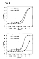
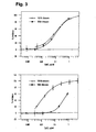
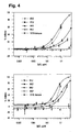
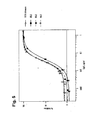
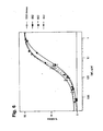
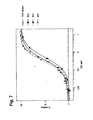
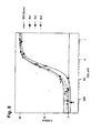
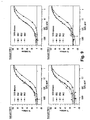
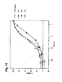
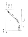
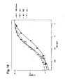
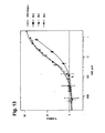

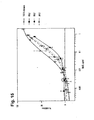


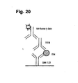
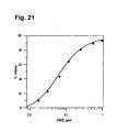
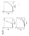
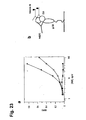


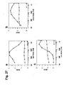
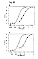
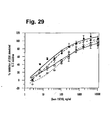

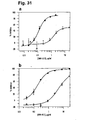
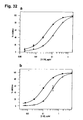


Classifications
-
- C—CHEMISTRY; METALLURGY
- C07—ORGANIC CHEMISTRY
- C07K—PEPTIDES
- C07K16/00—Immunoglobulins [IGs], e.g. monoclonal or polyclonal antibodies
- C07K16/18—Immunoglobulins [IGs], e.g. monoclonal or polyclonal antibodies against material from animals or humans
- C07K16/24—Immunoglobulins [IGs], e.g. monoclonal or polyclonal antibodies against material from animals or humans against cytokines, lymphokines or interferons
- C07K16/244—Interleukins [IL]
- C07K16/248—IL-6
-
- C—CHEMISTRY; METALLURGY
- C07—ORGANIC CHEMISTRY
- C07K—PEPTIDES
- C07K16/00—Immunoglobulins [IGs], e.g. monoclonal or polyclonal antibodies
- C07K16/42—Immunoglobulins [IGs], e.g. monoclonal or polyclonal antibodies against immunoglobulins
-
- A—HUMAN NECESSITIES
- A61—MEDICAL OR VETERINARY SCIENCE; HYGIENE
- A61K—PREPARATIONS FOR MEDICAL, DENTAL OR TOILETRY PURPOSES
- A61K39/00—Medicinal preparations containing antigens or antibodies
- A61K39/395—Antibodies; Immunoglobulins; Immune serum, e.g. antilymphocytic serum
-
- A—HUMAN NECESSITIES
- A61—MEDICAL OR VETERINARY SCIENCE; HYGIENE
- A61P—SPECIFIC THERAPEUTIC ACTIVITY OF CHEMICAL COMPOUNDS OR MEDICINAL PREPARATIONS
- A61P11/00—Drugs for disorders of the respiratory system
-
- A—HUMAN NECESSITIES
- A61—MEDICAL OR VETERINARY SCIENCE; HYGIENE
- A61P—SPECIFIC THERAPEUTIC ACTIVITY OF CHEMICAL COMPOUNDS OR MEDICINAL PREPARATIONS
- A61P11/00—Drugs for disorders of the respiratory system
- A61P11/06—Antiasthmatics
-
- A—HUMAN NECESSITIES
- A61—MEDICAL OR VETERINARY SCIENCE; HYGIENE
- A61P—SPECIFIC THERAPEUTIC ACTIVITY OF CHEMICAL COMPOUNDS OR MEDICINAL PREPARATIONS
- A61P17/00—Drugs for dermatological disorders
- A61P17/06—Antipsoriatics
-
- A—HUMAN NECESSITIES
- A61—MEDICAL OR VETERINARY SCIENCE; HYGIENE
- A61P—SPECIFIC THERAPEUTIC ACTIVITY OF CHEMICAL COMPOUNDS OR MEDICINAL PREPARATIONS
- A61P19/00—Drugs for skeletal disorders
- A61P19/02—Drugs for skeletal disorders for joint disorders, e.g. arthritis, arthrosis
-
- A—HUMAN NECESSITIES
- A61—MEDICAL OR VETERINARY SCIENCE; HYGIENE
- A61P—SPECIFIC THERAPEUTIC ACTIVITY OF CHEMICAL COMPOUNDS OR MEDICINAL PREPARATIONS
- A61P25/00—Drugs for disorders of the nervous system
- A61P25/04—Centrally acting analgesics, e.g. opioids
-
- A—HUMAN NECESSITIES
- A61—MEDICAL OR VETERINARY SCIENCE; HYGIENE
- A61P—SPECIFIC THERAPEUTIC ACTIVITY OF CHEMICAL COMPOUNDS OR MEDICINAL PREPARATIONS
- A61P25/00—Drugs for disorders of the nervous system
- A61P25/28—Drugs for disorders of the nervous system for treating neurodegenerative disorders of the central nervous system, e.g. nootropic agents, cognition enhancers, drugs for treating Alzheimer's disease or other forms of dementia
-
- A—HUMAN NECESSITIES
- A61—MEDICAL OR VETERINARY SCIENCE; HYGIENE
- A61P—SPECIFIC THERAPEUTIC ACTIVITY OF CHEMICAL COMPOUNDS OR MEDICINAL PREPARATIONS
- A61P29/00—Non-central analgesic, antipyretic or antiinflammatory agents, e.g. antirheumatic agents; Non-steroidal antiinflammatory drugs [NSAID]
-
- A—HUMAN NECESSITIES
- A61—MEDICAL OR VETERINARY SCIENCE; HYGIENE
- A61P—SPECIFIC THERAPEUTIC ACTIVITY OF CHEMICAL COMPOUNDS OR MEDICINAL PREPARATIONS
- A61P35/00—Antineoplastic agents
-
- A—HUMAN NECESSITIES
- A61—MEDICAL OR VETERINARY SCIENCE; HYGIENE
- A61P—SPECIFIC THERAPEUTIC ACTIVITY OF CHEMICAL COMPOUNDS OR MEDICINAL PREPARATIONS
- A61P37/00—Drugs for immunological or allergic disorders
-
- A—HUMAN NECESSITIES
- A61—MEDICAL OR VETERINARY SCIENCE; HYGIENE
- A61P—SPECIFIC THERAPEUTIC ACTIVITY OF CHEMICAL COMPOUNDS OR MEDICINAL PREPARATIONS
- A61P43/00—Drugs for specific purposes, not provided for in groups A61P1/00-A61P41/00
-
- A—HUMAN NECESSITIES
- A61—MEDICAL OR VETERINARY SCIENCE; HYGIENE
- A61P—SPECIFIC THERAPEUTIC ACTIVITY OF CHEMICAL COMPOUNDS OR MEDICINAL PREPARATIONS
- A61P9/00—Drugs for disorders of the cardiovascular system
-
- A—HUMAN NECESSITIES
- A61—MEDICAL OR VETERINARY SCIENCE; HYGIENE
- A61P—SPECIFIC THERAPEUTIC ACTIVITY OF CHEMICAL COMPOUNDS OR MEDICINAL PREPARATIONS
- A61P9/00—Drugs for disorders of the cardiovascular system
- A61P9/10—Drugs for disorders of the cardiovascular system for treating ischaemic or atherosclerotic diseases, e.g. antianginal drugs, coronary vasodilators, drugs for myocardial infarction, retinopathy, cerebrovascula insufficiency, renal arteriosclerosis
-
- C—CHEMISTRY; METALLURGY
- C07—ORGANIC CHEMISTRY
- C07K—PEPTIDES
- C07K16/00—Immunoglobulins [IGs], e.g. monoclonal or polyclonal antibodies
- C07K16/18—Immunoglobulins [IGs], e.g. monoclonal or polyclonal antibodies against material from animals or humans
- C07K16/24—Immunoglobulins [IGs], e.g. monoclonal or polyclonal antibodies against material from animals or humans against cytokines, lymphokines or interferons
-
- A—HUMAN NECESSITIES
- A61—MEDICAL OR VETERINARY SCIENCE; HYGIENE
- A61K—PREPARATIONS FOR MEDICAL, DENTAL OR TOILETRY PURPOSES
- A61K39/00—Medicinal preparations containing antigens or antibodies
- A61K2039/505—Medicinal preparations containing antigens or antibodies comprising antibodies
-
- C—CHEMISTRY; METALLURGY
- C07—ORGANIC CHEMISTRY
- C07K—PEPTIDES
- C07K2317/00—Immunoglobulins specific features
- C07K2317/20—Immunoglobulins specific features characterized by taxonomic origin
- C07K2317/24—Immunoglobulins specific features characterized by taxonomic origin containing regions, domains or residues from different species, e.g. chimeric, humanized or veneered
-
- C—CHEMISTRY; METALLURGY
- C07—ORGANIC CHEMISTRY
- C07K—PEPTIDES
- C07K2317/00—Immunoglobulins specific features
- C07K2317/30—Immunoglobulins specific features characterized by aspects of specificity or valency
- C07K2317/33—Crossreactivity, e.g. for species or epitope, or lack of said crossreactivity
-
- C—CHEMISTRY; METALLURGY
- C07—ORGANIC CHEMISTRY
- C07K—PEPTIDES
- C07K2317/00—Immunoglobulins specific features
- C07K2317/70—Immunoglobulins specific features characterized by effect upon binding to a cell or to an antigen
- C07K2317/76—Antagonist effect on antigen, e.g. neutralization or inhibition of binding
Definitions
- the present invention relates to immunoglobulins that specifically bind Oncostatin M (OSM) and in particular human OSM (hOSM). More particularly, the present invention relates to antibodies that specifically bind hOSM.
- the present invention also concerns methods of treating diseases or disorders with said immunoglobulins, pharmaceutical compositions comprising said immunoglobulins and methods of manufacture. Other aspects of the present invention will be apparent from the description below.
- Oncostatin M is a 28 KDa glycoprotein that belongs to the interleukin 6 (IL-6) family of cytokines which includes IL-6, Leukaemia Inhibitory Factor (LIF), ciliary neurotrophic factor (CNTF), cardiotropin-1 (CT-1) and cardiotrophin-1 like cytokine (See Kishimoto T et al (1995) Blood 86: 1243-1254 ), which share the gp130 transmembrane signalling receptor (See Taga T and Kishimoto T (1997) Annu. Rev. Immunol. 15: 797-819 ).
- IL-6 interleukin 6
- LIF Leukaemia Inhibitory Factor
- CNTF ciliary neurotrophic factor
- CT-1 cardiotropin-1
- cardiotrophin-1 like cytokine See Kishimoto T et al (1995) Blood 86: 1243-1254 ), which share the gp130 transmembrane signalling receptor (See Taga T and Kishimoto T (1997) Annu.
- OSM was originally discovered by its ability to inhibit the growth of the melanoma cell line A375 (See Malik N (1989) et al Mol Cell Biol 9: 2847-2853 ). Subsequently, more effects were discovered and it was found to be a multifunctional mediator like other members of the IL-6 family.
- OSM is produced in a variety of cell types including macrophages, activated T cells (See Zarling JM (1986) PNAS (USA) 83: 9739-9743 ), polymorphonuclear neutrophils (See Grenier A et al (1999) Blood 93:1413-1421 ), eosinophils (See Tamura S et al (2002) Dev. Dyn.
- Receptors for OSM are expressed on a wide range of cells including epithelial cells, chondrocytes, fibroblasts (See Langdon C et al (2003) J Immunol 170: 548-555 ), neuronal smooth muscle, lymph node, bone, heart, small intestine, lung and kidney (See Tamura S et al (2002) Mech Dev 115:127-131 ) and endothelial cells.
- endothelial cells are a primary target for OSM.
- OSM othelial growth factor
- OSM binds to the transmembrane signal transducing glycoprotein gp130.
- a key feature of the gp130 cytokines is the formation of oligomeric receptor complexes that comprise gp130 and one or more co-receptors depending on the ligand (Reviewed in Heinrich PC et al (2003) Biochem J. 374: 1-20 ). As a result, these cytokines can mediate both the shared and unique biological activities in vitro and in vivo depending on the composition of the receptor complex formed.
- hOSM Human OSM
- Figure 1 illustrates the interaction between hOSM and gp130, LIFR and OSMR.
- the crystal structure of hOSM has been solved and shown to comprise a four ⁇ helical bundle with two potential glycosylation sites. Two separate ligand binding sites have been identified by site-directed mutagenesis on the hOSM molecule (See Deller MC et al (2000) Structural Fold Des. 8:863-874 ).
- Site II (sometimes “site 2") interacts with gp130 and the second site, called Site III (sometimes "site 3"), at the opposite end of the molecule interacts with either LIFR or OSMR.
- Site II (sometimes "site 2") interacts with gp130
- Site III (sometimes "site 3"), at the opposite end of the molecule interacts with either LIFR or OSMR. Mutagenesis experiments have shown that the binding sites for LIFR and OSMR are almost identical but that a single amino acid mutation can discriminate between the two.
- OSM is synthesised as a proprotein containing a hydrophobic 25 amino acid (AA) N termimal signal sequence and a C-terminal propeptide of 33 AA, both of which are cleaved to generate mature OSM.
- the OSM proprotein does have biological activity but this is significantly increased by cleavage of the C terminal propeptide (see Bruce A.G. et al (1992) Prog.Growth Factor Res. 4: 157-170 , Malik N et al (1989) Mol.Cell Biol. 9: 2847-2853 ).
- OSM has been described as a "compact, barrel-shaped molecule" with dimensions of approximately 20 ⁇ x 27 ⁇ x 561 ⁇ .
- Helices A and C contain "kinks". The helices are joined by two overhand loops (AB loop 38-66AA, CD loop 130-158 AA) and are arranged as two anti-parallel pairs (A-D and B-C). (See Deller M.C et al (2000) Structure 8; 863-874 ).
- OSM binding via Site II to gp130 allows binding of another OSM molecule to gp130 by a Site III interaction.
- OSM will also bind to either LIFR or OSMR via Site III.
- OSM forms a complex with its receptor consisting of; one gp130, one LIFR or OSMR, and two OSM molecules.
- the important residues for Site II OSM-gp130 binding are Gln20, Gly120, Gln16 and Asn124.
- the important residues are Phe160 and Lys163.
- the OSM Site II interaction is therefore dependent on Gln20, Gly120, Asn124 and to a lesser extent Gln16 on hOSM.
- Three complementary residues in gp130 (Phe169. Tyr196 and Glu282) have been identified as of particular note in the interaction between OSM and gp130.
- amino acid sequence starting at position 1 for hOSM is set forth as SEQ.I.D.NO: 13
- Rheumatoid arthritis comprises a syndrome of distinct but interconnected pathogenic processes. These are: local and systemic inflammation, proliferation of synovial cells, angiogenesis and matrix deposition leading to formation of pannus tissue which invades and destroys cartilage and bone, resulting in deformity and disability. Underpinning this pathology is the chronic release of cytokines and inflammatory mediators from cells that enter and take up residence in the inflamed joint and from endogenous joint tissue cells (See Firestein G (2003) in Rheumatology. Eds Hochberg, Silman, Smolen, Weinblatt and Weisman. Pub. Mosby. 855-884 ).
- T lymphocytes The initiating events in RA are unknown but a wealth of evidence suggests that they involve activation of T lymphocytes by either a foreign or autologous "self" antigen (See Firestein G (2004) J Clin Invest 114: 471-4 ).
- the extent to which T cells are required to maintain the ongoing disease processes once they have been initiated is also uncertain although therapeutic agents such as CTLA4Ig, which specifically target T cells can be effective in advanced disease (See Kremer JM et al (2003) New Engl J Med 349: 1907-15 , Moreland L et al (2004) Annual meeting of the American College of Rheumatology Abstract 1475 ).
- rheumatoid synovitis The earliest events in the development of rheumatoid synovitis involve recruitment of mononuclear and polymorphonuclear cells to cross the endothelium in capillaries in the synovial-lining layer. While the polymorphs migrate into synovial fluid (SF) the lymphocytes remain close to the capillaries and may subsequently become organised into ectopic lymphoid follicles. This influx of immune cells is followed by proliferation of fibroblast-like synoviocytes (FLS). Unlike their normal counterparts, RA FLS appear to have escaped from the regulatory processes that result in arrest of proliferation and apoptosis leading to their continuing accumulation (See Yamanishi Y et al (2004) Arthritis Res Ther 7: 12-18 ).
- pannus tissue now develops new blood vessels supported by extracellular matrix to allow further expansion.
- This process involving fibroblast proliferation, matrix -remodelling and angiogenesis closely resembles an uncontrolled wound-healing event.
- Monocytes migrate into the developing pannus tissue and undergo differentation into macrophages with a chronically activated phenotype.
- B cells undergo terminal differentiation to form long-lived plasma cells which secrete antibodies including rheumatoid factors.
- the ability of the inflamed synovium to sustain local differentation of myeloid and lymphoid cells is based, in part, on local production of growth factors such as GMCSF and IL-6.
- RA is a systemic disease, probably as a result of the passage of inflammatory mediators from the joint into the blood. This affects many organ systems in the body including skin, eyes, liver, kidneys, brain and the vascular lining, leading to increased morbidity and mortality (See Matteson EL (2003) in Rheumatology. Eds Hochberg, Silman, Smolen, Weinblatt and Weisman. Pub. Mosby. 781-792 ). Much of the excess mortality is due to cardiovascular disease caused by atherosclerosis since many of the pathogenic processes involved in the development of rheumatoid synovitis are common to the formation of atherosclerotic plaques.
- RA Treatments for RA aim to control pain reduce inflammation and arrest the processes that result in tissue destruction.
- NSAIDS non-steroidal anti-inflammatory drugs
- DARDS disease modifying anti-rheumatic drugs
- MTX methotrexate
- sulphasalazine gold and Leflunomide.
- immunoglobulins especially antibodies that specifically bind OSM (e.g. hOSM, particularly Site II thereof) and modulate (i.e. inhibit or block) the interaction between OSM and gp130 in the treatment of diseases and disorders responsive to modulation of that interaction.
- OSM OSM is found in the SF of human RA patients (See Hui W et al (1997) 56: 184-7). These levels correlate with; the number of neutrophils in SF, levels of TNF alpha (sometimes "TNF") in SF, and markers of cartilage destruction ( Manicourt DH et al (2000) Arthritis Rheum 43: 281-288 ). Furthermore, the synovial tissue from RA patients secretes OSM spontaneously ex vivo (See Okamoto H et al (1997) Arthritis and Rheumatism 40: 1096-1105 ).
- OSM is present in synovial macrophages ( Cawston TE et al (1998) Arthritis Rheum 41: 1760-1771 ) and as discussed earlier, OSM receptors and gp130 are expressed on endothelial cells, synovial fibroblasts, chonodrocytes and osteoblasts. Furthermore, cells infiltrating atherosclerotic plaques and aortic aneurysms express OSM suggesting an association of this cytokine with chronic inflammation (See Mirshahi F et al (2001) Ann NY Acad Sci 936: 621-4 ).
- Endothelial cells express ten to twenty times the number of OSM receptors than other cell types (See Brown TJ et al (1991) J Immunol 147: 2175-2180 , Linsley PS et al (1989) J Biol Chem 264: 4282-4289 ).
- OSM alone, or synergistically in combination with other cytokines activates endothelium to release cytokines and chemokines and bind neutrophils, monocytes and lymphocytes mediating their extravasation into synovial tissue (See Modur V et al (1997) J Clin Invest 100: 158-168 ).
- OSM has also been demonstrated to be a potent stimulator of angiogenesis (See Vasse M et al (1999) Aterioscler Thromb Vasc Biol 19: 1835-1842 ) and activation and proliferation of synovial fibroblast (FLS) cells (thus facilitating the formation of pannus tissue, the release of IL-6, MMPs) and acts synergistically with TNF and IL-1 to induce this mediator release (See Langdon C et al (2000) Am J Pathol 157: 1187-1196 ). OSM has also been demonstrated to induce (with IL-1) collagen and proteoglycan release from cartilage (See Cawston T et al (1995) Biochem Biophys Res Commun 215: 377-385 ).
- OSM induces acute phase protein release and production of IL-6 receptor from hepatocytes (See Cichy J et al (1997) J Immunol 159: 5648-5643 , Kurash JK (2004) Exp Cell Res 292: 342-58 ) and may therefore contribute to the systemic effects of rheumatoid inflammation including fatigue.
- OSM induces osteoclast differentiation and activity in vitro (See Palmqvist P et al (2002) J Immunol 169: 3353-3362 ).
- Adenoviral expression of murine OSM (mOSM) in the joints of normal mice results in a severe inflammatory and erosive arthritis (See Langdon C et al (2000) Am J Pathol 157: 1187-1196 ).
- mOSM murine OSM
- aggressive disease is seen in knockout mice lacking TNF, IL-1, IL-6 and iNOS following adenoviral mOSM delivery (See de Hooge ASK et al (2003) Arthritis and Rheumatism 48:1750-1761 ), demonstrating that OSM can mediate all aspects of arthritis pathology.
- OSM injected subcutaneously induces an acute phase response and local chronic inflammation (See Loy JK et al (1999) Toxicol Pathol 27: 151 - 155 ). OSM has been demonstrated to induce mononuclear and PMN infiltration and proteoglycan release when injected into goat joints (See Bell MC et al (1999) Arthritis Rheum 42: 2543-2551 ). Transgenic over-expression of mOSM in mouse lymph nodes results in extrathymic T cell maturation, proliferation of memory T cells and failure to deplete autoimmune T cells (See Louis I et al (2003) Blood 102: 1397-1404 ). Transgenic over-expression of OSM in the pancreas causes extensive fibrosis similar to that seen in advanced RA synovium (See Malik N et al (1995) Mol Cell Biol 15: 2349-2358 ).
- the present inventors postulate that modulating (in particular blocking) the interaction between Site II of hOSM and gp130, with an antibody that specifically binds hOSM will modulate signalling by all of the potential OSM receptor complexes, effectively neutralising the biological activity of the cytokine to a therapeutically significant degree. Notwithstanding this, the present inventors have found that blockade of both the Site II and Site III sites of hOSM surprisingly improves neutralisation of this cytokine. Furthermore, the present inventors have found that the glycosylation of hOSM plays an unexpected role in the binding event between hOSM and an antibody that specifically binds hOSM.
- the present invention therefore provides a therapeutic antibody 15E10 or 10D3 (which maybe chimaeric, human, humanised, bispecific or antigen binding fragments thereof) which specifically binds hOSM and interacts with Site II of hOSM. See Table A below.
- a therapeutic antibody or antigen binding fragment thereof which specifically binds hOSM and modulates (i.e inhibits or blocks) the interaction between Site II of hOSM and gp130.
- the therapeutic antibody or antigen binding fragment thereof specifically binds Site II of hOSM.
- a therapeutic antibody or antigen binding fragment thereof which specifically binds hOSM and comprises the following CDRH3: SEQ.I.D.NO: 3 or SEQ.LD.NO:42.
- CDR CDR
- CDRL1 CDRL2
- CDRH3 CDRH3
- a murine therapeutic antibody or antigen binding fragment thereof comprising a V H domain having the sequence: SEQ.I.D.NO: 7 and a V L domain having the sequence: SEQ.I.D.NO: 8.
- a murine therapeutic antibody or antigen binding fragment thereof comprising a V H domain having the sequence: SEQ.I.D.NO: 46 and a V L domain having the sequence: SEQ.I.D.NO: 47.
- a humanised therapeutic antibody or antigen binding fragment thereof comprising a V H chain having the sequence set forth in SEQ.I.D.NO: 9 and a V L domain having the sequence set forth in SEa.I.D.NO:10.
- a humanised therapeutic antibody or antigen binding fragment thereof comprising a V H chain having the sequence set forth in SEQ.I.D.NO: 48 and a V L domain having the sequence set forth in SEQ.I.D.NO:49.
- a humanised therapeutic antibody which antibody comprises a heavy chain having the sequence set forth in SEQ.I.D.NO: 11 and a light chain having the sequence set forth in SEQ.I.D.NO:12.
- a humanised therapeutic antibody which antibody comprises a heavy chain having the sequence set forth in SEQ.I.D.NO: 50 and a light chain having the sequence set forth in SEQ.I.D.NO:51.
- a humanised therapeutic antibody or antigen binding fragment thereof which modulates (i.e. inhibits or blocks) the interaction between hOSM and gp130.
- an isolated V H domain of an antibody comprising (or consisting essentially of) SEQ.I.D.NO: 7 or SEQ.I.D.NO:9 or SEQ.I.D.NO:46 or SEQ.I.D.NO:48.
- a therapeutic antibody or antigen binding fragment thereof comprising a V H domain selected from the group consisting of; SEQ.I.D.NO: 7, SEQ.I.D.NO:9, SEQ.I.D.NO:46, SEQ.I.D.NO:48.
- a therapeutic antibody or antigen binding fragment thereof which competitively inhibits the binding of the therapeutic antibody comprising a CDRH3 of SEQ.I.D.NO:3.
- a therapeutic antibody or antigen binding fragment thereof which competitively inhibits the binding of the therapeutic antibody comprising CDRs of SEQ.I.D. NO: 1,2,3,4, 5 and 6 with hOSM.
- therapeutic antibody or antigen binding fragment thereof which competitively inhibits the binding of the therapeutic antibody comprising a heavy chain of SEQ.I.D.NO:11 and a light chain of SEQ.I.D.NO:12 with hOSM.
- a method of treating a human patient afflicted with a disease or disorder responsive to modulation of the interaction between hOSM and gp130 comprises the step of administering to said patient a therapeutically effective amount of the therapeutic antibody or antigen binding fragment thereof as described herein.
- a method of treating a human patient afflicted with an inflammatory disease or disorder comprises the step of administering to said patient a therapeutically effective amount of the therapeutic antibody or antigen binding fragment thereof as described herein.
- a method of treating a human patient afflicted with an arthritic disease particularly rheumatoid arthritis, juvenile onset arthritis or osteoarthritis which method comprises the step of administering to said patient a therapeutically effective amount of the therapeutic antibody or antigen binding fragment thereof as described herein.
- a method of reducing or preventing cartilage degradation in a human patient afflicted with (or suspectible to) such degradation comprises the step of administering a therapeutically effective amount of a therapeutic antibody or antigen binding fragment thereof to said patient as described herein.
- a method of treating the extraarticular manifestations of an arthritic disease or disorder comprises the step of administering a therapeutically effective amount of a therapeutic antibody or antigen binding fragment thereof as described herein to the human patient afflicted with the extraarticular manifestations of an arthritic disease or disorder.
- a method of treating a human patient afflicted with a disease of endothelial cell origin comprises the steps of administering to said patient a therapeutically effective amount of a therapeutic antibody or antigen binding fragment thereof as described herein.
- an assay for studying the interaction between OSM (particularly hOSM) and an interacting partner (such as gp130, LIFR, OSMR), which assay comprises the step of providing for said studying, a sample of glycosylated OSM (typically glycosylated by a vertebrate host cell such as mammalian host cell e.g. CHO glycosylated).
- OSM particularly hOSM
- an interacting partner such as gp130, LIFR, OSMR
- a therapeutic antibody that specifically binds native glycosylated hOSM and modulates (i.e. inhibits or blocks) the interaction between native glycosylated hOSM and an interacting partner selected from the group consisting of gp130, LIFR, OSMR ⁇ .
- Intact antibodies are usually heteromultimeric glycoproteins comprising at least two heavy and two light chains. Aside from IgM, intact antibodies are heterotetrameric glycoproteins of approximately 150Kda, composed of two identical light (L) chains and two identical heavy (H) chains. Typically, each light chain is linked to a heavy chain by one covalent disulfide bond while the number of disulfide linkages between the heavy chains of different immunoglobulin isotypes varies. Each heavy and light chain also has intrachain disulfide bridges. Each heavy chain has at one end a variable domain (V H ) followed by a number of constant regions.
- V H variable domain
- Each light chain has a variable domain (V L ) and a constant region at its other end; the constant region of the light chain is aligned with the first constant region of the heavy chain and the light chain variable domain is aligned with the variable domain of the heavy chain.
- V L variable domain
- the light chains of antibodies from most vertebrate species can be assigned to one of two types called Kappa and Lambda based on the amino acid sequence of the constant region.
- human antibodies can be assigned to five different classes, IgA, IgD, IgE, IgG and IgM.
- IgG and IgA can be further subdivided into subclasses, IgG1, IgG2, IgG3 and IgG4; and IgA1 and IgA2.
- Species variants exist with mouse and rat having at least IgG2a, IgG2b.
- the variable domain of the antibody confers binding specificity upon the antibody with certain regions displaying particular variability called complementarity determining regions (CDRs).
- CDRs complementarity determining regions
- the more conserved portions of the variable region are called framework regions (FR).
- the variable domains of intact heavy and light chains each comprise four FR connected by three CDRs.
- the CDRs in each chain are held together in close proximity by the FR regions and with the CDRs from the other chain contribute to the formation of the antigen binding site of antibodies.
- the constant regions are not directly involved in the binding of the antibody to the antigen but exhibit various effector functions such as participation in antibody dependent cell-mediated cytotoxicity (ADCC), phagocytosis via binding to Fc ⁇ receptor, half-life/clearance rate via neonatal Fc receptor (FcRn) and complement dependent cytotoxicity via the C1q component of the complement cascade.
- ADCC antibody dependent cell-mediated cytotoxicity
- FcRn neonatal Fc receptor
- complement dependent cytotoxicity via the C1q component of the complement cascade.
- an intact therapeutic antibody that specifically binds hOSM, which antibody modulates the interaction between hOSM and gp130.
- the antibody may specifically bind Site II of hOSM and inhibit or block the interaction between hOSM and its corresponding residues on gp130 involved in OSM interaction.
- the ELISA protocol of the examples may be used to determine whether any particular antibody or antigen binding fragment thereof modulates the interaction between hOSM and gp130.
- the intact therapeutic antibody may comprise a constant region (either heavy or light) of any isotype or subclass thereof described supra.
- the antibody is of the IgG isotype, particularly IgG1.
- the antibody may be rat, mouse, rabbit, primate or human.
- the antibody is primate (such as cynomolgus, Old World monkey or Great Ape, see e.g. WO99/55369 , WO93/02108 ) or human.
- an intact therapeutic antibody comprising a CDRH3 of SEQ.I.D.NO: 3 or SEQ.I.D.NO:42.
- an intact therapeutic antibody comprising a variable region having CDRs of SEQ.I.D.NO: 1, 2,3,4,5 and 6 or a variable region of SEQ.I.D.NO:40, 41,42,43,44 and 45.
- a murine intact therapeutic antibody or antigen binding fragment thereof comprising a V H domain having the sequence of SEQ.I.D.NO: 7 and a V L domain of the sequence of SEQ.I.D.NO: 8.
- a murine intact therapeutic antibody or antigen binding fragment thereof comprising a V H domain having the sequence of SEQ.I.D.NO: 46 and a V L domain of the sequence of SEQ.I.D.NO: 47.
- Human antibodies may be produced by a number of methods known to those of skill in the art. Human antibodies can be made by the hybridoma method using human myeloma or mouse-human heteromyeloma cells lines see Kozbor J.Immunol 133, 3001, (1984 ) and Brodeur, Monoclonal Antibody Production Techniques and Applications, pp51-63 (Marcel Dekker Inc, 1987 ). Alternative methods include the use of phage libraries or transgenic mice both of which utilize human V region repertories (see Winter G, (1994), Annu.Rev.lmmunol 12,433-455 , Green LL (1999), J.Immunol.methods 231, 11-23 ).
- mice Several strains of transgenic mice are now available wherein their mouse immunoglobulin loci has been replaced with human immunoglobulin gene segments (see Tomizuka K, (2000) PNAS 97,722-727 ; Fishwild D.M (1996) Nature Biotechnol. 14,845-851 , Mendez MJ, 1997, Nature Genetics, 15,146-156 ). Upon antigen challenge such mice are capable of producing a repertoire of human antibodies from which antibodies of interest can be selected.
- Phage display technology can be used to produce human antibodies (and fragments thereof), see McCafferty; Nature, 348, 552-553 (1990 ) and Griffiths AD et al (1994) EMBO 13:3245-3260 .
- antibody V domain genes are cloned in frame into either a major or minor coat of protein gene of a filamentous bacteriophage such as M13 or fd and displayed (usually with the aid of a helper phage) as functional antibody fragments on the surface of the phage particle. Selections based on the functional properties of the antibody result in selection of the gene encoding the antibody exhibiting those properties.
- the phage display technique can be used to select antigen specific antibodies from libraries made from human B cells taken from individuals afflicted with a disease or disorder described above or alternatively from unimmunized human donors (see Marks; J.Mol.Bio. 222,581-597, 1991 ). Where an intact human antibody is desired comprising a Fc domain it is necessary to reclone the phage displayed derived fragment into a mammalian expression vectors comprising the desired constant regions and establishing stable expressing cell lines.
- affinity maturation Marks; Bio/technol 10,779-783 (1992 )
- affinity maturation Marks; Bio/technol 10,779-783 (1992 )
- Variants of this technique such as "epitope imprinting" are now also available see WO 93/06213 . See also Waterhouse; Nucl.Acids Res 21, 2265-2266 (1993 ).
- a human intact therapeutic antibody or antigen binding fragment thereof which specifically binds hOSM and modulates (i.e. inhibits or blocks) the interaction between hOSM and gp130.
- a human intact therapeutic antibody or antigen binding fragment thereof which specifically binds Site II of hOSM and modulates (i.e. inhibits or blocks) the interaction between hOSM and gp130.
- a human intact therapeutic antibody or antigen binding fragment thereof comprising a CDRH3 of SEQ.I.D.NO: 3 or SEQ.I.D.NO:42 which specifically binds hOSM and modulates (i.e. inhibits or blocks) the interaction between hOSM and gp130.
- a human intact therapeutic antibody or antigen binding fragment thereof comprising a variable region having CDRs of SEQ.I.D.NO: 1, 2, 3, 4, 5 and 6 or a variable region having SEQ.I.D.NO:40,41,42,43,44 and 45.
- chimaeric antibodies which generally comprise a non-human (e.g. rodent such as mouse) variable domain fused to a human constant region.
- Chimaeric antibodies are typically produced using recombinant DNA methods.
- DNA encoding the antibodies e.g. cDNA
- oligonucleotide probes that are capable of binding specifically to genes encoding the H and L chains of the antibody of the invention, e.g. DNA encoding SEQ.I.D.NO 1,2,3,4,5 and 6 described supra ).
- Hybridoma cells serve as a typical source of such DNA.
- the DNA is placed into expression vectors which are then transfected into host cells such as E.Coli, COS cells, CHO cells or myeloma cells that do not otherwise produce immunoglobulin protein to obtain synthesis of the antibody.
- host cells such as E.Coli, COS cells, CHO cells or myeloma cells that do not otherwise produce immunoglobulin protein to obtain synthesis of the antibody.
- the DNA may be modified by substituting the coding sequence for human L and H chains for the corresponding non-human (e.g. murine) H and L constant regions see e.g. Morrison; PNAS 81, 6851 (1984 ).
- the second approach involves the generation of humanised antibodies wherein the non-human content of the antibody is reduced by humanizing the variable regions.
- Two techniques for humanisation have gained popularity.
- the first is humanisation by CDR grafting.
- CDRs build loops close to the antibody's N-terminus where they form a surface mounted in a scaffold provided by the framework regions.
- Antigen-binding specificity of the antibody is mainly defined by the topography and by the chemical characteristics of its CDR surface. These features are in turn determined by the conformation of the individual CDRs, by the relative disposition of the CDRs, and by the nature and disposition of the side chains of the residues comprising the CDRs.
- a large decrease in immunogenicity can be achieved by grafting only the CDRs of a non-human (e.g.
- human V regions showing the greatest sequence homology (typically 60% or greater) to the non-human donor antibody maybe chosen from a database in order to provide the human framework (FR).
- the selection of human FRs can be made either from human consensus or individual human antibodies. Where necessary key residues from the donor antibody are substituted into the human acceptor framework to preserve CDR conformations.
- Computer modelling of the antibody maybe used to help identify such structurally important residues, see WO99/48523
- humanisation maybe achieved by a process of "veneering".
- a statistical analysis of unique human and murine immunoglobulin heavy and light chain variable regions revealed that the precise patterns of exposed residues are different in human and murine antibodies, and most individual surface positions have a strong preference for a small number of different residues (see Padlan E.A. et al; (1991) Mol.Immunol.28, 489-498 and Pedersen J.T. et al (1994) J. Mol.Biol. 235; 959-973 ). Therefore it is possible to reduce the immunogenicity of a non-human Fv by replacing exposed residues in its framework regions that differ from those usually found in human antibodies.
- a chimaeric therapeutic antibody comprising a non-human (e.g. rodent) variable domain fused to a human constant region (which maybe of a IgG isotype e.g. IgG1) which specifically binds hOSM and modulates the interaction between Site II of hOSM and gp130.
- a non-human variable domain fused to a human constant region (which maybe of a IgG isotype e.g. IgG1) which specifically binds hOSM and modulates the interaction between Site II of hOSM and gp130.
- a chimaeric therapeutic antibody comprising a non-human (e.g. rodent) variable region and a human constant region (which maybe of an IgG isotype e.g. IgG1) which specifically binds hOSM, which antibody further comprises a CDRH3 of SEQ.I.D.NO:3 or SEO.I.D.NO:42.
- Such antibodies may further comprise a human constant region of the IgG isotype, e.g. IgG1
- a chimaeric therapeutic antibody comprising a non-human (e.g. rodent) variable region and a human constant region (which maybe of a IgG isotype e.g. IgG1) which specifically binds hOSM having the CDRs of SEQ.I.D.NO: 1, 2,3,4,5 and 6 or SEQ.I.D.NO:40, 41,42,43,44 and 45.
- a humanised therapeutic antibody or antigen binding fragment thereof which specifically binds hOSM and modulates (i.e. inhibits or blocks) the interaction between Site II of hOSM and gp130.
- a humanised therapeutic antibody or antigen binding fragment thereof which specifically binds hOSM and comprises a CDRH3 of SEQ.I.D.NO: 3 or SEQ.I.D.NO:42.
- Such antibodies may comprise a human constant region of the IgG isotype, e.g. IgG1.
- a humanised therapeutic antibody or antigen binding fragment thereof which specifically binds hOSM and comprises CDRs of SEQ.I.D.NO: 1, 2,3,4,5 and 6 or SEQ.I.D.NO:40, 41, 42, 43, 44 and 45.
- Such antibodies may comprise a human constant region of the IgG isotype, e.g. IgG1.
- a humanised therapeutic antibody or antigen binding fragment thereof which specifically binds hOSM and modulates the interaction between hOSM and gp130 and comprises (or consists essentially of) the heavy chain of SEQ.I.D.NO: 11 and a light chain of SEQ.I.D.NO: 12.
- a humanised therapeutic antibody or antigen binding fragment thereof which specifically binds hOSM and modulates the interaction between hOSM and gp130 which antibody comprises (or consists essentially of) a heavy chain of SEQ.I.D.NO:50 and a light chain of SEQ.I.D.NO:51.
- a humanised therapeutic antibody or antigen binding fragment thereof which specifically binds hOSM and modulates the interaction between hOSM and gp130 wherein said antibody or fragment thereof comprises CDRH3 of SEQ.I.D.NO: 3 optionally further comprising CDRs of SEQ.I.D.NO: 1,2,4,5 and 6 wherein the residues at positions 28,29,30,71 and 94 of the human acceptor heavy chain framework region and positions 49 and 71 of the human acceptor light chain framework are substituted by the corresponding residues found in the donor antibody framework from which CDRH3 is derived.
- a humanised therapeutic antibody or antigen binding fragment thereof which specifically binds hOSM and modulates the interaction between hOSM and gp130
- said antibody or fragment thereof comprises CDRH3 of SEQ.I.D.NO: 42 optionally further comprising CDRs of SEQ.I.D.NO: 40,41,43,44,45 wherein the residues at positions 28,44,48,67,69,71,73 of the human acceptor heavy chain framework region and positions 36,38,46,47,71 of the human acceptor light chain framework are substituted by the corresponding residues found in the donor antibody framework from which CDRH3 is derived
- a humanised therapeutic antibody or antigen binding fragment thereof which specifically binds hOSM wherein said antibody or fragment thereof comprises CDRH3 of SEQ.I.D.NO: 3 optionally further comprising CDRs of SEQ.I.D.NO: 1,2,4,5 and 6 wherein the human heavy chain framework comprises one or more (e.g. all) of the following residues (or a conservative substitute thereof): Position Residue 28 S 29 L 30 T 71 K 94 K and the human light chain comprises either or both of the following residues (or conservative substitute thereof); Position Residue 49 E 71 Y
- a humanised therapeutic antibody or antigen binding fragment thereof which specifically binds hOSM wherein said antibody or fragment thereof comprises CDRs of SEQ.I.D.NO: 1,2,3,4,5 and 6 wherein the human heavy chain framework comprises one or more (e.g. all) of the following residues (or a conservative substitute thereof): Position Residue 28 S 29 L 30 T 71 K 94 K and the human light chain comprises either or both of the following residues (or conservative substitute thereof); Position Residue 49 E 71 Y
- a humanised therapeutic antibody or antigen binding fragment thereof which specifically binds to hOSM wherein said antibody or fragment thereof comprises CDRH3 of SEQ.I.D.NO:42 optionally further comprising CDRs of SEQ.I.D.NO: 40,41,43,44,45 wherein the human heavy chain framework comprises one or more (e.g. all) of the following residues (or a conservative substitute thereof): Position Residue 28 I 48 I 44 K 67 A 69 L 71 V 73 K and the human light chain comprises one or more (e.g. all) of the following residues (or conservative substitute thereof); Position Residue 36 F 38 K 46 R 47 W 71 Y
- a humanised therapeutic antibody or antigen binding fragment thereof which specifically binds to hOSM wherein said antibody or fragment thereof comprises CDRs of SEQ.I.D.NO: 40,41,42,43,44,45 wherein the human heavy chain framework comprises one or more (e.g. all) of the following residues (or a conservative substitute thereof): Position Residue 28 I 48 I 44 K 67 A 69 L 71 V 73 K and the human light chain comprises one or more (e.g.all) of the following residues (or conservative substitute thereof); Position Residue 36 F 38 K 46 R 47 W 71 Y
- a bispecific antibody is an antibody having binding specificities for at least two different epitopes. Methods of making such antibodies are known in the art. Traditionally, the recombinant production of bispecific antibodies is based on the coexpression of two immunoglobulin H chain-L chain pairs, where the two H chains have different binding specificities see Millstein et al, Nature 305 537-539 (1983 ), WO93/08829 and Traunecker et al EMBO, 10, 1991, 3655-3659 . Because of the random assortment of H and L chains, a potential mixture of ten different antibody structures are produced of which only one has the desired binding specificity.
- variable domains with the desired binding specificities to heavy chain constant region comprising at least part of the hinge region, CH2 and CH3 regions. It is preferred to have the CH1 region containing the site necessary for light chain binding present in at least one of the fusions. DNA encoding these fusions, and if desired the L chain are inserted into separate expression vectors and are then cotransfected into a suitable host organism. It is possible though to insert the coding sequences for two or all three chains into one expression vector.
- the Bispecific antibody is composed of a H chain with a first binding specificity in one arm and a H-L chain pair, providing a second binding specificity in the other arm, see WO94/04690 . See also Suresh et al Methods in Enzymology 121, 210, 1986 .
- a bispecific therapeutic antibody wherein at least one binding specificity of said antibody is for hOSM, wherein said antibody modulates (i.e. inhibits or blocks) the interaction between Site II of hOSM and gp130.
- Such antibodies may further comprise a human constant region of the IgG isotype, e.g. IgG1
- a bispecific therapeutic antibody wherein at least one binding specificity of said antibody is for hOSM, wherein said antibody comprises at least one CDRH3 of SEQ.I.D.NO: 3 or SEQ.I.D.NO:42.
- Such antibodies may further comprise a human constant region of the IgG isotype, e.g. IgG1.
- a bispecific therapeutic antibody wherein at least one binding specificity of said antibody is for hOSM, wherein said antibody comprises at least CDRs of SEQ.I.D.NO: 1, 2,3,4,5 and 6 or SEQ.I.D.NO:40, 41,42,43,44 and 45.
- Such antibodies may further comprise a human constant region of the IgG isotype, e.g. IgG1.
- therapeutic antibody fragments which modulate the interaction between OSM (particularly hOSM) and gp130.
- Such fragments may be functional antigen binding fragments of intact and/or humanised and/or chimaeric antibodies such as Fab, Fd, Fab', F(ab') 2 , Fv, ScFv fragments of the antibodies described supra.
- fragments are produced by the proteolytic digestion of intact antibodies by e.g. papain digestion (see for example, WO 94/29348 ) but may be produced directly from recombinantly transformed host cells.
- For the production of ScFv see Bird et al; (1988) Science, 242, 423-426 .
- antibody fragments may be produced using a variety of engineering techniques as described below.
- Fv fragments appear to have lower interaction energy of their two chains than Fab fragments.
- V H and V L domains To stablise the association of the V H and V L domains, they have been linked with peptides ( Bird et al, (1988) Science 242, 423-426 , Huston et al, PNAS, 85, 5879-5883 ), disulphide bridges ( Glockshuber et al, (1990) Biochemistry, 29, 1362-1367 ) and "knob in hole” mutations ( Zhu et al (1997), Protein Sci., 6, 781-788 ).
- ScFv fragments can be produced by methods well known to those skilled in the art see Whitlow et al (1991) Methods companion Methods Enzymol, 2, 97-105 and Huston et al (1993) Int.Rev.Immunol 10, 195-217 .
- ScFv may be produced in bacterial cells such as E.Coli but are more typically produced in eukaryotic cells.
- One disadvantage of ScFv is the monovalency of the product, which precludes an increased avidity due to polyvalent binding, and their short half-life.
- ScFv' bivalent (ScFv') 2 produced from ScFV containing an additional C terminal cysteine by chemical coupling ( Adams et al (1993) Can.Res 53, 4026-4034 and McCartney et al (1995) Protein Eng. 8, 301-314 ) or by spontaneous site-specific dimerization of ScFv containing an unpaired C terminal cysteine residue (see Kipriyanov et al (1995) Cell. Biophys 26, 187-204 ).
- ScFv can be forced to form multimers by shortening the peptide linker to between 3 to 12 residues to form "diabodies", see Holliger et al PNAS (1993), 90, 6444-6448 .
- ScFv-Sc-Fv tandems may also be produced by linking two ScFv units by a third peptide linker, see Kurucz et al (1995) J.Immol.154, 4576-4582 .
- Bispecific diabodies can be produced through the noncovalent association of two single chain fusion products consisting of V H domain from one antibody connected by a short linker to the V L domain of another antibody, see Kipriyanov et al (1998), Int.J.Can 77,763-772 .
- bispecific diabodies can be enhanced by the introduction of disulphide bridges or "knob in hole” mutations as described supra or by the formation of single chain diabodies (ScDb) wherein two hybrid ScFv fragments are connected through a peptide linker see Kontermann et al (1999) J.Immunol.Methods 226 179-188 .
- Tetravalent bispecific molecules are available by e.g. fusing a ScFv fragment to the CH3 domain of an IgG molecule or to a Fab fragment through the hinge region see Coloma et al (1997) Nature Biotechnol. 15, 159-163 .
- tetravalent bispecific molecules have been created by the fusion of bispecific single chain diabodies (see Alt et al, (1999) FEBS Lett 454, 90-94 .
- Smaller tetravalent bispecific molecules can also be formed by the dimerization of either ScFv-ScFv tandems with a linker containing a helix-loop-helix motif (DiBi miniantibodies, see Muller et al (1998) FEBS Lett 432, 45-49 ) or a single chain molecule comprising four antibody variable domains (V H and V L ) in an orientation preventing intramolecular pairing (tandem diabody, see Kipriyanov et al, (1999) J.Mol.Biol.
- Bispecific F(ab')2 fragments can be created by chemical coupling of Fab' fragments or by heterodimerization through leucine zippers (see Shalaby et al, (1992) J.Exp.Med. 175, 217-225 and Kostelny et al (1992), J.Immunol. 148, 1547-1553 ). Also available are isolated V H and V L domains (Domantis pic), see US 6, 248,516 ; US 6,291,158 ; US 6, 172,197 .
- a therapeutic antibody fragment e.g. ScFv, Fab, Fd, Fab', F(ab') 2 ) or an engineered antibody fragment as described supra ) that specifically binds to hOSM and modulates (i.e. inhibits or blocks) the interaction between Site II of hOSM and gp130.
- the therapeutic antibody fragment may comprise a CDRH3 having the sequence of SEQ.I.D.NO: 3 optionally together with CDRs having the sequence set forth in SEQ.I.D.NO: 1,2,4,5 and 6 or a therapeutic antibody fragment comprising a CDRH3 of SEQ.I.D.NO:42 optionally together with CDRs having the sequence set forth in SEQ.I.D.NO: 40,41,43,44 and 45.
- Heteroconjugate antibodies also form an embodiment of the present invention.
- Heteroconjugate antibodies are composed of two covalently joined antibodies formed using any convenient cross-linking methods. See US 4,676,980 .
- Fc ⁇ R Fc receptors
- ADCC antibody-dependent cellular cytotoxicity
- phagocytosis phagocytosis
- half-life/clearance of the antibody Various modifications to the Fc region of antibodies of the invention may be carried out depending on the desired effector property. For example, specific mutations in the Fc region to render an otherwise lytic antibody, non-lyfic is detailed in EP 0629 240B1 and EP 0307434B2 or one may incorporate a salvage receptor binding epitope into the antibody to increase serum half life see US 5,739,277 .
- Fc ⁇ R There are five currently recognised human Fc ⁇ receptors, Fc ⁇ R (I), Fc ⁇ RIIa, Fc ⁇ RIIIb, Fc ⁇ RIIIa and neonatal FcRn. Shields et al, (2001) J.Biol.Chem 276, 6591-6604 demonstrated that a common set of IgG1 residues is involved in binding all Fc ⁇ Rs, while Fc ⁇ RII and Fc ⁇ RIII utilize distinct sites outside of this common set.
- One group of IgG1 residues reduced binding to all Fc ⁇ Rs when altered to alanine: Pro-238, Asp-265, Asp-270, Asn-297 and Pro-239. All are in the IgG CH2 domain and clustered near the hinge joining CH1 and CH2.
- Fc ⁇ RI utilizes only the common set of IgG1 residues for binding
- Fc ⁇ RII and Fc ⁇ RIII interact with distinct residues in addition to the common set.
- Alteration of some residues reduced binding only to Fc ⁇ RII (e.g. Arg-292) or Fc ⁇ RIII (e.g. Glu-293).
- Some variants showed improved binding to Fc ⁇ RII or Fc ⁇ RIII but did not affect binding to the other receptor (e.g. Ser-267AIa improved binding to Fc ⁇ RII but binding to Fc ⁇ RIII was unaffected).
- Other variants exhibited improved binding to Fc ⁇ RII or Fc ⁇ RIII with reduction in binding to the other receptor (e.g.
- the present invention therefore concerns antibodies of the invention having any one (or more) of the residue changes detailed above to modify half life/clearance and/or effector functions such as ADCC and/or complement lysis.
- a humanised therapeutic antibody which specifically binds hOSM and modulates the interaction between hOSM and gp130 having alanine (or other disrupting) substitutions at positions 235 (e.g. L235A) and 237 (e.g. G237A).
- a humanised therapeutic antibody which specifically binds hOSM and comprises a heavy chain of SEQ.I.D.NO:61 and a light chain of SEQ.I.D.NO:12.
- glycosylation variants of the antibodies of the invention include glycosylation variants of the antibodies of the invention. Glycosylation of antibodies at conserved positions in their constant regions is known to have a profound effect on antibody function, particularly effector functioning such as those described above, see for example, Boyd et al (1996), Mol.Immunol. 32, 1311-1318 . Glycosylation variants of the therapeutic antibodies or antigen binding fragments thereof of the present invention wherein one or more carbonhydrate moiety is added, substituted, deleted or modified are contemplated. Introduction of an asparagine-X-serine or asparagine-X-threonine motif creates a potential site for enzymatic attachment of carbonhydrate moieties and may therefore be used to manipulate the glycosylation of an antibody.
- the invention concerns a plurality of therapeutic (typically monoclonal) antibodies (which maybe of the IgG isotype, e.g. igG1) as described herein comprising a defined number (e.g. 7 or less, for example 5 or less such as two or a single) glycoform(s) of said antibodies or antigen binding fragments thereof.
- a defined number e.g. 7 or less, for example 5 or less such as two or a single
- Further embodiments of the invention include therapeutic antibodies of the invention or antigen binding fragments thereof coupled to a non-proteinaeous polymer such as polyethylene glycol (PEG), polypropylene glycol or polyoxyalkylene.
- PEG polyethylene glycol
- PEG polypropylene glycol
- polyoxyalkylene polyethylene glycol
- Conjugation of proteins to PEG is an established technique for increasing half-life of proteins, as well as reducing antigenicity and immunogenicity of proteins.
- the use of PEGylation with different molecular weights and styles has been investigated with intact antibodies as well as Fab' fragments, see Koumenis I.L. et al (2000) Int.J.Pharmaceut. 198:83-95 .
- BBB blood brain barrier
- the BBB posseses some specific receptors, which transport compounds from the circulating blood to the brain.
- some compounds like insulin see Duffy KR et al (1989) Brain Res. 420:32-38 ), transferin (see Fishman JB et al (1987) J.Neurosci 18:299-304 ) and insulin like growth factors 1 and 2 (see Pardridge WM (1986) Endocrine Rev.7:314-330 and Duffy KR et al (1986) Metabolism 37:136-140 ) traverse the BBB via receptor-mediated transcytosis.
- Receptors for these molecules thus provide a potential means for antibodies of the invention and/or antibody fragments of the invention to access the brain using so - called “vectored” antibodies (see Pardridge WM (1999) Advanced Drug Delivery Review 36:299-321 ).
- an antibody to transferrin receptor has been shown to be dynamically transported into the brain parenchyma (see Friden PM et al (1991) PNAS 88:4771-4775 and Friden PM et al (1993) Science 259:373-377 ).
- a bispecific antibody or bispecific fragment such as described supra wherein a first specificity is towards Site II of hOSM (e.g.
- the first specificity comprises CDRH3 of SEQ.I.D.NO: 3 optionally together with CDRs of SEQ.I.D.NO: 1,2,4,5 and 6 or comprises a CDRH3 of SEQ.I.D.NO:42 optionally together with CDRs of SEQ.I.D.NO:40,41,43,44,45) and a second specificity towards a transport receptor located at the BBB e.g. a second specificity towards the transferrin transport receptor.
- the present invention also provides immunoglobulins, antibodies and antigen binding fragments of antibodies and other protein entities such as immunoadhesins which specifically bind hOSM and competitively inhibit, the binding between hOSM and the therapeutic antibody of the invention or antigen binding fragment thereof comprising a heavy chain of SEQ.I.D.NO:11 and a light chain of SEQ.I.D.NO:12.
- the competing immunoglobulin, antibody and antigen binding fragments of antibodies and other protein entity such as immunoadhesin displays, at equimolar concentrations, at least 25% inhibition, typically 35% or greater, more typically at least 50% inhibition.
- a method of screening a candidate antibody or antibody fragment to determine whether the candidate antibody or antibody fragment is a competing antibody as herein described comprises the steps of;
- a competing therapeutic antibody or antigen binding fragment thereof which competitively inhibits the binding of a therapeutic antibody or antigen binding fragment thereof which therapeutic antibody or antigen binding fragment thereof comprises CDR having the sequences set forth in SEQ.I.D.NO: 1,2,3,4,5 and 6.
- a competing therapeutic antibody or antigen binding fragment thereof which competitively inhibits the binding of a therapeutic antibody or antigen binding fragment thereof which therapeutic antibody or antigen binding fragment thereof comprises a heavy chain of SEQ.I.D.NO:11 and a light chain of SEQ.I.D.NO:12.
- a competing therapeutic antibody or antigen binding fragment thereof maybe of any of the above antibody structures.
- the competing therapeutic antibody may be a primate or human intact antibody or a humanised antibody typically of an IgG isotype e.g. IgG1 or IgG4.
- Competing therapeutic antibody fragments maybe Fab, Fab', Fd, F(ab') 2 , ScFv and the like.
- a competing therapeutic antibody may be produced according to the methods disclosed within this present specification.
- 10D3 is an example of a competing antibody of the invention. See Table A below.
- a further aspect of the present invention is based in part on a finding that the glycosylation of hOSM plays an unexpected role in the binding event between an anti-hOSM antibody and hOSM.
- the present invention therefore extends to a method of screening an antibody which specifically binds hOSM which method comprises incubating said antibody with glycosylated OSM, particularly hOSM, under conditions permissive for binding and measuring the binding affinity of the antibody.
- the ELISA protocol detailed below enables such a method.
- Antibodies (which maybe any of the structures detailed above) maybe selected on the basis of having a binding affinity (Kd) greater than 1 uM, typically greater than 100nM, more typically greater than 1nM e.g. 100pM or greater.
- Antibodies may be further selected on the basis of their ability to bind non-glycosylated OSM, e.g. hOSM. Thus antibodies are typically selected on the basis that they are capable of binding glycosylated OSM e.g.hOSM and further also capable of binding non-glycosylated OSM, e.g. hOSM, to the same or similar degree (e.g. have same or similar binding affinity as measured in a BiacoreTM assay).
- Antibodies selected according to the present method maybe further engineered (e.g. humanised if necessary by for example manipulation of polynucleotides encoding the antibody) and incorporated into a pharmaceutical composition.
- Antibodies selected by the present method and polynucleotides encoding such antibodies form an embodiment of the present invention.
- the present invention provides a method of screening an antibody that putatively binds OSM, particularly hOSM (e.g. an antibody which has been raised against OSM/hOSM), which method comprises;
- an antibody e.g. intact, human, humanised, chimaeric
- an antibody which specifically binds native glycosylated hOSM (particularly binds a Site II epitope of native glycosylated hOSM) and modulates the interaction between said native glycosylated hOSM and gp130 in the manufacture of a medicament for the treatment of a disease or disorder detailed below
- antibodies which specifically bind native glycosylated hOSM with the same or similar binding affinity as non-glycosylated hOSM under the same experimental conditions is antibodies that specifically bind glycosylated OSM, particularly those that bind native glycosylated hOSM.
- Antibody 15E10 is an example of an antibody that specifically binds glycosylated hOSM.
- the method uses hOSM glycosylated by a mammalian host cell such as CHO or NS0.
- a mammalian host cell such as CHO or NS0.
- the method uses hOSM that has been glycosylated by a human cell e.g. a recombinantly transformed or transfected human host cell or native hOSM that has been isolated from the human body (for example hOSM made by cells found in the synovial fluid of an arthritic (e.g. RA) human patient).
- Antibodies of the invention maybe produced as a polyclonal population but are more typically produced as a monoclonal population (that is as a substantially homogenous population of identical antibodies directed against a specific antigenic binding site).
- Antibodies of the present invention may be produced in transgenic organisms such as goats (see Pollock et al (1999), J.Immunol.Methods 231:147-157 ), chickens (see Morrow KJJ (2000) Genet.Eng.News 20:1-55 ), mice (see Pollock et al ibid ) or plants (see Doran PM, (2000) Curr:Opinion Biotechnol. 11, 199-204 , Ma JK-C (1998), Nat.Med.
- Antibodies may also be produced by chemical synthesis. However, antibodies of the invention are typically produced using recombinant cell culturing technology well known to those skilled in the art. A polynucleotide encoding the antibody is isolated and inserted into a replicable vector such as a plasmid for further cloning (amplification) or expression.
- a replicable vector such as a plasmid for further cloning (amplification) or expression.
- One useful expression system is a glutamate synthetase system (such as sold by Lonza Biologics), particularly where the host cell is CHO or NS0 (see below).
- Polynucleotide encoding the antibody is readily isolated and sequenced using conventional procedures (e.g. oligonucleotide probes).
- Vectors that may be used include plasmid, virus, phage, transposons, minichromsomes of which plasmids are a typical embodiment.
- Such vectors further include a signal sequence, origin of replication, one or more marker genes, an enhancer element, a promoter and transcription termination sequences operably linked to the light and/or heavy chain polynucleotide so as to facilitate expression.
- Polynucleotide encoding the light and heavy chains may be inserted into separate vectors and introduced (e.g.
- a process of constructing a vector encoding the light and/or heavy chains of a therapeutic antibody or antigen binding fragment thereof of the invention comprises inserting into a vector, a polynucleotide encoding either a light chain and/or heavy chain of a therapeutic antibody of the invention. See Table A below.
- polynucleotide encoding a murine V H domain having the sequence set forth as SEQ.I.D.NO:15 or SEQ.I.D.NO:52
- polynucleotide encoding a murine V L domain having the sequence set forth as SEQ.I.D.NO: 16 or SEQ.I.D.NO:53.
- polynucletotide encoding a humanised V H domain having the sequence set forth as SEQ.I.D.NO: 17 or SEQ.I.D.NO:54.
- polynucleotide encoding a humanised V L chain having the sequence set forth as SEQ.I.D.NO: 18 or SEQ.I.D.NO:55.
- polynucleotide encoding a humanised heavy chain having the sequence set forth as SEQ.I.D.NO: 19 or SEQ.I.D.NO:56.
- polynucleotide encoding a humanised light chain having the sequence set forth as SEQ.I.D.NO:20 or SEQ.I.D.NO:57.
- Antibodies of the present invention maybe produced as a fusion protein with a heterologous signal sequence having a specific cleavage site at the N terminus of the mature protein.
- the signal sequence should be recognised and processed by the host cell.
- the signal sequence may be an alkaline phosphatase, penicillinase, or heat stable enterotoxin II leaders.
- yeast secretion the signal sequences may be a yeast invertase leader, ⁇ factor leader or acid phosphatase leaders see e.g. WO90/13646 .
- viral secretory leaders such as herpes simplex gD signal and a native immunoglobulin signal sequence (such as human lg heavy chain) are available.
- the signal sequence is ligated in reading frame to DNA encoding the antibody of the invention.
- Origin of replications are well known in the art with pBR322 suitable for most gram-negative bacteria, 2 ⁇ plasmid for most yeast and various viral origins such as SV40, polyoma, adenovirus, VSV or BPV for most mammalian cells.
- origin of replication component is not needed for mammalian expression vectors but the SV40 may be used since it contains the early promoter.
- Typical selection genes encode proteins that (a) confer resistance to antibiotics or other toxins e.g. ampicillin, neomycin, methotrexate or tetracycline or (b) complement auxiotrophic deficiencies or supply nutrients not available in the complex media.
- the selection scheme may involve arresting growth of the host cell. Cells, which have been successfully transformed with the genes encoding the therapeutic antibody of the present invention, survive due to e.g. drug resistance conferred by the selection marker.
- Another example is the so-called DHFR selection marker wherein transformants are cultured in the presence of methotrexate.
- CHO cells are a particularly useful cell line for the DHFR selection.
- Suitable promoters for expressing antibodies of the invention are operably linked to DNA/polynucleotide encoding the antibody.
- Promoters for prokaryotic hosts include phoA promoter, Beta-lactamase and lactose promoter systems, alkaline phosphatase, tryptophan and hybrid promoters such as Tac.
- Promoters suitable for expression in yeast cells include 3-phosphoglycerate kinase or other glycolytic enzymes e.g.
- Inducible yeast promoters include alcohol dehydrogenase 2, isocytochrome C, acid phosphatase, metallothionein and enzymes responsible for nitrogen metabolism or maltose/galactose utilization.
- Promoters for expression in mammalian cell systems include viral promoters such as polyoma, fowlpox and adenoviruses (e.g. adenovirus 2), bovine papilloma virus, avian sarcoma virus, cytomegalovirus (in particular the immediate early gene promoter), retrovirus, hepatitis B virus, actin, rous sarcoma virus (RSV) promoter and the early or late Simian virus 40.
- viral promoters such as polyoma, fowlpox and adenoviruses (e.g. adenovirus 2), bovine papilloma virus, avian sarcoma virus, cytomegalovirus (in particular the immediate early gene promoter), retrovirus, hepatitis B virus, actin, rous sarcoma virus (RSV) promoter and the early or late Simian virus 40.
- adenoviruses e.g. a
- an enhancer element operably linked to the promoter element in a vector may be used.
- suitable mammalian enhancer sequences include enhancer elements from globin, elastase, albumin, fetoprotein and insulin.
- an enhancer element from a eukaroytic cell virus such as SV40 enhancer (at bp100-270), cytomegalovirus early promoter enhancer, polyma enhancer, baculoviral enhancer or murine IgG2a locus (see WO04/009823 ).
- the enhancer is typically located on the vector at a site upstream to the promoter.
- Suitable host cells for cloning or expressing vectors encoding antibodies of the invention are prokaroytic, yeast or higher eukaryotic cells.
- Suitable prokaryotic cells include eubacteria e.g. enterobacteriaceae such as Escherichia e.g. E.Coli (for example ATCC 31,446; 31,537; 27,325), Enterobacter, Erwinia, Klebsiella Proteus, Salmonella e.g. Salmonella typhimurium , Serratia e.g.
- Serratia marcescans and Shigella as well as Bacilli such as B.subtilis and B.licheniformis (see DD 266 710 ), Pseudomonas such as P.aeruginosa and Streptomyces.
- yeast host cells Saccharomyces cerevisiae , schizosaccharomyces pombe , Kluyveromyces (e.g. ATCC 16,045; 12,424; 24178; 56,500), yarrowia ( EP402, 226 ), Pichia Pastoris ( EP183, 070 , see also Peng et al J.Biotechnol. 108 (2004) 185-192 ), Candida, Trichoderma reesia ( EP244, 234 ), Penicillin, Tolypocladium and Aspergillus hosts such as A.nidulans and A.niger are also contemplated.
- host cells of the present invention are vertebrate cells.
- Suitable vertebrate host cells include mammalian cells such as COS-1 (ATCC No.CRL 1650) COS-7 (ATCC CRL 1651), human embryonic kidney line 293, baby hamster kidney cells (BHK) (ATCC CRL.1632), BHK570 (ATCC NO: CRL 10314), 293 (ATCC NO.CRL 1573), Chinese hamster ovary cells CHO (e.g. CHO-K1, ATCC NO: CCL 61, DHFR-CHO cell line such as DG44 (see Urlaub et al, (1986) Somatic Cell Mol.Genet.
- CHO cell lines adapted for suspension culture mouse sertoli cells, monkey kidney cells, African green monkey kidney cells (ATCC CRL-1587), HELA cells, canine kidney cells (ATCC CCL 34), human lung cells (ATCC CCL 75), Hep G2 and myeloma or lymphoma cells e.g. NS0 (see US 5,807,715 ), Sp2/0, Y0.
- a stably transformed host cell comprising a vector encoding a heavy chain and/or light chain of the therapeutic antibody or antigen binding fragment thereof as described herein.
- host cells comprise a first vector encoding the light chain and a second vector encoding said heavy chain.
- Bacterial systems are particularly suited for the expression of antibody fragments. Such fragments are localised intracellularly or within the periplasma. Insoluble periplasmic proteins can be extracted and refolded to form active proteins according to methods known to those skilled in the art, see Sanchez et al (1999) J.Biotechnol. 72, 13-20 and Cupit PM et al (1999) Lett Appl Microbiol, 29, 273-277 .
- Host cells transformed with vectors encoding the therapeutic antibodies of the invention or antigen binding fragments thereof may be cultured by any method known to those skilled in the art.
- Host cells may be cultured in spinner flasks, roller bottles or hollow fibre systems but it is preferred for large scale production that stirred tank reactors are used particularly for suspension cultures.
- stirred tank reactors are adapted for aeration using e.g. spargers, baffles or low shear impellers.
- For bubble columns and airlift reactors direct aeration with air or oxygen bubbles maybe used.
- the host cells are cultured in a serum free culture media it is preferred that the media is supplemented with a cell protective agent such as pluronic F-68 to help prevent cell damage as a result of the aeration process.
- a cell protective agent such as pluronic F-68 to help prevent cell damage as a result of the aeration process.
- either microcarriers maybe used as growth substrates for anchorage dependent cell lines or the cells maybe adapted to suspension culture (which is typical).
- the culturing of host cells, particularly vertebrate host cells may utilise a variety of operational modes such as fed-batch, repeated batch processing (see Drapeau et al (1994) cytotechnology 15: 103-109 ), extended batch process or perfusion culture.
- recombinantly transformed mammalian host cells may be cultured in serum-containing media such media comprising fetal calf serum (FCS), it is preferred that such host cells are cultured in synthetic serum free media such as disclosed in Keen et al (1995) Cytotechnology 17:153-163 , or commercially available media such as ProCHO-CDM or UltraCHOTM (Cambrex NJ, USA), supplemented where necessary with an energy source such as glucose and synthetic growth factors such as recombinant insulin.
- FCS fetal calf serum
- synthetic serum free media such as disclosed in Keen et al (1995) Cytotechnology 17:153-163
- commercially available media such as ProCHO-CDM or UltraCHOTM (Cambrex NJ, USA)
- an energy source such as glucose and synthetic growth factors such as recombinant insulin.
- the serum-free culturing of host cells may require that those cells are adapted to grow in serum free conditions.
- One adaptation approach is to culture such host cells in serum containing media and repeatedly exchange 80% of the culture medium for the serum-free media so that the host cells learn to adapt in serum free conditions (see e.g. Scharfenberg K et al (1995) in Animal Cell technology: Developments towards the 21st century (Beuvery E.C. et al eds), pp619-623, Kluwer Academic publishers ).
- Antibodies of the invention secreted into the media may be recovered and purified from the media using a variety of techniques to provide a degree of purification suitable for the intended use.
- the use of therapeutic antibodies of the invention for the treatment of human patients typically mandates at least 95% purity, more typically 98% or 99% purity compared to the culture media comprising the therapeutic antibodies.
- cell debris from the culture media is typically removed using centrifugation followed by a clarification step of the supernatant using e.g. microfiltration, ultrafiltration and/or depth filtration.
- HA hydroxyapatite
- affinity chromatography optionally involving an affinity tagging system such as polyhistidine
- HIC hydrophobic interaction chromatography
- HIC hydrophobic interaction chromatography
- the antibodies of the invention following various clarification steps, are captured using Protein A or G affinity chromatography followed by further chromatography steps such as ion exchange and/or HA chromatography, anion or cation exchange, size exclusion chromatography and ammonium sulphate precipitation.
- virus removal steps are also employed (e.g. nanofiltration using e.g. a DV-20 filter).
- a purified (typically monoclonal) preparation comprising at least 75mg/ml or greater e.g. 100mg/ml or greater of the antibody of the invention or antigen binding fragment thereof is provided and therefore forms an embodiment of the invention.
- a purified preparation comprising at least 75mg/ml or greater e.g. 100mg/ml or greater of the antibody of the invention or antigen binding fragment thereof is provided and therefore forms an embodiment of the invention.
- Such preparations are substantially free of aggregated forms of antibodies of the invention.
- compositions for use in the treatment of human diseases and disorders such as those outlined above.
- compositions further comprise a pharmaceutically acceptable (i.e. inert) carrier as known and called for by acceptable pharmaceutical practice, see e.g. Remingtons Pharmaceutical Sciences, 16th ed, (1980), Mack Publishing Co .
- pharmaceutically acceptable carriers include sterilised carrier such as saline, Ringers solution or dextrose solution, buffered with suitable buffers to a pH within a range of 5 to 8.
- Pharmaceutical compositons for injection e.g.
- compositions by intravenous, intraperitoneal, intradermal, subcutaneous, intramuscular or intraportal) or continuous infusion are suitably free of visible particulate matter and may comprise between 0.1 ng to 100mg of antibody, typically between 5mg and 25mg of antibody. Methods for the preparation of such pharmaceutical compositions are well known to those skilled in the art. In one embodiment, pharmaceutical compositions comprise between 0.1ng to 100mg of therapeutic antibodies of the invention in unit dosage form, optionally together with instructions for use. Pharmaceutical compositions of the invention may be lyophilised (freeze dried) for reconstitution prior to administration according to methods well known or apparent to those skilled in the art.
- a chelator of copper such as citrate (e.g. sodium citrate) or EDTA or histidine may be added to the pharmaceutical composition to reduce the degree of copper-mediated degradation of antibodies of this isotype, see EP0612251 .
- Effective doses and treatment regimes for admnistering the antibody of the invention are generally determined empirically and are dependent on factors such as the age, weight and health status of the patient and disease or disorder to be treated. Such factors are within the purview of the attending physican. Guidance in selecting appropriate doses may be found in e.g. Smith et al (1977) Antibodies in human diagnosis and therapy, Raven Press, New York but will in general be between 1 mg and 1000mg.
- the dosing regime for treating a human patient afflicted with RA is 100mg or thereabout (i.e. between 50mg to 200mg) of antibody of the invention (or antigen binding fragment thereof) administered subcutaneously per week or every two weeks. Compositions of the present invention may also be used in prophylatically.
- compositions comprising a therapeutically effective amount of the antibody of the invention may be used simultaneously, separately or sequentially with an effective amount of another medicament such as an anti-inflammatory agent for example a NSAID, methotrexate, bucillamine, sodium thiomalate or one or more of an anti-TNF alpha treatment such as EnbrelTM (etanercept), RemicadeTM (infliximab), HumiraTM (adalimumab) and/or CDP870.
- an anti-TNF alpha treatment such as EnbrelTM (etanercept), RemicadeTM (infliximab), HumiraTM (adalimumab) and/or CDP870.
- Antibodies of the invention maybe used in combination with an effective amount of an anti-TNF-alpha receptor antibody, see Davis MW et al (2000) Ann Rheum Dis 59(Suppl 1): 41-43 .
- antibodies of the invention maybe used in combination with an effective amount of an agent directed against; IL-1/IL-1R (e.g. KineretTM), CTLA4-Ig, IL-6 (see Choy et al, (2002) Ann.Rheum.Dis 61 (suppl 1): 54 ), IL-8, IL-15, VEGF, IL-17, IL-18 (see Taylor et al (2001) Curr.Opin.Immunol.13: 611-616 ), anti-ICAM and/or anti-CD4 antibodies, agents directed against a member of the MMP family e.g. MMP-1, 2,3 and/or 13.
- IL-1/IL-1R e.g. KineretTM
- CTLA4-Ig IL-6
- IL-8 see Choy et al, (2002) Ann.Rheum.Dis 61 (suppl 1): 54
- IL-8 IL-15
- VEGF IL-17
- IL-18 see Taylor et al (2001) Curr
- Antibodies of the invention may also be used in combination with an agent that ablates cells known to be involved in the inflammatory process, e.g. CD20 positive B cells using for example MabtheraTM.
- Other therapies in combination with antibodies of the invention include anti-angiogenic therapies such as antagonists of the integrin ⁇ V ⁇ 3 , Kringles 1-5 (see Sumariwalla P et al (2003), Arthritis Res Ther 5:R32-R39 .), soluble Flt-1 (see Miotla et al, (2000) Lab.Invest. 80:1195-1205 ) or an anti-COX-2 agent.
- a pharmaceutical composition comprising a kit of parts of the antibody of the invention or antigen binding fragment thereof together with such another medicaments optionally together with instructions for use is also contemplated by the present invention.
- the invention furthermore provides a pharmaceutical composition comprising a therapeutically effective amount of monoclonal therapeutic antibody or antigen binding fragment thereof as described herein for use in the treatment of diseases responsive to modulation of the interaction between Site II OSM and gp130. Also provided is a pharmaceutical composition comprising a therapeutically effective amount of a monoclonal therapeutic antibody which antibody comprises a heavy chain having the sequence set forth in SEQ.I.D.NO: 11 and a light chain having the sequence set forth in SEQ.I.D.NO: 12.
- composition comprising a therapeutically effective amount of a monoclonal therapeutic antibody which antibody comprises a heavy chain having the sequence set forth in SEQ.I.D.NO: 50 and a light chain having the sequence set forth in SEQ.I.D.NO: 51.
- One aspect of the present invention is based, at least in part, on the unexpected finding that modulating the interaction of both Site II and Site III of the hOSM with their respective interacting partners (i.e. for Site II, gp130, for Site III OSMR ⁇ and/or LIFR, , and/or gp130 for binding of a second OSM molecule) displays synergy compared to modulating the interaction of either of these two sites alone.
- the present invention therefore provides a method of modulating the interaction between hOSM and gp130 and LIFR and/or OSMR ⁇ which method comprises providing a Site II antagonist capable of modulating (i.e. inhibiting or blocking) the interaction between Site II of hOSM with gp130 and providing a Site III antagonist capable of modulating (i.e. inhibiting or blocking) the interaction between Site III of hOSM and OSMR and/or LIFR, and gp130 (for binding of a second OSM molecule). ) displays synergy compared to modulating the interaction of either of these two sites alone.
- the present invention therefore provides a method of modulating the interaction between hOSM and gp130 and LIFR and/or OSMR ⁇ which method comprises providing a Site II antagonist capable of modulating (i.e. inhibiting or blocking) the interaction between Site II of hOSM with gp130 and providing a Site III antagonist capable of modulating (i.e. inhibiting or blocking) the interaction between Site III of hOSM and OSMR and/or LIFR.
- a pharmaceutical composition comprising a first therapeutic antibody which specifically binds hOSM and modulates the interaction between hOSM and gp130 (a Site II antibody, examples of which are provided by this specification) and a second therapeutic antibody which specifically binds hOSM and modulates the interaction between hOSM and OSMR and/or LIFR (a Site III antibody, an example of which is commercially available as MAB295, R&D systems).
- the second therapeutic antibody may be recognised by its ability to modulate (i.e. inhibit or block) the interaction between hOSM and OSMR ⁇ and/or LIFR in an ELISA based assay or as set forth in the examples, i.e. by its ability to neutralise OSM in the KB assay of the examples and not inhibit the binding of OSM and gp130 in the ELISA assay of the examples.
- a Site II antibody maybe recognised by its ability to inhibit OSM binding in the ELISA assay of the examples.
- both the first and second therapeutic antibodies are monoclonal.
- the pharmaceutical composition comprises two antagonist entities (e.g. two therapeutic antibody entities) since it is possible to provide e.g. a bispecific antibody that specifically binds hOSM and modulates both the interaction of Site II and Site III with their respective interacting partners.
- kit-of-parts comprising a first pharmaceutical composition comprising a therapeutic antibody which specifically binds hOSM and modulates the interaction between Site II of hOSM and gp130 and a second pharmaceutical composition comprising a therapeutic antibody which specifically binds hOSM and modulates the interaction between Site III of hOSM and OSMR ⁇ and/or LIFR optionally together with instructions for use.
- a disease or disorder responsive to the modulation of the interaction between hOSM and its interacting partners e.g. gp130 and OSMR ⁇ and/or LIFR
- an inflammatory disease or disorder e.g. arthritic diseases such as rheumatoid arthritis or osteoarthritis
- method comprises administering simultaneously, sequentially or separately a therapeutically effective amount of a first therapeutic antagonist (e.g. antibody) which specifically binds hOSM and modulates the interaction between Site II of hOSM and gp130 and a therapeutically effective amount of a second antagonist (e.g. antibody) which specifically binds hOSM and modulates the interaction between Site III of hOSM and OSMR ⁇ and/or LIFR.
- a first therapeutic antagonist e.g. antibody
- a second antagonist e.g. antibody
- At least a first antagonist such as an antibody that binds gp130 and modulates (e.g. blocks) the interaction between (a) gp130 and hOSM and also (b) OSMR ⁇ and/or LIFR and hOSM may achieve the same objective as set forth above.
- Antibodies of the invention may be used to treat a variety of diseases or disorders responsive to treatment that modulates the interaction between Site II of hOSM and gp130. Particular mention is made of diseases or disorders involving the production of pathological levels of TNF alpha (i.e. a TNF alpha mediated disease or disorder) and those diseases or disorders characterised by the breakdown or destruction of cartilage, particular articular cartilage. As described in detail supra, antibodies of the invention may be used in the treatment of inflammatory arthropathies such as RA either as a monotherapy or in combination with another treatment for such arthropathy.
- inflammatory arthropathies such as RA either as a monotherapy or in combination with another treatment for such arthropathy.
- Antibodies of the invention may be used to treat a clinically established form of the disease in question or to prevent onset in susceptible patients or to slow or halt the progress of the disease towards clinical significance.
- antibodies of the invention maybe used to prevent relapse once remission from the disease has occured.
- antibodies of the invention may be used to prolong the time interval between acute phases of the disease.
- Antibodies of the invention may also be used to treat the extra-articular manifestations of RA, e.g Feltys syndrome and/or treat the formation of atherosclerotic plaques.
- combinations of antibodies of the invention together with medicaments described supra may be used.
- Other arthritic diseases that may benefit from the administration of an antibody of the invention include Juvenile Onset arthritis, psoriatic arthritis and ankylosing spondylitis.
- Osteoarthritis is a chronic, degenerative disease of unknown origin characterised by the gradual loss of articular cartilage and joint function. It is classified currently into two groups. Primary OA maybe localised or generalised, the latter more commonly found in post-menopausal women, with the development of Heberdens nodes. Secondary OA has an underlying cause such as trauma, obesity, Paget's disease or inflammatory arthritis. Loss of articular cartilage is often accompanied by hypertrophic bone changes with osteophyte formation, subchrondral bone thickening and inflammation of the synovial membrane. Of particular concern is the disability afflicted to weight bearing joints such as the knee, hands and hip.
- OA is an extremely debilitating disease that at its severest requires joint replacement to restore mobility and to stop joint pain.
- Osteoarthritis of the hip has been divided into hypertrophic and atrophic forms (see Solomon L (1976) J Bone Joint Surg 58, 176 ) on the basis of a patient's tendency to develop large osteophytes; other joints may respond similarly to the presence of the diease.
- Hypertrophic OA maybe associated with pyrophosphate crystal deposition and diffuse idiopathic skeletal hyperostosis.
- Current treatments include the use of nonopioid analgesics such as acetaminophen, and Tramadol, NSAIDS such as a Cox-2 specific inhibitor e.g.
- a method of treating osteoarthritis e.g primary or secondary
- a method of treating osteoarthritis comprises administering to said patient a therapeutically effective amount of a therapeutic antibody or fragment thereof of the invention as described herein.
- the invention also concerns a combination of the therapeutic antibody of the invention together with another treatment particularly one or more of the treatments of OA described above.
- Psoriasis is a chronic skin disease with significant morbidity that affects approximately 2% of the Caucasian population. While for many it may be a relatively mild disease, it can have profound effects on those affected.
- the disability of hospital treated patients with psoriasis has been shown to be similar to that of patients with angina and approaches that of patients with cardiac failure ( Finlay et al, (1990); Br.J.Dermatol, 123, 751 ).
- the commonest form of psoriasis is chronic plaque disease. This presents as well-defined red scaly plaques typically distributed over the scalp, lower back and extensor aspects of the limbs.
- Clinical variants include guttate psoriasis, sebopsoriasis and pustular forms of the disease.
- a minority of patients also develop seronegative inflammatory arthritis.
- Microscopically, lesional skin shows increased proliferation and abnormal differentiation of keratinocytes, infiltration by activated T-helper lymphocytes and neutrophils and activation of the cutaneous vasculature. These changes correspond to overexpression of growth factors and their receptors, proinflammatory cytokines and angiogenic peptides.
- Other treatments include methotrexate, hydroxyurea, cyclosporin and calcineurin antagonists, 6-thioguanine, azathioprine, sulfasalazine and fumaric acid esters. More recently, biological treatments such as OntakTM (Denileukin Diftitox), ZenapaxTM (Daclizumab), Basiliximab, anti-CD4 antibodies, Efalizumab, AlefaceptTM, Siplizumab, IDEC-114 and BMS 188667 (CTLA4Ig) have been proposed or demonstrated to be useful in the treatment of this disease.
- anti-TNF alpha treatments such as EnbrelTM (etanercept), RemicadeTM (infliximab), HumiraTM (adalimumab) and/or CDP870 may be used in combination with antibodies of the invention for the treatment of psoriasis (including clinical variants thereof).
- Oncostatin M is secreted spontaneously by short-term organ cultures of lesional psoriatic skin (See Bonifati C et al ibid ). Furthermore constitutive activation of STAT3, the major signalling molecule down-stream of the OSM receptor, in mouse keratinocytes results in spontaneous development of psoriatic lesions. (See Sano S et al (2005) Nature Medicine 11:43-49 ).
- Antibodies of the present invention may therefore be used in the treatment of psoriasis (chronic plaque, guttate, sebopsoriasis, pustular, seronegative inflammatory arthritis associated psoriasis), atopic dermatitis/eczema, acne, ichythosis, pemphigus, viral warts either as a monotherapy or in combination with these treatments described supra.
- psoriasis chronic plaque, guttate, sebopsoriasis, pustular, seronegative inflammatory arthritis associated psoriasis
- atopic dermatitis/eczema acne
- ichythosis ichythosis
- pemphigus pemphigus
- viral warts either as a monotherapy or in combination with these treatments described supra.
- SLE Systemic lupus erythematosus
- SLE is a systemic autoimmune disease characterised by production of auto-antibodies, immune complex formation and immunologically mediated tissue damage (Reviewed in Rheumatology (2003). Eds Hochberg, Silman, Smolen, Weinblatt and Weisman. Pub. Mosby.1291-1430 ).
- Pathologic manifestations include fibrinoid necrosis, hemotoxylin bodies, vascular injury, and disruption of the dermal-epidermal junction of skin, inflammatory arthritis and glomerulonephritis.
- SLE can present at any age including in neonates.
- Non-steroidal anti-inflammatory drugs are an important first-line therapy for musculo-skeletal symptoms, constitutional signs and mild serositis.
- Anti-malarials e.g hydroxychloquine, chloroquine and quinacrine
- SLE hydroxychloquine
- chloroquine and quinacrine are used to treat musculo-skeletal symptoms and constitutional signs that are refractory to nos-steroidals and low-dose steroids.
- Most clinical manifestations of SLE respond to treatment with steroids but the side effects of these drugs may limit both dose and duration of treatment.
- Immunosuppressive drugs notably Azathioprine, may be used for more severe disease.
- Treatment with the B cell depleting antibody Rituxan has shown promising results in SLE (Reviewed in Looney RJ et al (2005) Curr Dir Autoimmune 8:193-205 ).
- Oncostatin M has been found at elevated levels in serum for SLE patients and levels shown to correlate with disease activity (See Robak E et al (1997) Eur Cytokine Netw 8: 281-286 ).
- the invention concerns use of antibodies of the invention in the treatment (either as a monotherapy or in combination with one or more of the current SLE treatments detailed above) of SLE.
- SS Systemis sclerosis
- Oncostatin M can stimulate excessive extracellular matrix accumulation (See Bamber B et al (1997) J Mol Med Abstract Vol 76: 61-69 ).
- Oncostatin M is produced spontaneously from cultured mononuclear cells from patients with systemic sclerosis (See Hasegawa M et al (1999) Rheumatology (Oxford) 38: 612-617 ) and is found in bronchoalveolar lavage fluid from pulmonary fibrosis in scleroderma (Reviewed in Atama SP and White B (2003) Cytokine growth Factor Rev 14: 537-550 ).
- the invention concerns the use of antibodies of the invention in the treatment of SS and variants thereof either as a monotherapy or in combination with another medicament.
- OSM has been detected in the bronchoaveolar lavage fluid of patients during acute lung injury, particularly in cases of pneumonia ( Tamura S et al (2002) Develop Dyman 225:327-331 ).
- Neutrophils appear to be the cellular source of OSM in these patients, and OSM concentrations in the BAL fluid correlate with PMN numbers. Since neutrophils are a source of OSM, and upon activation secrete OSM, it is probable that OSM will be present in the lungs of any patient where neutrophils are a significant component of airway inflammation, including COPD and severe asthma.
- OSM is also expressed by (mouse) tissue eosinophils and could be a significant source of OSM during inflammation see Tamura ibid ).
- the present invention concerns the use of antibodies of the invention (either as a monotherapy or in combination with another medicament) in the treatment of inflammatory lung diseases such as asthma and COPD (chronic obstructive pulmonary disorder).
- inflammatory lung diseases such as asthma and COPD (chronic obstructive pulmonary disorder).
- OSM has been detected in the brains of multiple sclerosis (MS) patients, where it localises to microglia, astrocytes and infiltrating leukocytes (see Ruprecht K et al Journal of Neuropathology & Experimental Neurology. 60(11): 1087-98, 2001 Nov ).
- MS multiple sclerosis
- OSM induces IL-6 and MCP-1 secretion from cerebral endothelial cells, and addition of TNF ⁇ with OSM causes a synergistic response.
- OSM also induces ICAM1 expression on cerebral microvascular endothelial cells, which could enhance leukocyte infiltration into brain tissue (Ruprecht K et al ibid ).
- OSM may directly contribute to neuron loss.
- OSM which is known to be secreted by both infiltrating activated T cells and monocytes, and microglia, is a potent inducer of ACT, and could thereby contribute to the AD pathology (see Kordula T et al (1998) J Biol.Chem. 273:4112-4118 and Kordula T Journal of Neuroscience. 20(20): 7510-6, 2000 ).
- OSM-/- mice have reduced noxious responses to chemical, thermal, visceral and mechanical pain, correlating with a reduction of VR1 +ve P2X3 +ve small neurons (see Morikawa, Y. et al (2004): J Neurosci 24, 1941-1947 ).
- the present invention also concerns the use (either as a monotherapy or in combination with another medicament) of antibodies of the invention in the treatment of central nervous system diseases or disorders such as described supra such as multiple sclerosis (MS), Alzheimer's disease (AD) and other dementias and furthermore concerns the use in the treatment of pain, particularly neuropathic and/or inflammatory pain.
- MS multiple sclerosis
- AD Alzheimer's disease
- other dementias furthermore concerns the use in the treatment of pain, particularly neuropathic and/or inflammatory pain.
- OSM is found in tissue macrophages in atherosclerotic lesions (see Modur V. et al J.Clin Invest. 100, 158-168 ) and as an angiogenic factor may promote the neovascularisation characteristic of atherosclerotic plaques that is thought to contribute to vessel wall fragility.
- OSM causes induction of both IL-6 secretion in endothelial cells, where its effects are additive or synergistic with IL-1 and TNF ⁇ respectively, and COX-2 expression (see Brown J.T et al (1991) J.Immunol.147: 2175-2180 ).
- Endothelial cell induction of COX2 is necessary for the angiogenic properties of OSM (see Brown J.T et al , ibid ).
- OSM also induces expression other angiogenic factors in endothelial cells; VEGF ( Vasse, M et al (1999) Arterioscler Thromb Vasc Biol. 19:1835-1842 ) and bFGF ( Wijelah E.S.et al (1997) J.Cell Sci 110:871-879 )
- VEGF Vasse, M et al (1999) Arterioscler Thromb Vasc Biol. 19:1835-1842
- bFGF Wijelah E.S.et al (1997) J.Cell Sci 110:871-879
- human endothelial cells have about 10-20 fold greater OSM receptor density than other cells (see Modur V. et al ibid ).
- OSM vascular smooth muscle cells
- VSMC vascular smooth muscle cells
- OSM also respectively induces and depresses tissue factor (TF) and TF pathway inhibitor (TFPI) secretion from VSMCs, resulting in a potent procoagulant activity in VSMC culture supernatants ( Mirshahi F. et al (2002) Blood Coag.Fibrinol. 13:449-455 ). Furthermore, OSM affects von-Willebrand factor, tissue-type plasminogen activator and PAI-1 secretion from endothelial cells in a way that suggests that "OSM could play a key role in the development of atherosclerotic lesions" ( Portau J et al (1998) Blood Coag.Fibrinol. 9,609-615 ).
- TF tissue factor
- TFPI TF pathway inhibitor
- Plasma levels of fibrinogen are an important vascular risk factor and OSM is a potent inducer of fibrinogen secretion in studies with a hepatoma cell line ( Vasse.M et al (1996) Haemostasis 26, Suppl 4, 331-339 ).
- OSM also increased human LDL receptor expression ( Liu et al (2003) Aterio.Thromb.Vasc.Biol.23: 90-96 ).
- OSM promotes cholesterol esterification in J774 monocyte-macrophages, and may therefore contribute to this process during Foam cell development in atherosclerotic lesions ( Maziere C et al (1996) Biochem. Biophys Acta 1300, 30-34 ).
- the present invention concerns the use of antibodies of the invention in the treatment of diseases or disorders of the cardiovascular system. Also contemplated is use of antibodies of the invention in the treatment of atherosclerosis and diseases of endothelial cell origin. Further contemplated is the use of antibodies of the invention in treating patients afflicted with HIV, particularly to treat conditions resulting from infection with the virus such as Karposi sarcoma.
- Antibodies of the invention may also be used in diseases of cell cycle regulation e.g. cancer (such as prostate cancer), myeloma.
- cancer such as prostate cancer
- myeloma myeloma
- SEQ.I.D.NO:56 Light chain (humanised, PN) SEQ.I.D.NO:20 SEQ.I.D.NO:57 V H domain (B4, humanised) SEQ.I.D.NO:21 N/A Heavy chain (humanised, Fc mutated SEQ.I.D.NO:61 N/A Heavy chain (humanised, Fc mutated, PN) SEQ.I.D.NO:62 N/A
- Examples 1 to 6 concern the production and engineering of antibody 15E10.
- Example 7 concerns the production and engineering of antibody 10D3.
- Monoclonal antibodies are produced by hybridoma cells generally in accordance with the method set forth in E Harlow and D Lane, Antibodies a Laboratory Manual, Cold Spring Harbor Laboratory, 1988 .
- the hybridoma cell is immortalised by the myeloma fusion partner while the capacity to produce antibodies is provided by the B lymphocyte.
- mice were immunised by intraperitoneal injection with glycosylated human OSM (hOSM) produced in CHO cells suspended in RIBI adjuvant (Sigma). The mice were boosted with hOSM only after 2 weeks then with hOSM neutralised with anti-site III monoclonal antibody (OM4/11.17; OSM: Mab 1:1.5 wt: wt) to drive the immune response towards Site II after a further 2 weeks then again with the OSM-MAb complex after another 2.5 weeks and finally with OSM only after 5 weeks.
- hOSM glycosylated human OSM
- 15E10 murine antibody was consistently more potent in the gp130 inhibition ELISA but had potency similar to 10D3 in the KB cell assay when human OSM was the target antigen. However, 15E10 murine antibody was much more potent than 10D3 against cynomolgus monkey OSM in both assays.
- Total RNA was extracted from pellets of 10 6 cells of hybridoma clone 15E10 using the SV Total RNA Isolation System from Promega according to manufacturer's instructions.
- the mixture of primers used is set forth in Jones ST and Bendig MM Bio/technology 9:88-89 (1991 )
- RNA encoding the variable heavy and light regions was carried out in duplicates using the Access RT-PCR System from Promega according to manufacturer's instructions. V H and V L forward and reverse primers were as described above.
- the DNA fragments were extracted and purified from the gel using the QlAquick TMGel extraction kit from Qiagen according to manufacturer's instructions.
- the purified RT-PCR fragments (2xV H and 2xV L ) were cloned into the pCR2.1 vector using the TA cloning kit from Invitrogen according to manufacturer's instructions.
- Ligated plasmids were transformed into TOP10F' cells according to TA cloning kit instructions. 50 ⁇ l) and 200 ⁇ l of transformed cells were spread on L-agar plates containing 100 ⁇ g/ml ampicillin and coated with 8 ⁇ l of 500mM IPTG solution and 16 ⁇ l of 50mg/ml X-Gal solution in DMF. Plates were incubated overnight at 37°C.
- pCR2.1 plasmids containing 15E10 V H and V L domains were extracted and purified using the Qiagen QIAprep Spin Miniprep kit according to manufacturer's instructions.
- V H and V L domains were sequenced using primers T7, M 13 for and M 13 rev.
- a chimaeric antibody consisting of parent murine V regions of 3.4 grafted onto human IgG1/k wild type C regions was designed to confirm the cloning of the correct murine V regions and also to be used as a reference when testing humanised constructs.
- the chimaeric antibody was expressed in CHO cells, purified and tested for affinity to OSM site II in the gp130 inhibition ELISA and KB cell assay (see below).
- the cloned murine V regions were amplified by PCR to introduce restriction sites required for cloning into mammalian expression vectors Rid and Rln.
- Hind III and Spe I sites were designed to frame the V H domain and allow cloning into a modified Rid vector containing the human ⁇ 1 wild type C region.
- Hind III and BsiW I sites were designed to frame the V L domain and allow cloning into a modified Rln vector containing the human ⁇ C region.
- the Hind III restriction site is underlined and Kozak sequence in bold.
- the BsiW I restriction site is underlined PCR reaction: water 66 ⁇ l 10x PCR buffer 10 ⁇ l dNTP (2mM) 10 ⁇ l primer 1 (5 ⁇ M) 4 ⁇ l primer 2 (5 ⁇ M) 4 ⁇ l AmpliTaq polymerase 2 ⁇ l purified plasmid 4 ⁇ l total vol 100 ⁇ l Primer 1: V H or V L forward primer Primer 2: V H or V L reverse primer
- Purified plasmid pCR2.1 V H or V L plasmid purified by Qiagen Minipreps (diluted 200x) PCR cycle: 1- 95°C for 4min 2- 95°C for 1 min 3- 55°C for 1 min 4- 72°C for 1 min 5- 72°C for 7min steps 2 to 4: were repeated 30 times
- the PCR products were purified using the MinElute PCR Purification kit from Qiagen according to manufacturer's instructions.
- V H PCR product and Rld hC ⁇ 1wt mammalian expression vector were digested Hind III-Spe I: 10x buffer (NEBuffer2) 5 ⁇ l BSA 100x (NEB) 0.5 ⁇ l DNA 5 ⁇ l Hind III (Promega) 2 ⁇ l Spe I (NEB) 2 ⁇ l water 35.5 ⁇ l total vol 50 ⁇ l
- DNA purified V H PCR product or Rid hC ⁇ 1wt vector (at 0.25mg/ml) Incubated at 2h at 37°C.
- V L PCR product and Rln hC ⁇ mammalian expression vector were digested Hind III-BsiW I: 10x buffer (NEBuffer2) 5 ⁇ l DNA 5 ⁇ l Hind III (Promega) 2 ⁇ l water 38 ⁇ l total vol 50 ⁇ l
- DNA purified V L PCR product or Rln hC ⁇ vector (at 0.25mg/ml) Incubated at 2h at 37°C. 2 ⁇ l of BsiW I (NEB) was added and incubated 2h at 55°C.
- restriction digests were loaded in gel loading solution on a preparative 1% agarose gel containing 0.01% ethidium bromide and run in TAE buffer at 100V for 1 hour and the Rld and Rln vector as well as V H and V L PCR fragment bands were excised. A 100bp DNA ladder was also run on the gel to allow identification of the V H , V L and vector bands.
- the DNA was extracted and purified from the gel using the QIAquick Gel extraction kit from Qiagen according to manufacturer's instructions.
- V H PCR fragment Hind III-Spe I digested was ligated into the Rld hC ⁇ 1wt vector Hind III-Spe I digested.
- V L PCR fragment Hind III-BsiW I digested was ligated into the Rln hC ⁇ vector Hind III-BsiW I digested.
- the ligation was carried out using the LigaFast Rapid DNA Ligation System from Promega according to manufacturer's instructions providing:
- the mixture was incubated for 45 sec at 42°C without shaking.
- V H and V L clones were cultured overnight at 37°C in 5ml LB medium supplemented with 100 ⁇ g/ml ampicillin.
- Rid and Rln plasmids containing V H and V L domains respectively were extracted and purified using the QIAprep Spin Miniprep kit from Qiagen according to manufacturer's instructions.
- the V H region was sequenced using forward primers in the Rid vector and signal sequence and reverse primer in the human C ⁇ 1 region.
- the V L region was sequenced using forward primers in the Rln vector and signal sequence and reverse primer in the human C K region.
- Rid and Rln plasmids containing 15E10 V H and V L domains respectively were transiently co-transfected into CHO cells and expressed.
- the chimaeric antibody produced was purified from cell culture supernatant by affinity chromatography on rProtein A Sepharose and its affinity for OSM was evaluated in the gp130 inhibition ELISA and KB cell assay (see below).
- DH5 ⁇ cells containing Rld-15E10V H and Rln-15E10V L plasmids were cultured in 5ml of LB media supplemented with 100 ⁇ g/ml ampicillin for 8h at 37°C in a shaker incubator. 200ml of LB media supplemented with 100 ⁇ g/ml ampicillin was inoculated with 1 ml of day culture and incubated overnight at 37°C in a shaker incubator.
- the plasmids were extracted and purified using the QIAfilter Plasmid Maxi kit from Qiagen according to manufacturer's instructions. The ethanol pellet was resuspended in 200 ⁇ l TE buffer and plasmid concentration was measured by absorbance at 260nm after 100-fold dilution of stock solution.
- CHO cells were cultured to confluence in Dulbecco's MEM with Glutamax-1 (DMEM) media supplemented with Ultra Low Fetal Bovine Serum and 1% Penicillin-Streptomycin in 4x175cm 2 BD Falcon tissue culture flasks at 37°C.
- DMEM Glutamax-1
- the mixture was incubated at 37°C for 1 h.
- the antibody was purified from clarified supernatant on an Amersham Biosciences Akta Explorer using Unicorn software.
- the column used was a 1ml HiTrap rProtein A Sepharose FF.
- the flow rate was 1ml/min.
- the column was equilibrated with 10CV of Dulbecco's PBS then loaded with clarified supernatant through pump A.
- the column was washed with 20CV of Dulbecco's PBS, pump A was washed to waste and a further 10CV of Dulbecco's PBS was passed through the column to ensure complete clearance of supernatant.
- the antibody was eluted with 10CV of ImmunoPure IgG Elution Buffer (Pierce) and collected in 1ml fractions containing 100 ⁇ l of 1 M Trizma-HCl pH8.0 neutralisation buffer.
- the column was re-equilibrated with 5CV of Dulbecco's PBS.
- Antibody in eluate fractions was quantified by reading the absorbance at 280nm against a blank solution containing 10 volumes of ImmunoPure IgG Elution Buffer + 1 volume of 1M Trizma-HCl pH8.0 and fractions with sufficient amounts of pure antibody were pooled and stored in 100 ⁇ l aliquots at -20°C.
- the 15E10 V H amino acid sequence was compared to all 15 mouse sequences from the database search and the following framework residues were identified as significant: Position 15E10 V H mouse occurence 75 R K 15/15 105 T Q 14/15
- Position is according to the Kabat et al numbering system, supra The 15E10 V L amino acid sequence was compared to 10 mouse sequences from the database search and the following framework residues were identified as significant: Position 15E10 V L mouse occurence 9 T A 8/10 38 E Q 10/10 49 E Y 10/10 60 A V 10/10
- the CDRs are underlined.
- the CDRs are underlined
- the CDRs are underlined.
- Humanised V H and V L constructs were prepared de novo by build up of overlapping oligonucleotides including restriction sites for cloning into RId and RIn mammalian expression vectors as well as a human signal sequence.
- Hind III and Spe I restriction sites were introduced to frame the V H domain containing the human signal sequence for cloning into RId containing the human ⁇ 1 wild type constant region.
- Hind III and BsiW I restriction sites were introduced to frame the V L domain containing the human signal sequence for cloning into RIn containing the human kappa constant region.
- Human signal sequence MGWSCIILFLVATATGVHS (SEQ.I.D.NO:39) Eight humanised V H constructs and two humanised V L constructs were designed. This would result in 16 different chain combinations. Since oligo build up of variable regions is time consuming, it was decided initially to prepare only the least and most backmutated constructs for the V H domain (A1, A4, B1 and B4) and produce humanised antibodies in combination with the two humanised V L constructs. 10 oligonucleotides 60 bases long with a minimum of 18 base overlap were designed for build up.
- Oligonucleotide pool solutions were prepared from 5 ⁇ l of each oligo stock solution at 100 ⁇ M. Synthesis of the humanised V H and V L genes by build up of overlapping oligonucleotides was carried out generally according to Stemmer WP et al (1995) Gene 164(1):49-53 using software described in Ertl PF et al (2003) Methods 31:199-206 .
- Primers 1 and 2 were the first upper and lower oligonucleotides used in the assembly PCR. The recovery PCR allows the amplification of the complete V gene.
- the recovery PCR products were purified using the MinElute PCR Purification kit from Qiagen according to manufacturer's instructions.
- the 15E10 humanised V H fragments Hind III-Spe I digested were ligated into the RId hC ⁇ 1wt vector Hind III-Spe I digested.
- the 15E10 humanised V L fragments Hind III-BsiW I digested were ligated into the RIn hC ⁇ vector Hind III-BsiW I digested.
- the ligation was carried out using the LigaFast Rapid DNA Ligation System from Promega according to manufacturer's instructions.
- Colonies from each reaction plate were cultured overnight at 37°C in 5ml LB medium supplemented with 100 ⁇ g/ml ampicillin.
- Plasmids were extracted and purified using the QIAprep Spin Miniprep kit from Qiagen according to manufacturer's instructions and sequenced using primers described in 4.2.5.
- V H constructs A1, A4, B1 and B4
- humanised V L constructs L1 and L2
- plasmid heavy chain-light chain combinations A1L1, A1L2, A4L2, B1L2, B4L1 and B4I2
- the antibodies produced in supernatant were analysed in the gp130 inhibition ELISA (see below).
- DH5 ⁇ cells containing one of the plasmids of section 6 were cultured in 5ml of LB media supplemented with 100 ⁇ g/ml ampicillin for 8h at 37°C in a shaker incubator.
- the plasmids were extracted and purified using the QIAfilter Plasmid Maxi kit from Qiagen according to manufacturer's instructions. The ethanol pellet was resuspended in 200 ⁇ l TE buffer and plasmid concentration was measured by absorbance at 260nm after 100 fold dilution of stock solution.
- Table 2 IC50 values for humanised antibodies B1L1, B1L2, B4L1 and B4L2 in the gp130 inhibition ELISA Human OSM Cynomolgus OSM B1L1 NA NA B1L2 0.334 0.110 B4L1 NA 0.167 B4L2 0.048 0.040 15E10 chimaera 0.070 0.060 IC50 values are expressed in ⁇ g/ml NA: inhibition is less than 50%
- the level of backmutations in expressed humanised antibodies had a direct effect on affinity for human and cynomologus OSM in the gp130 inhibition ELISA.
- the least backmutated antibody (B1L1) had no detectable affinity for cynomolgus OSM and just above background for human OSM.
- the most backmutated antibody (B4L2) had an affinity for human and cynomolgus OSM at least equivalent to that of chimaeric 15E10 antibody.
- the 2 humanised antibodies containing the backmutated light chain had higher affinity than the 2 humanised antibodies containing the straight graft light chain.
- V H construct based on human Group B frameworks (B4) in combination with the backmutated light chain (L2) proved to be the most potent humanised antibody.
- Three humanised antibodies comprising V H from Group B (B2L2, B3L2 and B4L2) were produced, purified and analysed to determine the humanised antibody most suitable for candidate selection.
- Three humanised V H containing plasmids (B2, B3 and B4) in combination with the most backmutated humanised V L containing plasmid (L2) from section 6 were transiently co-transfected into CHO cells and expressed.
- the 3 humanised antibodies produced were purified from cell culture supernatant by affinity chromatography on rProtein A Sepharose and their affinity for OSM was evaluated in gp130 inhibition ELISA and KB cell assay using 15E10 chimaeric antibody as reference.
- Plasmid purification was carried out as in 4.3.1. Transfection was carried out as in 4.3.2. Purification of humanised antibodies was carried out as in 4.3.3.
- Purified humanised antibodies from section 6.5 were analysed in the gp130 inhibition ELISA and KB cell assay (see below) for their potency in neutralising both human and cynomolgus OSM. Assays were conducted with human OSM from a variety of sources including CHO produced, CHO produced + 25% human AB serum, neutrophils and synovial fluid of RA patients.
- gp130 inhibition ELISA data from experiments are illustrated in Fig 5 to 10 .
- KB cell assay data from experiments are illustrated in Fig 11 to 16 . These results show that humanised antibodies (B3L2 and B4L2) have potency equivalent to 15E10 chimaeric antibody but higher than humanised antibody B2L2. This indicates that the humanisation strategy, especially the choice of backmutations resulted in complete recovery of affinity for antigen.
- the amino acid sequence of the V H chain of B4 is and the V L chain is SEQ.I.D.NO: 12.
- a therapeutic antibody or antigen binding fragment thereof comprising a V H chain of SEQ.I.D.NO: 21 and a V L chain of SEQ.I.D.NO: 12 may be considered a competing antibody of the invention and therefore forms an embodiment of the invention.
- the humanised antibody B3L2 was compared to 15E10 chimaeric and parent murine antibodies in the gp130 inhibition ELISA and KB cell assay (see below) using human and cynomolgus OSM as target antigen.
- a humanised B3L2 antibody carrying 2 point mutations in the constant heavy chain (AIa replaces Leu at position 235 and Gly at position 237) was designed, expressed in CHO cells and purified. The mutations reduce the ability of the antibody to engage effector functions especially recruitment of complement factors.
- Humanised antibody candidate B3L2 with intact heavy chain is referred to as B3L2 wt (wild type) while the mutated B3L2 antibody is called B3L2 mut in the figures 17 to 19 .
- Table 4 IC50 values for humanised B3L2 wild type compared with parent murine and chimaeric antibodies in the gp130 inhibition ELISA and KB cell assay gp130 ELISA KB cell assay Murine 15E10 0.009 0.053 Chimaeric 15E10 0.019 0.079 B3L2 wt 0.035 0.123 IC50 values are in ⁇ g/ml
- Amino acid sequence of humanised B3L2 heavy chain is set forth in SEQ.I.D.NO:11 and humanised B3L2 light chain is set forth in SEQ.I.D.NO:12.
- Hybridoma 10D3 was generated as detailed in Example 1 above.
- Total RNA was extracted from pellets of 106 cells of hybridoma clone 10D3 using the SV Total RNA Isolation System from Promega according to manufacturer's instructions.
- the mixture of primers used is set forth in Jones ST and Bendig MM Bio/technology 9:88-89 (1991 )
- RNA encoding the variable heavy and light regions was carried out in duplicates using the Access RT-PCR System from Promega according to manufacturer's instructions. V H and V L forward and reverse primers were as described above.
- the DNA fragments were extracted and purified from the gel using the QIAquickTM Gel extraction kit from Qiagen according to manufacturer's instructions.
- the purified RT-PCR fragments (2xV H and 2x ⁇ / L ) were cloned into the pCR2.1 vector using the TA cloning kit from Invitrogen according to manufacturer's instructions.
- Ligated plasmids were transformed into TOP10F' cells according to TA cloning kit instructions. 50 ⁇ l and 200 ⁇ l of transformed cells were spread on L-agar plates containing 100 ⁇ g/ml ampicillin and coated with 8 ⁇ l of 500mM IPTG solution and 16 ⁇ l of 50mg/ml X-Gal solution in DMF. Plates were incubated overnight at 37°C.
- pCR2.1 plasmids containing 10D3 V H and V L domains were extracted and purified using the Qiagen QIAprep Spin Miniprep kit according to manufacturer's instructions.
- V H and V L domains were sequenced using primers T7, M13 for and M 13 rev. 10D3 V H domain amino acid sequence (consensus of 10 clones from 2 RT-PCR reactions): SEQ.I.D.NO:46 10D3 V L domain amino acid sequence (consensus of 10 clones from 2 RT-PCR reactions): SEQ.I.D.NO:47
- a chimaeric antibody consisting of parent murine V regions of 7.3.4 grafted onto human IgG1/k wild type C regions was designed to confirm the cloning of the correct murine V regions and also to be used as a reference when testing humanised constructs.
- the chimaeric antibody was expressed in CHO cells, purified and tested for affinity to OSM site II in the gp130 inhibition ELISA and KB cell assay.
- the cloned murine V regions were amplified by PCR to introduce restriction sites required for cloning into mammalian expression vectors RId and RIn.
- Hind III and Spe I sites were designed to frame the V H domain and allow cloning into a modified RId vector containing the human ⁇ 1 wild type C region.
- Hind III and BsiW I sites were designed to frame the V L domain and allow cloning into a modified RIn vector containing the human ⁇ C region.
- the Hind III restriction site is underlined and Kozak sequence in bold.
- Hind III restriction site is underlined and Kozak sequence in bold.
- the BsiW I restriction site is underlined PCR reaction: water 66 ⁇ l 10x PCR buffer 10 ⁇ l dNTP (2mM) 10 ⁇ l primer 1 (5 ⁇ M) 4 ⁇ l primer 2 (5 ⁇ M) 4 ⁇ l AmpliTaq polymerase 2 ⁇ l purified plasmid 4 ⁇ l total vol 100 ⁇ l Primer 1: V H or V L forward primer Primer 2: V H or V L reverse primer
- Purified plasmid pCR2.1 V H or V L plasmid purified by Qiagen Minipreps (diluted 200x) PCR cycle: 1- 95°C for 4min 2- 95°C for 1min 3- 55°C for 1min 4- 72°C for 1min 5- 72°C for 7min steps 2 to 4: were repeated 30 times
- the PCR products were purified using the MinElute PCR Purification kit from Qiagen according to manufacturer's instructions.
- V H PCR product and RId hC ⁇ 1wt mammalian expression vector were digested Hind III-Spe I: 10x buffer (NEBuffer2) 5 ⁇ l BSA 100x (NEB) 0.5 ⁇ l DNA 5 ⁇ l Hind III (Promega) 2 ⁇ l Spe I (NEB) 2 ⁇ l water 35.5 ⁇ l total vol 50 ⁇ l
- DNA purified V H PCR product or RId hC ⁇ 1wt vector (at 0.25mg/ml) Incubated at 2h at 37°C.
- V L PCR product and RIn hC ⁇ mammalian expression vector were digested Hind III-BsiW I: 10x buffer (NEBuffer2) 5 ⁇ l DNA 5 ⁇ l Hind III (Promega) 2 ⁇ l water 38 ⁇ l total vol 50 ⁇ l
- DNA purified V L PCR product or RIn hC ⁇ vector (at 0.25mg/ml) Incubated at 2h at 37°C. 2 ⁇ l of BsiW I (NEB) was added and incubated 2h at 55°C.
- restriction digests were loaded in gel loading solution on a preparative 1% agarose gel containing 0.01% ethidium bromide and run in TAE buffer at 100V for 1 hour and the RId and RIn vector as well as V H and V L PCR fragment bands were excised. A 100bp DNA ladder was also run on the gel to allow identification of the V H , V L and vector bands.
- the DNA was extracted and purified from the gel using the QIAquick Gel extraction kit from Qiagen according to manufacturer's instructions.
- V H PCR fragment Hind III-Spe I digested was ligated into the RId hC ⁇ 1wt vector Hind III-Spe I digested.
- V L PCR fragment Hind III-BsiW I digested was ligated into the RIn hC ⁇ vector Hind III-BsiW I digested.
- the ligation was carried out using the LigaFast Rapid DNA Ligation System from Promega according to manufacturer's instructions providing:
- Ligated products were transformed into DH5 ⁇ competent cells: 200 ⁇ l DH5 ⁇ vials were thawed on ice. 50 ⁇ l aliquots were prepared in transformation tubes. 2 ⁇ l of ligation mixture was added and mixed gently with a pipette tip followed by incubation for 30min on ice.
- the mixture was incubated for 45sec at 42°C without shaking.
- V H and V L clones were cultured overnight at 37°C in 5ml LB medium supplemented with 100 ⁇ g/ml ampicillin.
- Rld and Rln plasmids containing V H and V L domains respectively were extracted and purified using the QlAprep Spin Miniprep kit from Qiagen according to manufacturer's instructions.
- the V H region was sequenced using forward primers in the Rld vector and signal sequence and reverse primer in the human C ⁇ 1 region.
- the V L region was sequenced using forward primers in the Rln vector and signal sequence and reverse primer in the human C ⁇ region.
- Rid and Rln plasmids containing 10D3 V H and V L domains respectively were transiently co-transfected into CHO cells and expressed.
- the chimaeric antibody produced was purified from cell culture supernatant by affinity chromatography on rProtein A Sepharose and its affinity for OSM was evaluated in the gp130 inhibition ELISA and KB cell assay (see below).
- DH5 ⁇ cells containing Rld-10D3V H and Rln-10D3V L plasmids were cultured in 5ml of LB media supplemented with 100 ⁇ g/ml ampicillin for 8h at 37°C in a shaker incubator. 200ml of LB media supplemented with 100 ⁇ g/ml ampicillin was inoculated with 1ml of day culture and incubated overnight at 37°C in a shaker incubator.
- the plasmids were extracted and purified using the QlAfilter Plasmid Maxi kit from Qiagen according to manufacturer's instructions. The ethanol pellet was resuspended in 200 ⁇ l TE buffer and plasmid concentration was measured by absorbance at 260nm after 100-fold dilution of stock solution.
- CHO cells were cultured to confluence in Dulbecco's MEM with Glutamax-1 (DMEM) media supplemented with Ultra Low Fetal Bovine Serum and 1% Penicillin-Streptomycin in 4x175cm 2 BD Falcon tissue culture flasks at 37°C.
- DMEM Glutamax-1
- the mixture was incubated for 10-15min at RT.
- DMEM media was removed from flask then the mixture was vortexed and added to flask.
- the antibody was purified from clarified supernatant on an Amersham Biosciences Akta Explorer using Unicorn software.
- the column used was a 1ml HiTrap rProtein A Sepharose FF.
- the flow rate was 1 ml/min.
- the column was equilibrated with 10CV of Dulbecco's PBS then loaded with clarified supernatant through pump A.
- the column was washed with 20CV of Dulbecco's PBS, pump A was washed to waste and a further 10CV of Dulbecco's PBS was passed through the column to ensure complete clearance of supernatant.
- the antibody was eluted with 10CV of ImmunoPure IgG Elution Buffer (Pierce) and collected in 1 ml fractions containing 100 ⁇ l of 1 M Trizma-HCl pH8.0 neutralisation buffer.
- the column was re-equilibrated with 5CV of Dulbecco's PBS.
- Antibody in eluate fractions was quantified by reading the absorbance at 280nm against a blank solution containing 10 volumes of ImmunoPure IgG Elution Buffer + 1 volume of 1 M Trizma-HCl pH8.0 and fractions with sufficient amounts of pure antibody were pooled and stored in 100 ⁇ l aliquots at -20°C.
- the 10D3 chimaeric antibody was analysed in the gp130 inhibition ELISA and KB cell assay (see below) for their potency in neutralising both human and cynomolgus OSM.
- Protocols for the gp130 inhibition ELISA and KB cell assay are set forth below.
- the 10D3 variable heavy and light domains can now be humanised.
- the murine variable regions were cloned and sequenced then grafted onto human ⁇ 1/k constant regions to produce a chimeric antibody.
- the chimeric 10D3 antibody showed potency against human and cynomolgus OSM equivalent to that of the parent murine antibody in the gp130 ELISA and KB cell assays (see below).
- the murine antibody was humanised using the "best fit" strategy.
- variable heavy domain a sequence with 65% identity was selected and the murine CDRs grafted onto the human frameworks.
- a number of constructs were designed with various backmutations in the frameworks to recover affinity. These constructs are: Construct Backmutations A T28I B T28I R71V, T73K C T28I V67A, M69L, R71V, T73K D T28I M48I, G44K, V67A, M69L, R71V, T73K
- variable light domain a sequence with 60.0% identity was selected and the murine CDRs grafted onto the human frameworks.
- a number of constructs were designed with various backmutations in the frameworks to recover affinity. These constructs are: Construct Backmutations LA none (straight graft) LB L46R, L47W LC Y36F, Q38K LD Y36F, Q38K, L46R, L47W LE Y36F, Q38K, L46R, L47W, F71Y
- IC50 values (gp130 ELISA) ( ⁇ g/ml): hOSM cOSM chimera 0.032 0.246 DLE 0.021 0.059
- Humanised antibody 10D3 DLE is at least as potent if not more potent than the chimeric antibody against human OSM and cynomolgus OSM in the gp130 ELISA.
- anti-human OSM site II antibody 10D3 has been successfully humanised and shows potency equivalent to that of the parent murine antibody.
- OSM binds sequentially to gp130 and either the OSM receptor or LIF receptor.
- the assay described here allows measurement of OSM (for example hOSM) bound to gp130 on an ELISA plate.
- the assay allows the measurement of inhibition of OSM binding to the gp130 receptor by antibodies raised against OSM site II.
- KB cells (a human epithelial cell line) express mRNA for gp130 together with LIF and OSM receptors ( Mosley, J. Biol Chem., 271 (50) 32635-32643 ). Both OSM and LIF induce IL-6 release from KB cells. This cell line has been used to identify monoclonal antibodies modulating the interaction between OSM and gp130.
- KB cells were obtained from ECACC (Accession no 94050408) and maintained in DMEM + 10% heat inactivated FCS, supplemented with glutamine ("KB medium"). Cells grow as a monolayer and were split twice weekly. Sigma non-enzymatic cell dissociation medium or Versene was used to detach the cells.
- This assay allows the measurement of inhibition of binding of the humanised antibody having a heavy chain of SEQ.I.D.NO: 11 and a light chain having a light chain of SEQ.I.D.NO: 12 (for the purpose of this example denoted as 15E10-B3L2) to soluble glycosylated hOSM by a candidate non-human antibody that specifically binds to Site II of hOSM.
- a schematic illustration of the assay of this example is set forth in Fig. 20 .
- the plate is coated with anti-site III monoclonal antibody (referred to herein as OM4-11.31).
- the candidate antibody serially diluted from 1 ⁇ g/ml is incubated with soluble glycosylated human OSM at 50ng/ml and 15E10-B3L2 at 150ng/ml.
- a Nunc Maxisorp Immunoplate was coated with 50 ⁇ l per well of anti-human OSM site III antibody (OM4-11.31, in-house) at 4 ⁇ g/ml in PBS. The plate was incubated overnight at 4°C.
- the plate was washed 3 times with PBS + 0.05% Tween (PBST). 100 ⁇ l of 1% BSA (Sigma A7030) in PBS was added to each well.
- the plate was incubated at room temperature for 2h with shaking.
- the pre-incubation plate was incubated at room temperature for 1 h under static conditions.
- the coated plate was washed 3 times with PBST. 45 ⁇ l of each standard and sample was transferred from the pre-incubation plate to equivalent wells on the coated plate. PBS was added to blank wells.
- the plate was incubated at room temperature for 2h under shaking.
- the plate was washed 3 times with PBST. 50 ⁇ l of goat anti-human ⁇ chain-peroxidase (Sigma A6029) diluted 2000 fold in block buffer was added to each well.
- the plate was incubated at room temperature for 1 h under shaking.
- the plate was washed 3 times with PBST.
- the OPD substrate (Sigma P9187) was prepared in water according to manufacturer's instructions. 50 ⁇ l was added to each well.
- the plate was incubated at room temperature.
- the plate was read at 490nm in a plate reader using blank wells as 0 absorbance.
- the complexed 15E10 concentration in the samples containing competing antibody was read off the standard curve. % inhibition was calculated as: 100 - 15 ⁇ E ⁇ 10 conc in sample in ng / ml + 150 ⁇ ng / ml x 100
- the curve of % inhibition against competing antibody concentration was plotted and the % inhibition of 15E10 at equimolarity of competing antibody (150ng/ml of competing antibody) was read off the curve.
- Murine 10D3 clone E9 antibody at 267 ⁇ g/ml (stock) was used as the competitor of 15E10.
- 10D3 has the light and heavy chain CDRs as set forth in Table A above.
- Example 11 Identification of antibodies that bind OSM and are specific for Site II or Site III of OSM.
- OSM For biological function, OSM has to interact with both gp130 and LIFR or OSMR ⁇ .
- the initial interaction of OSM with gp130 involves OSM site II, whilst OSM interaction with the OSMR ⁇ or LIFR occurs via site III. It follows that antibodies that target either site II or site III OSM sequences, or epitopes sufficiently near these sites such that antibody binding would occlude these sites, would neutralise OSM activity.
- Example 8 An assay for measuring OSM-gp130 binding is set forth in Example 8.
- a typical standard curve (at 1 ⁇ g/ml, gp130) is set forth in Fig.22 .
- glycosylated OSM also binds to gp130 in this assay. See fig.22c .
- the gp130-OSM ELISA assay was used as a primary hybridoma screen to detect antibodies generated in Example 1 that inhibited gp130-OSM interaction.
- hybridomas were also screened for detection of OSM binding activity.
- Hybridoma supernatants showing high OSM binding, but which did not inhibit OSM-gp130 binding in the ELISA assay of Example 8 were tested in the KB cell assay of Example 9 for OSM neutralisation. This identified a number of site III specific OSM antibodies.
- One such antibody is referred to as OM4-11.31.
- the site III OSM antibody, OM4-11.3.1 causes a biphasic dose-dependent potentiation of OSM binding.
- the signal is approximately double the OSM only signal, but as antibody concentrations decrease, the signal increases, presumably as a result of formation of antibody-OSM complexes that can bind to gp130, until a peak value is reached.
- Fig 25 demonstrates the great sensitivity of this ELISA in discriminating site II vs. non-site II specific antibodies, since the former inhibits, but the latter enhances OSM binding.
- site II antibodies When site II and site III OSM specific antibodies are mixed together, the site II antibodies have a dominant effect in the gp130-OSM ELISA of example 8, as shown in fig 26 .
- the OSM only signal is greatly enhanced by the site III specific OSM antibody OM4-11.17. Whilst this enhancement is unaffected by addition of a control IgG, addition of the site II specific OSM antibody, OM4-5.3, greatly reduces the signal. It is believed that the small detectable signal in the far right column of Fig.26 is due to a sub-optimal incubation time for the Site II mAb and Site III-OSM complex prior to addition to the gp130 plate.
- the gp130-OSM ELISA allows monitoring of the emergence of site II specific OSM antibodies in antisera of mice immunised with human OSM (see example 1), as illustrated in fig 27a, 27b and 27c .
- OSM site II and site III are essential for OSM function, a combination of antibodies that target both sites may operate synergistically in OSM neutralisation.
- OSM site III is used not only for interaction with OSMR ⁇ and LIFR but also in the binding of a second OSM molecule to gp130 and this could contribute to the increased potency of site III specific antibodies, compared with those against site II.
- Figure 28a and 28b illustrates the KB assay in which the combination of a site II specific and site III specific antibody greatly increases the potency of OSM neutralisation when compared with either antibody alone.
- site II and site III specific antibodies showed that the latter were more potent in OSM neutralisation.
- cross reactivity of site II and site III antibodies with OSM from other species was found to be different, since all the potent site II antibodies neutralised Cynomolgus monkey OSM (in gp130-OSM ELISA and KB cell assays), whilst site III antibodies did not (only in the KB assay).
- Blockade of OSM interaction with both gp130 and OSMR ⁇ or LIFR presumably underlies the synergistic effects of site II and site III antibodies in OSM neutralisation. However, it is also possible that binding of an antibody could facilitate binding of another antibody directed at a different site.
- Example 11.3 Optimisation of OSM neutralisation by combination of site II and site III specific OSM antibodies
- concentrations of site II and site III antibodies would be prepared ranging from 10 fold greater than the Kd values to I0 fold less than the Kd, using doubling dilutions.
- combinations of both antibodies would be prepared such that each concentration of the site II antibody is combined with every concentration of site III antibody, allowing exploration of equal binding of site II or site III antibodies to OSM, and dominance in site II and site III antibody binding. All antibody dilutions and combinations would be tested for OSM neutralisation in a KB cell assay. Data from this assay would allow selection of the antibody combination that was most potent in OSM neutralisation.
- Example 12 Ability of anti site II OSM specific antibody to inhibit OSM stimulation of RA synovial fibroblasts.
- site II and site III specific OSM antibodies can inhibit OSM stimulation of KB cells.
- these cells are epithelial, are transformed and may not be representative of cells found in the rheumatoid synovium.
- Fibroblasts were seeded into 96 well plates at 2 x 10 4 cells / well and cultured in 10% FCS in DMEM until nearly confluent, replacing the medium 3 times a week. Culture medium was then changed to fresh culture medium containing, either no OSM, 1 ng/ml OSM, or 1 ng/ml OSM that had been pre-incubated for 1 h with various concentrations of anti OSM antibody in the medium. After 48h, culture supernatants were removed and stored at -20°C until analysis of IL-6 concentrations by ELISA.
- Figure 29 illustrates representative data from 4 RA synovial fibroblast strains.
- the OSM antibody caused complete inhibition of OSM stimulated IL-6 secretion, although the potency of the antibody showed some variation between different strains.
- Anti OSM antibodies were raised by immunising mice with non-glycosylated OSM using methods previously described. Screening of these antibodies led to identification of a potent neutralising antibody (OM4-5.3) that interfered with OSM gp130 binding, as shown in fig.30 .
- FIGS 32a and 32b illustrate the activity against glycosylated and non-glycosylated OSM in the gp130-OSM ELISA of two site II specific OSM antibodies (15E10 and 5H2) derived from glycosylated OSM immunisation.
- OSM levels in sera and synovial fluids as measured by the ELISA assay set forth below are shown in the table below, and figure 33 illustrates the relationship between the two measurements. Samples were frozen following sampling and thawed just prior to these measurements. The correlation coefficient for these two parameters, as determined by linear regression is 0.9447 Patient serum [OSM] SF[OSM] pg/ml pg/ml 1 9.8 43.24 2 13.7 101.445 3 0 0 4 88.56 397 5 22.64 142.12 6 18 147.4 7 13 9.2 8 13.8 29.88 9 10.68 14.76 10 13.8 15.96
- Example 15 Measurement of OSM in synovial fluid (SF) and sera from OA patients
- cartilage degradation is a characteristic of osteoarthritis and OSM, particularly in synergy with IL-1 and other cytokines can induce cartilage breakdown, we measured OSM levels in synovial fluids and sera from OA patients.
- OSM concentrations in OA SFs were analysed using the ELISA assay of Example 16 in two experiments shown in figure 34a and b and 35 .
- Block buffer - To 500ml PBS add 10g BSA (2%). (add 800ul phenol red, and 5M NaOH until pH is neutral)
Landscapes
- Health & Medical Sciences (AREA)
- Chemical & Material Sciences (AREA)
- Organic Chemistry (AREA)
- Life Sciences & Earth Sciences (AREA)
- Medicinal Chemistry (AREA)
- General Health & Medical Sciences (AREA)
- Bioinformatics & Cheminformatics (AREA)
- Engineering & Computer Science (AREA)
- Pharmacology & Pharmacy (AREA)
- Veterinary Medicine (AREA)
- Animal Behavior & Ethology (AREA)
- Public Health (AREA)
- Chemical Kinetics & Catalysis (AREA)
- General Chemical & Material Sciences (AREA)
- Nuclear Medicine, Radiotherapy & Molecular Imaging (AREA)
- Immunology (AREA)
- Genetics & Genomics (AREA)
- Biochemistry (AREA)
- Proteomics, Peptides & Aminoacids (AREA)
- Molecular Biology (AREA)
- Biophysics (AREA)
- Neurosurgery (AREA)
- Pulmonology (AREA)
- Neurology (AREA)
- Biomedical Technology (AREA)
- Pain & Pain Management (AREA)
- Rheumatology (AREA)
- Cardiology (AREA)
- Heart & Thoracic Surgery (AREA)
- Vascular Medicine (AREA)
- Microbiology (AREA)
- Orthopedic Medicine & Surgery (AREA)
- Dermatology (AREA)
- Hospice & Palliative Care (AREA)
- Psychiatry (AREA)
- Urology & Nephrology (AREA)
- Physical Education & Sports Medicine (AREA)
- Mycology (AREA)
- Epidemiology (AREA)
- Medicines Containing Antibodies Or Antigens For Use As Internal Diagnostic Agents (AREA)
Applications Claiming Priority (3)
| Application Number | Priority Date | Filing Date | Title |
|---|---|---|---|
| GB0407193A GB0407193D0 (en) | 2004-03-30 | 2004-03-30 | Immunoglobulins |
| GB0407197A GB0407197D0 (en) | 2004-03-30 | 2004-03-30 | Immunoglobulins |
| EP05733043A EP1730191B1 (en) | 2004-03-30 | 2005-03-29 | Immunoglobulin binding hosm |
Related Parent Applications (2)
| Application Number | Title | Priority Date | Filing Date |
|---|---|---|---|
| EP05733043A Division EP1730191B1 (en) | 2004-03-30 | 2005-03-29 | Immunoglobulin binding hosm |
| PCT/GB2005/001147 Previously-Filed-Application WO2005095457A2 (en) | 2004-03-30 | 2005-03-29 | Immunoglobulins |
Publications (1)
| Publication Number | Publication Date |
|---|---|
| EP2404935A1 true EP2404935A1 (en) | 2012-01-11 |
Family
ID=35064447
Family Applications (2)
| Application Number | Title | Priority Date | Filing Date |
|---|---|---|---|
| EP11167424A Withdrawn EP2404935A1 (en) | 2004-03-30 | 2005-03-29 | Immunoglobulin binding HOSM |
| EP05733043A Expired - Lifetime EP1730191B1 (en) | 2004-03-30 | 2005-03-29 | Immunoglobulin binding hosm |
Family Applications After (1)
| Application Number | Title | Priority Date | Filing Date |
|---|---|---|---|
| EP05733043A Expired - Lifetime EP1730191B1 (en) | 2004-03-30 | 2005-03-29 | Immunoglobulin binding hosm |
Country Status (24)
| Country | Link |
|---|---|
| US (2) | US7858753B2 (enExample) |
| EP (2) | EP2404935A1 (enExample) |
| JP (1) | JP4809828B2 (enExample) |
| KR (1) | KR100942849B1 (enExample) |
| AR (1) | AR048516A1 (enExample) |
| AT (1) | ATE515515T1 (enExample) |
| AU (1) | AU2005229457B2 (enExample) |
| BR (1) | BRPI0509367A (enExample) |
| CA (1) | CA2562953A1 (enExample) |
| CY (1) | CY1112192T1 (enExample) |
| DK (1) | DK1730191T3 (enExample) |
| HR (1) | HRP20110657T1 (enExample) |
| IL (1) | IL178119A0 (enExample) |
| MA (1) | MA28541B1 (enExample) |
| NO (1) | NO20064886L (enExample) |
| NZ (1) | NZ550225A (enExample) |
| PE (1) | PE20060287A1 (enExample) |
| PL (1) | PL1730191T3 (enExample) |
| PT (1) | PT1730191E (enExample) |
| RU (5) | RU2429245C2 (enExample) |
| SG (1) | SG151292A1 (enExample) |
| SI (1) | SI1730191T1 (enExample) |
| TW (1) | TWI365746B (enExample) |
| WO (1) | WO2005095457A2 (enExample) |
Families Citing this family (44)
| Publication number | Priority date | Publication date | Assignee | Title |
|---|---|---|---|---|
| DE10303974A1 (de) | 2003-01-31 | 2004-08-05 | Abbott Gmbh & Co. Kg | Amyloid-β(1-42)-Oligomere, Verfahren zu deren Herstellung und deren Verwendung |
| EP1954718B1 (en) | 2005-11-30 | 2014-09-03 | AbbVie Inc. | Anti-a globulomer antibodies, antigen-binding moieties thereof, corresponding hybridomas, nucleic acids, vectors, host cells, methods of producing said antibodies, compositions comprising said antibodies, uses of said antibodies and methods of using said antibodies |
| RU2432362C2 (ru) | 2005-11-30 | 2011-10-27 | Эбботт Лэборетриз | Моноклональные антитела и их применения |
| WO2007098547A1 (en) * | 2006-03-01 | 2007-09-07 | Apollo Life Sciences Limited | A molecule and chimeric molecules thereof |
| EA017303B1 (ru) | 2006-05-25 | 2012-11-30 | Глаксо Груп Лимитед | Модифицированные гуманизированные антитела против интерлейкина-18 и их применение |
| US8455626B2 (en) | 2006-11-30 | 2013-06-04 | Abbott Laboratories | Aβ conformer selective anti-aβ globulomer monoclonal antibodies |
| EP2486928A1 (en) | 2007-02-27 | 2012-08-15 | Abbott GmbH & Co. KG | Method for the treatment of amyloidoses |
| PT2305300E (pt) | 2008-07-10 | 2016-06-16 | Toray Industries | Composição farmacêutica para o tratamento e prevenção do cancro |
| CN102170907A (zh) | 2008-08-05 | 2011-08-31 | 东丽株式会社 | 用于治疗和预防癌症的药物组合物 |
| JP2012501178A (ja) * | 2008-08-26 | 2012-01-19 | マクロジェニクス,インコーポレーテッド | T細胞受容体抗体およびその使用方法 |
| US8309688B2 (en) * | 2008-12-30 | 2012-11-13 | Centocor Ortho Biotech Inc. | Monkey homolog of human oncostatin M and methods of use thereof |
| EP2258723A1 (en) * | 2009-06-02 | 2010-12-08 | Max-Planck-Gesellschaft zur Förderung der Wissenschaften e.V. | Treatment of oncostatin M receptor ß mediated heart failure |
| CN102834113B (zh) | 2010-02-04 | 2016-03-02 | 东丽株式会社 | 癌的治疗和/或预防用药物组合物 |
| CA2788718C (en) | 2010-02-04 | 2019-05-21 | Shinichi Kobayashi | Caprin-1 antibody for treating and/or preventing cancer |
| CA2788715C (en) | 2010-02-04 | 2021-06-08 | Toray Industries, Inc. | Pharmaceutical composition for treating and/or preventing cancer |
| KR101801667B1 (ko) | 2010-02-04 | 2017-11-27 | 도레이 카부시키가이샤 | 암의 치료 및/또는 예방용 의약 조성물 |
| ES2543174T3 (es) | 2010-02-04 | 2015-08-17 | Toray Industries, Inc. | Composición medicinal para el tratamiento y/o la prevención del cáncer |
| WO2011130377A2 (en) | 2010-04-15 | 2011-10-20 | Abbott Laboratories | Amyloid-beta binding proteins |
| EP3533803B1 (en) | 2010-08-14 | 2021-10-27 | AbbVie Inc. | Anti-amyloid-beta antibodies |
| CA2814652C (en) | 2010-10-13 | 2021-05-18 | Janssen Biotech, Inc. | Human oncostatin m antibodies and methods of use |
| TWI504609B (zh) | 2010-11-23 | 2015-10-21 | Glaxo Group Ltd | 抗原結合蛋白質 |
| ES2609859T3 (es) | 2011-08-04 | 2017-04-24 | Toray Industries, Inc. | Composición de fármaco para el tratamiento y/o la prevención del cáncer |
| EP2740794B1 (en) | 2011-08-04 | 2018-04-04 | Toray Industries, Inc. | Pharmaceutical composition for treatment and/or prophylaxis of cancer |
| US9180188B2 (en) | 2011-08-04 | 2015-11-10 | Toray Industries, Inc. | Pharmaceutical composition for treatment and/or prophylaxis of cancer |
| MX357505B (es) | 2012-02-21 | 2018-07-12 | Toray Industries | Composicion farmaceutica para el tratamiento y/o prevencion del cancer. |
| CN104114581B (zh) | 2012-02-21 | 2018-11-30 | 东丽株式会社 | 癌的治疗和/或预防用药物组合物 |
| EP2818482B1 (en) | 2012-02-21 | 2019-05-22 | Toray Industries, Inc. | Pharmaceutical composition for treatment of cancer |
| RU2633505C2 (ru) | 2012-02-21 | 2017-10-12 | Торэй Индастриз, Инк. | Фармацевтическая композиция для лечения и/или профилактики рака |
| US9416193B2 (en) | 2012-03-30 | 2016-08-16 | Toray Industries, Inc. | Pharmaceutical composition for treatment and/or prevention of liver cancer |
| RU2649802C2 (ru) | 2012-03-30 | 2018-04-04 | Торэй Индастриз, Инк. | Фармацевтическая композиция для лечения и/или предотвращения рака желчного пузыря |
| EP3456743B1 (en) | 2013-05-30 | 2021-08-18 | Kiniksa Pharmaceuticals, Ltd. | Oncostatin m receptor antigen binding proteins |
| AU2014303375B2 (en) | 2013-08-09 | 2019-07-04 | Toray Industries, Inc. | Pharmaceutical composition for treatment and/or prevention of cancer |
| US9550828B2 (en) | 2013-09-05 | 2017-01-24 | Boise State University | Oncostatin M (OSM) antagonists for preventing cancer metastasis and IL-6 related disorders |
| KR102056963B1 (ko) | 2013-12-30 | 2019-12-17 | 에피맙 바이오테라퓨틱스 인코포레이티드 | Fabs-인-탠덤 면역글로불린 및 이의 용도 |
| CR20160444A (es) * | 2014-02-24 | 2017-04-21 | Takeda Gmbh | Proteínas de fusión uti |
| WO2016046738A1 (en) * | 2014-09-24 | 2016-03-31 | Università Degli Studi Di Padova | Composition to induce bone marrow stem cell mobilization |
| EP3250925B1 (en) * | 2015-01-29 | 2019-04-10 | Ares Trading S.A. | Immunoassays for highly positively charged proteins |
| JP7014724B2 (ja) | 2016-02-06 | 2022-02-01 | エピムアブ バイオセラピューティクス インコーポレイテッド | タンデム型Fab免疫グロブリンおよびその使用 |
| WO2017170334A1 (ja) | 2016-03-28 | 2017-10-05 | 東レ株式会社 | 癌の治療及び/又は予防用医薬組成物 |
| GB201614627D0 (en) * | 2016-08-30 | 2016-10-12 | Glaxosmithkline Ip Dev Ltd | Antigen binding proteins |
| CN112074541A (zh) | 2018-05-03 | 2020-12-11 | 上海岸迈生物科技有限公司 | Pd-1和lag-3的高亲和力抗体以及由其制备的双特异性结合蛋白 |
| FR3090637A1 (fr) * | 2018-12-21 | 2020-06-26 | Universite De Poitiers | Protéine de liaison spécifique capable de se lier spécifiquement à l’oncostatine M humaine (hOSM) et ses utilisations. |
| US11633457B2 (en) | 2019-04-11 | 2023-04-25 | Boise State University | Pharmaceutical compositions comprising oncostatin m (OSM) antagonist derivatives and methods of use |
| AU2021364308A1 (en) | 2020-10-19 | 2023-05-18 | Zoetis Services Llc | Antibodies to canine and feline oncostatin m receptor beta and uses thereof |
Citations (23)
| Publication number | Priority date | Publication date | Assignee | Title |
|---|---|---|---|---|
| EP0183070A2 (en) | 1984-10-30 | 1986-06-04 | Phillips Petroleum Company | Transformation of yeasts of the genus pichia |
| US4676980A (en) | 1985-09-23 | 1987-06-30 | The United States Of America As Represented By The Secretary Of The Department Of Health And Human Services | Target specific cross-linked heteroantibodies |
| EP0244234A2 (en) | 1986-04-30 | 1987-11-04 | Alko Group Ltd. | Transformation of trichoderma |
| DD266710A3 (de) | 1983-06-06 | 1989-04-12 | Ve Forschungszentrum Biotechnologie | Verfahren zur biotechnischen Herstellung van alkalischer Phosphatase |
| WO1990013646A1 (fr) | 1989-04-28 | 1990-11-15 | Transgene S.A. | Application de nouveaux fragments d'adn en tant que sequence codant pour un peptide signal pour la secretion de proteines matures par des levures recombinantes, cassettes d'expression, levures transformees et procede de preparation de proteines correspondant |
| EP0402226A1 (en) | 1989-06-06 | 1990-12-12 | Institut National De La Recherche Agronomique | Transformation vectors for yeast yarrowia |
| WO1993002108A1 (en) | 1991-07-25 | 1993-02-04 | Idec Pharmaceuticals Corporation | Recombinant antibodies for human therapy |
| WO1993006213A1 (en) | 1991-09-23 | 1993-04-01 | Medical Research Council | Production of chimeric antibodies - a combinatorial approach |
| WO1993008829A1 (en) | 1991-11-04 | 1993-05-13 | The Regents Of The University Of California | Compositions that mediate killing of hiv-infected cells |
| EP0307434B1 (en) | 1987-03-18 | 1993-09-08 | Scotgen Biopharmaceuticals, Inc. | Altered antibodies |
| WO1994004690A1 (en) | 1992-08-17 | 1994-03-03 | Genentech, Inc. | Bispecific immunoadhesins |
| EP0612251A1 (en) | 1991-10-28 | 1994-08-31 | The Wellcome Foundation Limited | Stabilised antibodies |
| WO1994029348A2 (en) | 1993-06-03 | 1994-12-22 | Therapeutic Antibodies Inc. | Production of antibody fragments |
| US5429746A (en) | 1994-02-22 | 1995-07-04 | Smith Kline Beecham Corporation | Antibody purification |
| US5739277A (en) | 1995-04-14 | 1998-04-14 | Genentech Inc. | Altered polypeptides with increased half-life |
| US5807715A (en) | 1984-08-27 | 1998-09-15 | The Board Of Trustees Of The Leland Stanford Junior University | Methods and transformed mammalian lymphocyte cells for producing functional antigen-binding protein including chimeric immunoglobulin |
| WO1999048523A2 (en) | 1998-03-26 | 1999-09-30 | Glaxo Group Limited | Antagonists of the inflammatory mediator oncostatin m (osm) |
| WO1999055369A1 (en) | 1998-04-28 | 1999-11-04 | Smithkline Beecham Corporation | Monoclonal antibodies with reduced immunogenicity |
| US6248516B1 (en) | 1988-11-11 | 2001-06-19 | Medical Research Council | Single domain ligands, receptors comprising said ligands methods for their production, and use of said ligands and receptors |
| US6291158B1 (en) | 1989-05-16 | 2001-09-18 | Scripps Research Institute | Method for tapping the immunological repertoire |
| EP0629240B1 (en) | 1992-02-19 | 2002-05-08 | SB2 Inc. | Altered antibodies, products and processes relating thereto |
| WO2004006955A1 (en) | 2001-07-12 | 2004-01-22 | Jefferson Foote | Super humanized antibodies |
| WO2004009823A1 (en) | 2002-07-18 | 2004-01-29 | Lonza Biologics Plc. | Method of expressing recombinant protein in cho cells |
Family Cites Families (1)
| Publication number | Priority date | Publication date | Assignee | Title |
|---|---|---|---|---|
| JP3622208B2 (ja) | 1993-06-11 | 2005-02-23 | 東ソー株式会社 | 骨粗鬆症治療方法及び治療薬 |
-
2005
- 2005-03-29 WO PCT/GB2005/001147 patent/WO2005095457A2/en not_active Ceased
- 2005-03-29 NZ NZ550225A patent/NZ550225A/en not_active IP Right Cessation
- 2005-03-29 SI SI200531371T patent/SI1730191T1/sl unknown
- 2005-03-29 KR KR1020067022671A patent/KR100942849B1/ko not_active Expired - Fee Related
- 2005-03-29 AU AU2005229457A patent/AU2005229457B2/en not_active Ceased
- 2005-03-29 DK DK05733043.3T patent/DK1730191T3/da active
- 2005-03-29 PL PL05733043T patent/PL1730191T3/pl unknown
- 2005-03-29 RU RU2006134477/10A patent/RU2429245C2/ru not_active IP Right Cessation
- 2005-03-29 JP JP2007505621A patent/JP4809828B2/ja not_active Expired - Fee Related
- 2005-03-29 CA CA002562953A patent/CA2562953A1/en not_active Abandoned
- 2005-03-29 PE PE2005000360A patent/PE20060287A1/es not_active Application Discontinuation
- 2005-03-29 HR HR20110657T patent/HRP20110657T1/hr unknown
- 2005-03-29 AT AT05733043T patent/ATE515515T1/de active
- 2005-03-29 EP EP11167424A patent/EP2404935A1/en not_active Withdrawn
- 2005-03-29 EP EP05733043A patent/EP1730191B1/en not_active Expired - Lifetime
- 2005-03-29 PT PT05733043T patent/PT1730191E/pt unknown
- 2005-03-29 BR BRPI0509367-8A patent/BRPI0509367A/pt not_active IP Right Cessation
- 2005-03-29 US US10/594,293 patent/US7858753B2/en not_active Expired - Fee Related
- 2005-03-29 SG SG200901959-7A patent/SG151292A1/en unknown
- 2005-03-30 TW TW094110049A patent/TWI365746B/zh not_active IP Right Cessation
- 2005-03-30 AR ARP050101233A patent/AR048516A1/es unknown
-
2006
- 2006-09-14 IL IL178119A patent/IL178119A0/en unknown
- 2006-10-18 MA MA29397A patent/MA28541B1/fr unknown
- 2006-10-26 NO NO20064886A patent/NO20064886L/no not_active Application Discontinuation
-
2009
- 2009-03-10 RU RU2009108147/10A patent/RU2009108147A/ru not_active Application Discontinuation
- 2009-03-10 RU RU2009108149/10A patent/RU2009108149A/ru not_active Application Discontinuation
- 2009-03-10 RU RU2009108148/10A patent/RU2009108148A/ru not_active Application Discontinuation
-
2010
- 2010-11-16 US US12/947,371 patent/US20110245470A1/en not_active Abandoned
-
2011
- 2011-05-30 RU RU2011121419/10A patent/RU2011121419A/ru not_active Application Discontinuation
- 2011-09-29 CY CY20111100943T patent/CY1112192T1/el unknown
Patent Citations (23)
| Publication number | Priority date | Publication date | Assignee | Title |
|---|---|---|---|---|
| DD266710A3 (de) | 1983-06-06 | 1989-04-12 | Ve Forschungszentrum Biotechnologie | Verfahren zur biotechnischen Herstellung van alkalischer Phosphatase |
| US5807715A (en) | 1984-08-27 | 1998-09-15 | The Board Of Trustees Of The Leland Stanford Junior University | Methods and transformed mammalian lymphocyte cells for producing functional antigen-binding protein including chimeric immunoglobulin |
| EP0183070A2 (en) | 1984-10-30 | 1986-06-04 | Phillips Petroleum Company | Transformation of yeasts of the genus pichia |
| US4676980A (en) | 1985-09-23 | 1987-06-30 | The United States Of America As Represented By The Secretary Of The Department Of Health And Human Services | Target specific cross-linked heteroantibodies |
| EP0244234A2 (en) | 1986-04-30 | 1987-11-04 | Alko Group Ltd. | Transformation of trichoderma |
| EP0307434B1 (en) | 1987-03-18 | 1993-09-08 | Scotgen Biopharmaceuticals, Inc. | Altered antibodies |
| US6248516B1 (en) | 1988-11-11 | 2001-06-19 | Medical Research Council | Single domain ligands, receptors comprising said ligands methods for their production, and use of said ligands and receptors |
| WO1990013646A1 (fr) | 1989-04-28 | 1990-11-15 | Transgene S.A. | Application de nouveaux fragments d'adn en tant que sequence codant pour un peptide signal pour la secretion de proteines matures par des levures recombinantes, cassettes d'expression, levures transformees et procede de preparation de proteines correspondant |
| US6291158B1 (en) | 1989-05-16 | 2001-09-18 | Scripps Research Institute | Method for tapping the immunological repertoire |
| EP0402226A1 (en) | 1989-06-06 | 1990-12-12 | Institut National De La Recherche Agronomique | Transformation vectors for yeast yarrowia |
| WO1993002108A1 (en) | 1991-07-25 | 1993-02-04 | Idec Pharmaceuticals Corporation | Recombinant antibodies for human therapy |
| WO1993006213A1 (en) | 1991-09-23 | 1993-04-01 | Medical Research Council | Production of chimeric antibodies - a combinatorial approach |
| EP0612251A1 (en) | 1991-10-28 | 1994-08-31 | The Wellcome Foundation Limited | Stabilised antibodies |
| WO1993008829A1 (en) | 1991-11-04 | 1993-05-13 | The Regents Of The University Of California | Compositions that mediate killing of hiv-infected cells |
| EP0629240B1 (en) | 1992-02-19 | 2002-05-08 | SB2 Inc. | Altered antibodies, products and processes relating thereto |
| WO1994004690A1 (en) | 1992-08-17 | 1994-03-03 | Genentech, Inc. | Bispecific immunoadhesins |
| WO1994029348A2 (en) | 1993-06-03 | 1994-12-22 | Therapeutic Antibodies Inc. | Production of antibody fragments |
| US5429746A (en) | 1994-02-22 | 1995-07-04 | Smith Kline Beecham Corporation | Antibody purification |
| US5739277A (en) | 1995-04-14 | 1998-04-14 | Genentech Inc. | Altered polypeptides with increased half-life |
| WO1999048523A2 (en) | 1998-03-26 | 1999-09-30 | Glaxo Group Limited | Antagonists of the inflammatory mediator oncostatin m (osm) |
| WO1999055369A1 (en) | 1998-04-28 | 1999-11-04 | Smithkline Beecham Corporation | Monoclonal antibodies with reduced immunogenicity |
| WO2004006955A1 (en) | 2001-07-12 | 2004-01-22 | Jefferson Foote | Super humanized antibodies |
| WO2004009823A1 (en) | 2002-07-18 | 2004-01-29 | Lonza Biologics Plc. | Method of expressing recombinant protein in cho cells |
Non-Patent Citations (188)
| Title |
|---|
| "Remingtons Pharmaceutical Sciences, 16th ed", 1980, MACK PUBLISHING CO. |
| AASLAND D ET AL., J.MOL.BIOL., vol. 315, 2002, pages 637 - 646 |
| ADAMS ET AL., CAN.RES, vol. 53, 1993, pages 4026 - 4034 |
| ALT ET AL., FEBS LETT, vol. 454, 1999, pages 90 - 94 |
| ATAMA SP; WHITE B, CYTOKINE GROWTH FACTOR REV, vol. 14, 2003, pages 537 - 550 |
| BABCOOK ET AL., PNAS, vol. 93, 1996, pages 7843 - 7848 |
| BAEZ J ET AL., BIOPHARM, vol. 13, 2000, pages 50 - 54 |
| BAMBER B ET AL., J MOL MED, vol. 76, 1997, pages 61 - 69 |
| BELL MC ET AL., ARTHRITIS RHEUM, vol. 42, 1999, pages 2543 - 2551 |
| BENIGNI F E, BLOOD, vol. 87, 1996, pages 1851 - 1854 |
| BERNARD C. ET AL., CIRC.RES., vol. 85, 1999, pages 1124 - 1131 |
| BIRD ET AL., SCIENCE, vol. 242, 1988, pages 423 - 426 |
| BOIFATI ET AL., ARCH.DERMATOL. RES, vol. 290, no. 9, 1998, pages 13 |
| BOYD ET AL., MOL.1MMUNOL., vol. 32, 1996, pages 1311 - 1318 |
| BRODEUR: "Monoclonal Antibody Production Techniques and Applications", 1987, MARCEL DEKKER INC, pages: 51 - 63 |
| BROWN J.T ET AL., J.IMMUNOL., vol. 147, 1991, pages 2175 - 2180 |
| BROWN TJ ET AL., BLOOD, vol. 82, 1993, pages 33 - 7 |
| BROWN TJ ET AL., J IMMUNOL, vol. 147, 1991, pages 2175 - 2180 |
| BRUCE A.G. ET AL., PROG.GROWTH FACTOR RES., vol. 4, 1992, pages 157 - 170 |
| CARROLL G ET AL: "Antagonism of the IL-6 cytokine subfamily--a potential strategy for more effective therapy in rheumatoid arthritis", INFLAMMATION RESEARCH, BIRKHAEUSER VERLAG, BASEL, CH, vol. 47, 1997, pages 1 - 7, XP002115636, ISSN: 1023-3830 * |
| CAWSTON T ET AL., BIOCHEM BIOPHYS RES, vol. 215, 1995, pages 377 - 385 |
| CAWSTON TE ET AL., ARTHRITIS RHEUM, vol. 41, 1998, pages 1760 - 1771 |
| CHOY ET AL., ANN.RHEUM.DIS, vol. 61, no. 1, 2002, pages 54 |
| CICHY J ET AL., J IMMUNOL, vol. 159, 1997, pages 5648 - 5643 |
| CICHY, J. ET AL., BIOCHEM.J, vol. 329, 1998, pages 335 - 339 |
| CO, M ET AL., NATURE, vol. 351, 1991, pages 501 - 502 |
| COLOMA ET AL., NATURE BIOTECHNOL., vol. 15, 1997, pages 159 - 163 |
| CUPIT PM ET AL., LETT APPL MICROBIOL, vol. 29, 1999, pages 273 - 277 |
| DAVIS ET AL., CHEM.REV., vol. 102, 2002, pages 579 |
| DAVIS MW ET AL., ANN RHEUM DIS, vol. 59, no. 1, 2000, pages 41 - 43 |
| DE HOOGE ASK ET AL., ARTHRITIS AND RHEUMATISM, vol. 48, 2003, pages 1750 - 1761 |
| DE HOOGE ASK, AM J PATHOL, vol. 160, 2002, pages 1733 - 1743 |
| DELLER M ET AL., STRUCTURE, vol. 8, 2000, pages 863 - 874 |
| DELLER M.C ET AL., STRUCTURE, vol. 8, 2000, pages 863 - 874 |
| DELLER MC ET AL., STRUCTURAL FOLD DES., vol. 8, 2000, pages 863 - 874 |
| DORAN PM, CURR:OPINION BIOTECHNOL., vol. 11, 2000, pages 199 - 204 |
| DRAPEAU ET AL., CYTOTECHNOLOGY, vol. 15, 1994, pages 103 - 109 |
| DUFFY KR ET AL., BRAIN RES., vol. 420, 1989, pages 32 - 38 |
| DUFFY KR ET AL., METABOLISM, vol. 37, 1986, pages 136 - 140 |
| E HARLOW; D LANE: "Antibodies a Laboratory Manual", 1988, COLD SPRING HARBOR LABORATORY |
| EREN R ET AL., IMMUNOLOGY, vol. 93, 1998, pages 154 - 161 |
| ERTL PF ET AL., METHODS, vol. 31, 2003, pages 199 - 206 |
| FINLAY ET AL., BR.J.DERMATOL, vol. 123, 1990, pages 751 |
| FIRESTEIN G, J CLIN INVEST, vol. 114, 2004, pages 471 - 4 |
| FIRESTEIN G: "Rheumatology", 2003, MOSBY, pages: 855 - 884 |
| FISHMAN JB ET AL., J.NEUROSCI, vol. 18, 1987, pages 299 - 304 |
| FISHWILD D.M, NATURE BIOTECHNOL., vol. 14, 1996, pages 845 - 851 |
| FRIDEN PM ET AL., PNAS, vol. 88, 1991, pages 4771 - 4775 |
| FRIDEN PM ET AL., SCIENCE, vol. 259, 1993, pages 373 - 377 |
| GEARING D.P, SCIENCE, vol. 225, 1992, pages 306 - 312 |
| GHETIE ET AL., ANNU.REV.LMMUNOL., vol. 18, 2000, pages 739 - 766 |
| GLOCKSHUBER ET AL., BIOCHEMISTRY, vol. 29, 1990, pages 1362 - 1367 |
| GORDON D; HASTINGS D: "Rheumatology", 2003, MOSBY., pages: 765 - 780 |
| GREEN LL, J.IMMUNOL.METHODS, vol. 231, 1999, pages 11 - 23 |
| GRENIER A ET AL., BLOOD, vol. 93, 1999, pages 1413 - 1421 |
| GRIFFITHS AD ET AL., EMBO, vol. 13, 1994, pages 3245 - 3260 |
| GYOTOKU E ET AL., ARCH.DERMATOL.RES, vol. 293, 2001, pages 508 - 514 |
| HANG ET AL., ACC.CHEM.RES, vol. 34, 2001, pages 727 |
| HASEGAWA M ET AL., RHEUMATOLOGY (OXFORD, vol. 38, 1999, pages 612 - 617 |
| HEINRICH PC ET AL., BIOCHEM J., vol. 374, 2003, pages 1 - 20 |
| HOCHBERG, SILMAN, SMOLEN, WEINBLATT AND WEISMAN.: "Rheumatology", 2003, MOSBY, pages: 1291 - 1430 |
| HOLLIGER ET AL., PNAS, vol. 90, 1993, pages 6444 - 6448 |
| HU ET AL., CANCER RES., vol. 56, 1996, pages 3055 - 3061 |
| HUSTON ET AL., INT.REV.LMMUNOL, vol. 10, 1993, pages 195 - 217 |
| HUSTON ET AL., PNAS, vol. 85, pages 5879 - 5883 |
| JARVIS M.F. ET AL., PNAS, vol. 99, 2002, pages 179 - 184 |
| JONES ET AL., NATURE, vol. 321, 1986, pages 522 - 525 |
| JONES ST; BENDIG MM, BIO/TECHNOLOGY, vol. 9, 1991, pages 88 - 89 |
| JUNGHANS R.P, LMMUNOL.RES, vol. 16, 1997, pages 29 - 57 |
| KABAT ET AL.: "Sequences of proteins of Immunological Interest", 1987, NIH |
| KAUFMAN R.J. ET AL., J.MOI.BIOL., vol. 159, 1982, pages 601 - 621 |
| KEEN ET AL., CYTOTECHNOLOGY, vol. 17, 1995, pages 153 - 163 |
| KERR C ET AL., J.INTERFER.CYTOKINE RES., vol. 19, 1999, pages 1195 - 1205 |
| KIPRIYANOV ET AL., CELL. BIOPHYS, vol. 26, 1995, pages 187 - 204 |
| KIPRIYANOV ET AL., INT.J.CAN, vol. 77, 1998, pages 763 - 772 |
| KIPRIYANOV ET AL., J.MOL.BIOL., vol. 293, 1999, pages 41 - 56 |
| KISHIMOTO T ET AL., BLOOD, vol. 86, 1995, pages 1243 - 1254 |
| KONTERMANN ET AL., J. IMMUNOL. METHODS, vol. 226, 1999, pages 179 - 188 |
| KORDULA T ET AL., J BIOL.CHEM., vol. 273, 1998, pages 4112 - 4118 |
| KORDULA T, JOURNAL OF NEUROSCIENCE, vol. 20, no. 20, 2000, pages 7510 - 6 |
| KORTT ET AL., PROTEIN ENG, vol. 10, 1997, pages 423 - 433 |
| KOSTEINY ET AL., J.IMMUNOL., vol. 148, 1992, pages 1547 - 1553 |
| KOUMENIS I.L. ET AL., INT.J.PHARMACEUT., vol. 198, 2000, pages 83 - 95 |
| KOZBOR, J.IMMUNOL, vol. 133, 1984, pages 3001 |
| KREMER JM ET AL., NEW ENGL J MED, vol. 349, 2003, pages 1907 - 15 |
| KURASH JK, EXP CELL RES, vol. 292, 2004, pages 342 - 58 |
| KURUCZ ET AL., J.IMMOL., vol. 154, 1995, pages 4576 - 4582 |
| LANGDON C ET AL., AM J PATHOL, vol. 157, 2000, pages 1187 - 1196 |
| LANGDON C ET AL., J IMMUNOL, vol. 170, 2003, pages 548 - 555 |
| LANGDON C ET AL., J.IMMUNOL., vol. 170, 2003, pages 548 - 555 |
| LE GALL ET AL., FEBS LETT, vol. 453, 1999, pages 164 - 168 |
| LI, H.L., J.DRUG TARG, vol. 10, 2002, pages 55 - 62 |
| LINSLEY PS ET AL., J BIOL CHEM, vol. 264, 1989, pages 4282 - 4289 |
| LIU ET AL., ATERIO.THROMB.VASC.BIOL., vol. 23, 2003, pages 90 - 96 |
| LOONEY RJ ET AL., CURR DIR AUTOIMMUNE, vol. 8, 2005, pages 193 - 205 |
| LOUIS I ET AL., BLOOD, vol. 102, 2003, pages 1397 - 1404 |
| LOY JK ET AL., TOXICOL PATHOL, vol. 27, 1999, pages 151 - 155 |
| MA JK-C, NAT.MED., vol. 4, 1998, pages 601 - 606 |
| MALIK N ET AL., MOL CELL BIOL, vol. 15, 1995, pages 2349 - 2358 |
| MALIK N ET AL., MOL CELL BIOL, vol. 9, 1989, pages 2847 - 2853 |
| MALIK N ET AL., MOL.CELL BIOL., vol. 9, 1989, pages 2847 - 2853 |
| MANICOURT DH ET AL., ARTHRITIS RHEUM, vol. 43, 2000, pages 281 - 288 |
| MANICOURT DH ET AL., ARTHRITIS RHEUM., vol. 43, no. 2, 2000, pages 281 - 88 |
| MARK G.E. ET AL.: "Handbook of Experimental Pharmacology vol. 113: The pharmacology of monoclonal Antibodies", vol. 113, 1994, SPRINGER-VERLAG, pages: 105 - 134 |
| MARKS, BIO/TECHNOL, vol. 10, 1992, pages 779 - 783 |
| MARKS, J.MOL.BIO., vol. 222, 1991, pages 581 - 597 |
| MATTESON EL: "Rheumatology", 2003, MOSBY, pages: 781 - 792 |
| MAZIERE C ET AL., BIOCHEM. BIOPHYS ACTA, vol. 1300, 1996, pages 30 - 34 |
| MCCAFFERTY, NATURE, vol. 348, 1990, pages 552 - 553 |
| MCCARTNEY ET AL., PROTEIN ENG., vol. 8, 1995, pages 301 - 314 |
| MENDEZ MJ, NATURE GENETICS, vol. 15, 1997, pages 146 - 156 |
| MILLSTEIN ET AL., NATURE, vol. 305, 1983, pages 537 - 539 |
| MIOTLA ET AL., LAB.LNVEST., vol. 80, 2000, pages 1195 - 1205 |
| MIRSHAHI F ET AL., ANN NY ACAD SCI, vol. 936, 2001, pages 621 - 4 |
| MIRSHAHI F. ET AL., BLOOD COAG.FIBRINOL., vol. 13, 2002, pages 449 - 455 |
| MODUR V E, J CLIN INVEST, vol. 100, 1997, pages 158 - 168 |
| MODUR V ET AL., J CLIN INVEST, vol. 100, 1997, pages 158 - 168 |
| MODUR V. ET AL., J.CLIN INVEST., vol. 100, pages 158 - 168 |
| MORELAND L ET AL., ANNUAL MEETING OF THE AMERICAN COLLEGE OF RHEUMATOLOGY, 2004 |
| MORIKAWA, Y. ET AL., J NEUROSCI, vol. 24, 2004, pages 1941 - 1947 |
| MORRISON, PNAS, vol. 81, 1984, pages 6851 |
| MORROW KJJ, GENET.ENG.NEWS, vol. 20, 2000, pages 1 - 55 |
| MOSLEY, J. BIOL CHEM., vol. 271, no. 50, pages 32635 - 32643 |
| MULLER ET AL., FEBS LETT, vol. 432, 1998, pages 45 - 49 |
| MURAKAMI-MORI K ET AL., J CLIN INVEST, vol. 96, 1995, pages 1319 - 1327 |
| NICKOLOFF ET AL., ARCH.DERMATOT., vol. 135, 1999, pages 546 - 552 |
| OGATA I ET AL: "Oncostatin M is produced during pregnancy by decidual cells and stimulates the release of HCG.", MOLECULAR HUMAN REPRODUCTION. AUG 2000, vol. 6, no. 8, August 2000 (2000-08-01), pages 750 - 757, XP002364877, ISSN: 1360-9947 * |
| OKAMOTO H ET AL., ARTHRITIS AND RHEUMATISM, vol. 40, 1997, pages 1096 - 1105 |
| PACK ET AL., BIOCHEMISTRY, vol. 31, 1992, pages 1579 - 1584 |
| PADLAN E.A. ET AL., MOUMMUNOT, vol. 28, 1991, pages 489 - 498 |
| PALMQVIST P ET AL., J IMMUNOL, vol. 169, 2002, pages 3353 - 3362 |
| PARDRIDGE WM, ADVANCED DRUG DELIVERY REVIEW, vol. 36, 1999, pages 299 - 321 |
| PARDRIDGE WM, ENDOCRINE REV., vol. 7, 1986, pages 314 - 330 |
| PEDERSEN J.T. ET AL., J.MOL.BIOL., vol. 235, 1994, pages 959 - 973 |
| PENG ET AL., J.BIOTECHNOL., vol. 108, 2004, pages 185 - 192 |
| PLATER-ZYBERK C ET AL., ARTHRITIS AND RHEUMATISM, vol. 44, 2001, pages 2697 - 2702 |
| POLLOCK ET AL., J.IMMUNOLMETHODS, vol. 231, 1999, pages 147 - 157 |
| PORTAU J ET AL., BLOOD COAG.FIBRINOL., vol. 9, 1998, pages 609 - 615 |
| PSENAK O ET AL., ACTA HAEMATOL, vol. 109, 2003, pages 68 - 75 |
| QUEEN C ET AL., PNAS, vol. 86, 1989, pages 10,029 - 10,033 |
| RAJU ET AL., BIOCHEMISTRY, vol. 40, 2001, pages 8868 - 8876 |
| ROBAK E ET AL., EUR CYTOKINE NETW, vol. 8, 1997, pages 281 - 286 |
| ROBERTS L; MCCOLL GJ, INTERN MED J, vol. 34, 2004, pages 687 - 93 |
| RUPRECHT K ET AL., JOURNAL OF NEUROPATHOLOGY & EXPERIMENTAL NEUROLOGY, vol. 60, no. 11, November 2001 (2001-11-01), pages 1087 - 98 |
| SANCHEZ ET AL., J.BIOTECHNOL., vol. 72, 1999, pages 13 - 20 |
| SANO S ET AL., NATURE MEDICINE, vol. 11, 2005, pages 43 - 49 |
| SCAFFIDI, A.K. ET AL., BRIT.J.PHARAMCOL, vol. 136, 2002, pages 793 - 801 |
| SCHARFENBERG K ET AL.: "Animal Cell technology: Developments towards the 21st century", 1995, KLUWER ACADEMIC PUBLISHERS, pages: 619 - 623 |
| SEARS ET AL., SCIENCE, vol. 291, 2001, pages 2344 |
| SHALABY ET AL., J.EXP.MED., vol. 175, 1992, pages 217 - 225 |
| SHIELDS ET AL., J.BIOL.CHEM, vol. 276, 2001, pages 6591 - 6604 |
| SHIOI A. ET AL., CIRC.RES., vol. 91, 2002, pages 9 - 16 |
| SMITH ET AL.: "Antibodies in human diagnosis and therapy", 1977, RAVEN PRESS |
| SOLOMON L, J BONE JOINT SURG, vol. 58, 1976, pages 176 |
| SPORENO E, J.BIOL.CHEM., vol. 269, 1994, pages 10991 - 10995 |
| STAUNTON D ET AL., PROT.ENGINEER, vol. 11, 1998, pages 1093 - 1102 |
| STEMMER WP ET AL., GENE, vol. 164, no. 1, 1995, pages 49 - 53 |
| STINCHCOMB ET AL., NATURE, vol. 282, 1979, pages 38 |
| STOGER E ET AL., PLANT MOL.BIOL., vol. 42, 2000, pages 583 - 590 |
| SUDA T ET AL., CYTOKINE, vol. 17, 2002, pages 335 - 340 |
| SUMARIWALLA P ET AL., ARTHRITIS RES THER, vol. 5, 2003, pages R32 - R39 |
| SURESH ET AL., METHODS IN ENZYMOLOGY, vol. 121, 1986, pages 210 |
| TAGA T; KISHIMOTO T, ANNU. REV. IMMUNOL., vol. 15, 1997, pages 797 - 819 |
| TAMURA S ET AL., DEV. DYN., vol. 225, 2002, pages 327 - 31 |
| TAMURA S ET AL., DEVELOP DYMAN, vol. 225, 2002, pages 327 - 331 |
| TAMURA S ET AL., EUR.J.NEUROSCI., vol. 17, 2003, pages 2287 - 2298 |
| TAMURA S ET AL., MECH DEV, vol. 115, 2002, pages 127 - 131 |
| TAYLOR ET AL., CURR.OPIN.IMMUNOL., vol. 13, 2001, pages 611 - 616 |
| TIMMERMANN A ET AL., FEBS LETT., vol. 468, 2000, pages 120 - 124 |
| TOMIZUKA K, PNAS, vol. 97, 2000, pages 722 - 727 |
| TRAUNECKER ET AL., EMBO, vol. 10, 1991, pages 3655 - 3659 |
| VASSE M ET AL., ARTERIOSCLER THROMB VASC BIOL, vol. 19, 1999, pages 1835 - 1842 |
| VASSE M ET AL., ATERIOSCLER THROMB VASC BIOL, vol. 19, 1999, pages 1835 - 1842 |
| VASSE, M ET AL., ARTERIOSCLER THROMB VASC BIOL., vol. 19, 1999, pages 1835 - 1842 |
| VASSE.M ET AL., HAEMOSTASIS, vol. 26, no. 4, 1996, pages 331 - 339 |
| VERHOEYEN M ET AL., SCIENCE, vol. 239, 1988, pages 1534 - 1536 |
| WACKER ET AL., SCIENCE, vol. 298, 2002, pages 1790 |
| WALKER K.M ET AL., J. PHARMACOL. EXP. THER, vol. 304, 2003, pages 56 - 62 |
| WATERHOUSE, NUCT.ACIDS RES, vol. 21, 1993, pages 2265 - 2266 |
| WEMER RG; NOE W; KOPP K,SCHLUTER M: "Appropriate mammalian expression systems for biopharmaceuticals", ARZNEIMITTEL-FORSCHUNG., vol. 48, no. 8, 1998, pages 870 - 80 |
| WHITLOW ET AL., METHODS COMPANION METHODS ENZYMOL, vol. 2, 1991, pages 97 - 105 |
| WIJELAH E.S. ET AL., J.CELL SCI, vol. 110, 1997, pages 871 - 879 |
| WINTER G, ANNU.REV.LMMUNOL, vol. 12, 1994, pages 433 - 455 |
| YAMANISHI Y ET AL., ARTHRITIS RES THER, vol. 7, 2004, pages 12 - 18 |
| ZARLING JM, PNAS (USA, vol. 83, 1986, pages 9739 - 9743 |
| ZHANG ET AL., SCIENCE, vol. 303, 2004, pages 371 |
| ZHU ET AL., PROTEIN SCI., vol. 6, 1997, pages 781 - 788 |
| ZNOYKO L ET AL., ANAT REC A DISCOV MOL CELL EVOL BIOL, vol. 283, 2005, pages 182 - 186 |
Also Published As
Similar Documents
| Publication | Publication Date | Title |
|---|---|---|
| EP1730191B1 (en) | Immunoglobulin binding hosm | |
| JP7106386B2 (ja) | コロニー刺激因子1受容体(csf1r)を結合する抗体によって状態を治療する方法 | |
| EP1163271B1 (en) | Recombinant il-18 antibodies and their use | |
| US8828384B2 (en) | Compounds | |
| US8940303B2 (en) | CD127 binding proteins | |
| RU2569109C2 (ru) | Композиции и способы для лечения воспалительных нарушений | |
| JP2012531902A (ja) | ポリペプチドおよび治療の方法 | |
| JP5015949B2 (ja) | Nogoを標的とする免疫グロブリン | |
| JP2005518802A (ja) | 抗インターロイキン−1ベータ類縁体 | |
| JP6964410B2 (ja) | コロニー刺激因子1受容体(csf1r)に結合する抗体で疾患を治療する方法 | |
| EP1423432A2 (en) | Interleukin-1 beta antibodies | |
| ZA200608095B (en) | Immunoglobulins | |
| HK1103747B (en) | Immunoglobulin binding hosm | |
| ES2368926T3 (es) | Hosm de unión a inmunoglobulina. | |
| MXPA06011240A (en) | Immunoglobulins |
Legal Events
| Date | Code | Title | Description |
|---|---|---|---|
| 17P | Request for examination filed |
Effective date: 20110525 |
|
| AC | Divisional application: reference to earlier application |
Ref document number: 1730191 Country of ref document: EP Kind code of ref document: P |
|
| AK | Designated contracting states |
Kind code of ref document: A1 Designated state(s): AT BE BG CH CY CZ DE DK EE ES FI FR GB GR HU IE IS IT LI LT LU MC NL PL PT RO SE SI SK TR |
|
| AX | Request for extension of the european patent |
Extension state: HR LV |
|
| PUAI | Public reference made under article 153(3) epc to a published international application that has entered the european phase |
Free format text: ORIGINAL CODE: 0009012 |
|
| STAA | Information on the status of an ep patent application or granted ep patent |
Free format text: STATUS: THE APPLICATION IS DEEMED TO BE WITHDRAWN |
|
| 18D | Application deemed to be withdrawn |
Effective date: 20120712 |