EP2397070B1 - Analyse des intraokularen Drucks - Google Patents
Analyse des intraokularen Drucks Download PDFInfo
- Publication number
- EP2397070B1 EP2397070B1 EP11168235.7A EP11168235A EP2397070B1 EP 2397070 B1 EP2397070 B1 EP 2397070B1 EP 11168235 A EP11168235 A EP 11168235A EP 2397070 B1 EP2397070 B1 EP 2397070B1
- Authority
- EP
- European Patent Office
- Prior art keywords
- cornea
- derived
- intraocular pressure
- analysis
- recited
- Prior art date
- Legal status (The legal status is an assumption and is not a legal conclusion. Google has not performed a legal analysis and makes no representation as to the accuracy of the status listed.)
- Active
Links
Images


Classifications
-
- A—HUMAN NECESSITIES
- A61—MEDICAL OR VETERINARY SCIENCE; HYGIENE
- A61B—DIAGNOSIS; SURGERY; IDENTIFICATION
- A61B3/00—Apparatus for testing the eyes; Instruments for examining the eyes
- A61B3/10—Objective types, i.e. instruments for examining the eyes independent of the patients' perceptions or reactions
- A61B3/16—Objective types, i.e. instruments for examining the eyes independent of the patients' perceptions or reactions for measuring intraocular pressure, e.g. tonometers
- A61B3/165—Non-contacting tonometers
-
- A—HUMAN NECESSITIES
- A61—MEDICAL OR VETERINARY SCIENCE; HYGIENE
- A61B—DIAGNOSIS; SURGERY; IDENTIFICATION
- A61B3/00—Apparatus for testing the eyes; Instruments for examining the eyes
- A61B3/10—Objective types, i.e. instruments for examining the eyes independent of the patients' perceptions or reactions
- A61B3/107—Objective types, i.e. instruments for examining the eyes independent of the patients' perceptions or reactions for determining the shape or measuring the curvature of the cornea
Definitions
- the invention relates to an ophthalmological analysis method for measuring an intraocular pressure in an eye with an analysis system, as well as an analysis system of this type having the features of claims 1 and 16.
- Analysis methods and systems are well known and are primarily used to measure an intraocular pressure in an eye as precisely as possible and without contact.
- a non-contact tonometer is used for this purpose, with the help of which a puff of air is applied to the eye to be examined, the strength of the puff being selected so that the cornea of the eye is pressed in with the formation of a concave surface shape.
- the cornea Before reaching a maximum deformation of the cornea or before folding the cornea in the direction of the eye lens, the cornea briefly forms a flat surface, which is referred to as the so-called first applanation point. After the cornea has been deflected to the maximum and folded back into its original state, the cornea traverses a second applanation point.
- the measured values determined with the non-contact tonometer are set in relation to comparison measured values which were determined with a relatively more accurately measuring applanation tonometer or contact tonometer, so that an intraocular pressure that approximates the actual intraocular pressure can be derived as a result.
- an intraocular pressure measured with a non-contact tonometer is not yet sufficiently accurate compared to a pressure measurement with an applanation tonometer, since a measurement is falsified by the cornea, among other things.
- attempts have therefore been made to take into account the influence of the cornea on the measurement for example by means of a thickness measurement or a measurement of corneal radii before the start of the non-contact tonometer measurement.
- a cornea has a modulus of elasticity that is the same in all areas of the cornea.
- a modulus of elasticity varies from eye to eye individually and also, depending on the respective corneal regions, within the cornea itself. Including such material parameters and calculating the measurement result cannot therefore lead to satisfactorily precise measurement results. It is also known to include the biomechanical properties of a cornea during a non-contact tonometer measurement or to determine them in this way already during the measurement.
- a blast of air is applied to a cornea, with a pump pressure being continuously recorded by means of a pressure sensor during the course of the measurement. Furthermore, a temporal course of the measurement is recorded and a first and a second applanation point of the cornea are optically detected.
- An intraocular pressure can now be derived, for example, by determining the pressures prevailing at the time of the first and second applanation, in particular since the forces necessary to bend the cornea when folding the cornea in and out are assumed to be equal and thus cancel each other out. Consequently, an intraocular pressure results from an average value of the force used to fold in and fold out the cornea, applied by the puff of air.
- the first and second applanation points are optically detected and related to the time course of a pressure curve of a pump, that is, an associated time value and a pressure value are determined for each applanation point. Since the cornea is folded in or the first applanation point is reached at a higher pressure value than it is folded out or the second applanation point is reached, this pressure difference can be used to determine the hysteresis as a material property of the cornea.
- the disadvantage of these measuring methods is that a movement of the cornea caused by a blast of air is subject to dynamic effects which can falsify such time / pressure measurements, especially since the dynamic effects in the non-contact tonometer measurements described cannot be taken into account.
- a speed of the air puff is minimized as far as possible in order not to falsify a measurement result through an undesired corneal movement.
- a mechanical pump used to generate a blast of air such as a piston pump, cannot achieve this time synchronicity precisely, for example due to inertia or friction, and thus a measurement result can be falsified.
- the air blast is also pressure-monitored, that is to say influenced as required during the course of the measurement.
- the puff of air is reduced or switched off in order to prevent excessive indentation of the cornea.
- this also requires continuous pressure monitoring of a pump pressure as well as monitoring of a time profile of the same relative to the times of the first and second applanation, which results in the inclusion of a number of possible sources of error which falsify a measurement.
- the analysis methods and systems known from the prior art with interdependent, parallel pressure and time measurement with simultaneous detection of the applanation points are still comparatively imprecise compared to a measurement with a contact tonometer.
- the EP 1 692 998 A1 describes a non-contact tonometer and a use of a Scheimpflug system for obtaining sectional images of the cornea and an analysis device by means of which the intraocular pressure of an eye can be derived from image information or the sectional images of the cornea.
- the article Dynamic Sensing of Human Eye published as part of the International Conference on Robotics and Automation, describes a consideration of a rigidity and a damping of a cornea in connection with a measurement of an intraocular pressure.
- the intraocular pressure is measured with a non-contact tonometer, with a deformation of the cornea being recorded with a high-speed camera.
- the high-speed camera is obviously positioned orthogonally to a visual axis.
- the U.S. 6,120,444 relates to an ophthalmological analysis device which is formed from a non-contact tonometer and a placido system.
- a Scheimpflug system can also be provided.
- An intraocular voltage is derived from the image information obtained in each case, the image information being compared with image information stored in a database and the voltage being calculated on the basis of the comparison.
- a non-contact tonometer or a method for measuring an intraocular pressure is known in which a pressure curve of a tonometer is set in relation to applanation points of the eye. From a position of the applanation points on a time axis relative to the pressure curve, an influence of the cornea on an intraocular pressure is calculated and this is thus corrected.
- the US 2007/0121120 A1 discloses an ophthalmological analysis device which, among other things, comprises a laser.
- a cornea is set in vibration by means of the device, an intraocular pressure being derived from a speed of a surface of the eye.
- the US 2005/0030473 A1 describes a method for measuring an intraocular pressure of an eye by means of an ophthalmological analysis device.
- a correction factor is used in particular, which is intended to describe a resistance of the cornea to deflection and which varies as a function of a thickness and of the viscoelastic properties of the cornea.
- a related rigidity of the cornea can also result a force applied to the cornea with a non-contact tonometer or a puff of air and an applanation surface generated thereby.
- a deformation of the eye is recorded with a high-speed camera which is arranged to the side of the eye.
- the present invention is therefore based on the object of proposing an ophthalmological analysis method for measuring an intraocular pressure in an eye and such an analysis system with which a comparatively improved measurement accuracy can be achieved.
- the analysis system comprises an actuation device with which a cornea of the eye is deformed without contact, with the actuation device applying a puff of air to the eye to deform the cornea , an observation system with which the deformation of the cornea is observed and recorded, wherein the observation system comprises a camera and an illumination device in a Scheimpflug arrangement, with the camera being used to record sectional images of the undeformed and deformed cornea, and an analysis device, with which from the sectional images
- the objective intraocular pressure is derived from the cornea, being in the Analysis device, a material property of the cornea is derived from the sectional images of the cornea, a stiffness of the cornea being derived as a material property, a maximum amplitude of a geometric deformation course of the cornea in the direction of a visual axis of the eye or a device axis of the analysis system being derived from the sectional images of the cornea wherein a function of the
- stiffness is explicitly not to be understood here as a modulus of elasticity or Youngscher's modulus, but as a material property that is characterized by or opposes a pressure load acting on the eye, i.e. relates to the load case that actually occurs during a tonometer measurement.
- the stiffness is therefore a direction-dependent parameter of the material of the cornea.
- the rigidity is determined by the material of the cornea itself and not by other, external influences. Intrinsic tensions also act in the material of the cornea, which influence the rigidity of the cornea.
- the intraocular pressure and the rigidity of the cornea can be determined separately from one another as a material property describing the cornea.
- a first intraocular pressure is determined by applying a puff of air using a conventional tonometric method.
- the stiffness of the cornea is derived from the deformation of the cornea, which is recorded by the observation system during the deformation. Since the rigidity of the cornea has a significant influence on the deformation behavior of the cornea or the measurement of the first intraocular pressure of the eye, can the influence of the cornea on the measurement of the first intraocular pressure can be taken into account.
- the previously measured, first intraocular pressure is corrected for the influence of the cornea on the measurement, so that an objective intraocular pressure is derived as a result of the measurement.
- the rigidity of the cornea is essentially an approximately linear function of the first, measured subjective intraocular pressure of the eye and a measured maximum amplitude of the deformation of the cornea.
- the subjective intraocular pressure is mapped, for example, on an ordinate axis and the maximum amplitude of the deformation on an abscissa axis, the stiffness then essentially having the shape of a straight line with a negative slope.
- the objective intraocular pressure is derived from the determined stiffness or can be plotted from the straight line of the stiffness from an intersection of the value for the subjective intraocular pressure or the value for the maximum amplitude with the straight line for the stiffness.
- the stiffness of the cornea is always re-determined as a material property for each measurement, that is to say, a material property that is constant for any given eye is not assumed, as is known from the prior art.
- a series or multiplicity of sectional images of the cornea are recorded during the measurement or the deformation process of the cornea. This makes it possible to follow a deformation of the cornea in detail and to derive the corresponding material property or an objective intraocular pressure from the deformation sequence by means of image processing of the sectional images.
- a pressure measurement of a pump pressure is not necessary. So is everyone Any measurement of an intraocular pressure is always carried out with the same, constant pump pressure. Since there is no need to vary the level of the pump pressure or a time synchronization of the pump pressure, a number of possible sources of error can be excluded and a particularly precise measurement can be carried out.
- the material property of the cornea determined in this way can also be used in the context of refractive eye surgery, for example in order to match the respective corneal properties during a LASIK procedure.
- the observation system comprises a camera and an illumination device in a Scheimpflug arrangement, the sectional images being recorded by means of the camera.
- the camera is arranged in a Scheimpflug arrangement relative to an optical axis of a slit illumination device for illuminating the eye, so that an illuminated cross-sectional image of the eye can be recorded with the camera.
- a camera can also be used as a high-speed camera that can take at least 4000 images per second.
- the optical axis of the slit illumination device also coincides with or coincides with a visual axis of the eye.
- An effective direction of the air blast can then preferably also run coaxially to the optical axis of the slit illumination device.
- an amplitude of the deformation of the cornea is derived from the sectional images of the cornea.
- the exact geometric course of the deformation can easily be traced. That is to say, the geometric shape of the deformation that exists at this point in time can be recorded at any point in time of the deformation, so that the geometric course of the deformation can be recorded in the manner of a film of the deformation.
- a ringing is also the Cornea so easy to grasp after folding it out or the second applanation point.
- the rigidity is derived as a material property of the cornea that is independent of the intraocular pressure.
- the intraocular pressure and the rigidity of the cornea can then be determined separately from one another in a particularly precise manner as an independent material property that describes the cornea.
- a pump pressure for generating the air blast runs in the form of a bell curve relative to a period of time of the same.
- the pump pressure can act identically and completely unaffected for each individual measurement in the form of a puff of air on the cornea.
- the bell curve can, among other things, have a symmetrical shape.
- a maximum pump pressure for generating the puff of air can also be the same for previous and subsequent measurements. This enables particularly good comparability of different measurements.
- the maximum pump pressure can be, for example, 70 mm Hg.
- a pump pressure for generating the puff of air can be measured when an applanation point of the cornea is reached.
- a pump can have a pressure sensor that enables the pump pressure to be monitored during the entire measurement. In this way, possible errors with regard to the pump pressure during the measurement can be excluded and continuity of successive measurements can be ensured.
- a period of time between the beginning and the end of the deformation of the cornea can also be measured will.
- all the recorded cross-sectional images can each be assigned to a specific point in time of the measurement, so that a chronological sequence of the deformation can be traced.
- a point in time of the first and the second applanation of the cornea and thus a time interval can be precisely determined. The determination of this time period can also be sufficient to determine the relevant material property.
- a time segment of the total deformation of the cornea can be used to derive the material property.
- a speed of the moving cornea can be measured. If, in particular, the time course of a deformation of the cornea is known, a dynamics of the deformation can also be examined in order to evaluate particular dynamic effects during the deformation with regard to the relevant material property. For example, a post-oscillation of the cornea in the event of a puff of air can no longer have a falsifying effect on a measurement result if the post-oscillation is taken into account in the measurement. In this way, the speed of a puff of air can also be selected as desired for a measurement in relation to otherwise undesirable dynamic effects. It is also possible to deduce an indentation depth or maximum amplitude from the measured speed, since there is a functional relationship between these variables.
- a maximum deformation of the cornea can be derived from the sectional images of the cornea to derive the material property.
- a maximum indentation depth of the cornea can be determined from the sectional images of the cornea, with a point in time of the maximum deformation of the cornea also being able to be determined at least relative to one of the applanation points.
- a curvature of the undeformed and / or deformed cornea from the sectional images can be used to derive the material property even more precisely the cornea. Since the sectional images of the cornea also describe a geometry of the same, in particular before the application of the air blast, the geometry of the cornea in connection with the relevant material property of the cornea can be included in the calculation of the objective, intraocular pressure. This means that the radii of curvature or a curvature of the cornea can be derived from the sectional images on an outer and / or inner corneal surface by means of image processing. The radii of curvature in the case of the undeformed cornea can be included in the measurement as a correction factor and, for example, the thickness of the cornea in the case of the deformed cornea and used as an indicator of the material property.
- a size of a flat applanation surface can also be measured to derive a further material property when an applanation point of the cornea is reached.
- a size of the applanation surface or its diameter and / or its shape can be taken into account as an indicator of the rigidity of the cornea.
- a concave depression is formed in the cornea which differs significantly from the first applanation surface.
- a comparison of the deformation area of the depression that deviates from the first applanation area with the first applanation area enables the further material property of the cornea to be defined, since the deformation area is also formed as a function of the further material property.
- a reference scale for the deviation can be the first applanation area or the diameter d 1 of the first applanation area. If the comparison takes place with the diameter d n of the deformation surface of the cornea, the comparison can be carried out particularly easily.
- the diameter d n can be determined particularly easily in particular in the case of a deformation movement of the cornea after passing the first applanation surface or a first applanation point, since the deformation surface then assumes a concave shape. Furthermore, the deformation area or the diameter d n in a certain time segment of the deformation relative to the first applanation area or another measurable point or a position of the cornea during the deformation can be used to define the deviating deformation area of the cornea.
- the determined deviation and the relative values of the relevant diameters can also be stored in a database and compared. For example, an objective intraocular pressure or a corresponding correction value can be known for the values stored in the database, so that the objective intraocular pressure of the measured eye can be derived taking into account the further material properties of the cornea.
- a diameter d 2 of a deformation surface of the cornea with a maximum deformation of the cornea in the direction of a visual axis or device axis can be determined.
- the maximum deformation of the cornea can be determined from a series of sectional images of the deformed cornea. It is thus possible for each measurement to define a point in time of the deformation or a geometry of the cornea, which can serve as a benchmark for the first applanation surface of the cornea.
- the diameter d 2 can then simply be determined by defining it as a distance between two opposite points in a longitudinal sectional plane of the cornea in the state of maximum deformation, the points each representing the points closest to the analysis system.
- a ratio between the diameter d 1 of the first applanation surface of the cornea and a diameter d 3 of a second applanation surface of the cornea can be determined.
- the second applanation surface thus represents a geometrical reference point which is clearly recognizable in the sectional images and which can be used to define the further material property by means of a comparison with the first applanation surface.
- a further material property of the cornea can be defined or determined by a possible deviation in the diameter of the applanation surfaces.
- the radii of the cornea adjoining the respective applanation surface can also be used as a further indicator.
- the further material property of the cornea can be further differentiated if the deformation of the cornea is continued by a free oscillation of the cornea, and if the free oscillation of the cornea is then determined as a further material property. Swinging out of the cornea after applying the puff of air and reaching the original shape of the cornea regularly differs in different eyes.
- the oscillation of the cornea can thus be defined as a further material property of the cornea, on the basis of which an intraocular pressure can be corrected. Consequently, it can be provided that, by means of the observation system, sectional images of the cornea are recorded beyond the actual deformation of the cornea, in order to determine the oscillation or the free oscillation of the cornea.
- a free oscillation of the cornea can easily be determined by the fact that a frequency and / or The amplitude of the free oscillation is measured. This makes it possible to use the frequency and / or an amplitude size and decrease during a decay to define the further material property.
- a shear modulus (G) of the cornea can be derived as a material property.
- a shear modulus can be used as a linear material parameter as a particularly simplified indication of the rigidity of the cornea, in particular since such a linear material behavior can be interpreted in an uncomplicated manner within the scope of the analysis device.
- a nonlinear rigidity of the cornea can also be derived as a material property. Taking the non-linear stiffness into account can lead to significantly more precise measurement results, since all of the load variables or values acting on the cornea during the deformation can be taken into account here.
- a function of the stiffness itself can be calculated for each individual measurement of the intraocular pressure from the ratio of the measured, subjective intraocular pressure and the maximum amplitude of the deformation.
- the stiffness functions contained in the database can have been determined from a large number of test series with different pressure measurements from different eyes.
- a tension in the cornea can be derived as a further material property, tension in the material of the cornea being visualized, the intraocular pressure being derived taking into account further structure and / or material properties of the cornea.
- Another material property is defined in the present context as a property that is independent of the material inherent from external influences.
- a structural property is a property that is influenced by external influences in the material or by a shape of the material. It can thus be provided to visualize stresses in the cornea by recording the sectional images.
- the intraocular pressure can be corrected taking the stress into account.
- the intraocular pressure can be corrected in particular by comparing a ratio of the tensions of the undeformed and the deformed cornea at a defined point or a position of the deformed cornea.
- it can be provided to compare the visualized stresses with visualized stresses stored in a database in order to correct the intraocular pressure.
- an objective intraocular pressure or a corresponding correction value can be known for the values stored in the database, so that the objective intraocular pressure of the measured eye can be derived taking into account the stresses in the cornea.
- a stress-optical representation of the cornea can then be used as a sectional image.
- a stress-optical representation enables a simple visualization of a stress distribution in translucent bodies, whereby a distribution and size of the mechanical stress at all points of the cornea, or also in other translucent areas of the eye, can be simply represented and evaluated by means of image processing.
- stresses occurring in the plane of the sectional view can be visualized will.
- Tensions running transversely to the plane of the sectional image are then not taken into account, although this is not absolutely necessary for correcting the intraocular pressure.
- the further structural and / or material properties of the cornea can be derived particularly easily from the stress lines of the stress-optical representation.
- the tension lines can be visually recognized particularly well and it is then also possible to differentiate between the further structural and material properties of the cornea.
- tension lines that change due to the load on the cornea caused by the blast of air and tension lines present in it due to the shape of the cornea itself, which do not change significantly relative to the cornea can be distinguished.
- the analysis system can be designed in the manner of a polariscope, wherein the observation system can then comprise an illumination device and a camera device, each of which has a polarizer.
- light scattering of the cornea can be derived from a sectional image of the cornea, the elasticity of the cornea being derived from the light scattering of a single sectional image as a material property.
- An optical clouding of the cornea can be used as an indicator of material aging of the cornea, in which case conclusions can be drawn about its elasticity from the age of the cornea.
- a cornea is comparatively less elastic than a cornea with increasing cloudiness and thus increased light scattering Cornea with less light scattering.
- the elasticity of the cornea can be taken into account as an individual elasticity module of the measured eye.
- the measurement can be further improved by assigning material properties that differ from one another to different regions of the cornea. Assuming the same thickness of the cornea, the material properties in different areas of a cross section of the cornea or in relation to a surface area of the cornea can vary or be different from one another.
- FIG. 1a to 1e show selected deformation states of a cornea 10 of an eye 11 during a single measurement of an intraocular pressure by an analysis system not shown here.
- the representations are each longitudinal sectional representations along a visual axis 12 of the eye 11.
- Fig. 2 is a diagram with a time t on the abscissa axis and a pump pressure p on the ordinate axis.
- the pump pressure runs independently of the use of an observation system (not shown here) or a Scheimpflug camera with a gap lighting device in the form of a symmetrical bell curve 13, starting with a pressure P 0 at a starting point T 0 of the pump up to a maximum pump pressure P 2 at time T 2 and falling again to the pump pressure P 0 at an end point T 4 .
- the puff of air delivered to the cornea 10 by the start of the pump at T 0 leads to a first deformation of the cornea 10, which can be recorded by means of the observation system, immediately after the point in time A 0 .
- Fig. 1a represents the shape of the not yet deformed cornea 10 at time A 0 .
- a diameter d 2 of a concave deformation surface 18 is formed and recorded.
- the diameter d 2 is defined by a distance between two opposite points of a longitudinal sectional plane of the cornea 10, the points each representing the points of the cornea 10 facing the analysis system. Subsequently there is a return movement or a swinging out of the cornea 10, wherein at the time A 3 of the so-called second applanation, as shown in Fig. 1d is achieved.
- a diameter d 3 and a distance X 3 are also recorded here. It is also optionally possible to have a pump pressure P 3 to be determined at the same point in time T 3.
- the cornea 10 After the pump pressure has decreased to the original value P 0 at time T 4 , the cornea 10 also returns to its inside position at time A 4 Fig. 1e presented starting position.
- the Figures 1a to 1e Deformation states of the cornea 10 shown, which are characterized by the respective points in time marked A 0 to A 4 , are determined.
- time intervals between the relevant points in time A 0 to A 4 and the dimensions or indentation depths X 1 , X 2 and X 3 are recorded, a rigidity of the cornea 10 being derived from these parameters.
- a measured intraocular pressure is then corrected by a value determined by the rigidity of the cornea, so that an objective intraocular pressure is output as a result of the measurement.
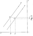
- Fig. 3 shows a diagram with a subjective, measured intraocular pressure on the ordinate axis and a deflection of an amplitude of a maximum deformation of the cornea 10 on the abscissa axis.
- a stiffness S 1 results as an essentially linear function with a negative slope.
- the rigidity S 1 can also deviate from a linear function and be designed as a line with a comparatively large radius of curvature.
- An objective intraocular pressure P o1 can be taken as a variable from the straight line defined by the stiffness S 1.
- stress lines 19 are visible in the material of the cornea 10, which represent main stresses along and across the visual axis 12.
- the Figure 4a thus shows tensions in the eye 11 in a state of rest of the cornea 10 and the Figure 4b Stresses in the eye 11 of the deformed cornea 10, which differ significantly from the stresses in the resting state.
- a comparison of the tension on the basis of the tension lines 19 thus enables a definition of a structural and / or material property of the cornea, which can be used to correct a measured intraocular pressure and thus to derive an objective intraocular pressure.
Landscapes
- Life Sciences & Earth Sciences (AREA)
- Health & Medical Sciences (AREA)
- Medical Informatics (AREA)
- Biophysics (AREA)
- Ophthalmology & Optometry (AREA)
- Engineering & Computer Science (AREA)
- Biomedical Technology (AREA)
- Heart & Thoracic Surgery (AREA)
- Physics & Mathematics (AREA)
- Molecular Biology (AREA)
- Surgery (AREA)
- Animal Behavior & Ethology (AREA)
- General Health & Medical Sciences (AREA)
- Public Health (AREA)
- Veterinary Medicine (AREA)
- Eye Examination Apparatus (AREA)
Priority Applications (17)
| Application Number | Priority Date | Filing Date | Title |
|---|---|---|---|
| EP11168235.7A EP2397070B1 (de) | 2010-06-21 | 2011-05-31 | Analyse des intraokularen Drucks |
| JP2011136550A JP5314090B2 (ja) | 2010-06-21 | 2011-06-20 | 眼科分析方法と分析システム |
| JP2011136552A JP5378457B2 (ja) | 2010-06-21 | 2011-06-20 | 眼科分析方法と分析システム |
| JP2011136548A JP5566957B2 (ja) | 2010-06-21 | 2011-06-20 | 眼科分析方法と分析システム |
| US13/165,496 US8551014B2 (en) | 2010-06-21 | 2011-06-21 | Ophthalmological analysis method and analysis system |
| CN201110169734.9A CN102309312B (zh) | 2010-06-21 | 2011-06-21 | 眼科分析方法和分析系统 |
| CN201110169845.XA CN102283636B (zh) | 2010-06-21 | 2011-06-21 | 眼科分析方法和分析系统 |
| KR1020110060129A KR101301894B1 (ko) | 2010-06-21 | 2011-06-21 | 안과 분석 방법 및 분석 시스템 |
| AU2011202971A AU2011202971B8 (en) | 2010-06-21 | 2011-06-21 | Ophthalmological analysis method and analysis system |
| AU2011202970A AU2011202970B2 (en) | 2010-06-21 | 2011-06-21 | Ophthalmological analysis method and analysis system |
| AU2011202972A AU2011202972C1 (en) | 2010-06-21 | 2011-06-21 | Opthalmological analysis method and analysis system |
| US13/165,432 US8551013B2 (en) | 2010-06-21 | 2011-06-21 | Ophthalmological analysis method and analysis system |
| CN201110169822.9A CN102309313B (zh) | 2010-06-21 | 2011-06-21 | 眼科分析方法以及分析系统 |
| US13/165,541 US8556823B2 (en) | 2010-06-21 | 2011-06-21 | Ophthalmological analysis method and analysis system |
| KR1020110060128A KR101301880B1 (ko) | 2010-06-21 | 2011-06-21 | 안과 분석 방법 및 분석 시스템 |
| KR1020110060130A KR101301891B1 (ko) | 2010-06-21 | 2011-06-21 | 안과 분석 방법 및 분석 시스템 |
| HK12104786.6A HK1165242B (en) | 2010-06-21 | 2012-05-16 | Ophthalmological analysis method and analysis system |
Applications Claiming Priority (4)
| Application Number | Priority Date | Filing Date | Title |
|---|---|---|---|
| EP10166681 | 2010-06-21 | ||
| DE102010049633 | 2010-10-28 | ||
| DE102010049634 | 2010-10-28 | ||
| EP11168235.7A EP2397070B1 (de) | 2010-06-21 | 2011-05-31 | Analyse des intraokularen Drucks |
Publications (2)
| Publication Number | Publication Date |
|---|---|
| EP2397070A1 EP2397070A1 (de) | 2011-12-21 |
| EP2397070B1 true EP2397070B1 (de) | 2021-10-13 |
Family
ID=44644898
Family Applications (3)
| Application Number | Title | Priority Date | Filing Date |
|---|---|---|---|
| EP11168235.7A Active EP2397070B1 (de) | 2010-06-21 | 2011-05-31 | Analyse des intraokularen Drucks |
| EP11168234.0A Active EP2397069B1 (de) | 2010-06-21 | 2011-05-31 | Analyse des intraokularen Drucks |
| EP11168232.4A Active EP2397068B1 (de) | 2010-06-21 | 2011-05-31 | Analyse des intraokularen Drucks |
Family Applications After (2)
| Application Number | Title | Priority Date | Filing Date |
|---|---|---|---|
| EP11168234.0A Active EP2397069B1 (de) | 2010-06-21 | 2011-05-31 | Analyse des intraokularen Drucks |
| EP11168232.4A Active EP2397068B1 (de) | 2010-06-21 | 2011-05-31 | Analyse des intraokularen Drucks |
Country Status (8)
| Country | Link |
|---|---|
| US (3) | US8556823B2 (pl) |
| EP (3) | EP2397070B1 (pl) |
| JP (3) | JP5314090B2 (pl) |
| KR (3) | KR101301880B1 (pl) |
| CN (3) | CN102309312B (pl) |
| AU (2) | AU2011202970B2 (pl) |
| ES (3) | ES2806024T3 (pl) |
| PL (2) | PL2397068T3 (pl) |
Families Citing this family (29)
| Publication number | Priority date | Publication date | Assignee | Title |
|---|---|---|---|---|
| DE102011076793A1 (de) * | 2011-05-31 | 2012-12-06 | Oculus Optikgeräte GmbH | Ophthalmologisches Analyseverfahren und Analysesystem |
| TWI474802B (zh) * | 2012-05-04 | 2015-03-01 | 光學眼壓量測裝置及其運作方法 | |
| TWI507170B (zh) * | 2012-10-24 | 2015-11-11 | Crystalvue Medical Corp | 光學裝置及其運作方法 |
| JP2014171722A (ja) * | 2013-03-11 | 2014-09-22 | Canon Inc | 非接触式眼科装置及びその制御方法 |
| JP2014217424A (ja) * | 2013-05-01 | 2014-11-20 | キヤノン株式会社 | 非接触式眼圧計 |
| ITMI20131262A1 (it) * | 2013-07-26 | 2015-01-27 | Phronema S R L | Apparecchiatura e metodo per determinare l'orientazione di strutture anatomiche corneali |
| US9357912B2 (en) * | 2013-09-09 | 2016-06-07 | Yan Zhang | Apparatus and method for characterizing biomechanical properties of eye tissue |
| US10368759B2 (en) * | 2014-03-07 | 2019-08-06 | Lions Eye Institute Limited | Method and system for determining intracranial pressure |
| US11026576B2 (en) * | 2015-04-15 | 2021-06-08 | Cats Tonometer, Llc | Reducing errors of tonometric measurements by using a tonometer tip with a curved cornea-contacting surface |
| WO2016167827A1 (en) * | 2015-04-15 | 2016-10-20 | Mccafferty Sean J | Optical instrument |
| TW201703722A (zh) * | 2015-07-21 | 2017-02-01 | 明達醫學科技股份有限公司 | 量測裝置及其運作方法 |
| CN105231990B (zh) * | 2015-11-17 | 2017-04-19 | 深圳市亿领科技有限公司 | 基于oct三维成像分析角膜生物力学性能的装置及方法 |
| TWI568408B (zh) | 2015-12-23 | 2017-02-01 | 財團法人工業技術研究院 | 一種眼壓檢測裝置及其檢測方法 |
| WO2017151921A1 (en) * | 2016-03-03 | 2017-09-08 | Biolight Engineering Llc | Methods and devices for fundus photography employing trans-palpebral and trans-scleral illumination |
| US20190192115A1 (en) * | 2016-05-08 | 2019-06-27 | The Cleveland Clinic Foundation | Measurement of biomechanical properties of tissue |
| CN106108841A (zh) * | 2016-06-29 | 2016-11-16 | 无锡市康明医疗器械有限公司 | 一种非接触光摄眼压计及眼压测量方法 |
| CN107095643B (zh) * | 2017-04-11 | 2019-01-25 | 佛山科学技术学院 | 基于低相干光干涉的非接触眼压检测系统及其检测方法 |
| EP3449812A1 (en) | 2017-09-05 | 2019-03-06 | Frey Spolka Jawna | Ophthalmic analysis system for measuring the intraocular pressure in the eye |
| US20210022608A1 (en) * | 2018-03-12 | 2021-01-28 | Ioppreceyese Ltd. | Non-Contact Home-Tonometry System for Measuring Intraocular Pressure |
| US11026577B2 (en) | 2018-06-13 | 2021-06-08 | Reichert, Inc. | Rebound tonometry method and apparatus |
| JP7288949B2 (ja) * | 2018-07-06 | 2023-06-08 | センシメド ソシエテ・アノニム | 眼圧測定および/またはモニタリング装置 |
| JP2022526645A (ja) | 2019-04-10 | 2022-05-25 | スマートレンズ, インコーポレイテッド | 眼内圧監視デバイスおよびそれを使用する方法 |
| US12268449B2 (en) | 2019-04-10 | 2025-04-08 | Smartlens, Inc. | Intraocular pressure monitoring devices and methods of using the same |
| KR102708982B1 (ko) * | 2019-05-13 | 2024-10-15 | 베릴리 라이프 사이언시즈 엘엘시 | 이식가능 안압 센서의 광학적 질의를 위한 시스템, 장치, 및 방법 |
| CN110731751A (zh) * | 2019-11-15 | 2020-01-31 | 刘果 | 一种方便、快捷测量眼内压的方法 |
| US20240016381A1 (en) * | 2020-11-17 | 2024-01-18 | N.M.B. Medical Applications Ltd | Device and method for non-contact measurement of an intraocular pressure |
| EP4342362A4 (en) | 2021-05-18 | 2024-07-24 | Quovisu LLC | NON-CONTACT TYPE EYE GLOBE PHYSICAL PROPERTY MEASUREMENT DEVICE |
| CN117012367A (zh) * | 2022-05-06 | 2023-11-07 | 香港科技大学 | 对眼睛的检测及分类系统和用于眼睛检测的装置 |
| KR20250074895A (ko) * | 2023-11-21 | 2025-05-28 | 유한회사 웰씨 | 각막 돌출부 교정 장치 및 방법 |
Family Cites Families (35)
| Publication number | Priority date | Publication date | Assignee | Title |
|---|---|---|---|---|
| US3406681A (en) * | 1963-05-13 | 1968-10-22 | Vishay Intertechnology Inc | Method of determining strain condition of the eye |
| US3585849A (en) * | 1968-10-09 | 1971-06-22 | American Optical Corp | Method and apparatus for measuring intraocular pressure |
| JPS61321A (ja) * | 1984-06-12 | 1986-01-06 | 株式会社トプコン | 非接触式眼圧計 |
| JP3221699B2 (ja) * | 1991-08-31 | 2001-10-22 | 株式会社ニデック | 非接触式眼圧計 |
| JPH08109B2 (ja) * | 1993-04-02 | 1996-01-10 | 株式会社トプコン | 非接触式眼圧計 |
| JP3108261B2 (ja) * | 1993-12-20 | 2000-11-13 | 株式会社トプコン | 眼科器械 |
| JP3308416B2 (ja) * | 1994-10-26 | 2002-07-29 | 株式会社トプコン | 眼科器械 |
| JPH08322803A (ja) * | 1995-05-31 | 1996-12-10 | Canon Inc | 眼圧計 |
| US5822035A (en) * | 1996-08-30 | 1998-10-13 | Heidelberg Engineering Optische Messysteme Gmbh | Ellipsometer |
| US6544193B2 (en) | 1996-09-04 | 2003-04-08 | Marcio Marc Abreu | Noninvasive measurement of chemical substances |
| JPH10309265A (ja) * | 1997-05-12 | 1998-11-24 | Konan:Kk | 眼科撮影装置 |
| JP3862869B2 (ja) * | 1998-06-05 | 2006-12-27 | 株式会社トプコン | 非接触式眼圧計 |
| JP2000254101A (ja) * | 1999-01-06 | 2000-09-19 | Konan Inc | 眼科検査装置 |
| JP2002263069A (ja) * | 2001-03-13 | 2002-09-17 | Konan Medical Inc | 眼硬性測定装置 |
| JP3970141B2 (ja) * | 2002-09-11 | 2007-09-05 | キヤノン株式会社 | 非接触式眼圧計 |
| JP3885015B2 (ja) * | 2002-09-17 | 2007-02-21 | キヤノン株式会社 | 非接触眼圧計 |
| US7004902B2 (en) * | 2003-03-21 | 2006-02-28 | Reichert, Inc. | Method and apparatus for measuring biomechanical characteristics of corneal tissue |
| US20050030473A1 (en) * | 2003-06-12 | 2005-02-10 | Welch Allyn, Inc. | Apparatus and method for determining intraocular pressure and corneal thickness |
| DE202005002562U1 (de) * | 2005-02-16 | 2005-06-09 | Oculus Optikgeräte GmbH | Ophthalmisches Analysesystem zur Messung des intraocularen Drucks im Auge |
| JP2007121174A (ja) * | 2005-10-31 | 2007-05-17 | Univ Nagoya | 応力検出装置 |
| US7798962B2 (en) * | 2005-09-08 | 2010-09-21 | Reichert, Inc. | Method and apparatus for measuring corneal resistance |
| US7481767B2 (en) | 2005-09-08 | 2009-01-27 | Reichert, Inc. | Method and apparatus for determining true intraocular pressure |
| JP5028057B2 (ja) * | 2005-11-01 | 2012-09-19 | 株式会社ニデック | 眼科装置 |
| US20070103538A1 (en) | 2005-11-07 | 2007-05-10 | Busch Brian D | Thermal printing head with two-dimensional array of resistive heating elements, and method for printing using same |
| US20070121120A1 (en) * | 2005-11-16 | 2007-05-31 | Schachar Ronald A | Apparatus and method for measuring scleral curvature and velocity of tissues of the eye |
| JP4824400B2 (ja) * | 2005-12-28 | 2011-11-30 | 株式会社トプコン | 眼科装置 |
| JP4948922B2 (ja) * | 2006-06-30 | 2012-06-06 | 株式会社ニデック | 眼科装置 |
| JP5448353B2 (ja) | 2007-05-02 | 2014-03-19 | キヤノン株式会社 | 光干渉断層計を用いた画像形成方法、及び光干渉断層装置 |
| JP5209341B2 (ja) * | 2008-02-27 | 2013-06-12 | 株式会社ニデック | 非接触式眼圧計 |
| JP5268053B2 (ja) * | 2008-05-15 | 2013-08-21 | 晃太郎 石井 | 眼球組織固有振動数測定装置及びそれを利用した非接触式眼圧計 |
| CN102438549B (zh) * | 2009-03-04 | 2015-07-15 | 完美Ip有限公司 | 用于形成和修改晶状体的系统以及由此形成的晶状体 |
| US8025401B2 (en) * | 2009-08-05 | 2011-09-27 | Sis Ag, Surgical Instrument Systems | Ophthalmological measuring device and measurement method |
| DE102011082363B4 (de) * | 2010-10-28 | 2018-03-29 | Oculus Optikgeräte GmbH | Beleuchtungssystem, ophthalmologisches Analysegerät und Verfahren |
| JP5340434B2 (ja) * | 2011-02-25 | 2013-11-13 | キヤノン株式会社 | 眼科装置、処理装置、眼科システム、処理方法および眼科装置の制御方法、プログラム |
| DE102011076793A1 (de) * | 2011-05-31 | 2012-12-06 | Oculus Optikgeräte GmbH | Ophthalmologisches Analyseverfahren und Analysesystem |
-
2011
- 2011-05-31 PL PL11168232T patent/PL2397068T3/pl unknown
- 2011-05-31 ES ES11168232T patent/ES2806024T3/es active Active
- 2011-05-31 EP EP11168235.7A patent/EP2397070B1/de active Active
- 2011-05-31 EP EP11168234.0A patent/EP2397069B1/de active Active
- 2011-05-31 EP EP11168232.4A patent/EP2397068B1/de active Active
- 2011-05-31 PL PL11168234T patent/PL2397069T3/pl unknown
- 2011-05-31 ES ES11168234T patent/ES2806934T3/es active Active
- 2011-05-31 ES ES11168235T patent/ES2899473T3/es active Active
- 2011-06-20 JP JP2011136550A patent/JP5314090B2/ja active Active
- 2011-06-20 JP JP2011136552A patent/JP5378457B2/ja active Active
- 2011-06-20 JP JP2011136548A patent/JP5566957B2/ja active Active
- 2011-06-21 KR KR1020110060128A patent/KR101301880B1/ko active Active
- 2011-06-21 AU AU2011202970A patent/AU2011202970B2/en active Active
- 2011-06-21 CN CN201110169734.9A patent/CN102309312B/zh active Active
- 2011-06-21 US US13/165,541 patent/US8556823B2/en active Active
- 2011-06-21 KR KR1020110060130A patent/KR101301891B1/ko active Active
- 2011-06-21 AU AU2011202972A patent/AU2011202972C1/en active Active
- 2011-06-21 CN CN201110169845.XA patent/CN102283636B/zh active Active
- 2011-06-21 US US13/165,496 patent/US8551014B2/en active Active
- 2011-06-21 CN CN201110169822.9A patent/CN102309313B/zh active Active
- 2011-06-21 US US13/165,432 patent/US8551013B2/en active Active
- 2011-06-21 KR KR1020110060129A patent/KR101301894B1/ko active Active
Non-Patent Citations (1)
| Title |
|---|
| None * |
Also Published As
Similar Documents
| Publication | Publication Date | Title |
|---|---|---|
| EP2397070B1 (de) | Analyse des intraokularen Drucks | |
| EP2529662B1 (de) | Ophthalmologisches Analyseverfahren und Analysesystem | |
| EP2671505B1 (de) | Verfahren und Analysegerät zur Messung einer Hornhaut | |
| DE60130284T2 (de) | Verfahren der berührungslosen Tonometrie | |
| EP3682794B1 (de) | Verfahren und sehprüfsystem zum überprüfen der augen | |
| WO2011110553A1 (de) | Vorrichtung zur rechnergestützten verarbeitung von biegeinformationen eines menschlichen oder tierischen körpers, insbesondere eines rückens | |
| EP4322858B1 (de) | Verfahren zur messung eines innendrucks eines kompartments | |
| DE102011015178B3 (de) | Vorrichtung und Verfahren zur automatisierten Bestimmung des Intraokulardruckes | |
| DE102012022059A1 (de) | Verfahren zur Bestimmung der Gesamtbrechkraft der Hornhaut eines Auges | |
| HK1165242B (en) | Ophthalmological analysis method and analysis system | |
| WO2006047801A1 (de) | Vorrichtung zur bestimmung des augeninnendrucks |
Legal Events
| Date | Code | Title | Description |
|---|---|---|---|
| AK | Designated contracting states |
Kind code of ref document: A1 Designated state(s): AL AT BE BG CH CY CZ DE DK EE ES FI FR GB GR HR HU IE IS IT LI LT LU LV MC MK MT NL NO PL PT RO RS SE SI SK SM TR |
|
| AX | Request for extension of the european patent |
Extension state: BA ME |
|
| PUAI | Public reference made under article 153(3) epc to a published international application that has entered the european phase |
Free format text: ORIGINAL CODE: 0009012 |
|
| 17P | Request for examination filed |
Effective date: 20120529 |
|
| STAA | Information on the status of an ep patent application or granted ep patent |
Free format text: STATUS: EXAMINATION IS IN PROGRESS |
|
| 17Q | First examination report despatched |
Effective date: 20180718 |
|
| GRAP | Despatch of communication of intention to grant a patent |
Free format text: ORIGINAL CODE: EPIDOSNIGR1 |
|
| STAA | Information on the status of an ep patent application or granted ep patent |
Free format text: STATUS: GRANT OF PATENT IS INTENDED |
|
| INTG | Intention to grant announced |
Effective date: 20210520 |
|
| GRAS | Grant fee paid |
Free format text: ORIGINAL CODE: EPIDOSNIGR3 |
|
| GRAA | (expected) grant |
Free format text: ORIGINAL CODE: 0009210 |
|
| STAA | Information on the status of an ep patent application or granted ep patent |
Free format text: STATUS: THE PATENT HAS BEEN GRANTED |
|
| AK | Designated contracting states |
Kind code of ref document: B1 Designated state(s): AL AT BE BG CH CY CZ DE DK EE ES FI FR GB GR HR HU IE IS IT LI LT LU LV MC MK MT NL NO PL PT RO RS SE SI SK SM TR |
|
| REG | Reference to a national code |
Ref country code: GB Ref legal event code: FG4D Free format text: NOT ENGLISH |
|
| REG | Reference to a national code |
Ref country code: CH Ref legal event code: EP |
|
| REG | Reference to a national code |
Ref country code: DE Ref legal event code: R096 Ref document number: 502011017252 Country of ref document: DE |
|
| REG | Reference to a national code |
Ref country code: IE Ref legal event code: FG4D Free format text: LANGUAGE OF EP DOCUMENT: GERMAN |
|
| REG | Reference to a national code |
Ref country code: AT Ref legal event code: REF Ref document number: 1437508 Country of ref document: AT Kind code of ref document: T Effective date: 20211115 |
|
| REG | Reference to a national code |
Ref country code: LT Ref legal event code: MG9D |
|
| REG | Reference to a national code |
Ref country code: ES Ref legal event code: FG2A Ref document number: 2899473 Country of ref document: ES Kind code of ref document: T3 Effective date: 20220311 |
|
| PG25 | Lapsed in a contracting state [announced via postgrant information from national office to epo] |
Ref country code: RS Free format text: LAPSE BECAUSE OF FAILURE TO SUBMIT A TRANSLATION OF THE DESCRIPTION OR TO PAY THE FEE WITHIN THE PRESCRIBED TIME-LIMIT Effective date: 20211013 Ref country code: LT Free format text: LAPSE BECAUSE OF FAILURE TO SUBMIT A TRANSLATION OF THE DESCRIPTION OR TO PAY THE FEE WITHIN THE PRESCRIBED TIME-LIMIT Effective date: 20211013 Ref country code: FI Free format text: LAPSE BECAUSE OF FAILURE TO SUBMIT A TRANSLATION OF THE DESCRIPTION OR TO PAY THE FEE WITHIN THE PRESCRIBED TIME-LIMIT Effective date: 20211013 Ref country code: BG Free format text: LAPSE BECAUSE OF FAILURE TO SUBMIT A TRANSLATION OF THE DESCRIPTION OR TO PAY THE FEE WITHIN THE PRESCRIBED TIME-LIMIT Effective date: 20220113 |
|
| PG25 | Lapsed in a contracting state [announced via postgrant information from national office to epo] |
Ref country code: IS Free format text: LAPSE BECAUSE OF FAILURE TO SUBMIT A TRANSLATION OF THE DESCRIPTION OR TO PAY THE FEE WITHIN THE PRESCRIBED TIME-LIMIT Effective date: 20220213 Ref country code: SE Free format text: LAPSE BECAUSE OF FAILURE TO SUBMIT A TRANSLATION OF THE DESCRIPTION OR TO PAY THE FEE WITHIN THE PRESCRIBED TIME-LIMIT Effective date: 20211013 Ref country code: PT Free format text: LAPSE BECAUSE OF FAILURE TO SUBMIT A TRANSLATION OF THE DESCRIPTION OR TO PAY THE FEE WITHIN THE PRESCRIBED TIME-LIMIT Effective date: 20220214 Ref country code: PL Free format text: LAPSE BECAUSE OF FAILURE TO SUBMIT A TRANSLATION OF THE DESCRIPTION OR TO PAY THE FEE WITHIN THE PRESCRIBED TIME-LIMIT Effective date: 20211013 Ref country code: NO Free format text: LAPSE BECAUSE OF FAILURE TO SUBMIT A TRANSLATION OF THE DESCRIPTION OR TO PAY THE FEE WITHIN THE PRESCRIBED TIME-LIMIT Effective date: 20220113 Ref country code: NL Free format text: LAPSE BECAUSE OF FAILURE TO SUBMIT A TRANSLATION OF THE DESCRIPTION OR TO PAY THE FEE WITHIN THE PRESCRIBED TIME-LIMIT Effective date: 20211013 Ref country code: LV Free format text: LAPSE BECAUSE OF FAILURE TO SUBMIT A TRANSLATION OF THE DESCRIPTION OR TO PAY THE FEE WITHIN THE PRESCRIBED TIME-LIMIT Effective date: 20211013 Ref country code: HR Free format text: LAPSE BECAUSE OF FAILURE TO SUBMIT A TRANSLATION OF THE DESCRIPTION OR TO PAY THE FEE WITHIN THE PRESCRIBED TIME-LIMIT Effective date: 20211013 Ref country code: GR Free format text: LAPSE BECAUSE OF FAILURE TO SUBMIT A TRANSLATION OF THE DESCRIPTION OR TO PAY THE FEE WITHIN THE PRESCRIBED TIME-LIMIT Effective date: 20220114 |
|
| REG | Reference to a national code |
Ref country code: DE Ref legal event code: R097 Ref document number: 502011017252 Country of ref document: DE |
|
| PG25 | Lapsed in a contracting state [announced via postgrant information from national office to epo] |
Ref country code: SM Free format text: LAPSE BECAUSE OF FAILURE TO SUBMIT A TRANSLATION OF THE DESCRIPTION OR TO PAY THE FEE WITHIN THE PRESCRIBED TIME-LIMIT Effective date: 20211013 Ref country code: SK Free format text: LAPSE BECAUSE OF FAILURE TO SUBMIT A TRANSLATION OF THE DESCRIPTION OR TO PAY THE FEE WITHIN THE PRESCRIBED TIME-LIMIT Effective date: 20211013 Ref country code: RO Free format text: LAPSE BECAUSE OF FAILURE TO SUBMIT A TRANSLATION OF THE DESCRIPTION OR TO PAY THE FEE WITHIN THE PRESCRIBED TIME-LIMIT Effective date: 20211013 Ref country code: EE Free format text: LAPSE BECAUSE OF FAILURE TO SUBMIT A TRANSLATION OF THE DESCRIPTION OR TO PAY THE FEE WITHIN THE PRESCRIBED TIME-LIMIT Effective date: 20211013 Ref country code: DK Free format text: LAPSE BECAUSE OF FAILURE TO SUBMIT A TRANSLATION OF THE DESCRIPTION OR TO PAY THE FEE WITHIN THE PRESCRIBED TIME-LIMIT Effective date: 20211013 Ref country code: CZ Free format text: LAPSE BECAUSE OF FAILURE TO SUBMIT A TRANSLATION OF THE DESCRIPTION OR TO PAY THE FEE WITHIN THE PRESCRIBED TIME-LIMIT Effective date: 20211013 |
|
| PLBE | No opposition filed within time limit |
Free format text: ORIGINAL CODE: 0009261 |
|
| STAA | Information on the status of an ep patent application or granted ep patent |
Free format text: STATUS: NO OPPOSITION FILED WITHIN TIME LIMIT |
|
| 26N | No opposition filed |
Effective date: 20220714 |
|
| PG25 | Lapsed in a contracting state [announced via postgrant information from national office to epo] |
Ref country code: AL Free format text: LAPSE BECAUSE OF FAILURE TO SUBMIT A TRANSLATION OF THE DESCRIPTION OR TO PAY THE FEE WITHIN THE PRESCRIBED TIME-LIMIT Effective date: 20211013 |
|
| PG25 | Lapsed in a contracting state [announced via postgrant information from national office to epo] |
Ref country code: SI Free format text: LAPSE BECAUSE OF FAILURE TO SUBMIT A TRANSLATION OF THE DESCRIPTION OR TO PAY THE FEE WITHIN THE PRESCRIBED TIME-LIMIT Effective date: 20211013 |
|
| REG | Reference to a national code |
Ref country code: CH Ref legal event code: PL |
|
| REG | Reference to a national code |
Ref country code: BE Ref legal event code: MM Effective date: 20220531 |
|
| PG25 | Lapsed in a contracting state [announced via postgrant information from national office to epo] |
Ref country code: MC Free format text: LAPSE BECAUSE OF FAILURE TO SUBMIT A TRANSLATION OF THE DESCRIPTION OR TO PAY THE FEE WITHIN THE PRESCRIBED TIME-LIMIT Effective date: 20211013 Ref country code: LU Free format text: LAPSE BECAUSE OF NON-PAYMENT OF DUE FEES Effective date: 20220531 Ref country code: LI Free format text: LAPSE BECAUSE OF NON-PAYMENT OF DUE FEES Effective date: 20220531 Ref country code: CH Free format text: LAPSE BECAUSE OF NON-PAYMENT OF DUE FEES Effective date: 20220531 |
|
| PG25 | Lapsed in a contracting state [announced via postgrant information from national office to epo] |
Ref country code: LI Free format text: LAPSE BECAUSE OF NON-PAYMENT OF DUE FEES Effective date: 20220531 Ref country code: IE Free format text: LAPSE BECAUSE OF NON-PAYMENT OF DUE FEES Effective date: 20220531 Ref country code: CH Free format text: LAPSE BECAUSE OF NON-PAYMENT OF DUE FEES Effective date: 20220531 |
|
| PGRI | Patent reinstated in contracting state [announced from national office to epo] |
Ref country code: LI Effective date: 20230131 Ref country code: CH Effective date: 20230131 |
|
| PG25 | Lapsed in a contracting state [announced via postgrant information from national office to epo] |
Ref country code: BE Free format text: LAPSE BECAUSE OF NON-PAYMENT OF DUE FEES Effective date: 20220531 |
|
| P01 | Opt-out of the competence of the unified patent court (upc) registered |
Effective date: 20230525 |
|
| REG | Reference to a national code |
Ref country code: AT Ref legal event code: MM01 Ref document number: 1437508 Country of ref document: AT Kind code of ref document: T Effective date: 20220531 |
|
| PG25 | Lapsed in a contracting state [announced via postgrant information from national office to epo] |
Ref country code: AT Free format text: LAPSE BECAUSE OF NON-PAYMENT OF DUE FEES Effective date: 20220531 |
|
| PG25 | Lapsed in a contracting state [announced via postgrant information from national office to epo] |
Ref country code: HU Free format text: LAPSE BECAUSE OF FAILURE TO SUBMIT A TRANSLATION OF THE DESCRIPTION OR TO PAY THE FEE WITHIN THE PRESCRIBED TIME-LIMIT; INVALID AB INITIO Effective date: 20110531 |
|
| PG25 | Lapsed in a contracting state [announced via postgrant information from national office to epo] |
Ref country code: MK Free format text: LAPSE BECAUSE OF FAILURE TO SUBMIT A TRANSLATION OF THE DESCRIPTION OR TO PAY THE FEE WITHIN THE PRESCRIBED TIME-LIMIT Effective date: 20211013 Ref country code: CY Free format text: LAPSE BECAUSE OF FAILURE TO SUBMIT A TRANSLATION OF THE DESCRIPTION OR TO PAY THE FEE WITHIN THE PRESCRIBED TIME-LIMIT Effective date: 20211013 |
|
| PG25 | Lapsed in a contracting state [announced via postgrant information from national office to epo] |
Ref country code: TR Free format text: LAPSE BECAUSE OF FAILURE TO SUBMIT A TRANSLATION OF THE DESCRIPTION OR TO PAY THE FEE WITHIN THE PRESCRIBED TIME-LIMIT Effective date: 20211013 |
|
| PG25 | Lapsed in a contracting state [announced via postgrant information from national office to epo] |
Ref country code: MT Free format text: LAPSE BECAUSE OF FAILURE TO SUBMIT A TRANSLATION OF THE DESCRIPTION OR TO PAY THE FEE WITHIN THE PRESCRIBED TIME-LIMIT Effective date: 20211013 |
|
| PGFP | Annual fee paid to national office [announced via postgrant information from national office to epo] |
Ref country code: GB Payment date: 20250522 Year of fee payment: 15 Ref country code: ES Payment date: 20250616 Year of fee payment: 15 |
|
| PGFP | Annual fee paid to national office [announced via postgrant information from national office to epo] |
Ref country code: IT Payment date: 20250530 Year of fee payment: 15 |
|
| PGFP | Annual fee paid to national office [announced via postgrant information from national office to epo] |
Ref country code: FR Payment date: 20250526 Year of fee payment: 15 |
|
| PGFP | Annual fee paid to national office [announced via postgrant information from national office to epo] |
Ref country code: CH Payment date: 20250601 Year of fee payment: 15 |
|
| PGFP | Annual fee paid to national office [announced via postgrant information from national office to epo] |
Ref country code: DE Payment date: 20250722 Year of fee payment: 15 |