EP2385143A2 - Non-invasive fetal genetic screening by digital analysis - Google Patents
Non-invasive fetal genetic screening by digital analysis Download PDFInfo
- Publication number
- EP2385143A2 EP2385143A2 EP20110175845 EP11175845A EP2385143A2 EP 2385143 A2 EP2385143 A2 EP 2385143A2 EP 20110175845 EP20110175845 EP 20110175845 EP 11175845 A EP11175845 A EP 11175845A EP 2385143 A2 EP2385143 A2 EP 2385143A2
- Authority
- EP
- European Patent Office
- Prior art keywords
- dna
- maternal
- pcr
- fetal
- genetic material
- Prior art date
- Legal status (The legal status is an assumption and is not a legal conclusion. Google has not performed a legal analysis and makes no representation as to the accuracy of the status listed.)
- Granted
Links
- 230000001605 fetal effect Effects 0.000 title claims abstract description 104
- 238000010252 digital analysis Methods 0.000 title description 7
- 238000010448 genetic screening Methods 0.000 title description 2
- 238000000034 method Methods 0.000 claims abstract description 141
- 230000008774 maternal effect Effects 0.000 claims abstract description 105
- 238000006243 chemical reaction Methods 0.000 claims abstract description 81
- 238000001514 detection method Methods 0.000 claims abstract description 51
- 230000003321 amplification Effects 0.000 claims abstract description 38
- 238000003199 nucleic acid amplification method Methods 0.000 claims abstract description 38
- 208000036878 aneuploidy Diseases 0.000 claims abstract description 20
- 208000037280 Trisomy Diseases 0.000 claims abstract description 19
- 108020004414 DNA Proteins 0.000 claims description 224
- 108090000623 proteins and genes Proteins 0.000 claims description 69
- 210000002381 plasma Anatomy 0.000 claims description 58
- 102000004169 proteins and genes Human genes 0.000 claims description 44
- 239000000203 mixture Substances 0.000 claims description 36
- 238000004458 analytical method Methods 0.000 claims description 32
- 210000004027 cell Anatomy 0.000 claims description 23
- 238000012163 sequencing technique Methods 0.000 claims description 23
- 108091093088 Amplicon Proteins 0.000 claims description 21
- 230000002068 genetic effect Effects 0.000 claims description 20
- 239000012634 fragment Substances 0.000 claims description 19
- 210000001519 tissue Anatomy 0.000 claims description 16
- 239000000839 emulsion Substances 0.000 claims description 15
- 238000002360 preparation method Methods 0.000 claims description 12
- 230000005856 abnormality Effects 0.000 claims description 11
- 238000000926 separation method Methods 0.000 claims description 11
- 238000005516 engineering process Methods 0.000 claims description 10
- 239000008346 aqueous phase Substances 0.000 claims description 7
- 230000003322 aneuploid effect Effects 0.000 claims description 6
- 210000005259 peripheral blood Anatomy 0.000 claims description 5
- 238000003786 synthesis reaction Methods 0.000 claims description 3
- 239000007790 solid phase Substances 0.000 claims description 2
- 238000004873 anchoring Methods 0.000 claims 1
- 230000015572 biosynthetic process Effects 0.000 claims 1
- 210000000349 chromosome Anatomy 0.000 abstract description 100
- 210000004369 blood Anatomy 0.000 abstract description 41
- 239000008280 blood Substances 0.000 abstract description 41
- 201000010374 Down Syndrome Diseases 0.000 abstract description 25
- 238000007847 digital PCR Methods 0.000 abstract description 24
- 210000003754 fetus Anatomy 0.000 abstract description 15
- 231100001075 aneuploidy Toxicity 0.000 abstract description 14
- 230000008569 process Effects 0.000 abstract description 7
- 230000008859 change Effects 0.000 abstract description 5
- 210000004252 chorionic villi Anatomy 0.000 abstract description 3
- 238000002669 amniocentesis Methods 0.000 abstract description 2
- 230000000869 mutational effect Effects 0.000 abstract description 2
- 238000005070 sampling Methods 0.000 abstract description 2
- 239000000523 sample Substances 0.000 description 117
- 239000011324 bead Substances 0.000 description 46
- 239000000047 product Substances 0.000 description 22
- 239000000872 buffer Substances 0.000 description 20
- 150000007523 nucleic acids Chemical class 0.000 description 18
- 108020004707 nucleic acids Proteins 0.000 description 17
- 102000039446 nucleic acids Human genes 0.000 description 17
- 230000035772 mutation Effects 0.000 description 15
- 239000000499 gel Substances 0.000 description 14
- 239000000463 material Substances 0.000 description 14
- 108700028369 Alleles Proteins 0.000 description 12
- 108091034117 Oligonucleotide Proteins 0.000 description 12
- 239000007850 fluorescent dye Substances 0.000 description 12
- 108020004999 messenger RNA Proteins 0.000 description 11
- YBJHBAHKTGYVGT-ZKWXMUAHSA-N (+)-Biotin Chemical group N1C(=O)N[C@@H]2[C@H](CCCCC(=O)O)SC[C@@H]21 YBJHBAHKTGYVGT-ZKWXMUAHSA-N 0.000 description 10
- 108010014303 DNA-directed DNA polymerase Proteins 0.000 description 10
- 102000016928 DNA-directed DNA polymerase Human genes 0.000 description 10
- 238000009826 distribution Methods 0.000 description 10
- -1 hemachromatosis Proteins 0.000 description 10
- 230000001965 increasing effect Effects 0.000 description 10
- 102100031181 Glyceraldehyde-3-phosphate dehydrogenase Human genes 0.000 description 9
- JLCPHMBAVCMARE-UHFFFAOYSA-N [3-[[3-[[3-[[3-[[3-[[3-[[3-[[3-[[3-[[3-[[3-[[5-(2-amino-6-oxo-1H-purin-9-yl)-3-[[3-[[3-[[3-[[3-[[3-[[5-(2-amino-6-oxo-1H-purin-9-yl)-3-[[5-(2-amino-6-oxo-1H-purin-9-yl)-3-hydroxyoxolan-2-yl]methoxy-hydroxyphosphoryl]oxyoxolan-2-yl]methoxy-hydroxyphosphoryl]oxy-5-(5-methyl-2,4-dioxopyrimidin-1-yl)oxolan-2-yl]methoxy-hydroxyphosphoryl]oxy-5-(6-aminopurin-9-yl)oxolan-2-yl]methoxy-hydroxyphosphoryl]oxy-5-(6-aminopurin-9-yl)oxolan-2-yl]methoxy-hydroxyphosphoryl]oxy-5-(6-aminopurin-9-yl)oxolan-2-yl]methoxy-hydroxyphosphoryl]oxy-5-(6-aminopurin-9-yl)oxolan-2-yl]methoxy-hydroxyphosphoryl]oxyoxolan-2-yl]methoxy-hydroxyphosphoryl]oxy-5-(5-methyl-2,4-dioxopyrimidin-1-yl)oxolan-2-yl]methoxy-hydroxyphosphoryl]oxy-5-(4-amino-2-oxopyrimidin-1-yl)oxolan-2-yl]methoxy-hydroxyphosphoryl]oxy-5-(5-methyl-2,4-dioxopyrimidin-1-yl)oxolan-2-yl]methoxy-hydroxyphosphoryl]oxy-5-(5-methyl-2,4-dioxopyrimidin-1-yl)oxolan-2-yl]methoxy-hydroxyphosphoryl]oxy-5-(6-aminopurin-9-yl)oxolan-2-yl]methoxy-hydroxyphosphoryl]oxy-5-(6-aminopurin-9-yl)oxolan-2-yl]methoxy-hydroxyphosphoryl]oxy-5-(4-amino-2-oxopyrimidin-1-yl)oxolan-2-yl]methoxy-hydroxyphosphoryl]oxy-5-(4-amino-2-oxopyrimidin-1-yl)oxolan-2-yl]methoxy-hydroxyphosphoryl]oxy-5-(4-amino-2-oxopyrimidin-1-yl)oxolan-2-yl]methoxy-hydroxyphosphoryl]oxy-5-(6-aminopurin-9-yl)oxolan-2-yl]methoxy-hydroxyphosphoryl]oxy-5-(4-amino-2-oxopyrimidin-1-yl)oxolan-2-yl]methyl [5-(6-aminopurin-9-yl)-2-(hydroxymethyl)oxolan-3-yl] hydrogen phosphate Polymers Cc1cn(C2CC(OP(O)(=O)OCC3OC(CC3OP(O)(=O)OCC3OC(CC3O)n3cnc4c3nc(N)[nH]c4=O)n3cnc4c3nc(N)[nH]c4=O)C(COP(O)(=O)OC3CC(OC3COP(O)(=O)OC3CC(OC3COP(O)(=O)OC3CC(OC3COP(O)(=O)OC3CC(OC3COP(O)(=O)OC3CC(OC3COP(O)(=O)OC3CC(OC3COP(O)(=O)OC3CC(OC3COP(O)(=O)OC3CC(OC3COP(O)(=O)OC3CC(OC3COP(O)(=O)OC3CC(OC3COP(O)(=O)OC3CC(OC3COP(O)(=O)OC3CC(OC3COP(O)(=O)OC3CC(OC3COP(O)(=O)OC3CC(OC3COP(O)(=O)OC3CC(OC3COP(O)(=O)OC3CC(OC3COP(O)(=O)OC3CC(OC3CO)n3cnc4c(N)ncnc34)n3ccc(N)nc3=O)n3cnc4c(N)ncnc34)n3ccc(N)nc3=O)n3ccc(N)nc3=O)n3ccc(N)nc3=O)n3cnc4c(N)ncnc34)n3cnc4c(N)ncnc34)n3cc(C)c(=O)[nH]c3=O)n3cc(C)c(=O)[nH]c3=O)n3ccc(N)nc3=O)n3cc(C)c(=O)[nH]c3=O)n3cnc4c3nc(N)[nH]c4=O)n3cnc4c(N)ncnc34)n3cnc4c(N)ncnc34)n3cnc4c(N)ncnc34)n3cnc4c(N)ncnc34)O2)c(=O)[nH]c1=O JLCPHMBAVCMARE-UHFFFAOYSA-N 0.000 description 9
- 238000003556 assay Methods 0.000 description 9
- 238000010790 dilution Methods 0.000 description 9
- 239000012895 dilution Substances 0.000 description 9
- 108020004445 glyceraldehyde-3-phosphate dehydrogenase Proteins 0.000 description 9
- 210000003917 human chromosome Anatomy 0.000 description 9
- 108091032973 (ribonucleotides)n+m Proteins 0.000 description 8
- 238000011529 RT qPCR Methods 0.000 description 8
- 210000003743 erythrocyte Anatomy 0.000 description 8
- 238000002474 experimental method Methods 0.000 description 8
- 238000000605 extraction Methods 0.000 description 8
- 239000002773 nucleotide Substances 0.000 description 8
- 239000004971 Cross linker Substances 0.000 description 7
- 238000007792 addition Methods 0.000 description 7
- 238000005119 centrifugation Methods 0.000 description 7
- 239000003550 marker Substances 0.000 description 7
- 125000003729 nucleotide group Chemical group 0.000 description 7
- 210000002826 placenta Anatomy 0.000 description 7
- 230000002441 reversible effect Effects 0.000 description 7
- 238000012360 testing method Methods 0.000 description 7
- 102000053602 DNA Human genes 0.000 description 6
- IAZDPXIOMUYVGZ-UHFFFAOYSA-N Dimethylsulphoxide Chemical compound CS(C)=O IAZDPXIOMUYVGZ-UHFFFAOYSA-N 0.000 description 6
- 208000026350 Inborn Genetic disease Diseases 0.000 description 6
- 108091093037 Peptide nucleic acid Proteins 0.000 description 6
- 206010044688 Trisomy 21 Diseases 0.000 description 6
- 230000002159 abnormal effect Effects 0.000 description 6
- 239000003795 chemical substances by application Substances 0.000 description 6
- 230000001351 cycling effect Effects 0.000 description 6
- 239000012530 fluid Substances 0.000 description 6
- 208000016361 genetic disease Diseases 0.000 description 6
- 239000004530 micro-emulsion Substances 0.000 description 6
- 239000003921 oil Substances 0.000 description 6
- BASFCYQUMIYNBI-UHFFFAOYSA-N platinum Chemical compound [Pt] BASFCYQUMIYNBI-UHFFFAOYSA-N 0.000 description 6
- 230000035935 pregnancy Effects 0.000 description 6
- 230000035945 sensitivity Effects 0.000 description 6
- WSFSSNUMVMOOMR-UHFFFAOYSA-N Formaldehyde Chemical compound O=C WSFSSNUMVMOOMR-UHFFFAOYSA-N 0.000 description 5
- 102100034343 Integrase Human genes 0.000 description 5
- 108091028043 Nucleic acid sequence Proteins 0.000 description 5
- 229960002685 biotin Drugs 0.000 description 5
- 235000020958 biotin Nutrition 0.000 description 5
- 239000011616 biotin Substances 0.000 description 5
- 238000000684 flow cytometry Methods 0.000 description 5
- 238000009396 hybridization Methods 0.000 description 5
- 238000002372 labelling Methods 0.000 description 5
- 238000007403 mPCR Methods 0.000 description 5
- 238000002844 melting Methods 0.000 description 5
- 230000008018 melting Effects 0.000 description 5
- 210000004379 membrane Anatomy 0.000 description 5
- 239000012528 membrane Substances 0.000 description 5
- 239000003068 molecular probe Substances 0.000 description 5
- 238000003753 real-time PCR Methods 0.000 description 5
- 210000002966 serum Anatomy 0.000 description 5
- 239000000243 solution Substances 0.000 description 5
- 239000006228 supernatant Substances 0.000 description 5
- 230000005945 translocation Effects 0.000 description 5
- ZRLNBWWGLOPJIC-PYQRSULMSA-N A'-neogammacerane Chemical compound C([C@]1(C)[C@H]2CC[C@H]34)CCC(C)(C)[C@@H]1CC[C@@]2(C)[C@]4(C)CC[C@@H]1[C@]3(C)CC[C@@H]1C(C)C ZRLNBWWGLOPJIC-PYQRSULMSA-N 0.000 description 4
- KCXVZYZYPLLWCC-UHFFFAOYSA-N EDTA Chemical compound OC(=O)CN(CC(O)=O)CCN(CC(O)=O)CC(O)=O KCXVZYZYPLLWCC-UHFFFAOYSA-N 0.000 description 4
- 108091005904 Hemoglobin subunit beta Proteins 0.000 description 4
- TWRXJAOTZQYOKJ-UHFFFAOYSA-L Magnesium chloride Chemical compound [Mg+2].[Cl-].[Cl-] TWRXJAOTZQYOKJ-UHFFFAOYSA-L 0.000 description 4
- 108010090804 Streptavidin Proteins 0.000 description 4
- ISAKRJDGNUQOIC-UHFFFAOYSA-N Uracil Chemical compound O=C1C=CNC(=O)N1 ISAKRJDGNUQOIC-UHFFFAOYSA-N 0.000 description 4
- 238000000137 annealing Methods 0.000 description 4
- 238000013459 approach Methods 0.000 description 4
- 230000006037 cell lysis Effects 0.000 description 4
- 210000000170 cell membrane Anatomy 0.000 description 4
- 239000002299 complementary DNA Substances 0.000 description 4
- 238000001816 cooling Methods 0.000 description 4
- 208000037265 diseases, disorders, signs and symptoms Diseases 0.000 description 4
- 239000000975 dye Substances 0.000 description 4
- 238000007667 floating Methods 0.000 description 4
- 238000010438 heat treatment Methods 0.000 description 4
- 238000010348 incorporation Methods 0.000 description 4
- 239000011886 peripheral blood Substances 0.000 description 4
- 238000000746 purification Methods 0.000 description 4
- 238000004445 quantitative analysis Methods 0.000 description 4
- 238000007619 statistical method Methods 0.000 description 4
- WCKQPPQRFNHPRJ-UHFFFAOYSA-N 4-[[4-(dimethylamino)phenyl]diazenyl]benzoic acid Chemical compound C1=CC(N(C)C)=CC=C1N=NC1=CC=C(C(O)=O)C=C1 WCKQPPQRFNHPRJ-UHFFFAOYSA-N 0.000 description 3
- BZTDTCNHAFUJOG-UHFFFAOYSA-N 6-carboxyfluorescein Chemical compound C12=CC=C(O)C=C2OC2=CC(O)=CC=C2C11OC(=O)C2=CC=C(C(=O)O)C=C21 BZTDTCNHAFUJOG-UHFFFAOYSA-N 0.000 description 3
- LFQSCWFLJHTTHZ-UHFFFAOYSA-N Ethanol Chemical compound CCO LFQSCWFLJHTTHZ-UHFFFAOYSA-N 0.000 description 3
- 108010054218 Factor VIII Proteins 0.000 description 3
- 102000001690 Factor VIII Human genes 0.000 description 3
- 208000009292 Hemophilia A Diseases 0.000 description 3
- 108010092799 RNA-directed DNA polymerase Proteins 0.000 description 3
- VYPSYNLAJGMNEJ-UHFFFAOYSA-N Silicium dioxide Chemical compound O=[Si]=O VYPSYNLAJGMNEJ-UHFFFAOYSA-N 0.000 description 3
- 108010006785 Taq Polymerase Proteins 0.000 description 3
- 230000027455 binding Effects 0.000 description 3
- 238000004364 calculation method Methods 0.000 description 3
- 230000002759 chromosomal effect Effects 0.000 description 3
- 230000000295 complement effect Effects 0.000 description 3
- 238000013461 design Methods 0.000 description 3
- 201000010099 disease Diseases 0.000 description 3
- 230000005284 excitation Effects 0.000 description 3
- 229960000301 factor viii Drugs 0.000 description 3
- 238000002955 isolation Methods 0.000 description 3
- WSFSSNUMVMOOMR-NJFSPNSNSA-N methanone Chemical compound O=[14CH2] WSFSSNUMVMOOMR-NJFSPNSNSA-N 0.000 description 3
- 238000000386 microscopy Methods 0.000 description 3
- 238000005192 partition Methods 0.000 description 3
- 239000012071 phase Substances 0.000 description 3
- 229910052697 platinum Inorganic materials 0.000 description 3
- 238000012545 processing Methods 0.000 description 3
- 238000011002 quantification Methods 0.000 description 3
- 238000013139 quantization Methods 0.000 description 3
- 239000000376 reactant Substances 0.000 description 3
- 239000011535 reaction buffer Substances 0.000 description 3
- 239000011541 reaction mixture Substances 0.000 description 3
- 238000004557 single molecule detection Methods 0.000 description 3
- 206010053884 trisomy 18 Diseases 0.000 description 3
- XLYOFNOQVPJJNP-UHFFFAOYSA-N water Chemical compound O XLYOFNOQVPJJNP-UHFFFAOYSA-N 0.000 description 3
- GVJHHUAWPYXKBD-UHFFFAOYSA-N (±)-α-Tocopherol Chemical compound OC1=C(C)C(C)=C2OC(CCCC(C)CCCC(C)CCCC(C)C)(C)CCC2=C1C GVJHHUAWPYXKBD-UHFFFAOYSA-N 0.000 description 2
- XKRFYHLGVUSROY-UHFFFAOYSA-N Argon Chemical compound [Ar] XKRFYHLGVUSROY-UHFFFAOYSA-N 0.000 description 2
- 101100268670 Caenorhabditis elegans acc-3 gene Proteins 0.000 description 2
- OYPRJOBELJOOCE-UHFFFAOYSA-N Calcium Chemical compound [Ca] OYPRJOBELJOOCE-UHFFFAOYSA-N 0.000 description 2
- 241000283707 Capra Species 0.000 description 2
- 108091061744 Cell-free fetal DNA Proteins 0.000 description 2
- HEDRZPFGACZZDS-UHFFFAOYSA-N Chloroform Chemical compound ClC(Cl)Cl HEDRZPFGACZZDS-UHFFFAOYSA-N 0.000 description 2
- 208000036086 Chromosome Duplication Diseases 0.000 description 2
- 102100022641 Coagulation factor IX Human genes 0.000 description 2
- 201000003883 Cystic fibrosis Diseases 0.000 description 2
- NPOJQCVWMSKXDN-UHFFFAOYSA-N Dacthal Chemical compound COC(=O)C1=C(Cl)C(Cl)=C(C(=O)OC)C(Cl)=C1Cl NPOJQCVWMSKXDN-UHFFFAOYSA-N 0.000 description 2
- 102100037707 Down syndrome critical region protein 4 Human genes 0.000 description 2
- 201000006360 Edwards syndrome Diseases 0.000 description 2
- 102000004190 Enzymes Human genes 0.000 description 2
- 108090000790 Enzymes Proteins 0.000 description 2
- 241000588724 Escherichia coli Species 0.000 description 2
- 108700039887 Essential Genes Proteins 0.000 description 2
- 101150104226 F8 gene Proteins 0.000 description 2
- 101150112014 Gapdh gene Proteins 0.000 description 2
- SXRSQZLOMIGNAQ-UHFFFAOYSA-N Glutaraldehyde Chemical compound O=CCCCC=O SXRSQZLOMIGNAQ-UHFFFAOYSA-N 0.000 description 2
- 102100021519 Hemoglobin subunit beta Human genes 0.000 description 2
- 208000031220 Hemophilia Diseases 0.000 description 2
- 101000880955 Homo sapiens Down syndrome critical region protein 4 Proteins 0.000 description 2
- 101710203526 Integrase Proteins 0.000 description 2
- COLNVLDHVKWLRT-QMMMGPOBSA-N L-phenylalanine Chemical compound OC(=O)[C@@H](N)CC1=CC=CC=C1 COLNVLDHVKWLRT-QMMMGPOBSA-N 0.000 description 2
- 108091092878 Microsatellite Proteins 0.000 description 2
- UEEJHVSXFDXPFK-UHFFFAOYSA-N N-dimethylaminoethanol Chemical compound CN(C)CCO UEEJHVSXFDXPFK-UHFFFAOYSA-N 0.000 description 2
- 208000009905 Neurofibromatoses Diseases 0.000 description 2
- 108020005187 Oligonucleotide Probes Proteins 0.000 description 2
- 238000012408 PCR amplification Methods 0.000 description 2
- 238000010222 PCR analysis Methods 0.000 description 2
- 238000002944 PCR assay Methods 0.000 description 2
- 201000009928 Patau syndrome Diseases 0.000 description 2
- CXOFVDLJLONNDW-UHFFFAOYSA-N Phenytoin Chemical compound N1C(=O)NC(=O)C1(C=1C=CC=CC=1)C1=CC=CC=C1 CXOFVDLJLONNDW-UHFFFAOYSA-N 0.000 description 2
- 102000004576 Placental Lactogen Human genes 0.000 description 2
- 108010003044 Placental Lactogen Proteins 0.000 description 2
- 239000000381 Placental Lactogen Substances 0.000 description 2
- 239000004743 Polypropylene Substances 0.000 description 2
- 102000013009 Pyruvate Kinase Human genes 0.000 description 2
- 108020005115 Pyruvate Kinase Proteins 0.000 description 2
- 101150108975 Rhd gene Proteins 0.000 description 2
- 102000006382 Ribonucleases Human genes 0.000 description 2
- 108010083644 Ribonucleases Proteins 0.000 description 2
- 241000519995 Stachys sylvatica Species 0.000 description 2
- 238000000692 Student's t-test Methods 0.000 description 2
- 208000022292 Tay-Sachs disease Diseases 0.000 description 2
- 239000007983 Tris buffer Substances 0.000 description 2
- 206010044686 Trisomy 13 Diseases 0.000 description 2
- 208000006284 Trisomy 13 Syndrome Diseases 0.000 description 2
- 208000007159 Trisomy 18 Syndrome Diseases 0.000 description 2
- 239000013504 Triton X-100 Substances 0.000 description 2
- 229920004890 Triton X-100 Polymers 0.000 description 2
- 208000026928 Turner syndrome Diseases 0.000 description 2
- 229930003448 Vitamin K Natural products 0.000 description 2
- 210000001766 X chromosome Anatomy 0.000 description 2
- 210000002593 Y chromosome Anatomy 0.000 description 2
- HCHKCACWOHOZIP-UHFFFAOYSA-N Zinc Chemical compound [Zn] HCHKCACWOHOZIP-UHFFFAOYSA-N 0.000 description 2
- IPBVNPXQWQGGJP-UHFFFAOYSA-N acetic acid phenyl ester Natural products CC(=O)OC1=CC=CC=C1 IPBVNPXQWQGGJP-UHFFFAOYSA-N 0.000 description 2
- 239000011543 agarose gel Substances 0.000 description 2
- 150000001413 amino acids Chemical group 0.000 description 2
- 208000005980 beta thalassemia Diseases 0.000 description 2
- 229960005069 calcium Drugs 0.000 description 2
- 239000011575 calcium Substances 0.000 description 2
- 229910052791 calcium Inorganic materials 0.000 description 2
- 235000001465 calcium Nutrition 0.000 description 2
- 239000003153 chemical reaction reagent Substances 0.000 description 2
- HVYWMOMLDIMFJA-DPAQBDIFSA-N cholesterol Chemical compound C1C=C2C[C@@H](O)CC[C@]2(C)[C@@H]2[C@@H]1[C@@H]1CC[C@H]([C@H](C)CCCC(C)C)[C@@]1(C)CC2 HVYWMOMLDIMFJA-DPAQBDIFSA-N 0.000 description 2
- 239000013611 chromosomal DNA Substances 0.000 description 2
- 235000017471 coenzyme Q10 Nutrition 0.000 description 2
- ACTIUHUUMQJHFO-UPTCCGCDSA-N coenzyme Q10 Chemical compound COC1=C(OC)C(=O)C(C\C=C(/C)CC\C=C(/C)CC\C=C(/C)CC\C=C(/C)CC\C=C(/C)CC\C=C(/C)CC\C=C(/C)CC\C=C(/C)CC\C=C(/C)CCC=C(C)C)=C(C)C1=O ACTIUHUUMQJHFO-UPTCCGCDSA-N 0.000 description 2
- SUYVUBYJARFZHO-RRKCRQDMSA-N dATP Chemical compound C1=NC=2C(N)=NC=NC=2N1[C@H]1C[C@H](O)[C@@H](COP(O)(=O)OP(O)(=O)OP(O)(O)=O)O1 SUYVUBYJARFZHO-RRKCRQDMSA-N 0.000 description 2
- SUYVUBYJARFZHO-UHFFFAOYSA-N dATP Natural products C1=NC=2C(N)=NC=NC=2N1C1CC(O)C(COP(O)(=O)OP(O)(=O)OP(O)(O)=O)O1 SUYVUBYJARFZHO-UHFFFAOYSA-N 0.000 description 2
- RGWHQCVHVJXOKC-SHYZEUOFSA-J dCTP(4-) Chemical compound O=C1N=C(N)C=CN1[C@@H]1O[C@H](COP([O-])(=O)OP([O-])(=O)OP([O-])([O-])=O)[C@@H](O)C1 RGWHQCVHVJXOKC-SHYZEUOFSA-J 0.000 description 2
- HAAZLUGHYHWQIW-KVQBGUIXSA-N dGTP Chemical compound C1=NC=2C(=O)NC(N)=NC=2N1[C@H]1C[C@H](O)[C@@H](COP(O)(=O)OP(O)(=O)OP(O)(O)=O)O1 HAAZLUGHYHWQIW-KVQBGUIXSA-N 0.000 description 2
- 229960002887 deanol Drugs 0.000 description 2
- 238000004925 denaturation Methods 0.000 description 2
- 230000036425 denaturation Effects 0.000 description 2
- 238000003745 diagnosis Methods 0.000 description 2
- 230000009977 dual effect Effects 0.000 description 2
- 238000001962 electrophoresis Methods 0.000 description 2
- ZMMJGEGLRURXTF-UHFFFAOYSA-N ethidium bromide Chemical compound [Br-].C12=CC(N)=CC=C2C2=CC=C(N)C=C2[N+](CC)=C1C1=CC=CC=C1 ZMMJGEGLRURXTF-UHFFFAOYSA-N 0.000 description 2
- 229960005542 ethidium bromide Drugs 0.000 description 2
- 238000002866 fluorescence resonance energy transfer Methods 0.000 description 2
- 238000005194 fractionation Methods 0.000 description 2
- 238000005286 illumination Methods 0.000 description 2
- 238000011065 in-situ storage Methods 0.000 description 2
- 238000011534 incubation Methods 0.000 description 2
- 238000007834 ligase chain reaction Methods 0.000 description 2
- 210000004698 lymphocyte Anatomy 0.000 description 2
- 229910001629 magnesium chloride Inorganic materials 0.000 description 2
- 208000030454 monosomy Diseases 0.000 description 2
- 230000002969 morbid Effects 0.000 description 2
- 201000004931 neurofibromatosis Diseases 0.000 description 2
- 239000002751 oligonucleotide probe Substances 0.000 description 2
- 230000003287 optical effect Effects 0.000 description 2
- 229940049953 phenylacetate Drugs 0.000 description 2
- WLJVXDMOQOGPHL-UHFFFAOYSA-N phenylacetic acid Chemical compound OC(=O)CC1=CC=CC=C1 WLJVXDMOQOGPHL-UHFFFAOYSA-N 0.000 description 2
- COLNVLDHVKWLRT-UHFFFAOYSA-N phenylalanine Natural products OC(=O)C(N)CC1=CC=CC=C1 COLNVLDHVKWLRT-UHFFFAOYSA-N 0.000 description 2
- SHUZOJHMOBOZST-UHFFFAOYSA-N phylloquinone Natural products CC(C)CCCCC(C)CCC(C)CCCC(=CCC1=C(C)C(=O)c2ccccc2C1=O)C SHUZOJHMOBOZST-UHFFFAOYSA-N 0.000 description 2
- 238000003752 polymerase chain reaction Methods 0.000 description 2
- 102000054765 polymorphisms of proteins Human genes 0.000 description 2
- 229920001155 polypropylene Polymers 0.000 description 2
- 238000003793 prenatal diagnosis Methods 0.000 description 2
- 238000011158 quantitative evaluation Methods 0.000 description 2
- 238000010791 quenching Methods 0.000 description 2
- 230000000171 quenching effect Effects 0.000 description 2
- 238000011084 recovery Methods 0.000 description 2
- 238000011160 research Methods 0.000 description 2
- 230000004044 response Effects 0.000 description 2
- 238000003757 reverse transcription PCR Methods 0.000 description 2
- 208000007056 sickle cell anemia Diseases 0.000 description 2
- XOAAWQZATWQOTB-UHFFFAOYSA-N taurine Chemical compound NCCS(O)(=O)=O XOAAWQZATWQOTB-UHFFFAOYSA-N 0.000 description 2
- 238000005382 thermal cycling Methods 0.000 description 2
- 125000003396 thiol group Chemical group [H]S* 0.000 description 2
- LENZDBCJOHFCAS-UHFFFAOYSA-N tris Chemical compound OCC(N)(CO)CO LENZDBCJOHFCAS-UHFFFAOYSA-N 0.000 description 2
- 229940035893 uracil Drugs 0.000 description 2
- 210000002700 urine Anatomy 0.000 description 2
- 230000000007 visual effect Effects 0.000 description 2
- 235000019156 vitamin B Nutrition 0.000 description 2
- 239000011720 vitamin B Substances 0.000 description 2
- 235000019168 vitamin K Nutrition 0.000 description 2
- 239000011712 vitamin K Substances 0.000 description 2
- 150000003721 vitamin K derivatives Chemical class 0.000 description 2
- 229940046010 vitamin k Drugs 0.000 description 2
- DGVVWUTYPXICAM-UHFFFAOYSA-N β‐Mercaptoethanol Chemical compound OCCS DGVVWUTYPXICAM-UHFFFAOYSA-N 0.000 description 2
- NMOVJBAGBXIKCG-VKAVYKQESA-N (3z)-n-(2-hydroxy-3-piperidin-1-ylpropoxy)pyridine-3-carboximidoyl chloride Chemical compound C1CCCCN1CC(O)CO\N=C(/Cl)C1=CC=CN=C1 NMOVJBAGBXIKCG-VKAVYKQESA-N 0.000 description 1
- IYVCXLAGSZWAEQ-UHFFFAOYSA-N 1,1,2,2,3,3,4,4,4a,5,5,6,6,7,7,8,8,8a-octadecafluoronaphthalene;1-[1,2,2,3,3,4,5,5,6,6-decafluoro-4-(trifluoromethyl)cyclohexyl]-2,2,3,3,4,4,5,5,6,6-decafluoropiperidine Chemical compound FC1(F)C(F)(F)C(F)(F)C(F)(F)C2(F)C(F)(F)C(F)(F)C(F)(F)C(F)(F)C21F.FC1(F)C(F)(F)C(C(F)(F)F)(F)C(F)(F)C(F)(F)C1(F)N1C(F)(F)C(F)(F)C(F)(F)C(F)(F)C1(F)F IYVCXLAGSZWAEQ-UHFFFAOYSA-N 0.000 description 1
- TZCPCKNHXULUIY-RGULYWFUSA-N 1,2-distearoyl-sn-glycero-3-phosphoserine Chemical compound CCCCCCCCCCCCCCCCCC(=O)OC[C@H](COP(O)(=O)OC[C@H](N)C(O)=O)OC(=O)CCCCCCCCCCCCCCCCC TZCPCKNHXULUIY-RGULYWFUSA-N 0.000 description 1
- PDNHLCRMUIGNBV-UHFFFAOYSA-N 1-pyridin-2-ylethanamine Chemical compound CC(N)C1=CC=CC=N1 PDNHLCRMUIGNBV-UHFFFAOYSA-N 0.000 description 1
- 108020004463 18S ribosomal RNA Proteins 0.000 description 1
- QKNYBSVHEMOAJP-UHFFFAOYSA-N 2-amino-2-(hydroxymethyl)propane-1,3-diol;hydron;chloride Chemical compound Cl.OCC(N)(CO)CO QKNYBSVHEMOAJP-UHFFFAOYSA-N 0.000 description 1
- HYPYXGZDOYTYDR-HAJWAVTHSA-N 2-methyl-3-[(2e,6e,10e,14e)-3,7,11,15,19-pentamethylicosa-2,6,10,14,18-pentaenyl]naphthalene-1,4-dione Chemical compound C1=CC=C2C(=O)C(C/C=C(C)/CC/C=C(C)/CC/C=C(C)/CC/C=C(C)/CCC=C(C)C)=C(C)C(=O)C2=C1 HYPYXGZDOYTYDR-HAJWAVTHSA-N 0.000 description 1
- FWMNVWWHGCHHJJ-SKKKGAJSSA-N 4-amino-1-[(2r)-6-amino-2-[[(2r)-2-[[(2r)-2-[[(2r)-2-amino-3-phenylpropanoyl]amino]-3-phenylpropanoyl]amino]-4-methylpentanoyl]amino]hexanoyl]piperidine-4-carboxylic acid Chemical compound C([C@H](C(=O)N[C@H](CC(C)C)C(=O)N[C@H](CCCCN)C(=O)N1CCC(N)(CC1)C(O)=O)NC(=O)[C@H](N)CC=1C=CC=CC=1)C1=CC=CC=C1 FWMNVWWHGCHHJJ-SKKKGAJSSA-N 0.000 description 1
- ALYNCZNDIQEVRV-UHFFFAOYSA-N 4-aminobenzoic acid Chemical compound NC1=CC=C(C(O)=O)C=C1 ALYNCZNDIQEVRV-UHFFFAOYSA-N 0.000 description 1
- 108010085238 Actins Proteins 0.000 description 1
- 102000007469 Actins Human genes 0.000 description 1
- 108010090849 Amyloid beta-Peptides Proteins 0.000 description 1
- 102000013455 Amyloid beta-Peptides Human genes 0.000 description 1
- 101000719121 Arabidopsis thaliana Protein MEI2-like 1 Proteins 0.000 description 1
- JEBFVOLFMLUKLF-IFPLVEIFSA-N Astaxanthin Natural products CC(=C/C=C/C(=C/C=C/C1=C(C)C(=O)C(O)CC1(C)C)/C)C=CC=C(/C)C=CC=C(/C)C=CC2=C(C)C(=O)C(O)CC2(C)C JEBFVOLFMLUKLF-IFPLVEIFSA-N 0.000 description 1
- 241000894006 Bacteria Species 0.000 description 1
- 102100022548 Beta-hexosaminidase subunit alpha Human genes 0.000 description 1
- LSNNMFCWUKXFEE-UHFFFAOYSA-M Bisulfite Chemical compound OS([O-])=O LSNNMFCWUKXFEE-UHFFFAOYSA-M 0.000 description 1
- RZZPDXZPRHQOCG-OJAKKHQRSA-M CDP-choline(1-) Chemical compound O[C@@H]1[C@H](O)[C@@H](COP([O-])(=O)OP([O-])(=O)OCC[N+](C)(C)C)O[C@H]1N1C(=O)N=C(N)C=C1 RZZPDXZPRHQOCG-OJAKKHQRSA-M 0.000 description 1
- 101001061353 Chattonella marina var. antiqua Glyceraldehyde-3-phosphate dehydrogenase Proteins 0.000 description 1
- 102000011022 Chorionic Gonadotropin Human genes 0.000 description 1
- 108010062540 Chorionic Gonadotropin Proteins 0.000 description 1
- 102100021809 Chorionic somatomammotropin hormone 1 Human genes 0.000 description 1
- 206010008805 Chromosomal abnormalities Diseases 0.000 description 1
- 208000031404 Chromosome Aberrations Diseases 0.000 description 1
- 208000031639 Chromosome Deletion Diseases 0.000 description 1
- 102100026735 Coagulation factor VIII Human genes 0.000 description 1
- 206010053567 Coagulopathies Diseases 0.000 description 1
- ACTIUHUUMQJHFO-UHFFFAOYSA-N Coenzym Q10 Natural products COC1=C(OC)C(=O)C(CC=C(C)CCC=C(C)CCC=C(C)CCC=C(C)CCC=C(C)CCC=C(C)CCC=C(C)CCC=C(C)CCC=C(C)CCC=C(C)C)=C(C)C1=O ACTIUHUUMQJHFO-UHFFFAOYSA-N 0.000 description 1
- 102000012289 Corticotropin-Releasing Hormone Human genes 0.000 description 1
- 108010022152 Corticotropin-Releasing Hormone Proteins 0.000 description 1
- 239000000055 Corticotropin-Releasing Hormone Substances 0.000 description 1
- 108091029523 CpG island Proteins 0.000 description 1
- FBPFZTCFMRRESA-FSIIMWSLSA-N D-Glucitol Natural products OC[C@H](O)[C@H](O)[C@@H](O)[C@H](O)CO FBPFZTCFMRRESA-FSIIMWSLSA-N 0.000 description 1
- FBPFZTCFMRRESA-JGWLITMVSA-N D-glucitol Chemical compound OC[C@H](O)[C@@H](O)[C@H](O)[C@H](O)CO FBPFZTCFMRRESA-JGWLITMVSA-N 0.000 description 1
- 108010017826 DNA Polymerase I Proteins 0.000 description 1
- 102000004594 DNA Polymerase I Human genes 0.000 description 1
- 238000007400 DNA extraction Methods 0.000 description 1
- 230000007067 DNA methylation Effects 0.000 description 1
- 108050009160 DNA polymerase 1 Proteins 0.000 description 1
- 238000013382 DNA quantification Methods 0.000 description 1
- 230000007023 DNA restriction-modification system Effects 0.000 description 1
- QXNVGIXVLWOKEQ-UHFFFAOYSA-N Disodium Chemical compound [Na][Na] QXNVGIXVLWOKEQ-UHFFFAOYSA-N 0.000 description 1
- 230000010777 Disulfide Reduction Effects 0.000 description 1
- 206010013883 Dwarfism Diseases 0.000 description 1
- 241000701959 Escherichia virus Lambda Species 0.000 description 1
- 108010076282 Factor IX Proteins 0.000 description 1
- 201000003542 Factor VIII deficiency Diseases 0.000 description 1
- 241000287828 Gallus gallus Species 0.000 description 1
- 206010064571 Gene mutation Diseases 0.000 description 1
- 239000009429 Ginkgo biloba extract Substances 0.000 description 1
- WQZGKKKJIJFFOK-GASJEMHNSA-N Glucose Natural products OC[C@H]1OC(O)[C@H](O)[C@@H](O)[C@@H]1O WQZGKKKJIJFFOK-GASJEMHNSA-N 0.000 description 1
- ZWZWYGMENQVNFU-UHFFFAOYSA-N Glycerophosphorylserin Natural products OC(=O)C(N)COP(O)(=O)OCC(O)CO ZWZWYGMENQVNFU-UHFFFAOYSA-N 0.000 description 1
- 108010054147 Hemoglobins Proteins 0.000 description 1
- 102000001554 Hemoglobins Human genes 0.000 description 1
- SQUHHTBVTRBESD-UHFFFAOYSA-N Hexa-Ac-myo-Inositol Natural products CC(=O)OC1C(OC(C)=O)C(OC(C)=O)C(OC(C)=O)C(OC(C)=O)C1OC(C)=O SQUHHTBVTRBESD-UHFFFAOYSA-N 0.000 description 1
- 241000282412 Homo Species 0.000 description 1
- 101000895818 Homo sapiens Chorionic somatomammotropin hormone 1 Proteins 0.000 description 1
- 101000911390 Homo sapiens Coagulation factor VIII Proteins 0.000 description 1
- 101001066129 Homo sapiens Glyceraldehyde-3-phosphate dehydrogenase Proteins 0.000 description 1
- 101000898093 Homo sapiens Protein C-ets-2 Proteins 0.000 description 1
- 101000857677 Homo sapiens Runt-related transcription factor 1 Proteins 0.000 description 1
- 101000889443 Homo sapiens Trefoil factor 1 Proteins 0.000 description 1
- 101000743596 Homo sapiens Vacuolar protein sorting-associated protein 26C Proteins 0.000 description 1
- 208000023105 Huntington disease Diseases 0.000 description 1
- OUYCCCASQSFEME-QMMMGPOBSA-N L-tyrosine Chemical compound OC(=O)[C@@H](N)CC1=CC=C(O)C=C1 OUYCCCASQSFEME-QMMMGPOBSA-N 0.000 description 1
- 102000016267 Leptin Human genes 0.000 description 1
- 108010092277 Leptin Proteins 0.000 description 1
- FYYHWMGAXLPEAU-UHFFFAOYSA-N Magnesium Chemical compound [Mg] FYYHWMGAXLPEAU-UHFFFAOYSA-N 0.000 description 1
- 241000713869 Moloney murine leukemia virus Species 0.000 description 1
- 208000016679 Monosomy X Diseases 0.000 description 1
- PVNIIMVLHYAWGP-UHFFFAOYSA-N Niacin Chemical compound OC(=O)C1=CC=CN=C1 PVNIIMVLHYAWGP-UHFFFAOYSA-N 0.000 description 1
- 241000283973 Oryctolagus cuniculus Species 0.000 description 1
- 239000012807 PCR reagent Substances 0.000 description 1
- 108010002747 Pfu DNA polymerase Proteins 0.000 description 1
- 201000011252 Phenylketonuria Diseases 0.000 description 1
- 239000004698 Polyethylene Substances 0.000 description 1
- 208000020584 Polyploidy Diseases 0.000 description 1
- 229920001213 Polysorbate 20 Polymers 0.000 description 1
- KNAHARQHSZJURB-UHFFFAOYSA-N Propylthiouracile Chemical compound CCCC1=CC(=O)NC(=S)N1 KNAHARQHSZJURB-UHFFFAOYSA-N 0.000 description 1
- 102100021890 Protein C-ets-2 Human genes 0.000 description 1
- 238000002123 RNA extraction Methods 0.000 description 1
- 102100025373 Runt-related transcription factor 1 Human genes 0.000 description 1
- 241000239226 Scorpiones Species 0.000 description 1
- 208000037492 Sex Chromosome Aberrations Diseases 0.000 description 1
- 108020004682 Single-Stranded DNA Proteins 0.000 description 1
- 229920002385 Sodium hyaluronate Polymers 0.000 description 1
- NWGKJDSIEKMTRX-AAZCQSIUSA-N Sorbitan monooleate Chemical compound CCCCCCCC\C=C/CCCCCCCC(=O)OC[C@@H](O)[C@H]1OC[C@H](O)[C@H]1O NWGKJDSIEKMTRX-AAZCQSIUSA-N 0.000 description 1
- 241001613917 Staphylococcus virus 29 Species 0.000 description 1
- 229930006000 Sucrose Natural products 0.000 description 1
- CZMRCDWAGMRECN-UGDNZRGBSA-N Sucrose Chemical compound O[C@H]1[C@H](O)[C@@H](CO)O[C@@]1(CO)O[C@@H]1[C@H](O)[C@@H](O)[C@H](O)[C@@H](CO)O1 CZMRCDWAGMRECN-UGDNZRGBSA-N 0.000 description 1
- 208000035199 Tetraploidy Diseases 0.000 description 1
- 208000002903 Thalassemia Diseases 0.000 description 1
- 101000865057 Thermococcus litoralis DNA polymerase Proteins 0.000 description 1
- 108091023040 Transcription factor Proteins 0.000 description 1
- 102000040945 Transcription factor Human genes 0.000 description 1
- 102100039175 Trefoil factor 1 Human genes 0.000 description 1
- 208000026487 Triploidy Diseases 0.000 description 1
- 206010071762 Trisomy 14 Diseases 0.000 description 1
- 206010062757 Trisomy 15 Diseases 0.000 description 1
- 102000006943 Uracil-DNA Glycosidase Human genes 0.000 description 1
- 108010072685 Uracil-DNA Glycosidase Proteins 0.000 description 1
- 229920001807 Urea-formaldehyde Polymers 0.000 description 1
- 102100038397 Vacuolar protein sorting-associated protein 26C Human genes 0.000 description 1
- 229930003270 Vitamin B Natural products 0.000 description 1
- 229930003427 Vitamin E Natural products 0.000 description 1
- 239000000370 acceptor Substances 0.000 description 1
- 230000009471 action Effects 0.000 description 1
- 230000004913 activation Effects 0.000 description 1
- 239000000654 additive Substances 0.000 description 1
- 230000000996 additive effect Effects 0.000 description 1
- 239000000443 aerosol Substances 0.000 description 1
- 238000000246 agarose gel electrophoresis Methods 0.000 description 1
- 150000001299 aldehydes Chemical class 0.000 description 1
- 238000007844 allele-specific PCR Methods 0.000 description 1
- 230000004075 alteration Effects 0.000 description 1
- KRMDCWKBEZIMAB-UHFFFAOYSA-N amitriptyline Chemical compound C1CC2=CC=CC=C2C(=CCCN(C)C)C2=CC=CC=C21 KRMDCWKBEZIMAB-UHFFFAOYSA-N 0.000 description 1
- 229960000836 amitriptyline Drugs 0.000 description 1
- BFNBIHQBYMNNAN-UHFFFAOYSA-N ammonium sulfate Chemical compound N.N.OS(O)(=O)=O BFNBIHQBYMNNAN-UHFFFAOYSA-N 0.000 description 1
- 229910052921 ammonium sulfate Inorganic materials 0.000 description 1
- 210000001776 amniocyte Anatomy 0.000 description 1
- 239000003146 anticoagulant agent Substances 0.000 description 1
- 229940127219 anticoagulant drug Drugs 0.000 description 1
- 229910052786 argon Inorganic materials 0.000 description 1
- 235000013793 astaxanthin Nutrition 0.000 description 1
- MQZIGYBFDRPAKN-ZWAPEEGVSA-N astaxanthin Chemical compound C([C@H](O)C(=O)C=1C)C(C)(C)C=1/C=C/C(/C)=C/C=C/C(/C)=C/C=C/C=C(C)C=CC=C(C)C=CC1=C(C)C(=O)[C@@H](O)CC1(C)C MQZIGYBFDRPAKN-ZWAPEEGVSA-N 0.000 description 1
- 229940022405 astaxanthin Drugs 0.000 description 1
- 239000001168 astaxanthin Substances 0.000 description 1
- 208000025341 autosomal recessive disease Diseases 0.000 description 1
- 238000000098 azimuthal photoelectron diffraction Methods 0.000 description 1
- 108010058966 bacteriophage T7 induced DNA polymerase Proteins 0.000 description 1
- WQZGKKKJIJFFOK-VFUOTHLCSA-N beta-D-glucose Chemical compound OC[C@H]1O[C@@H](O)[C@H](O)[C@@H](O)[C@@H]1O WQZGKKKJIJFFOK-VFUOTHLCSA-N 0.000 description 1
- 229950002409 bimoclomol Drugs 0.000 description 1
- 210000000601 blood cell Anatomy 0.000 description 1
- 210000001124 body fluid Anatomy 0.000 description 1
- 239000010839 body fluid Substances 0.000 description 1
- 210000000481 breast Anatomy 0.000 description 1
- 239000008366 buffered solution Substances 0.000 description 1
- 238000010804 cDNA synthesis Methods 0.000 description 1
- 239000004227 calcium gluconate Substances 0.000 description 1
- 229960004494 calcium gluconate Drugs 0.000 description 1
- 235000013927 calcium gluconate Nutrition 0.000 description 1
- NEEHYRZPVYRGPP-UHFFFAOYSA-L calcium;2,3,4,5,6-pentahydroxyhexanoate Chemical compound [Ca+2].OCC(O)C(O)C(O)C(O)C([O-])=O.OCC(O)C(O)C(O)C(O)C([O-])=O NEEHYRZPVYRGPP-UHFFFAOYSA-L 0.000 description 1
- 238000004422 calculation algorithm Methods 0.000 description 1
- 150000001720 carbohydrates Chemical class 0.000 description 1
- 229910052799 carbon Inorganic materials 0.000 description 1
- 125000003178 carboxy group Chemical group [H]OC(*)=O 0.000 description 1
- 230000015556 catabolic process Effects 0.000 description 1
- 210000003756 cervix mucus Anatomy 0.000 description 1
- 239000007795 chemical reaction product Substances 0.000 description 1
- 238000000546 chi-square test Methods 0.000 description 1
- 230000035606 childbirth Effects 0.000 description 1
- 235000012000 cholesterol Nutrition 0.000 description 1
- 150000001841 cholesterols Chemical class 0.000 description 1
- WLNARFZDISHUGS-MIXBDBMTSA-N cholesteryl hemisuccinate Chemical compound C1C=C2C[C@@H](OC(=O)CCC(O)=O)CC[C@]2(C)[C@@H]2[C@@H]1[C@@H]1CC[C@H]([C@H](C)CCCC(C)C)[C@@]1(C)CC2 WLNARFZDISHUGS-MIXBDBMTSA-N 0.000 description 1
- 238000004587 chromatography analysis Methods 0.000 description 1
- 208000037516 chromosome inversion disease Diseases 0.000 description 1
- 229960001284 citicoline Drugs 0.000 description 1
- GKIRPKYJQBWNGO-OCEACIFDSA-N clomifene Chemical compound C1=CC(OCCN(CC)CC)=CC=C1C(\C=1C=CC=CC=1)=C(\Cl)C1=CC=CC=C1 GKIRPKYJQBWNGO-OCEACIFDSA-N 0.000 description 1
- 238000010367 cloning Methods 0.000 description 1
- 230000035602 clotting Effects 0.000 description 1
- 239000003086 colorant Substances 0.000 description 1
- 150000001875 compounds Chemical class 0.000 description 1
- 238000004590 computer program Methods 0.000 description 1
- 239000000356 contaminant Substances 0.000 description 1
- 238000011109 contamination Methods 0.000 description 1
- 229940041967 corticotropin-releasing hormone Drugs 0.000 description 1
- KLVRDXBAMSPYKH-RKYZNNDCSA-N corticotropin-releasing hormone (human) Chemical compound C([C@@H](C(=O)N[C@@H](CO)C(=O)N[C@@H](CC(N)=O)C(=O)N[C@@H](CCCNC(N)=N)C(=O)N[C@@H](CCCCN)C(=O)N[C@@H](CC(C)C)C(=O)N[C@@H](CCSC)C(=O)N[C@@H](CCC(O)=O)C(=O)N[C@H](C(=O)N[C@@H]([C@@H](C)CC)C(N)=O)[C@@H](C)CC)NC(=O)[C@H](C)NC(=O)[C@H](CCC(N)=O)NC(=O)[C@H](CCC(N)=O)NC(=O)[C@H](C)NC(=O)[C@H](CC(C)C)NC(=O)[C@H](CCC(N)=O)NC(=O)[C@H](CCC(O)=O)NC(=O)[C@H](C)NC(=O)[C@H](CCCNC(N)=N)NC(=O)[C@H](C)NC(=O)[C@H](CCSC)NC(=O)[C@H](CCC(O)=O)NC(=O)[C@H](CC(C)C)NC(=O)[C@@H](NC(=O)[C@H](CCC(O)=O)NC(=O)[C@H](CCCNC(N)=N)NC(=O)[C@H](CC(C)C)NC(=O)[C@H](CC(C)C)NC(=O)[C@H](CC=1N=CNC=1)NC(=O)[C@H](CC=1C=CC=CC=1)NC(=O)[C@@H](NC(=O)[C@H](CC(C)C)NC(=O)[C@H](CC(O)=O)NC(=O)[C@H](CC(C)C)NC(=O)[C@H](CO)NC(=O)[C@@H](NC(=O)[C@H]1N(CCC1)C(=O)[C@H]1N(CCC1)C(=O)[C@H](CCC(O)=O)NC(=O)[C@H](CCC(O)=O)NC(=O)[C@@H](N)CO)[C@@H](C)CC)C(C)C)C(C)C)C1=CNC=N1 KLVRDXBAMSPYKH-RKYZNNDCSA-N 0.000 description 1
- 238000010168 coupling process Methods 0.000 description 1
- 238000005859 coupling reaction Methods 0.000 description 1
- 230000002559 cytogenic effect Effects 0.000 description 1
- OPTASPLRGRRNAP-UHFFFAOYSA-N cytosine Chemical group NC=1C=CNC(=O)N=1 OPTASPLRGRRNAP-UHFFFAOYSA-N 0.000 description 1
- 230000003247 decreasing effect Effects 0.000 description 1
- 230000002950 deficient Effects 0.000 description 1
- 230000007850 degeneration Effects 0.000 description 1
- 238000012217 deletion Methods 0.000 description 1
- 230000037430 deletion Effects 0.000 description 1
- 239000003599 detergent Substances 0.000 description 1
- 230000004069 differentiation Effects 0.000 description 1
- 229940064790 dilantin Drugs 0.000 description 1
- 238000007865 diluting Methods 0.000 description 1
- 239000000539 dimer Substances 0.000 description 1
- 208000035475 disorder Diseases 0.000 description 1
- 238000006073 displacement reaction Methods 0.000 description 1
- 239000012153 distilled water Substances 0.000 description 1
- 230000000694 effects Effects 0.000 description 1
- 239000012149 elution buffer Substances 0.000 description 1
- 230000002708 enhancing effect Effects 0.000 description 1
- 230000007613 environmental effect Effects 0.000 description 1
- 239000002532 enzyme inhibitor Substances 0.000 description 1
- 239000000284 extract Substances 0.000 description 1
- 229960004222 factor ix Drugs 0.000 description 1
- 238000012921 fluorescence analysis Methods 0.000 description 1
- 238000001215 fluorescent labelling Methods 0.000 description 1
- SLGWESQGEUXWJQ-UHFFFAOYSA-N formaldehyde;phenol Chemical compound O=C.OC1=CC=CC=C1 SLGWESQGEUXWJQ-UHFFFAOYSA-N 0.000 description 1
- WIGCFUFOHFEKBI-UHFFFAOYSA-N gamma-tocopherol Natural products CC(C)CCCC(C)CCCC(C)CCCC1CCC2C(C)C(O)C(C)C(C)C2O1 WIGCFUFOHFEKBI-UHFFFAOYSA-N 0.000 description 1
- 238000001502 gel electrophoresis Methods 0.000 description 1
- 238000001641 gel filtration chromatography Methods 0.000 description 1
- 238000001879 gelation Methods 0.000 description 1
- 238000012252 genetic analysis Methods 0.000 description 1
- 102000054766 genetic haplotypes Human genes 0.000 description 1
- 229940068052 ginkgo biloba extract Drugs 0.000 description 1
- 235000020686 ginkgo biloba extract Nutrition 0.000 description 1
- 239000008103 glucose Substances 0.000 description 1
- 229960001031 glucose Drugs 0.000 description 1
- 239000001046 green dye Substances 0.000 description 1
- 208000035474 group of disease Diseases 0.000 description 1
- 230000003394 haemopoietic effect Effects 0.000 description 1
- 208000009429 hemophilia B Diseases 0.000 description 1
- 238000004128 high performance liquid chromatography Methods 0.000 description 1
- 238000011141 high resolution liquid chromatography Methods 0.000 description 1
- 102000047486 human GAPDH Human genes 0.000 description 1
- 210000005260 human cell Anatomy 0.000 description 1
- 235000020256 human milk Nutrition 0.000 description 1
- 210000004251 human milk Anatomy 0.000 description 1
- 150000002443 hydroxylamines Chemical class 0.000 description 1
- 239000000819 hypertonic solution Substances 0.000 description 1
- 229940021223 hypertonic solution Drugs 0.000 description 1
- 238000007901 in situ hybridization Methods 0.000 description 1
- 229960000367 inositol Drugs 0.000 description 1
- CDAISMWEOUEBRE-GPIVLXJGSA-N inositol Chemical compound O[C@H]1[C@H](O)[C@@H](O)[C@H](O)[C@H](O)[C@@H]1O CDAISMWEOUEBRE-GPIVLXJGSA-N 0.000 description 1
- 229940039781 leptin Drugs 0.000 description 1
- NRYBAZVQPHGZNS-ZSOCWYAHSA-N leptin Chemical compound O=C([C@H](CO)NC(=O)[C@H](CC(C)C)NC(=O)[C@H](CC(O)=O)NC(=O)[C@H](CC(C)C)NC(=O)[C@H](CCC(N)=O)NC(=O)[C@H](CC=1C2=CC=CC=C2NC=1)NC(=O)[C@H](CC(C)C)NC(=O)[C@@H](NC(=O)[C@H](CC(O)=O)NC(=O)[C@H](CCC(N)=O)NC(=O)[C@H](CC(C)C)NC(=O)[C@H](CO)NC(=O)CNC(=O)[C@H](CCC(N)=O)NC(=O)[C@@H](N)CC(C)C)CCSC)N1CCC[C@H]1C(=O)NCC(=O)N[C@@H](CS)C(O)=O NRYBAZVQPHGZNS-ZSOCWYAHSA-N 0.000 description 1
- 229940059904 light mineral oil Drugs 0.000 description 1
- 230000000670 limiting effect Effects 0.000 description 1
- 239000011777 magnesium Substances 0.000 description 1
- 229910052749 magnesium Inorganic materials 0.000 description 1
- 235000001055 magnesium Nutrition 0.000 description 1
- 229940091250 magnesium supplement Drugs 0.000 description 1
- 238000007885 magnetic separation Methods 0.000 description 1
- 238000013507 mapping Methods 0.000 description 1
- VLPIATFUUWWMKC-UHFFFAOYSA-N mexiletine Chemical compound CC(N)COC1=C(C)C=CC=C1C VLPIATFUUWWMKC-UHFFFAOYSA-N 0.000 description 1
- 239000002480 mineral oil Substances 0.000 description 1
- 235000010446 mineral oil Nutrition 0.000 description 1
- 238000002156 mixing Methods 0.000 description 1
- 238000012986 modification Methods 0.000 description 1
- 230000004048 modification Effects 0.000 description 1
- 238000012544 monitoring process Methods 0.000 description 1
- 229960002259 nedocromil sodium Drugs 0.000 description 1
- 239000013642 negative control Substances 0.000 description 1
- 210000000653 nervous system Anatomy 0.000 description 1
- 230000007935 neutral effect Effects 0.000 description 1
- 229960003512 nicotinic acid Drugs 0.000 description 1
- 235000001968 nicotinic acid Nutrition 0.000 description 1
- 239000011664 nicotinic acid Substances 0.000 description 1
- 230000009871 nonspecific binding Effects 0.000 description 1
- 239000013307 optical fiber Substances 0.000 description 1
- 238000005457 optimization Methods 0.000 description 1
- 201000003738 orofaciodigital syndrome VIII Diseases 0.000 description 1
- 238000012856 packing Methods 0.000 description 1
- 230000036961 partial effect Effects 0.000 description 1
- 239000002245 particle Substances 0.000 description 1
- 230000008775 paternal effect Effects 0.000 description 1
- 230000002093 peripheral effect Effects 0.000 description 1
- 230000002572 peristaltic effect Effects 0.000 description 1
- 229920001568 phenolic resin Polymers 0.000 description 1
- 229960002036 phenytoin Drugs 0.000 description 1
- 230000003169 placental effect Effects 0.000 description 1
- 239000004033 plastic Substances 0.000 description 1
- 229920003023 plastic Polymers 0.000 description 1
- 229920000573 polyethylene Polymers 0.000 description 1
- ODGAOXROABLFNM-UHFFFAOYSA-N polynoxylin Chemical compound O=C.NC(N)=O ODGAOXROABLFNM-UHFFFAOYSA-N 0.000 description 1
- 108091033319 polynucleotide Proteins 0.000 description 1
- 102000040430 polynucleotide Human genes 0.000 description 1
- 239000002157 polynucleotide Substances 0.000 description 1
- 239000000256 polyoxyethylene sorbitan monolaurate Substances 0.000 description 1
- 235000010486 polyoxyethylene sorbitan monolaurate Nutrition 0.000 description 1
- 235000010482 polyoxyethylene sorbitan monooleate Nutrition 0.000 description 1
- 229920000053 polysorbate 80 Polymers 0.000 description 1
- 239000001267 polyvinylpyrrolidone Substances 0.000 description 1
- 229920000036 polyvinylpyrrolidone Polymers 0.000 description 1
- 235000013855 polyvinylpyrrolidone Nutrition 0.000 description 1
- 201000011461 pre-eclampsia Diseases 0.000 description 1
- 150000003141 primary amines Chemical class 0.000 description 1
- 238000005086 pumping Methods 0.000 description 1
- 239000010453 quartz Substances 0.000 description 1
- 230000008707 rearrangement Effects 0.000 description 1
- NPCOQXAVBJJZBQ-UHFFFAOYSA-N reduced coenzyme Q9 Natural products COC1=C(O)C(C)=C(CC=C(C)CCC=C(C)CCC=C(C)CCC=C(C)CCC=C(C)CCC=C(C)CCC=C(C)CCC=C(C)CCC=C(C)C)C(O)=C1OC NPCOQXAVBJJZBQ-UHFFFAOYSA-N 0.000 description 1
- 230000009467 reduction Effects 0.000 description 1
- 230000002829 reductive effect Effects 0.000 description 1
- 238000012340 reverse transcriptase PCR Methods 0.000 description 1
- 238000012552 review Methods 0.000 description 1
- 239000003161 ribonuclease inhibitor Substances 0.000 description 1
- 210000003296 saliva Anatomy 0.000 description 1
- 238000012216 screening Methods 0.000 description 1
- CDAISMWEOUEBRE-UHFFFAOYSA-N scyllo-inosotol Natural products OC1C(O)C(O)C(O)C(O)C1O CDAISMWEOUEBRE-UHFFFAOYSA-N 0.000 description 1
- 210000003765 sex chromosome Anatomy 0.000 description 1
- 230000020509 sex determination Effects 0.000 description 1
- 239000000377 silicon dioxide Substances 0.000 description 1
- 229940010747 sodium hyaluronate Drugs 0.000 description 1
- YWIVKILSMZOHHF-QJZPQSOGSA-N sodium;(2s,3s,4s,5r,6r)-6-[(2s,3r,4r,5s,6r)-3-acetamido-2-[(2s,3s,4r,5r,6r)-6-[(2r,3r,4r,5s,6r)-3-acetamido-2,5-dihydroxy-6-(hydroxymethyl)oxan-4-yl]oxy-2-carboxy-4,5-dihydroxyoxan-3-yl]oxy-5-hydroxy-6-(hydroxymethyl)oxan-4-yl]oxy-3,4,5-trihydroxyoxane-2- Chemical compound [Na+].CC(=O)N[C@H]1[C@H](O)O[C@H](CO)[C@@H](O)[C@@H]1O[C@H]1[C@H](O)[C@@H](O)[C@H](O[C@H]2[C@@H]([C@@H](O[C@H]3[C@@H]([C@@H](O)[C@H](O)[C@H](O3)C(O)=O)O)[C@H](O)[C@@H](CO)O2)NC(C)=O)[C@@H](C(O)=O)O1 YWIVKILSMZOHHF-QJZPQSOGSA-N 0.000 description 1
- 238000000638 solvent extraction Methods 0.000 description 1
- 239000000600 sorbitol Substances 0.000 description 1
- 235000010356 sorbitol Nutrition 0.000 description 1
- 241000894007 species Species 0.000 description 1
- 230000003595 spectral effect Effects 0.000 description 1
- 238000010186 staining Methods 0.000 description 1
- 238000010561 standard procedure Methods 0.000 description 1
- 239000007858 starting material Substances 0.000 description 1
- 238000006467 substitution reaction Methods 0.000 description 1
- 239000000758 substrate Substances 0.000 description 1
- 239000005720 sucrose Substances 0.000 description 1
- 229960004793 sucrose Drugs 0.000 description 1
- FRGKKTITADJNOE-UHFFFAOYSA-N sulfanyloxyethane Chemical compound CCOS FRGKKTITADJNOE-UHFFFAOYSA-N 0.000 description 1
- 230000001629 suppression Effects 0.000 description 1
- 239000000725 suspension Substances 0.000 description 1
- 210000004243 sweat Anatomy 0.000 description 1
- 208000024891 symptom Diseases 0.000 description 1
- 238000012353 t test Methods 0.000 description 1
- 230000008685 targeting Effects 0.000 description 1
- 229960003080 taurine Drugs 0.000 description 1
- 210000001138 tear Anatomy 0.000 description 1
- 229940090016 tegretol Drugs 0.000 description 1
- 208000027223 tetraploidy syndrome Diseases 0.000 description 1
- 238000012546 transfer Methods 0.000 description 1
- 239000001226 triphosphate Substances 0.000 description 1
- 235000011178 triphosphate Nutrition 0.000 description 1
- 206010044689 trisomy 22 Diseases 0.000 description 1
- WGIWBXUNRXCYRA-UHFFFAOYSA-H trizinc;2-hydroxypropane-1,2,3-tricarboxylate Chemical compound [Zn+2].[Zn+2].[Zn+2].[O-]C(=O)CC(O)(CC([O-])=O)C([O-])=O.[O-]C(=O)CC(O)(CC([O-])=O)C([O-])=O WGIWBXUNRXCYRA-UHFFFAOYSA-H 0.000 description 1
- OUYCCCASQSFEME-UHFFFAOYSA-N tyrosine Natural products OC(=O)C(N)CC1=CC=C(O)C=C1 OUYCCCASQSFEME-UHFFFAOYSA-N 0.000 description 1
- 229940035936 ubiquinone Drugs 0.000 description 1
- 235000019165 vitamin E Nutrition 0.000 description 1
- 229940046009 vitamin E Drugs 0.000 description 1
- 239000011709 vitamin E Substances 0.000 description 1
- 150000003712 vitamin E derivatives Chemical class 0.000 description 1
- 235000019143 vitamin K2 Nutrition 0.000 description 1
- 239000011728 vitamin K2 Substances 0.000 description 1
- 229940046001 vitamin b complex Drugs 0.000 description 1
- 229940041603 vitamin k 3 Drugs 0.000 description 1
- 238000003260 vortexing Methods 0.000 description 1
- 239000011534 wash buffer Substances 0.000 description 1
- 239000011701 zinc Substances 0.000 description 1
- 229910052725 zinc Inorganic materials 0.000 description 1
- 239000011746 zinc citrate Substances 0.000 description 1
- 235000006076 zinc citrate Nutrition 0.000 description 1
- 229940068475 zinc citrate Drugs 0.000 description 1
- 229940061639 zonegran Drugs 0.000 description 1
- UBQNRHZMVUUOMG-UHFFFAOYSA-N zonisamide Chemical compound C1=CC=C2C(CS(=O)(=O)N)=NOC2=C1 UBQNRHZMVUUOMG-UHFFFAOYSA-N 0.000 description 1
Images
Classifications
-
- C—CHEMISTRY; METALLURGY
- C12—BIOCHEMISTRY; BEER; SPIRITS; WINE; VINEGAR; MICROBIOLOGY; ENZYMOLOGY; MUTATION OR GENETIC ENGINEERING
- C12Q—MEASURING OR TESTING PROCESSES INVOLVING ENZYMES, NUCLEIC ACIDS OR MICROORGANISMS; COMPOSITIONS OR TEST PAPERS THEREFOR; PROCESSES OF PREPARING SUCH COMPOSITIONS; CONDITION-RESPONSIVE CONTROL IN MICROBIOLOGICAL OR ENZYMOLOGICAL PROCESSES
- C12Q1/00—Measuring or testing processes involving enzymes, nucleic acids or microorganisms; Compositions therefor; Processes of preparing such compositions
- C12Q1/68—Measuring or testing processes involving enzymes, nucleic acids or microorganisms; Compositions therefor; Processes of preparing such compositions involving nucleic acids
- C12Q1/6876—Nucleic acid products used in the analysis of nucleic acids, e.g. primers or probes
- C12Q1/6883—Nucleic acid products used in the analysis of nucleic acids, e.g. primers or probes for diseases caused by alterations of genetic material
-
- C—CHEMISTRY; METALLURGY
- C12—BIOCHEMISTRY; BEER; SPIRITS; WINE; VINEGAR; MICROBIOLOGY; ENZYMOLOGY; MUTATION OR GENETIC ENGINEERING
- C12Q—MEASURING OR TESTING PROCESSES INVOLVING ENZYMES, NUCLEIC ACIDS OR MICROORGANISMS; COMPOSITIONS OR TEST PAPERS THEREFOR; PROCESSES OF PREPARING SUCH COMPOSITIONS; CONDITION-RESPONSIVE CONTROL IN MICROBIOLOGICAL OR ENZYMOLOGICAL PROCESSES
- C12Q1/00—Measuring or testing processes involving enzymes, nucleic acids or microorganisms; Compositions therefor; Processes of preparing such compositions
- C12Q1/68—Measuring or testing processes involving enzymes, nucleic acids or microorganisms; Compositions therefor; Processes of preparing such compositions involving nucleic acids
- C12Q1/6809—Methods for determination or identification of nucleic acids involving differential detection
-
- C—CHEMISTRY; METALLURGY
- C12—BIOCHEMISTRY; BEER; SPIRITS; WINE; VINEGAR; MICROBIOLOGY; ENZYMOLOGY; MUTATION OR GENETIC ENGINEERING
- C12Q—MEASURING OR TESTING PROCESSES INVOLVING ENZYMES, NUCLEIC ACIDS OR MICROORGANISMS; COMPOSITIONS OR TEST PAPERS THEREFOR; PROCESSES OF PREPARING SUCH COMPOSITIONS; CONDITION-RESPONSIVE CONTROL IN MICROBIOLOGICAL OR ENZYMOLOGICAL PROCESSES
- C12Q1/00—Measuring or testing processes involving enzymes, nucleic acids or microorganisms; Compositions therefor; Processes of preparing such compositions
- C12Q1/68—Measuring or testing processes involving enzymes, nucleic acids or microorganisms; Compositions therefor; Processes of preparing such compositions involving nucleic acids
- C12Q1/6844—Nucleic acid amplification reactions
- C12Q1/6851—Quantitative amplification
-
- C—CHEMISTRY; METALLURGY
- C12—BIOCHEMISTRY; BEER; SPIRITS; WINE; VINEGAR; MICROBIOLOGY; ENZYMOLOGY; MUTATION OR GENETIC ENGINEERING
- C12Q—MEASURING OR TESTING PROCESSES INVOLVING ENZYMES, NUCLEIC ACIDS OR MICROORGANISMS; COMPOSITIONS OR TEST PAPERS THEREFOR; PROCESSES OF PREPARING SUCH COMPOSITIONS; CONDITION-RESPONSIVE CONTROL IN MICROBIOLOGICAL OR ENZYMOLOGICAL PROCESSES
- C12Q1/00—Measuring or testing processes involving enzymes, nucleic acids or microorganisms; Compositions therefor; Processes of preparing such compositions
- C12Q1/68—Measuring or testing processes involving enzymes, nucleic acids or microorganisms; Compositions therefor; Processes of preparing such compositions involving nucleic acids
- C12Q1/6844—Nucleic acid amplification reactions
- C12Q1/6858—Allele-specific amplification
-
- C—CHEMISTRY; METALLURGY
- C12—BIOCHEMISTRY; BEER; SPIRITS; WINE; VINEGAR; MICROBIOLOGY; ENZYMOLOGY; MUTATION OR GENETIC ENGINEERING
- C12Q—MEASURING OR TESTING PROCESSES INVOLVING ENZYMES, NUCLEIC ACIDS OR MICROORGANISMS; COMPOSITIONS OR TEST PAPERS THEREFOR; PROCESSES OF PREPARING SUCH COMPOSITIONS; CONDITION-RESPONSIVE CONTROL IN MICROBIOLOGICAL OR ENZYMOLOGICAL PROCESSES
- C12Q1/00—Measuring or testing processes involving enzymes, nucleic acids or microorganisms; Compositions therefor; Processes of preparing such compositions
- C12Q1/68—Measuring or testing processes involving enzymes, nucleic acids or microorganisms; Compositions therefor; Processes of preparing such compositions involving nucleic acids
- C12Q1/6876—Nucleic acid products used in the analysis of nucleic acids, e.g. primers or probes
- C12Q1/6881—Nucleic acid products used in the analysis of nucleic acids, e.g. primers or probes for tissue or cell typing, e.g. human leukocyte antigen [HLA] probes
-
- C—CHEMISTRY; METALLURGY
- C12—BIOCHEMISTRY; BEER; SPIRITS; WINE; VINEGAR; MICROBIOLOGY; ENZYMOLOGY; MUTATION OR GENETIC ENGINEERING
- C12Q—MEASURING OR TESTING PROCESSES INVOLVING ENZYMES, NUCLEIC ACIDS OR MICROORGANISMS; COMPOSITIONS OR TEST PAPERS THEREFOR; PROCESSES OF PREPARING SUCH COMPOSITIONS; CONDITION-RESPONSIVE CONTROL IN MICROBIOLOGICAL OR ENZYMOLOGICAL PROCESSES
- C12Q2600/00—Oligonucleotides characterized by their use
- C12Q2600/156—Polymorphic or mutational markers
-
- C—CHEMISTRY; METALLURGY
- C12—BIOCHEMISTRY; BEER; SPIRITS; WINE; VINEGAR; MICROBIOLOGY; ENZYMOLOGY; MUTATION OR GENETIC ENGINEERING
- C12Q—MEASURING OR TESTING PROCESSES INVOLVING ENZYMES, NUCLEIC ACIDS OR MICROORGANISMS; COMPOSITIONS OR TEST PAPERS THEREFOR; PROCESSES OF PREPARING SUCH COMPOSITIONS; CONDITION-RESPONSIVE CONTROL IN MICROBIOLOGICAL OR ENZYMOLOGICAL PROCESSES
- C12Q2600/00—Oligonucleotides characterized by their use
- C12Q2600/158—Expression markers
-
- Y—GENERAL TAGGING OF NEW TECHNOLOGICAL DEVELOPMENTS; GENERAL TAGGING OF CROSS-SECTIONAL TECHNOLOGIES SPANNING OVER SEVERAL SECTIONS OF THE IPC; TECHNICAL SUBJECTS COVERED BY FORMER USPC CROSS-REFERENCE ART COLLECTIONS [XRACs] AND DIGESTS
- Y10—TECHNICAL SUBJECTS COVERED BY FORMER USPC
- Y10T—TECHNICAL SUBJECTS COVERED BY FORMER US CLASSIFICATION
- Y10T436/00—Chemistry: analytical and immunological testing
- Y10T436/14—Heterocyclic carbon compound [i.e., O, S, N, Se, Te, as only ring hetero atom]
- Y10T436/142222—Hetero-O [e.g., ascorbic acid, etc.]
- Y10T436/143333—Saccharide [e.g., DNA, etc.]
Definitions
- PCR assays for a variety of fetal genetic screens - gender, Rh, and thalassemia.
- the technique remains limited for two primary reasons: first, the PCR assays trade off sensitivity for specificity, making it difficult to identify particular mutations, and second, the most common genetic disorder, Down Syndrome, is a chromosomal trisomy and therefore cannot be detected by conventional PCR in a mixed sample.
- Digital PCR refers to a quantitative, limited dilution of a nucleic acid sample, such as into multiwell plates, then the amplification of nucleic acid molecules in a well, which due to the dilution, should be either 0 or 1 molecule.
- Digital PCR using multiwell plates has been used previously to detect rare mutations by either serial analysis of single molecule (i.e., clonal) amplicons ( Vogelstein B, Kinzler KW. Proc Natl Acad Sci U S A. 1999 Aug 3; 96 (16): 9236-41 ) or by enhancing the sensitivity of differential amplification (http://www.fluidigm.com/didIFC.htm). Described below is an invention whereby digital PCR can be applied to noninvasive fetal diagnostics in order to detect fetal mutations with specificity and sensitivity beyond what is possible with conventional PCR analysis.
- digital PCR can be used to detect aneuploidy, such as the trisomy that causes Down Syndrome. Since aneuploidies do not present a mutational change in sequence, and are merely a change in the number of chromosomes, it has not been possible to detect them in a fetus without resorting to invasive techniques such as amniocentesis or chorionic villi sampling ( Science 309, 2 September 2005 pp. 1476-8 ).
- the fetal DNA may be enriched relative to maternal DNA.
- Chan, et al., "Size Distribution of Maternal and Fetal DNA in Maternal Plasm,” Clin. Chem. 50(1): 88-92 (2004 ) reports that plasma DNA molecules are mainly short DNA fragments.
- the DNA fragments in the plasm of pregnant women are significantly longer than DNA fragments from non-pregnant women, and longer than fetal DNA.
- PNA peptide-nucleic acids
- the present invention is directed to a method of differential detection of target sequences in a mixture of maternal and fetal genetic material.
- the maternal tissue is maternal peripheral blood or blood plasma.
- plasma may include plasma or serum.
- the genetic material may be genomic DNA or DNA, preferably mRNA. In the case of mRNA, one may choose target sequences corresponding to genes that are highly expressed in the placenta for fetal genetic material.
- the genetic material (e.g., DNA) in each reaction sample is detected with a sequence specific reactant directed to at least one of two target sequences in the genetic material to obtain a detectible reaction product if the target sequence is present in the reaction sample.
- a probe specific to chromosome 21 is bound to the reaction sample, along with a control probe specific to another chromosome.
- the results will be from maternal DNA, but a small number of results will be obtained from fetal DNA.
- the labeling and detection in the present method is used to distinguish the presence or absence of a single target sequence, referred to as "digital analysis," although it may be performed with sensitive nucleic acid detection methods which distinguish between one and more than one target sequence in a discrete sample. Many fluorescent techniques have this sensitivity.
- the target sequences are chosen so that a maternal sequence and a fetal sequence are distinguishable, such as two copies of a maternal sequence versus two copies of a fetal sequence.
- the genetic material thus obtained is distributed into discrete samples, where each sample will contain, on average not more than about one target sequence per sample.
- the average of one target sequence means that, for practical reasons, the sample will contain, preferably 0.1 to 0.8 genome equivalents per discrete sample, ideally 0.5 genome equivalent per sample.
- the method may be performed with dilutions whereby more target sequences are detected in samples containing a trisomic or increased copy number of target sequence. That is, if one is analyzing chromosome 21, the mixture may be diluted such that, on average, one may detect two chromosomes present in a maternal DNA, and three chromosomes in a Down Syndrome fetal DNA.
- the discrete samples are in reaction samples where the target sequences can be analyzed.
- the reaction samples may be, for Example, wells in a microtiter plate, aqueous phases in an emulsion, areas in an array surface, or reaction chambers in a microfluidic device.
- the reaction samples may be used for PCR analysis of the discrete samples.
- the discrete samples are contacted with a plurality of PCR.
- primers including at least one (or one forward and one reverse) primer directed specifically to a maternal control sequence, expected to be the same in both mother and fetus.
- PCR primers are also directed specifically to a fetal sequence, i.e. one which may be present in both mother and fetus, but is amplified or altered in the fetus.
- PCR amplification will allow detection of these two different sequences, and, according to the present method, there will be a differential in the case of an abnormal fetal target sequence.
- the PCR method may be (but is not necessarily) quantitative. Quantitative real time PCR, which includes hybridizing target sequences with a nucleic acid having a fluorescent label, may be used. A fluorescent probe hybridizing to the target sequence may also be used. A number of "digital PCR" protocols are known for this purpose, as well as bead-based or emulsion PCR. While florescent probes are readily available and may be used to provide sensitive results, e.g., in FRET combinations, other labeling techniques may be used.
- the number of discrete samples is chosen according to the results desired. In one aspect, it is preferred that a high degree of statistical significance is obtained, and the number of samples is at least about 10,000. In order to improve statistical confidence, it is preferable to employ large numbers ofreactions, preferably between 500 and 100,000, more preferably between 10,000 and 100,000 or more reactions, depending on the percentage of fetal DNA present in the mixture.
- the results to be obtained should be statistically significant for purposes of the analysis conducted, e.g. initial screening, primary diagnosis, etc.
- a commonly used measure of statistical significance when a highly significant result is desired is p ⁇ 0.01, i.e., a 99% confidence interval based on a chi-square or t-test.
- results can be obtained with less, e.g. on the order of about 500 samples, placed in separate reaction samples. Fewer discrete samples may be analyzed where the genetic material is present in a higher concentration in the mixture.
- the mixture may be enriched for fetal genetic material.
- One method to enrich plasma DNA for fetal DNA is size separation, whereby a preparation comprising only DNA fragments less than about 300 bp are used for measuring target sequences.
- a variety of genetic abnormalities may be detected according to the present method, including known alterations in one or more of the genes: CFTR, Factor VIII (F8 gene), beta globin, hemachromatosis, G6PD, neurofibromatosis, GAPDH, beta amyloid, and pyruvate kinase.
- the sequences and common mutations of these genes are known.
- Other genetic abnormalities may be detected, such as those involving a sequence which is deleted in a human chromosome, is moved in a translocation or inversion, or is duplicated in a chromosome duplication, wherein said sequence is characterized in a known genetic disorder in the fatal genetic material not present in the maternal genetic material.
- chromosome trisomies may include partial, mosaic, ring, 18, 14, 13, 8, 6, 4 etc.
- a listing of known abnormalities may be found in the OMIM Morbid map, http://www.ncbi.nlm.nih.gov/Omim/getmorbid.cgi.
- the present method of differential detection of target sequences may involve direct sequencing of target sequences the genetic material. Single molecules sequencing, as is known, is further described below.
- the method may also comprise sequencing of amplified derivatives of the target sequences clones or amplicons of the genetic material. That is, a target sequence in a discrete sample is amplified by PCR, i.e, as an amplicon, or closed into a vector that is grown up and thereby amplified by obtaining multiple copies of the vector insert.
- the present invention comprises materials selected and combined for carrying out the present methods.
- a kit for differential detection of target sequences in maternal and fetal DNA in a mixed DNA sample comprising primers specific for a genetically abnormal sequence and a control sequence, such as two chromosomes, one of which is possibly aneuploid and one of which is presumed diploid; a PCR reaction buffer for forming a PCR reaction sample with the primers in a device having separate reaction samples; and a size separation medium for separating the DNA sample into a traction having Less than about 1000 bp.
- the size separation medium may be gel or centrifugation material for recovering smaller DNA fragments and thus enriching fetal DNA.
- the kit may further comprise a pair of primers specific to chromosome 21.
- the kit may further comprise the device having separate reaction samples for discrete samples.
- the device may be a microfluidic device or a microtiter plate having at least 1,000 discrete reaction samples.
- tissue sample preferably serum or, more preferably, plasma
- a tissue sample preferably serum or, more preferably, plasma
- the present invention involves the analysis of maternal blood for a genetic condition, wherein the mixed fetal and maternal DNA in the maternal blood is analyzed to distinguish a fetal mutation or genetic abnormality from the background of the maternal DNA. It has been found that, using a combination of steps, a DNA sample containing DNA from both the mother and the fetus can be analyzed to distinguish a genetic condition present in a minor fraction of the DNA, which represents the fetal DNA.
- the method employs "digital analysis," in which the DNA in the sample is isolated to a nominal single target molecule in a small reaction volume. Each sample mixture has a possibility of having distributed in it less than 1 target (i.e., 0 target) or more than one target.
- the target molecules are detected in each reaction well, preferably as target sequences which are amplified, which may include a quantization of starting copy number of the target sequence, that is, 0, 1, 2, 3, etc.
- a control sequence is used to distinguish an abnormal increase in the target sequence, e.g., a trisonomy.
- target sequences one of which is chosen to represent a normal genotype present in both mother and offspring, and one of which is chosen for detection of an abnormal genotype in the offspring, where the target sequence in the offspring will be different from that of the mother, e.g. in trisomy.
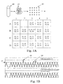
- Peak 18 representing well 3B will be 50% higher than the peaks from the other well, or the peaks from a reference sequence on chromosomes 22.
- Well A2 lacking either 21 or 22, will have no peak.
- the peaks are shown at 20.
- a single run will have numerous random variations, such as wells that have no target sequence, or have duplication through sample variability. Also, samples with no target will clearly result in no peak at all; wells with two or more targets, will give peaks significantly higher than peak 18, i.e., 2X or 2.5 X controls.
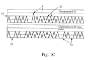
- Fig. 1C illustrates an embodiment where the DNA is distributed in a more dilute fashion (less than 1, or about one half genome equivalents per well).
- chromosome 21 labels will generate more positives than chromosome 22 (a diploid chromosome) specific labels (e.g., primers) due simply to the slightly greater abundance of chromosome 21 in a trisomy-containing sample.
- some wells will contain positives 20 for both chromosomes, some will contain negatives 22 for both chromosomes, but some will contain blanks 24 for the diploid chromosome and peaks for the trisomic chromosome, due to its greater abundance.
- the data from a higher peak 18 is not used in this mode. As explained below, this slight difference can be made statistically significant by examining a large number of wells, and by the sensitivity of the present method to a single molecule.
- the present method comprises generally the following steps:
- the present method is directed to non-invasive testing.
- the preferred starting material is maternal peripheral venous blood.
- 10-20 mL of blood be drawn, in order to obtain about at least 10,000 genome equivalents of total DNA.
- This sample size is based on an estimate of fetal DNA being present as roughly 25 genome equivalents/mL of maternal plasma in early pregnancy, and a fetal DNA concentration of about 3.4% of total plasma DNA.
- less blood may be drawn for a genetic screen where less statistical significance is required, or the DNA sample is enriched for fetal DNA.
- fetal RNA found in maternal blood may be analyzed as well.
- mRNA of placentas origin is readily detectable in maternal plasma
- hPL human placental lactogen
- hCG human chorionic gonadotropin
- mRNA transcripts were detectable in maternal plasma, as analyzed using the respective real-time RT-PCR assays.
- mRNA encoding genes expressed in the placenta and present on the chromosome of interest are used.
- DSCR4 Down syndrome critical region 4
- DSCR4 Down syndrome critical region 4
- mRNA sequence may be found at GenBank NM_005867.
- RNase H- RNase H- reverse transcriptases
- RTs RNase H minus (RNase H-) reverse transcriptases
- RNase H- RTs are available from several manufacturers, with SuperScriptTM II (Invitrogen) being the most widely used.
- Reverse transcriptase PCR may be used as described below for chromosomal DNA.
- the maternal blood may be processed to enrich the fetal DNA concentration in the total DNA, as described in Li et al., supra. Briefly, circulatory DNA is extracted from 5-to 10-mL maternal plasma using commercial column technology (Roche High Pure Template DNA Purification Kit; Roche, Basel, Switzerland) in combination with a vacuum pump. After extraction, the DNA is separated by agarose gel (1%) electrophoresis (Invitrogen, Basel, Switzerland), and the gel fraction containing circulatory DNA with a size of approximately 300 bp is carefully excised.
- the DNA is extracted from this gel slice by using an extraction kit (QIAEX II Gel Extraction Kit; Qiagen, Basel, Switzerland) and eluted into a final volume of 40- ⁇ L sterile 10-mM trishydrochloric acid, pH 8.0 (Roche).
- DNA may be concentrated by known methods, including centrifugation and various enzyme inhibitors.
- the RNA is bound to a selective membrane (e.g., silica) to separate it from contaminants.
- the DNA is preferably enriched for fragments circulating in the plasma, which are less than 1000 base pairs in length, generally less than 300 bp. This size selection is done on a DNA size separation medium, such as an electrophoretic gel or chromatography material.
- enrichment may be accomplished by suppression of certain alleles through the use of peptide nucleic acids (PNAs), which bind to their complementary target sequences, but do not amplify.
- PNAs peptide nucleic acids
- Plasma RNA extraction is described in Enders et al., "The Concentration of Circulating Corticotropin-releasing Hormone mRNA in Maternal Plasma Is Increased in Preeclampsia," Clinical Chemistry 49: 727-731, 2003 .
- plasma harvested after centrifugation steps is mixed Trizol LS reagent (Invitrogen) and chloroform. The mixture is centrifuged, and the aqueous layer transferred to new tubes. Ethanol is added to the aqueous layer. The mixture is then applied to an RNeasy mini column (Qiagen) and processed according to the manufacturer's recommendations.
- Agents that impede cell lysis or stabilize cell membranes can be added to the tubes including but not limited to formaldehyde, and derivatives of formaldehyde, formalin, glutaraldehyde, and derivatives of glutaraldehyde, crosslinkers, primary amine reactive crosslinkers, sulfhydryl reactive crosslinkers, sulfhydryl addition or disulfide reduction, carbohydrate reactive crosslinkers, carboxyl reactive crosslinkers, photoreactive crosslinkers, cleavable crosslinkers, etc. Any concentration of agent that stabilizes cell membranes or impedes cell lysis can be added. In a preferred embodiment, the agent that stabilizes cell membranes or impedes cell lysis is added at a concentration that does not impede or hinder subsequent reactions.
- Flow cytometry techniques can also be used to enrich fetal cells (Sberg et al., PNAS 76: 1453-1455 (1979 ); Bianchi et al., PNAS 87: 3279-3283 (1990 ); Bruch et al., Prenatal Diagnosis 11: 787-798 (1991 )).
- U.S. Pat. No. 5,432,054 also describes a technique for separation of fetal nucleated red blood cells, using a tube having a wide top and a narrow, capillary bottom made of polyethylene. Centrifugation using a variable speed program results in a stacking of red blood cells in the capillary based on the density of the molecules.
- the density fraction containing low-density red blood cells is recovered and then differentially hemolyzed to preferentially destroy maternal red blood cells.
- a density gradient in a hypertonic medium is used to separate red blood cells, now enriched in the fetal red blood cells from lymphocytes and ruptured maternal cells.
- the use of a hypertonic solution shrinks the red blood cells, which increases their density, and facilitates purification from the more dense lymphocytes.
- fetal DNA can be purified using standard techniques in the art.
- an agent that stabilizes cell membranes may be added to the maternal blood to reduce maternal cell lysis including but not limited to aldehydes, urea formaldehyde, phenol formaldehyde, DMAE (dimethylaminoethanol), cholesterol, cholesterol derivatives, high concentrations of magnesium, vitamin E, and vitamin E derivatives, calcium, calcium gluconate, taurine, niacin, hydroxylamine derivatives, bimoclomol, sucrose, astaxanthin, glucose, amitriptyline, isomer A hopane tetral phenylacetate, isomer B hopane tetral phenylacetate, citicoline, inositol, vitamin B, vitamin B complex, cholesterol hemisuccinate, sorbitol, calcium, coenzyme Q, ubiquinone, vitamin K, vitamin K complex, menaquinone, zonegran, zinc, ginkgo biloba extract, diphenylhydantoin, perftoran, polyviny
- An example of a protocol for using this agent is as follows: The blood is stored at 4°C until processing. The tubes are spun at 1000 rpm for ten minutes in a centrifuge with braking power set at zero. The tubes are spun a second time at 1000 rpm for ten minutes. The supernatant (the plasma) of each sample is transferred to a new tube and spun at 3000 rpm for ten minutes with the brake set at zero. The supernatant is transferred to a new tube and stored at -80°C, Approximately two milliliters of the "buffy coat," which contains maternal cells, is placed into a separate tube and stored at -80°C.
- Genomic DNA may be isolated from the plasma using the Qiagen Midi Kit for purification of DNA from blood cells, following the manufacturer's instructions (QIAmp DNA Blood Midi Kit, Catalog number 51183). DNA is eluted in 100mu.l of distilled water. The Qiagen Midi Kit also is used to isolate DNA from the maternal cells contained in the "buffy coat.”
- tissue saliva, urine, tear, vaginal secretion, breast fluid, breast milk, or sweat.
- the genomic DNA obtained from a maternal tissue as described above is diluted into multiple reaction samples, e.g. in multiwell plates, so that there is, on average, less than one genome equivalent per well.
- the DNA (Chromosome) to be analyzed will, on average, either be present or absent, permitting so-called "digital analysis.”
- a reaction sample in general will contain a single template molecule (haplotype), two target molecules (diploid) or three target molecules (trisomy).
- the wells provide discrete reaction samples and may be implemented in a number of devices, such as a microtiter plates, beads in an emulsion, or a microfluidic device. These are described in detail below.
- the device must be capable of carrying out a large number of discrete amplification reactions. As described below, this number should be, at a minimum, 10,000 reactions, and preferably on the order of 100,000 reactions.
- the reaction sample is preferable holds about 10-100 ⁇ L of a PCR reaction sample containing the genomic DNA, nucleotides (dNTPs), polymerase and appropriate PCR primers. The primers are used in conjunction with a label for rapid quantitative detection of PCR products.
- labeling will depend on the amplification/detection system used, e.g., a "molecular beacon" fluorescent probe for microtiter plate based amplification. This type of probe is described, for example, in Vogelstein et al, supra.
- labeling may be done with SYBR Green, which has very low fluorescence in the absence of double stranded DNA and very high fluorescence in the presence of double stranded DNA.
- Another form of parallel analysis useful in the present invention is single molecule analysis.
- a sample is diluted to contain less than a nominal single genome equivalent of DNA, and the presence of the target of interest (i.e., chromosome 21 trisomy) can be determined in a large number of samples.
- the fetal DNA can be distinguished from the maternal DNA. This is termed a "digital analysis,” because each well will have, on average, one genome equivalent per cell, and furthermore, the dilution may be read as a binary "yes-no" result as to the presence of the chromosome or other sequence to be counted.
- DLA direct linear analysis
- the presence of the DNA sequence or chromosome of interest must be quantified. This may be done either in single molecule mode, or with an amplified product.
- the method of amplification maybe, for example, self-sustained sequence reaction, ligase chain reaction, rapid amplification of cDNA ends, polymerase, chain reaction and ligase chain reaction, Q-beta phage amplification, strand displacement amplification, or splice overlap extension polymerase chain reaction.
- detection may be conveniently be carried out by a sequence specific probe, detection may also be carried out by directly sequencing a region of interest to determine if it is the target sequence of interest.
- PCR methods may be multiplexed, that is, run with multiple primers to multiple targets, it is preferred to limit the number of primer pairs in a given reaction.
- primer pairs there will be two primer pairs: one for amplifying a test sequence, and another pair for amplifying a control sequence.
- Primers are designed according to known parameters for avoiding secondary structures and self-hybridization. Further, both primer pairs should anneal and melt at about the same temperatures.
- Primers can be prepared by a variety of methods including but not limited to cloning of appropriate sequences and direct chemical synthesis using methods well known in the art ( Narang et al., Methods Enzymol. 68:90 (1979 ); Brown et al., Methods Enzymol. 68:109 (1979 )). Primers can also be obtained from commercial sources such as Operon Technologies, Amersham Pharmacia Biotech, Sigma, and Life Technologies.
- the primers can have an identical melting temperature.
- the lengths of the primers can be extended or shortened at the 5' end or the 3' end to produce primers with desired melting temperatures.
- the annealing position of each primer pair can be designed such that the sequence and, length of the primer pairs yield the desired melting temperature.
- Computer programs can also be used to design, primers, including but not limited to Array Designer Software (Arrayit Inc.), Oligonucleotide Probe Sequence Design Software for Genetic Analysis (Olympus Optical Co.), NetPrimer, and DNAsis from Hitachi Software Engineering.
- the TM (melting or annealing temperature) of each primer is calculated using software programs such as Oligo Design, available from Invitrogen Corp.
- the annealing temperature of the primers can be recalculated and increased after any cycle of amplification, including but not limited to cycle 1, 2, 3, 4, 5, cycles 6-10, cycles 10-15, cycles 15-20, cycles 20-25, cycles 25-30, cycles 30-35, or cycles 35-40.
- the 5' half of the primers is incorporated into the products from each loci of interest, thus the TM can be recalculated based on both the sequences of the 5' half and the 3' half of each primer.
- Any DNA polymerase that catalyzes primer extension can be used including but not limited to E. coli DNA polymerase, Klenow fragment of E.
- coli DNA polymerase 1 T7 DNA polymerase, T4 DNA, polymerase, Taq polymerase, Pfu DNA polymerase, Vent DNA polymerase, bacteriophage 29, REDTaq.TM. Genomic DNA polymerase, or sequenase.
- a thermostable DNA polymerase is used.
- a "hot start” PCR can also be performed wherein the reaction is heated to 95°C for two minutes prior to addition of the polymerase or the polymerase can be kept inactive until the first heating step in cycle 1.
- "Hot start” PCR can be used to minimize nonspecific amplification. Any number of PCR cycles can be used to amplify the DNA, including but not limited to 2, 5, 10, 15, 20, 25, 30, 35, 40, or 45 cycles. In a most preferred embodiment, the number of PCR cycles performed is such that equimolar amounts of each loci of interest arc produced.
- PCR primers are useful in the present process, such as those disclosed in technical literature of Qiagen. That literature describes a protocol where DNA was purified from peripheral blood and amniocyte cultures using the QIAmp DNA Blood Mini Kit. For amplification of the amyloid gene on chromosome 21, (NCBI gene ID 473931, accession NC_006488) primer and probe sequences were:
- primers and probe were: forward primer SEQ ID NO: 3 , 5'-CCC CAC ACA CAT GCA CTT ACC-3'; reverse primer, SEQ ID NO: 4 , 5'-CCT ACT CCC AGG GCT TTG ATT-3'; and probe, SEQ ID-NO: 5 , 5'-(VIC) AAA GAG CTA GGA AGG ACA GGC AAC TTG GC (TAMRA)-3'.
- PCR was performed using the TaqMan system, with 2 ⁇ l of template DNA in each 25 ⁇ l reaction and final concentrations of 300 nmol/liter of each primer and 150 nmol/liter of each dual-labeled TaqMan probe. Cycling conditions were incubation at 50°C for 2 minutes, then 95°C for 10 minutes, followed by 40 cycles of 60°C, 1 minute and 95°C, 15 seconds.
- Fluorescent probe-based technologies which can be performed on the PCR products "in situ” (i.e., in the same wells), are particularly well suited for this application. This method is described in detail in Vogelstein PNAS 96:9236 , above, and Vogelstein et al. "Digital Amplification," US 6,440,705 , hereby incorporated by reference for its description of this amplification procedure.
- the composition of the reactions is: 67 mM Tris, pH 8.8, 16.6 mM NH 4 SO 4 , 6.7 mM MgCl 2 , 10 mM ⁇ -mercaptoethanol, 1 mM dATP, 1 mM dCTP, 1 mM dGTP, 1 mM TTP, 6% DMSO, 1 ⁇ M primer F1, 1 ⁇ M primer R1, 0.05 units/ ⁇ l Platinum Taq polymerase (Life Technologies, Inc.), and "one-half gnome equivalent" of DNA.
- DNA samples are serially diluted and tested via PCR.
- the amount that yielded amplification products in half the wells is defined as "one-half genome equivalent" and used in each well of subsequent Digital Amplification experiments.
- Fifty ⁇ l light mineral oil (Sigma M-3516) is added to each well and reactions performed in a HybAid Thermal cycles at the following temperatures: denaturation at 94°C for one min; 60 cycles of 94°C for 15 sec, 55°C for 35 sec., 70°C for 15 seconds; 70°C for five minutes.
- MB or molecular beacon probes, which become fluorescent on binding to the target sequence(s), as described in more detail below, may be used as follows:
- MB probes are oligonucleotides with stem-loop structures that contain a fluorescent dye at the 5' end and a quenching agent (Dabcyl) at the 3' end.
- the degree of quenching via fluorescence energy resonance transfer is inversely proportional to the 6th power of the distance between the Dabcyl group and the fluorescent dye.
- MB probes reform a stem-loop structure, which quenches the fluorescent signal from the dye. If a PCR product whose sequence is complementary to the loop sequence is present during the heating/cooling cycle, hybridization of the M.B to one strand of the PCR product will increase the distance between the Dabcyl and the dye, resulting in increased fluorescence.
- RNA as well as DNA. Isolation of plasma RNA is described below. In this case, cDNA copies are made and then amplified by DNA polymerase-based PCR. Different primers may be used for cDNA synthesis. Specific templates, based on genetic sequences in the chromosomes of interest are preferred. See. Bustina et al., "Pitfalls of Quantitative Real-Time Reverse-Transcription Polymerase Chain Reaction," Journal of Biomolecular Techniques, 15:155-166 (2004 ). Use of mRNA from consitutively expressed, i.e., housekeeping genes, may be used for a control, and genes that are highly expressed in placenta (described below) are preferred.
- MessageSensorTM RT Kit includes an RNase H+ MMLV RT.
- MessageSensor includes a total RNA control, a control human GAPDH primer set, RNase inhibitor, and nucleotides, as well as a buffer additive that enables detection with SYBE ® Green dye.
- Ambion recommends using 18S rRNA as an internal control because it shows less variance in expression across treatment conditions than ⁇ -actin and GAPDH.
- a chromosomes 21-encoded gene (LOC90625) which shows strong expression in first trimester placenta similar to CSH1 (human placental lactogen) and was selected for plasma analysis in Oudejans et al., "Detection of Chromosome 21-encodcd mRNA of Placental Origin in Maternal Plasma," Clinical Chemistry 49: 1445-1449, 2003 . Specific primers for use with this gene are given in this paper.
- chromosome 21 transcripts are described at Gardiner et al., "Analysis of human chromosome 21: correlation of physical and cytogenetic maps; gene and CpG island distributions," E.M.B.O.J. 9(1):25-34 (1990 ), namely cDNA of identified products ETS2, MX1, MX2, CBS, COL6AI and BCEI, which can be partially sequenced or mapped according to eh present methods.
- Emulsion PCR has been used to prepare small beads with clonally amplified DNA - in essence, each bead contains one type of amplicon of digital PCR.
- Dressman et al Proc. Natl. Acad. Sci. U S A.100, 8817 (Jul 22, 2003 )
- the non-anchored primers with different fluorophores and use a single bead type.
- the positive beads (amplicons) of each type can be counted with methods such as flow cytometry or simply by counting them in a suitably equipped microscope.
- Step 1 Magnetic beads covalently coated with streptavidin are bound to biotinylated oligonucleotides ("oligos").
- Step 2 an aqueous mix containing all the necessary components for PCR plus primer-bound beads and template DNA are stirred together with an oil/detergent mix to create microemulsions.
- the aqueous compartments (which may be illustrated as small droplets in an oil layer) contain an average of ⁇ 1 template molecule and ⁇ 1 bead. Different templates (control and test) may be pictured in one or less droplets to represent two template molecules whose sequences differ by one or many nucleotides.
- the microemulsions are temperature cycled as in a conventional PCR. If a DNA template and a bead are present together in a single aqueous compartment, the bead bound oligonucleotides act as primers for amplification. Then, one may picture straight lines corresponding to PCR. products attached to the corresponding templates connected to the beads to represent extension products from the two different kinds of templates.
- Step 4 the emulsions are broken and the beads are purified with a magnet.
- Step 5 after denaturation, the beads are incubated with oligonucleotides that can distinguish between the sequences of the different kinds of templates. Fluorescently labeled antibodies are then used to label the bound hybridization probes. This renders the beads containing PCR product as different colors (e.g., red or green) upon appropriate laser excitation.
- Step 6 flow cytometry is used to count the red and green beads.
- each bead is bound to at least 10, 50, 100, 500, or 1000 molecules of the same nucleic acid sequence.
- Step 1-Coupling oligonucleotides to beads Superparamagnetic beads of 1.05 ⁇ 0.1 um in diameter, covalently bound to streptavidin, are purchased from Dynal Biotech, Inc. (650.01, Lake Success; NY). Beads are washed once with 1x PCR buffer (53286, Invitrogen, Carlsbad, CA) then suspended in Bind and Wash Buffer (BWB) (5 mMTris-HCl, 0.5 mM EDTA, 1.0 MNaCl, pH 7.5). Beads are incubated in BWB for 30 min at room temperature in the presence of 10 ⁇ M oligonucleotides.
- Bind and Wash Buffer BWB
- oligonucleotides are modified with a dual biotin group at the 5'end with the biotin groups separated by a six-carbon linker (IDT, Coralville, IA). After binding, the beads are washed 3 times with 1x PCR buffer to thoroughly remove unbound oligonucleotides.
- IDT six-carbon linker
- Microemulsions for PCR are prepared in an oil phase that is composed of 4.5% Span 80 (S6760, Sigma, St. Louis, MO), 0.40 % Tween 80 (Sigma S-8074), and 0.05% Triton X-100 (Sigma T-9284) in mineral oil (Sigma M-3516).
- the aqueous phase consists of 67 mMTris-HCl (pH 8.
- the forward primer is an oligonucleotide whose sequence is identical to the 3'20-22 nt of that described in step 1 and is not modified with biotin.
- Water-in-oil microemulsions are prepared by drop wise addition of 200 microliters of the aqueous phase to 400 microliters of the oil phase previously placed in a 2 ml round bottom cryogenic vial (430661, Coming, Coming, NY).
- the drop wise addition is performed over-one minute white the mixture is being stirred at 1400 RPM with a magnetic microstir bar (58948-353, VWR, Plainfield, NJ) on a VWR model 565 Magnetic Stirrer. After the addition of the aqueous phase, the mixture continued to be stirred for a total time of 30 minutes. Two emulsions are made at once by placing two tubes in a rack placed at the center of the magnetic stirrer.
- Step 3-PCR cycling The emulsions are aliquotted into five wells of a 96 well PCR plate, each containing 100 ⁇ l. PCR is carried out under the following cycling conditions: 94°C for 2 minutes; 40 cycles of 94°C for 15 seconds, 57°C for 30 seconds, 70°C for 30 seconds. The PCR products analyzed in this study ranged from 189 to 239 bp,
- Step 4-Magnetic capture of beads After PCR cycling, the microemulsion from five wells of a PCR plate are pooled and broken by the addition800 microliters of NX buffer (100 mMNaCl containing 1% Triton X-100, 10 mMTris-HCl, pH 7.5, 1 mM EDTA) in a 1.5 ml tube (Corning 430909). After vortexing for-20 sec. the beads are pelleted by centrifugation in a microcentrifuge at 8000 rpm (5000 g) for 90 seconds. The top oil phase, and all but-300 microliters of the aqueous phase, is removed from the tube and 600 microliters of NX buffer is added. These steps are repeated.
- NX buffer 100 mMNaCl containing 1% Triton X-100, 10 mMTris-HCl, pH 7.5, 1 mM EDTA
- the tube is then placed on a magnet (Dynal MPC-S) and the rest of the supernatant is carefully pipetted off.
- the beads are washed an additional 3 times with 1x PCR buffer using magnetic separation rather than centrifugation and finally re-suspended in 100 microliters of Ix PCR buffer.
- Step 5-Sequence differentiation Two oligonucleotide probes are used for each reaction. One is 5'-labeled with 6-carboxyfluorescein (6-FAM) and is specific for one allele while the second is 5'-labeled with biotin and is specific for the other allele. Probes are synthesized by IDT. The 30 microliters hybridization reactions contained 10 ⁇ M of each probe and 5-25 million beads inl x PCR buffer. Reactions are performed in PCR plates on a thermal cycler by heating to94°C for 30 seconds then cooling to75°C at a rate of 0.5°C per second, cooling to 45°C at 0.2°C per second, and finally cooled to 30°C at 1°C per second.
- 6-FAM 6-carboxyfluorescein
- the beads are then incubated for 10 minutes in a total volume of 100 microliters B-PCR buffer containing 3 ⁇ g ofAlexa-488 rabbit anti-fluorescein antibody (Molecular ProbesA-11090, Eugene, OR) and 3 ⁇ g of Nutravidin labeled with R-phycoerytbrin (Molecular Probes A-2660) in B-PCR buffer.
- B-PCR buffer containing 3 ⁇ g ofAlexa-488 rabbit anti-fluorescein antibody (Molecular ProbesA-11090, Eugene, OR) and 3 ⁇ g of Nutravidin labeled with R-phycoerytbrin (Molecular Probes A-2660) in B-PCR buffer.
- the beads are washed three times and resuspended in B-PCR buffer as described above.
- B-PCR buffer containing 6 ⁇ g of Alexa 488-conjugated chicken anti-rabbit antibody (Molecular Probes A-21441) and 3 ⁇ g of biotinylated goat anti-avidin antibody (BA-0300, Vector Laboratories, Burlingame, CA). The beads are washed three times and resuspended in B-PCR buffer as described above.
- Step 6-Flow Cytometry The bead suspension is diluted to a concentration of- 106-107 beads per ml in 10 mMTris-HCI, 1 mMEDTA (351-010-131, Quality Biological, Inc., Gaithersburg, MD) and analyzed using a LSR instrument (BD Biosciences, Franklin Lakes, NJ). The instrument is set up for standard two- color analysis using an argon laser and optical filters that distinguished between the two fluorescent dyes. No spectral deconvolution is required as the major bead populations are well separated. In some cases, scanning is performed with FACScan or FACSCalibur instruments (BD Biosciences).
- Another approach to digital PCR involves the use of microfluidics to achieve the digital PCR conditions used in the present method.
- a DNA sample obtained as described above is diluted into an appropriate concentration, mixed with PCR reagents, primers, dNTPs, etc. and flowed through a number of channels which may be closed off in multiple segments, resulting in a number of discrete reaction samples, or chambers.
- the chambers may be subjected to PCR thermal cycling and the products quantitatively detected by florescence, as described above.
- a suitable microfluidic device is produced by Fluidigm Corporation, termed the Digital Isolation and Detection IFC (integrated fluid circuit).
- a suitable device is also described in United States Patent 6,960,437 to Enzelberger, et al., issued November 1, 2005 entitled "Nucleic acid amplification utilizing microfluidic devices," hereby incorporated by reference for purposes of describing a microfluidic device capable of supporting multiple parallel nucleic acid amplifications and detections.
- one exemplary microfluidic device for conducting thermal cycling reactions includes in the layer with the flow channels a plurality of sample inputs, a mixing T-junction, a central circulation loop (i.e., the substantially circular flow channel), and an output channel.
- a control channel with a flow channel can form a microvalve. This is so because the control and flow channels are separated by a thin elastomeric membrane that can be deflected into the flow channel or retracted therefrom. Deflection or retraction of the elastomeric membrane is achieved by generating a force that causes the deflection or retraction to occur. In certain systems, this is accomplished by increasing or decreasing pressure in the control channel as compared to the flow channel with which the control channel intersects.
- a wide variety of other approaches can be utilized to actuate the valves including various electrostatic, magnetic, electrolytic and electrokinetic approaches.
- microfluidic device adapted to perform PCR reactions, and useful in the present methods, is described in US 2005/0252773 by McBride, et al., published November 17, 2005 , entitled "Thermal reaction device and method for using the same.”
- the substantially circular central loop and the control channels that intersect with it form the central part of the rotary pump.
- the pump(s) that cause solution to be flowed through the substantially circular flow channel consist of a set of at least three control channels that are adjacent to one another and which intersect the substantially circular branch flow channel (i.e., the central loop).
- the fluid in the central loop can be peristaltically pumped in a chosen direction, either clockwise or counterclockwise.
- the peristaltic pumping action results from the sequential deflection of the membranes separating the control channels and flow channel into or out of the flow channel. In general, the higher the actuation frequency, the faster the fluid rotates through the central loop.
- One system exemplified has two sets of pumps and (i.e., two sets of three control channels that overlay the substantially circular flow channel) a single pump can be utilized (i.e., a single set of three control channels overlaying the substantially circular flow channel). Furthermore, while each pump is shown as including three control channels, more control channels can be utilized. It should also be understood that the three control channels can be different segments of a single control channel that overlay the flow channel.
- DNA polymerase may be employed to image sequence information in a single DNA template as its complementary strand is synthesized.
- the nucleotides are inserted sequentially; only the time resolution to discriminate successive incorporations is required. After each successful incorporation event, a fluorescent signal is measured and then nulled by photobleaching. This method lends itself to massive parallelism.
- this technique permits observations of single molecule fluorescence by a conventional microscope equipped with total internal reflection illumination, which reduces background fluorescence.
- the surface of a quartz slide is chemically treated to specifically anchor DNA templates while preventing nonspecific binding of free nucleotides, and a plastic flow cell is attached to the surface to exchange solutions.
- DNA template oligonucleotides are hybridized to a fluorescently labeled primer and bound to the surface via streptavidin and biotin with a surface density low enough to resolve single molecules.
- the primed templates are detected through their fluorescent tags, their locations are recorded for future reference, and the tags arc photobleached.
- Labeled nucleotide triphosphates and DNA polymerase enzyme are then washed in and out of the flow cell while the known locations of the DNA templates are monitored for the appearance of fluorescence.
- the technique uses a combination of evanescent wave microscopy and single-pair fluorescence resonance energy transfer (spFRET) to reject unwanted noise.
- the donor fluorophore excites acceptors only within the Forster radius, thus effectively creating an extremely high-resolution near-field source. Because the Forster radius of this fluorophore pair is 5 nm, the spatial resolution of this method exceeds the diffraction limit by a factor of 50 and conventional near-field microscopy by an order of magnitude.
- the genomic DNA from the tissue taken from the mother i.e. the mixture of fetal and material genetic material
- a target specific sequence e.g., a unique region of chromosome 21, e.g., AML1.
- Further guidance for the preparation of chromosome 21-unique sequences may be found, for example, in Fuscoe et al., "An Efficient Method for Selecting Unique-Sequence Clones from DNA Libraries and Its Application To Fluorescent Staining of Human Chromosome 21 Using in situ Hybridization," Genomics, vol. 5, 1989, pp. 100-109 .
- a methodology useful in the present invention platform is based on massively parallel sequencing of millions of fragments using attachment of randomly fragmented genomic DNA to a planar, optically transparent surface and solid phase amplification to create a high density sequencing flow cell with millions of clusters, each containing ⁇ 1,000 copies of template per sq. cm.
- These templates are sequenced using four-color DNA sequencing-by-synthesis technology. See, products offered by Illumina, Inc., San Diego California. Also, see US 2003/0022207 to Balasubramanian, et al., published January 30, 2003 , entitled "Arrayed polynucleotides and their use in genome analysis.”
- Sequencing may be combined with amplification-based methods in a microfluidic chip having reaction chambers for both PCR and microscopic template-based sequencing. Only about 30 bp of random sequence information are needed to identify a sequence as belonging to a specific human chromosome. Longer sequences can uniquely identify more particular targets.
- An algorithm for designing unique sequences is described in Yamada, et al. "PrimerStation: a highly specific multiplex genomic PCR primer design server for the human genome," Nucleic Acids Res., July 1, 2006; 34(Web Server issue): W665 - W669 , illustrative of software methods that can be used to identify a sequence in comparison to the known genome sequence.
- DLA Direct Linear Analysis
- Another method of determining the identity of genomic DNA from the present samples is termed direct linear analysis, and is described in Chan et al. "DNA Mapping Using Microfluidic Stretching and Single-Molecule Detection of Fluorescent Site-Specific Tags," Genome Research 14:1137-1146 (2004 ).
- a microfluidic device is used for stretching DNA molecules in elongational flow that is coupled to a multicolor detection system capable of single-fluorophore sensitivity. Double-stranded DNA molecules are tagged at sequence-specific motif sites with fluorescent bisPNA (Peptide Nucleic Acid) tags.
- the DNA molecules are then stretched in the microfluidic device and driven in a flow stream past confocal fluorescence detectors.
- DLA can provide the spatial locations of multiple specific sequence motifs along individual DNA molecules, and thousands of individual molecules can be analyzed per minute.
- a microchip configuration and operating conditions may be prepared according to this publication that are adequate for stretching 50-kb long DNA.
- the chip includes a post field, a funnel with a 10:1 1 taper reduction ratio, a taper shape providing W(x)1/x2 profile (W is the channel width, and x is the coordinate along the flow direction), and a 5 ⁇ m-wide interrogation channel.
- the interrogation channel has uniform cross-section to ensure constant solution velocity, which was 10-15 ⁇ m/msec.
- the excitation laser beams are directed into the microscope objective with a dichroic mirror that reflects the light with 532 nm (beam ExI) and 633 nm (beams ExII and ExIII) wavelengths, but is transparent to the fluorescence emission excited by these beams.
- the emission is further split by another dichroic mirror and bandpass filters.
- Fluorescence excited by the green laser is delivered by optical fiber to the photon-counting avalanche photodiode (APD) for signal detection in data channel 1.
- APD avalanche photodiode
- the above-described device may be configured with larger path.lengths in order to accommodate larger DNA strands, presumably up to entire chromosome lengths.
- the genomic sample is probed with a chromosome 21 specific probe threaded through the interrogation channel, and the presence of one or more chromosomes is detected.
- Digital PCR allows the detection of aneuploidy merely by counting transcripts, as illustrated by the following calculation.
- chromosome A is euploid and represents an internal control
- chromosome B is aneuploid and is the target to be measured, then one can amplify representative segments from both chromosomes via digital PCR.
- the requisite statistical confidence to resolve the difference in proportions can be estimated as follows. There is a random statistical variation associated with the initial sample size, which goes roughly as the square root of the number of samples taken. It is in fact often difficult to precisely start with a fixed number of cell equivalents, and in the previous example we expect statistical error of order 32 amplicons (32 ⁇ square root(1,000)) for most sample preparation techniques. This is the same size as the signal we are trying to detect and thus in practice one requires more than 1,000 cell equivalents for robust detection. Precisely how many one requires depends on the statistical certainty that is required.
- the number of amplicons N must be at least 10,000 for Down Syndrome detection.
- the number of target sequences needed for statistical sequences may be reduced by using controls sequences, and, in addition, the sample may be enriched for fetal DNA,
- the present invention is particularly adapted to detecting genetic abnormalities that involve quantitative differences between maternal, and fetal genetic sequences.
- genetic abnormalities include mutations that may be heterozygous and homozygous between maternal and fetal DNA, and to aneuploidies.
- a missing copy of chromosome X results in Turner's Syndrome
- an additional copy of chromosome 21 results in Down Syndrome.
- Other diseases such as Edward's Syndrome and Patau Syndrome are caused by an additional copy of chromosome 18, and chromosome 13, respectively.
- the present method may be used for detection of a translocation, addition, amplification, transversion, inversion, aneuploidy, polyploidy, monosomy, trisomy, trisomy 21, trisomy 13, trisomy 14, trisomy 15, trisomy 16, trisomy 18, trisomy 22, triploidy, tetraploidy, and sex chromosome abnormalities including but not limited to XO, XXY, XYY, and XXX.
- chromosome specific primers arc disclosed in United States Patent Application 20050164241 to Hahn, Sinuhe, et al., published July 28, 2005 , entitled “Non-invasive detection of fetal genetic traits,” hereby incorporated by reference in its entirety for describing methods of sample preparation and certain PCR primers, described as follows:
- UniSTS is a comprehensive database of sequence tagged sites (STSs) derived from STS-based maps and other experiments. STSs are defined by PCR primer pairs and are associated with additional information such as genomic position, genes, and sequences. Similarly, primer sequences for other human chromosomes can be found by appropriately modifying the search query.
- Examples of diseases where the target sequence may exist in one copy in the maternal DNA (heterozygous) but cause disease in a fetus (homozygous), include sickle cell anemia, cystic fibrosis, hemophilia, and Tay Sachs disease. Accordingly, using the methods described here, one may distinguish genomes with one mutation from genomes with two mutations.
- Sickle-cell anemia is an autosomal recessive disease.
- Nine-percent of US blacks are heterozygous, while 0.2% are homozygous recessive.
- the recessive allele causes a single amino acid substitution in the beta chains of hemoglobin.
- Tay-Sachs Disease is an autosomal recessive resulting in degeneration of the nervous system. Symptoms manifest after birth. Children homozygous recessive for this allele rarely survive past five years of age. Sufferers lack the ability to make the enzyme N-acetyl-hexosaminidase, which breaks down the GM2 ganglioside lipid.
- PKU phenylketonuria
- Hemophilia is a group of diseases in which blood does not clot normally. Factors in blood are involved in clotting. Hemophiliacs lacking the normal Factor VIII are said to have Hemophilia A, and those who lack Factor IX have hemophilia B. These genes are carried on the X chromosome, so primers and probes may be used in the present method to detect whether or not a fetus inherited the mother's defective X chromosome, or the father's normal allele.
- the number of positive reaction samples is used, disregarding increased intensity from three versus two chromosomes in a reaction sample. That is, as described above, trisomy can be detected either by looking for an increased signal from a single well having multiple chromosomal DNA copies, or by diluting a sample and counting the frequency of responses of the trisomic marker versus a control diploid marker.
- Fetal DNA circulating in maternal plasma is here used to provide sufficient material for chromosomal analysis.
- DNA is extracted from a blood sample and aliquotted to different reaction chambers on the basis of genome equivalents, i.e., the entire genomic content of a single normal cell (46 chromosomes). This weighs about 6.6 pg.
- genome equivalent is used to refer to the calculated distribution of sample DNA based on a calculated genome size and DNA weight. In practice, there will be some experimental variation in DNA sample size, and, due to random fragment distribution, a given genome equivalent will not contain exactly the DNA fragments corresponding only to a single complete diploid genome, but a large number, on average, will.
- reaction mix For each panel on the Digital Array chip, 10ul of reaction mix is required. To achieve ⁇ 1/3 panel filled, the required final concentration of template in reaction mix should be approximately 48 copies/ ⁇ l (every 0.33 template per 7nl chamber). Thus for a 10 ⁇ l reaction volume (1 panel), 480 copies ( ⁇ 240 genome equivalents, "GE") of total free-floating DNA is required. These calculations are based on Chiu et al., "Effects of Blood-Processing Protocols on Fetal and Total DNA Quantification in Maternal Plasma," Clinical Chemistry, 47:9, 1607-1613.
- a 20 ml blood draw should contain enough total DNA less than 300 bp (which is about 85% fetal DNA) for about 10 panels, enough, as shown below, to achieve statistical significance.
- the following sample protocol provides a procedure for use in preparing a sample from maternal plasma and increasing the signal from chromosome 21.
- Plasma Collection collect 20 ml peripheral blood from the pregnant subject. This is collected in 2 tubes with EDTA as anticoagulant. Process blood within 2 hours of sample collection. The blood is processed first by centrifugation at 1600g for 10 min. One aliquots plasma to polypropylene tubes (1ml each), with care not to disturb buffy coat layer. Next, the supernatant is microcentrifuged at 16000g (full speed) for 10 min to remove residual maternal cells. Then, one extracts DNA from plasma with QIAamp Blood Mini Kit ("body fluid protocol"). 800 ⁇ l of plasma is applied per column and eluted in 40 ⁇ l buffer.
- body fluid protocol body fluid protocol
- DNA is subjected to gel electrophoresis ( Li et al Clin. Chem. 50:6 1002-1011 2004 ) to separate smaller sized DNA fragments.
- a UV gel tray is prepared with 1% agarose gel with 0.5 mg/L ethidium bromide. 100 bp ladder and HindIII digested Lambda phage DNA is used as markers.
- the extracted DNA is loaded on a gel; the gel is run at 80V for 1 hour.
- the DNA is extracted from the gel by first excising DNA ⁇ 300 bp with clean razor blade. This band is recovered with QIAEX II Gel Extraction Kit (Qiagen) and eluted 40ul in elution buffer.
- the amount of total free-floating DNA can be quantified using primers and Taqman probe designed for GAPDH gene (Chromosome 12). Real time PCR is run with GAPDH and Amyloid (Chromosome 21) primers and probes before running with Digital Array to confirm that the amplification regions are intact. To increase the signal from Chromosome 21, an additional set of primers and probes can be used.
- the Digital Isolation and Detection (DID) chip works by partitioning a sample/assay (TaqMan® assays) mixture into hundreds to tens of thousands of reaction chambers, where real-time QPCR reactions are contiguously monitored by a dynamic array reader.
- the DID chip described here contains inputs for 12 sample/assay mixtures, and its architecture partitions 7.5 ⁇ L of fluid for each input into 1,200 reaction chambers. These are shown as 12 panels in Figures 2 and 3 . Instrumentation is used to drive the sample/assay mixtures from the wells in the carrier into the appropriate reaction chambers.
- white spots indicate the location of reaction chambers positive for the indicated primer and dye.
- the sum of the positive wells in each section will be consistent with the gene/chromosome copies that were measured in the sample.
- the number of light spots shown represents the number of positive reaction chambers; no quantification was used in these experiments, and the results do not depend on quantization of a signal from an individual (discrete) sample mixture. Such quantization can be used, but can also be a source of error in methods that depend on this.
- kits with pre-optimized reagents such as the Qiagen QuantiTect Multiplex PCR Kit, which contains QuantiTect Multiplex PCR Buffer, having synthetic factor MP and an optimized combination of KCl and (NH 4 ) 2 SO 4 , which promote specific and stable annealing of primers to templates.
- This kit also contains HotStarTaq DNA Polymerase: Since this polymerase requires incubation at 95°C for activation, misprimed products and primer-dimers, which can compete for reactants, are not formed during reaction setup.
- the preferred concentration of DNA sample should be ⁇ 140-240 copies/ ⁇ l, i.e., ⁇ 70-120 GE/ ⁇ l. This corresponds to ⁇ 3.4 to 2 ⁇ l of required template volume in a digital PCR reaction volume of 10 ⁇ l.
- the mixture of maternal and fetal genetic material obtained from the mother is diluted to achieve a high likelihood that only one target sequence will be present in a given sample to be analyzed. As shown in Fig. 1A , it is also possible to carry out this process with less dilution and less empty sample sites if quantitation is used to distinguish a number of target sequences in a sample.
- HEX hexachloro-6-carboxyfluorescein
- FAM 6-carboxy-fluorescein
- Amyloid Forward 5' GGG AGC TGG TAC AGA AAT GAC TTC 3' (SEQ ID NO: 11)
- Amyloid Reverse 5' TTG CTC ATT GCG CTG ACA A 3' (SEQ ID NO: 12)
- Amyloid Probe 5' (FAM) AGC CAT CCT TCC CGG GCC TAG G (BHQ)3' (SEQ ID NO: 13 )
- GAPDH Forward 5' CCC CAC ACA CAT GCA CTT ACC 3'(SEQ ID NO: 14 )
- GAPDH Reverse 5' CCT AGT CCC AGG GCT TTG ATT 3' (SEQ ID NO: 15 )
- GAPDH Probe 5' (HEX)AAA GAG CTA GGA AGG ACA GGC AAC TTG GC (BHQ)3' (SEQ ID NO: 16 ) primers and probes were synthesized by IDT (Integrated DNA Technologies)).
- DNA samples were analyzed by digital PCR using microfluidic Digital PCR on a Fluidigm® microfluidic chip having 12 panels with 765 (wells) partitions each.
- Various mixtures of normal and Downs DNA (representing a mixture of fetal and maternal cells in a blood sample) were analyzed.
- Small amounts of each template were pipetted directly into each PCR mix; alternatively, a mixture of templates could be prepared first, then pipetted into the PCR mix, which should yield more accurate results. This alternate method was used in these experiments. In these examples, trisomy is detected based on the number of wells showing the triplicate chromosomal marker, i.e., the analysis illustrated in Fig. 1C .
- Intensity data of the triplicate chromosome are not used, except as a ratio to a normal chromosome marker. Because the sample is dilute many of the wells will have no Chromosome of interest (or marker fragment), as can be seen in Figs. 2 and 3 , which show photographs of chips from Example 2.
- Protocol combine Primers 300 nM; Probes 150 nM; iTaq supermix with ROX or iQ supermix. Tween20 (0.1%); DNA template (2 ⁇ l, premixed with the desired percentage of Downs DNA); Water (make up to total reaction volume of 10 ⁇ l).
- Each panel was loaded with reaction mix of 10 ⁇ l, and PCR was performed on a thermal cycler similar to the commercially available BIOMARK System from Fluidigm according to manufacturer's instructions. Cycling conditions were: 98°C 30s, 97°C 30s, 95°C 2min, [56°C. 30s, 58°C 30s, 60°C 30s, 98°C 13s] x 40 cycles, 60°C for 10 min.
- a MATLAB program was written to subtract the image of the chip taken before cycling from that taken at cycle 40 for each fluorescent channel. The number of positive wells in each fluorescent channel was counted.
- X actual input copy number of Chromosome 12
- Y actual input copy number of Chromosome 21
- X log ⁇ 1 - X * / N / log ⁇ 1 - 1 / N
- Y log ⁇ 1 - Y * / N / log ⁇ 1 - 1 / N
- Confidence interval k (Y-X)/sqrt(Y)
- Fig. 4 the confidence level obtained for a single chip was plotted against percent downs DNA and compared to the predicted confidence interval (line).
- the confidence interval k corresponds to the standard deviation, where a higher standard deviation indicates a greater difference between the normal and the Down's DNA. Even at the lowest concentration used (30%) and with only 10 panels analyzed, statistical analysis showed the feasibility of the present method.
- Figs. 2 and 3 show results from 100% Downs sample, for easy of visual analysis.
- the number of white spots indicates the positive wells for the markers tested.
- Chromosome 21 can be seen to have more spots by simple visual observation, distinguishing the trisomic from the normal chromosome.
- a 30% mixture depicting an enriched maternal blood sample
- the results were analyzed statistically.
- Table 1 shows the results for each panel (numbered as in Figs. 2 and 3 ) in a single experiment using 30% Down's DNA.
- Table 1 Panel Sample FAM HEX Ratio 1 Normal 221 213 1.04 2 Normal 254 264 0.96 3 Normal 271 252 1.08 4 Normal 246 257 0.96 5 Normal 241 238 1.01 8 30% Downs 270 222 1.22 9 30% Downs 219 194 1.13 10 30% Downs 249 234 1.06 11 30% Downs 230 223 1.03 12 30% Downs 216 189 1.14
- the "FAM” column shows the compartments (wells) positive for chromosome 21, and the "HEX” column shows the compartments positive for chromosome 12.
- the significance of the higher ratios in the Downs cases is shown in Fig. 4 , and was also analyzed in a Student's T-test, with a value of 0.036599344.
Landscapes
- Chemical & Material Sciences (AREA)
- Life Sciences & Earth Sciences (AREA)
- Organic Chemistry (AREA)
- Health & Medical Sciences (AREA)
- Proteomics, Peptides & Aminoacids (AREA)
- Zoology (AREA)
- Wood Science & Technology (AREA)
- Engineering & Computer Science (AREA)
- Analytical Chemistry (AREA)
- Genetics & Genomics (AREA)
- Immunology (AREA)
- Molecular Biology (AREA)
- Microbiology (AREA)
- Biotechnology (AREA)
- Biochemistry (AREA)
- Bioinformatics & Cheminformatics (AREA)
- General Engineering & Computer Science (AREA)
- General Health & Medical Sciences (AREA)
- Biophysics (AREA)
- Physics & Mathematics (AREA)
- Chemical Kinetics & Catalysis (AREA)
- Pathology (AREA)
- Cell Biology (AREA)
- Measuring Or Testing Involving Enzymes Or Micro-Organisms (AREA)
- Dispersion Chemistry (AREA)
Abstract
Description
- This application claims priority from
U.S. Provisional Patent Application No. 60/764,420 filed on February 2, 2006 - This invention was made with U.S. Government support. The U.S. Government may have certain rights in this invention.
- Applicants assert that the paper copy of the Sequence Listing is identical to the Sequence Listing in computer readable form found on the accompanying computer disk. Applicants incorporate the contents of the sequence listing by reference in its entirety.
- The present invention relates to the field of fetal genetic screening and to the field of quantitative nucleic acid analysis.
- It is now recognized that fetal DNA sheds from the placenta and mixes with the mother's blood at fairly high levels - between 3% and 6% of DNA in the mother's blood is from the fetus. This observation has been used in conjunction with. PCR assays for a variety of fetal genetic screens - gender, Rh, and thalassemia. However, the technique remains limited for two primary reasons: first, the PCR assays trade off sensitivity for specificity, making it difficult to identify particular mutations, and second, the most common genetic disorder, Down Syndrome, is a chromosomal trisomy and therefore cannot be detected by conventional PCR in a mixed sample.
- It has now been found that these problems can be solved by quantitative examination of large numbers of chromosome samples through the use of highly scalable techniques. This approach is termed here "digital analysis," and involves the separation of the extracted genomic material into discrete units so that the detection of a target sequence (e.g., chromosome 21) may be simply quantified as binary (0, 1) or simple multiples, 2, 3, etc. The primary example of a technique that can be used to yield such "digital" results is "digital PCR," which allows efficient amplification from single molecules, followed by subsequent quantitative analysis. Digital PCR, as the term is used here, refers to a quantitative, limited dilution of a nucleic acid sample, such as into multiwell plates, then the amplification of nucleic acid molecules in a well, which due to the dilution, should be either 0 or 1 molecule. Digital PCR using multiwell plates has been used previously to detect rare mutations by either serial analysis of single molecule (i.e., clonal) amplicons (Vogelstein B, Kinzler KW. Proc Natl Acad Sci U S A. 1999 Aug 3; 96 (16): 9236-41) or by enhancing the sensitivity of differential amplification (http://www.fluidigm.com/didIFC.htm). Described below is an invention whereby digital PCR can be applied to noninvasive fetal diagnostics in order to detect fetal mutations with specificity and sensitivity beyond what is possible with conventional PCR analysis.
- Furthermore, as also described in connection with the invention described below, digital PCR can be used to detect aneuploidy, such as the trisomy that causes Down Syndrome. Since aneuploidies do not present a mutational change in sequence, and are merely a change in the number of chromosomes, it has not been possible to detect them in a fetus without resorting to invasive techniques such as amniocentesis or chorionic villi sampling (Science 309, 2 September 2005 pp. 1476-8).
- Another form of digital PCR has been described as emulsion PCR, which has been used to prepare small beads with clonally amplified DNA - in essence, each bead contains one amplicon of digital PCR. (Dressman ct al, Proc Natl Acad Sci U S A.100, 8817 (Jul 22, 2003)).
- Another form of Digital PCR can be carried out using microfluidics. In this embodiment, described below, DNA is diluted and separated into small, discrete samples for forming reaction samples by a series of channels and valves.
- An example of a suitable method for single molecule analysis that may be adapted to the present methods is given in Braslavsky et al., "Sequence information can be obtained from single DNA molecules, Proc. Nat. Acad Sci. 100(7): 3960-3964 (2003), which uses sequential incorporation of labeled nucleotides onto an immobilized single stranded DNA template and monitoring by fluorescent microscopy.
- Another aspect of the relevant art involves sample preparation in order to carry out the present processes. That is, the fetal DNA may be enriched relative to maternal DNA. Chan, et al., "Size Distribution of Maternal and Fetal DNA in Maternal Plasm," Clin. Chem. 50(1): 88-92 (2004) reports that plasma DNA molecules are mainly short DNA fragments. The DNA fragments in the plasm of pregnant women are significantly longer than DNA fragments from non-pregnant women, and longer than fetal DNA.
- Vogelstein et al., "Digital Amplification,"
US 6,440,705, issued Aug. 27,2002 , discloses the identification of pre-defined mutations expected to be present in a minor fraction of a cell population. - Lo, "Fetal DNA in Maternal Plasma: Biology and Diagnostic Applications," Clin. Chem. 46:1903-1906 (2000) discloses the demonstration of fetal DNA in material plasma. The authors found a mean fractional level of 3,4% fetal DNA in maternal DNA in plasma during early pregnancy. The authors report detection of the RhD gene and microsatellite polymorphisms in the plasma of pregnant women.
- Li et al., "Detection of Paternally Inherited Fetal Point Mutations for β-Thalassemia Using Size Fractionated Cell-Free DNA in Maternal Plasma," J. Amer. Med. Assoc. 293:843-849 (Feb. 16, 2005) discloses that the analysis of cell-free fetal DNA in maternal plasma has proven to be remarkably reliable for the assessment of fetal loci absent from the maternal genome, Such as Y-chromosome specific sequences or the RhD gene in pregnant women who are Rh-negative. The authors report on the extraction and size fractionation of maternal plasma DNA using agarose gel electrophoresis. Then, peptide-nucleic acids (PNA) were used to bind specifically to a maternal allele to suppress PCR amplification of the of the wild type maternal allele, thereby enriching for the presence of paternally inherited mutant sequences. Four distinct point mutations in the β-globin gene were examined. It was found that the PNA step was necessary for the detection of mutant alleles using allele specific PCR.
- Lo et al., "Quantitative Analysis of Fetal DNA in Maternal Plasma and Serum: Implications for Noninvasive Prenatal Diagnosis," Am. J. Hum. Genet 62:768-775 (1998) discloses a real-time quantitative PCR assay to measure the concentration of fetal DNA in maternal plasma and serum. The authors found a mean of 25.4 genome equivalents/ml of fetal DNA in early pregnancy. This corresponds to about 3.4% of total DNA in early pregnancy.
- Chan et al., "Size Distribution of Maternal and Fetal DNA in Maternal Plasma," Clin. Chem. 50:89-92 (Jan. 2004) investigated the size distribution of plasma DNA in non-pregnant women and pregnant women, using a panel of quantitative PCR assays with different amplicon sizes targeting the leptin gens. They found that the DNA fragments in the plasma of pregnant women are significantly longer than those in the plasma of non-pregnant women, and the maternal-derived DNA molecules are longer than the fetal-derived ones,
- Tufan et al., "Analysis of Cell-Free Fetal DNA from Maternal Plasma and Serum Using a Conventional Multiplex PCR: Factors Influencing Success," Turk. J. Med. Sci. 35: 85-92 (2005) compared the success rates of two different DNA extraction techniques, the heat based direct method and the QIAMP DNA blood mini kit method. The crucial role of PCR optimization was also reported. The authors used the DYS14 marker for the Y chromosome and the GAPH gene for a control. The QIAMP mini kit was found to give the best results in sex determination analysis using multiplex PCR and ethidium bromide staining on gels.
- Hromadnikova et al., "Quantitative analysis of DNA levels in maternal plasma in normal and Down Syndrome pregnancies," BMC Pregnancy and Childbirth 2(4): 1-5 (2002), investigated total DNA levels in maternal plasma and found no difference in fetal DNA levels between the patients carrying Down Syndrome fetuses and the controls. Real time quantitative PCR analysis was performed using primers to the β-globin gene and the SRY locus.
- Grundevikk and Rosen, "Molecular Diagnosis of Aneuploidies," published on line at http://www.molbiotech.chalmers.se/research/mk/mbtk/Molecular%20diagnostics%20of%20ancu ploidies%20-%20rapport.pdf, suggests that non-invasive methods for detection of aneuploidies (such as Down Syndrome, Edwards Syndrome or extra sex chromosomes) may be carried out on fetal nucleated cells isolated from maternal blood. In their review, the authors also describe quantitative fluorescence polymerase chain reaction (QF-PCR), based on amplification, of short tandem repeats specific for the chromosome to be tested. They describe tests where DNA was amplified from amniotic or chorionic villus samples. The authors suggest that the STR markers will give PCR products of different size, and these size differences may be studied by analyzing peak sizes in electrophoresis. It is also proposed that quantitative real time PCR may be used to diagnose Down Syndrome by comparing the amount of a gene located on
chromosome 12 to the amount of a gene located on another autosomal chromosome. If the ratio of these two genes is 1:1., the fetus is normal, but if the ratio of these genes is 3:2, it indicates Down Syndrome. The authors propose the use of Down Syndrome marker DSCR3. They also suggest that the housekeeping gene GAPDH onchromosome 12 can be used as a reference. - Poon et al., "Differential DNA Methylation between Fetus and Mother as a Strategy for Detecting Fetal DNA in Maternal Plasma," Clin. Chem. 48(1): 35-41 discloses the detection of genes or mutations in a fetus where the same mutation or condition is also present in maternal DNA. That is, the use of fetal DNA in material plasma is limited due to the low amount of fetal DNA compared to maternal DNA. The authors overcame this limitation by detecting the IGF2-H19 locus, which is maintained in a methylated DNA status in the paternal allele and is unmethylated in the maternal allele. The authors used a bisulfite modification kit whereby unmethylated cytosine residues were converted to uracil. The sequence difference between methylated and unmethylated DNA sequences could be distinguished with different PCR primers. DNA extracted from buffy coat was used.
- Science 309:1476 (2 Sept. 2005) News Focus "An Earlier Look at Baby's Genes" describes attempts to develop tests for Down Syndrome using maternal blood. Early attempts to detect Down Syndrome using fetal cells from maternal blood were called "just modestly encouraging." The report also describes work by Dennis Lo to detect the Rh gene in a fetus where it is absent in the mother. Other mutations passed on from the father have reportedly been detected as well, such as cystic fibrosis, beta-thalassemia, a type of dwarfism and Huntington's disease. However, these results have not always been reproducible.
- United States Patent Application
20040137470 to Dhallan, Ravinder S, published July 15, 2004 , entitled "Methods for detection of genetic disorders," describes a method for detecting genetic disorders using PCR of known template DNA and restriction analysis. Also described is an enrichment procedure for fetal DNA. It also describes a method used to detect mutations, and chromosomal abnormalities including but not limited to translocation, transversion, monosomy, trisomy, and other aneuploidies, deletion, addition, amplification, fragment, translocation, and rearrangement. Numerous abnormalities can be detected simultaneously. The method is said to provide a non-invasive method to determine the sequence of fetal DNA from a tissue, such as blood, drawn from a pregnant female, and a method for isolating free nucleic acid from a sample containing nucleic acid. - The following brief summary is not intended to include all features and aspects of the present invention, nor does it imply that the invention must include all features and aspects discussed in this summary.
- Briefly, the present invention is directed to a method of differential detection of target sequences in a mixture of maternal and fetal genetic material. One obtains maternal tissue containing both maternal and fetal genetic material. Preferably, the maternal tissue is maternal peripheral blood or blood plasma. The term "plasma" may include plasma or serum, The genetic material may be genomic DNA or DNA, preferably mRNA. In the case of mRNA, one may choose target sequences corresponding to genes that are highly expressed in the placenta for fetal genetic material. The genetic material (e.g., DNA) in each reaction sample is detected with a sequence specific reactant directed to at least one of two target sequences in the genetic material to obtain a detectible reaction product if the target sequence is present in the reaction sample. For example, a probe specific to
chromosome 21 is bound to the reaction sample, along with a control probe specific to another chromosome. In most cases, the results will be from maternal DNA, but a small number of results will be obtained from fetal DNA. In order to distinguish random variation from fetal results, a large number of reactions are run, and statistical methods are applied to the results. The labeling and detection in the present method is used to distinguish the presence or absence of a single target sequence, referred to as "digital analysis," although it may be performed with sensitive nucleic acid detection methods which distinguish between one and more than one target sequence in a discrete sample. Many fluorescent techniques have this sensitivity. The target sequences are chosen so that a maternal sequence and a fetal sequence are distinguishable, such as two copies of a maternal sequence versus two copies of a fetal sequence. - The genetic material thus obtained is distributed into discrete samples, where each sample will contain, on average not more than about one target sequence per sample. The average of one target sequence means that, for practical reasons, the sample will contain, preferably 0.1 to 0.8 genome equivalents per discrete sample, ideally 0.5 genome equivalent per sample. The method may be performed with dilutions whereby more target sequences are detected in samples containing a trisomic or increased copy number of target sequence. That is, if one is analyzing
chromosome 21, the mixture may be diluted such that, on average, one may detect two chromosomes present in a maternal DNA, and three chromosomes in a Down Syndrome fetal DNA. Alternatively, the method may be performed with dilutions whereby more reaction samples are positive in this situation. The presence or absence of different target sequences in the discrete samples is detected; and the results are analyzed whereby the number of results from the discrete samples will provide data sufficient to obtain results distinguishing different target sequences. In one aspect, the method involves an analysis of a trisomy. In this method, one of the different target sequences (e.g. chromosome 21) is diploid in maternal genetic material and aneuploid in fetal genetic material and another of the different target sequences (e.g. chromosome 12) is diploid in both maternal and fetal genetic material. - The discrete samples are in reaction samples where the target sequences can be analyzed. The reaction samples may be, for Example, wells in a microtiter plate, aqueous phases in an emulsion, areas in an array surface, or reaction chambers in a microfluidic device. The reaction samples may be used for PCR analysis of the discrete samples. The discrete samples are contacted with a plurality of PCR. primers, including at least one (or one forward and one reverse) primer directed specifically to a maternal control sequence, expected to be the same in both mother and fetus. PCR primers are also directed specifically to a fetal sequence, i.e. one which may be present in both mother and fetus, but is amplified or altered in the fetus. PCR amplification will allow detection of these two different sequences, and, according to the present method, there will be a differential in the case of an abnormal fetal target sequence. The PCR method may be (but is not necessarily) quantitative. Quantitative real time PCR, which includes hybridizing target sequences with a nucleic acid having a fluorescent label, may be used. A fluorescent probe hybridizing to the target sequence may also be used. A number of "digital PCR" protocols are known for this purpose, as well as bead-based or emulsion PCR. While florescent probes are readily available and may be used to provide sensitive results, e.g., in FRET combinations, other labeling techniques may be used.
- The number of discrete samples is chosen according to the results desired. In one aspect, it is preferred that a high degree of statistical significance is obtained, and the number of samples is at least about 10,000. In order to improve statistical confidence, it is preferable to employ large numbers ofreactions, preferably between 500 and 100,000, more preferably between 10,000 and 100,000 or more reactions, depending on the percentage of fetal DNA present in the mixture. The results to be obtained should be statistically significant for purposes of the analysis conducted, e.g. initial screening, primary diagnosis, etc. A commonly used measure of statistical significance when a highly significant result is desired is p< 0.01, i.e., a 99% confidence interval based on a chi-square or t-test.
- However, as shown below, results can be obtained with less, e.g. on the order of about 500 samples, placed in separate reaction samples. Fewer discrete samples may be analyzed where the genetic material is present in a higher concentration in the mixture. The mixture may be enriched for fetal genetic material. One method to enrich plasma DNA for fetal DNA is size separation, whereby a preparation comprising only DNA fragments less than about 300 bp are used for measuring target sequences.
- A variety of genetic abnormalities may be detected according to the present method, including known alterations in one or more of the genes: CFTR, Factor VIII (F8 gene), beta globin, hemachromatosis, G6PD, neurofibromatosis, GAPDH, beta amyloid, and pyruvate kinase. The sequences and common mutations of these genes are known. Other genetic abnormalities may be detected, such as those involving a sequence which is deleted in a human chromosome, is moved in a translocation or inversion, or is duplicated in a chromosome duplication, wherein said sequence is characterized in a known genetic disorder in the fatal genetic material not present in the maternal genetic material. For example chromosome trisomies may include partial, mosaic, ring, 18, 14, 13, 8, 6, 4 etc. A listing of known abnormalities may be found in the OMIM Morbid map, http://www.ncbi.nlm.nih.gov/Omim/getmorbid.cgi.
- In general, the term "aneuploidy" is used to refer to the occurrence of one or more extra or missing chromosomes.
- In one aspect, the present method of differential detection of target sequences may involve direct sequencing of target sequences the genetic material. Single molecules sequencing, as is known, is further described below. The method may also comprise sequencing of amplified derivatives of the target sequences clones or amplicons of the genetic material. That is, a target sequence in a discrete sample is amplified by PCR, i.e, as an amplicon, or closed into a vector that is grown up and thereby amplified by obtaining multiple copies of the vector insert.
- In another aspect, the present invention comprises materials selected and combined for carrying out the present methods. Thus is provided a kit for differential detection of target sequences in maternal and fetal DNA in a mixed DNA sample, comprising primers specific for a genetically abnormal sequence and a control sequence, such as two chromosomes, one of which is possibly aneuploid and one of which is presumed diploid; a PCR reaction buffer for forming a PCR reaction sample with the primers in a device having separate reaction samples; and a size separation medium for separating the DNA sample into a traction having Less than about 1000 bp. The size separation medium may be gel or centrifugation material for recovering smaller DNA fragments and thus enriching fetal DNA. The kit may further comprise a pair of primers specific to
chromosome 21. The kit may further comprise the device having separate reaction samples for discrete samples. The device may be a microfluidic device or a microtiter plate having at least 1,000 discrete reaction samples. -
-
Figure 1 is a schematic illustration of the present analytical method, showing distribution of genetic material into compartments (1A), chromosome peaks of different height (1B), and statistical analysis of chromosomes (1C); -
Figure 2 is a photograph of a microfluidic chip having 12 panels (numbered 1-12) containing DNA withchromosome 21 labeled; -
Figures 3 is a photograph of a microfluidic chip having 12 panels (numbered 1-12) containing DNA withchromosome 12 labeled; and -
Figure 4 is a graph showing results from experiments done using digital analysis of mixed normal and trisomic (Down Syndrome, trisomy 21) DNA. - The methods and materials described below apply techniques for analyzing numerous nucleic acids contained in a tissue sample (preferably serum or, more preferably, plasma) containing a mixture of DNA from both the mother and the fetus, and allowing detection of small but statistically significant differences.
- The present invention involves the analysis of maternal blood for a genetic condition, wherein the mixed fetal and maternal DNA in the maternal blood is analyzed to distinguish a fetal mutation or genetic abnormality from the background of the maternal DNA. It has been found that, using a combination of steps, a DNA sample containing DNA from both the mother and the fetus can be analyzed to distinguish a genetic condition present in a minor fraction of the DNA, which represents the fetal DNA. The method employs "digital analysis," in which the DNA in the sample is isolated to a nominal single target molecule in a small reaction volume. Each sample mixture has a possibility of having distributed in it less than 1 target (i.e., 0 target) or more than one target. Next, the target molecules are detected in each reaction well, preferably as target sequences which are amplified, which may include a quantization of starting copy number of the target sequence, that is, 0, 1, 2, 3, etc. A control sequence is used to distinguish an abnormal increase in the target sequence, e.g., a trisonomy. Thus there is a differential detection of target sequences, one of which is chosen to represent a normal genotype present in both mother and offspring, and one of which is chosen for detection of an abnormal genotype in the offspring, where the target sequence in the offspring will be different from that of the mother, e.g. in trisomy.
-
Fig. 1A illustrates an embodiment where quantitative detection, e.g. quantitative real time PCR, is used. Blood 1.0 is processed to obtainplasma DNA 12, which is diluted and distributed intoaliquots 14. These are added to reactions wells 1A through 5D. Shown in the wells aretargets representing chromosomes Peak 18 representing well 3B will be 50% higher than the peaks from the other well, or the peaks from a reference sequence onchromosomes 22. Well A2, lacking either 21 or 22, will have no peak. The peaks are shown at 20. A single run will have numerous random variations, such as wells that have no target sequence, or have duplication through sample variability. Also, samples with no target will clearly result in no peak at all; wells with two or more targets, will give peaks significantly higher thanpeak 18, i.e., 2X or 2.5 X controls. These results are distinguished by running a multitude of reactions, followed by statistical analysis that can discriminate random variations from true results. -
Fig. 1C illustrates an embodiment where the DNA is distributed in a more dilute fashion (less than 1, or about one half genome equivalents per well). In thiscase chromosome 21 labels (primers) will generate more positives than chromosome 22 (a diploid chromosome) specific labels (e.g., primers) due simply to the slightly greater abundance ofchromosome 21 in a trisomy-containing sample. As shown, some wells will containpositives 20 for both chromosomes, some will containnegatives 22 for both chromosomes, but some will containblanks 24 for the diploid chromosome and peaks for the trisomic chromosome, due to its greater abundance. The data from ahigher peak 18 is not used in this mode. As explained below, this slight difference can be made statistically significant by examining a large number of wells, and by the sensitivity of the present method to a single molecule. - Thus, the present method comprises generally the following steps:
- 1. Obtaining a tissue containing DNA from a pregnant subject, which DNA is known to have about 3% fetal DNA. This material is preferably drawn blood, and the circulating DNA is found in the blood plasma, rather than in cells. The blood or plasma may optionally be enriched for fetal DNA by known method, such as size fractionation to select for DNA fragments less than about 300 bp. Alternatively, maternal DNA, which tends to be larger than about 500 bp may be excluded. Another enrichment step may be to treat the blood sample with formaldehyde, as described in Dhallan et al. "Methods to Increase the Percentage of Free Fetal DNA Recovered From the Maternal Circulation," J.Am. Med. Soc. 291(9): 1114-1119 (March 2004).
- 2. Distributing single DNA molecules from this sample to a number of discrete reaction samples, where the number of reaction samples is selected to give a statistically significant result for the number of copies of a target in the DNA molecules. Further, the reaction sample is confined to a small volume to bring the reaction molecules into close approximation. The amount of DNA molecule per reaction sample is preferably on the order of one copy of the chromosome of interest equivalent per reaction sample.
- 3. Detecting the presence of the target in the DNA in a large number of reaction samples, preferably with a sequence specific technique such as highly multiplexed short read sequencing or a PCR reaction wherein the PCR product is labeled to give a convenient quantitative read out. The detection step is referred to here as "digital PCR" and may be carried out by a variety of methods, such as (a) by PCR on samples diluted into individual wells of a microtiter plate; (b) PCR on samples diluted into emulsions containing primers immobilized to beads; or (c) PCR on samples trapped in a microfluidic chamber; and
- 4. Quantitative analysis of the detection of the maternal and fetal target sequences. In some cases this may include targets to different regions, such as probes to a target on a chromosome suspected of being present in an abnormal copy number (trisonomy) compared to a normal diploid chromosome, which is used as a control
- The present method is directed to non-invasive testing. The preferred starting material is maternal peripheral venous blood. In order to obtain sufficient DNA for testing, it is preferred that 10-20 mL of blood be drawn, in order to obtain about at least 10,000 genome equivalents of total DNA. This sample size is based on an estimate of fetal DNA being present as roughly 25 genome equivalents/mL of maternal plasma in early pregnancy, and a fetal DNA concentration of about 3.4% of total plasma DNA. However, less blood may be drawn for a genetic screen where less statistical significance is required, or the DNA sample is enriched for fetal DNA.
- It should be noted that, while the present description refers throughout to DNA, fetal RNA found in maternal blood may be analyzed as well. As described in Ng et al., "mRNA of placentas origin is readily detectable in maternal plasma," Proc. Nat. Acad. Sci. 100(8): 4748-4753 (2003), hPL (human placental lactogen) and hCG (human chorionic gonadotropin) mRNA transcripts were detectable in maternal plasma, as analyzed using the respective real-time RT-PCR assays. In the present method, mRNA encoding genes expressed in the placenta and present on the chromosome of interest are used. For example, DSCR4 (Down syndrome critical region 4) is found on
chromosome 21 and is mainly expressed in the placenta. Its mRNA sequence may be found at GenBank NM_005867. In this case, it is preferred to use RNase H minus (RNase H-) reverse transcriptases (RTs) to prepare cDNA for detection. RNase H- RTs are available from several manufacturers, with SuperScript™ II (Invitrogen) being the most widely used. Reverse transcriptase PCR may be used as described below for chromosomal DNA. - The maternal blood may be processed to enrich the fetal DNA concentration in the total DNA, as described in Li et al., supra. Briefly, circulatory DNA is extracted from 5-to 10-mL maternal plasma using commercial column technology (Roche High Pure Template DNA Purification Kit; Roche, Basel, Switzerland) in combination with a vacuum pump. After extraction, the DNA is separated by agarose gel (1%) electrophoresis (Invitrogen, Basel, Switzerland), and the gel fraction containing circulatory DNA with a size of approximately 300 bp is carefully excised. The DNA is extracted from this gel slice by using an extraction kit (QIAEX II Gel Extraction Kit; Qiagen, Basel, Switzerland) and eluted into a final volume of 40-µL sterile 10-mM trishydrochloric acid, pH 8.0 (Roche).
- DNA may be concentrated by known methods, including centrifugation and various enzyme inhibitors. The RNA is bound to a selective membrane (e.g., silica) to separate it from contaminants. The DNA is preferably enriched for fragments circulating in the plasma, which are less than 1000 base pairs in length, generally less than 300 bp. This size selection is done on a DNA size separation medium, such as an electrophoretic gel or chromatography material. Such a material is described in Huber et al., "High-resolution liquid chromatography of DNA fragments on non-porous poly(styrene-di-vinylbenzene) particles," Nucleic Acids Res, 1993 March 11; 21(5): 1061-1066, gel filtration chromatography, TSK gel, as described in Kato et al., "A New Packing for Separation of DNA Restriction Fragments by High Performance Liquid Chromatography," J. Biochem, 1984, Vol. 95, No. 1 83-86.
- In addition, enrichment may be accomplished by suppression of certain alleles through the use of peptide nucleic acids (PNAs), which bind to their complementary target sequences, but do not amplify.
- Plasma RNA extraction is described in Enders et al., "The Concentration of Circulating Corticotropin-releasing Hormone mRNA in Maternal Plasma Is Increased in Preeclampsia," Clinical Chemistry 49: 727-731, 2003. As described there, plasma harvested after centrifugation steps is mixed Trizol LS reagent (Invitrogen) and chloroform. The mixture is centrifuged, and the aqueous layer transferred to new tubes. Ethanol is added to the aqueous layer. The mixture is then applied to an RNeasy mini column (Qiagen) and processed according to the manufacturer's recommendations.
- United States Patent Application
20040137470 to Dhallan, Ravinder S, published July 15, 2004 , entitled "Methods for detection of genetic disorders," describes an enrichment procedure for fetal DNA," in which blood is collected into 9 ml EDTA Vacuette tubes (catalog number NC9897284) and 0.225 ml of 10% neutral buffered solution containing formaldehyde (4% w/v), is added to each tube, and each tube gently is inverted. The tubes are stored at 4°C until ready for processing. - Agents that impede cell lysis or stabilize cell membranes can be added to the tubes including but not limited to formaldehyde, and derivatives of formaldehyde, formalin, glutaraldehyde, and derivatives of glutaraldehyde, crosslinkers, primary amine reactive crosslinkers, sulfhydryl reactive crosslinkers, sulfhydryl addition or disulfide reduction, carbohydrate reactive crosslinkers, carboxyl reactive crosslinkers, photoreactive crosslinkers, cleavable crosslinkers, etc. Any concentration of agent that stabilizes cell membranes or impedes cell lysis can be added. In a preferred embodiment, the agent that stabilizes cell membranes or impedes cell lysis is added at a concentration that does not impede or hinder subsequent reactions.
- Flow cytometry techniques can also be used to enrich fetal cells (Herzenberg et al., PNAS 76: 1453-1455 (1979); Bianchi et al., PNAS 87: 3279-3283 (1990); Bruch et al., Prenatal Diagnosis 11: 787-798 (1991)).
U.S. Pat. No. 5,432,054 also describes a technique for separation of fetal nucleated red blood cells, using a tube having a wide top and a narrow, capillary bottom made of polyethylene. Centrifugation using a variable speed program results in a stacking of red blood cells in the capillary based on the density of the molecules. The density fraction containing low-density red blood cells, including fetal red blood cells, is recovered and then differentially hemolyzed to preferentially destroy maternal red blood cells. A density gradient in a hypertonic medium is used to separate red blood cells, now enriched in the fetal red blood cells from lymphocytes and ruptured maternal cells. The use of a hypertonic solution shrinks the red blood cells, which increases their density, and facilitates purification from the more dense lymphocytes. After the fetal cells have been isolated, fetal DNA can be purified using standard techniques in the art. - Further, an agent that stabilizes cell membranes may be added to the maternal blood to reduce maternal cell lysis including but not limited to aldehydes, urea formaldehyde, phenol formaldehyde, DMAE (dimethylaminoethanol), cholesterol, cholesterol derivatives, high concentrations of magnesium, vitamin E, and vitamin E derivatives, calcium, calcium gluconate, taurine, niacin, hydroxylamine derivatives, bimoclomol, sucrose, astaxanthin, glucose, amitriptyline, isomer A hopane tetral phenylacetate, isomer B hopane tetral phenylacetate, citicoline, inositol, vitamin B, vitamin B complex, cholesterol hemisuccinate, sorbitol, calcium, coenzyme Q, ubiquinone, vitamin K, vitamin K complex, menaquinone, zonegran, zinc, ginkgo biloba extract, diphenylhydantoin, perftoran, polyvinylpyrrolidone, phosphatidylserine, tegretol, PABA, disodium cromglycate, nedocromil sodium, phenyloin, zinc citrate, mexitil, dilantin, sodium hyaluronate, or polaxamer 188.
- An example of a protocol for using this agent is as follows: The blood is stored at 4°C until processing. The tubes are spun at 1000 rpm for ten minutes in a centrifuge with braking power set at zero. The tubes are spun a second time at 1000 rpm for ten minutes. The supernatant (the plasma) of each sample is transferred to a new tube and spun at 3000 rpm for ten minutes with the brake set at zero. The supernatant is transferred to a new tube and stored at -80°C, Approximately two milliliters of the "buffy coat," which contains maternal cells, is placed into a separate tube and stored at -80°C.
- Genomic DNA may be isolated from the plasma using the Qiagen Midi Kit for purification of DNA from blood cells, following the manufacturer's instructions (QIAmp DNA Blood Midi Kit, Catalog number 51183). DNA is eluted in 100mu.l of distilled water. The Qiagen Midi Kit also is used to isolate DNA from the maternal cells contained in the "buffy coat."
- Finally, it is noted that, in certain embodiments, one may also use samples from tissue, saliva, urine, tear, vaginal secretion, breast fluid, breast milk, or sweat.
- In the illustrated method, the genomic DNA obtained from a maternal tissue as described above is diluted into multiple reaction samples, e.g. in multiwell plates, so that there is, on average, less than one genome equivalent per well. Thus, when the individual discrete samples are analyzed for the presence of the genetic abnormality to be tested, the DNA (Chromosome) to be analyzed will, on average, either be present or absent, permitting so-called "digital analysis." A reaction sample in general will contain a single template molecule (haplotype), two target molecules (diploid) or three target molecules (trisomy).
- The wells provide discrete reaction samples and may be implemented in a number of devices, such as a microtiter plates, beads in an emulsion, or a microfluidic device. These are described in detail below. The device must be capable of carrying out a large number of discrete amplification reactions. As described below, this number should be, at a minimum, 10,000 reactions, and preferably on the order of 100,000 reactions. The reaction sample is preferable holds about 10-100 µL of a PCR reaction sample containing the genomic DNA, nucleotides (dNTPs), polymerase and appropriate PCR primers. The primers are used in conjunction with a label for rapid quantitative detection of PCR products. The type of labeling will depend on the amplification/detection system used, e.g., a "molecular beacon" fluorescent probe for microtiter plate based amplification. This type of probe is described, for example, in Vogelstein et al, supra. Alternatively, labeling may be done with SYBR Green, which has very low fluorescence in the absence of double stranded DNA and very high fluorescence in the presence of double stranded DNA.
- Another form of parallel analysis useful in the present invention is single molecule analysis. Again, a sample is diluted to contain less than a nominal single genome equivalent of DNA, and the presence of the target of interest (i.e.,
chromosome 21 trisomy) can be determined in a large number of samples. By analyzing a large number of samples, the fetal DNA can be distinguished from the maternal DNA. This is termed a "digital analysis," because each well will have, on average, one genome equivalent per cell, and furthermore, the dilution may be read as a binary "yes-no" result as to the presence of the chromosome or other sequence to be counted. - Another method for single molecule analysis involves the use of site-specific fluorescent tags that are detected as the DNA is drawn through a microfluidic device in a single molecule, elongated flow. An example of this technique, described below, is termed "direct linear analysis," or DLA.
- Having isolated the sample DNA into a nominal genome equivalent, the presence of the DNA sequence or chromosome of interest must be quantified. This may be done either in single molecule mode, or with an amplified product.
- While the preferred embodiment of the invention is described in terms of PCR, the invention is primarily directed to the use of multiple individual genetic sequence detection. In some embodiments, the method of amplification maybe, for example, self-sustained sequence reaction, ligase chain reaction, rapid amplification of cDNA ends, polymerase, chain reaction and ligase chain reaction, Q-beta phage amplification, strand displacement amplification, or splice overlap extension polymerase chain reaction.
- Also, while detection may be conveniently be carried out by a sequence specific probe, detection may also be carried out by directly sequencing a region of interest to determine if it is the target sequence of interest.
- While the presently know PCR methods may be multiplexed, that is, run with multiple primers to multiple targets, it is preferred to limit the number of primer pairs in a given reaction. Generally, there will be two primer pairs: one for amplifying a test sequence, and another pair for amplifying a control sequence. Primers are designed according to known parameters for avoiding secondary structures and self-hybridization. Further, both primer pairs should anneal and melt at about the same temperatures.
- Primers can be prepared by a variety of methods including but not limited to cloning of appropriate sequences and direct chemical synthesis using methods well known in the art (Narang et al., Methods Enzymol. 68:90 (1979); Brown et al., Methods Enzymol. 68:109 (1979)). Primers can also be obtained from commercial sources such as Operon Technologies, Amersham Pharmacia Biotech, Sigma, and Life Technologies. The primers can have an identical melting temperature. The lengths of the primers can be extended or shortened at the 5' end or the 3' end to produce primers with desired melting temperatures. Also, the annealing position of each primer pair can be designed such that the sequence and, length of the primer pairs yield the desired melting temperature. The simplest equation for determining the melting temperature of primers smaller than 25 base pairs is the Wallace Rule (Td=2(A+T)+4(G+C)). Computer programs can also be used to design, primers, including but not limited to Array Designer Software (Arrayit Inc.), Oligonucleotide Probe Sequence Design Software for Genetic Analysis (Olympus Optical Co.), NetPrimer, and DNAsis from Hitachi Software Engineering. The TM (melting or annealing temperature) of each primer is calculated using software programs such as Oligo Design, available from Invitrogen Corp.
- The annealing temperature of the primers can be recalculated and increased after any cycle of amplification, including but not limited to
cycle coli DNA polymerase 1, T7 DNA polymerase, T4 DNA, polymerase, Taq polymerase, Pfu DNA polymerase, Vent DNA polymerase, bacteriophage 29, REDTaq.TM. Genomic DNA polymerase, or sequenase. Preferably, a thermostable DNA polymerase is used. A "hot start" PCR can also be performed wherein the reaction is heated to 95°C for two minutes prior to addition of the polymerase or the polymerase can be kept inactive until the first heating step incycle 1. "Hot start" PCR can be used to minimize nonspecific amplification. Any number of PCR cycles can be used to amplify the DNA, including but not limited to 2, 5, 10, 15, 20, 25, 30, 35, 40, or 45 cycles. In a most preferred embodiment, the number of PCR cycles performed is such that equimolar amounts of each loci of interest arc produced. - A number of specific PCR primers are useful in the present process, such as those disclosed in technical literature of Qiagen. That literature describes a protocol where DNA was purified from peripheral blood and amniocyte cultures using the QIAmp DNA Blood Mini Kit. For amplification of the amyloid gene on
chromosome 21, (NCBI gene ID 473931, accession NC_006488) primer and probe sequences were: - SEQ ID NO: 1: forward primer, 5'-CGG AGC TGG TAC ALGA AAT GAC TTC-3'; reverse primer, SEQ ID NO: 10: 5'-.TTG CTC Art GCG CTG ACA A-3'; and probe, SEQ ID NO: 2 5'-(FAM) AGC CAT CCT TCC CGG GCC TAG G (TAMRA)-3'.
- For amplification of GAPDH, (GenBank locus 12p13.31-p13.1) primers and probe were: forward primer SEQ ID NO: 3, 5'-CCC CAC ACA CAT GCA CTT ACC-3'; reverse primer, SEQ ID NO: 4, 5'-CCT ACT CCC AGG GCT TTG ATT-3'; and probe, SEQ ID-NO: 5, 5'-(VIC) AAA GAG CTA GGA AGG ACA GGC AAC TTG GC (TAMRA)-3'. PCR was performed using the TaqMan system, with 2 µl of template DNA in each 25 µl reaction and final concentrations of 300 nmol/liter of each primer and 150 nmol/liter of each dual-labeled TaqMan probe. Cycling conditions were incubation at 50°C for 2 minutes, then 95°C for 10 minutes, followed by 40 cycles of 60°C, 1 minute and 95°C, 15 seconds.
- Using the above exemplary protocol, the different ratio of the amyloid gene and the GAPDH gene in karyotypically normal and
trisomy 21 samples was clearly distinguishable in the multiplex PCR assay, as reported in the Qiagen product literature. Assays using a dilution series of the DNA template showed that the difference remained clear over a wide range of template concentrations and with starting concentrations of DNA as low as 10 mg/liter. Of course, in a maternal blood sample, the concentration of fetal DNA would be much lower. - Fluorescent probe-based technologies, which can be performed on the PCR products "in situ" (i.e., in the same wells), are particularly well suited for this application. This method is described in detail in Vogelstein PNAS 96:9236, above, and Vogelstein et al. "Digital Amplification,"
US 6,440,705 , hereby incorporated by reference for its description of this amplification procedure. - The "digi-PCR" method of Vogelstein et al. is described in the above-mentioned patent. An exemplary protocol as set forth in that patent is as follows: PCR is performed in 7 µl volumes in 96 well polypropylene PCR plates (Marsh Biomedical Products, Rochester, N.Y.). The composition of the reactions is: 67 mM Tris, pH 8.8, 16.6 mM NH4 SO4, 6.7 mM MgCl2, 10 mM β-mercaptoethanol, 1 mM dATP, 1 mM dCTP, 1 mM dGTP, 1 mM TTP, 6% DMSO, 1 µM primer F1, 1 µM primer R1, 0.05 units/µl Platinum Taq polymerase (Life Technologies, Inc.), and "one-half gnome equivalent" of DNA.
- To determine the amount of DNA corresponding to one-half genome equivalent, DNA samples are serially diluted and tested via PCR. The amount that yielded amplification products in half the wells, usually about.1.5 pg of total DNA, is defined as "one-half genome equivalent" and used in each well of subsequent Digital Amplification experiments. Fifty µl light mineral oil (Sigma M-3516) is added to each well and reactions performed in a HybAid Thermal cycles at the following temperatures: denaturation at 94°C for one min; 60 cycles of 94°C for 15 sec, 55°C for 35 sec., 70°C for 15 seconds; 70°C for five minutes.
- MB, or molecular beacon probes, which become fluorescent on binding to the target sequence(s), as described in more detail below, may be used as follows:
- For fluorescence analysis, 3.5 µl of a solution with the following composition is added to each well: 67 mM Tris, pH 8.8, 16.6 mM NH4 SO4, 6,7 mM MgCl2, 10 mM β-mercaptoethanol, 1 mM dATP, 1 mM dCTP, 1 mM dGTP, 1 mM TTP, 6% DMSO, 5 µM primer, 1 µM MB-PREEN, 1 µM MB-RED, 0.1 units/µl Platinum Taq polymerase. The plates are centrifuged for 20 seconds at 6000 g and fluorescence read at excitation/emission wavelengths of 485 nm/530 nm for MB-GREEN and 530 nm/590 mn for MB-RED. The plates are then placed in a thermal cycler for asymmetric amplification at the following temperatures: 94°C for one minute; 10-15 cycles of 94°C for 15 see, 55°C for 15 sec., 70°C for 15 seconds; 94°C for one minute; and 60°C for five minutes. The plates are then incubated at room temperature for ten to sixty minutes and fluorescence measured as described above.
- MB probes are oligonucleotides with stem-loop structures that contain a fluorescent dye at the 5' end and a quenching agent (Dabcyl) at the 3' end. The degree of quenching via fluorescence energy resonance transfer is inversely proportional to the 6th power of the distance between the Dabcyl group and the fluorescent dye. After heating and cooling, MB probes reform a stem-loop structure, which quenches the fluorescent signal from the dye. If a PCR product whose sequence is complementary to the loop sequence is present during the heating/cooling cycle, hybridization of the M.B to one strand of the PCR product will increase the distance between the Dabcyl and the dye, resulting in increased fluorescence.
- The examples below use a PCR protocol, which also relies on MB type probes, except in connection with a microfluidic device.
- The present digital PCR methods may be used with RNA as well as DNA. Isolation of plasma RNA is described below. In this case, cDNA copies are made and then amplified by DNA polymerase-based PCR. Different primers may be used for cDNA synthesis. Specific templates, based on genetic sequences in the chromosomes of interest are preferred. See. Bustina et al., "Pitfalls of Quantitative Real-Time Reverse-Transcription Polymerase Chain Reaction," Journal of Biomolecular Techniques, 15:155-166 (2004). Use of mRNA from consitutively expressed, i.e., housekeeping genes, may be used for a control, and genes that are highly expressed in placenta (described below) are preferred. Currently four different chemistries, TaqMan® (Applied Biosystems, Foster City, CA, USA), Molecular Beacons, Scorpions® and SYBR® Green (Molecular Probes), are available for real-time PCR. All of these chemistries allow detection of PCR products via the generation of a fluorescent signal and may be adapted to reverse-transcription PCR. Ambion's MessageSensor™ RT Kit includes an RNase H+ MMLV RT. MessageSensor includes a total RNA control, a control human GAPDH primer set, RNase inhibitor, and nucleotides, as well as a buffer additive that enables detection with SYBE® Green dye. Ambion recommends using 18S rRNA as an internal control because it shows less variance in expression across treatment conditions than β-actin and GAPDH. A chromosomes 21-encoded gene (LOC90625) which shows strong expression in first trimester placenta similar to CSH1 (human placental lactogen) and was selected for plasma analysis in Oudejans et al., "Detection of Chromosome 21-encodcd mRNA of Placental Origin in Maternal Plasma," Clinical Chemistry 49: 1445-1449, 2003. Specific primers for use with this gene are given in this paper. Uniquely expressed
chromosome 21 transcripts are described at Gardiner et al., "Analysis of human chromosome 21: correlation of physical and cytogenetic maps; gene and CpG island distributions," E.M.B.O.J. 9(1):25-34 (1990), namely cDNA of identified products ETS2, MX1, MX2, CBS, COL6AI and BCEI, which can be partially sequenced or mapped according to eh present methods. - Emulsion PCR has been used to prepare small beads with clonally amplified DNA - in essence, each bead contains one type of amplicon of digital PCR. (Dressman et al, Proc. Natl. Acad. Sci. U S A.100, 8817 ()). By using specific primers for regions of chromosomes A and B while performing emulsion PCR, one will create beads with digital amplicons from only these two chromosomes, and it is only necessary to count the number of positive beads of each type. There are many ways to do this; we will point out two of them. First, use two different species of beads (either in size or fluorescent labeling) to anchor the two amplicons respectively. Alternatively, one could label the non-anchored primers with different fluorophores and use a single bead type. After amplification, the positive beads (amplicons) of each type can be counted with methods such as flow cytometry or simply by counting them in a suitably equipped microscope.
- This technique is further described in Dressman et al (supra) and
Dressman et al. PCT publication WO2005010145 , "METHOD AND COMPOSITIONS FOR DETECTION AND ENUMERATION OF GENETIC VARIATIONS," published 2005-02-03, and hereby incorporated by reference for its description of a bead-based process. Briefly, inStep 1, Magnetic beads covalently coated with streptavidin are bound to biotinylated oligonucleotides ("oligos"). InStep 2, an aqueous mix containing all the necessary components for PCR plus primer-bound beads and template DNA are stirred together with an oil/detergent mix to create microemulsions. The aqueous compartments (which may be illustrated as small droplets in an oil layer) contain an average of< 1 template molecule and < 1 bead. Different templates (control and test) may be pictured in one or less droplets to represent two template molecules whose sequences differ by one or many nucleotides. InStep 3, the microemulsions are temperature cycled as in a conventional PCR. If a DNA template and a bead are present together in a single aqueous compartment, the bead bound oligonucleotides act as primers for amplification. Then, one may picture straight lines corresponding to PCR. products attached to the corresponding templates connected to the beads to represent extension products from the two different kinds of templates. InStep 4, the emulsions are broken and the beads are purified with a magnet. InStep 5, after denaturation, the beads are incubated with oligonucleotides that can distinguish between the sequences of the different kinds of templates. Fluorescently labeled antibodies are then used to label the bound hybridization probes. This renders the beads containing PCR product as different colors (e.g., red or green) upon appropriate laser excitation. InStep 6, flow cytometry is used to count the red and green beads. Preferably, each bead is bound to at least 10, 50, 100, 500, or 1000 molecules of the same nucleic acid sequence. - For purposes of detailed description, the following example is taken from the above-quoted PCT publication:
- Step 1-Coupling oligonucleotides to beads. Superparamagnetic beads of 1.05 ± 0.1 um in diameter, covalently bound to streptavidin, are purchased from Dynal Biotech, Inc. (650.01, Lake Success; NY). Beads are washed once with 1x PCR buffer (53286, Invitrogen, Carlsbad, CA) then suspended in Bind and Wash Buffer (BWB) (5 mMTris-HCl, 0.5 mM EDTA, 1.0 MNaCl, pH 7.5). Beads are incubated in BWB for 30 min at room temperature in the presence of 10 µM oligonucleotides. These oligonucleotides are modified with a dual biotin group at the 5'end with the biotin groups separated by a six-carbon linker (IDT, Coralville, IA). After binding, the beads are washed 3 times with 1x PCR buffer to thoroughly remove unbound oligonucleotides.
- Step 2-Preparing microemulsions. Microemulsions for PCR are prepared in an oil phase that is composed of 4.5% Span 80 (S6760, Sigma, St. Louis, MO), 0.40 % Tween 80 (Sigma S-8074), and 0.05% Triton X-100 (Sigma T-9284) in mineral oil (Sigma M-3516). The aqueous phase consists of 67 mMTris-HCl (
pH 8. 8), 16.6 mM NH4S04, 6.7 mMMgC12, 10 mM(3-mercaptoethanol,1 mMdATP,1 mMdCTP,1 mMdGTP,1 mMdTTP, 0.05 µM forward primer, 25 µM reverse primer, 45 units Platinum Taq(Invitrogen 10966-034), various amounts of template DNA, and ∼108 oligonucleotide-coupled beads in a total volume of 300 µl. The forward primer is an oligonucleotide whose sequence is identical to the 3'20-22 nt of that described instep 1 and is not modified with biotin. - Water-in-oil microemulsions are prepared by drop wise addition of 200 microliters of the aqueous phase to 400 microliters of the oil phase previously placed in a 2 ml round bottom cryogenic vial (430661, Coming, Coming, NY).
- The drop wise addition is performed over-one minute white the mixture is being stirred at 1400 RPM with a magnetic microstir bar (58948-353, VWR, Plainfield, NJ) on a VWR model 565 Magnetic Stirrer. After the addition of the aqueous phase, the mixture continued to be stirred for a total time of 30 minutes. Two emulsions are made at once by placing two tubes in a rack placed at the center of the magnetic stirrer.
- Step 3-PCR cycling. The emulsions are aliquotted into five wells of a 96 well PCR plate, each containing 100 µl. PCR is carried out under the following cycling conditions: 94°C for 2 minutes; 40 cycles of 94°C for 15 seconds, 57°C for 30 seconds, 70°C for 30 seconds. The PCR products analyzed in this study ranged from 189 to 239 bp,
- Step 4-Magnetic capture of beads. After PCR cycling, the microemulsion from five wells of a PCR plate are pooled and broken by the addition800 microliters of NX buffer (100 mMNaCl containing 1% Triton X-100, 10 mMTris-HCl, pH 7.5, 1 mM EDTA) in a 1.5 ml tube (Corning 430909). After vortexing for-20 sec. the beads are pelleted by centrifugation in a microcentrifuge at 8000 rpm (5000 g) for 90 seconds. The top oil phase, and all but-300 microliters of the aqueous phase, is removed from the tube and 600 microliters of NX buffer is added. These steps are repeated. The tube is then placed on a magnet (Dynal MPC-S) and the rest of the supernatant is carefully pipetted off. The beads are washed an additional 3 times with 1x PCR buffer using magnetic separation rather than centrifugation and finally re-suspended in 100 microliters of Ix PCR buffer.
- Step 5-Sequence differentiation. Two oligonucleotide probes are used for each reaction. One is 5'-labeled with 6-carboxyfluorescein (6-FAM) and is specific for one allele while the second is 5'-labeled with biotin and is specific for the other allele. Probes are synthesized by IDT. The 30 microliters hybridization reactions contained 10 µM of each probe and 5-25 million beads inl x PCR buffer. Reactions are performed in PCR plates on a thermal cycler by heating to94°C for 30 seconds then cooling to75°C at a rate of 0.5°C per second, cooling to 45°C at 0.2°C per second, and finally cooled to 30°C at 1°C per second.
- All subsequent steps are performed at room temperature. The reactions are transferred to a 96 well Costar plate (Corning 3797) and placed on a 96 well magnet. Beads are collected magnetically by exposing them to the magnet for 2 minutes- The supernatant is removed and the beads washed 3 times with 1x PCR buffer by pipetting them and collecting for two minutes. They arc finally resuspended in 100 microliters B-PCR buffer (Img/mL BSA in 1x PCR buffer).
- The beads are then incubated for 10 minutes in a total volume of 100 microliters B-PCR buffer containing 3 µg ofAlexa-488 rabbit anti-fluorescein antibody (Molecular ProbesA-11090, Eugene, OR) and 3 µg of Nutravidin labeled with R-phycoerytbrin (Molecular Probes A-2660) in B-PCR buffer. The beads are washed three times and resuspended in B-PCR buffer as described above. They are then incubated for ten minutes in a total volume of 100 microliters B-PCR buffer containing 6 µg of Alexa 488-conjugated chicken anti-rabbit antibody (Molecular Probes A-21441) and 3 µg of biotinylated goat anti-avidin antibody (BA-0300, Vector Laboratories, Burlingame, CA). The beads are washed three times and resuspended in B-PCR buffer as described above. They are then incubated for ten minutes in a total volume of 100 microliters B-PCR buffer containing 3 µg of an Alexa 488-conjugated goat anti-chicken antibody (Molecular Probes A-11039) and 3 micrograms of R-phycoerytbrin-labeled streptavidin (Molecular Probes S-866). This solution is then washed an additional 3 times with 1x PCR buffer and resuspended in 20 microliters of 1 X PCR buffer.
- Step 6-Flow Cytometry. The bead suspension is diluted to a concentration of- 106-107 beads per ml in 10 mMTris-HCI, 1 mMEDTA (351-010-131, Quality Biological, Inc., Gaithersburg, MD) and analyzed using a LSR instrument (BD Biosciences, Franklin Lakes, NJ). The instrument is set up for standard two- color analysis using an argon laser and optical filters that distinguished between the two fluorescent dyes. No spectral deconvolution is required as the major bead populations are well separated. In some cases, scanning is performed with FACScan or FACSCalibur instruments (BD Biosciences).
- Another approach to digital PCR involves the use of microfluidics to achieve the digital PCR conditions used in the present method.
- Generally, a DNA sample obtained as described above is diluted into an appropriate concentration, mixed with PCR reagents, primers, dNTPs, etc. and flowed through a number of channels which may be closed off in multiple segments, resulting in a number of discrete reaction samples, or chambers. The chambers may be subjected to PCR thermal cycling and the products quantitatively detected by florescence, as described above.
- A suitable microfluidic device is produced by Fluidigm Corporation, termed the Digital Isolation and Detection IFC (integrated fluid circuit). A suitable device is also described in United States Patent 6,960,437 to Enzelberger, et al., issued November 1, 2005 entitled "Nucleic acid amplification utilizing microfluidic devices," hereby incorporated by reference for purposes of describing a microfluidic device capable of supporting multiple parallel nucleic acid amplifications and detections. As described in this patent, one exemplary microfluidic device for conducting thermal cycling reactions includes in the layer with the flow channels a plurality of sample inputs, a mixing T-junction, a central circulation loop (i.e., the substantially circular flow channel), and an output channel. The intersection of a control channel with a flow channel can form a microvalve. This is so because the control and flow channels are separated by a thin elastomeric membrane that can be deflected into the flow channel or retracted therefrom. Deflection or retraction of the elastomeric membrane is achieved by generating a force that causes the deflection or retraction to occur. In certain systems, this is accomplished by increasing or decreasing pressure in the control channel as compared to the flow channel with which the control channel intersects. However, a wide variety of other approaches can be utilized to actuate the valves including various electrostatic, magnetic, electrolytic and electrokinetic approaches. Another microfluidic device, adapted to perform PCR reactions, and useful in the present methods, is described in
US 2005/0252773 by McBride, et al., published November 17, 2005 , entitled "Thermal reaction device and method for using the same." - The substantially circular central loop and the control channels that intersect with it form the central part of the rotary pump. The pump(s) that cause solution to be flowed through the substantially circular flow channel consist of a set of at least three control channels that are adjacent to one another and which intersect the substantially circular branch flow channel (i.e., the central loop). When a series of on/off actuation sequences are applied to the control channels, the fluid in the central loop can be peristaltically pumped in a chosen direction, either clockwise or counterclockwise. The peristaltic pumping action results from the sequential deflection of the membranes separating the control channels and flow channel into or out of the flow channel. In general, the higher the actuation frequency, the faster the fluid rotates through the central loop. However, a point of saturation is eventually reached at which increased frequency does not result in faster fluid flow. This is primarily due to limitations in the rate at which the membrane can return to an unactuated position. One system exemplified has two sets of pumps and (i.e., two sets of three control channels that overlay the substantially circular flow channel) a single pump can be utilized (i.e., a single set of three control channels overlaying the substantially circular flow channel). Furthermore, while each pump is shown as including three control channels, more control channels can be utilized. It should also be understood that the three control channels can be different segments of a single control channel that overlay the flow channel.
- The detailed description of multiple sample analysis being carried out in wells doses not mean that the target sequences need to be physically separated into wells, as the sequences may be in samples which are isolated simply by being on different beads (as described above) or by adherence to different areas of a substrate (as described below).
- It should be appreciated that methods involving PCR or other amplification are not the only way to detect or enumerate the molecules in a given discrete reaction sample. It is possible to use single molecule flow cytometry to count single molecules that have been labeled with a sequence-specific fluorescent probe. It is also possible to sequence the target sequence in the reaction sample directly, either after amplification or at the single molecule level.
- As described in the above-referenced PNAS publication by Braslavsky et al., DNA polymerase may be employed to image sequence information in a single DNA template as its complementary strand is synthesized. The nucleotides are inserted sequentially; only the time resolution to discriminate successive incorporations is required. After each successful incorporation event, a fluorescent signal is measured and then nulled by photobleaching. This method lends itself to massive parallelism.
- Briefly, this technique permits observations of single molecule fluorescence by a conventional microscope equipped with total internal reflection illumination, which reduces background fluorescence. The surface of a quartz slide is chemically treated to specifically anchor DNA templates while preventing nonspecific binding of free nucleotides, and a plastic flow cell is attached to the surface to exchange solutions. DNA template oligonucleotides are hybridized to a fluorescently labeled primer and bound to the surface via streptavidin and biotin with a surface density low enough to resolve single molecules. The primed templates are detected through their fluorescent tags, their locations are recorded for future reference, and the tags arc photobleached. Labeled nucleotide triphosphates and DNA polymerase enzyme are then washed in and out of the flow cell while the known locations of the DNA templates are monitored for the appearance of fluorescence. The technique uses a combination of evanescent wave microscopy and single-pair fluorescence resonance energy transfer (spFRET) to reject unwanted noise. The donor fluorophore excites acceptors only within the Forster radius, thus effectively creating an extremely high-resolution near-field source. Because the Forster radius of this fluorophore pair is 5 nm, the spatial resolution of this method exceeds the diffraction limit by a factor of 50 and conventional near-field microscopy by an order of magnitude.
- The genomic DNA from the tissue taken from the mother, i.e. the mixture of fetal and material genetic material, may be distributed into discrete samples which are anchored to a surface and sequenced or monitored by labeled probes to detect a target specific sequence, e.g., a unique region of
chromosome 21, e.g., AML1. Further guidance for the preparation of chromosome 21-unique sequences may be found, for example, in Fuscoe et al., "An Efficient Method for Selecting Unique-Sequence Clones from DNA Libraries and Its Application To Fluorescent Staining of . A methodology useful in the present invention platform is based on massively parallel sequencing of millions of fragments using attachment of randomly fragmented genomic DNA to a planar, optically transparent surface and solid phase amplification to create a high density sequencing flow cell with millions of clusters, each containing ∼1,000 copies of template per sq. cm. These templates are sequenced using four-color DNA sequencing-by-synthesis technology. See, products offered by Illumina, Inc., San Diego California. Also, seeUS 2003/0022207 to Balasubramanian, et al., published January 30, 2003 , entitled "Arrayed polynucleotides and their use in genome analysis." - Sequencing may be combined with amplification-based methods in a microfluidic chip having reaction chambers for both PCR and microscopic template-based sequencing. Only about 30 bp of random sequence information are needed to identify a sequence as belonging to a specific human chromosome. Longer sequences can uniquely identify more particular targets. An algorithm for designing unique sequences is described in Yamada, et al. "PrimerStation: a highly specific multiplex genomic PCR primer design server for the human genome," Nucleic Acids Res., July 1, 2006; 34(Web Server issue): W665 - W669, illustrative of software methods that can be used to identify a sequence in comparison to the known genome sequence. See, also Zhu et al., "Single molecule profiling of alternative pre-mRNA splicing," Science. 2003 , describing a single-molecule-based technology for studying mRNA.
- Another method of determining the identity of genomic DNA from the present samples is termed direct linear analysis, and is described in Chan et al. "DNA Mapping Using Microfluidic Stretching and Single-Molecule Detection of Fluorescent Site-Specific Tags," Genome Research 14:1137-1146 (2004). In this method, a microfluidic device is used for stretching DNA molecules in elongational flow that is coupled to a multicolor detection system capable of single-fluorophore sensitivity. Double-stranded DNA molecules are tagged at sequence-specific motif sites with fluorescent bisPNA (Peptide Nucleic Acid) tags. The DNA molecules are then stretched in the microfluidic device and driven in a flow stream past confocal fluorescence detectors. DLA can provide the spatial locations of multiple specific sequence motifs along individual DNA molecules, and thousands of individual molecules can be analyzed per minute.
- A microchip configuration and operating conditions may be prepared according to this publication that are adequate for stretching 50-kb long DNA. The chip includes a post field, a funnel with a 10:1 1 taper reduction ratio, a taper shape providing W(x)1/x2 profile (W is the channel width, and x is the coordinate along the flow direction), and a 5 µm-wide interrogation channel. The interrogation channel has uniform cross-section to ensure constant solution velocity, which was 10-15 µm/msec. Once inside the channel, stretched and tagged DNA molecules travel through spots of focused laser light that excites fluorescence. Epi-illumination of the sample and confocal detection are arranged within a fluorescence microscope.
- The excitation laser beams are directed into the microscope objective with a dichroic mirror that reflects the light with 532 nm (beam ExI) and 633 nm (beams ExII and ExIII) wavelengths, but is transparent to the fluorescence emission excited by these beams. The emission is further split by another dichroic mirror and bandpass filters. Fluorescence excited by the green laser is delivered by optical fiber to the photon-counting avalanche photodiode (APD) for signal detection in
data channel 1. Fluorescence excited by red beams ExII and ExIII is directed to the APDs ofdata channels - The above-described device may be configured with larger path.lengths in order to accommodate larger DNA strands, presumably up to entire chromosome lengths. The genomic sample is probed with a
chromosome 21 specific probe threaded through the interrogation channel, and the presence of one or more chromosomes is detected. - Digital PCR allows the detection of aneuploidy merely by counting transcripts, as illustrated by the following calculation. Suppose that fetal DNA is present in material blood at a fraction level of ε, and that we are trying to discover an aneuploidy of order a relative to euploidy e (in the example relating to detection of Down Syndrome in humans, e=2 is euploidy and the Down Syndrome trisomy α=3). If chromosome A is euploid and represents an internal control, and chromosome B is aneuploid and is the target to be measured, then one can amplify representative segments from both chromosomes via digital PCR. In comparing the amplicons of each type, one expects to find that for every e amplicons from chromosome A there are e(1-ε)+αε amplicons from chromosome B. In the case of a trisomy and ε =3%, then for every 2 amplicons from chromosome A one expects 2.03 amplicons from chromosome B. While this difference is small, it can be measured. For example, if one amplifies a sample from 1,000 cell equivalents, then one expects 2,000 amplicons from chromosome A and 2,030 from chromosome B, The difference of 30 amplicons is in principle detectable.
- The requisite statistical confidence to resolve the difference in proportions can be estimated as follows. There is a random statistical variation associated with the initial sample size, which goes roughly as the square root of the number of samples taken. It is in fact often difficult to precisely start with a fixed number of cell equivalents, and in the previous example we expect statistical error of order 32 amplicons (32∼square root(1,000)) for most sample preparation techniques. This is the same size as the signal we are trying to detect and thus in practice one requires more than 1,000 cell equivalents for robust detection. Precisely how many one requires depends on the statistical certainty that is required. If one would like a result that is significant to k standard deviations, then

or N = (k/(ε(α-ε)))2 - Using the values of the previous example, if we require k = 3 standard deviations, then the number of amplicons N must be at least 10,000 for Down Syndrome detection. However, as discussed above, the number of target sequences needed for statistical sequences may be reduced by using controls sequences, and, in addition, the sample may be enriched for fetal DNA,
- The present invention is particularly adapted to detecting genetic abnormalities that involve quantitative differences between maternal, and fetal genetic sequences. These genetic abnormalities include mutations that may be heterozygous and homozygous between maternal and fetal DNA, and to aneuploidies. For example, a missing copy of chromosome X (monosomy X) results in Turner's Syndrome, while an additional copy of
chromosome 21 results in Down Syndrome. Other diseases such as Edward's Syndrome and Patau Syndrome are caused by an additional copy ofchromosome 18, and chromosome 13, respectively. The present method may be used for detection of a translocation, addition, amplification, transversion, inversion, aneuploidy, polyploidy, monosomy, trisomy,trisomy 21, trisomy 13,trisomy 14, trisomy 15,trisomy 16,trisomy 18,trisomy 22, triploidy, tetraploidy, and sex chromosome abnormalities including but not limited to XO, XXY, XYY, and XXX. - Other chromosome specific primers arc disclosed in United States Patent Application
20050164241 to Hahn, Sinuhe, et al., published July 28, 2005 , entitled "Non-invasive detection of fetal genetic traits," hereby incorporated by reference in its entirety for describing methods of sample preparation and certain PCR primers, described as follows: - The primers for the genes are prepared on the basis of nucleotide sequences obtained from databases such as GenBank, EMBL and the like. The names of the polymorphic primers and the sequences of the primers for the genes will be shown for the respective chromosomes in the following examples (#2, Examples 1; #4, Example 6, #14, Example 9; #22, Example 2). The following genetic markers and polymorphic makers (Polymorphic STS Primer Pairs: D2S207, D2S 177, D2S156 and D2S159, BIOS Laboratories, Inc.) are used to identify
chromosome # 2. - There are more than 1,000
chromosome 21 specific PCR primer sets listed at the NIH UniSTS web site, which can be located at http://www.ncbi.nlm.nih.gov/entrez/query.fegi?db=unists and found with the search phrase "human[organism] AND 21[chr]". UniSTS is a comprehensive database of sequence tagged sites (STSs) derived from STS-based maps and other experiments. STSs are defined by PCR primer pairs and are associated with additional information such as genomic position, genes, and sequences. Similarly, primer sequences for other human chromosomes can be found by appropriately modifying the search query. - Examples of diseases where the target sequence may exist in one copy in the maternal DNA (heterozygous) but cause disease in a fetus (homozygous), include sickle cell anemia, cystic fibrosis, hemophilia, and Tay Sachs disease. Accordingly, using the methods described here, one may distinguish genomes with one mutation from genomes with two mutations.
- Sickle-cell anemia is an autosomal recessive disease. Nine-percent of US blacks are heterozygous, while 0.2% are homozygous recessive. The recessive allele causes a single amino acid substitution in the beta chains of hemoglobin.
- Tay-Sachs Disease is an autosomal recessive resulting in degeneration of the nervous system. Symptoms manifest after birth. Children homozygous recessive for this allele rarely survive past five years of age. Sufferers lack the ability to make the enzyme N-acetyl-hexosaminidase, which breaks down the GM2 ganglioside lipid.
- Another example is phenylketonuria (PKU), a recessively inherited disorder whose sufferers lack the ability to synthesize an enzyme to convert the amino acid phenylalanine into tyrosine Individuals homozygous recessive for this allele have a buildup of phenylalanine and abnormal breakdown products in the urine and blood.
- Hemophilia is a group of diseases in which blood does not clot normally. Factors in blood are involved in clotting. Hemophiliacs lacking the normal Factor VIII are said to have Hemophilia A, and those who lack Factor IX have hemophilia B. These genes are carried on the X chromosome, so primers and probes may be used in the present method to detect whether or not a fetus inherited the mother's defective X chromosome, or the father's normal allele.
- A listing of gene mutations for which the present method may be adapted is found at http://www.gdb.org/gdb, The GDB Human Genome Database, The Official World-Wide Database for the Annotation of the Human Genome Hosted by RTI International, North Carolina USA.
- In this protocol, the number of positive reaction samples is used, disregarding increased intensity from three versus two chromosomes in a reaction sample. That is, as described above, trisomy can be detected either by looking for an increased signal from a single well having multiple chromosomal DNA copies, or by diluting a sample and counting the frequency of responses of the trisomic marker versus a control diploid marker.
- Fetal DNA circulating in maternal plasma is here used to provide sufficient material for chromosomal analysis. DNA is extracted from a blood sample and aliquotted to different reaction chambers on the basis of genome equivalents, i.e., the entire genomic content of a single normal cell (46 chromosomes). This weighs about 6.6 pg. The term "nominal genome equivalent" is used to refer to the calculated distribution of sample DNA based on a calculated genome size and DNA weight. In practice, there will be some experimental variation in DNA sample size, and, due to random fragment distribution, a given genome equivalent will not contain exactly the DNA fragments corresponding only to a single complete diploid genome, but a large number, on average, will.
- For each panel on the Digital Array chip, 10ul of reaction mix is required. To achieve ∼1/3 panel filled, the required final concentration of template in reaction mix should be approximately 48 copies/µl (every 0.33 template per 7nl chamber). Thus for a 10 µl reaction volume (1 panel), 480 copies (∼240 genome equivalents, "GE") of total free-floating DNA is required. These calculations are based on Chiu et al., "Effects of Blood-Processing Protocols on Fetal and Total DNA Quantification in Maternal Plasma," Clinical Chemistry, 47:9, 1607-1613. 2001, where real time quantitative PCR was used to estimate plasma DNA isolated under different protocols, and Li Y, Zimmermann et al., "Size Separation of Circulatory DNA in Maternal Plasma Permits Ready Detection of Fetal DNA Polymorphisms," Clinical Chemistry, 50:6. 1002-1011.2004.
- Assuming 55% blood volume is plasma, one may obtain 80% recovery from gel extraction with a DNA preparation such as with a QIAEX II kit. If there is 20 ml of blood collected, the volume of plasma = 20 ml blood * 0.55 = 11 ml. The total free-floating DNA = I I ml * 1000 GE/ml = 11000 GE. Therefore, one may calculate the amount of DNA < 300 bp, in that 11000 GE * 0.27 = 2970 GE. The amount of DNA <300 bp after recovery = 2970 GE * 0.8 = 2376 GE = 4752 copies.
- Thus, a 20 ml blood draw should contain enough total DNA less than 300 bp (which is about 85% fetal DNA) for about 10 panels, enough, as shown below, to achieve statistical significance.
- The following sample protocol provides a procedure for use in preparing a sample from maternal plasma and increasing the signal from
chromosome 21. - Plasma Collection: collect 20 ml peripheral blood from the pregnant subject. This is collected in 2 tubes with EDTA as anticoagulant. Process blood within 2 hours of sample collection. The blood is processed first by centrifugation at 1600g for 10 min. One aliquots plasma to polypropylene tubes (1ml each), with care not to disturb buffy coat layer. Next, the supernatant is microcentrifuged at 16000g (full speed) for 10 min to remove residual maternal cells. Then, one extracts DNA from plasma with QIAamp Blood Mini Kit ("body fluid protocol"). 800 µl of plasma is applied per column and eluted in 40 µl buffer.
- Depending on actual DNA concentration in plasma, one may need to process all plasma in a single column (with Midi or Maxi kit) to achieve a higher final concentration of DNA. Then, the DNA is subjected to gel electrophoresis (Li et al Clin. Chem. 50:6 1002-1011 2004) to separate smaller sized DNA fragments. A UV gel tray is prepared with 1% agarose gel with 0.5 mg/L ethidium bromide. 100 bp ladder and HindIII digested Lambda phage DNA is used as markers. The extracted DNA is loaded on a gel; the gel is run at 80V for 1 hour. The DNA is extracted from the gel by first excising DNA <300 bp with clean razor blade. This band is recovered with QIAEX II Gel Extraction Kit (Qiagen) and eluted 40ul in elution buffer.
- The amount of total free-floating DNA can be quantified using primers and Taqman probe designed for GAPDH gene (Chromosome 12). Real time PCR is run with GAPDH and Amyloid (Chromosome 21) primers and probes before running with Digital Array to confirm that the amplification regions are intact. To increase the signal from
Chromosome 21, an additional set of primers and probes can be used. - One possible candidate is the following (See Blood 104(1):149-158 (2004):
- DSCR1 (Downs Syndrome Critical Region 1)
Chr 21
SEQ ID NO: 6: 5' (probe) -AGG TTG TGA AAA CAG CAG CAA TGC AAT GT- (quencher) P3'
Forward: (SEQ ID NO: 7) 5' CCA CAG GAA GCC GCC TAG T 3'
Reverse: (SEQ ID NO: 8): 5' TGA GGG AAG AAA GGA AAC GCT 3'
Amplification region (with primers underlined) SEQ ID NO: 9): CCACAGGAAGCCGCCTAGTGCAGAGAGGTTGTGAAAACAGCAGCAATGCAATGTGG AAATTGTAGCGTTTCCTTTCTTCCCTCA. An additional set of primers and probes can be designed to increase signal from the control (these primers can be forchromosome 12 or any other chromosomes except Chromosome 21). - If an automated microfluidic device is used, appropriate channels and valves are provided for introduction of PCR reactants and, if used, a probe.
- In the Examples below, a Fluidigm prototype DID chip was used. The Digital Isolation and Detection (DID) chip works by partitioning a sample/assay (TaqMan® assays) mixture into hundreds to tens of thousands of reaction chambers, where real-time QPCR reactions are contiguously monitored by a dynamic array reader. The DID chip described here contains inputs for 12 sample/assay mixtures, and its architecture partitions 7.5 µL of fluid for each input into 1,200 reaction chambers. These are shown as 12 panels in
Figures 2 and3 . Instrumentation is used to drive the sample/assay mixtures from the wells in the carrier into the appropriate reaction chambers. As shown inFigures 2 and3 , white spots indicate the location of reaction chambers positive for the indicated primer and dye. The sum of the positive wells in each section will be consistent with the gene/chromosome copies that were measured in the sample. The number of light spots shown represents the number of positive reaction chambers; no quantification was used in these experiments, and the results do not depend on quantization of a signal from an individual (discrete) sample mixture. Such quantization can be used, but can also be a source of error in methods that depend on this. - This chip is further described in Ottesen et al., "Microfluidic Digital PCR Enables Multigene Analysis of Individual Environmental Bacteria," Science 314:1464-1467 (December 1 2006). As discussed there, the DNA sample is suspended in a PCR reaction buffer and loaded into the microfluidic device. The present work was done a more recent version of that microfluidic device. This device is further described below. As an alternative to the above protocol, one may use a kit with pre-optimized reagents, such as the Qiagen QuantiTect Multiplex PCR Kit, which contains QuantiTect Multiplex PCR Buffer, having synthetic factor MP and an optimized combination of KCl and (NH4)2SO4, which promote specific and stable annealing of primers to templates. This kit also contains HotStarTaq DNA Polymerase: Since this polymerase requires incubation at 95°C for activation, misprimed products and primer-dimers, which can compete for reactants, are not formed during reaction setup.
- One also uses the following anti-contamination procedures:
- 1. Use aerosol resistant pipette tips
- 2. Preamplification treatment by use of uracil N-glycosylase, which destroyed uracil-containing PCR products/RNA
- 3. Negative water blank
- 4. Negative blank gel slices
- 5. Negative control panel on Digital Array
- Alter extraction from blood and purification, the preferred concentration of DNA sample should be ∼140-240 copies/µl, i.e., ∼70-120 GE/µl. This corresponds to ∼3.4 to 2 µl of required template volume in a digital PCR reaction volume of 10 µl.
- In this protocol and the following examples, the mixture of maternal and fetal genetic material obtained from the mother is diluted to achieve a high likelihood that only one target sequence will be present in a given sample to be analyzed. As shown in
Fig. 1A , it is also possible to carry out this process with less dilution and less empty sample sites if quantitation is used to distinguish a number of target sequences in a sample. - Presented below are data obtained from genomic DNA extracted from a normal human cell line and from a Down Syndrome cell line (trisomy 21). These cell lines were purchased from ATCC. Taqman PCR primers specific for
chromosome 21 andchromosome 12 were adapted from a reference: Zimmermann B et al, "Novel Real-Time Quantitative PCR Test forTrisomy 21". Clinical Chemistry. 48 (no. 2). 2002. 362-363. HEX (hexachloro-6-carboxyfluorescein) and FAM (6-carboxy-fluorescein) are well known fluorescent dyes; BHQ® quencher is black hole quencher dye (BHQ, Biosearch Technologies, Novato, CA).
Amyloid Forward: 5' GGG AGC TGG TAC AGA AAT GAC TTC 3' (SEQ ID NO: 11)
Amyloid Reverse: 5' TTG CTC ATT GCG CTG ACA A 3' (SEQ ID NO: 12)
Amyloid Probe: 5' (FAM) AGC CAT CCT TCC CGG GCC TAG G (BHQ)3' (SEQ ID NO: 13)
GAPDH Forward: 5' CCC CAC ACA CAT GCA CTT ACC 3'(SEQ ID NO: 14)
GAPDH Reverse: 5' CCT AGT CCC AGG GCT TTG ATT 3' (SEQ ID NO: 15)
GAPDH Probe: 5' (HEX)AAA GAG CTA GGA AGG ACA GGC AAC TTG GC (BHQ)3' (SEQ ID NO: 16) primers and probes were synthesized by IDT (Integrated DNA Technologies)). DNA samples were analyzed by digital PCR using microfluidic Digital PCR on a Fluidigm® microfluidic chip having 12 panels with 765 (wells) partitions each. Various mixtures of normal and Downs DNA (representing a mixture of fetal and maternal cells in a blood sample) were analyzed. Small amounts of each template were pipetted directly into each PCR mix; alternatively, a mixture of templates could be prepared first, then pipetted into the PCR mix, which should yield more accurate results. This alternate method was used in these experiments. In these examples, trisomy is detected based on the number of wells showing the triplicate chromosomal marker, i.e., the analysis illustrated inFig. 1C . Intensity data of the triplicate chromosome are not used, except as a ratio to a normal chromosome marker. Because the sample is dilute many of the wells will have no Chromosome of interest (or marker fragment), as can be seen inFigs. 2 and3 , which show photographs of chips from Example 2. - Protocol: combine Primers 300 nM; Probes 150 nM; iTaq supermix with ROX or iQ supermix. Tween20 (0.1%); DNA template (2 µl, premixed with the desired percentage of Downs DNA); Water (make up to total reaction volume of 10 µl).
- Each panel was loaded with reaction mix of 10 µl, and PCR was performed on a thermal cycler similar to the commercially available BIOMARK System from Fluidigm according to manufacturer's instructions. Cycling conditions were: 98°C 30s, 97°C 30s, 95°C 2min, [56°C. 30s, 58°C 30s, 60°C 30s, 98°C 13s] x 40 cycles, 60°C for 10 min.
- A MATLAB program was written to subtract the image of the chip taken before cycling from that taken at cycle 40 for each fluorescent channel. The number of positive wells in each fluorescent channel was counted.
- Experiments were done with samples that contained 100% Downs, 60% Downs, 50% Downs, 40% Downs, 30% Downs, and 0% Downs (i.e., 100% Normal) DNA. The results from these experiments are shown in
Fig. 4 , where each bar and data point represents a different concentration of Down's DNA. - The data from the Data were analyzed as follows:
- X*=number of HEX counts (Chromosome 12)
- Y*=number of FAM counts (Chromosome 21)
- There is a characteristic response for digital PCR. At low copy number, as described further in Warren L, Bryder D, Weissman IR, Quake SR. Transcription factor profiling in individual hematopoietic progenitors by digital RT-PCR. See, PNAS 2006. 103: 17807-17812.).
X=actual input copy number ofChromosome 12
Y=actual input copy number of Chromosome 21

N=total number of partition per panel = 765
Confidence interval k = (Y-X)/sqrt(Y) - In
Fig. 4 , the confidence level obtained for a single chip was plotted against percent downs DNA and compared to the predicted confidence interval (line). The observed confidence interval at, for example 30% DNA, was less than predicted, but none the less showed a confidence interval of > 1 for only one panel. - This calculation was done as follows:
- For 1 genomic equivalence,
- Copy number of
Chromosome 12 = 2(1-ε) + 2ε = 2 - Copy number of
Chromosome 21 = 2(1-ε) + 3ε = 2+ε - Difference between copy numbers of Chr21 and Chr12 = ε
- Where ε = fetal DNA/total free floating DNA * 100%
- For m genomic equivalence
- Y=copy number of
Chromosome 21 - X=copy number of
Chromosome 12 - Difference between copy numbers of Chr21 and Chr12 = D = Y-X = mε
- D=kσy = k*sqrt(Y), assuming that the distribution of Y follows that of Poisson (mean = standard deviation = Y)


- If 1/3 of the panel is used (i.e., 1 positive compartment in every 3 compartments)
- N=number of compartments
- Y*=N/3

- k varies with N as shown in the graph.
- The confidence interval k corresponds to the standard deviation, where a higher standard deviation indicates a greater difference between the normal and the Down's DNA. Even at the lowest concentration used (30%) and with only 10 panels analyzed, statistical analysis showed the feasibility of the present method.
-
Figs. 2 and3 show results from 100% Downs sample, for easy of visual analysis. In each panel, the number of white spots indicates the positive wells for the markers tested.Chromosome 21 can be seen to have more spots by simple visual observation, distinguishing the trisomic from the normal chromosome. In a 30% mixture (representing an enriched maternal blood sample), the results were analyzed statistically. - Table 1 below shows the results for each panel (numbered as in
Figs. 2 and3 ) in a single experiment using 30% Down's DNA.Table 1 Panel Sample FAM HEX Ratio 1 Normal 221 213 1.04 2 Normal 254 264 0.96 3 Normal 271 252 1.08 4 Normal 246 257 0.96 5 Normal 241 238 1.01 8 30% Downs 270 222 1.22 9 30% Downs 219 194 1.13 10 30% Downs 249 234 1.06 11 30% Downs 230 223 1.03 12 30% Downs 216 189 1.14 - The "FAM" column shows the compartments (wells) positive for
chromosome 21, and the "HEX" column shows the compartments positive forchromosome 12. The significance of the higher ratios in the Downs cases is shown inFig. 4 , and was also analyzed in a Student's T-test, with a value of 0.036599344. - The above analysis shows that the statistical reliability of the present method can be dramatically improved simply by increasing the number of wells tested. Since about 240 genome equivalents is required per panel, and about 4,700 genome equivalents are found in a 20 ml sample, it is possible, given the present description, to simply run additional analyses to increase statistical significance.
- The present examples, methods, procedures, specific compounds and molecules are meant to exemplify and illustrate the invention and should in no way be seen as limiting the scope of the invention, which is defined by the literal and equivalent scope of the appended claims. Any patents or publications mentioned in this specification are indicative of levels of those skilled in the art to which the patent pertains and are intended to convey details of the invention which may not be explicitly set out but would be understood by workers in the field. Such patents or publications are hereby incorporated by reference to the same extent as if each was specifically and individually incorporated by reference and for the purpose of describing and enabling the method or material referred to. The exemplary protocols given are for the convenience of the reader and are not to be construed as necessary to one of ordinary skill in the art, given the teachings of the present specification regarding the various methods and materials to be used.
- The application provides amongst other things the following clauses:
- 1. A method of differential detection of target sequences in a mixture of maternal and fetal genetic material, comprising the steps of:
- a) obtaining maternal tissue containing both maternal and fetal genetic material;
- b) distributing the genetic material into discrete samples, each sample containing on average not more than about one target sequence per sample;
- c) measuring the presence of different target sequences in the discrete samples; and
- d) analyzing a number of the discrete samples sufficient to obtain results distinguishing different target sequences.
- 2. The method of
clause 1 wherein one of the different target sequences is diploid in maternal genetic material and aneuploid in fetal genetic material and another of the different target sequences is diploid in both maternal and fetal genetic material. - 3. The method of
clause 1 wherein the maternal tissue is maternal peripheral blood or
blood plasma. - 4. The method of
clause 1 wherein the discrete samples are in reaction samples selected from the group consisting of wells in a microtiter plate, aqueous phases in an emulsion, and reaction chambers in a microfluidic device. - 5. The method of clause I wherein the discrete samples are contacted with a plurality of PCR primers, including at least one primer directed specifically to a maternal sequence and at least one primer directed specifically to a fetal sequence, and further including the step of amplifying the maternal sequence and the fetal sequence.
- 6. The method of
clause 5 wherein said measuring the presence of a target sequence further includes hybridizing it with a nucleic acid having a fluorescent label. - 7. The method of
clause 6 wherein said nucleic acid is in a sample for binding to a target sequence and generating a resulting change in fluorescence. - 8. The method of
clause 1 wherein the number of samples is at least about 10,000, - 9. The method of
clause 1 wherein said measuring the presence of different target sequences comprises the use of nucleic acids specific forhuman chromosome 21 and specific for another human chromosome. - 10. The method of
clause 1 wherein said measuring the presence of different target sequences comprises hybridization with nucleic acids which hybridize to a sequence selected from the group consisting of one or more of the genes: CFTR, Factor VIII (F8 gene), beta globin, hemachromatosis, G6PD, neurofibromatosis, GAPDH, beta arnyloid, and pyruvate kinase. - 11. The method of clause I wherein said measuring the presence of different target sequences detects a sequence deleted in a human chromosome deletion, is moved in a translocation or inversion, or is duplicated in a chromosome duplication, wherein said sequence is characterized in a known genetic disorder in the fetal genetic material not present in the maternal genetic material.
- 12. The method of clause I wherein said measuring the presence of different target sequences detects a sequence of a mutated form of a human gene listed in the OMIM Morbid map, littp://www.ncbi.nlannih.gov/Omirri/getmorbid.cgi.
- 13. The method of
clause 1 further comprising the step of enriching the mixture for fetal genetic material by size separation, whereby a preparation comprising only DNA fragments less than about 300 by are used for measuring in step (c). - 14. The method of
clause 1 in which the measuring comprises direct sequencing of the genetic material. - 15. The method of clause I in which the measuring comprises sequencing of amplified derivatives of the target sequences clones or amplicons of the genetic material.
- 16. The method of
clause 16 where the amplified derivative is selected from the group consisting of an amplicon and a clone. - 17. The method of
clause 1 in which the genetic material is eDNA derived from RNA in the tissue. - 18. A method of differential detection of target sequences in a mixture of maternal and fetal genetic material, comprising the steps of:
- a)obtaining from maternal plasma a mixture containing maternal and featal genetic material;
- b) distributing the mixture into at least five hundred separate reaction samples, each reaction sample containing less than about one target sequence;
- c) hybridizing DNA in each separate reaction sample with nucleic acids hybridizing to one of two different target sequences, one of which is used as a control sequence to detect targets equally present on both maternal and fetal DNA and the other is used to detect a fetal genetic abnormality; and
- d) analyzing results of said labeling to obtain results distinguishing a difference in target sequences in fetal DNA and maternal DNA.
- 19. The method of
clause 18 wherein the distributing is into at least 10,000 separate reaction samples. - 20. The method of
clause 18 wherein said difference in target sequences is a fetal aneuploidy, - 21. The method of
clause 18 wherein said probes comprise a probe specific forhuman chromosome 21. - 22. A kit for differential detection of target sequences in maternal and fetal DNA in a mixed DNA sample, comprising:
- (a) primers specific for two chromosomes, one of which is possibly aneuploid and one of which is presumed diploid;
- (b) a PCR reaction buffer for forming a PCR reaction sample with the primers; and
- (c) a size separation medium for separating the DNA sample into a fraction having less than about 1000 bp.
- 23. The kit of
clause 22 where the primers comprise a pair of primers specific tochromosome 21. - 24. The kit of
clause 22 further comprising a reaction chamber having at least 1,000 discrete reaction samples.


Claims (11)
- A method for detecting a genetic abnormality that involves a quantitative difference between maternal and fetal genetic sequences by differential detection of target sequences in a mixture of maternal and fetal genetic material, comprising the steps of:a) obtaining maternal tissue containing both maternal and fetal genetic material;b) distributing the genetic material into discrete samples, each sample containing on average not more than one target sequence per sample, wherein the discrete samples are in reaction samples;c) measuring the presence of different target sequences in the discrete samples by a method comprising direct sequencing of the genetic material; andd) analyzing a number of the discrete samples sufficient to obtain results distinguishing different target sequences,
thereby detecting a genetic abnormality that involves a quantitative difference between maternal and fetal genetic sequences. - The method of claim 1, wherein the method involves an analysis of a trisomy.
- The method of claim 1 or 2 wherein one of the different target sequences is diploid in the maternal genetic material and aneuploid in the fetal genetic material and another of the different target sequences is diploid in both maternal and fetal genetic material.
- The method of any preceding claim wherein said mixture of maternal and fetal genetic material is contained within maternal tissue and wherein said maternal tissue is maternal blood peripheral blood or blood plasma.
- The method of any preceding claim wherein the genetic material is genomic DNA.
- The method of any preceding claim wherein said reaction samples are selected from the group consisting of wells in a microtiter plate, aqueous phases in an emulsion, areas on an array surface; and reactions chambers in a microfluidic device.
- The method of any preceding claim wherein the sequencing comprises single molecule sequencing.
- The method of any preceding claim wherein the sequencing comprises sequencing of amplified derivatives of the target sequences, clones, or amplicons of the genetic material.
- The method of any preceding claim wherein the sequencing comprises anchoring the genomic DNA in the discrete samples to a surface and sequencing of the anchored genomic DNA molecules.
- The method of any preceding claim wherein the sequencing is massively parallel sequencing of millions of DNA fragments by a method comprising attachment of randomly fragmented genomic DNA to a planar, optically transparent surface and solid phase amplification to create a high density sequencing flow cell with millions of clusters each containing about 1000 copies of template per square centimeter, and wherein said templates are sequenced by a four-color DNA sequencing-by synthesis technology.
- The method of any preceding claim further comprising the step of enriching the mixture for fetal genetic material by size separation, whereby a preparation comprising only DNA fragments less than about 300 bp are used for the measuring in step (c).
Priority Applications (7)
| Application Number | Priority Date | Filing Date | Title |
|---|---|---|---|
| EP19172977.1A EP3591068A1 (en) | 2006-02-02 | 2007-02-02 | Non-invasive fetal genetic screening by digital analysis |
| SI200731842A SI2385143T1 (en) | 2006-02-02 | 2007-02-02 | Non-invasive fetal genetic screening by digital analysis |
| EP15195673.7A EP3002339B1 (en) | 2006-02-02 | 2007-02-02 | Non-invasive fetal genetic screening by digital analysis |
| PL15195663T PL3002338T3 (en) | 2006-02-02 | 2007-02-02 | Non-invasive fetal genetic screening by digital analysis |
| EP15195663.8A EP3002338B1 (en) | 2006-02-02 | 2007-02-02 | Non-invasive fetal genetic screening by digital analysis |
| DK15195663.8T DK3002338T3 (en) | 2006-02-02 | 2007-02-02 | NON-INVASIVE FEET GENETIC SCREENING BY DIGITAL ANALYSIS |
| CY20191100841T CY1122112T1 (en) | 2006-02-02 | 2019-08-07 | NON-INVASIVE FETAL GENETIC SCAN WITH DIGITAL ANALYSIS |
Applications Claiming Priority (2)
| Application Number | Priority Date | Filing Date | Title |
|---|---|---|---|
| US76442006P | 2006-02-02 | 2006-02-02 | |
| EP07763674.4A EP1981995B2 (en) | 2006-02-02 | 2007-02-02 | Non-invasive fetal genetic screening by digital analysis |
Related Parent Applications (3)
| Application Number | Title | Priority Date | Filing Date |
|---|---|---|---|
| EP07763674.4 Division | 2007-02-02 | ||
| EP07763674.4A Division-Into EP1981995B2 (en) | 2006-02-02 | 2007-02-02 | Non-invasive fetal genetic screening by digital analysis |
| EP07763674.4A Division EP1981995B2 (en) | 2006-02-02 | 2007-02-02 | Non-invasive fetal genetic screening by digital analysis |
Related Child Applications (5)
| Application Number | Title | Priority Date | Filing Date |
|---|---|---|---|
| EP15195663.8A Division EP3002338B1 (en) | 2006-02-02 | 2007-02-02 | Non-invasive fetal genetic screening by digital analysis |
| EP15195663.8A Division-Into EP3002338B1 (en) | 2006-02-02 | 2007-02-02 | Non-invasive fetal genetic screening by digital analysis |
| EP15195673.7A Division EP3002339B1 (en) | 2006-02-02 | 2007-02-02 | Non-invasive fetal genetic screening by digital analysis |
| EP15195673.7A Division-Into EP3002339B1 (en) | 2006-02-02 | 2007-02-02 | Non-invasive fetal genetic screening by digital analysis |
| EP19172977.1A Division EP3591068A1 (en) | 2006-02-02 | 2007-02-02 | Non-invasive fetal genetic screening by digital analysis |
Publications (3)
| Publication Number | Publication Date |
|---|---|
| EP2385143A2 true EP2385143A2 (en) | 2011-11-09 |
| EP2385143A3 EP2385143A3 (en) | 2011-12-21 |
| EP2385143B1 EP2385143B1 (en) | 2016-07-13 |
Family
ID=38345760
Family Applications (6)
| Application Number | Title | Priority Date | Filing Date |
|---|---|---|---|
| EP15195673.7A Active EP3002339B1 (en) | 2006-02-02 | 2007-02-02 | Non-invasive fetal genetic screening by digital analysis |
| EP07763674.4A Active EP1981995B2 (en) | 2006-02-02 | 2007-02-02 | Non-invasive fetal genetic screening by digital analysis |
| EP15195663.8A Active EP3002338B1 (en) | 2006-02-02 | 2007-02-02 | Non-invasive fetal genetic screening by digital analysis |
| EP20110182181 Withdrawn EP2423334A3 (en) | 2006-02-02 | 2007-02-02 | Non-invasive fetal genetic screening by digital analysis |
| EP11175845.4A Active EP2385143B1 (en) | 2006-02-02 | 2007-02-02 | Non-invasive fetal genetic screening by digital analysis |
| EP19172977.1A Pending EP3591068A1 (en) | 2006-02-02 | 2007-02-02 | Non-invasive fetal genetic screening by digital analysis |
Family Applications Before (4)
| Application Number | Title | Priority Date | Filing Date |
|---|---|---|---|
| EP15195673.7A Active EP3002339B1 (en) | 2006-02-02 | 2007-02-02 | Non-invasive fetal genetic screening by digital analysis |
| EP07763674.4A Active EP1981995B2 (en) | 2006-02-02 | 2007-02-02 | Non-invasive fetal genetic screening by digital analysis |
| EP15195663.8A Active EP3002338B1 (en) | 2006-02-02 | 2007-02-02 | Non-invasive fetal genetic screening by digital analysis |
| EP20110182181 Withdrawn EP2423334A3 (en) | 2006-02-02 | 2007-02-02 | Non-invasive fetal genetic screening by digital analysis |
Family Applications After (1)
| Application Number | Title | Priority Date | Filing Date |
|---|---|---|---|
| EP19172977.1A Pending EP3591068A1 (en) | 2006-02-02 | 2007-02-02 | Non-invasive fetal genetic screening by digital analysis |
Country Status (13)
| Country | Link |
|---|---|
| US (11) | US7888017B2 (en) |
| EP (6) | EP3002339B1 (en) |
| CY (2) | CY1118095T1 (en) |
| DK (3) | DK2385143T3 (en) |
| ES (4) | ES2739483T3 (en) |
| HK (1) | HK1119206A1 (en) |
| HU (2) | HUE045358T2 (en) |
| LT (2) | LT3002338T (en) |
| PL (3) | PL3002338T3 (en) |
| PT (2) | PT3002338T (en) |
| SI (3) | SI2385143T1 (en) |
| TR (1) | TR201910868T4 (en) |
| WO (1) | WO2007092473A2 (en) |
Cited By (3)
| Publication number | Priority date | Publication date | Assignee | Title |
|---|---|---|---|---|
| US9493831B2 (en) | 2010-01-23 | 2016-11-15 | Verinata Health, Inc. | Methods of fetal abnormality detection |
| US10704090B2 (en) | 2006-06-14 | 2020-07-07 | Verinata Health, Inc. | Fetal aneuploidy detection by sequencing |
| US11525134B2 (en) | 2017-10-27 | 2022-12-13 | Juno Diagnostics, Inc. | Devices, systems and methods for ultra-low volume liquid biopsy |
Families Citing this family (365)
| Publication number | Priority date | Publication date | Assignee | Title |
|---|---|---|---|---|
| ES2784011T3 (en) | 2002-10-16 | 2020-09-21 | Streck Inc | Procedure and device to collect and preserve cells for analysis |
| GB0307428D0 (en) | 2003-03-31 | 2003-05-07 | Medical Res Council | Compartmentalised combinatorial chemistry |
| US20060078893A1 (en) | 2004-10-12 | 2006-04-13 | Medical Research Council | Compartmentalised combinatorial chemistry by microfluidic control |
| US8133371B2 (en) | 2004-02-02 | 2012-03-13 | The University Of British Columbia | Scodaphoresis and methods and apparatus for moving and concentrating particles |
| US9186685B2 (en) | 2012-01-13 | 2015-11-17 | The University Of British Columbia | Multiple arm apparatus and methods for separation of particles |
| US8518228B2 (en) | 2011-05-20 | 2013-08-27 | The University Of British Columbia | Systems and methods for enhanced SCODA |
| US8529744B2 (en) | 2004-02-02 | 2013-09-10 | Boreal Genomics Corp. | Enrichment of nucleic acid targets |
| US10337054B2 (en) | 2004-02-02 | 2019-07-02 | Quantum-Si Incorporated | Enrichment of nucleic acid targets |
| US20050221339A1 (en) | 2004-03-31 | 2005-10-06 | Medical Research Council Harvard University | Compartmentalised screening by microfluidic control |
| US8024128B2 (en) * | 2004-09-07 | 2011-09-20 | Gene Security Network, Inc. | System and method for improving clinical decisions by aggregating, validating and analysing genetic and phenotypic data |
| US7968287B2 (en) | 2004-10-08 | 2011-06-28 | Medical Research Council Harvard University | In vitro evolution in microfluidic systems |
| US20060223178A1 (en) * | 2005-04-05 | 2006-10-05 | Tom Barber | Devices and methods for magnetic enrichment of cells and other particles |
| US20070196820A1 (en) | 2005-04-05 | 2007-08-23 | Ravi Kapur | Devices and methods for enrichment and alteration of cells and other particles |
| US20070026418A1 (en) * | 2005-07-29 | 2007-02-01 | Martin Fuchs | Devices and methods for enrichment and alteration of circulating tumor cells and other particles |
| EP2477029A1 (en) * | 2005-06-02 | 2012-07-18 | Fluidigm Corporation | Analysis using microfluidic partitioning devices |
| US10083273B2 (en) | 2005-07-29 | 2018-09-25 | Natera, Inc. | System and method for cleaning noisy genetic data and determining chromosome copy number |
| US20070178501A1 (en) * | 2005-12-06 | 2007-08-02 | Matthew Rabinowitz | System and method for integrating and validating genotypic, phenotypic and medical information into a database according to a standardized ontology |
| US8921102B2 (en) | 2005-07-29 | 2014-12-30 | Gpb Scientific, Llc | Devices and methods for enrichment and alteration of circulating tumor cells and other particles |
| US11111543B2 (en) | 2005-07-29 | 2021-09-07 | Natera, Inc. | System and method for cleaning noisy genetic data and determining chromosome copy number |
| US8532930B2 (en) * | 2005-11-26 | 2013-09-10 | Natera, Inc. | Method for determining the number of copies of a chromosome in the genome of a target individual using genetic data from genetically related individuals |
| US9424392B2 (en) | 2005-11-26 | 2016-08-23 | Natera, Inc. | System and method for cleaning noisy genetic data from target individuals using genetic data from genetically related individuals |
| US8515679B2 (en) * | 2005-12-06 | 2013-08-20 | Natera, Inc. | System and method for cleaning noisy genetic data and determining chromosome copy number |
| US11111544B2 (en) | 2005-07-29 | 2021-09-07 | Natera, Inc. | System and method for cleaning noisy genetic data and determining chromosome copy number |
| US20070027636A1 (en) * | 2005-07-29 | 2007-02-01 | Matthew Rabinowitz | System and method for using genetic, phentoypic and clinical data to make predictions for clinical or lifestyle decisions |
| US10081839B2 (en) | 2005-07-29 | 2018-09-25 | Natera, Inc | System and method for cleaning noisy genetic data and determining chromosome copy number |
| WO2007044974A2 (en) * | 2005-10-12 | 2007-04-19 | The Research Foundation Of State University Of New York | Absolute pcr quantification |
| EP3913375A1 (en) | 2006-01-11 | 2021-11-24 | Bio-Rad Laboratories, Inc. | Microfluidic devices and methods of use in the formation and control of nanoreactors |
| EP3002339B1 (en) | 2006-02-02 | 2019-05-08 | The Board of Trustees of The Leland Stanford Junior University | Non-invasive fetal genetic screening by digital analysis |
| EP1994164A4 (en) * | 2006-03-06 | 2010-07-21 | Univ Columbia | Specific amplification of fetal dna sequences from a mixed, fetal-maternal source |
| US20090191556A1 (en) * | 2006-04-12 | 2009-07-30 | Medical Research Council | Method |
| US9562837B2 (en) | 2006-05-11 | 2017-02-07 | Raindance Technologies, Inc. | Systems for handling microfludic droplets |
| WO2007133710A2 (en) | 2006-05-11 | 2007-11-22 | Raindance Technologies, Inc. | Microfluidic devices and methods of use thereof |
| EP2602321B1 (en) | 2006-05-31 | 2017-08-23 | Sequenom, Inc. | Methods and compositions for the extraction and amplification of nucleic acid from a sample |
| US8372584B2 (en) | 2006-06-14 | 2013-02-12 | The General Hospital Corporation | Rare cell analysis using sample splitting and DNA tags |
| US20080050739A1 (en) | 2006-06-14 | 2008-02-28 | Roland Stoughton | Diagnosis of fetal abnormalities using polymorphisms including short tandem repeats |
| US8137912B2 (en) | 2006-06-14 | 2012-03-20 | The General Hospital Corporation | Methods for the diagnosis of fetal abnormalities |
| US20080124721A1 (en) * | 2006-06-14 | 2008-05-29 | Martin Fuchs | Analysis of rare cell-enriched samples |
| EP2077912B1 (en) | 2006-08-07 | 2019-03-27 | The President and Fellows of Harvard College | Fluorocarbon emulsion stabilizing surfactants |
| US7902345B2 (en) | 2006-12-05 | 2011-03-08 | Sequenom, Inc. | Detection and quantification of biomolecules using mass spectrometry |
| WO2008097559A2 (en) | 2007-02-06 | 2008-08-14 | Brandeis University | Manipulation of fluids and reactions in microfluidic systems |
| US8592221B2 (en) | 2007-04-19 | 2013-11-26 | Brandeis University | Manipulation of fluids, fluid components and reactions in microfluidic systems |
| AU2013203079B2 (en) * | 2007-07-23 | 2014-05-08 | The Chinese University Of Hong Kong | Diagnosing fetal chromosomal aneuploidy using genomic sequencing |
| US20100112590A1 (en) | 2007-07-23 | 2010-05-06 | The Chinese University Of Hong Kong | Diagnosing Fetal Chromosomal Aneuploidy Using Genomic Sequencing With Enrichment |
| MX346069B (en) | 2007-07-23 | 2017-03-06 | Univ Hong Kong Chinese | Diagnosing fetal chromosomal aneuploidy using genomic sequencing. |
| AU2013200581B2 (en) * | 2007-07-23 | 2014-06-05 | The Chinese University Of Hong Kong | Diagnosing cancer using genomic sequencing |
| AU2013202141B2 (en) * | 2007-07-23 | 2015-09-24 | The Chinese University Of Hong Kong | Determining a nucleic acid sequence imbalance using epigenetic markers |
| US20090053719A1 (en) * | 2007-08-03 | 2009-02-26 | The Chinese University Of Hong Kong | Analysis of nucleic acids by digital pcr |
| US9404150B2 (en) | 2007-08-29 | 2016-08-02 | Sequenom, Inc. | Methods and compositions for universal size-specific PCR |
| JP5707132B2 (en) | 2007-09-07 | 2015-04-22 | フルイダイム コーポレイション | Copy number variation determination, method and system |
| WO2009092035A2 (en) * | 2008-01-17 | 2009-07-23 | Sequenom, Inc. | Methods and compositions for the analysis of biological molecules |
| US8475641B2 (en) | 2008-02-01 | 2013-07-02 | The University Of British Columbia | Methods and apparatus for particle introduction and recovery |
| US8173080B2 (en) | 2008-02-14 | 2012-05-08 | Illumina, Inc. | Flow cells and manifolds having an electroosmotic pump |
| US20110033862A1 (en) * | 2008-02-19 | 2011-02-10 | Gene Security Network, Inc. | Methods for cell genotyping |
| WO2009114543A2 (en) | 2008-03-11 | 2009-09-17 | Sequenom, Inc. | Nucleic acid-based tests for prenatal gender determination |
| DE102008019132A1 (en) * | 2008-04-16 | 2009-10-22 | Olympus Life Science Research Europa Gmbh | A method for quantitatively determining the copy number of a predetermined sequence in a sample |
| WO2009146335A1 (en) * | 2008-05-27 | 2009-12-03 | Gene Security Network, Inc. | Methods for embryo characterization and comparison |
| US12038438B2 (en) | 2008-07-18 | 2024-07-16 | Bio-Rad Laboratories, Inc. | Enzyme quantification |
| EP2315629B1 (en) | 2008-07-18 | 2021-12-15 | Bio-Rad Laboratories, Inc. | Droplet libraries |
| WO2010012002A1 (en) * | 2008-07-25 | 2010-01-28 | Saryna Medical Corporation | Methods and systems for genetic analysis of fetal nucleated red blood cells |
| JP5378518B2 (en) * | 2008-08-04 | 2013-12-25 | エヌイーシー ヨーロッパ リミテッド | Method for facilitating communication in a mobile communication system and mobile communication system |
| EP2321642B1 (en) | 2008-08-04 | 2017-01-11 | Natera, Inc. | Methods for allele calling and ploidy calling |
| DK3663411T3 (en) | 2008-08-12 | 2022-01-10 | Stokes Bio Ltd | PROCEDURES FOR DIGITAL PCR |
| US20100069250A1 (en) * | 2008-08-16 | 2010-03-18 | The Board Of Trustees Of The Leland Stanford Junior University | Digital PCR Calibration for High Throughput Sequencing |
| US8962247B2 (en) | 2008-09-16 | 2015-02-24 | Sequenom, Inc. | Processes and compositions for methylation-based enrichment of fetal nucleic acid from a maternal sample useful for non invasive prenatal diagnoses |
| US8476013B2 (en) | 2008-09-16 | 2013-07-02 | Sequenom, Inc. | Processes and compositions for methylation-based acid enrichment of fetal nucleic acid from a maternal sample useful for non-invasive prenatal diagnoses |
| AU2009293232B2 (en) | 2008-09-16 | 2015-05-14 | Sequenom Center For Molecular Medicine | Processes and compositions for methylation-based enrichment of fetal nucleic acid from a maternal sample useful for non invasive prenatal diagnoses |
| HUE031848T2 (en) * | 2008-09-20 | 2017-08-28 | Univ Leland Stanford Junior | Noninvasive diagnosis of fetal aneuploidy by sequencing |
| AU2015202167B2 (en) * | 2008-09-20 | 2017-12-21 | The Board Of Trustees Of The Leland Stanford Junior University | Noninvasive diagnosis of fetal aneuploidy by sequencing |
| US9417190B2 (en) | 2008-09-23 | 2016-08-16 | Bio-Rad Laboratories, Inc. | Calibrations and controls for droplet-based assays |
| US9492797B2 (en) | 2008-09-23 | 2016-11-15 | Bio-Rad Laboratories, Inc. | System for detection of spaced droplets |
| US20120252015A1 (en) * | 2011-02-18 | 2012-10-04 | Bio-Rad Laboratories | Methods and compositions for detecting genetic material |
| US11130128B2 (en) | 2008-09-23 | 2021-09-28 | Bio-Rad Laboratories, Inc. | Detection method for a target nucleic acid |
| US8951939B2 (en) | 2011-07-12 | 2015-02-10 | Bio-Rad Laboratories, Inc. | Digital assays with multiplexed detection of two or more targets in the same optical channel |
| US9764322B2 (en) | 2008-09-23 | 2017-09-19 | Bio-Rad Laboratories, Inc. | System for generating droplets with pressure monitoring |
| US9156010B2 (en) | 2008-09-23 | 2015-10-13 | Bio-Rad Laboratories, Inc. | Droplet-based assay system |
| US8633015B2 (en) | 2008-09-23 | 2014-01-21 | Bio-Rad Laboratories, Inc. | Flow-based thermocycling system with thermoelectric cooler |
| US9222128B2 (en) | 2011-03-18 | 2015-12-29 | Bio-Rad Laboratories, Inc. | Multiplexed digital assays with combinatorial use of signals |
| US12090480B2 (en) | 2008-09-23 | 2024-09-17 | Bio-Rad Laboratories, Inc. | Partition-based method of analysis |
| US9132394B2 (en) | 2008-09-23 | 2015-09-15 | Bio-Rad Laboratories, Inc. | System for detection of spaced droplets |
| US8709762B2 (en) | 2010-03-02 | 2014-04-29 | Bio-Rad Laboratories, Inc. | System for hot-start amplification via a multiple emulsion |
| US9399215B2 (en) | 2012-04-13 | 2016-07-26 | Bio-Rad Laboratories, Inc. | Sample holder with a well having a wicking promoter |
| US10512910B2 (en) | 2008-09-23 | 2019-12-24 | Bio-Rad Laboratories, Inc. | Droplet-based analysis method |
| US9194861B2 (en) | 2009-09-02 | 2015-11-24 | Bio-Rad Laboratories, Inc. | Method of mixing fluids by coalescence of multiple emulsions |
| US20100112588A1 (en) * | 2008-11-04 | 2010-05-06 | Caerus Molecular Diagnostics, Inc. | Methods for sanger sequencing using particle associated clonal amplicons and highly parallel electrophoretic size-based separation |
| AU2009319742B2 (en) * | 2008-11-26 | 2014-10-23 | Illumina, Inc. | Electroosmotic pump with improved gas management |
| US9051602B2 (en) * | 2008-12-22 | 2015-06-09 | Celula, Inc. | Methods and genotyping panels for detecting alleles, genomes, and transcriptomes |
| US20100167271A1 (en) * | 2008-12-30 | 2010-07-01 | Streck, Inc. | Method for screening blood using a preservative that may be in a substantially solid state form |
| US11634747B2 (en) | 2009-01-21 | 2023-04-25 | Streck Llc | Preservation of fetal nucleic acids in maternal plasma |
| DE202010018561U1 (en) | 2009-01-21 | 2017-08-28 | Streck Inc. | Blood collection tube |
| US20100298453A1 (en) * | 2009-01-26 | 2010-11-25 | Invista North America S.A R.L. | Board stock foam having biobased content |
| US8304187B2 (en) | 2009-02-18 | 2012-11-06 | Streck, Inc. | Preservation of cell-free RNA in blood samples |
| US10196700B2 (en) | 2009-03-24 | 2019-02-05 | University Of Chicago | Multivolume devices, kits and related methods for quantification and detection of nucleic acids and other analytes |
| US9447461B2 (en) | 2009-03-24 | 2016-09-20 | California Institute Of Technology | Analysis devices, kits, and related methods for digital quantification of nucleic acids and other analytes |
| US9464319B2 (en) | 2009-03-24 | 2016-10-11 | California Institute Of Technology | Multivolume devices, kits and related methods for quantification of nucleic acids and other analytes |
| CN104722342B (en) | 2009-03-24 | 2017-01-11 | 芝加哥大学 | Slip chip device and method |
| WO2010115016A2 (en) | 2009-04-03 | 2010-10-07 | Sequenom, Inc. | Nucleic acid preparation compositions and methods |
| US8877028B2 (en) | 2009-04-21 | 2014-11-04 | The University Of British Columbia | System and methods for detection of particles |
| WO2010123625A1 (en) | 2009-04-24 | 2010-10-28 | University Of Southern California | Cd133 polymorphisms predict clinical outcome in patients with cancer |
| WO2011014741A1 (en) * | 2009-07-31 | 2011-02-03 | Artemis Health, Inc. | Methods and compositions for cell stabilization |
| ES2640776T3 (en) | 2009-09-30 | 2017-11-06 | Natera, Inc. | Methods for non-invasively calling prenatal ploidy |
| WO2011053790A2 (en) * | 2009-10-30 | 2011-05-05 | Fluidigm Corporation | Assay of closely linked targets in fetal diagnosis and coincidence detection assay for genetic analysis |
| MX2012005214A (en) | 2009-11-05 | 2012-09-21 | Sequenom Inc | Fetal genomic analysis from a maternal biological sample. |
| EP3719140A1 (en) * | 2009-11-06 | 2020-10-07 | The Board Of Trustees Of The Leland Stanford Junior University | Non-invasive diagnosis of graft rejection in organ transplant patients |
| EA034241B1 (en) | 2009-11-06 | 2020-01-21 | Те Чайниз Юниверсити Ов Гонконг | Method for performing prenatal diagnosis of a sequence imbalance |
| EP3103883A1 (en) * | 2009-11-09 | 2016-12-14 | Streck, Inc. | Stabilization of rna in and extracting from intact cells within a blood sample |
| EP2504448B1 (en) * | 2009-11-25 | 2016-10-19 | Bio-Rad Laboratories, Inc. | Methods and compositions for detecting genetic material |
| US20120288861A1 (en) | 2009-12-21 | 2012-11-15 | Heinz-Josef Lenz | Germline polymorphisms in the sparc gene associated with clinical outcome in gastric cancer |
| US8574842B2 (en) * | 2009-12-22 | 2013-11-05 | The Board Of Trustees Of The Leland Stanford Junior University | Direct molecular diagnosis of fetal aneuploidy |
| DK2516680T3 (en) | 2009-12-22 | 2016-05-02 | Sequenom Inc | Method and kits to identify aneuploidy |
| EP2517025B1 (en) | 2009-12-23 | 2019-11-27 | Bio-Rad Laboratories, Inc. | Methods for reducing the exchange of molecules between droplets |
| WO2011085334A1 (en) | 2010-01-11 | 2011-07-14 | University Of Southern California | Cd44 polymorphisms predict clinical outcome in patients with gastric cancer |
| US20120100548A1 (en) | 2010-10-26 | 2012-04-26 | Verinata Health, Inc. | Method for determining copy number variations |
| PL2366031T3 (en) | 2010-01-19 | 2015-08-31 | Verinata Health Inc | Sequencing methods in prenatal diagnoses |
| US9260745B2 (en) | 2010-01-19 | 2016-02-16 | Verinata Health, Inc. | Detecting and classifying copy number variation |
| WO2011090556A1 (en) | 2010-01-19 | 2011-07-28 | Verinata Health, Inc. | Methods for determining fraction of fetal nucleic acid in maternal samples |
| US10388403B2 (en) | 2010-01-19 | 2019-08-20 | Verinata Health, Inc. | Analyzing copy number variation in the detection of cancer |
| US9323888B2 (en) | 2010-01-19 | 2016-04-26 | Verinata Health, Inc. | Detecting and classifying copy number variation |
| EP2526415B1 (en) | 2010-01-19 | 2017-05-03 | Verinata Health, Inc | Partition defined detection methods |
| US10662474B2 (en) * | 2010-01-19 | 2020-05-26 | Verinata Health, Inc. | Identification of polymorphic sequences in mixtures of genomic DNA by whole genome sequencing |
| EP2534267B1 (en) * | 2010-02-12 | 2018-04-11 | Raindance Technologies, Inc. | Digital analyte analysis |
| US10351905B2 (en) | 2010-02-12 | 2019-07-16 | Bio-Rad Laboratories, Inc. | Digital analyte analysis |
| US9366632B2 (en) | 2010-02-12 | 2016-06-14 | Raindance Technologies, Inc. | Digital analyte analysis |
| US9399797B2 (en) | 2010-02-12 | 2016-07-26 | Raindance Technologies, Inc. | Digital analyte analysis |
| US8399198B2 (en) | 2010-03-02 | 2013-03-19 | Bio-Rad Laboratories, Inc. | Assays with droplets transformed into capsules |
| US8774488B2 (en) | 2010-03-11 | 2014-07-08 | Cellscape Corporation | Method and device for identification of nucleated red blood cells from a maternal blood sample |
| CA2767114A1 (en) | 2010-03-25 | 2011-09-29 | Bio-Rad Laboratories, Inc. | Droplet transport system for detection |
| EP2550351A4 (en) | 2010-03-25 | 2014-07-09 | Quantalife Inc | Detection system for droplet-based assays |
| CA2767182C (en) | 2010-03-25 | 2020-03-24 | Bio-Rad Laboratories, Inc. | Droplet generation for droplet-based assays |
| EP2553463B1 (en) | 2010-03-26 | 2017-05-03 | Hongzhi Zou | Methods and materials for detecting colorectal neoplasm |
| US9068017B2 (en) | 2010-04-08 | 2015-06-30 | Ibis Biosciences, Inc. | Compositions and methods for inhibiting terminal transferase activity |
| US11339429B2 (en) | 2010-05-18 | 2022-05-24 | Natera, Inc. | Methods for non-invasive prenatal ploidy calling |
| EP2854058A3 (en) | 2010-05-18 | 2015-10-28 | Natera, Inc. | Methods for non-invasive pre-natal ploidy calling |
| US11322224B2 (en) | 2010-05-18 | 2022-05-03 | Natera, Inc. | Methods for non-invasive prenatal ploidy calling |
| AU2015252046B2 (en) * | 2010-05-18 | 2017-10-19 | Natera, Inc. | Methods for Non-Invasive Prenatal Ploidy Calling |
| US11939634B2 (en) | 2010-05-18 | 2024-03-26 | Natera, Inc. | Methods for simultaneous amplification of target loci |
| US11332793B2 (en) | 2010-05-18 | 2022-05-17 | Natera, Inc. | Methods for simultaneous amplification of target loci |
| US9677118B2 (en) | 2014-04-21 | 2017-06-13 | Natera, Inc. | Methods for simultaneous amplification of target loci |
| US11332785B2 (en) | 2010-05-18 | 2022-05-17 | Natera, Inc. | Methods for non-invasive prenatal ploidy calling |
| US11326208B2 (en) | 2010-05-18 | 2022-05-10 | Natera, Inc. | Methods for nested PCR amplification of cell-free DNA |
| US20190010543A1 (en) | 2010-05-18 | 2019-01-10 | Natera, Inc. | Methods for simultaneous amplification of target loci |
| US10316362B2 (en) | 2010-05-18 | 2019-06-11 | Natera, Inc. | Methods for simultaneous amplification of target loci |
| US11408031B2 (en) | 2010-05-18 | 2022-08-09 | Natera, Inc. | Methods for non-invasive prenatal paternity testing |
| US8700338B2 (en) | 2011-01-25 | 2014-04-15 | Ariosa Diagnosis, Inc. | Risk calculation for evaluation of fetal aneuploidy |
| US20130040375A1 (en) | 2011-08-08 | 2013-02-14 | Tandem Diagnotics, Inc. | Assay systems for genetic analysis |
| US20140342940A1 (en) | 2011-01-25 | 2014-11-20 | Ariosa Diagnostics, Inc. | Detection of Target Nucleic Acids using Hybridization |
| US10533223B2 (en) | 2010-08-06 | 2020-01-14 | Ariosa Diagnostics, Inc. | Detection of target nucleic acids using hybridization |
| US20130261003A1 (en) | 2010-08-06 | 2013-10-03 | Ariosa Diagnostics, In. | Ligation-based detection of genetic variants |
| US11031095B2 (en) | 2010-08-06 | 2021-06-08 | Ariosa Diagnostics, Inc. | Assay systems for determination of fetal copy number variation |
| US10167508B2 (en) | 2010-08-06 | 2019-01-01 | Ariosa Diagnostics, Inc. | Detection of genetic abnormalities |
| US11203786B2 (en) | 2010-08-06 | 2021-12-21 | Ariosa Diagnostics, Inc. | Detection of target nucleic acids using hybridization |
| US20120034603A1 (en) | 2010-08-06 | 2012-02-09 | Tandem Diagnostics, Inc. | Ligation-based detection of genetic variants |
| WO2012045012A2 (en) | 2010-09-30 | 2012-04-05 | Raindance Technologies, Inc. | Sandwich assays in droplets |
| EP2630257B1 (en) | 2010-10-22 | 2017-08-02 | Oslo Universitetssykehus HF | Methods and kits for detection of 5-hydroxymethylcytosine |
| EP2633311A4 (en) | 2010-10-26 | 2014-05-07 | Univ Stanford | Non-invasive fetal genetic screening by sequencing analysis |
| DE202011110979U1 (en) | 2010-11-01 | 2017-12-04 | Bio-Rad Laboratories, Inc. | System for forming emulsions |
| CN103620055A (en) | 2010-12-07 | 2014-03-05 | 利兰·斯坦福青年大学托管委员会 | Non-invasive determination of fetal inheritance of parental haplotypes at the genome-wide scale |
| CA2821906C (en) | 2010-12-22 | 2020-08-25 | Natera, Inc. | Methods for non-invasive prenatal paternity testing |
| EP3680909B1 (en) | 2011-01-05 | 2021-06-30 | The Chinese University Of Hong Kong | Noninvasive prenatal genotyping of fetal sex chromosomes |
| US11270781B2 (en) | 2011-01-25 | 2022-03-08 | Ariosa Diagnostics, Inc. | Statistical analysis for non-invasive sex chromosome aneuploidy determination |
| US10131947B2 (en) | 2011-01-25 | 2018-11-20 | Ariosa Diagnostics, Inc. | Noninvasive detection of fetal aneuploidy in egg donor pregnancies |
| ES2728131T3 (en) | 2011-01-25 | 2019-10-22 | Ariosa Diagnostics Inc | Risk calculation for the evaluation of fetal aneuploidia |
| US9994897B2 (en) | 2013-03-08 | 2018-06-12 | Ariosa Diagnostics, Inc. | Non-invasive fetal sex determination |
| US8756020B2 (en) | 2011-01-25 | 2014-06-17 | Ariosa Diagnostics, Inc. | Enhanced risk probabilities using biomolecule estimations |
| CN110129436A (en) | 2011-02-02 | 2019-08-16 | 精密科学公司 | Digital sequence analysis of DNA methylation |
| JP2014506465A (en) | 2011-02-09 | 2014-03-17 | バイオ−ラド ラボラトリーズ,インコーポレイティド | Nucleic acid analysis |
| EP3760731A1 (en) | 2011-02-09 | 2021-01-06 | Natera, Inc. | Methods for non-invasive prenatal ploidy calling |
| BR112013020220B1 (en) * | 2011-02-09 | 2020-03-17 | Natera, Inc. | METHOD FOR DETERMINING THE PLOIDIA STATUS OF A CHROMOSOME IN A PREGNANT FETUS |
| EP2673614B1 (en) | 2011-02-11 | 2018-08-01 | Raindance Technologies, Inc. | Method for forming mixed droplets |
| WO2012112804A1 (en) | 2011-02-18 | 2012-08-23 | Raindance Technoligies, Inc. | Compositions and methods for molecular labeling |
| US12097495B2 (en) | 2011-02-18 | 2024-09-24 | Bio-Rad Laboratories, Inc. | Methods and compositions for detecting genetic material |
| US20120219950A1 (en) | 2011-02-28 | 2012-08-30 | Arnold Oliphant | Assay systems for detection of aneuploidy and sex determination |
| WO2012120374A2 (en) | 2011-03-10 | 2012-09-13 | Oslo Universitetssykehus Hf | Methods and biomarkers for detection of gastrointestinal cancers |
| EP4039820A1 (en) | 2011-04-12 | 2022-08-10 | Verinata Health, Inc. | Resolving genome fractions using polymorphism counts |
| GB2484764B (en) | 2011-04-14 | 2012-09-05 | Verinata Health Inc | Normalizing chromosomes for the determination and verification of common and rare chromosomal aneuploidies |
| US9411937B2 (en) | 2011-04-15 | 2016-08-09 | Verinata Health, Inc. | Detecting and classifying copy number variation |
| JP2014512826A (en) | 2011-04-25 | 2014-05-29 | バイオ−ラド ラボラトリーズ インコーポレイテッド | Methods and compositions for nucleic acid analysis |
| EP3378954B1 (en) | 2011-04-29 | 2021-02-17 | Sequenom, Inc. | Quantification of a minority nucleic acid species |
| US9956281B2 (en) | 2011-05-04 | 2018-05-01 | Streck, Inc. | Inactivated virus compositions and methods of preparing such compositions |
| US8993341B2 (en) | 2011-05-12 | 2015-03-31 | Exact Sciences Corporation | Removal of PCR inhibitors |
| US9000146B2 (en) | 2011-05-12 | 2015-04-07 | Exact Sciences Corporation | Isolation of nucleic acids |
| US8808990B2 (en) | 2011-05-12 | 2014-08-19 | Exact Sciences Corporation | Serial isolation of multiple DNA targets from stool |
| US8841071B2 (en) | 2011-06-02 | 2014-09-23 | Raindance Technologies, Inc. | Sample multiplexing |
| US9556470B2 (en) | 2011-06-02 | 2017-01-31 | Raindance Technologies, Inc. | Enzyme quantification |
| WO2012177792A2 (en) | 2011-06-24 | 2012-12-27 | Sequenom, Inc. | Methods and processes for non-invasive assessment of a genetic variation |
| MY172864A (en) | 2011-06-29 | 2019-12-13 | Bgi Shenzhen Co Ltd | Noninvasive detection of fetal genetic abnormality |
| US8658430B2 (en) | 2011-07-20 | 2014-02-25 | Raindance Technologies, Inc. | Manipulating droplet size |
| WO2013019714A1 (en) | 2011-07-29 | 2013-02-07 | The Trustees Of Columbia University In The City Of New York | Mems affinity sensor for continuous monitoring of analytes |
| WO2013019751A1 (en) | 2011-07-29 | 2013-02-07 | Bio-Rad Laboratories, Inc., | Library characterization by digital assay |
| US8712697B2 (en) | 2011-09-07 | 2014-04-29 | Ariosa Diagnostics, Inc. | Determination of copy number variations using binomial probability calculations |
| WO2013044217A1 (en) * | 2011-09-23 | 2013-03-28 | The Trustees Of Columbia University In The City Of New York | Isolation and enrichment of nucleic acids on microchip |
| US9367663B2 (en) | 2011-10-06 | 2016-06-14 | Sequenom, Inc. | Methods and processes for non-invasive assessment of genetic variations |
| US10424394B2 (en) | 2011-10-06 | 2019-09-24 | Sequenom, Inc. | Methods and processes for non-invasive assessment of genetic variations |
| EP2764458B1 (en) | 2011-10-06 | 2021-04-07 | Sequenom, Inc. | Methods and processes for non-invasive assessment of genetic variations |
| US9984198B2 (en) | 2011-10-06 | 2018-05-29 | Sequenom, Inc. | Reducing sequence read count error in assessment of complex genetic variations |
| US10196681B2 (en) | 2011-10-06 | 2019-02-05 | Sequenom, Inc. | Methods and processes for non-invasive assessment of genetic variations |
| US8688388B2 (en) | 2011-10-11 | 2014-04-01 | Sequenom, Inc. | Methods and processes for non-invasive assessment of genetic variations |
| AU2012327251A1 (en) | 2011-10-27 | 2013-05-23 | Verinata Health, Inc. | Set membership testers for aligning nucleic acid samples |
| JP2014533100A (en) | 2011-11-04 | 2014-12-11 | オスロ ウニヴェルスィテーツスィーケフース ハーエフOslo Universitetssykehus Hf | Methods and biomarkers for the analysis of colorectal cancer |
| ES2872073T3 (en) | 2011-12-13 | 2021-11-02 | Univ Oslo Hf | Methylation Status Detection Kits and Procedures |
| EP2798089B1 (en) | 2011-12-30 | 2018-05-23 | Bio-rad Laboratories, Inc. | Methods and compositions for performing nucleic acid amplification reactions |
| US9855559B2 (en) | 2011-12-30 | 2018-01-02 | Abbott Molecular Inc. | Microorganism nucleic acid purification from host samples |
| EP2802673B1 (en) | 2012-01-09 | 2019-07-03 | Oslo Universitetssykehus HF | Methods and biomarkers for analysis of colorectal cancer |
| PL2805280T3 (en) | 2012-01-20 | 2022-11-21 | Sequenom, Inc. | Diagnostic processes that factor experimental conditions |
| EP2817627A2 (en) | 2012-02-21 | 2014-12-31 | Oslo Universitetssykehus HF | Methods and biomarkers for detection and prognosis of cervical cancer |
| KR101256206B1 (en) * | 2012-03-02 | 2013-04-19 | 의료법인 제일의료재단 | An analysis method for determining the fetal gender and apparatus therefor |
| WO2013131021A1 (en) | 2012-03-02 | 2013-09-06 | Sequenom Inc. | Methods and processes for non-invasive assessment of genetic variations |
| US9892230B2 (en) | 2012-03-08 | 2018-02-13 | The Chinese University Of Hong Kong | Size-based analysis of fetal or tumor DNA fraction in plasma |
| FI2831279T3 (en) | 2012-03-26 | 2023-05-23 | Univ Johns Hopkins | Rapid aneuploidy detection |
| US20130267425A1 (en) | 2012-04-06 | 2013-10-10 | The Chinese University Of Hong Kong | Noninvasive prenatal diagnosis of fetal trisomy by allelic ratio analysis using targeted massively parallel sequencing |
| WO2013166444A2 (en) | 2012-05-04 | 2013-11-07 | Boreal Genomics Corp. | Biomarker analysis using scodaphoresis |
| US20150094210A1 (en) * | 2012-05-14 | 2015-04-02 | Bgi Diagnosis Co., Ltd. | Method, system and computer readable medium for determining base information in predetermined area of fetus genome |
| US10289800B2 (en) | 2012-05-21 | 2019-05-14 | Ariosa Diagnostics, Inc. | Processes for calculating phased fetal genomic sequences |
| US10504613B2 (en) | 2012-12-20 | 2019-12-10 | Sequenom, Inc. | Methods and processes for non-invasive assessment of genetic variations |
| EP2852682B1 (en) | 2012-05-21 | 2017-10-04 | Fluidigm Corporation | Single-particle analysis of particle populations |
| US9920361B2 (en) | 2012-05-21 | 2018-03-20 | Sequenom, Inc. | Methods and compositions for analyzing nucleic acid |
| US11261494B2 (en) | 2012-06-21 | 2022-03-01 | The Chinese University Of Hong Kong | Method of measuring a fractional concentration of tumor DNA |
| US10497461B2 (en) | 2012-06-22 | 2019-12-03 | Sequenom, Inc. | Methods and processes for non-invasive assessment of genetic variations |
| US20140004105A1 (en) | 2012-06-29 | 2014-01-02 | Sequenom, Inc. | Age-related macular degeneration diagnostics |
| WO2014011928A1 (en) | 2012-07-13 | 2014-01-16 | Sequenom, Inc. | Processes and compositions for methylation-based enrichment of fetal nucleic acid from a maternal sample useful for non-invasive prenatal diagnoses |
| AU2013292287A1 (en) | 2012-07-19 | 2015-02-19 | Ariosa Diagnostics, Inc. | Multiplexed sequential ligation-based detection of genetic variants |
| WO2014018080A1 (en) | 2012-07-24 | 2014-01-30 | Natera, Inc. | Highly multiplex pcr methods and compositions |
| AU2013302756C1 (en) | 2012-08-14 | 2018-05-17 | 10X Genomics, Inc. | Microcapsule compositions and methods |
| US10400280B2 (en) | 2012-08-14 | 2019-09-03 | 10X Genomics, Inc. | Methods and systems for processing polynucleotides |
| US9951386B2 (en) | 2014-06-26 | 2018-04-24 | 10X Genomics, Inc. | Methods and systems for processing polynucleotides |
| US10752949B2 (en) | 2012-08-14 | 2020-08-25 | 10X Genomics, Inc. | Methods and systems for processing polynucleotides |
| US10273541B2 (en) | 2012-08-14 | 2019-04-30 | 10X Genomics, Inc. | Methods and systems for processing polynucleotides |
| US9701998B2 (en) | 2012-12-14 | 2017-07-11 | 10X Genomics, Inc. | Methods and systems for processing polynucleotides |
| US10323279B2 (en) | 2012-08-14 | 2019-06-18 | 10X Genomics, Inc. | Methods and systems for processing polynucleotides |
| US10221442B2 (en) | 2012-08-14 | 2019-03-05 | 10X Genomics, Inc. | Compositions and methods for sample processing |
| US11591637B2 (en) | 2012-08-14 | 2023-02-28 | 10X Genomics, Inc. | Compositions and methods for sample processing |
| US20140100126A1 (en) | 2012-08-17 | 2014-04-10 | Natera, Inc. | Method for Non-Invasive Prenatal Testing Using Parental Mosaicism Data |
| US9212392B2 (en) | 2012-09-25 | 2015-12-15 | Exact Sciences Corporation | Normalization of polymerase activity |
| CA3120521A1 (en) | 2012-10-04 | 2014-04-10 | Sequenom, Inc. | Methods and processes for non-invasive assessment of genetic variations |
| US10482994B2 (en) | 2012-10-04 | 2019-11-19 | Sequenom, Inc. | Methods and processes for non-invasive assessment of genetic variations |
| CA2887206A1 (en) * | 2012-10-05 | 2014-04-10 | Rustem F. Ismagilov | Methods and systems for microfluidics imaging and analysis |
| US9710596B2 (en) | 2012-11-21 | 2017-07-18 | Exact Sciences Corporation | Methods for quantifying nucleic acid variations |
| CA2894694C (en) | 2012-12-14 | 2023-04-25 | 10X Genomics, Inc. | Methods and systems for processing polynucleotides |
| US10533221B2 (en) | 2012-12-14 | 2020-01-14 | 10X Genomics, Inc. | Methods and systems for processing polynucleotides |
| US10643738B2 (en) | 2013-01-10 | 2020-05-05 | The Chinese University Of Hong Kong | Noninvasive prenatal molecular karyotyping from maternal plasma |
| US20130309666A1 (en) | 2013-01-25 | 2013-11-21 | Sequenom, Inc. | Methods and processes for non-invasive assessment of genetic variations |
| WO2014124336A2 (en) | 2013-02-08 | 2014-08-14 | 10X Technologies, Inc. | Partitioning and processing of analytes and other species |
| WO2014168711A1 (en) | 2013-03-13 | 2014-10-16 | Sequenom, Inc. | Primers for dna methylation analysis |
| AU2014244608B2 (en) | 2013-03-14 | 2017-09-14 | Exact Sciences Corporation | Detecting neoplasm |
| US9340835B2 (en) | 2013-03-15 | 2016-05-17 | Boreal Genomics Corp. | Method for separating homoduplexed and heteroduplexed nucleic acids |
| US10106836B2 (en) | 2013-03-15 | 2018-10-23 | The Chinese University Of Hong Kong | Determining fetal genomes for multiple fetus pregnancies |
| FI2981921T3 (en) | 2013-04-03 | 2023-03-09 | Sequenom Inc | Methods and processes for non-invasive assessment of genetic variations |
| WO2014184684A2 (en) | 2013-05-16 | 2014-11-20 | Oslo Universitetssykehus Hf | Methods and biomarkers for detection of hematological cancers |
| CA2910205C (en) | 2013-05-24 | 2023-04-04 | Sequenom, Inc. | Methods and processes for non-invasive assessment of genetic variations |
| IL303830A (en) | 2013-06-21 | 2023-08-01 | Sequenom Inc | Methods and processes for non-invasive assessment of genetic variations |
| EP3024323B1 (en) | 2013-07-24 | 2022-10-19 | Streck, Inc. | Compositions and methods for stabilizing circulating tumor cells |
| CA2924669C (en) | 2013-09-20 | 2023-03-21 | The Regents Of The University Of Michigan | Compositions and methods for the analysis of radiosensitivity |
| US10262755B2 (en) | 2014-04-21 | 2019-04-16 | Natera, Inc. | Detecting cancer mutations and aneuploidy in chromosomal segments |
| WO2015048535A1 (en) | 2013-09-27 | 2015-04-02 | Natera, Inc. | Prenatal diagnostic resting standards |
| US10577655B2 (en) | 2013-09-27 | 2020-03-03 | Natera, Inc. | Cell free DNA diagnostic testing standards |
| US11901041B2 (en) | 2013-10-04 | 2024-02-13 | Bio-Rad Laboratories, Inc. | Digital analysis of nucleic acid modification |
| PL3053071T3 (en) | 2013-10-04 | 2024-03-18 | Sequenom, Inc. | Methods and processes for non-invasive assessment of genetic variations |
| CA2925111C (en) | 2013-10-07 | 2024-01-16 | Sequenom, Inc. | Methods and processes for non-invasive assessment of chromosome alterations |
| WO2015066695A1 (en) | 2013-11-04 | 2015-05-07 | Exact Sciences Corporation | Multiple-control calibrators for dna quantitation |
| US9944977B2 (en) | 2013-12-12 | 2018-04-17 | Raindance Technologies, Inc. | Distinguishing rare variations in a nucleic acid sequence from a sample |
| WO2015095689A1 (en) | 2013-12-19 | 2015-06-25 | Exact Sciences Corporation | Synthetic nucleic acid control molecules |
| WO2015103367A1 (en) | 2013-12-31 | 2015-07-09 | Raindance Technologies, Inc. | System and method for detection of rna species |
| WO2015107430A2 (en) | 2014-01-16 | 2015-07-23 | Oslo Universitetssykehus Hf | Methods and biomarkers for detection and prognosis of cervical cancer |
| EP3736344A1 (en) | 2014-03-13 | 2020-11-11 | Sequenom, Inc. | Methods and processes for non-invasive assessment of genetic variations |
| WO2015153283A1 (en) | 2014-03-31 | 2015-10-08 | Mayo Foundation For Medical Education And Research | Detecting colorectal neoplasm |
| CN114534806B (en) | 2014-04-10 | 2024-03-29 | 10X基因组学有限公司 | Fluidic devices, systems and methods for packaging and partitioning reagents and uses thereof |
| CA2945962C (en) | 2014-04-21 | 2023-08-29 | Natera, Inc. | Detecting mutations and ploidy in chromosomal segments |
| CN113249435B (en) | 2014-06-26 | 2024-09-03 | 10X基因组学有限公司 | Method for analyzing nucleic acid from single cell or cell population |
| CA2953473A1 (en) * | 2014-06-26 | 2015-12-30 | 10X Genomics, Inc. | Methods and compositions for sample analysis |
| JP2017522908A (en) | 2014-07-25 | 2017-08-17 | ユニヴァーシティ オブ ワシントン | Method for determining tissue and / or cell type producing cell-free DNA, and method for identifying disease or abnormality using the same |
| EP3760739A1 (en) | 2014-07-30 | 2021-01-06 | Sequenom, Inc. | Methods and processes for non-invasive assessment of genetic variations |
| WO2016022696A1 (en) | 2014-08-05 | 2016-02-11 | The Trustees Of Columbia University In The City Of New York | Method of isolating aptamers for minimal residual disease detection |
| EP3212807B1 (en) | 2014-10-29 | 2020-09-02 | 10X Genomics, Inc. | Methods and compositions for targeted nucleic acid sequencing |
| US9975122B2 (en) | 2014-11-05 | 2018-05-22 | 10X Genomics, Inc. | Instrument systems for integrated sample processing |
| US10011878B2 (en) | 2014-12-12 | 2018-07-03 | Exact Sciences Development Company | Compositions and methods for performing methylation detection assays |
| US10465249B2 (en) | 2014-12-12 | 2019-11-05 | Exact Sciences Development Company, Llc | Method of characterizing ZDHHC1 DNA |
| BR112017014902A2 (en) | 2015-01-12 | 2018-03-13 | 10X Genomics Inc | processes and systems for the preparation of nucleic acid sequencing libraries and libraries prepared using them |
| US10364467B2 (en) | 2015-01-13 | 2019-07-30 | The Chinese University Of Hong Kong | Using size and number aberrations in plasma DNA for detecting cancer |
| SG11201706529TA (en) | 2015-02-10 | 2017-09-28 | Univ Hong Kong Chinese | Detecting mutations for cancer screening and fetal analysis |
| WO2016137973A1 (en) | 2015-02-24 | 2016-09-01 | 10X Genomics Inc | Partition processing methods and systems |
| EP3936619A1 (en) | 2015-02-24 | 2022-01-12 | 10X Genomics, Inc. | Methods for targeted nucleic acid sequence coverage |
| US11168351B2 (en) | 2015-03-05 | 2021-11-09 | Streck, Inc. | Stabilization of nucleic acids in urine |
| EP3274440A4 (en) | 2015-03-27 | 2019-03-06 | Exact Sciences Corporation | Detecting esophageal disorders |
| EP3294906B1 (en) | 2015-05-11 | 2024-07-10 | Natera, Inc. | Methods for determining ploidy |
| WO2016185284A1 (en) | 2015-05-20 | 2016-11-24 | Boreal Genomics, Inc. | Method for isolating target nucleic acid using heteroduplex binding proteins |
| EP3298166B1 (en) | 2015-05-22 | 2020-02-12 | Nipd Genetics Public Company Limited | Multiplexed parallel analysis of targeted genomic regions for non-invasive prenatal testing |
| CN104951671B (en) * | 2015-06-10 | 2017-09-19 | 东莞博奥木华基因科技有限公司 | The device of fetal chromosomal aneuploidy is detected based on single sample peripheral blood |
| US10955411B2 (en) | 2015-08-04 | 2021-03-23 | Psomagen, Inc. | Manipulation of sample droplets with an electrode system |
| US10647981B1 (en) | 2015-09-08 | 2020-05-12 | Bio-Rad Laboratories, Inc. | Nucleic acid library generation methods and compositions |
| WO2017074094A1 (en) * | 2015-10-29 | 2017-05-04 | Biocore Co., Ltd. | A method for prenatal diagnosis using digital pcr. |
| KR101848438B1 (en) | 2015-10-29 | 2018-04-13 | 바이오코아 주식회사 | A method for prenatal diagnosis using digital PCR |
| CA3233310A1 (en) | 2015-10-30 | 2017-05-04 | Exact Sciences Development Company, Llc | Multiplex amplification detection assay and isolation and detection of dna from plasma |
| US20170145475A1 (en) | 2015-11-20 | 2017-05-25 | Streck, Inc. | Single spin process for blood plasma separation and plasma composition including preservative |
| SG10202108763UA (en) | 2015-12-04 | 2021-09-29 | 10X Genomics Inc | Methods and compositions for nucleic acid analysis |
| WO2017155894A1 (en) | 2016-03-07 | 2017-09-14 | Cfgenome, Llc | Noninvasive molecular controls |
| WO2017180909A1 (en) | 2016-04-13 | 2017-10-19 | Nextgen Jane, Inc. | Sample collection and preservation devices, systems and methods |
| EP3452616A4 (en) | 2016-05-05 | 2020-01-22 | Exact Sciences Development Company, LLC | Detection of lung neoplasia by analysis of methylated dna |
| US20170321286A1 (en) | 2016-05-05 | 2017-11-09 | Exact Sciences Corporation | Detection of lung neoplasia by amplification of rna sequences |
| WO2017197338A1 (en) | 2016-05-13 | 2017-11-16 | 10X Genomics, Inc. | Microfluidic systems and methods of use |
| CN109642228B (en) | 2016-07-19 | 2022-09-13 | 精密科学发展有限责任公司 | Methylation control DNA |
| CA3029838A1 (en) | 2016-07-19 | 2018-01-25 | Exact Sciences Development Company, Llc | Nucleic acid control molecules from non-human organisms |
| EP3491560A1 (en) | 2016-07-27 | 2019-06-05 | Sequenom, Inc. | Genetic copy number alteration classifications |
| WO2018022991A1 (en) | 2016-07-29 | 2018-02-01 | Streck, Inc. | Suspension composition for hematology analysis control |
| ES2956257T3 (en) | 2016-09-02 | 2023-12-18 | Mayo Found Medical Education & Res | Detection of hepatocellular carcinoma |
| CA3037366A1 (en) | 2016-09-29 | 2018-04-05 | Myriad Women's Health, Inc. | Noninvasive prenatal screening using dynamic iterative depth optimization |
| WO2018067517A1 (en) | 2016-10-04 | 2018-04-12 | Natera, Inc. | Methods for characterizing copy number variation using proximity-litigation sequencing |
| MY195527A (en) | 2016-10-24 | 2023-01-30 | Grail Inc | Methods And Systems For Tumor Detection |
| US10011870B2 (en) | 2016-12-07 | 2018-07-03 | Natera, Inc. | Compositions and methods for identifying nucleic acid molecules |
| US10011872B1 (en) | 2016-12-22 | 2018-07-03 | 10X Genomics, Inc. | Methods and systems for processing polynucleotides |
| US10550429B2 (en) | 2016-12-22 | 2020-02-04 | 10X Genomics, Inc. | Methods and systems for processing polynucleotides |
| US10815525B2 (en) | 2016-12-22 | 2020-10-27 | 10X Genomics, Inc. | Methods and systems for processing polynucleotides |
| WO2018127786A1 (en) | 2017-01-06 | 2018-07-12 | Oslo Universitetssykehus Hf | Compositions and methods for determining a treatment course of action |
| EP3574424A1 (en) | 2017-01-24 | 2019-12-04 | Sequenom, Inc. | Methods and processes for assessment of genetic variations |
| EP3574108B1 (en) | 2017-01-25 | 2024-08-28 | The Chinese University Of Hong Kong | Diagnostic applications using nucleic acid fragments |
| KR20240074896A (en) | 2017-01-27 | 2024-05-28 | 이그젝트 싸이언스 디블롭먼트 컴패니, 엘엘씨 | Detection of colon neoplasia by analysis of methylated dna |
| CN117512066A (en) | 2017-01-30 | 2024-02-06 | 10X基因组学有限公司 | Method and system for droplet-based single cell bar coding |
| WO2018156418A1 (en) | 2017-02-21 | 2018-08-30 | Natera, Inc. | Compositions, methods, and kits for isolating nucleic acids |
| EP4230746A3 (en) | 2017-05-26 | 2023-11-01 | 10X Genomics, Inc. | Single cell analysis of transposase accessible chromatin |
| US10844372B2 (en) | 2017-05-26 | 2020-11-24 | 10X Genomics, Inc. | Single cell analysis of transposase accessible chromatin |
| EP3631008A1 (en) | 2017-06-02 | 2020-04-08 | Affymetrix, Inc. | Array-based methods for analysing mixed samples using differently labelled allele-specific probes |
| CN108342455B (en) * | 2017-06-25 | 2021-11-30 | 北京新羿生物科技有限公司 | Method for detecting fetal aneuploid chromosome from maternal peripheral blood and kit thereof |
| DK3649259T3 (en) | 2017-07-07 | 2022-08-15 | Nipd Genetics Public Company Ltd | TARGET-ENRICHED MULTIPLEXED PARALLEL ANALYSIS FOR ASSESSMENT OF RISK FOR GENETIC CONDITIONS |
| EP3649258B1 (en) | 2017-07-07 | 2022-05-04 | Nipd Genetics Public Company Limited | Target-enriched multiplexed parallel analysis for assessment of fetal dna samples |
| US20200157602A1 (en) | 2017-07-07 | 2020-05-21 | Nipd Genetics Public Company Limited | Enrichment of targeted genomic regions for multiplexed parallel analysis |
| CA3068122A1 (en) | 2017-07-07 | 2019-01-10 | Nipd Genetics Public Company Limited | Target-enriched multiplexed parallel analysis for assessment of tumor biomarkers |
| CA3072591A1 (en) * | 2017-08-11 | 2019-02-14 | Atila Biosystems Incorporated | Digital amplification with primers of limited nucleotide composition |
| SK862017A3 (en) | 2017-08-24 | 2020-05-04 | Grendar Marian Doc Mgr Phd | Method of using fetal fraction and chromosome representation in the determination of aneuploid state in non-invasive prenatal testing |
| US12020779B1 (en) | 2017-09-06 | 2024-06-25 | Myriad Women's Health, Inc. | Noninvasive prenatal screening using dynamic iterative depth optimization with depth-scaled variance determination |
| SG11201913654QA (en) | 2017-11-15 | 2020-01-30 | 10X Genomics Inc | Functionalized gel beads |
| US10829815B2 (en) | 2017-11-17 | 2020-11-10 | 10X Genomics, Inc. | Methods and systems for associating physical and genetic properties of biological particles |
| CN118773309A (en) | 2017-11-30 | 2024-10-15 | 梅约医学教育与研究基金会 | Method for characterizing a biological sample |
| US10648025B2 (en) | 2017-12-13 | 2020-05-12 | Exact Sciences Development Company, Llc | Multiplex amplification detection assay II |
| WO2019118926A1 (en) | 2017-12-14 | 2019-06-20 | Tai Diagnostics, Inc. | Assessing graft suitability for transplantation |
| KR102036609B1 (en) * | 2018-02-12 | 2019-10-28 | 바이오코아 주식회사 | A method for prenatal diagnosis using digital PCR |
| EP3759239A4 (en) * | 2018-02-28 | 2021-12-01 | Chromacode, Inc. | Molecular targets for fetal nucleic acid analysis |
| US11230731B2 (en) | 2018-04-02 | 2022-01-25 | Progenity, Inc. | Methods, systems, and compositions for counting nucleic acid molecules |
| CN112236520A (en) | 2018-04-02 | 2021-01-15 | 格里尔公司 | Methylation signatures and target methylation probe plates |
| EP3775278A1 (en) | 2018-04-02 | 2021-02-17 | Illumina Inc. | Compositions and methods for making controls for sequence-based genetic testing |
| WO2019195166A1 (en) | 2018-04-06 | 2019-10-10 | 10X Genomics, Inc. | Systems and methods for quality control in single cell processing |
| US12024738B2 (en) | 2018-04-14 | 2024-07-02 | Natera, Inc. | Methods for cancer detection and monitoring |
| CN109097451A (en) * | 2018-04-23 | 2018-12-28 | 上海浦东解码生命科学研究院 | A kind of kit of detection G6PD deficiency disease Disease-causing gene mutation |
| WO2020006129A1 (en) * | 2018-06-28 | 2020-01-02 | The Regents Of The University Of California | Producing a composite image of a stained tissue sample by combining image data |
| US11525159B2 (en) | 2018-07-03 | 2022-12-13 | Natera, Inc. | Methods for detection of donor-derived cell-free DNA |
| US20230287497A1 (en) | 2018-07-03 | 2023-09-14 | Natera, Inc. | Methods for detection of donor-derived cell-free dna |
| EP3847280A1 (en) | 2018-09-07 | 2021-07-14 | Sequenom, Inc. | Methods, and systems to detect transplant rejection |
| US11408030B2 (en) | 2018-09-10 | 2022-08-09 | Andy Madrid | Test for detecting Alzheimer's disease |
| CN113286881A (en) | 2018-09-27 | 2021-08-20 | 格里尔公司 | Methylation signatures and target methylation probe plates |
| WO2020099938A2 (en) | 2018-11-16 | 2020-05-22 | Oslo Universitetssykehus Hf | Methods and compositions for characterizing bladder cancer |
| CA3123847A1 (en) | 2018-12-17 | 2020-06-25 | Natera, Inc. | Methods for analysis of circulating cells |
| US11358137B2 (en) | 2018-12-26 | 2022-06-14 | Industrial Technology Research Institute | Tubular structure for producing droplets and method for producing droplets |
| WO2020172164A1 (en) | 2019-02-19 | 2020-08-27 | Sequenom, Inc. | Compositions, methods, and systems to detect hematopoietic stem cell transplantation status |
| WO2020206170A1 (en) | 2019-04-02 | 2020-10-08 | Progenity, Inc. | Methods, systems, and compositions for counting nucleic acid molecules |
| CN114930028A (en) | 2019-10-29 | 2022-08-19 | 宽腾矽公司 | Peristaltic pumping of fluids and associated methods, systems, and devices |
| CA3154354A1 (en) | 2019-10-31 | 2021-05-06 | William R. Taylor | Detecting ovarian cancer |
| US11211147B2 (en) | 2020-02-18 | 2021-12-28 | Tempus Labs, Inc. | Estimation of circulating tumor fraction using off-target reads of targeted-panel sequencing |
| US11211144B2 (en) | 2020-02-18 | 2021-12-28 | Tempus Labs, Inc. | Methods and systems for refining copy number variation in a liquid biopsy assay |
| US11475981B2 (en) | 2020-02-18 | 2022-10-18 | Tempus Labs, Inc. | Methods and systems for dynamic variant thresholding in a liquid biopsy assay |
| CA3173571A1 (en) | 2020-02-28 | 2021-09-02 | Laboratory Corporation Of America Holdings | Compositions, methods, and systems for paternity determination |
| WO2021237105A1 (en) * | 2020-05-22 | 2021-11-25 | Invitae Corporation | Methods for determining a genetic variation |
| US20230357852A1 (en) | 2020-08-19 | 2023-11-09 | Mayo Foundation For Medical Education And Research | Detecting non-hodgkin lymphoma |
| CA3195721A1 (en) | 2020-09-21 | 2022-03-24 | Progenity, Inc. | Compositions and methods for isolation of cell-free dna |
| CA3226132A1 (en) | 2021-04-22 | 2022-10-27 | Natera, Inc. | Methods for determining velocity of tumor growth |
| WO2023014597A1 (en) | 2021-08-02 | 2023-02-09 | Natera, Inc. | Methods for detecting neoplasm in pregnant women |
| WO2023021330A1 (en) | 2021-08-16 | 2023-02-23 | University Of Oslo | Compositions and methods for determining a treatment course of action |
| AU2022339791A1 (en) | 2021-09-01 | 2024-03-14 | Natera, Inc. | Methods for non-invasive prenatal testing |
| EP4460584A1 (en) | 2022-01-04 | 2024-11-13 | Natera, Inc. | Methods for cancer detection and monitoring |
Citations (9)
| Publication number | Priority date | Publication date | Assignee | Title |
|---|---|---|---|---|
| US5432054A (en) | 1994-01-31 | 1995-07-11 | Applied Imaging | Method for separating rare cells from a population of cells |
| US6440705B1 (en) | 1998-10-01 | 2002-08-27 | Vincent P. Stanton, Jr. | Method for analyzing polynucleotides |
| US20030022207A1 (en) | 1998-10-16 | 2003-01-30 | Solexa, Ltd. | Arrayed polynucleotides and their use in genome analysis |
| US20040137470A1 (en) | 2002-03-01 | 2004-07-15 | Dhallan Ravinder S. | Methods for detection of genetic disorders |
| WO2005010145A2 (en) | 2003-07-05 | 2005-02-03 | The Johns Hopkins University | Method and compositions for detection and enumeration of genetic variations |
| US20050164241A1 (en) | 2003-10-16 | 2005-07-28 | Sinuhe Hahn | Non-invasive detection of fetal genetic traits |
| US6960437B2 (en) | 2001-04-06 | 2005-11-01 | California Institute Of Technology | Nucleic acid amplification utilizing microfluidic devices |
| US20050252773A1 (en) | 2003-04-03 | 2005-11-17 | Fluidigm Corporation | Thermal reaction device and method for using the same |
| WO2007092473A2 (en) | 2006-02-02 | 2007-08-16 | The Board Of Trustees Of The Leland Stanford Junior University | Non-invasive fetal genetic screening by digital analysis |
Family Cites Families (74)
| Publication number | Priority date | Publication date | Assignee | Title |
|---|---|---|---|---|
| US994963A (en) * | 1910-12-16 | 1911-06-13 | Louis O Sutton | Game apparatus. |
| US5641628A (en) | 1989-11-13 | 1997-06-24 | Children's Medical Center Corporation | Non-invasive method for isolation and detection of fetal DNA |
| AU2765992A (en) * | 1991-10-03 | 1993-05-03 | Indiana University Foundation | Method for screening for alzheimer's disease |
| US5695934A (en) | 1994-10-13 | 1997-12-09 | Lynx Therapeutics, Inc. | Massively parallel sequencing of sorted polynucleotides |
| US6100029A (en) | 1996-08-14 | 2000-08-08 | Exact Laboratories, Inc. | Methods for the detection of chromosomal aberrations |
| US20010051341A1 (en) | 1997-03-04 | 2001-12-13 | Isis Innovation Limited | Non-invasive prenatal diagnosis |
| GB9704444D0 (en) | 1997-03-04 | 1997-04-23 | Isis Innovation | Non-invasive prenatal diagnosis |
| US6143496A (en) | 1997-04-17 | 2000-11-07 | Cytonix Corporation | Method of sampling, amplifying and quantifying segment of nucleic acid, polymerase chain reaction assembly having nanoliter-sized sample chambers, and method of filling assembly |
| US6566101B1 (en) * | 1997-06-16 | 2003-05-20 | Anthony P. Shuber | Primer extension methods for detecting nucleic acids |
| WO2000050642A1 (en) | 1999-02-23 | 2000-08-31 | Caliper Technologies Corp. | Sequencing by incorporation |
| WO2000058507A1 (en) | 1999-03-30 | 2000-10-05 | Solexa Ltd. | Polynucleotide sequencing |
| US6818395B1 (en) | 1999-06-28 | 2004-11-16 | California Institute Of Technology | Methods and apparatus for analyzing polynucleotide sequences |
| US6440706B1 (en) | 1999-08-02 | 2002-08-27 | Johns Hopkins University | Digital amplification |
| US6664056B2 (en) | 2000-10-17 | 2003-12-16 | The Chinese University Of Hong Kong | Non-invasive prenatal monitoring |
| US20020164816A1 (en) | 2001-04-06 | 2002-11-07 | California Institute Of Technology | Microfluidic sample separation device |
| US7118907B2 (en) * | 2001-06-06 | 2006-10-10 | Li-Cor, Inc. | Single molecule detection systems and methods |
| US20050037388A1 (en) | 2001-06-22 | 2005-02-17 | University Of Geneva | Method for detecting diseases caused by chromosomal imbalances |
| US6927028B2 (en) | 2001-08-31 | 2005-08-09 | Chinese University Of Hong Kong | Non-invasive methods for detecting non-host DNA in a host using epigenetic differences between the host and non-host DNA |
| AU2002352858A1 (en) | 2001-11-20 | 2003-06-10 | Exact Sciences Corporation | Automated sample preparation methods and devices |
| US7691333B2 (en) | 2001-11-30 | 2010-04-06 | Fluidigm Corporation | Microfluidic device and methods of using same |
| ES2403560T3 (en) | 2001-11-30 | 2013-05-20 | Fluidigm Corporation | Microfluidic device and procedures for its use |
| US7727720B2 (en) | 2002-05-08 | 2010-06-01 | Ravgen, Inc. | Methods for detection of genetic disorders |
| US20070178478A1 (en) | 2002-05-08 | 2007-08-02 | Dhallan Ravinder S | Methods for detection of genetic disorders |
| US7442506B2 (en) | 2002-05-08 | 2008-10-28 | Ravgen, Inc. | Methods for detection of genetic disorders |
| US7106905B2 (en) * | 2002-08-23 | 2006-09-12 | Hewlett-Packard Development Company, L.P. | Systems and methods for processing text-based electronic documents |
| US7704687B2 (en) | 2002-11-15 | 2010-04-27 | The Johns Hopkins University | Digital karyotyping |
| CN101260429B (en) | 2003-01-17 | 2011-03-30 | 香港中文大学 | Circulating mRNA as diagnostic markers of pregnancy related disorders |
| US8394582B2 (en) | 2003-03-05 | 2013-03-12 | Genetic Technologies, Inc | Identification of fetal DNA and fetal cell markers in maternal plasma or serum |
| EP1606417A2 (en) * | 2003-03-07 | 2005-12-21 | Rubicon Genomics Inc. | In vitro dna immortalization and whole genome amplification using libraries generated from randomly fragmented dna |
| US7476363B2 (en) | 2003-04-03 | 2009-01-13 | Fluidigm Corporation | Microfluidic devices and methods of using same |
| US20040197832A1 (en) | 2003-04-03 | 2004-10-07 | Mor Research Applications Ltd. | Non-invasive prenatal genetic diagnosis using transcervical cells |
| EP1615721B1 (en) | 2003-04-03 | 2014-06-18 | Fluidigm Corporation | Microfluidic devices and methods of using same |
| US20050145496A1 (en) | 2003-04-03 | 2005-07-07 | Federico Goodsaid | Thermal reaction device and method for using the same |
| EP2354253A3 (en) | 2003-09-05 | 2011-11-16 | Trustees of Boston University | Method for non-invasive prenatal diagnosis |
| JP2007508017A (en) | 2003-10-08 | 2007-04-05 | ザ トラスティーズ オブ ボストン ユニバーシティ | Methods for prenatal diagnosis of chromosomal abnormalities |
| WO2005039389A2 (en) | 2003-10-22 | 2005-05-06 | 454 Corporation | Sequence-based karyotyping |
| AU2004286845A1 (en) | 2003-10-30 | 2005-05-19 | Tufts-New England Medical Center | Prenatal diagnosis using cell-free fetal DNA in amniotic fluid |
| US7667001B1 (en) * | 2004-01-27 | 2010-02-23 | Compugen Ltd. | Nucleotide and amino acid sequences, and assays and methods of use thereof for diagnosis of lung cancer |
| US20100216153A1 (en) | 2004-02-27 | 2010-08-26 | Helicos Biosciences Corporation | Methods for detecting fetal nucleic acids and diagnosing fetal abnormalities |
| US20070059710A1 (en) | 2004-02-27 | 2007-03-15 | Applera Corporation | Genetic polymorphisms associated with stroke, methods of detection and uses thereof |
| US20060046258A1 (en) | 2004-02-27 | 2006-03-02 | Lapidus Stanley N | Applications of single molecule sequencing |
| US20100216151A1 (en) | 2004-02-27 | 2010-08-26 | Helicos Biosciences Corporation | Methods for detecting fetal nucleic acids and diagnosing fetal abnormalities |
| US20050233338A1 (en) * | 2004-04-20 | 2005-10-20 | Barrett Michael T | Methods and compositions for assessing chromosome copy number |
| US7709194B2 (en) | 2004-06-04 | 2010-05-04 | The Chinese University Of Hong Kong | Marker for prenatal diagnosis and monitoring |
| DE102004036285A1 (en) | 2004-07-27 | 2006-02-16 | Advalytix Ag | Method for determining the frequency of sequences of a sample |
| WO2006090389A2 (en) | 2005-02-24 | 2006-08-31 | Compugen Ltd. | Novel diagnostic markers, especially for in vivo imaging, and assays and methods of use thereof |
| TWI367259B (en) | 2005-03-18 | 2012-07-01 | Univ Hong Kong Chinese | A method for the detection of chromosomal aneuploidies |
| CN101137761B (en) | 2005-03-18 | 2010-12-08 | 香港中文大学 | Markers for prenatal diagnosis and monitoring |
| US20070196820A1 (en) | 2005-04-05 | 2007-08-23 | Ravi Kapur | Devices and methods for enrichment and alteration of cells and other particles |
| EP2477029A1 (en) | 2005-06-02 | 2012-07-18 | Fluidigm Corporation | Analysis using microfluidic partitioning devices |
| WO2007001259A1 (en) | 2005-06-16 | 2007-01-04 | Government Of The United States Of America, Represented By The Secretary, Department Of Health And Human Services | Methods and materials for identifying polymorphic variants, diagnosing susceptibilities, and treating disease |
| US20070059680A1 (en) | 2005-09-15 | 2007-03-15 | Ravi Kapur | System for cell enrichment |
| EP1996728B1 (en) | 2006-02-28 | 2011-05-04 | University of Louisville Research Foundation | Detecting fetal chromosomal abnormalities using tandem single nucleotide polymorphisms |
| EP1994164A4 (en) | 2006-03-06 | 2010-07-21 | Univ Columbia | Specific amplification of fetal dna sequences from a mixed, fetal-maternal source |
| US20080038733A1 (en) | 2006-03-28 | 2008-02-14 | Baylor College Of Medicine | Screening for down syndrome |
| US8058055B2 (en) | 2006-04-07 | 2011-11-15 | Agilent Technologies, Inc. | High resolution chromosomal mapping |
| US7754428B2 (en) | 2006-05-03 | 2010-07-13 | The Chinese University Of Hong Kong | Fetal methylation markers |
| US7901884B2 (en) | 2006-05-03 | 2011-03-08 | The Chinese University Of Hong Kong | Markers for prenatal diagnosis and monitoring |
| US8372584B2 (en) | 2006-06-14 | 2013-02-12 | The General Hospital Corporation | Rare cell analysis using sample splitting and DNA tags |
| EP2029779A4 (en) | 2006-06-14 | 2010-01-20 | Living Microsystems Inc | Use of highly parallel snp genotyping for fetal diagnosis |
| US8137912B2 (en) | 2006-06-14 | 2012-03-20 | The General Hospital Corporation | Methods for the diagnosis of fetal abnormalities |
| EP2061801A4 (en) | 2006-06-14 | 2009-11-11 | Living Microsystems Inc | Diagnosis of fetal abnormalities by comparative genomic hybridization analysis |
| US20080050739A1 (en) | 2006-06-14 | 2008-02-28 | Roland Stoughton | Diagnosis of fetal abnormalities using polymorphisms including short tandem repeats |
| US20080124721A1 (en) | 2006-06-14 | 2008-05-29 | Martin Fuchs | Analysis of rare cell-enriched samples |
| AU2007260750A1 (en) | 2006-06-16 | 2007-12-21 | Sequenom, Inc. | Methods and compositions for the amplification, detection and quantification of nucleic acid from a sample |
| WO2008010610A1 (en) | 2006-07-21 | 2008-01-24 | Showa Denko K.K. | Transparent composite material |
| US20080113358A1 (en) | 2006-07-28 | 2008-05-15 | Ravi Kapur | Selection of cells using biomarkers |
| WO2008098142A2 (en) | 2007-02-08 | 2008-08-14 | Sequenom, Inc. | Nucleic acid-based tests for rhd typing, gender determination and nucleic acid quantification |
| WO2008135986A2 (en) | 2007-05-04 | 2008-11-13 | Mor Research Applications Ltd | System, method and device for comprehensive individualized genetic information or genetic counseling |
| MX346069B (en) | 2007-07-23 | 2017-03-06 | Univ Hong Kong Chinese | Diagnosing fetal chromosomal aneuploidy using genomic sequencing. |
| US20090053719A1 (en) | 2007-08-03 | 2009-02-26 | The Chinese University Of Hong Kong | Analysis of nucleic acids by digital pcr |
| HUE031848T2 (en) | 2008-09-20 | 2017-08-28 | Univ Leland Stanford Junior | Noninvasive diagnosis of fetal aneuploidy by sequencing |
| US20120270739A1 (en) | 2010-01-19 | 2012-10-25 | Verinata Health, Inc. | Method for sample analysis of aneuploidies in maternal samples |
| US8756020B2 (en) | 2011-01-25 | 2014-06-17 | Ariosa Diagnostics, Inc. | Enhanced risk probabilities using biomolecule estimations |
-
2007
- 2007-02-02 EP EP15195673.7A patent/EP3002339B1/en active Active
- 2007-02-02 DK DK11175845.4T patent/DK2385143T3/en active
- 2007-02-02 EP EP07763674.4A patent/EP1981995B2/en active Active
- 2007-02-02 DK DK07763674.4T patent/DK1981995T4/en active
- 2007-02-02 EP EP15195663.8A patent/EP3002338B1/en active Active
- 2007-02-02 US US11/701,686 patent/US7888017B2/en active Active
- 2007-02-02 PT PT15195663T patent/PT3002338T/en unknown
- 2007-02-02 PL PL15195663T patent/PL3002338T3/en unknown
- 2007-02-02 DK DK15195663.8T patent/DK3002338T3/en active
- 2007-02-02 SI SI200731842A patent/SI2385143T1/en unknown
- 2007-02-02 TR TR2019/10868T patent/TR201910868T4/en unknown
- 2007-02-02 PT PT111758454T patent/PT2385143T/en unknown
- 2007-02-02 SI SI200731330T patent/SI1981995T2/en unknown
- 2007-02-02 EP EP20110182181 patent/EP2423334A3/en not_active Withdrawn
- 2007-02-02 ES ES15195663T patent/ES2739483T3/en active Active
- 2007-02-02 LT LT15195663T patent/LT3002338T/en unknown
- 2007-02-02 HU HUE15195663A patent/HUE045358T2/en unknown
- 2007-02-02 ES ES11175845.4T patent/ES2595373T3/en active Active
- 2007-02-02 ES ES07763674T patent/ES2429408T5/en active Active
- 2007-02-02 HU HUE11175845A patent/HUE030215T2/en unknown
- 2007-02-02 EP EP11175845.4A patent/EP2385143B1/en active Active
- 2007-02-02 SI SI200732119T patent/SI3002338T1/en unknown
- 2007-02-02 EP EP19172977.1A patent/EP3591068A1/en active Pending
- 2007-02-02 PL PL11175845T patent/PL2385143T3/en unknown
- 2007-02-02 WO PCT/US2007/003209 patent/WO2007092473A2/en active Application Filing
- 2007-02-02 PL PL07763674T patent/PL1981995T5/en unknown
- 2007-02-02 LT LTEP11175845.4T patent/LT2385143T/en unknown
- 2007-02-02 ES ES15195673T patent/ES2739484T3/en active Active
-
2008
- 2008-12-02 HK HK08113119.1A patent/HK1119206A1/en active IP Right Maintenance
-
2009
- 2009-02-26 US US12/393,833 patent/US20090170114A1/en not_active Abandoned
- 2009-02-26 US US12/393,803 patent/US8008018B2/en active Active
-
2010
- 2010-01-19 US US12/689,517 patent/US9777328B2/en active Active
- 2010-01-19 US US12/689,548 patent/US9777329B2/en active Active
- 2010-06-15 US US12/815,647 patent/US8293470B2/en active Active
- 2010-06-15 US US12/815,674 patent/US20100255492A1/en not_active Abandoned
- 2010-06-15 US US12/816,043 patent/US9441273B2/en active Active
-
2014
- 2014-12-23 US US14/581,225 patent/US10072295B2/en active Active
-
2016
- 2016-10-12 CY CY20161101018T patent/CY1118095T1/en unknown
-
2018
- 2018-08-03 US US16/053,943 patent/US11692225B2/en active Active
-
2019
- 2019-08-07 CY CY20191100841T patent/CY1122112T1/en unknown
-
2023
- 2023-04-26 US US18/307,093 patent/US20230287503A1/en active Pending
Patent Citations (9)
| Publication number | Priority date | Publication date | Assignee | Title |
|---|---|---|---|---|
| US5432054A (en) | 1994-01-31 | 1995-07-11 | Applied Imaging | Method for separating rare cells from a population of cells |
| US6440705B1 (en) | 1998-10-01 | 2002-08-27 | Vincent P. Stanton, Jr. | Method for analyzing polynucleotides |
| US20030022207A1 (en) | 1998-10-16 | 2003-01-30 | Solexa, Ltd. | Arrayed polynucleotides and their use in genome analysis |
| US6960437B2 (en) | 2001-04-06 | 2005-11-01 | California Institute Of Technology | Nucleic acid amplification utilizing microfluidic devices |
| US20040137470A1 (en) | 2002-03-01 | 2004-07-15 | Dhallan Ravinder S. | Methods for detection of genetic disorders |
| US20050252773A1 (en) | 2003-04-03 | 2005-11-17 | Fluidigm Corporation | Thermal reaction device and method for using the same |
| WO2005010145A2 (en) | 2003-07-05 | 2005-02-03 | The Johns Hopkins University | Method and compositions for detection and enumeration of genetic variations |
| US20050164241A1 (en) | 2003-10-16 | 2005-07-28 | Sinuhe Hahn | Non-invasive detection of fetal genetic traits |
| WO2007092473A2 (en) | 2006-02-02 | 2007-08-16 | The Board Of Trustees Of The Leland Stanford Junior University | Non-invasive fetal genetic screening by digital analysis |
Non-Patent Citations (40)
| Title |
|---|
| "An Earlier Look at Baby's Genes", SCIENCE, vol. 309, 2 September 2005 (2005-09-02), pages 1476 |
| BIANCHI ET AL., PNAS, vol. 87, 1990, pages 3279 - 3283 |
| BLOOD, vol. 104, no. 1, 2004, pages 149 - 158 |
| BRASLAVSKY ET AL.: "Sequence information can be obtained from single DNA molecules", PROC. NAT. ACAD SCI., vol. 100, no. 7, 2003, pages 3960 - 3964 |
| BROWN ET AL., METHODS ENZYMOL., vol. 68, 1979, pages 109 |
| BRUCH ET AL., PRENATAL DIAGNOSIS, vol. 11, 1991, pages 787 - 798 |
| BUSTINA: "Pitfalls of Quantitative Real-Time Reverse-Transcription Polymerase Chain Reaction", JOURNAL OF BIOMOLECULAR TECHNIQUES, vol. 15, 2004, pages 155 - 166 |
| CHAN ET AL.: "DNA Mapping Using Microfluidic Stretching and Single-Molecule Detection of Fluorescent Site-Specific Tags", GENOME RESEARCH, vol. 14, 2004, pages 1137 - 1146 |
| CHAN ET AL.: "Size Distribution of Maternal and Fetal DNA in Maternal Plasma", CLIN. CHEM., vol. 50, January 2004 (2004-01-01), pages 89 - 92 |
| CHAN ET AL.: "Size Distribution of Maternal and Fetal DNA in Maternal Plasma", CLIN. CHEM., vol. 50, no. 1, 2004, pages 88 - 92 |
| CHIU ET AL.: "Effects of Blood-Processing Protocols on Fetal and Total DNA Quantification in Maternal Plasma", CLINICAL CHEMISTRY, vol. 47, no. 9, 2001, pages 1607 - 1613 |
| DHALLAN ET AL.: "Methods to Increase the Percentage of Free Fetal DNA Recovered From the Maternal Circulation", J.AM. MED. SOC., vol. 291, no. 9, March 2004 (2004-03-01), pages 1114 - 1119 |
| DRESSMAN ET AL., PROC. NATL. ACAD. SCI. U S A., vol. 100, 22 July 2003 (2003-07-22), pages 8817 |
| DRESSMAN, PROC NATL ACAD SCI U S A, vol. 100, 22 July 2003 (2003-07-22), pages 8817 |
| ENDERS ET AL.: "The Concentration of Circulating Corticotropin-releasing Hormone mRNA in Maternal Plasma Is Increased in Preeclaµpsia", CLINICAL CHEMISTRY, vol. 49, 2003, pages 727 - 731 |
| FUSCOE ET AL.: "An Efficient Method for Selecting Unique-Sequence Clones from DNA Libraries and Its Application To Fluorescent Staining of Human Chromosome 21 Using in Situ Hybridization", GENOMICS, vol. 5, 1989, pages 100 - 109 |
| GARDINER ET AL.: "Analysis of human chromosome 21: correlation of physical and cytogenetic maps; gene and CpG island distributions", E.M.B.O.J., vol. 9, no. 1, 1990, pages 25 - 34 |
| GRUNDEVIKK, ROSEN, MOLECULAR DIAGNOSIS OF ANEUPLOIDIES |
| HERZENBERG ET AL., PNAS, vol. 76, 1979, pages 1453 - 1455 |
| HROMADNIKOVA ET AL.: "Quantitative analysis of DNA levels in maternal plasma in normal and Down Syndrome pregnancies", BMC PREGNANCY AND CHILDBIRTH, vol. 2, no. 4, 2002, pages 1 - 5 |
| HUBER ET AL.: "High-resolution liquid chromatography of DNA fragments on non-porous poly(styrene-divinylbenzene) particles", NUCLEIC ACIDS RES, vol. 21, no. 5, 11 March 1993 (1993-03-11), pages 1061 - 1066 |
| KATO ET AL.: "A New Packing for Separation of DNA Restriction Fragments by High Performance Liquid Chromatography", J BIOCHEM, vol. 95, no. 1, 1984, pages 83 - 86 |
| LI ET AL., CLIN. CHEM., vol. 50, no. 6, 2004, pages 1002 - 1011 |
| LI ET AL.: "Detection of Paternally Inherited Fetal Point Mutations for ?-Thalassemia Using Size Fractionated Cell-Free DNA in Maternal Plasma", J. AMER. MED ASSOC., vol. 293, 16 February 2005 (2005-02-16), pages 843 - 849 |
| LI Y, ZIMMERMANN ET AL.: "Size Separation of Circulatory DNA in Maternal Plasma Permits Ready Detection of Fetal DNA Polymorphisms", CLINICAL CHEMISTRY, vol. 50, no. 6, 2004, pages 1002 - 1011 |
| LO ET AL.: "Quantitative Analysis of Fetal DNA in Maternal Plasma and Serum: Implications for Noninvasive Prenatal Diagnosis", AM. J. HUM. GENET., vol. 62, 1998, pages 768 - 775 |
| LO: "Fetal DNA in Maternal Plasma: Biology and Diagnostic Applications", CLIN. CHEM., vol. 46, 2000, pages 1903 - 1906 |
| NARANG ET AL., METHODS ENZYMOL., vol. 68, 1979, pages 90 |
| NG ET AL.: "mRNA of placental origin is readily detectable in maternal plasma", PROC. NAT. ACAD. SCI., vol. 100, no. 8, 2003, pages 4748 - 4753 |
| OTTESEN ET AL.: "Microfluidic Digital PCR Enables Multigene Analysis of Individual Environmental Bacteria", SCIENCE, vol. 314, 1 December 2006 (2006-12-01), pages 1464 - 1467 |
| OUDEJANS: "Detection of Chromosome 21-encodcd mRNA of Placental Origin in Maternal Plasma", CLINICAL CHEMISTRY, vol. 49, 2003, pages 1445 - 1449 |
| PNAS, vol. 103, 2006, pages 17807 - 17812 |
| POON ET AL.: "Differential DNA Methylation between Fetus and Mother as a Strategy for Detecting Fetal DNA in Maternal Plasma", CLIN. CHEM., vol. 48, no. 1, pages 35 - 41 |
| SCIENCE, vol. 309, 2 September 2005 (2005-09-02), pages 1476 - 8 |
| TUFAN ET AL.: "Analysis of Cell-Free Fetal DNA from Maternal Plasma and Serum Using a Conventional Multiplex PCR: Factors Influencing Success", TURK. J. MED. SCI., vol. 35, 2005, pages 85 - 92 |
| VOGELSTEIN B, KINZLER KW., PROC NATL ACAD SCI USA., vol. 96, no. 16, 3 August 1999 (1999-08-03), pages 9236 - 41 |
| VOGELSTEIN, PNAS, vol. 96, pages 9236 |
| YAMADA: "PrimerStation: a highly specific multiplex genomic PCR primer design server for the human genome", NUCLEIC ACIDS RES., vol. 34, 1 July 2006 (2006-07-01), pages W665 - W669 |
| ZHU ET AL.: "Single molecule profiling of alternative pre-mRNA splicing", SCIENCE, vol. 301, no. 5634, 8 August 2003 (2003-08-08), pages 836 - 838 |
| ZIMMERMANN B ET AL.: "Novel Real-Time Quantitative PCR Test for Trisomy 21", CLINICAL CHEMISTRY, vol. 48, no. 2, 2002, pages 362 - 363 |
Cited By (5)
| Publication number | Priority date | Publication date | Assignee | Title |
|---|---|---|---|---|
| US10704090B2 (en) | 2006-06-14 | 2020-07-07 | Verinata Health, Inc. | Fetal aneuploidy detection by sequencing |
| US11674176B2 (en) | 2006-06-14 | 2023-06-13 | Verinata Health, Inc | Fetal aneuploidy detection by sequencing |
| US9493831B2 (en) | 2010-01-23 | 2016-11-15 | Verinata Health, Inc. | Methods of fetal abnormality detection |
| US10718020B2 (en) | 2010-01-23 | 2020-07-21 | Verinata Health, Inc. | Methods of fetal abnormality detection |
| US11525134B2 (en) | 2017-10-27 | 2022-12-13 | Juno Diagnostics, Inc. | Devices, systems and methods for ultra-low volume liquid biopsy |
Also Published As
Similar Documents
| Publication | Publication Date | Title |
|---|---|---|
| US20230287503A1 (en) | Non-invasive fetal genetic screening by digtal analysis | |
| US9863000B2 (en) | Cystic fibrosis transmembrane conductance regulator gene mutations | |
| US20130210644A1 (en) | Fetal aneuploidy detection by sequencing | |
| WO2010065470A2 (en) | Compositions and methods for detecting background male dna during fetal sex determination | |
| US5840549A (en) | Male infertility Y-deletion detection battery | |
| CA2905461A1 (en) | Method for detecting cystic fibrosis | |
| EP1743946A9 (en) | Method and kit for the genotypification of hla-drb based on real time pcr | |
| WO2010008809A2 (en) | Compositions and methods for early stage sex determination |
Legal Events
| Date | Code | Title | Description |
|---|---|---|---|
| AC | Divisional application: reference to earlier application |
Ref document number: 1981995 Country of ref document: EP Kind code of ref document: P |
|
| AK | Designated contracting states |
Kind code of ref document: A2 Designated state(s): AT BE BG CH CY CZ DE DK EE ES FI FR GB GR HU IE IS IT LI LT LU LV MC NL PL PT RO SE SI SK TR |
|
| AX | Request for extension of the european patent |
Extension state: AL BA HR MK RS |
|
| PUAL | Search report despatched |
Free format text: ORIGINAL CODE: 0009013 |
|
| PUAI | Public reference made under article 153(3) epc to a published international application that has entered the european phase |
Free format text: ORIGINAL CODE: 0009012 |
|
| AK | Designated contracting states |
Kind code of ref document: A3 Designated state(s): AT BE BG CH CY CZ DE DK EE ES FI FR GB GR HU IE IS IT LI LT LU LV MC NL PL PT RO SE SI SK TR |
|
| AX | Request for extension of the european patent |
Extension state: AL BA HR MK RS |
|
| RIC1 | Information provided on ipc code assigned before grant |
Ipc: C12P 19/34 20060101ALI20111111BHEP Ipc: C12Q 1/68 20060101AFI20111111BHEP |
|
| 17P | Request for examination filed |
Effective date: 20120619 |
|
| REG | Reference to a national code |
Ref country code: HK Ref legal event code: DE Ref document number: 1159694 Country of ref document: HK |
|
| TPAC | Observations filed by third parties |
Free format text: ORIGINAL CODE: EPIDOSNTIPA |
|
| 17Q | First examination report despatched |
Effective date: 20121008 |
|
| TPAC | Observations filed by third parties |
Free format text: ORIGINAL CODE: EPIDOSNTIPA |
|
| GRAP | Despatch of communication of intention to grant a patent |
Free format text: ORIGINAL CODE: EPIDOSNIGR1 |
|
| INTG | Intention to grant announced |
Effective date: 20160127 |
|
| RIN1 | Information on inventor provided before grant (corrected) |
Inventor name: FAN, HEI-MUN CHRISTINA Inventor name: QUAKE, STEPHEN |
|
| GRAS | Grant fee paid |
Free format text: ORIGINAL CODE: EPIDOSNIGR3 |
|
| GRAA | (expected) grant |
Free format text: ORIGINAL CODE: 0009210 |
|
| AC | Divisional application: reference to earlier application |
Ref document number: 1981995 Country of ref document: EP Kind code of ref document: P |
|
| AK | Designated contracting states |
Kind code of ref document: B1 Designated state(s): AT BE BG CH CY CZ DE DK EE ES FI FR GB GR HU IE IS IT LI LT LU LV MC NL PL PT RO SE SI SK TR |
|
| RAP1 | Party data changed (applicant data changed or rights of an application transferred) |
Owner name: THE BOARD OF TRUSTEES OF THE LELAND STANFORD JUNIO |
|
| REG | Reference to a national code |
Ref country code: GB Ref legal event code: FG4D |
|
| REG | Reference to a national code |
Ref country code: AT Ref legal event code: REF Ref document number: 812382 Country of ref document: AT Kind code of ref document: T Effective date: 20160715 Ref country code: CH Ref legal event code: EP |
|
| REG | Reference to a national code |
Ref country code: IE Ref legal event code: FG4D |
|
| REG | Reference to a national code |
Ref country code: DE Ref legal event code: R096 Ref document number: 602007047030 Country of ref document: DE |
|
| REG | Reference to a national code |
Ref country code: NL Ref legal event code: FP |
|
| REG | Reference to a national code |
Ref country code: DK Ref legal event code: T3 Effective date: 20160913 |
|
| REG | Reference to a national code |
Ref country code: RO Ref legal event code: EPE |
|
| REG | Reference to a national code |
Ref country code: PT Ref legal event code: SC4A Ref document number: 2385143 Country of ref document: PT Date of ref document: 20161018 Kind code of ref document: T Free format text: AVAILABILITY OF NATIONAL TRANSLATION Effective date: 20161011 |
|
| REG | Reference to a national code |
Ref country code: CH Ref legal event code: NV Representative=s name: PATENTANWAELTE SCHAAD, BALASS, MENZL AND PARTN, CH |
|
| REG | Reference to a national code |
Ref country code: SE Ref legal event code: TRGR |
|
| REG | Reference to a national code |
Ref country code: EE Ref legal event code: FG4A Ref document number: E012721 Country of ref document: EE Effective date: 20161011 |
|
| REG | Reference to a national code |
Ref country code: ES Ref legal event code: FG2A Ref document number: 2595373 Country of ref document: ES Kind code of ref document: T3 Effective date: 20161229 |
|
| REG | Reference to a national code |
Ref country code: SK Ref legal event code: T3 Ref document number: E 22149 Country of ref document: SK |
|
| REG | Reference to a national code |
Ref country code: FR Ref legal event code: PLFP Year of fee payment: 11 |
|
| REG | Reference to a national code |
Ref country code: GR Ref legal event code: EP Ref document number: 20160402521 Country of ref document: GR Effective date: 20170130 |
|
| REG | Reference to a national code |
Ref country code: DE Ref legal event code: R026 Ref document number: 602007047030 Country of ref document: DE |
|
| PLBI | Opposition filed |
Free format text: ORIGINAL CODE: 0009260 |
|
| REG | Reference to a national code |
Ref country code: HU Ref legal event code: AG4A Ref document number: E030215 Country of ref document: HU |
|
| PLAX | Notice of opposition and request to file observation + time limit sent |
Free format text: ORIGINAL CODE: EPIDOSNOBS2 |
|
| 26 | Opposition filed |
Opponent name: HOFFMANN EITLE Effective date: 20170413 |
|
| REG | Reference to a national code |
Ref country code: GB Ref legal event code: 732E Free format text: REGISTERED BETWEEN 20170504 AND 20170510 |
|
| REG | Reference to a national code |
Ref country code: GB Ref legal event code: S72Z Free format text: COUNTERCLAIM LODGED; COUNTERCLAIM FOR REVOCATION LODGED AT THE PATENTS COURT ON 09 FEBRUARY 2017 (HC-2015-001175) Ref country code: GB Ref legal event code: S75Z Free format text: APPLICATION OPEN FOR OPPOSITION; PATENT COURT ACTION NUMBER: HC-2015-001175 TITLE OF PATENT: NON-INVASIVE FETAL GENETIC SCREENING BY DIGITAL ANALYSIS INTERNATIONAL CLASSIFICATION: C12Q NAME OF PROPRIETOR: THE BOARD OF TRUSTEES OF THE LELAND STANFORD _JUNIOR UNIVERSITY PROPRIETOR'S ADDRESS FOR SERVICE: POWELL GILBERT LLP, 85 FLEET STREET, LONDON, EC4Y 1AE THESE AMENDMENTS MAY BE VIEWED ON OUR WEBSITE AND HAVE BEEN OFFERED ON A CONDITIONAL BASIS. |
|
| REG | Reference to a national code |
Ref country code: HK Ref legal event code: GR Ref document number: 1159694 Country of ref document: HK |
|
| PLAF | Information modified related to communication of a notice of opposition and request to file observations + time limit |
Free format text: ORIGINAL CODE: EPIDOSCOBS2 |
|
| PLBB | Reply of patent proprietor to notice(s) of opposition received |
Free format text: ORIGINAL CODE: EPIDOSNOBS3 |
|
| REG | Reference to a national code |
Ref country code: BE Ref legal event code: FP Effective date: 20161003 Ref country code: BE Ref legal event code: QB Free format text: DETAILS LICENCE: LICENCE, ENREGISTREMENT NOUVELLE LICENCE Name of requester: VERINATA HEALTH, INC. Effective date: 20171127 |
|
| REG | Reference to a national code |
Ref country code: FR Ref legal event code: PLFP Year of fee payment: 12 |
|
| PG25 | Lapsed in a contracting state [announced via postgrant information from national office to epo] |
Ref country code: PL Free format text: LAPSE BECAUSE OF NON-PAYMENT OF DUE FEES Effective date: 20170202 |
|
| PLCK | Communication despatched that opposition was rejected |
Free format text: ORIGINAL CODE: EPIDOSNREJ1 |
|
| REG | Reference to a national code |
Ref country code: DE Ref legal event code: R100 Ref document number: 602007047030 Country of ref document: DE |
|
| REG | Reference to a national code |
Ref country code: AT Ref legal event code: UEP Ref document number: 812382 Country of ref document: AT Kind code of ref document: T Effective date: 20160713 |
|
| PLBN | Opposition rejected |
Free format text: ORIGINAL CODE: 0009273 |
|
| STAA | Information on the status of an ep patent application or granted ep patent |
Free format text: STATUS: OPPOSITION REJECTED |
|
| 27O | Opposition rejected |
Effective date: 20181028 |
|
| P01 | Opt-out of the competence of the unified patent court (upc) registered |
Effective date: 20230527 |
|
| PGFP | Annual fee paid to national office [announced via postgrant information from national office to epo] |
Ref country code: LU Payment date: 20240202 Year of fee payment: 18 |
|
| PGFP | Annual fee paid to national office [announced via postgrant information from national office to epo] |
Ref country code: GR Payment date: 20240202 Year of fee payment: 18 |
|
| PGFP | Annual fee paid to national office [announced via postgrant information from national office to epo] |
Ref country code: IS Payment date: 20240202 Year of fee payment: 18 |
|
| PGFP | Annual fee paid to national office [announced via postgrant information from national office to epo] |
Ref country code: LT Payment date: 20240110 Year of fee payment: 18 |
|
| PGFP | Annual fee paid to national office [announced via postgrant information from national office to epo] |
Ref country code: IE Payment date: 20240226 Year of fee payment: 18 Ref country code: NL Payment date: 20240202 Year of fee payment: 18 Ref country code: ES Payment date: 20240301 Year of fee payment: 18 |
|
| PGFP | Annual fee paid to national office [announced via postgrant information from national office to epo] |
Ref country code: AT Payment date: 20240202 Year of fee payment: 18 |
|
| PGFP | Annual fee paid to national office [announced via postgrant information from national office to epo] |
Ref country code: MC Payment date: 20240226 Year of fee payment: 18 |
|
| PGFP | Annual fee paid to national office [announced via postgrant information from national office to epo] |
Ref country code: RO Payment date: 20240131 Year of fee payment: 18 Ref country code: HU Payment date: 20240119 Year of fee payment: 18 Ref country code: FI Payment date: 20240202 Year of fee payment: 18 Ref country code: EE Payment date: 20240110 Year of fee payment: 18 Ref country code: CZ Payment date: 20240111 Year of fee payment: 18 Ref country code: CY Payment date: 20240123 Year of fee payment: 18 Ref country code: BG Payment date: 20240115 Year of fee payment: 18 Ref country code: CH Payment date: 20240301 Year of fee payment: 18 Ref country code: GB Payment date: 20240214 Year of fee payment: 18 Ref country code: PT Payment date: 20240122 Year of fee payment: 18 Ref country code: SK Payment date: 20240110 Year of fee payment: 18 |
|
| PGFP | Annual fee paid to national office [announced via postgrant information from national office to epo] |
Ref country code: SI Payment date: 20240111 Year of fee payment: 18 |
|
| PGFP | Annual fee paid to national office [announced via postgrant information from national office to epo] |
Ref country code: TR Payment date: 20240311 Year of fee payment: 18 Ref country code: SE Payment date: 20240226 Year of fee payment: 18 Ref country code: LV Payment date: 20240112 Year of fee payment: 18 Ref country code: IT Payment date: 20240207 Year of fee payment: 18 Ref country code: FR Payment date: 20240229 Year of fee payment: 18 Ref country code: DK Payment date: 20240202 Year of fee payment: 18 Ref country code: BE Payment date: 20240202 Year of fee payment: 18 |
|
| PGFP | Annual fee paid to national office [announced via postgrant information from national office to epo] |
Ref country code: DE Payment date: 20240405 Year of fee payment: 18 |
|
| REG | Reference to a national code |
Ref country code: ES Ref legal event code: GD2A Effective date: 20241003 |