WO2017180909A1 - Sample collection and preservation devices, systems and methods - Google Patents
Sample collection and preservation devices, systems and methods Download PDFInfo
- Publication number
- WO2017180909A1 WO2017180909A1 PCT/US2017/027482 US2017027482W WO2017180909A1 WO 2017180909 A1 WO2017180909 A1 WO 2017180909A1 US 2017027482 W US2017027482 W US 2017027482W WO 2017180909 A1 WO2017180909 A1 WO 2017180909A1
- Authority
- WO
- WIPO (PCT)
- Prior art keywords
- sample
- biological sample
- chamber
- vaginal
- extractor
- Prior art date
Links
Classifications
-
- A—HUMAN NECESSITIES
- A61—MEDICAL OR VETERINARY SCIENCE; HYGIENE
- A61B—DIAGNOSIS; SURGERY; IDENTIFICATION
- A61B10/00—Other methods or instruments for diagnosis, e.g. instruments for taking a cell sample, for biopsy, for vaccination diagnosis; Sex determination; Ovulation-period determination; Throat striking implements
- A61B10/0045—Devices for taking samples of body liquids
-
- A—HUMAN NECESSITIES
- A61—MEDICAL OR VETERINARY SCIENCE; HYGIENE
- A61B—DIAGNOSIS; SURGERY; IDENTIFICATION
- A61B10/00—Other methods or instruments for diagnosis, e.g. instruments for taking a cell sample, for biopsy, for vaccination diagnosis; Sex determination; Ovulation-period determination; Throat striking implements
- A61B10/0096—Casings for storing test samples
-
- A—HUMAN NECESSITIES
- A61—MEDICAL OR VETERINARY SCIENCE; HYGIENE
- A61B—DIAGNOSIS; SURGERY; IDENTIFICATION
- A61B5/00—Measuring for diagnostic purposes; Identification of persons
- A61B5/43—Detecting, measuring or recording for evaluating the reproductive systems
- A61B5/4306—Detecting, measuring or recording for evaluating the reproductive systems for evaluating the female reproductive systems, e.g. gynaecological evaluations
- A61B5/4318—Evaluation of the lower reproductive system
- A61B5/4337—Evaluation of the lower reproductive system of the vagina
-
- B—PERFORMING OPERATIONS; TRANSPORTING
- B01—PHYSICAL OR CHEMICAL PROCESSES OR APPARATUS IN GENERAL
- B01L—CHEMICAL OR PHYSICAL LABORATORY APPARATUS FOR GENERAL USE
- B01L3/00—Containers or dishes for laboratory use, e.g. laboratory glassware; Droppers
-
- B—PERFORMING OPERATIONS; TRANSPORTING
- B01—PHYSICAL OR CHEMICAL PROCESSES OR APPARATUS IN GENERAL
- B01L—CHEMICAL OR PHYSICAL LABORATORY APPARATUS FOR GENERAL USE
- B01L3/00—Containers or dishes for laboratory use, e.g. laboratory glassware; Droppers
- B01L3/50—Containers for the purpose of retaining a material to be analysed, e.g. test tubes
- B01L3/502—Containers for the purpose of retaining a material to be analysed, e.g. test tubes with fluid transport, e.g. in multi-compartment structures
- B01L3/5029—Containers for the purpose of retaining a material to be analysed, e.g. test tubes with fluid transport, e.g. in multi-compartment structures using swabs
-
- B—PERFORMING OPERATIONS; TRANSPORTING
- B01—PHYSICAL OR CHEMICAL PROCESSES OR APPARATUS IN GENERAL
- B01L—CHEMICAL OR PHYSICAL LABORATORY APPARATUS FOR GENERAL USE
- B01L3/00—Containers or dishes for laboratory use, e.g. laboratory glassware; Droppers
- B01L3/52—Containers specially adapted for storing or dispensing a reagent
- B01L3/523—Containers specially adapted for storing or dispensing a reagent with means for closing or opening
-
- C—CHEMISTRY; METALLURGY
- C12—BIOCHEMISTRY; BEER; SPIRITS; WINE; VINEGAR; MICROBIOLOGY; ENZYMOLOGY; MUTATION OR GENETIC ENGINEERING
- C12Q—MEASURING OR TESTING PROCESSES INVOLVING ENZYMES, NUCLEIC ACIDS OR MICROORGANISMS; COMPOSITIONS OR TEST PAPERS THEREFOR; PROCESSES OF PREPARING SUCH COMPOSITIONS; CONDITION-RESPONSIVE CONTROL IN MICROBIOLOGICAL OR ENZYMOLOGICAL PROCESSES
- C12Q1/00—Measuring or testing processes involving enzymes, nucleic acids or microorganisms; Compositions therefor; Processes of preparing such compositions
- C12Q1/68—Measuring or testing processes involving enzymes, nucleic acids or microorganisms; Compositions therefor; Processes of preparing such compositions involving nucleic acids
- C12Q1/6806—Preparing nucleic acids for analysis, e.g. for polymerase chain reaction [PCR] assay
-
- A—HUMAN NECESSITIES
- A01—AGRICULTURE; FORESTRY; ANIMAL HUSBANDRY; HUNTING; TRAPPING; FISHING
- A01N—PRESERVATION OF BODIES OF HUMANS OR ANIMALS OR PLANTS OR PARTS THEREOF; BIOCIDES, e.g. AS DISINFECTANTS, AS PESTICIDES OR AS HERBICIDES; PEST REPELLANTS OR ATTRACTANTS; PLANT GROWTH REGULATORS
- A01N1/00—Preservation of bodies of humans or animals, or parts thereof
- A01N1/02—Preservation of living parts
- A01N1/0205—Chemical aspects
- A01N1/021—Preservation or perfusion media, liquids, solids or gases used in the preservation of cells, tissue, organs or bodily fluids
-
- A—HUMAN NECESSITIES
- A61—MEDICAL OR VETERINARY SCIENCE; HYGIENE
- A61B—DIAGNOSIS; SURGERY; IDENTIFICATION
- A61B10/00—Other methods or instruments for diagnosis, e.g. instruments for taking a cell sample, for biopsy, for vaccination diagnosis; Sex determination; Ovulation-period determination; Throat striking implements
- A61B2010/0003—Other methods or instruments for diagnosis, e.g. instruments for taking a cell sample, for biopsy, for vaccination diagnosis; Sex determination; Ovulation-period determination; Throat striking implements including means for analysis by an unskilled person
- A61B2010/0006—Other methods or instruments for diagnosis, e.g. instruments for taking a cell sample, for biopsy, for vaccination diagnosis; Sex determination; Ovulation-period determination; Throat striking implements including means for analysis by an unskilled person involving a colour change
-
- A—HUMAN NECESSITIES
- A61—MEDICAL OR VETERINARY SCIENCE; HYGIENE
- A61B—DIAGNOSIS; SURGERY; IDENTIFICATION
- A61B10/00—Other methods or instruments for diagnosis, e.g. instruments for taking a cell sample, for biopsy, for vaccination diagnosis; Sex determination; Ovulation-period determination; Throat striking implements
- A61B10/0045—Devices for taking samples of body liquids
- A61B2010/0074—Vaginal or cervical secretions
-
- A—HUMAN NECESSITIES
- A61—MEDICAL OR VETERINARY SCIENCE; HYGIENE
- A61B—DIAGNOSIS; SURGERY; IDENTIFICATION
- A61B10/00—Other methods or instruments for diagnosis, e.g. instruments for taking a cell sample, for biopsy, for vaccination diagnosis; Sex determination; Ovulation-period determination; Throat striking implements
- A61B10/02—Instruments for taking cell samples or for biopsy
- A61B2010/0216—Sampling brushes
-
- A—HUMAN NECESSITIES
- A61—MEDICAL OR VETERINARY SCIENCE; HYGIENE
- A61B—DIAGNOSIS; SURGERY; IDENTIFICATION
- A61B2560/00—Constructional details of operational features of apparatus; Accessories for medical measuring apparatus
- A61B2560/04—Constructional details of apparatus
- A61B2560/0406—Constructional details of apparatus specially shaped apparatus housings
-
- B—PERFORMING OPERATIONS; TRANSPORTING
- B01—PHYSICAL OR CHEMICAL PROCESSES OR APPARATUS IN GENERAL
- B01L—CHEMICAL OR PHYSICAL LABORATORY APPARATUS FOR GENERAL USE
- B01L2200/00—Solutions for specific problems relating to chemical or physical laboratory apparatus
- B01L2200/06—Fluid handling related problems
- B01L2200/0621—Control of the sequence of chambers filled or emptied
-
- B—PERFORMING OPERATIONS; TRANSPORTING
- B01—PHYSICAL OR CHEMICAL PROCESSES OR APPARATUS IN GENERAL
- B01L—CHEMICAL OR PHYSICAL LABORATORY APPARATUS FOR GENERAL USE
- B01L2200/00—Solutions for specific problems relating to chemical or physical laboratory apparatus
- B01L2200/16—Reagents, handling or storing thereof
-
- B—PERFORMING OPERATIONS; TRANSPORTING
- B01—PHYSICAL OR CHEMICAL PROCESSES OR APPARATUS IN GENERAL
- B01L—CHEMICAL OR PHYSICAL LABORATORY APPARATUS FOR GENERAL USE
- B01L2300/00—Additional constructional details
- B01L2300/06—Auxiliary integrated devices, integrated components
- B01L2300/069—Absorbents; Gels to retain a fluid
-
- B—PERFORMING OPERATIONS; TRANSPORTING
- B01—PHYSICAL OR CHEMICAL PROCESSES OR APPARATUS IN GENERAL
- B01L—CHEMICAL OR PHYSICAL LABORATORY APPARATUS FOR GENERAL USE
- B01L2300/00—Additional constructional details
- B01L2300/08—Geometry, shape and general structure
- B01L2300/0832—Geometry, shape and general structure cylindrical, tube shaped
-
- B—PERFORMING OPERATIONS; TRANSPORTING
- B01—PHYSICAL OR CHEMICAL PROCESSES OR APPARATUS IN GENERAL
- B01L—CHEMICAL OR PHYSICAL LABORATORY APPARATUS FOR GENERAL USE
- B01L2300/00—Additional constructional details
- B01L2300/08—Geometry, shape and general structure
- B01L2300/0848—Specific forms of parts of containers
- B01L2300/0851—Bottom walls
-
- B—PERFORMING OPERATIONS; TRANSPORTING
- B01—PHYSICAL OR CHEMICAL PROCESSES OR APPARATUS IN GENERAL
- B01L—CHEMICAL OR PHYSICAL LABORATORY APPARATUS FOR GENERAL USE
- B01L2400/00—Moving or stopping fluids
- B01L2400/06—Valves, specific forms thereof
- B01L2400/0677—Valves, specific forms thereof phase change valves; Meltable, freezing, dissolvable plugs; Destructible barriers
- B01L2400/0683—Valves, specific forms thereof phase change valves; Meltable, freezing, dissolvable plugs; Destructible barriers mechanically breaking a wall or membrane within a channel or chamber
-
- C—CHEMISTRY; METALLURGY
- C12—BIOCHEMISTRY; BEER; SPIRITS; WINE; VINEGAR; MICROBIOLOGY; ENZYMOLOGY; MUTATION OR GENETIC ENGINEERING
- C12M—APPARATUS FOR ENZYMOLOGY OR MICROBIOLOGY; APPARATUS FOR CULTURING MICROORGANISMS FOR PRODUCING BIOMASS, FOR GROWING CELLS OR FOR OBTAINING FERMENTATION OR METABOLIC PRODUCTS, i.e. BIOREACTORS OR FERMENTERS
- C12M33/00—Means for introduction, transport, positioning, extraction, harvesting, peeling or sampling of biological material in or from the apparatus
- C12M33/02—Means for introduction, transport, positioning, extraction, harvesting, peeling or sampling of biological material in or from the apparatus by impregnation, e.g. using swabs or loops
-
- C—CHEMISTRY; METALLURGY
- C12—BIOCHEMISTRY; BEER; SPIRITS; WINE; VINEGAR; MICROBIOLOGY; ENZYMOLOGY; MUTATION OR GENETIC ENGINEERING
- C12Q—MEASURING OR TESTING PROCESSES INVOLVING ENZYMES, NUCLEIC ACIDS OR MICROORGANISMS; COMPOSITIONS OR TEST PAPERS THEREFOR; PROCESSES OF PREPARING SUCH COMPOSITIONS; CONDITION-RESPONSIVE CONTROL IN MICROBIOLOGICAL OR ENZYMOLOGICAL PROCESSES
- C12Q1/00—Measuring or testing processes involving enzymes, nucleic acids or microorganisms; Compositions therefor; Processes of preparing such compositions
- C12Q1/02—Measuring or testing processes involving enzymes, nucleic acids or microorganisms; Compositions therefor; Processes of preparing such compositions involving viable microorganisms
- C12Q1/04—Determining presence or kind of microorganism; Use of selective media for testing antibiotics or bacteriocides; Compositions containing a chemical indicator therefor
-
- C—CHEMISTRY; METALLURGY
- C12—BIOCHEMISTRY; BEER; SPIRITS; WINE; VINEGAR; MICROBIOLOGY; ENZYMOLOGY; MUTATION OR GENETIC ENGINEERING
- C12Q—MEASURING OR TESTING PROCESSES INVOLVING ENZYMES, NUCLEIC ACIDS OR MICROORGANISMS; COMPOSITIONS OR TEST PAPERS THEREFOR; PROCESSES OF PREPARING SUCH COMPOSITIONS; CONDITION-RESPONSIVE CONTROL IN MICROBIOLOGICAL OR ENZYMOLOGICAL PROCESSES
- C12Q1/00—Measuring or testing processes involving enzymes, nucleic acids or microorganisms; Compositions therefor; Processes of preparing such compositions
- C12Q1/68—Measuring or testing processes involving enzymes, nucleic acids or microorganisms; Compositions therefor; Processes of preparing such compositions involving nucleic acids
- C12Q1/6876—Nucleic acid products used in the analysis of nucleic acids, e.g. primers or probes
- C12Q1/6883—Nucleic acid products used in the analysis of nucleic acids, e.g. primers or probes for diseases caused by alterations of genetic material
-
- C—CHEMISTRY; METALLURGY
- C12—BIOCHEMISTRY; BEER; SPIRITS; WINE; VINEGAR; MICROBIOLOGY; ENZYMOLOGY; MUTATION OR GENETIC ENGINEERING
- C12Q—MEASURING OR TESTING PROCESSES INVOLVING ENZYMES, NUCLEIC ACIDS OR MICROORGANISMS; COMPOSITIONS OR TEST PAPERS THEREFOR; PROCESSES OF PREPARING SUCH COMPOSITIONS; CONDITION-RESPONSIVE CONTROL IN MICROBIOLOGICAL OR ENZYMOLOGICAL PROCESSES
- C12Q2600/00—Oligonucleotides characterized by their use
- C12Q2600/158—Expression markers
-
- G—PHYSICS
- G01—MEASURING; TESTING
- G01N—INVESTIGATING OR ANALYSING MATERIALS BY DETERMINING THEIR CHEMICAL OR PHYSICAL PROPERTIES
- G01N1/00—Sampling; Preparing specimens for investigation
- G01N1/02—Devices for withdrawing samples
- G01N2001/028—Sampling from a surface, swabbing, vaporising
Landscapes
- Health & Medical Sciences (AREA)
- Chemical & Material Sciences (AREA)
- Life Sciences & Earth Sciences (AREA)
- General Health & Medical Sciences (AREA)
- Engineering & Computer Science (AREA)
- Chemical Kinetics & Catalysis (AREA)
- Molecular Biology (AREA)
- Clinical Laboratory Science (AREA)
- Surgery (AREA)
- Animal Behavior & Ethology (AREA)
- Public Health (AREA)
- Veterinary Medicine (AREA)
- Medical Informatics (AREA)
- Heart & Thoracic Surgery (AREA)
- Biomedical Technology (AREA)
- Pathology (AREA)
- Analytical Chemistry (AREA)
- Hematology (AREA)
- Organic Chemistry (AREA)
- Proteomics, Peptides & Aminoacids (AREA)
- Wood Science & Technology (AREA)
- Zoology (AREA)
- Biophysics (AREA)
- Physics & Mathematics (AREA)
- Medicinal Chemistry (AREA)
- Genetics & Genomics (AREA)
- Bioinformatics & Cheminformatics (AREA)
- General Engineering & Computer Science (AREA)
- Immunology (AREA)
- Biochemistry (AREA)
- Biotechnology (AREA)
- Microbiology (AREA)
- Gynecology & Obstetrics (AREA)
- Reproductive Health (AREA)
- Investigating Or Analysing Biological Materials (AREA)
- Measuring Or Testing Involving Enzymes Or Micro-Organisms (AREA)
- Apparatus Associated With Microorganisms And Enzymes (AREA)
Abstract
The present disclosure provides systems, methods, devices, and kits for analysis of vaginal biological samples. A device for the analysis of vaginal biological samples can include a sample collector, an extractor, and an assay cartridge. A method for the analysis of vaginal biological samples can include detecting the presence or absence of a pathology, a disease, an immune disorder, a reproductive disorder of a subject. The method may further comprise preserving, storing, or transporting the vaginal biological samples. A kit for the analysis of vaginal biological samples can include probe, reagents and instructions for detecting a nucleic acid in the vaginal biological samples.
Description
SAMPLE COLLECTION AND PRESERVATION DEVICES, SYSTEMS AND METHODS
CROSS-REFERENCE
[0001] This application claims priority to U.S. Provisional Patent Application No. 62/321,987 filed April 13, 2016, and U.S. Provisional Patent Application no. 62/460,329, filed February 17, 2017, each of which is entirely incorporated herein by reference.
BACKGROUND
[0002] Although accurate statistics on sexual assault are hard to come by, it is estimated that one out of every six American adult women has been the victim of an attempted or completed sexual assault in her lifetime. Considering the social stigma, shame, and fear associated with rape, it is not surprising that rape is the most under reported crime. Accordingly, semen detection tests, confirmatory tests and forensic DNA testing are indispensable tools for solving a case of rape and assault in order to bring perpetrators to justice.
[0003] Along these lines, five of the top ten reportable diseases in the United States are sexually transmitted diseases ("STDs"). The Centers for Disease Control and Prevention ("CDC") estimates of February 2013 show that there are about 20 million new sexually transmitted infections ("STIs") in the United States each year, costing the American healthcare system nearly $16 billion in direct medical costs alone. The CDC's data suggests that there are more than 110 million total (both new and existing) STIs among women and men across the nation. Young people (ages 15-24) are particularly affected, accounting for half (50%) of all new STIs. Some of these STIs have the potential to cause serious health problems, especially if not diagnosed and treated early.
[0004] STIs remain a major public health challenge in the United States, more so among women, who often disproportionately bear the long-term consequences of STIs. Women are more at risk for STIs due to the large surface area and the thin lining of the vagina. Women are more likely to be asymptomatic for common STIs and also have a greater biological susceptibility to infections. Women are also more likely to confuse an STI with a tame yeast infection or to have internal symptoms that may go unnoticed. STIs such as gonorrhea and chlamydia can lead to pelvic inflammatory disease ("PID") when left untreated. Chlamydia in particular can also cause asymptomatic infection of the fallopian tubes, and consequently, infertility. Furthermore, pregnant women have an increased risk of passing STIs to their babies, either during pregnancy or during vaginal birth.
[0005] Besides STIs, there are myriad health conditions that are important, not only to women's health, but also to long-term fertility management. Reproductive cancers such as cervical, ovarian, uterine, and endometrial are of particular concern as they are often asymptomatic and present in late stages of disease.
[0006] Many nutritional deficiencies such as folate, iron, and other vitamins essential for the healthy development of the fetus, and anemia caused by deficiencies in these minerals, can cause birth defects, allergy sensitizations, and preterm birth.
[0007] Many hormones that work in concert to provide the optimal environment for pregnancy and fertility can often become dysregulated and may prevent a woman from getting or staying pregnant. Dysregulation can also cause diseases such as endometriosis and polycystic ovarian syndrome that may prevent a woman from becoming pregnant.
[0008] Even during pregnancy there are many health factors that a woman can monitor to help reduce the risk for preterm birth and infections such as yeast infections and Strep B. Fetal Fibronectin, which if found in vaginal secretions during 19-32 weeks of pregnancy can be indicative of a preterm birth.
[0009] A cascade of changes occur during perimenopause as women transition into their non-reproductive years that can be measured and provide information to women on what is going on in their bodies at a chemical level that may help inform them of health and lifestyle choices as they age.
[0010] Current blood-based diagnostic methods have reduced patient compliance because they require either a trip to an external facility, where a trained professional can perform venipuncture in a sterile environment, or a finger prick to collect a small aliquot of blood. Analysis of a blood sample is usually done in a laboratory by a different trained professional. Venipuncture in a doctor's office involves a non-trivial time commitment, travel and labor costs, and often psychological and physical pain that may prevent individuals from undergoing regular monitoring of blood-based health markers. Even finger pricks done at home can be psychologically daunting and difficult to enforce on a regular basis. In addition, finger pricks produce only a small amount of blood and subsequently limit the types of diagnostics that can be run at home. The friction that blood acquisition, alone, introduces into the health-care system down regulates the vigor with which consumers proactively monitor their health.
[0011] Although other diagnostic techniques, such as the Papanicolaou ("Pap") smear, do not involve a blood sample, they still involve a trip to the doctor's office. Current vaginal swab technologies involve a specific swab that is inserted into the vaginal cavity. The protocol of collecting specimens from the vaginal cavity using a traditional vaginal swab is very precise, and an inaccurate procedure can lead to loss of sample and unreliable identification of desired biomarkers. Because of this, a trained medical professional typically administers vaginal swab collections. Moreover, the United States Preventive Task Force ("USPTF") currently recommends Pap smears only every three years. Even an annual checkup by an obstetrics and gynecology professional ("OB-GYN") may not guarantee a gonorrhea or chlamydia screening, and it may not be ideal to detect a pathogen that can lead to inflammation of the genital tract within weeks. This screening frequency impairs the identification and diagnosis of asymptomatic infections in particular.
SUMMARY
[0012] The present disclosure relates generally to a medical system for detecting and monitoring health conditions. In some aspects, the present disclosure relates to medical devices, methods, systems, and kits for collecting and analyzing biological samples from cervicovaginal samples, such as cervicovaginal fluid. In some aspects, the present disclosure relates to medical devices, methods, systems, and kits for monitoring a subject's health status. In some aspects, the present disclosure relates to medical devices,
methods, systems, and kits for collecting and identifying a nucleic acid modification associated with a disease, infection, immune disorder, nutritional deficiency, pregnancy and/or reproduction disorder.
[0013] Methods for screening for human papillomaviruses ("HPV"), and for screening human genital papillomaviruses that are associated with neoplasia such as cervical cancer, distinguish between different HPV types in a biological sample. However, such methods do not provide a device and method for self-analysis of one's blood samples.
[0014] Blood tests are often used in health care to determine physiological and biochemical states, such as disease, mineral content, pharmaceutical drug effectiveness, and organ function. However, current blood tests involve extracting blood from a subject with a needle, which often causes pain, and involves the performance of a trained professional. Menstrual flow contains more than just blood, cervicovaginal fluids, extracellular fluids, and plasma; it is also rich with cells shed by the ovaries and uterus. These samples can be paired with genomics tools to open up a window on a subject's health and give early warning of cancer and reproductive diseases as well as give health prophylactic advice.
[0015] Often, people are deterred from monitoring their health due to the inconvenience of visits to a doctor's office or other reasons such as privacy issues. Though tests for sexually transmitted infections are now available for free in clinics, privacy remains an issue. Women, for example, may benefit by monitoring themselves for common infections like chlamydia in private.
[0016] "Point of care" devices that are capable of detecting biological macromolecular activity or drug concentration levels are in high demand because they eliminate the need for patient lab visits, thus providing savings in both time and expense. Modern diagnostic technologies such as next generation sequencing and microarray provide the ability to detect STI, viral infection, bacterial infection, biological macromolecular dysfunction, and malformation or mutation resulting in disease.
[0017] A portable device for the collection, storage, transport, and separation of biological materials can be used to detect the presence of pathogens in a laboratory. However, such techniques involve the use of acquired skills as well as expensive and specialized equipment. Other methods include comparing the detection of STIs with results of tampon-collected specimens analyzed by polymerase chain reaction ("PCR"). However, such methods involve post-collection analysis work in a laboratory setting using specialized equipment.
[0018] Disclosed herein are devices, methods, systems, and kits for collecting biological materials from a subject. In various aspects, the devices, methods, systems, and kits provide extraction, analysis, storage and transportation of clinical samples such as blood, cervicovaginal fluids, vaginal mucosa, female genital tract microbes, yeast, fungi, bacteria, and semen. The biological sample may be collected without involvement of a surgery, biopsy, or other invasive procedures. The biological sample may be collected without involvement of a trained medical or health care personnel. The biological sample may be collected non-invasively without causing pain, bleeding, or other side effects. The devices and methods allow for collection and analysis of biological materials for STIs in a single device, without the need for further expensive laboratory equipment and professionals. In some aspects, the devices, methods, systems and kits provide acceptable, accurate, and available point-of-care ("POC") tests for diagnosing sexually transmitted
diseases (STDs). Stigma, privacy, and confidentiality issues make STDs/STIs optimal areas for POC tests at healthcare facilities and for over-the-counter assays performed at home. In some aspects, the devices, methods, systems and kits provide storage and transportation of biological materials over a long period of time and/or distance for downstream clinical, molecular, genetics and pathological analysis. For example, biological samples collected using devices, methods, systems and kits disclosed herein can be used for clinical diagnostic or prophylactic analysis. Non-limiting applications include screening for Gonorrhea, Chlamydia, Trichomoniasis, Syphilis, Bacterial Vaginosis, pelvic inflammatory disease (PID), endometriosis, polycystic ovarian syndrome and ovarian reserve, human papillomavirus (HPV) infection, yeast infection, hepatitis virus infection, fetal trophoblasts, human immunodeficiency virus (HIV), CD4 monitoring, preterm birth and recurrent pregnancy loss, breast cancer, ovarian cancer, cancers, cervical cancer, uterine cancer, endometrial cancer, pre-pregnancy nutrition, metabolic hormones, fertility and menopause hormones, environmental toxins, alcohol abuse, drug abuse, nutrition deficiency, and semen exposure. As another example, biological samples collected using devices, methods, systems and kits disclosed herein can be used for identifying mutations, biomarkers, and chemical markers associated with a disease. The disease can be auto-immune conditions, endometriosis, uterine fibroids, polycystic ovarian syndrome, adenomyosis, and reproductive disorders. In some embodiments, the identification comprises sequencing the biomarker or nucleic acid of interest using established sequencing platform, e.g., an Illumina, Inc. ("Illumina") sequencing platform.
[0019] An aspect of the present disclosure provides a system for analysis of a vaginal biological sample, the system comprising: a sample collector insertable in a vaginal canal for collecting a vaginal biological sample; an extractor comprising: a sample receptacle configured to receive the sample collector via an opening, an extraction mechanism configured to provide pressure to the sample collector, and a reservoir, the reservoir being configured to receive the vaginal biological sample from the sample collector in response to the extraction mechanism being applied within the sample receptacle; and a cartridge comprising a chamber with a docking mechanism configured to fluidly communicate with the reservoir of the extractor, wherein the cartridge is under vacuum.
[0020] In some embodiments, the system comprises a filter configured to separate particles from components of fluid comprising the vaginal biological sample that is specific to the size of filter pores, and wherein the filter is engaged upon activation of the extraction mechanism. In some embodiments, the extraction mechanism comprises a mechanical, a pneumatic, or a hydraulic mechanism. In some embodiments, the extraction mechanism comprises a spring, threaded screw, lever, air-tight plunger, roller, or manual push syringe configured to force a compression element in the sample receptacle inwards into the sample receptacle in response to activation of the compression mechanism.
[0021] In some embodiments, the sample collector comprises an absorbent-diffuse material configured to collect, retain, or release the vaginal biological sample. In some embodiments, the absorbent-diffuse material comprises one or more of a plant fiber material, a disposable material, a flushable material, a biodegradable material, an organic material, a dissolvable material, a synthetic material, or polymatrix beads. In some embodiments, the sample collector comprises a cup, a rod, a
cylinder, a pad, or a threaded matrix. In some embodiments, the docking mechanism is configured to bring the cartridge to the extractor via a docking unit. In some embodiments, the docking unit comprises a one-way pressure valve, a resealable slit, or cannula.
[0022] In some embodiments, the vaginal biological sample comprises one or more of cervicovaginal fluid, blood, vaginal mucosa, semen, interstitial fluid, cervical secretions, fetal tissues, reproductive cells, cervical cells, endometrial cells, fallopian cells, ovarian cells, natural flora in a female reproductive tract, invasive pathogens, and trophoblast cells. In some embodiments, the vaginal biological sample comprises blood. In some embodiments, the vaginal biological sample comprises an endometrial cell. In some embodiments, the vaginal biological sample comprises an ovarian cell. In some embodiments, the vaginal biological sample comprises cervicovaginal fluid.
[0023] In some embodiments, the extractor comprises a solution comprising a reagent for dissolving the sample collector. In some embodiments, the receptacle or cartridge contains a reagent for (i) hydrolyzing, diffusing, or releasing the vaginal biological sample, (ii) analyzing, preserving or extracting deoxyribonucleic acid, ribonucleic acid or protein in the vaginal biological sample, (iii) reducing analysis background noise, (iv) precipitating or removing a contaminant in the vaginal biological sample, or (v) testing the vaginal biological sample for a presence or absence of an analyte in the vaginal biological sample. In some embodiments, the system further comprises a container for storing or transporting the cartridge containing the vaginal biological sample.
[0024] In some embodiments, the system further comprises a cartridge reader for (1) detecting an analyte in the vaginal biological sample, and (2) capturing and interpreting a result from the detecting. In some embodiments, the analyte is for testing a presence or absence of a health condition of the subject selected from the group consisting of sexually transmitted infections, yeast infection, fungal infection, bacterial infection, viral infection, viroid infection, parasite infection, protozoa infection, cancer, pregnancy, fertility, semen, hormone levels, endometriosis, polycystic ovarian syndrome, reproductive disorders, immune disorders, and nutrient levels.
[0025] In some embodiments, the system further comprises detecting a presence or absence of a biomarker in the vaginal biological sample. In some embodiments, a change of expression level of the biomarker indicates a pathological condition associated with endometriosis. In some embodiments, a change of expression level of the biomarker indicates a pathological condition associated with endometrial cancer. In some embodiments, a change of expression level of the biomarker indicates a pathological condition associated with an immune disorder in a female genital tract. In some embodiments, a change of expression level of the biomarker indicates a pathological condition associated with cervical cancer, ovarian cancer, or a sexually transmitted infection. In some embodiments, a change of nucleotide sequence of the biomarker indicates a pathological condition associated with endometriosis. In some embodiments, a change of nucleotide sequence of the biomarker indicates a pathological condition associated with endometrial cancer. In some embodiments, a change of nucleotide sequence of the biomarker indicates a pathological condition associated with an immune disorder in a female genital tract. In some embodiments, a change of nucleotide sequence of the biomarker indicates a pathological condition associated with
cervical cancer, ovarian cancer, or a sexually transmitted infection. In some embodiments, the biomarker comprises a bacterial nucleotide sequence, a viral nucleotide sequence, a fungal nucleotide sequence, or a human nucleotide sequence. In some embodiments, the system further comprises sequencing nucleic acids in the vaginal biological sample.
[0026] In some embodiments, the cartridge comprises a clinical sample collection tube.
[0027] Another aspect of the disclosure provides a method for monitoring a health condition of a subject, the method comprising: using a sample collector to non-invasively collect a vaginal biological sample from a vaginal canal of the subject, wherein the sample collector is configured to collect and retain the vaginal biological sample from the vaginal canal; bringing the sample collector in proximity to an extractor comprising (i) a sample receptacle configured to receive the sample collector via an opening, and (ii) a reservoir, wherein the reservoir is configured to retain a solution comprising the vaginal biological sample from the sample collector when the sample collector is in the sample receptacle; depositing the sample collector through the opening into the extractor, thereby contacting the sample collector with the solution in the extractor; applying an extraction mechanism to the sample collector, thereby releasing the vaginal biological sample into the extractor, and docking a cartridge comprising a chamber with the extractor, wherein the chamber is under vacuum prior to docking, thereby bringing the chamber in fluid communication with the reservoir, and wherein upon the chamber coming in fluid communication with the reservoir, the solution comprising the vaginal biological sample is subjected to flow from the reservoir to the chamber.
[0028] In some embodiments, the sample collector comprises an absorbent-diffuse material configured to collect, retain, or release the vaginal biological sample. In some embodiments, the absorbent-diffuse material comprises one or more of a plant fiber material, a disposable material, a flushable material, a biodegradable material, an organic material, a dissolvable material, a synthetic material, and polymatrix beads. In some embodiments, the method further comprises inserting the sample collector in the vaginal canal of the subject. In some embodiments, the sample collector comprises a cup, a rod, a cylinder, a pad, or a threaded matrix.
[0029] In some embodiments, the vaginal biological sample comprises one or more of cervicovaginal fluid, blood, vaginal mucosa, semen, interstitial fluid, cervical secretions, fetal tissues, reproductive cells, cervical cells, endometrial cells, fallopian cells, ovarian cells, and trophoblast cells. In some embodiments, the vaginal biological sample comprises blood. In some embodiments, the vaginal biological sample comprises an endometrial cell. In some embodiments, the vaginal biological sample comprises an ovarian cell. In some embodiments, the vaginal biological sample comprises cervicovaginal fluid.
[0030] In some embodiments, the extractor contains a reagent for dissolving the sample collector. In some embodiments, the solution comprises a reagent for (i) hydrolyzing, diffusing, or releasing the vaginal biological sample, (ii) analyzing, preserving or extracting deoxyribonucleic acid, ribonucleic acid or protein in the vaginal biological sample, (iii) reducing analysis background noise, (iv) precipitating or removing a contaminant in the vaginal biological sample, or (v) testing the vaginal biological sample for a presence or absence of an analyte in the vaginal biological sample. In some embodiments, the method
further comprises storing or transporting the cartridge containing the vaginal biological sample.
[0031] In some embodiments, the method further comprises using a cartridge reader for (1) detecting an analyte in the vaginal biological sample, and (2) capturing and interpreting a result from the detecting. In some embodiments, the method further comprises using the analyte for testing a presence or absence of the health condition of the subject selected from the group consisting of sexually transmitted infections, yeast infection, fungal infection, bacterial infection, viral infection, viroid infection, parasite infection, protozoa infection, cancer, pregnancy, fertility, semen, hormone levels, endometriosis, polycystic ovarian syndrome, reproductive disorders, immune disorders, and nutrient levels.
[0032] In some embodiments, the method further comprises detecting a presence or absence of a biomarker in the vaginal biological sample. In some embodiments, a change of expression level of the biomarker indicates a pathological condition associated with endometriosis. In some embodiments, a change of expression level of the biomarker indicates a pathological condition associated with endometrial cancer. In some embodiments, a change of expression level of the biomarker indicates a pathological condition associate with an immune disorder in a female genital tract. In some embodiments, a change of expression level of the biomarker indicates a pathological condition associated with cervical cancer, ovarian cancer, or a sexually transmitted infection. In some embodiments, a change of nucleotide sequence of the biomarker indicates a pathological condition associated with endometriosis. In some embodiments, a change of nucleotide sequence of the biomarker indicates a pathological condition associated with endometrial cancer. In some embodiments, a change of nucleotide sequence of the biomarker indicates a pathological condition associate with an immune disorder in a female genital tract. In some embodiments, a change of nucleotide sequence of the biomarker indicates a pathological condition associated with cervical cancer, ovarian cancer, or a sexually transmitted infection. In some embodiments, the biomarker comprises a bacterial nucleotide sequence, a viral nucleotide sequence, a fungal nucleotide sequence, or a human nucleotide sequence.
[0033] In some embodiments, the method further comprises monitoring the health condition regularly. In some embodiments, the method further comprises monitoring the health condition about every 10 to 90 days. In some embodiments, the monitoring of the health condition is performed by the subject.
[0034] In some embodiments, the method further comprises transporting that the sample collector in the cartridge to a Clinical Laboratory Improvement Amendment (CLIA) certified laboratory for analysis. In some embodiments, the method further comprises sequencing nucleic acids in the vaginal biological sample.
[0035] In some embodiments, the cartridge comprises a clinical sample collection tube.
[0036] Another aspect of the disclosure provides a system for processing a vaginal biological sample of a subject, the system comprising: a sample collector that non-invasively collects the vaginal biological sample from a vaginal canal of a subject, wherein the sample collector collects and retains the vaginal biological sample from the vaginal canal; an extractor comprising (i) a sample receptacle configured to retain a reagent and receive the sample collector via an opening, wherein upon coming in contact with the reagent, the sample collector dissolves and releases the vaginal biological sample into the sample
receptacle, and (ii) a reservoir, wherein the reservoir is configured to retain a solution comprising the vaginal biological sample from the sample collector when the sample collector is in the sample receptacle; and a cartridge comprising a chamber configured to come in fluid communication with the reservoir upon docking with the extractor, wherein upon the chamber coming in fluid communication with the reservoir, the solution comprising the vaginal biological sample is subjected to flow from the reservoir to the cartridge.
[0037] In some embodiments, the sample collector comprises an absorbent-diffuse material that collects, retains, or releases the vaginal biological sample. In some embodiments, the absorbent-diffuse material comprises one or more of a plant fiber material, a disposable material, a flushable material, a biodegradable material, an organic material, a dissolvable material, a synthetic material, or polymatrix beads. In some embodiments, the sample collector is insertable in the vaginal canal. In some embodiments, the sample collector comprises a cup, a rod, a cylinder, a pad, or a threaded matrix.
[0038] In some embodiments, the system further comprises a docking unit that brings the cartridge in fluid communication with the reservoir. In some embodiments, the docking unit comprises a one-way pressure valve, a resealable slit, or cannula. In some embodiments, the system further comprises a filter which separates particles and components of fluid comprising the vaginal biological sample that is specific to the size of filter pores.
[0039] In some embodiments, the vaginal biological sample comprises one or more of cervicovaginal fluid, blood, vaginal mucosa, semen, interstitial fluid, cervical secretions, fetal tissues, reproductive cells, cervical cells, endometrial cells, fallopian cells, ovarian cells, and trophoblast cells. In some embodiments, the vaginal biological sample comprises blood. In some embodiments, the vaginal biological sample comprises an endometrial cell. In some embodiments, the vaginal biological sample comprises an ovarian cell. In some embodiments, the vaginal biological sample comprises cervicovaginal fluid.
[0040] In some embodiments, the solution comprises a reagent for (i) hydrolyzing, diffusing, or releasing the vaginal biological sample, (ii) analyzing, preserving or extracting deoxyribonucleic acid, ribonucleic acid or protein in the vaginal biological sample, (iii) reducing analysis background noise, (iv) precipitating or removing a contaminant in the vaginal biological sample, or (v) testing the vaginal biological sample for a presence or absence of an analyte in the vaginal biological sample.
[0041] In some embodiments, the system further comprises a container for storing or transporting the cartridge containing the vaginal biological sample. In some embodiments, the system further comprises a cartridge reader for (1) detecting an analyte in the vaginal biological sample, and (2) capturing and interpreting a result from the detecting. In some embodiments, the analyte is for testing a presence or absence of a health condition of the subject selected from the group consisting of sexually transmitted infections, yeast infection, fungal infection, bacterial infection, viral infection, viroid infection, parasite infection, protozoa infection, cancer, pregnancy, fertility, semen, hormone levels, endometriosis, polycystic ovarian syndrome, reproductive disorders, immune disorders, and nutrient levels.
[0042] In some embodiments, the system further comprises detecting a presence or absence of a biomarker in the vaginal biological sample. In some embodiments, a change of expression level of the
biomarker indicates a pathological condition associated with endometriosis. In some embodiments, a change of expression level of the biomarker indicates a pathological condition associated with endometrial cancer. In some embodiments, a change of expression level of the biomarker indicates a pathological condition associated with an immune disorder in a female genital tract. In some embodiments, a change of expression level of the biomarker indicates a pathological condition associated with cervical cancer, ovarian cancer, or a sexually transmitted infection. In some embodiments, a change of nucleotide sequence of the biomarker indicates a pathological condition associated with endometriosis. In some embodiments, a change of nucleotide sequence of the biomarker indicates a pathological condition associated with endometrial cancer. In some embodiments, a change of nucleotide sequence of the biomarker indicates a pathological condition associated with an immune disorder in a female genital tract. In some embodiments, a change of nucleotide sequence of the biomarker indicates a pathological condition associated with cervical cancer, ovarian cancer, or a sexually transmitted infection. In some embodiments, the biomarker comprises a bacterial nucleotide sequence, a viral nucleotide sequence, a fungal nucleotide sequence, or a human nucleotide sequence. In some embodiments, the system further comprises sequencing nucleic acids in the vaginal biological sample. In some embodiments, the cartridge comprises a clinical sample collection tube.
[0043] Another aspect of the disclosure provides a method for monitoring a health condition of a subject, the method comprising: using a sample collector to non-invasively collect a vaginal biological sample from a vaginal canal of the subject, wherein the sample collector collects and retains the vaginal biological sample from the vaginal canal; bringing the sample collector in proximity to an extractor comprising (i) a sample receptacle configured to receive the sample collector via an opening, wherein the sample receptacle retains a reagent, and wherein upon coming in contact with the reagent, the sample collector dissolves and releases the vaginal biological sample into the sample receptacle, and (ii) a reservoir, wherein the reservoir retains a solution comprising the vaginal biological sample from the sample collector when the sample collector is in the sample receptacle; depositing the sample collector through the opening into the reservoir, thereby contacting the sample collector with the reagent in the reservoir; and docking a cartridge comprising a chamber with the extractor, thereby bringing the chamber in fluid communication with the reservoir, wherein upon the chamber coming in fluid communication with the reservoir, the solution comprising the vaginal biological sample is subjected to flow from the reservoir to the cartridge.
[0044] In some embodiments, the sample collector comprises an absorbent-diffuse material that collects, retains, or releases the vaginal biological sample. In some embodiments, the absorbent-diffuse material comprises one or more of a plant fiber material, a disposable material, a flushable material, a biodegradable material, an organic material, a dissolvable material, a synthetic material, and polymatrix beads. In some embodiments, the method further comprises inserting the sample collector in the vaginal canal of the subject. In some embodiments, the sample collector comprises a cup, a rod, a cylinder, a pad, or a threaded matrix.
[0045] In some embodiments, the vaginal biological sample comprises one or more of cervicovaginal
fluid, blood, vaginal mucosa, semen, interstitial fluid, cervical secretions, fetal tissues, reproductive cells, cervical cells, endometrial cells, fallopian cells, ovarian cells, and trophoblast cells. In some embodiments, the vaginal biological sample comprises blood. In some embodiments, the vaginal biological sample comprises an endometrial cell. In some embodiments, the vaginal biological sample comprises an ovarian cell. In some embodiments, the vaginal biological sample comprises cervicovaginal fluid. In some embodiments, the solution comprises a reagent for (i) hydrolyzing, diffusing, or releasing the vaginal biological sample, (ii) analyzing, preserving or extracting deoxyribonucleic acid, ribonucleic acid or protein in the vaginal biological sample, (iii) reducing analysis background noise, (iv) precipitating or removing a contaminant in the vaginal biological sample, or (v) testing the vaginal biological sample for a presence or absence of an analyte in the vaginal biological sample. In some embodiments, the method further comprises storing or transporting the cartridge containing the vaginal biological sample.
[0046] In some embodiments, the method further comprises using a cartridge reader for (1) detecting an analyte in the vaginal biological sample, and (2) capturing and interpreting a result from the detecting. In some embodiments, the method further comprises using the analyte for testing a presence or absence of the health condition of the subject selected from the group consisting of sexually transmitted infections, yeast infection, fungal infection, bacterial infection, viral infection, viroid infection, parasite infection, protozoa infection, cancer, pregnancy, fertility, semen, hormone levels, endometriosis, polycystic ovarian syndrome, reproductive disorders, immune disorders, and nutrient levels.
[0047] In some embodiments, the method further comprises detecting a presence or absence of a biomarker in the vaginal biological sample. In some embodiments, a change of expression level of the biomarker indicates a pathological condition associated with endometriosis. In some embodiments, a change of expression level of the biomarker indicates a pathological condition associated with endometrial cancer. In some embodiments, a change of expression level of the biomarker indicates a pathological condition associated with an immune disorder in a female genital tract. In some embodiments, a change of expression level of the biomarker indicates a pathological condition associated with cervical cancer, ovarian cancer, or a sexually transmitted infection. In some embodiments, a change of nucleotide sequence of the biomarker indicates a pathological condition associated with endometriosis. In some embodiments, a change of nucleotide sequence of the biomarker indicates a pathological condition associated with endometrial cancer. In some embodiments, a change of nucleotide sequence of the biomarker indicates a pathological condition associated with an immune disorder in a female genital tract. In some embodiments, a change of nucleotide sequence of the biomarker indicates a pathological condition associated with cervical cancer, ovarian cancer, or a sexually transmitted infection. In some embodiments, the biomarker comprises a bacterial nucleotide sequence, a viral nucleotide sequence, a fungal nucleotide sequence, or a human nucleotide sequence.
[0048] In some embodiments, the method further comprises monitoring the health condition regularly. In some embodiments, the method further comprises monitoring the health condition about every 10 to about 90 days. In some embodiments, monitoring of the health condition is performed by the subject.
[0049] In some embodiments, the method further comprises transporting that the sample collector in
the cartridge containing the vaginal biological sample to a Clinical Laboratory Improvement Amendment (CLIA) certified laboratory for analysis. In some embodiments, the method further comprises sequencing nucleic acids in the vaginal biological sample.
[0050] In some embodiments, the cartridge comprises a clinical sample collection tube.
[0051] Another aspect of the disclosure provides a system for analysis of a vaginal biological sample, the system comprising: a sample collector for collecting a vaginal biological sample, an extractor comprising: a sample receptacle configured to receive the sample collector via an opening, wherein the receptacle comprises an outer shell enclosing the sample receptacle, and at least one chamber that contains a reagent compartment with a breakable (or pierceable) seal; and an extraction mechanism with a compression element, the compression element being movable towards the at least one chamber to apply a compression force in response to closure of the sample receptacle, wherein the compression force releases the vaginal biological sample into the at least one chamber, and wherein the closure of the sample receptacle removes air in the at least one chamber and breaks the breakable seal to release the reagent into the at least one chamber, thereby bringing the reagent into contact with the vaginal biological sample and forming a mixture of the reagent with the released vaginal biological sample.
[0052] In some embodiments, the system further comprises a hinge for opening and closing of the outer shell. In some embodiments, the system further comprises a fastening unit for closing and sealing the outer shell. In some embodiments, the at least one chamber is elastic. In some embodiments, the at least one chamber comprises a membrane or a monolithic bag. In some embodiments, the reagent compartment comprises a reagent for preserving, storing, or analyzing the vaginal biological sample.
[0053] In some embodiments, the system further comprises a cartridge for collecting the mixture of the reagent and the released vaginal biological sample, wherein the cartridge comprises a chamber with a docking mechanism configured to fluidly communicate with the sample receptacle. In some embodiments, the cartridge is under vacuum. In some embodiments, the system further comprises a sample reservoir for collecting the mixture of the reagent and the released vaginal biological sample. In some embodiments, the sample reservoir is connected with the at least one chamber via a one-way pressure valve, a resealable slit, or cannula. In some embodiments, the sample reservoir comprises a docking mechanism configured to fluidly communicate with a cartridge. In some embodiments, the cartridge is a vacutainer. In some embodiments, the extraction mechanism comprises a clamshell pressure mechanism.
[0054] In some embodiments, the sample collector comprises an absorbent-diffuse material that collects, retains, or releases the vaginal biological sample. In some embodiments, the absorbent-diffuse material comprises one or more of a plant fiber material, a disposable material, a flushable material, a biodegradable material, an organic material, a dissolvable material, a synthetic material, and polymatrix beads. In some embodiments, the sample collector comprises a cup, a rod, a cylinder, a pad, or a threaded matrix. In some embodiments, the vaginal biological sample comprises one or more of cervicovaginal fluid, blood, vaginal mucosa, semen, interstitial fluid, cervical secretions, fetal tissues, reproductive cells, cervical cells, endometrial cells, fallopian cells, ovarian cells, natural flora in a female reproductive tract, invasive pathogens, and trophoblast cells. In some embodiments, the vaginal biological sample comprises
blood. In some embodiments, the vaginal biological sample comprises an endometrial cell. In some embodiments, the vaginal biological sample comprises an ovarian cell. In some embodiments, the vaginal biological sample comprises cervicovaginal fluid.
[0055] In some embodiments, the at least one chamber or cartridge contains a reagent for (i) hydrolyzing, diffusing, or releasing the vaginal biological sample, (ii) analyzing, preserving or extracting deoxyribonucleic acid, ribonucleic acid or protein in the vaginal biological sample, (iii) reducing analysis background noise, (iv) precipitating or removing a contaminant in the vaginal biological sample, or (v) testing the vaginal biological sample for a presence or absence of an analyte in the vaginal biological sample. In some embodiments, the system further comprises a container for storing or transporting the cartridge containing the vaginal biological sample.
[0056] In some embodiments, the system further comprises a cartridge reader for (1) detecting an analyte in the vaginal biological sample, and (2) capturing and interpreting a result from the detecting. In some embodiments, the analyte is for testing a presence or absence of a health condition of the subject selected from the group consisting of sexually transmitted infections, yeast infection, fungal infection, bacterial infection, viral infection, viroid infection, parasite infection, protozoa infection, cancer, pregnancy, fertility, semen, hormone levels, endometriosis, polycystic ovarian syndrome, reproductive disorders, immune disorders, and nutrient levels.
[0057] In some embodiments, the system further comprises detecting a presence or absence of a biomarker in the vaginal biological sample. In some embodiments, a change of expression level of the biomarker indicates a pathological condition associated with endometriosis. In some embodiments, a change of expression level of the biomarker indicates a pathological condition associated with endometrial cancer. In some embodiments, a change of expression level of the biomarker indicates a pathological condition associated with an immune disorder in a female genital tract. In some embodiments, a change of expression level of the biomarker indicates a pathological condition associated with cervical cancer, ovarian cancer, or a sexually transmitted infection. In some embodiments, a change of nucleotide sequence of the biomarker indicates a pathological condition associated with endometriosis. In some embodiments, a change of nucleotide sequence of the biomarker indicates a pathological condition associated with endometrial cancer. In some embodiments, a change of nucleotide sequence of the biomarker indicates a pathological condition associated with an immune disorder in a female genital tract. In some embodiments, a change of nucleotide sequence of the biomarker indicates a pathological condition associated with cervical cancer, ovarian cancer, or a sexually transmitted infection. In some embodiments, the biomarker comprises a bacterial nucleotide sequence, a viral nucleotide sequence, a fungal nucleotide sequence, or a human nucleotide sequence. In some embodiments, the system further comprises sequencing nucleic acids in the biological sample.
[0058] In some embodiments, the cartridge comprises a clinical sample collection tube.
[0059] Another aspect of the disclosure provides a method for monitoring a health condition of a subject, the method comprising: using a sample collector to non-invasively collect a vaginal biological sample from a vaginal canal of the subject, wherein the sample collector collects and retains the vaginal
biological sample from the vaginal canal; bringing the sample collector in proximity to an extractor comprising (i) a sample receptacle configured to receive the sample collector via an opening, wherein the receptacle comprises an outer shell enclosing the sample receptacle, and at least one chamber that contains a reagent compartment with a breakable seal (e.g., hermetic seal), and (ii) an extraction mechanism with a compression element, the compression element being movable towards the at least one chamber to apply a compression force in response to closure of the sample receptacle; depositing the sample collector through the opening into the sample receptacle; closing the sample receptacle to apply a compression force to move the compression element towards the at least one chamber, thereby applying a compression force to remove air in the at least one chamber, and to seal the sample receptacle, and to release the vaginal biological sample from the sample collector, wherein the closing breaks the breakable seal and releases the reagent from the reagent compartment, thereby contacting the released vaginal biological sample with the reagent in the at least one chamber and forming a mixture of the reagent and the released vaginal biological sample; and collecting the mixture of the reagent and the released vaginal biological sample in a cartridge. In some embodiments, the cartridge comprises a chamber with a docking mechanism configured to fluidly communicate with the sample receptacle. In some embodiments, the cartridge is under vacuum. In some embodiments, the method further comprises docking a cartridge comprising a chamber with the extractor, thereby bringing the chamber in fluid communication with the extractor, wherein upon the chamber coming in fluid communication with the extractor, the mixture comprising the reagent and the biological sample is subjected to flow from the reservoir to the cartridge
[0060] In some embodiments, the sample receptacle comprises a hinge for opening and closing of the outer shell. In some embodiments, the sample receptacle comprises a fastening unit, such as a locking clasp, for closing and sealing the outer shell. In some embodiments, the at least one chamber is elastic. In some embodiments, the at least one chamber comprises a membrane or a monolithic bag. In some embodiments, the system further comprises preserving, storing, or analyzing the vaginal biological sample using the reagent released from the reagent compartment. In some embodiments, the sample receptacle further comprising a sample reservoir for collecting the mixture of the reagent and the released vaginal biological sample. In some embodiments, the sample reservoir is connected with the at least one chamber via a one-way pressure valve, a resealable slit, or cannula. In some embodiments, the sample reservoir comprises a docking mechanism configured to fluidly communicate with a cartridge. In some embodiments, the cartridge is a vacutainer. In some embodiments, the extraction mechanism comprises a clamshell pressure mechanism.
[0061] In some embodiments, the sample collector comprises an absorbent-diffuse material that collects, retains, or releases the vaginal biological sample. In some embodiments, the absorbent-diffuse material comprises one or more of a plant fiber material, a disposable material, a flushable material, a biodegradable material, an organic material, a dissolvable material, a synthetic material, and polymatrix beads. In some embodiments, the sample collector comprises a cup, a rod, a cylinder, a pad, or a threaded matrix.
[0062] In some embodiments, the method further comprises inserting the sample collector in the
vaginal canal of the subject. In some embodiments, the vaginal biological sample comprise one or more of cervicovaginal fluid, blood, vaginal mucosa, semen, interstitial fluid, cervical secretions, fetal tissues, reproductive cells, cervical cells, endometrial cells, fallopian cells, ovarian cells, natural flora in a female reproductive tract, invasive pathogens, and trophoblast cells. In some embodiments, the vaginal biological sample comprises blood. In some embodiments, the vaginal biological sample comprises an endometrial cell. In some embodiments, the vaginal biological sample comprises an ovarian cell. In some embodiments, the vaginal biological sample comprises cervicovaginal fluid.
[0063] In some embodiments, the at least one chamber or cartridge contains a reagent necessary for (i) hydrolyzing, diffusing, or releasing the vaginal biological sample, (ii) analyzing, preserving or extracting deoxyribonucleic acid, ribonucleic acid or protein in the vaginal biological sample, (iii) reducing analysis background noise, (iv) precipitating or removing a contaminant in the vaginal biological sample, or (v) testing the vaginal biological sample for a presence or absence of an analyte in the vaginal biological sample. In some embodiments, the method further comprises storing or transporting the cartridge containing the mixture of the reagent and the vaginal biological sample.
[0064] In some embodiments, the method further comprises using a cartridge reader for (1) detecting an analyte in the vaginal biological sample, and (2) capturing and interpreting a result from the detecting. In some embodiments, the method further comprises using the analyte is for testing a presence or absence of a health condition of the subject selected from the group consisting of sexually transmitted infections, yeast infection, fungal infection, bacterial infection, viral infection, viroid infection, parasite infection, protozoa infection, cancer, pregnancy, fertility, semen, hormone levels, endometriosis, polycystic ovarian syndrome, reproductive disorders, immune disorders, and nutrient levels.
[0065] In some embodiments, the method further comprises detecting a presence or absence of a biomarker in the vaginal biological sample. In some embodiments, a change of expression level of the biomarker indicates a pathological condition associated with endometriosis. In some embodiments, a change of expression level of the biomarker indicates a pathological condition associated with endometrial cancer. In some embodiments, a change of expression level of the biomarker indicates a pathological condition associated with an immune disorder in a female genital tract. In some embodiments, a change of expression level of the biomarker indicates a pathological condition associated with cervical cancer, ovarian cancer, or a sexually transmitted infection. In some embodiments, a change of nucleotide sequence of the biomarker indicates a pathological condition associated with endometriosis. In some embodiments, a change of nucleotide sequence of the biomarker indicates a pathological condition associated with endometrial cancer. In some embodiments, a change of nucleotide sequence of the biomarker indicates a pathological condition associated with an immune disorder in a female genital tract. In some embodiments, a change of nucleotide sequence of the biomarker indicates a pathological condition associated with cervical cancer, ovarian cancer, or a sexually transmitted infection. In some embodiments, the biomarker comprises a bacterial nucleotide sequence, a viral nucleotide sequence, a fungal nucleotide sequence, or a human nucleotide sequence. In some embodiments, the method further comprises sequencing nucleic acids in the biological sample.
[0066] In some embodiments, the cartridge comprises a clinical sample collection tube.
[0067] In some embodiments, method further comprises monitoring the health condition regularly. In some embodiments, method further comprises monitoring the health condition about every 10 to about 90 days.
[0068] According to one aspect of the present disclosure, a medical kit for analysis of vaginal biological samples includes a sample collector, an extractor, and an assay cartridge. The sample collector is insertable in a vaginal canal for collecting biological samples and is compressible. The sample collector also includes a cup-shaped head configured to cradle a uterine cervix. The extractor includes a sample receptacle configured to receive the sample collector via an open end, and a compression mechanism with a compression element and a release element. The compression element is movable inwards into the open end of the sample receptacle to apply a compression force in response to activation of the release element. The extractor further includes a reservoir in fluid communication with the sample receptacle, the reservoir receiving the biological samples from the sample collector in response to the compression force being applied within the sample receptacle, through a filter that allows for purification of serum and other biological components from cellular debris. The assay cartridge has a docking mechanism configured to fluidly communicate with the reservoir of the extractor.
[0069] According to another aspect of the disclosure, a method for home-care monitoring of a health condition includes inserting a sample collector in a vaginal canal and collecting biological samples. The sample collector is removed from the vaginal canal and is placed inside a sample receptacle of an extractor. The sample collector is compressed within the sample receptacle by applying a force via a compression mechanism. In one embodiment, a diluent housed behind a punctureable membrane is released during the compression to wash the sample from the sample collector and release analytes of interest into the first chamber of the extractor. The biological samples are received from the sample collector into a reservoir of the extractor. An assay cartridge is docked in fluid communication with the reservoir, thereby allowing at least some of the biological samples to make contact with diluents or reagents of the assay cartridge. A health condition is determined based on a reaction between the biological samples and the diluents or reagents.
[0070] According to yet another aspect of the disclosure, a medical kit for analysis of biological samples includes a sample collector, an extractor, an assay cartridge, and a cartridge reader. The sample collector is insertable in a body cavity for collecting biological samples, is compressible, and includes an absorbent-diffuse material for absorbing and releasing fluids. The extractor acquires the biological samples from the sample collector, and includes a receptacle in which the sample collector is received. The extractor includes a compression mechanism for applying a force within the receptacle to release the biological samples from an inserted sample collector. The assay cartridge has an extractor interface and a reader interface, the extractor interface being configured to be coupled in fluid communication with the extractor. The biological samples are transferred from the extractor to the assay cartridge via the extractor interface. The cartridge reader has a cartridge interface configured for interfacing with the reader interface,
the cartridge reader receiving assay data from the assay cartridge and communicating at least some of the assay data to a mobile device via a mobile interface.
[0071] According to yet another aspect of the disclosure, a sample -collection method and device utilizes proprietary and/or widely available commercial tampons without the need for a special swab that is not widely available to the general population. The sample -collection device also promotes correct insertion into the vaginal cavity and promotes more accurate and efficient collection of specimen due to its large surface area and precise contour. The sample -collection device further collects a larger volume of specimen than traditional vaginal swabs allowing for a more accurate analysis of the specimen and higher probability of capturing biomarkers or analytes of interest.
[0072] According to yet another aspect of the disclosure, a sample collection device for analysis of the cellular components of the vaginal canal, in which a removable filter cassette housed within the extractor filters out cellular components of cervicovaginal fluid including blood, cervical, endometrial, fallopian, ovarian, and trophoblast cells for analysis through microscopy or other cellular imagine technologies for assessment of the health of reproductive cells within the biological matrix collected through the collection device.
[0073] According to yet another aspect of the disclosure, a sample -collection device is suitable for regular and painless collection of cervicovaginal fluid and rich biological matrix without the need for skin puncture or a skilled technician. The sample-collection device can be included in a medical kit that provides a simple diagnostic assay that can be run in the privacy of one's home.
[0074] According to yet a further aspect of the disclosure, a device and method is directed to self-analysis of one's cervicovaginal fluid samples for pathogens, hormones, protein analytes (indicative of health status), mineral levels, genetic material (indicative of disease, disorders, or predispositions thereof), or the presence of semen.
[0075] According to yet a further aspect of the disclosure, a sample-collection device for regular, easy collection of specimen from the vaginal cavity. The sample collection kit may include preservation buffers to maintain sample quality for a mail-in service. The sample can be processed and analyzed in a centralized lab and results delivered to the customer at a later date.
[0076] In another aspect, disclosed herein is a system for analysis of a vaginal biological sample, the system comprising a sample collector for collecting a vaginal biological sample and an extractor. The extractor can comprise a first chamber comprising a sample receptacle configured to receive the sample collector via an opening; a second chamber comprising a reagent compartment capable of retaining a solution; at least a hard seal configured to separate the first chamber and the second chamber; and at least a frangible seal configured to separate the first chamber and the second chamber, wherein the frangible seal is breakable in response to a compression force, wherein the compression force releases the reagent contained in the second chamber into the first chamber containing the vaginal biological sample, thereby bringing the solution into contact with the vaginal biological sample and forming a mixture of the solution with the released vaginal biological sample.
[0077] In some embodiments, at least one chamber is elastic. In some embodiments, the reagent compartment further comprises a pouch configured to retain the solution.
[0078] In some embodiments, the solution is suitable for preserving, storing, or analyzing the vaginal biological sample.
[0079] The system can further comprise a cartridge for collecting the mixture of the solution and the released vaginal biological sample. The cartridge can be under vacuum. The cartridge can be connected with the second chamber via a one-way pressure valve, a resealable slit, or cannula. The cartridge can be a vacutainer. The cartridge can comprise a clinical sample collection tube. The cartridge can comprise a syringe used to collect the sample. The second chamber can further comprise a sample recovery port with a docking mechanism configured to fluidly communicate with a cartridge. The system can further comprise a container for storing or transporting the cartridge containing the vaginal biological sample.
[0080] The sample collector can comprise an absorbent-diffuse material that collects, retains, or releases the vaginal biological sample. The absorbent-diffuse material can comprise one or more of a plant fiber material, a disposable material, a flushable material, a biodegradable material, an organic material, a dissolvable material, a synthetic material, and polymatrix beads. The sample collector can comprise a cup, a rod, a cylinder, a pad, or a threaded matrix.
[0081] The vaginal biological sample can comprise one or more of cervicovaginal fluid, blood, vaginal mucosa, semen, interstitial fluid, cervical secretions, fetal tissues, reproductive cells, cervical cells, endometrial cells, fallopian cells, ovarian cells, natural flora in a female reproductive tract, invasive pathogens, and trophoblast cells.
[0082] In some embodiments, the vaginal biological sample comprises blood.
[0083] In some embodiments, the vaginal biological sample comprises an endometrial cell.
[0084] In some embodiments, the vaginal biological sample comprises an ovarian cell.
[0085] In some embodiments, the vaginal biological sample comprises cervicovaginal fluid.
[0086] In some embodiments, the second chamber can retain a solution comprising a reagent for (i) hydrolyzing, diffusing, or releasing the vaginal biological sample, (ii) analyzing, preserving or extracting deoxyribonucleic acid, ribonucleic acid or protein in the vaginal biological sample, (iii) reducing analysis background noise, (iv) precipitating or removing a contaminant in the vaginal biological sample, or (v) testing the vaginal biological sample for a presence or absence of an analyte in the vaginal biological sample.
[0087] The system can further comprise a cartridge reader for (1) detecting an analyte in the vaginal biological sample, and (2) capturing and interpreting a result from the detecting. The analyte can be for testing a presence or absence of a health condition of the subject selected from the group consisting of sexually transmitted infections, yeast infection, fungal infection, bacterial infection, viral infection, viroid infection, parasite infection, protozoa infection, cancer, pregnancy, fertility, semen, hormone levels, endometriosis, polycystic ovarian syndrome, reproductive disorders, immune disorders, and nutrient levels.
[0088] In some embodiments, the analysis can comprise detecting a presence or absence of a biomarker in the vaginal biological sample. A change of expression level of the biomarker can indicate a
pathological condition associated with endometriosis, a pathological condition associated with endometrial cancer, a pathological condition associated with an immune disorder in a female genital tract, or a pathological condition associated with cervical cancer, ovarian cancer, or a sexually transmitted infection. A change of nucleotide sequence of the biomarker can indicate a pathological condition associated with endometriosis, a pathological condition associated with endometrial cancer, a pathological condition associated with an immune disorder in a female genital tract, or a pathological condition associated with cervical cancer, ovarian cancer, or a sexually transmitted infection. The biomarker can comprise a bacterial nucleotide sequence, a viral nucleotide sequence, a fungal nucleotide sequence, or a human nucleotide sequence.
[0089] In some embodiments, the analysis can comprise sequencing nucleic acids in the biological sample.
[0090] In another aspect, disclosed herein is a method for monitoring a health condition of a subject, the method comprising: using a sample collector to non-invasively collect a vaginal biological sample from a vaginal canal of the subject, wherein the sample collector collects and retains the vaginal biological sample from the vaginal canal; bringing the sample collector in proximity to an extractor comprising (i) a first chamber comprising a sample receptacle configured to receive the sample collector via an opening; (ii) a second chamber comprising a reagent compartment capable of retaining a solution and being in fluid communication with the sample receptacle; (iii) a least a hard seal configured to separate the first chamber and the second chamber; and (iv) at least a frangible seal configured to separate the first chamber and the second chamber, wherein the frangible seal is breakable in response to a compression force, depositing the sample collector through the opening into the sample receptacle; closing the sample receptacle; applying a compression force, wherein the vaginal biological sample is released from the sample collector, and wherein the compression force breaks the frangible seal and releases the solution from the second chamber, , thereby bringing the sample receptacle in fluid communication with the second chamber and contacting the released vaginal biological sample with the solution to form a mixture of the solution and the released vaginal biological sample; and collecting the mixture of the solution and the released vaginal biological sample in a cartridge.
[0091] In some embodiments, the second chamber can further comprise a sample recovery port with a docking mechanism configured to fluidly communicate with the cartridge. The second chamber can further comprise a pouch for retaining the solution. The second chamber can be connected with the cartridge via a one-way pressure valve, a resealable slit, or cannula.
[0092] In some embodiments, the cartridge can be under vacuum. The cartridge can be a vacutainer. The cartridge can be a clinical sample collection tube. The cartridge can comprise a syringe used to collect the sample.
[0093] In some embodiments, the method can further comprise docking a cartridge comprising a chamber with the extractor, thereby bringing the chamber in fluid communication with the extractor, wherein upon the chamber coming in fluid communication with the extractor, the mixture comprising the solution and the biological sample is subjected to flow from the second chamber to the cartridge.
[0094] In some embodiments, at least one of the two chambers can be elastic.
[0095] In some embodiments, the method can further comprise preserving, storing, or analyzing the vaginal biological sample using the solution released from the reagent compartment.
[0096] In some embodiments, the sample collector can comprise an absorbent-diffuse material that collects, retains, or releases the vaginal biological sample. The absorbent-diffuse material can comprise one or more of a plant fiber material, a disposable material, a flushable material, a biodegradable material, an organic material, a dissolvable material, a synthetic material, and polymatrix beads. The sample collector can comprise a cup, a rod, a cylinder, a pad, or a threaded matrix.
[0097] The method can further comprise inserting the sample collector in the vaginal canal of the subject.
[0098] In some embodiments, the vaginal biological sample can comprise one or more of cervicovaginal fluid, blood, vaginal mucosa, semen, interstitial fluid, cervical secretions, fetal tissues, reproductive cells, cervical cells, endometrial cells, fallopian cells, ovarian cells, natural flora in a female reproductive tract, invasive pathogens, and trophoblast cells.
[0099] In some embodiments, the vaginal biological sample comprises blood.
[00100] In some embodiments, the vaginal biological sample comprises an endometrial cell.
[00101] In some embodiments, the vaginal biological sample comprises an ovarian cell.
[00102] In some embodiments, the vaginal biological sample comprises cervicovaginal fluid.
[00103] In some embodiments, the second chamber can retain a solution comprising a reagent for (i) hydrolyzing, diffusing, or releasing the vaginal biological sample, (ii) analyzing, preserving or extracting deoxyribonucleic acid, ribonucleic acid or protein in the vaginal biological sample, (iii) reducing analysis background noise, (iv) precipitating or removing a contaminant in the vaginal biological sample, or (v) testing the vaginal biological sample for a presence or absence of an analyte in the vaginal biological sample.
[00104] In some embodiments, the method can further comprise storing or transporting the cartridge containing the mixture of the reagent and the vaginal biological sample.
[00105] In some embodiments, the method can further comprise using a cartridge reader for (1) detecting an analyte in the vaginal biological sample, and (2) capturing and interpreting a result from the detecting.
[00106] In some embodiments, the method can further comprise using the analyte for testing a presence or absence of a health condition of the subject selected from the group consisting of sexually transmitted infections, yeast infection, fungal infection, bacterial infection, viral infection, viroid infection, parasite infection, protozoa infection, cancer, pregnancy, fertility, semen, hormone levels, endometriosis, polycystic ovarian syndrome, reproductive disorders, immune disorders, and nutrient levels.
[00107] In some embodiments, the method can further comprise detecting a presence or absence of a biomarker in the vaginal biological sample. A change of expression level of the biomarker can indicate a pathological condition associated with endometriosis, a pathological condition associated with endometrial
cancer, a pathological condition associated with an immune disorder in a female genital tract, or a pathological condition associated with cervical cancer, ovarian cancer, or a sexually transmitted infection. A change of nucleotide sequence of the biomarker can indicate a pathological condition associated with endometriosis, a pathological condition associated with endometrial cancer, a pathological condition associated with an immune disorder in a female genital tract, or a pathological condition associated with cervical cancer, ovarian cancer, or a sexually transmitted infection. The biomarker can comprise a bacterial nucleotide sequence, a viral nucleotide sequence, a fungal nucleotide sequence, or a human nucleotide sequence.
[00108] In some embodiments, the analysis can comprise sequencing nucleic acids in the biological sample
[00109] In some embodiments, the health condition can be monitored regularly. The health condition can be monitored about every 10 to about 90 days.
[00110] In another aspect, disclosed herein is a system for analysis of a vaginal biological sample. The system can comprise a sample collector for collecting a vaginal biological sample and an extractor. The extractor can comprise: a first chamber configured to receive the sample collector via an opening; and a second chamber adjacent to the first chamber, wherein the second chamber comprises a reagent compartment capable of retaining a solution, wherein at least one of the first chamber and the second chamber is breakable in response to a compression force, and wherein the compression force brings the first chamber in fluid communication with the second chamber, thereby bringing the solution into contact with the vaginal biological sample and to form a mixture of the solution with the vaginal biological sample.
[00111] In some embodiments, the extractor can further comprise a hard seal configured to separate the first chamber and the second chamber, wherein the hard seal does not break in response to the compression force.
[00112] In some embodiments, the extractor can further comprise a frangible seal configured to separate the first chamber and the second chamber, wherein the frangible seal is breakable in response to the compression force.
[00113] In some embodiments, the system comprises an outer shell configured to enclose the extractor, wherein, upon enclosure, the outer shell is configured to apply the compression force towards at least one of the first chamber and the second chamber. The outer shell can comprise a hinge configured to open or close the outer shell. The outer shell can comprise a fastening unit configured to close and seal the outer shell.
[00114] In some embodiments, the solution is suitable for preserving, storing, or analyzing the vaginal biological sample.
[00115] In some embodiments, the extractor comprises a docking unit. In some embodiments, the docking unit is at least one of a one-way pressure valve, a resealable slit, and a cannula.
[00116] In some embodiments, the system further comprises a cartridge for collecting the mixture of the solution and the vaginal biological sample. In some embodiments, the extractor comprises a docking unit, wherein the docking unit is configured to fluidly communicate with the cartridge. In some
embodiments, the cartridge is under vacuum. In some embodiments, the cartridge is a vacutainer. In some embodiments, the cartridge comprises a clinical sample collection tube.
[00117] In some embodiments, the system can further comprise a cartridge reader for (1) detecting an analyte in the vaginal biological sample, and (2) capturing and interpreting a result from the detecting. In some embodiments, the analyte can be for testing a presence or absence of a health condition of the subject selected from the group consisting of sexually transmitted infections, yeast infection, fungal infection, bacterial infection, viral infection, viroid infection, parasite infection, protozoa infection, cancer, pregnancy, fertility, semen, hormone levels, endometriosis, polycystic ovarian syndrome, reproductive disorders, immune disorders, and nutrient levels.
[00118] In some embodiments, the sample collector comprises an absorbent-diffuse material that collects, retains, or releases the vaginal biological sample. In some embodiments, the absorbent-diffuse material comprises one or more of a plant fiber material, a disposable material, a flushable material, a biodegradable material, an organic material, a dissolvable material, a synthetic material, and polymatrix beads.
[00119] In some embodiments, the sample collector comprises at least one of a cup, a rod, a cylinder, a pad, and a threaded matrix.
[00120] In some embodiments, the vaginal biological sample comprises one or more of cervicovaginal fluid, blood, vaginal mucosa, semen, interstitial fluid, cervical secretions, fetal tissues, reproductive cells, cervical cells, endometrial cells, fallopian cells, ovarian cells, natural flora in a female reproductive tract, invasive pathogens, and trophoblast cells.
[00121] In some embodiments, the vaginal biological sample comprises blood.
[00122] In some embodiments, the vaginal biological sample comprises an endometrial cell.
[00123] In some embodiments, the vaginal biological sample comprises an ovarian cell.
[00124] In some embodiments, the vaginal biological sample comprises cervicovaginal fluid.
[00125] In some embodiments, the solution comprises a reagent for (i) hydrolyzing, diffusing, or releasing the vaginal biological sample, (ii) analyzing, preserving or extracting deoxyribonucleic acid, ribonucleic acid or protein in the vaginal biological sample, (iii) reducing analysis background noise, (iv) precipitating or removing a contaminant in the vaginal biological sample, or (v) testing the vaginal biological sample for a presence or absence of an analyte in the vaginal biological sample.
[00126] In some embodiments, the analysis of the vaginal biological sample comprises detecting a presence or absence of a biomarker in the vaginal biological sample.
[00127] In some embodiments, a change of expression level of the biomarker is indicative of a pathological condition associated with endometriosis.
[00128] In some embodiments, a change of expression level of the biomarker is indicative of a pathological condition associated with endometrial cancer.
[00129] In some embodiments, a change of expression level of the biomarker is indicative of a pathological condition associated with an immune disorder in a female genital tract.
[00130] In some embodiments, a change of expression level of the biomarker is indicative of a pathological condition associated with at least one of cervical cancer, ovarian cancer, and a sexually transmitted infection.
[00131] In some embodiments, a change of nucleotide sequence of the biomarker is indicative of a pathological condition associated with at least one of endometriosis and endometrial cancer.
[00132] In some embodiments, a change of nucleotide sequence of the biomarker is indicative of a pathological condition associated with an immune disorder in a female genital tract.
[00133] In some embodiments, a change of nucleotide sequence of the biomarker is indicative of a pathological condition associated with at least one of cervical cancer, ovarian cancer, and a sexually transmitted infection.
[00134] In some embodiments, the biomarker comprises a bacterial nucleotide sequence, a viral nucleotide sequence, a fungal nucleotide sequence, or a human nucleotide sequence.
[00135] In some embodiments, the analysis comprises sequencing nucleic acids in the biological sample.
[00136] In another aspect, disclosed herein is a method for monitoring a health condition of a subject. The method can comprise: using a sample collector to non-invasively collect a vaginal biological sample from a vaginal canal of the subject, wherein the sample collector collects and retains the vaginal biological sample from the vaginal canal; bringing the sample collector in proximity to an extractor comprising (i) a first chamber configured to receive the sample collector via an opening and (ii) a second chamber adjacent to the first chamber, wherein the second chamber comprises a reagent compartment capable of retaining a solution, wherein at least one of the first chamber and the second chamber is breakable in response to a compression force; depositing the sample collector through the opening into the first chamber; and applying the compression force to bring the first chamber in fluid communication with the second chamber, thereby bringing the solution into contact with the vaginal biological sample to form a mixture of the solution with the vaginal biological sample.
[00137] In some embodiments, the extractor further comprises a hard seal configured to separate the first chamber and the second chamber, wherein the hard seal does not break in response to the compression force.
[00138] In some embodiments, the extractor further comprises a frangible seal configured to separate the first chamber and the second chamber, wherein the frangible seal is breakable in response to the compression force.
[00139] In some embodiments, the method can further comprise enclosing the extractor in an outer shell, wherein upon enclosure, the outer shell is configured to apply the compression force towards at least one of the first chamber and the second chamber. In some embodiments, the outer shell comprises a hinge configured to open or close the outer shell. In some embodiments, the outer shell comprises a fastening unit for closing and sealing the outer shell.
[00140] In some embodiments, the method can further comprise preserving, storing, or analyzing the vaginal biological sample by the solution.
[00141] In some embodiments, the extractor comprises a docking unit. In some embodiments, the docking unit is at least one of a one-way pressure valve, a resealable slit, and a cannula.
[00142] In some embodiments, the method can further comprise collecting the mixture of the solution and the vaginal biological sample in a cartridge. In some embodiments, the extractor comprises a docking unit, wherein the docking unit is configured to fluidly communicate with the cartridge. In some embodiments, the cartridge is under vacuum. In some embodiments, the cartridge is a vacutainer. In some embodiments, the cartridge comprises a clinical sample collection tube.
[00143] In some embodiments, the method can further comprise ( 1) reading the cartridge by a cartridge reader to detect an analyte in the vaginal biological sample, and (2) capturing and interpreting a result from the reading.
[00144] In some embodiments, the method can further comprise using the analyte to test a presence or absence of a health condition of the subject selected from the group consisting of sexually transmitted infections, yeast infection, fungal infection, bacterial infection, viral infection, viroid infection, parasite infection, protozoa infection, cancer, pregnancy, fertility, semen, hormone levels, endometriosis, polycystic ovarian syndrome, reproductive disorders, immune disorders, and nutrient levels.
[00145] In some embodiments, the sample collector comprises an absorbent-diffuse material that collects, retains, or releases the vaginal biological sample. In some embodiments, the absorbent-diffuse material comprises one or more of a plant fiber material, a disposable material, a flushable material, a biodegradable material, an organic material, a dissolvable material, a synthetic material, and polymatrix beads.
[00146] In some embodiments, the sample collector comprises at least one of a cup, a rod, a cylinder, a pad, and a threaded matrix.
[00147] In some embodiments, the vaginal biological sample comprises one or more of cervicovaginal fluid, blood, vaginal mucosa, semen, interstitial fluid, cervical secretions, fetal tissues, reproductive cells, cervical cells, endometrial cells, fallopian cells, ovarian cells, natural flora in a female reproductive tract, invasive pathogens, and trophoblast cells.
[00148] In some embodiments, the vaginal biological sample comprises blood.
[00149] In some embodiments, the vaginal biological sample comprises an endometrial cell.
[00150] In some embodiments, the vaginal biological sample comprises an ovarian cell. In some embodiments, the vaginal biological sample comprises cervicovaginal fluid.
[00151] In some embodiments, the method can further comprise using a reagent in the solution for (i) hydrolyzing, diffusing, or releasing the vaginal biological sample, (ii) analyzing, preserving or extracting deoxyribonucleic acid, ribonucleic acid or protein in the vaginal biological sample, (iii) reducing analysis background noise, (iv) precipitating or removing a contaminant in the vaginal biological sample, or (v) testing the vaginal biological sample for a presence or absence of an analyte in the vaginal biological sample.
[00152] In some embodiments, the method can further comprise detecting a presence or absence of a biomarker in the vaginal biological sample.
[00153] In some embodiments, a change of expression level of the biomarker is indicative of a pathological condition associated with at least one of endometriosis and endometrial cancer.
[00154] In some embodiments, a change of expression level of the biomarker is indicative of a pathological condition associated with an immune disorder in a female genital tract.
[00155] In some embodiments, a change of expression level of the biomarker is indicative of a pathological condition associated with at least one of cervical cancer, ovarian cancer, and a sexually transmitted infection.
[00156] In some embodiments, a change of nucleotide sequence of the biomarker is indicative of a pathological condition associated with at least one of endometriosis and endometrial cancer.
[00157] In some embodiments, a change of nucleotide sequence of the biomarker is indicative of a pathological condition associated with an immune disorder in a female genital tract.
[00158] In some embodiments, a change of nucleotide sequence of the biomarker is indicative of a pathological condition associated with at least one of cervical cancer, ovarian cancer, and a sexually transmitted infection.
[00159] In some embodiments, the biomarker comprises a bacterial nucleotide sequence, a viral nucleotide sequence, a fungal nucleotide sequence, or a human nucleotide sequence.
[00160] In some embodiments, the method can further comprise sequencing nucleic acids in the biological sample.
[00161] In some embodiments, the method can further comprise monitoring the health condition about every 10 days to about every 90 days.
[00162] In some embodiments, the method can further comprise closing the first chamber subsequent to depositing the sample collector in the first chamber.
[00163] Additional aspects and advantages of the present disclosure will become readily apparent to those skilled in this art from the following detailed description, wherein only illustrative embodiments of the present disclosure are shown and described. As will be realized, the present disclosure is capable of other and different embodiments, and its several details are capable of modifications in various obvious respects, all without departing from the disclosure. Accordingly, the drawings and description are to be regarded as illustrative in nature, and not as restrictive.
INCORPORATION BY REFERENCE
[00164] All publications, patents, and patent applications mentioned in this specification are herein incorporated by reference to the same extent as if each individual publication, patent, or patent application was specifically and individually indicated to be incorporated by reference. To the extent publications and patents or patent applications incorporated by reference contradict the disclosure contained in the specification, the specification is intended to supersede and/or take precedence over any such contradictory material.
BRIEF DESCRIPTION OF THE DRAWINGS
[00165] FIG. 1 is a schematic view illustrating a method and system for collecting and analyzing a biological sample.
[00166] FIG. 2 is a partial cross-sectional perspective view of a feminine hygiene device, in accordance with one exemplary embodiment.
[00167] FIG. 3 is an exploded perspective view of an extraction device, in accordance with one exemplary embodiment.
[00168] FIG. 4A is a side view of an extraction device, in accordance with another exemplary embodiment.
[00169] FIG. 4B illustrates a spring-loaded compressor of the extraction device shown in FIG. 3A.
[00170] FIG. 4C illustrates a reservoir of the extraction device shown in FIG. 3A.
[00171] FIG. 5A is a front perspective view of a Snap-on adapter for attachment of an assay cartridge to a mobile telephone, in accordance with one exemplary embodiment.
[00172] FIG. 5B is a back perspective view of the Snap-on adapter shown in FIG. 5A.
[00173] FIG. 6A is a front view of the Snap-on adapter shown in FIG. 5A.
[00174] FIG. 6B is a left view of the Snap-on adapter shown in FIG. 5A.
[00175] FIG. 6C is a bottom view of the Snap-on adapter shown in FIG. 5A.
[00176] FIG. 6D is a right view of the Snap-on adapter shown in FIG. 5A.
[00177] FIG. 6E is a top view of the Snap-on adapter shown in FIG. 5A.
[00178] FIG. 7A is a perspective view of a sample collector, in accordance with one exemplary embodiment.
[00179] FIG. 7B is a top view of the sample collector shown in FIG. 7A.
[00180] FIG. 7C is a side view of the sample collector shown in FIG. 7A.
[00181] FIG. 8A is a front perspective view of an assay reader, in accordance with one exemplary embodiment.
[00182] FIG. 8B is a back perspective view of the assay reader shown in FIG. 8A.
[00183] FIG. 9A is a front view of the assay reader shown in FIG. 8A.
[00184] FIG. 9B is a left view of the assay reader shown in FIG. 8A.
[00185] FIG. 9C is a top view of the assay reader shown in FIG. 8A.
[00186] FIG. 9D is a right view of the assay reader shown in FIG. 8A.
[00187] FIG. 9E is a cross-sectional view along lines "9E-9E" of FIG. 9C.
[00188] FIG. 10A is a back view of the assay reader shown in FIG. 8A.
[00189] FIG. 10B is a cross-sectional view along lines "10B-10B" of FIG. 10A.
[00190] FIG. IOC is a cross-sectional view along lines "10C-10C" of FIG. 10A.
[00191] FIG. 1 1 is an exploded perspective view of an extractor system with an extractor top and an extractor bottom, in accordance with one exemplary embodiment.
[00192] FIG. 12A is a side view of the extractor top shown in FIG. 1 1.
[00193] FIG. 12B is a bottom view of the extractor top shown in FIG. 1 1.
[00194] FIG. 12C is a top view of the extractor top shown in FIG. 1 1.
[00195] FIG. 12D is a cross-sectional view along lines "12D-12D" of FIG. 12A.
[00196] FIG. 13A is a side view of the extractor bottom shown in FIG. 1 1.
[00197] FIG. 13B is a bottom view of the extractor bottom shown in FIG. 1 1.
[00198] FIG. 13C is a top view of the extractor bottom shown in FIG. 1 1.
[00199] FIG. 13D is a cross-sectional view along lines "13D-13D" of FIG. 13A.
[00200] FIG. 14A is a perspective view of an assay cartridge, in accordance with one exemplary embodiment
[00201] FIG. 14B is a side view of the assay cartridge shown in FIG. 14A.
[00202] FIG. 14C is a cross-sectional view along lines "14C-14C" of FIG. 14B.
[00203] FIG. 14D is a back view of the assay cartridge shown in FIG. 14A.
[00204] FIG. 14E is a cross-sectional view along lines "14E-14D" of FIG. 14D.
[00205] FIG. 15A depicts an assembled sample extraction device.
[00206] FIG. 15B depicts the top of the extractor and the luer lock adaptor device of the device shown in FIG. 15A
[00207] FIG. 15C depicts the reservoir of the device shown in FIG. 15A.
[00208] FIG. 15D is a fluid port that connects to the luer lock adapter device shown in FIG. 15 A.
[00209] FIG. 16A shows an exterior view of a sample storage device.
[00210] FIG. 16B shows an interior view of a sample storage device.
[00211] FIG. 16C shows a top view of a sample storage device.
[00212] FIG. 16D shows a bottom view of a sample storage device.
[00213] FIG. 17 illustrates various components of an exemplary computer system according to various embodiments of the present disclosure.
[00214] FIG. 18 shows a side view of an extractor comprising one chamber.
[00215] FIG. 19 shows a side view of an extractor comprising two chambers.
[00216] FIG. 20 shows a side view of an extractor comprising two chambers with a living spring.
[00217] FIG. 21 depicts a pouch with a frangible seal and a hard seal.
DETAILED DESCRIPTION
[00218] While various embodiments of the invention have been shown and described herein, it will be obvious to those skilled in the art that such embodiments are provided by way of example only. Numerous variations, changes, and substitutions may occur to those skilled in the art without departing from the invention. It should be understood that various alternatives to the embodiments of the invention described herein may be employed.
[00219] Unless defined otherwise, all technical and scientific terms used in this disclosure have the same meanings as commonly understood by one of ordinary skill in the art to which this disclosure belongs.
[00220] As used herein the term "comprising" or "comprises" is used in reference to systems, methods, devices, kits, and respective component(s) thereof, that are essential to the disclosure, yet open to the inclusion of unspecified elements, whether essential or not.
[00221] The singular terms "a," "an," and "the" include plural referents unless context clearly indicates otherwise. Similarly, the word "or" is intended to include "and" unless the context clearly indicates otherwise.
[00222] Other than in the operating examples, or where otherwise indicated, all numbers expressing quantities of ingredients or reaction conditions used herein should be understood as modified in all instances by the term "about." The term "about" when used in connection with percentages may mean ±5% of the value being referred to. For example, about 100 means from 95 to 105.
[00223] Although methods and materials similar or equivalent to those described herein can be used in the practice or testing of this disclosure, suitable methods and materials are described below. The term "comprises" is equivalent to "includes." The abbreviation, "e.g." is derived from the Latin exempli gratia, and is used herein to indicate a non-limiting example. Thus, the abbreviation "e.g." is synonymous with the term "for example."
[00224] As used herein the term "consisting essentially of refers to those elements required for a given embodiment. The term permits the presence of elements that do not materially affect the basic and novel or functional characteristic(s) of that embodiment of the disclosure.
[00225] The term "assay" as used herein refers to the analysis of a sample to determine the presence, absence, quantity or edited nature of one or more components.
[00226] The term "assay cartridge" or "cartridge" are used interchangeably herein. They refer to the part of the device that contains the diluents, materials, and reagents necessary for testing for certain markers. This cartridge can insert into a pressure valve of a reservoir end of an extractor, thus enabling the transfer of cervicovaginal fluid from the reservoir to the assay cartridge, where the cervicovaginal fluid comes into contact with the diluents and reagents.
[00227] The term "assay chamber," "chamber," and "sample collection chamber" are used interchangeably herein. They refer to the part of the device that comes in fluid communication with the reservoir. In some cases, they refer to the part of the device that comprises a reagent or buffer for releasing biological material from a sample collector, and/or for preserving or analyzing the released biological materials.
[00228] The terms "sample collection reservoir," "assay delivery reservoir" and "reservoir" are used interchangeable herein. They refer to the part of the device that receives the collected fluid from the extractor. In some cases, they refer to the part of the device that comprises a reagent or buffer for releasing the biological material from the sample collector, and/or for preserving or analyzing the released biological materials.
[00229] The terms "biological fluid," "biological sample," "biological material," and "collected fluid" are used interchangeably. They refer to the fluid, cells, tissues or debris released from the sample collector.
[00230] The term "subject," as used herein, generally refers to a biological entity containing expressed genetic materials. The biological entity can be a plant, animal, or microorganism, including, e.g., bacteria, viruses, fungal, and protozoa. The biological entity can be a mammal. The biological entity can be a human. The human may be diagnosed or suspected of being at high risk for a disease. The human may not be
diagnosed or suspected or being at high risk for a disease. The biological entity can be a primate, an ape, or a monkey. The biological entity can be a shrew or a bat. The subject can be tissues, cells and their progeny of a biological entity obtained in vivo or cultured in vitro.
[00231] As used herein, a "nucleic acid sample" can refer to any substance containing or presumed to contain nucleic acid. The sample can be a biological sample obtained from a subject. The nucleic acids can be RNA, DNA, e.g., genomic DNA, mitochondrial DNA, viral DNA, synthetic DNA, or cDNA reverse transcribed from RNA. The nucleic acid may be extracted from a biological sample, e.g., from tissue, cell, or biopsy.
[00232] As used herein, a "biological sample" can refer to a sample collected from a subject. The biological sample can be a liquid sample, e.g., whole blood, plasma, serum, vaginal fluid, cervicovaginal fluid, menstrual flow, menstrual fluid, cervical secretion, mucosal secretion, ascites, cerebrospinal fluid, sweat, urine, tears, saliva, buccal sample, cavity rinse, or organ rinse. The liquid sample can be an essentially cell-free liquid sample (e.g., plasma, serum, sweat, blood, cerebrospinal fluid, vaginal fluid, cervicovaginal fluid, cervical secretion, mucosal secretion, urine, sweat, tears, saliva, sputum, amniotic fluid, and bodily fluid). The cell-free liquid sample can comprise cell-free DNA samples from the subject or microbes living in the subject's genital tract. The biological sample can be a solid biological sample, e.g., feces or tissue biopsy, e.g., a tumor biopsy. A biological sample can also comprise in vitro cell culture constituents (including but not limited to conditioned medium resulting from the growth of cells in cell culture medium, recombinant cells and cell components). The biological sample can comprise a single cell, e.g., a cancer cell, a circulating tumor cell, a cancer stem cell, and the like. A biological sample can be media, e.g., culture media obtained from cultured cells, e.g., human cell lines, e.g., human cell lines derived from tumor tissue. The media can comprise DNA, e.g., tumor DNA, e.g., circulating tumor DNA, e.g., circulating tumor nucleic acid molecules, e.g., circulating tumor RNA.
[00233] The terms "polynucleotides," "nucleic acid," "nucleotides" and "oligonucleotides" can be used interchangeably. They can refer to a polymeric form of nucleotides of any length, either
deoxyribonucleotides or ribonucleotides, or analogs thereof. Polynucleotides may have any
three-dimensional structure, and may perform any function, known or unknown. The following are non-limiting examples of polynucleotides: coding or non-coding regions of a gene or gene fragment, loci (locus) defined from linkage analysis, exons, introns, messenger RNA (mRNA), transfer RNA, ribosomal RNA, ribozymes, cDNA, recombinant polynucleotides, branched polynucleotides, plasmids, vectors, isolated DNA of any sequence, isolated RNA of any sequence, nucleic acid probes, and primers. A polynucleotide may comprise modified nucleotides, such as methylated nucleotides and nucleotide analogs. If present, modifications to the nucleotide structure may be imparted before or after assembly of the polymer. The sequence of nucleotides may be interrupted by non-nucleotide components. A polynucleotide may be further modified after polymerization, such as by conjugation with a labeling component.
[00234] The term "genomic sequence", as used herein, generally refers to a sequence that occurs in a genome. Because RNAs are transcribed from a genome, this term encompasses sequences that exist in the
nuclear genome of an organism, as well as sequences that are present in a cDNA copy of an RNA (e.g., an mRNA) transcribed from such a genome.
[00235] As used herein, "amplification" of a nucleic acid sequence generally refers to in vitro techniques for enzymatically increasing the number of copies of a target sequence. Amplification methods include both asymmetric methods (in which the predominant product is single -stranded) and conventional methods (in which the predominant product is double-stranded). A "round" or "cycle" of amplification can refer to a PCR cycle in which a double stranded template DNA molecule is denatured into single-stranded templates, forward and reverse primers are hybridized to the single stranded templates to form
primer/template duplexes, and primers are extended by a DNA polymerase from the primer/template duplexes to form extension products. In subsequent rounds of amplification the extension products are denatured into single stranded templates and the cycle is repeated.
[00236] The terms "template," "template strand," "template DNA" and "template nucleic acid" can be used interchangeably herein to refer to a strand of DNA that is copied by an amplification cycle .
[00237] The terms "determining," "measuring," "evaluating," "assessing," "assaying," and
"analyzing" can be used interchangeably herein to refer to any form of measurement, and include determining if an element is present or not. These terms can include both quantitative and/or qualitative determinations. Assessing may be relative or absolute. "Assessing the presence of can include determining the amount of something present, as well as determining whether it is present or absent.
[00238] The term "sequencing," as used herein, can refer to a method by which the identity of at least 10 consecutive nucleotides (e.g., the identity of at least 20, at least 50, at least 100, at least 200, or at least 500 or more consecutive nucleotides) of a polynucleotide are obtained.
[00239] The term "mutation," as used herein, generally refers to a change of the nucleotide sequence of a genome as compared to a reference. Mutations can involve large sections of DNA (e.g., copy number variation). Mutations can involve whole chromosomes (e.g., aneuploidy). Mutations can involve small sections of DNA. Examples of mutations involving small sections of DNA include, e.g., point mutations or single nucleotide polymorphisms, multiple nucleotide polymorphisms, insertions (e.g., insertion of one or more nucleotides at a locus), multiple nucleotide changes, deletions (e.g., deletion of one or more nucleotides at a locus), and inversions (e.g., reversal of a sequence of one or more nucleotides).
[00240] The term "locus," as used herein, can refer to a location of a gene, nucleotide, or sequence on a chromosome. An "allele" of a locus, as used herein, can refer to an alternative form of a nucleotide or sequence at the locus. A "wild-type allele" generally refers to an allele that has the highest frequency in a population of subjects. A "wild-type" allele generally is not associated with a disease. A "mutant allele" generally refers to an allele that has a lower frequency that a "wild-type allele" and may be associated with a disease. A "mutant allele" may not have to be associated with a disease. The term "interrogated allele" generally refers to the allele that an assay is designed to detect.
[00241] The term "single nucleotide polymorphism" or "SNP," as used herein, generally refers to a type of genomic sequence variation resulting from a single nucleotide substitution within a sequence.
"SNP alleles" or "alleles of a SNP" generally refer to alternative forms of the SNP at particular locus. The term "interrogated SNP allele" generally refers to the SNP allele that an assay is designed to detect.
[00242] The term "copy number variation" or "CNV" refers to differences in the copy number of genetic information. In many aspects it refers to differences in the per genome copy number of a genomic region. For example, in a diploid organism the expected copy number for autosomal genomic regions is 2 copies per genome. Such genomic regions should be present at 2 copies per cell. For a recent review, see Zhang et al. Annu. Rev. Genomics Hum. Genet. 2009. 10:451 -81. CNV is a source of genetic diversity in humans and can be associated with complex disorders and disease, for example, by altering gene dosage, gene disruption, or gene fusion. They can also represent benign polymorphic variants. CNVs can be large, for example, larger than 1 Mb, but many are smaller, for example between 100 bases and 1 megabases (Mb). More than 38,000 CNVs greater than 100 bases (and less than 3 Mb) have been reported in humans. Along with SNPs, these CNVs account for a significant amount of phenotypic variation between individuals. In addition to having deleterious impacts, e.g., causing disease, they may also result in advantageous variation.
[00243] The term "genotyping," as used herein, generally refers to a process of determining differences in the genetic make-up (genotype) of an individual by examining the individual's DNA sequence using biological assays and comparing it to another individual's sequence or a reference sequence.
[00244] As used herein in reference to fibers, the term "degradafaie" refers to a material that degrades when in contact with a reagent. The reagent may be acidic, alkaline, or have neutral pH. The reagent can be mucosal tissue. The reagent may resemble the pH of mucosal tissue. The reagent is non-toxic to cells or tissues in the form used, and do not provoke an inflammatory or immune response in the individual to whom they are administered. Degradation can occur o ver a range of minutes, hours, days, weeks or even months, but to the extent that a fibrous material is degraded or bioabsorbed over time, it is "degradabfe." [00245] The term "biodurable" generally refers to fibers that do not substantially degrade when placed in contact with a reagent, e.g., mucosal tissue. Examples of biodurable fibers include polyethylene and polypropylene fibers.
[00246] As used herein, the term "biocompatible" refers to materials that are tolerated by the body and its tissues upon admin stration by, e.g., implantation, ingestion, or contacting with a mucosal tissue.
Biocompatible materials are substantially not toxic to cells or tissues in the form used, and do not provoke an inflammatory or immune response in the individual to whom they are administered.
[00247] The term "binary readout" as used herein refers to the results given by the cartridge reader that are expressed as either "positive" or "negative."
[00248] The term "quantitative readout" as used herein refers to a reported measurement of a specific quantity of a substance and reflects an absolute amount or concentration.
[00249] The term "cartridge reader" as used herein refers to the part of the device that connects with the assay cartridge and gives a binary or quantitative readout of the test result.
[00250] The term "cradle" as used herein refers to how the sample collector fits against the os of the cervix. The fit can be partial or full, as long as the device absorbs fluid readily.
[00251] The term "dense" as used herein refers to the state of being closely compacted.
[00252] The terms "extractor" or "extraction device" as used herein refers to the part of the device that comprises the sample collector receptacle and the reservoir. The extractor can further comprise a puncturable membrane. The extractor can comprise a diluent or buffer. A filter separates the receptacle and the reservoir, and a pressure valve at the bottom of the reservoir can enable attachment of the assay cartridge and subsequent transfer of the cervicovaginal fluid into the cartridge. The extractor can have a cap that houses a spring-loaded compressor and a button, which, if pushed, compresses the sample collector, thereby allowing the cervicovaginal fluid from the sample collector to pass through the filter into the reservoir.
[00253] The term "filter" as used herein refers to the porous material between the sample collector receptacle and the reservoir, which serves to remove endometrial tissue, red blood cells, peripheral blood mononuclear cells and other cellular debris from the extracted sample to ultimately yield the filtered cervicovaginal fluid, as well as purified cellular material on the filter, which can be removed for downstream analysis. The filter can be located between any two compartments. The filter can be applied to any fluid path.
[00254] The term "mobile interface" as used herein refers to an interactive mobile application which ties data acquisition facilitated by the device to comprehensive behavioral management.
[00255] The term "sample collector" as used herein refers to a device that is inserted into the vagina to absorb cervicovaginal fluids and can both absorb quickly as well as release fluid with ease. Alternatively, a sample collector may be configured to collect cervicovaginal fluids outside the body.
[00256] The term "optimal" as used herein refers to the most favorable outcome.
[00257] The term "external orifice of the uterus," "ostium of uterus," "external os," "uterus cervix," "os of the cervix," or "cervical os" as used herein refers to the opening of the uterine cervix to a vagina which is covered by squamous epithelium.
[00258] The term "permeated thread matrix" as used herein refers to a thread matrix that is spread throughout the inner shell of the sample collector.
[00259] The term "plant fiber" as used herein refers to any fibers, threads, ribbons, or beads that are absorbent in nature.
[00260] The term "pressure valve" as used herein refers to the cylindrical pipe connected to the bottom of the reservoir. In one embodiment, this is a normally closed, low pressure, one-way check valve with a luer slip that facilitates the unidirectional movement of the filtered cervicovaginal fluid from the reservoir to the assay cartridge, and prevents back flow when the luer slip is engaged. Insertion of the assay cartridge and application of low pressure opens the valve.
[00261] The term "reinforced" as used herein refers to the state of being strengthened and supported so as to reduce leakage.
[00262] The term "reservoir" as used herein refers to the part of the extractor which receives the filtered blood or cervicovaginal fluid after it passes through the filter from the sample collector receptacle.
[00263] The term "spring-loaded compressor" as used herein refers to the elastic device inside the extractor cap, which compresses the sample collector (e.g., tampon), thereby allowing the cervicovaginal fluid from the sample collector to pass through the filter into the reservoir.
[00264] The term "sample receptacle" as used herein refers to the part of the extractor which houses the sample collector (e.g., used tampon).
[00265] The term "time -independent signal amplification immunoassay" as used herein refers to an immunoassay for the detection of analytes which can be flexibly conducted without rigid adherence to time limits or storage conditions.
[00266] The term "tooth-like shape" as used herein refers to two projections at the tip of a feminine hygiene device that is configured to fit the cervical os or uterine cervix.
[00267] The terms "cervicovaginal fluid," "menstruation," "menstrual fluid," and "menstrual flow" are used interchangeably herein. They refers to any biological fluids and/or matrix contained within or expelled from the vagina, such as blood, extracellular fluid, plasma, semen, vaginal mucosa, interstitial fluid, cervical secretions, or shed reproductive, endometrial and fetal tissues, or any combination thereof.
[00268] The term "web-based interface" as used herein refers to a website that facilitates bi-directional communication with a target audience.
OVERVIEW
[00269] Aspects of the application relate to systems, methods, devices and kits for collecting a biological sample from a subject for screening for a particular pathology, diagnosing a disease, monitoring for a status of a disease, or monitoring for effectiveness of a medical or therapeutic regimen. The biological sample can be collected from a subject's cervicovaginal canal. The biological sample can be menstrual flow from a subject. The biological sample can comprise blood, shed cells from the ovaries and/or uterus, cervicovaginal fluids, mucus or secretion from a subject's reproductive system. The subject can be a female. The subject can be a pre-menstrual female. The disease can be cancer, a disease related to the reproductive system, a disease related to the immune system, or other disease. The pathology can be related to a microbial infection, nutrition imbalance, or a disease.
[00270] Disclosed herein are systems, methods, devices and kits for monitoring a health condition, a status of a disease, monitoring effectiveness of a medical or therapeutic regimen, prognosis of a disease, or prophylactic treatment of a disease. The systems, methods, devices and kits may comprise collecting a biological sample from a subject. The systems, methods, devices and kits may comprise analyzing, preserving, storing, and/or transporting the collected biological sample. The systems, methods, devices and kits may further comprise capturing and interpreting a test result of the biological sample, and communicating the test result to the subject, a physician, a health care professional, or any personnel authorized by the subject. The systems, methods, devices and kits may further comprise prescribing or recommending a diet, supplement, medical or therapeutic regimen to the subject. The systems, methods, devices and kits described herein provides for self-monitoring of a health condition, a status of a disease, or effectiveness of a medical or therapeutic regimen in a regular basis. The self-monitoring can be performed about every day. The self-monitoring can be performed about every week. The self-monitoring can be
performed about every month. The self-monitoring can be performed about every 28-40 days. The self-monitoring period can be synchronized with a woman's menstrual cycle. The subject can be a female. The subject can be pregnant. In some cases, the subject can be diagnosed with a disease, for example, HIV, HPV, STD, microbial infection (e.g., yeast infection, fungal infection, bacterial infection), female genital tract disease, immune disorder, reproductive disorder, nutrition deficiency, or a mental health issue. In some cases, the subject can be on a prescribed medical or therapeutic regimen for a diagnosed disease. In some cases, the subject can have never been diagnosed of a disease related to the female genital tract, reproductive system, immune system, HIV, HPV, or microbial infections. The systems, methods, devices and kits described herein provide for prophylactic treatment or prognosis of a disease in a subject, wherein monitoring a health condition in a subject's cervicovaginal canal protects the subject's reproductive system, immune system and prevents health problems.
[00271] Some aspects of the disclosure relates to a system for collecting a biological sample from a subject, comprising a sample collector that non-invasively collects the biological sample from the subject. The sample collector can be inserted into the subject's vaginal canal to collect the biological sample. The system described herein can collect a large volume of biological sample comprising menstrual blood, cervicovaginal fluid, secreted mucus, shed uterus cells, and shed ovary cells. The sample collector can be made of materials that are capable of collecting and retaining the biological sample. The sample collector can be made of highly absorbent materials that absorb a liquid sample rapidly. The sample collector can be made of materials that release absorbed liquid samples rapidly, such as when a compression mechanism (e.g., pressure, force) is applied to the sample collector. The system may comprise an extractor for extracting the biological sample from the sample collector. The extractor may comprise a component for applying a compression mechanism to the sample collector. Components for applying compression mechanisms can include but are not limited to a spring, threaded screw, lever, air-tight plunger, or roller-based compression. For example, the liquid sample absorbed on a sample collector can be extracted by applying a compression mechanism to the sample collector. The extractor may comprise a sample receptacle that receives the sample collector via an opening, and a reservoir that is in fluid communication with the sample receptacle for receiving the biological sample released from the sample collector. The reservoir and/or receptacle may contain a solution comprising one or more reagents for analyzing, preserving, storing, or transporting the collected biological sample. In some cases, the one or more reagents are necessary for hydrolyzing, diffusing, or releasing the biological sample. In some cases, the one or more reagents are necessary for analyzing, preserving, or extracting deoxyribonucleic acid, ribonucleic acid, or protein in the biological sample . In some cases, the one or more reagents are necessary for reducing analysis background noise. In some cases, the one or more reagents are necessary for precipitating or removing a contaminant in the biological sample. In some cases, the one or more reagents are necessary for testing the biological sample for a presence or absence of an analyte in the biological sample. In some cases, the receptacle contains a reagent that is necessary for dissolving the sample collector upon coming in contact with the sample collector. Accordingly, the sample collector can be made of materials that dissolve upon contact with the reagent stored in the receptacle, thereby releasing the biological sample into the reservoir.
The system may further comprise a cartridge comprising a chamber, wherein the cartridge and/or the chamber is connected to the reservoir via a docking unit, such that upon the cartridge and/or the chamber coming in contact with the reservoir, the released biological sample flows into the cartridge and/or the chamber. The docking unit may comprise a one-way pressure valve. The docking unit may comprise a resealable slit. The cartridge containing the collected biological sample may be covered or sealed. The cartridge containing the collected biological sample may be transported without causing damage or degradation to the collected biological sample. In some cases, the system further comprises software or bioinformatics for analyzing the presence of a pathology or disease, and recommending a treatment. The software can be a FDA-approved software. For example, the system can comprise recommending a diet to a subject indicated of nutrition deficiency in the test results, without involving a dietitian or any health care professional.
[00272] Some aspects of the disclosure relate to a method for monitoring a health condition of a subject, comprising using a sample collector disclosed herein to non-invasively collect a biological sample from a vaginal canal of the subject. The disclosure provides a method for collecting a large volume of biological sample comprising menstrual blood, cervicovaginal fluid, secreted mucus, shed uterus cells, and shed ovary cells. In some cases, the sample collector is inserted into the subject's vaginal canal to collect the biological sample. The sample collector can be made of materials that are capable of collecting and retaining the biological sample, and rapidly releasing the biological sample from the sample collector, such as to a sample receptacle or reservoir. The method may further comprise bringing the sample collector in proximity to an extractor comprising a sample receptacle that is configured to receive the sample collector via an opening. The extractor can further comprise a reservoir that is in fluid communication with the sample receptacle such that any extracted biological sample can flow from the receptacle into the receptacle and/or reservoir. The method further comprises depositing the sample collector through the opening of the extractor into the reservoir. The reservoir and/or receptacle may contain a solution comprising one or more reagents for analyzing, preserving, storing, or transporting the collected biological sample. In some cases, the one or more reagents are necessary for hydrolyzing, diffusing, or releasing the biological sample. In some cases, the one or more reagents are necessary for analyzing, preserving, or extracting
deoxyribonucleic acid, ribonucleic acid, or protein in the biological sample. In some cases, the one or more reagents are necessary for reducing analysis background noise. In some cases, the one or more reagents are necessary for precipitating or removing a contaminant in the biological sample. In some cases, the one or more reagents are necessary for testing the biological sample for a presence or absence of an analyte in the biological sample. The method may further comprise applying a compression mechanism to the sample collector to release the biological sample into the receptacle. A variety of compression mechanisms may be applied, including but not limited to pressing, twisting, pulling, pushing, and smashing. In some cases, releasing the biological sample from the sample collector involves dissolving the sample collector. The method may comprise adding a reagent necessary for dissolving the sample collector to the receptacle that contains the sample collector, and bringing the sample collector into contact with the reagent. The method may further comprise swirling or mixing the sample collector with the reagent to facilitate the dissolving.
The dissolving may further comprise adding a catalyst into the reagent, and/or modifying pH value or temperature in the reagent. The method may comprise connecting the extractor with a cartridge, wherein the cartridge comprises a chamber. Connecting the extractor with the cartridge may comprise docking the cartridge with the extractor to bring the chamber in fluid communication with the reservoir, such that upon the chamber coming in contact with the reservoir, the released biological sample flows from the reservoir into the cartridge or the chamber. The method may further comprise covering or sealing the cartridge. The method may further comprise transporting the cartridge to a Clinical Laboratory Improvement Amendment (CLIA) certified laboratory for analysis. For example, the biological sample can be analyzed for a presence or absence of a health condition in the subject. The health condition being detected can be selected from the group consisting of sexually transmitted infections, yeast infections, fungal infections, bacterial infections, viral infections, viroid infections, parasite infections, protozoa infections, cancer, pregnancy, fertility, semen, hormone levels, endometriosis, polycystic ovarian syndrome, reproductive disorders, immune disorders, and nutrient levels. In some cases, the testing involves detecting the presence or absence of endometriosis in the subject. In some cases, the testing involves detecting the presence or absence of endometriosis in a biological sample collected from a particular menstrual phase (e.g., proliferation phase, early secretory phase, mid-secretory phase) of the subject. In some cases, the testing involves detecting the presence or absence of endometrial cancer. In some cases, the testing involves detecting the presence or absence of ovarian cancer. In some cases, the testing involves detecting the presence or absence of a sexually transmitted disease. In some cases, the testing involves detecting the presence or absence of a microbial infection. In some cases, the testing involves detecting the presence or absence of an immune disorder. In some cases, the testing involves detecting the presence or absence of nutrition deficiency. Detection of the presence of the pathology or disease can be achieved by detecting the presence or absence, or change of expression level of at least one biomarker disclosed herein. Detection of the presence of the pathology or disease can be achieved by detecting the presence or absence, or change of expression level of at least one set of biomarkers disclosed herein. The method provides for self-monitoring of a health condition. The method can be performed by the subject at home, or outside of a hospital or a clinic. The method can provide for self-diagnosis of a health condition. The method can provide for analyzing the presence of a pathology or disease and recommending a treatment. For example, the method can recommend a diet to a subject indicated of nutrition deficiency from the test, without involving a dietitian or any health care professional. The method can comprise regular monitoring of the subject's health condition. For example, the subject can be monitored for a health condition every day, week, or month. The subject can be monitored for a health condition every 28-40 days. The subject can be monitored for a health condition during the subject's menstruation period. The subject can be monitored for a health condition at any time of the day.
[00273] Some aspects of the disclosure relates to a kit for monitoring a health condition of a subject, comprising a system disclosed herein for collecting a biological sample from the subject. The system can comprise an extractor, wherein the extractor comprises a sample receptacle and a reservoir. The system can further comprise a cartridge comprising a chamber. The kit can further comprise a set of reagents for
analyzing the collected biological sample. The kit can further comprise a set of reagents for preserving, storing, or transporting the collected biological sample. The kit can further comprise at least one probe for a biomarker for detecting a presence or absence of a pathology or disease in the collected biological sample. The kit can further comprise at least one set of probes for biomarkers for detecting a presence or absence of a pathology or disease in the collected biological sample. The kit can further comprise a set of instructions for detecting the presence or absence of a pathology or disease in the collected biological sample.
COMPONENTS
[00274] As described herein, the system for collecting a biological sample from a subject comprises one or more components. The system can comprise a collection device, an extractor for extracting the biological sample, a receiving device or reservoir for receiving the extracted biological sample, and/or a detachable device or a cartridge or chamber for storing or transporting the collected biological sample. Different components can perform different combinations of one or more functions described herein. For example, an extractor can perform all of the functions of extracting, receiving, storing, and transporting of a biological sample.
[00275] One purpose of the collection device is to use cervicovaginal samples to regularly provide women with informative data about their health so that they can better and more accurately assess the complex nature of their personal fertility and overall well-being.
[00276] Referring to FIG. 1, a representative and exemplary system 100 includes at least a few of the following five components: a specialized sample collector 102 (illustrated by way of example in the form of a tampon) to optimize collection of biological materials or sample such as cervicovaginal fluid, blood, extracellular fluid, plasma, vaginal mucosa, female genital tract microbes, yeast, fungi, bacteria, or semen for testing; a biological matrix extractor 104 with a compression top 105 to pull the biological materials into an assay delivery reservoir 106, through a filter 150; an assay cartridge 108 to evaluate the biological content of the biological matrix; a cartridge reader 110 which automates assay development, result capture, and result interpretation; and a mobile application interface 112 (illustrated by way of example on a mobile phone with a camera 115) that interprets and tracks a user's results and curates validated recommendations for health and behavior. The cartridge reader may electronically interface with the mobile phone by coupling to the mobile phone, such as to one or more ports of the phone (e.g., headphone port, charging port, USB port, etc.). A web-based interface 114 can provide access to easy intervention services, such as food shopping, vitamin stores, and health facilities for therapeutics, and can provide a positive behavioral feedback loop to increase prevention adherence.
[00277] The biological materials may be collected in the assay delivery reservoir 106 and transported to a facility for downstream analysis. Exemplary facilities include but are not limited to Clinical Laboratory Improvement Amendments (CLIA) laboratories, College of American Pathologists (CAP) laboratories, American Society of Crime Lab Director (ASCLD) laboratories, government (e.g., Food and Drug Administration) approved diagnostic laboratories, private diagnostic test laboratories, pregnancy test centers, hospitals, and private clinics.
Sample Collector
[00278] Referring to FIG. 2, a sample collector 120 is configured to be inserted into a human body cavity (e.g., the vagina or vaginal canal), in accordance with one exemplary embodiment. Such use of the sample collector 120 has facilitated rapid device design and implementation. The sample collector 120 absorbs quickly and releases fluid with ease, as the volume of cervicovaginal fluid can vary for every woman. The sample collector can have a dense outer shell 122 of absorbent plant fiber. The plant fiber is of similar construction and make as that in a commercially available tampon. In other embodiments, the plant fiber is flax, hemp or bamboo.
[00279] An inner shell 124 of the sample collector can be looser than the dense outer shell 122. The inner shell can be diffusely permeated with a thread matrix. The threads can provide sufficient structure to help the sample collector maintain its shape and function. However, the threads can also be configured to be distributed in the inner chamber in such a manner as to facilitate collapse upon pressure applied via the extractor. This sample collector can soak up fluid readily, but can also compress easily to release fluid. In some embodiments, a tip 126 of the sample collector has a tooth-like shape that is bifurcated to specifically cradle the os of the cervix. This design maximizes correct placement of the sample collector around the os for optimal specimen collection. A base 128 of the sample collector may be composed of multiple layers of absorbent plant fiber material that form a reinforced seal to prevent leakage.
[00280] In some embodiments, the sample collector has a removal element 130 attached to the base 128. The removal element 130 can be configured for pulling. In some embodiments, the removal element 130 is a loop or a knot that can be pulled via hooking into the loop or the knot. In some embodiments, the removal element 130 is a string. In some embodiments, the sample collector is included in a monthly kit. In some cases, a commercially available product (e.g., branded tampons) may be provided. In other cases, custom tampons configured to have the abovementioned characteristics (e.g., absorbent and collapsible) may be provided.
[00281] Referring to FIGs. 7A-7C, a sample collector 400 is configured to be inserted into a human body cavity (e.g., the vagina or vaginal canal), in accordance with one exemplary embodiment. For example, the sample collector 400 is a cylinder having a head 402 configured to cradle the os of the cervix. Opposite to the head 402, and separated by a main body 403, the sample collector 400 has a removal element 404 which can be used to remove the sample collector 400 from the body cavity, e.g., by pulling on the removal element 404. By way of example, the removal element 404 is a string.
[00282] The sample collector 400 can include a material configured to release collected biological samples, such as cervicovaginal fluids, and may include a hydrogel material and/or a dissolvable material. According to one embodiment, the sample collector 400 may include cotton or other organic fiber-based apparatus that is inserted into the vaginal canal for the purpose of collecting biological samples. The sample collector 400 can collect menstrual fluid, reproductive tissue, mucosa, shed uterus cells, shed ovary cells, female genital tract microbes, yeast, fungi, bacteria, and foreign bodies. The sample collector may collect a large volume of biological sample comprising menstrual blood, cervicovaginal fluid, secreted
mucus, shed uterus cells, and shed ovary cells. The sample collector may collect a biological sample of between about 0.1 to 1000, 100 to 900, 200 to 800, 300 to 700, 400 to 600, 500 to 700, 600 to 800, 700 to 900, 1 to 90, 2 to 80, 3 to 70, 4 to 60, 5 to 50, 4 to 40, 3 to 30, 2 to 20, 10 to 30, 5 tol5, 4 to 12, 3 to 8, or 2 to 6 mililiters (mL). The sample collector may collect a biological sample of at least about 0.1 mL, 1 mL, 2 mL, 3 mL, 4 mL, 5 mL, 6 mL, 7 mL, 8 mL, 9 mL, 10 mL, 12 mL, 15 mL, 18 mL, 20 mL, 25 mL, 30 mL, 35 mL, 40 mL, 45 mL, 50 mL, 60 mL, 70 mL, 80 mL, 90 mL, 100 mL, 200 mL, 300 mL, 400 mL, 500 mL, 600 mL, 700 mL, 800 mL, 900 mL, 1000 mL, or more. The sample collector may collect a biological sample of at most about 0.1 mL, 1 mL, 2 mL, 3 mL, 4 mL, 5 mL, 6 mL, 7 mL, 8 mL, 9 mL, 10 mL, 12 mL, 15 mL, 18 mL, 20 mL, 25 mL, 30 mL, 35 mL, 40 mL, 45 mL, 50 mL, 60 mL, 70 mL, 80 mL, 90 mL, 100 mL, 200 mL, 300 mL, 400 mL, 500 mL, 600 mL, 700 mL, 800 mL, 900 mL, or 1000 mL. The sample collector 400 can be configured to be removed via a string, a loop, or other handle. The sample collector can be configured for insertion via an outer shell applicator. The sample collector 400 may be absorbent but diffuse, such as to readily absorb and release fluids.
[00283] A variety of materials may be suitable for the sample collector 400 or 120. Materials for the sample collector are described in U.S. Publication No: 20140128345 Al, and U.S. Patent No. 8,241,086, which are incorporated herein in their entireties. The sample collector can comprise disposable, flushable, biodegradable, organic, natural, synthetic materials, or combinations thereof. The sample collector can comprise hydrophobic, hydrophilic, or both. The sample collector can comprise polymers, polymer matrix or polymatrix, polymer fibers, absorbent polymers, polyglycolic acid (PGA) beads, absorbent beads, glass fiber, carbon, aramid, brass, nylon, wool silk, viscose, cotton, or combinations thereof. The polymer matrix or polymatrix may have one or more disulfide bonds. The polymer fibers may comprise a polymer selected from the group consisting of poly(lactide-co-glycolide) (PLGA), polylactic acid (PLA), poly
ε-caprolactone (PCL), polyvinyl alcohol (PVA), polyethylene oxide (PEG), polyvinylpyrrolidone (PVP), poly methaerylic acid (PMAA), and ethyl cellulose (EC).
[00284] In some embodiments, the sample collector 400 or 120 comprises super absorbent polymers. In general, super absorbent polymers are polymerized with hydro monomers such as OH, -NH2,
— COOH,— S03H, with cross-linkers to form networks of three-dimensional composition. For hydro monomers, partially neutralized acrylic acid may be used.
[00285] In some cases, the material for the sample collector is absorbent, and is dissolvable or biodegradable in the presence of a reagent. The reagent may have a pH that resembles the pH of a vaginal canal. The reagent may have a pH different from the pH of a vaginal canal. The reagent may resemble the pH of mucosal tissue. The reagent may be acidic, alkaline, or have neutral pH. The reagent may have a pH in a range between about pH 0.1 to pH 14.0, pH 0.5 to pH 13.5, pH 0.1 to pH 6.9, pH 1 .0 to pH 7.0, pH 3.0 to pH 6.0, pH 2.0 to pi f 8.0, pH 4.0 to pH 12.0, pi I 5.0 to pH 9.0, pH 6.0 to pH 7.5, pH 6.5 to pi I 8.5, or pH 7.1 to pH 8.5. The reagent may have a pH of at least about pH 0.1, pH 0.5, pH 1.0, pH 1.5, pH 2.0, pH 2.5, pH 3.0, pH 3.5, pH 4.0, pH 4.5, pi i 5.0, pH 5.5, pH 6.0, pH 6.5, pH 7.0, pi i 7.5, pH 8.0, pH 8.5, pH 9.0, pH 9.5, pH 10.0, pH 10.5, pH 01 1.0, pH 11.5, pH 12.0, pH 12.5, pH 13.0, pH 13.5, pH 13.9, or pH 14.0. The reagent may have a pH of at most about pH 0.1, pH 0.5, pH 1.0, pH 1.5, pH 2.0, pH 2.5, pH 3.0, pH 3.5, pH
4.0, pH 4.5, H 5.0, pH 5.5, pH 6.0, pH 6.5, pH 7.0, pH 7.5, pH 8.0, pH 8.5, pH 9.0, pH 9.5, pH 10.0, pH 10.5, pH 11.0, pH 11.5, pH 12.0, pH 12.5, pH 13.0, pH 13.5, pH 13.9, or pH 14.0.
[00286] The reagent for releasing the collected biological sample from the sample collector can be non-toxic to cells or tissues in the form used. The reagent may not provoke an inflammatory or immune response in the individual to whom they are administered, in some cases, the reagent does not damage cell structures, lyse cells, cause nucleic acid degradation, or biological sample degradation. In some cases, the reagent damage cell structures and lyse cells to release the cell content including nucleic acids. The reagent may preserve the collected fluid released from the sample collector.
[00287] The sample collector material may absorb and retain a biological sample rapidly. The sample collector material may degrade and release the absorbed biological sample in the presence of the reagent. Degradation of the sampl e col lector material or sample collector does not affect the quality or quantity of the biological sample. Degradation can occur over a range of seconds, minutes, hours, days, weeks, months, years, or decades. Degradation may occur within a range of time between about 0.1 to 1000, 10 to 900, 100 to 800, 200 to 600, 50 to 500, 20 to 250, 5 to 15, 1 to 2, or 0.5 to 1 hours. Degradation may occur within at most about 0.1 , 0,5, 1, 2, 3, 4, 5, 6, 7, 8, 9, 10, 15, 20, 30, 40, 50, 60, 70, 80, 90, 100, 200, 500, 800, or 1000 hours. Degradation may occur within 30 seconds. Degradation may occur withm 30 minutes. Degradation may occur within at least about 0.1, 0.5, 1, 2, 3, 4, 5, 6, 7, 8, 9, 10, 15, 20, 30, 40, 50, 60, 70, 80, 90, 100, 200, 500, 800, 1000, or more hours. Degradation may occur within about 0.1 tolOOO, 10 to 900, 100 to 800, 200 to 600, 50 to 500, 20 to 250, 5 to 15, 1 to 2, or 0.5 to 1 years. Degradation may occur within a range of time between about 0.1 to 1000, 10 to 900, 100 to 800, 200 to 600, 50 to 500, 20 to 250, 5 tol5, 1 to 2, or 0.5 to 1 years. Degradation may occur within at most about 0.1, 0.5, 1, 2, 3, 4, 5, 6, 7, 8, 9, 10, 15, 20, 30, 40, 50, 60, 70, 80, 90, 100, 200, 500, 800, or 1000 years. Degradation may occur within at least about 0.1, 0.5, 1, 2, 3, 4, 5, 6, 7, 8, 9, 10, 15, 20, 30, 40, 50, 60, 70, 80, 90, 100, 200, 500, 800, or 1000 years.
[00288] The sample collector material may absorb and retain a biological sample rapidly. The sample collector material may dissolve and release the absorbed biological sample in the presence of the reagent. Dissolving of the sample collector material or sample collector does not affect the quality or quantity of the biological sample. Dissolving can occur over a range of seconds, minutes, hours, days, w eks, months, years, or decades. Dissolving may occur within a range of time between about 0.1 to 60, 1 to 50, 2 to 40, 3 to 30, 4 to 20, 5 to 10, 6 to 55, 7 to 45, 8 to 35, 9 to 25, 1 to 10, 2 to 9, 3 to 7, 4 to 6, 5 to 7, 6 to 8, 7 to 9, or 1 to 2 minutes. Dissolving may occur within a range of time between about 0.1 to 60, 1 to 50, 2 to 40, 3 to 30, 4 to 20, 5 to 10, 6 to 55, 7 to 45, 8 to 35, 9 to 25, 1 to 10, 2 to 9, 3 to 7, 4 to 6, 5 to 7, 6 to 8, 7 to 9, or 1 to 2 hours. Dissolving may occur within at most about 0.1, 0.5, 1, 2, 3, 4, 5, 6, 7, 8, 9, 10, 15, 2.0, 30, 40, 50, 60, 70, 80, 90, 100, 200, 500, 800, or 1000 minutes. Dissolving may occur withm 30 seconds. Dissolving may occur within 30 minutes. Dissolving may occur within at least about 0.1, 0.5, 1, 2, 3, 4, 5, 6, 7, 8, 9, 10, 15, 20, 30, 40, 50, 60, 70, 80, 90, 100, 200, 500, 800, 1000, or more minutes. Dissolving may occur within about 0.1 to 1000, 10 to 900, 100 to 800, 200 to 600, 50 to 500, 20 to 250, 5 to 15, 1 to 2, or 0.5 to I days. Dissolving may occur withm a range of time between about 0.1 to 1000, 10 to 900, 100 to 800, 200 to 600, 50 to 500, 20 to 250, 5 tol5, 1 to 2, or 0.5 to 1 day. Dissolving may occur within at most about 0.1, 0.5, 1 , 2,
3, 4, 5, 6, 7, 8, 9, 10, 15, 20, 30, 40, 50, 60, 70, 80, 90, 100, 200, 500, 800, or 1000 days. Dissolving may occur within at least about 0.1, 0.5, 1, 2, 3, 4, 5, 6, 7, 8, 9, 10, 15, 20, 30, 40, 50, 60, 70, 80, 90, 100, 200, 500, 800, or 1000 days.
[00289] Hie sample collector material may be temperature sensitive such that the reagent may absorb and retain the biological sample at a first temperature (or first temperature range) and dissolve or biodegrade at a different temperature (or different temperature range). As an example, the sample collector material may absorb and retain the biological sample at a temperature close to the human body temperature. The sample collector material may absorb and retain the biological sample at a temperature between about 25 to 45, 30 to 40, 34 to 39, 35 to 38, or 36 to 37 °C. The sample collector material may absorb and retain the biological sample at a temperature between about 35 to 38 °C. The sample collector material may dissolve or biodegrade to release the biological sample at a temperature between about 0.1 to 100, 1 to 35, 5 to 10, 15 to 25, or 20 to 30 °C. The sample collector material may dissolve or biodegrade to release the biological sample at a temperature between about 15 to 30 °C. The sample collector material may dissolve or biodegrade to release the biological sample at room temperature .
[00290] In some cases, the sample collector is a collection cup inserted into a body cavity. The cup can be worn internally, such as around the cervix to collect menstrual flow or cervicovaginal fluids.
Non-limiting examples of collection cups are Softcup and DivaCup. The collected menstrual flow or cervicovaginal fluids can be extracted, stored, or transported using devices, methods, systems and kits disclosed herein.
[00291] In some cases, instead of being configured to be inserted into a body cavity, the sample collector is configured to collect or absorb biological samples such as cervicovaginal fluids external to the body. Non-limiting examples include a cup or receptacle (e.g., a diva cup or a funnel with a reservoir) and/or an external absorber (e.g., an absorbent pad or a reusable cloth). In some embodiments, the external sample collector is composed from the materials described herein for internal sample collectors, and soaks up fluid readily, but also compresses easily to release fluid.
Extractor
[00292] In an embodiment shown in FIGs. 3 and 4A-4C, an extraction device is included in the kit and includes a cylindrical housing or receptacle 140 in which a woman places her used sample collector 120 into the receptacle 140 via an open end 142 immediately upon removal of the sample collector from the vaginal canal. The extraction device is then sealed with a cap 144, which contains a spring-loaded compressor 146. After the extraction device is sealed, a button 148 located on the cap 144 is pressed, which releases the spring 146 and compresses the sample collector 120.
[00293] In some embodiments, a twist mechanism is employed to compress the sample collector 120. As the sample collector 120 is compressed, cervicovaginal fluid, blood, vaginal mucosa, female genital tract microbes, yeast, fungi, bacteria, or semen are squeezed out and passed through a filter 150 to remove cellular debris or mucosa that may clog the pressure valve leading to the assay cartridge. In some embodiments, the pore size of the filter 150 is about 10 microns, 25 microns, or 40 microns. In other
embodiments, the diameter of the filter 150 is between about 28 mm and 30 mm. The sample collector and filter retain most of the endometrial tissue and cervicovaginal fluids. However, some of the vaginal mucosa and possibly un-clotted red blood cells are sheared through the extraction process. This shearing of vaginal mucosa allows for intracellular organisms to be passed through the filter into a sample collection reservoir 152 along with the extracted cervicovaginal fluid.
[00294] In some embodiments, a series of filters with sequentially decreasing pore size are present in the extractor. The filters collect analytes of various sizes, starting with larger analytes such as whole cells, and decreasing in size to cell fragments, organelles and macromolecules (e.g., proteins, nucleic acids, carbohydrates, lipids, etc.). The filters may be individually removable so the analytes of various sizes may be separately assayed, such as either by using the assay cartridge or by being packaged and sent to an outside lab. In some embodiments, a filter is sized to separate out sperm cells so that the sperm cells may be sent to an outside lab for DNA analysis. In some embodiments, the filters have pore sizes from about 0.01 millimeter ("mm") to 100 mm, 0.1 mm to 1 mm, 1 mm to 2 mm, 2 mm to 3 mm, 5 mm to 10 mm, or 3 mm to 30 mm, in diameter. In some embodiments, the filters have pore sizes of at least about 1 mm, 2 mm, 3 mm, 4 mm, 5 mm, 6 mm, 7 mm, 8 mm, 9 mm, 10 mm, 20 mm, 30 mm, 40 mm, 50 mm, 60 mm, 70 mm, 80 mm, 90 mm, 100 mm, or more in diameter. In some embodiments, the filters have pore sizes of at most 1 mm, 2 mm, 3 mm, 4 mm, 5 mm, 6 mm, 7 mm, 8 mm, 9 mm, 10 mm, 20 mm, 30 mm, 40 mm, 50 mm, 60 mm, 70 mm, 80 mm, 90 mm, or 100 mm in diameter. In some embodiments, the filters have pore sizes from about 0.01 micrometers ("microns") and 100 microns, 0.1 microns to 1 microns, 1 micron to 2 microns, 2 microns to 3 microns, 5 microns to 10 microns, or 3 microns to 30 microns, in diameter. In some embodiments, the filters have pore sizes of at least about 1 micron, 2 microns, 3 microns, 4 microns, 5 microns, 6 microns, 7 microns, 8 microns, 9 microns, 10 microns, 20 microns, 30 microns, 40 microns, 50 microns, 60 microns, 70 microns, 80 microns, 90 microns, 100 microns, or more in diameter. In some embodiments, the filters have pore sizes of at most about 1 mm, 2 mm, 3 mm, 4 mm, 5 mm, 6 mm, 7 mm, 8 mm, 9 mm, 10 mm, 20 mm, 30 mm, 40 mm, 50 mm, 60 mm, 70 mm, 80 mm, 90 mm, or 100 mm in diameter.
[00295] In some embodiments, the serum or sample collection reservoir may be divided into two or more detachable compartments, such that each compartment can store an aliquot of a sample for storage and/or for different downstream analyses without contamination. For example, one compartment may be detached and sent to an outside lab for more detailed analysis, if needed.
[00296] Within the walls of the sample collection reservoir is a pressure valve 154 that inserts into the assay cartridge, thereby allowing for one-way passage of the extracted fluids into the assay cartridge. In some embodiments, the valve opens under a pressure of between about 1.5 pounds per square inch ("PSI") and 5 PSI. For example, the valve opens under a pressure of about 1.5 PSI, 3 PSI or 5 PSI. In some embodiments, the valve has a diameter from about 0.01 mm to 100 mm. In some embodiments, the valve has a diameter of from about 1 mm to 10 mm. In some embodiments, the valve has a diameter from about 3 mm to 5 mm. In some embodiments, the valve has a diameter of at least about 0.01 mm, 0.1 mm, 1 mm, 2 mm, 3 mm, 4 mm, 5 mm, 6 mm, 7 mm, 8 mm, 9 mm, 10 mm, 20 mm, 30 mm, 40 mm, 50 mm, 60 mm, 70
mm, 80 mm, 90 mm, 100 mm, or more. In some embodiments, the valve has a diameter of at most about 0.01 mm, 0.1 mm, 1 mm, 2 mm, 3 mm, 4 mm, 5 mm, 6 mm, 7 mm, 8 mm, 9 mm, 10 mm, 20 mm, 30 mm, 40 mm, 50 mm, 60 mm, 70 mm, 80 mm, 90 mm, or 100 mm.
[00297] In some embodiments, a press-lever mechanism is employed to compress the sample collector 120. In some embodiments, a manual-push mechanism is employed to compress the sample collector 120. In some embodiments, an air-tight plunger mechanism is employed to compress the sample collector 120. In some embodiments, a pressure-based mechanism is employed to compress the sample collector 120. In some embodiments, a roller-based mechanism is employed to compress the sample collector 120. In some embodiments, a compressible chamber is employed to compress the sample collector 120.
[00298] In an embodiment shown in FIGs. 11-13, an extractor system 600 includes an extractor top 602 and an extractor bottom 604. The extractor top 602 can include an external thread 606 that is configured to threadedly engage an internal thread 608 of the extractor bottom 604 as the extractor top 602 is rotated and inserted within the extractor bottom 604. The extractor top 602 further includes a fluid port 610 centrally located along a top surface 612 via which biological fluids or other samples from within the extractor system 600 are fluidly communicated to the assay cartridge 500 (shown in FIGs. 14A-14E). In some embodiments, the fluid port comprises a luer lock valve, a one-way pressure valve, or a rubber resealable puncture slit. In some embodiments, the fluid port comprises a different valve.
[00299] The extractor system 600 has a sample receptacle 620 configured to receive a sample collector, such as the sample collector 400 illustrated in FIGs. 7A-7C. The sample receptacle 620 can include a top receptacle 622 and a bottom receptacle 624. The top receptacle 622 is within the extractor top 602 and is generally defined by top wall 624 of the extractor top 602. The bottom receptacle 624 is within the extractor bottom 604 and is generally defined by a bottom wall 626.
[00300] When a sample collector is placed within the sample receptacle 620 and the extractor top 602 is threaded within the extractor bottom 604, the resulting compressive force squeezes the sample collector causing it to release biological fluids F collected thereon. In turn, at least some of the biological fluids F are directed to flow towards the fluid port 610, passing through a filter 630 located in the extractor top 602. The filter 630 includes a plurality of pass-through holes 632 through which the fluid F exits towards the fluid port 610.
[00301] Although the above -de scribed extractor system 600 is generally a screw-based compressor, in alternative embodiments, the extractor system can be a pressure-based compressor, a spring-loaded compressor, a roller-based compressor, a press-lever compressor, a manual push compressor, a wringing action, or an air-tight plunger compressor.
[00302] The extractor, e.g., extractor system 600, may have a roller-based mechanism to extract the collected bodily fluid. For example, the collected bodily fluid (e.g., blood, cervicovaginal fluid) may be extracted from the sample collector by squeezing it through a set of two rollers. The rollers may be free to rotate but are spaced apart in a way that applies local compression to the sample collector as it is passing through the rollers. This mechanism may be actuated by a twisting or a knob or spinning of a thumb wheel that mechanically drives the rollers. The rollers may have texture on them to engage the sample collector
and move it through the rollers. As another example, the collected bodily fluid may be extracted from the sample collector through a wringing action. One side of the sample collector can be clamped and the other side can be rotated along the axis, causing the sample collector to twist on itself and release the collected bodily fluid. As yet another example, the collected bodily fluid may be extracted from the sample collector by placing the sample collector in a cylinder with an eccentric roller inside. Without being bound to any theory, rotating the eccentric roller may trap the sample collector between the inner wall of the cylinder and the eccentric roller. This mechanism may be actuated through a mechanical or electrical rotation of the roller.
[00303] The various, different mechanical configurations described herein are capable of extracting fluid from a sample collector. One or more configurations can include a compressible chamber. The filtration of the sample can include a filter of a specific pore size, a combination of filters, filter components that specifically bind hemoglobin, paper-based filters, silica filters, and/or microfluidic filters. The extractor, e.g., the extractor system 600, can include one or more buffers or reagents to dilute the sample to minimize background noise in the downstream assay, buffers or reagents with exogenous control compounds, buffers or reagents with spike-ins to normalize downstream data outputs, buffers to aid in elution ofbiological fluid, buffers or reagents to extract particular biological components (e.g., DNA, R A, proteins, etc.), buffers or reagents to precipitate or otherwise remove hemoglobin or other biological components that can interfere with assay or results (e.g., ZnS04, etc.), buffers or reagents to bind and remove particular biological components, buffers to hydrolyze or dissolve the sample collector, buffers or reagents that are lyophilized, buffers that are housed in dissolvable membranes, and/or buffers or reagents that are housed in a puncturable or breakable (or pierceable) membrane. The extractor, e.g., extractor system 600, can include components that are biodegradable and/or recyclable. The extractor, e.g., extractor system 600 can have a sample outflow in which the outlet valve (e.g., the fluid port 610) has a rubber resealable puncture slit and/or a one-way pressure valve luer lock valve.
[00304] Referring to FIGs. 15A-15D, in an example, an assembled sample extraction collector 1500 comprises a top of an extractor 1505, an extractor chamber 1506 and reservoir 1510, a luer lock adaptor device 1503, and a sample collection chamber 1509. The top of the extractor 1505 is configured to have two winged features or protrusions 1501 to reduce the force needed to screw and otherwise compress the extractor. The top of the extractor chamber 1506 can have threads 1507 that are complementary to the threads (not shown) on the bottom interior of the top of the extractor 1505. The extraction collector 1500 comprises a small hole 1502 at the junction of 1506 and 1510. The small hole 1502 can be about 0.001 mm to 100 mm in diameter with a cannula fitted to the inside of the reservoir. The small hole 1502 can be about lmm to 10 mm in diameter. The small hole 1502 can be about 3mm to 6 mm in diameter. The small hole 1502 can be about 1 mm to 3 mm in diameter. In some embodiments, the small hole 1502 is about 2 mm to 3 mm in diameter with a cannula fitted to the inside of the reservoir. The cannula can be plastic. The cannula can be metal. The cannula can be polypropylene. The cannula can be silicon. The small hole 1502 can relieve internal pressure of the extractor chamber 1506 and reservoir 1510 to enhance sample flow during sample extraction. The bottom of 1506 comprises a threaded fluid port 1504 for the connection to
the luer lock adaptor device 1503. The sample collection chamber 1509 is inserted into the cavity of a luer lock adaptor device 1503 to collect flow through from the sample extractor. For example, a woman places her used sample collector 120 via an open end 1508 immediately upon removal from the vaginal canal. The extraction device is closed by placing the top of the extractor 1505 over the extractor chamber and reservoir 1506, and using the winged features or protrusions 1501 to twist the top of the extractor 1505 over the threads 1507 on the extraction chamber and reservoir 1506 to seal the extraction device 1500 and prevent leakage. The twisting mechanism employed on the extractor 1505 compresses the used sample 120 to release absorbed fluids to the extraction chamber and reservoir 1506. The compressing releases the absorbed fluid and cells, debris or molecules collected on the sample collector. In some embodiments, releasing the absorbed fluids involves pushing, smashing, or pulling the top of the extractor 1505 against 1506. Exemplary absorbed fluids include but are not limited to cervicovaginal fluid, blood, vaginal mucosa, female genital tract microbes, yeast, fungi, bacteria, and semen. Exemplary cells, debris and molecules include but are not limited to uterus lining cells, shed uterus cells, and shed vaginal cells. The small hole 1502 relieves internal pressure of 1506 to allow more efficient sample flow between the extractor chamber 1506 and reservoir 1510. The flow through is collected in a sample collection chamber 1509 via the threaded fluid port 1504. The sample collection chamber can be a clinical specimen collection tube, e.g., BD Vacutainer® CPT™ Mononuclear Cell Preparation Tube, BD Vacutainer® PPT™ Plasma Preparation Tube. The fluid port 1504 may be threaded for connection to a luer lock adapter device on the sample collection chamber 1509. In some embodiments, the fluid port 1504 is not threaded and has a Snap-on adaptor for connection to a device on the sample collection chamber 1509. In some cases, connecting the fluid port 1504 to the sample collection chamber 1509 involves a twist mechanism. In some cases, connecting the fluid port 1504 to the sample collection chamber 1509 involves a push mechanism. The connection of the fluid port 1504 and the sample collection chamber 1509 can prevent leakage and allow the absorbed fluid to flow from the extractor chamber 1506 to the sample collection chamber 1509. In some cases, connecting the fluid port 1504 to the sample collection chamber 1509 seals the junction between the fluid port 1504 and the sample collection chamber 1509. The sample collection chamber 1509 can be connected to the fluid port 1504 directly. The top of the extractor 1505 can be wider than the extractor chamber 1506. The top of the extractor 1505 and the extractor chamber 1506 can form a cylinder tube, a box, or a vase. In some embodiments, the top of the extractor 1505 and the extractor chamber 1506 are connected through a Snap-On device. The top of the extractor 1505 and the extractor chamber 1506 can be connected through a screw, push, or twist mechanism. The top of the extractor can have winged features or protrusions on the side for easy gripping and handling. The top of the extractor can have loops or ears on the side for easy gripping or handling. The top of the extractor can have groves or threads on the external side for easy gripping or handling.
[00305] The sample collection chamber 1509 can be in any form or material. For example, the sample collection chamber 1509 can be plastic, glass, polypropylene, silicon, or metal. The sample collection chamber 1509 can be cylindrical, rectangular, square, oval, or spherical. The sample collection chamber 1509 can be a tube, a vail, a vase, or a cup. The inner side of the sample collection chamber 1509 can be
silicone coated. The sample collection chamber 1509 can be a BD™ P100 Blood Collection System, BD™ P800 Blood Collection System, PAXgene® Blood RNA Tube, or PAXgene® Blood DNA Tube.
[00306] The sample collection chamber 1509 may contain a preservation housing buffer or reagent for storage and transportation of the sample. The housing buffer or reagent may dissolve the sample collector to release the collected fluid, cells or tissues. In some cases, the housing buffer or reagent dissolves the sample collector in response to pH change, e.g., an increase or a decrease of pH in the housing buffer or reagent. In some cases, the housing buffer or reagent dissolves the sample collector in response to temperature change, e.g., an increase or a decrease of temperature in the housing buffer or reagent. The housing buffer or reagent can comprise blood anti -coagulants, buffer and sample preservatives for downstream analysis.
Non-limiting examples of housing buffer or reagent and sample preservatives include silica gels, silica beads, buffered citrate, sodium fluoride, potassium oxalate, sodium heparin, inert polymer gel, lithium heparin silica clot activator, EDTA (K2), acid citrate dextrose solution (ACD A), and acid citrate dextrose solution (ACD B). The housing buffer or reagent may comprise silica gels or beads. The housing buffer or reagent may comprise dyes, fluorescent beads, cooling agents, DNA extraction reagents, RNA extraction reagents, protein extraction reagents, and antibodies. The sample collection chamber 1509 may contain silica gels or beads, or a combination thereof that may come into direct contact with the sample collector 120 or menstrual sample fluid to allow rapid adsorption of biomolecules. The silica gels, or beads, or a combination thereof can allow the filtered components to be extracted for downstream analysis. The silica gels and beads can enhance absorption of biomolecules from the sample collector. The silica gels and beads can enhance extraction of biomolecules from the sample collector. The silica gels, or beads, or a combination thereof may be stored in the sample collection chamber 1509. The silica gels, or beads, or a combination thereof may be stored in the extractor chamber 1506. The silica gels, or beads, or a combination thereof may be stored in the extractor chamber 1506 and the sample collection chamber 1509.
[00307] The sample collection chamber 1502 may have a diameter from about 0.01 mm to 100 mm. The sample collection chamber 1502 may have a diameter from about 1 mm to 10 mm. The sample collection chamber 1502 may have a diameter from about 3 mm to 5 mm. The sample collection chamber 1502 may have a diameter of at least about 0.01 mm, 0.1 mm, 1 mm, 2 mm, 3 mm, 4 mm, 5 mm, 6 mm, 7 mm, 8 mm, 9 mm, 10 mm, 20 mm, 30 mm, 40 mm, 50 mm, 60 mm, 70 mm, 80 mm, 90 mm, 100 mm, or more. The sample collection chamber 1502 may have a diameter of at most about 0.01 mm, 0.1 mm, 1 mm, 2 mm, 3 mm, 4 mm, 5 mm, 6 mm, 7 mm, 8 mm, 9 mm, 10 mm, 20 mm, 30 mm, 40 mm, 50 mm, 60 mm, 70 mm, 80 mm, 90 mm, or 100 mm.
[00308] In an example, FIGs. 16A-16D show a sample storage device comprising a cylindrical top 1601 and receptacle 1602. The receptacle 1602 has threads 1603 on the exterior, which are complementary to threads (not shown) on the interior of the top 1601. The top of the storage device 1601 comprises a top interior reservoir 1604 for a preservation housing buffer or wash buffer. The bottom of the top interior reservoir 1604 is covered with a breakable membrane 1605 that breaks to release the preservation housing buffer or wash buffer upon closure of the storage device. The receptacle 1602 comprises an internal retention wall 1606 that punctures the breakable membrane 1605 upon closure of the storage device. The
receptacle 1602 further comprises a receptacle internal reservoir 1607 to house a sample collector, e.g., sample collector 120, for transportation and storage. The top of the storage device 1601 further comprises an internal O-ring 1608 to ensure proper sealing and prevent sample leakage during storage or transportation. For example, a woman places her used sample collector 120 via an open end 1609 into the receptacle internal reservoir 1607 immediately upon removal from the vaginal canal. The storage device is closed by placing the top 1601 over the receptacle 1602, and by twisting the top 1601 over the threads 1603 to seal the storage device. The twist mechanism on the top 1601 closes the storage device and brings the internal retention wall 1606 in contact with the top interior reservoir 1604 to puncture and break the breakable membrane 1605. Upon breakage of the breakable membrane 1605, preservation housing buffer or reagent or washing buffer stored in the top interior reservoir 1604 is released to the receptacle internal reservoir 1607, where the sample is stored. The sample storage device can be transported off-site for downstream analysis. For example, the sample storage device can be shipped to a clinical laboratory for downstream processing, molecular analysis, genetics analysis, or pathological analysis.
[00309] The sample storage device may contain a preservation housing buffer or reagent or washing buffer for storage and transportation of the sample. The housing buffer or reagent can comprise blood anti -coagulants, buffers and sample preservatives for downstream analysis. Non-limiting examples of housing buffers or reagents and sample preservatives include silica gels, silica beads, buffered citrate, sodium fluoride, potassium oxalate, sodium heparin, inert polymer gel, lithium heparin silica clot activator, EDTA (K2), acid citrate dextrose solution (ACD A), and acid citrate dextrose solution (ACD B). The housing buffer or reagent may comprise dyes, fluorescent beads, cooling agents, and antibodies. The housing buffers or reagents may comprise silica gels or beads. The sample storage device may contain silica gels or beads, or a combination thereof within the compartments that may come into direct contact with the sample collector 120 or menstrual sample fluid to allow rapid adsorption of biomolecules. The silica gels, or beads, or a combination thereof can allow the filtered components to be extracted for downstream analysis. The silica gels and beads can enhance absorption of biomolecules from the sample collector. The silica gels and beads can enhance extraction of biomolecules from the sample collector. The silica gels, or beads, or a combination thereof may be stored in the top interior reservoir 1604. The silica gels, or beads, or a combination thereof may be stored in the receptacle internal reservoir 1607. The silica gels, or beads, or a combination thereof may be stored in both the top interior 1604 and the receptacle internal reservoir 1607. The inner sides of the top internal reservoir 1604 and the receptacle internal reservoir 1607 can be silicone coated.
[00310] The storage device can store biological samples and absorbed fluids without degradation over a long period of time. The storage device can store and preserve the sample and absorbed fluids for at least about 1 day, 2 days, 3 days, 4 days, 5 days, 6 days, 7 days, 8 days, 9 days, 10 days, 15 days, 30 days, 2 months, 3 months, 6 months, 12 months, 2 years, 5 years, 10 years, or longer. In some cases, the storage device can store and preserve a sample and absorbed fluids for more than 1 day. In some cases, the storage device can store and preserve the sample and absorbed fluids for more than 1 month. In some cases, the storage device can store and preserve the sample and absorbed fluids for more than 1 year. The storage
device can be kept or transported at room temperature. The storage device can be kept in a temperature regulator to keep the temperature desirable for the sample and absorbed fluids. Exemplary temperature regulators include but are not limited to refrigerator, dry ice, and liquid nitrogen. The storage device can comprise a thermal insulating layer to keep a desirable internal temperature to prevent degradation of the sample and absorbed fluids. Exemplary thermal insulators include but are not limited to glass wool, cellulose, rock wool, polystyrene foam (e.g., Styrofoam), urethane foam, vermiculite, perlite, and cork.
[00311] Referring to FIG. 18, in an example, an assembled sample extraction collector 1800 comprises an opening of the extractor 1801, an extractor chamber 1802, a reagent compartment or fluid reservoir 1803, a clam shell pressure unit 1804, a pressure breakable seal 1805, and a docking unit 1806. The clam shell mechanism 1804 may surround and enclose the extraction chamber 1802. The clam shell pressure unit 1804 may have an opening that allows, for example, a sample collector 120 for entering the extractor chamber 1802. The clam shell pressure unit 1804 may have a hinge that allows opening and/or closing of the clam shell pressure unit 1804. The clam shell pressure unit 1804 can be elastic. The extraction chamber
1802 can be elastic. The reagent compartment or fluid reservoir 1803 can be elastic. The clam shell pressure unit 1804 can be squeezable. The clam shell pressure unit 1804 can be solid. The clam shell pressure unit 1804 can be a compression element. Closing of the clam shell pressure unit 1804 may generate a compression force or pressure that moves towards the extractor chamber 1802 and the reagent compartment or fluid reservoir 1803. Closing of the clam shell pressure unit 1804 may generate a compression force or pressure that breaks the pressure breakable seal 1805 to release any reagent or fluid stored in the reagent compartment or fluid reservoir 1803. Closing of the clam shell pressure unit 1804 may generate a compression force or pressure that squeezes a sample collector 120 to release a biological sample retained in the sample collector 120. The reagent compartment or fluid reservoir 1803 can contain a reagent, fluid, solution, or buffer for hydrating the sample collector. The reagent compartment or fluid reservoir 1803 can be attached to the inner wall of the first extraction chamber 1802. The reagent compartment or fluid reservoir 1803 can be suspended inside the first extraction chamber 1802. The reagent compartment or fluid reservoir 1803 can contain a reagent, fluid, solution, or buffer that accelerates, facilitates, or enhances the release of biological sample from the sample collector 120. The reagent compartment or fluid reservoir
1803 can contain a reagent, fluid, solution, or buffer for dissolving the sample collector 120 and releasing any biological samples from the sample collector 120. The released biological sample and reagent, fluid, solution, or buffer released from the reagent compartment or fluid reservoir 1803 can form a mixture of solution comprising the biological sample and the reagent, fluid, solution, or buffer, which can be collected in a sample collection tube via a docking unit 1806. The docking unit can comprise a luer lock adaptor, a one-way pressure valve, a resealable slit, or a cannula. The cannula can be plastic. The cannula can be metal. The cannula can be polypropylene. The cannula can be silicon. The docking unit comprises a docking mechanism for a sample collection chamber that can be inserted into the cavity of a luer lock adaptor to collect flow through from the sample extractor. For example, a woman places her used sample collector 120 via an opening 1802 upon removal from the vaginal canal. The extraction device is closed by the clam shell pressure unit, such as by applying a clam shell pressure mechanism to generate an inward
movement of the extraction chamber 1802 and the reagent compartment or fluid reservoir 1803. The clam shell pressure mechanism employed on the clam shell pressure unit 1804 compresses both the used sample collector 120 to release any biological sample to the extraction chamber 1802, and the reagent compartment or fluid reservoir 1803 to release any reagent, fluid, solution or buffer from the reagent compartment or fluid reservoir into the extraction chamber 1802, thereby bringing the sample collector 120 and the released biological sample into contact with the reagent, fluid, solution or buffer to form a mixture of biological sample and reagent, fluid, solution or buffer. The compressing can release the absorbed fluid and cells, debris or molecules on the sample collector. In some embodiments, releasing the absorbed fluids involves squeezing or pressing the clam shell pressure unit toward the extraction chamber 1802 and the reagent compartment or fluid reservoir 1803. Exemplary absorbed fluids include but are not limited to cervicovaginal fluid, blood, vaginal mucosa, cervicovaginal cells, female genital tract microbes, yeast, fungi, bacteria, and semen. Exemplary cells, debris and molecules include but are not limited to uterus lining cells, shed uterus cells, and shed vaginal cells. The flow through is collected in a sample collection chamber via the docking unit. The sample collection chamber can be a clinical specimen collection tube, a vacutainer, e.g., BD Vacutainer® CPT™ Mononuclear Cell Preparation Tube, BD Vacutainer® PPT™ Plasma Preparation Tube.
[00312] The sample collection chamber can be in any shape, form or material. For example, the sample collection chamber can be plastic, glass, polypropylene, silicon, or metal. The sample collection chamber can be cylindrical, rectangular, square, oval, or spherical. The sample collection chamber can be a tube, a vail, a vase, or a cup. The inner side of the sample collection chamber can be silicone coated. The sample collection chamber can be a BD™ PI 00 Blood Collection System, BD™ P800 Blood Collection System, PAXgene® Blood RNA Tube, or PAXgene® Blood DNA Tube.
[00313] The sample collection chamber may contain a preservation housing buffer or reagent for storage and transportation of the sample. The housing buffer or reagent may dissolve the sample collector to release the collected fluid, cells or tissues. In some cases, the housing buffer or reagent dissolves the sample collector in response to pH change, e.g., an increase or a decrease of pH in the housing buffer or reagent. In some cases, the housing buffer or reagent dissolves the sample collector in response to temperature change, e.g., an increase or a decrease of temperature in the housing buffer or reagent. The housing buffer or reagent can comprise blood anti -coagulants, buffers and sample preservatives for downstream analysis.
Non-limiting examples of housing buffers or reagents and sample preservatives include silica gels, silica beads, buffered citrate, sodium fluoride, potassium oxalate, sodium heparin, inert polymer gel, lithium heparin silica clot activator, EDTA (K2), acid citrate dextrose solution (ACD A), and acid citrate dextrose solution (ACD B). The housing buffer or reagent may comprise silica gels or beads. The housing buffer or reagent may comprise dyes, fluorescent beads, cooling agents, DNA extraction reagents, RNA extraction reagents, protein extraction reagents, and antibodies. The sample collection chamber may contain silica gels or beads, or a combination thereof that may come into direct contact with the sample collector 120 or menstrual sample fluid to allow rapid adsorption of biomolecules. The silica gels, or beads, or a combination thereof can allow the filtered components to be extracted for downstream analysis. The silica
gels and beads can enhance absorption of biomolecules from the sample collector. The silica gels and beads can enhance extraction of biomolecules from the sample collector. The silica gels, or beads, or a combination thereof may be stored in the sample collection chamber.
[00314] Referring to FIG. 19, in an example, an assembled sample extraction collector 1900 comprises an opening of the extractor 1901, a first extractor chamber 1902, a reagent compartment or fluid reservoir
1903, a clam shell pressure unit 1904, a pressure breakable seal 1905, a one-way pressure valve 1906, a second extraction chamber 1907, and a docking unit 1908. The clam shell pressure unit 1904 may surround and enclose the extraction chamber 1902. The clam shell pressure unit 1904 may have an opening that allows, for example, a sample collector 120 for entering the first extractor chamber 1902. The clam shell pressure unit 1904 may have a hinge that allows opening and/or closing of the clam shell pressure unit
1904. The clam shell pressure unit 1904 can be elastic. The extraction chamber 1902 can be elastic. The reagent compartment or fluid reservoir 1903 can be elastic. The clam shell pressure unit 1904 can be squeezable. The clam shell pressure unit 1904 can be solid. The clam shell pressure unit 1904 can be a compression element. Closing of the clam shell pressure unit 1904 may generate a compression force or pressure that moves towards the first extractor chamber 1902 and the reagent compartment or fluid reservoir 1903. Closing of the clam shell pressure unit 1904 may generate a compression force or pressure that breaks the pressure breakable seal 1905 to release any reagent or fluid stored in the reagent compartment or fluid reservoir 1903. Closing of the clam shell pressure unit 1904 may generate a compression force or pressure that squeezes a sample collector 120 to release a biological sample retained in the sample collector 120. The reagent compartment or fluid reservoir 1903 can be attached to the inner wall of the first extraction chamber 1902. The reagent compartment or fluid reservoir 1903 can be suspended inside the first extraction chamber 1902. The reagent compartment or fluid reservoir 1903 can contain a reagent, fluid, solution, or buffer for hydrating the sample collector. The reagent compartment or fluid reservoir 1903 can contain a reagent, fluid, solution, or buffer that accelerates, facilitates, or enhances the release of biological sample from the sample collector 120. The reagent compartment or fluid reservoir 1903 can contain a reagent, fluid, solution, or buffer for dissolving the sample collector 120 and releasing any biological samples from the sample collector 120. The released biological sample and reagent, fluid, solution, or buffer released from the reagent compartment or fluid reservoir 1903 can form a mixture of solution comprising the biological sample and the reagent, fluid, solution, or buffer, which can be collected in a sample collection tube via a docking unit 1908. The device can further comprise a second extraction chamber 1907, wherein the first extraction chamber 1902 and the second extraction chamber 1907 are connected via a one-way pressure valve 1906. The one-way pressure valve 1906 may have a diameter from about 0.01 mm to 100 mm. The one-way pressure valve 1906 may have a diameter from about 1 mm to 10 mm. The one-way pressure valve 1906 may have a diameter from about 3 mm to 5 mm. The one-way pressure valve 1906 may have a diameter of at least about 0.01 mm, 0.1 mm, 1 mm, 2 mm, 3 mm, 4 mm, 5 mm, 6 mm, 7 mm, 8 mm, 9 mm, 10 mm, 20 mm, 30 mm, 40 mm, 50 mm, 60 mm, 70 mm, 80 mm, 90 mm, 100 mm, or more. The sample collection chamber 1502 may have a diameter of at most about 0.01 mm, 0.1 mm, 1 mm, 2 mm, 3 mm, 4 mm, 5 mm, 6 mm, 7 mm, 8 mm, 9 mm, 10 mm, 20 mm, 30 mm, 40 mm, 50 mm,
60 mm, 70 mm, 80 mm, 90 mm, or 100 mm. The docking unit 1908 can comprise a luer lock adaptor, a one-way pressure valve, a resealable slit, or cannula. The cannula can be plastic. The cannula can be metal. The cannula can be polypropylene. The cannula can be silicon. The docking unit comprises a docking mechanism for a sample collection chamber inserted into the cavity of a luer lock adaptor to collect flow through from the sample extractor. For example, a woman places her used sample collector 120 into a sample receptacle via an opening 1902 upon removal from the vaginal canal. The extraction device is closed by the clam shell pressure unit, such as by applying a clam shell pressure mechanism to generate an inward movement of the extraction chamber 1902 and the reagent compartment or fluid reservoir 1903. The clam shell pressure mechanism employed on the clam shell pressure unit 1904 compresses both the used sample collector 120 to release any biological sample to the extraction chamber 1902, and the reagent compartment or fluid reservoir 1903 to release any reagent, fluid, solution or buffer from the reagent compartment or fluid reservoir into the extraction chamber 1902, thereby bringing the sample collector 120 and the released biological sample into contact with the reagent, fluid, solution or buffer to form a mixture of biological sample and reagent, fluid, solution or buffer. The compressing releases the absorbed fluid and cells, debris or molecules on the sample collector. In some embodiments, releasing the absorbed fluids involves squeezing or pressing the clam shell pressure unit toward the extraction chamber 1902 and the reagent compartment or fluid reservoir 1903. In some embodiments, the one-way pressure valve allows the mixture of biological sample and reagent, fluid, solution or buffer, but not debris or particles from the sample collector 120, to flow through into the second extraction chamber 1907. Exemplary absorbed fluids include but are not limited to at least one of cervicovaginal fluid, blood, vaginal mucosa, cervicovaginal cells, female genital tract microbes, yeast, fungi, bacteria, and semen. Exemplary cells, debris and molecules include but are not limited to uterus lining cells, shed uterus cells, and shed vaginal cells. The flow through is collected in a sample collection chamber via the docking unit. The sample collection chamber can be a clinical specimen collection tube, a vacutainer, e.g., BD Vacutainer® CPT™
Mononuclear Cell Preparation Tube, BD Vacutainer® PPT™ Plasma Preparation Tube.
[00315] The sample collection chamber can be in any form or material. For example, the sample collection chamber can be plastic, glass, polypropylene, silicon, or metal. The sample collection chamber can be cylindrical, rectangular, square, oval, or spherical. The sample collection chamber can be a tube, a vail, a vase, or a cup. The inner side of the sample collection chamber can be silicone coated. The sample collection chamber can be a BD™ PI 00 Blood Collection System, BD™ P800 Blood Collection System, PAXgene® Blood RNA Tube, or PAXgene® Blood DNA Tube.
[00316] The sample collection chamber may contain a preservation housing buffer or reagent for storage and transportation of the sample. The housing buffer or reagent may dissolve the sample collector to release the collected fluid, cells or tissues. In some cases, the housing buffer or reagent can dissolve the sample collector in response to pH change, e.g., an increase or a decrease of pH in the housing buffer or reagent. In some cases, the housing buffer or reagent can dissolve the sample collector in response to temperature change, e.g., an increase or a decrease of temperature in the housing buffer or reagent. The housing buffer or reagent can comprise blood anti-coagulants, buffer and sample preservatives for
downstream analysis. Non-limiting examples of housing buffers or reagents and sample preservatives include silica gels, silica beads, buffered citrate, sodium fluoride, potassium oxalate, sodium heparin, inert polymer gel, lithium heparin silica clot activator, EDTA (K2), acid citrate dextrose solution (ACD A), and acid citrate dextrose solution (ACD B). The housing buffer or reagent may comprise silica gels or beads. The housing buffer or reagent may comprise dyes, fluorescent beads, cooling agents, DNA extraction reagents, RNA extraction reagents, protein extraction reagents, and antibodies. The sample collection chamber may contain silica gels or beads, or a combination thereof that may come into direct contact with the sample collector 120 or menstrual sample fluid to allow rapid adsorption of biomolecules. The silica gels, or beads, or a combination thereof can allow the filtered components to be extracted for downstream analysis. The silica gels and beads can enhance absorption of biomolecules from the sample collector. The silica gels and beads can enhance extraction of biomolecules from the sample collector. The silica gels, or beads, or a combination thereof may be stored in the sample collection chamber.
[00317] Referring to FIG. 20, in an example, an assembled sample extraction collector 2000 comprises an opening of the extractor 2001, a first extractor chamber 2002, a reagent compartment or fluid reservoir 2003, a clam shell pressure unit 2004, a pressure breakable seal 2005, a one-way pressure valve 2006, a second extraction chamber 2007, a docking unit 2008, a locking clasp 2009, and a living spring 2010. The clam shell pressure unit 2004 may surround and enclose the extraction chamber 2002. The clam shell pressure unit 2004 may have an opening that allows a sample collector 120 for entering the first extractor chamber 2002. The clam shell pressure unit 2004 may have a hinge that allows opening and/or closing of the clam shell pressure unit 2004. The claim shell pressure unit can comprise a locking clasp for locking and sealing the first extraction chamber 2002 and the second extraction chamber 2007. The clam shell pressure unit can comprise a living spring that upon activation exerts a compression force onto the first extraction chamber 2002 and the reagent compartment or fluid reservoir 2003. The clam shell pressure unit 2004 can be elastic. The extraction chamber 2002 can be elastic. The reagent compartment or fluid reservoir 2003 can be elastic. The clam shell pressure unit 2004 can be squeezable. The clam shell pressure unit 2004 can be solid. The clam shell pressure unit 2004 can be a compression element. Closing of the clam shell pressure unit 2004 may generate a compression force or pressure that moves towards the first extractor chamber
2002 and the reagent compartment or fluid reservoir 2003. Closing of the clam shell pressure unit 2004 may generate a compression force or pressure that breaks the pressure breakable seal 2005 to release any reagent or fluid stored in the reagent compartment or fluid reservoir 2003. Closing of the clam shell pressure unit 2004 may generate a compression force or pressure that squeezes, for example, a sample collector 120, to release a biological sample retained in the sample collector 120. The reagent compartment or fluid reservoir
2003 can be attached to the inner wall of the first extraction chamber 2002. The reagent compartment or fluid reservoir 2003 can be suspended inside the first extraction chamber 2002. The reagent compartment or fluid reservoir 2003 can contain a reagent, fluid, solution, or buffer for hydrating the sample collector. The reagent compartment or fluid reservoir 2003 can contain a reagent, fluid, solution, or buffer that accelerates, facilitates, or enhances the release of the biological sample from the sample collector 120. The reagent compartment or fluid reservoir 2003 can contain a reagent, fluid, solution, or buffer for dissolving the
sample collector 120 and releasing any biological samples from the sample collector 120. The released biological sample and reagent, fluid, solution, or buffer released from the reagent compartment or fluid reservoir 2003 can form a mixture of solution comprising the biological sample and the reagent, fluid, solution, or buffer, which can be collected in a sample collection tube via a docking unit 2008. The device further comprises a second extraction chamber 2007, wherein the first extraction chamber 2002 and the second extraction chamber 2007 is connected via a one-way pressure valve 2006. The one-way pressure valve 2006 may have a diameter from about 0.01 mm to 100 mm. The one-way pressure valve 2006 may have a diameter from 1 mm to 10 mm. The one-way pressure valve 2006 may have a diameter from about 3 mm to 5 mm. The one-way pressure valve 2006 may have a diameter of at least about 0.01 mm, 0.1 mm, 1 mm, 2 mm, 3 mm, 4 mm, 5 mm, 6 mm, 7 mm, 8 mm, 9 mm, 10 mm, 20 mm, 30 mm, 40 mm, 50 mm, 60 mm, 70 mm, 80 mm, 90 mm, 100 mm, or more. The sample collection chamber 1502 may have a diameter of at most about 0.01 mm, 0.1 mm, 1 mm, 2 mm, 3 mm, 4 mm, 5 mm, 6 mm, 7 mm, 8 mm, 9 mm, 10 mm, 20 mm, 30 mm, 40 mm, 50 mm, 60 mm, 70 mm, 80 mm, 90 mm, or 100 mm. The docking unit 2008 can comprise a luer lock adaptor, a one-way pressure valve, a resealable slit, or cannula. The cannula can be plastic. The cannula can be metal. The cannula can be polypropylene. The cannula can be silicon. The docking unit comprises a docking mechanism for a sample collection chamber inserted into the cavity of a luer lock adaptor to collect flow through from the sample extractor. For example, a woman places her used sample collector 120 via an opening 2002 upon removal from the vaginal canal. She then closes and seals the clam shell pressure unit 2004 by locking the locking clasp 2009. Next, she applies a compression pressure by squeezing on living spring 2010 residing on the upper half of the clam shell pressure unit 2004. Closing of the clam shell pressure unit generates a clam shell pressure mechanism and an inward movement of the extraction chamber 2002 and the reagent compartment or fluid reservoir 2003. The clam shell pressure mechanism employed on 2004 compresses both the used sample collector 120 to release any biological sample to the extraction chamber 2002, and the reagent compartment or fluid reservoir 2003 to release any reagent, fluid, solution or buffer from the reagent compartment or fluid reservoir into the extraction chamber 2002, thereby bringing the sample collector 120 and the released biological sample into contact with the reagent, fluid, solution or buffer to form a mixture of biological sample and reagent, fluid, solution or buffer. The compressing releases the absorbed fluid and cells, debris or molecules on the sample collector. In some embodiments, releasing the absorbed fluids involves squeezing or pressing the clam shell pressure unit toward the extraction chamber 2002 and the reagent compartment or fluid reservoir 2003. In some embodiments, the one-way pressure valve allows the mixture of biological sample and reagent, fluid, solution or buffer, but not debris or particles from the sample collector 120, to flow through into the second extraction chamber 2007. Exemplary absorbed fluids include but are not limited to cervicovaginal fluid, blood, vaginal mucosa, cervicovaginal cells, female genital tract microbes, yeast, fungi, bacteria, and semen. Exemplary cells, debris and molecules include but are not limited to uterus lining cells, shed uterus cells, and shed vaginal cells. The flow through is collected in a sample collection chamber via the docking unit. The sample collection chamber can be a clinical specimen collection tube, a vacutainer, e.g., BD
Vacutainer® CPT™ Mononuclear Cell Preparation Tube, BD Vacutainer® PPT™ Plasma Preparation Tube.
[00318] The sample collection chamber can be in any form or material. For example, the sample collection chamber can be plastic, glass, polypropylene, silicon, or metal. The sample collection chamber can be cylindrical, rectangular, square, oval, or spherical. The sample collection chamber can be a tube, a vail, a vase, or a cup. The inner side of the sample collection chamber can be silicone coated. The sample collection chamber can be a BD™ PI 00 Blood Collection System, BD™ P800 Blood Collection System, PAXgene® Blood RNA Tube, or PAXgene® Blood DNA Tube.
[00319] The sample collection chamber may contain a preservation housing buffer or reagent for storage and transportation of the sample. The housing buffer or reagent may dissolve the sample collector to release the collected fluid, cells or tissues. In some cases, the housing buffer or reagent dissolves the sample collector in response to pH change, e.g., an increase or a decrease of pH in the housing buffer or reagent. In some cases, the housing buffer or reagent dissolves the sample collector in response to temperature change, e.g., an increase or a decrease of temperature in the housing buffer or reagent. The housing buffer or reagent can comprise blood anti -coagulants, buffers and sample preservatives for downstream analysis.
Non-limiting examples of housing buffers or reagents and sample preservatives include silica gels, silica beads, buffered citrate, sodium fluoride, potassium oxalate, sodium heparin, inert polymer gel, lithium heparin silica clot activator, EDTA (K2), acid citrate dextrose solution (ACD A), and acid citrate dextrose solution (ACD B). The housing buffer or reagent may comprise silica gels or beads. The housing buffer or reagent may comprise dyes, fluorescent beads, cooling agents, DNA extraction reagents, RNA extraction reagents, protein extraction reagents, and antibodies. The sample collection chamber may contain silica gels or beads, or a combination thereof that may come into direct contact with the sample collector 120 or menstrual sample fluid to allow rapid adsorption of biomolecules. The silica gels, or beads, or a combination thereof can allow the filtered components to be extracted for downstream analysis. The silica gels and beads can enhance absorption of biomolecules from the sample collector. The silica gels and beads can enhance extraction of biomolecules from the sample collector. The silica gels, or beads, or a combination thereof may be stored in the sample collection chamber.
[00320] Referring to FIG. 21, in an example, an assembled sample extraction collector 2100 comprises a sample receptacle opening 2101, a sample extraction receptacle 2102, a hard seal 2103, a pouch 2104, a sample reservoir 2105, a frangible seal 2106, and a sample recovery port 2107. The sample receptacle opening 2101 may have an open position and a closed position. The sample receptacle opening 2101 may comprise a lid, a cover, a stopper, a seal, and/or other mechanism to open and/or close the opening. In some cases, the sample receptacle opening 2101 may initially be in an open position. For example, the sample collector (e.g., used tampon) can be placed in the sample receptacle 2102 within the sample extraction collector 2100 through the open sample receptacle opening 2101. After the sample collector has been placed in the sample receptacle 2102, the opening can be placed in a closed position. When closed, the sample receptacle opening 2101 can prevent any leaking of the biological sample from the opening and/or the extraction collector 2100. The sample receptacle opening 2101 may be sealable and/or resealable. The
sample receptacle opening 2101 may comprise a magnetic seal, a snap seal, and/or a zipper. In some cases, the sample receptacle opening 2101 may be sealable only once (e.g., permanently, such as via adhesives). For example, after the first seal, the sample collector (e.g., used tampon) may be recovered from the sample extraction collector 2100 only by permanently tearing and/or otherwise damaging at least a part of the sample extraction collector 2100. In some instances, signs of damage to the extraction collector 2100 can be indicative of compromise to the sample collector and/or the biological sample collected thereon.
[00321] The pouch 2104 and the sample reservoir 2105 may be located in the same compartment, and may be collectively referred to as a reagent compartment 2108. The sample receptacle 2102 and the reagent compartment 2108 may be adjacent to one another. The sample receptacle 2102 and the reagent compartment 2108 may be connected. The sample receptacle 2102 and the reagent compartment 2108 may share at least a part of their boundaries. The sample receptacle 2102 and the reagent compartment 2108 may be configured to allow fluid communication between the sample receptacle 2102 and the reagent compartment 2108 under certain conditions (e.g., broken seal, pierced seal, etc.). The sample receptacle
2102 and the reagent compartment 2108 may initially be fluidically isolated from each other via the hard seal 2103 and/or the frangible seal 2106. The sample receptacle 2102 and the reagent compartment 2108 may be separated. The sample receptacle 2102 and the reagent compartment 2108 may be separated by a hard seal 2103. Alternatively or in addition, the sample receptacle 2102 and the reagent compartment 2108 may be separated by a frangible seal 2106. The hard seal 2103 can be a hermetic seal. The frangible seal 2106 can be a hermetic seal. For example, the sample receptacle 2102 and the reagent compartment 2108 may be separated by both a hard seal 2103 and a frangible seal 2106. The hard seal 2103 can be adjacent to the frangible seal 2106 such that any shared boundary of the sample receptacle 2102 and the reagent compartment 2108 is either the hard seal 2103 or the frangible seal 2106 but not both. Alternatively, the hard seal 2103 can be adjacent to the frangible seal 2106 such that at least a part of the shared boundaries of the sample receptacle 2102 and the reagent compartment 2108 can be both the hard seal 2103 and the frangible seal 2106 layered on top of each other.
[00322] The hard seal 2103 may be flexible and movable in any direction in response to an external compression force or pressure applied to the sample extraction collector 2100. Alternatively, the hard seal
2103 may be not flexible and not movable. In some embodiments, the hard seal 2103 can be a hermatic seal. No fluid may pass through the hard seal 2103. For example, fluid may not communicate between the sample receptacles 2102 and the reagent compartment 2108 via the hard seal 2103. The frangible seal 2106 may be brittle, breakable, or fragile. The frangible seal 2106 may be breakable in response to a pressure force, compression force, vibration, shaking, or motion that applies a pressure or force onto the frangible seal 2106. For example, the frangible seal 2106 can be breakable in response to squeezing, rotating, pressing, pulling, pushing, or centrifuging. In some instances, the frangible seal 2106 can be breakable by a relatively weak pressure, such as manually squeezing the reagent compartment 2108 and/or the sample receptacle 2102 compartment between two or more fingers. In other instances, the frangible seal can be breakable by a relatively strong pressure that requires aid of a tool and/or a machine . The frangible seal may not be resealable upon breaking. No fluid may pass through the frangible seal 2106 before the seal is
broken. Fluid may pass through the frangible seal 2106 when the seal is broken, such as to allow fluid communication between the sample receptacle 2102 and the reagent compartment 2108.
[00323] The frangible seal 2106 may further comprise a membrane and/or a filter. Alternatively, the sample extraction collector 2100 may comprise a separate membrane and/or filter adjacent to the frangible seal 2106, such that any fluid passing through the frangible seal 2106 may also pass through the membrane and/or filter. The membrane and/or filter may have pore sizes from about 0.01 millimeter ("mm") to 100 mm, 0.1 mm to 1 mm, 1 mm to 2 mm, 2 mm to 3 mm, 5 mm to 10 mm, or 3 mm to 30 mm, in diameter. In some embodiments, the filters have pore sizes of at least 1 mm, 2 mm, 3 mm, 4 mm, 5 mm, 6 mm, 7 mm, 8 mm, 9 mm, 10 mm, 20 mm, 30 mm, 40 mm, 50 mm, 60 mm, 70 mm, 80 mm, 90 mm, 100 mm, or more in diameter. In some embodiments, the filters have pore sizes of at most about 1 mm, 2 mm, 3 mm, 4 mm, 5 mm, 6 mm, 7 mm, 8 mm, 9 mm, 10 mm, 20 mm, 30 mm, 40 mm, 50 mm, 60 mm, 70 mm, 80 mm, 90 mm, or 100 mm in diameter. In some embodiments, the membrane and/or filter has pore sizes from about 0.01 micrometers ("microns") and 100 microns, 0.1 microns to 1 microns, 1 micron to 2 microns, 2 microns to 3 microns, 5 microns to 10 microns, or 3 microns to 30 microns, in diameter. In some embodiments, the membrane and/or filter has pore sizes of at least about 1 micron, 2 microns, 3 microns, 4 microns, 5 microns, 6 microns, 7 microns, 8 microns, 9 microns, 10 microns, 20 microns, 30 microns, 40 microns, 50 microns, 60 microns, 70 microns, 80 microns, 90 microns, 100 microns, or more in diameter. In some embodiments, the membrane and/or filter have pore sizes of at most about 1 mm, 2 mm, 3 mm, 4 mm, 5 mm, 6 mm, 7 mm, 8 mm, 9 mm, 10 mm, 20 mm, 30 mm, 40 mm, 50 mm, 60 mm, 70 mm, 80 mm, 90 mm, or 100 mm in diameter.
[00324] The sample receptacle 2102 may contain a buffer, a solution, or reagent for preserving, hydrolyzing, or analyzing the biological sample, and/or for facilitating release of the biological sample from the sample collector. Alternatively and in a preferred embodiment, the sample receptacle 2102 may not contain a buffer or reagent. The pouch 2104 and/or the sample reservoir 2105 (e.g., collectively, reagent compartment 2108) may contain a buffer, a solution, or reagent for preserving, hydrolyzing, or analyzing the biological sample, and/or for facilitating, accelerating, or enhancing the release of the biological sample from the sample collector. The pouch 2104 may comprise a breakable membrane, such as the frangible seal 2106. Alternatively, the pouch 2104 may not comprise a breakable membrane. The pouch 2104 may comprise a permeable membrane. Alternatively, the pouch 2104 may not comprise a permeable membrane. Before the frangible seal 2106 is broken, the buffer, solution, and/or reagent can be isolated in the reagent compartment 2108. Beneficially, this may reduce exposure of such buffer, solution, and/or reagent to lay users of the sample extractor collector 2100 (e.g., source individual of the biological sample), such as via the sample receptacle opening 2101. The isolation may also prevent compromise, leak, and/or loss of the buffer, solution, and/or reagent. For example, the frangible seal 2106 can be broken after placing the sample collector in the sample receptacle 2102 and securely closing the sample receptacle opening 2101.
[00325] Fluid in the sample reservoir 2105 can exit the sample extraction collector 2100 through the sample recovery port 2107. The sample recovery port 2107 may comprise a docking unit. The docking unit may be a flexible docking unit. The docking unit may comprise a luer lock adaptor, a one-way pressure
valve, a resealable slit, and/or a cannula. The cannula can be plastic. The cannula can be metal. The cannula can be polypropylene. The cannula can be silicon. The docking unit can comprise a docking mechanism to allow a sample collection chamber inserted into the cavity of a luer lock adaptor to collect flow through from the sample extraction collector 2100 (e.g., sample reservoir 2105). For example, the sample recovery port 2107 may comprise a docking unit for inserting into the cavity of a luer lock adaptor of a cartridge for receiving the extracted biological sample. In some embodiments, the sample recovery port 2017 does not have a docking unit while a cartridge configured for receiving the extracted biological sample comprises a docking unit.
[00326] The sample extraction collector 2100 may be compressible, elastic, and/or durable.
Non-limiting examples of materials suitable for the sample extraction collector 2100 include plastic, polyester, rubber, polyvinyl chloride resin (PVC), stretch vinyl, spandex, nylon, synthetic fibers, and natural plant fibers. The sample extraction collector 2100 may be rigid, or substantially rigid. Non-limiting examples of materials suitable for the sample extraction collector 2100 include metal, glass, and wood. The sample extraction collector 2100 may extract a biological sample from a sample collector.
[00327] For example, a woman places her used sample collector (e.g., used tampon), such as sample collector 120, into the sample receptacle 2102 via the sample receptacle opening 2101 upon removal of the used sample collector from the vaginal canal. She then closes and seals the sample receptacle opening 2102. Next, she applies a compression force by squeezing on the outer surface of the sample extraction collector 2100. For example, she may squeeze an outer surface of the sample receptacle 2102 and/or the reagent compartment 2108. In some embodiments, closing and sealing the sample receptacle generates a compression force or pressure on the sample receptacle 2102 and/or the reagent compartment 2108. The compression force or pressure breaks the frangible seal 2106. The hard seal 2103 may remain intact. Breaking the frangible seal 2106 releases the buffer, solution, or reagent contained in the reagent compartment 2108 into the sample receptacle 2102, wherein the biological sample collected on the sample collector 120 is brought into contact with the buffer, solution, or reagent to form a mixture of biological sample and reagent, fluid, solution or buffer. The compression can release the absorbed fluid and cells, debris or molecules on the sample collector. In some instances, a second compression force can be applied on the sample receptacle 2102 to compress the sample collector 120 to release the absorbed fluid and cells, debris or molecules on the sample collector. In some embodiments, releasing the absorbed fluids involves squeezing or pressing the sample receptacle 2102 and/or the sample collector 120. The released biological sample and/or mixture of the released biological sample and reagent, solution, or buffer, can flow back into the reagent compartment 2108 through the broken frangible seal 2106. In some embodiments, the frangible seal 2106 can be configured to allow the mixture of biological sample, reagent, fluid, solution and/or buffer, but not debris or particles from the sample collector 120, to flow through the frangible seal 2106 into the reagent compartment 2108 when the frangible seal 2106 is broken, such as via a membrane and/or filter of the frangible seal 2106 or via a separate membrane and/or filter of the sample extraction collector 2100. The membrane and/or filter of the frangible seal 2106 may remain intact upon breaking of the frangible seal 2106. Exemplary absorbed fluids include but are not limited to cervicovaginal fluid, blood, vaginal mucosa,
cervicovaginal cells, female genital tract microbes, yeast, fungi, bacteria, and semen. Exemplary cells, debris and molecules include but are not limited to uterus lining cells, shed uterus cells, and shed vaginal cells. The flow through is collected in a sample collection chamber via the sample recovery port 2107. The sample collection chamber can be a clinical specimen collection tube, a vacutainer, e.g., BD Vacutainer® CPT™ Mononuclear Cell Preparation Tube, BD Vacutainer® PPT™ Plasma Preparation Tube. The sample extraction collector 2100 can be disposable after a single use . In some instances, at least a part of the sample extraction collector 2100 (e.g., sample recovery port 2107) may be recycled.
[00328] The sample collection chamber can be in any form or material. For example, the sample collection chamber can be plastic, glass, polypropylene, silicon, or metal. The sample collection chamber can be cylindrical, rectangular, square, oval, or spherical. The sample collection chamber can be a tube, a vail, a vase, or a cup. The inner side of the sample collection chamber can be silicone coated. The sample collection chamber can be a BD™ PI 00 Blood Collection System, BD™ P800 Blood Collection System, PAXgene® Blood RNA Tube, or PAXgene® Blood DNA Tube.
[00329] The sample collection chamber may contain preservation a housing buffer or reagent for storage and transportation of the sample. The housing buffer or reagent may dissolve the sample collector to release the collected fluid, cells or tissues. In some cases, the housing buffer or reagent dissolves the sample collector in response to pH change, e.g., an increase or a decrease of pH in the housing buffer or reagent. In some cases, the housing buffer or reagent dissolves the sample collector in response to temperature change, e.g., an increase or a decrease of temperature in the housing buffer or reagent. The housing buffer or reagent can comprise blood anti -coagulants, buffers and sample preservatives for downstream analysis.
Non-limiting examples of housing buffers or reagents and sample preservatives include silica gels, silica beads, buffered citrate, sodium fluoride, potassium oxalate, sodium heparin, inert polymer gel, lithium heparin silica clot activator, EDTA (K2), acid citrate dextrose solution (ACD A), and acid citrate dextrose solution (ACD B). The housing buffer or reagent may comprise silica gels or beads. The housing buffer or reagent may comprise dyes, fluorescent beads, cooling agents, DNA extraction reagents, RNA extraction reagents, protein extraction reagents, and antibodies. The sample collection chamber may contain silica gels or beads, or a combination thereof that may come into direct contact with the sample collector 120 or menstrual sample fluid to allow rapid adsorption of biomolecules. The silica gels, or beads, or a combination thereof can allow the filtered components to be extracted for downstream analysis. The silica gels and beads can enhance absorption of biomolecules from the sample collector. The silica gels and beads can enhance extraction of biomolecules from the sample collector. The silica gels, or beads, or a combination thereof may be stored in the sample collection chamber.
Assay cartridge
[00330] The assay cartridge or cartridge can be a small, cuvette-shaped device that contains diluents, reagents, test strips, and other necessary chemistries for testing of the presence of certain fungi, bacteria, viruses, viroids, parasites, protozoa, biological markers present on these pathogens, markers present on molecules produced or induced by these pathogens, or antibodies produced in response to infection. The assay can detect STIs. The STIs can be gonorrhea and/or chlamydia. The assay can detect markers (e.g.,
chemical markers or biomarkers) indicative of cancer, such as breast cancer, cervical cancer, ovarian cancer, uterine cancer, endometrial cancer, fallopian tube cancer, a tumor, a leukemia such as acute leukemia, acute t-cell leukemia, acute lymphocytic leukemia, acute myelocytic leukemia, myeloblastic leukemia, promyelocytic leukemia, myelomonocytic leukemia, monocytic leukemia, erythroleukemia, chronic leukemia, chronic myelocytic (granulocytic) leukemia, or chronic lymphocytic leukemia, polycythemia vera, lymphomas such as Hodgkin's lymphoma, follicular lymphoma or non-Hodgkin's lymphoma, multiple myeloma, Waldenstrom's macroglobulinemia, heavy chain disease, solid tumors, sarcomas, carcinomas such as, e.g., fibrosarcoma, myxosarcoma, liposarcoma, chondrosarcoma, osteogenic sarcoma, lymphangiosarcoma, mesothelioma, Ewing's tumor, leiomyosarcoma,
rhabdomyosarcoma, colon carcinoma, colorectal cancer, pancreatic cancer, squamous cell carcinoma, basal cell carcinoma, adenocarcinoma, sweat gland carcinoma, sebaceous gland carcinoma, papillary carcinoma, papillary adenocarcinomas, cystadenocarcinoma, medullary carcinoma, bronchogenic, carcinoma, renal cell carcinoma, hepatoma, bile duct carcinoma, choriocarcinoma, seminoma, embryonal carcinoma, Wilms' tumor, lung carcinoma, small cell lung carcinoma, bladder carcinoma, epithelial carcinoma, glioma, craniopharyngioma, ependymoma, pinealoma, hemangioblastoma, acoustic neuroma, oligodendroglioma, meningioma, melanoma, neuroblastoma, retinoblastoma, endometrial cancer, or non-small cell lung cancer. In some cases, the assay detect markers (e.g., chemical markers or biomarkers) can be indicative of breast cancer, cervical cancer, ovarian cancer, uterine cancer, endometrial cancer, or fallopian tube cancer. The assay can detect markers (e.g., chemical markers or biomarkers) present in semen, such as prostate-specific antigen (PSA), prostatic acid phosphatase (PAP). The reagents can also be used to measure hormone levels, detect pregnancy, or indicate other disease or disorders. The assay can detect markers (e.g., chemical markers or biomarkers) indicative of fertility, such as luteinizing hormone (LH), follicle-stimulating hormone (FSH), anti-Mullerian hormone (AMH), thyroid-stimulating hormone (TSH), progesterone. The assay can detect markers (e.g., chemical markers or biomarkers) indicative of pre-pregnancy health and/or nutrition, such as TSH, bisphenol-A (BPA), iron, folate, vitamin D. The assay can detect markers (e.g., chemical markers or biomarkers) indicative of pre-term birth, such as pH, fetal fibronectin (fFN), cathepsin-E. The assay can detect markers (e.g., chemical markers or biomarkers) indicative of endometriosis. The assay can detect markers (e.g., chemical markers or biomarkers) indicative of polycystic ovarian syndrome. The assay can detect markers (e.g., chemical markers or biomarkers) indicative of yeast infections, bacterial vaginosis, drug abuse, and alcohol abuse. The assay can detect markers (e.g., chemical markers or biomarkers) indicative of uterine fibroids. The assay can detect markers (e.g., chemical markers or biomarkers) indicative of adenomyosis. The assay can detect markers (e.g., chemical markers or biomarkers) indicative of immune disorders. The assay can detect markers (e.g., chemical markers or biomarkers) indicative of feminine reproductive disorders.
[00331] The assay cartridge can be configured to run a plurality of assays from a single sample. The assay cartridge may be configured to run 2, 3, 4, 5, 6, 7, 8, 9, 10 or more assays either in parallel or in series.
[00332] The assay cartridge can allow for expansion of future biomarkers that are aimed at long-term fertility management and pre-pregnancy health. The assay cartridge can be completely self-contained. The
pressure valve located at the bottom of the reservoir/extractor is pushed down once it is docked with the assay cartridge, allowing a small amount of cervicovaginal fluid to enter into the assay cartridge.
[00333] Once the cervicovaginal fluid has transferred to the assay cartridge, the cartridge is undocked from the reservoir/extractor. The extractor device may be reused. The extractor device may be discarded. The extractor can be self-contained and allow for a hygienic way of disposing of the used sample extractor. Upon undocking of the assay cartridge from the reservoir/extractor, the assay cartridge is inserted into the cartridge reader and set aside, or put into a purse where the assay develops (see, e.g., FIG. 1).
[00334] Referring to FIGs. 14A-14E, the assay cartridge 500 includes an internal assay reservoir 502, a plurality of assay slots 504, a docking end 506, a docking port 508, and a cartridge window 510. The docking end 506 is configured to be placed adjacent to and/or in contact with the top surface 612 of the extractor top 602, and the docking port 508 inserted within and in fluid communication with the fluid port 610. Biological fluid (e.g., biological sample or mixture containing a biological asample) F from the extractor system 600 is transferred into the assay reservoir 502, from which the biological fluid F is further transmitted to one or more assays (located in the assay slots 504) to run desired tests and analysis. The results are displayed and/or viewed through the cartridge window 510. When the assay cartridge is inserted through the cartridge opening 300 of the assay reader 300 (shown in FIG. 8B), the cartridge window 510 is aligned with the reader window 302 for providing a clear viewing path to the camera 115 of the mobile phone 112 (which is attached to the assay reader 300 via the Snap-on adapter 200).
[00335] The assay cartridge 500 may include a puncture apparatus (e.g., a needle) to connect to an extractor system for fluid extraction. The assay cartridge 500 may include a docking or luer lock system to connect to an extractor system. The assay cartridge 500 may include a reservoir that stores a small amount of extracted fluid (e.g., 100 microliters).
[00336] The assay cartridge 500 may include one or more buffers or reagents for sample processing. For example, the buffers or reagents may include a buffer to dilute the sample for minimizing background noise in the assay; a buffer with exogenous controls or spike-ins to normalize downstream data outputs; a buffer to preserve cells, DNA, RNA, or other biological components for analysis at a later date or to send a sample for analysis by experts; a buffer to extract particular biological components (e.g., DNA, RNA, proteins, etc.); a buffer to chemically remove particular biological components (e.g., ZnS04 to precipitate hemoglobin); a buffer to bind and remove particular biological components; a buffer housed in dissolvable or puncturable membrane; and/or a buffer that is free-floating.
[00337] The assay cartridge 500 may have an assay chamber or chamber configured with multiple chamber slots to house one or more slots (e.g., similar to assay slots 504). The assay chamber and a sample collection reservoir (e.g., the assay reservoir 502) can be separated by a dissolvable membrane, a puncturable membrane, a chromatography plate for further filtration, and/or a porous filter to retain precipitated materials from a solution.
[00338] The assay chamber and the sample collection reservoir may form a single chamber/reservoir such that they are not separated. For example, if the sample is intended to be preserved and/or shipped to a referral laboratory, the assay cartridge 500 can lack an assay chamber, having only a sample collection
reservoir. The sample can be sent to a Clinical Laboratory Improvement Amendments (CLIA) laboratory where downstream analyses are performed on the biological sample collected. These downstream analyses include Enzyme-Linked Immunosorbent Assays and Next Generation sequencing such as DNA, RNA, microRNA, Methylome, strand-specific, and bacterial biome sequencing. Other downstream test may include, but are not limited to Mass Spectrometry, High Performance Liquid Chromatography, quantitative PCR, PCR, complete blood counts, proteomic analysis and small molecule analysis, cell and bacterial/viral cultures, and other informative analysis tools as needed for proper diagnosis of specific health conditions.
[00339] The assay cartridge 500 may have electrodes for voltaic recording of electrochemical reactions and/or electrodes for passing a current through a test and migrating a charge molecule (such as DNA, RNA, or protein). The assay cartridge 500 may have electrodes to generate heat needed to catalyze a reaction.
Assays
[00340] The assay cartridge 500 may be further configured to include various multiplexed assays, chemistries, assay reporter systems, lateral flow materials, and/or controls. The multiplexed assays can include, by way of example, assays in series, which are likely to relay a more complete result than individual tests. Some exemplary multiplexed assays includes a rape test (e.g., PSA, ACP, and/or PEG), a sexual transmitted infection (STI) test (e.g., gonorrhea and/or chlamydia), a cancer screening test (e.g., endometrial, cervical, fallopian tube, ovarian, uteran), a preterm birth test (e.g., pH, fFN, catehpsin-E), a fertility test (e.g., LH, FSH, AMH, TSH, progesterone), a nutrition and/or pre-pregnancy test (e.g., TSH, BPA, iron, folate, vitamin D), and/or other tests (e.g., PCOS, endometriosis). Some exemplary chemistries include a lateral flow chemistry, an isothermal PCR chemistry, a chemistry with DNA or RNA switches and gel electrophoresis, an aptamer-based amplification chemistry, and/or a voltaic enzyme linked assays chemistry.
[00341] The assay reporter systems of the assay cartridge 500 can include, for example, a
colorimetric -based or a chromogenic-based enzyme reporter. The configurations can include colloidal gold and paramagnetic mono-dispersed latex particles. In alternative embodiments the assay reporter system includes a fluorogenic enzyme-based reporter, a dye-based reporter, an Atto 430-LS-based reporter, an Atto 465-based reporter, a brilliant violet 605-based reporter, a chromeo 494-based reporter, an Alexa fluor 532-based reporter, an R-Phycoerythrin-based reporter, an SYBR-based reporter, a TAMRA-based reporter, a FAM-based reporter, and/or a voltaic reporter by enzyme catalysis of charged ions.
[00342] The lateral flow materials of the assay cartridge 500 include, for example, a hydrophilic surface with consistent flow rates; a highly regular surface, yielding cosmetically high-quality lines; a three-dimensional matrix with consistent pore size, thickness, and protein-binding capacity; and/or a true capillary flow with a variety of wicking rates. Some beneficial criteria of the lateral flow material include material thickness, with the desired criterion including having the material be as thin as reasonably possible; and material shelf-life, with the desired criterion including good fluid flow characteristics and/or low flow coefficients (Cvs) for capillary rise time over its entire shelf-life (independent of treatment). Other examples of beneficial lateral flow materials include materials with minimal metal contaminants and/or low background fluorescence; materials that are stable in storage and/or are non-flammable;
materials that can be activated for covalent linkage; materials with multiple functionality that can act as a conjugate application area, a sample application area, a reaction surface, a separation medium, and a wick, all in one; and/or materials with a pore size in the range of about 8-15 microns. The beneficial lateral flow materials may have a pore size in the range of about 1 micron to 100 microns, 5 microns to 20 microns, 10 microns to 30 microns, or 2 microns to 25 microns. The beneficial lateral flow materials may have a pore size of at least about 1 micron, 2 microns, 3 microns, 4 microns, 5 microns, 7 microns, 8 microns, 9 microns, 10 microns, 12 microns, 15 microns, 20 microns, 30 microns, 40 microns, 50 microns, 60 microns, 70 microns, 80 microns, 90 microns, 100 microns, or more. In some embodiments, the beneficial lateral flow materials have a pore size of at most about 1 micron, 2 microns, 3 microns, 4 microns, 5 microns, 7 microns, 8 microns, 9 microns, 10 microns, 12 microns, 15 microns, 20 microns, 30 microns, 40 microns, 50 microns, 60 microns, 70 microns, 80 microns, 90 microns, or 100 microns.
[00343] The controls of the assay cartridge 500 include, for example, a series of controls to base algorithmic extrapolation of data to biologically relevant venous blood levels; internal controls to record a blood dilution factor; internal controls or standard dilutions to extrapolate concentrations of individual biomarker results; and/or external controls to normalize lot variations. In other embodiments, the controls include controls to measure extent of hemolysis within a biological sample and/or controls to measure cell shearing within the biological sample.
[00344] After the cervicovaginal fluid is in the assay cartridge, the fluid may come in contact with diluents and reagents that are housed in dissolvable membranes or on test strips. The delayed release of these reagents can be dependent upon the thickness of the dissolvable membranes that come in contact with a serum or the cervicovaginal fluid. The membranes can be housed in the upper portion of the assay cartridge. For example, the membranes can seal the upper portion of the cartridge from the lower portion, where the test strips are housed. After the membranes are dissolved, the cervicovaginal fluid and reagents can flow down to the test strips for assay development. In some embodiments, the dissolvable membranes are made of an aqueous polymer matrix. The polymer matrix may have one or more disulfide bonds.
[00345] The diluents and/or reagents may be housed in a puncturable membrane. The puncturable membrane can be punctured once the assay cartridge docks with and comes into fluid communication with the extractor. After the membrane is punctured, the cervicovaginal fluid and diluents and/or reagents can flow down the test strips for assay development. The puncturable membranes can be made of a flexible polymer matrix. The polymer matrix may have one or more disulfide bonds.
[00346] The test strips can be attached to plastic housings within the lower portion of the assay cartridge. The test strips may consist of nitrocellulose. The test strips may consist of Whatman® filter paper. The test strips may consist of other porous polymer materials suitable for biological processing with pre-designated wicking parameters. The first element of the test strips can act as a sponge and hold an excess of sample fluid. The fluid then migrates to the second element of the test strips which have conjugated reagents for detection of one or more specific analytes in a dried format. After binding of the analyte to the conjugated reagent, the sample/reagent complex then migrates to a portion of the strip where
a capture molecule binds the complex. A time-released amplification solution is then released by the cartridge reader, and the resulting signal is amplified via a colorimetric amplification.
[00347] Several methods have been applied to the detection of pathogens and markers from clinical samples. These methods include, but are not limited to, conventional and real-time polymerase chain reaction (PCR), Isothermal PCR, restriction enzyme analysis, DNA, RNA, microRNA, methylome, and bacterial biome sequencing, DNA microarray analysis, flow cytometry, enzyme-linked immunosorbent assays ("ELISAs"), fluorescence in situ hybridization ("FISH"), and aptameric sensing platforms.
PCR-based systems use consensus or degenerate primer sequences to allow for amplification and identification of DNA/RNA sequences associated with specific markers. ELISAs typically use antigens to detect the presence of specific antibodies that are made in response to infection, or antibodies that react with antigens, including markers of infection, disease or disorders.
[00348] The assay may use existing rapid diagnostic technologies. The disclosed diagnostic tests use readily available and inexpensive materials (e.g., paper) and reagents (e.g., stable organic compounds, antibodies) to develop an immunoassay for the detection of analytes. The disclosed diagnostic tests can use direct, indirect, and sandwich assays on paper supports, gel electrophoresis based tests, PCR based isothermal tests (in vitro or in silico), and other oligo- and probe- based technologies, as well as electrochemical sensing technologies.
[00349] The assay may be paper-based. Paper provides a number of advantages over supports used in other assays. For example, paper is commercially available, fabricated on a large scale all over the world, is widely abundant, inexpensive, biodegradable, renewable and allows for one-step functionalization (e.g., by periodate oxidation to form aldehyde-functionalized paper in wet solution or gas-phase silanization). The assays are also energy efficient, not requiring the use of pumps for liquids, as liquid wetting of the various components utilized is driven by capillary action. The assays do not require staining, instead allowing detection of analytes by more direct methods (e.g., direct visualization without the use of a stain). Because the support is paper, washing of the support is rapid and effective due to the large pore size of paper as compared to other supports, such as nylon membrane with smaller pores. The assays are flexible, allowing detection of both antigens (e.g., in direct, or sandwich methods) and antibodies (e.g., in indirect methods) as the analyte. Because of this flexibility, the disclosed diagnostics allow for detection of antigen or antibody analytes associated with any disease for which an antibody or antigen analyte is known (e.g., gonorrhea, chlamydia, HPV, etc.).
[00350] Paper supports useful in the assays include all types of cellulose materials that allow printing of wax-demarcated test zones. Wax printing can include two steps and can produce hydrophobic barriers (for the test zones) that extend through the thickness of the paper. After wax printing, the paper is heated, and the wax melts and spreads vertically into the paper, creating the hydrophobic barrier used to confine test reagents. Examples of useful paper supports include Whatman® filter papers, chromatography papers, polymeric-based membranes, and cotton or nylon fabric.
[00351] The paper support may be functionalized by oxidizing the surface with an oxidation agent to provide aldehyde-functionalized paper for antigen/antibody immobilization. The paper can be coated with
agarose, which is then oxidized to provide the aldehyde functionalities useful for antigen/antibody immobilization. In some embodiments, the paper is coated with chitosan, which is then reacted with glutaraldehyde to provide the aldehyde functionalities useful for antigen/antibody immobilization. In some cases, multiple layers of antigen/antibody are prepared on the paper by alternatively adding
antigen/antibody and glutaraldehyde on the paper. In some embodiments, the first layer of
antigen/antibody is formed by using the original aldehyde functionalities present on the paper, followed by treatment with glutaraldehyde, which anchors the second layer to the first via cross-linking. In some embodiments, the exposed aldehyde functionalities are then reacted with an antigen or antibody to covalently bond these components to the paper support. In some embodiments, the unreacted aldehyde moieties are then blocked by treating the paper support with a non-reacting component (e.g., bovine serum albumin, casein, or ethanolamine) to provide a stable paper support ready to be shipped or used immediately in a diagnostic test.
[00352] The assay may use functionalized antibodies. A functionalized antibody is an antibody with affinity for an analyte or another antibody which is functionalized with and coupled to a polymerization catalyst.
[00353] In a direct assay the antigen analyte is immobilized on the paper support and the paper support is subsequently treated with a primary antibody functionalized with a polymerization catalyst. In some embodiments, the antigen analyte is present in the clinical sample suspected of containing the antigen analyte, and the sample is contacted with the paper support. The primary antibody has affinity for and binds the antigen analyte and thereby becomes immobilized on the paper support through the antigen analyte. The paper support is then contacted with a monomer composition and exposed to a polymerization initiator, which initiates polymerization of the monomer composition on the areas of the paper support in proximity to the primary antibody functionalized with the polymerization catalyst. Unreacted monomer composition may then be washed away, leaving polymer only on areas of the paper support in proximity to the primary antibody and the antigen analyte.
[00354] Presence of the polymer, indicating the presence of the analyte, is then detected. Exemplary detection methods include, but are not limited to, direct visual observation, colorimetric readout, staining, pH change, scanning, and spectroscopic methods such as fluorescence, UV absorption or transmission.
[00355] An indirect assay is similar to the direct method, except it is used to detect an antibody analyte. An antigen having affinity for the primary (analyte) antibody is immobilized on the paper support. A species-specific secondary antibody having affinity for the primary (analyte) antibody is coupled to a polymerization catalyst. Accordingly, the antigen has affinity for and binds the primary (analyte) antibody, and the secondary antibody has affinity for and binds the primary antibody, both of which become immobilized on the paper support, the primary antibody immobilized through the antigen, and the secondary antibody immobilized through the primary antibody, which is in turn immobilized through the antigen. Accordingly, in some embodiments, the analyte is present in the clinical sample suspected of containing the analyte, and the sample is contacted with the paper support. As in the direct method, the paper support is then contacted with a monomer composition and exposed to a polymerization initiator,
which initiates polymerization of the monomer composition on the areas of the paper support in proximity to the secondary antibody functionalized with the polymerization catalyst. Unreacted monomer composition may then be washed away, leaving polymer only on areas of the paper support in proximity to the secondary antibody, primary antibody, and the antigen. Presence of the polymer, indicating the presence of the analyte, is then detected. Exemplary detection methods are as disclosed above with respect to the direct method.
[00356] The sandwich method is similar to the direct and indirect methods, except a capture antibody is bound to the paper support in place of the antigen. The antigen analyte is then immobilized on the paper support through the capture antibody, and the paper support is subsequently treated with a secondary antibody functionalized with a polymerization catalyst. In some embodiments, the antigen analyte is present in the clinical sample suspected of containing the antigen analyte, and the sample is contacted with the paper support (which comprises the capture antibody). The secondary antibody has affinity for and binds the antigen analyte, becoming immobilized on the paper support through the antigen analyte and the capture antibody. The paper support is then contacted with a monomer composition and exposed to a polymerization initiator, which initiates polymerization of the monomer composition on the areas of the paper support in proximity to the secondary antibody functionalized with the polymerization catalyst. Unreacted monomer composition may then be washed away, leaving polymer only on areas of the paper support in proximity to the secondary antibody, antigen analyte and capture antibody. Presence of the polymer, indicating the presence of the analyte, is then detected. Exemplary detection methods are as disclosed above with respect to the direct method.
[00357] The resultant polymer in turn becomes immobilized to the paper support, and can be clearly distinguished from polymers formed in bulk solution, which are easily washed away. Without wishing to be bound by theory, it is postulated that reaction of immobilized/activated radicals with radical species of the polymer in a termination step is responsible for the polymer immobilization phenomenon. Other mechanisms of polymer immobilization may involve some physical interactions between the polymer and the proteins on the surface, or interaction with paper support. In addition, in some embodiments the polymer is not soluble in water and so after attaching to the surface it cannot be washed away. In some embodiments, the polymer forms a hydrogel.
[00358] The assays may include (1) a paper support, (2) antibody functionalized with a polymerization catalyst, (3) a monomer composition capable of being polymerized in the presence of said polymerization catalyst, and (4) a polymerization initiator. In an exemplary assay, a clinical sample suspected of containing an analyte of interest is contacted either directly with the paper support (e.g., in a direct method) or to a paper support functionalized with an antigen having affinity for the primary (analyte) antibody (e.g., in an indirect method) or to a capture antibody having affinity for the antigen analyte (e.g., in a sandwich method) to immobilize at least a portion of the analyte, and unbound sample is removed by washing.
[00359] A functionalized antibody having affinity for the analyte of interest is then contacted with the resulting support, and excess functionalized antibody is removed by washing. The support is then treated with a monomer composition, and an initiator is introduced to induce polymerization via the
polymerization catalyst. Polymerization results in hydrogel formation only in the areas of the support comprising bound analytes due to the fact that the polymerization catalyst is only present in these areas of the support due to the selective binding of the functionalized antibody to these areas. Unpolymerized monomer composition is removed by washing, and the analyte of interest can be detected by observing the areas of the support which comprise hydrogel, either directly (e.g., via a color change in the polymerized monomer composition in a colorimetric method) or indirectly (e.g., by various chemical, electrical, or spectroscopic methods, such as staining, scanning, fluorescence, UV absorption, magnetism, etc.).
[00360] The assay can be enzyme based, producing a time-dependent signal (i.e., producing a signal which changes over time). This type of assay can involve recording test results after a specific set time.
[00361] The assay can be non-enzyme based. This type of assay demonstrates improved stability over enzyme-based methods due to the lack of unstable enzymes. In some embodiments, the assay is based on gold-nanoparticle conjugated antibodies. Gold-nanoparticle-based assays in part eliminate the time -dependency problems of enzyme-linked antibody assays. This allows for signal amplification by polymerization to be conducted either immediately after capturing the antigen/antibody or at a later time, without affecting the diagnosis outcome. Notably, a typical assay according to the present disclosure is time -independent at a number of stages, providing for a flexible diagnostic method which can be readily prepared, shipped, and stored, and testing procedures which can be flexibly conducted without rigid adherence to time limits or storage conditions.
[00362] The assay may include eosin as the polymerization catalyst and a tertiary amine co-initiator. Although eosin is oxygen-sensitive, the conditions and time scales of the assay overcome oxygen inhibition, allowing detection in an ambient environment (Kaastrup et al., Lab Chip 12:4055-4058; 2012). This is particularly useful in non-laboratory settings. This type of assay is specific (avoiding false positive results), sensitive (avoiding false negative results), user-friendly (simple to perform, using specimens obtained by non-invasive methods), rapid, and deliverable (readily accessible to end users). This type of assay is low cost, fast, time -independent, sensitive and consistent.
[00363] An exemplary step-wise procedure for manufacturing an exemplary assay according to the present disclosure is depicted below:
1) react paper support with oxidizing agent to provide aldehyde-functionalized paper;
2) immobilize capture antibody on aldehyde-functionalized paper;
3) block unreacted aldehyde sites with non-reacting component;
4) treat paper support with sample suspected of containing analyte of interest;
5) wash paper support to remove unbound analyte;
6) treat paper support with functionalized antibody;
7) wash paper support to remove unbound functionalized antibody;
8) treat paper support with monomer composition;
9) expose paper support to stimulus to polymerize monomer composition in areas containing functionalized antibody bound to analyte;
10) wash to remove unpolymerized monomer composition; and
1 1) detect formation of the polymer formed in areas containing functionalized antibody bound to analyte.
[00364] As discussed herein, the items listed above can be categorized into three separate steps: (a) support preparation, steps 1 - 3; (b) analyte capture, steps 4 -7; and (c) analyte detection, steps 8 - 11. After step 3 (block unreacted aldehyde sites with non-reacting component), a paper is produced which can be stored and shipped (for example, as part of a kit). The assay process can also be stopped indefinitely without risk of degradation of the components of the test after step 7 (wash paper support to remove unbound functionalized antibody). Further, the polymerization reaction can be largely time -independent (i.e., the polymerization can precisely be turned "on" and "off with the stimulus), meaning the time after which step 11 is carried out (i.e., detect formation of the polymer formed in areas containing functionalized antibody bound to analyte) is not critical to the results of the test. The eosin molecules may be immobilized on a support and be capable of initiating polymerization after six months or more. The time of the detection process (i.e., the initiation step) can be short (about 60 seconds), in contrast with the time scale on the order of minutes for enzyme-based immunoassays. The initiation step itself can be performed in less than 35 seconds. The detection step can be effectively terminated (i.e., turned "on" or "off) by removing the light source, something which is not easily achieved in enzyme-linked immunoassays. The development of the color used as readouts may not be dependent on the time between the taking of sample and initiating the assay; that is, the color produced can be stable with time.
[00365] Step 11 referenced above can be achieved by adding phenolphthalein to the monomer composition. This assay mode is particularly useful under resource-limited settings, with no need for staining, scanning, or the use of spectroscopic methods. Phenolphthalein is colorless at a pH range of about 0 to less than 8.2, and does not affect polymerization. Upon polymerization (step 9), the indicator is trapped in the polymer which in turn is immobilized on the paper support. Its color changes to pink upon the addition of a basic solution (for example, about 2 to about 6 μί, of about 0.01 to about 0.51 M NaOH), thus providing a visual photometric detection of the polymer, which in turn can indicate the presence of analyte.
[00366] Disclosed herein is a method of detecting an analyte of interest in a clinical sample, the method comprising (a) providing a paper support; (b) contacting the paper support with a sample, the paper support capturing at least a portion of any analyte present in the sample; (c) contacting the paper support with a first antibody; wherein the first antibody has affinity for and binds to the analyte; and wherein the first antibody comprises a polymerization catalyst; (d) contacting the paper support with a monomer composition;
wherein the monomer composition comprises a monomer component capable of being polymerized in the presence of the polymerization catalyst; wherein at least a portion of the monomer component forms a polymer in the presence of the polymerization catalyst, resulting in a polymer; and wherein detecting the presence of the polymer indicates presence of the analyte.
[00367] The method may further comprise the step of (e) applying a polymerization initiator to the paper support, initiating polymerization in the presence of the polymerization catalyst.
[00368] The method may comprise the step of (f) removing unpolymerized monomer composition from the paper support by washing with a first liquid. In some embodiments, the first liquid is deionized water.
[00369] The monomer composition may be adjusted or buffered to an appropriate pH. In some embodiments where the detection step requires a specific pH range, the monomer composition is adjusted or buffered appropriately to ensure this pH range is not reached until detection is desired. In some embodiments, the monomer composition comprises phenolphthalein, and the pH of the monomer composition is adjusted or buffered using an acid prior to the detection step. In some embodiments, the acid is hydrochloric acid.
[00370] The paper support may directly capture at least a portion of any analyte present in the sample. The paper support may be covalently bound to a capture antibody or antigen which has affinity for the analyte.
[00371] The paper may have affinity for the analyte and/or may not be nitrocellulose.
[00372] The capture antibody or antigen may be covalently bound to the paper support by reacting the capture antibody or antigen with an aldehyde -functionalized paper to produce the paper support.
[00373] The capture antibody or antigen may be covalently bound to the paper support by reacting the capture antibody or antigen with an aldehyde -functionalized paper, followed by blocking unreacted aldehydes to produce the paper support. The unreacted aldehydes may be blocked with an agent selected from at least one of bovine serum albumin, casein and ethanolamine.
[00374] The analyte may be selected from an antigen and an antibody.
[00375] The polymerization initiator may be selected from the group consisting of at least one of light, heat, cooling, application of a magnetic field, application of an electrical field, application of electrical current, a chemical reagent and electricity. The polymerization initiator can be light. The light may comprise light having a wavelength of about 522 nm. The polymerization initiator can be light. Thelight can be applied by way of a light box. The light box may comprise a timer. The light source can be an array of light-emitting diodes ("LEDs") which is capable of pulsing light at about 522 nm wavelength (about 30 miliwatts per centimeter squared (mW/cm2)). The light box can apply light from above the paper support.
[00376] The monomer composition may further comprise an indicator. The indicator can be at least one of pH-sensitive, light-sensitive, temperature-sensitive, sensitive to electrical field or current, and sensitive to magnetic field. The indicator can comprise phenolphthalein and the method can further comprise the step of treating the paper support with a base prior to detecting formation of the polymer. The indicator can comprise phenolphthalein.
[00377] The detecting formation of the polymer may comprise observing a color change mediated by phenolphthalein under basic conditions.
[00378] The paper support can be covalently bound to the capture antibody or antigen.
[00379] The capture antibody or antigen can be covalently bound to the paper support by reacting the capture antibody or antigen with an aldehyde -functionalized paper to produce the paper support.
[00380] The capture antibody or antigen can be covalently bound to the paper support by reacting the capture antibody or antigen with an aldehyde -functionalized paper, followed by blocking unreacted
aldehydes to produce the paper support. The unreacted aldehydes can be blocked with an agent selected from at least one of bovine serum albumin, casein and ethanolamine.
[00381] The assay can be carried out by Loop Mediated Isothermal Polymerase Chain Reaction LAMP), which is a single tube technique for the amplification of DNA. LAMP is specifically beneficial over regular PCR in that it amplifies DNA and RNA target sequences at a constant temperature (60-65°C) without sophisticated instrumentation. In LAMP, either two or three sets of primers and a polymerase with high strand displacement activity and replication activity are used to amplify target sequences.
[00382] The detection of amplification product through LAMP can be determined via photometry for turbidity caused by an increasing quantity of magnesium pyrophosphate precipitate in solution as a byproduct of amplification. This allows easy visualization by the naked eye. The reaction can be visualized either by measuring the turbidity or by fluorescence using intercalating dyes such as SYTO 9, or colorimetric dyes such as SYBR green. In-tube detection of DNA amplification is possible using manganese loaded calcine which starts fluorescing upon complexation of manganese by pyrophosphate during in vitro DNA synthesis.
[00383] LAMP detection can be paired with a set of DNA-based standards of known size and quantity in order to calculate an exact quantity of measured analyte, giving a quantitative readout.
[00384] A paper filter can be utilized with dry or encapsulated reagents in order to perform nucleic acid extraction and purification for downstream isothermal amplification of target sequence or analyte.
[00385] The assay may utilize aptamer-sensing technologies for proteins and small molecule detection. Aptamers are single-stranded DNA/RNA oligonucleotides with characteristic 3D structures artificially selected from synthesized random-sequence nucleic acid libraries by in vitro evolution process called SELEX (Systematic Evolution of Ligands by Exponential Enrichment). Aptamers are able to bind their targets with high affinity and specificity, and they themselves by and large undergo the conformational transition that can be generally employed for designing analysis systems. Electrochemical aptameric assays are based on two signal transduction mechanisms: target binding -induced conformational change and strand displacement originating from competitive binding of target molecules with complementary oligonucleotides for recognition elements.
[00386] In some embodiments of aptamer-based assays the addition of gold nanoparticles, or other redox active moieties, mediators, enzymes, groove binders, or intercalators are used with modified electrode sensors. In the presence of target molecules are analytes, a detectable electrochemical signal can be generated and recorded for quantitative analysis of target analyte. Detection techniques can include cyclic voltammetry, differential pulse voltammetry, square wave voltammetry, anodic stripping voltammetry, chronopotentiaometric detection, and electrochemical impedance spectroscopy.
[00387] In some embodiments of aptamer-based assays, a fluorescent marker is used instead of electrochemical sensors, giving a fluorescent emission that can be imaged through a mobile device or external image capture device. This can be done using two methods. Aptamer-sensing assays can be converted to fluorescent sensors by either modification with fluorescent oligonucleotide analogs, or
double-end-labeling with fluorescent marker and quencher. These systems allow for fluorescent emission upon conformation change of aptamer upon binding of target molecule or ligand.
[00388] The assays can be coupled with carbon ink or silver/silver chloride ink printed electrode sensors (e.g., a working electrode, a counter electrode, and a reference electrode). In this indirect detection method, target metal ions conjugates are detected by printed electrodes upon migration to electrode front using lateral flow and capillary action.
[00389] The assays may allow for the monitoring and/or diagnosis of a wide variety of biological analytes and diseases, and enable mass screening by a limited number of health professionals, as well as self-testing by patients at home (which can also be developed and analyzed later, upon arrival at a health care facility). The assays may allow for detection of antigen, antibody, mineral, vitamin, hormone, or protein analytes present in semen, or associated with any disease, nutritive, or metabolic state for which an analyte, or with any disease, for which an antibody, antigen, mineral, vitamin, hormone, or protein analyte is known (e.g., gonorrhea, chlamydia, HPV, anemia, infertility, cancer, hypothyroidism, etc.).
Assay Reader
[00390] A small assay reader, for example the assay reader 110 shown in FIG. 1, is included in the first shipment of the testing kit. This assay reader is not disposable, and can be used repeatedly on a month-to-month basis. In some embodiments, the assay reader fits securely onto the headphone jack of a mobile device. In some embodiments, the assay reader can be coupled to other ports of a mobile device (e.g., charging port, USB port, microUSB port, lightning port, miniUSB port, etc.).
[00391] The reader can contain either rudimentary optics or the ability to hook up to available optics on the consumer's mobile device, and a small LED exposure light emitting a wide range of visual and hyperspectral wavelengths for colorimetric and fluorescent detection. In some embodiments, the assay cartridge slides into the assay reader and locks into place. Once the lock is engaged, an internal circuit begins a countdown to initiate the different steps in assay development. The circuit regulates the time of assay development and coordinates additional steps in the process of development and imaging of assay results. In some embodiments, after the analytes have bound to conjugated reagents, the assay reader rotates a lever that punctures a small pouch containing polymer solution. The polymer solution covers the assay test strip. The reader then briefly turns on an LED light that initiates catalyzed amplification of polymer formation. Once polymerization has occurred, the reader sends a notification to the mobile device that the assay is ready for development. The user can then initiate development and reading of test results. In some embodiments, the development and reading of results is done through a button initiator on the reader itself. After the assay has developed, the assay reader takes a burst of images from the assay cartridge. In some embodiments, the development and reading of results is done through an app on the mobile device.
[00392] The LED light can be reflected through a series of mirrors that directs the light to the assay result portion of the inserted assay cartridge. This allows the light to illuminate the assay results in a directed fashion and illuminates the test results for the optics to record the image. Some embodiments have one LED light or a series of LED lights housed within the reader to diffract light at specific angles to record
more accurate absorptions and/or fluorescence. The assay results are stored in the reader and can be transmitted to a mobile device to the user. Results can be transmitted from the reader to a mobile device or a computer through a cable connecting the reader to the device through the audio port, or other port, of the mobile device. In some instances, the reader and the mobile device can communicate wirelessly (e.g., via Wi-Fi, Bluetooth, Near Field Communication (NFC) methods, etc.). These images can be used to standardize and read each individual assay and can be subject to both binary and quantitative analyses for future assay implementation. The data can then be used to track the patient's health through the comprehensive mobile interface, or it can be sent via short-message service ("SMS") to designated health professionals for further testing and treatment. The data can be communicated to designated health professionals and other personnel via other communications means, such as email, telephone call, voice messages, and fax.
[00393] The assay reader can couple to an adapter that fits securely onto a mobile device, using existing optics of the mobile device. The assay reader can be a stand-alone device with internal optics. The assay reader can have built-in Bluetooth or Wi-Fi connectivity to relay data to a mobile device or computer. The assay reader can relay data to a mobile device or computer through a USB or other data transfer port.
[00394] The results can be recorded as an image. The results can be recorded by the light spectrum emitted by the colored result front, and a small optical spectrophotometer can image the light spectrum emitted. The results can be recorded by a voltmeter which senses a voltage change or a charge differential.
[00395] As an example shown in FIGs. 8A-10B, the assay reader 300 is configured to connect with the Snap-on adapter 200 and the camera 115 of the mobile phone 112, as discussed above in reference to FIGs. 5A-6E. In addition to the reader window 302, the reader latch 304, the latch inserts 306, and the bottom surface 308, the assay reader 300 can also comprise a cartridge opening 310 configured to receive and connect an assay cartridge 500 (shown in FIGs. 14A-14E). The cartridge opening 310 can include a locating feature 312 configured for proper positioning and/or alignment of an assay cartridge within the assay reader 300. The assay reader 300 may lack the reader window 302 (i.e., is a window-less assay reader).
[00396] The assay reader 300 is configured to attach to a mobile device, such as the mobile phone 112 to interface with optics, an audio port, or a power source from the mobile device to run, image, record, and/or transfer data from an assay received internally via an assay cartridge. In another embodiment, the assay reader 300 includes integrated optics, audio port, and/or power source to independently perform an analysis of the assay. For example, the assay reader 300 includes one or more lenses, sensors, filters, and/or voltaic electrodes to run and image assay test results.
[00397] The assay reader 300 can include data recording. In some embodiments, assay test result data are recorded through a lens and sensor, with the lens being either internal or external and the sensor being configured to measure electromagnetic spectra in the visible or nonvisible spectra. The lens is configured to optically enhance a test image. By way of example, the sensor is a voltmeter that records charge differential. By way of a further example, the sensor records non-visual spectra or hyper spectral light wavelengths. The transmission of data occurs via one or more modes of communication including, for
example wavelengths. In some embodiments, the sensor is a voltmeter. In some embodiments, the assay reader transmits data to a mobile device via a wired connection (e.g., an audio port adapter, a USB port, etc.) or a wireless connection (e.g., Wi-Fi, Bluetooth, etc.).
[00398] The reader 300 may include a filter, or a series of filters, configured to capture specific wavelengths. The filter can be configured to reduce noise generated by a respective assay.
[00399] The assay reader 300 may include one or more automation features. For example, the assay reader 300 includes a mechanical lever to automate the release of fluids, to puncture buffer membranes, and/or to initiate an initial catalyst for a reaction. In another example, the assay reader 300 includes a basic relay of assay status to report to a mobile device and/or to initiate next steps, e.g., imaging. In yet another example, the assay reader 300 includes electrodes to connect to an assay cartridge and/or to initiate electric components, such as a voltmeter. The electrodes, by way of further example, initiate a charge to separate ions and charged molecules.
[00400] The assay reader 300 may include a light source. For example, the light source is a single light-emitting diode (LED) or a series of LEDs that are housed within the assay reader 300 to diffract light at specific angles for recording absorptions and/or fluorescence with increased accuracy.
[00401] The assay reader can be an independent unit that does not associate with a mobile device. In these embodiments, the assay reader has its own power source or power adapter. Some embodiments contain lenses, sensors, filters, voltaic electrodes or any combination thereof to run and image assay results.
Snap-on Adapter
[00402] As an example shown in FIGs. 5A-6E, a Snap-on adapter 200 is configured for attaching an assay reader 300 (shown in FIGs. 8 A- IOC) to a mobile phone. The Snap-on adapter 200 includes atop-left end 202, a top-right end 203, a bottom-left end 204, and a bottom-right end 205. Each of the ends 202-205 flexibly conforms to capture within an internal area of a mobile phone (such as mobile phone 112 shown in FIG. 1), with an internal surface 206 of the Snap-on adapter 200 being in contact with a front surface of the mobile phone 112 when the Snap-on adapter 200 is attached to the mobile phone 112.
[00403] The Snap-on adapter 200 further includes a viewing window 210, a reader interface 212, and a locating element 214. The viewing window 210 is in proximity to the top-left end 202 and is configured to rest over the camera 115 of the mobile phone 112. The viewing window 210 is further configured to align the camera 115 with a reader window 302 (shown in FIG. 8B) such that external light leaks are prevented or greatly reduced. The reader interface 212 is configured to receive a reader latch 304 of the assay reader 300 (shown in FIG. 8B) and facilitate the direct coupling of the Snap-on adapter 200 and the assay cartridge 300. Specifically, the reader interface 212 has a three-pronged mating surface 218 with receiving holes 220 in-between each of the prongs for receiving respective latch inserts 306 of the reader latch 304.
[00404] The locating element 214 cooperates with the reader interface 212 to support a bottom surface 308 (shown in FIG. 9A) of the assay reader 300 and facilitate proper alignment and positioning of the assay reader 300 when the latch 304 is secured within the reader interface 212. To couple the Snap-on adapter 200 and the assay reader 300, the two components are aligned such that the latch inserts 306 are initially aligned, respectively, with the receiving holes 220. Then, the Snap-on adapter 200 and the assay reader 300
are rotated relative to each other to secure in place the latch inserts 306 relative and internal to the prongs of the three-pronged mating surface 218. The locating element 214 provides a stopping point for the rotation motion when the bottom surface 308 makes contact with a top surface of the locating element 214.
Mobile Interface
[00405] Systems, devices and kits disclosed herein can be connected with an interactive mobile application (app), such as the mobile app 112 illustrated in FIG. 1. The mobile app can tie data acquisition to comprehensive behavioral management. For example, the diagnostic assays can focus on STIs, where the mobile app may track monthly results. The individual user can detect the presence of semen in the cervicovaginal fluid sample. The individual user can track therapeutic interventions as prescribed by primary care physicians to that particular user. Therapeutic options, safe sex options, and education are all aspects of the mobile app. The app can recommend locations and clinics for women based on user verified and highly curated reviews and other data on the internet, such as recommendation sites.
[00406] The technology allows for assessment of pre-pregnancy health, including iron-deficiency, folate deficiency and vitamin optimization (ensuring a balance of all nutrients). A mobile app bundle can help women plan for pregnancy and recommend healthy habits before, during, and after pregnancy.
[00407] The individual user can detect and track hormone levels, nutrition markers, fertility markers such as AMH, LH and FSH, shed reproductive cancer cells, reproductive disorders such as endometriosis and polycystic ovarian syndrome, environmental toxins, and other blood based or mucosa based health biomarkers.
[00408] A website and emailing list can be used to form a community of users who rely on the extraction device to keep them informed of relevant health issues. In some embodiments, the website is a bi-directional mechanism to engage with a target audience and provide education on health risks and factors.
[00409] The website can serve as a portal to collect phenotypic and demographic data on consumers. Lifestyle choices, predispositions for certain diseases, age, pre-existing conditions and other health factors determine what tests a woman should be testing for on a regular basis. User engagement with the website, such as what conditions she researches, user input on proprietary mobile applications, such as a pain diary, and responses to explicit questions help customize her experience and allow for personalized
recommendations on test selection and frequency.
[00410] Proprietary algorithms can determine a user's non-prescription based needs and offer to seamlessly facilitate the purchase and delivery of items such as food, consumables or OTC medications to her doorstep through integration with other vendors such as Amazon®, Target®, Walmart®, Whole Foods®, and other retailers.
[00411] Proprietary algorithms can determine appropriate support groups, on-line communities and other consumer introductions the user may want to access and be open to considering. The aggregate biological and phenotypic data provided by users can facilitate a unique opportunity to connect clients with appropriate resources, groups and other users.
[00412] The mobile app 112 can further include one or more automation protocols, secure data transmission features, data visualization features, and/or other functionalities. The automation protocols include, for example, a stored protocol or run parameters for lateral flow, isothermal PCR, aptamers, DNA/R A switches, Voltage assays, and/or gel electrophoresis. In another example, for algorithm calculations, the automation protocols include imaging protocols for increased signal-to-noise ratios. In yet another example, the automation protocols include actionable next-step protocols after results are reported. The next-step protocols can include, for example, medical recommendations, health tips, nutrition suggestions, and/or purchasing on partner websites. In yet a further example, the automation protocols include curation protocols to external sites and/or partners for initiating next-step protocol
recommendations .
[00413] The secure data transmission features include, for example, HIPPA compliance, anonymous log -in, identified log -in, de-identified data transmission, data encryption, and/or data transfer initiation to the cloud for storage and/or analysis. In another example, the secure data transmission features include transfer of data to medical personnel, a third-party insurer, and/or other individual, group, or entity. In yet another example, the secure data transmission features include syncing with other health applications and/or services.
[00414] The data visualization features include, by way of example, visualization of data trends from month to month, and/or throughout the medical history of the respective patient or user. In another example, the data visualization includes a comparison of data to national and/or company averages. In yet another example, the data visualization includes charting of personal reference ranges and/or correlation discovery between different analyte trends.
[00415] Other functionalities include, by way of example, importing data from previous doctor visits, adding to data trends, recording all tests throughout a user's history, and importing old data from a storage facility (such as from the Cloud). According to other examples, functionalities include collection of third-party insurance information, including copays and cost structure of medical codes, and/or collection of medical personnel information.
[00416] Some exemplary embodiments of various aspects of the disclosure disclosed herein can be described by one or more of the following:
1. A medical kit for analysis of vaginal biological samples, the kit comprising:
a sample collector insertable in a vaginal canal for collecting biological samples, the sample collector being compressible and including a cup-shaped head configured to cradle a uterus cervix;
an extractor comprising
a sample receptacle configured to receive the sample collector via an open end, a compression mechanism with a compression element and a release element, the compression element being movable inwards into the open end of the sample receptacle to apply a compression force in response to activation of the release element, and
a filter which separates particles and components of biological fluid specific to the size of filter pores and is engaged upon activation of compression force, and
a reservoir in fluid communication with the sample receptacle via the filter, the reservoir receiving the biological samples from the sample collector in response to the compression force being applied within the sample receptacle; and
an assay cartridge with a docking mechanism configured to fluidly communicate with the reservoir of the extractor.
2. The medical kit of paragraph 1, wherein the sample collector comprises:
an inner shell with a diffusely permeated thread matrix that facilitates collapse of the sample collector in response to a compressive force;
an outer shell with a dense and absorbent plant fiber material; and
a base with at least one layer of absorbent cotton material for forming a reinforced seal.
3. The medical kit of paragraph 1 or 2, wherein the sample collector comprises a material selected from a group consisting of a disposable material, a flushable material, a biodegradable material, an organic material, and a natural material.
4. The medical kit of any one of paragraphs 1-3, wherein the sample collector comprises a body connected to a removal element.
5. The medical kit of any one of paragraphs 1-4, wherein the compression mechanism comprises a spring, threaded screw, lever, or manual push syringe, coupled between the release element and the compression element, the spring, threaded screw, lever, or manual push syringe forcing the compression element inwards into the open end of the sample receptacle in response to the activation of the release element.
6. The medical kit of any one of paragraphs 1-5, wherein the filter is a removable filter.
7. The medical kit of any one of paragraphs 1-6, wherein the reservoir of the extractor comprises a plurality of detachable compartments, each detachable compartment of the plurality of detachable compartments being configured to receive a portion of the biological samples.
8. The medical kit of any one of paragraphs 1-7, wherein the extractor further comprises a filter having a plurality of pores and being selected from a group consisting of a cellulose filter, a plastic filter, a metal filter, and any combination thereof, wherein the filter is positioned between the sample receptacle and the reservoir.
9. The medical kit of any one of paragraphs 1-8, wherein the extractor further comprises a one-way pressure valve or resealable rubber slit, positioned within the reservoir, the pressure valve or rubber slit releasing biological samples collected in the reservoir in response to the docking mechanism of the assay cartridge being connected with the reservoir.
10. The medical kit of any one of paragraphs 1-9, wherein the assay cartridge comprises a viewing window for visualization of assay results.
11. The medical kit of any one of paragraphs 1-10, further comprising a cartridge reader comprising cartridge optics, a cartridge interface, and a mobile interface, the cartridge interface configured to receive the assay cartridge, the mobile interface being configured to communicate with a mobile device.
12. A method for home-care monitoring of a health condition, the method comprising:
inserting a sample collector in a vaginal canal and collecting biological samples;
removing the sample collector from the vaginal canal and placing the sample collector inside a sample receptacle of an extractor;
compressing the sample collector within the sample receptacle by applying a force via a compression mechanism;
eluting the biological material from the sample collector through a breakable pouch;
receiving the biological samples and buffer from the sample collector into a reservoir of the extractor;
docking an assay cartridge in fluid communication with the reservoir, thereby allowing at least some of the biological samples to make contact with diluents or reagents of the assay cartridge; and
determining a health condition based on a reaction between the biological samples and the diluents or reagents.
13. The method of paragraph 12, further comprising inserting the assay cartridge in a cartridge reader, the cartridge reader having internal circuitry for determining the health condition.
14. The method of paragraph 12 or 13, further comprising inserting the assay cartridge in a cartridge reader, the cartridge reader communicating data associated with the biological samples to an external device.
15. The method of any one of paragraphs 12-14, further comprising receiving health next-step instructions based on the determined health condition.
16. The method of any one of paragraphs 12-15, wherein the health condition is related to sexually transmitted infections (STIs), semen, cancer, fertility, or nutrient levels.
17. A medical kit for analysis of biological samples, the kit comprising:
a sample collector insertable in a body cavity for collecting biological samples, the sample collector being compressible and including an absorbent-diffuse material for absorbing and releasing fluids;
an extractor for acquiring the biological samples from the sample collector, the extractor including a receptacle in which the sample collector is received, the extractor including a compression mechanism for applying a force within the receptacle to release the biological samples from an inserted sample collector; the extractor including a breakable pouch of buffer or reagent to aid in elution of biological sample from sample collector;
an assay cartridge with an extractor interface and a reader interface, the extractor interface configured to be coupled in fluid communication with the extractor, the biological samples being transferred from the extractor to the assay cartridge via the extractor interface; and
a cartridge reader with a cartridge interface configured for interfacing with the reader interface, the cartridge reader receiving assay data from the assay cartridge and communicating at least some of the assay data to a mobile device via a mobile interface.
18. The medical kit of paragraph 17, wherein the assay cartridge comprises internal circuitry configured to determine a health condition based on the biological samples.
19. The medical kit of paragraph 18, wherein the health condition is automatically determined without user intervention.
20. The medical kit of any one of paragraphs 17-19, wherein the assay cartridge comprises one or more readouts selected from a group consisting of a visual readout, a colorimetric readout, a fluorescent readout, a voltage readout, or a hyperspectral readout, the one or more readouts indicating the health condition.
21. The medical kit of any one of paragraphs 17-20, wherein the assay cartridge comprises a pouch with one or more reagents or buffers and the cartridge reader includes a puncture element, the pouch being punctured to release at least one of the one or more reagents or buffers in response to activation of the puncture element.
METHODS OF USE
[00417] Disclosed herein are systems, devices and methods for collecting and analyzing a biological sample from a subject. The subject can be a female. The subject can be pregnant. The subject can be non-pregnant. Some aspects relate to methods for collecting and analyzing a biological sample, e.g., menstrual flow, blood, cervicovaginal fluid, or secretion, from the subject's cervicovaginal canal. The
methods may comprise detecting or testing the presence of nucleic acids of interest, disease, and/or infections in the collected biological sample. The methods may comprise analyzing and determining the status of a disease and/or infection. The methods may comprise communicating the analysis result to the subject. The methods may further comprise recommending a medicine, a therapeutic regimen, a physician, a medical specialist, and/or a diet to the subject. The methods may further comprise preserving, storing and/or transporting the collected biological sample to a different location (e.g., CLIA certified clinical laboratory) for downstream analysis.
[00418] Clinical research often involves collecting blood sample from a subject for screening for a disease, monitoring a disease status, monitoring effectiveness of a therapeutic regimen, verifying a diagnosed disease, or follow-up analysis of a test. The maximum allo wable total blood drawn volume from a subject is often determined by the subject's body weight (Table 1) and growth. According to a guideline from the University of California Davis Medical Center, the maximum allowable volume in one blood draw from an adult subject having about 40-45 kilograms (kg) body weight is about 82-90 mL. The Stanford Hospital recommends that the minimum whole blood volume for laboratory testing on chemistry is between about 0.6 mL to 1.2 mL, on hematology and coagulation is between about 0.5 to 1 .8 mL, and on microbiology and transfusion is between about 0.5 mL to 20 mL. See
www. stanfordlab.com/LabTestGuide/Docum ents/Minimum%20 Volume %20%2010-2-12%20RevB.pdf. For example, the recommended minimum whole blood volume for laboratory tests for blood culture for an adult is about 20 mL for aerobic bacteria and about 10 ml. for anaerobic bacteria.
Table 1. Maximum allowable total blood draw volumes for clinical and research purposes.

8 17.6 640 16 32 7.0 9.0-10.0
9 19.8 720 18 36 7.0 9.0-10.0
10 22 800 20 40 7.0 9.0-10.0
1 1-15 24-33 880-1200 22-30 44-60 7.0 9.0-10.0
16-20 35-44 1280-1600 32-40 64-80 7.0 9.0-10.0
21-25 46-55 1680-2000 42-50 64-100 7.0 9.0-10.0
26-30 57-66 2080-2400 52-60 104-120 7.0 9.0-10.0
31-35 68-77 2480-2800 62-70 124-140 7.0 9.0-10.0
36-40 79-88 2880-3200 72-80 144-160 7.0 9.0-10.0
41-45 90-99 3280-3600 82-90 164-180 7.0 9.0-10.0
46-50 101-1 10 3680-4000 92-100 184-200 7.0 9.0-10.0
51-55 1 12-121 4080-4400 102-1 10 204-220 7.0 9.0-10.0
56-60 123-132 4480-4800 1 12-120 224-240 7.0 9.0-10.0
61-65 134-143 4880-5200 122-130 244-260 7.0 9.0-10.0
68-70 145-154 5280-5600 132-140 264-280 7.0 9.0-10.0
71-75 156-185 5680-6000 142-150 284-300 7.0 9.0-10.0
76-80 167-176 6080-6400 152-160 304-360 7.0 9.0-10.0
81-85 178-187 6480-6800 162-170 324-340 7.0 9.0-10.0
86-90 189-198 6880-7200 172-180 344-360 7.0 9.0-10.0
91-95 200-209 7280-7600 182-190 364-380 7.0 9.0-10.0
96-100 21 1-220 7680-8000 192-200 384-400 7.0 9.0-10.0
[00419] In a randomly selected group of premenopausal women, the most common amount of menstrual flow (measured in a laboratory from all collected tampons and pads) is about two tablespoons (30 mL) in a whole period (www.cemcor.ubc.ca/resources/very-heavy-menstrual-flow). The amount of flow is highly variable and can range from a spot to over two cups (540 mL) in one period. The usual length of menstrual bleeding is about 3 to 7 days. The usual amount of blood loss per period is about 10 mL to about 35 mL. Because menstruation is a natural monthly bleeding of a pre-menopause woman and the procedure for collecting menstrual flow does not involve surgical procedure or performance of a trained profession,
collecting menstrual flow provides opportunities for self-collecting large volume of blood, and bodily fluid, and shed cells for clinical test on a regular base. Exemplary clinical blood tests include, but are not limited to, tests for one or more of hormone, lipid, HIV, HPV, sexually transmitted diseases, blood sugar level, blood toxicity level, iron and nutrition concentration, and presence of antigen. Further, the menstrual flow comprises more than just blood. Often, shed cells from the lining of the uterus and endometriosis, secreted mucus, and cervicovaginal fluid (CVF) in the cervicovaginal canal are discharged along with the menstrual blood, which can be collected for diagnostic and/or screening test for diseases. The present application provides for systems, methods, devices and kits for self-collecting blood and/or bodily fluid from a female subject for use in health monitoring.
[00420] The first day of menstrual flow is defined as day number one of the next menstrual cycle. Menstruation lasts for approximately 3 to 7 days, although some women have shorter or longer periods. The average menstrual cycle lasts about 28 days, but can last anywhere from 24 to 42 days. There are three major phases of the menstrual cycle: the menstrual phase, the proliferate phase, and the secretory phase. The menstrual phase usually occurs during the first 1-7 days of a 28-day menstrual cycle. The menstrual phase is the phase during which the lining of the uterus, called the endometrium, is shed as menstrual flow out of the cervix and vagina. This process is what women experience during their menstrual periods or menstruation.
[00421] The proliferative phase, also referred to as the follicular phase, is the part of the menstrual cycle during which follicles inside the ovaries develop and mature in preparation for ovulation. The proliferative phase usually occurs from day 7-14 of a 28-day menstrual cycle. The levels of follicle -stimulating hormone (FSH) increase in the bloodstream during the proliferative phase, stimulating the maturation of follicles. Each follicle contains an ovum, or egg. A mature follicle may release the ovum at the time of ovulation.
[00422] Also during the proliferative phase, the ovaries produce estrogen. The rising levels of estrogen cause the lining of the uterus to begin thickening. Once the levels of estrogen are at their peak, the pituitary gland slows the secretion of FSH, and instead begins to secrete luteinizing hormone (LH). As a result of the increase in LH, the mature follicle ruptures and releases the ovum from inside, also termed ovulation. Ovulation occurs about 14 days before the beginning of the next menstrual period.
[00423] The secretary phase or luteal phase occurs after ovulation. LH causes the burst follicle to develop into a structure called the corpus luteum. The corpus luteum is a small yellow structure in the ovary that secretes the hormones estrogen and progesterone. The progesterone and the estrogen are at a high level during the secretory phase, and they help prepare the endometrium to secrete nutrients that may nourish a conceptus if a fertilized egg were to implant in it. If conception and implantation do not occur, the pituitary gland may reduce LH and FSH production. Without the presence of LH, the corpus luteum deteriorates and subsequently the estrogen and progesterone levels decrease. The drop in estrogen and progesterone levels triggers the shedding of the endometrium, causing menstruation to begin and the cycle starts over again.
[00424] The endometrium is the tissue that lines the inside of the uterus. Endometriosis is defined as the presence of endometrial-like tissue (glands and stroma) outside the uterus, which induces a chronic inflammatory reaction, scar tissue, and adhesions that may distort a woman's pelvic anatomy (Bulletti et.
al., Endometriosis and infertility. J Assist Reprod Genet. 2010 Aug; 27(8): 441-447, which is incorporated herein in its entirety.). Endometriosis is primarily found in young women, but its occurrence is not related to ethnic or social group distinctions. Patients with endometriosis mainly complain of pelvic pain, dysmenorrhea, and/or dyspareunia. The associated symptoms can impact the patient's general physical, mental, and/or social well-being.
[00425] Endometriosis can be assessed by determining the presence or absence of a biomarker in a biological sample collected from the endometrium or uterus lining. Methods for assessing endometriosis are described in International Patent Application Publication No. WO2015050875, which is incorporated herein in its entirety. The biological sample can be an endometrium tissue collected by biopsy. The biological sample can be a cell from shedding of the endometrium during the menstrual cycle. The biological sample can be collected during menstrual phase (MP), proliferative phase (PE), early secretory phase (ESE), mid-secretory phase (MSE), and/or independent of cycle phase. The presence or absence, or change of expression level of a biomarker may be associated with the presence and/or severity of endometriosis. Thus, determining the expression level of a biomarker in a tissue sample provides a diagnosis of the presence, absence and/or severity of endometriosis based on the association. The biomarker can comprise nucleic acids encoding a gene set forth in Tables 2, 3, 4, 5, 6, 7, 8, 9 and/or 10. In some embodiments, the expression level of a set of biomarkers is determined. The set of biomarkers can comprise about 1-10,000, 1-1000, 1-100, 200-8000, 300-5000, 400-2000, 500-1000, 50-200, 20-100, 15-30, 10-20, 5-15, or 1-10 genes. The set of biomarkers can comprise about 1, 2, 3, 4, 5, 6, 7, 8, 9, 10, 15, 20, 25, 30, 40, 50, 60, 70, 80, 90, 100, or more genes. The set of biomarkers can comprise at most about 1, 2, 3, 4, 5, 6, 7, 8, 9, 10, 15, 20, 25, 30, 40, 50, 60, 70, 80, 90, 100, 1000, or 10,000 genes. The expression level of the biomarker in the endometriosis sample from a subject diagnosed of endometriosis can be increased or decreased when compared to the expression level of the biomarker in a sample from a second subject that is not diagnosed with endometriosis. The expression level of the biomarker in the
endometriosis sample from a subject diagnosed of endometriosis can be increased or decreased when compared to the expression level of the biomarker in a sample from the subject that is not is expressed in a different region of the body other than the reproductive system, e.g., muscle from a limb, hair follicle. The expression level of the biomarker in the endometriosis sample can be increased or decreased by about 1-100%, 10-90%, 20-80%, 30-70%, 40-60%, 15-85%, 25-75%, 35-65%, or 45-55%. The expression level of the biomarker in the endometriosis sample can be increased or decreased by at least about 10%, 15%, 20%, 30% 40%, 50%, 60%, 70%, 80%, 90%, 95%, 99%, or 100%. The determination of the biomarker may comprise determining the nucleic acid sequence by using an Illumina sequencing platform or any sequencing techniques and platforms in the field. The biomarker may have a change of nucleic acid sequence in the endometriosis sample. The change of nucleic acid can be a mutation, e.g., point mutation or single nucleotide polymorphism, multiple nucleotide polymorphism, insertion (e.g., insertion of one or more nucleotides at a locus), multiple nucleotide change, deletion (e.g., deletion of one or more nucleotides at a locus), and inversion (e.g., reversal of a sequence of one or more nucleotides).
[00426] In general, biological sample comprising endometrial cells collected using systems, methods, and devices described herein are tested for the presence or absence of pathology. The testing may involve evaluating a change of expression level of biomarkers set forth in Tables 2, 3, 5, 6, 8, and 9. Evaluation of a biomarker expression level can be achieved by QPCR, microarray, deep-sequencing, or Western blot. Once a biological sample is determined to have pathology, the biological sample is evaluated for the type of pathology. The pathology can be endometriosis, uterine pathology, or pelvic pathology. For instance, the biological sample is evaluated for the presence of endometriosis pathology or non-endometriosis pathology. Subsequently, the biological sample is evaluated for severity of the endometriosis pathology, e.g., mild or moderate. The severity of endometriosis can be evaluated by associating the expression level of at least one biomarker or at least one set of biomarkers set forth in Tables 4, 7, and 15. The evaluation can be menstrual cycle phase specific (e.g., PE, ESE, MSE) or non-phase specific.
[00427] In some embodiments, biomarkers are used for determining the presence or absence of pathology in a biological sample comprising endometrial cells, wherein the biological sample is collected from proliferation phase (PE) using systems, methods and devices described herein. The biomarkers can comprise at least one set of genes set forth in Table 2.
Table 2. Biomarkers for determining the presence or absence of a pathology in a biological sample comprising endometrial cells from proliferation phase (PE).
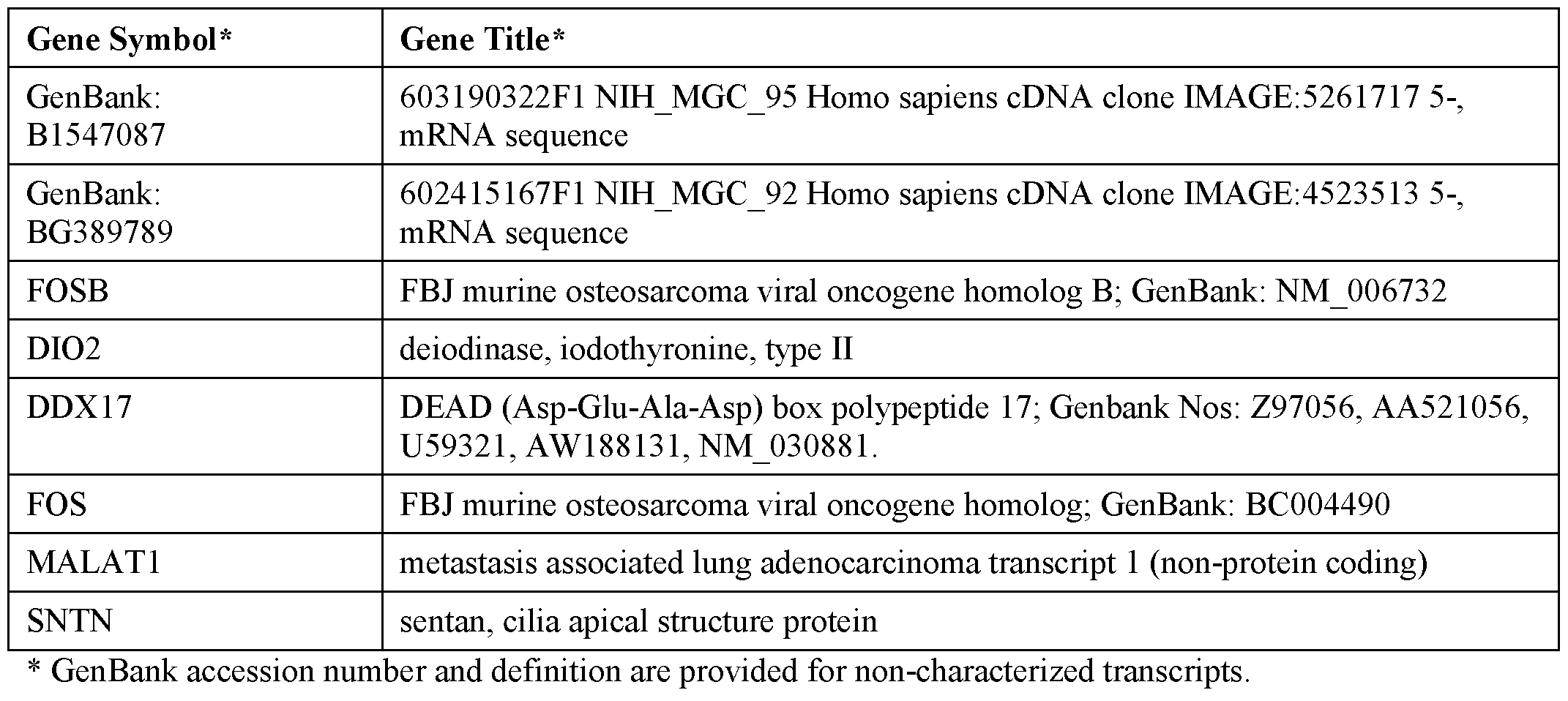
[00428] As described herein, biomarkers can be used for determining the presence or absence of endometriosis or other pathology in a biological sample that has been determined to have pathology. The pathology can comprise endometriosis, uterine pathology, or pelvic pathology. The biomarkers can comprise at least one set of genes set forth in Table 3.
Table 3. Biomarkers for determining the presence or absence of endometriosis in a biological sample comprising endometrial cells from proliferation phase (PE).

* GenBank accession num 3er and definition are provided for non-characterized transcripts.
[00429] As described herein, biomarkers can be used for determining the severity of endometriosis in a biological sample that has been determined to have endometriosis. Severity of endometriosis can be classified as mild or moderate. The biomarkers can comprise at least one set of genes set forth in Table 4.
Table 4. Biomarkers for determining the severity of endometriosis in a biological sample comprising endometrial cells from proliferation phase (PE).

LOC100505967 hypothetical LOCI 00505967
GenBank: Homo sapiens mRNA; cDNA DKFZp686A22111 (from clone
AL832142 DKFZp686A22111)
GenBank: Homo sapiens cDNA: FLI22384 fis, clone HRC07594
AK026037
CASP8AP2 caspase 8 associated protein 2
LTF lactotransferrin
FBN1 fibrillin 1; Genbank: NM_000138, AW955612.
CDH3 cadherin 3, type 1, P-cadherin (placental)
EPHA2 EPH receptor A2
GSTT1 glutathione S-transferase theta 1
MAPRE3 microtubule-associated protein, RP/EB family, member 3
PRKX /// PRKY protein kinase, X-linked /// protein kinase, Y-linked
PRKX protein kinase, X-linked
GSTM4 glutathione S-transferase mu 4
SLC12A2 solute carrier family 12 (sodium/potassium/chloride transporters), member 2
GSTM2 glutathione S-transferase mu 2 (muscle)
FOSL1 FOS-like antigen 1
GSTM1 glutathione S-transferase mu 1
HSD17B2 hydroxysteroid (17-beta) dehydrogenase 2
NMT2 N-myristoyltransferase 2
GABRP gamma-aminobutyric acid (GABA) A receptor, pi
PDZK1 PDZ domain containing 1
VN 1 vanin 1
PLCL1 phospholipase C-like 1
REN renin
CDKN2A carcinoembryonic antigen-related cell adhesion molecule 1 (biliary
glycoprotein)
CRYBB2 /// crystallin, beta B2 /// crystalin, beta B2 pseudogene 1
CRYBB2P1
SCGB 102 secretoglobin, family ID, member 2
LPHN2 latrophilin 2
CDKN2A cyclin-dependent kinase inhibitor 2A (melanoma, pi 6, inhibits CDK4)
SFRP5 secreted frizzled-related protein 5
D4S234E DNA segment on chromosome 4 (unique) 234 expressed sequence
BMP7 bone morphogenetic protein 7
MYCN v-myc myelocytomatosis viral related oncogene, neuroblastoma derived
(avian)
Gene Symbol* Gene Title*
PTPN11 protein tyrosine phosphatase, non-receptor type 11
SF1 splicing factor 1
GZMH granzyme H (cathepsin G-like 2, protein h-CCPX)
FGF18 fibroblast growth factor 18
PEG 10 paternally expressed 10
SLC7A1 solute carrier family 7 (cationic amino acid transporter, y+ system), member 1
MUC5B mucin 58, oligomeric mucus/gel-forming
GenBank: tb81b07.xl NCI CGAP Lu26 Homo sapiens cDNA clone IMAGE:2060725 A1345238 3- similar to gb:M10119 FERRITIN LIGHT CHAIN (HUMAN);, mRNA sequence
CEACAM21 carcinoembryonic antigen-related cell adhesion molecule 21
GenBank: H92070 ys84f02.sl Soares retina N2b4HR Homo sapiens cDNA clone
IMAGE:221499 3-similar to contains Alu repetitive element;contains PTR5 repetitive element ;, mRNA sequence
CALD1 caldesmon 1
LOC100507804 /// tryptase alpha- 1 -like /// tryptase alpha/beta 1
TPSAB1
HYMAI hydatidiform mole associated and imprinted (non-protein coding)
LOC642869 /// SET translocation (myeloid leukemia-associated) pseudogene /// SET nuclear SET oncogene
K1AA1661 KIAA1661 protein
FAM48A Family with sequence similarity 48, member A
BEX1 brain expressed, X-linked 1
SYBU syntabulin (syntaxin-interacting)
ECEL1 endothelin converting enzyme -like 1
HELLS helicase, lymphoid-specific
ZBBX zinc finger, B-box domain containing
IQCG IQ motif containing G
KLHL24 kelch-like 24 (Drosophila)
LOC389906 hypothetical LOC389906
LOCI 00510224 hypothetical LOC100510224
WHSC1L1 Wolf-Hirschhorn syndrome candidate 1 -like 1
TMEM106B transmembrane protein 106B
GNG12 guanine nucleotide binding protein (G protein), gamma 12
ENPP3 ectonucleotide pyrophatase/phosphodiesterase 3
FOXP1 forkhead box PI
PR02852 hypothetical protein PR02852
SECISBP2 SECIS binding protein 2
Gene Symbol* Gene Title*
MS4A8B membrane-spanning 4-domains, subfamily A, member 8B
MALATl metasis associated lung adenocarcinoma transcript 1 (non-protein coding)
PDK4 pyruvate dehydrogenase kinase, isozyme 4
SNRPN small nuclear ribonucleoprotein polypeptide N
FAM110C family with sequence similarity 110, member C
LOC100131564 hypothetical LOC 100131564
LOC727820 hypothetical protein LOC727820
ERAP2 endoplasmic reticulum aminopeptidase 2
SDCCAG8 serologically defined colon cancer antigen 8
NKAIN4 Na+/K+ transporting ATPase interacting 4
GenBank: nk67dl0.sl NCI_CGAP_Schl Homo sapiens cDNA clone IMAGE: 1018579 AA601031 3-, mRNA sequence
CDC42SE2 CDC42 small effector 2
EMID2 EMI domain containing 2
GOLT1A golgi transport 1A
SLC20A1 solute carrier family 20 (phosphate transporter), member 1
PKHD1L1 polycystic kidney and hepatic disease 1 (autosomal recessive)-like 1
GenBank: 7g45a06.xl NCI_CGAP_Pr28 Homo sapiens cDNA clone IMAGE:33093943 BE858984 mRNA sequence
GenBank: tw52g09.xl NCI_CGAP_Utl Homo sapiens cDNA clone IMAGE:2263360 A1683621 3-, mRNA sequence
LOC100506125 hypothetical LOC 100506125
FBX015 F-box protein 15
GenBank: AV660825 GLC Homo sapiens cDNA clone GLCGLG03 3-, mRNA sequence AV660825
LOC253039 hypothetical LOC253039
GenBank: Homo sapiens genomic DNA; cDNA DKFZp434Kl 111 (from clone AL157491 DKFZp434Kl l l l)
GenBank: Homo sapiens clone IMAGE:297403, mRNA sequence
AF339813
SP3 spa transcription factor
GenBank: AU 144005 HEMBA1 Homo sapiens cDNA clone HEMBA 1000622 3-, AU144005 mRNA sequence
GenBank: Homo sapiens PRO 1550 mRNA, partial cds
AF 119847
Gen Bank: Ul-H-BWO-aiy-a-04-0-Ul.sl NCI CGAP Sub6 Homo sapiens cDNA clone AW297731 IMAGE:2730894 3-, mRNA sequence
GenBank: 60176331811 N H MGC 20 Homo sapiens cDNA clone IMAGE: 4026173 5-, BF125564 mRNA sequence
ILDR1 immunoglobulin-like domain containing receptor 1
Gene Symbol* Gene Title*
GenBank: ar55f07.xl Barstead aorta HPLRB6 Homo sapiens cDNA clone
A1431345 IMAGE:2126533 3-, mRNA sequence
GenBank: zo89el0.x5 Stratagene ovarian cancer (#937219) Homo sapiens cDNA clone A1732617 IMAGE:594090 3-, mRNA sequence
GenBank: nc39f01.rl NCI_CGAP_Pr2 Homo sapiens cDNA clone IMAGE: 1010521, AA228366 mRNA sequence
UNC5A unc-5 homolog A (C. elegans)
GenBank: xm49b03.xl NCI_CGAP_GC6 Homo sapiens cDNA clone IMAGE:2686541 AW 197431 3-, similar to contains element KER repetitive element;, mRNA sequence
NAA25 N(alpha)-acetyltransferase 25, NatB auxiliary subunit
GenBank: hu05hl2.xl NCI_CGAP_Lu24 Homo sapiens cDNA clone IMAGE:3165767 BE222109 3-, mRNA sequence
PRKRA protein kinase, interferon-inducible double stranded RNA dependent activator
GenBank: UI-H-BIl-afr-e-09-O-UI.sl NCI CGAP Sub3 Homo sapiens cDNA clone AW205632 IMAGE:2722673 3-, mRNA sequence
RXFP1 relaxin/insulin-like family peptide receptor 1
GenBank: 7q07e l2.xl NCI_CGAP_Pr28 Homo sapiens cDNA clone IMAGE:3676918 BF438300 3-, mRNA sequence
GenBank: 601176827F1 NIH_MGC_17 Homo sapiens cDNA clone IMAGE:3532039 BE295812 5-, mRNA sequence
FLJ39739 Hypothetical FLI39739
GenBank: UI-H-BIl-aeu-f-12-0-UI.sl NCI CGAP Sub3 Homo sapiens cDNA clone AW203986 IMAGE:2720782 3-, mRNA sequence
GenBank: oll0a05sl Soares NFL T GBC S I Homo sapiens cDNA clone
AA908970 IMAGE: 1523024 3-, mRNA sequence
TULP4 Tubby like protein 4
FAM81B family with sequence similarity 81 , member B
GenBank: ht05b06.xl NCI_CGAP_Kidl3 Homo sapiens cDNA clone IMAGE:3145811 BE349858 3-, mRNA sequence
GenBank: UI-H-BI4-aop-a-02-0-UI.s l NCI CGAP Sub8 Homo sapiens cDNA clone BF508634 IMAGE:3085347 3-, mRNA sequence
WDR1 WD repeat domain 1
GenBank: hz75g08.xl NCI CGAP Lu24 Homo sapiens cDNA clone
BE467916 IMAGE:32135854 3-, mRNA sequence
C21orfl21 chromosome 21 open reading frame 121
GenBank: yi43b01.sl Soares placenta Nb2HP Homo sapiens cDNA clone
R68807 IMAGE: 141961 3-, mRNA sequence
GenBank: xd86f06.xl Soares NFL T GBC S 1 Homo sapiens cDNA clone
AW 117264 IMAGE:2604515 3-, mRNA sequence
NUPL1 nucleoporin like 1
LOC400931 hypothetical LOC400931
GenBank: EST374531 MAGE resequences, MAGG Homo sapiens cDNA, mRNA
Gene Symbol* Gene Title*
AW962458 sequence
QKI Quaking homolog, KH domain RNA binding (mouse)
GenBank: EST388740 MAGE resequences, MAGN Homo sapiens cDNA, mRNA AW976631 sequence
GenBank: af03h05.sl Soares testis NHT Homo sapiens cDNA clone IMAGE: 1030617 AA608834 3-, mRNA sequence
SNRPA1 Small nuclear ribionucleoprotein polypeptide A'
TMF1 TATA element modulatory factor 1
IREB2 iron-responsive element binding protein 2
ASXL1 additional sex combs like 1 (Drosophila)
GenBank: 7j75e01.xl Soares NSF F8 9W OT PA P Si Homo sapiens cDNA clone BF055144 IMAGE:3392282 3-, mRNA sequence
LRPAP1 low density lipoprotein receptor-related protein associated protein 1
GenBank: N39188 yv2d08.sl Soares fetal liver spleen 1NFLS Homo sapiens cDNA clone
IMAGE:243855 3- similar to contains Alu repetitive element;contains element MER35 repetitive element;, mRNA sequence
GenBank: A1650364 wa90a01.xl NCI_CGAP_GC6 Homo sapiens cDNA clone IMAGE:2303400
3- similar to contains Alu repetitive element;, mRNA sequence
GenBank: A1467945 tj 84d07.xl Soares NSF F8 9W OT PA P S 1 Homo sapiens cDNA clone
IMAGE:2148205 3-, mRNA sequence
GenBank: AA682674 zj20h 10. si Soares fetal liver spleen 1NFLS S 1 Homo sapiens cDNA clone
IMAGE:450883 3-, mRNA sequence
RAB18 RAB 18, member RAS oncogene family
GJC1 gap junction protein, gamma 1, 45kDa
CMIP C-Maf-inducing protein
GenBank: AV691872 AV691872 GKC Homo sapiens cDNA clone GKCDSB09 5-, mRNA
sequence
GenBank: EST384976 MAGE resequences, MAGL Homo sapiens cDNA, mRNA
AW972881 sequence
GenBank accession number and definition arc provided for non-characterized transcripts.
[00430] In some embodiments, biomarkers are used for determining the presence or absence of pathology in a biological sample comprising endometrial cells, wherein the biological sample is collected from early secretary phase (ESE) using systems, methods and devices described herein. The biomarkers can comprise at least one set of genes set forth in Table 5.
Table 5. Biomarkers for determining the presence or absence of a pathology in a biological sample comprising endometrial cells from early secretary phase (ESE).
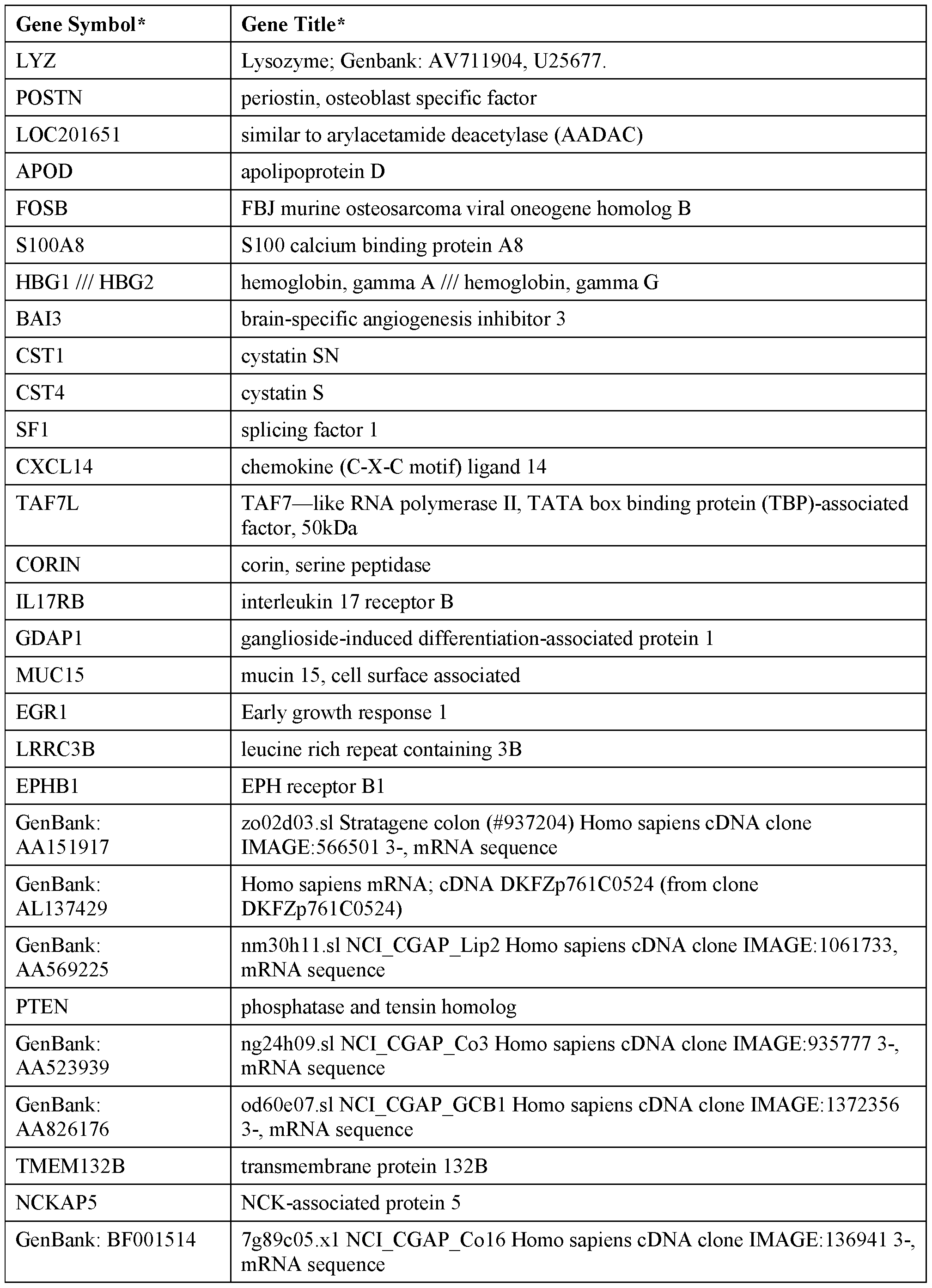
GenBank: R36546.1 yh89fl l .sl Soares placentia Nb2HP Homo sapiens cDNA clone
IMAGE: 136941 3-, mRNA sequence
GenBank: T70087.1 ycl7gl l .sl Stratagene lung (#937210) Homo sapiens cDNA clone
IMAGE: 80996 3-, mRNA sequence
NAMPT Nicotinamide phosphoribosyltransferase
GenBank: EST3871 18 MAGE resequences, MAGN Homo sapiens cDNA, mRNA AW975013 sequence
NUS 193 Nuclear undecaprenyl pyrophosphate synthase 1 homolog (S. cerevisiae)
pseudogene 3
* GenBank accession number and definition arc provided for non-characterized transcripts.
[00431] As described herein, biomarkers can be used for determining the presence or absence of endometriosis or other pathology in a biological sample that has been determined to have pathology. The pathology can comprise endometriosis, uterine pathology, or pelvic pathology. The biomarkers can comprise at least one set of genes set forth in Table 6.
Table 6. Biomarkers for determining the presence or absence of endometriosis in a biological sample comprising endometrial cells from early secretary phase (ESE).
Gene Symbol* Gene Title*
CEE /// LOC100508206 carboxyl ester lipase (bile salt-stimulated lipase) /// bile salt-activated lipase-like
GenBank: Ul-l-BB lp-aut-f-08-0-Ul.sl NCI CGAP PI6 Homo sapiens cDNA clone BC1024490 Ul-l-BB lp-aut-f-08-O-UI 3-, mRNA sequence
GenBank: AGENCOURT 10609489 NIH MGC 126 Homo sapiens cDNA clone BU955063 IMAGE:6726950 5-, mRNA sequence
THBS 1 thrombospondin 1; Genbank Nos: BF109732, AW956580, BF084105,
A1812030, NM_003246, BF055462, AV726673.
HBA1 /// HBA2 hemoglobin, alpha 1 /// hemoglobin, alpha 2
CD52 CD52 molecule
CFTR cystic fibrosis transmembrane conductance regulator (ATP-binding cassette sub-family C, member 7)
GSTT2 glutathione S-transferase theta 2
GPR64 G protein-coupled receptor 64; Genbank: NM 005756.
CRISP3 cysteine-rich secretory protein 3
HBB hemoglobin, beta
SLC9A3R2 solute carrier family 9 (sodium/hydrogen exchanger), member 3 regulator 2
ART3 ADP-ribosytransferase 3
HIST1H2BG histone cluster 1, H2bg
OLFM4 olfactomedin 4
SOS1 son of sevenless homolog 1 (Drosophila)
MUC5B mucin 5B, oligorneric mucus/gel-forming
Gene Symbol* Gene Title*
GAL galanin prepropeptide
IF 144 interferon-induced protein 44
ODAM odontogenic, ameloblast asssociated
CATSPERB cation channel, sperm-associated beta
AGTR2 angiotensin II receptor, type 2
C15orf48 chromosome 15 open reading frame 48
PPP1R1B protein phosphatase 1, regulatory (inhibitor) subunit IB
ZG16B zymogen granule protein 16 homolog B (rat)
C20orf54 chromosome 20 open reading frame 54
GenBank: nk67dl0.sl NCI_CGAP_Schl Homo sapiens cDNA clone IMAGE: 1018579 3-, AA601031 mRNA sequence
GenBank: qb34a07.xl Soares_pregnant uterus NbHPU Homo sapiens cDNA clone A1147867 IMAGE: 1698132 3-, mRNA sequence
GenBank: Ul-H-BWO-aiy-a-04-0-Ul.sl NCI CGAP Sub6 Homo sapiens cDNA clone AW297731 IMAGE:2730894 3-, mRNA sequence
CCDC58 coiled-coil domain containing 58
GenBank: 7g55h08.xl NCI_CGAP_Pr28 Homo sapiens cDNA clone IMAGE:33104313 BF003148 3-, mRNA sequence
GenBank: Ul-H-B14-aop-a-02-0-Ul.sl NCI CGAP Sub 8 Homo sapiens cDNA clone BF508634 IMAGE:3085347 3-, mRNA sequence
GenBank: wb32f06.xl NCI_CGAP_GC6 sequence cDNA clone IMAGE:2307395 3-, A1672553 mRNA sequence
GenBank: zk89g09.sl Soares_pregnant_uterus_NbHPU Homo sapiens cDNA clone AA121544 IMAGE:490048 3- similar to contains element PTR5 repetitive element ;, mRNA sequence
GenBank accession number and definition arc provided for non-characterized transcripts.
[00432] As described herein, biomarkers can be used for determining the severity of endometriosis in a biological sample that has been determined to have endometriosis. The severity of endometriosis can be classified as mild or moderate. The biomarkers can comprise at least one set of genes set forth in Table 7.
Table 7. Biomarkers for determining the severity of endometriosis in a biological sample comprising endometrial cells from early secretary phase (ESE).

CPA3 carboxypeptidase A3 (mast cell)
PROK1 prokineticin 1; Genbank: AW183087
PHACTR2 phosphatase and actin regulator 2
* GenBank accession number and definition arc provided for non-characterized transcripts.
[00433] In some embodiments, biomarkers are used for determining the presence or absence of pathology in a biological sample comprising endometrial cells, wherein the biological sample is collected from mid-secretary phase (MSE) using systems, methods and devices described herein. The biomarkers can comprise at least one set of genes set forth in Table 8.
Table 8. Biomarkers for determining the presence or absence of a pathology in a biological sample comprising endometrial cells from mid-secretary phase (MSE).
Gene Symbol Gene Title
JAK1 Janus kinase 1
PHF21A PHD finger protein 21A
CTN B 1 catenin (cadherin-associated protein), beta 1, 88kDa
CBX3 chromobox homolog 3; Genbank: NM 016587
SLC39A6 solute carrier family 39 (zinc transporter), member 6
CP ceruloplasmin (ferroxidase)
LUZP1 leucine zipper protein 1
ADAMTS5 ADAM metallopeptidase with thrombospondin type 1 motif, 5
CLIPl CAP-GLY domain containing linker protein 1
SOCS2-AS1 SOCS2 antisense RNA 1 (non-protein coding)
CACNB2 calcium channel, voltage-dependent, beta 2 subunit
NMRK1 nicotinamide riboside kinase 1
RARA retinoic acid receptor, alpha
MACC1 metastasis associated in colon cancer 1
ACTR2 ARP2 actin-related protein 2 homolog (yeast)
RERE arginine-glutamic acid dipeptide (RE) repeats
JU B jun B proto-oncogene
EGR1 early growth response 1
TBL1X transducin (beta)-like lX-linked
PKP4 plakophillin 4
MX1 myxovirus (influenza virus) resistance 1, interferon-inducible protein p78
(mouse)
TACSTD2 tumor-associated calcium signal transducer 2
SERPINE1 serpin peptidase inhibitor, clade E (nexin, plasminogen activator inhibitor type 1), member 1
Gene Symbol Gene Title
EFNB2 ephrin-B2
FOSB FBJ murine osteosarcoma viral oncogene homolog B
MMP14 matrix metallopeptidase 14 (membrane-inserted)
PRDM2 PR domain containing 2, with ZNF domain
PSD3 pleckstrin and Sec7 domain containing 3
DI02 deiodinase, iodothyronine, type II
AQP3 aquaporin 3 (Gill blood group)
SLC4A4 solute carrier family 4, sodium bicarbonate eotransporter, member 4
HBA1 /// HBA2 hemoglobin, alpha 1 /// hemoglobin, alpha 2
POMZP3 /// ZP3 POM121 and ZP3 fusion /// zona pellucida glycoprotein 3 (sperm receptor);
Genbank: NM_012230
EEA1 early endosome antigen 1
MSLN mesothelin
LYPD3 LY6/PLAUR domain containing 3
FGB fibrinogen beta chain
ENPP1 ectonucleotide pyrophosphatase/phosphodiesterase 1
CLE3B /// EXOSC7 C-type lectin domain family 3, member B /// exosome component 7
IGFBP1 insulin-like growth factor binding protein 1
KLK11 kallikrein-related peptidase 11
PIP5K1B phosphatidylinositol-4-phosphate-5-kinase, type I, beta
MMP10 matrix metallopeptidase 10 (stromelysin 2)
GPR64 G protein-coupled receptor 64
LEFTY2 left-right determination factor 2
CST1 cystatin SN
SPINK 1 serine peptidase inhibitor, Kazal type 1
PRLPv prolactin receptor
EPYC epiphycan
CYP24A1 cytochrome P450, family 24, subfamily A, polypeptide 1
TRPC6 transient receptor potential cation channel, subfamily C, member 6
SOGA1 suppressor of glucose, autophagy associated 1
CPJSP3 cysteine-rich secretory protein 3
CDC42 cell division cycle 42 (GTP binding protein, 25kDa)
CADM1 cell adhesion molecule 1
HBB hemoglobin, beta
FOS FBJ murine osterosarcoma viral oncogene homolog
CHI3L1 chitinase 3 -like 1 (cartilage glycoprotein-39)
ABAT 4-aminobutyrate aminotransferase
Gene Symbol Gene Title
CTSZ cathespin Z
UPK1B uroplakin IB
POMZP3 POM1211 and ZP3 fusion; Genbank: BC000487
IL6ST interleukin 6 signal transducer (gpl30, oncostatin M receptor)
NF1 neurofibromin 1
DHX9 DEAH (Asp-Glu-Ala-His) box polypeptide 9
EIF1 eukaryotic translation initiation factor 1
SECISBP2L SECIS binding protein 2-like
MFAP4 microfibrillar-associated protein 4
SOS1 son of sevenless homolog 1 (Drosophila)
MFAP5 microfibrillar associated protein 5
LR C15 leucine rich repeat containing 15
SST somatostatin
ID2 /// ID2B inhibitor of DNA binding 2, dominant negative helix-loop-helix protein /// inhibitor of DNA binding 2B, dominant negative helix-loop-helix protein (pseudogene)
CTBP1 C-terminal binding protein 1
CYP2C9 cytochrome P450, family 2, subfamily C, polypeptide 9
HSPA12A heat shock 70kDa protein 12A
TWISTNB TWIST neighbor
GUSBP3 /// GUSBP9 /// glucuronidase, beta pseudogene 3 /// glucuronidase, beta pseudogene 9 /// SMA4 /// SMA5 glucuronidase, beta pseudogene /// glucuronidase, beta pseudogene
DYRK1B dual -specificity tyrosine-(Y)-phosphorylation regulated kinase IB
ATP 13 A3 ATPase type 13 A3
CHODL chondrolectin
ALDH8A1 aldehyde dehydrogenase 8 family, member A 1
TGFB2 transforming growth factor, beta 2
SETD2 SET domain containing 2
UGCG UDP-glucose ceramide glucosyltransferase
ABHD2 abhydrolase domain containing 2
VPS35 vacuolar protein sorting 35 homolog (S. cerevisiae)
ZCCHC2 zinc finger, CCHC domain containing 2
TEX101 testis expressed 101
NUPL1 nucleoporin like 1
ANGPTL1 angiopoietin-like 1
LOCI 00507645 /// uncharacterized LOCI 00507645 /// metastasis associated lung
MALAT1 adenocarcinoma transcript 1 (non-protein coding)
WASF2 WAS protein family, member 2
Gene Symbol Gene Title
CPEB4 cytoplasmic polyadenylation element binding protein 4
SLAIN2 SLAIN motif family, member 2
BTBD7 BTB (POZ) domain containing 7
EDIL3 EGF-like repeats and discoidin I-like domains 3
FBX032 F-box protein 32
CUX1 cut-like homeobox 1
ITGB6 integrin, beta 6
ZNF800 zinc finger protein 800
C12orf35 chromosome 12 open reading frame 35
HS3ST3B1 heparan sulfate (glucosamine) 3-O-sulfotransferase 3B 1
LOC100653132 uncharacterized LOC100653132
MALAT1 metastasis associated lung adenocarcinoma transcript 1 (non-protein coding)
SORCS1 sortillin-related VPS 10 domain containing receptor 1
CAPN8 calpain 8
IHH Indian hedgehog
DDX17 DEAD (Asp-Glu-Ala-Asp) box helicase 17
FER fer (fps/fes related) tyrosine kinase
U2AF1 U2 small nuclear RNA auxilliary factor 1
LOCI 00287497 /// uncharacterized LOC100287497 /// uncharacterized LOC100287934 LOC100287934
BOD1L1 biorientation of chromosomes in cell division 1 -like 1
RAB12 RAB 12, member RAS oncogene family
GALNTL2 UDP-N-acetyl-alpha-D-galactosamine:polypeptide
N-acetylgalactosaminyltransferase-like 2
LOC100505989 uncharacterized LOC100505989
LOC100506582 uncharacterized LOCI 00506582
CLK4 CDC-like kinase 4
HECTD1 HECT domain containing E3 ubiquitin protein ligase 1
ZNF24 Zinc finger protein 24
PHKB phosphorylase kinase, beta
NIPBL Nipped-B homolog (Drosophila)
TMED8 transmembrane emp24 protein transport domain containing 8
PHACTR2 phosphatase and actin regulator 2
[00434] As described herein, biomarkers can be used for determining the presence or absence of endometriosis or other pathology in a biological sample that has been determined to have pathology. The pathology can comprise endometriosis, uterine pathology, or pelvic pathology. The biomarkers can comprise at least one set of genes set forth in Table 9.
Table 9. Biomarkers for determining the presence or absence of endometriosis in a biological sample comprising endometrial cells from mid-secretary phase (MSE).
Gene Symbol Gene Title
CDC42SE2 CDC42 small effector 2; Genbank: NM_020240
CDYL2 chromodomain protein, Y-like 2
WBSCR27 Williams Beuren syndrome chromosome region 27
CEL carboxyl ester lipase (bile salt-stimulated lipase)
NT5E 5'-nucleotidase, ecto (CD73)
Clorf210 chromosome 1 open reading frame 210
ZBED1 zinc finger, BED-type containing 1
CYP4B 1 cytochrome P450, family 4, subfamily B, polypeptide 1
LINC00476 long intergenic non-protein coding R A 476
CP ceruloplasmin (ferroxidase)
LOC201477 uncharacterized LOC201477
SLC8A1 solute carrier family 8 (sodium/calcium exchanger), member 1
SYTL3 synaptotagmin-like 3
DEFB124 defensin, beta 124
SERPINE1 serpin peptidase inhibitor, clade E (nexin, plasminogen activator inhibitor type 1), member 1
DACT2 dapper, antagonist of beta-catenin, homolog 2
(Xenopus laevis)
BCLAF1 BCL2 -associated transcription factor 1
ATP IB 1 ATPase, Na+/K+ transporting, beta 1 polypeptide
LTF lactotransferrin
CPNE3 copine III
ITPR2 inositol 1,4,5-trisphosphate receptor, type 2
S100A8 S I 00 calcium binding protein A8
STMN2 stathmin-like 2
MY06 myosin VI
ATXN1 ataxin 1
HLA-DQA1 major histocompatibility complex, class II, DQ alpha 1
Gene Symbol Gene Title
F13A1 coagulation factor XIII, Al polypeptide
ABP1 amiloride binding protein 1 (amine oxidase
(copper-containing))
FGFR2 fibroblast growth factor receptor 2
PLA2G2A phospholipase A2, group IIA (platelets, synovial fluid)
HMOX1 heme oxygenase (decycling) 1
PRKAR2B protein kinase, cAMP-dependent, regulatory, type
II, beta
PCYOX1 prenylcysteine oxidase 1
PCCA propionyl CoA carboxylase, alpha polypeptide
VCAM1 vascular cell adhesion molecule 1
HNMT histamine N-methyltransferase
POMZP3 /// ZP3 POM 121 and ZP3 fusion /// zona pellucida glycoprotein 3 (sperm receptor)
S100A2 S I 00 calcium binding protein A2
FGFR3 fibroblast growth factor receptor 3
KYNU kynureninase
ACPP acid phosphatase, prostate
MMP1 matrix metallopeptidase 1 (interstitial collagenase)
MAL mal, T-cell differentiation protein
OR M l orosomucoid 1; Genbank: NM_000607
OR M l /// ORM2 orosomucoid 1 /// orosomucoid 2; Genbank:
NM_000607 /// NM_000608.
CFTR cystic fibrosis transmembrane conductance regulator (ATP-binding cassette sub-family C, member 7)
PSPH phosphoserine phosphatase
SLC26A2 solute carrier family 26 (sulfate transporter), member 2
CXCL13 chemokine (C-X-C motif) ligand 13
TMSB15A ///TMSB15B thymosin beta 15a /// thymosin beta 15B
MST1R macrophage stimulating 1 receptor (c-met related tyrosine kinase)
PPP1R1A protein phosphatase 1, regulatory (inhibitor) subunit
1A
PCSK5 proprotein convertase subtilisin/kexin type 5
RASGRP1 RAS guanyl releasing protein 1 calcium and
DAG-regulated)
MMP10 matrix metallopeptidase 10 (stromelysin 2)
Gene Symbol Gene Title
BCL2A1 BCL2 -related protein Al
ABLIM3 actin binding LIM protein family, member 3
CYP3A5 cytochrome P450, family 3, subfamily A, polypeptide 5
LEFTY2 left-right determination factor 2
CST1 cystatin SN
SPINK 1 serine peptidase inhibitor, Kazal type 1
GRP gastrin-releasing peptide
SLC1A1 solute carrier family 1 (neuronal/epithelial high affinity glutamate transporter, system Xag), member 1
CDH16 cadherin 16, KSP-cadherin
GAGE12B /// GAGE12C /// GAGE 12D /// G antigen 12B /// G antigen 12C /// G antigen 12D /// GAGE12E /// IGAGE12F /// GAGE12G /// G antigen 12E /// G antigen 12F /// G antigen 12G /// GAGE12H /// GAGE 121 /// GAGE2A /// GAGE2B G antigen 12H /// G antigen 121/// G antigen 2 A /// G /// GAGE2C /// GAGE4 /// GAGE5 /// GAGE6 /// antigen 2B ///G antigen 2C /// G antigen 4 /// G GAGE7 antigen 5 /// G antigen 6 /// G antigen 7
HOXC6 homeobox C6
NFIC nuclear factor 1/C (CCAAT-binding transcription factor)
GABRA2 gamma-aminobutyric acid (GABA) A receptor, alpha 2
CSF2RA colony stimulating factor 2 receptor, alpha, low-affinity (granulocyte-macrophage)
GAGE1 /// GAGE12B /// GAGE12C ///GAGE 12D G antigen 1 /// G antigen 12B /// G antigen 12C /// G /// GAGE12E /// GAGE12F ///GAGE12G /// antigen 12D /// G antigen 12E ///G antigen 12F /// G GAGE12H /// GAGE 121 ///GAGE12J /// GAGE2A antigen 12G /// G antigen 12H /// G antigen 121 /// G /// GAGE2B /// GAGE2C/// GAGE2D /// GAGE2E antigen 12J /// G antigen 2 A /// G antigen 2B /// G /// GAGE4 /// GAGE5/// GAGE6 /// GAGE7 /// antigen 2C /// G antigen 2D /// G antigen 2E/// G GAGE8 antigen 4 /// G antigen 5 /// G antigen 6 /// G antigen
7 /// G antigen 8
FAM107A /// LOCI 00506924 family with sequence similarity 107, member A /// uncharacterized LOC100506924
GAGE3 G antigen3
GAGE1 /// GAGE12C /// GAGE 12D /// GAGE12E G antigen 1 /// G antigen 12C /// G antigen 12D /// G /// GAGE12F /// GAGE12G /// GAGE12H /// antigen 12E /// G antigen 12F /// G antigen 12G /// G GAGE 121 /// GAGE12J /// GAGE2A /// GAGE2B antigen 12H /// G antigen 121 /// G antigen 12J /// G /// GAGE2C /// GAGE2D /// GAGE2E /// GAGE3 antigen 2A /// G antigen 2B /// G antigen 2C /// G /// GAGE4 /// GAGE5 /// GAGE6 /// GAGE7 /// antigen 2D /// G antigen 2E /// G antigen 3 /// G GAGE8 antigen 4 /// G antigen 5 /// G antigen 6 /// G antigen
7 /// G antigen 8
GAST gastrin
GAGE1 /// GAGE12C /// GAGE 12D /// GAGE12E G antigen 1 /// G antigen 12C /// G antigen 12D /// G /// GAGE12F /// GAGE12G /// GAGE12H /// antigen 12E /// G antigen 12F /// G antigen 12G /// G GAGE 121 /// GAGE12J /// GAGE2D /// GAGE4 /// antigen 12H /// G antigen 121/// G antigen 12J /// G
Gene Symbol Gene Title
GAGE5 /// GAGE6 /// GAGE7 antigen2D /// G antigen 4 /// G antigen 5 /// G antigen 6 /// G antigen 7
GAGE12F /// GAGE12G /// GAGE 121 /// GAGE4 G antigen 12F /// G antigen 12G /// G antigen 121 /// /// GAGE5 /// GAGE6 /// GAGE7 G antigen 4 /// G antigen 5 /// G antigen 6 /// G antigen 7
DMBT1 deleted in malignant brain tumors 1
WNT4 wingless-type MMTV integration site family, member 4
TOPI topoisomerase (DNA) I
HBB hemoglobin, beta
NR2F2 nuclear receptor subfamily 2, group F , member 2
KLHDC10 ketch domain containing 10
LAMB 3 laminin, beta 3
HLA-DQB 1 major histocompatibility complex, class II , DQ beta
1
PNMA2 paraneoplastic Ma antigen 2
ADH1B alcohol dehydrogenase IB (class I), beta polypeptide
HLA-DRB4 /// LOCI 00509582 major histocompatibility complex, class II DR beta
4 /// HLA class II histocompatibility antigen, DR beta 4 chain-like
CRISP2 cysteine-rich secretory protein 2
MT1G Metallothionein 1G
RORA RAR-related orphan receptor A
CYR61 cysteine-rich, angiogenic inducer, 61
POMZP3 POM 121 and ZP3 fusion
LEPR leptin receptor
KIR3DL1 killer cell immunoglobulin-like receptor, three domains, long cytoplasmic tail, 1
CXCR4 chemokine (C-X-C motif) receptor 4
PRRC2C proline-rich coiled-coil 2C
IGFBP3 insulin like growth factor binding protein 3
SULF1 sulfatase 1
MFAP4 microfibrillar-associated protein 4
OLFM4 olfactomedin 4
IGHM immunoglobulin heavy constant mu
APOE Apolipoprotein E
HLA-DQB 1 /// LOCI 00293977 major histocompatibility complex, class II, DQ beta
1 /// HLA class II histocompatibility antigen, DQ beta 1 chain-like
Gene Symbol Gene Title
LTBP4 latent transforming growth factor beta binding protein 4
MUC5B mucin 5B, oligomeric mucus gel-forming
CFH complement factor H
HLA-DQA1 /// LOC100507718 /// LOC100509457 major histocompatibility complex, class II, DQ alpha 1 /// HLA class II histocompatibility antigen, DQ alpha 1 chain-like /// HLA class II histocompatibility antigen, DQ alpha 1 chain-like
EEF1E1 Eukaryotic translation elongation factor 1 epsilon 1
CTCF CCCTC -binding factor (zinc finger protein)
CYP2C9 cytochrome P450, family 2, subfamily C, polypeptide 9
ADAMTS2 ADAM metallopeptidase with thrombospondin type
1 motif, 2
CDC42BPA CDC42 binding protein kinase alpha (DMPK-like)
CFH /// CFHR1 complement factor H /// complement factor
H-related 1
DACT1 dapper, antagonist of beta-catenin, homolog 1
(Xenopus laevis)
FAM118A family with sequence similarity 118, member A
HPCAL4 hippocalcin like 4
DCF16 DDB 1 and CUL4 associated factor 16
BCMOl beta-carotene 15,15'-monooxygenase 1
SPDEF SAM pointed domain containing ets transcription factor
CATSPERB catsper channel auxiliary subunit beta
LRRC31 leucine rich repeat containing 31
ST6GALNAC5 ST6
(alpha-N-acetyl-neuraminy 1-2,3 -beta-galactosy 1 - 1 , 3 ) -N-acetylgalacto saminide
alpha-2,6-sialyltransferase 5
COLEC12 collectin sub-family member
HLA-DRB1 /// HLA-DRB3 /// HLA-DRB4 /// major histocompatibility complex, class II, DR beta HLA-DRB5 /// LOCI 00507709 /// LOCI 00507714 1 /// major histocompatibility complex, class II, DR /// LOCI 00509582 beta 3 /// major histocompatibility complex, class II,
DR beta4 /// major histocompatibility complex, class II, DR beta 5 /// HLA class II histocompatibility antigen, DRBl-7 beta chain-like /// HLA class II histocompatibility antigen, DRBl-7 beta chain-like /// HLA class II histocompatibility antigen, DR beta 4 chain-like
LOC1006530 uncharacterized LOC100653010
GDF15 growth differentiation factor 15
Gene Symbol Gene Title
SIKE1 suppressor of IKBKE 1
TFG TRK-fused gene
PTER phosphotriesterase relate
COL4A3BP collagen, type IV, alpha 3 (Goodpasture antigen) binding protein
CFC /// CFC1B cripto, FRL-1, cryptic family 1 /// crypto, FRL-1, cryptic family IB
SLC46A2 solute carrier family 46, member 2
MS4A8B membrane -spanning 4-domains, subfamily A, member 8B
H19 /// MIR675 H19, imprinted maternally expressed transcript
(non-protein coding) /// microRNA 675
LIFR leukemia inhibitory factor receptor alpha
COL12A1 collagen, type XII, alpha 1
BPIFB1 BPI fold containing family B, member 1
DNER delta/notch-like EGF repeat containing
MEGF6 multiple EGF-like -domains 6
CCDC146 coiled-coil domain containing 146
TAOK1 TAO kinase 1
ERAP2 endoplasmic reticulum aminopeptidase 2
LOC100505806 uncharacterized LOC 100505806
NAPSB napsin B aspartic peptidase pseudogene
ZG16B zymogen granule protein 16 homolog B (rat)
IGSF11 immunoglobulin superfamily, member 11
NFYA nuclear transcription factor y, alpha
LOC 100506029 /// LOCI 00506051 uncharacterized LOC 100506029 /// uncharacterized
LOC100506051
THRB thyroid hormone receptor, beta
CYS1 cystin 1
MCTP2 multiple C2 domains, transmembrane 2
NPAS3 neuronal PAS domain protein 3
C20orf85 chromosome 20 open reading frame 85
FAM69C family with sequence similarity 69, member C
SCARA5 scavenger receptor class A, member 5 (putative)
FNDC3B fibronectin type III domain containing 3B
PI15 peptidase inhibitor 15
SCGB3A1 secretoglobin, family 3A, member 1
KLF9 Kruppe 171 i.ke factor 9
Gene Symbol Gene Title
GBP1 guanylate binding protein 1, interferon- inducible
MAVS mitochondrial antiviral signaling protein
ANKRD33B ankyrin repeat domain 33B
SNORD3B-1 /// SNORD3B-2 /// SNORD3D small nucleolar R A, C/D box 3B-1 /// small nucleolar RNA, C/D box 3B-2 /// small nucleolar RNA, C/D box 3D
FAM178A family with sequence similarity 178, member A
THAP6 THAP domain containing 6
LOC100422737 uncharacterized LOC100422737
SCARA5 scavenger receptor class A, member 5 (putative)
SUZ12P Suppressor of zeste 12 homolog pseudogene
BCL2L10 BCL2-like 10 (apoptosis facilitator)
RIMKLB ribosomal modification protein rimK-like family member B
PLEKHA2 pleckstrin homology domain containing, family A
(phosphoinositide binding specific) member 2
EIF4E3 eukaryotic translation initiation factor 4E family member 3
SGPP2 sphingosine-1 -phosphate phosphatase 2
RAB3IP RAB3A interacting protein (rabin3)
DOK7 docking protein 7
MIB2 mindbomb E3 ubiquitin protein ligase 2
LOC100653229 uncharacterized LOC100653229
ITGB8 integrin, beta 8
WDR38 WD repeat domain 38
SHISA8 shisa homolog 8 (Xenopus laevis)
[00435] As described herein, biomarkers can be used for determining the severity of endometriosis in a biological sample that has been determined to have endometriosis. The severity of endometriosis can be classified as mild or moderate. The biomarkers can comprise at least one set of genes set forth in Table 10.
Table 10. Biomarkers for determining the severity of endometriosis in a biological sample comprising endometrial cells from mid-secretary phase (MSE).
Gene Symbol Gene Title
HSPA6 heat shock 70kDa protein 6 (HSP70B1); Genbank: NM 002155,
X51757.
1 1 IRA thyroid hormone receptor, alpha
GIMAP1 GTPase, IMAP family member 1
Gene Symbol Gene Title
TIRAP toll-interleukin 1 receptor (TIR) domain containing adaptor protein
ACVR1C activin A receptor, type IC
IL12RB1 interleukin 12 receptor, beta 1
JAK1 Janus kinase 1
RAD51L3-RFFL /// RFFL RAD51L3-RFFL readthrough /// ring finger and FYVE-like domain containing E3 ubiquitin protein ligase
ZNF417 zinc finger protein 417
SEC62 SEC62 homolog (S. cerevisiae)
SIGLEC10 sialic acid binding lg-like lectin 10
KCNG3 potassium voltage-gated channel, subfamily G, member 3
CD300LF CD300 molecule-like family member f
MOGAT1 monoacylglycerol O-acyltransferase 1
SLC5A3 solute carrier family 5 (sodium/myo-inositol cotransporter), member 3
FOXC1 forkhead box C 1
PRF1 perforin 1 (pore forming protein)
WBSCR27 Williams Beuren syndrome chromosome region 27
ARSB arylsulfatase B
CCDC60 coiled-coil domain containing 60
COCH coagulation factor C homolog, cochlin (Limulus polyphemus)
SLC25A48 solute carrier family 25, member 48
CELF2 CUGBP, Elav-like family member 2
DUOXA1 dual oxidase maturation factor 1
METTL8 methyltransferase like 8
TACC1 transforming, acidic coiled -coil containing protein 1
TBC1D16 TBC1 domain family, member 16
ZBED1 zinc finger, BED-type containing 1
DOK5 docking protein 5
FCER1G Fc fragment of IgE, high affinity I, receptor for; gamma polypeptide
ATF3 activating transcription factor 3
FCH02 FCH domain only 2
CCNL1 cyan LI
CYP4B1 cytochrome P450, family 4, subfamily B, polypeptide 1
CLEC7A C-type lectin domain family 7, member A
TRIB3 tribbles homolog 3 (Drosophila)
Gene Symbol Gene Title
LOC284454 uncharacterized LOC284454
CACNA1D calcium channel, voltage-dependent, L type, alpha ID subunit
DIAPH3-AS1 DIAPH3 anti sense R A 1 (non-protein coding)
LOC100506523 /// ZNF814 uncharacterized LOC 100506523 /// zinc finger protein 814
RPPH1 ribonuclease P RNA component HI
SERPINB6 serpin peptidase inhibitor, clade B (ovalbumin), member 6
LEPR leptin receptor
LOC100507250 uncharacterized LOC 100507250
LOC100506258 uncharacterized LOC 100506258
ACSL4 Acyl-CoA synthetase long-chain family member 4
BIN3 bridging integrator 3
PTRF polymerase I and transcript release factor
ZKSCAN1 zinc finger with KRAB and SCAN domains 1
ZNF587 /// ZNF587B zinc finger protein 587 /// zinc finger protein 587B
MIR1204 /// PVT1 microRNA 1204 /// Pvtl oncogene (non-protein coding)
ZDHHC18 zinc finger, DHHC-type containing 18
SIRT2 sirtuin 2
AHNAK2 AHNAK nucleoprotein 2
Clorf53 chromosome 1 open reading frame 53
LOC100507645 /// MALAT1 uncharacterized LOC 100507645 /// metastasis associated lung adenocarcinoma transcript 1 (non-protein coding)
ZNF321P /// ZNF816 /// zinc finger protein 321, pseudogene /// zinc finger protein 816 /// ZNF816-ZNF321P ZNF816-ZNF321P readthrough
CACNB2 calcium channel, voltage-dependent, beta 2 subunit
LOC642852 uncharacterized LOC642852
FLJ38717 FLJ8717 protein
SFXN3 Sideroflexin 3
LOC100506387 uncharacterized LOC 100506387
LOC201477 uncharacterized LOC201477
SLC8A1 solute carrier family 8 (sodium/calcium exchanger), member 1
KIAA1908 uncharacterized LOC I 14796
SF3B14 Splicing factor 3B, 14 kDa subunit
OR7D2 olfactory receptor, family 7, subfamily D, member
TNRC18 trinucleotide repeat containing 18
LOC100630923 LOC100289561-PRKRIP 1 readthrough
ATF1 activating transcription factor 1
Gene Symbol Gene Title
IKZF1 IKAROS family zinc finger 1 (Ikaros)
PNN pinin desmosome associated protein
CD74 CD74 molecule, major histocompatibility complex, class II invariant chain
PAAF1 proteasomal ATPase-associated factor 1
BRE-AS1 BRE antisense RNA 1 (non-protein coding)
LINC00240 long intergenic non-protein coding RNA 240
ANKRD20A1 /// ANKRD20A11P /// ankyrin repeat domain 20 family, member Al /// ankyrin repeat ANKRD20A2 /// ANKRD20A3 /// domain 20 family, member Al l, pseudogene /// ankyrin repeat ANKRD20A4 /// ANKRD20A5P /// domain 20 family, member A2 /// ankyrin repeat domain 20 ANKRD20A9P /// LOC644339 family, member A3 /// ankyrin repeat domain 20 family, member
A4 /// ankyrin repeat domain 20 family, member A5, pseudogene /// ankyrin repeat domain 20 family, member A9, pseudogene /// ankyrin repeat domain-containing protein 20B-like
CATSPERB catsper channel auxiliary subunit beta
SCD stearoyl-CoA desaturase (delta-9-desaturase
DHCR24 24-dehydrocholesterol reductase
DUSP1 dual specificity phosphatase 1
CYR61 cysteine-rich, angiogenic inducer, 61
NREP neuronal regeneration related protein homolog (rat)
GPX3 glutathione peroxidase 3 (plasma)
MYH11 myosin, heavy chain 1 1, smooth muscle
ZFP36 zinc finger protein 36, C3H type, homolog (mouse)
INSIG1 insulin induced gene 1
TNC tenascin C
ACSL3 acyl-CoA synthetase long-chain family member 3
NIPSNAP1 nipsnap homolog 1 (C. elegans)
ENG endoglin
CPD carboxypeptidase D
PPP1R12B protein phosphatase 1, regulatory subunit 12B
LTF lactotransferrin
DKK3 dickkopf 3 homolog (Xenopus lavaeis)
AMFR autocrine motility factor receptor, E3 ubiquitin protein ligase
NR4A1 nuclear receptor subfamily 4, group A, member 1
COL1A2 collagen, type I, alpha 2
IGF2 /// INS-IGF insulin-like growth factor 2 (somatomedin A) /// INS-IGF2 readthrough
KIAA0101 KIAA0101
Gene Symbol Gene Title
DHFR dihydrofolate reductase
NRIP1 nuclear receptor interacting protein 1
ICAM1 intercellular adhesion molecule 1
SERTAD2 SERTA domain containing 2
GPX2 glutathione peroxidase 2 (gastrointestinal)
ANPEP alanyl (membrane) aminopeptidase
ADM adrenomedullin
SOX9 SRY (sex determining region Y)-box 9
CAPN6 calpain 6
STMN2 stathmin-like 2
FH fumarate hydratase
C2 complement component 2
FBN2 fibrillin 2
ST3GAL5 ST3 beta-galactoside alpha-2,3-sialyltransferase 5
TLE1 transducin-like enhancer of split 1 (E(sp l) homolog, Drosophila)
ATXN1 ataxin 1
FCGBP Fc fragment of IgG binding protein
CDH3 cadherin 3, type 1, P-cadherin (placental)
HLA-DQA1 major histocompatibility complex, class II, DQ alpha 1
PSD3 pleckstrin and Sec7 domain containing 3
EPN2 epsin 2
S100A9 S 100 calcium binding protein A9
KLF9 Kruppel-like factor 9
LOXL1 lysyl oxidase-like 1
CSF3R colony stimulating factor 3 receptor (granulocyte)
GPRC5B G protein-coupled receptor, family C, group 5, member B
PLA2G2A phospholipase A2, group IIA (platelets, synovial fluid)
BCL2 B-cell CLL/lymphoma 2
PI3 peptidase inhibitor 3, skin-derived
PDE4B phosphodiesterase 4B, cAMP -specific
MPZL2 myelin protein zero-like 2
SEMA3C sema domain, immunoglobulin domain (lg), short basic domain, secreted, (semaphorin) 3C
PCGF2 polycomb group ring finger 2
GSTT1 glutathione S-transferase theta 1
TSPAN8 tetraspanin 8
Gene Symbol Gene Title
SCG5 secretogranin V (7B2 protein)
MMP9 matrix metallopeptidase 9 (gelatinase B, 92kDa gelatinase,
92kDa type IV collagenase)
HBA1 /// HBA2 hemoglobin, alpha 1 /// hemoglobin, alpha 2
RFC4 replication factor C (activator 1) 4, 37kDa
CTAGE5 CTAGE family, member 5
AG API ArfGAP with GTPase domain, ankyrin repeat and PH domain 1
PRAME preferentially expressed antigen in melanoma
IL2RG interleukin 2 receptor, gamma
GADD45G growth arrest and DNA-damage-inducible, gamma
GSTM4 glutathione S-transferase mu 4
ENPP4 Ectonucleotide
pyrophosphatase/phosphodiesterase 4 (putative)
CD37 CD37 molecule
S100A2 S 100 calcium binding protein A2
SKI v-ski sarcoma viral oncogene homolog (avian)
FARS2 phenylalanyl-tRNA synthetase 2, mitochondrial
PROM1 prominin 1
AK4 /// LOC100507855 adenylate kinase 4 /// adenylate kinase isoenzyme 4, mitochondrial-like
SLC43A1 solute carrier family 43, member 1
GSTM2 glutathione S-transferase mu 2 (muscle)
FOLR1 folate receptor 1 (adult)
IFI44L interferon-induced protein 44-like
MMP1 matrix metallopeptidase 1 (interstitial collagenase)
CDC7 cell division cycle 7 homolog (S. cerevisiae)
TOX thymocyte selection-associated high mobility group box
CXCL10 chemokine (C-X-C motif) ligand 10
GABRE /// MIR224 /// MIR452 gamma-aminobutyric acid (GABA) A receptor, epsilon /// microRNA 224 /// microR A 452
GSTM1 glutathione S-transferase mu 1
APOC2 /// APOC4 /// APOC4- apolipoprotein C-II /// apolipoprotein C-IV /// APOC4-APOC2 APOC2 readthrough
ABCG1 ATP -binding cassette, sub-family G (WHITE), member 1
MMP12 matrix metallopeptidase 12 (macrophage elastase)
DKK1 dickkopf 1 homolog (Xenopus laevis)
SERPINB2 serpin peptidase inhibitor, Glade B (ovalbumin) member 2
Gene Symbol Gene Title
TFF3 trefoil factor 3 (intestinal)
SRD5A1 steroid-5-alpha-reductase, alpha polypeptide 1 (3-oxo-5 alpha-steroid delta 4-dehydrogenase alpha 1)
ABCA8 ATP -binding cassette, sub-family A (ABCl), member 8
RIMS3 regulating synaptic membrane exocytosis 3
DUSP2 dual specificity phosphatase 2
CKM creatine kinase, muscle
FOLR2 folate receptor 2 (fetal)
MLH3 mutL homolog 3 (E. coli)
ENPEP glutamyl aminopeptidase (aminopeptidase A)
MSLN mesothelin
LYPD3 LY6/PLAUR domain containing 3
ASNS asparagine synthetase (glutamine-hydrolyzing)
PSPH phosphoserine phosphatase
AOX1 aldehyde oxidase 1
SLC26A2 solute carrier family 26 (sulfate transporter), member 2
CCR1 chemokine (C-C motif) receptor 1
NEFM neurofilament, medium polypeptide
CCL3 /// CCL3L1 /// CCL3L3 chemokine (C-C motif) ligand 3 /// chemokine (C-C motif) ligand
3 -like 1 /// chemokine (C-C motif) ligand 3 -like 3
PTGS1 prostaglandin-endoperoxide synthase 1 (prostaglandin G/H
synthase and cyclooxygenase)
ACTC1 actin, alpha, cardiac muscle 1
ITGB3BP integrin beta 3 binding protein (beta3-endonexin)
AP1S1 adaptor-related protein complex 1, sigma 1 subunit
HCAR3 hydroxycarboxylic acid receptor 3
SOD3 superoxide dismutase 3, extracellular
LIF leukemia inhibitory factor
IGFBP1 insulin-like growth factor binding protein 1
TMSB15A /// TMSB15B thymosin beta 15a /// thymosin beta 15B
GGCX gamma-glutamyl carboxylase
CBR3 carbonyl reductase 3
PRSS2 protease, serine, 2 (trypsin 2)
SLC22A3 solute carrier family 22 (extraneuronal monoamine transporter), member 3
GSTT2 glutathione s-transferase theta 2
PRL prolactin
Gene Symbol Gene Title
MST1R macrophage stimulating 1 receptor (c-met-related tyrosine kinase)
CD3E CD3e molecule, epsilon (CD3-TCR complex)
KLK11 kallikrein-related peptidase 11
GZMA granzyme A (granzyme 1, cytotoxic T-lymphocyte-associated serine esterase 3)
GNLY granulysin
AVIL advillin
BPI bactericidal/permeability-increasing protein
HRH1 histamine receptor HI
NOS3 nitric oxide synthase 3 (endothelial cell)
OLFM1 olfactomedin 1
C4BPA complement component 4 binding protein alpha
OASL 2'-5'-oligoadenylate synthetase-like
TPSAB1 tryptase alpha/beta 1
SYNGR3 synaptogyrin 3
CBLN1 cerebellin 1 precursor
CD8A CD8a molecule
CYP3A5 cytochrome P450, family 3, subfamily A, polypeptide 5
WISP2 WNT1 inducible signaling pathway protein 2
CD2 CD2 molecule
PARTI prostate androgen-regulated transcript 1 (non-protein coding)
SLC7A4 solute carrier family 7 (orphan transporter), member 4
GABBR1 /// UBD gamma-aminobutyric acid (GABA) B receptor, 1 /// ubiquitin D
SLC22A4 solute carrier family 22 (organic cation/ergothioneine transporter) member 4
PLCL1 phospholipase C-like 1
EPHA1 EPH receptor Al
HABP2 hyaluronan binding protein 2
LEFTY2 left-right determination factor 2
TNFAIP6 tumor necrosis factor, alpha-induced protein 6
ACADL acyl-CoA dehydrogenase, long chain
PTPRR protein tyrosine phosphatase, receptor type, R
LRRC37A3 leucine rich repeat containing 37, member A3
MATN3 matrilin 3
Gene Symbol Gene Title
UGT1A1 /// UGT1A10 /// UGT1A3 UDP glucuronosyltransferase 1 family, polypeptide Al /// UDP /// UGT1A4 /// UGT1A5 /// UGT1A6 glucuronosyltransferase 1 family, polypeptide A10 /// UDP /// UGT1A7 /// UGT1A8 /// UGT1A9 glucuronosyltransferase 1 family, polypeptide A3 /// UDP glucuronosyltransferase 1 family, polypeptide A4 /// UDP glucuronosyltransferase 1 family, polypeptide
A5 /// UDP glucuronosyltransferase 1 family, polypeptide A6 /// UDP glucuronosyltransferase 1 family, polypeptide A7 /// UDP glucuronosyltransferase 1 family, polypeptide A8 /// UDP glucuronosyltransferase 1 family, polypeptide A9
KLK8 kallikrein-related peptidase 8
CYP4F11 cytochrome P450, family 4, subfamily F, polypeptide 1 1
ARHGAP6 Rho GTPase activating protein 6
IL13RA2 interleukin 13 receptor, alpha 2
CST1 cystatin SN
MMP17 matrix metallopeptidase 17 (membrane-inserted)
ARHGAP22 Rho GTPase activating protein 22
FAM155B family with sequence similarity 155, member B
PTHLH parathyroid hormone-like hormone
SPINK2 serine peptidase inhibitor, Kazal type 2 (acrosin-trypsin inhibitor)
GRP gastrin-releasing peptide
CXCL6 chemokine (C-X-C motif) ligand 6 (granulocyte chemotactic protein 2)
COX6A2 cytochrome c oxidase subunit Via polypeptide 2
XCL1 chemokine (C motif) ligand 1
SCGB2A2 secretoglobin, family 2A, member 2
PF4 platelet factor 4
B4GALNT1 beta- 1,4-N -acetyl -galactosaminyl transferase 1
S1PR4 sphingosine-1 -phosphate receptor 4
LTC4S leukotriene C4 synthase
ABAT 4-aminobutyrate aminotransferase
AKR1B10 aldo-keto reductase family 1, member B IO (aldose reductase)
LY96 lymphocyte antigen 96
SLC16A5 solute carrier family 16, member 5 (monocarboxylic acid transporter 6)
ZMYM5 zinc finger, MYM-type 5
GP2 glycoprotein 2 (zymogen granule membrane)
FAM65B family with sequence similarity 65, member B
CRYBB2 /// CRYBB2P1 crystallin, beta B2 /// crystallin, beta B2 pseudogene 1
WISP1 WNT1 inducible signaling pathway protein 1
Gene Symbol Gene Title
PAEP progestagen-associated endometrial protein
IL11 interleukin 1 1
BGLAP /// PMF1-BGLAP bone gamma-carboxyglutamate (gla) protein ///
PMF 1 -BGLAP readthrough
TNF tumor necrosis factor
TPSB2 tryptase beta 2 (gene/pseudogene)
DI03 deiodinase, iodothyronine, type III
ALOX12 arachidonate 12-lipoxygenase
CD300C CD300c molecule
CD209 CD209 molecule
KIR3DL1 /// KIR3DL2 /// killer cell immunoglobulin-like receptor, three domains, long LOC727787 cytoplasmic tail, 1 /// killer cell immunoglobulin-like receptor, three domains, long cytoplasmic tail, 2 /// killer cell immunoglobulin-like receptor, three domains, long cytoplasmic tail, 2-like
KIR3DL2 /// LOC727787 killer cell immunoglobulin-like receptor, three domains, long cytoplasmic tail, 2 /// killer cell immunoglobulin-like receptor, three domains, long cytoplasmic tail, 2-like
DEFB4A /// DEFB4B defensin, beta 4A /// defensin, beta
RAC2 ras-related C3 botulinum toxin substrate 2 (rho family, small
GTP binding protein Rac2)
GZMM granzyme M (lymphocyte met-ase 1)
PIR pirin (iron-binding nuclear protein)
BDKRB1 bradykinin receptor B 1
GADD45B growth arrest and DNA-damage-inducible, beta
PSG9 pregnancy specific beta- 1 -glycoprotein 9
GAGE1 /// GAGE12C /// GAGE12D G antigen 1 /// G antigen 12C /// G antigen 12D /// G antigen 12E /// GAGE12E /// GAGE12F /// /// G antigen 12F /// G antigen 12G /// G antigen 12H /// G antigen GAGE12G /// GAGE12H /// 121/// G antigen 12 J /// G antigen 2 A /// G antigen 2B /// G antigen GAGE 121 /// GAGE12J /// GAGE2A 2C /// G antigen 2D /// G antigen 2E /// G antigen 3 /// G antigen 4 /// GAGE2B /// GAGE2C /// /// G antigen 5 /// G antigen 6 /// G antigen 7 /// G antigen 8 GAGE2D /// GAGE2E /// GAGE3 ///
GAGE4 /// GAGE5 /// GAGE6 ///
GAGE7 /// GAGE8
FGFR1 fibroblast growth factor receptor 1
MUC1 mucin 1, cell surface associated
KRT13 keratin 13
NFAT5 nuclear factor of activated T-cells 5, tonicity-responsive
LINC00597 long intergenic non-protein coding R A 597
KIR2DS3 killer cell immunoglobulin-like receptor, two domains, short cytoplasmic tail, 3
Gene Symbol Gene Title
PTGIS prostaglandin 12 (prostacyclin) synthase
GAST gastrin
KIR2DS1 killer cell immunoglobulin-like receptor, two domains, short cytoplasmic tail, 1
KIR2DS5 killer cell immunoglobulin-like receptor, two domains, short cytoplasmic tail, 5
DMBT1 deleted in malignant brain tumors 1
TH tyrosine hydroxylase
ANK1 ankyrin 1, erythrocytic
KIR2DL2 /// KIR2DL4 /// killer cell immunoglobulin-like receptor, two domains, long KIR2DL5A /// KIR2DL5B /// cytoplasmic tail, 2 /// killer cell immunoglobulin-like receptor, KIR3DL3 /// KIR3DS1 /// two domains, long cytoplasmic tail, 4 /// killer cell LOC100287534 immunoglobulin-like receptor, two domains, long cytoplasmic tail, 5A /// killer cell immunoglobulin-like receptor, two domains, long cytoplasmic tail, 5B /// killer cell immunoglobulin-like receptor three domains long cytoplasmic tail 3 /// killer cell immunoglobulin-like receptor, three domains, short cytoplasmic tail, 1 /// killer cell immunoglobulin-like receptor 2DL4-like
RASA4 /// RASA4B /// RASA4CP/// RAS p21 protein activator 4 /// RAS p21 protein activator 4B /// UPK3BL RAS p21 protein activator 4C, pseudogene /// uroplakin 3B-like
WNT4 wingless-type MMTV integration site family, member 4
AP3D1 adaptor-related protein complex 3, delta 1 subunit
LGALS8 lectin, galactoside-binding, soluble, 8
UPF1 UPF 1 regulator of nonsense transcripts homolog (yeast)
KRT7 keratin 7
COROIA coronin, actin binding protein 1A
UBN1 ubinuclein 1
HBB hemoglobin, beta
AKR1C3 aldo-keto reductase family 1, member C3 (3 -alpha hydroxysteroid dehydrogenase, type II)
FOS FBJ murine osteosarcoma viral oncogene homolog
TFPI2 tissue factor pathway inhibitor 2
CA2 carbonic anhydrase II
AZGP1 alpha-2 -glycoprotein 1, zinc-binding
RGS16 regulator of G-protein signaling 16
MALL mal, T-cell differentiation protein-like
SCAF11 SR-related CTD-associated factor 1 1
DLK1 delta-like 1 homolog (Drosophila)
CES1 /// LOC100653057 carboxylesterase 1 /// liver carboxylesterase 1 -like
Gene Symbol Gene Title
HLA-DRB4 /// LOC100509582 major histocompatibility complex, class II, DR beta 4 /// HLA class II histocompatibility antigen, DR beta 4 chain-like
NR1D2 nuclear receptor subfamily 1, group D, member 2
RRM2 ribonucleotide reductase M2
CXCL2 chemokine (C-X-C motif) ligand 2
CASP6 caspase 6, apoptosis-related cysteine peptidase
KLK10 kallikrein-related peptidase 10
TARP TCR gamma alternate reading frame protein
SPP1 secreted phosphoprotein 1
TNNC1 troponin C type 1 (slow)
TGFB2 transforming growth factor, beta 2
SLC7A11 solute carrier family 7 (anionic amino acid transporter light chain, xc- system), member 1 1
CD247 CD247 molecule
RND1 Rho family GTPase 1
MAPK13 mitogen -activated protein kinase 13
UPK1B uroplakin IB
ARC activity-regulated cytoskeleton-associated protein
CYP4B1 cytochrome P450, family 4, subfamily B, polypeptide 1
PLA2G4A phospholipase A2, group IVA (cytosolic, calcium-dependent)
GZMB granzyme B (granzyme 2, cytotoxic T-lymphocyte-associated serine esterase 1)
IRX5 iroquois homeobox 5
DLG5 discs, large homolog 5 (Drosophila)
CTAG1A /// CTAG1B cancer/testis antigen 1A /// cancer testis antigen IB
CCL23 chemokine (C-C motif) ligand 23
LAIR1 leukocyte-associated immunoglobulin-like receptor 1
NRTN neurturin
CLDN14 claudin 14
SLC43A3 solute carrier family 43, member 3
NCR3 natural cytotoxicity triggering receptor 3
POSTN periostin, osteoblast specific factor
Gene Symbol Gene Title
KIR2DL1 /// KIR2DL2 /// KIR2DL3 killer cell immunoglobulin-like receptor, two domains, long /// KIR2DL4 /// KIR2DL5A /// cytoplasmic tail, 1 /// killer cell immunoglobulin-like receptor, KIR2DL5B /// KIR3DL3 /// two domains, long cytoplasmic tail, 2 /// killer cell KIR3DS1 /// LOC100287534 /// immunoglobulin-like receptor, two domains, long cytoplasmic LOC100653050 tail, 3 /// killer cell immunoglobulin-like receptor, two domains, long cytoplasmic tail, 4 /// killer cell immunoglobulin-like receptor, two domains, long cytoplasmic tail, 5A /// killer cell immunoglobulin-like receptor, two domains, long cytoplasmic tail, 5B /// killer cell immunoglobulin-like receptor three domains long cytoplasmic tail 3 /// killer cell immunoglobulin-like receptor, three domains, short cytoplasmic tail, 1 /// killer cell immunoglobulin-like receptor 2DL4-like /// killer cell immunoglobulin-like receptor 2DL2-like
AP3D1 adaptor-related protein complex 3, delta 1 subunit
HLA-DRA major histocompatibility complex, class II, DR alpha
HGF hepatocyte growth factor (hepapoietin A; scatter factor)
PSTPIP1 proline-serine-threonine phosphatase interacting protein 1
KIR2DL4 killer cell immunoglobulin-like receptor, two domains, long cytoplasmic tail, 4
KIR2DL2 killer cell immunoglobulin-like receptor, two domains, long cytoplasmic tail, 2
MAPK11 mitogen-activated protein kinase 11
KIR2DS1 /// KIR2DS2 /// KIR2DS3 killer cell immunoglobulin-like receptor, two domains, short /// KIR2DS4 /// KIR2DS5 /// cytoplasmic tail, 1 /// killer cell immunoglobulin-like receptor, KIR3DL3 two domains, short cytoplasmic tail, 2 /// killer cell immunoglobulin-like receptor, two domains, short cytoplasmic tail, 3 /// killer cell immunoglobulin-like receptor, two domains, short cytoplasmic tail, 4 /// killer cell immunoglobulin-like receptor, two domains, short cytoplasmic tail, 5 /// killer cell immunoglobulin-like receptor three domains long cytoplasmic tail 3
CEACAM6 carcinoembryonic antigen-related cell adhesion molecule 6
(non-specific cross reacting antigen)
KIR3DL1 killer cell immunoglobulin-like receptor, three domains, long cytoplasmic tail, 1
PRG2 proteoglycan 2, bone marrow (natural killer cell activator, eosinophil granule major basic protein)
COL4A2 collagen, type IV, alpha 2
HLA-DPA1 major histocompatibility complex, class II, DP alpha 1
WNK1 WNK lysine deficient protein kinase 1
DAG1 dystroglycan 1 (dystrophin-associated glycoprotein 1)
FNBP4 formin binding protein 4
PIK3R1 phosphoinositide-3 -kinase, regulatory subunit 1 (alpha)
SLC7A1 solute carrier family 7 (cationic amino acid transporter, y+ system) , member 1
Gene Symbol Gene Title
CLASP2 cytoplasmic linker associated protein 2
MYOID myosin ID
KHNYN KH and NYN domain containing
SEP6 septin 6
CERS6 ceramide synthase 6
COL5A1 collagen, type V, alpha 1
IL1RN interleukin 1 receptor antagonist
RASA4 /// RASA4B /// RASA4CP RAS p21 protein activator 4 /// RAS p21 protein activator 4B ///
RAS p21 protein activator 4C pseudogene
NFATC2IP nuclear factor of activated T-cells, cytoplasmic, calcineurin-dependent 2 interacting protein
PLEKHG3 pleckstrin homology domain containing, family G (with RhoGef domain) member 3
SUPV3L1 suppressor of varl, 3 -like 1 (S. cerevisiae)
COL6A1 collagen, type VI, alpha 1
BBX bobby sox homolog (Drosophila)
GATAD1 GATA zinc finger domain containing 1
CHI3L2 chitinase 3 -like 2
NEK3 NIMA (never in mitosis gene a)-related kinase 3
TIAM1 T-cell lymphoma invasion and metastasis 1
PLCB1 phospholipase C, beta 1 (phosphoinositide-specific)
K 1 4 keratin 4
ZNF248 zinc finger protein 248
TCF25 transcription factor 25 (basic helix-loop-helix)
PAQR3 progestin and adipoQ receptor family member III
MUC5B mucin 5B, oligomeric mucus/gel-forming
RUFY3 RUN and FYVE domain containing 3
NPTX2 neuronal pentraxin II
TRA2A transformer 2 alpha homolog (Drosophila)
ENOSF1 enolase superfamily member 1
SLC1A1 solute carrier family 1 (neuronal/epithelial high affinity glutamate transporter, system Xag), member 1
FAM69A family with sequence similarity 69, member A
GLB1L2 galactosidase, beta 1 -like 2
KSR1 kinase suppressor of ras 1
STARD5 StAR-related lipid transfer (START) domain containing 5
CLMN calmin (calponin-like, transmembrane)
Gene Symbol Gene Title
THSD7A thrombospondin, type I, domain containing 7A
PAX8 paired box 8
RPGRIP1L RPGRIPl-like
ZAP 70 zeta-chain (TCR) associated protein kinase 70kDa
CCL8 chemokine (C-C motif) ligand 8
GSN gelsolin
PTPRD protein tyrosine phosphatase, receptor type, D
MBD4 methyl-CpG binding domain protein 4
CD7 CD7 molecule
MYRIP myosin VIIA and Rab interacting protein
GNAS GNAS complex locus
ABCB9 ATP-binding cassette, sub-family B (MDR/TAP), member 9
GAL galanin prepropeptide
DKK3 dickkopf 3 homolog (Xenopus laevis)
RPL17 /// RPL17-C180RF32 ribosomal protein L17 /// RPL17-C18orf32 readthrough
MUC5AC mucin 5AC, oligomeric mucus/gel-forming
NOV nephroblastoma overexpressed
JUND jun D proto-oncogene
RASGRP2 RAS guanyl releasing protein 2 (calcium and DAG-regulated)
HSPA12A heat shock 70kDa protein 12A
CTSW cathepsin W
CDC42BPA CDC42 binding protein kinase alpha (DMPK-like)
KLRB1 killer cell lectin-like receptor subfamily B, member
ADAMTS2 ADAM metallopeptidase with thrombospondin type 1 motif, 2
CD7 CD7 molecule
LILRP2 leukocyte immunoglobulin-like receptor pseudogene 2
XCL1 /// XCL2 chemokine (C motif) ligand 1 /// chemokine(C motif) ligand 2
MNX1 motor neuron and pancreas homeobox 1
SEP10 septin 10
ADD1 adducin 1 (alpha)
HSPB6 heat shock protein, alpha-crystallin-related, B6
N4BP3 NEDD4 binding protein 3
MEGF8 multiple EGF-like-domains 8
CTTN cortactin
SP140L SP140 nuclear body protein-like
ATP2C2 ATPase, Ca++ transporting, type 2C, member 2
Gene Symbol Gene Title
DOK5 docking protein 5
LOC100170939 glucuronidase, beta pseudogene
CXCL5 chemokine (C-X-C motif) ligand 5
TM4SF1 transmembrane 4 L six family member 1
RC3H1 ring finger and CCCH-type domains 1
SLC35E2 solute carrier family 35, member E2
KRT86 /// LOC100509764 keratin 86 /// uncharacterized LOC 100509764
PRSS3P2 protease, serine, 3 pseudogene 2
HLA-DQB2 major histocompatibility complex, class II, DQ beta 2
CTAG2 cancer/testis antigen 2
DUOX1 dual oxidase 1
TARP /// TRGC2 TCR gamma alternate reading frame protein /// T cell receptor gamma constant 2
PTGDR prostaglandin D2 receptor (DP)
GABRA2 gamma-aminobutyric acid (GABA) A receptor, alpha 2
TRDV3 T eel receptor delta variable 3
SMPD1 sphingomyelin phosphodiesterase 1, acid lysosomal
FAS Fas (TNF receptor superfamily, member 6)
LOC100288594 uncharacterized LOC 100288594
TPSAB1 /// TPSB2 tryptase alpha/beta 1 /// tryptase beta 2 (gene pseudogene)
CCL2 chemokine (C-C motif) ligand 2
KIR3DL3 killer cell immunoglobulin-like receptor, three domains, long cytoplasmic tail, 3
FAM48A family with sequence similarity 48, member A
RGS1 regulator of G-protein signaling 1
YME1L1 YMEl-like 1 (S. cerevisiae)
C14orfl chromosome 14 open reading frame 1
LOC100287387 Uncharacterized LOC 100287387
COL7A1 collagen, type VII, alpha 1
KLK13 kallikrein-related peptidase 13
LOC283683 /// LOC646278 uncharacterized LOC283683 /// programmed cell death 6 interacting protein pseudogene
HAL histidine ammonia-lyase
SGSM2 small G protein signaling modulator 2
TRIM44 tripartite motif containing 44
RNASET2 ribonuclease T2
CXCL14 chemokine (C-X-C motif) ligand 14
Gene Symbol Gene Title
NUSAP1 nucleolar and spindle associated protein 1
CLDN1 claudin 1
MLPH melanophilin
C1QA complement component 1, q subcomponent, A chain
TYW1 /// TYW1B tR A-yW synthesizing protein 1 homolog S. cerevisiae) /// tR A-yW synthesizing protein 1 homolog B (S. cerevisiae)
SNX10 sorting nexin 10
GCFC1 GC-rich sequence DNA-binding factor 1
LIMD2 LIM domain containing 2
UPF3B UPF3 regulator of nonsense transcripts homolog B (yeast)
ACP6 acid phosphatase 6, lysophosphatidic
COL5A3 collagen, type V. alpha 3
SPRR3 small proline-rich protein 3
ASPN asporin
DACT1 dapper, antagonist of beta-catenin, homolog 1 (Xenopus laevis)
HSPA14 heat shock 70kDa protein 14
ZNF331 zinc finger protein 331
ECHDC3 enoyl CoA hydratase domain containing 3
IFT81 intraflagellar transport 81 homolog (Chlamydomonas)
NKAINl Na+K+ transporting ATPase interacting 1
RAB3IL1 RAB3A interacting protein (rabin3)-like 1
ZNF767 zinc finger family member 767
ZNF606 zinc finger protein 606
ATP8A2 ATPase, aminophospholipid transporter, class I, type 8A, member 2
RASAL1 RAS protein activator like 1 (GAP 1 like)
ERAP2 endoplasmic reticulum aminopeptidase 2
DENND1A DEN /MADD domain containing 1A
FZD10 frizzled family receptor 10
PVRIG poliovirus receptor related immunoglobulin domain containing
FKRP fukutin related protein
Clorfll6 chromosome 1 open reading frame 1 16
CHODL chondrolectin
FRAT1 frequently rearranged in advanced T-cell lymphomas
MAGIX MAGI family member, X-linked
APBB1IP amyloid beta (A4) precursor protein-binding, family B, member
1 interacting protein
Gene Symbol Gene Title
ZNF750 zinc finger protein 750
EPHX3 epoxide hydrolase 3
STAP1 signal transducing adaptor family member 1
CSPP1 centrosome and spindle pole associated protein 1
FXYD7 FXYD domain containing ion transport regulator 7
ALDH8A1 aldehyde dehydrogenase 8 family, member Al
FAM86C1 family with sequence similarity 86, member CI
GPR97 G protein-coupled receptor 97
UBASH3A ubiquitin associated and SH3 domain containing A
CHD9 chromodomain helicase DNA binding protein 9
UIMC1 ubiquitin interaction motif containing 1
WDR19 WD repeat domain 19
ST6GALNAC5 ST6 (alpha-N-acetyl-neuraminyl -2,3-beta-galactosyl- l,3)-N-acetylgalactosaminide alpha-2,6- sialyltransferase 5
CHST8 carbohydrate (N-acetylgalactosamine 4-0) sulfotransferase 8
DENND1C DEN /MADD domain containing 1 C
OTOR otoraplin
BACH2 BTB and CNC homology 1, basic leucine zipper transcription factor 2
YIPF5 Yip 1 domain family, member 5
TBL1XR1 Transducin (beta)-like 1 X-linked receptor 1
B4GALT5 UDP-Gal:betaGIcNAc beta 1,4- galactosyltransferase, polypeptide 5
HLA-DRB1 /// HLA-DRB3 /// major histocompatibility complex, class II, DR beta 1 /// major HLA-DRB4 /// HLA-DRB5 /// histocompatibility complex, class II, DR beta 3 /// major LOC100507709 /// LOC100507714 histocompatibility complex, class II, DR beta 4 /// major /// LOC100509582 histocompatibility complex, class II, DR beta 5 /// HLA class II histocompatibility antigen, DRB 1 -7 beta chain-like /// HLA class II histocompatibility antigen, DRB 1-7 beta chain-like /// HLA class II histocompatibility antigen, DR beta 4 chain-like
PARD3 par-3 partitioning defective 3 homolog (C. elegans)
BHLHE41 basic helix-loop-helix family, member e41
GDF15 growth differentiation factor 15
ZNF83 zinc finger protein 83
AGMAT agmatine ureohydrolase (agmatinase)
NLRP2 NLR family, pyrin domain containing 2
PIK3IP1 phosphoinositide-3 -kinase interacting protein 1
UGCG UDP-glucose ceramide glucosyltransferase
ANGEL2 angel homolog 2 (Drosophila)
Gene Symbol Gene Title
HNRNPA1 heterogeneous nuclear ribonucleoprotein Al
FLJ42627 uncharacterized LOC645644
SLC04C1 solute carrier organic anion transporter family, member 4C 1
FAM63B family with sequence similarity 63, member B
DESI2 desumoylating isopeptidase 2
EGOT eosinophil granule ontogeny transcript (non-protein coding)
C4orf34 chromosome 4 open reading frame 34
TUBBP5 tubulin, beta pseudogene 5
PDCD6 Programmed cell death 6
APPL1 adaptor protein, phosphotyrosine interaction, PH domain and leucine zipper containing 1
CPPED1 calcineurin-like phosphoesterase domain containing 1
ZAK sterile alpha motif and leucine zipper containing kinase AZK
MANEA mannosidase, endo-alpha
ANKH ankylosis, progressive homolog (mouse)
TRIM8 tripartite motif containing 8
CGN cingulin
GJB2 gap junction protein, beta 2, 26kDa
MS4A7 membrane -spanning 4-domains, subfamily A, member 7
C21orf56 chromosome 21 open reading frame 56
GBP3 guanylate binding protein 3
CRISPLD1 cysteine-rich secretory protein LCCL domain containing 1
C15orf48 chromosome 15 open reading frame 48
MGEA5 meningioma expressed antigen 5 (hyaluronidase)
SEMA6B sema domain, transmembrane domain (TM), and cytoplasmic domain, (semaphorin) 6B
MALAT1 metastasis associated lung adenocarcinoma transcript 1
(non-protein coding)
ZMYND12 zinc finger, MYND-type containing 12
SLC4A11 solute carrier family 4, sodium borate transporter, member 1 1
DI030S DI03 opposite strand/antisense RNA (non-protein coding)
TEX101 testis expressed 101
CISH cytokine inducible SH2 -containing protein
CRLS1 cardiolipin synthase 1
PXMP4 peroxisomal membrane protein 4, 24kDa
Gene Symbol Gene Title
PCDHA1 /// PCDHA10 /// protocadhenn alpha 1 /// protocadherin alpha 10 /// protocadherin PCDHA11 /// PCDHA12 /// alpha 1 1 /// protocadherin alpha 12 /// protocadherin alpha 13 /// PCDHA13 /// PCDHA2 /// PCDHA3 protocadherin alpha 2 /// protocadherin alpha 3 /// protocadherin /// PCDHA4 /// PCDHA5 /// alpha 4 /// protocadherin alpha 5 /// protocadherin alpha 6 /// PCDHA6 /// PCDHA7 /// PCDHA8 protocadherin alpha 7 /// protocadherin alpha 8 /// protocadherin /// PCDHA9 /// PCDHAC1 /// alpha 9 /// protocadherin alpha subfamily C, 1 /// protocadherin PCDHAC2 alpha subfamily C, 2
MS4A8B membrane -spanning 4-domains, subfamily A, member 8B
BEX2 brain expressed X-linked 2
TRPM6 transient receptor potential cation channel, subfamily M, member
6
ARHGAP9 Rho GTPase activating protein 9
SMEK2 SMEK homolog 2, suppressor of mekl (Dictyostelium)
KREMEN1 kringle containing transmembrane protein 1
TNFRSF18 tumor necrosis factor receptor superfamily, member 18
WASF2 WAS protein family, member 2
SNHG1 /// SNORD22 /// SNORD25 small nucleolar RNA host gene 1 (non-protein coding) /// small /// SNORD26 /// SNORD27 /// nucleolar RNA, C/D box 22 /// small nucleolar RNA, C/D box 25 SNORD28 /// SNORD29 /// /// small nucleolar RNA, C/D box 26 /// small nucleolar RNA, SNORD31 C/D box 27 /// small nucleolar RNA, C/D box 28 /// small nucleolar RNA, C/D box 29 /// small nucleolar RNA, C/D box 31
GPATCH4 G patch domain containing 4
H19 /// MIR675 HI 9, imprinted maternally expressed transcript (non-protein coding) /// microRNA 675
LOC100506548 /// RPL37 uncharacterized LOC 100506548 /// ribosomal protein L37
GPCPD1 glycerophosphocholine phosphodiesterase GDE1 homolog (S.
cerevisiae)
SLAIN2 SLAIN motif family, member 2
PDPR pyruvate dehydrogenase phosphatase regulatory subunit
ASPH aspartate beta-hydroxylase
SPIRE1 spire homolog 1 (Drosophila)
ST3GAL1 ST3 beta-galactoside alpha-2,3-sialyltransferase 1
RABEP1 rabaptin, RAB GTPase binding effector protein 1
OGFOD1 2-oxoglutarate and iron-dependent oxygenase domain containing
1
TMEM18 transmembrane protein 18
SLC1A2 solute carrier family 1 (glial high affinity glutamate transporter), member 2
ZNF295 zinc finger protein 295
MRPL50 mitochondrial ribosomal protein L50
SLC45A4 solute carrier family 45, member 4
Gene Symbol Gene Title
PAG1 phosphoprotein associated with glycosphingolipid microdomains
1
COL12A1 collagen, type XII, alpha 1
CEP95 centrosomal protein 95kDa
HNRNPU-AS1 HNRNPU antisense RNA 1 (non-protein coding)
CGNL1 cingulin-like 1
EIF2C2 eukaryotic translation initiation factor 2C, 2
PHLDA1 pleckstrin homology-like domain, family A, member 1
DDHD1 DDHD domain containing 1
BPIFB1 BPI fold containing family B, member 1
SYT13 synaptotagmin XIII
ELL2 elongation factor, RNA polymerase II, 2
ZFP90 zinc finger protein 90 homolog (mouse)
LOC100288152 uncharacterized LOC100288152
COL8A1 collagen, type VIII, alpha 1
CLCN5 chloride channel, voltage -sensitive 5
DNER delta/notch-like EGF repeat containing
SPTBN1 spectrin, beta, non-erythrocytic 1
ZMAT1 zinc finger, matrin-type 1
AMMECR1 Alport syndrome, mental retardation, midface hypoplasia and elliptocytosis chromosomal region gene 1
RMI2 RMI2, RecQ mediated genome instability 2, homolog (S.
cerevisiae)
TM7SF3 transmembrane 7 superfamily member 3
NHSL1 NHS-like 1
HINT3 histidine triad nucleotide binding protein 3
CD109 CD 109 molecule
GTPBP5 GTP binding protein 5 (putative)
ZNF251 zinc finger protein 251
C8orf42 chromosome 8 open reading frame 42
ADAMTS9 ADAM metallopeptidase with thrombospondin type 1 motif, 9
FST follistatin
UNC5B unc-5 homolog B (C. elegans)
LRIG3 leucine -rich repeats and immunoglobulin-like domains 3
SOX8 SRY (sex determining region Y)-box 8
DEPDC1B DEP domain containing IB
NOTCH2NL notch 2 N-terminal like
Gene Symbol Gene Title
GLT8D2 glycosyltransferase 8 domain containing 2
INHBA inhibin, beta A
ELOVL7 ELOVL fatty acid elongase 7
SUSD3 sushi domain containing 3
KIAA1211 KIAA121 1
POC5 POC5 centriolar protein homolog (Chlamydomonas)
CCT6P1 /// CCT6P3 chaperonin containing TCP l, subunit 6 (zeta) pseudogene 1 /// chaperonin containing TCPl, subunit 6 (zeta) pseudogene 3
VGLL3 vestigial like 3 (Drosophila)
FOXQ1 forkhead box Q 1
MGC16121 /// MIR503 uncharacterized protein MGC16121 /// microRNA 503
GFRA1 GDNF family receptor alpha 1
TSPAN11 tetraspanin 1 1
FBXL16 F-box and leucine-rich repeat protein 16
TMEM63C transmembrane protein 63 C
RBMXL1 RNA binding motif protein, X-linked-like 1
PDCD5 programmed cell death 5
C16orf74 chromosome 16 open reading frame 74
FMNL3 formin-like 3
LOC154761 family with sequence similarity 1 15, member C pseudogene
LOC100506234 /// TMEM185A uncharacterized LOC100506234 /// transmembrane protein 185A
FGD4 FYVE, RhoGEF and PH domain containing 4
ZG16B zymogen granule protein 16 homolog B (rat)
LRCH3 leucine-rich repeats and calponin homology (CH) domain containing 3
CTXN1 cortexin 1
CP ceruloplasmin (ferroxidase)
SORCS1 sortilin-related VPS 10 domain containing receptor 1
ZNF252P zinc finger protein 252, pseudogene
GAS5 /// SNORD44 /// SNORD47 /// growth arrest-specific 5 (non-protein coding) /// small nucleolar SNORD76 /// SNORD77 /// RNA, C/D box 44 /// small nucleolar RNA, C/D box 47 /// small SNORD79 /// SNORD80 /// nucleolar RNA, C/D box 76 /// small nucleolar RNA, C/D box 77 SNORD81 /// small nucleolar RNA, C/D box 79 /// small nucleolar RNA,
C/D box 80 /// small nucleolar RNA, C/D box 81
AGR3 anterior gradient 3 homolog (Xenopus laevis)
LOC283788 FSHD region gene 1 pseudogene
CLDN11 claudin 11
NANOS1 nanos homolog 1 (Drosophila)
Gene Symbol Gene Title
Clorfl62 chromosome 1 open reading frame 162
DPP6 dipeptidyl-peptidase 6
ODF2L outer dense fiber of sperm tails 2-like
SNHG9 /// SNORA78 small nucleolar RNA host gene 9 (non-protein coding) /// small nucleolar RNA, H/ACA box 78
SOX7 SRY (sex determining region Y)-box 7
FLJ43663 uncharacterized LOC378805
RAB27B RAB27B, member RAS oncogene family
CD36 CD36 molecule (thrombospondin receptor)
PTGR1 prostaglandin reductase 1
ATF7 activating transcription factor 7
DERL3 derlin 3
CES4A carboxylesterase 4 A
DACH1 dachshund homolog 1 (Drosophila)
C9orf24 chromosome 9 open reading frame 24
SARNP SAP domain containing ribonucleoprotein
C17orfl00 chromosome 17 open reading frame 100
PRTG protogenin
PROK1 prokineticin 1
PRTG protogenin
ATG9B autophagy related 9B
LOC728613 programmed cell death 6 pseudogene
ANKRD28 ankyrin repeat domain 28
ATG16L2 autophagy related 16-like 2 (S. cerevisiae)
RBM26 RNA binding motif protein 26
IFIT3 interferon-induced protein with tetratricopeptide repeats 3
FAM46B family with sequence similarity 46, member B
C14orfll8 chromosome 14 open reading frame 1 18
ZNF502 zinc finger protein 502
C20orf85 chromosome 20 open reading frame 85
DISP2 dispatched homolog 2 (Drosophila)
FAM132B family with sequence similarity 132, member B
LOC728431 uncharacterized LOC728431
SMTNL2 smoothelin-like 2
ZNF207 zinc finger protein 207
SNAP23 synaptosomal-associated protein, 23kDa
Gene Symbol Gene Title
FAM166B family with sequence similarity 166, member B
PI15 peptidase inhibitor 15
EWSR1 Ewing sarcoma breakpoint region 1
RNF213 ring finger protein 213
CDCA7 cell division cycle associated 7
PITPNM3 PITPNM family member 3
LOC220729 /// SDHA /// SDHAP1 /// succinate dehydrogenase complex, subunit A, flavoprotein (Fp) SDHAP2 pseudogene /// succinate dehydrogenase complex, subunit A, flavoprotein (Fp) /// succinate dehydrogenase complex, subunit A, flavoprotein pseudogene 1 /// succinate dehydrogenase complex, subunit A, flavoprotein pseudogene 2
USP53 ubiquitin specific peptidase 53
F2RL2 coagulation factor II (thrombin) receptor-like 2
DDX17 DEAD (Asp-Glu-Ala-Asp) box helicase 17
LOC100507100 uncharacterized LOC I 00507100
C2orf82 chromosome 2 open reading frame 82
LPAR5 lysophosphatidic acid receptor 5
BAG5 BCL2 -associated athanogene 5
LOC100507008 uncharacterized LOC 100507008
PKHD1L1 polycystic kidney and hepatic disease 1 (autosomal recessive)-like 1
MIR210HG MIR210 host gene (non-protein coding)
FAM210A family with sequence similarity 210, member A
LOC100505875 uncharacterized LOC 100505875
ACRBP acrosin binding protein
SPG7 spastic paraplegia 7 (pure and complicated autosomal recessive)
PALM3 paralemmin 3
Clorfl94 chromosome 1 open reading frame 194
Clorfl92 chromosome 1 open reading frame 192
MIR30C2 microRNA 30c-2
IP6K3 inositol hexakisphosphate kinase 3
WIPF1 WAS/WASL interacting protein family, member 1
FDPSL2A MGC44478
GBP1 guanylate binding protein 1, interferon -inducible
GJB6 gap junction protein, beta 6, 30kDa
EOMES eomesodermin
NOG noggin
Gene Symbol Gene Title
FLJ14186 /// LOC441124 /// uncharacterized LOC401 149 /// uncharacterized LOC441 124 /// LOC729021 /// LOC729218 uncharacterized LOC729021 /// uncharacterized LOC729218
KRT80 keratin 80
NCKAP5L NCK-associated protein 5 -like
C16orf53 chromosome 16 open reading frame 53
DCAF17 DDB 1 and CUL4 associated factor 17
IKZF2 IKAROS family zinc finger 2 (Helios)
FILIP1 filamin A interacting protein 1
BICDl bicaudal D homolog 1 (Drosophila)
ZNF678 zinc finger protein 678
EPPK1 epiplakin 1
NKD2 naked cuticle homolog 2 (Drosophila)
ULK4 unc-51 -like kinase 4 (C. elegans)
SLA2 Src-like-adaptor 2
ZNF880 zinc finger protein 880
ZNF274 zinc finger protein 274
COL3A1 Collagen, type III, alpha 1
TRMT13 tRNA methyltransferase 13 homolog (S. cerevisiae)
RAL GAP A2 Ral GTPase activating protein, alpha subunit 2 (catalytic)
MEGF10 multiple EGF -like-domains 10
SP3 Sp3 transcription factor
PROK2 prokineticin 2
LOXL1-AS1 LOXL1 antisense RNA 1 (non-protein coding)
ANXA1 Annexin Al
NTNG2 netrin G2
CCDC114 coiled-coil domain containing 1 14
KIAA1609 KIAA1609
RAB12 RAB 12, member RAS oncogene family
KCNK3 potassium channel, subfamily K, member 3
GNGT2 guanine nucleotide binding protein (G protein), gamma transducing activity polypeptide 2
GIMAP8 GTPase, IMAP family member 8
C14orf28 chromosome 14 open reading frame 28
LOC100507316 uncharacterized LOC 100507316
LRPAP1 low density lipoprotein receptor-related protein associated protein 1
Gene Symbol Gene Title
DLGAP1 discs, large (Drosophila) homolog-associated protein 1
GPAT2 glycerol-3 -phosphate acyltransferase 2, mitochondrial
MASP1 mannan-binding lectin serine peptidase 1 (C4/C2 activating component of Ra-reactive factor)
LOC100422737 uncharacterized LOC 100422737
MRT04 mRNA turnover 4 homolog (S. cerevisiae)
SCARA5 scavenger receptor class A, member 5 (putative)
YPEL4 yippee-like 4 (Drosophila)
CDK9 cyclin-dependent kinase 9
KIAA1609 KIAA1609
CAPSL calcyphosine-like
VPS13B vacuolar protein sorting 13 homolog B (yeast)
RDH5 retinol dehydrogenase 5 (1 l-cis/9-cis)
FAM3C family with sequence similarity 3, member C
PTPN5 protein tyrosine phosphatase, non-receptor type 5
(striatum -enriched)
TMEM132B transmembrane protein 132B
GPR110 G protein-coupled receptor 1 10
BCL2L10 BCL2-like 10 (apoptosis facilitator)
ZNF667 zinc finger protein 667
GSG1L GSGl -like
CCDC78 coiled-coil domain containing 78
LHFPL3 lipoma HMGIC fusion partner-like 3
HOXB-AS3 HOXB cluster antisense RNA 3 (non-protein coding)
HGD Homogentisate 1,2-dioxygenase
SLC6A13 solute carrier family 6 (neurotransmitter transporter, GABA), member 13
PRKRA protein kinase, interferon-inducible double stranded RNA dependent activator
PCNP PEST proteolytic signal containing nuclear protein
SOX5 SRY (sex determining region Y)-box 5
PLEKHA2 pleckstrin homology domain containing, family A
(phosphoinositide binding specific) member 2
ARID IB AT rich interactive domain IB (SWIl-like)
HAP1 huntingtin-associated protein 1
TMEM136 transmembrane protein 136
Cllorf80 chromosome 1 1 open reading frame 80
Gene Symbol Gene Title
Clorfl68 chromosome 1 open reading frame 168
MTHFD2L methylenetetrahydrofolate dehydrogenase (NADP+ dependent)
2-like
LOC494150 prohibitin pseudogene
AVPR1A arginine vasopressin receptor 1A
NSUN7 NOP2/Sun domain family, member 7
DOCK8 dedicator of cytokinesis 8
MTHFR methylenetetrahydrofolate reductase (NAD(P)H)
ZNF786 zinc finger protein 786
LOC100505912 uncharacterized LOC 100505912
FBXL20 F-box and leucine-rich repeat protein 20
PLCXD3 phosphatidylinositol-specific phospholipase C, X domain containing 3
CEP152 centrosomal protein 152kDa
RBP1 retinol binding protein 1, cellular
HOXA11-AS HOXA1 1 antisense R A (non -protein coding)
ACOXL acyl-CoA oxidase-like
ZFYVE16 zinc finger, FYVE domain containing 16
HR hairless homolog (mouse)
CCDC15 coiled-coil domain containing 15
NUPL1 nucleoporin like 1
SCNN1G sodium channel, non-voltage-gated 1, gamma subunit
C6orfl32 chromosome 6 open reading frame 132
CPM carboxypeptidase M
NFKBID nuclear factor of kappa light polypeptide gene enhancer in
B-cells inhibitor, delta
XDH xanthine dehydrogenase
ANKRD33 ankyrin repeat domain 33
C1QTNF6 Clq and tumor necrosis factor related protein 6
LOC100505648 uncharacterized LOC 100505648
ZNF420 zinc finger protein 420
LOC642236 FSHD region gene 1 pseudogene
MAP6D1 MAP6 domain containing 1
LOC100506303 /// LOC100653149 uncharacterized LOC 100506303 /// uncharacterized /// LOC400879 LOC100653149 /// uncharacterized LOC400879
PIP5KL1 phosphatidylinositol-4-phosphate 5-kinase-like 1
DCAF8 DDB 1 and CUL4 associated factor 8
Gene Symbol Gene Title
CASZ1 castor zinc finger 1
KANSL1 KAT8 regulatory NSL complex subunit 1
WDR38 WD repeat domain 38
ZNF793 zinc finger protein 793
ZNF300P1 zinc finger protein 300 pseudogene 1
LOC100505679 uncharacterized LOC 100505679
CYCS cytochrome c, somatic
MTHFSD methenyltetrahydrofolate synthetase domain containing
PHACTR2 phosphatase and actin regulator 2
SGPP2 sphingosine-1 -phosphate phosphatase 2
CRP C-reactive protein, pentraxin-related
AQP3 aquaporin 3 Gill blood group)
EPOR erythropoietin receptor
CELSR1 cadherin, EGF LAG seven-pass G-type receptor 1 (flamingo homolog, Drosophila)
LZTS1 leucine zipper, putative tumor suppressor 1
RAB15 RAB 15, member RAS oncogene family
ZNF814 zinc finger protein 814
ZNF718 Zinc finger protein 718
DUSP5P dual specificity phosphatase 5 pseudogene
MFSD2A major facilitator superfamily domain containing 2A
HINT1 histidine triad nucleotide binding protein 1
VASH1 Vasohibin 1
LOC440993 uncharacterized LOC440993
SLC38A10 solute carrier family 38, member 10
RPS16P5 ribosomal protein S 16 pseudogene 5
SNORD8 small nucleolar RNA, C/D box 8
DEFB124 defensin, beta 124
LOC100505812 uncharacterized LOC 100505812
TRIM13 tripartite motif containing 13
GPBP1L1 GC -rich promoter binding protein 1 -like 1
TECR trans-2,3-enoyl-CoA reductase
MLX MAX-like protein X
MPZL3 myelin protein zero-like 3
LSM4 LSM4 homolog, U6 small nuclear RNA associated (S.
cerevisiae)
Gene Symbol Gene Title
PCBP2 poly(rC) binding protein 2
MYL6 myosin, light chain 6, alkali, smooth muscle and non-muscle
NENF Neudesin neurotrophic factor
SH3BP2 SH3 -domain binding protein 2
LOC100653010 uncharacterized LOC 100653010
ERV3-2 endogenous retrovirus group 3, member 2
PR02852 uncharacterized protein PR02852
LMCD1 LIM and cysteine -rich domains 1
NUDT4 Nudix (nucleoside diphosphate linked moiety X)-type 4 motif
CRIM1 Cysteine rich transmembrane BMP regulator 1 (chordin-like)
SRGAP2P1 SLIT-ROBO Rho GTPase activating protein 2 pseudogene 1
DCBLD2 Discoidin, CUB and LCCL domain containing 2
ORAI2 ORAI calcium release-activated calcium modulator 2
LOC100653336 /// PGM5-AS1 uncharacterized LOC 100653336 /// PGM5 antisense RNA 1
(non-protein coding)
RAPH1 Ras association (RalGDS/AF-6) and pleckstrin homology domains 1
CDAN1 Congenital dyserythropoietic anemia, type I
LOC100506941 uncharacterized LOC 100506941
L0C100506165 uncharacterized LOC 100506165
B2M Beta-2 -microglobulin
KRRl KRRl, small subunit (SSU) processome component, homolog
(yeast)
BCAR1 breast cancer anti-estrogen resistance 1
EBF1 Early B-cell factor 1
UBE2I ubiquitin-conjugating enzyme E2I
CDC14B CDC 14 cell division cycle 14 homolog B (S. cerevisiae)
SNORD3B-1 /// SNORD3B-2 /// small nucleolar RNA, C/D box 3B-1 /// small nucleolar RNA, SNORD3D C/D box 3B-2 /// small nucleolar RNA, C/D box 3D
NSD1 nuclear receptor binding SET domain protein 1
DCAF7 DDB 1 and CUL4 associated factor 7
SUZ12P Suppressor of zeste 12 homolog pseudogene
IFNAR1 Interferon (alpha, beta and omega) receptor 1
NUP62 Nucleoporin 62kDa
LOC100134445 uncharacterized LOC100134445
WWC1 WW and C2 domain containing 1
IRS1 insulin receptor substrate 1
Gene Symbol Gene Title
LOC100653149 uncharacterized LOC 100653149
RNF144B Ring finger protein 144B
DAPK1-IT1 DAPK1 intronic transcript 1 (non-protein coding)
SLC2A8 Solute carrier family 2 (facilitated glucose transporter), member
8
LOC441179 uncharacterized LOC441179
ZFAND6 Zinc finger, AN 1 -type domain 6
LOC100507153 uncharacterized LOC 100507153
PSMG4 Proteasome (prosome, macropain) assembly chaperone 4
NAMPT Nicotinamide phosphoribosyltransferase
ZNF652 Zinc finger protein 652
RAB18 RAB 18, member RAS oncogene family
MUC20 mucin 20, cell surface associated
[00436] In some cases, detection of endometriosis is achieved by detecting differential expression of methylation biomarkers set forth in Table 11.
Table 11. Overview of methylation biomarkers for detecting endometriosis

SF-1 GATA2 ATP6V0A4 PGR FAM20A
HOXA10 RYK TMEM213 MMP3 ABCA9
E-Cadherin PPP2R3A ZC3HAV1 DDX10 SLC16A5
Aromatase ZBTB38 NOS3 APOA1 RHBDF2
COX-2 PLOD2 ATG9B ZBTB44 SEPT9
Syncytin-1 LOC100287227 SMARCD3 WNT5B TIMP2
KIAA1751 TIPARP WDR86 CACNA1C TBC1D 16
PRKCZ CLDN11 PRKAG2 VWF SLC38A10
CHD5 PLD1 KBTBD11 SCNN1A RALBP1
CASZ1 NLGN1 GATA4 CDKN1B GATA6
ALPL PDE6B LONRF1 FAR2 DOK6
EPHB2 SPON2 DLC1 PKP2 MBP
NIPAL3 LOC100130872 SH2D4A CNTN1 ADAT3
RBBP4 FGFR3 BMP1 KRT7 SCAMP4
SYNC BST1 ZNF395 HOXC9 GNG7
KIAA1522 SEL1L3 GSR HOXC8 LPHN1
STK40 RBM47 GPR124 HOXC4 CD97
RSPOl PRDM8 RAB11FIP1 HOXC5 NOTCH3
MGC12982 ARHGAP24 TACC1 HOXC6 MY09B
FOXD2 HERC6 IDOl IRAK3 BST2
AK3L1 DAPP1 ASPH TSPAN8 PDE4C
SGIP1 HADH ASPH SOCS2 MAG
LRRC8C LOC641518 C8orB4 AN04 PSG9
ABCA4 LEF1 JPH1 TXNRD1 FOSB
KIAA1324 PDE5A MATN2 TBX3 MEIS3
ATP1A1 USP38 ZFPM2 P2RX7 SIGLECl l
CTSK PDGFC ANGPT1 VPS37B RPS9
Gene Symbol
ARNT STOX2 KIAA0196 FAM101A LENG9
SPRR2C ENPP6 NDRG1 TMEM132B CDC42EP5
S100A4 SORBS2 NFIB RIMBP2 SIRPA
ADAM 15 LPCAT1 SHB WASF3 SMOX
SYT11 SEMA5A TJP2 NBEA RRBP1
NES FAM105A DAPK1 MAB21L1 KIF3B
ITLN1 TTC33 NR5A1 MIR548F5 CHD6
TSTD1 PPAP2A COL5A1 DCLK1 TOMM34
RGS5 RNF138P1 DNLZ FAM124A TFAP2C
FMO l RGNEF CARD9 ABCC4 NPEPL1
FAM20B ENC1 ITIH5 COL4A1 ZBTB46
GLT25D2 DMGDH CUGBP2 COL4A2 PDE9A
IP09 BHMT2 CUGBP2 RASA3 HIC2
SOX 13 FEM1C FAM107B REC8 RAC2
NFASC SNCAIP ARHGAP12 ADCY4
KLHDC8A C5orf32 ITGB 1 MAP4K5
SERTAD4 C5orf62 CCNY FRMD6
Clorfl33 GPX3 CXCL12 NID2
HHAT KCNIPl C10orf25 NRXN3
LBR DBN1 ZNF22 DI02
CABC1 GMDS ARHGAP22 PTPN21
NIDI SERPINB6 TMEM26 MEG3
GPR137B RIPK1 JMJD1C MEG3
COLEC11 NEDD9 LOC84989 DI030S
RNF144A ATXN1 REEP3 WDR20
HPCAL1 RBM24 PLAU MTMR15
KCNK3 FLJ22536 C10orf55 MTMR10
Gene Symbol
BRE RNF39 ClOorfl l GATM
BRE DDR1 ZMIZ1 LOC145663
QPCT C6orf47 C10orf58 SEMA6D
CYP1B1 ATF6B PTEN TNFAIP8L3
HNRPL FKBPL SLC16A12 RAB8B
EML4 PRRT1 MXI1 IGDCC4
PLEKHH2 NUDT3 AFAP1L2 ISLR
LOC728819 GLOl EMX20S STRA6
PRKCE TRERF1 EMX2 CYP11A1
SOCS5 MEA1 CTBP2 SEMA7A
B3GNT2 KLHDC3 DOCK1 RPP25
SERTAD2 TRAM2 FAM196A MIR549
MEIS 1 ZNF451 VENTX KIAA1199
ARHGAP25 PRDM1 HCCA2 MESDC1
CYP26B 1 FOX03 DUSP8 ADAMTSL3
CCT7 SMPDL3A KRTAP5-2 AKAP13
SEMA4F TPD52L1 KRTAP5-4 RAB11FIP3
LIMS1 L3MBTL3 LOC338651 MSLN
ARHGEF4 EPB41L2 FAM99B CLUAP1
RND3 TCF21 CTSD SNX29
RBMS 1 HECA MRPL23 NDE1
WIPF1 LRP11 MIR483 EEF2K
HOXD10 ESR1 IGF2AS XP06
GULP1 ZDHHC14 INS-IGF2 ZNF423
SDPR DLL1 IGF2; ADCY7
C2orf69 PRKAR1B ASCL2 SALL1
GPR1 AD API CD81 LPCAT2
Gene Symbol
IRS 1 MAD 1L1 OSBPL5 EXOC3L
HTR2B C7orf27 OR51E2 ELM03
PSMD 1 TTYH3 DCHS 1 CDH3
EFHD 1 CARD 1 1 OLFML1 CMIP
COL6A3 FOXK1 DEN D5A E2F4
MLPH DFNA5 DKK3 HSD 17B2
SNED 1 HOXA2 TMEM216 WFDC 1
CHL1 HOXA3 DKFZP434K028 KIAA0513
VGLL4 HOXA5 C I lor© ZFPM1
FBLN2 HOXA7 EEF 1G NXN
TOP2B HOXA9 TUT1 KIAA0664
[00437] In some cases, detection of endometriosis is achieved by detecting differential expression of long-noncoding RNAs such as biomarkers set forth in Table 12.
Table 12. Overview of non-noncoding RNA biomarkers for detecting endometriosis
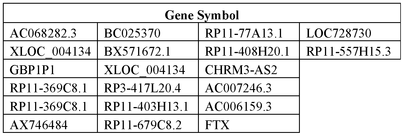
[00438] In some cases, detection of endometriosis is achieved by detecting differential expression of microRNAs such as biomarkers set forth in Table 13.
Table 13. Overview of microRNA biomarkers for detecting endometriosis

hsa-let-7b hsa-miR-101 hsa-miR-30e-5p hsa-mir-21 hsa-miR-106a hsa-miR-34a hsa-let-7c hsa-miR-106b hsa-miR-365 hsa-let-7g hsa-miR-126 hsa-miR-368 hsa-mir-143 hsa-miR-130a hsa-miR-375 hsa-mir-103 hsa-miR-130b hsa-miR-376a hsa-mir-140-3p hsa-miR-132 hsa-miR-379 hsa-mir-29a hsa-miR-143 hsa-miR-411 hsa-mir-101 hsa-miR-145 hsa-miR-425-5p hsa-let-7e hsa-miR-148a hsa-miR-486 hsa-mir-320a hsa-miR-150 hsa-miR-493-5p hsa-let-7i hsa-miR-17-5p hsa-miR-503 hsa-mir-26a hsa-miR-182 hsa-miR-638 hsa-mir-1 hsa-miR-183 hsa-miR-663 hsa-mir-145 hsa-miR-186 hsa-miR-671 hsa-let-7d hsa-miR-196b hsa-miR-768-3p hsa-mir-424 hsa-miR-199a hsa-miR-768-5p hsa-mir-451 hsa-miR-200a hsa-miR-93 hsa-mir-107 hsa-miR-200b hsa-miR-99a hsa-mir-191 hsa-miR-200c miR-34c-5p hsa-mir-29c hsa-miR-202 miR-34b* hsa-mir-25 hsa-miR-20a miR-34c-3p hsa-mir-221 hsa-miR-221 miR-9 hsa-mir-99a hsa-miR-25 miR-9* hsa-mir-152 hsa-miR-28 miRPlus_42 780 hsa-mir-23a hsa-miR-299-5p
[00439] The systems, methods, devices and kits disclosed herein can provide for methods for detecting a presence or absence of endometrial cancer. Endometrial cancer, also termed uterine cancer, is the most frequent invasive tumors of the female genital tract and the fourth most common in women in western countries (Jamel et. al., Cancer statistics. 2008. CA Cancer J. Clin 58:71-96). Biomarkers and methods for the detection diagnosis, and prognosis of endometrial cancer are described in U.S. Patent Application Publication No. 20120122726 , which is incorporated herein in its entirety. For example, biomarkers are differentially expressed in control samples as compared to samples from patients having endometrial cancer. The differential expression of these biomarkers can be used for detecting endometrial cancer in a biological sample collected using systems, methods, devices and kits described herein. In some cases, expression of a biomarker is used for detecting endometrial cancer. In some cases, expression of a set of biomarkers is used for detecting endometrial cancer using systems, methods, devices and kits described herein. The set of biomarkers can comprise nucleic acids encoding a gene set forth in Tables 2, 3, 4, 5, 6, 7, 8, 9 and/or 10. In some embodiments, the expression level of a set of biomarkers is determined. The set of biomarkers can comprise about 1 to 10,000, 1 to 1000, 1 to 100, 200 to 8000, 300 to 5000, 400 to 2000, 500 to 1000, 50 to 200, 20 to 100, 15 to 30, 10 to 20, 5 to 15, 1 to 10, 2 to 8, 3 to 7, 4 to 6, 5 to 7, 6 to 7, 7 to 9, or 2 to 12 genes. The set of biomarkers can comprise about 1, 2, 3, 4, 5, 6, 7, 8, 9, 10, 15, 20, 25, 30, 40, 50, 60, 70, 80, 90, 100, or more genes. The set of biomarkers can comprise at most about 1, 2, 3, 4, 5, 6, 7, 8, 9, 10, 15, 20, 25, 30, 40, 50, 60, 70, 80, 90, 100, 1000, or 10,000 genes. The expression level of the biomarker in the endometriosis sample can be increased or decreased when compared to the expression level of the biomarker in a sample that is not diagnosed with endometriosis. The expression level of the biomarker in the endometriosis sample can be increased or decreased by about 1-100%, 10-90%, 20-80%, 30-70%, 40-60%, 15-85%, 25-75%, 35-65%, or 45-55%. The expression level of the biomarker in the endometriosis sample can be increased or decreased by at least about 10%, 15%, 20%, 30% 40%, 50%, 60%, 70%, 80%, 90%, 95%, 99%, or 100%. The determination of the biomarker may comprise determining the nucleic acid sequence by using an Illumina sequencing platform or any sequencing techniques and platforms in the field. The biomarker may have a change of nucleic acid sequence in the endometriosis sample. The change of nucleic acid can be a mutation, e.g., point mutation or single nucleotide polymorphism, multiple nucleotide polymorphism, insertion (e.g., insertion of one or more nucleotides at a locus), multiple nucleotide change, deletion (e.g., deletion of one or more nucleotides at a locus), and inversion (e.g., reversal of a sequence of one or more nucleotides) . In some embodiments, the biomarkers for use to detect endometrial cancer are set forth in Table 14.
Table 14. Overview of biomarkers for detecting endometrial cancer.
Gene Symbol Gene Title
ACAA1 Acetyl-CoA Acyiiransferase 1
AP1M2 Adaptor Related Protein. Complex 1 Mu 2 Subunit
CGN Cmguim
DDR1 Discoidirt Domain Receptor 'Tyrosine Kinase 1
Gene Symbol Gene Title
EPS8L2 EPS 8 Like 2
FASTKD1 FAST Kinase Domains 1
GMIP GEM interacting Protein
IKBKE inhibitor Of Kappa Light Polypeptide Gene Enhancer in B-Ceil 5, Kinase Epsiion
P2RX4 Purinergie Receptor P2X 4
P4HB Prolyl 4-Hydroxylase Subunit Beta
PHKG2 Phosphorylase Kinase Gamma Subunit 2
PPFIBP2 PPFIA Binding Protein 2
PPP1R16A Protein Phosphatase 1 Regulatory Subunit 16A
RASSF7 Ras Association Domain Family Member 7
RNF183 Ring Finger Protein 183
SIRT6 Sirtuin 6
TJP3 Tight Junction Protein 3
EFEMP2 EGF Containing Fibuiin-Like Extracellular Matrix Protein 2
SOCS2 Suppressor Of Cytokine Signaling 2
DCN Decorin
[00440] The systems, methods, devices and kits disclosed herein can provide for methods for detecting a presence or absence of gynecological pathologies in a subject. Cervicovaginal fluid (CVF) has an important function in the homeostasis and immunity of the lower female genital tract. Analysis of the cervicovaginal fluid proteome may therefore yield important information about the pathogenesis of numerous gynecological pathologies. Additionally, cervicovaginal fluid has great potential as a source of biomarkers for these conditions. See Zegels et al., Use of cervicovaginal fluid for the identification of biomarkers for pathologies of the female genital tract. Proteome Science. 2010.8:63, which is incorporated herein in its entirety. The female genital tract is characterized by a unique immunologic
micro-environment. It is essential for human reproduction that the immune system of the female genital tract is modulated correctly as it needs to tolerate the presence of sperm and fertilized oocytes without starting an immune reaction. Nevertheless, a strong immune system in the genital tract is very important to protect the interior organs and the fetus or embryo against the large pathogenic stress. This obligation of a dual immune system, one that tolerates and one that reacts efficiently, is a biological challenge. The lower female genital tract is covered by numerous commensal bacteria which form an important protective factor. The normal vaginal bacterial flora consists predominantly of Lactobacillus spp. including, Lactobacillus jenensii, Lactobacillus crispatus, Lactobacillus iners, Lactobacillus rhamnosus, and Lactobacillus acidophilus, and facultative anaerobic species such as Gardnerella vaginalis.
[00441] The lower genital tract forms an important entry gate for a wide variety of pathogens such as
Candida spp., Trichomonas vaginalis, human immunodeficiency virus (HIV) and human papillomavirus
(HPV). The upper female genital tract is believed to be sterile under normal circumstances. Pathogens such as Neisseria gonorrhoeae and Chlamydia trachomatis can occasionally ascend from the lower to the higher
genital tract and can cause pathogenic conditions such as pelvic inflammatory disease (PID). The clinical impact of these infections may not be underestimated since they can lead to serious conditions such as preterm birth, increased susceptibility to sexually transmitted infections (STI), infertility and cancer. As such, early diagnosis is essential for the successful treatment of the underlying pathology and prevention of irreversible complications. For this reason, there is a high demand for new diagnostic tools, such as biomarkers, which allow for better and earlier treatment of gynecological pathologies.
[00442] CVF is another vital element of the immune system of the female genital tract. CVF comprises (i) vulvar secretions from sebaceous, sweat, Bartholins and Skene glands, (ii) plasma transudate through the vaginal wall, (iii) exfoliated cells, (iv) bacterial products, (v) cervical mucus (vi) endometrial and oviductal fluids and (vii) secretions from vaginal immune cells. Cervical mucus, endometrial and oviductal fluids, and secretions from vaginal immune cells are influenced by sex steroid hormones e.g., during the menstrual cycle and pregnancy. CVF comprises water and contains many different factors such as cholesterol, lipids, mucin, carbohydrates, amino acids, proteins and inorganic ions. It covers the lower female genital tract and hydrates the mucosa, creating a physical barrier for microbial invasion. CVF may be used as samples for the analysis of proteins, thereby finding correlations between different expression profiles and specific pathologies.
[00443] CVF may be used for the identification of diagnostic or prognostic biomarkers. The low cost and ease of sample collection, circumvention of the risk associated with biopsies and the possibility of observing high numbers of patients using multiple samples are clear advantages of CVF over body fluid such as plasma, serum, cerebrospinal fluid, urine, saliva, bronchoalveolar lavage fluid, nipple aspirate fluid, tear fluid and amniotic fluid. Four different methods are frequently applied for the collection of CVF: cervicovaginal washings (e.g., the cervicovaginal is rinsed with washing buffer after which the fluid is collected), cervicovaginal swabs (e.g., swabs or brushes are applied to the mucosal wall and rotated to collect the CVF), cervicovaginal wicks (e.g., wicks such as tampons, strips or sponges are inserted in the female genital tract which absorb cervicovaginal secretions), and diaphragm-like devices (e.g., cups placed over the cervix which collect cervical fluid).
[00444] In addition, it can be expected that biomarkers found in CVF, being a "proximal" fluid, show more specificity and sensitivity for gynecopathological conditions as compared to circulating blood, plasma, or serum. Table 15 shows potential biomarkers that are infection-correlated. The biomarkers are discovered using human CVF samples.
Table 15. Overview of potential CVF protein/peptide biomarkers for different diseases, conditions or statuses of the female genital tract.
Condition or patient status Potential CVF protein/peptide biomarker
Human papillomavirus (HPV)
11-6
infection and cervical cancer
11-8
IL-12p40n
Condition or patient status Potential CVF protein/peptide biomarker
IFN-γ
11-10
TGF-βΙ
TNF-a
11- 1β
11-5
Anti HPV IgG
Anti HPV IgA
antigalactosyl (al→3) galactose antibodies
Mum an imm unodeHctency vi rus
Anti-HIV IgA and IgG antibodies
(HIV) resistance
RANTES
antimicrobial peptides (e.g., SLPI, defensins,
Bacterial Vaginosis
lysozyme and lactoferrin)
IL-Ιβ
IL-8
IL-10
IL-4
Preterm Birth fetal fibronectin
C-reactive protein
interleukin-6
interleukin-8
interleukin-ΐβ
granulocytic elastase al-antiprotease prolactin
sialidase
monocyte chemotactic protein 1
insulin-like growth factor-binding protein- 1 defensins
lactoferrin
matrix metalloproteinases
β-human chorionic gonadotrophin
Preterm premature rupture of
β-human chorionic gonadotropin
membranes (PPROM)
insulin-like growth factor binding protein- 1 diamine oxidase
active ceruloplasmin
fetal fibronectin
C-reactive protein
a-fetoprotein
prolactin
placental a-microglobulin-1
Condition or patient status Potential CVF protein/peptide biomarker
interleukin-1 receptor antagonist
[00445] Human papillomavirus (HPV) is one of the most common sexually transmitted infections. It is estimated that about 70%-80% of the sexually active female population may acquire HPV before the age of 50 and about 80% of the HPV -infected patients may spontaneously clear the virus within 2 years. HPV is associated with high morbidity and mortality worldwide, since it can cause precancerous lesions and cervical cancer. Worldwide, cervical cancer is both the fourth-most common cause of cancer and the fourth-most common cause of death from cancer in women (World Cancer Report 2014. World Health Organization. 2014. pp. Chapter 5.12. ISBN 9283204298, which is incorporated herein in its entirety). There are more than 150 HPV types and almost 40 of them infect the anogenital region. HPV can be divided in low and high risk HPV. The most common low-risk types are type 6 and 1 1. The most common high-risk types are 16, 18, 45, 31 and 33. Cervical cancer is mainly caused by an infection with a high risk type. Infection of squamous or glandular epithelial cells with HPV causes morphological alterations which lead to the formation of squamous or glandular intra-epithelial lesions. The severity of this precancerous condition depends on the extent, the site as well as on the depth of the infected epithelial layer.
[00446] Because of the high prevalence, the severe consequences and the lack of a good treatment of advanced cervical carcinoma, the emphasis of managing this condition may lie on prevention and diagnosis in a stadium as early as possible. The prophylactic HPV vaccination may prevent at least about 70%, 80%, 90%, 95%, or 98% of infections with HPV 16 and 18 but may also prevent HPV 31 (about 89%), HPV 33 (about 82%), HPV 45 (about 100%), and any high risk HPV type
16/18/31/33/35/39/45/51/52/56/58/59/66/68 (about 70%). As such, the HPV vaccination may prevent about 70% - 80% of all cervical cancers. Therefore, women between the ages of 25 and 65 may need to be screened on a frequent basis using, for example, a Pap test (liquid based cytology) for general testing or for a type specific HPV testing. Screening using the classical Pap test has led to a considerable reduction of the mortality, yet there are still some notable shortcomings. The current screening is (1) labor intensive, (2) difficult to automatize, (3) vulnerable to inter- and intra-observer variability, and (4) is not very sensitive. Testing for the presence of nucleic acids by, e.g., sequencing, may increase the sensitivity. HPV DNA or RNA can be isolated and purified from a biological sample obtained from the cervicovaginal canal of a subject, and tested for the presence of HPV DNA or RNA. The biological sample can be from a Pap test. The biological sample can be from a cotton swab. The biological sample can be from a biopsy. One disadvantage of nucleic acids sequencing is that it cannot predict progression to intra-epithelial lesions and eventually cervical cancer as it involves collection of a biological sample from the subject by a trained profession and a visit to a clinic. The systems, methods, and devices described herein provide for tools for self-collection of a biological sample from the cervicovaginal canal of a female subject in her privacy, without the involvement of a trained professional. The biological sample can be collected regularly by the female subject and sent to a laboratory for analysis including, but not limited to, testing for chemistry,
testing for hematology, testing for microbiology, and testing for CVF biomarkers. Potential CVF biomarkers associated with diseases, conditions or statuses of the female genital tract are listed in Table 12.
[00447] Screening for CVF biomarkers may provide for HIV resistance. Exemplary CVF biomarkers for HIV resistance screening are listed in Table 12. In addition, nucleic acids isolated from CVF may be used for detecting nucleic acid changes in genes that are associated with HIV resistance, or for detecting the absence or presence of a gene or protein. For example, mutation of C-C chemokine receptor type 5 (CCR5 or CD 195) induces upregulation of RANTES, ΜΐΡ-Ια and ΜΪΡ-1β in exposed seronegative individuals (ESNs). ESNs are individuals (<5% of the population) from high-risk cohorts that remain TgG -seronegative despite frequent HIV -exposure and therefor show a certain degree of in vitro HIV resistance. As another example, the presence of nucleic acids encoding for a protein, antigen, or antibody, in ESNs suggests that the presence or upregulation of these nucleic acids may be associated with HI resistance. Exemplary protein, antigen, or antibody are gp41, p24, anti-H!V IgA, anti-HIV IgG, or elafm/trappin-2. Therefore, detecting mutations in a gene associated with ESN may provide insight for developing microbicides, e.g., chemical entities that can prevent or reduce transmission infections, which can be applied locally as a potential approach in HIV prevention.
[00448] Bacterial vaginosis (BV) is a frequently observed condition by which the normal vaginal flora is disrupted. The imbalance of vaginal flora may be revealed by screening CVF biomarkers (see Table 12) in a biological sample obtained from the cervicovaginal canal of a subject. BV is a non-inflammatory condition characterized by the loss of hydrogen peroxide forming Lactobacillus spp. (e.g., L. crispatus, L. acidophilus, L. rhamnosus) and by an increased growth of anaerobic species (e.g., Atopobium vaginae, Mobiluncus spp., Prevotella spp.). Population change of commensal bacteria or presence of nonresident bacteria may be used to assess the balance of vaginal flora. The presence of absence of Lactobacillus spp., Atopobium vaginae, Mobiluncus spp., and Prevotella spp. can be detected by, e.g., nucleic acid sequencing, Western blot.
[00449] CVF also comprises biomarkers for assessing the risk of the occurrence of preterm birth and preterm premature rupture of membranes (Table 12). Preterm birth is defined as occurring before 37 complete weeks of gestation by the World Health Organization (WHO). Preterm birth is a major cause of neonatal morbidity and mortality. Preterm birth is difficult to treat and therapy is often inefficient because the underlying cause is seldom determined and the diagnosis is frequently made too late. As such, treatment for preterm birth relies on preventive strategies. Quantifying CV fetal fibronectin (FFN) concentration in combination with cervical ultrasound measurements may reveal correlations of risk for preterm labor/birth. For example, CVF can be collected from a female subject using the systems, methods, devices, and kits disclosed herein. The female subject may perform analysis on the CVF using the systems, methods, devices and kits described herein. The CVF can be shipped to a laboratory for analysis.
Sequencing
[00450] Detecting the presence or absence, or expression level of a biomarker associated with a disease or pathology can involve identifying a nucleic acid or polynucleotide sequence of the biomarker. The
biomarker can be a gene or a part of a gene that encodes a functional protein. The identity of a biomarker (e.g., a gene or a partial of a gene) can involve sequencing, de novo sequencing, massive parallel sequencing, RNA sequencing, DNA sequencing, deep sequencing, methylation sequencing, bisulfite sequencing, or next-generation sequencing. For example, a biomarker (e.g., a gene or a partial of a gene) can be verified by sequencing of nucleic acids from a biological sample collected from a subject's cervicovaginal canal.
[00451] The next-generation sequencing platform can be a commercially available platform.
Commercially available platforms include, e.g., platforms for sequencing-by synthesis, ion semiconductor sequencing, pyrosequencing, reversible dye terminator sequencing, sequencing by ligation,
single-molecule sequencing, sequencing by hybridization, and nanopore sequencing. Platforms for sequencing by synthesis are available from, e.g., Illumina, Methylation Sequencing, 454 Life Sciences, Helicos Biosciences, and Qiagen. Illumina platforms can include, e.g., Illumina's Solexa platform, Illumina' s Genome analyzer, which are described in Gudmundsson et al., Genome-wide association and replication studies identity four variants associated with prostate cancer susceptibility. Nat. Genet. 2009 41 : 1 122-1 126, Out et al. Deep sequencing to reveal new variants in pooled DNA samples. Hum. Mutat. 2009 30: 1703-12, Turner, Massively parallel exon capture and library-free resequencing across 16 genomes. Nat. Methods 2009 6:315-6, U.S. Patent Application Publication nos. 20080160580 and 20080286795, and U.S. Patent Nos. 6306597, 71 15400, and 7232656, which are hereby incorporated in their entireties. 454 Life Science platforms include, e.g., the GS Flex and GS Junior, and are described in U.S. Patent No. 7,323,305, which patent is hereby incorporated in its entirety. Platforms from Helicos Biosciences include the True Single Molecule Sequencing platform. Platforms for ion semiconductor sequencing include, e.g., the Ion Torrent Personal Genome Machine (PGM) and are described in U.S. Patent No. 7948015, which patent is hereby incorporated in its entirety. Platforms for pyrosequencing include the GS Flex 454 system and are described in U.S. Patent Nos. 721 1390; 7244559; 7264929, which patents are hereby incorporated in their entireties. Platforms and methods for sequencing by ligation include, e.g., the SOLiD sequencing platform and are described in U.S. Patent No. 5750341. Platforms for single-molecule sequencing include the SMRT system from Pacific Bioscience and the Helicos True Single Molecule Sequencing platform. Other platforms for sequencing can include, but are not limited to, Large Whole-Genome Sequencing, Small Whole-Genome Sequencing, Exome Sequencing, Targeted Gene Sequencing, Whole-Transcriptome Sequencing, Gene Expression Profiling with mRNA-Seq, Targeted Gene Expression Profiling, miRNA & Small RNA Analysis, and 16S Metagenomic Sequencing.
Computer control systems
[00452] The present disclosure provides computer systems that are programmed to implement methods of the disclosure. Such computer systems may perform data analysis or presentation of the biological sample test results. FIG. 17 shows a computer system 1701 that is programmed or otherwise configured to perform methods of the present disclosure, including data analysis. The computer system 1701 can be an electronic device of a user or a computer system (e.g., computer server) that is remotely located with respect to the electronic device. The electronic device can be a mobile electronic device.
[00453] The computer system 1701 includes a central processing unit (CPU, also "processor" and "computer processor" herein) 1705, which can be a single core or multi core processor, or a plurality of processors for parallel processing. The computer system 1701 also includes memory or memory location 1710 (e.g., random-access memory, read-only memory, flash memory), electronic storage unit 1715 (e.g., hard disk), communication interface 1720 (e.g., network adapter) for communicating with one or more other systems, and peripheral devices 1725, such as cache, other memory, data storage and/or electronic display adapters. The memory 1710, storage unit 1715, interface 1720 and peripheral devices 1725 are in communication with the CPU 1705 through a communication bus (solid lines), such as a motherboard. The storage unit 1715 can be a data storage unit (or data repository) for storing data. The computer system 1701 can be operatively coupled to a computer network ("network") 1730 with the aid of the communication interface 1720. The network 1730 can be the Internet, an internet and/or extranet, or an intranet and/or extranet that is in communication with the Internet. The network 1730 in some cases is a
telecommunication and/or data network. The network 1730 can include one or more computer servers, which can enable distributed computing, such as cloud computing. The network 1730, in some cases with the aid of the computer system 1701, can implement a peer-to-peer network, which may enable devices coupled to the computer system 1701 to behave as a client or a server.
[00454] The CPU 1705 can execute a sequence of machine-readable instructions, which can be embodied in a program or software. The instructions may be stored in a memory location, such as the memory 1710. The instructions can be directed to the CPU 1705, which can subsequently program or otherwise configure the CPU 1705 to implement methods of the present disclosure. Examples of operations performed by the CPU 1705 can include fetch, decode, execute, and write back.
[00455] The CPU 1705 can be part of a circuit, such as an integrated circuit. One or more other components of the system 1701 can be included in the circuit. In some cases, the circuit is an application specific integrated circuit (ASIC).
[00456] The storage unit 1715 can store files, such as drivers, libraries and saved programs. The storage unit 1715 can store user data, e.g., user preferences and user programs. The computer system 1701 in some cases can include one or more additional data storage units that are external to the computer system 1701, such as located on a remote server that is in communication with the computer system 1701 through an intranet or the Internet.
[00457] The computer system 1701 can communicate with one or more remote computer systems through the network 1730. For instance, the computer system 1701 can communicate with a remote computer system of a user (e.g., patient or healthcare provider). Examples of remote computer systems include personal computers (e.g., portable PC), slate or tablet PC's (e.g., Apple® iPad, Samsung® Galaxy Tab), telephones, Smart phones (e.g., Apple® iPhone, Android-enabled device, Blackberry®), or personal digital assistants. The user can access the computer system 1701 via the network 1730.
[00458] Methods as described herein can be implemented by way of machine (e.g., computer processor) executable code stored on an electronic storage location of the computer system 1701, such as, for example, on the memory 1710 or electronic storage unit 1715. The machine executable or machine
readable code can be provided in the form of software. During use, the code can be executed by the processor 1705. In some cases, the code can be retrieved from the storage unit 1715 and stored on the memory 1710 for ready access by the processor 1705. In some situations, the electronic storage unit 1715 can be precluded, and machine-executable instructions are stored on memory 1710.
[00459] The code can be pre-compiled and configured for use with a machine having a processor adapted to execute the code, or can be compiled during runtime. The code can be supplied in a programming language that can be selected to enable the code to execute in a pre-compiled or as-compiled fashion.
[00460] Aspects of the systems and methods provided herein, such as the computer system 1701, can be embodied in programming. Various aspects of the technology may be thought of as "products" or "articles of manufacture" typically in the form of machine (or processor) executable code and/or associated data that is carried on or embodied in a type of machine readable medium. Machine -executable code can be stored on an electronic storage unit, such as memory (e.g., read-only memory, random-access memory, flash memory) or a hard disk. "Storage" type media can include any or all of the tangible memory of the computers, processors or the like, or associated modules thereof, such as various semiconductor memories, tape drives, disk drives and the like, which may provide non-transitory storage at any time for the software programming. All or portions of the software may at times be communicated through the Internet or various other telecommunication networks. Such communications, for example, may enable loading of the software from one computer or processor into another, for example, from a management server or host computer into the computer platform of an application server. Thus, another type of media that may bear the software elements includes optical, electrical and electromagnetic waves, such as used across physical interfaces between local devices, through wired and optical landline networks and over various air-links. The physical elements that carry such waves, such as wired or wireless links, optical links or the like, also may be considered as media bearing the software. As used herein, unless restricted to non-transitory, tangible "storage" media, terms such as computer or machine "readable medium" refer to any medium that participates in providing instructions to a processor for execution.
[00461] Hence, a machine readable medium, such as computer-executable code, may take many forms, including but are not limited to, a tangible storage medium, a carrier wave medium or physical transmission medium. Non-volatile storage media include, for example, optical or magnetic disks, such as any of the storage devices in any computer(s) or the like, such as may be used to implement the databases, etc. shown in the drawings. Volatile storage media include dynamic memory, such as main memory of such a computer platform. Tangible transmission media include coaxial cables; copper wire and fiber optics, including the wires that comprise a bus within a computer system. Carrier-wave transmission media may take the form of electric or electromagnetic signals, or acoustic or light waves such as those generated during radio frequency (RF) and infrared (IR) data communications. Common forms of computer-readable media therefore include for example: a floppy disk, a flexible disk, hard disk, magnetic tape, any other magnetic medium, a CD-ROM, DVD or DVD-ROM, any other optical medium, punch cards paper tape, any other physical storage medium with patterns of holes, a RAM, a ROM, a PROM and EPROM, a
FLASH-EPROM, any other memory chip or cartridge, a carrier wave transporting data or instructions, cables or links transporting such a carrier wave, or any other medium from which a computer may read programming code and/or data. Many of these forms of computer readable media may be involved in carrying one or more sequences of one or more instructions to a processor for execution.
[00462] The computer system 1701 can include or be in communication with an electronic display 1735 that comprises a user interface (UI) 1740. Examples of UFs include, without limitation, a graphical user interface (GUI) and web-based user interface.
[00463] Methods and systems of the present disclosure can be implemented by way of one or more algorithms. An algorithm can be implemented by way of software upon execution by the central processing unit 1705.
[00464] The disclosure is further illustrated by the following examples which should not be construed as limiting. The examples are illustrative only, and are not intended to limit, in any manner, any of the aspects described herein.
EXAMPLES
[00465] The following examples illustrate some embodiments and aspects of the disclosure. It will be apparent to those skilled in the relevant art that various modifications, additions, substitutions, and the like can be performed without altering the spirit or scope of the disclosure, and such modifications and variations are encompassed within the scope of the disclosure as defined in the claims which follow. The following examples do not in any way limit the disclosure.
[00466] The device as disclosed herein can be applied to the detection of a wide variety of infections, as it allows for detection of antigen or antibody analytes associated with any disease or disorder for which an antibody or antigen analyte is known, such as for a variety of STIs and cancers. The device can also be used to detect the presence of semen, identify genetic signatures and genomic material, as well as detect small molecules like environmental toxins such as BPA. In some embodiments, the device is used to assay for one of the following conditions listed as examples below. In some embodiments, the device is used to simultaneously assay for more than one of the following conditions using a single sample of cervicovaginal fluid. In some embodiments, the device is used to assay for more than one of the following conditions at different times using a single sample of cervicovaginal fluid. In some embodiments, the device is used to assay for more than one of the following conditions at different times using different samples of cervicovaginal fluid.
Example 1: Gonorrhea
[00467] Gonorrhea is an infection caused by the bacterium Neisseria gonorrhoeae. Transmission of this pathogen occurs during vaginal, oral, or anal sex (Moran, Clin. Evid. 200: 1604; 2007). While men often experience painful urination upon infection, women are mostly asymptomatic. If left untreated, gonorrhea can cause local disease such as pelvic inflammatory disease (PID), or can also affect other parts of the body, such as the joints and heart valves.
[00468] Traditionally, gonorrhea was diagnosed with gram stain and culture; however, newer PCR-based testing methods are becoming more common. The USPTF recommends screening for
gonorrhea in women at increased risk of infection, which includes all sexually active women younger than 25 years (Meyers et al., Am. Fam. Physician 77:819-824; 2008). Screening for gonorrhea in women who are or intend to become pregnant, and who are found to be at high risk for sexually transmitted diseases, is recommended as part of prenatal care in the United States.
[00469] Past treatments for gonorrhea included a range of antibiotics. However, as of 2010, injectable ceftriaxone appears to be one of the few effective antibiotics, due to increasing rates of antibiotic resistance (Deguchi et al., J. Urol, 184:851-858; 2010).
[00470] In some embodiments, the disclosed device can be used to detect gonorrheal infections from menstrual blood or cervico vaginal fluids.
Example 2: Chlamydia
[00471] Chlamydia is a common STI that is caused by the bacterium Chlamydia trachomatis.
Transmission occurs during vaginal, anal, or oral sex, but the bacterium can also be passed from an infected mother to her baby during vaginal childbirth. It is estimated that about 1 million individuals in the United States are infected with this bacterium, making chlamydia one of the most common STIs worldwide. Like gonorrhea, chlamydial infection is asymptomatic for a majority of women. If symptoms are present, they include unusual vaginal bleeding or discharge, pain in the abdomen, painful sexual intercourse, fever, painful urination or the urge to urinate more frequently than usual. Of those who develop asymptomatic infection, approximately half may develop PID. Infants born to mothers with chlamydia may suffer from pneumonia and conjunctivitis, which may lead to blindness. They may also be subject to spontaneous abortion or premature birth.
[00472] Diagnosis of chlamydial infection is usually done by nucleic acid amplification techniques, such as PCR, using samples collected from cervical swabs or urine specimens (Gaydos et al., J. Clin.
Microbio., 42:3041-3045; 2004). Treatment involves various antibiotic regimens.
[00473] In some embodiments, the disclosed device can be used to detect chlamydial infections from menstrual blood or cervico vaginal fluids.
Example 3: Trichomoniasis
[00474] Trichomoniasis is considered the most common curable sexually transmitted disease. In the United States, an estimated 3.7 million people have the infection, but only about 30% develop any symptoms of the disease (Center for Disease Control fact sheet, 2015). In women, the most commonly infected part of the body is the lower genital tract. Nearly 70% of infections are asymptomatic. Not only can infection with Trichomonas increase one's risk of contracting and spreading other STIs, and pregnant women with trichomoniasis are more likely to go into preterm labor, but also, babies born to infected mothers are more likely to have officially low birth weight - less than 5.5 pounds (CDC fact sheet, 2015). Trichomoniasis can also lead to pelvic inflammatory disease, which may lead to infertility if untreated.
[00475] Diagnosis of Trichomoniasis includes a visit to the doctor's office, a physical exam, and sampling of vaginal secretions by a wet preparation test to visualize bacterial flagella present on the trichomonas bacteria (L. Campbell et al, Journal of Clinical Microbio, 2008). More recent technologies for diagnosis include rapid dipstick immunoassay and antigen tests directed at flagellar proteins of the bacteria.
Treatment for Trichomoniasis is a one dose administration of an antibiotic, either metronidazole or tinidazole (CDC fact sheet, 2015).
[00476] In some embodiments, the disclosed device can be used to detect trichomoniasis infections from menstrual blood or cervicovaginal fluids.
Example 4: Syphilis
[00477] Syphilis is an STI that can cause long-term complications if not treated correctly. Symptoms in adults are divided into stages. These stages are primary, secondary, latent, and late syphilis. In pregnant women, having syphilis can lead to giving birth to a low birth weight baby. It can also lead to delivering the baby too early or stillborn (CDC fact sheet, 2015).
[00478] Although T pallidum cannot be grown in culture, there are many tests for the direct and indirect diagnosis of syphilis. Still, there is no single optimal test. Direct diagnostic methods include the detection of T pallidum by microscopic examination of fluid or smears from lesions, histological examination of tissues or nucleic acid amplification methods such as polymerase chain reaction (PCR). Indirect diagnosis is based on serological tests for the detection of antibodies (Ratnam S, Can J Infect Dis Med Microbiol 2005). Treatment includes a single dose of intramuscular administration of penicillin (2.4 Million units).
[00479] In some embodiments, the disclosed device can be used to detect syphilis infections from menstrual blood or cervicovaginal fluids.
Example 5: Bacterial Vaginosis
[00480] Bacterial Vaginosis (BV) is an infection caused when too much of certain bacteria change the normal balance of bacteria in the vagina. Bacterial vaginosis (BV) is one of the most common lower genital tract conditions, occurring in 35% of women attending sexually transmitted infection (STI) clinics, 15% to 20% of pregnant women, and 5% to 15% of women attending gynecology clinics (Eschenbach DA, Am J Obstet Gynecol 1993). Pregnant women with BV are more likely to have babies who are born premature (early) or with low birth weight than women who do not have BV while pregnant. Low birth weight means having a baby that weighs less than 5.5 pounds at birth (CDC fact sheet, 2015).
[00481] Diagnosis of BV is typically done through a vaginal swab to assess the presence and balance of certain bacteria within the vaginal flora through PCR. A wet mount, whiff test, or pH test can also be performed in order to diagnose a possible bacterial infection.
[00482] In some embodiments, the disclosed device can be used to detect bacterial vaginosis from menstrual blood or cervicovaginal fluids.
Example 6: Pelvic Inflammatory Disease (PID)
[00483] Chronic and untreated infection with gonorrhea and chlamydia commonly leads to PID, a generic term for infection of the uterus, fallopian tubes, and/or ovaries. As the immune system tries to fight off the invading pathogens, it causes local inflammation and scarring. There are no tests for PID. A diagnosis is usually based on a combination of a patient's medical history, physical exam, and other test results. Since the most common causes of PID are gonorrhea and chlamydia, prevention of PID usually involves prompt diagnosis and treatment of these infections. However, since treatment of PID may not undo any damage that has already happened to one's reproductive system, successful treatment is heavily
dependent on early diagnosis. Some patients may not realize they have PID because symptoms may be mild or nonexistent. However, if symptoms do exist, they include pain in the lower abdomen, fever, unusual discharge associated with odor, painful intercourse associated with bleeding, burning sensation during urination, or bleeding between periods. Women who have had a history of PID are more likely to have a diagnosis of endometriosis. Consequently, they are also more likely to be in need of a hysterectomy, have an ectopic pregnancy, or suffer from infertility.
[00484] In some embodiments, the disclosed device focuses on detecting gonorrheal and chlamydial infections from menstrual blood or cervicovaginal fluids, and includes the ability to send results to a physician, and educate on safe sex practice and available interventions/therapeutics. It also includes the ability to track monthly results and options for coping and dealing with STIs.
Example 7: Endometriosis
[00485] Endometriosis is a gynecological condition in which cells from the lining of the uterus (endometrium) appear and flourish outside the uterine cavity, most commonly on the membrane which lines the abdominal cavity, the peritoneum. Although the exact cause of endometriosis is not certain, there are several possible explanations, such as retrograde menstruation, surgical scar implantation, immune disorders, as well as heredity. Significantly, there is an established association between endometriosis and infertility (Buletti et al., J. Assist Reprod. Genet. 27:441-447; 2010). Current diagnostic methods for endometriosis involve a laparoscopy, an invasive surgical procedure. There is no cure for endometriosis, but it can be treated in a variety of ways, including with pain medication, hormonal drugs, and surgery.
[00486] In some embodiments the disclosed device focuses on detecting markers associated with endometriosis from menstrual blood or cervicovaginal fluids. This embodiment allows women to identify and track staging of the disease and helps them navigate therapeutic options such as hormonal therapy, nonsteroidal anti-inflammatory drugs ("NSAIDs") and surgery.
[00487] In some embodiments the disclosed device focuses on detecting markers associated with endometriosis from menstrual blood or cervicovaginal fluids. The markers, e.g., biomarkers disclosed herein or conventionally used for detecting the presence of endometriosis, can be detected through sequencing a biological sample collected from a female subject's vaginal canal. For example, the collected biological sample is subject to 16S rRNA sequencing, methylation sequencing, long-noncoding RNA sequencing, microRNA sequencing, RNA deep sequencing, genomic DNA sequencing, and/or cDNA sequencing.
Example 8: Polycystic Ovarian Syndrome and Ovarian Reserve
[00488] Antimullerian hormone (AMH) is produced in the adult female exclusively by granulosa cells, declines with age, and is widely considered a highly sensitive marker of ovarian reserve. Serum AMH level is increased significantly more in women with polycystic ovary syndrome (PCOS).
[00489] Serum AMH level seems to be related to the severity of PCOS and correlates with its clinical diagnostic hallmarks, including hyperandrogenism, oligo/ anovulation and polycystic ovarian morphology. AMH level may also be associated with qualitative assisted reproductive technology outcomes such as pregnancy and live birth rates independent of age (Tal R, et al, Amer J of OB & GYN, 2014).
[00490] The current method of evaluating a women's AMH level is through Enzyme Linked
Immuno-Sorbedent Assay (ELISA), and is performed on blood serum. This test provides an absolute quantification of the amount of AMH circulating in the blood.
[00491] In some embodiments the disclosed device focuses on diagnosing or assessing risk of PCOS and ovarian reserve from menstrual blood or cervicovaginal fluids. In some embodiments the disclosed device focuses on detecting markers associated with polycystic ovarian syndrome and ovarian reserve from menstrual blood or cervicovaginal fluids. The markers, e.g., biomarkers disclosed herein or conventionally used for detecting the presence of polycystic ovarian syndrome and ovarian reserve, can be detected through sequencing a biological sample collected from a female subject's vaginal canal. For example, the collected biological sample is subject to 16S rRNA sequencing, methylation sequencing, long-noncoding RNA sequencing, microRNA sequencing, RNA deep sequencing, genomic DNA sequencing, and/or cDNA sequencing.
Example 9: Human Papillomavirus (HPV) Infection
[00492] Human genital papillomaviruses are amongst the most prevalent sexually transmitted human pathogens. Most genital HPV infections in women produce transient squamous cell abnormalities of the cervix that resolve completely, and so the probability of any one HPV infection progressing to cervical cancer is quite small. Nevertheless, HPV infection is a cause of nearly all cases of cervical cancer (Lynge et al., APMIS 122:667-673; 2014). Persistent infections increase the risk of precancerous lesions, which can progress to invasive cancer. Progression to invasive cancer can be prevented when subclinical HPV infection is detected early and regular examinations are performed.
[00493] The Pap smear is the current gold standard for the detection of HPV infection. Pap smears have reduced the incidence and fatalities of cervical cancer in the developed world; however, the USPTF now recommends Pap smears only every three years. Recently developed HPV vaccines (Cervarix and Gardasil), which prevent infection with HPV types 6, 11, 16, and 18, may lead to further decreases.
However, these vaccines are currently only recommended for women of age 25 or younger.
[00494] In some embodiments, the disclosed device can be used to detect HPV infections from menstrual blood or cervicovaginal fluids. In some embodiments the disclosed device focuses on detecting markers associated with HPV infections from menstrual blood or cervicovaginal fluids. The markers, e.g., biomarkers disclosed herein or conventionally used for detecting the presence of HPV, can be detected through sequencing a biological sample collected from a female subject's vaginal canal. For example, the collected biological sample is subject to 16S rRNA sequencing, methylation sequencing, long-noncoding RNA sequencing, microRNA sequencing, RNA deep sequencing, genomic DNA sequencing, and/or cDNA sequencing.
Example 10: Yeast Infection
[00495] Vaginitis is one of the most common complaints for physician visits in the United States (Paavon J, et al, Infec Dis Clin North Am 1987) that results in 10 million office visits per year (Sparsk JM, J Reprod Med, 1991). 30% of all vaginitis cases are caused by infection with the Candida species,
commonly referred to as yeast infections. Untreated vaginal candidiasis in pregnant women can result in passing the infection to the baby during delivery and the development of oral thrush in the newborn.
[00496] Current recommended guidelines regarding screening for Candida, as published by the Centers for Disease Control and Prevention (CDC) in 2004, consist of microscopy, saline wet mount, whiff test, pH determination, or gram stain. More current diagnostic tools include a rapid dipstick antigen test. Treatment for yeast infections is now available over the counter and includes oral administration as well as topical lotions. Probiotic treatment has also been shown to be effective in reestablishing vaginal flora to help treat and prevent yeast infections.
[00497] Although candidiasis can occur without any identifiable precipitating factor, certain conditions that disrupt the balance of normal vaginal flora can predispose women to the development of symptomatic infection. The use of antibiotics, oral contraceptive pills, contraceptive devices, high estrogen levels (as during pregnancy and hormone replacement therapy), or certain medical conditions such as uncontrolled diabetes mellitus and HIV can increase an individual's risk of the development of candidiasis (Sparsk JM, J Reprod Med, 1991).
[00498] In some embodiments, the disclosed device can be used to detect candidiasis based yeast infections from menstrual blood or cervicovaginal fluids.
Example 11 : Fetal Trophoblasts
[00499] Trophoblast cells have been picked up in transcervical retrieval methods such as cervical lavage and brushing. These cells have a wealth of information that can be interrogated for data on the health of the fetus.
[00500] It is believed that small areas of erosions allow trophoblast cells to cross the decidua capillaries and reach the uterine cavity (Imudia, 2010).
[00501] Using a Y chromosome antibody, fetal trophoblast cells can be assayed for the presence or absence of Y chromosome DNA to uncover, at very early stages, the sex of the fetus.
[00502] Imudia et al. states that changes in the amount of fetal trophoblasts cells can be indicative of abnormal pregnancy. For example, a dramatic reduction of trophoblasts in cervical secretions is indicative of an ectopic pregnancy, as the fetal trophoblast does not enter into the uterine cavity (Imudia, 2010).
[00503] HLA-G is a fetal specific protein associated with fetal trophoblast and can be used to quantify the amount or relative number of trophoblasts present in a sample. Quantification of HLA-G as a proxy for trophoblast quantity can allow for the detection of abnormal pregnancies such as ectopic and molar pregnancies.
[00504] In some embodiments, the disclosed device can be used to detect the health of a fetus and pregnancy through monitoring trophoblasts, assess trophoblasts for development abnormalities and sex determination, and quantify number of trophoblasts from cervicovaginal fluids.
Example 12: HIV and CD4 monitoring
[00505] According to the CDC, at the end of 201 1 , 23% of all people living with HIV in the United States were women. Not all US women who are living with HIV are getting the care they need. Of all
women living with HIV in 201 1, only 45% were engaged in care, and only 32% had achieved viral suppression (CDC Fact Sheet, 2015).
[00506] The risk of getting HIV during vaginal sex without a condom is much higher for women than it is for men. Women who have been sexually abused may be more likely than women with no abuse history to engage in sexual behaviors like exchanging sex for drugs, having multiple partners, or having sex with a partner who is physically abusive when asked to use a condom.
[00507] Diagnosis of the disease can occur at a doctor's office either through a blood test for either the virus or antibodies for the virus (either polymerase chain reaction or immunoassay). A rapid test is also available, both point-of-care and over-the-counter. Treatment can include a lifelong regiment of antiretroviral drugs. If HIV infection is suspected, a course of antiretroviral drugs can be given up to 72 hours after the potential exposure and greatly reduces one's risk of contracting the disease. This therapy is known as Post Exposure Prophylaxis (PEP).
[00508] Once an individual tests positive, regular doctor's visits are needed to test for the total number of T4 immune cells that are found in the blood. This particular cell type is the target for HIV infection and a low level of T4 cells is indicative of advanced disease. Therefore, constant monitoring is needed to assess therapeutic effectiveness and progression of the disease. Currently this is done in the laboratory through a process called flow cytometry. However more rapid tests are currently being developed, including the potential for a later flow, semi -quantitative dipstick test that may test for CD4 cells.
[00509] In some embodiments, the disclosed device can be used to detect HIV infection and monitor CD4 cell counts in menstrual blood or cervicovaginal fluids.
Example 13: Preterm Birth and Recurrent Pregnancy Loss
[00510] Preterm delivery and recurrent pregnancy loss are some of the most challenging problems in obstetrics to date, and the diagnosis of preterm labor is often inaccurate (Leitich, 1999). Normal and preterm birth is initiated through a cascade of physical and enzymatic changes that prepare the reproductive system to begin the birthing process (Quinzio, 2007). A key marker that can be used to determine a women's risk of preterm labor is the presence of fetal fibronectin (fFN) between 24-34 weeks in vaginal secretion. fFN plays an important role in securing the fetal sac to the uterine lining.
[00511] During early pregnancy, fFN is shed into the cervical matrix at high amounts as the fetus implants and secures into the uterine wall. In normal pregnancy, the levels of fFN dramatically drop until late in pregnancy as the fetus prepares for the birthing process and the adhesion of the fetal sac to the uterine wall begins to degrade. However, in preterm labor, fFN can be detected in weeks 24-34 as the adhesion between the fetal sac and uterine wall begin to prematurely degrade.
[00512] fFN is therefore used, and is available as a rapid lateral flow assay, to determine a women's risk of preterm labor. If caught early, administration of progesterone can help to prevent further degradation of the adhesion interface of the fetal sac and uterine wall, thus improving a women's chance of carrying the fetus to term (da Fonseca, 2003). Currently this test is performed in the clinic, where a cervical sponge is inserted into the vaginal canal, and placed against the uterine cervix to collect cervical fluid. This cervical
fluid is then assayed for the presence of fFN. Other tests such as IL-6, PAMG-1, and IGFB are other similar immunotests that are utilized in some clinics to assess a women's imminent risk of preterm delivery.
[00513] Another marker that is used to determine the onset of the birthing process and risk of preterm labor is a change in pH of vaginal secretions due to the introduction of amniotic fluid as it begins to leak from the fetal sac before rupture. This pH change can increase the overall pH of the vagina from a normal range for 4.5-6 to a pH reading over 7. Currently a rapid pH test, known as a nitrazine stick, can be used to assess the pH of the vaginal canal to determine if amniotic leakage has occurred and can be used as a proxy for impending labor.
[00514] Both fFN and Nitrazine can be leveraged to assess a women's risk of preterm labor and provide actionable steps with a physician to treat the condition and improve preterm labor outcomes.
[00515] In some embodiments, the disclosed device can be used to detect and monitor preterm birth risk and recurrent pregnancy loss risk within cervicovaginal fluids.
Example 14: Breast Cancer
[00516] Signs of breast cancer may include a lump in the breast, a change in breast shape, dimpling of the skin, fluid coming from the nipple, or a red scaly patch of skin. In those patients exhibiting metastasis, there may be bone pain, swollen lymph nodes, shortness of breath, or yellow skin. Age and sex are the two primary risk factors for breast cancer. Other risk factors include obesity, lack of physical exercise, alcohol consumption, hormone replacement therapy during menopause, ionizing radiation, early age at first menstruation, and having children late or not at all. A small minority of breast cancer cases are due to genes inherited from a person's parents, including BRCA1 and BRCA2 among others.
[00517] Current methods of breast cancer screening include clinical and self-breast exams, mammography, genetic screening, ultrasound, and magnetic resonance imaging. The USPTF recommends mammography every two years in women between the ages of 50 and 74. The risks of more frequent mammograms include a small but significant increase in breast cancer induced by radiation.
[00518] Most breast cancer cases are discovered when the woman feels a lump in her breast. Lumps found in lymph nodes located in the armpits can also indicate the presence of breast cancer. However, currently, the earliest breast cancers are detected by a mammogram. Even so, most symptoms of breast disorders, including most lumps, do not turn out to represent underlying breast cancer, and in fact, fewer than 20% of lumps are cancerous.
[00519] Treatment of breast cancer usually involves a combination of surgery, radiation and chemotherapy. In some embodiments the disclosed device focuses on detecting markers associated with breast cancer from menstrual blood or cervicovaginal fluids. This embodiment allows women to identify and track staging of the disease and helps them navigate therapeutic options such as hormonal therapy, NSAIDs and surgery.
[00520] In some embodiments the disclosed device focuses on detecting markers associated with breast cancer from menstrual blood or cervicovaginal fluids. The markers, e.g., biomarkers disclosed herein or conventionally used for detecting the presence of breast cancer, can be detected through sequencing a biological sample collected from a female subject's vaginal canal. For example, the collected biological
sample is subject to 16S rRNA sequencing, methylation sequencing, long-noncoding R A sequencing, microR A sequencing, RNA deep sequencing, genomic DNA sequencing, and/or cDNA sequencing. Example 15: Ovarian Cancer
[00521] There are often no early signs of ovarian cancer. Later symptoms include bloating, pelvic pain, and abdominal swelling, among others. Ovarian cancer occurs more frequently in women who ovulate more, therefore, those who never have children are at increased risk. Other risk factors include hormone therapy after menopause, use of fertility medication, smoking, and obesity. Factors that decrease the risk include hormonal birth control, tubal ligation, and breast feeding. About 10% of cases are hereditary and those with the gene mutations BRCA1 and BRCA2 have an approximately 50% risk of developing the disease. Ovarian carcinomas are the most common type of ovarian cancer, making up more than 95% of breast cancer cases. They include five main subtypes, of which high-grade serous carcinoma is most common. These tumors are believed to usually start from the cells covering the ovaries, though some may form from the fallopian tubes (Piek et al., Adv. Exp. Med. Biol. 622:79-87; 2008). Less common types of ovarian cancer include germ cell tumors and sex cord stromal tumors.
[00522] Diagnosis of ovarian cancer starts with a physical examination (including a pelvic examination), a blood test (for CA-125 and sometimes other markers) and a transvaginal ultrasound. The diagnosis is confirmed by examination of a biopsy usually removed during surgery. If treated, early ovarian cancer may be curable. Treatments often include some combination of surgery, radiation therapy and chemotherapy.
[00523] Central to the application of the disclosed device is the identification of markers for ovarian cancer, including circulating and shed tumor cells. Ovarian cancer is curable if detected and treated early enough. However, signs of the disease can be absent in the early stages. Many of the symptoms are also non-specific (bloating, pelvic pain, etc.) and therefore difficult for a woman to disambiguate.
[00524] As a result, diagnosis often occurs in stages III/IV of the cancer. The literature points to multiple markers, such as CA-125, serum alpha-fetoprotein and lactate dehydrogenase ("LDH"), which can offer insight into diagnosis (Chudecka-Glaz et al., J. Ovarian Res. Epub; 2014; Jashnani et al., Indian J. Pathol. Microbiol. 56:54-56; 2013). Given the especially silent nature of ovarian cancer, a sentinel system optimized for constant surveillance is particularly germane to improve overall outcome of the disease.
[00525] In some embodiments the disclosed device focuses on detecting markers associated with ovarian cancer from menstrual blood or cervicovaginal fluids. The markers, e.g., biomarkers disclosed herein or conventionally used for detecting the presence of ovarian cancer, can be detected through sequencing a biological sample collected from a female subject's vaginal canal. For example, the collected biological sample is subject to 16S rRNA sequencing, methylation sequencing, long-noncoding RNA sequencing, microRNA sequencing, RNA deep sequencing, genomic DNA sequencing, and/or cDNA sequencing.
Example 16: Cervical Cancer
[00526] More than 90% of cervical cancer cases occur as a result of HPV infection. Most people who have had HPV infections, however, do not develop cervical cancer (Robbins Basic Pathology (8th ed.) pp.
718-721). Other risk factors for cervical cancer include smoking, immunosuppression, use of hormonal birth control pills, and early onset of sexual activity coupled with having multiple sexual partners. Early in infection, there are typically no symptoms. Later symptoms may include abnormal vaginal bleeding, pelvic pain or pain during sex. Diagnosis typically occurs by cervical screening with Pap smears followed by a biopsy. Medical imaging is then done to determine whether or not the cancer has spread.
[00527] Cervical cancer screening using the Pap smear or acetic acid can identify precancerous changes which when treated can prevent the development of cancer. Treatment of cervical cancer may consist of some combination of surgery, chemotherapy and radiotherapy.
[00528] In some embodiments the disclosed device focuses on detecting markers associated with cervical cancer from menstrual blood or cervicovaginal fluid. The markers, e.g., biomarkers disclosed herein or conventionally used for detecting the presence of cervical cancer, can be detected through sequencing a biological sample collected from a female subject's vaginal canal. For example, the collected biological sample is subject to 16S rR A sequencing, methylation sequencing, long-noncoding RNA sequencing, microRNA sequencing, RNA deep sequencing, genomic DNA sequencing, and/or cDNA sequencing.
Example 17: Uterine or Endometrial Cancer
[00529] Uterine or Endometrial cancer is both the most common type of uterine cancer and the most common cancer of the female reproductive system, accounting for approximately 6 percent of all cancers in women in the United States (National Cancer Institute). Most uterine cancers start in the endometrium (the inner lining of the uterus). This is called endometrial cancer. Most endometrial cancers are
adenocarcinomas (cancers that begin in cells that make mucus and other fluids). The most common sign of endometrial cancer is unusual vaginal bleeding. Since 2002, overall incidence rates have not changed significantly, whereas mortality rates have been slowly rising since 2001 (National Cancer Institute). Although the incidence rate of endometrial cancer is only slightly higher in African American women than in whites, the mortality rate of African American women is nearly twice as high as that of all other racial/ethnic groups.
[00530] Diagnosis for endometrial cancer is done either through an endometrial biopsy, through a procedure known as a dilatation and curettage - a procedure used to remove tissue from inner lining of the uterus, through physical exams and transvaginal ultrasound, or a CT scan. Because endometrial cancer begins inside the uterus, it does not usually show up in the results of a Pap test. For this reason, a sample of endometrial tissue may be removed and checked under a microscope to look for cancer cells. A recent genomic study characterized nearly 400-endometrial tumors identifying molecular signatures specific to endometrial cancer (CGASN, 2013). This work allows for future characterization of endometrial tumors for possible screening and more advanced diagnostics.
[00531] Diagnosis for endometrial cancer can be done through sequencing a biological sample collected from a female subject's vaginal canal. For example, the collected biological sample is subject to 16S rRNA sequencing, methylation sequencing, long-noncoding RNA sequencing, microRNA sequencing, RNA deep sequencing, and/or genomic DNA sequencing.
[00532] Uterine cancer is treated by one or a combination of treatments, including surgery, radiation therapy, chemotherapy, and hormone therapy. Combinations of treatments are often recommended.
Surgery can include partial or full hysterectomy. Often the stage of cancer determines the specific combination of therapy.
[00533] In some embodiments the disclosed device focuses on detecting markers associated with uterine or endometrial cancers from menstrual blood or cervicovaginal fluid.
[00534] In one example, the biological materials collected using devices, methods, kits and compositions disclosed herein is used to extract blood, extracellular fluid, cervicovaginal fluid, and shed uterus cells, for nucleic acid molecules extraction. The nucleic acid molecules can be DNA, genomic DNA or RNA. The nucleic acid molecules are determined using next generation sequencing, e.g., Illumina sequencing technology. The sequencing result is used to determine the status of one biomarker or a set of biomarkers typically used to diagnose diseases, e.g., endometriosis, cancer, uterine cancer. The biomarkers can be FDA approved biomarkers. The sequencing result can be used to determine personalized medicine to treat a disease or for prophylactic advice on a disease. The sequencing result can be used to monitor the effectiveness of a drug that is used to treat the disease. The sequencing result can be used to test family genetic disorders and diseases.
Example 18: Pre-pregnancy Nutrition
[00535] It has been shown that many vitamins and minerals are essential for healthy pregnancy. For example, low maternal folate levels are associated with allergy sensitization and asthma (Lin J et al, J Allergy Clin Immunol, 2013). Low maternal iron levels have been associated with lower mental development (Chang S. et al, Pediatrics, 2013), and low iron may even increase a mother's risk of post-partum depression. Vitamin B 12, which is essential for red blood cell formation, is essential for pregnant women and the health of their fetus. Folate, Iron, and Vitamin B 12 can all cause anemia and increase a pregnant woman's risk of preterm labor, developmental delays of the child, as well as neural tube defects during development. Based on a WHO review of nationally representative samples from 1993 to 2005, 42 percent of pregnant women have anemia. Other essential vitamins and minerals that promote a healthy pregnancy are well validated and include Vitamins A, D, E, Other B Vitamins, Calcium, and Zinc.
[00536] In some embodiments the disclosed device focuses on detecting levels of vitamins and minerals from menstrual blood or cervicovaginal fluid that may help maintain healthy levels within the body for pregnancy.
Example 19: Hormones - Metabolism
[00537] The thyroid gland is primarily involved in the control of metabolism. Abnormal thyroid function directly and indirectly affects reproduction as well. Infertility and adverse pregnancy outcomes are more common when the thyroid gland is hypo- or hyperactive. Higher miscarriage rate, more frequent preterm deliveries, increased hypertension, diabetic complications, higher risk for placental abruption, and adverse fetal effects have all been reported with thyroid dysfunction in pregnancy. At least half of implanted embryos may not survive to delivery, and, on average, 20% of clinical pregnancies are lost (Schwartz N. et al, J Clin Enocrinol Metab. 2010).
[00538] During pregnancy, a 30%-40% increased need for thyroid hormones is the result of increased placental uptake, higher thyroid-binding globulin levels, and greater blood volume (Schwartz N. et al, J Clin Enocrinol Metab . 2010). Those with subclinical hypothyroidism and/ or high-normal TSH levels at the beginning of pregnancy may not be able to meet these needs and may show signs of thyroid insufficiency during pregnancy.
[00539] Women with thyroid disease visit clinicians 2-4 times per year to check for thyroid hormone levels to adjust medications. And before pregnancy, regular monitoring of thyroid hormone and treatment can be an effective way of maintaining healthy TSH levels during pregnancy.
[00540] In some embodiments the disclosed device focuses on detecting levels of thyroid stimulating hormone from menstrual blood or cervicovaginal fluid.
Example 20: Hormones - Fertility and Menopause
[00541] Fertility - Progesterone is one of the most important hormones for pregnancy with myriad functions from ensuring implantation of the egg into a healthy uterine wall, to ensuring embryo survival and prevention of immune rejection of the developing baby. Many other hormones act in concert with progesterone, like Follicular Stimulating Hormone (FSH) and Luteinizing Hormone (LH) and can be used to assess optimal fertility windows on a monthly basis. And in fact an over dominant production of estrogen can lead to progesterone deficiency and thus difficulty getting or staying pregnant. It is important that women not only monitor FSH and LH to determine optimal fertility for getting pregnant, but ensure that sufficient levels or progesterone are being produced to ensure pregnancy and viability of the fetus. A study from the British Medical Journal, 2012, demonstrated that a single progesterone level test can help discriminate between viable and nonviable pregnancies. Among women who had an ultrasound, 73 percent had nonviable pregnancies. But among women with progesterone levels below 3 to 6 nanograms per milliliter, the probability of a nonviable pregnancy rose to more than 99 percent (Gallos L et al. British Medical J, 2012).
[00542] Perimenopause - Monitoring hormone levels during the menopausal transition may help women better understand important changes in their body and allow them to make more informed decisions about health, diet, and lifestyle. According to Hale GE (Best Pract Res Clin Obstet Gynaecol, 2009), data from endocrine studies on women throughout the menopausal transition show changes in levels of steroid hormones and gonadotropins (Progesterone, Estradiol, LH, FSH and AMH) and follicle -stimulating hormone undergoes the first detectable change while menstrual cycles remain regular. Erratic and less predictable changes in steroid hormones follow, especially with the onset of irregular cycles. Later serum hormone studies on the inhibins and anti-Mullerian hormone established that diminishing ovarian follicle number contributes to the endocrine changes with advancing reproductive age.
[00543] Many fertility issues revolve around genetic, anatomical or other disorders that may either prevent a woman from becoming pregnant and/or staying pregnant. Some of these disorders include hormonal imbalances, diabetes, a short or insufficient cervix, and acute or chronic infections. A cascade of genes has been implicated in the occurrence of getting and staying pregnant. These genes have been studied using genotyping, gene expression, and proteomic analysis to assess a woman's ability to stay pregnant.
[00544] In some embodiments the disclosed device focuses on detecting levels of Progesterone, LH, FSH, Estradiol, AMH, genotyping, gene expression through R A and methylome sequencing, qPCR and proteomic analysis for fertility and menopause management from menstrual blood or cervicovaginal fluid. Example 21 : Environmental Toxins
[00545] There is growing evidence that bisphenol A (BPA) may adversely affect humans. BPA is an endocrine disruptor that has been shown to be harmful in laboratory animal studies. As reported by Rochester J (Reproductive Toxicology, 2013) BPA has been shown to affect many endpoints of fertility, including poor ovarian response, viability of oocytes, and reduced yield of viable oocytes. BPA has also been correlated with PCOS, endometrial disorders, an increased rate of miscarriages, premature delivery, and lower birth weights.
[00546] Current methods of detecting BPA in blood are done through mass spectrometry. Monitoring of BPA levels in blood may help reduce or eliminate certain sources of BPA in a women's environment, aiding in overall health.
[00547] In some embodiments the disclosed device focuses on detecting levels of BPA toxin from menstrual blood or cervicovaginal fluid.
Example 22: Alcohol abuse
[00548] Clinicians can use several biochemical measurements to objectively assess patients' current or past alcohol use. Several more experimental markers hold promise for measuring acute alcohol consumption and relapse. These include certain alcohol byproducts, such as acetaldehyde, ethyl glucuronide (EtG), and fatty acid ethyl esters (FAEE), as well as two measures of sialic acid, a carbohydrate that appears to be altered in alcoholics (Peterson K, Alcohol Research and Health, 2005). Clinicians have had access to a group of biomarkers that indicate a person's alcohol intake. Several of these reflect the activity of certain liver enzymes: serum gamma-glutamyltransferase (GGT), aspartate aminotransferase (AST), alanine aminotransferase (ALT), and carbohydrate-deficient transferrin (CDT), a protein that has received much attention in recent years. Another marker, N-acetyl- -hexosaminidase (beta-Hex), indicates that liver cells, as well as other cells, have been breaking down carbo- hydrates, which are found in great numbers in alcohol (Javors and Johnson 2003).
[00549] In some embodiments the disclosed device focuses on detecting markers associated with alcohol abuse from menstrual blood or cervicovaginal fluid.
Example 23: Semen Exposure
[00550] In many cases of sexual assault, traces of semen are left behind in the vagina, allowing for later collection and analysis. Semen consists of a variety of proteins, vitamins, nutrients, blood group antigens, and DNA. The preservation and/or analysis of semen can facilitate later development of a DNA profile. In some embodiments, the disclosed kit allows for at-home detection of analytes from semen.
[00551] Devices, systems and methods of the present disclosure can be combined with or modified by other devices, systems and methods, such as, for example, those described in International Patent Publication No. WO/2016/025332, which is entirely incorporated herein by reference.
Example 24: Vaginal Microbiomes
[00552] The devices and systems disclosed herein can be used for collecting biological samples, such as vaginal fluid, cells, and/or menstrual fluid, for analysis of vaginal microbiomes. The collected biological sample is used for sequencing to detect a presence or absence of a vaginal microbiome, using any of the sequencing platforms described herein. The collected biological sample is subject to 16S rR A sequencing, methylation sequencing, long-noncoding RNA sequencing, microRNA sequencing, R A deep sequencing, genomic DNA sequencing, and/or cDNA sequencing. Common microbiomes found in the vagina canal include bacteria from the species of the genus Lactobacillus, such as Lactobacillus crispatus, Lactobacillus iners, Lactobacillus jensenii, and Lactobacillus gasseri, bacterial species, such as Atopobium vaginae, Gardnerella vaginalis, Chlamydia trachomatis, Neisseria gonorrhoeae, Treponema pallidum, parasites such Trichomonas vaginalis, bacterial vaginosis associated bacteria in the order Clostridiales, Megasphaera species, and Sneathia species. The detection of one or more vaginal microbiome can indicate a female subject's vaginal health, pregnancy complications, preterm labor, and/or bacterial infections.
[00553] While preferred embodiments of the present invention have been shown and described herein, it will be obvious to those skilled in the art that such embodiments are provided by way of example only. It is not intended that the invention be limited by the specific examples provided within the specification. While the invention has been described with reference to the aforementioned specification, the descriptions and illustrations of the embodiments herein are not meant to be construed in a limiting sense. Numerous variations, changes, and substitutions will now occur to those skilled in the art without departing from the invention. Furthermore, it shall be understood that all aspects of the invention are not limited to the specific depictions, configurations or relative proportions set forth herein which depend upon a variety of conditions and variables. It should be understood that various alternatives to the embodiments of the invention described herein may be employed in practicing the invention. It is therefore contemplated that the invention shall also cover any such alternatives, modifications, variations or equivalents. It is intended that the following claims define the scope of the invention and that methods and structures within the scope of these claims and their equivalents be covered thereby.
Claims
1. A system for analysis of a vaginal biological sample, said system comprising:
a sample collector for collecting a vaginal biological sample; and
an extractor comprising:
a first chamber configured to receive said sample collector via an opening; and a second chamber adjacent to said first chamber, wherein said second chamber comprises a reagent compartment capable of retaining a solution;
wherein at least one of said first chamber and said second chamber is breakable in response to a compression force;
wherein said compression force brings said first chamber in fluid communication with said second chamber, thereby bringing said solution into contact with said vaginal biological sample and to form a mixture of said solution with said vaginal biological sample.
2. The system of claim 1, wherein said extractor further comprises a hard seal configured to separate said first chamber and said second chamber, wherein said hard seal does not break in response to said compression force.
3. The system of claim 1, wherein said extractor further comprises a frangible seal configured to separate said first chamber and said second chamber, wherein said frangible seal is breakable in response to said compression force.
4. The system of claim 1, wherein said system comprises an outer shell configured to enclose said extractor, wherein, upon enclosure, said outer shell is configured to apply said compression force towards at least one of said first chamber and said second chamber.
5. The system of claim 4, wherein said outer shell comprises a hinge configured to open or close said outer shell.
6. The system of claim 4, wherein said outer shell comprises a fastening unit configured to close and seal said outer shell.
7. The system of claim 1, wherein said solution is suitable for preserving, storing, or analyzing said vaginal biological sample.
8. The system of claim 1, wherein said extractor comprises a docking unit.
9. The system of claim 8, wherein said docking unit is at least one of a one-way pressure valve, a resealable slit, and a cannula.
10. The system of claim 1, further comprising a cartridge for collecting said mixture of said solution and said vaginal biological sample.
11. The system of claim 10, wherein said extractor comprises a docking unit, wherein said docking unit is configured to fluidly communicate with said cartridge.
12. The system of claim 10, wherein said cartridge is under vacuum.
13. The system of claim 10, wherein said cartridge is a vacutainer.
14. The system of claim 10, wherein said cartridge comprises a clinical sample collection tube.
15. The system of claim 10, further comprising a cartridge reader for (1) detecting an analyte in said vaginal biological sample, and (2) capturing and interpreting a result from said detecting.
16. The system of claim 15, wherein said analyte is for testing a presence or absence of a health condition of said subject selected from the group consisting of sexually transmitted infections, yeast infection, fungal infection, bacterial infection, viral infection, viroid infection, parasite infection, protozoa infection, cancer, pregnancy, fertility, semen, hormone levels, endometriosis, polycystic ovarian syndrome, reproductive disorders, immune disorders, and nutrient levels.
17. The system of claim 1, wherein said sample collector comprises an absorbent-diffuse material that collects, retains, or releases said vaginal biological sample.
18. The system of claim 17, wherein said absorbent-diffuse material comprises one or more of a plant fiber material, a disposable material, a flushable material, a biodegradable material, an organic material, a dissolvable material, a synthetic material, and polymatrix beads.
19. The system of claim 1, wherein said sample collector comprises at least one of a cup, a rod, a cylinder, a pad, and a threaded matrix.
20. The system of claim 1, wherein said vaginal biological sample comprises one or more of cervicovaginal fluid, blood, vaginal mucosa, semen, interstitial fluid, cervical secretions, fetal tissues, reproductive cells, cervical cells, endometrial cells, fallopian cells, ovarian cells, natural flora in a female reproductive tract, invasive pathogens, and trophoblast cells.
21. The system of claim 1, wherein said vaginal biological sample comprises blood.
22. The system of claim 1, wherein said vaginal biological sample comprises an endometrial cell.
23. The system of claim 1, wherein said vaginal biological sample comprises an ovarian cell.
24. The system of claim 1, wherein said vaginal biological sample comprises cervicovaginal fluid.
25. The system of claim 1, wherein said solution comprises a reagent for (i) hydrolyzing, diffusing, or releasing said vaginal biological sample, (ii) analyzing, preserving or extracting deoxyribonucleic acid, ribonucleic acid or protein in said vaginal biological sample, (iii) reducing analysis background noise, (iv) precipitating or removing a contaminant in said vaginal biological sample, or (v) testing said vaginal biological sample for a presence or absence of an analyte in said vaginal biological sample.
26. The system of claim 1, wherein said analysis of said vaginal biological sample comprises detecting a presence or absence of a biomarker in said vaginal biological sample.
27. The system of claim 26, wherein a change of expression level of said biomarker is indicative of a pathological condition associated with endometriosis.
28. The system of claim 26, wherein a change of expression level of said biomarker is indicative of a pathological condition associated with endometrial cancer.
29. The system of claim 26, wherein a change of expression level of said biomarker is indicative of a pathological condition associated with an immune disorder in a female genital tract.
30. The system of claim 26, wherein a change of expression level of said biomarker is indicative of a pathological condition associated with at least one of cervical cancer, ovarian cancer, and a sexually transmitted infection.
31. The system of claim 26, wherein a change of nucleotide sequence of said biomarker is indicative of a pathological condition associated with at least one of endometriosis and endometrial cancer.
32. The system of claim 26, wherein a change of nucleotide sequence of said biomarker is indicative of a pathological condition associated with an immune disorder in a female genital tract.
33. The system of claim 26, wherein a change of nucleotide sequence of said biomarker is indicative of a pathological condition associated with at least one of cervical cancer, ovarian cancer, and a sexually transmitted infection.
34. The system of claim 26, wherein said biomarker comprises a bacterial nucleotide sequence, a viral nucleotide sequence, a fungal nucleotide sequence, or a human nucleotide sequence.
35. The system of claim 1, wherein said analysis comprises sequencing nucleic acids in said biological sample.
36. A method for monitoring a health condition of a subject, said method comprising:
(a) using a sample collector to non-invasively collect a vaginal biological sample from a vaginal canal of said subject, wherein said sample collector collects and retains said vaginal biological sample from said vaginal canal;
(b) bringing said sample collector in proximity to an extractor comprising (i) a first chamber configured to receive said sample collector via an opening and (ii) a second chamber adjacent to said first chamber, wherein said second chamber comprises a reagent compartment capable of retaining a solution, wherein at least one of said first chamber and said second chamber is breakable in response to a compression force;
(c) depositing said sample collector through said opening into said first chamber; and
(d) applying said compression force to bring said first chamber in fluid communication with said second chamber, thereby bringing said solution into contact with said vaginal biological sample to form a mixture of said solution with said vaginal biological sample.
37. The method of claim 36, wherein said extractor further comprises a hard seal configured to separate said first chamber and said second chamber, wherein said hard seal does not break in response to said compression force.
38. The method of claim 36, wherein said extractor further comprises a frangible seal configured to separate said first chamber and said second chamber, wherein said frangible seal is breakable in response to said compression force.
39. The method of claim 36, further comprising enclosing said extractor in an outer shell, wherein upon enclosure, said outer shell is configured to apply said compression force towards at least one of said first chamber and said second chamber.
40. The method of claim 39, wherein said outer shell comprises a hinge configured to open or close said outer shell.
41. The method of claim 39, wherein said outer shell comprises a fastening unit for closing and sealing said outer shell.
42. The method of claim 36, further comprising, subsequent to (d), preserving, storing, or analyzing said vaginal biological sample by said solution.
43. The method of claim 36, wherein said extractor comprises a docking unit.
44. The method of claim 43, wherein said docking unit is at least one of a one-way pressure valve, a resealable slit, and a cannula.
45. The method of claim 36, further comprising, subsequent to (d), collecting said mixture of said solution and said vaginal biological sample in a cartridge.
46. The method of claim 45, wherein said extractor comprises a docking unit, wherein said docking unit is configured to fluidly communicate with said cartridge.
47. The method of claim 45 wherein said cartridge is under vacuum.
48. The method of claim 45, wherein said cartridge is a vacutainer.
49. The method of claim 45, wherein said cartridge comprises a clinical sample collection tube.
50. The method of claim 45, further comprising (1) reading said cartridge by a cartridge reader to detect an analyte in said vaginal biological sample, and (2) capturing and interpreting a result from said reading.
51. The method of claim 50, further comprising, using said analyte to test a presence or absence of a health condition of said subject selected from the group consisting of sexually transmitted infections, yeast infection, fungal infection, bacterial infection, viral infection, viroid infection, parasite infection, protozoa infection, cancer, pregnancy, fertility, semen, hormone levels, endometriosis, polycystic ovarian syndrome, reproductive disorders, immune disorders, and nutrient levels.
52. The method of claim 36, wherein said sample collector comprises an absorbent-diffuse material that collects, retains, or releases said vaginal biological sample.
53. The method of claim 52, wherein said absorbent-diffuse material comprises one or more of a plant fiber material, a disposable material, a flushable material, a biodegradable material, an organic material, a dissolvable material, a synthetic material, and polymatrix beads.
54. The method of claim 36, wherein said sample collector comprises at least one of a cup, a rod, a cylinder, a pad, and a threaded matrix.
55. The method of claim 36, wherein said vaginal biological sample comprises one or more of cervicovaginal fluid, blood, vaginal mucosa, semen, interstitial fluid, cervical secretions, fetal tissues, reproductive cells, cervical cells, endometrial cells, fallopian cells, ovarian cells, natural flora in a female reproductive tract, invasive pathogens, and trophoblast cells.
56. The method of claim 36, wherein said vaginal biological sample comprises blood.
57. The method of claim 36, wherein said vaginal biological sample comprises an endometrial cell.
58. The method of claim 36, wherein said vaginal biological sample comprises an ovarian cell.
59. The method of claim 36, wherein said vaginal biological sample comprises cervicovaginal fluid.
60. The method of claim 36, further comprising using a reagent in said solution for (i) hydrolyzing, diffusing, or releasing said vaginal biological sample, (ii) analyzing, preserving or extracting
deoxyribonucleic acid, ribonucleic acid or protein in said vaginal biological sample, (iii) reducing analysis
background noise, (iv) precipitating or removing a contaminant in said vaginal biological sample, or (v) testing said vaginal biological sample for a presence or absence of an analyte in said vaginal biological sample.
61. The method of claim 36, further comprising detecting a presence or absence of a biomarker in said vaginal biological sample.
62. The method of claim 61, wherein a change of expression level of said biomarker is indicative of a pathological condition associated with at least one of endometriosis and endometrial cancer.
63. The method of claim 61, wherein a change of expression level of said biomarker is indicative of a pathological condition associated with an immune disorder in a female genital tract.
64. The method of claim 61, wherein a change of expression level of said biomarker is indicative of a pathological condition associated with at least one of cervical cancer, ovarian cancer, and a sexually transmitted infection.
65. The method of claim 61 , wherein a change of nucleotide sequence of said biomarker is indicative of a pathological condition associated with at least one of endometriosis and endometrial cancer.
66. The method of claim 61 , wherein a change of nucleotide sequence of said biomarker is indicative of a pathological condition associated with an immune disorder in a female genital tract.
67. The method of claim 61 , wherein a change of nucleotide sequence of said biomarker is indicative of a pathological condition associated with at least one of cervical cancer, ovarian cancer, and a sexually transmitted infection.
68. The method of claim 61 , wherein said biomarker comprises a bacterial nucleotide sequence, a viral nucleotide sequence, a fungal nucleotide sequence, or a human nucleotide sequence.
69. The method of claim 36, further comprising sequencing nucleic acids in said biological sample.
70. The method of claim 36, further comprising monitoring said health condition about every 10 days to about every 90 days.
71. The method of claim 36, further comprising closing said first chamber subsequent to (c).
Priority Applications (3)
| Application Number | Priority Date | Filing Date | Title |
|---|---|---|---|
| EP17783161.7A EP3442706A4 (en) | 2016-04-13 | 2017-04-13 | Sample collection and preservation devices, systems and methods |
| US16/153,389 US11446011B2 (en) | 2016-04-13 | 2018-10-05 | Sample collection and preservation devices, systems and methods |
| US17/888,136 US11864740B2 (en) | 2016-04-13 | 2022-08-15 | Sample collection and preservation devices, systems and methods |
Applications Claiming Priority (4)
| Application Number | Priority Date | Filing Date | Title |
|---|---|---|---|
| US201662321987P | 2016-04-13 | 2016-04-13 | |
| US62/321,987 | 2016-04-13 | ||
| US201762460329P | 2017-02-17 | 2017-02-17 | |
| US62/460,329 | 2017-02-17 |
Related Child Applications (1)
| Application Number | Title | Priority Date | Filing Date |
|---|---|---|---|
| US16/153,389 Continuation US11446011B2 (en) | 2016-04-13 | 2018-10-05 | Sample collection and preservation devices, systems and methods |
Publications (1)
| Publication Number | Publication Date |
|---|---|
| WO2017180909A1 true WO2017180909A1 (en) | 2017-10-19 |
Family
ID=60041911
Family Applications (1)
| Application Number | Title | Priority Date | Filing Date |
|---|---|---|---|
| PCT/US2017/027482 WO2017180909A1 (en) | 2016-04-13 | 2017-04-13 | Sample collection and preservation devices, systems and methods |
Country Status (3)
| Country | Link |
|---|---|
| US (2) | US11446011B2 (en) |
| EP (1) | EP3442706A4 (en) |
| WO (1) | WO2017180909A1 (en) |
Cited By (13)
| Publication number | Priority date | Publication date | Assignee | Title |
|---|---|---|---|---|
| WO2019232465A1 (en) * | 2018-06-01 | 2019-12-05 | Hua Hui | Methods and systems for sample collection |
| JP2020003509A (en) * | 2014-08-12 | 2020-01-09 | ネクストジェン ジェイン, インコーポレイテッド | System and method for monitoring health based on collected bodily fluid |
| WO2020096024A1 (en) * | 2018-11-08 | 2020-05-14 | 国立大学法人東京大学 | Use of vaginal microflora as ovarian cancer biomarker |
| CN111157680A (en) * | 2019-12-31 | 2020-05-15 | 北京辰安科技股份有限公司 | Indoor volatile substance leakage tracing method and device |
| WO2020157388A1 (en) * | 2019-02-01 | 2020-08-06 | Ahlstrom-Munksjö Oyj | Device for collecting, preserving and storing eukaryotic cells contained in a sample of biological fluid for further cell analysis |
| WO2021016140A1 (en) * | 2019-07-19 | 2021-01-28 | Micropoint Bioscience, Inc. | Micro-assay cartridges |
| US11029321B2 (en) | 2017-02-17 | 2021-06-08 | MFB Fertility, Inc. | Method of evaluating corpus luteum function by recurrently evaluating progesterone non-serum bodily fluids on multiple days |
| US11061026B2 (en) | 2017-02-17 | 2021-07-13 | MFB Fertility, Inc. | System of evaluating corpus luteum function by recurrently evaluating progesterone non-serum bodily fluids on multiple days |
| CN113337419A (en) * | 2021-04-26 | 2021-09-03 | 李雪琛 | Preparation method of microbial agent for soil acidification area |
| US11131665B1 (en) | 2018-08-22 | 2021-09-28 | MFB Fertility, Inc. | Method for evaluating urine of a subject to estimate the fertile window |
| CN113848202A (en) * | 2021-09-03 | 2021-12-28 | 东北农业大学 | Screening and identifying method for soybean lipoxygenase and 7S and 11S globulin subunit deletion hybrid progeny |
| US11446011B2 (en) | 2016-04-13 | 2022-09-20 | Nextgen Jane, Inc. | Sample collection and preservation devices, systems and methods |
| GB2623764A (en) * | 2022-10-24 | 2024-05-01 | James Webber Peter | Apparatus for manual acquisition of a biological sample and use thereof |
Families Citing this family (21)
| Publication number | Priority date | Publication date | Assignee | Title |
|---|---|---|---|---|
| US20170340254A1 (en) * | 2013-09-23 | 2017-11-30 | Alice McKinstry Davis | Real-time blood detection system |
| WO2016094409A1 (en) * | 2014-12-09 | 2016-06-16 | Villarreal Anna | Methods and devices for female health monitoring |
| JP6704920B2 (en) * | 2015-10-14 | 2020-06-03 | アルプスアルパイン株式会社 | Flow path structure and measuring device for liquid to be measured |
| US11857169B1 (en) * | 2018-04-23 | 2024-01-02 | George Mason Research Foundation, Inc. | Collapsible fluid collection system for point-of-care diagnostics |
| EP3617690B1 (en) * | 2018-08-31 | 2022-10-26 | Endress + Hauser Conducta GmbH+Co. KG | Sampling system and sampling method for processing a liquid sample |
| US20200185071A1 (en) * | 2018-12-10 | 2020-06-11 | Binx Health Limited | Facilitating sexually transmitted infection services |
| US11717202B2 (en) * | 2019-04-10 | 2023-08-08 | Foothold Labs Inc. | Mobile lab-on-a-chip diagnostic system |
| EP3989838A1 (en) * | 2019-06-28 | 2022-05-04 | Anne's Day Ltd. | Vaginal detection methods and kits using a tampon |
| JP2022547023A (en) * | 2019-09-03 | 2022-11-10 | ザ・リージエンツ・オブ・ザ・ユニバーシテイ・オブ・コロラド、ア・ボデイー・コーポレイト | Systems, methods, and compositions for rapid early detection of host RNA biomarkers of infection and early identification of COVID-19 coronavirus infection in humans. |
| CN112578125B (en) * | 2019-09-27 | 2022-10-25 | 成都中医药大学 | Application of reagent for detecting content of calprotectin in preparation of ovarian lesion screening kit |
| WO2021087395A1 (en) * | 2019-11-01 | 2021-05-06 | Nextgen Jane, Inc. | Methods and systems for menstrualome analysis |
| KR20230011266A (en) * | 2020-02-19 | 2023-01-20 | 아스피라 위민스 헬스 인크. | Compositions for Endometriosis Assessment with Improved Specificity |
| CN111254192A (en) * | 2020-02-27 | 2020-06-09 | 上海交通大学 | Application of DNA methylation marker LPCAT1 in preparation of PCOS diagnosis kit |
| GB202100624D0 (en) * | 2021-01-18 | 2021-03-03 | Vanguard Ip Ltd | A Measurement Device |
| CN112630436A (en) * | 2021-01-25 | 2021-04-09 | 安士(广州)医疗科技有限公司 | Preparation method and application of fluorescent reagent strip for quantitatively detecting concentration of TEX101 |
| WO2022250988A1 (en) * | 2021-05-24 | 2022-12-01 | Koniku Inc. | System and methods for detection of compounds and metabolites |
| US11549112B1 (en) * | 2021-06-21 | 2023-01-10 | Arrowhead Pharmaceuticals, Inc. | RNAi agents for inhibiting expression of xanthine dehydrogenase (XDH), pharmaceutical compositions thereof, and methods of use |
| WO2023033413A1 (en) * | 2021-08-31 | 2023-03-09 | 주식회사 씨젠 | Sample collection device and sample collection method |
| CN114295761A (en) * | 2022-01-19 | 2022-04-08 | 广东省中鼎检测技术有限公司 | Method for detecting cyenopyrafen |
| CN114689847B (en) * | 2022-03-07 | 2023-07-18 | 西南大学 | Novel lateral flow chromatography test strip based on molecular imprinting, preparation method and application thereof in detection of small molecular substance thiodiglycol |
| WO2023172579A1 (en) * | 2022-03-08 | 2023-09-14 | Aeena Dx, Inc. | Devices for disease detection |
Citations (10)
| Publication number | Priority date | Publication date | Assignee | Title |
|---|---|---|---|---|
| US3876503A (en) * | 1971-10-29 | 1975-04-08 | Frederick C Mennen | Method and instrument for the detection of neisseria gonorrheae without culture |
| US3954563A (en) * | 1971-10-29 | 1976-05-04 | Mennen Frederick C | Apparatus especially useful for detection of neisseria gonorrhoeae and the like in females |
| US4770853A (en) * | 1986-12-03 | 1988-09-13 | New Horizons Diagnostics Corporation | Device for self contained solid phase immunodiffusion assay |
| US4927605A (en) * | 1987-04-22 | 1990-05-22 | Wadley Technologies, Inc. | Specimen collection and sampling container |
| US5128104A (en) * | 1987-04-27 | 1992-07-07 | Murphy Harold R | Cuvette for automated testing machine |
| US20040170536A1 (en) * | 2001-03-05 | 2004-09-02 | Victory Daykin | Biological specimen collection apparatus |
| US20080003144A1 (en) * | 2006-06-29 | 2008-01-03 | Cumberland Brendi M | Sampling device |
| US20080226554A1 (en) * | 2003-12-23 | 2008-09-18 | Mount Sinai Hospital | Methods For Detecting Markers Associated With Endometrial Disease or Phase |
| US20120329081A1 (en) * | 2008-05-13 | 2012-12-27 | Nerys Bennion | Sampling devices and methods of use |
| WO2016025332A1 (en) | 2014-08-12 | 2016-02-18 | President And Fellows Of Harvard College | System and method for monitoring health based on collected bodily fluid |
Family Cites Families (178)
| Publication number | Priority date | Publication date | Assignee | Title |
|---|---|---|---|---|
| US2905169A (en) * | 1955-03-10 | 1959-09-22 | Herbert E Nieburgs | Device for cancer detection |
| US3815580A (en) * | 1972-08-31 | 1974-06-11 | C Oster | Apparatus for and method of collecting and preserving cytologic samples |
| US4257427A (en) | 1973-02-05 | 1981-03-24 | Louis Bucalo | Method for collecting body fluids |
| US4675286A (en) | 1985-01-28 | 1987-06-23 | Aspen Diagnostics, Inc. | Fetal cell separation and testing |
| AU658132B2 (en) | 1989-11-13 | 1995-04-06 | Children's Medical Center Corporation | Non-invasive method for isolation and detection of fetal DNA |
| US5393496A (en) * | 1990-12-18 | 1995-02-28 | Saliva Diagnostic Systems, Inc. | Saliva sampling device and sample adequacy system |
| US5477863A (en) * | 1993-04-14 | 1995-12-26 | Grant; Michael A. | Collection kit with a sample collector |
| US5445164A (en) * | 1993-05-11 | 1995-08-29 | Gynetech, Inc. | Cervical tissue sampling device |
| CA2131489C (en) | 1993-09-23 | 1997-01-21 | James F. Monthony | Collecting and transport system |
| US5976824A (en) | 1993-11-24 | 1999-11-02 | Abbott Laboratories | Method and apparatus for collecting a cell sample from a liquid specimen |
| US5843575A (en) | 1994-02-17 | 1998-12-01 | The Procter & Gamble Company | Absorbent members comprising absorbent material having improved absorbent property |
| FR2718009B1 (en) | 1994-03-29 | 1996-05-15 | Bernard Chaffringeon | Single-use device for collecting, and possibly analyzing, a body fluid. |
| US5750341A (en) | 1995-04-17 | 1998-05-12 | Lynx Therapeutics, Inc. | DNA sequencing by parallel oligonucleotide extensions |
| US5856174A (en) | 1995-06-29 | 1999-01-05 | Affymetrix, Inc. | Integrated nucleic acid diagnostic device |
| US5725481A (en) * | 1996-05-17 | 1998-03-10 | A. Fem Medical Corporation | Method and apparatus for collecting vaginal fluid and exfoliated vaginal cells for diagnostic purposes |
| US5798266A (en) | 1996-08-27 | 1998-08-25 | K-Quay Enterprises, Llc | Methods and kits for obtaining and assaying mammary fluid samples for breast diseases, including cancer |
| US8106001B2 (en) | 1996-08-27 | 2012-01-31 | University Of South Florida | Diagnostic markers of human female infertility |
| US6409713B1 (en) | 1996-08-30 | 2002-06-25 | The Procter & Gamble Company | Emollient-treated absorbent interlabial application |
| US5891126A (en) | 1996-08-30 | 1999-04-06 | The Procter & Gamble Company | Absorbent interlabial device treated with a polysiloxane emollient |
| ES2563643T3 (en) | 1997-04-01 | 2016-03-15 | Illumina Cambridge Limited | Nucleic acid sequencing method |
| US7744916B2 (en) | 1997-06-11 | 2010-06-29 | Umd, Inc. | Coated vaginal device for delivery of anti-migraine and anti-nausea drugs |
| US20050276836A1 (en) | 1997-06-11 | 2005-12-15 | Michelle Wilson | Coated vaginal devices for vaginal delivery of therapeutically effective and/or health-promoting agents |
| US6465713B1 (en) * | 1997-06-30 | 2002-10-15 | Mcneil-Ppc, Inc. | Tampon having an apertured film cover |
| JP2000146957A (en) * | 1997-10-13 | 2000-05-26 | Kikkoman Corp | Specimen extracting tool and instrument for smear test |
| US6830935B1 (en) * | 1998-07-07 | 2004-12-14 | Lamina, Inc. | Method for mixing and processing specimen samples |
| US6787308B2 (en) | 1998-07-30 | 2004-09-07 | Solexa Ltd. | Arrayed biomolecules and their use in sequencing |
| AR021833A1 (en) | 1998-09-30 | 2002-08-07 | Applied Research Systems | METHODS OF AMPLIFICATION AND SEQUENCING OF NUCLEIC ACID |
| US20130209993A1 (en) | 1998-11-16 | 2013-08-15 | Jack L. Aronowitz | Sample collection system and method for use thereof |
| US6887693B2 (en) | 1998-12-24 | 2005-05-03 | Cepheid | Device and method for lysing cells, spores, or microorganisms |
| US6206839B1 (en) | 1999-01-15 | 2001-03-27 | Marcy L. Zwelling-Aamot | Cervical cancer self-screening methods and apparatus |
| AU3395900A (en) | 1999-03-12 | 2000-10-04 | Human Genome Sciences, Inc. | Human lung cancer associated gene sequences and polypeptides |
| US6126616A (en) | 1999-06-21 | 2000-10-03 | Sanyal; Mrinal K. | Collection of biological products from human accessory reproductive organs by absorbent systems |
| US6479727B1 (en) | 1999-06-29 | 2002-11-12 | Donald C. Roe | Diagnostic panel |
| US7244559B2 (en) | 1999-09-16 | 2007-07-17 | 454 Life Sciences Corporation | Method of sequencing a nucleic acid |
| US7211390B2 (en) | 1999-09-16 | 2007-05-01 | 454 Life Sciences Corporation | Method of sequencing a nucleic acid |
| EP1268541A1 (en) | 2000-03-17 | 2003-01-02 | Human Genome Sciences, Inc. | 7 human ovarian and ovarian cancer associated proteins |
| US6649359B2 (en) | 2000-06-01 | 2003-11-18 | The Brigham & Women's Hospital, Inc. | Diagnosis of endometrial precancers |
| US7083924B2 (en) | 2000-07-10 | 2006-08-01 | Btg International Limited | Diagnostic method for the identification of foetal DNA in a material sample |
| EP2130558A1 (en) | 2000-11-13 | 2009-12-09 | Ensisheim Partners LLC | Methods and devices for collecting, handling and processing mammary fluid samples for evaluating breast diseases, including cancer |
| US6531435B1 (en) | 2000-11-28 | 2003-03-11 | Kimberly-Clark Worldwide, Inc. | Compositions for the inhibition of exoprotein production from Gram positive bacteria |
| US6656913B1 (en) | 2000-11-28 | 2003-12-02 | Kimberly-Clark Worldwide, Inc. | Inhibition of exoprotein production from gram positive bacteria |
| WO2002048820A2 (en) | 2000-12-14 | 2002-06-20 | Haupt, Mark | Managing a medical procedure |
| US6582665B2 (en) | 2001-01-25 | 2003-06-24 | Biomedical Polymers, Inc. | Universal collection and transfer system |
| US6811549B2 (en) | 2001-02-16 | 2004-11-02 | William H. Fleming | Administration of therapeutic or diagnostic agents using interlabial pad |
| US7314453B2 (en) | 2001-05-14 | 2008-01-01 | Youti Kuo | Handheld diagnostic device with renewable biosensor |
| GR1004154B (en) | 2001-05-21 | 2003-02-20 | Method of determining endometrial status by tests applied on endometrial menstruation tissue (period tissue) | |
| US7115116B2 (en) | 2001-06-08 | 2006-10-03 | Kimberly-Clark Worldwide, Inc. | Labial pad having various means |
| US6890325B2 (en) | 2001-06-08 | 2005-05-10 | Kimberly-Clark Worldwide, Inc. | Labial pad having a tab |
| US7291477B2 (en) | 2001-07-03 | 2007-11-06 | Xenotope Diagnostics, Inc. | Method and device for trichomonas detection |
| US6702759B2 (en) | 2001-07-31 | 2004-03-09 | Private Concepts, Inc. | Intra-vaginal self-administered cell collecting device and method |
| US6899700B2 (en) | 2001-08-29 | 2005-05-31 | Kimberly-Clark Worldwide, Inc. | Therapeutic agent delivery tampon |
| US7879293B2 (en) | 2001-09-28 | 2011-02-01 | Orasure Technologies, Inc. | Sample collector and test device |
| US20140008210A1 (en) | 2012-07-06 | 2014-01-09 | Aviva Biosciences Corporation | Methods and compositions for separating or enriching cells |
| DE60233248D1 (en) | 2001-11-15 | 2009-09-17 | Childrens Medical Center | PROCESS FOR THE ISOLATION, EXPANSION AND DIFFERENTIATION OF FEDERAL STRAIN CELLS FROM CHORION ZOTTE, FRUIT WATER AND PLAZENTA AND THERAPEUTIC USES THEREOF |
| DE10163114B4 (en) | 2001-12-24 | 2005-03-31 | Johnson & Johnson Gmbh | Tampon, in particular for feminine hygiene |
| EP1321768A1 (en) | 2001-12-18 | 2003-06-25 | Roche Diagnostics GmbH | Diagnosis of endometriosis from menstrual blood |
| US20030120180A1 (en) | 2001-12-21 | 2003-06-26 | Kimberly-Clark Worldwide, Inc. | Method and apparatus for collecting and testing biological samples |
| US6888043B2 (en) | 2001-12-21 | 2005-05-03 | Kimberly-Clark Worldwide, Inc. | Feminine care products for the delivery of therapeutic substances |
| US20030120225A1 (en) | 2001-12-21 | 2003-06-26 | Kimberly-Clark Worldwide, Inc. | Therapeutic agent delivery labial pad |
| US7225689B2 (en) * | 2002-01-14 | 2007-06-05 | Rapid Medical Diagnostic Corporation | Sample testing device with funnel collector |
| US6634243B1 (en) | 2002-01-14 | 2003-10-21 | Rapid Medical Diagnostics Corporation | Sample testing device |
| US20040121379A1 (en) | 2002-10-11 | 2004-06-24 | Ohan Mark Peter | Methods and devices for acquisition of genetic material |
| ES2338654T5 (en) | 2003-01-29 | 2017-12-11 | 454 Life Sciences Corporation | Pearl emulsion nucleic acid amplification |
| EP1590668A2 (en) | 2003-02-06 | 2005-11-02 | Adeza Biomedical Corporation | Screeninig and treatment methods for prevention of preterm delivery |
| KR100524665B1 (en) | 2003-02-17 | 2005-11-01 | (주)메드플랜 | Apparatus for gathering the cervical cells |
| AU2003900944A0 (en) | 2003-02-28 | 2003-03-13 | The University Of Queensland | Cell isolation & enrichment |
| AU2003256702A1 (en) | 2003-03-31 | 2004-11-23 | West Virginia University Research Corporation | Method for detecting pathogenic agents |
| JP3901680B2 (en) | 2003-11-14 | 2007-04-04 | 日本精工株式会社 | Spindle device |
| US20050106753A1 (en) | 2003-07-11 | 2005-05-19 | Oakville Trading Hong Kong Limited | Sanitary fluid collection, application and storage device and methods of use of same |
| JP4362478B2 (en) | 2003-07-14 | 2009-11-11 | 独立行政法人科学技術振興機構 | Method for determining endometriosis-related diseases |
| US20050112547A1 (en) * | 2003-09-22 | 2005-05-26 | Florida Atlantic University | Vaginal infection test system and method |
| WO2005050167A2 (en) | 2003-11-14 | 2005-06-02 | Oakville Hong Kong Co., Limited | Rapid sample collection and analysis device |
| US20050187507A1 (en) | 2004-01-08 | 2005-08-25 | Reed Andre' L. | Method and apparatus for removal of menses, reduction of menses interference during coitus, and enhancement of sexual pleasure |
| CA2604075C (en) | 2004-04-09 | 2011-03-29 | Research Think Tank, Inc. | Devices and methods for collection, storage and transportation of biological specimens |
| US7887750B2 (en) | 2004-05-05 | 2011-02-15 | Bayer Healthcare Llc | Analytical systems, devices, and cartridges therefor |
| US20090227930A1 (en) | 2004-07-15 | 2009-09-10 | Crisp William E | Hygiene materials and absorbents |
| US20060024725A1 (en) | 2004-07-30 | 2006-02-02 | Robert Hussa | Oncofetal fibronectin as a marker for pregnancy-related indications |
| US20060036138A1 (en) | 2004-08-06 | 2006-02-16 | Adam Heller | Devices and methods of screening for neoplastic and inflammatory disease |
| WO2006047744A2 (en) | 2004-10-26 | 2006-05-04 | Agennix Incorporated | Compositions of lactoferrin related peptides and uses thereof |
| US20060093675A1 (en) | 2004-10-29 | 2006-05-04 | Mathew Ebmeier | Intravaginal treatment of vaginal infections with metronidazole compositions |
| US20060252087A1 (en) | 2005-01-18 | 2006-11-09 | Biocept, Inc. | Recovery of rare cells using a microchannel apparatus with patterned posts |
| EP1844322A4 (en) | 2005-01-31 | 2009-06-10 | Insilicos Llc | Methods of identification of biomarkers with mass spectrometry techniques |
| EP1903946B1 (en) | 2005-05-31 | 2016-01-20 | Aprovix AB | Sampling system |
| US9056291B2 (en) * | 2005-11-30 | 2015-06-16 | Micronics, Inc. | Microfluidic reactor system |
| US7803567B2 (en) | 2005-12-09 | 2010-09-28 | Board Of Regents, The University Of Texas System | Methods and compositions for detecting trichomonas in a sample contacted with fixative |
| GB0600595D0 (en) | 2006-01-12 | 2006-02-22 | Colonix Ltd | Container |
| CN101389965A (en) | 2006-01-23 | 2009-03-18 | 奎多公司 | Rapid test apparatus |
| US7888017B2 (en) | 2006-02-02 | 2011-02-15 | The Board Of Trustees Of The Leland Stanford Junior University | Non-invasive fetal genetic screening by digital analysis |
| JP2007218503A (en) | 2006-02-16 | 2007-08-30 | Daikin Ind Ltd | Humidity controller |
| WO2007106579A2 (en) | 2006-03-15 | 2007-09-20 | Micronics, Inc. | Integrated nucleic acid assays |
| WO2007126982A1 (en) | 2006-04-03 | 2007-11-08 | Dorothee Goldman | Methods and kit for endometriosis screening |
| WO2007131138A2 (en) | 2006-05-03 | 2007-11-15 | Mayo Foundation For Medical Education And Research | Swab extraction device and methods of using |
| JP5069871B2 (en) * | 2006-05-30 | 2012-11-07 | シスメックス株式会社 | Novel cancer cell detection sample preparation kit and cancer cell detection kit using the same |
| US20080113358A1 (en) | 2006-07-28 | 2008-05-15 | Ravi Kapur | Selection of cells using biomarkers |
| US20080113391A1 (en) | 2006-11-14 | 2008-05-15 | Ian Gibbons | Detection and quantification of analytes in bodily fluids |
| WO2008073878A2 (en) | 2006-12-11 | 2008-06-19 | Board Of Regents, The University Of Texas System | Gene expression profiling of esophageal carcinomas |
| EP4134667A1 (en) | 2006-12-14 | 2023-02-15 | Life Technologies Corporation | Apparatus for measuring analytes using fet arrays |
| WO2008089519A1 (en) | 2007-01-25 | 2008-07-31 | Symbion Pathology Pty Limited | Human papillomavirus screening method |
| WO2009014787A2 (en) | 2007-04-30 | 2009-01-29 | Nanogen, Inc. | Multianalyte assay |
| CA2679725C (en) | 2007-02-28 | 2015-07-07 | Quantrx Biomedical Corporation | Folded perineal pad |
| WO2008109878A2 (en) | 2007-03-07 | 2008-09-12 | California Institute Of Technology | Testing device |
| US8506908B2 (en) | 2007-03-09 | 2013-08-13 | Vantix Holdings Limited | Electrochemical detection system |
| CA2703136A1 (en) | 2007-03-12 | 2008-09-18 | Vasgene Therapeutics, Inc. | Use of ephb4 as a diagnostic marker and a therapeutic target for ovarian cancer |
| WO2008115601A1 (en) | 2007-03-22 | 2008-09-25 | The Johns Hopkins University | 67lr antibodies and their uses in the treatment of cancer |
| US7935860B2 (en) | 2007-03-23 | 2011-05-03 | Kimberly-Clark Worldwide, Inc. | Absorbent articles comprising high permeability superabsorbent polymer compositions |
| US20100296087A1 (en) | 2007-05-31 | 2010-11-25 | Juergen Gailer | Method for assessing trace element related disorders in blood plasma |
| GB2451620A (en) | 2007-07-26 | 2009-02-11 | Keltie | Therapeutic drug monitoring |
| EP2171420A1 (en) | 2007-07-31 | 2010-04-07 | Micronics, Inc. | Sanitary swab collection system, microfluidic assay device, and methods for diagnostic assays |
| PA8796101A1 (en) | 2007-09-14 | 2009-04-23 | ANALYSIS OF INFECTIOUS DISEASES IN MONTHLY FLUID, ENDOMETRIC / MONTHLY CELLS, AMNIOTIC FLUID, BLOOD OF UMBILICAL CORDON OR OTHER SAMPLES | |
| KR20100063089A (en) | 2007-09-14 | 2010-06-10 | 크리오-셀 인터내셔날, 인코퍼레이티드. | Infectious disease testing of menstrual fluid, endometrial/menstrual cells, amniotic fluid, umbilical cord blood or other smaples |
| US7994387B2 (en) | 2007-10-17 | 2011-08-09 | The Procter & Gamble Company | Tampon having zoned apertured overwrap |
| CA2703531A1 (en) | 2007-10-23 | 2009-04-30 | Lotus Bio (Nymphaea) Ltd. | Cartridge for a biologic sample |
| US20110027795A1 (en) | 2008-02-18 | 2011-02-03 | Genetic Technologies Limited | Cell processing and/or enrichment methods |
| WO2009105711A1 (en) | 2008-02-21 | 2009-08-27 | Decision Biomarkers, Inc. | Assays based on liquid flow over arrays |
| EP2287331A4 (en) * | 2008-05-12 | 2012-01-25 | Olympus Corp | Method of processing excrement and container for processing excrement |
| KR200443281Y1 (en) | 2008-06-17 | 2009-02-05 | 김진성 | Stuffed Toy Filled with Super Absorbent Polymer |
| US20100022916A1 (en) | 2008-07-24 | 2010-01-28 | Javanbakhsh Esfandiari | Method and Apparatus for Collecting and Preparing Biological Samples for Testing |
| US8697007B2 (en) | 2008-08-06 | 2014-04-15 | The Trustees Of The University Of Pennsylvania | Biodetection cassette with automated actuator |
| EP2350917A4 (en) | 2008-10-06 | 2012-06-06 | Somalogic Inc | Ovarian cancer biomarkers and uses thereof |
| US20100221752A2 (en) | 2008-10-06 | 2010-09-02 | Somalogic, Inc. | Ovarian Cancer Biomarkers and Uses Thereof |
| US20120165217A1 (en) | 2008-10-06 | 2012-06-28 | Somalogic, Inc. | Cancer Biomarkers and Uses Thereof |
| WO2010056337A2 (en) | 2008-11-12 | 2010-05-20 | Caris Mpi, Inc. | Methods and systems of using exosomes for determining phenotypes |
| AU2010207877B2 (en) | 2009-01-27 | 2013-07-18 | Genetic Technologies Limited | Biological sampling device |
| CN103154735B (en) | 2009-05-11 | 2018-10-02 | 连接Dx股份有限公司 | Method and composition for testing and analyzing object |
| RU2596394C2 (en) | 2009-07-24 | 2016-09-10 | Хеадик Биотек, Аиэ. | Markers for endometrial cancer |
| US20110111386A1 (en) | 2009-11-11 | 2011-05-12 | Norchip As | Cervical cell collection method |
| ES1071535Y (en) | 2009-11-24 | 2010-06-04 | Durviz S L U | DEVICE FOR COLLECTION, TRANSPORTATION AND FILTRATION OF PARASITES IN HECES |
| CN102859358B (en) | 2010-01-28 | 2015-05-13 | 埃吕梅有限公司 | Sampling and testing device for the human or animal body |
| EP2537016B1 (en) | 2010-02-01 | 2016-04-20 | Oasis Diagnostics Corporation | Biological sample collection system |
| US8293537B2 (en) | 2010-02-10 | 2012-10-23 | Selinfreund Richard H | Systems and methods for hemoglobin analysis |
| KR102027441B1 (en) | 2010-03-09 | 2019-10-01 | 에이엔디이 코포레이션 | Unitary biochip providing sample-in to results-out processing and methods of manufacture |
| WO2011127467A1 (en) | 2010-04-09 | 2011-10-13 | Companion Diagnostics, Inc. | Devices, systems, and methods for biomarker stabilization |
| CA2797680A1 (en) | 2010-04-28 | 2011-11-03 | Siemens Healthcare Diagnostics Inc. | Sample analysis system and method of use |
| GB201012662D0 (en) | 2010-07-28 | 2010-09-15 | Valirx Plc | Method for detecting the presence of a gynaecological growth |
| WO2012078308A1 (en) | 2010-11-10 | 2012-06-14 | Halushka Perry V | Devices and methods for concentration and analysis of fluids |
| PT105553B (en) | 2011-03-01 | 2020-04-20 | Infogene Lda | PORTABLE DEVICE FOR THE STORAGE, TRANSPORT AND RECOVERY OF BIOLOGICAL MATERIAL |
| US8911988B2 (en) | 2011-03-01 | 2014-12-16 | Empire Technology Development Llc | Menstrual fluid analysis |
| CN103596556A (en) | 2011-05-02 | 2014-02-19 | 阿普塔利斯医药科技公司 | Rapid dissolve tablet compositions for vaginal administration |
| US20180177733A9 (en) | 2011-05-02 | 2018-06-28 | Adare Pharmaceuticals, Inc. | Rapid dissolve tablet compositions for vaginal administration |
| US9719129B2 (en) | 2011-06-10 | 2017-08-01 | Hitachi Chemical Co., Ltd. | Methods for isolating vesicles from biological fluids |
| EP3150702B1 (en) | 2011-06-19 | 2021-05-19 | DNA Genotek, Inc. | Devices, solutions and methods for sample collection |
| US9810704B2 (en) | 2013-02-18 | 2017-11-07 | Theranos, Inc. | Systems and methods for multi-analysis |
| US9417210B2 (en) | 2011-09-30 | 2016-08-16 | Pandora Genomics, LLC | System, apparatus and method for evaluating samples or analytes using a point-of-care device |
| US9075039B2 (en) | 2011-11-08 | 2015-07-07 | Becton, Dickinson And Company | Container and cap for a biological specimen |
| GB2500658A (en) | 2012-03-28 | 2013-10-02 | Dna Electronics Ltd | Biosensor device and system |
| US20130331298A1 (en) | 2012-06-06 | 2013-12-12 | Great Basin Scientific | Analyzer and disposable cartridge for molecular in vitro diagnostics |
| EP4008270A1 (en) | 2012-06-22 | 2022-06-08 | Preprogen LLC | Method for obtaining fetal cells and fetal cellular components |
| CN104583384B (en) * | 2012-06-22 | 2020-03-17 | 莱卡生物系统努斯洛赫有限责任公司 | Biopsy tissue sample transport device and method of use thereof |
| WO2014015192A1 (en) | 2012-07-18 | 2014-01-23 | Martello Jeannette | Time indicator tampon |
| US9717841B2 (en) | 2012-09-11 | 2017-08-01 | Gary L. McNeil | Closed-circuit device and methods for isolation, modification, and re-administration of specific constituents from a biological fluid source |
| JP6421123B2 (en) | 2012-10-19 | 2018-11-07 | ウェイン ステイト ユニヴァーシティ | Identification and analysis of fetal trophoblast cells in cervical mucus for prenatal diagnosis |
| CN103776660B (en) | 2012-10-24 | 2016-03-09 | 艾博生物医药(杭州)有限公司 | A kind of device |
| US9393216B2 (en) | 2012-11-06 | 2016-07-19 | University Of Washington Through Its Center For Commercialization | Vaginal matrices: nanofibers for contraception and prevention of HIV infection |
| CN105122031B (en) | 2012-11-20 | 2019-02-01 | 纽约哥伦比亚大学董事会 | Medical Devices and method for collecting biological sample |
| CN109171766A (en) | 2012-11-30 | 2019-01-11 | 木兰医药技术股份有限公司 | Body fluid barrier means and the method for completely cutting off body fluid using body fluid barrier means |
| US20140309606A1 (en) | 2013-04-10 | 2014-10-16 | Kimberly-Clark Worldwide, Inc. | Absorbent Article Containing A Particulate Superabsorbent Polymer Composition Having Improved Stability |
| WO2014182847A1 (en) * | 2013-05-07 | 2014-11-13 | Micronics, Inc. | Device for preparation and analysis of nucleic acids |
| ECSP13012745A (en) | 2013-06-28 | 2015-03-31 | Zavala Carlos Fidel Miranda | Endometrial Collector |
| US9808225B2 (en) * | 2013-08-29 | 2017-11-07 | Mrinal K. Sanyal | Retrieval of biological materials from the human uterus, ovary and cervix by suction |
| EP3046507A4 (en) | 2013-09-20 | 2017-03-15 | Neograft Technologies, Inc. | Graft devices with spines and related systems and methods |
| US20150087935A1 (en) | 2013-09-23 | 2015-03-26 | Alice McKinstry Davis | Real-time blood detection system |
| WO2015050875A1 (en) | 2013-10-01 | 2015-04-09 | The Regents Of The University Of California | Endometriosis classifier |
| US11008631B2 (en) * | 2013-10-22 | 2021-05-18 | Ilex Medical Ltd. | Kit and method for collecting body fluid for medical diagnosis |
| US8734364B1 (en) | 2013-11-07 | 2014-05-27 | Genetic Technologies Limited | Device and method for obtaining a biological sample |
| US20150283284A1 (en) | 2014-04-07 | 2015-10-08 | Evonik Corporation | Superabsorbent polymer having fast absorption |
| AU2015274660B2 (en) * | 2014-06-10 | 2020-07-16 | Dxterity Diagnostics Incorporated | Devices and methods for collecting and stabilizing biological samples |
| US20170113221A1 (en) * | 2014-06-11 | 2017-04-27 | Micronics, Inc. | Microfluidic cartridges and apparatus with integrated assay controls for analysis of nucleic acids |
| WO2016025726A1 (en) | 2014-08-13 | 2016-02-18 | Vivebio, Llc | An analytic membrane array, and plasma separation device incorporating the same |
| AU2015303880B2 (en) | 2014-08-21 | 2020-06-11 | Qurasense Inc. | System and method for non-invasive analysis of bodily fluids |
| WO2016033287A1 (en) | 2014-08-29 | 2016-03-03 | Hitachi Chemical Co., Ltd. | Methods for collecting cervical-vaginal fluids and isolating exosome and microvesicles for molecular analysis |
| MX2017002795A (en) | 2014-09-02 | 2018-01-23 | Clinical Genomics Pty Ltd | Test device and method. |
| AU2015329709B2 (en) | 2014-10-10 | 2020-09-10 | Wayne State University | Methods and compositions relating to assays of fetal extravillous trophoblast cells |
| WO2016094409A1 (en) | 2014-12-09 | 2016-06-16 | Villarreal Anna | Methods and devices for female health monitoring |
| CN113941381A (en) * | 2015-02-20 | 2022-01-18 | 文塔纳医疗系统公司 | Assembly for storing and transporting tissue samples immersed in a fluid |
| EP3440209A4 (en) | 2016-04-06 | 2019-11-27 | Wayne State University | Isolation and analysis of fetal dna from extravillous trophoblast cells retrieved from the endocervical canal |
| WO2017180909A1 (en) | 2016-04-13 | 2017-10-19 | Nextgen Jane, Inc. | Sample collection and preservation devices, systems and methods |
| KR20200049824A (en) | 2017-08-30 | 2020-05-08 | 닷 레버러토리즈, 인크. | Methods and compositions for the detection and treatment of endometriosis |
| GB201820867D0 (en) | 2018-12-20 | 2019-02-06 | Arquer Diagnostics Ltd | Detection method |
-
2017
- 2017-04-13 WO PCT/US2017/027482 patent/WO2017180909A1/en active Application Filing
- 2017-04-13 EP EP17783161.7A patent/EP3442706A4/en active Pending
-
2018
- 2018-10-05 US US16/153,389 patent/US11446011B2/en active Active
-
2022
- 2022-08-15 US US17/888,136 patent/US11864740B2/en active Active
Patent Citations (10)
| Publication number | Priority date | Publication date | Assignee | Title |
|---|---|---|---|---|
| US3876503A (en) * | 1971-10-29 | 1975-04-08 | Frederick C Mennen | Method and instrument for the detection of neisseria gonorrheae without culture |
| US3954563A (en) * | 1971-10-29 | 1976-05-04 | Mennen Frederick C | Apparatus especially useful for detection of neisseria gonorrhoeae and the like in females |
| US4770853A (en) * | 1986-12-03 | 1988-09-13 | New Horizons Diagnostics Corporation | Device for self contained solid phase immunodiffusion assay |
| US4927605A (en) * | 1987-04-22 | 1990-05-22 | Wadley Technologies, Inc. | Specimen collection and sampling container |
| US5128104A (en) * | 1987-04-27 | 1992-07-07 | Murphy Harold R | Cuvette for automated testing machine |
| US20040170536A1 (en) * | 2001-03-05 | 2004-09-02 | Victory Daykin | Biological specimen collection apparatus |
| US20080226554A1 (en) * | 2003-12-23 | 2008-09-18 | Mount Sinai Hospital | Methods For Detecting Markers Associated With Endometrial Disease or Phase |
| US20080003144A1 (en) * | 2006-06-29 | 2008-01-03 | Cumberland Brendi M | Sampling device |
| US20120329081A1 (en) * | 2008-05-13 | 2012-12-27 | Nerys Bennion | Sampling devices and methods of use |
| WO2016025332A1 (en) | 2014-08-12 | 2016-02-18 | President And Fellows Of Harvard College | System and method for monitoring health based on collected bodily fluid |
Non-Patent Citations (1)
| Title |
|---|
| See also references of EP3442706A4 |
Cited By (19)
| Publication number | Priority date | Publication date | Assignee | Title |
|---|---|---|---|---|
| JP2020003509A (en) * | 2014-08-12 | 2020-01-09 | ネクストジェン ジェイン, インコーポレイテッド | System and method for monitoring health based on collected bodily fluid |
| US11864740B2 (en) | 2016-04-13 | 2024-01-09 | Nextgen Jane, Inc. | Sample collection and preservation devices, systems and methods |
| US11446011B2 (en) | 2016-04-13 | 2022-09-20 | Nextgen Jane, Inc. | Sample collection and preservation devices, systems and methods |
| US11573225B2 (en) | 2017-02-17 | 2023-02-07 | MFB Fertility, Inc. | System for evaluating urine for the presence or absence of pregnanediol glucuronide and other hormones and analytes |
| US11029321B2 (en) | 2017-02-17 | 2021-06-08 | MFB Fertility, Inc. | Method of evaluating corpus luteum function by recurrently evaluating progesterone non-serum bodily fluids on multiple days |
| US11061026B2 (en) | 2017-02-17 | 2021-07-13 | MFB Fertility, Inc. | System of evaluating corpus luteum function by recurrently evaluating progesterone non-serum bodily fluids on multiple days |
| WO2019232465A1 (en) * | 2018-06-01 | 2019-12-05 | Hua Hui | Methods and systems for sample collection |
| US11131665B1 (en) | 2018-08-22 | 2021-09-28 | MFB Fertility, Inc. | Method for evaluating urine of a subject to estimate the fertile window |
| WO2020096024A1 (en) * | 2018-11-08 | 2020-05-14 | 国立大学法人東京大学 | Use of vaginal microflora as ovarian cancer biomarker |
| WO2020157388A1 (en) * | 2019-02-01 | 2020-08-06 | Ahlstrom-Munksjö Oyj | Device for collecting, preserving and storing eukaryotic cells contained in a sample of biological fluid for further cell analysis |
| FR3092397A1 (en) * | 2019-02-01 | 2020-08-07 | Ahlstrom-Munksjö Oyj | DEVICE FOR COLLECTION, PRESERVATION AND STORAGE OF EUKARYOTIC CELLS CONTAINED IN A SAMPLE OF BIOLOGICAL FLUID FOR SUBSEQUENT CELL ANALYSIS |
| WO2021016140A1 (en) * | 2019-07-19 | 2021-01-28 | Micropoint Bioscience, Inc. | Micro-assay cartridges |
| CN111157680B (en) * | 2019-12-31 | 2021-10-26 | 北京辰安科技股份有限公司 | Indoor volatile substance leakage tracing method and device |
| CN111157680A (en) * | 2019-12-31 | 2020-05-15 | 北京辰安科技股份有限公司 | Indoor volatile substance leakage tracing method and device |
| CN113337419A (en) * | 2021-04-26 | 2021-09-03 | 李雪琛 | Preparation method of microbial agent for soil acidification area |
| CN113337419B (en) * | 2021-04-26 | 2023-06-13 | 沈阳华清源农业发展有限公司 | Preparation method of microbial agent for soil acidification areas |
| CN113848202A (en) * | 2021-09-03 | 2021-12-28 | 东北农业大学 | Screening and identifying method for soybean lipoxygenase and 7S and 11S globulin subunit deletion hybrid progeny |
| CN113848202B (en) * | 2021-09-03 | 2024-03-01 | 东北农业大学 | Soybean lipoxygenase and screening and identifying method of 7S and 11S globulin subunit deletion hybrid offspring |
| GB2623764A (en) * | 2022-10-24 | 2024-05-01 | James Webber Peter | Apparatus for manual acquisition of a biological sample and use thereof |
Also Published As
| Publication number | Publication date |
|---|---|
| US20190125316A1 (en) | 2019-05-02 |
| EP3442706A4 (en) | 2020-02-19 |
| US11864740B2 (en) | 2024-01-09 |
| US11446011B2 (en) | 2022-09-20 |
| EP3442706A1 (en) | 2019-02-20 |
| US20230064804A1 (en) | 2023-03-02 |
Similar Documents
| Publication | Publication Date | Title |
|---|---|---|
| US11864740B2 (en) | Sample collection and preservation devices, systems and methods | |
| JP7382779B2 (en) | System and method for health monitoring based on collected body fluids | |
| US20210230699A1 (en) | Endometriosis Classifier | |
| Stephens et al. | Post-translational modifications and protein-specific isoforms in endometriosis revealed by 2D DIGE | |
| Zachariah et al. | Circulating cell-free DNA as a potential biomarker for minimal and mild endometriosis | |
| Chan et al. | Discovery of biomarkers of endometrial receptivity through a minimally invasive approach: a validation study with implications for assisted reproduction | |
| Danese et al. | Circulating molecular biomarkers for screening or early diagnosis of colorectal cancer: which is ready for prime time? | |
| Kim et al. | Early prediction of hypertensive disorders of pregnancy using cell-free fetal DNA, cell-free total DNA, and biochemical markers | |
| US20170283789A1 (en) | Methods for collecting cervical-vaginal fluids and isolating exosome and microvesicles for molecular analysis | |
| Goulielmos et al. | Endometriosis research in the-omics era | |
| WO2014110098A1 (en) | Biomarkers for the prediction of preterm birth | |
| Zhang et al. | Diagnostic value of dysregulated microribonucleic acids in the placenta and circulating exosomes in gestational diabetes mellitus | |
| Khorshid et al. | Early fetal gender determination using real-time PCR analysis of cell-free fetal DNA during 6th-10th weeks of gestation | |
| Wagner et al. | Prenatal maternal biomarkers for the early diagnosis of congenital malformations: A review | |
| RU2466390C2 (en) | Method for prediction of developing endometrial cancer in pathological endometrial processes in females of reproductive age | |
| Jin et al. | High mobility group box 1 protein polymorphism affects susceptibility to recurrent pregnancy loss by up-regulating gene expression in chorionic villi | |
| US20210269862A1 (en) | Methods for assessing endometrial transformation | |
| Florim et al. | Fetal microchimerism in kidney biopsies of lupus nephritis patients may be associated with a beneficial effect | |
| EP2857521A1 (en) | Method for assessing endometriosis | |
| Panta et al. | Salivary biomarkers in oral cancer | |
| US20230017064A1 (en) | Methods for the characterization of fluid | |
| Galey et al. | Detection of ovarian cancer using samples sourced from the vaginal microenvironment | |
| JP2010131006A (en) | Neuroglioma prognosis prediction method and kit usable therefore | |
| Kumar et al. | Dynamic alteration in the vaginal secretory proteome across the early and mid-trimesters of pregnancy | |
| CN113755570A (en) | Biomarker for predicting recurrent abortion with unknown cause and application thereof |
Legal Events
| Date | Code | Title | Description |
|---|---|---|---|
| NENP | Non-entry into the national phase |
Ref country code: DE |
|
| WWE | Wipo information: entry into national phase |
Ref document number: 2017783161 Country of ref document: EP |
|
| ENP | Entry into the national phase |
Ref document number: 2017783161 Country of ref document: EP Effective date: 20181113 |
|
| 121 | Ep: the epo has been informed by wipo that ep was designated in this application |
Ref document number: 17783161 Country of ref document: EP Kind code of ref document: A1 |