EP2370142B1 - Système pour générer un écoulement de fluide dans les tissus nerveux - Google Patents
Système pour générer un écoulement de fluide dans les tissus nerveux Download PDFInfo
- Publication number
- EP2370142B1 EP2370142B1 EP09837120.6A EP09837120A EP2370142B1 EP 2370142 B1 EP2370142 B1 EP 2370142B1 EP 09837120 A EP09837120 A EP 09837120A EP 2370142 B1 EP2370142 B1 EP 2370142B1
- Authority
- EP
- European Patent Office
- Prior art keywords
- nerve
- scaffold
- manifold
- tissue
- reduced pressure
- Prior art date
- Legal status (The legal status is an assumption and is not a legal conclusion. Google has not performed a legal analysis and makes no representation as to the accuracy of the status listed.)
- Active
Links
- 239000012530 fluid Substances 0.000 title claims description 73
- 210000000944 nerve tissue Anatomy 0.000 title description 26
- 210000005036 nerve Anatomy 0.000 claims description 103
- 239000000835 fiber Substances 0.000 claims description 47
- 239000000463 material Substances 0.000 claims description 32
- 102000004169 proteins and genes Human genes 0.000 claims description 25
- 108090000623 proteins and genes Proteins 0.000 claims description 25
- 239000003102 growth factor Substances 0.000 claims description 16
- 238000004891 communication Methods 0.000 claims description 13
- 102000008186 Collagen Human genes 0.000 claims description 12
- 108010035532 Collagen Proteins 0.000 claims description 12
- 229920001436 collagen Polymers 0.000 claims description 12
- 230000007547 defect Effects 0.000 claims description 11
- 108010073385 Fibrin Proteins 0.000 claims description 8
- 102000009123 Fibrin Human genes 0.000 claims description 8
- BWGVNKXGVNDBDI-UHFFFAOYSA-N Fibrin monomer Chemical compound CNC(=O)CNC(=O)CN BWGVNKXGVNDBDI-UHFFFAOYSA-N 0.000 claims description 8
- 229950003499 fibrin Drugs 0.000 claims description 8
- 239000012867 bioactive agent Substances 0.000 claims description 7
- 239000011148 porous material Substances 0.000 claims description 5
- 239000003242 anti bacterial agent Substances 0.000 claims description 3
- 230000003115 biocidal effect Effects 0.000 claims description 2
- 230000003412 degenerative effect Effects 0.000 claims 1
- 210000001519 tissue Anatomy 0.000 description 85
- 210000004027 cell Anatomy 0.000 description 31
- 208000028389 Nerve injury Diseases 0.000 description 25
- 230000008764 nerve damage Effects 0.000 description 25
- 230000017423 tissue regeneration Effects 0.000 description 21
- 239000011159 matrix material Substances 0.000 description 20
- 238000000034 method Methods 0.000 description 15
- 230000015572 biosynthetic process Effects 0.000 description 14
- 238000011069 regeneration method Methods 0.000 description 14
- 239000000126 substance Substances 0.000 description 14
- 238000002560 therapeutic procedure Methods 0.000 description 14
- 230000012010 growth Effects 0.000 description 12
- 230000037361 pathway Effects 0.000 description 12
- 230000001413 cellular effect Effects 0.000 description 10
- 102000010834 Extracellular Matrix Proteins Human genes 0.000 description 9
- 108010037362 Extracellular Matrix Proteins Proteins 0.000 description 9
- 230000008929 regeneration Effects 0.000 description 9
- 102000004127 Cytokines Human genes 0.000 description 8
- 108090000695 Cytokines Proteins 0.000 description 8
- 229920000954 Polyglycolide Polymers 0.000 description 8
- -1 but not limited to Chemical class 0.000 description 8
- 230000008021 deposition Effects 0.000 description 8
- 210000002744 extracellular matrix Anatomy 0.000 description 8
- 230000035876 healing Effects 0.000 description 8
- 238000013508 migration Methods 0.000 description 8
- 239000004633 polyglycolic acid Substances 0.000 description 8
- 230000008439 repair process Effects 0.000 description 8
- 208000027418 Wounds and injury Diseases 0.000 description 7
- 230000033001 locomotion Effects 0.000 description 7
- 230000005012 migration Effects 0.000 description 7
- 230000004044 response Effects 0.000 description 7
- 239000000853 adhesive Substances 0.000 description 6
- 230000001070 adhesive effect Effects 0.000 description 6
- 230000010261 cell growth Effects 0.000 description 6
- 230000007246 mechanism Effects 0.000 description 6
- 229920000642 polymer Polymers 0.000 description 6
- 230000008467 tissue growth Effects 0.000 description 6
- 230000029663 wound healing Effects 0.000 description 6
- 239000012620 biological material Substances 0.000 description 5
- 238000005516 engineering process Methods 0.000 description 5
- 230000008520 organization Effects 0.000 description 5
- 206010052428 Wound Diseases 0.000 description 4
- 230000004075 alteration Effects 0.000 description 4
- 230000000975 bioactive effect Effects 0.000 description 4
- 239000000560 biocompatible material Substances 0.000 description 4
- 230000008859 change Effects 0.000 description 4
- 239000003795 chemical substances by application Substances 0.000 description 4
- 230000008878 coupling Effects 0.000 description 4
- 238000010168 coupling process Methods 0.000 description 4
- 238000005859 coupling reaction Methods 0.000 description 4
- 238000013461 design Methods 0.000 description 4
- 150000004676 glycans Chemical class 0.000 description 4
- 238000000338 in vitro Methods 0.000 description 4
- 238000001727 in vivo Methods 0.000 description 4
- 230000000977 initiatory effect Effects 0.000 description 4
- 208000014674 injury Diseases 0.000 description 4
- 239000004626 polylactic acid Substances 0.000 description 4
- 229920001282 polysaccharide Polymers 0.000 description 4
- 239000005017 polysaccharide Substances 0.000 description 4
- 239000004814 polyurethane Substances 0.000 description 4
- 229920002635 polyurethane Polymers 0.000 description 4
- 230000000451 tissue damage Effects 0.000 description 4
- 231100000827 tissue damage Toxicity 0.000 description 4
- 102000004269 Granulocyte Colony-Stimulating Factor Human genes 0.000 description 3
- 108010017080 Granulocyte Colony-Stimulating Factor Proteins 0.000 description 3
- 238000013459 approach Methods 0.000 description 3
- 230000012292 cell migration Effects 0.000 description 3
- 230000006378 damage Effects 0.000 description 3
- 230000004069 differentiation Effects 0.000 description 3
- 239000002657 fibrous material Substances 0.000 description 3
- 239000006260 foam Substances 0.000 description 3
- 229910052588 hydroxylapatite Inorganic materials 0.000 description 3
- 238000002513 implantation Methods 0.000 description 3
- 230000001965 increasing effect Effects 0.000 description 3
- 239000007788 liquid Substances 0.000 description 3
- 230000003204 osmotic effect Effects 0.000 description 3
- XYJRXVWERLGGKC-UHFFFAOYSA-D pentacalcium;hydroxide;triphosphate Chemical compound [OH-].[Ca+2].[Ca+2].[Ca+2].[Ca+2].[Ca+2].[O-]P([O-])([O-])=O.[O-]P([O-])([O-])=O.[O-]P([O-])([O-])=O XYJRXVWERLGGKC-UHFFFAOYSA-D 0.000 description 3
- 229920001223 polyethylene glycol Polymers 0.000 description 3
- 239000004800 polyvinyl chloride Substances 0.000 description 3
- 238000001179 sorption measurement Methods 0.000 description 3
- KIUKXJAPPMFGSW-DNGZLQJQSA-N (2S,3S,4S,5R,6R)-6-[(2S,3R,4R,5S,6R)-3-Acetamido-2-[(2S,3S,4R,5R,6R)-6-[(2R,3R,4R,5S,6R)-3-acetamido-2,5-dihydroxy-6-(hydroxymethyl)oxan-4-yl]oxy-2-carboxy-4,5-dihydroxyoxan-3-yl]oxy-5-hydroxy-6-(hydroxymethyl)oxan-4-yl]oxy-3,4,5-trihydroxyoxane-2-carboxylic acid Chemical compound CC(=O)N[C@H]1[C@H](O)O[C@H](CO)[C@@H](O)[C@@H]1O[C@H]1[C@H](O)[C@@H](O)[C@H](O[C@H]2[C@@H]([C@@H](O[C@H]3[C@@H]([C@@H](O)[C@H](O)[C@H](O3)C(O)=O)O)[C@H](O)[C@@H](CO)O2)NC(C)=O)[C@@H](C(O)=O)O1 KIUKXJAPPMFGSW-DNGZLQJQSA-N 0.000 description 2
- 208000023275 Autoimmune disease Diseases 0.000 description 2
- 102000007350 Bone Morphogenetic Proteins Human genes 0.000 description 2
- 108010007726 Bone Morphogenetic Proteins Proteins 0.000 description 2
- VTYYLEPIZMXCLO-UHFFFAOYSA-L Calcium carbonate Chemical compound [Ca+2].[O-]C([O-])=O VTYYLEPIZMXCLO-UHFFFAOYSA-L 0.000 description 2
- 229920001661 Chitosan Polymers 0.000 description 2
- LYCAIKOWRPUZTN-UHFFFAOYSA-N Ethylene glycol Chemical compound OCCO LYCAIKOWRPUZTN-UHFFFAOYSA-N 0.000 description 2
- 102000018233 Fibroblast Growth Factor Human genes 0.000 description 2
- 108050007372 Fibroblast Growth Factor Proteins 0.000 description 2
- 102000016359 Fibronectins Human genes 0.000 description 2
- 108010067306 Fibronectins Proteins 0.000 description 2
- 102000018997 Growth Hormone Human genes 0.000 description 2
- 108010051696 Growth Hormone Proteins 0.000 description 2
- 102000003745 Hepatocyte Growth Factor Human genes 0.000 description 2
- 108090000100 Hepatocyte Growth Factor Proteins 0.000 description 2
- 108090000723 Insulin-Like Growth Factor I Proteins 0.000 description 2
- 108010025020 Nerve Growth Factor Proteins 0.000 description 2
- 102000015336 Nerve Growth Factor Human genes 0.000 description 2
- 239000004677 Nylon Substances 0.000 description 2
- 108010038512 Platelet-Derived Growth Factor Proteins 0.000 description 2
- 102000010780 Platelet-Derived Growth Factor Human genes 0.000 description 2
- 229920003171 Poly (ethylene oxide) Polymers 0.000 description 2
- 239000004952 Polyamide Substances 0.000 description 2
- 229920002732 Polyanhydride Polymers 0.000 description 2
- 229920000331 Polyhydroxybutyrate Polymers 0.000 description 2
- 239000004793 Polystyrene Substances 0.000 description 2
- 239000004372 Polyvinyl alcohol Substances 0.000 description 2
- 102000013275 Somatomedins Human genes 0.000 description 2
- 239000004809 Teflon Substances 0.000 description 2
- 229920006362 Teflon® Polymers 0.000 description 2
- 102000006747 Transforming Growth Factor alpha Human genes 0.000 description 2
- 101800004564 Transforming growth factor alpha Proteins 0.000 description 2
- 108060008682 Tumor Necrosis Factor Proteins 0.000 description 2
- 102000000852 Tumor Necrosis Factor-alpha Human genes 0.000 description 2
- 108010073929 Vascular Endothelial Growth Factor A Proteins 0.000 description 2
- 102000005789 Vascular Endothelial Growth Factors Human genes 0.000 description 2
- 108010019530 Vascular Endothelial Growth Factors Proteins 0.000 description 2
- 125000002252 acyl group Chemical group 0.000 description 2
- 235000010443 alginic acid Nutrition 0.000 description 2
- 229920000615 alginic acid Polymers 0.000 description 2
- 230000003190 augmentative effect Effects 0.000 description 2
- 230000006399 behavior Effects 0.000 description 2
- 230000008901 benefit Effects 0.000 description 2
- 210000004369 blood Anatomy 0.000 description 2
- 239000008280 blood Substances 0.000 description 2
- 210000001124 body fluid Anatomy 0.000 description 2
- 229940112869 bone morphogenetic protein Drugs 0.000 description 2
- DQXBYHZEEUGOBF-UHFFFAOYSA-N but-3-enoic acid;ethene Chemical compound C=C.OC(=O)CC=C DQXBYHZEEUGOBF-UHFFFAOYSA-N 0.000 description 2
- 239000000648 calcium alginate Substances 0.000 description 2
- 235000010410 calcium alginate Nutrition 0.000 description 2
- 229960002681 calcium alginate Drugs 0.000 description 2
- OSGAYBCDTDRGGQ-UHFFFAOYSA-L calcium sulfate Chemical compound [Ca+2].[O-]S([O-])(=O)=O OSGAYBCDTDRGGQ-UHFFFAOYSA-L 0.000 description 2
- OKHHGHGGPDJQHR-YMOPUZKJSA-L calcium;(2s,3s,4s,5s,6r)-6-[(2r,3s,4r,5s,6r)-2-carboxy-6-[(2r,3s,4r,5s,6r)-2-carboxylato-4,5,6-trihydroxyoxan-3-yl]oxy-4,5-dihydroxyoxan-3-yl]oxy-3,4,5-trihydroxyoxane-2-carboxylate Chemical compound [Ca+2].O[C@@H]1[C@H](O)[C@H](O)O[C@@H](C([O-])=O)[C@H]1O[C@H]1[C@@H](O)[C@@H](O)[C@H](O[C@H]2[C@H]([C@@H](O)[C@H](O)[C@H](O2)C([O-])=O)O)[C@H](C(O)=O)O1 OKHHGHGGPDJQHR-YMOPUZKJSA-L 0.000 description 2
- 229920002301 cellulose acetate Polymers 0.000 description 2
- 210000003169 central nervous system Anatomy 0.000 description 2
- 238000009792 diffusion process Methods 0.000 description 2
- 239000005038 ethylene vinyl acetate Substances 0.000 description 2
- 229940126864 fibroblast growth factor Drugs 0.000 description 2
- 239000000122 growth hormone Substances 0.000 description 2
- 229920002674 hyaluronan Polymers 0.000 description 2
- 229960003160 hyaluronic acid Drugs 0.000 description 2
- 239000007943 implant Substances 0.000 description 2
- 230000001976 improved effect Effects 0.000 description 2
- 230000001939 inductive effect Effects 0.000 description 2
- 230000004941 influx Effects 0.000 description 2
- 230000010354 integration Effects 0.000 description 2
- 238000012423 maintenance Methods 0.000 description 2
- 229940053128 nerve growth factor Drugs 0.000 description 2
- 210000002569 neuron Anatomy 0.000 description 2
- 235000015097 nutrients Nutrition 0.000 description 2
- 229920001778 nylon Polymers 0.000 description 2
- 229920001983 poloxamer Polymers 0.000 description 2
- 229920002006 poly(N-vinylimidazole) polymer Polymers 0.000 description 2
- 229920001308 poly(aminoacid) Polymers 0.000 description 2
- 229920001200 poly(ethylene-vinyl acetate) Polymers 0.000 description 2
- 239000005015 poly(hydroxybutyrate) Substances 0.000 description 2
- 229920000218 poly(hydroxyvalerate) Polymers 0.000 description 2
- 229920002463 poly(p-dioxanone) polymer Polymers 0.000 description 2
- 229920002627 poly(phosphazenes) Polymers 0.000 description 2
- 229920000058 polyacrylate Polymers 0.000 description 2
- 229920002647 polyamide Polymers 0.000 description 2
- 229920001610 polycaprolactone Polymers 0.000 description 2
- 239000004632 polycaprolactone Substances 0.000 description 2
- 229920000515 polycarbonate Polymers 0.000 description 2
- 239000004417 polycarbonate Substances 0.000 description 2
- 229920002721 polycyanoacrylate Polymers 0.000 description 2
- 239000000622 polydioxanone Substances 0.000 description 2
- 229920000098 polyolefin Polymers 0.000 description 2
- 229920006324 polyoxymethylene Polymers 0.000 description 2
- 229920002223 polystyrene Polymers 0.000 description 2
- 229920002451 polyvinyl alcohol Polymers 0.000 description 2
- 229920000915 polyvinyl chloride Polymers 0.000 description 2
- 229920002620 polyvinyl fluoride Polymers 0.000 description 2
- 229920000036 polyvinylpyrrolidone Polymers 0.000 description 2
- 235000013855 polyvinylpyrrolidone Nutrition 0.000 description 2
- 239000001267 polyvinylpyrrolidone Substances 0.000 description 2
- 238000007634 remodeling Methods 0.000 description 2
- 230000002441 reversible effect Effects 0.000 description 2
- 210000000130 stem cell Anatomy 0.000 description 2
- 230000004936 stimulating effect Effects 0.000 description 2
- 230000009772 tissue formation Effects 0.000 description 2
- 239000013598 vector Substances 0.000 description 2
- 230000035899 viability Effects 0.000 description 2
- 239000011800 void material Substances 0.000 description 2
- MZOFCQQQCNRIBI-VMXHOPILSA-N (3s)-4-[[(2s)-1-[[(2s)-1-[[(1s)-1-carboxy-2-hydroxyethyl]amino]-4-methyl-1-oxopentan-2-yl]amino]-5-(diaminomethylideneamino)-1-oxopentan-2-yl]amino]-3-[[2-[[(2s)-2,6-diaminohexanoyl]amino]acetyl]amino]-4-oxobutanoic acid Chemical compound OC[C@@H](C(O)=O)NC(=O)[C@H](CC(C)C)NC(=O)[C@H](CCCN=C(N)N)NC(=O)[C@H](CC(O)=O)NC(=O)CNC(=O)[C@@H](N)CCCCN MZOFCQQQCNRIBI-VMXHOPILSA-N 0.000 description 1
- NMWKYTGJWUAZPZ-WWHBDHEGSA-N (4S)-4-[[(4R,7S,10S,16S,19S,25S,28S,31R)-31-[[(2S)-2-[[(1R,6R,9S,12S,18S,21S,24S,27S,30S,33S,36S,39S,42R,47R,53S,56S,59S,62S,65S,68S,71S,76S,79S,85S)-47-[[(2S)-2-[[(2S)-4-amino-2-[[(2S)-2-[[(2S)-2-[[(2S)-2-[[(2S)-2-[[(2S)-2-amino-3-methylbutanoyl]amino]-3-methylbutanoyl]amino]-3-hydroxypropanoyl]amino]-3-(1H-imidazol-4-yl)propanoyl]amino]-3-phenylpropanoyl]amino]-4-oxobutanoyl]amino]-3-carboxypropanoyl]amino]-18-(4-aminobutyl)-27,68-bis(3-amino-3-oxopropyl)-36,71,76-tribenzyl-39-(3-carbamimidamidopropyl)-24-(2-carboxyethyl)-21,56-bis(carboxymethyl)-65,85-bis[(1R)-1-hydroxyethyl]-59-(hydroxymethyl)-62,79-bis(1H-imidazol-4-ylmethyl)-9-methyl-33-(2-methylpropyl)-8,11,17,20,23,26,29,32,35,38,41,48,54,57,60,63,66,69,72,74,77,80,83,86-tetracosaoxo-30-propan-2-yl-3,4,44,45-tetrathia-7,10,16,19,22,25,28,31,34,37,40,49,55,58,61,64,67,70,73,75,78,81,84,87-tetracosazatetracyclo[40.31.14.012,16.049,53]heptaoctacontane-6-carbonyl]amino]-3-methylbutanoyl]amino]-7-(3-carbamimidamidopropyl)-25-(hydroxymethyl)-19-[(4-hydroxyphenyl)methyl]-28-(1H-imidazol-4-ylmethyl)-10-methyl-6,9,12,15,18,21,24,27,30-nonaoxo-16-propan-2-yl-1,2-dithia-5,8,11,14,17,20,23,26,29-nonazacyclodotriacontane-4-carbonyl]amino]-5-[[(2S)-1-[[(2S)-1-[[(2S)-3-carboxy-1-[[(2S)-1-[[(2S)-1-[[(1S)-1-carboxyethyl]amino]-4-methyl-1-oxopentan-2-yl]amino]-4-methyl-1-oxopentan-2-yl]amino]-1-oxopropan-2-yl]amino]-1-oxopropan-2-yl]amino]-3-(1H-imidazol-4-yl)-1-oxopropan-2-yl]amino]-5-oxopentanoic acid Chemical compound CC(C)C[C@H](NC(=O)[C@H](CC(C)C)NC(=O)[C@H](CC(O)=O)NC(=O)[C@H](C)NC(=O)[C@H](Cc1c[nH]cn1)NC(=O)[C@H](CCC(O)=O)NC(=O)[C@@H]1CSSC[C@H](NC(=O)[C@@H](NC(=O)[C@@H]2CSSC[C@@H]3NC(=O)[C@H](Cc4ccccc4)NC(=O)[C@H](CCC(N)=O)NC(=O)[C@@H](NC(=O)[C@H](Cc4c[nH]cn4)NC(=O)[C@H](CO)NC(=O)[C@H](CC(O)=O)NC(=O)[C@@H]4CCCN4C(=O)[C@H](CSSC[C@H](NC(=O)[C@@H](NC(=O)CNC(=O)[C@H](Cc4c[nH]cn4)NC(=O)[C@H](Cc4ccccc4)NC3=O)[C@@H](C)O)C(=O)N[C@@H](CCCNC(N)=N)C(=O)N[C@@H](Cc3ccccc3)C(=O)N[C@@H](CC(C)C)C(=O)N[C@@H](C(C)C)C(=O)N[C@@H](CCC(N)=O)C(=O)N[C@@H](CCC(O)=O)C(=O)N[C@@H](CC(O)=O)C(=O)N[C@@H](CCCCN)C(=O)N3CCC[C@H]3C(=O)N[C@@H](C)C(=O)N2)NC(=O)[C@H](CC(O)=O)NC(=O)[C@H](CC(N)=O)NC(=O)[C@H](Cc2ccccc2)NC(=O)[C@H](Cc2c[nH]cn2)NC(=O)[C@H](CO)NC(=O)[C@@H](NC(=O)[C@@H](N)C(C)C)C(C)C)[C@@H](C)O)C(C)C)C(=O)N[C@@H](Cc2c[nH]cn2)C(=O)N[C@@H](CO)C(=O)NCC(=O)N[C@@H](Cc2ccc(O)cc2)C(=O)N[C@@H](C(C)C)C(=O)NCC(=O)N[C@@H](C)C(=O)N[C@@H](CCCNC(N)=N)C(=O)N1)C(=O)N[C@@H](C)C(O)=O NMWKYTGJWUAZPZ-WWHBDHEGSA-N 0.000 description 1
- 102000019034 Chemokines Human genes 0.000 description 1
- 108010012236 Chemokines Proteins 0.000 description 1
- 229920001651 Cyanoacrylate Polymers 0.000 description 1
- 206010061818 Disease progression Diseases 0.000 description 1
- 206010063560 Excessive granulation tissue Diseases 0.000 description 1
- 108010080379 Fibrin Tissue Adhesive Proteins 0.000 description 1
- 102100021866 Hepatocyte growth factor Human genes 0.000 description 1
- 206010061218 Inflammation Diseases 0.000 description 1
- 102000015696 Interleukins Human genes 0.000 description 1
- 108010063738 Interleukins Proteins 0.000 description 1
- 102000007547 Laminin Human genes 0.000 description 1
- 108010085895 Laminin Proteins 0.000 description 1
- 102000007651 Macrophage Colony-Stimulating Factor Human genes 0.000 description 1
- 108010046938 Macrophage Colony-Stimulating Factor Proteins 0.000 description 1
- MWCLLHOVUTZFKS-UHFFFAOYSA-N Methyl cyanoacrylate Chemical compound COC(=O)C(=C)C#N MWCLLHOVUTZFKS-UHFFFAOYSA-N 0.000 description 1
- 239000004698 Polyethylene Substances 0.000 description 1
- 239000002202 Polyethylene glycol Substances 0.000 description 1
- 229920001710 Polyorthoester Polymers 0.000 description 1
- 102000016611 Proteoglycans Human genes 0.000 description 1
- 108010067787 Proteoglycans Proteins 0.000 description 1
- 206010037779 Radiculopathy Diseases 0.000 description 1
- 208000026062 Tissue disease Diseases 0.000 description 1
- 102000004887 Transforming Growth Factor beta Human genes 0.000 description 1
- 108090001012 Transforming Growth Factor beta Proteins 0.000 description 1
- 230000001594 aberrant effect Effects 0.000 description 1
- 238000010521 absorption reaction Methods 0.000 description 1
- 230000001133 acceleration Effects 0.000 description 1
- 230000009471 action Effects 0.000 description 1
- 239000013543 active substance Substances 0.000 description 1
- 230000000840 anti-viral effect Effects 0.000 description 1
- 239000003443 antiviral agent Substances 0.000 description 1
- 229910052586 apatite Inorganic materials 0.000 description 1
- 238000003491 array Methods 0.000 description 1
- 230000008275 binding mechanism Effects 0.000 description 1
- 239000005312 bioglass Substances 0.000 description 1
- 229920001400 block copolymer Polymers 0.000 description 1
- 210000000988 bone and bone Anatomy 0.000 description 1
- 229910000019 calcium carbonate Inorganic materials 0.000 description 1
- 239000001506 calcium phosphate Substances 0.000 description 1
- 229910000389 calcium phosphate Inorganic materials 0.000 description 1
- 235000011010 calcium phosphates Nutrition 0.000 description 1
- 239000002775 capsule Substances 0.000 description 1
- 150000004649 carbonic acid derivatives Chemical class 0.000 description 1
- 230000006041 cell recruitment Effects 0.000 description 1
- 230000003833 cell viability Effects 0.000 description 1
- 239000000919 ceramic Substances 0.000 description 1
- 230000005465 channeling Effects 0.000 description 1
- 239000002131 composite material Substances 0.000 description 1
- 150000001875 compounds Chemical class 0.000 description 1
- 230000001010 compromised effect Effects 0.000 description 1
- 230000001276 controlling effect Effects 0.000 description 1
- 229920001577 copolymer Polymers 0.000 description 1
- 230000001419 dependent effect Effects 0.000 description 1
- 201000010099 disease Diseases 0.000 description 1
- 230000005750 disease progression Effects 0.000 description 1
- 208000037265 diseases, disorders, signs and symptoms Diseases 0.000 description 1
- 239000003814 drug Substances 0.000 description 1
- 210000000416 exudates and transudate Anatomy 0.000 description 1
- 239000004744 fabric Substances 0.000 description 1
- 239000007789 gas Substances 0.000 description 1
- 239000000499 gel Substances 0.000 description 1
- 210000001126 granulation tissue Anatomy 0.000 description 1
- 210000003714 granulocyte Anatomy 0.000 description 1
- 239000013003 healing agent Substances 0.000 description 1
- 230000023597 hemostasis Effects 0.000 description 1
- 230000002209 hydrophobic effect Effects 0.000 description 1
- WGCNASOHLSPBMP-UHFFFAOYSA-N hydroxyacetaldehyde Natural products OCC=O WGCNASOHLSPBMP-UHFFFAOYSA-N 0.000 description 1
- 229940124644 immune regulator Drugs 0.000 description 1
- 230000028993 immune response Effects 0.000 description 1
- 230000002163 immunogen Effects 0.000 description 1
- 230000005847 immunogenicity Effects 0.000 description 1
- 208000015181 infectious disease Diseases 0.000 description 1
- 230000008595 infiltration Effects 0.000 description 1
- 238000001764 infiltration Methods 0.000 description 1
- 230000004054 inflammatory process Effects 0.000 description 1
- 102000006495 integrins Human genes 0.000 description 1
- 108010044426 integrins Proteins 0.000 description 1
- 238000005342 ion exchange Methods 0.000 description 1
- 150000002500 ions Chemical class 0.000 description 1
- 230000002262 irrigation Effects 0.000 description 1
- 238000003973 irrigation Methods 0.000 description 1
- 230000004807 localization Effects 0.000 description 1
- 230000014759 maintenance of location Effects 0.000 description 1
- 229920002529 medical grade silicone Polymers 0.000 description 1
- 229910052751 metal Inorganic materials 0.000 description 1
- 239000002184 metal Substances 0.000 description 1
- 150000002739 metals Chemical class 0.000 description 1
- 239000000203 mixture Substances 0.000 description 1
- 238000009581 negative-pressure wound therapy Methods 0.000 description 1
- 210000004126 nerve fiber Anatomy 0.000 description 1
- 210000004498 neuroglial cell Anatomy 0.000 description 1
- 210000000056 organ Anatomy 0.000 description 1
- 238000000059 patterning Methods 0.000 description 1
- VSIIXMUUUJUKCM-UHFFFAOYSA-D pentacalcium;fluoride;triphosphate Chemical compound [F-].[Ca+2].[Ca+2].[Ca+2].[Ca+2].[Ca+2].[O-]P([O-])([O-])=O.[O-]P([O-])([O-])=O.[O-]P([O-])([O-])=O VSIIXMUUUJUKCM-UHFFFAOYSA-D 0.000 description 1
- 210000000578 peripheral nerve Anatomy 0.000 description 1
- 239000008177 pharmaceutical agent Substances 0.000 description 1
- 229920000573 polyethylene Polymers 0.000 description 1
- 229920001451 polypropylene glycol Polymers 0.000 description 1
- 238000002360 preparation method Methods 0.000 description 1
- 230000008569 process Effects 0.000 description 1
- 230000001737 promoting effect Effects 0.000 description 1
- 230000010069 protein adhesion Effects 0.000 description 1
- ZAHRKKWIAAJSAO-UHFFFAOYSA-N rapamycin Natural products COCC(O)C(=C/C(C)C(=O)CC(OC(=O)C1CCCCN1C(=O)C(=O)C2(O)OC(CC(OC)C(=CC=CC=CC(C)CC(C)C(=O)C)C)CCC2C)C(C)CC3CCC(O)C(C3)OC)C ZAHRKKWIAAJSAO-UHFFFAOYSA-N 0.000 description 1
- 238000011084 recovery Methods 0.000 description 1
- 230000009467 reduction Effects 0.000 description 1
- 238000007670 refining Methods 0.000 description 1
- 230000001105 regulatory effect Effects 0.000 description 1
- 239000012266 salt solution Substances 0.000 description 1
- QFJCIRLUMZQUOT-HPLJOQBZSA-N sirolimus Chemical compound C1C[C@@H](O)[C@H](OC)C[C@@H]1C[C@@H](C)[C@H]1OC(=O)[C@@H]2CCCCN2C(=O)C(=O)[C@](O)(O2)[C@H](C)CC[C@H]2C[C@H](OC)/C(C)=C/C=C/C=C/[C@@H](C)C[C@@H](C)C(=O)[C@H](OC)[C@H](O)/C(C)=C/[C@@H](C)C(=O)C1 QFJCIRLUMZQUOT-HPLJOQBZSA-N 0.000 description 1
- 229960002930 sirolimus Drugs 0.000 description 1
- 239000002904 solvent Substances 0.000 description 1
- 239000000758 substrate Substances 0.000 description 1
- 210000000242 supportive cell Anatomy 0.000 description 1
- 238000001356 surgical procedure Methods 0.000 description 1
- 230000002123 temporal effect Effects 0.000 description 1
- 229920001897 terpolymer Polymers 0.000 description 1
- ZRKFYGHZFMAOKI-QMGMOQQFSA-N tgfbeta Chemical compound C([C@H](NC(=O)[C@H](C(C)C)NC(=O)CNC(=O)[C@H](CCC(O)=O)NC(=O)[C@H](CCCNC(N)=N)NC(=O)[C@H](CC(N)=O)NC(=O)[C@H](CC(C)C)NC(=O)[C@H]([C@@H](C)O)NC(=O)[C@H](CCC(O)=O)NC(=O)[C@H]([C@@H](C)O)NC(=O)[C@H](CC(C)C)NC(=O)CNC(=O)[C@H](C)NC(=O)[C@H](CO)NC(=O)[C@H](CCC(N)=O)NC(=O)[C@@H](NC(=O)[C@H](C)NC(=O)[C@H](C)NC(=O)[C@@H](NC(=O)[C@H](CC(C)C)NC(=O)[C@@H](N)CCSC)C(C)C)[C@@H](C)CC)C(=O)N[C@@H]([C@@H](C)O)C(=O)N[C@@H](C(C)C)C(=O)N[C@@H](CC=1C=CC=CC=1)C(=O)N[C@@H](C)C(=O)N1[C@@H](CCC1)C(=O)N[C@@H]([C@@H](C)O)C(=O)N[C@@H](CC(N)=O)C(=O)N[C@@H](CCC(O)=O)C(=O)N[C@@H](C)C(=O)N[C@@H](CC=1C=CC=CC=1)C(=O)N[C@@H](CCCNC(N)=N)C(=O)N[C@@H](C)C(=O)N[C@@H](CC(C)C)C(=O)N1[C@@H](CCC1)C(=O)N1[C@@H](CCC1)C(=O)N[C@@H](CCCNC(N)=N)C(=O)N[C@@H](CCC(O)=O)C(=O)N[C@@H](CCCNC(N)=N)C(=O)N[C@@H](CO)C(=O)N[C@@H](CCCNC(N)=N)C(=O)N[C@@H](CC(C)C)C(=O)N[C@@H](CC(C)C)C(O)=O)C1=CC=C(O)C=C1 ZRKFYGHZFMAOKI-QMGMOQQFSA-N 0.000 description 1
- 230000001225 therapeutic effect Effects 0.000 description 1
- 230000007704 transition Effects 0.000 description 1
- 238000002054 transplantation Methods 0.000 description 1
- 230000008736 traumatic injury Effects 0.000 description 1
- QORWJWZARLRLPR-UHFFFAOYSA-H tricalcium bis(phosphate) Chemical compound [Ca+2].[Ca+2].[Ca+2].[O-]P([O-])([O-])=O.[O-]P([O-])([O-])=O QORWJWZARLRLPR-UHFFFAOYSA-H 0.000 description 1
- VBEQCZHXXJYVRD-GACYYNSASA-N uroanthelone Chemical compound C([C@@H](C(=O)N[C@H](C(=O)N[C@@H](CS)C(=O)N[C@@H](CC(N)=O)C(=O)N[C@@H](CS)C(=O)N[C@H](C(=O)N[C@@H]([C@@H](C)CC)C(=O)NCC(=O)N[C@@H](CC=1C=CC(O)=CC=1)C(=O)N[C@@H](CO)C(=O)NCC(=O)N[C@@H](CC(O)=O)C(=O)N[C@@H](CCCNC(N)=N)C(=O)N[C@@H](CS)C(=O)N[C@@H](CCC(N)=O)C(=O)N[C@@H]([C@@H](C)O)C(=O)N[C@@H](CCCNC(N)=N)C(=O)N[C@@H](CC(O)=O)C(=O)N[C@@H](CC(C)C)C(=O)N[C@@H](CCCNC(N)=N)C(=O)N[C@@H](CC=1C2=CC=CC=C2NC=1)C(=O)N[C@@H](CC=1C2=CC=CC=C2NC=1)C(=O)N[C@@H](CCC(O)=O)C(=O)N[C@@H](CC(C)C)C(=O)N[C@@H](CCCNC(N)=N)C(O)=O)C(C)C)[C@@H](C)O)NC(=O)[C@H](CO)NC(=O)[C@H](CC(O)=O)NC(=O)[C@H](CC(C)C)NC(=O)[C@H](CO)NC(=O)[C@H](CCC(O)=O)NC(=O)[C@@H](NC(=O)[C@H](CC=1NC=NC=1)NC(=O)[C@H](CCSC)NC(=O)[C@H](CS)NC(=O)[C@@H](NC(=O)CNC(=O)CNC(=O)[C@H](CC(N)=O)NC(=O)[C@H](CC(C)C)NC(=O)[C@H](CS)NC(=O)[C@H](CC=1C=CC(O)=CC=1)NC(=O)CNC(=O)[C@H](CC(O)=O)NC(=O)[C@H](CC=1C=CC(O)=CC=1)NC(=O)[C@H](CO)NC(=O)[C@H](CO)NC(=O)[C@H]1N(CCC1)C(=O)[C@H](CS)NC(=O)CNC(=O)[C@H]1N(CCC1)C(=O)[C@H](CC=1C=CC(O)=CC=1)NC(=O)[C@H](CO)NC(=O)[C@@H](N)CC(N)=O)C(C)C)[C@@H](C)CC)C1=CC=C(O)C=C1 VBEQCZHXXJYVRD-GACYYNSASA-N 0.000 description 1
- 230000002792 vascular Effects 0.000 description 1
- 239000002699 waste material Substances 0.000 description 1
Images


Classifications
-
- A—HUMAN NECESSITIES
- A61—MEDICAL OR VETERINARY SCIENCE; HYGIENE
- A61M—DEVICES FOR INTRODUCING MEDIA INTO, OR ONTO, THE BODY; DEVICES FOR TRANSDUCING BODY MEDIA OR FOR TAKING MEDIA FROM THE BODY; DEVICES FOR PRODUCING OR ENDING SLEEP OR STUPOR
- A61M1/00—Suction or pumping devices for medical purposes; Devices for carrying-off, for treatment of, or for carrying-over, body-liquids; Drainage systems
-
- A—HUMAN NECESSITIES
- A61—MEDICAL OR VETERINARY SCIENCE; HYGIENE
- A61B—DIAGNOSIS; SURGERY; IDENTIFICATION
- A61B17/00—Surgical instruments, devices or methods, e.g. tourniquets
- A61B17/11—Surgical instruments, devices or methods, e.g. tourniquets for performing anastomosis; Buttons for anastomosis
- A61B17/1128—Surgical instruments, devices or methods, e.g. tourniquets for performing anastomosis; Buttons for anastomosis of nerves
-
- A—HUMAN NECESSITIES
- A61—MEDICAL OR VETERINARY SCIENCE; HYGIENE
- A61F—FILTERS IMPLANTABLE INTO BLOOD VESSELS; PROSTHESES; DEVICES PROVIDING PATENCY TO, OR PREVENTING COLLAPSING OF, TUBULAR STRUCTURES OF THE BODY, e.g. STENTS; ORTHOPAEDIC, NURSING OR CONTRACEPTIVE DEVICES; FOMENTATION; TREATMENT OR PROTECTION OF EYES OR EARS; BANDAGES, DRESSINGS OR ABSORBENT PADS; FIRST-AID KITS
- A61F13/00—Bandages or dressings; Absorbent pads
- A61F13/00051—Accessories for dressings
- A61F13/00063—Accessories for dressings comprising medicaments or additives, e.g. odor control, PH control, debriding, antimicrobic
-
- A61F13/05—
-
- A—HUMAN NECESSITIES
- A61—MEDICAL OR VETERINARY SCIENCE; HYGIENE
- A61L—METHODS OR APPARATUS FOR STERILISING MATERIALS OR OBJECTS IN GENERAL; DISINFECTION, STERILISATION OR DEODORISATION OF AIR; CHEMICAL ASPECTS OF BANDAGES, DRESSINGS, ABSORBENT PADS OR SURGICAL ARTICLES; MATERIALS FOR BANDAGES, DRESSINGS, ABSORBENT PADS OR SURGICAL ARTICLES
- A61L27/00—Materials for grafts or prostheses or for coating grafts or prostheses
- A61L27/14—Macromolecular materials
- A61L27/22—Polypeptides or derivatives thereof, e.g. degradation products
- A61L27/24—Collagen
-
- A—HUMAN NECESSITIES
- A61—MEDICAL OR VETERINARY SCIENCE; HYGIENE
- A61L—METHODS OR APPARATUS FOR STERILISING MATERIALS OR OBJECTS IN GENERAL; DISINFECTION, STERILISATION OR DEODORISATION OF AIR; CHEMICAL ASPECTS OF BANDAGES, DRESSINGS, ABSORBENT PADS OR SURGICAL ARTICLES; MATERIALS FOR BANDAGES, DRESSINGS, ABSORBENT PADS OR SURGICAL ARTICLES
- A61L27/00—Materials for grafts or prostheses or for coating grafts or prostheses
- A61L27/50—Materials characterised by their function or physical properties, e.g. injectable or lubricating compositions, shape-memory materials, surface modified materials
- A61L27/56—Porous materials, e.g. foams or sponges
-
- A—HUMAN NECESSITIES
- A61—MEDICAL OR VETERINARY SCIENCE; HYGIENE
- A61M—DEVICES FOR INTRODUCING MEDIA INTO, OR ONTO, THE BODY; DEVICES FOR TRANSDUCING BODY MEDIA OR FOR TAKING MEDIA FROM THE BODY; DEVICES FOR PRODUCING OR ENDING SLEEP OR STUPOR
- A61M1/00—Suction or pumping devices for medical purposes; Devices for carrying-off, for treatment of, or for carrying-over, body-liquids; Drainage systems
- A61M1/90—Negative pressure wound therapy devices, i.e. devices for applying suction to a wound to promote healing, e.g. including a vacuum dressing
- A61M1/91—Suction aspects of the dressing
- A61M1/915—Constructional details of the pressure distribution manifold
-
- A—HUMAN NECESSITIES
- A61—MEDICAL OR VETERINARY SCIENCE; HYGIENE
- A61M—DEVICES FOR INTRODUCING MEDIA INTO, OR ONTO, THE BODY; DEVICES FOR TRANSDUCING BODY MEDIA OR FOR TAKING MEDIA FROM THE BODY; DEVICES FOR PRODUCING OR ENDING SLEEP OR STUPOR
- A61M1/00—Suction or pumping devices for medical purposes; Devices for carrying-off, for treatment of, or for carrying-over, body-liquids; Drainage systems
- A61M1/90—Negative pressure wound therapy devices, i.e. devices for applying suction to a wound to promote healing, e.g. including a vacuum dressing
- A61M1/92—Negative pressure wound therapy devices, i.e. devices for applying suction to a wound to promote healing, e.g. including a vacuum dressing with liquid supply means
-
- A—HUMAN NECESSITIES
- A61—MEDICAL OR VETERINARY SCIENCE; HYGIENE
- A61M—DEVICES FOR INTRODUCING MEDIA INTO, OR ONTO, THE BODY; DEVICES FOR TRANSDUCING BODY MEDIA OR FOR TAKING MEDIA FROM THE BODY; DEVICES FOR PRODUCING OR ENDING SLEEP OR STUPOR
- A61M27/00—Drainage appliance for wounds or the like, i.e. wound drains, implanted drains
-
- A—HUMAN NECESSITIES
- A61—MEDICAL OR VETERINARY SCIENCE; HYGIENE
- A61P—SPECIFIC THERAPEUTIC ACTIVITY OF CHEMICAL COMPOUNDS OR MEDICINAL PREPARATIONS
- A61P17/00—Drugs for dermatological disorders
- A61P17/02—Drugs for dermatological disorders for treating wounds, ulcers, burns, scars, keloids, or the like
-
- A—HUMAN NECESSITIES
- A61—MEDICAL OR VETERINARY SCIENCE; HYGIENE
- A61P—SPECIFIC THERAPEUTIC ACTIVITY OF CHEMICAL COMPOUNDS OR MEDICINAL PREPARATIONS
- A61P25/00—Drugs for disorders of the nervous system
-
- A—HUMAN NECESSITIES
- A61—MEDICAL OR VETERINARY SCIENCE; HYGIENE
- A61M—DEVICES FOR INTRODUCING MEDIA INTO, OR ONTO, THE BODY; DEVICES FOR TRANSDUCING BODY MEDIA OR FOR TAKING MEDIA FROM THE BODY; DEVICES FOR PRODUCING OR ENDING SLEEP OR STUPOR
- A61M1/00—Suction or pumping devices for medical purposes; Devices for carrying-off, for treatment of, or for carrying-over, body-liquids; Drainage systems
- A61M1/90—Negative pressure wound therapy devices, i.e. devices for applying suction to a wound to promote healing, e.g. including a vacuum dressing
- A61M1/94—Negative pressure wound therapy devices, i.e. devices for applying suction to a wound to promote healing, e.g. including a vacuum dressing with gas supply means
Definitions
- the present application relates generally to tissue engineering and in particular to apparatuses and systems suitable for use in treatment of damaged nerve tissue.
- Scaffolds for reduced pressure treatment are described in, e.g ., WO08/091521 , WO07/092397 , WO07/196590 , WO07/106594 .
- the adequacy of current scaffolds for reduced pressure treatment can be evaluated in light of current knowledge of wound healing.
- Injury to body tissues results in a wound healing response with sequential stages of healing that include hemostasis (seconds to hours), inflammation (hours to days), repair (days to weeks), and remodeling (weeks to months).
- a high level of homology exists across most tissue types with regards to the early phases of the wound healing process.
- the stages of healing for various tissues begin to diverge as time passes, with the involvement of different types of growth factors, cytokines, and cells.
- the later stages of the wound healing response are dependent upon the previous stages, with increasing complexity in the temporal patterning of and interrelationships between each component of the response.
- Synthetic and biologic scaffolds have been utilized to provide three-dimensional frameworks for augmenting endogenous cell attachment, migration, and colonization. To date nearly all scaffolds have been designed with the idea that they can be made to work with the biology. Traditional scaffolding technologies, however, rely on the passive influx of endogenous proteins, cytokines, growth factors, and cells into the interstitium of the porous scaffold. As such, the colonization of endogenous cells into the scaffold is limited by the distance away from vascular elements, which provide nutrient support within a diffusion limit of the scaffold, regardless of tissue type. In addition, the scaffolds can also elicit an immunogenic or foreign body response that leads to an elongated repair process and formation of a fibrous capsule around the implant. Taken together, these complications can all lead to less than functional tissue regeneration at the injury site.
- the present invention provides such systems.
- an apparatus for providing reduced pressure therapy and facilitating growth of nerve tissue in a patient includes a nerve conduit and nested manifold adaptable for implantation at a damaged nerve site, wherein the manifold provides and distributes reduced pressure at the site of damaged nerve tissue.
- a manifold according to the invention may also be coupled to a scaffold material which further distributes reduced pressure and provides a structural matrix for growth of the tissue.
- a system for providing reduced pressure therapy and facilitating growth of nerve tissue in a patient comprises a source of reduced pressure for supplying reduced pressure and a manifold nested in a nerve conduit adaptable for implantation at the tissue site, where the manifold is in fluid communication with the source of reduced pressure.
- a system may also comprise a scaffold material coupled to the manifold which further distributes reduced pressure and provides a structural matrix for the growth of nerve tissue.
- such a system may further comprise a canister for fluid capture and/or a valve for control of reduced pressure in fluid communication with, and positioned between, the manifold and the reduced pressure source.
- a system according to the invention further comprises a fluid source in fluid communication with the manifold and the damaged nerve tissue.
- a method of providing reduced pressure therapy and facilitating growth of nerve tissue at a site of nerve tissue damage in a patient includes implanting a nerve conduit and nested manifold at the tissue site, where the manifold provides a reduced pressure to the damaged nerve tissue.
- the manifold may also be coupled to a scaffold material, wherein the scaffold material provides a structural matrix for the growth of the nerve tissue.
- the method further comprises providing a manifold in fluid communication with a fluid source, wherein the fluid source may be used to deliver a fluid to the manifold and the damaged nerve tissue.
- the fluid source may comprise a fluid comprising one or more bioactive compounds including, but not limited to, an antibiotic, an antiviral, a cytokine, a chemokine, an antibody, and a growth factor.
- FIG. 1 is a schematic, perspective view of a reduced-pressure system for repairing a severed nerve including a nerve conduit and a first embodiment of a manifold and a scaffold having fiber guides with a section of the nerve conduit removed to show the manifold and scaffold;
- FIG. 2 is a schematic, perspective view of a reduced-pressure system of for repairing a severed nerve including a nerve conduit and a second embodiment of a manifold and scaffold having fiber guides with a section of the nerve conduit removed to show the manifold;
- FIG. 3 is a schematic, perspective view of the scaffold and fiber guides of the reduced-pressure systems shown in FIGS. 1 and 2 ;
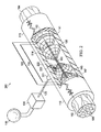
- FIG. 4 is a schematic, side view of three embodiments of the fiber guides shown in FIG. 3 ;
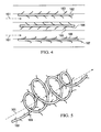
- FIG. 5 is a schematic, perspective view of a fourth embodiment of the fiber guides shown in FIG. 3 ;
- FIG. 6 is a schematic, perspective view of the system in FIGS. 1 and 2 showing the nerve conduit enclosing the damaged nerve;
- FIG. 7 is a schematic view of a fluid control system for the system shown in FIGS. 1 and 2 .
- a reduced pressure therapy system 100 for applying reduced pressure at a tissue site in the body of a patient to repair a defect such as, for example, a damaged nerve.
- the damaged nerve may have been pinched, partially disconnected or severed, or partially degenerated as a result of disease.
- the damaged nerve in FIG. 1 is a severed nerve 102 having a proximal segment 104 and a distal segment 106 relative to the central nervous system (CNS) of the patient.
- the severed nerve 102 has been damaged at a nerve damage site 108 that has been severed or degenerated.
- the severed nerve 102 may be branched or unbranched at the nerve damage site 108.
- nerve damage site refers to a wound or defect located on or within any nerve tissue, including, but not limited to, a disconnected or partially disconnected nerve, a degenerated or partially degenerated nerve, and a compressed or pinched nerve.
- reduced pressure tissue treatment may be used to enhance repair or regrowth of existing nerve tissue or to facilitate growth or grafted or transplanted nerve tissue and/or cells.
- the reduced pressure therapy system 100 comprises a nerve conduit 110 that surrounds the severed nerve 102 at the nerve damage site 108 with a section of the nerve conduit 110 removed to show the nerve damage site 108.
- the nerve conduit 110 is substantially tubular in shape and closes the nerve damage site 108 and a portion of the proximal segment 104 and the distal segment 106 that has not been damaged.
- the nerve conduit 110 has an inner surface 112 that forms a nerve gap 114 with the surface of the nerve damage site 108, i.e., a luminal space between the inner surface 112 of the nerve conduit 110 and the surface of the nerve damage site 108.
- the reduced pressure therapy system 100 also comprises a reduced pressure source 115 for providing a reduced pressure and a manifold 120 fluidly coupled to the reduced pressure source 115 via a first conduit 125.
- the manifold 120 is generally tubular or cylindrical in shape (see, e.g., the manifold disclosed in copending U.S. Patent Appln. No. 12/648,463 , incorporated herein by reference) and positioned within the nerve gap 114.
- the manifold 120 may have a variety of shapes depending on the type of nerve damage, and in this particular embodiment has a tubular shape to occupy a portion of the nerve gap 114.
- the manifold 120 may also contain scaffold material 134 that provides structure for tissue growth and repair.
- the reduced pressure therapy system 100 further comprises a canister 130 fluidly coupled between the reduced pressure source 115 and the manifold 120 via the conduit 125 to collect bodily fluids such as blood or exudate that are drawn from the scaffold 134 and the nerve gap 114.
- the reduced pressure source 115 and the canister 130 are integrated into a single housing structure.
- a further embodiment of a reduced-pressure system 200 is shown in FIG. 2 and is substantially similar to the reduced-pressure system 100.
- the reduced pressure therapy system 200 comprises a nerve conduit 110 that surrounds the severed nerve 102 at the nerve damage site 108 with a section of the nerve conduit 110 removed to show the nerve damage site 108.
- the nerve conduit 110 is substantially tubular in shape and closes the nerve damage site 108 and a portion of the proximal segment 104 and the distal segment 106 that has not been damaged.
- the nerve conduit 110 has an exterior surface 113 and an inner surface 112 that forms a nerve gap 114 with the surface of the nerve damage site 108, i.e., a luminal space between the inner surface 112 of the nerve conduit 110 and the surface of the nerve damage site 108.
- the reduced-pressure system 200 also comprises a reduced pressure source 115 for providing a reduced pressure and a manifold 220 fluidly coupled to the pressure source 115 via a first conduit 125.
- the manifold 220 (see, e.g., the manifold disclosed in copending U.S. Patent Appln. No. 12/648,458 , incorporated herein by reference) is contained within a manifold chamber 221 having a flange 222 extending from one end of the manifold chamber 221 for securing the manifold chamber 221 to the nerve conduit 110.
- the other end of the manifold chamber 221 is connected to the first conduit 125 so that the manifold 220 is held in fluid communication with the first conduit 125.
- the manifold chamber 221 may be constructed of any biocompatible material that is substantially impermeable to preserve the manifold's 220 fluid communication between the nerve gap 114 and the first conduit 125.
- the manifold chamber 221 is secured to the nerve conduit 110 by the flange 222 such that the manifold 220 is in fluid communication with the nerve gap 114 surrounding the surface of the nerve damage site 108, but positioned outside of the nerve gap 114.
- the flange 222 is secured to the nerve conduit 110 with an adhesive.
- the flange 222 is detachably secured to the nerve conduit 110 such that the flange 222 and manifold chamber 221 can be removed from the nerve conduit 110 after reduced pressure therapy is complete.
- the manifold 220 extends through the wall of the nerve conduit 110 in direct fluid contact with the nerve gap 114.
- the flange 222 is secured to the exterior surface 113 of the nerve conduit 110 so that the manifold 220 is positioned adjacent to the exterior surface 113 to be in fluid communication with the nerve gap 114 via the porous wall of the nerve conduit 110.
- Coupled includes direct coupling or indirect coupling via a separate object.
- the term “coupled” also encompasses two or more components that are continuous with one another by virtue of each of the components formed from the same piece of material.
- the term “coupled” may include chemical, mechanical, thermal, or electrical coupling.
- Fluid coupling means that fluid is in communication with designated parts or locations.
- reduced pressure generally refers to a pressure that is less than the ambient pressure at a tissue site that is subjected to treatment. In most cases, this reduced pressure will be less than the atmospheric pressure of the location at which the patient is located.
- vacuum and “negative pressure” may be used to describe the pressure applied to the tissue site, the actual pressure applied to the tissue site may be significantly greater than the pressure normally associated with a complete vacuum. Consistent with this nomenclature, an increase in reduced pressure or vacuum pressure refers to a relative reduction of absolute pressure, while a decrease in reduced pressure or vacuum pressure refers to a relative increase of absolute pressure.
- - ⁇ p means change in reduced pressure.
- a greater ( i.e., more negative) - ⁇ p means increased negative pressure (i.e., a greater change in pressure from ambient pressure).
- Reduced pressure treatment typically applies reduced pressure at -5 mm Hg to -500 mm Hg, more usually -5 to -300 mm Hg, including but not limited to -50, -125 or -175 mm Hg.
- Reduced pressure may be constant at a particular pressure level or may be varied over time. For example, reduced pressure may be applied and stopped periodically or ramped-up or - down over time.
- the systems 100 and 200 further comprise a scaffold structure 134 including one or more scaffold guides positioned within the nerve gap 114 in fluid communication with the manifold 20 on one or both sides of the manifold 20 such as, for example, scaffold guide 135.
- the scaffold guide 135 has a generally frusto-tubular shape with a base opening 136 at the base end of the frustum and a vertex opening 137 at the other end of the frustum.
- the scaffold guide 135 is positioned in the nerve gap 114 and oriented therein so that the vertex opening 137 is positioned closer to the manifold 20 than the base opening 136 which faces the proximal segment 104 when the vertex opening 137 is on the proximal side of the manifold 20 or faces the distal segment 106 when the vertex opening 137 is on the distal side of the manifold 20.
- the scaffold structure 134 of the systems 100, 200 comprise six scaffold guides (only four shown in FIGS.
- scaffold guides 135, 141, 143, 145, 147, and 149 (collectively the “scaffold guides") wherein the scaffold guides 135, 141, and 143 are positioned on the proximal side of the manifold 20 so that their vertex openings are closer to the manifold 20 than their base openings which face the proximal segment 104.
- a system according to the invention may, however, comprise 1, 2, 3, 4, 5, 6, 7, 8 or more scaffold guides.
- the scaffold guides 145, 147, and 149 are positioned on the distal side of the manifold 20 so that their vertex openings are closer to the manifold 20 than their base openings which face the distal segment 106.
- the scaffold guides may be formed of a scaffold fabric material or a web-like material 139 as illustrated by the concentric rings 139a and ribs 139b. In either embodiment, the scaffold guides function as nodes within the nerve gap 114 for protein absorption and the initialization point for fibril formation. The structure of the scaffold guides also wicks and directs slow-moving fluid within the nerve gap 114 from the base opening 136 toward the vertex opening 137 and the source of the reduced pressure, i.e., the manifold 20.
- the scaffold guides may be composed of any biocompatible material, but is preferably a bioabsorbable material.
- the scaffold structure 134 may further comprise one or more fiber guides 160 extending through the vertex openings of each of the scaffold guides from the proximal segment 104 to the distal segment 106 of the severed nerve 102.
- the fiber guides 160 may form a fiber bundle 162 also extending between the proximal segment 104 and the distal segment 106 including, but not limited to, as many as one hundred fiber guides 160.
- the fiber guides 160 may also be fluidly and/or mechanically coupled to the proximal segment 104 and/or the distal segment 106 of the severed nerve 102. Additionally, the fiber guide 160 may be fluidly and/or mechanically coupled to the manifold 20.
- the scaffold guides wick and direct fluid toward the vertex openings of the scaffold guides and the fiber guides 160 which facilitate fibril formation between the scaffold guides and ultimately extending between the base openings of the scaffold guides.
- fibril formation commences with direct fluid flow toward the vertex openings of the scaffold guides and along the fiber guides 160 as indicated by the fibers 151 that grow along the path created by the fiber guides 160.
- the fibers 151 may constitute either provisional matrix fibers such as but not limited to fibrin, collagens, proteoglycans, and laminin, or cellular based structures such as nerve fibers or supportive cell types.
- the provisional matrix and cellular based fibers may be derived either from endogenous host sources, or from the introduction of exogenous fluids. As fibril formation increases the density of the fibers 151, fibril formation expands outwardly between the base openings of the scaffold guides as indicated by fibers 153. Ultimately, fibers 155 begin forming between the scaffold guides having vertex openings facing each other, e.g., scaffold guides 135 and 145.
- the fiber guides 160 act to guide cell migration and growth through the entire scaffold structure 134 and may be composed of any biocompatible material such as a bioabsorbable material. In certain cases, the fiber guides are composed of a biological material such as collagen or fibrin.
- fiber guides 162, 164, and 166 comprise a strand of fiber material including protrusions for binding of proteins and cells from the slow-moving fluid within the nerve gap 114 to facilitate fibril formation. More specifically, fiber guides 162 and 164 include small barbs 163 and 165 extending from the strands of the fibril guides 162 and 164 in a direction with fluid flow and against fluid flow, respectively, depending upon the fluidics within the nerve gap 114.
- the fiber guide 166 includes protrusions in the shape of hooks 167 to facilitate protein binding and initiation sites of fibril formation through the nerve gap 114 without being aligned against fluid flow as are the barbs 165 of the fiber guide 164.
- the fiber protrusions 163, 165, and 167 may also be oriented in a direction extending either toward the proximal segment 104 or the distal segment 106 of the severed nerve 102 as may be required by the fluidics within the nerve gap 114.
- the fiber guides 162, 164, and 166 including their fiber protrusions 163, 165, and 167, respectively, are composed of the same materials described for use in other scaffold materials such as, for example, collagen or fibrin. Referring to FIG.
- a fiber guide 160 may comprise a strand of fiber material that has a form other than the linear form described above and shown in FIG. 4 .
- the strand of a fiber guide 168 is shaped in the form of a spiral having a longitudinal axis substantially parallel to the flow of fluid through the nerve gap 114 as indicated by dashed arrow 161.
- the fiber guide 168 may also have protrusions extending from the strand such as, for example, barbs 169 which are similar to the barbs 163 of the fiber guide 162.
- the nerve conduit 110 is shown in FIGS. 1 and 2 with a section removed to show the manifolds 120, 220 and is shown as completely surrounding the nerve damage sites 108 as a closed nerve conduit 410 in FIG. 6 .
- the nerve conduit 410 may be sealed by utilizing one or more stitches 415 or any other fastening device known in the art.
- the nerve conduit 410 may be composed of a bioabsorbable or a bioinert material.
- Materials that may be used for nerve conduits include, without limitation, polylactic acid (PLA), polyglycolic acid (PGA), polylactide-co-glycolide (PLGA), polyvinylpyrrolidone, polycaprolactone, polycarbonates, polyfumarates, caprolactones, polyamides, polysaccharides (including alginates ( e.g ., calcium alginate) and chitosan), hyaluronic acid, polyhydroxybutyrate, polyhydroxyvalerate, polydioxanone, polyorthoesthers, polyethylene glycols, poloxamers, polyphosphazenes, polyanhydrides, polyamino acids, polyacetals, polycyanoacrylates, polyurethanes, polyacrylates, ethylene-vinyl acetate polymers and other acyl substituted cellulose acetates and derivatives thereof, polystyrenes, polyvinyl chloride, polyvinyl fluoride, poly(viny
- a nerve conduit 110, 410 may be an unbroken substantially tubular structure fitted across a gap between a proximal and distal nerve stump such as depicted in FIG. 6 .
- substantially tubular nerve conduits also referred to as nerve guides, include without limitation NEURAGEN ® and NEUROFLEX TM collagen conduits.
- a nerve conduit may also be formed from a wrap that is positioned around a disconnected or damaged ( e.g ., pinched) nerve and sealed with a closure, such as a suture.
- wrap-type nerve conduits include, without limitation, NEUROMEND TM and NEURAWRAP TM collagen conduits.
- the nerve conduit is made of a material that encloses the damaged nerve, so as to exclude infiltration of non-nerve cells such as glia.
- the nerve conduit material is permeable, thereby allowing fluid and protein factors to diffuse through the conduit.
- the pores in a nerve conduit may be sufficiently small so as to exclude the entry of cells into the conduit lumen (e.g ., pores having an interior diameter or average interior diameter of between about 5 ⁇ m and 50 ⁇ m, 10 ⁇ m and 30 ⁇ m or 10 ⁇ m and 20 ⁇ m).
- the conduits comprise an internal diameter of less than about 6.0 mm, such as about 5, 4, 3, 2.5 or 2 mm.
- the reduced-pressure systems 100, 200, 700 may further comprise a pressure sensor 740 operably connected to the first conduit 125 to measure the reduced pressure being applied to the manifolds 120, 220.
- the systems 100, 200, 700 may further include a control unit 745 electrically connected to the pressure sensor 740 and the reduced pressure source 115.
- the pressure sensor 740 measures the reduced pressure within the nerve gap 114 and also may indicate whether the first conduit 125 is occluded with blood or other bodily fluids.
- the pressure sensor 740 also provides feedback to control unit 745 which regulates the reduced pressure therapy being applied by the reduced pressure source 115 through the first conduit 125 to the manifolds 120, 220.
- the reduced-pressure systems 100, 200, 700 may also comprise a fluid supply 750 fluidly coupled to the first conduit 125 via a second conduit 752 and operatively connected to the control unit 745.
- the fluid supply 750 may be used to deliver growth and/or healing agents to the nerve damage site 108 including, without limitation, an antibacterial agent, an antiviral agent, a cell-growth promotion agent, an irrigation fluid, antibodies or other chemically active agents.
- the systems 100, 200, 700 further comprises a first valve 754 positioned in the second conduit 752 to control the flow of fluid therethrough, and a second valve 756 positioned in the first conduit 125 between the reduced pressure supply 115 and the juncture between the first conduit 125 and the second conduit 752 to control the flow of reduced pressure.
- the control unit 745 is operatively connected to the first and second valves 754, 756 to control the delivery of reduced pressure and/or fluid from the fluid supply 750, respectively, to the manifolds 120, 220 as required by the particular therapy being administered to the patient.
- the fluid supply 150 may deliver the liquids as indicated above, but may also deliver air to the manifolds 120, 220 to promote healing and facilitate drainage at the site of the nerve damage site 108.
- manifold refers to a substance or structure that is provided to assist in directing reduced pressure to, delivering fluids to, or removing fluids from a tissue site.
- a manifold can include a plurality of flow channels or pathways that are interconnected to improve distribution of fluids provided to and removed from the area of tissue around the manifold.
- manifolds may include, without limitation, devices that have structural elements arranged to form flow channels, cellular foams such as open-cell foam, porous tissue collections, and liquids, gels and foams that include or cure to include flow channels.
- scaffold refers to a substance or structure applied to or in a wound or defect that provides a structural matrix for the growth of cells and/or the formation of tissue.
- a scaffold is often a three dimensional porous structure.
- the scaffold can be infused with, coated with, or comprised of cells, growth factors, extracellular matrix components, nutrients, integrins, or other substances to promote cell growth.
- a scaffold can take on characteristics of a manifold by directing flow through the matrix.
- a manifold can transmit flow to the scaffold and tissue; in the context of reduced pressure treatment, the manifold can be in fluid communication with the scaffold.
- the invention disclosed here discloses methods and apparatuses for controlling cellular-level based patterns of fluid flow that would allow for control of patterned protein organization at a microscopic, nanoscopic, or mesoscopic scale amenable to provide a structured manifold and, optionally, a scaffold material for cellular migration, differentiation, and like behavior necessary for functional regeneration of tissues.
- the methods, scaffolds, manifolds, flow sources and systems disclosed herein provide an active mechanism by which to promote the endogenous deposition of proteins and organization of the provisional matrix with biochemical and physical cues to direct cellular colonization of a scaffold or tissue space.
- the present invention thus enhances current technology by exploiting the active force of directed fluid flow, providing a framework upon which to design manifolds and scaffolds based upon the need of the biology under the influence of fluid flow.
- Flow vectors and pathways are utilized to enhance protein deposition and cellular colonization.
- the systems provided herein are designed to promote establishment of a provisional matrix network with a seamless transition from the healthy tissue edges through a scaffold or tissue site to promote a functional tissue continuum.
- the apparatuses, methods and systems disclosed herein provide a means for active guidance of tissue regeneration through an implanted scaffold or within a tissue site to promote functional recovery.
- This active guidance occurs through mechanisms of controlled fluid flow, which can be used to initiate or augment the early stages of the body's own natural healing process; a manifold can provide the active guidance necessary to create a controlled fluid flow.
- the controlled flow vectors that the manifolds provide can be used to facilitate the directed influx of cells and proteins into a scaffold. Creation of specific flow pathways within a tissue site or scaffold can lead to patterned deposition of proteins, such as collagen and fibrin within the manifold, scaffold or tissue space.
- Biochemical cues from cytokines, growth factors, and cells bound within the provisional matrix can work in conjunction with the natural physical cues of the provisional matrix and extracellular matrix to guide the subsequent migration of endogenous cells during the repair stages of healing. These cues act to establish a biological continuum that emanates from the healthy tissues and passes through the scaffolding or tissue space to facilitate a continuous guidance pathway for organized tissue regeneration.
- the invention concerns a new approach to wound healing, flow (or gradient) activated tissue engineering.
- this approach involves a source or generator of flow that forms a gradient for controlled movement of either endogenous or exogenous fluids into, out of, or through a tissue space for the organized deposition of proteins and/or spatial concentration of cytokines and growth factors, with subsequent formation of a directionally oriented provisional matrix.
- the tissue space being defined here includes, but is not limited to, the region surrounding a site of nerve tissue damage.
- Fluid flow into, through, or out of the nerve tissue space can be refined and directed through the inclusion of additional elements to the system including manifolds and/or scaffolds.
- the coordinated elements of the system are designed to create flow parameters, pathways, and patterns sufficiently detailed in scale as to be able to influence and direct the controlled adsorption of proteins, the organization of matrix, and organized colonization of specific cell types. Individual elements of the system are as follows.
- Source or Generator of Flow is induced into, through, or out of the nerve tissue space by methods or apparatuses that introduce changes in mechanical, chemical, and/or electrical potentials. These generators of flow provide either a gradient or a change in potential from the site or reservoir of endogenous or exogenous fluids to the placement position of the flow generator or its extension element ( i.e ., manifold or scaffold).
- the source of flow comprises a source of reduced pressure.
- Systems and apparatuses according to the invention may also comprise valves or arrays of valves that control the application and amount of negative pressure applied to the manifold.
- nerve conduits and/or manifolds described herein comprise a pressure sensor.
- the amount of negative pressure applied by a source is regulated based on the amount of negative pressure that is sensed in the manifold or nerve conduit or at the site of tissue damage.
- Manifold The flow generators are the driving force for stimulating the flow of fluids.
- Manifolds are apparatuses for refining the pattern of flow between the source or generator of flow and the tissue space.
- the macroscale level of flow is refined by specialized manifolds utilized for directed localization to a single point or to a plurality of selectively positioned points for creating initiation sites for microscale flow pathways within the manifold/scaffold and, ultimately, the tissue space.
- the manifold may also serve as a conduit for the removal of fluids from and as an apparatus for the delivery of exogenous fluids to the tissue space.
- a manifold generally refers to a physical substance or structure that serves to assist in applying and translating a mechanical, chemical, electrical or similar alterations into changes in the flow of a fluid, herein defined as the movement of liquids, gases, and other deformable substances such as proteins, cells, and other like moieties.
- this physical device includes a single point or plurality of points for the egress or evacuation of pressure, fluids, and like substances capable of translating the movement of fluids in a scaffold, as defined above. This can include, but is not limited to, the introduction of exogenous factors such as cells and/or therapeutic moieties into the scaffold through the lumen or plurality of lumens present in the manifold.
- a manifold includes a single point or plurality of points for the ingress or introduction of fluid from the scaffold back toward the point source of flow.
- Flow distributed by the manifold can direct the movement of endogenous proteins, growth factors, cytokines, and cells from their resident locations within the host to the tissue space or scaffold in an organized manner.
- the establishment of flow along these pathways leads to the deposition of proteins and provisional matrix that creates an interfacial endogenous network connecting the host to the scaffold. Extensions of this matrix can be established within the scaffold through selective positioning of the manifold flow initiation sites with flow promoting scaffolding designs.
- the organized protein deposition and provisional matrix provide a biochemical and physical framework that stimulates the attachment and migration of cells along directed pathways throughout the scaffold and the tissue space.
- the resulting endogenous network of proteins, growth factors, and cells provides a foundation upon which subsequent phases of the body's own tissue repair and regeneration mechanisms can build.
- Flow generating sources include, but are not limited to generators of negative pressure; generators of positive pressure; and generators of osmotic flow.
- the flow gradient established in the manifold may be further refined through the scaffold, to deliver a flow gradient to the scaffold to optimize flow through the scaffold as needed for the particular defect.
- Many of the embodiments disclosed herein are manifolds capable of translating changes in pressure and the like into controlled movement of fluids, optionally through a physical scaffold, for the purposes of directed tissue regeneration. These embodiments are generally specified for a particular application in the regeneration of specific tissues, but are not limited to a particular tissue therein.
- alterations in the aforementioned mechanical, chemical, or electrical impetus must be translated from the singular gradient source toward a physical substrate or scaffold to elicit cellular-level changes in protein adsorption, matrix organization, cell migration, and other tissue regeneration-related behaviors.
- These alterations are multivariate in nature and can include mechanical changes that elicit a physical change in pressure applied to the scaffold as applied to the site of the wound or desired site of tissue regeneration, chemical changes that elicit a gradient in protein and/or ion concentrations, which result in the creation of osmotic gradients capable of inducing flow, or electrical changes that create a gradient of current/ion exchange allowing for propagation of electrical signals from the point source.
- the manifold comprises a physical structure in close apposition to or within the contents of a scaffold and serves to propagate an alteration in a physical parameter, whether it be mechanical, chemical, electrical, or something similar in nature, for the means of directing these changes from its point source to the scaffolding material.
- the placement of this manifold with respect to its location with regard to that of the scaffold may be of crucial importance for facilitating controlled and directed regeneration of specific tissue types. For example, in peripheral nerve where regeneration primarily occurs in a unidirectional manner from the proximal to distal nerve stumps, it may be important to place the manifold along the length of a nerve conduit more toward it distal end to help direct regeneration toward that end. However, it may also be important to not place the manifold at the most distal aspect of the scaffold/conduit as soluble factors derived from the distal stump have been shown to be important for directing nerve regeneration toward its source.
- Manifolds may be composed of a bioabsorbable or bioinert material. Examples include non-bioabsorbable materials such as medical grade silicone polymers, metals, polyvinylchloride (PVC), and polyurethane. Bioabsorbable polymers such as collagen, polylactic acid (PLA), polyglycolic acid (PGA), polylactide-co-glycolide (PLGA), a polysaccharide, a hydroxyapatite, or a polyethylene glycol, or combinations thereof, can also be used. Some manifolds are also a mix of non-bioresorbable and bioresorbable materials. In general material used for a scaffold may also be used to compose a manifold and such materials are further detailed below. In certain aspects, manifold materials are structured to include a high void fraction for improved bioabsorption properties.
- PVC polyvinylchloride
- Bioabsorbable polymers such as collagen, polylactic acid (PLA), polyglycolic acid (PGA), polyl
- Manifold support structures such as a manifold chamber and/or flange may be composed of any acceptable biocompatible material.
- a support structure will typically be impermeable and surround the manifold so as to maintain manifold pressure.
- a portion of the support couples the manifold and the nerve conduit.
- a flange is attached to the exterior surface of a nerve conduit with an adhesive such as a fibrin glue, cyanoacrylate, or other biologically derived adhesive.
- a support may also be connected to a nerve conduit via reversible mechanisms other than an adhesive, such as chemical, thermal, osmotic, mechanical (snap or other interference fit, threaded, etc), magnetic, and electrostatic mechanisms.
- the manifold may be used to deliver agents that reverse the action of the binding mechanism in order to detach the support from the nerve conduit (e.g., upon completion of therapy). For example, electrostatic binding may be released through introduction of salt solutions or biocompatible solvents may be used to release adhesives.
- a scaffold for use according to the invention is coupled to a manifold, provides physical guidance to direct the pathway of fluid flow in the tissue site, creating avenues for the movement and migration of adhesive proteins and cells, respectively, which are integral to the establishment of a provisional matrix in predetermined patterns of organization within the tissue space.
- the methods and apparatuses described for fluid flow-induced and gradient-induced generation of tissues have direct implications into the design of the scaffolds.
- scaffolds serve to refine the pathways of fluid flow within the tissue space to cellular level patterns from the fluid source to the point(s) of flow initiation within the manifold.
- a scaffold may embody characteristics of a manifold or be combined in conjunction with a manifold for refinement of the flow pathways within the tissue site.
- a scaffold is a reticulated structure comprising high void fraction for improved bioabsorption properties.
- Scaffolds may also comprise retention structures as described herein such as funnel guides and fiber guides.
- funnel guides may be used to direct the diffusion and/or migration of cells or growth factors at a site of nerve damage.
- multiple funnel guides such as 2, 3, 4, 5, 6, 7, 8 or more funnel guides are comprised in a scaffold.
- a funnel guide may be composed of a hydrophobic material and may be bioabsorbable so as to degrade as nerve tissue grows into the site of the nerve damage.
- Funnel guides may also be hydrophilic to assist in the directed movement of slow moving fluids e.g., by wicking.
- Funnel guides may also have bioabsorption properties that differ from the narrow to the wide end of the funnel guide.
- the narrow end of the funnel guide may be absorbed at a faster rate so that the aperture of the narrow end becomes wider as tissue regrowth occurs.
- funnel guides closer to the proximal end of the nerve damage site may be composed of a material that is absorbed at a faster rate so that funnel structures closer to the proximal end of a nerve damage site are absorbed more rapidly.
- Fiber guides in scaffolds may also be composed of bioabsorbable material such that the guides are absorbed as tissue growth or regrowth occurs.
- fiber guides may comprise protrusions (e.g ., barbs) or hook structures and may be essentially linear or form a spiral as they extend through the scaffold at the nerve damage site.
- the fiber guides and associated structures e.g ., fiber protrusions and hooks
- the fiber structures comprise bioactive molecules as part of their structure.
- fiber structures may comprise growth factors that enhance cell growth along the length of the fibers or binding moieties (such as antibodies) that bind cells or growth factors to the fibers to enhance the growth or regrowth of nerve tissue.
- Nonlimiting examples of suitable scaffold, funnel and fiber materials include extracellular matrix proteins such as fibrin, collagen or fibronectin, and synthetic or naturally occurring polymers, including bioabsorbable or non-bioabsorbable polymers, such as polylactic acid (PLA), polyglycolic acid (PGA), polylactide-co-glycolide (PLGA), polyvinylpyrrolidone, polycaprolactone, polycarbonates, polyfumarates, caprolactones, polyamides, polysaccharides (including alginates ( e.g ., calcium alginate) and chitosan), hyaluronic acid, polyhydroxybutyrate, polyhydroxyvalerate, polydioxanone, polyethylene glycols, poloxamers, polyphosphazenes, polyanhydrides, polyamino acids, polyortho esters, polyacetals, polycyanoacrylates, polyurethanes, polyacrylates, ethylene-vinyl acetate polymers and other acy
- the scaffold can also comprise ceramics such as hydroxyapatite, coralline apatite, calcium phosphate, calcium sulfate, calcium carbonate or other carbonates, bioglass, allografts, autografts, xenografts, decellularized tissues, or composites of any of the above.
- the scaffold comprises collagen, polylactic acid (PLA), polyglycolic acid (PGA), polylactide-co-glycolide (PLGA), a polyurethane, a polysaccharide, an hydroxyapatite, or a polytherylene glycol.
- the scaffold can comprise combinations of any two, three or more materials, either in separate areas of the scaffold, or combined noncovalently, or covalently (e.g ., copolymers such as a polyethylene oxide-polypropylene glycol block copolymers, or terpolymers), or combinations thereof.
- Suitable matrix materials are discussed in, for example, Ma and Elisseeff, 2005, and Saltzman, 2004.
- the apparatuses and methods according to the invention concern bioactive agents.
- Bioactive agents may, in some cases, be incorporated directly onto a manifold or scaffold material ( i.e ., to generate a bioactive manifold and/or scaffold).
- agents that facilitate tissue growth such as collagen or fibrin may be directly incorporated onto or into a manifold or scaffold material.
- immune regulator agents such as rapamycin may be incorporated into manifold or scaffold structures.
- soluble bioactive agents may be introduced at a site of tissue damage by virtue of flow through the tissue site.
- a manifold may be in fluid communication with a fluid source and a bioactive agent may be introduced into the fluid source and thereby into the manifold and damaged nerve tissue.
- Nonlimiting examples of useful bioactive growth factors for various applications are growth hormone (GH), a bone morphogenetic protein (BMP), transforming growth factor- ⁇ (TGF- ⁇ ), a TGF- ⁇ , a fibroblast growth factor (FGF), granulocyte-colony stimulating factor (G-CSF), granulocyte/macrophage-colony stimulating factor (GM-CSF), epidermal growth factor (EGF), platelet derived growth factor (PDGF), insulin-like growth factor (IGF), vascular endothelial growth factor (VEGF), hepatocyte growth factor/scatter factor (HGF/SF), an interleukin, tumor necrosis factor- ⁇ (TNF- ⁇ ) or nerve growth factor (NGF).
- GH growth hormone
- BMP bone morphogenetic protein
- TGF- ⁇ transforming growth factor- ⁇
- TGF- ⁇ TGF- ⁇
- FGF fibroblast growth factor
- G-CSF granulocyte-colony stimulating factor
- GM-CSF
- Nerve tissue repair and engineering The apparatuses and systems disclosed herein can be used for nerve tissue repair and engineering in various contexts including the following.
- a generator of flow may be combined with manifolds and/or scaffolds to direct the regeneration of lost tissue at a site of injury or compromised function.
- Tissues lost from traumatic injury, surgery, bums, or other causes e.g ., infection or autoimmune disease
- Functional nerve tissue is directed to regenerate.
- a generator of flow may be combined with manifolds and/or scaffolds to retard disease progression of an affected nerve tissue such as occurs, e.g ., in autoimmune disease.
- a generator of flow may be combined with manifolds and/or scaffolds to maintain the viability of explanted tissues either for in vitro study, ex vivo scaffold or implant preparation, or in vivo transplant.
- a generator of flow combined with a manifold may be used to provide fluid flow to the tissue and to control waste removal from the tissue.
- a generator of flow may be combined with manifolds and/or scaffolds to promote the expansion of existing tissues.
- the methods, scaffolds, manifolds, flow sources and systems of the invention can be used to direct the growth of tissues where additional tissue quantity is needed or desired
- a generator of flow may be combined with manifolds and/or scaffolds to accelerate the rate of tissue formation within a natural healing response.
- the methods, scaffolds, manifolds, flow sources and systems of the invention may be used to accelerate tissue growth by augmenting formation of provisional matrices, facilitating its stable positioning, and aiding in recruitment of cells to the tissue space.
- a generator of flow may be combined with manifolds and/or scaffolds to stimulate differentiation of stem cells or other pluripotent cells into specific lineages.
- Application of flow using the methods, scaffolds, manifolds, flow sources and systems of the invention may be used to direct pluripotent cells into specific cell lineages needed to foster growth in the tissue space.
- a generator of flow may be combined with manifolds and/or scaffolds to introduce exogenous growth factors, proteins, cells, or pharmaceutical agents into the tissue space to augment tissue repair, regeneration, and/or maintenance.
- a generator of flow may be combined with manifolds and/or scaffolds to facilitate formation of matrices in vitro that may subsequently be used for in vivo transplantation.
- a generator of flow may be combined with manifolds and/or scaffolds to promote integration of transplanted tissue into the host environment. This can be applied to autograft, allograft, or xenograft transplants.
- a flow generator may be combined with manifolds and/or scaffolds to guide the directed deposition and orientation of ECM expressed by cells and tissues.
- the directed orientation of ECM has an impact in organizing and directing the attachment and colonization of subsequent cell layers and tissues.
Claims (15)
- Appareil pour appliquer une pression réduite à un défaut sur un site tissulaire d'un nerf, l'appareil comprenant :un conduit nerveux ayant une paroi luminale pour entourer le site tissulaire et former un espace luminal entre le site tissulaire et la paroi luminale ;un collecteur ayant un raccord pour recevoir une pression réduite afin de distribuer cette pression réduite au défaut ; etun squelette ayant une forme tronconique tubulaire s'étendant entre une ouverture de base à une extrémité et une ouverture au sommet à l'autre extrémité, le squelette étant positionné dans l'espace luminal avec l'ouverture au sommet orientée plus près du collecteur.
- Appareil selon la revendication 1, dans lequel le défaut est un nerf sectionné.
- Appareil selon la revendication 1, dans lequel le défaut est un nerf pincé, partiellement sectionné ou dégénérescent s'étendant à travers l'ouverture au sommet du squelette.
- Appareil selon la revendication 1, dans lequel le squelette comprend des pores qui sont suffisamment petits pour se lier à des cellules et à des protéines.
- Appareil selon la revendication 1, dans lequel le squelette comprend par ailleurs au moins un guide-fibre s'étendant à travers l'espace luminal et l'ouverture au sommet du squelette.
- Appareil selon la revendication 5, dans lequel le au moins un guide-fibre comprend des saillies fibreuses s'étendant dans l'espace luminal.
- Appareil selon la revendication 5, dans lequel le au moins un guide-fibre est constitué de collagène ou de fibrine.
- Appareil selon la revendication 1, dans lequel le collecteur est positionné adjacent au côté distal du nerf .
- Appareil selon la revendication 1, dans lequel le collecteur a une forme sensiblement tubulaire ou cylindrique et est positionné à l'intérieur de l'espace luminal.
- Appareil selon la revendication 1, dans lequel le collecteur s'étend à travers la paroi du conduit nerveux en communication de fluide avec l'espace luminal.
- Appareil selon la revendication 1, dans lequel le squelette comprend un agent bioactif.
- Appareil selon la revendication 11, dans lequel l'agent bioactif est au moins un agent choisi parmi un antibiotique, un anticorps et un facteur de croissance.
- Appareil selon la revendication 1, dans lequel le conduit nerveux est composé d'un matériau bioabsorbable.
- Appareil selon la revendication 1, dans lequel le conduit nerveux comprend des pores suffisamment petits pour exclure l'entrée de cellules provenant du tissu entourant le conduit nerveux dans l'espace luminal.
- Produit pour appliquer une pression réduite à un défaut sur un site tissulaire d'un nerf, le produit comprenant :une source de pression pour fournir une pression réduite ; etun appareil selon l'une quelconque des revendications précédentes,dans lequel le collecteur est raccordé à la source de pression.
Applications Claiming Priority (5)
| Application Number | Priority Date | Filing Date | Title |
|---|---|---|---|
| US14205308P | 2008-12-31 | 2008-12-31 | |
| US14206508P | 2008-12-31 | 2008-12-31 | |
| US23469209P | 2009-08-18 | 2009-08-18 | |
| US23877009P | 2009-09-01 | 2009-09-01 | |
| PCT/US2009/069715 WO2010078347A2 (fr) | 2008-12-31 | 2009-12-29 | Système pour générer un écoulement de fluide dans les tissus nerveux |
Publications (3)
| Publication Number | Publication Date |
|---|---|
| EP2370142A2 EP2370142A2 (fr) | 2011-10-05 |
| EP2370142A4 EP2370142A4 (fr) | 2012-09-05 |
| EP2370142B1 true EP2370142B1 (fr) | 2013-07-31 |
Family
ID=42285800
Family Applications (5)
| Application Number | Title | Priority Date | Filing Date |
|---|---|---|---|
| EP09837119.8A Active EP2370118B1 (fr) | 2008-12-31 | 2009-12-29 | Système pour générer un écoulement de fluide dans les tissus nerveux |
| EP14197314.9A Active EP2848268B1 (fr) | 2008-12-31 | 2009-12-29 | Échafaudages de rouleau de tissu |
| EP09837117.2A Active EP2370117B1 (fr) | 2008-12-31 | 2009-12-29 | Système pour appliquer un débit de fluide aux tissus nerveux |
| EP09837122.2A Active EP2370119B1 (fr) | 2008-12-31 | 2009-12-29 | Échafaudage pour tissus enroulés |
| EP09837120.6A Active EP2370142B1 (fr) | 2008-12-31 | 2009-12-29 | Système pour générer un écoulement de fluide dans les tissus nerveux |
Family Applications Before (4)
| Application Number | Title | Priority Date | Filing Date |
|---|---|---|---|
| EP09837119.8A Active EP2370118B1 (fr) | 2008-12-31 | 2009-12-29 | Système pour générer un écoulement de fluide dans les tissus nerveux |
| EP14197314.9A Active EP2848268B1 (fr) | 2008-12-31 | 2009-12-29 | Échafaudages de rouleau de tissu |
| EP09837117.2A Active EP2370117B1 (fr) | 2008-12-31 | 2009-12-29 | Système pour appliquer un débit de fluide aux tissus nerveux |
| EP09837122.2A Active EP2370119B1 (fr) | 2008-12-31 | 2009-12-29 | Échafaudage pour tissus enroulés |
Country Status (12)
| Country | Link |
|---|---|
| US (11) | US8257372B2 (fr) |
| EP (5) | EP2370118B1 (fr) |
| JP (4) | JP5472758B2 (fr) |
| KR (4) | KR20110102929A (fr) |
| CN (4) | CN102264405B (fr) |
| AU (4) | AU2009335117B2 (fr) |
| CA (4) | CA2745462C (fr) |
| MX (4) | MX2011006991A (fr) |
| RU (4) | RU2011122544A (fr) |
| SG (4) | SG172021A1 (fr) |
| TW (4) | TW201029686A (fr) |
| WO (4) | WO2010078349A2 (fr) |
Cited By (17)
| Publication number | Priority date | Publication date | Assignee | Title |
|---|---|---|---|---|
| US10660994B2 (en) | 2012-03-12 | 2020-05-26 | Smith & Nephew Plc | Reduced pressure apparatus and methods |
| USD898925S1 (en) | 2018-09-13 | 2020-10-13 | Smith & Nephew Plc | Medical dressing |
| US10898388B2 (en) | 2015-04-27 | 2021-01-26 | Smith & Nephew Plc | Reduced pressure apparatuses and methods |
| US11096831B2 (en) | 2016-05-03 | 2021-08-24 | Smith & Nephew Plc | Negative pressure wound therapy device activation and control |
| US11116669B2 (en) | 2016-08-25 | 2021-09-14 | Smith & Nephew Plc | Absorbent negative pressure wound therapy dressing |
| US11123471B2 (en) | 2017-03-08 | 2021-09-21 | Smith & Nephew Plc | Negative pressure wound therapy device control in presence of fault condition |
| US11160915B2 (en) | 2017-05-09 | 2021-11-02 | Smith & Nephew Plc | Redundant controls for negative pressure wound therapy systems |
| US11173240B2 (en) | 2016-05-03 | 2021-11-16 | Smith & Nephew Plc | Optimizing power transfer to negative pressure sources in negative pressure therapy systems |
| US11285047B2 (en) | 2016-04-26 | 2022-03-29 | Smith & Nephew Plc | Wound dressings and methods of use with integrated negative pressure source having a fluid ingress inhibition component |
| US11305047B2 (en) | 2016-05-03 | 2022-04-19 | Smith & Nephew Plc | Systems and methods for driving negative pressure sources in negative pressure therapy systems |
| US11497653B2 (en) | 2017-11-01 | 2022-11-15 | Smith & Nephew Plc | Negative pressure wound treatment apparatuses and methods with integrated electronics |
| US11554203B2 (en) | 2017-11-01 | 2023-01-17 | Smith & Nephew Plc | Negative pressure wound treatment apparatuses and methods with integrated electronics |
| US11564847B2 (en) | 2016-09-30 | 2023-01-31 | Smith & Nephew Plc | Negative pressure wound treatment apparatuses and methods with integrated electronics |
| US11564845B2 (en) | 2017-09-13 | 2023-01-31 | Smith & Nephew Plc | Negative pressure wound treatment apparatuses and methods with integrated electronics |
| US11701265B2 (en) | 2017-09-13 | 2023-07-18 | Smith & Nephew Plc | Negative pressure wound treatment apparatuses and methods with integrated electronics |
| US11707564B2 (en) | 2017-11-01 | 2023-07-25 | Smith & Nephew Plc | Safe operation of integrated negative pressure wound treatment apparatuses |
| US11723809B2 (en) | 2016-03-07 | 2023-08-15 | Smith & Nephew Plc | Wound treatment apparatuses and methods with negative pressure source integrated into wound dressing |
Families Citing this family (98)
| Publication number | Priority date | Publication date | Assignee | Title |
|---|---|---|---|---|
| EP2085049B1 (fr) * | 2003-08-11 | 2019-06-19 | Cook Medical Technologies LLC | Implant chirurgical avec pointe pénétrante |
| US9820888B2 (en) | 2006-09-26 | 2017-11-21 | Smith & Nephew, Inc. | Wound dressing |
| WO2008140413A1 (fr) * | 2007-05-15 | 2008-11-20 | NEUROGEN MEDICAL INNOVATION I UMEå | Conduit de réparation de nerfs à base de fibrine et sa méthode d'obtention |
| GB0804654D0 (en) | 2008-03-13 | 2008-04-16 | Smith & Nephew | Vacuum closure device |
| MX2011006991A (es) * | 2008-12-31 | 2011-08-04 | Kci Licensing Inc | Andamios en rollo para tejido. |
| GB0902368D0 (en) | 2009-02-13 | 2009-04-01 | Smith & Nephew | Wound packing |
| DE102009057962B4 (de) * | 2009-12-11 | 2012-09-20 | Karlsruher Institut für Technologie | Nervenprothese und Verfahren zur Herstellung einer Nervenprothese |
| EP2539013B1 (fr) * | 2010-02-23 | 2018-04-04 | L-Vad Technology Inc. | Appareil percutané assisté par le vide |
| US8623047B2 (en) | 2010-04-30 | 2014-01-07 | Kci Licensing, Inc. | System and method for sealing an incisional wound |
| KR101679426B1 (ko) * | 2010-04-30 | 2016-11-25 | 에스케이이노베이션 주식회사 | 미전환유를 이용한 고급 윤활기유의 제조 방법 |
| US8758374B2 (en) | 2010-09-15 | 2014-06-24 | University Of Utah Research Foundation | Method for connecting nerves via a side-to-side epineurial window using artificial conduits |
| US20160346444A1 (en) * | 2010-09-24 | 2016-12-01 | Kci Licensing, Inc. | Cellular control and tissue regeneration systems and methods |
| US9408956B2 (en) * | 2010-09-24 | 2016-08-09 | Kci Licensing, Inc. | Cellular control and tissue regeneration systems and methods |
| CA2821515A1 (fr) | 2010-12-01 | 2012-06-07 | Alder Biopharmaceuticals, Inc. | Compositions anti-ngf et leur utilisation |
| US9078878B2 (en) | 2010-12-01 | 2015-07-14 | Alderbio Holdings Llc | Anti-NGF antibodies that selectively inhibit the association of NGF with TrkA, without affecting the association of NGF with p75 |
| US9884909B2 (en) | 2010-12-01 | 2018-02-06 | Alderbio Holdings Llc | Anti-NGF compositions and use thereof |
| US9539324B2 (en) | 2010-12-01 | 2017-01-10 | Alderbio Holdings, Llc | Methods of preventing inflammation and treating pain using anti-NGF compositions |
| US11214610B2 (en) | 2010-12-01 | 2022-01-04 | H. Lundbeck A/S | High-purity production of multi-subunit proteins such as antibodies in transformed microbes such as Pichia pastoris |
| US9067988B2 (en) | 2010-12-01 | 2015-06-30 | Alderbio Holdings Llc | Methods of preventing or treating pain using anti-NGF antibodies |
| EP2670312B1 (fr) | 2011-02-04 | 2020-11-18 | University of Massachusetts | Dispositif de fermeture de plaie par pression négative |
| US9421132B2 (en) | 2011-02-04 | 2016-08-23 | University Of Massachusetts | Negative pressure wound closure device |
| US9302034B2 (en) | 2011-04-04 | 2016-04-05 | Smith & Nephew, Inc. | Negative pressure wound therapy dressing |
| WO2012142473A1 (fr) | 2011-04-15 | 2012-10-18 | University Of Massachusetts | Système de drainage et fermeture de cavité chirurgicale |
| EP2714111B1 (fr) | 2011-05-31 | 2021-03-17 | LifeCell Corporation | Matrices de tissu adipeux |
| WO2013066619A1 (fr) * | 2011-10-17 | 2013-05-10 | University Of Utah Research Foundation | Procédés et dispositifs de liaison de nerfs |
| US10842494B2 (en) | 2011-10-17 | 2020-11-24 | University Of Utah Research Foundation | Methods and devices for connecting nerves |
| CA2855972C (fr) * | 2011-11-18 | 2019-08-27 | Kci Licensing, Inc. | Systemes et procedes de traitement de tissus comprenant un substrat poreux dote d'une region comprimee et d'une region expansee |
| WO2015008054A1 (fr) | 2013-07-16 | 2015-01-22 | Smith & Nephew Plc | Appareil pour le traitement des plaies |
| CN104661601B (zh) | 2012-05-22 | 2018-06-22 | 史密夫及内修公开有限公司 | 用于伤口治疗的设备和方法 |
| EP2852419B1 (fr) | 2012-05-22 | 2019-11-20 | Smith & Nephew plc | Dispositif de fermeture de blessure |
| JP6285421B2 (ja) | 2012-05-24 | 2018-02-28 | スミス アンド ネフュー インコーポレイテッド | 陰圧により創傷を治療および閉鎖するデバイスおよび方法 |
| US11090338B2 (en) | 2012-07-13 | 2021-08-17 | Lifecell Corporation | Methods for improved treatment of adipose tissue |
| MX369689B (es) | 2012-07-16 | 2019-11-19 | Smith & Nephew Inc | Dispositivo de cierre de herida de presión negativa. |
| AU2013323747B2 (en) | 2012-09-26 | 2017-02-02 | Lifecell Corporation | Processed adipose tissue |
| TWI507181B (zh) * | 2012-10-30 | 2015-11-11 | Univ Nat Cheng Kung | 腹腔內力學刺激製造高強度組織工程材料的系統 |
| US9968488B2 (en) | 2012-11-12 | 2018-05-15 | Kci Usa, Inc. | Externally-applied patient interface system and method |
| DE102012025124A1 (de) | 2012-12-21 | 2014-06-26 | Paul Hartmann Ag | Saugkörper zur therapeutischen Behandlung einer Wunde mittels Unterdruck |
| EP3659633A1 (fr) * | 2013-02-06 | 2020-06-03 | LifeCell Corporation | Procédés pour modification localisée de produits tissulaires |
| US10085892B2 (en) * | 2013-03-07 | 2018-10-02 | Life Sciences Llc | Apparatus and method for wound infection prevention |
| CA2902776C (fr) | 2013-03-13 | 2023-03-07 | Smith & Nephew Inc. | Appareil de traitement des plaies et son utilisation |
| RU2015142877A (ru) | 2013-03-14 | 2017-04-18 | СМИТ ЭНД НЕФЬЮ ПиЭлСи | Сжимаемые наполнители раны и системы и способы их применения в лечении ран с применением отрицательного давления |
| US20140276654A1 (en) * | 2013-03-15 | 2014-09-18 | Acclarent, Inc. | Nasal Suction Device |
| US9604041B2 (en) | 2013-03-15 | 2017-03-28 | Acclarent, Inc. | Nasal fluid management device |
| US9408955B2 (en) | 2013-03-15 | 2016-08-09 | Acclarent, Inc. | Nasal fluid management device |
| US9408756B2 (en) | 2013-03-15 | 2016-08-09 | Acclarent, Inc. | Nasal fluid management device |
| EP2986448B1 (fr) * | 2013-04-17 | 2019-12-11 | Mölnlycke Health Care AB | Pansements pour plaies |
| US10660992B2 (en) | 2013-10-21 | 2020-05-26 | Smith & Nephew, Inc. | Negative pressure wound closure device |
| RU2016133735A (ru) | 2014-01-21 | 2018-02-28 | СМИТ ЭНД НЕФЬЮ ПиЭлСи | Сжимаемая повязка для лечения раны отрицательным давлением |
| MX2016009476A (es) | 2014-01-21 | 2016-10-13 | Smith & Nephew | Aparatos para tratamiento de heridas. |
| CN105213076A (zh) * | 2014-06-12 | 2016-01-06 | 微创心脉医疗科技(上海)有限公司 | 一种人造瘤颈及其制备方法 |
| CN104127215B (zh) * | 2014-07-18 | 2016-12-07 | 韦有芳 | 一种双层的硅胶神经导管 |
| CN107921237A (zh) | 2015-04-27 | 2018-04-17 | 反射医学公司 | 交感神经心肺神经调节系统和方法 |
| EP4209201A1 (fr) | 2015-04-29 | 2023-07-12 | Smith & Nephew, Inc. | Dispositif de fermeture de plaie par pression négative |
| BR112018001647A2 (pt) | 2015-07-29 | 2018-09-18 | Innovative Therapies, Inc. | sistema de controle e monitoramento de pressão de dispositivo de terapia de feridas |
| EP3349705A4 (fr) * | 2015-09-17 | 2019-05-22 | Hollister Incorporated | Méthode et système d'administration de soin des plaies à base d'échafaudage |
| US10814049B2 (en) | 2015-12-15 | 2020-10-27 | University Of Massachusetts | Negative pressure wound closure devices and methods |
| US10575991B2 (en) | 2015-12-15 | 2020-03-03 | University Of Massachusetts | Negative pressure wound closure devices and methods |
| US11246879B2 (en) | 2016-02-09 | 2022-02-15 | Tulai Therapeutics, Inc. | Methods, agents, and devices for local neuromodulation of autonomic nerves |
| EP3419715B1 (fr) * | 2016-02-26 | 2021-09-15 | Neuronano AB | Méthode d'implantation d'agrégats cellulaires et de fragments tissulaires |
| EP3463500A1 (fr) | 2016-06-03 | 2019-04-10 | LifeCell Corporation | Procédés de modification localisée de produits tissulaires |
| AU2017290353B2 (en) | 2016-06-29 | 2023-05-18 | Tulavi Therapeutics, Inc. | Treatment of sepsis and related inflammatory conditions by local neuromodulation of the autonomic nervous system |
| ES2841427T3 (es) | 2016-07-05 | 2021-07-08 | Lifecell Corp | Matrices de tejido que incorporan múltiples tipos de tejido |
| WO2018041805A1 (fr) | 2016-08-30 | 2018-03-08 | Smith & Nephew Plc | Systèmes pour appliquer une thérapie à pression réduite |
| EP3518847B1 (fr) | 2016-09-27 | 2023-03-01 | Smith & Nephew plc | Dispositifs de fermeture de plaie à des parties solubles |
| JP2019532769A (ja) * | 2016-10-28 | 2019-11-14 | ザ ジェネラル ホスピタル コーポレイション | 組織移植片を組み立てるためのシステムおよび方法 |
| JP2019532774A (ja) | 2016-11-02 | 2019-11-14 | スミス アンド ネフュー インコーポレイテッド | 創傷閉鎖器具 |
| US11045583B2 (en) | 2016-12-22 | 2021-06-29 | Lifecell Corporation | Devices and methods for tissue cryomilling |
| CN108452377A (zh) * | 2017-02-21 | 2018-08-28 | 北京中科再康生物技术有限公司 | 用于修复神经损伤的生长因子复合胶原支架材料及功能胶原支架材料 |
| CN107126290B (zh) * | 2017-04-26 | 2021-07-09 | 吴恩德 | 一种实验动物用人工视神经套管 |
| US11324876B2 (en) | 2017-06-13 | 2022-05-10 | Smith & Nephew Plc | Collapsible structure and method of use |
| CN110662516B (zh) | 2017-06-13 | 2022-02-22 | 史密夫及内修公开有限公司 | 伤口闭合装置和使用方法 |
| CA3063859A1 (fr) | 2017-06-14 | 2018-12-20 | Smith & Nephew, Inc. | Gestion de l'elimination de liquide et maitrise de la fermeture d'une plaie dans un traitement de plaie |
| US11395873B2 (en) | 2017-06-14 | 2022-07-26 | Smith & Nephew, Inc. | Control of wound closure and fluid removal management in wound therapy |
| EP3638174A1 (fr) | 2017-06-14 | 2020-04-22 | Smith & Nephew plc | Feuille pliable pour fermeture de plaie et procédé d'utilisation |
| EP3638170B1 (fr) | 2017-06-14 | 2024-03-13 | Smith & Nephew PLC | Structure pliable pour fermeture de plaie et méthode d'utilisation |
| KR102653455B1 (ko) * | 2017-07-26 | 2024-04-02 | 뉴랩티브 테라퓨틱스, 인크. | 치료제를 신경에 투여하기 위한 유체 격납 필드를 생성하는 장치 및 방법 |
| US11607344B2 (en) | 2017-07-27 | 2023-03-21 | Smith & Nephew Plc | Customizable wound closure device and method of use |
| US11590030B2 (en) | 2017-08-07 | 2023-02-28 | Smith & Nephew Plc | Wound closure device with protective layer and method of use |
| US11375923B2 (en) | 2017-08-29 | 2022-07-05 | Smith & Nephew Plc | Systems and methods for monitoring wound closure |
| JPWO2019054407A1 (ja) * | 2017-09-15 | 2020-10-29 | 東洋紡株式会社 | 神経保護材 |
| US11123375B2 (en) | 2017-10-18 | 2021-09-21 | Lifecell Corporation | Methods of treating tissue voids following removal of implantable infusion ports using adipose tissue products |
| CN111201046B (zh) | 2017-10-18 | 2022-06-28 | 生命细胞公司 | 脂肪组织产品以及产生方法 |
| US11246994B2 (en) | 2017-10-19 | 2022-02-15 | Lifecell Corporation | Methods for introduction of flowable acellular tissue matrix products into a hand |
| US11826488B2 (en) | 2017-10-19 | 2023-11-28 | Lifecell Corporation | Flowable acellular tissue matrix products and methods of production |
| KR102023574B1 (ko) * | 2018-04-13 | 2019-09-24 | 한국과학기술연구원 | 말초 신경 고정 장치 |
| CN112638437B (zh) * | 2018-07-02 | 2023-12-08 | 图拉维治疗股份有限公司 | 原位形成神经帽的方法和装置 |
| US10292709B1 (en) * | 2018-11-13 | 2019-05-21 | King Saud University | Device for sutureless repair of an injured nerve |
| WO2020124038A1 (fr) | 2018-12-13 | 2020-06-18 | University Of Massachusetts | Dispositifs et méthodes de fermeture de plaie par pression négative |
| EP3976127A1 (fr) | 2019-05-30 | 2022-04-06 | LifeCell Corporation | Implant mammaire biologique |
| CN110227184B (zh) * | 2019-07-16 | 2020-04-24 | 南通大学 | 差异性组织工程化神经及应用 |
| CN110403727B (zh) * | 2019-08-01 | 2021-05-14 | 吉林大学 | 一种坐骨神经慢性压迫模型的构建装置 |
| CN115361912A (zh) * | 2020-01-13 | 2022-11-18 | 图拉维治疗股份有限公司 | 原位形成具有快速释放的神经帽的方法和装置 |
| EP3919005A1 (fr) * | 2020-06-05 | 2021-12-08 | Université de Strasbourg | Dispositif de réparation nerveuse |
| EP4196064A1 (fr) * | 2020-08-14 | 2023-06-21 | KCI Manufacturing Unlimited Company | Bandage de décompression |
| US20230389926A1 (en) * | 2020-10-16 | 2023-12-07 | The Johns Hopkins University | Biodegradable nanofiber conical conduits for nerve repair |
| WO2023014726A1 (fr) * | 2021-08-02 | 2023-02-09 | Mary Hitchcock Memorial Hospital, For Itself And On Behalf Of Dartmouth-Hitchcock Clinic | Coupleur de nerf et son procédé d'utilisation |
| CN114732960B (zh) * | 2022-03-30 | 2023-01-13 | 上海市第六人民医院 | 一种负载脂肪干细胞的神经导管及其制备方法 |
| CN115645610B (zh) * | 2022-11-10 | 2023-09-15 | 深圳先进技术研究院 | 神经导管、制备方法及其应用 |
Family Cites Families (215)
| Publication number | Priority date | Publication date | Assignee | Title |
|---|---|---|---|---|
| US275409A (en) * | 1883-04-10 | Heating device | ||
| US1355846A (en) * | 1920-02-06 | 1920-10-19 | David A Rannells | Medical appliance |
| US2547758A (en) | 1949-01-05 | 1951-04-03 | Wilmer B Keeling | Instrument for treating the male urethra |
| US2632443A (en) | 1949-04-18 | 1953-03-24 | Eleanor P Lesher | Surgical dressing |
| GB692578A (en) | 1949-09-13 | 1953-06-10 | Minnesota Mining & Mfg | Improvements in or relating to drape sheets for surgical use |
| US2682873A (en) | 1952-07-30 | 1954-07-06 | Johnson & Johnson | General purpose protective dressing |
| NL189176B (nl) | 1956-07-13 | 1900-01-01 | Hisamitsu Pharmaceutical Co | Pleister op basis van een synthetische rubber. |
| US2969057A (en) | 1957-11-04 | 1961-01-24 | Brady Co W H | Nematodic swab |
| US3066672A (en) | 1960-09-27 | 1962-12-04 | Jr William H Crosby | Method and apparatus for serial sampling of intestinal juice |
| US3367332A (en) | 1965-08-27 | 1968-02-06 | Gen Electric | Product and process for establishing a sterile area of skin |
| GB1168845A (en) * | 1966-12-23 | 1969-10-29 | Allen & Hanburys Ltd | Novel Fused-Ring Thiazine Compounds |
| US3520300A (en) * | 1967-03-15 | 1970-07-14 | Amp Inc | Surgical sponge and suction device |
| US3568675A (en) | 1968-08-30 | 1971-03-09 | Clyde B Harvey | Fistula and penetrating wound dressing |
| US3682180A (en) | 1970-06-08 | 1972-08-08 | Coilform Co Inc | Drain clip for surgical drain |
| BE789293Q (fr) | 1970-12-07 | 1973-01-15 | Parke Davis & Co | Pansement medico-chirugical pour brulures et lesions analogues |
| US3786817A (en) * | 1972-06-01 | 1974-01-22 | Palma J | Method and apparatus for aiding severed nerves to join |
| US3826254A (en) * | 1973-02-26 | 1974-07-30 | Verco Ind | Needle or catheter retaining appliance |
| US3833002A (en) * | 1973-09-10 | 1974-09-03 | J Palma | Apparatus for aiding severed nerves to join |
| DE2527706A1 (de) * | 1975-06-21 | 1976-12-30 | Hanfried Dr Med Weigand | Einrichtung zum einleiten von kontrastmittel in einen kuenstlichen darmausgang |
| DE2640413C3 (de) | 1976-09-08 | 1980-03-27 | Richard Wolf Gmbh, 7134 Knittlingen | Katheter-Überwachungsgerät |
| NL7710909A (nl) * | 1976-10-08 | 1978-04-11 | Smith & Nephew | Samengestelde hechtstrook. |
| GB1562244A (en) | 1976-11-11 | 1980-03-05 | Lock P M | Wound dressing materials |
| US4080970A (en) * | 1976-11-17 | 1978-03-28 | Miller Thomas J | Post-operative combination dressing and internal drain tube with external shield and tube connector |
| US4139004A (en) * | 1977-02-17 | 1979-02-13 | Gonzalez Jr Harry | Bandage apparatus for treating burns |
| US4184510A (en) * | 1977-03-15 | 1980-01-22 | Fibra-Sonics, Inc. | Valued device for controlling vacuum in surgery |
| US4165748A (en) | 1977-11-07 | 1979-08-28 | Johnson Melissa C | Catheter tube holder |
| US4256109A (en) | 1978-07-10 | 1981-03-17 | Nichols Robert L | Shut off valve for medical suction apparatus |
| SE414994B (sv) * | 1978-11-28 | 1980-09-01 | Landstingens Inkopscentral | Venkateterforband |
| GB2047543B (en) | 1978-12-06 | 1983-04-20 | Svedman Paul | Device for treating tissues for example skin |
| US4266545A (en) | 1979-04-06 | 1981-05-12 | Moss James P | Portable suction device for collecting fluids from a closed wound |
| US4284079A (en) | 1979-06-28 | 1981-08-18 | Adair Edwin Lloyd | Method for applying a male incontinence device |
| US4261363A (en) | 1979-11-09 | 1981-04-14 | C. R. Bard, Inc. | Retention clips for body fluid drains |
| US4569348A (en) * | 1980-02-22 | 1986-02-11 | Velcro Usa Inc. | Catheter tube holder strap |
| ATE14835T1 (de) | 1980-03-11 | 1985-08-15 | Schmid Eduard | Hauttransplantations-druckverband. |
| US4297995A (en) | 1980-06-03 | 1981-11-03 | Key Pharmaceuticals, Inc. | Bandage containing attachment post |
| US4333468A (en) | 1980-08-18 | 1982-06-08 | Geist Robert W | Mesentery tube holder apparatus |
| US4465485A (en) * | 1981-03-06 | 1984-08-14 | Becton, Dickinson And Company | Suction canister with unitary shut-off valve and filter features |
| US4392853A (en) * | 1981-03-16 | 1983-07-12 | Rudolph Muto | Sterile assembly for protecting and fastening an indwelling device |
| US4373519A (en) | 1981-06-26 | 1983-02-15 | Minnesota Mining And Manufacturing Company | Composite wound dressing |
| US4392858A (en) * | 1981-07-16 | 1983-07-12 | Sherwood Medical Company | Wound drainage device |
| US4419097A (en) | 1981-07-31 | 1983-12-06 | Rexar Industries, Inc. | Attachment for catheter tube |
| AU550575B2 (en) | 1981-08-07 | 1986-03-27 | Richard Christian Wright | Wound drainage device |
| SE429197B (sv) * | 1981-10-14 | 1983-08-22 | Frese Nielsen | Anordning for behandling av sar |
| DE3146266A1 (de) | 1981-11-21 | 1983-06-01 | B. Braun Melsungen Ag, 3508 Melsungen | Kombinierte vorrichtung fuer eine medizinische saugdrainage |
| US4551139A (en) | 1982-02-08 | 1985-11-05 | Marion Laboratories, Inc. | Method and apparatus for burn wound treatment |
| US4475909A (en) * | 1982-05-06 | 1984-10-09 | Eisenberg Melvin I | Male urinary device and method for applying the device |
| EP0100148B1 (fr) | 1982-07-06 | 1986-01-08 | Dow Corning Limited | Pansement médico-chirurgical et son procédé de production |
| NZ206837A (en) | 1983-01-27 | 1986-08-08 | Johnson & Johnson Prod Inc | Thin film adhesive dressing:backing material in three sections |
| US4548202A (en) * | 1983-06-20 | 1985-10-22 | Ethicon, Inc. | Mesh tissue fasteners |
| US4540412A (en) | 1983-07-14 | 1985-09-10 | The Kendall Company | Device for moist heat therapy |
| US4543100A (en) * | 1983-11-01 | 1985-09-24 | Brodsky Stuart A | Catheter and drain tube retainer |
| US4525374A (en) | 1984-02-27 | 1985-06-25 | Manresa, Inc. | Treating hydrophobic filters to render them hydrophilic |
| US4778467A (en) | 1984-04-25 | 1988-10-18 | The University Of Utah | Prostheses and methods for promoting nerve regeneration and for inhibiting the formation of neuromas |
| GB2157958A (en) | 1984-05-03 | 1985-11-06 | Ernest Edward Austen Bedding | Ball game net support |
| US4897081A (en) | 1984-05-25 | 1990-01-30 | Thermedics Inc. | Percutaneous access device |
| US5215522A (en) | 1984-07-23 | 1993-06-01 | Ballard Medical Products | Single use medical aspirating device and method |
| GB8419745D0 (en) * | 1984-08-02 | 1984-09-05 | Smith & Nephew Ass | Wound dressing |
| US4872450A (en) | 1984-08-17 | 1989-10-10 | Austad Eric D | Wound dressing and method of forming same |
| US4655754A (en) | 1984-11-09 | 1987-04-07 | Stryker Corporation | Vacuum wound drainage system and lipids baffle therefor |
| US4826494A (en) | 1984-11-09 | 1989-05-02 | Stryker Corporation | Vacuum wound drainage system |
| US4605399A (en) * | 1984-12-04 | 1986-08-12 | Complex, Inc. | Transdermal infusion device |
| US4664862A (en) * | 1985-04-01 | 1987-05-12 | General Motors Corporation | Method of producing glass fiber mat reinforced plastic panels without the fiber readout defect |
| US5037397A (en) | 1985-05-03 | 1991-08-06 | Medical Distributors, Inc. | Universal clamp |
| DE3677072D1 (de) * | 1985-06-25 | 1991-02-28 | Sanden Corp | Kaffeeroester. |
| US4640688A (en) * | 1985-08-23 | 1987-02-03 | Mentor Corporation | Urine collection catheter |
| US4710165A (en) | 1985-09-16 | 1987-12-01 | Mcneil Charles B | Wearable, variable rate suction/collection device |
| US4758220A (en) | 1985-09-26 | 1988-07-19 | Alcon Laboratories, Inc. | Surgical cassette proximity sensing and latching apparatus |
| US4733659A (en) * | 1986-01-17 | 1988-03-29 | Seton Company | Foam bandage |
| WO1987004626A1 (fr) | 1986-01-31 | 1987-08-13 | Osmond, Roger, L., W. | Systeme de succion pour drainage de blessures et drainage gastro-intestinal |
| US4838883A (en) * | 1986-03-07 | 1989-06-13 | Nissho Corporation | Urine-collecting device |
| JPS62281965A (ja) | 1986-05-29 | 1987-12-07 | テルモ株式会社 | カテ−テルおよびカテ−テル用固定部材 |
| GB8621884D0 (en) | 1986-09-11 | 1986-10-15 | Bard Ltd | Catheter applicator |
| GB2195255B (en) | 1986-09-30 | 1991-05-01 | Vacutec Uk Limited | Apparatus for vacuum treatment of an epidermal surface |
| US4743232A (en) * | 1986-10-06 | 1988-05-10 | The Clinipad Corporation | Package assembly for plastic film bandage |
| DE3634569A1 (de) | 1986-10-10 | 1988-04-21 | Sachse Hans E | Kondomkatheter, ein harnroehrenkatheter zur verhinderung von aufsteigenden infektionen |
| JPS63135179A (ja) | 1986-11-26 | 1988-06-07 | 立花 俊郎 | 薬物の経皮投与具 |
| GB8628564D0 (en) | 1986-11-28 | 1987-01-07 | Smiths Industries Plc | Anti-foaming agent suction apparatus |
| US4787906A (en) | 1987-03-02 | 1988-11-29 | Haris Andras G | Controlled tissue growth and graft containment |
| GB8706116D0 (en) | 1987-03-14 | 1987-04-15 | Smith & Nephew Ass | Adhesive dressings |
| US4787888A (en) | 1987-06-01 | 1988-11-29 | University Of Connecticut | Disposable piezoelectric polymer bandage for percutaneous delivery of drugs and method for such percutaneous delivery (a) |
| US4863449A (en) * | 1987-07-06 | 1989-09-05 | Hollister Incorporated | Adhesive-lined elastic condom cathether |
| US5176663A (en) * | 1987-12-02 | 1993-01-05 | Pal Svedman | Dressing having pad with compressibility limiting elements |
| US4906240A (en) * | 1988-02-01 | 1990-03-06 | Matrix Medica, Inc. | Adhesive-faced porous absorbent sheet and method of making same |
| US4985019A (en) | 1988-03-11 | 1991-01-15 | Michelson Gary K | X-ray marker |
| GB8812803D0 (en) | 1988-05-28 | 1988-06-29 | Smiths Industries Plc | Medico-surgical containers |
| US4919654A (en) | 1988-08-03 | 1990-04-24 | Kalt Medical Corporation | IV clamp with membrane |
| US5000741A (en) | 1988-08-22 | 1991-03-19 | Kalt Medical Corporation | Transparent tracheostomy tube dressing |
| EP0379416B1 (fr) * | 1989-01-16 | 1995-03-08 | Roussel-Uclaf | Dérivés d'azabicycloheptène et leurs sels, leur procédé de préparation, leur application comme médicaments et les compositions les renfermant |
| GB8906100D0 (en) | 1989-03-16 | 1989-04-26 | Smith & Nephew | Laminates |
| US5100396A (en) * | 1989-04-03 | 1992-03-31 | Zamierowski David S | Fluidic connection system and method |
| US4969880A (en) | 1989-04-03 | 1990-11-13 | Zamierowski David S | Wound dressing and treatment method |
| US5261893A (en) | 1989-04-03 | 1993-11-16 | Zamierowski David S | Fastening system and method |
| US5527293A (en) * | 1989-04-03 | 1996-06-18 | Kinetic Concepts, Inc. | Fastening system and method |
| JP2719671B2 (ja) * | 1989-07-11 | 1998-02-25 | 日本ゼオン株式会社 | 創傷被覆材 |
| US5358494A (en) | 1989-07-11 | 1994-10-25 | Svedman Paul | Irrigation dressing |
| US5232453A (en) * | 1989-07-14 | 1993-08-03 | E. R. Squibb & Sons, Inc. | Catheter holder |
| GB2235877A (en) | 1989-09-18 | 1991-03-20 | Antonio Talluri | Closed wound suction apparatus |
| US5134994A (en) * | 1990-02-12 | 1992-08-04 | Say Sam L | Field aspirator in a soft pack with externally mounted container |
| US5092858A (en) * | 1990-03-20 | 1992-03-03 | Becton, Dickinson And Company | Liquid gelling agent distributor device |
| US6102921A (en) * | 1990-12-21 | 2000-08-15 | University Of New Mexico | Nerve anastomosis sling and method |
| US5149331A (en) * | 1991-05-03 | 1992-09-22 | Ariel Ferdman | Method and device for wound closure |
| US5278100A (en) * | 1991-11-08 | 1994-01-11 | Micron Technology, Inc. | Chemical vapor deposition technique for depositing titanium silicide on semiconductor wafers |
| US5636643A (en) | 1991-11-14 | 1997-06-10 | Wake Forest University | Wound treatment employing reduced pressure |
| US5645081A (en) | 1991-11-14 | 1997-07-08 | Wake Forest University | Method of treating tissue damage and apparatus for same |
| US5279550A (en) | 1991-12-19 | 1994-01-18 | Gish Biomedical, Inc. | Orthopedic autotransfusion system |
| US5167613A (en) | 1992-03-23 | 1992-12-01 | The Kendall Company | Composite vented wound dressing |
| FR2690617B1 (fr) | 1992-04-29 | 1994-06-24 | Cbh Textile | Pansement adhesif transparent. |
| DE4306478A1 (de) * | 1993-03-02 | 1994-09-08 | Wolfgang Dr Wagner | Drainagevorrichtung, insbesondere Pleuradrainagevorrichtung, und Drainageverfahren |
| US5342376A (en) | 1993-05-03 | 1994-08-30 | Dermagraphics, Inc. | Inserting device for a barbed tissue connector |
| US6241747B1 (en) * | 1993-05-03 | 2001-06-05 | Quill Medical, Inc. | Barbed Bodily tissue connector |
| US5344415A (en) * | 1993-06-15 | 1994-09-06 | Deroyal Industries, Inc. | Sterile system for dressing vascular access site |
| US5437651A (en) * | 1993-09-01 | 1995-08-01 | Research Medical, Inc. | Medical suction apparatus |
| US5549584A (en) * | 1994-02-14 | 1996-08-27 | The Kendall Company | Apparatus for removing fluid from a wound |
| US5441482A (en) * | 1994-05-11 | 1995-08-15 | The Regents Of The University Of Minnesota | Jet driven surgical suction device and method of using |
| US5607388A (en) * | 1994-06-16 | 1997-03-04 | Hercules Incorporated | Multi-purpose wound dressing |
| US5556375A (en) * | 1994-06-16 | 1996-09-17 | Hercules Incorporated | Wound dressing having a fenestrated base layer |
| US5664270A (en) | 1994-07-19 | 1997-09-09 | Kinetic Concepts, Inc. | Patient interface system |
| ES2223977T3 (es) | 1994-08-22 | 2005-03-01 | Kci Licensing, Inc. | Recipiente. |
| DE29504378U1 (de) | 1995-03-15 | 1995-09-14 | Mtg Medizinisch Tech Geraeteba | Elektronisch geregelte Niedervakuumpumpe für die Thorax- und Wunddrainage |
| GB9506796D0 (en) * | 1995-04-01 | 1995-05-24 | Giltech Ltd | Device |
| GB9523253D0 (en) | 1995-11-14 | 1996-01-17 | Mediscus Prod Ltd | Portable wound treatment apparatus |
| US7346945B2 (en) | 1996-11-18 | 2008-03-25 | Kci Licensing, Inc. | Bariatric treatment system and related methods |
| US5868764A (en) * | 1996-12-12 | 1999-02-09 | Cornell Research Foundation, Inc. | Perfusion and occlusion device and method |
| DE19722075C1 (de) | 1997-05-27 | 1998-10-01 | Wilhelm Dr Med Fleischmann | Vorrichtung zur Applikation von Wirkstoffen an einer Wundoberfläche |
| US6135116A (en) | 1997-07-28 | 2000-10-24 | Kci Licensing, Inc. | Therapeutic method for treating ulcers |
| US7214202B1 (en) | 1997-07-28 | 2007-05-08 | Kci Licensing, Inc. | Therapeutic apparatus for treating ulcers |
| GB9719520D0 (en) | 1997-09-12 | 1997-11-19 | Kci Medical Ltd | Surgical drape and suction heads for wound treatment |
| AU755496B2 (en) | 1997-09-12 | 2002-12-12 | Kci Licensing, Inc. | Surgical drape and suction head for wound treatment |
| US6071267A (en) * | 1998-02-06 | 2000-06-06 | Kinetic Concepts, Inc. | Medical patient fluid management interface system and method |
| US6488643B1 (en) | 1998-10-08 | 2002-12-03 | Kci Licensing, Inc. | Wound healing foot wrap |
| US6767334B1 (en) | 1998-12-23 | 2004-07-27 | Kci Licensing, Inc. | Method and apparatus for wound treatment |
| WO2000038755A2 (fr) | 1998-12-23 | 2000-07-06 | Kci Licensing, Inc. | Procede et appareil permettant de traiter les plaies |
| US6287316B1 (en) | 1999-03-26 | 2001-09-11 | Ethicon, Inc. | Knitted surgical mesh |
| US20070021697A1 (en) | 2004-07-26 | 2007-01-25 | Kci Licensing, Inc. | System and method for use of agent in combination with subatmospheric tissue treatment |
| US6856821B2 (en) | 2000-05-26 | 2005-02-15 | Kci Licensing, Inc. | System for combined transcutaneous blood gas monitoring and vacuum assisted wound closure |
| US7799004B2 (en) * | 2001-03-05 | 2010-09-21 | Kci Licensing, Inc. | Negative pressure wound treatment apparatus and infection identification system and method |
| US6994702B1 (en) | 1999-04-06 | 2006-02-07 | Kci Licensing, Inc. | Vacuum assisted closure pad with adaptation for phototherapy |
| EP1169071B1 (fr) | 1999-04-09 | 2012-02-29 | KCI Licensing, Inc. | Dispositif de therapie de blessure |
| US6695823B1 (en) | 1999-04-09 | 2004-02-24 | Kci Licensing, Inc. | Wound therapy device |
| US6103255A (en) | 1999-04-16 | 2000-08-15 | Rutgers, The State University | Porous polymer scaffolds for tissue engineering |
| GB9909301D0 (en) | 1999-04-22 | 1999-06-16 | Kci Medical Ltd | Wound treatment apparatus employing reduced pressure |
| US6365146B1 (en) | 1999-04-23 | 2002-04-02 | Rutgers, The State University Of New Jersey | Polymer encapsulation of hydrophobic materials |
| US6991643B2 (en) | 2000-12-20 | 2006-01-31 | Usgi Medical Inc. | Multi-barbed device for retaining tissue in apposition and methods of use |
| US8679523B2 (en) | 1999-12-30 | 2014-03-25 | Kimberly-Clark Worldwide, Inc. | Oxygen-delivery closed cell foam matrix for wound treatment |
| TR200401397T4 (tr) | 2000-02-24 | 2004-07-21 | Venetec International, Inc. | Evrensel kateter takma sistemi. |
| CA2419817C (fr) * | 2000-08-16 | 2014-11-18 | Duke University | Constructions de tissu decellularise issues de l'ingenierie tissulaire et tissus ainsi produits |
| US6685681B2 (en) * | 2000-11-29 | 2004-02-03 | Hill-Rom Services, Inc. | Vacuum therapy and cleansing dressing for wounds |
| US6696074B2 (en) | 2000-12-04 | 2004-02-24 | Tei Biosciences, Inc. | Processing fetal or neo-natal tissue to produce a scaffold for tissue engineering |
| US7070584B2 (en) | 2001-02-20 | 2006-07-04 | Kci Licensing, Inc. | Biocompatible wound dressing |
| US7700819B2 (en) * | 2001-02-16 | 2010-04-20 | Kci Licensing, Inc. | Biocompatible wound dressing |
| US6540705B2 (en) | 2001-02-22 | 2003-04-01 | Core Products International, Inc. | Ankle brace providing upper and lower ankle adjustment |
| AU2002258637A1 (en) | 2001-03-27 | 2002-10-08 | Board Of Regents The University Of Texas System | Biodegradable, electrically conductiong polymer for tissue engineering applications |
| US7108683B2 (en) | 2001-04-30 | 2006-09-19 | Kci Licensing, Inc | Wound therapy and tissue management system and method with fluid differentiation |
| US7645269B2 (en) | 2001-04-30 | 2010-01-12 | Kci Licensing, Inc. | Gradient wound treatment system and method |
| US6846294B2 (en) * | 2001-05-10 | 2005-01-25 | Ppt Llc | External counterpulsation cardiac assist device |
| JP4740482B2 (ja) * | 2001-07-09 | 2011-08-03 | グンゼ株式会社 | 神経再生チューブ |
| US7004915B2 (en) | 2001-08-24 | 2006-02-28 | Kci Licensing, Inc. | Negative pressure assisted tissue treatment system |
| US20030225347A1 (en) * | 2002-06-03 | 2003-12-04 | Argenta Louis C. | Directed tissue growth employing reduced pressure |
| US7465298B2 (en) * | 2002-06-28 | 2008-12-16 | Mercator Medsystems, Inc. | Methods and systems for delivering liquid substances to tissues surrounding body lumens |
| WO2004006964A1 (fr) | 2002-07-11 | 2004-01-22 | Targeson, Llc | Compositions a base de microbulles, et leurs procedes de preparation et d'utilisation |
| US20070100358A2 (en) * | 2002-08-01 | 2007-05-03 | Texas Scottish Rite Hospital For Children | A Biomimetic Synthetic Nerve Implant |
| CA2495385A1 (fr) * | 2002-08-21 | 2004-03-04 | Hill-Rom Services, Inc. | Pansement permettant d'empecher une blessure de se refermer |
| US7351250B2 (en) | 2002-08-21 | 2008-04-01 | Kci Licensing, Inc. | Circumferential medical closure device and method |
| US7846141B2 (en) | 2002-09-03 | 2010-12-07 | Bluesky Medical Group Incorporated | Reduced pressure treatment system |
| US7666134B2 (en) | 2002-09-28 | 2010-02-23 | Kci Licensing, Inc. | System and method for transplantation of dermal tissue |
| GB0224986D0 (en) | 2002-10-28 | 2002-12-04 | Smith & Nephew | Apparatus |
| WO2004060148A2 (fr) | 2002-12-31 | 2004-07-22 | Kci Licensing, Inc. | Systeme de traitement de fermeture de tissu et methode presentant une interface de patient appliquee de maniere externe |
| US6951553B2 (en) | 2002-12-31 | 2005-10-04 | Kci Licensing, Inc | Tissue closure treatment system and method with externally-applied patient interface |
| US7976519B2 (en) | 2002-12-31 | 2011-07-12 | Kci Licensing, Inc. | Externally-applied patient interface system and method |
| US20040175690A1 (en) | 2003-03-03 | 2004-09-09 | Kci Licensing, Inc. | Tissue harvesting device and method |
| US20040186488A1 (en) * | 2003-03-17 | 2004-09-23 | Droese Karl William | Method of peripheral nerve reconstruction using a micro suction connector |
| US7169151B1 (en) * | 2003-04-10 | 2007-01-30 | Kci Licensing, Inc. | Bone regeneration device for long bones, and method of use |
| EP1649008A2 (fr) | 2003-07-16 | 2006-04-26 | Boston Scientific Limited | Structures d'echafaudage alignees pour regeneration myocardique accrue |
| AU2004258980B2 (en) | 2003-07-22 | 2010-09-09 | Solventum Intellectual Properties Company | Negative pressure wound treatment dressing |
| GB0325126D0 (en) | 2003-10-28 | 2003-12-03 | Smith & Nephew | Apparatus with heat |
| GB0325120D0 (en) | 2003-10-28 | 2003-12-03 | Smith & Nephew | Apparatus with actives |
| US20050171467A1 (en) * | 2004-01-30 | 2005-08-04 | Jaime Landman | Multiple function surgical device |
| EP1761177B1 (fr) * | 2004-02-10 | 2019-05-15 | Spinal Elements, Inc. | Systeme de protection de structures neurovasculaires |
| US7909805B2 (en) | 2004-04-05 | 2011-03-22 | Bluesky Medical Group Incorporated | Flexible reduced pressure treatment appliance |
| US8529548B2 (en) | 2004-04-27 | 2013-09-10 | Smith & Nephew Plc | Wound treatment apparatus and method |
| JP2008504933A (ja) | 2004-06-30 | 2008-02-21 | ザ ボード オブ トラスティーズ オブ ザ ユニバーシティ オブ イリノイ | 幹細胞による柔組織移植片の形態と大きさの維持 |
| HUE026731T2 (en) * | 2004-10-20 | 2016-07-28 | Ethicon Inc | Reinforced, absorbable, multilayer tissue for application in medical devices and preparation process |
| JP2008536539A (ja) | 2005-03-07 | 2008-09-11 | ジョージア テック リサーチ コーポレイション | 組織再生のためのナノフィラメントの足場 |
| WO2006127853A2 (fr) | 2005-05-24 | 2006-11-30 | Wanda M. Weder & William F. Straeter Not Individually But Solely As Trustees Of The Family Trust U/T/A Dated 12/8/1995 | Materiau d'enveloppement decoratif preforme et ses procedes d'utilisation |
| JP4864348B2 (ja) * | 2005-05-27 | 2012-02-01 | 川澄化学工業株式会社 | 神経再生チューブ |
| US7438705B2 (en) * | 2005-07-14 | 2008-10-21 | Boehringer Technologies, L.P. | System for treating a wound with suction and method detecting loss of suction |
| US20070027414A1 (en) * | 2005-07-28 | 2007-02-01 | Integra Lifesciences Corporation | Laminar construction negative pressure wound dressing including bioabsorbable material |
| US7655584B2 (en) * | 2005-07-29 | 2010-02-02 | Gore Enterprise Holdings, Inc. | Highly porous self-cohered web materials |
| DE102005054941A1 (de) * | 2005-11-17 | 2007-05-31 | Gelita Ag | Nervenleitschiene |
| WO2007067685A2 (fr) | 2005-12-06 | 2007-06-14 | Kci Licensing Inc | Systeme d'extraction et d'isolation d'exsudat de plaies |
| US8038595B2 (en) | 2006-01-25 | 2011-10-18 | Beth Israel Deaconess Medical Center | Devices and methods for tissue transplant and regeneration |
| US7651484B2 (en) | 2006-02-06 | 2010-01-26 | Kci Licensing, Inc. | Systems and methods for improved connection to wound dressings in conjunction with reduced pressure wound treatment systems |
| US20080033324A1 (en) * | 2006-03-14 | 2008-02-07 | Cornet Douglas A | System for administering reduced pressure treatment having a manifold with a primary flow passage and a blockage prevention member |
| US8267918B2 (en) | 2006-03-14 | 2012-09-18 | Kci Licensing, Inc. | System and method for percutaneously administering reduced pressure treatment using a flowable manifold |
| JP4943497B2 (ja) | 2006-03-14 | 2012-05-30 | ケーシーアイ ライセンシング インコーポレイテッド | 減圧組織治療中に減圧装置をパージするシステムおよび方法 |
| US20070276309A1 (en) | 2006-05-12 | 2007-11-29 | Kci Licensing, Inc. | Systems and methods for wound area management |
| US20070276195A1 (en) | 2006-05-12 | 2007-11-29 | Kci Licensing, Inc. | Systems and methods for wound area management |
| US8079991B2 (en) | 2006-06-02 | 2011-12-20 | Kci Licensing Inc. | Wound suction peg apparatus |
| US20070292397A1 (en) | 2006-06-19 | 2007-12-20 | Mcnulty Amy K | Method for the detection and neutralization of bacteria |
| WO2008027449A2 (fr) * | 2006-08-30 | 2008-03-06 | Southeastern Medical Technologies | Méthodes, compositions et appareils pour traiter des blessures au moyen de pressions altérées à partir de pressions atmosphériques |
| BRPI0714993A2 (pt) | 2006-09-19 | 2013-07-30 | Kci Licensing Inc | sistema de tratamento com pressço reduzida e mÉtodo de tratamento de um sÍtio de tecido |
| US7876546B2 (en) | 2006-09-19 | 2011-01-25 | Kci Licensing Inc. | Component module for a reduced pressure treatment system |
| EP2263605A1 (fr) * | 2006-11-20 | 2010-12-22 | SeptRx, Inc. | Dispositif et procédé d'empêchement le debit de l'embolie non souhaité entre veines et artères |
| ES2353827T3 (es) * | 2006-11-20 | 2011-03-07 | Septrx, Inc. | Dispositivo para la prevención del paso no deseado de émbolos desde un depósito sanguíneo venoso a un depósito sanguíneo arterial. |
| US8030534B2 (en) * | 2006-11-28 | 2011-10-04 | Boehringer Technologies, L.P. | Tunnel dressing for use with negative pressure wound therapy system |
| BRPI0806210A2 (pt) | 2007-02-09 | 2011-08-30 | Kci Licensing Inc | aparelho e método para a administração de pressão reduzida a um sìtio de tecido |
| CN101801311A (zh) | 2007-05-01 | 2010-08-11 | 布赖汉姆妇女医院有限公司 | 创伤愈合装置 |
| US9782300B2 (en) * | 2008-02-01 | 2017-10-10 | Kci Licensing, Inc. | Fiber-microsphere bioresorbable composite scaffold for wound healing |
| US8021347B2 (en) | 2008-07-21 | 2011-09-20 | Tyco Healthcare Group Lp | Thin film wound dressing |
| US20090234306A1 (en) * | 2008-03-13 | 2009-09-17 | Tyco Healthcare Group Lp | Vacuum wound therapy wound dressing with variable performance zones |
| US8007481B2 (en) | 2008-07-17 | 2011-08-30 | Tyco Healthcare Group Lp | Subatmospheric pressure mechanism for wound therapy system |
| EP3219337B1 (fr) * | 2008-06-25 | 2021-03-03 | 3M Innovative Properties Company | Systèmes et collecteurs résorbables à pression réduite |
| US8251979B2 (en) | 2009-05-11 | 2012-08-28 | Tyco Healthcare Group Lp | Orientation independent canister for a negative pressure wound therapy device |
| US8216198B2 (en) | 2009-01-09 | 2012-07-10 | Tyco Healthcare Group Lp | Canister for receiving wound exudate in a negative pressure therapy system |
| MX2011006991A (es) * | 2008-12-31 | 2011-08-04 | Kci Licensing Inc | Andamios en rollo para tejido. |
-
2009
- 2009-12-29 MX MX2011006991A patent/MX2011006991A/es not_active Application Discontinuation
- 2009-12-29 JP JP2011543716A patent/JP5472758B2/ja not_active Expired - Fee Related
- 2009-12-29 CN CN2009801527211A patent/CN102264405B/zh active Active
- 2009-12-29 EP EP09837119.8A patent/EP2370118B1/fr active Active
- 2009-12-29 CA CA2745462A patent/CA2745462C/fr not_active Expired - Fee Related
- 2009-12-29 RU RU2011122544/14A patent/RU2011122544A/ru unknown
- 2009-12-29 RU RU2011122956/14A patent/RU2011122956A/ru unknown
- 2009-12-29 US US12/648,458 patent/US8257372B2/en active Active
- 2009-12-29 US US12/648,463 patent/US9351882B2/en active Active
- 2009-12-29 US US12/648,475 patent/US9125766B2/en not_active Expired - Fee Related
- 2009-12-29 JP JP2011543712A patent/JP5611979B2/ja active Active
- 2009-12-29 AU AU2009335117A patent/AU2009335117B2/en not_active Ceased
- 2009-12-29 MX MX2011007079A patent/MX2011007079A/es not_active Application Discontinuation
- 2009-12-29 SG SG2011041670A patent/SG172021A1/en unknown
- 2009-12-29 KR KR1020117017850A patent/KR20110102929A/ko not_active Application Discontinuation
- 2009-12-29 SG SG2011041720A patent/SG172025A1/en unknown
- 2009-12-29 AU AU2009335114A patent/AU2009335114A1/en not_active Abandoned
- 2009-12-29 CA CA2745676A patent/CA2745676A1/fr not_active Abandoned
- 2009-12-29 CN CN2009801529452A patent/CN102264408B/zh not_active Expired - Fee Related
- 2009-12-29 WO PCT/US2009/069718 patent/WO2010078349A2/fr active Application Filing
- 2009-12-29 KR KR1020117017858A patent/KR20110102930A/ko not_active Application Discontinuation
- 2009-12-29 CA CA2745903A patent/CA2745903C/fr active Active
- 2009-12-29 WO PCT/US2009/069715 patent/WO2010078347A2/fr active Application Filing
- 2009-12-29 KR KR1020117017774A patent/KR20110102924A/ko not_active Application Discontinuation
- 2009-12-29 EP EP14197314.9A patent/EP2848268B1/fr active Active
- 2009-12-29 KR KR1020117017762A patent/KR20110102921A/ko not_active Application Discontinuation
- 2009-12-29 AU AU2009335121A patent/AU2009335121B2/en not_active Ceased
- 2009-12-29 SG SG2011041639A patent/SG172019A1/en unknown
- 2009-12-29 US US12/648,642 patent/US8734474B2/en active Active
- 2009-12-29 EP EP09837117.2A patent/EP2370117B1/fr active Active
- 2009-12-29 WO PCT/US2009/069713 patent/WO2010078345A2/fr active Application Filing
- 2009-12-29 WO PCT/US2009/069710 patent/WO2010078342A2/fr active Application Filing
- 2009-12-29 CA CA2745697A patent/CA2745697C/fr not_active Expired - Fee Related
- 2009-12-29 RU RU2011122549/14A patent/RU2011122549A/ru unknown
- 2009-12-29 EP EP09837122.2A patent/EP2370119B1/fr active Active
- 2009-12-29 JP JP2011543718A patent/JP5611980B2/ja not_active Expired - Fee Related
- 2009-12-29 EP EP09837120.6A patent/EP2370142B1/fr active Active
- 2009-12-29 CN CN2009801527616A patent/CN102264431B/zh active Active
- 2009-12-29 RU RU2011122547/14A patent/RU2011122547A/ru unknown
- 2009-12-29 JP JP2011543715A patent/JP5553417B2/ja not_active Expired - Fee Related
- 2009-12-29 MX MX2011006992A patent/MX2011006992A/es not_active Application Discontinuation
- 2009-12-29 AU AU2009335119A patent/AU2009335119B9/en active Active
- 2009-12-29 CN CN200980152944.8A patent/CN102264407B/zh not_active Expired - Fee Related
- 2009-12-29 MX MX2011007081A patent/MX2011007081A/es not_active Application Discontinuation
- 2009-12-29 SG SG2011041571A patent/SG172013A1/en unknown
- 2009-12-30 TW TW098146137A patent/TW201029686A/zh unknown
- 2009-12-30 TW TW098146146A patent/TW201029687A/zh unknown
- 2009-12-30 TW TW098146130A patent/TW201028184A/zh unknown
- 2009-12-30 TW TW098146133A patent/TW201029685A/zh unknown
-
2012
- 2012-07-20 US US13/554,724 patent/US8449562B2/en active Active
-
2013
- 2013-04-25 US US13/870,059 patent/US8992552B2/en active Active
-
2014
- 2014-04-14 US US14/252,664 patent/US8998934B2/en active Active
-
2015
- 2015-07-28 US US14/811,355 patent/US10155074B2/en active Active
-
2016
- 2016-04-20 US US15/134,161 patent/US10493186B2/en active Active
-
2018
- 2018-11-09 US US16/186,097 patent/US20190091387A1/en not_active Abandoned
-
2019
- 2019-11-01 US US16/671,460 patent/US11357904B2/en active Active
Cited By (22)
| Publication number | Priority date | Publication date | Assignee | Title |
|---|---|---|---|---|
| US11903798B2 (en) | 2012-03-12 | 2024-02-20 | Smith & Nephew Plc | Reduced pressure apparatus and methods |
| US11129931B2 (en) | 2012-03-12 | 2021-09-28 | Smith & Nephew Plc | Reduced pressure apparatus and methods |
| US10660994B2 (en) | 2012-03-12 | 2020-05-26 | Smith & Nephew Plc | Reduced pressure apparatus and methods |
| US10898388B2 (en) | 2015-04-27 | 2021-01-26 | Smith & Nephew Plc | Reduced pressure apparatuses and methods |
| US11723809B2 (en) | 2016-03-07 | 2023-08-15 | Smith & Nephew Plc | Wound treatment apparatuses and methods with negative pressure source integrated into wound dressing |
| US11285047B2 (en) | 2016-04-26 | 2022-03-29 | Smith & Nephew Plc | Wound dressings and methods of use with integrated negative pressure source having a fluid ingress inhibition component |
| US11305047B2 (en) | 2016-05-03 | 2022-04-19 | Smith & Nephew Plc | Systems and methods for driving negative pressure sources in negative pressure therapy systems |
| US11096831B2 (en) | 2016-05-03 | 2021-08-24 | Smith & Nephew Plc | Negative pressure wound therapy device activation and control |
| US11896465B2 (en) | 2016-05-03 | 2024-02-13 | Smith & Nephew Plc | Negative pressure wound therapy device activation and control |
| US11173240B2 (en) | 2016-05-03 | 2021-11-16 | Smith & Nephew Plc | Optimizing power transfer to negative pressure sources in negative pressure therapy systems |
| US11648152B2 (en) | 2016-08-25 | 2023-05-16 | Smith & Nephew Plc | Absorbent negative pressure wound therapy dressing |
| US11116669B2 (en) | 2016-08-25 | 2021-09-14 | Smith & Nephew Plc | Absorbent negative pressure wound therapy dressing |
| US11564847B2 (en) | 2016-09-30 | 2023-01-31 | Smith & Nephew Plc | Negative pressure wound treatment apparatuses and methods with integrated electronics |
| US11123471B2 (en) | 2017-03-08 | 2021-09-21 | Smith & Nephew Plc | Negative pressure wound therapy device control in presence of fault condition |
| US11160915B2 (en) | 2017-05-09 | 2021-11-02 | Smith & Nephew Plc | Redundant controls for negative pressure wound therapy systems |
| US11564845B2 (en) | 2017-09-13 | 2023-01-31 | Smith & Nephew Plc | Negative pressure wound treatment apparatuses and methods with integrated electronics |
| US11701265B2 (en) | 2017-09-13 | 2023-07-18 | Smith & Nephew Plc | Negative pressure wound treatment apparatuses and methods with integrated electronics |
| US11497653B2 (en) | 2017-11-01 | 2022-11-15 | Smith & Nephew Plc | Negative pressure wound treatment apparatuses and methods with integrated electronics |
| US11554203B2 (en) | 2017-11-01 | 2023-01-17 | Smith & Nephew Plc | Negative pressure wound treatment apparatuses and methods with integrated electronics |
| US11707564B2 (en) | 2017-11-01 | 2023-07-25 | Smith & Nephew Plc | Safe operation of integrated negative pressure wound treatment apparatuses |
| USD999914S1 (en) | 2018-09-13 | 2023-09-26 | Smith & Nephew Plc | Medical dressing |
| USD898925S1 (en) | 2018-09-13 | 2020-10-13 | Smith & Nephew Plc | Medical dressing |
Also Published As
Similar Documents
| Publication | Publication Date | Title |
|---|---|---|
| US11357904B2 (en) | System for providing fluid flow to nerve tissues | |
| AU2015230820B2 (en) | System for providing fluid flow to nerve tissues |
Legal Events
| Date | Code | Title | Description |
|---|---|---|---|
| PUAI | Public reference made under article 153(3) epc to a published international application that has entered the european phase |
Free format text: ORIGINAL CODE: 0009012 |
|
| 17P | Request for examination filed |
Effective date: 20110413 |
|
| AK | Designated contracting states |
Kind code of ref document: A2 Designated state(s): AT BE BG CH CY CZ DE DK EE ES FI FR GB GR HR HU IE IS IT LI LT LU LV MC MK MT NL NO PL PT RO SE SI SK SM TR |
|
| DAX | Request for extension of the european patent (deleted) | ||
| REG | Reference to a national code |
Ref country code: DE Ref legal event code: R079 Ref document number: 602009017737 Country of ref document: DE Free format text: PREVIOUS MAIN CLASS: A61M0037000000 Ipc: A61M0001000000 |
|
| A4 | Supplementary search report drawn up and despatched |
Effective date: 20120803 |
|
| RIC1 | Information provided on ipc code assigned before grant |
Ipc: A61F 13/00 20060101ALI20120730BHEP Ipc: A61M 27/00 20060101ALI20120730BHEP Ipc: A61M 1/00 20060101AFI20120730BHEP Ipc: A61B 17/08 20060101ALI20120730BHEP |
|
| GRAP | Despatch of communication of intention to grant a patent |
Free format text: ORIGINAL CODE: EPIDOSNIGR1 |
|
| GRAS | Grant fee paid |
Free format text: ORIGINAL CODE: EPIDOSNIGR3 |
|
| GRAA | (expected) grant |
Free format text: ORIGINAL CODE: 0009210 |
|
| AK | Designated contracting states |
Kind code of ref document: B1 Designated state(s): AT BE BG CH CY CZ DE DK EE ES FI FR GB GR HR HU IE IS IT LI LT LU LV MC MK MT NL NO PL PT RO SE SI SK SM TR |
|
| REG | Reference to a national code |
Ref country code: GB Ref legal event code: FG4D Ref country code: CH Ref legal event code: EP |
|
| REG | Reference to a national code |
Ref country code: AT Ref legal event code: REF Ref document number: 624246 Country of ref document: AT Kind code of ref document: T Effective date: 20130815 |
|
| REG | Reference to a national code |
Ref country code: IE Ref legal event code: FG4D |
|
| REG | Reference to a national code |
Ref country code: DE Ref legal event code: R096 Ref document number: 602009017737 Country of ref document: DE Effective date: 20131002 |
|
| REG | Reference to a national code |
Ref country code: AT Ref legal event code: MK05 Ref document number: 624246 Country of ref document: AT Kind code of ref document: T Effective date: 20130731 |
|
| REG | Reference to a national code |
Ref country code: NL Ref legal event code: VDEP Effective date: 20130731 |
|
| REG | Reference to a national code |
Ref country code: LT Ref legal event code: MG4D |
|
| PG25 | Lapsed in a contracting state [announced via postgrant information from national office to epo] |
Ref country code: BE Free format text: LAPSE BECAUSE OF FAILURE TO SUBMIT A TRANSLATION OF THE DESCRIPTION OR TO PAY THE FEE WITHIN THE PRESCRIBED TIME-LIMIT Effective date: 20130731 Ref country code: HR Free format text: LAPSE BECAUSE OF FAILURE TO SUBMIT A TRANSLATION OF THE DESCRIPTION OR TO PAY THE FEE WITHIN THE PRESCRIBED TIME-LIMIT Effective date: 20130731 Ref country code: LT Free format text: LAPSE BECAUSE OF FAILURE TO SUBMIT A TRANSLATION OF THE DESCRIPTION OR TO PAY THE FEE WITHIN THE PRESCRIBED TIME-LIMIT Effective date: 20130731 Ref country code: AT Free format text: LAPSE BECAUSE OF FAILURE TO SUBMIT A TRANSLATION OF THE DESCRIPTION OR TO PAY THE FEE WITHIN THE PRESCRIBED TIME-LIMIT Effective date: 20130731 Ref country code: PT Free format text: LAPSE BECAUSE OF FAILURE TO SUBMIT A TRANSLATION OF THE DESCRIPTION OR TO PAY THE FEE WITHIN THE PRESCRIBED TIME-LIMIT Effective date: 20131202 Ref country code: SE Free format text: LAPSE BECAUSE OF FAILURE TO SUBMIT A TRANSLATION OF THE DESCRIPTION OR TO PAY THE FEE WITHIN THE PRESCRIBED TIME-LIMIT Effective date: 20130731 Ref country code: NO Free format text: LAPSE BECAUSE OF FAILURE TO SUBMIT A TRANSLATION OF THE DESCRIPTION OR TO PAY THE FEE WITHIN THE PRESCRIBED TIME-LIMIT Effective date: 20131031 Ref country code: CY Free format text: LAPSE BECAUSE OF FAILURE TO SUBMIT A TRANSLATION OF THE DESCRIPTION OR TO PAY THE FEE WITHIN THE PRESCRIBED TIME-LIMIT Effective date: 20130619 Ref country code: IS Free format text: LAPSE BECAUSE OF FAILURE TO SUBMIT A TRANSLATION OF THE DESCRIPTION OR TO PAY THE FEE WITHIN THE PRESCRIBED TIME-LIMIT Effective date: 20131130 |
|
| PG25 | Lapsed in a contracting state [announced via postgrant information from national office to epo] |
Ref country code: SI Free format text: LAPSE BECAUSE OF FAILURE TO SUBMIT A TRANSLATION OF THE DESCRIPTION OR TO PAY THE FEE WITHIN THE PRESCRIBED TIME-LIMIT Effective date: 20130731 Ref country code: GR Free format text: LAPSE BECAUSE OF FAILURE TO SUBMIT A TRANSLATION OF THE DESCRIPTION OR TO PAY THE FEE WITHIN THE PRESCRIBED TIME-LIMIT Effective date: 20131101 Ref country code: LV Free format text: LAPSE BECAUSE OF FAILURE TO SUBMIT A TRANSLATION OF THE DESCRIPTION OR TO PAY THE FEE WITHIN THE PRESCRIBED TIME-LIMIT Effective date: 20130731 Ref country code: PL Free format text: LAPSE BECAUSE OF FAILURE TO SUBMIT A TRANSLATION OF THE DESCRIPTION OR TO PAY THE FEE WITHIN THE PRESCRIBED TIME-LIMIT Effective date: 20130731 Ref country code: FI Free format text: LAPSE BECAUSE OF FAILURE TO SUBMIT A TRANSLATION OF THE DESCRIPTION OR TO PAY THE FEE WITHIN THE PRESCRIBED TIME-LIMIT Effective date: 20130731 Ref country code: NL Free format text: LAPSE BECAUSE OF FAILURE TO SUBMIT A TRANSLATION OF THE DESCRIPTION OR TO PAY THE FEE WITHIN THE PRESCRIBED TIME-LIMIT Effective date: 20130731 |
|
| PG25 | Lapsed in a contracting state [announced via postgrant information from national office to epo] |
Ref country code: CY Free format text: LAPSE BECAUSE OF FAILURE TO SUBMIT A TRANSLATION OF THE DESCRIPTION OR TO PAY THE FEE WITHIN THE PRESCRIBED TIME-LIMIT Effective date: 20130731 |
|
| PG25 | Lapsed in a contracting state [announced via postgrant information from national office to epo] |
Ref country code: CZ Free format text: LAPSE BECAUSE OF FAILURE TO SUBMIT A TRANSLATION OF THE DESCRIPTION OR TO PAY THE FEE WITHIN THE PRESCRIBED TIME-LIMIT Effective date: 20130731 Ref country code: EE Free format text: LAPSE BECAUSE OF FAILURE TO SUBMIT A TRANSLATION OF THE DESCRIPTION OR TO PAY THE FEE WITHIN THE PRESCRIBED TIME-LIMIT Effective date: 20130731 Ref country code: RO Free format text: LAPSE BECAUSE OF FAILURE TO SUBMIT A TRANSLATION OF THE DESCRIPTION OR TO PAY THE FEE WITHIN THE PRESCRIBED TIME-LIMIT Effective date: 20130731 Ref country code: DK Free format text: LAPSE BECAUSE OF FAILURE TO SUBMIT A TRANSLATION OF THE DESCRIPTION OR TO PAY THE FEE WITHIN THE PRESCRIBED TIME-LIMIT Effective date: 20130731 Ref country code: SK Free format text: LAPSE BECAUSE OF FAILURE TO SUBMIT A TRANSLATION OF THE DESCRIPTION OR TO PAY THE FEE WITHIN THE PRESCRIBED TIME-LIMIT Effective date: 20130731 |
|
| PG25 | Lapsed in a contracting state [announced via postgrant information from national office to epo] |
Ref country code: IT Free format text: LAPSE BECAUSE OF FAILURE TO SUBMIT A TRANSLATION OF THE DESCRIPTION OR TO PAY THE FEE WITHIN THE PRESCRIBED TIME-LIMIT Effective date: 20130731 Ref country code: ES Free format text: LAPSE BECAUSE OF FAILURE TO SUBMIT A TRANSLATION OF THE DESCRIPTION OR TO PAY THE FEE WITHIN THE PRESCRIBED TIME-LIMIT Effective date: 20130731 |
|
| PLBE | No opposition filed within time limit |
Free format text: ORIGINAL CODE: 0009261 |
|
| STAA | Information on the status of an ep patent application or granted ep patent |
Free format text: STATUS: NO OPPOSITION FILED WITHIN TIME LIMIT |
|
| 26N | No opposition filed |
Effective date: 20140502 |
|
| REG | Reference to a national code |
Ref country code: CH Ref legal event code: PL |
|
| REG | Reference to a national code |
Ref country code: DE Ref legal event code: R097 Ref document number: 602009017737 Country of ref document: DE Effective date: 20140502 |
|
| PG25 | Lapsed in a contracting state [announced via postgrant information from national office to epo] |
Ref country code: LU Free format text: LAPSE BECAUSE OF FAILURE TO SUBMIT A TRANSLATION OF THE DESCRIPTION OR TO PAY THE FEE WITHIN THE PRESCRIBED TIME-LIMIT Effective date: 20131229 |
|
| REG | Reference to a national code |
Ref country code: IE Ref legal event code: MM4A |
|
| PG25 | Lapsed in a contracting state [announced via postgrant information from national office to epo] |
Ref country code: CH Free format text: LAPSE BECAUSE OF NON-PAYMENT OF DUE FEES Effective date: 20131231 Ref country code: IE Free format text: LAPSE BECAUSE OF NON-PAYMENT OF DUE FEES Effective date: 20131229 Ref country code: LI Free format text: LAPSE BECAUSE OF NON-PAYMENT OF DUE FEES Effective date: 20131231 |
|
| PG25 | Lapsed in a contracting state [announced via postgrant information from national office to epo] |
Ref country code: MC Free format text: LAPSE BECAUSE OF FAILURE TO SUBMIT A TRANSLATION OF THE DESCRIPTION OR TO PAY THE FEE WITHIN THE PRESCRIBED TIME-LIMIT Effective date: 20130731 |
|
| PG25 | Lapsed in a contracting state [announced via postgrant information from national office to epo] |
Ref country code: SM Free format text: LAPSE BECAUSE OF FAILURE TO SUBMIT A TRANSLATION OF THE DESCRIPTION OR TO PAY THE FEE WITHIN THE PRESCRIBED TIME-LIMIT Effective date: 20130731 |
|
| PG25 | Lapsed in a contracting state [announced via postgrant information from national office to epo] |
Ref country code: TR Free format text: LAPSE BECAUSE OF FAILURE TO SUBMIT A TRANSLATION OF THE DESCRIPTION OR TO PAY THE FEE WITHIN THE PRESCRIBED TIME-LIMIT Effective date: 20130731 |
|
| PG25 | Lapsed in a contracting state [announced via postgrant information from national office to epo] |
Ref country code: MK Free format text: LAPSE BECAUSE OF FAILURE TO SUBMIT A TRANSLATION OF THE DESCRIPTION OR TO PAY THE FEE WITHIN THE PRESCRIBED TIME-LIMIT Effective date: 20130731 Ref country code: HU Free format text: LAPSE BECAUSE OF FAILURE TO SUBMIT A TRANSLATION OF THE DESCRIPTION OR TO PAY THE FEE WITHIN THE PRESCRIBED TIME-LIMIT; INVALID AB INITIO Effective date: 20091229 Ref country code: BG Free format text: LAPSE BECAUSE OF FAILURE TO SUBMIT A TRANSLATION OF THE DESCRIPTION OR TO PAY THE FEE WITHIN THE PRESCRIBED TIME-LIMIT Effective date: 20130731 |
|
| PG25 | Lapsed in a contracting state [announced via postgrant information from national office to epo] |
Ref country code: MT Free format text: LAPSE BECAUSE OF FAILURE TO SUBMIT A TRANSLATION OF THE DESCRIPTION OR TO PAY THE FEE WITHIN THE PRESCRIBED TIME-LIMIT Effective date: 20130731 |
|
| REG | Reference to a national code |
Ref country code: FR Ref legal event code: PLFP Year of fee payment: 7 |
|
| REG | Reference to a national code |
Ref country code: FR Ref legal event code: PLFP Year of fee payment: 8 |
|
| REG | Reference to a national code |
Ref country code: FR Ref legal event code: PLFP Year of fee payment: 9 |
|
| REG | Reference to a national code |
Ref country code: DE Ref legal event code: R082 Ref document number: 602009017737 Country of ref document: DE Representative=s name: SIMMONS & SIMMONS LLP, DE |
|
| REG | Reference to a national code |
Ref country code: GB Ref legal event code: 732E Free format text: REGISTERED BETWEEN 20201203 AND 20201209 |
|
| REG | Reference to a national code |
Ref country code: DE Ref legal event code: R081 Ref document number: 602009017737 Country of ref document: DE Owner name: SOLVENTUM INTELLECTUAL PROPERTIES CO. (N.D.GES, US Free format text: FORMER OWNER: KCI LICENSING, INC., SAN ANTONIO, TEX., US Ref country code: DE Ref legal event code: R082 Ref document number: 602009017737 Country of ref document: DE Representative=s name: SIMMONS & SIMMONS LLP, DE Ref country code: DE Ref legal event code: R081 Ref document number: 602009017737 Country of ref document: DE Owner name: 3M INNOVATIVE PROPERTIES COMPANY, ST. PAUL, US Free format text: FORMER OWNER: KCI LICENSING, INC., SAN ANTONIO, TEX., US |
|
| PGFP | Annual fee paid to national office [announced via postgrant information from national office to epo] |
Ref country code: GB Payment date: 20231121 Year of fee payment: 15 |
|
| PGFP | Annual fee paid to national office [announced via postgrant information from national office to epo] |
Ref country code: FR Payment date: 20231122 Year of fee payment: 15 Ref country code: DE Payment date: 20231121 Year of fee payment: 15 |
|
| REG | Reference to a national code |
Ref country code: GB Ref legal event code: 732E Free format text: REGISTERED BETWEEN 20240208 AND 20240214 |
|
| REG | Reference to a national code |
Ref country code: DE Ref legal event code: R081 Ref document number: 602009017737 Country of ref document: DE Owner name: SOLVENTUM INTELLECTUAL PROPERTIES CO. (N.D.GES, US Free format text: FORMER OWNER: 3M INNOVATIVE PROPERTIES COMPANY, ST. PAUL, MN, US |