EP2292798A1 - In-situ-rna-hybridisierung - Google Patents
In-situ-rna-hybridisierung Download PDFInfo
- Publication number
- EP2292798A1 EP2292798A1 EP09750576A EP09750576A EP2292798A1 EP 2292798 A1 EP2292798 A1 EP 2292798A1 EP 09750576 A EP09750576 A EP 09750576A EP 09750576 A EP09750576 A EP 09750576A EP 2292798 A1 EP2292798 A1 EP 2292798A1
- Authority
- EP
- European Patent Office
- Prior art keywords
- dummy
- oligonucleic
- rna
- acid probes
- oligonucleic acid
- Prior art date
- Legal status (The legal status is an assumption and is not a legal conclusion. Google has not performed a legal analysis and makes no representation as to the accuracy of the status listed.)
- Granted
Links
Images


Classifications
-
- C—CHEMISTRY; METALLURGY
- C12—BIOCHEMISTRY; BEER; SPIRITS; WINE; VINEGAR; MICROBIOLOGY; ENZYMOLOGY; MUTATION OR GENETIC ENGINEERING
- C12Q—MEASURING OR TESTING PROCESSES INVOLVING ENZYMES, NUCLEIC ACIDS OR MICROORGANISMS; COMPOSITIONS OR TEST PAPERS THEREFOR; PROCESSES OF PREPARING SUCH COMPOSITIONS; CONDITION-RESPONSIVE CONTROL IN MICROBIOLOGICAL OR ENZYMOLOGICAL PROCESSES
- C12Q1/00—Measuring or testing processes involving enzymes, nucleic acids or microorganisms; Compositions therefor; Processes of preparing such compositions
- C12Q1/68—Measuring or testing processes involving enzymes, nucleic acids or microorganisms; Compositions therefor; Processes of preparing such compositions involving nucleic acids
- C12Q1/6813—Hybridisation assays
- C12Q1/6832—Enhancement of hybridisation reaction
-
- C—CHEMISTRY; METALLURGY
- C12—BIOCHEMISTRY; BEER; SPIRITS; WINE; VINEGAR; MICROBIOLOGY; ENZYMOLOGY; MUTATION OR GENETIC ENGINEERING
- C12Q—MEASURING OR TESTING PROCESSES INVOLVING ENZYMES, NUCLEIC ACIDS OR MICROORGANISMS; COMPOSITIONS OR TEST PAPERS THEREFOR; PROCESSES OF PREPARING SUCH COMPOSITIONS; CONDITION-RESPONSIVE CONTROL IN MICROBIOLOGICAL OR ENZYMOLOGICAL PROCESSES
- C12Q1/00—Measuring or testing processes involving enzymes, nucleic acids or microorganisms; Compositions therefor; Processes of preparing such compositions
- C12Q1/68—Measuring or testing processes involving enzymes, nucleic acids or microorganisms; Compositions therefor; Processes of preparing such compositions involving nucleic acids
- C12Q1/6813—Hybridisation assays
- C12Q1/6841—In situ hybridisation
Definitions
- the present invention relates to RNA in situ hybridization that enables easy pathological and histochemical detection and quantification of gene expression in research and diagnosis.
- digoxigenin (Dig) is used as the label, which is then detected by the anti-digoxigenin antibody portion of an alkaline phosphatase-conjugated anti-digoxigenin antibody protein, and sensitized by alkaline phosphatase-NBT/BCIP chromogenic reaction (Patent Documents 3, 4, 5, and 6).
- Complementary RNA obtained by the transcription of the full-length or partial sequence of the target mRNA in an in vitro transcription system, or chemically synthesized oligo DNA (Patent Documents 7 and 8), or oligo RNA has been used as the nucleic acid probe.
- Non-Patent Document 1 Pardue and Gall
- Non-Patent Document 2 John et al.
- Patent Document 9 Non-radioisotopic approach
- the nucleic acid probe for the target mRNA is the complementary RNA obtained by transcription in an in vitro transcription system
- a Dig label is attached upon incorporation of Dig-UTP by the transcribed complementary RNA in the in vitro transcription system mixed with Dig-UTP (Patent Documents 3, 4, 5, and 6).
- Patent Documents 3, 4, 5, and 6 there is no control over the number of Dig-UTPs incorporated into the individual complementary RNA probe molecules, and the locations of the Dig-UTPs on the probe sequences.
- complementary RNA is used as the probe, an effort to obtain strong signals has been made through the use of fragmented probes of 300 to 500 bases long prepared by hydrolysis from a long complementary RNA synthesized in an in vitro transcription system.
- the probes are obtained as a mixture of various fragments.
- One way to avoid such a mixture of various fragments is to select one other gene region of about 300 to 500 bases long of high attributes from the full length of the target mRNA using a computer, and to perform RNA in situ hybridization with a complementary RNA probe created in an in vitro transcription system after cloning the selected region.
- the selection of such a region for cloning involves different probe lengths for different genes because of the limitations in the end sequences of the region for the PCR primers, or in the GC content, an important parameter for hybridization.
- Another drawback is the addition of unnecessary sequences such as a vector used at the 3' end of the complementary RNA, in addition to the selected region, in the in vitro transcription system.
- the use of complementary RNA as the probe using an in vitro transcription system involves different probe lengths and uncontrollability in the position and number of the labels added. This is problematic because the probes used for the detection and quantification of the expressed mRNA cannot be quantified.
- the conventional complementary RNA probes are particularly unsuited for the comparison of the expression levels of different genes, because hybridization conditions are different for the probes used. Further, the use of complementary RNA as a nucleic acid probe involves difficulties in setting experimental conditions.
- the hybridization process is a type of equilibrium reaction, it requires a search for the probe concentration conditions and hybridization temperature conditions that allow the nucleic acid probes to hybridize with as many mRNA molecules as possible for improved detection sensitivity. Such conditions, however, differ depending on the length and the GC content of the complementary RNA probe used.
- the process for detecting the hybridized complementary RNA probe also requires a conditional search for antibody concentration, because the antibody reaction for the label detection is also an equilibrium reaction. Further, control of reaction time is required, because the color intensity in the sensitization such as the chromogenic reaction using alkaline phosphatase increases in a manner that depends on reaction time.
- the complementary RNA created in an in vitro transcription system for use as a nucleic acid probe is not readily usable because it requires complex processes and conditional searches.
- a fluorescent molecule such as Cy3 and fluorescein is used as the label molecule of the oligonucleic acid probe, and the weak fluorescence that glows from the label fluorescent molecule is captured as a bright spot using a high-magnification objective lens (60 ⁇ to 100 ⁇ ).
- the images of large numbers of optical sections along the Z-axis direction are then taken with a CCD camera, and observed after defining the images using a computer algorithm (Patent Document 13). Because the method uses computer processes, the luminance can be measured pixelwise, and the mRNA can be quantified.
- Another approach includes a technique in which the number of label molecules is increased by adding tails to the outer sides of the probe molecule sequence complementary to the target mRNA, and a method in which signals are sensitized for detection and observation using a sensitization method called TSA (tyramide sensitivity amplification; Patent Documents 10, 11, and 12).
- TSA tacamide sensitivity amplification
- the alkaline phosphatase-NBT/BCIP chromogenic sensitization and TSA sensitization have the tendency to increase background noise by the sensitization, and while qualitative microscopy of expression strength has been made, quantification of mRNA levels has not been possible. Further, it has not been possible to compare expression levels between the mRNAs of different genes in the same tissue sample.
- Quantitative PCR methods are used for the quantification of expressed mRNA.
- the quantitative PCR involves homogenizing the tissue and disrupting the tissue structure, and uses a sample mixture that contains the mRNAs derived from a variety of cells present in the tissue, quantification of the mRNAs expressed in individual cells is not possible. If localization and histochemical quantification of the expressed mRNAs were possible, it would be possible to quantify mRNAs expressed in various cells in the tissue, and provide opportunities for science and industry applications.
- cancer tissue includes both cancer cells and normal cells, and a variety of cell types exist for the normal cells. Further, many of the cancer tissues include cancer cells with high and low degrees of differentiation. Histochemical detection and quantification of the expressed mRNAs in the cancer cells of such cancer tissues would be highly promising in terms of cancer chemotherapy, if it were usable for the diagnosis of anticancer drug response.
- RNA in situ hybridization method is a method for histochemically localizing mRNA in a tissue sample.
- complementary RNA created in an in vitro transcription system has been commonly used as the probe in the RNA in situ hybridization method.
- the complementary RNA probe is not suitable for quantification, because it does not tell how much mRNA is present, even if pathological and histochemical detection (localization) of the mRNA were possible.
- a fluorescent molecule-labeled oligo DNA probe can be used to quantify the expressed mRNA through the measurement of fluorescence intensity using a computer, the detection of fluorescent signals requires a high-magnification objective lens (10 ⁇ to 60 ⁇ ).
- the detectable range of the tissue sample is very narrow, and the mRNA detection involves difficulties in the usual pathological and histochemical methodology in which diagnosis is made over a wide range of a tissue sample using a 10 ⁇ to 40 ⁇ objective lens.
- the detection of mRNA is not easy because the method requires taking images in large numbers of optical sections along the Z-axis direction with the use of an expensive microscope, and defining the images using a special computer algorithm.
- sensitization methods such as TSA sensitization, used for the detection of mRNA in a tissue sample using a 10 ⁇ to 40 ⁇ objective lens simultaneously amplify not only target signals but background noise. Accordingly signal-noise ratio (SN ratio) is poor, and quantification of mRNA is difficult.
- RNA in situ hybridization Generally, fish (typically, salmon) sperm DNA or yeast tRNA is used to lower background noise in RNA in situ hybridization.
- the tissue sample being a biological sample, includes large numbers of sites for the non-specific adsorption of nucleic acid probes molecularly equivalent to biomacromolecules.
- the nucleic acid probes non-specifically adsorbed to the tissue sample cause background noise. This is particularly problematic when signals are sensitized using a sensitization method, as it simultaneously amplifies background noise and degrades SN ratio.
- the background noise can be lowered by preventing the non-specific adsorption of the nucleic acid probes to the tissue sample.
- the fish sperm DNA or yeast tRNA is used as a mixture with the nucleic acid probes in a hybridization solution.
- the single-stranded fragments of fish sperm DNA of about 2,000 bases may present hybridization sites for the oligonucleic acid probes. These sites may compete with the target mRNA in the tissue sample intended for hybridization, preventing hybridization of the oligonucleic acid probes with the target mRNA, and possibly lowering the intended signals.
- the oligonucleic acid probes hybridized with the fish sperm DNA cause background noise, which is amplified by sensitization.
- the oligonucleic acid probes are still shorter than the fish sperm DNA, and are therefore highly permeable to the tissue sample. Accordingly, the tissue sample may present non-specific adsorption sites inaccessible to the fish sperm DNA. This may also lead to increased background noise.
- the present invention has been made in view of the foregoing problems, and an object of the invention is to realize accurate and easy detection of mRNA expression level-dependent changes in signal intensity with the use of a 10 ⁇ to 40 ⁇ objective lens after signal amplification with reduced background noise, and to thereby provide means by which pathological and histochemical detection and quantification of expressed mRNA can be realized and used for research and diagnostic purposes.
- the present inventors used a dummy oligonucleic acid of substantially the same length as the length of an oligonucleic acid probe for the purpose of preventing non-specific adsorption of the oligonucleic acid probe to the tissue sample, instead of using the sperm DNA of fish such as salmon commonly used in RNA in situ hybridization, and found that this improves the SN ratio and enhances signals in images taken by simple means of a CCD camera with a fluorescence microscope equipped with a 10 ⁇ to 20 ⁇ objective lens according to a sensitization method. It was also found that, with the use of the dummy oligonucleic acid, the observed signal intensity additively increases with increase in the number of oligonucleic acid probe labels.
- the observed signal intensity was also found to additively increase as the number of oligonucleic acid probe sequences with the same number of labels is increased in the detection of the expressed mRNA of a single gene. Another finding is that the observed signal intensity increases as an increasing function of the GC content in the oligonucleic acid probe sequence. It was also found that the observed signal intensity increases as an increasing function of the probe Tm value.
- the present invention provides the followings based on the foregoing novel findings by the present inventors.
- the present invention enables pathological and histochemical detection and quantification of expressed mRNA both easily and accurately, and thus drives the development of basic research directed to, for example, finding the cause and treatment of various diseases.
- the invention therefore has large contributions to improving the diagnosis accuracy of various diseases.
- Invention (1) is characterized by the use of one or more dummy oligonucleic acids that neither hybridize with a target gene mRNA in regions hybridized with oligonucleic acid probes nor with the oligonucleic acid probes.
- single-stranded oligonucleic acid probes are contacted with a tissue sample for hybridization with the target gene mRNA after the tissue sample is pretreated (prehybridized) with single-stranded dummy oligonucleic acids, or a mixture of the single-stranded oligonucleic acid probes and the single-stranded dummy oligonucleic acids is contacted with the tissue sample to hybridize the single-stranded oligonucleic acid probes with the target gene mRNA.
- the target genes are about 1 to 10 gene mRNAs expressed in the tissue sample.
- the dummy oligonucleic acids can be chemically synthesized under the condition that the dummy oligonucleic acids neither hybridize with regions of the target gene mRNA with which the oligonucleic acid probes hybridize nor with the oligonucleic acid probes.
- Commercially available automated DNA synthesizers can be used for the chemical synthesis, or the dummy oligonucleic acids may be synthesized using DNA synthesis services.
- the condition that the dummy oligonucleic acids do not hybridize with the target gene mRNA in regions hybridized with the oligonucleic acid probes has the same meaning as having a different base sequence as the oligonucleic acid probe that hybridizes with the target gene mRNA.
- a different base sequence may be measured with reference to a percentage match of 30% or less, preferably 20% or less, further preferably 10% or less in a base sequence comparison using, for example, BLAST.
- the measure of a different base sequence may be a difference in a contiguous base sequence that corresponds to at least 70%, preferably at least 80%, further preferably at least 90% of the full length of the oligonucleic acid probe.
- the condition that the dummy oligonucleic acids do not hybridize with the oligonucleic acid probes may be measured with reference to a percentage match of 30% or less, preferably 20% or less, further preferably 10% or less in a base sequence comparison with the complementary sequence of the oligonucleic acid probe using, for example, BLAST.
- the measure of difference may be a difference in a contiguous base sequence that corresponds to at least 70%, preferably at least 80%, further preferably at least 90% of the full length of the complementary sequence of the oligonucleic acid probe.
- One or more (2 to 5) kinds of dummy oligonucleic acids may be used. Two of the different kinds of dummy oligonucleic acids may have complementary sequences to each other.
- the amount of dummy oligonucleic acid may be 2 to 10 times, preferably 6 to 8 times higher than the amount of oligonucleic acid probe, specifically, the concentration of the oligonucleic acid probe in the hybridization solution.
- the total concentration value becomes the oligonucleic acid probe amount.
- the total concentration value becomes the dummy oligonucleic acid amount.
- the dummy oligonucleic acid has substantially the same length as the oligonucleic acid probe.
- substantially the same length means a difference of ⁇ 10%, preferably ⁇ 5%, further preferably ⁇ 3%, particularly preferably ⁇ 0% (completely the same).
- the oligonucleic acid probe may be 20 bp to 70 bp long. Accordingly, the dummy oligonucleic acid, having substantially the same length as the oligonucleic acid probe, has this range of base length.
- a nucleic acid sequence that does not hybridize with the target gene mRNA, but hybridizes with the mRNAs of other genes may be selected as the dummy oligonucleic acid. Further, a sequence of substantially the same length as the oligonucleic acid probe may be selected from the retrotransposon-derived repeat sequences that recurrently occur in mammal genomes. Further, a sequence of substantially the same length may be selected from the gene sequences of plants or microorganisms not present in mammals.
- a simple computer program can be used to calculate the GC content in a partial sequence of a desired length created by the base-by-base 5' base shifting of the genome repeat sequence or the plant or microorganism gene sequence used for the dummy oligonucleic acid selection.
- a listing of oligonucleic acid sequences with desired GC contents can then be made.
- the oligo sequences in the listing are then run through a BLAST search to ascertain the degree of match with the oligonucleic acid probes, and those having a poor match with the oligonucleic acid probes are selected. Because the BLAST search is simultaneously run for the complementary strands, those having a poor match with the complementary strands of the oligonucleic acid probes are simultaneously selected.
- the dummy oligonucleic acid may be, for example, an oligonucleic acid obtained by the sequential A-to-T, T-to-A, G-to-C, and C-to-G substitutions of the oligonucleic acid probe base sequence from the 5' side.
- the dummy oligonucleic acid also includes the substitution of M ⁇ 0.2 (rounded up to the nearest integer) to M ⁇ 0.8 (rounded down to the nearest integer) bases in the contiguous sequence with the complementary bases.
- the oligonucleic acid probe has a low-molecular-weight compound label added to at least one of the bases.
- Two or more kinds of low-molecular-weight compound labels may be added.
- digoxigenin (Dig) or fluorescent dyes such as FITC (fluorescein isothiocyanate; Non-Patent Document 3) used in conventional methods may be used as the low-molecular-weight compound label.
- FITC fluorescein isothiocyanate
- Non-Patent Document 3 used in conventional methods may be used as the low-molecular-weight compound label.
- an alkaline phosphatase-linked anti-digoxigenin antibody protein is used to detect the Dig label at the anti-digoxigenin antibody portion, and a chromogenic reaction is induced by alkaline phosphatase-NBT/BCIP for sensitization (Patent Documents 3, 4, 5, and 6).
- sensitization methods such as tyramide sensitization (TSA sensitization) that involves the reaction of tyramide-fluorescent dye molecules with a peroxidase-conjugated anti-digoxigenin antibody protein or a peroxidase-conjugated anti-FITC antibody protein may be used (Patent Documents 10, 11, and 12).
- the addition of the low-molecular-weight compound label is made at the 5' end base and the 3' end base of the oligonucleic acid probe. Synthesis of such labeled oligonucleic acids can easily be made using oligo DNA synthesis services.
- One or more oligonucleic acid probes are hybridized with different regions of a single target gene mRNA.
- Hybridization time can be reduced with the use of more than one oligonucleic acid probe.
- the concentration range of the oligonucleic acid probe may be from 0.01 nM to 10 nM.
- Hybridization time also can be reduced particularly by increasing the concentration within this range.
- the oligonucleic acid probes have substantially the same GC content.
- substantially the same GC content means a difference of ⁇ 10%, preferably ⁇ 5%, further preferably ⁇ 3%, particularly preferably ⁇ 0% (completely the same).
- the 5' end and the 3' end of the probes are separated from each other by at least 8 bases.
- the 5' end and the 3' end of the probes are separated from each other by the distance equal to or greater than the lengths of the oligonucleic acid probes used.
- More than one oligonucleic acid probe for the target gene mRNA can be designed as follows. First, using a simple computer program, GC content calculations are performed in a window of a desired oligonucleic acid probe length while shifting the window base by base from the 5' end to the 3' end of the target gene mRNA. As a result, a listing of candidate probe sequences with the desired GC contents is easily created. Then, the specificity of the listed candidate sequences as the probe sequences is ascertained using the BLAST search available from NCBI (National Center for Biotechnology Information, U.S.A.). The complementary strands of the sequences with high specificity can then be easily selected as the oligonucleic acid probe sequences.
- the solutions described in Patent Document 1 can be used as the buffer for the hybridization (hybridization solution).
- 12.5% to 25% formamide, 3 ⁇ SSPE (Invitrogen), 1 ⁇ Denhardt (Wako Pure Chemical Industries, Ltd.), 10% dextran (V/V; Sigma), and 0.2% CHAPS (Sigma-Aldrich) were used in the final solution.
- yeast or Escherichia coli tRNA is added thereto.
- the hybridization temperature which depends on the length and GC content of the oligo DNA probe used, ranges from 30°C to 45°C. For example, when the oligo DNA probe is 40 bases long and has the GC content of 50%, hybridization is preferably performed at a temperature of from 40°C to 42°C.
- Hybridization time is 12 hours to 24 hours, preferably 16 hours.
- Quantitative detection of the target gene mRNA is possible based on the foregoing RNA in situ hybridization method. For example, when a digoxigenin (Dig) label is used, TSA sensitization using a tyramide-fluorescent dye is performed with a peroxidase (POD)-conjugated anti-digoxigenin antibody. The tissue sample is then taken with a CCD camera through a fluorescence microscope equipped with a 10 ⁇ to 40 ⁇ objective lens. The resulting micrographs are then computer processed using image processing software such as Image J (NIH, http://rsb.info.nih.gov/ij/) to determine the signal intensity of the fluorescent dye. Quantitative detection of the expression level of the target gene mRNA can easily be performed in this manner.
- image processing software such as Image J (NIH, http://rsb.info.nih.gov/ij/
- Reagents and tyramide-fluorescent molecules for tyramide sensitization are commercially available from Perkin Elmer and Invitrogen, and these can be used herein.
- Antibodies for the POD-conjugated label molecules are commercially available from Dako and Roche, and these can be used herein.
- oligonucleic acid probes including two or more labels i added as the low-molecular-weight compound labels sensitization is performed stepwise by tyramide sensitivity amplification using a tyramide-fluorescent dye i, with the use of antibodies for the POD-conjugated labels i, followed by multiple detection of fluorescent dye i signals using a fluorescence microscope.
- tyramide sensitivity amplification using a tyramide-fluorescent dye i
- multiple detection of fluorescent dye i signals using a fluorescence microscope.
- the detection range can be widened by increasing the number of oligonucleic acid probes that hybridize with the target gene mRNAs, or by increasing the concentration of the oligonucleic acid probes.
- the detection range can be narrowed down to the level of the gene mRNAs of low expression levels by decreasing the number of labels for the oligonucleic acid probes that hybridize with the target gene mRNA, or by decreasing the number or concentration of the oligonucleic acid probes, or the concentration of the tyramide-fluorescent dye used.
- the mRNA of more than one gene with different expression levels can be localized and quantified at the same levels of signal intensity and range, and used for pathological tissue diagnosis or other applications.
- SEQ ID NOS: 5, 7, and 8 represent oligonucleic acid base sequences that comprise the partial sequences of human transposon repeat sequences.
- SEQ ID NOS: 9 - 11 represent oligonucleic acid base sequences that comprise the complementary sequences of SEQ ID NOS: 5, 7, and 8.
- SEQ ID NO: 6 represents an oligonucleic acid base sequence that comprises a partial sequence of an Arabidopsis thaliana POD gene.
- SEQ ID NO: 12 represents an oligonucleic acid base sequence that hybridizes with mouse alb gene mRNA.
- SEQ ID NOS: 13 and 14 represent oligonucleic acid base sequences that hybridize with mouse Arntl gene mRNA.
- SEQ ID NOS: 15 and 16 represent the base sequences of a PCR primer set for a mouse Arntl gene.
- SEQ ID NO: 17 represents the base sequence of a TaqMan Probe for a mouse Arntl gene.
- SEQ ID NOS: 18 and 19 represent the base sequences of a primer set for a mouse Cyp1a2 gene.
- SEQ ID NO: 20 represents the base sequence of a TaqMan Probe for a mouse Cyp1a2 gene.
- SEQ ID NOS: 21 and 22 represent the base sequences of a primer set for a mouse Alb gene.
- SEQ ID NO: 23 represents the base sequence of a TaqMan Probe for a mouse Alb gene.
- SEQ ID NOS: 24 - 27 represent oligonucleic acid base sequences that hybridize with mouse Cyp1a2 gene mRNA.
- SEQ ID NO: 28 represents an oligonucleic acid base sequence that hybridizes with mouse Alb gene mRNA.
- SEQ ID NO: 29 represents an oligonucleic acid base sequence that hybridizes with rat Actb gene mRNA.
- SEQ ID NOS: 30 - 34 represent oligonucleic acid base sequences that hybridize with rat Gapdh gene mRNA.
- SEQ ID NOS: 35 - 37 represent oligonucleic acid base sequences that hybridize with rat Actb gene mRNA.
- SEQ ID NO: 38 represents an oligonucleic acid base sequence synthesized by the sequential A-to-T, T-to-A, G-to-C, and C-to-G substitutions of the oligonucleic acid of SEQ ID NO: 27 from the 5' side, and by the substitution of the TTTT contiguous sequence with ATAA.
- the optimum addition ratio of dummy oligo DNA for probe concentration was determined.
- Mouse liver was used as the subject tissue, and the gene Cyp1a2 was used as the detection target.
- the liver of a male mouse, 8 weeks of age was prepared into a paraffin block using the usual formalin fixation and paraffin embedding, and 5-micron thick serial sections were made. Following deparaffinization, the specimens were treated with Protease K (Invitrogen, Proteinase K SOL. RNA, 25530049), and RNA in situ hybridization was performed.
- Four single-stranded oligo DNA probes SEQ ID NOS: 1 to 4
- FITC-labeled at the both ends were used as the probes for detecting Cyp1a2 gene mRNA.
- SEQ ID NOS: 1 to 4 are aligned in this order from the 5' end to the 3' end on the Cyp1a2 gene mRNA.
- the adjacent oligo DNA probes are separated from each other by a distance of 594 bases between SEQ ID NOS: 1 and 2, 16 bases between SEQ ID NOS: 2 and 3, and 61 bases between SEQ ID NOS: 3 and 4.
- a dummy oligo DNA (single-stranded) represented by SEQ ID NO: 5 was used.
- the anti-FITC rabbit polyclonal antibody P5100 (Dako) was used, and TSA sensitization was performed using a tyramide-FLU (Perkin-Elmer, TSA Plus Fluorescein System, NEL741B001KT).
- tyramide-FLU Perkin-Elmer, TSA Plus Fluorescein System, NEL741B001KT.
- 1 represents the results of quantitative RNA in situ hybridization performed with hybridization solutions that contained the dummy oligo DNA at 1 ⁇ , 2 ⁇ , 4 ⁇ , 6 ⁇ , 8 ⁇ , and 10 ⁇ concentrations with respect to each different concentration 1 nM (nanomol), 2 nM, 3 nM, 4 nM, and 5 nM of the four probes, specifically, each total concentration of 4 nM, 8 nM, 12 nM, 16 nM, and 20 nM.
- 200 nM dummy oligo DNA was added for a probe concentration of 0 nM.
- FIG. 2 represents the relationship between the addition ratio of the dummy oligo DNA and the resulting signal intensity obtained by the detection of the Cyp1a2 gene mRNA.
- the horizontal axis represents the addition ratio of the dummy oligo DNA, and the vertical axis represents signal intensity (IntDen).
- the ratio indicates an increase in background noise when the RNA in situ hybridization signal is weak at low probe concentrations as in, for example, 1 nM. Specifically, it can be seen from Fig.3 that the ratio is greater and the background noise is higher at the dummy oligo DNA addition ratio of 1 than at 8. On the other hand, at high oligo DNA probe concentrations, the ratio indicates an increase in the signal intensity of RNA in situ hybridization. The signal intensity of the image as a whole becomes lower at the addition ratio of 8, because of the lower background noise than at the addition ratio of 1. However, the rate of ratio increase is higher at the addition ratio of 8 than 1 as the oligo DNA probe concentration increases, showing that the dynamic range is wider for the addition ratio of 8. It can be seen from FIG. 3 that the dynamic range is 1.375 times greater at the addition ratio of 8 than 1.
- RNA detection by quantitative RNA in situ hybridization was examined between dummy oligo DNA, salmon sperm DNA, and without the addition of these components.
- mouse liver tissue samples were prepared by formalin fixation and paraffin embedding, and serial sections were made as in Example 1. Further, as in Example 1, Cyp1a2 was used as the detected gene, and four single-stranded oligo DNA probes of SEQ ID NOS: 1 to 4 FITC-labeled at the both ends were used.
- Two dummy oligo DNAs (single-stranded) of SEQ ID NO: 5 (L1C1) and SEQ ID NO: 6 (arbp) were used at the concentrations with the addition ratio of 8 with respect to each different concentration 0 nM (nanomol), 1 nM, 2 nM, 3 nM, 4 nM, 5 nM of the four probes. Further, a salmon sperm DNA (Salmonsperm DNA solution, catalog number 15632-011; Invitrogen) was added to each different oligo DNA probe concentration to make the final concentration 100 ⁇ g/ml (microgram/milliliter; equivalent of 80 nM).
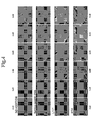
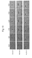
- FIG. 4 represents in situ hybridization images obtained from the same region of the serial sections. The images are for no addition, images wih the addition of salmon sperm DNA, images with addition of dummy oligo DNA L1C1, and images with addition of dummy oligo DNA arbp, respectively, from above.
- the concentrations of the four oligo DNA probes are 0 nM (nanomol), 1 nM, 2 nM, 3 nM, 4 nM, and 5 nM from the left.
- the images were taken in the same regions of the serial sections with a 10 ⁇ objective lens, using a Zeiss fluorescence microscope Axioplan2 and a CCD camera AxioCam.
- the images were computer processed using Image J software (NIH, http://rsb.info.nih.gov/ij/), and signal intensity was determined at each concentration.
- the image signal intensity is the background fluorescence intensity (IntDen) at the oligo DNA probe concentration of 0 nM.
- FIG. 5 represents the background fluorescence intensity for samples with no addition, and for samples with the addition of the salmon sperm DNA, and the dummy oligo DNAs L1C1 and arbp.
- the background fluorescence intensity is higher in samples with the addition of the salmon sperm DNA than in other samples.
- a true signal intensity at each concentration was determined by subtracting the background fluorescence intensity from the signal intensity of each different concentration.
- FIG. 6 represents the results. Samples with the addition of the dummy oligo DNA arbp at the oligo DNA probe concentration of 1 nM has a weaker true signal intensity than other samples. However, the fact that the signals are observed with good contrast suggests that the addition of the oligo DNA probes involves only a small increase in background noise. It can be seen from FIG.
- RNA detection by quantitative RNA in situ hybridization was examined between dummy oligo DNA and salmon sperm DNA.
- mouse liver tissue samples were prepared by formalin fixation and paraffin embedding, and serial sections were made as in Example 1. Further, as in Example 1, Cyp1a2 was used as the detected gene, and four single-stranded oligo DNA probes of SEQ ID NOS: 1 to 4 FITC-labeled at the both ends were used.
- a dummy oligo DNA (single-stranded) of SEQ ID NO: 5 was used at the concentration with the addition ratio of 8 with respect to each different concentration 0 nM (nanomol), 1 nM, 2 nM, 3 nM, 4 nM, 5 nM of the four oligo DNA probes. Further, a salmon sperm DNA (Salmonsperm DNA solution, catalog number 15632-011; Invitrogen) was added to each different probe concentration to make the final concentration 100 ⁇ g/ml (microgram/milliliter; equivalent of 80 nM).
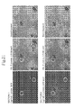
- FIG. 7 represents in situ hybridization images obtained from the same region of the serial sections. The upper images are from samples with the addition of the dummy oligo DNA, the lower images from samples with the addition of the salmon sperm DNA. The concentrations of the four probes are 1 nM (nanomol), 2 nM, 3 nM, 4 nM, and 5 nM from the left.
- FIG. 7 The images were taken in the same regions of the serial sections with a 10 ⁇ objective lens, using a Zeiss fluorescence microscope Axioplan2 and a CCD camera AxioCam.
- a signal region S with signals, and a noise region N without signals were set as in FIG. 7 , and the signal intensity of each region was determined using Image J software (NIH, http://rsb.info.nih.gov/ij/).
- the ratio of signal intensities in regions S and N was then determined as the SN ratio (signal-to-noise ratio).
- FIG. 8 compares the SN ratios for samples with the addition of the dummy oligo DNA and for samples with the addition of the salmon sperm DNA.
- the vertical axis represents SN ratio
- the horizontal axis represents probe concentration.
- the relationship between oligo DNA probe concentration and SN ratio is bell shaped, with the dummy oligo DNA producing better SN ratios than the salmon sperm DNA at the oligo DNA probe concentrations of 2 nM and higher.
- Example 1 The type and number of dummy oligo DNA sequences were tested in this Example.
- mouse liver tissue samples were prepared by formalin fixation and paraffin embedding, and serial sections were made as in Example 1. Following deparaffinization, the specimens were treated with Protease K (Invitrogen, Proteinase K SOL. RNA, 25530049), and RNA in situ hybridization was performed, as in Example 1. Further, as in Example 1, Cyp1a2 was used as the detected gene, and single-stranded oligo DNA probes of SEQ ID NOS: 1 to 4 FITC-labeled at the both ends were used. In accord with the result of Example 2, the four probes were used at the concentration of 2 nM (nanomol).
- mice liver tissue samples were prepared by formalin fixation and paraffin embedding, and serial sections were made as in Example 1. Following deparaffinization, the specimens were treated with Protease K (Invitrogen, Proteinase K SOL. RNA, 25530049), and RNA in situ hybridization was performed, as in Example 1. Further, as in Example 1, Cyp1a2 was used as the detected gene, and single-stranded oligo DNA probes of SEQ ID NOS: 1 to 4 FITC-labeled at the both ends were used. The four oligo DNA probes were used at the concentration of 2 nM (nanomol), and experiments were performed with one, two, three, and four oligo DNA probes, and without the oligo DNA probe.
- Protease K Invitrogen, Proteinase K SOL. RNA, 25530049
- a dummy oligo DNA (L1C1) of SEQ ID NO: 5 was used as the dummy oligo DNA (single-stranded) at a concentration 8 times the total concentration of the oligo DNA probes used. Specifically, the concentration of the dummy oligo DNA was 16 nM, 32 nM, 48 nM, and 64 nM for one, two, three, and four oligo DNA probes, respectively. The concentration of the dummy oligo DNA was 64 nM for samples with no oligo DNA probe.
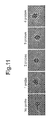
- the anti-FITC rabbit polyclonal antibody P5100 (Dako) was used, and sensitization was performed using a tyramide-FLU (Perkin-Elmer, TSA Plus Fluorescein System, NEL741B001KT). Images were taken in the same regions of the serial sections with a 10 ⁇ objective lens, using a Zeiss fluorescence microscope Axioplan2 and a CCD camera AxioCam. The images are shown in FIG. 11 .
- the signal intensity of each image was determined using Image J software (NIH, http://rsb.info.nih.gov/ij/), and the value obtained by subtracting the signal intensity of the image without the oligo DNA probe from this preliminary signal intensity was determined as the signal intensity of the image using each number of probe.
- the results are plotted in FIG. 12 , in which the vertical axis represents signal intensity, and the horizontal axis represents the number of oligo DNA probes. As shown in the figure, with the addition of the dummy oligo DNA, the relationship between the number of oligo DNA probes and signal intensity is linear, producing a definite, upward straight line.
- Hybridization is a type of equilibrium reaction, and, theoretically, the following relation is established.
- K H / f ⁇ R ⁇ f ⁇ P , where K is the equilibrium constant, [H] is the concentration of a hybridized product, [fR] is the free mRNA concentration, and [fP] is the free oligo DNA probe concentration.
- P0 [H] + [fP] for the concentration of the hybridized oligo DNA probe
- R0 [H] + [fR] for the mRNA concentration in a tissue sample (constant in the tissue sample are true)
- the hybridized product increases as the concentration of the hybridized oligo DNA probe is increased.
- This Example demonstrates that the signal intensity also increases linearly with increasing oligo DNA probe concentrations.
- mice liver tissue samples were prepared by formalin fixation and paraffin embedding, and serial sections were made as in Example 1. Following deparaffinization, the specimens were treated with Protease K (Invitrogen, Proteinase K SOL. RNA, 25530049), and RNA in situ hybridization was performed, as in Example 1. Albumin Alb was used as the detected gene, and a single-stranded oligo DNA probe of SEQ ID NO: 12 FITC-labeled at the both ends was used in six different concentrations 0 nM (no probe), 0.1 nM, 0.25 nM, 0.5 nM, 1 nM, and 1.5 nM.
- Protease K Invitrogen, Proteinase K SOL. RNA, 25530049
- a dummy oligo DNA (single-stranded) of SEQ ID NO: 5 was used as the dummy oligo DNA at a concentration 8 times the concentration of the oligo DNA probe used.
- a 12 nM dummy oligo DNA was used in a hybridization solution that contained no oligo DNA probe.
- the anti-FITC rabbit polyclonal antibody P5100 Dako was used, and sensitization was performed using a tyramide-FLU (Perkin-Elmer, TSA Plus Fluorescein System, NEL741 B001 KT).
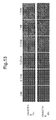
- the images are shown in FIG. 13 .
- the images are shown in FIG. 13 (the upper images with a 10 ⁇ objective lens, and the lower images with a 20 ⁇ objective lens).
- the signal intensity of the image at each concentration was determined using a computer with Image J software (NIH, http://rsb.info.nih.gov/ij/), and the value obtained by subtracting the signal intensity of the image at the oligo DNA probe concentration 0 nM from the computed signal intensity was determined as the true signal intensity.
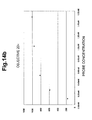
- the results are plotted in FIG.
- FIG. 14 in which the vertical axis represents signal intensity, and the horizontal axis represents probe concentration
- FIG. 14a represents the signal intensity for the images taken with a 10 ⁇ objective lens shown in FIG. 13
- FIG. 14b represents the signal intensity for the images taken with a 20 ⁇ objective lens shown in FIG. 13
- the signal intensity linearly increases with increasing probe concentrations, before saturating above a certain concentration (1 nM in the case of Alb gene in FIG. 14 ).
- the result is the reflection of very high expression levels of Alb gene in the liver, as demonstrated by the result of quantitative PCR in Example 9.
- mice liver tissue samples were prepared by formalin fixation and paraffin embedding, and serial sections were made as in Example 1. Following deparaffinization, the specimens were treated with Protease K (Invitrogen, Proteinase K SOL. RNA, 25530049), and RNA in situ hybridization was performed, as in Example 1. As in Examples 1 and 2, Cyp1a2 was used as the detected gene, and single-stranded oligo DNA probes of SEQ ID NO: 1 to 4 FITC-labeled at the both ends were used.
- Protease K Invitrogen, Proteinase K SOL. RNA, 25530049
- the dummy oligo DNA was used at the concentration of 64 nM for a hybridization solution that contained no oligo DNA probe.
- the anti-FITC rabbit polyclonal antibody P5100 (Dako) was used, and sensitization was performed using a tyramide-FLU (Perkin-Elmer, TSA Plus Fluorescein System, NEL741B001KT).
- Tissue samples were prepared by formalin fixation and paraffin embedding, and serial sections were made as in Example 1. Following deparaffinization, the specimens were treated with Protease K (Invitrogen, Proteinase K SOL. RNA, 25530049), and RNA in situ hybridization experiment was performed, as in Example 1. Single-stranded oligo DNA probes of SEQ ID NOS: 13 and 14 Dig-labeled at the both ends were used as the oligonucleic acid probes for the detected gene Arntl (21 bases on the Arntl gene mRNA). Each probe was used at the concentration of 2 nM (nanomol).
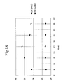
- FIG. 18 represents changes in the expression levels of the target gene Arntl and Actb in tissue samples collected every 4 hour. The values are shown as changes in Ct values determined by quantitative PCR.
- FIG. 19 represents changes in signal intensity determined by the RNA in situ hybridization for the target gene Arntl.
- FIG. 20 represents the relationship between quantitative PCR Ct value and RNA in situ hybridization signal intensity. In the figure, the Ct values determined by the quantitative PCR for the target gene Arntl in the liver collected from each individual are plotted against the mean values in each time group of the signal intensities determined by RNA in situ hybridization. As represented in FIG.
- Example 8 The liver of an individual male mouse, 8 weeks of age, collected at 1 p.m. in Example 8 was used as the tissue sample. TSA sensitization was performed stepwise, using oligo DNA probes Dig-labeled at the both ends for the target gene Cyp1a2, and oligo DNA probes FITC-labeled at the both ends for the target gene Alb. The quantitative detection of these two target genes was performed with the fluorescent dyes of two colors, and the detection results were compared with the Ct values obtained by the quantitative PCR for these genes.
Landscapes
- Chemical & Material Sciences (AREA)
- Organic Chemistry (AREA)
- Life Sciences & Earth Sciences (AREA)
- Zoology (AREA)
- Wood Science & Technology (AREA)
- Proteomics, Peptides & Aminoacids (AREA)
- Health & Medical Sciences (AREA)
- Engineering & Computer Science (AREA)
- Microbiology (AREA)
- Biochemistry (AREA)
- Physics & Mathematics (AREA)
- Molecular Biology (AREA)
- Biotechnology (AREA)
- Biophysics (AREA)
- Analytical Chemistry (AREA)
- Immunology (AREA)
- Bioinformatics & Cheminformatics (AREA)
- General Engineering & Computer Science (AREA)
- General Health & Medical Sciences (AREA)
- Genetics & Genomics (AREA)
- Chemical Kinetics & Catalysis (AREA)
- Measuring Or Testing Involving Enzymes Or Micro-Organisms (AREA)
Applications Claiming Priority (2)
| Application Number | Priority Date | Filing Date | Title |
|---|---|---|---|
| JP2008131302 | 2008-05-19 | ||
| PCT/JP2009/059219 WO2009142214A1 (ja) | 2008-05-19 | 2009-05-19 | RNA in situハイブリダイゼーション |
Publications (3)
| Publication Number | Publication Date |
|---|---|
| EP2292798A1 true EP2292798A1 (de) | 2011-03-09 |
| EP2292798A4 EP2292798A4 (de) | 2011-11-16 |
| EP2292798B1 EP2292798B1 (de) | 2013-12-25 |
Family
ID=41340145
Family Applications (1)
| Application Number | Title | Priority Date | Filing Date |
|---|---|---|---|
| EP09750576.2A Not-in-force EP2292798B1 (de) | 2008-05-19 | 2009-05-19 | In-situ-rna-hybridisierung |
Country Status (6)
| Country | Link |
|---|---|
| US (1) | US9051610B2 (de) |
| EP (1) | EP2292798B1 (de) |
| JP (1) | JP5733977B2 (de) |
| CN (1) | CN102037137B (de) |
| AU (1) | AU2009250528B2 (de) |
| WO (1) | WO2009142214A1 (de) |
Families Citing this family (10)
| Publication number | Priority date | Publication date | Assignee | Title |
|---|---|---|---|---|
| WO2011056185A2 (en) | 2009-11-04 | 2011-05-12 | President And Fellows Of Harvard College | Reactivity-dependent and interaction-dependent pcr |
| DK2563935T3 (da) * | 2010-04-30 | 2014-06-23 | Exiqon As | In situ-hybridiseringsfremgangsmåde og -buffer |
| EP2989214A4 (de) * | 2013-04-23 | 2016-12-28 | Harvard College | In-situ-interaktionsbestimmung |
| CN105861695A (zh) * | 2016-05-11 | 2016-08-17 | 江苏省肿瘤医院 | 一种检测乳腺癌细胞耐药性的方法及检测试剂盒 |
| GB201621514D0 (en) | 2016-12-16 | 2017-02-01 | Q-Linea Ab | Padlock probe detection method |
| CN108707662B (zh) * | 2017-04-05 | 2021-11-19 | 益善生物技术股份有限公司 | 一种ar-v7表达检测试剂盒 |
| CN108517352A (zh) * | 2018-04-25 | 2018-09-11 | 内蒙古华星康为生物科技有限公司 | 一种检测微生物的方法及其应用 |
| CN110205357A (zh) * | 2019-06-22 | 2019-09-06 | 福建农林大学 | 一种多引物oligo探针的制备方法 |
| CN111426667A (zh) * | 2020-04-23 | 2020-07-17 | 天津科技大学 | 一种基于量子点-核酸适配体-氧化石墨烯建立的对β-乳球蛋白检测的荧光方法 |
| CN114622000B (zh) * | 2020-12-14 | 2024-05-07 | 厦门大学 | 一种高特异性的检测靶核酸序列的方法 |
Citations (3)
| Publication number | Priority date | Publication date | Assignee | Title |
|---|---|---|---|---|
| WO1994002642A1 (en) * | 1992-07-17 | 1994-02-03 | Aprogenex, Inc. | Background-reducing compounds for probe-mediated in-situ fluorimetric assays |
| WO2005111235A2 (en) * | 2004-05-04 | 2005-11-24 | Dak Denmark A/S | Methods for detecting chromosome aberrations |
| WO2008046056A1 (en) * | 2006-10-13 | 2008-04-17 | Welldoc Communications, Inc. | Reduction of nonspecific binding in nucleic acid assays and nucleic acid synthesis reactions |
Family Cites Families (15)
| Publication number | Priority date | Publication date | Assignee | Title |
|---|---|---|---|---|
| CA1251119A (en) | 1984-04-05 | 1989-03-14 | John P. Coghlan | Hybridization histochemistry |
| US5597692A (en) | 1984-04-05 | 1997-01-28 | Howard Florey Institute Of Experimental Physiology And Medicine | Hybridization histochemistry method for determining the presence and location in animal or plant tissue of RNA |
| US4888278A (en) | 1985-10-22 | 1989-12-19 | University Of Massachusetts Medical Center | In-situ hybridization to detect nucleic acid sequences in morphologically intact cells |
| US5985549A (en) | 1985-10-22 | 1999-11-16 | University Of Massachusetts | Non-isotopic in-situ hybridization method for detection of nucleic acids |
| US5354657A (en) | 1988-01-12 | 1994-10-11 | Boehringer Mannheim Gmbh | Process for the highly specific detection of nucleic acids in solid |
| DE3813278A1 (de) | 1988-01-12 | 1989-07-20 | Boehringer Mannheim Gmbh | Verfahren zum nachweis von nukleinsaeuren |
| US5196306A (en) | 1989-03-29 | 1993-03-23 | E. I. Du Pont De Nemours And Company | Method for the detection or quantitation of an analyte using an analyte dependent enzyme activation system |
| US5681702A (en) * | 1994-08-30 | 1997-10-28 | Chiron Corporation | Reduction of nonspecific hybridization by using novel base-pairing schemes |
| US5750340A (en) | 1995-04-07 | 1998-05-12 | University Of New Mexico | In situ hybridization solution and process |
| US5866331A (en) | 1995-10-20 | 1999-02-02 | University Of Massachusetts | Single molecule detection by in situ hybridization |
| US6534266B1 (en) | 1999-04-22 | 2003-03-18 | Albert Einstein College Of Medicine Of Yeshiva University | Assay of transcription sites by multi-fluor fish |
| CA2410974A1 (en) * | 2000-05-16 | 2002-11-15 | Dnavec Research Inc. | Polypeptides cleaving telomeric repeats |
| AU2002306768A1 (en) * | 2001-03-20 | 2002-10-03 | Ortho-Clinical Diagnostics, Inc. | Expression profiles and methods of use |
| JP2004267033A (ja) * | 2003-03-05 | 2004-09-30 | Japan Science & Technology Agency | カヤツリグサ科植物のレトロトランスポゾン遺伝子 |
| WO2008068636A2 (en) * | 2006-12-01 | 2008-06-12 | Dako Denmark A/S | Blocking agents comprising non-natural nucleic acids and detection methods using such blocking agents |
-
2009
- 2009-05-19 US US12/993,338 patent/US9051610B2/en not_active Expired - Fee Related
- 2009-05-19 JP JP2010513029A patent/JP5733977B2/ja active Active
- 2009-05-19 EP EP09750576.2A patent/EP2292798B1/de not_active Not-in-force
- 2009-05-19 CN CN200980117915.8A patent/CN102037137B/zh not_active Expired - Fee Related
- 2009-05-19 AU AU2009250528A patent/AU2009250528B2/en not_active Ceased
- 2009-05-19 WO PCT/JP2009/059219 patent/WO2009142214A1/ja active Application Filing
Patent Citations (3)
| Publication number | Priority date | Publication date | Assignee | Title |
|---|---|---|---|---|
| WO1994002642A1 (en) * | 1992-07-17 | 1994-02-03 | Aprogenex, Inc. | Background-reducing compounds for probe-mediated in-situ fluorimetric assays |
| WO2005111235A2 (en) * | 2004-05-04 | 2005-11-24 | Dak Denmark A/S | Methods for detecting chromosome aberrations |
| WO2008046056A1 (en) * | 2006-10-13 | 2008-04-17 | Welldoc Communications, Inc. | Reduction of nonspecific binding in nucleic acid assays and nucleic acid synthesis reactions |
Non-Patent Citations (3)
| Title |
|---|
| BLOCK DE M ET AL: "RNA-RNA IN SITU HYBRIDIZATION USING DIGOXIGENIN-LABELED PROBES: THEUSE OF HIGH-MOLECULAR-WEIGHT POLYVINYL ALCOHOL IN THE ALKALINE PHOSPHATASE INDOXYL-NITROBLUE TETRAZOLIUM REACTION", ANALYTICAL BIOCHEMISTRY, ACADEMIC PRESS INC, NEW YORK, vol. 215, no. 1, 15 November 1993 (1993-11-15), pages 86-89, XP000413713, ISSN: 0003-2697, DOI: 10.1006/ABIO.1993.1558 * |
| PUSKAS L G ET AL: "REDUCTION OF MISPRIMING IN AMPLIFICATION REACTIONS WITH RESTRICTED PCR", GENOME RESEARCH, COLD SPRING HARBOR LABORATORY PRESS, WOODBURY, NY, US, vol. 5, no. 3, 1 October 1995 (1995-10-01) , pages 309-311, XP009039371, ISSN: 1088-9051 * |
| See also references of WO2009142214A1 * |
Also Published As
| Publication number | Publication date |
|---|---|
| JPWO2009142214A1 (ja) | 2011-09-29 |
| CN102037137B (zh) | 2017-11-28 |
| EP2292798A4 (de) | 2011-11-16 |
| JP5733977B2 (ja) | 2015-06-10 |
| EP2292798B1 (de) | 2013-12-25 |
| US9051610B2 (en) | 2015-06-09 |
| US20110183331A1 (en) | 2011-07-28 |
| CN102037137A (zh) | 2011-04-27 |
| WO2009142214A1 (ja) | 2009-11-26 |
| AU2009250528B2 (en) | 2016-02-18 |
| AU2009250528A1 (en) | 2009-11-26 |
Similar Documents
| Publication | Publication Date | Title |
|---|---|---|
| EP2292798B1 (de) | In-situ-rna-hybridisierung | |
| AU2004213871B2 (en) | Use of intronic RNA to measure gene expression | |
| EP1469067A1 (de) | Verfahren und vorrichtung zum nachweis von nukleinsäuredaten | |
| EP2500436B1 (de) | Verfahren, Sonde und Kit zur In-situ-Hybridisierung von DNA und Verwendung davon | |
| JP6203752B2 (ja) | 前立腺癌の治療/予防処置の診断、予測及び評価のための材料及び方法 | |
| US8535914B2 (en) | Probe, probe set and information acquisition method using the same | |
| JP2006520206A (ja) | プローブ、バイオチップおよびそれらの使用方法 | |
| US20130217115A1 (en) | Plasmid standard for use in quantitative assays using fluorescent quantitative pcr | |
| US20200075125A1 (en) | Comparative genomic hybridization array method for preimplantation genetic screening | |
| WO2020191521A1 (zh) | 核酸序列、rna目标区域测序文库的构建方法及应用 | |
| CN112980997A (zh) | 侵袭性毛霉病病原菌的引物和探针及其实现方法、检测系统 | |
| US20200109457A1 (en) | Chromosomal assessment to diagnose urogenital malignancy in dogs | |
| Behzadi et al. | The application of Microarray in Medicine | |
| EP1722309A1 (de) | Verfahren zur Normalisierung von Genexpressionsdaten | |
| JP2022119753A (ja) | 高精度な遺伝子変異検出法 | |
| Horelli-Kuitunen et al. | Mapping ESTs by Fiber–FISH | |
| JP2015504665A (ja) | 膵胆管癌の診断、予後、再発のモニターおよび治療的/予防的治療の評価のための物質および方法 | |
| CN110343750A (zh) | 用于检测外泌体中核酸表达水平的内参基因及其应用 | |
| Menshanov et al. | Methodological aspects of read mapping and assembly of transcriptomes derived from the brain tissue samples of Rattus norvegicus | |
| Gabrieli et al. | Genome-wide epigenetic profiling of 5-hydroxymethylcytosine by long-read optical mapping | |
| EP4417705A1 (de) | Verwendung von mir-625-3p als biomarker zur diagnose der schwere von psoriasis | |
| Singh et al. | In situ hybridization in clinical biomarker development | |
| CN117512112A (zh) | 筛查nab2-stat6融合情况的方法、引物和探针以及试剂盒 | |
| CN107828879A (zh) | 一种pkp2基因突变检测试剂盒及其检测方法 | |
| EP1748082A1 (de) | Verfahren zur nukleinsäureanalyse |
Legal Events
| Date | Code | Title | Description |
|---|---|---|---|
| PUAI | Public reference made under article 153(3) epc to a published international application that has entered the european phase |
Free format text: ORIGINAL CODE: 0009012 |
|
| 17P | Request for examination filed |
Effective date: 20101216 |
|
| AK | Designated contracting states |
Kind code of ref document: A1 Designated state(s): AT BE BG CH CY CZ DE DK EE ES FI FR GB GR HR HU IE IS IT LI LT LU LV MC MK MT NL NO PL PT RO SE SI SK TR |
|
| AX | Request for extension of the european patent |
Extension state: AL BA RS |
|
| DAX | Request for extension of the european patent (deleted) | ||
| A4 | Supplementary search report drawn up and despatched |
Effective date: 20111018 |
|
| RIC1 | Information provided on ipc code assigned before grant |
Ipc: G01N 33/58 20060101ALI20111012BHEP Ipc: C12Q 1/68 20060101AFI20111012BHEP Ipc: C12N 15/09 20060101ALI20111012BHEP |
|
| RIN1 | Information on inventor provided before grant (corrected) |
Inventor name: NAKANO, TATSUHIRO Inventor name: MATSUOKA, MASAHIRO Inventor name: DOI, HIROFUMI Inventor name: NAGAYOSHI, CHIZURU |
|
| 17Q | First examination report despatched |
Effective date: 20130103 |
|
| GRAP | Despatch of communication of intention to grant a patent |
Free format text: ORIGINAL CODE: EPIDOSNIGR1 |
|
| INTG | Intention to grant announced |
Effective date: 20130613 |
|
| RIN1 | Information on inventor provided before grant (corrected) |
Inventor name: NAKANO, TATSUHIRO Inventor name: MATSUOKA, MASAHIRO Inventor name: NAGAYOSHI, CHIZURU Inventor name: DOI, HIROFUMI |
|
| GRAP | Despatch of communication of intention to grant a patent |
Free format text: ORIGINAL CODE: EPIDOSNIGR1 |
|
| GRAS | Grant fee paid |
Free format text: ORIGINAL CODE: EPIDOSNIGR3 |
|
| GRAA | (expected) grant |
Free format text: ORIGINAL CODE: 0009210 |
|
| INTG | Intention to grant announced |
Effective date: 20131029 |
|
| AK | Designated contracting states |
Kind code of ref document: B1 Designated state(s): AT BE BG CH CY CZ DE DK EE ES FI FR GB GR HR HU IE IS IT LI LT LU LV MC MK MT NL NO PL PT RO SE SI SK TR |
|
| REG | Reference to a national code |
Ref country code: GB Ref legal event code: FG4D |
|
| REG | Reference to a national code |
Ref country code: CH Ref legal event code: EP |
|
| REG | Reference to a national code |
Ref country code: AT Ref legal event code: REF Ref document number: 646696 Country of ref document: AT Kind code of ref document: T Effective date: 20140115 |
|
| REG | Reference to a national code |
Ref country code: IE Ref legal event code: FG4D |
|
| REG | Reference to a national code |
Ref country code: DE Ref legal event code: R096 Ref document number: 602009020994 Country of ref document: DE Effective date: 20140213 |
|
| PG25 | Lapsed in a contracting state [announced via postgrant information from national office to epo] |
Ref country code: NO Free format text: LAPSE BECAUSE OF FAILURE TO SUBMIT A TRANSLATION OF THE DESCRIPTION OR TO PAY THE FEE WITHIN THE PRESCRIBED TIME-LIMIT Effective date: 20140325 Ref country code: HR Free format text: LAPSE BECAUSE OF FAILURE TO SUBMIT A TRANSLATION OF THE DESCRIPTION OR TO PAY THE FEE WITHIN THE PRESCRIBED TIME-LIMIT Effective date: 20131225 Ref country code: FI Free format text: LAPSE BECAUSE OF FAILURE TO SUBMIT A TRANSLATION OF THE DESCRIPTION OR TO PAY THE FEE WITHIN THE PRESCRIBED TIME-LIMIT Effective date: 20131225 Ref country code: LT Free format text: LAPSE BECAUSE OF FAILURE TO SUBMIT A TRANSLATION OF THE DESCRIPTION OR TO PAY THE FEE WITHIN THE PRESCRIBED TIME-LIMIT Effective date: 20131225 Ref country code: SE Free format text: LAPSE BECAUSE OF FAILURE TO SUBMIT A TRANSLATION OF THE DESCRIPTION OR TO PAY THE FEE WITHIN THE PRESCRIBED TIME-LIMIT Effective date: 20131225 |
|
| REG | Reference to a national code |
Ref country code: NL Ref legal event code: VDEP Effective date: 20131225 |
|
| REG | Reference to a national code |
Ref country code: AT Ref legal event code: MK05 Ref document number: 646696 Country of ref document: AT Kind code of ref document: T Effective date: 20131225 |
|
| REG | Reference to a national code |
Ref country code: LT Ref legal event code: MG4D |
|
| PG25 | Lapsed in a contracting state [announced via postgrant information from national office to epo] |
Ref country code: LV Free format text: LAPSE BECAUSE OF FAILURE TO SUBMIT A TRANSLATION OF THE DESCRIPTION OR TO PAY THE FEE WITHIN THE PRESCRIBED TIME-LIMIT Effective date: 20131225 |
|
| PG25 | Lapsed in a contracting state [announced via postgrant information from national office to epo] |
Ref country code: EE Free format text: LAPSE BECAUSE OF FAILURE TO SUBMIT A TRANSLATION OF THE DESCRIPTION OR TO PAY THE FEE WITHIN THE PRESCRIBED TIME-LIMIT Effective date: 20131225 Ref country code: IS Free format text: LAPSE BECAUSE OF FAILURE TO SUBMIT A TRANSLATION OF THE DESCRIPTION OR TO PAY THE FEE WITHIN THE PRESCRIBED TIME-LIMIT Effective date: 20140425 |
|
| PG25 | Lapsed in a contracting state [announced via postgrant information from national office to epo] |
Ref country code: CY Free format text: LAPSE BECAUSE OF FAILURE TO SUBMIT A TRANSLATION OF THE DESCRIPTION OR TO PAY THE FEE WITHIN THE PRESCRIBED TIME-LIMIT Effective date: 20131225 Ref country code: NL Free format text: LAPSE BECAUSE OF FAILURE TO SUBMIT A TRANSLATION OF THE DESCRIPTION OR TO PAY THE FEE WITHIN THE PRESCRIBED TIME-LIMIT Effective date: 20131225 Ref country code: CZ Free format text: LAPSE BECAUSE OF FAILURE TO SUBMIT A TRANSLATION OF THE DESCRIPTION OR TO PAY THE FEE WITHIN THE PRESCRIBED TIME-LIMIT Effective date: 20131225 Ref country code: PL Free format text: LAPSE BECAUSE OF FAILURE TO SUBMIT A TRANSLATION OF THE DESCRIPTION OR TO PAY THE FEE WITHIN THE PRESCRIBED TIME-LIMIT Effective date: 20131225 Ref country code: AT Free format text: LAPSE BECAUSE OF FAILURE TO SUBMIT A TRANSLATION OF THE DESCRIPTION OR TO PAY THE FEE WITHIN THE PRESCRIBED TIME-LIMIT Effective date: 20131225 Ref country code: PT Free format text: LAPSE BECAUSE OF FAILURE TO SUBMIT A TRANSLATION OF THE DESCRIPTION OR TO PAY THE FEE WITHIN THE PRESCRIBED TIME-LIMIT Effective date: 20140428 Ref country code: SK Free format text: LAPSE BECAUSE OF FAILURE TO SUBMIT A TRANSLATION OF THE DESCRIPTION OR TO PAY THE FEE WITHIN THE PRESCRIBED TIME-LIMIT Effective date: 20131225 Ref country code: RO Free format text: LAPSE BECAUSE OF FAILURE TO SUBMIT A TRANSLATION OF THE DESCRIPTION OR TO PAY THE FEE WITHIN THE PRESCRIBED TIME-LIMIT Effective date: 20131225 Ref country code: ES Free format text: LAPSE BECAUSE OF FAILURE TO SUBMIT A TRANSLATION OF THE DESCRIPTION OR TO PAY THE FEE WITHIN THE PRESCRIBED TIME-LIMIT Effective date: 20131225 |
|
| REG | Reference to a national code |
Ref country code: DE Ref legal event code: R097 Ref document number: 602009020994 Country of ref document: DE |
|
| PG25 | Lapsed in a contracting state [announced via postgrant information from national office to epo] |
Ref country code: DK Free format text: LAPSE BECAUSE OF FAILURE TO SUBMIT A TRANSLATION OF THE DESCRIPTION OR TO PAY THE FEE WITHIN THE PRESCRIBED TIME-LIMIT Effective date: 20131225 |
|
| PLBE | No opposition filed within time limit |
Free format text: ORIGINAL CODE: 0009261 |
|
| STAA | Information on the status of an ep patent application or granted ep patent |
Free format text: STATUS: NO OPPOSITION FILED WITHIN TIME LIMIT |
|
| 26N | No opposition filed |
Effective date: 20140926 |
|
| PG25 | Lapsed in a contracting state [announced via postgrant information from national office to epo] |
Ref country code: LU Free format text: LAPSE BECAUSE OF FAILURE TO SUBMIT A TRANSLATION OF THE DESCRIPTION OR TO PAY THE FEE WITHIN THE PRESCRIBED TIME-LIMIT Effective date: 20140519 |
|
| REG | Reference to a national code |
Ref country code: DE Ref legal event code: R097 Ref document number: 602009020994 Country of ref document: DE Effective date: 20140926 |
|
| PG25 | Lapsed in a contracting state [announced via postgrant information from national office to epo] |
Ref country code: MC Free format text: LAPSE BECAUSE OF FAILURE TO SUBMIT A TRANSLATION OF THE DESCRIPTION OR TO PAY THE FEE WITHIN THE PRESCRIBED TIME-LIMIT Effective date: 20131225 |
|
| REG | Reference to a national code |
Ref country code: IE Ref legal event code: MM4A |
|
| PG25 | Lapsed in a contracting state [announced via postgrant information from national office to epo] |
Ref country code: IE Free format text: LAPSE BECAUSE OF NON-PAYMENT OF DUE FEES Effective date: 20140519 |
|
| PG25 | Lapsed in a contracting state [announced via postgrant information from national office to epo] |
Ref country code: SI Free format text: LAPSE BECAUSE OF FAILURE TO SUBMIT A TRANSLATION OF THE DESCRIPTION OR TO PAY THE FEE WITHIN THE PRESCRIBED TIME-LIMIT Effective date: 20131225 |
|
| PG25 | Lapsed in a contracting state [announced via postgrant information from national office to epo] |
Ref country code: MT Free format text: LAPSE BECAUSE OF FAILURE TO SUBMIT A TRANSLATION OF THE DESCRIPTION OR TO PAY THE FEE WITHIN THE PRESCRIBED TIME-LIMIT Effective date: 20131225 |
|
| REG | Reference to a national code |
Ref country code: FR Ref legal event code: PLFP Year of fee payment: 8 |
|
| PG25 | Lapsed in a contracting state [announced via postgrant information from national office to epo] |
Ref country code: BG Free format text: LAPSE BECAUSE OF FAILURE TO SUBMIT A TRANSLATION OF THE DESCRIPTION OR TO PAY THE FEE WITHIN THE PRESCRIBED TIME-LIMIT Effective date: 20131225 Ref country code: IT Free format text: LAPSE BECAUSE OF FAILURE TO SUBMIT A TRANSLATION OF THE DESCRIPTION OR TO PAY THE FEE WITHIN THE PRESCRIBED TIME-LIMIT Effective date: 20131225 Ref country code: GR Free format text: LAPSE BECAUSE OF FAILURE TO SUBMIT A TRANSLATION OF THE DESCRIPTION OR TO PAY THE FEE WITHIN THE PRESCRIBED TIME-LIMIT Effective date: 20140326 |
|
| PG25 | Lapsed in a contracting state [announced via postgrant information from national office to epo] |
Ref country code: TR Free format text: LAPSE BECAUSE OF FAILURE TO SUBMIT A TRANSLATION OF THE DESCRIPTION OR TO PAY THE FEE WITHIN THE PRESCRIBED TIME-LIMIT Effective date: 20131225 Ref country code: HU Free format text: LAPSE BECAUSE OF FAILURE TO SUBMIT A TRANSLATION OF THE DESCRIPTION OR TO PAY THE FEE WITHIN THE PRESCRIBED TIME-LIMIT; INVALID AB INITIO Effective date: 20090519 |
|
| REG | Reference to a national code |
Ref country code: FR Ref legal event code: PLFP Year of fee payment: 9 |
|
| REG | Reference to a national code |
Ref country code: FR Ref legal event code: PLFP Year of fee payment: 10 |
|
| PG25 | Lapsed in a contracting state [announced via postgrant information from national office to epo] |
Ref country code: MK Free format text: LAPSE BECAUSE OF FAILURE TO SUBMIT A TRANSLATION OF THE DESCRIPTION OR TO PAY THE FEE WITHIN THE PRESCRIBED TIME-LIMIT Effective date: 20131225 |
|
| REG | Reference to a national code |
Ref country code: DE Ref legal event code: R082 Ref document number: 602009020994 Country of ref document: DE |
|
| PGFP | Annual fee paid to national office [announced via postgrant information from national office to epo] |
Ref country code: FR Payment date: 20210525 Year of fee payment: 13 Ref country code: DE Payment date: 20210520 Year of fee payment: 13 |
|
| PGFP | Annual fee paid to national office [announced via postgrant information from national office to epo] |
Ref country code: BE Payment date: 20210519 Year of fee payment: 13 Ref country code: CH Payment date: 20210519 Year of fee payment: 13 Ref country code: GB Payment date: 20210525 Year of fee payment: 13 |
|
| REG | Reference to a national code |
Ref country code: DE Ref legal event code: R119 Ref document number: 602009020994 Country of ref document: DE |
|
| REG | Reference to a national code |
Ref country code: CH Ref legal event code: PL |
|
| REG | Reference to a national code |
Ref country code: BE Ref legal event code: MM Effective date: 20220531 |
|
| GBPC | Gb: european patent ceased through non-payment of renewal fee |
Effective date: 20220519 |
|
| PG25 | Lapsed in a contracting state [announced via postgrant information from national office to epo] |
Ref country code: LI Free format text: LAPSE BECAUSE OF NON-PAYMENT OF DUE FEES Effective date: 20220531 Ref country code: CH Free format text: LAPSE BECAUSE OF NON-PAYMENT OF DUE FEES Effective date: 20220531 |
|
| PG25 | Lapsed in a contracting state [announced via postgrant information from national office to epo] |
Ref country code: FR Free format text: LAPSE BECAUSE OF NON-PAYMENT OF DUE FEES Effective date: 20220531 |
|
| PG25 | Lapsed in a contracting state [announced via postgrant information from national office to epo] |
Ref country code: GB Free format text: LAPSE BECAUSE OF NON-PAYMENT OF DUE FEES Effective date: 20220519 Ref country code: DE Free format text: LAPSE BECAUSE OF NON-PAYMENT OF DUE FEES Effective date: 20221201 Ref country code: BE Free format text: LAPSE BECAUSE OF NON-PAYMENT OF DUE FEES Effective date: 20220531 |