EP2282210A1 - Évaluation de la qualité d'un embryo sur la base de la division et du mouvement des blastomères - Google Patents
Évaluation de la qualité d'un embryo sur la base de la division et du mouvement des blastomères Download PDFInfo
- Publication number
- EP2282210A1 EP2282210A1 EP10172399A EP10172399A EP2282210A1 EP 2282210 A1 EP2282210 A1 EP 2282210A1 EP 10172399 A EP10172399 A EP 10172399A EP 10172399 A EP10172399 A EP 10172399A EP 2282210 A1 EP2282210 A1 EP 2282210A1
- Authority
- EP
- European Patent Office
- Prior art keywords
- embryo
- cell
- period
- embryos
- division
- Prior art date
- Legal status (The legal status is an assumption and is not a legal conclusion. Google has not performed a legal analysis and makes no representation as to the accuracy of the status listed.)
- Withdrawn
Links
- 210000001161 mammalian embryo Anatomy 0.000 title claims abstract description 147
- 230000033001 locomotion Effects 0.000 title claims abstract description 52
- 210000001109 blastomere Anatomy 0.000 title description 77
- 238000001303 quality assessment method Methods 0.000 title description 2
- 230000032823 cell division Effects 0.000 claims abstract description 88
- 238000000034 method Methods 0.000 claims abstract description 49
- 230000001413 cellular effect Effects 0.000 claims abstract description 29
- 210000003463 organelle Anatomy 0.000 claims abstract description 27
- 238000009826 distribution Methods 0.000 claims abstract description 16
- 238000012544 monitoring process Methods 0.000 claims abstract description 6
- 230000009087 cell motility Effects 0.000 claims description 39
- 238000002054 transplantation Methods 0.000 claims description 5
- 210000002257 embryonic structure Anatomy 0.000 abstract description 102
- 230000004720 fertilization Effects 0.000 abstract description 18
- 238000000338 in vitro Methods 0.000 abstract description 7
- 230000000694 effects Effects 0.000 description 64
- 210000004027 cell Anatomy 0.000 description 35
- 210000002459 blastocyst Anatomy 0.000 description 27
- 101150080085 SEG1 gene Proteins 0.000 description 21
- 101100421134 Schizosaccharomyces pombe (strain 972 / ATCC 24843) sle1 gene Proteins 0.000 description 21
- 238000004458 analytical method Methods 0.000 description 19
- 241000283690 Bos taurus Species 0.000 description 18
- 101100202858 Saccharomyces cerevisiae (strain ATCC 204508 / S288c) SEG2 gene Proteins 0.000 description 17
- 230000008859 change Effects 0.000 description 17
- 238000011161 development Methods 0.000 description 14
- 230000018109 developmental process Effects 0.000 description 14
- 238000005259 measurement Methods 0.000 description 14
- 230000035899 viability Effects 0.000 description 14
- 230000004899 motility Effects 0.000 description 13
- 238000012546 transfer Methods 0.000 description 13
- 230000008707 rearrangement Effects 0.000 description 11
- 230000013020 embryo development Effects 0.000 description 9
- 230000001086 cytosolic effect Effects 0.000 description 8
- 239000000463 material Substances 0.000 description 8
- 210000000472 morula Anatomy 0.000 description 8
- 210000000287 oocyte Anatomy 0.000 description 8
- 238000000513 principal component analysis Methods 0.000 description 8
- 210000004291 uterus Anatomy 0.000 description 7
- 238000011156 evaluation Methods 0.000 description 6
- 238000013467 fragmentation Methods 0.000 description 6
- 238000006062 fragmentation reaction Methods 0.000 description 6
- 210000000170 cell membrane Anatomy 0.000 description 5
- 230000012447 hatching Effects 0.000 description 5
- 238000002513 implantation Methods 0.000 description 5
- 238000011534 incubation Methods 0.000 description 5
- 230000002035 prolonged effect Effects 0.000 description 5
- 230000001360 synchronised effect Effects 0.000 description 5
- 230000005945 translocation Effects 0.000 description 5
- 230000030833 cell death Effects 0.000 description 4
- 238000005056 compaction Methods 0.000 description 4
- 210000000805 cytoplasm Anatomy 0.000 description 4
- 239000007943 implant Substances 0.000 description 4
- 230000008774 maternal effect Effects 0.000 description 4
- 239000002609 medium Substances 0.000 description 4
- 230000029058 respiratory gaseous exchange Effects 0.000 description 4
- 238000007619 statistical method Methods 0.000 description 4
- 238000013179 statistical model Methods 0.000 description 4
- 230000014616 translation Effects 0.000 description 4
- 235000001014 amino acid Nutrition 0.000 description 3
- 150000001413 amino acids Chemical class 0.000 description 3
- 238000013459 approach Methods 0.000 description 3
- 238000013528 artificial neural network Methods 0.000 description 3
- 230000009089 cytolysis Effects 0.000 description 3
- 230000003111 delayed effect Effects 0.000 description 3
- 230000005484 gravity Effects 0.000 description 3
- 239000001963 growth medium Substances 0.000 description 3
- 238000010191 image analysis Methods 0.000 description 3
- 230000036512 infertility Effects 0.000 description 3
- 208000000509 infertility Diseases 0.000 description 3
- 231100000535 infertility Toxicity 0.000 description 3
- 239000012528 membrane Substances 0.000 description 3
- 210000001672 ovary Anatomy 0.000 description 3
- 230000035935 pregnancy Effects 0.000 description 3
- 238000001243 protein synthesis Methods 0.000 description 3
- 238000010187 selection method Methods 0.000 description 3
- 210000004340 zona pellucida Anatomy 0.000 description 3
- 241000124008 Mammalia Species 0.000 description 2
- 230000001594 aberrant effect Effects 0.000 description 2
- 230000002159 abnormal effect Effects 0.000 description 2
- 238000004422 calculation algorithm Methods 0.000 description 2
- 238000003776 cleavage reaction Methods 0.000 description 2
- 238000012258 culturing Methods 0.000 description 2
- 210000001771 cumulus cell Anatomy 0.000 description 2
- 230000034994 death Effects 0.000 description 2
- 238000001514 detection method Methods 0.000 description 2
- 238000002474 experimental method Methods 0.000 description 2
- 239000012530 fluid Substances 0.000 description 2
- 230000006870 function Effects 0.000 description 2
- 239000007789 gas Substances 0.000 description 2
- 238000002347 injection Methods 0.000 description 2
- 239000007924 injection Substances 0.000 description 2
- 239000000203 mixture Substances 0.000 description 2
- 230000000877 morphologic effect Effects 0.000 description 2
- 230000003287 optical effect Effects 0.000 description 2
- 210000003101 oviduct Anatomy 0.000 description 2
- 230000008569 process Effects 0.000 description 2
- 238000012545 processing Methods 0.000 description 2
- 108090000623 proteins and genes Proteins 0.000 description 2
- 230000000284 resting effect Effects 0.000 description 2
- 230000007017 scission Effects 0.000 description 2
- 238000011282 treatment Methods 0.000 description 2
- 230000000007 visual effect Effects 0.000 description 2
- BVKZGUZCCUSVTD-UHFFFAOYSA-M Bicarbonate Chemical compound OC([O-])=O BVKZGUZCCUSVTD-UHFFFAOYSA-M 0.000 description 1
- 230000005653 Brownian motion process Effects 0.000 description 1
- KRKNYBCHXYNGOX-UHFFFAOYSA-K Citrate Chemical compound [O-]C(=O)CC(O)(CC([O-])=O)C([O-])=O KRKNYBCHXYNGOX-UHFFFAOYSA-K 0.000 description 1
- CEAZRRDELHUEMR-URQXQFDESA-N Gentamicin Chemical compound O1[C@H](C(C)NC)CC[C@@H](N)[C@H]1O[C@H]1[C@H](O)[C@@H](O[C@@H]2[C@@H]([C@@H](NC)[C@@](C)(O)CO2)O)[C@H](N)C[C@@H]1N CEAZRRDELHUEMR-URQXQFDESA-N 0.000 description 1
- 229930182566 Gentamicin Natural products 0.000 description 1
- SQUHHTBVTRBESD-UHFFFAOYSA-N Hexa-Ac-myo-Inositol Natural products CC(=O)OC1C(OC(C)=O)C(OC(C)=O)C(OC(C)=O)C(OC(C)=O)C1OC(C)=O SQUHHTBVTRBESD-UHFFFAOYSA-N 0.000 description 1
- 241000282412 Homo Species 0.000 description 1
- 238000003744 In vitro fertilisation Methods 0.000 description 1
- 208000034702 Multiple pregnancies Diseases 0.000 description 1
- 229920005372 Plexiglas® Polymers 0.000 description 1
- QAOWNCQODCNURD-UHFFFAOYSA-L Sulfate Chemical compound [O-]S([O-])(=O)=O QAOWNCQODCNURD-UHFFFAOYSA-L 0.000 description 1
- 238000010521 absorption reaction Methods 0.000 description 1
- 239000003242 anti bacterial agent Substances 0.000 description 1
- 229940088710 antibiotic agent Drugs 0.000 description 1
- QVGXLLKOCUKJST-UHFFFAOYSA-N atomic oxygen Chemical compound [O] QVGXLLKOCUKJST-UHFFFAOYSA-N 0.000 description 1
- 238000005537 brownian motion Methods 0.000 description 1
- 238000004364 calculation method Methods 0.000 description 1
- 230000022131 cell cycle Effects 0.000 description 1
- 230000006037 cell lysis Effects 0.000 description 1
- 230000001427 coherent effect Effects 0.000 description 1
- 238000004590 computer program Methods 0.000 description 1
- 230000027326 copulation Effects 0.000 description 1
- 238000013461 design Methods 0.000 description 1
- 238000003745 diagnosis Methods 0.000 description 1
- 239000000428 dust Substances 0.000 description 1
- 235000013601 eggs Nutrition 0.000 description 1
- 230000029142 excretion Effects 0.000 description 1
- 238000013401 experimental design Methods 0.000 description 1
- 239000012634 fragment Substances 0.000 description 1
- 230000004927 fusion Effects 0.000 description 1
- 230000002068 genetic effect Effects 0.000 description 1
- 238000005469 granulation Methods 0.000 description 1
- 230000003179 granulation Effects 0.000 description 1
- 238000003384 imaging method Methods 0.000 description 1
- 238000007373 indentation Methods 0.000 description 1
- CDAISMWEOUEBRE-GPIVLXJGSA-N inositol Chemical compound O[C@H]1[C@H](O)[C@@H](O)[C@H](O)[C@H](O)[C@@H]1O CDAISMWEOUEBRE-GPIVLXJGSA-N 0.000 description 1
- 229960000367 inositol Drugs 0.000 description 1
- 230000009027 insemination Effects 0.000 description 1
- 238000004519 manufacturing process Methods 0.000 description 1
- 238000013178 mathematical model Methods 0.000 description 1
- 239000011159 matrix material Substances 0.000 description 1
- IBIKHMZPHNKTHM-RDTXWAMCSA-N merck compound 25 Chemical compound C1C[C@@H](C(O)=O)[C@H](O)CN1C(C1=C(F)C=CC=C11)=NN1C(=O)C1=C(Cl)C=CC=C1C1CC1 IBIKHMZPHNKTHM-RDTXWAMCSA-N 0.000 description 1
- 108020004999 messenger RNA Proteins 0.000 description 1
- 239000002207 metabolite Substances 0.000 description 1
- 238000000386 microscopy Methods 0.000 description 1
- 239000002480 mineral oil Substances 0.000 description 1
- 235000010446 mineral oil Nutrition 0.000 description 1
- 239000012299 nitrogen atmosphere Substances 0.000 description 1
- 230000006911 nucleation Effects 0.000 description 1
- 238000010899 nucleation Methods 0.000 description 1
- 210000000056 organ Anatomy 0.000 description 1
- 230000010355 oscillation Effects 0.000 description 1
- 239000001301 oxygen Substances 0.000 description 1
- 229910052760 oxygen Inorganic materials 0.000 description 1
- 238000003909 pattern recognition Methods 0.000 description 1
- 239000004033 plastic Substances 0.000 description 1
- 229920003023 plastic Polymers 0.000 description 1
- 210000004508 polar body Anatomy 0.000 description 1
- 239000004926 polymethyl methacrylate Substances 0.000 description 1
- 235000018102 proteins Nutrition 0.000 description 1
- 102000004169 proteins and genes Human genes 0.000 description 1
- 238000011002 quantification Methods 0.000 description 1
- 230000001105 regulatory effect Effects 0.000 description 1
- 238000011160 research Methods 0.000 description 1
- CDAISMWEOUEBRE-UHFFFAOYSA-N scyllo-inosotol Natural products OC1C(O)C(O)C(O)C(O)C1O CDAISMWEOUEBRE-UHFFFAOYSA-N 0.000 description 1
- 210000002966 serum Anatomy 0.000 description 1
- 241000894007 species Species 0.000 description 1
- 230000020347 spindle assembly Effects 0.000 description 1
- 238000003860 storage Methods 0.000 description 1
- 239000000126 substance Substances 0.000 description 1
- 238000013519 translation Methods 0.000 description 1
- 238000009827 uniform distribution Methods 0.000 description 1
- 239000013598 vector Substances 0.000 description 1
- 238000003260 vortexing Methods 0.000 description 1
- 238000005406 washing Methods 0.000 description 1
- 230000003442 weekly effect Effects 0.000 description 1
Images


Classifications
-
- C—CHEMISTRY; METALLURGY
- C12—BIOCHEMISTRY; BEER; SPIRITS; WINE; VINEGAR; MICROBIOLOGY; ENZYMOLOGY; MUTATION OR GENETIC ENGINEERING
- C12Q—MEASURING OR TESTING PROCESSES INVOLVING ENZYMES, NUCLEIC ACIDS OR MICROORGANISMS; COMPOSITIONS OR TEST PAPERS THEREFOR; PROCESSES OF PREPARING SUCH COMPOSITIONS; CONDITION-RESPONSIVE CONTROL IN MICROBIOLOGICAL OR ENZYMOLOGICAL PROCESSES
- C12Q1/00—Measuring or testing processes involving enzymes, nucleic acids or microorganisms; Compositions therefor; Processes of preparing such compositions
- C12Q1/02—Measuring or testing processes involving enzymes, nucleic acids or microorganisms; Compositions therefor; Processes of preparing such compositions involving viable microorganisms
-
- C—CHEMISTRY; METALLURGY
- C12—BIOCHEMISTRY; BEER; SPIRITS; WINE; VINEGAR; MICROBIOLOGY; ENZYMOLOGY; MUTATION OR GENETIC ENGINEERING
- C12N—MICROORGANISMS OR ENZYMES; COMPOSITIONS THEREOF; PROPAGATING, PRESERVING, OR MAINTAINING MICROORGANISMS; MUTATION OR GENETIC ENGINEERING; CULTURE MEDIA
- C12N5/00—Undifferentiated human, animal or plant cells, e.g. cell lines; Tissues; Cultivation or maintenance thereof; Culture media therefor
- C12N5/06—Animal cells or tissues; Human cells or tissues
- C12N5/0602—Vertebrate cells
- C12N5/0603—Embryonic cells ; Embryoid bodies
- C12N5/0604—Whole embryos; Culture medium therefor
-
- A—HUMAN NECESSITIES
- A61—MEDICAL OR VETERINARY SCIENCE; HYGIENE
- A61B—DIAGNOSIS; SURGERY; IDENTIFICATION
- A61B17/00—Surgical instruments, devices or methods, e.g. tourniquets
- A61B17/42—Gynaecological or obstetrical instruments or methods
- A61B17/425—Gynaecological or obstetrical instruments or methods for reproduction or fertilisation
- A61B17/435—Gynaecological or obstetrical instruments or methods for reproduction or fertilisation for embryo or ova transplantation
-
- C—CHEMISTRY; METALLURGY
- C12—BIOCHEMISTRY; BEER; SPIRITS; WINE; VINEGAR; MICROBIOLOGY; ENZYMOLOGY; MUTATION OR GENETIC ENGINEERING
- C12M—APPARATUS FOR ENZYMOLOGY OR MICROBIOLOGY; APPARATUS FOR CULTURING MICROORGANISMS FOR PRODUCING BIOMASS, FOR GROWING CELLS OR FOR OBTAINING FERMENTATION OR METABOLIC PRODUCTS, i.e. BIOREACTORS OR FERMENTERS
- C12M21/00—Bioreactors or fermenters specially adapted for specific uses
- C12M21/06—Bioreactors or fermenters specially adapted for specific uses for in vitro fertilization
-
- C—CHEMISTRY; METALLURGY
- C12—BIOCHEMISTRY; BEER; SPIRITS; WINE; VINEGAR; MICROBIOLOGY; ENZYMOLOGY; MUTATION OR GENETIC ENGINEERING
- C12M—APPARATUS FOR ENZYMOLOGY OR MICROBIOLOGY; APPARATUS FOR CULTURING MICROORGANISMS FOR PRODUCING BIOMASS, FOR GROWING CELLS OR FOR OBTAINING FERMENTATION OR METABOLIC PRODUCTS, i.e. BIOREACTORS OR FERMENTERS
- C12M41/00—Means for regulation, monitoring, measurement or control, e.g. flow regulation
- C12M41/46—Means for regulation, monitoring, measurement or control, e.g. flow regulation of cellular or enzymatic activity or functionality, e.g. cell viability
-
- C—CHEMISTRY; METALLURGY
- C12—BIOCHEMISTRY; BEER; SPIRITS; WINE; VINEGAR; MICROBIOLOGY; ENZYMOLOGY; MUTATION OR GENETIC ENGINEERING
- C12M—APPARATUS FOR ENZYMOLOGY OR MICROBIOLOGY; APPARATUS FOR CULTURING MICROORGANISMS FOR PRODUCING BIOMASS, FOR GROWING CELLS OR FOR OBTAINING FERMENTATION OR METABOLIC PRODUCTS, i.e. BIOREACTORS OR FERMENTERS
- C12M41/00—Means for regulation, monitoring, measurement or control, e.g. flow regulation
- C12M41/48—Automatic or computerized control
-
- G—PHYSICS
- G01—MEASURING; TESTING
- G01N—INVESTIGATING OR ANALYSING MATERIALS BY DETERMINING THEIR CHEMICAL OR PHYSICAL PROPERTIES
- G01N33/00—Investigating or analysing materials by specific methods not covered by groups G01N1/00 - G01N31/00
- G01N33/48—Biological material, e.g. blood, urine; Haemocytometers
- G01N33/50—Chemical analysis of biological material, e.g. blood, urine; Testing involving biospecific ligand binding methods; Immunological testing
- G01N33/68—Chemical analysis of biological material, e.g. blood, urine; Testing involving biospecific ligand binding methods; Immunological testing involving proteins, peptides or amino acids
- G01N33/689—Chemical analysis of biological material, e.g. blood, urine; Testing involving biospecific ligand binding methods; Immunological testing involving proteins, peptides or amino acids related to pregnancy or the gonads
-
- G—PHYSICS
- G01—MEASURING; TESTING
- G01N—INVESTIGATING OR ANALYSING MATERIALS BY DETERMINING THEIR CHEMICAL OR PHYSICAL PROPERTIES
- G01N2800/00—Detection or diagnosis of diseases
- G01N2800/38—Pediatrics
- G01N2800/385—Congenital anomalies
Definitions
- the present invention relates to a method and to a system for selecting embryos for in vitro fertilization based on the timing, duration, spatial distribution and extent of observed cell divisions and associated cellular and organelle movement.
- Infertility affects more than 80 million people worldwide. It is estimated that 10% of all couples experience primary or secondary infertility (Vayena et al. 2001).
- In vitro fertilization (IVF) is an elective medical treatment that may provide a couple who has been otherwise unable to conceive a chance to establish a pregnancy. It is a process in which eggs (oocytes) are taken from a woman's ovaries and then fertilized with sperm in the laboratory. The embryos created in this process are then placed into the uterus for potential implantation. To avoid multiple pregnancies and multiple births only a few embryos are transferred (normally less than four and ideally only one (Bhattacharya et al. 2004)).
- time-lapse image acquisition during embryo development This has mainly been done by placing a research microscope inside an incubator or building an "incubator stage” onto a microscope stage with automated image acquisition.
- the "incubator” maintain acceptable temperature (37 °C), humidity (> 90%) and gas composition (5% CO2 and in some cases reduced oxygen concentration).
- Manual assessment of time-lapse images has yielded important information about timing and time interval between onset of consecutive cell divisions (Grisart et al. 1994, Holm et al. 1998, Majerus et al. 2000, Holm et al. 2002, Holm et al. 2003, Lequarre et al. 2003, Motosugi et al. 2005).
- the present invention relates to a method and to a system to facilitate the selection of optimal embryos to be implanted after in vitro fertilization (IVF) based on the timing, duration, spatial distribution, and extent of observed cell divisions and associated cellular and organelle movement.
- IVF in vitro fertilization
- the invention relates to a method for determining embryo quality comprising monitoring the embryo for a time period, said time period having a length sufficient to comprise at least one cell division period and at least a part of an inter-division period, and determining: i) the duration of the at least one cell division period; and/or ii) determining the extent and/or spatial distribution of Cellular or organelle movement during the cell division period; and/or iii) determining duration of an inter-division period; and/or iv) determining the extent and/or spatial distribution of cellular or organelle movement during the inter-division period thereby obtaining an embryo quality measure.
- the obtained embryo quality measure may then be used for identifying and selecting embryos suitable of transplantation into the uterus of a female in order to provide a pregnancy and live-born baby.
- the invention relates to a method for selecting an embryo suitable for transplantation, said method comprising monitoring the embryo as defined above obtaining an embryo quality measure, and selecting the embryo having the highest embryo quality measure.
- the invention relates to a system having means for carrying out the methods described above.
- Said system may be any suitable system, such as a computer comprising computer code portions constituting means for executing the methods as described above.
- the system may further comprise means for acquiring images of the embryo at different time intervals, such as the system described in pending PCT application entitled “Determination of a change in a cell population", filed Oct 16 2006.
- the invention relates to a data carrier comprising computer code portions constituting means for executing the methods as described above.
- Cell division period the period of time from the first observation of indentations in the cell membrane (indicating onset of cytoplasmic division) to the cytoplasmic cell division is complete so that the cytoplasm of the ensuing daughter cells is segregated in two separate cells.
- Inter-division period the period of time from end of one cell division period to the onset of the subsequent cell division period.
- Cellular movement Movement of the center of the cell and the outer cell membrane. Internal movement of organelles within the cell is NOT cellular movement. The outer cell membrane is a dynamic structure, so the cell boundary will continually change position slightly. However, these slight fluctuations are not considered cellular movement. Cellular movement is when the center of gravity for the cell and its position with respect to other cells change as well as when cells divide. Cellular movement can be quantified by calculating the difference between two consecutive digital images of the moving cell. An example of such quantification is described in detail in the pending PCT application entitled “Determination of a change in a cell population", filed Oct 16 2006. However, other methods to determine movement of the cellular center of gravity, and or position of the cytoplasm membrane may be envisioned e.g. by using FertiMorph software (ImageHouse Medical, Copenhagen, Denmark) to semiautomatically outline the boundary of each blastomere in consecutive optical transects through an embryo.
- FertiMorph software ImageHouse Medical, Copenhagen, Denmark
- Organelle movement Movement of internal organelles and organelle membranes within the embryo which may be visible by microscopy. Organelle movement is not Cellular movement in the context of this application.
- Movement spatial rearrangement of objects. Movements are characterized and/or quantified and/or described by many different parameters including but restricted to: extent of movement, area and/or volume involved in movement, rotation, translation vectors, orientation of movement, speed of movement, resizing, inflation/deflation etc. Different measurements of cellular or organelle movement may thus be used for different purposes some of these reflect the extent or magnitude of movement, some the spatial distribution of moving objects, some the trajectories or volumes being afflicted by the movement.
- embryo In some cases the term "embryo" is used to describe a fertilized oocyte after implantation in the uterus until 8 weeks after fertilization at which stage it becomes a foetus. According to this definition the the fertilized oocyte is often called a pre-embryo until implantation occurs. However, throughout this patent application we will use a broader definition of the term embryo, which includes the pre-embryo phase. It thus encompasses all developmental stages from the fertilization of the oocyte through morula, blastocyst stages hatching and implantation.
- Embryo quality is a measure of the ability of said embryo to successfully implant and develop in the uterus after transfer. Embryos of high quality will successfully implant and develop in the uterus after transfer whereas low quality embryos will not.
- Embryo viability is a measure of the ability of said embryo to successfully implant and develop in the uterus after transfer. Embryos of high viability will successfully implant and develop in the uterus after transfer whereas low viability embryos will not. Viability and quality are used interchangeably in this document Embryo quality (or viability) measurement is a parameter intended to reflect the quality (or viability) of an embryo such that embryos with high values of the quality parameter have a high probability of being of high quality (or viability), and low probability of being low quality (or viability). Whereas embryos with an associated low value for the quality (or viability) parameter only have a low probability of having a high quality (or viability) and a high probability of being low quality (or viability)
- An embryo is approximately spherical and is composed of one or more cells (blastomeres) surrounded by a gelatine-like shell, the acellular matrix known as the zona pellucida.
- the zona pellucida performs a variety of functions until the embryo hatches, and is a good landmark for embryo revaluation.
- the zona is spherical and translucent, and should be clearly distinguishable from cellular debris.
- An embryo is formed when an oocyte is fertilized by fusion or injection of a sperm cell (spermatozoa).
- spermatozoa a sperm cell
- the term is traditionally used also after hatching (i.e. rupture of zona pelucida) and the ensuing implantation.
- the fertilized oocyte is traditionally called an embryo for the first 8 weeks. After that (i.e. after eight weeks and when all major organs have been formed) it is called a foetus. However the distinction between embryo and foetus is not generally well defined.
- blastomere numbers increase geometrically (1-2-4-8-16- etc.). Synchronous cell division is generally maintained to the 16-cell stage in embryos. After that, cell division becomes asynchronous and finally individual cells possess their own cell cycle.
- bovine embryos The blastomeres composing the embryo should be easily identifiable until at least the 16-cell stages as spherical cells. At about the 32-cell stage (morula stage), embryos undergo compaction, as inter-cell adhesion occur when adhesion proteins are expressed. As a result, individual cells in the embryo are difficult to evaluate an enumerate beyond this stage. For human embryos compaction occurs somewhat earlier and individual blastomere can not readily be identified at the 16 cell stage.
- Human embryos produced during infertility treatment are usually transferred to the recipient before the morula stage, whereas other mammalian embryos often are cultured experimentally to a further development stage (expanded blastocysts) before transfer to the recipient or discharge. In some cases human embryos are also cultivated to the blastocyst stage before transfer. This is preferably done when many good quality embryos are available or prolonged incubation is necessary to await the result of a preimplantation genetic diagnosis (PGD).
- PPD preimplantation genetic diagnosis
- embryo is used in the following to denote each of the stages fertilized oocyte, zygote, 2-cell, 4-cell, 8-cell, 16-cell, morula, blastocyst, expanded blastocyst and hatched blastocyst, as well as all stages in between (e.g. 3 - cell or 5-cell)
- the present invention provides an embryo quality measurement [See definition of embryo quality measurement above] being based on one or more determinations of the embryo, such as i) the duration of the at least one cell division period; and/or ii) determining the extent and/or spatial distribution of cellular or organelle movement during the cell division period; and/or iii) determining duration of an inter-division period; and/or iv) determining the extent and/or spatial distribution of cellular or organelle movement during the inter-division period thereby obtaining an embryo quality measure.
- the invention relies on the observation that the cell positions are usually relatively stationary between cell divisions (i.e. little cellular movement), except for a short time interval around each cell division, where the division of one cell into two leads to brief but considerable rearrangement of the dividing cells as well as the surrounding cells (i.e. pronounced cellular movement),
- a particular use of the invention is to evaluate image series of developing embryos (time-lapse images), These time-lapse images may be analyzed by difference imaging (see in pending PCT application entitled “Determination of a change in a cell population”, filed Oct 16 2006). The resulting difference images can be used to quantify the amount of change occurring between consecutive frames in an image series.
- the invention may be applied to analysis of difference image data, where the changing positions of the cell boundaries (i.e. cell membranes) as a consequence of cellular movement causes a range parameters derived from the difference image to rise temporarily (see pending PCT application entitled “Determination of a change in a cell population", filed Oct 16 2006). These parameters include (but are not restricted to) a rise in the mean absolute intensity or variance. Cell divisions and their duration and related cellular re-arrangement can thus be detected by temporary change, an increase or a decrease, in standard deviation for all pixels in the difference image or any other of the derived parameters for "blastomeres activity" listed in pending PCT application entitled “Determination of a change in a cell population", filed Oct 16 2006.
- selection criteria may also be applied to visual observations and analysis of time-lapse images and other temporally resolved data (e.g. excretion or uptake of metabolites, changes in physical or chemical appearance, diffraction, scatter, absorption etc.) related to embryo development that are not related to blastomere activity as defined in pending PCT application entitled “Determination of a change in a cell copulation", filed Oct 16 2006.
- time-lapse images and other temporally resolved data e.g. excretion or uptake of metabolites, changes in physical or chemical appearance, diffraction, scatter, absorption etc.
- peaks or valleys in derived parameter values.
- peaks or valleys frequently denote cell division events
- the timing and duration of these events as well as the parameter values observed during and between the events are used to characterize the embryo, and to evaluate its development potentials.
- the shape of each peak also provides additional information as may the size of the peak in general.
- a peak may also denote an abrupt collapse of a blastomere and concurrent cell death.
- the baseline of most parameters are usually not affected by cell division whereas cell lysis is frequently accompanied by a marked change in the baseline value (for most parameters in a decrease following lysis.)
- the embryo quality measure comprises information about cellular and organelle movement during at least one cell division, and/or at least a part of one inter-division period such as i) the duration of the at least one cell division period; and/or ii) determining the extent and/or spatial distribution of cellular or organelle movement during the cell division period; and/or iii) determining duration of an inter-division period; and/or iv) determining the extent and/or spatial distribution of cellular or organelle movement during the inter-division period.
- the embryo quality measure comprises information of two or more of the determinations described herein, such as three or more of the determinations described herein.
- the embryo quality measure comprises information of all the determinations described herein.
- the embryo quality measure comprises information about the length of the cell division period and the length of the interdivision period, or the embryo quality measure comprises information comprises information about the movement In the cell division period and the movement in the interdivision period. In another embodiment the embryo quality measure comprises information about the length of a period and the movement in the same period.
- the embryo quality measure is based on the following observations:
- a neural network or other quantitative pattern recognition algorithms may be used to evaluate the complex cell motility patterns described above, Such a network may be used to find the best quality embryos for transfer in IVF treatments.
- Example 6 describes an approach to derive key parameters for embryo development from “Blastomere activity” (see pending PCT application entitled “Determination of a change in a cell population”, filed Oct 16 2006) during embryo development, and subsequently evaluate the derived parameters using different mathematical models (linear, Princepal component analysis, Markov models etc.)
- a final analysis step could include a comparison of the made observations with similar observations of embryos of different quality and development competence, as well as comparing parameter values for a given embryo with other quantitative measurements made on the same embryo. This may include a comparison with online measurements such as blastomer motility, respiration rate, amino acid uptake etc. A combined dataset of blastomer motility analysis, respiration rates and other quantitative parameters are likely to improve embryo selection and reliably enable embryologist to choose the best embryos for transfer.
- the method according to the invention may be combined with other measurements in order to evaluate the embryo in question, and may be used for selection of competent embryos for transfer to the recipient.
- respiration rate amino acid uptake
- motility analysis blastomer motility
- morphology blastomere size
- blastomere granulation fragmentation
- blastomere color polar body orientation
- nucleation spindle formation and integrity
- numerous other qualitative measurements may be selected from the group of respiration rate, amino acid uptake, motility analysis, blastomer motility, morphology, blastomere size, blastomere granulation, fragmentation, blastomere color, polar body orientation, nucleation, spindle formation and integrity, and numerous other qualitative measurements.
- the respiration measurement may be conducted as described in PCT publication no. WO 2004/056265 .
- the observations are conducted during cultivation of the cell population, such as wherein the cell population is positioned in a culture medium.
- Means for culturing cell population are known in the art. An example of culturing an embryo is described in PCT publication no. WO 2004/056265 .
- the invention further relates to a data carrier comprising a computer program directly loadable in the memory of a digital processing device and comprising computer code portions constituting means for executing the method of the invention as described above.
- the data carrier may be a magnetic or optical disk or in the shape of an electronic card of the type EEPROM or Flash, and designed to be loaded into existing digital processing means.
- the present invention further provides a method for selecting an embryo for transplantation,
- the method implies that the embryo has been monitored for determining a change in the embryo as described above in order to determine when cell divisions have occurred and optionally whether cell death has occurred as well as the quality of cell divisions and overall quality of embryo. It is preferred to select an embryo having substantially synchronous cell division giving rise to sharp derived parameters for the difference images, and more preferred to select an embryo having no cell death.
- the selection or identifying method may be combined with other measurements as described above in order to evaluate the quality of the embryo.
- the important criteria in a morphological evaluation of embryos are: (1) shape of the embryo including number of blastomeres and degree of fragmentation; (2) presence and quality of a zona pellucida; (3) size; (4) colour and texture; (5) knowledge of the age of the embryo in relation to its developmental stage, and (6) blastomere membrane integrity.
- the transplantation may then be conducted by any suitable method known to the skilled person.
- Bovine immature cumulus-oocyte complexes were aspirated from slaughterhouse-derived ovaries, selected and matured for 24 h in fourwell dishes (Nunc, Roskilde, Denmark). Each well contained 400 ⁇ L of bicarbonate buffered TCM-199 medium (Gibco BRL, Paisley, UK) supplemented with 15% cattle serum (CS; Danish Veterinary Institute, Frederiksberg, Denmark), 10 IU/mL eCG and 5 IU/mL hCG (Suigonan Vet; lntervet Scandinavia, Skovlunde, Denmark). The embryos were matured under mineral oil at 38.5 °C in 5% CO2 in humidified air. Fertilization was performed in modified Tyrode's medium using frozen-thawed, Percoll-selected sperm.
- the 4-well culture dish was placed on the microscopic stage (MultiControl 2000 Scanning stage,Marchigan, Germany) of an inverted Nikon TMD microscope (Diaphot, DFA A/S, Copenhagen, Denmark).
- a black plexiglas incubator box regulated by an air temperature controller Air-ThermTM, World Precision Instruments, Aston, UK
- Air-ThermTM World Precision Instruments, Aston, UK
- This culture box has previously been proved useful for in-vitro embryo culture (Holm et al. 1998, 2003), providing stable temperature and humidity conditions.
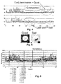
- the blastomere activity of 41 embryos is displayed as a pseudo-gel-image in Fig. 1 where motility peaks are indicated by dark bands and inactivity is white.
- Embryos that develop to blastocysts such as the left panel in Fig. 5 have uniformly distributed blastomere activity. Embryos that do not have uniformly distributed blastomere activity such as the right panel in Fig. 5 never develops into a blastocyst.
- the amount of cellular movement in different time intervals is a good indicator of embryo quality.
- a quality related parameter can be calculated from a ratio of average movement in different time-segments and/or a ratio of standard deviations in different time-segments Embryo selection procedures can be established based on the value of these parameters.
- Fig. 8 shows the excellent correspondence between automatic and manual determination of onset of cell division. Very early onset of the first cell division is an indication of high embryo quality. Very late onset of first (and subsequent cell divisions) indicates low quality embryos. However, for the majority of the embryos, the exact onset of the first cell division alone does not provide a clear indication of embryo quality as is shown in Fig. 8 below. While the average onset of cell divisions was delayed for the bad embryos, the large inherent standard deviation makes the absolute values a poor selection criteria except in extreme cases. (e.g. first division before 30 hours signifies a good embryo. First division after 35 hours signifies a bad embryo but the vast majority of the bovine embryos investigated have intermediate division times that are not easily interpreted.
- a typical time series of blastomeres activities consist of a few measurements every hour during incubation (e.g. approximately 150 data points for each embryo measured during the first 2 to 3 days which is the diagnostically interesting time window). Most statistical methods have difficulties with analysing data with such a high dimension. Thus, it is important to find robust methods for reducing the dimensions by extracting derived parameters. To achieve this, the blastomere activity was divided into three intervals: 0-32, 32-52 and 52-72 hours after image acquisition was started ( Fig. 9 ). Within each of these intervals three peaks were found using the following method: The first peak was the highest blastomere activity. The second peak was the highest activity value that was at least 3.5h before or after the first peak.
- Statistical models of embryo quality can be developed based on the above derived parameters, If each embryo has be evaluated according to the final development a number of different statistical methods exists for analysis the relation between the derived parameters and the final development. These methods includes: linear and non-linear models, Bayesians network, neural networks, hidden Markov models, nearest neighbours, principal component analysis and others.
- Fig. 12 below shows an example of a Principal Component Analysis (PCA) of the data.
- PCA Principal Component Analysis
- the statistical model can be evaluated and/or extended as new data are generated. To facilitate this it is important to find a robust data structure and set of derived parameters.
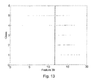
- parameter 39 baseline value of blastomere activity in the third time segment (76 to 96 hrs after fertilization) can to some extend to sort out abnormal and non-viable embryos. Based on this single parameter it is thus possible to automatically select embryos of good quality with with 72 % accuracy.
- a typical time series of blastomere activities consist of a few measurements every hour during incubation (e.g. approximately 150 data points for each embryo measured measured during the first 2 to 3 days which is the diagnostically interesting time window). Most statistical methods have difficulties with analysing data with such a high dimension. Thus, it is important to find robust methods for reducing the dimensions by extracting derived parameters. To achieve this, the blastomere activity was divided into three intervals: 0-32, 32-52 and 52-72 hours after image acquisition was started ( Figure 9 ). The three time intervals was selected to reflect three developmental stages for bovine embryos. Segment 1 : initial cell divisions from 1-cell to 8-cells. Segment 2: resting stage with relatively little activity and movements. It is believed the embryonic genome is activated at this stage.
- Segment 3 Resuming cell division an developing into a morula. It is often impossible to count individual blastomeres at this stage, but the time-lapse images reveal that cell division has resumed.
- the first peak was the highest blastomere activity.
- the second peak was the highest activity value that was at least 3.5h before or after the first peak.
- the third peak was the highest activity that was at least 3.5h from both the first and second peak.
- Peak 1 Peak 1, value, 2 Peak 1 time 3 Peak 1 mean 4 Valley 1, value 5 Valley 1 time 6 Valley 1 mean 7 Peak 2, value 8 Peak 2 time 9 Peak 2 mean 10 Valley 2, value 11 Valley 2 time 12 Valley 2 mean 13 Peak 3, value 14 Peak 3 time 15 Peak 3 mean
- Peak mean divided by peak value will always be ⁇ 1, with a value close to one indicating a broad peak and a value close to 0 a very sharp peak.
- the parameter set of 21 parameters shown above is used for a fast analysis as it only include information that can be gained from the first segment i.e. 32 hours of incubation.
- the small set contain important information that can me used to classify embryos in viable and not viable.
- the analysis can be repeated for the two following segments.
- the global average value, the global StDev and the global Minimum and maximum are included in the full parameter set of 59 parameters shown below:
- Statistical models of embryo quality can be developed based on the above derived parameters. If each embryo has be evaluated according to the final development a number of different statistical methods exists for analysis the relation between the derived parameters and the final development. These methods incudes but are not limited to: linear and non-linear models, Bayesians network, neural networks, hidden Markov models, nearest neighbours, principal component analysis and others. Figure 11 below shows an example of a Principal Component Analysis (PCA) of the data.
- PCA Principal Component Analysis
- Example 7 An example of the use of a linear model is shown in Example 7.
- the statistical model can be evaluated and/or extended as new data are generated. To facilitate this it is important to find a robust data structure and set of derived parameters.
- parameter 39 baseline value of blastomere activity in the third time segment (76 to 96 hrs after fertilization) can to some extend to sort out abnormal and non-viable embryos. Based on this single parameter it is thus possible to automatically select embryos of good quality with with 72 % accuracy.
- Example 7 Comparison of selection of embryos based on automated detection or embryologist detection
- Time-lapse images were acquired inside an incubator box fitted onto an inverted Nikon microscope stage mounted with a sensitive video camera. Results.
- the fully automated image analysis procedure generated a quantitative measure of cell blastomere activity based on the observed movement between consecutive images in the time-lapse series.
- the correlation between blastomere activity and cell division was confirmed by comparing automated and manual analysis of the time-lapse image series. Pronounced peaks in blastomere activity were found to be associated with cell-divisions. The exact onset and duration of cell-divisions could be quantified based on position, shape and size of the recorded peaks.
- the blastomere activity pattern of a given embryo could thus be reduced to a set of key parameters corresponding to peak height, position and width for prominent peaks as well as similar parameters describing the blastomere activity level between peaks.
- a total of 55 parameters for each embryo was used in a simple linear model to classify the embryo as "viable” or "non-viable".
- the model was trained on a subset of the observed embryo patterns and evaluated on a different independent subset. The same time-lapse series of images was evaluated by a skilled embryologist attempting to predict whether the embryo would develop to an expanded blastocyst or not.
Landscapes
- Health & Medical Sciences (AREA)
- Life Sciences & Earth Sciences (AREA)
- Engineering & Computer Science (AREA)
- Chemical & Material Sciences (AREA)
- Organic Chemistry (AREA)
- Zoology (AREA)
- Wood Science & Technology (AREA)
- Bioinformatics & Cheminformatics (AREA)
- Biomedical Technology (AREA)
- Genetics & Genomics (AREA)
- Biotechnology (AREA)
- General Health & Medical Sciences (AREA)
- Biochemistry (AREA)
- Microbiology (AREA)
- General Engineering & Computer Science (AREA)
- Analytical Chemistry (AREA)
- Molecular Biology (AREA)
- Reproductive Health (AREA)
- Gynecology & Obstetrics (AREA)
- Cell Biology (AREA)
- Sustainable Development (AREA)
- Immunology (AREA)
- Proteomics, Peptides & Aminoacids (AREA)
- Hematology (AREA)
- Developmental Biology & Embryology (AREA)
- Urology & Nephrology (AREA)
- Physics & Mathematics (AREA)
- Pregnancy & Childbirth (AREA)
- Surgery (AREA)
- Computer Hardware Design (AREA)
- Food Science & Technology (AREA)
- Medicinal Chemistry (AREA)
- General Physics & Mathematics (AREA)
- Pathology (AREA)
- Heart & Thoracic Surgery (AREA)
- Biophysics (AREA)
- Transplantation (AREA)
- Medical Informatics (AREA)
- Animal Behavior & Ethology (AREA)
- Public Health (AREA)
Applications Claiming Priority (5)
| Application Number | Priority Date | Filing Date | Title |
|---|---|---|---|
| US81411506P | 2006-06-16 | 2006-06-16 | |
| DKPA200600821 | 2006-06-16 | ||
| PCT/DK2006/000581 WO2007042044A1 (fr) | 2005-10-14 | 2006-10-16 | Determination d'un changement dans une population cellulaire |
| DKPA200700571 | 2007-04-19 | ||
| EP07722668A EP2035548B1 (fr) | 2006-06-16 | 2007-06-15 | Évaluation de la qualité d'un embryo sur la base de la division et du mouvement des blastomères |
Related Parent Applications (1)
| Application Number | Title | Priority Date | Filing Date |
|---|---|---|---|
| EP07722668.6 Division | 2007-06-15 |
Publications (1)
| Publication Number | Publication Date |
|---|---|
| EP2282210A1 true EP2282210A1 (fr) | 2011-02-09 |
Family
ID=43414894
Family Applications (2)
| Application Number | Title | Priority Date | Filing Date |
|---|---|---|---|
| EP10172399A Withdrawn EP2282210A1 (fr) | 2006-06-16 | 2007-06-15 | Évaluation de la qualité d'un embryo sur la base de la division et du mouvement des blastomères |
| EP07722668A Revoked EP2035548B1 (fr) | 2006-06-16 | 2007-06-15 | Évaluation de la qualité d'un embryo sur la base de la division et du mouvement des blastomères |
Family Applications After (1)
| Application Number | Title | Priority Date | Filing Date |
|---|---|---|---|
| EP07722668A Revoked EP2035548B1 (fr) | 2006-06-16 | 2007-06-15 | Évaluation de la qualité d'un embryo sur la base de la division et du mouvement des blastomères |
Country Status (8)
| Country | Link |
|---|---|
| US (4) | US20100041090A1 (fr) |
| EP (2) | EP2282210A1 (fr) |
| JP (2) | JP5731748B2 (fr) |
| CN (2) | CN101495619B (fr) |
| AT (1) | ATE477499T1 (fr) |
| DE (1) | DE602007008419D1 (fr) |
| DK (1) | DK2035548T3 (fr) |
| WO (1) | WO2007144001A2 (fr) |
Cited By (8)
| Publication number | Priority date | Publication date | Assignee | Title |
|---|---|---|---|---|
| US8323177B2 (en) | 2009-08-22 | 2012-12-04 | The Board Of Trustees Of The Leland Stanford Junior University | Imaging and evaluating embryos, oocytes, and stem cells |
| WO2012163363A1 (fr) * | 2011-05-31 | 2012-12-06 | Unisense Fertilitech A/S | Evaluation de la qualité des embryons basée sur le clivage des blastomères et morphologie |
| WO2013004239A1 (fr) * | 2011-07-02 | 2013-01-10 | Unisense Fertilitech A/S | Critères adaptatifs de sélection d'embryon optimisés par personnalisation et collaboration itératives |
| WO2014001312A1 (fr) * | 2012-06-25 | 2014-01-03 | Unisense Fertilitech A/S | Procédé et appareil |
| WO2014033210A1 (fr) * | 2012-08-30 | 2014-03-06 | Unisense Fertilitech A/S | Surveillance automatique d'embryons incubés in vitro |
| US9482659B2 (en) | 2010-09-27 | 2016-11-01 | Progyny, Inc. | Apparatus, method, and system for the automated imaging and evaluation of embryos, oocytes and stem cells |
| US9879307B2 (en) | 2011-02-23 | 2018-01-30 | The Board Of Trustees Of The Leland Stanford Junior University | Methods of detecting aneuploidy in human embryos |
| US10241108B2 (en) | 2013-02-01 | 2019-03-26 | Ares Trading S.A. | Abnormal syngamy phenotypes observed with time lapse imaging for early identification of embryos with lower development potential |
Families Citing this family (28)
| Publication number | Priority date | Publication date | Assignee | Title |
|---|---|---|---|---|
| CN101331500B (zh) | 2005-10-14 | 2015-04-29 | 尤尼森斯繁殖技术公司 | 细胞群的变化的测定 |
| DE602008005746D1 (de) | 2007-06-29 | 2011-05-05 | Unisense Fertilitech As | Vorrichtung, system und verfahren zur beobachtung und/oder kultivierung mikroskopischer objekte |
| GB0719037D0 (en) | 2007-09-28 | 2007-11-07 | Vitrolife Sweden Ab | Sampling needle |
| US20110111447A1 (en) * | 2008-07-05 | 2011-05-12 | Unisense Fertilitech A/S | One to one identification system |
| GB201006046D0 (en) * | 2010-04-12 | 2010-05-26 | Ge Healthcare Uk Ltd | System and method for determining motion of a biological object |
| JP5807288B2 (ja) * | 2010-06-30 | 2015-11-10 | 大日本印刷株式会社 | 体外培養による胚の製造方法、並びに胚を選別するための方法、装置、及びシステム |
| JP5962828B2 (ja) * | 2010-06-30 | 2016-08-03 | 大日本印刷株式会社 | 体外培養による胚の製造方法、並びに胚を選別するための方法、装置、及びシステム |
| GR1007617B (el) * | 2011-01-24 | 2012-06-20 | Ιδρυμα Τεχνολογιας Και Ερευνας - Ιτε, | Χρηση μη-γραμμικων απεικονιστικων τεχνικων για την αξιολογηση της ποιοτητας εμβρυων προεμφυτευτικου σταδιου και την προωθηση επιτυχους εγκυμοσυνης |
| AU2013269608B2 (en) | 2012-05-31 | 2016-05-05 | Unisense Fertilitech A/S | Embryo quality assessment based on blastocyst development |
| WO2013181549A2 (fr) * | 2012-05-31 | 2013-12-05 | Auxogyn, Inc. | Procédés de prédiction de blastocyste embryonnaire in vitro |
| WO2014134527A1 (fr) * | 2013-02-28 | 2014-09-04 | Auxogyn, Inc. | Appareil, procédé et système de dépistage automatisé et non invasif d'activité cellulaire |
| JP2014193145A (ja) * | 2013-03-01 | 2014-10-09 | Naoki Yamashita | 胚盤胞培養液中のノルエピネフリン量によるヒト胚盤胞の評価方法 |
| EP3011047B1 (fr) * | 2013-06-18 | 2018-08-01 | INSERM - Institut National de la Santé et de la Recherche Médicale | Procédés pour déterminer la qualité d'un embryon |
| ES2837840T3 (es) | 2014-03-20 | 2021-07-01 | Ares Trading Sa | Medida cuantitativa de la cinética de desarrollo de la morfología de mórula y blastocisto humanos |
| WO2015192352A1 (fr) * | 2014-06-19 | 2015-12-23 | 湖南光琇高新生命科技有限公司 | Procédé de classification par grade pour évaluer un embryon obtenu par traitement de fécondation in vitro en fonction des comportements de division |
| US10510143B1 (en) * | 2015-09-21 | 2019-12-17 | Ares Trading S.A. | Systems and methods for generating a mask for automated assessment of embryo quality |
| US11494578B1 (en) | 2015-09-21 | 2022-11-08 | Ares Trading S.A. | Systems and methods for automated assessment of embryo quality using image based features |
| JP2018014991A (ja) * | 2016-07-13 | 2018-02-01 | ソニー株式会社 | 情報処理装置、情報処理方法及び情報処理システム |
| JP2018022216A (ja) * | 2016-08-01 | 2018-02-08 | ソニー株式会社 | 情報処理装置、情報処理方法、及びプログラム |
| EP3530725A4 (fr) | 2016-11-02 | 2019-10-23 | Sony Corporation | Dispositif de traitement d'informations, procédé de traitement d'informations et système de traitement d'informations |
| JP6977293B2 (ja) * | 2017-03-31 | 2021-12-08 | ソニーグループ株式会社 | 情報処理装置、情報処理方法、プログラム及び観察システム |
| JP7100431B2 (ja) * | 2017-07-14 | 2022-07-13 | 株式会社ニコン | 受精卵判定装置、受精卵判定システム、及び受精卵判定プログラム |
| WO2019082617A1 (fr) | 2017-10-26 | 2019-05-02 | ソニー株式会社 | Dispositif de traitement d'informations, procédé de traitement d'informations, programme et système d'observation |
| JP6732722B2 (ja) * | 2017-12-11 | 2020-08-05 | 憲隆 福永 | 胚選抜システム |
| JP2020036580A (ja) * | 2018-06-20 | 2020-03-12 | Jcrファーマ株式会社 | 初期胚を選別するための解析ソフトウエア及び装置 |
| GB201810634D0 (en) * | 2018-06-28 | 2018-08-15 | Vitrolife As | Methods and apparatus for assessing embryo development |
| CN109214437A (zh) * | 2018-08-22 | 2019-01-15 | 湖南自兴智慧医疗科技有限公司 | 一种基于机器学习的ivf-et早孕胚胎发育预测系统 |
| CN116758539B (zh) * | 2023-08-17 | 2023-10-31 | 武汉互创联合科技有限公司 | 一种基于数据增强的胚胎图像卵裂球识别方法 |
Citations (1)
| Publication number | Priority date | Publication date | Assignee | Title |
|---|---|---|---|---|
| US20030138942A1 (en) * | 2002-01-24 | 2003-07-24 | Genx International, Inc. | Biological specimen-culturing system and method with onboard specimen development sensors |
Family Cites Families (27)
| Publication number | Priority date | Publication date | Assignee | Title |
|---|---|---|---|---|
| US3618734A (en) * | 1969-06-10 | 1971-11-09 | Res Foundation Of Children S H | Specimen incubator |
| DE2940446C2 (de) * | 1979-10-05 | 1982-07-08 | B. Braun Melsungen Ag, 3508 Melsungen | Züchtung von tierischen Zellen in Suspensions- und Monolayerkulturen in Fermentationsgefäßen |
| US4724543A (en) * | 1985-09-10 | 1988-02-09 | Beckman Research Institute, City Of Hope | Method and apparatus for automatic digital image analysis |
| JP2559760B2 (ja) * | 1987-08-31 | 1996-12-04 | 株式会社日立製作所 | 細胞搬送方法 |
| US5196168A (en) * | 1991-12-19 | 1993-03-23 | Eastman Kodak Company | Incubator with positioning device for slide elements |
| EP0590485B1 (fr) * | 1992-09-28 | 1998-07-29 | Becton, Dickinson and Company | Insert pour culture de cellules |
| US5763279A (en) * | 1993-09-09 | 1998-06-09 | Synthecon, Inc. | Gas permeable bioreactor and method of use |
| US5968340A (en) * | 1997-04-07 | 1999-10-19 | Marine Biological Laboratory | Polarographic self-referencing probe and method for using |
| US6228636B1 (en) * | 1998-09-21 | 2001-05-08 | Matsushita Electric Industrial Co., Ltd. | Incubator |
| DE19903506C2 (de) * | 1999-01-29 | 2002-04-04 | Inst Chemo Biosensorik | Verfahren, Gefäß und Vorrichtung zur Überwachung der Stoffwechselaktivität von Zellkulturen in flüssigen Medien |
| US6391577B1 (en) * | 1999-03-03 | 2002-05-21 | Susan R. Mikkelsen | Rapid electrochemical assay for antibiotic and cytotoxic drug susceptibility in microorganisms |
| JP3442357B2 (ja) * | 2000-08-25 | 2003-09-02 | 株式会社日立製作所 | 両生類卵母細胞試料導入装置、両生類卵母細胞試料導入システム、両生類卵母細胞試料導入方法、両生類卵母細胞の製造方法、両生類卵母細胞及びそれを販売又は譲渡する方法、スクリーニング用のセンサーとして用いる方法、容器、及び解析方法 |
| US6434320B1 (en) * | 2000-10-13 | 2002-08-13 | Comtrak Technologies, Llc | Method of searching recorded digital video for areas of activity |
| US6555365B2 (en) * | 2000-12-07 | 2003-04-29 | Biocrystal, Ltd. | Microincubator comprising a cell culture apparatus and a transparent heater |
| AU2003211104B2 (en) * | 2002-02-13 | 2009-01-29 | Reify Corporation | Method and apparatus for acquisition, compression, and characterization of spatiotemporal signals |
| US7326565B2 (en) * | 2002-11-19 | 2008-02-05 | Sanyo Electric Co., Ltd. | Storage apparatus |
| US20060201883A1 (en) * | 2003-02-19 | 2006-09-14 | Hofmeister William H | Polymer matrixes having nanoscale channels and uses |
| US7372985B2 (en) * | 2003-08-15 | 2008-05-13 | Massachusetts Institute Of Technology | Systems and methods for volumetric tissue scanning microscopy |
| US7239719B2 (en) * | 2003-08-22 | 2007-07-03 | Bbn Technologies Corp. | Automatic target detection and motion analysis from image data |
| CA2553673A1 (fr) * | 2003-08-28 | 2005-03-10 | Reprocure, Llc | Methode de determination de la qualite d'embryons |
| US20070015289A1 (en) * | 2003-09-19 | 2007-01-18 | Kao H P | Dispenser array spotting |
| JP3950972B2 (ja) * | 2003-11-14 | 2007-08-01 | 国立大学法人名古屋大学 | 生物試料の生物発光測定装置 |
| US7336401B2 (en) * | 2003-12-19 | 2008-02-26 | Xerox Corporation | Systems and methods for estimating an image marking process using event mapping of scanned image attributes |
| US7919319B2 (en) * | 2003-12-19 | 2011-04-05 | University Of Waterloo | Cultured cell and method and apparatus for cell culture |
| JP4333426B2 (ja) * | 2004-03-19 | 2009-09-16 | ソニー株式会社 | 化合物半導体の製造方法、及び半導体装置の製造方法 |
| CN101331500B (zh) * | 2005-10-14 | 2015-04-29 | 尤尼森斯繁殖技术公司 | 细胞群的变化的测定 |
| US20080056952A1 (en) * | 2006-08-25 | 2008-03-06 | Angros Lee H | Analytic plates with markable portions and methods of use |
-
2007
- 2007-06-15 CN CN200780027901.8A patent/CN101495619B/zh not_active Expired - Fee Related
- 2007-06-15 AT AT07722668T patent/ATE477499T1/de not_active IP Right Cessation
- 2007-06-15 DK DK07722668.6T patent/DK2035548T3/da active
- 2007-06-15 JP JP2009514641A patent/JP5731748B2/ja active Active
- 2007-06-15 US US12/304,905 patent/US20100041090A1/en not_active Abandoned
- 2007-06-15 WO PCT/DK2007/000291 patent/WO2007144001A2/fr active Application Filing
- 2007-06-15 EP EP10172399A patent/EP2282210A1/fr not_active Withdrawn
- 2007-06-15 CN CN201210428780.0A patent/CN103074410B/zh not_active Expired - Fee Related
- 2007-06-15 EP EP07722668A patent/EP2035548B1/fr not_active Revoked
- 2007-06-15 DE DE602007008419T patent/DE602007008419D1/de active Active
-
2012
- 2012-01-31 US US13/362,895 patent/US20120309043A1/en not_active Abandoned
- 2012-07-31 US US13/562,989 patent/US20130102837A1/en not_active Abandoned
- 2012-10-01 US US13/632,505 patent/US20130225917A1/en not_active Abandoned
-
2013
- 2013-07-05 JP JP2013141334A patent/JP5732110B2/ja active Active
Patent Citations (1)
| Publication number | Priority date | Publication date | Assignee | Title |
|---|---|---|---|---|
| US20030138942A1 (en) * | 2002-01-24 | 2003-07-24 | Genx International, Inc. | Biological specimen-culturing system and method with onboard specimen development sensors |
Non-Patent Citations (9)
| Title |
|---|
| GONZALES D S ET AL: "Prediction of the developmental potential of hamster embryos in vitro by precise timing of the third cell cycle", JOURNAL OF REPRODUCTION AND FERTILITY, JOURNALS OF REPRODUCTION AND FERTILITY LTD, GB, vol. 105, no. 1, September 1995 (1995-09-01), pages 1 - 8, XP009090408, ISSN: 0022-4251 * |
| GRISART B ET AL: "CINEMATOGRAPHIC ANALYSIS OF BOVINE EMBRYO DEVELOPMENT IN SERUM-FREE OVIDUCT-CONDITIONED MEDIUM", JOURNAL OF REPRODUCTION AND FERTILITY, JOURNALS OF REPRODUCTION AND FERTILITY LTD, GB, vol. 101, no. 2, 1994, pages 257 - 264, XP009079440, ISSN: 0022-4251 * |
| HOLM P ET AL: "Development kinetics of the first cell cycles of bovine in vitro produced embryos in relation to their in vitro viability and sex", THERIOGENOLOGY, vol. 50, no. 8, December 1998 (1998-12-01), pages 1285 - 1299, XP002455977, ISSN: 0093-691X * |
| LEQUARRE A S ET AL: "Cell cycle duration at the time of maternal zygotic transition for in vitro produced bovine embryos: Effect of oxygen tension and transcription inhibition.", BIOLOGY OF REPRODUCTION, vol. 69, no. 5, November 2003 (2003-11-01), pages 1707 - 1713, XP002455978, ISSN: 0006-3363 * |
| SCHATTEN ET AL: "The significance of mitochondria for embryo development in cloned farm animals", MITOCHONDRION, ELSEVIER, AMSTERDAM, NL, vol. 5, no. 5, October 2005 (2005-10-01), pages 303 - 321, XP005088701, ISSN: 1567-7249 * |
| SQUIRRELL J M ET AL: "Imaging mitochondrial organization in living primate oocytes and embryos using multiphoton microscopy", MICROSCOPY AND MICROANALYSIS, SPRINGER, NEW YORK, NY, US, vol. 9, no. 3, June 2003 (2003-06-01), pages 190 - 201, XP009091191, ISSN: 1431-9276 * |
| SQUIRRELL J M ET AL: "LONG-TERM TWO-PHOTON FLUORESCENCE IMAGING OF MAMMALIAN EMBRYOS WITHOUT COMPROMISING VIABILITY", NATURE BIOTECHNOLOGY, NATURE PUBLISHING GROUP, NEW YORK, NY, US, vol. 11, no. 17, August 1999 (1999-08-01), pages 763 - 767, XP001074651, ISSN: 1087-0156 * |
| TOKURA T ET AL: "Sequential observation of mitochondrial distribution in mouse oocytes and embryos", JOURNAL OF ASSISTED REPRODUCTION AND GENETICS, PLENUM PUBLISHING, US, vol. 10, no. 6, August 1993 (1993-08-01), pages 417 - 426, XP009091192, ISSN: 1058-0468 * |
| VAN BLERKOM J ET AL: "A microscopic and biochemical study of fragmentation phenotypes in stage-appropriate human embryos.", HUMAN REPRODUCTION (OXFORD, ENGLAND) APR 2001, vol. 16, no. 4, April 2001 (2001-04-01), pages 719 - 729, XP002468388, ISSN: 0268-1161 * |
Cited By (20)
| Publication number | Priority date | Publication date | Assignee | Title |
|---|---|---|---|---|
| US8721521B2 (en) | 2009-08-22 | 2014-05-13 | The Board Of Trustees Of The Leland Stanford Junior University | Imaging and evaluating embryos, oocytes, and stem cells |
| US8337387B2 (en) | 2009-08-22 | 2012-12-25 | The Board Of Trustees Of The Leland Stanford Junior University | Imaging and evaluating embryos, oocytes, and stem cells |
| US9228931B2 (en) | 2009-08-22 | 2016-01-05 | The Board Of Trustees Of The Leland Stanford Junior University | Imaging and evaluating embryos, oocytes, and stem cells |
| US8989475B2 (en) | 2009-08-22 | 2015-03-24 | The Board Of Trustees Of The Leland Stanford Junior University | Imaging and evaluating embryos, oocytes, and stem cells |
| US8951184B2 (en) | 2009-08-22 | 2015-02-10 | The Board Of Trustees Of The Leland Stanford Junior University | Imaging and evaluating embryos, oocytes, and stem cells |
| US8323177B2 (en) | 2009-08-22 | 2012-12-04 | The Board Of Trustees Of The Leland Stanford Junior University | Imaging and evaluating embryos, oocytes, and stem cells |
| US9482659B2 (en) | 2010-09-27 | 2016-11-01 | Progyny, Inc. | Apparatus, method, and system for the automated imaging and evaluation of embryos, oocytes and stem cells |
| US9879307B2 (en) | 2011-02-23 | 2018-01-30 | The Board Of Trustees Of The Leland Stanford Junior University | Methods of detecting aneuploidy in human embryos |
| EP2811016A1 (fr) * | 2011-05-31 | 2014-12-10 | Unisense Fertilitech A/S | Évaluation de la qualité des embryons basée sur le clivage des blastomères et morphologie |
| CN103748216A (zh) * | 2011-05-31 | 2014-04-23 | 尤尼森斯繁殖技术公司 | 基于卵裂球的卵裂和形态的胚胎质量评估 |
| CN104232566A (zh) * | 2011-05-31 | 2014-12-24 | 尤尼森斯繁殖技术公司 | 基于卵裂球的卵裂和形态的胚胎质量评估 |
| US20140087415A1 (en) * | 2011-05-31 | 2014-03-27 | Unisense Fertilitech A/S | Embryo quality assessment based on blastomere cleavage and morphology |
| CN103748216B (zh) * | 2011-05-31 | 2018-01-02 | 尤尼森斯繁殖技术公司 | 基于卵裂球的卵裂和形态的胚胎质量评估 |
| WO2012163363A1 (fr) * | 2011-05-31 | 2012-12-06 | Unisense Fertilitech A/S | Evaluation de la qualité des embryons basée sur le clivage des blastomères et morphologie |
| US20140128667A1 (en) * | 2011-07-02 | 2014-05-08 | Unisense Fertilitech A/S | Adaptive embryo selection criteria optimized through iterative customization and collaboration |
| WO2013004239A1 (fr) * | 2011-07-02 | 2013-01-10 | Unisense Fertilitech A/S | Critères adaptatifs de sélection d'embryon optimisés par personnalisation et collaboration itératives |
| CN104411817A (zh) * | 2012-06-25 | 2015-03-11 | 尤尼森斯繁殖技术公司 | 方法和设备 |
| WO2014001312A1 (fr) * | 2012-06-25 | 2014-01-03 | Unisense Fertilitech A/S | Procédé et appareil |
| WO2014033210A1 (fr) * | 2012-08-30 | 2014-03-06 | Unisense Fertilitech A/S | Surveillance automatique d'embryons incubés in vitro |
| US10241108B2 (en) | 2013-02-01 | 2019-03-26 | Ares Trading S.A. | Abnormal syngamy phenotypes observed with time lapse imaging for early identification of embryos with lower development potential |
Also Published As
| Publication number | Publication date |
|---|---|
| US20130225917A1 (en) | 2013-08-29 |
| JP2013198503A (ja) | 2013-10-03 |
| US20120309043A1 (en) | 2012-12-06 |
| US20130102837A1 (en) | 2013-04-25 |
| JP5731748B2 (ja) | 2015-06-10 |
| CN103074410A (zh) | 2013-05-01 |
| JP2009539387A (ja) | 2009-11-19 |
| CN101495619B (zh) | 2014-01-08 |
| DK2035548T3 (da) | 2010-11-22 |
| JP5732110B2 (ja) | 2015-06-10 |
| EP2035548B1 (fr) | 2010-08-11 |
| WO2007144001A2 (fr) | 2007-12-21 |
| DE602007008419D1 (de) | 2010-09-23 |
| CN101495619A (zh) | 2009-07-29 |
| US20100041090A1 (en) | 2010-02-18 |
| EP2035548A2 (fr) | 2009-03-18 |
| CN103074410B (zh) | 2016-12-07 |
| WO2007144001A3 (fr) | 2008-06-05 |
| ATE477499T1 (de) | 2010-08-15 |
Similar Documents
| Publication | Publication Date | Title |
|---|---|---|
| EP2035548B1 (fr) | Évaluation de la qualité d'un embryo sur la base de la division et du mouvement des blastomères | |
| EP2279868A1 (fr) | Récipient de liquide d'impression | |
| EP1949297B1 (fr) | Determination d'un changement dans une population cellulaire | |
| US9228931B2 (en) | Imaging and evaluating embryos, oocytes, and stem cells | |
| EP2855694B1 (fr) | Procédés de prédiction de blastocyste embryonnaire in vitro | |
| Sugimura et al. | Selection of viable in vitro-fertilized bovine embryos using time-lapse monitoring in microwell culture dishes | |
| EP2951574B1 (fr) | Mesure du potentiel de développement et d'implantation d'un embryon, faisant appel à des paramètres de durée et de phénotype résultant de la première cytokinèse | |
| Kim et al. | Prediction of blastocyst development and implantation potential in utero based on the third cleavage and compaction times in mouse pre-implantation embryos | |
| CN104411817A (zh) | 方法和设备 | |
| CN103757087B (zh) | 基于卵裂球分裂和运动的胚胎质量评估 | |
| ES2353622T3 (es) | Evaluación de la calidad del embrión basada en la división y el movimiento del blastómero. | |
| WO2016036697A1 (fr) | Méthodes de détection du mosaïcisme de l'embryon | |
| Morgan et al. | Fertilization and early embryology: Use of videocinematography to assess morphological qualities of conventionally cultured and cocultured embryos | |
| GB2513908A (en) | Method |
Legal Events
| Date | Code | Title | Description |
|---|---|---|---|
| PUAI | Public reference made under article 153(3) epc to a published international application that has entered the european phase |
Free format text: ORIGINAL CODE: 0009012 |
|
| AC | Divisional application: reference to earlier application |
Ref document number: 2035548 Country of ref document: EP Kind code of ref document: P |
|
| AK | Designated contracting states |
Kind code of ref document: A1 Designated state(s): AT BE BG CH CY CZ DE DK EE ES FI FR GB GR HU IE IS IT LI LT LU LV MC MT NL PL PT RO SE SI SK TR |
|
| 17P | Request for examination filed |
Effective date: 20110809 |
|
| 17Q | First examination report despatched |
Effective date: 20120820 |
|
| R17C | First examination report despatched (corrected) |
Effective date: 20120820 |
|
| STAA | Information on the status of an ep patent application or granted ep patent |
Free format text: STATUS: EXAMINATION IS IN PROGRESS |
|
| GRAP | Despatch of communication of intention to grant a patent |
Free format text: ORIGINAL CODE: EPIDOSNIGR1 |
|
| STAA | Information on the status of an ep patent application or granted ep patent |
Free format text: STATUS: GRANT OF PATENT IS INTENDED |
|
| INTG | Intention to grant announced |
Effective date: 20190717 |
|
| STAA | Information on the status of an ep patent application or granted ep patent |
Free format text: STATUS: THE APPLICATION IS DEEMED TO BE WITHDRAWN |
|
| 18D | Application deemed to be withdrawn |
Effective date: 20191128 |