EP2271672B1 - Protéines de récepteurs d'immunoglobuline et/ou de récepteurs toll associées à des troubles prolifératifs hématologiques myélogènes et utilisations correspondantes - Google Patents
Protéines de récepteurs d'immunoglobuline et/ou de récepteurs toll associées à des troubles prolifératifs hématologiques myélogènes et utilisations correspondantes Download PDFInfo
- Publication number
- EP2271672B1 EP2271672B1 EP09725022.9A EP09725022A EP2271672B1 EP 2271672 B1 EP2271672 B1 EP 2271672B1 EP 09725022 A EP09725022 A EP 09725022A EP 2271672 B1 EP2271672 B1 EP 2271672B1
- Authority
- EP
- European Patent Office
- Prior art keywords
- antibody
- antibodies
- cells
- expression
- cell
- Prior art date
- Legal status (The legal status is an assumption and is not a legal conclusion. Google has not performed a legal analysis and makes no representation as to the accuracy of the status listed.)
- Not-in-force
Links
Images


Classifications
-
- C—CHEMISTRY; METALLURGY
- C07—ORGANIC CHEMISTRY
- C07K—PEPTIDES
- C07K16/00—Immunoglobulins [IGs], e.g. monoclonal or polyclonal antibodies
- C07K16/18—Immunoglobulins [IGs], e.g. monoclonal or polyclonal antibodies against material from animals or humans
- C07K16/28—Immunoglobulins [IGs], e.g. monoclonal or polyclonal antibodies against material from animals or humans against receptors, cell surface antigens or cell surface determinants
- C07K16/2866—Immunoglobulins [IGs], e.g. monoclonal or polyclonal antibodies against material from animals or humans against receptors, cell surface antigens or cell surface determinants against receptors for cytokines, lymphokines, interferons
-
- A—HUMAN NECESSITIES
- A61—MEDICAL OR VETERINARY SCIENCE; HYGIENE
- A61K—PREPARATIONS FOR MEDICAL, DENTAL OR TOILETRY PURPOSES
- A61K39/00—Medicinal preparations containing antigens or antibodies
- A61K39/395—Antibodies; Immunoglobulins; Immune serum, e.g. antilymphocytic serum
- A61K39/39533—Antibodies; Immunoglobulins; Immune serum, e.g. antilymphocytic serum against materials from animals
- A61K39/39558—Antibodies; Immunoglobulins; Immune serum, e.g. antilymphocytic serum against materials from animals against tumor tissues, cells, antigens
-
- A—HUMAN NECESSITIES
- A61—MEDICAL OR VETERINARY SCIENCE; HYGIENE
- A61K—PREPARATIONS FOR MEDICAL, DENTAL OR TOILETRY PURPOSES
- A61K45/00—Medicinal preparations containing active ingredients not provided for in groups A61K31/00 - A61K41/00
- A61K45/06—Mixtures of active ingredients without chemical characterisation, e.g. antiphlogistics and cardiaca
-
- A—HUMAN NECESSITIES
- A61—MEDICAL OR VETERINARY SCIENCE; HYGIENE
- A61P—SPECIFIC THERAPEUTIC ACTIVITY OF CHEMICAL COMPOUNDS OR MEDICINAL PREPARATIONS
- A61P35/00—Antineoplastic agents
-
- A—HUMAN NECESSITIES
- A61—MEDICAL OR VETERINARY SCIENCE; HYGIENE
- A61P—SPECIFIC THERAPEUTIC ACTIVITY OF CHEMICAL COMPOUNDS OR MEDICINAL PREPARATIONS
- A61P35/00—Antineoplastic agents
- A61P35/02—Antineoplastic agents specific for leukemia
-
- C—CHEMISTRY; METALLURGY
- C07—ORGANIC CHEMISTRY
- C07K—PEPTIDES
- C07K16/00—Immunoglobulins [IGs], e.g. monoclonal or polyclonal antibodies
- C07K16/18—Immunoglobulins [IGs], e.g. monoclonal or polyclonal antibodies against material from animals or humans
- C07K16/28—Immunoglobulins [IGs], e.g. monoclonal or polyclonal antibodies against material from animals or humans against receptors, cell surface antigens or cell surface determinants
-
- G—PHYSICS
- G01—MEASURING; TESTING
- G01N—INVESTIGATING OR ANALYSING MATERIALS BY DETERMINING THEIR CHEMICAL OR PHYSICAL PROPERTIES
- G01N33/00—Investigating or analysing materials by specific methods not covered by groups G01N1/00 - G01N31/00
- G01N33/48—Biological material, e.g. blood, urine; Haemocytometers
- G01N33/50—Chemical analysis of biological material, e.g. blood, urine; Testing involving biospecific ligand binding methods; Immunological testing
- G01N33/53—Immunoassay; Biospecific binding assay; Materials therefor
- G01N33/569—Immunoassay; Biospecific binding assay; Materials therefor for microorganisms, e.g. protozoa, bacteria, viruses
- G01N33/56966—Animal cells
-
- G—PHYSICS
- G01—MEASURING; TESTING
- G01N—INVESTIGATING OR ANALYSING MATERIALS BY DETERMINING THEIR CHEMICAL OR PHYSICAL PROPERTIES
- G01N33/00—Investigating or analysing materials by specific methods not covered by groups G01N1/00 - G01N31/00
- G01N33/48—Biological material, e.g. blood, urine; Haemocytometers
- G01N33/50—Chemical analysis of biological material, e.g. blood, urine; Testing involving biospecific ligand binding methods; Immunological testing
- G01N33/53—Immunoassay; Biospecific binding assay; Materials therefor
- G01N33/574—Immunoassay; Biospecific binding assay; Materials therefor for cancer
- G01N33/57407—Specifically defined cancers
- G01N33/57426—Specifically defined cancers leukemia
-
- A—HUMAN NECESSITIES
- A61—MEDICAL OR VETERINARY SCIENCE; HYGIENE
- A61K—PREPARATIONS FOR MEDICAL, DENTAL OR TOILETRY PURPOSES
- A61K39/00—Medicinal preparations containing antigens or antibodies
- A61K2039/505—Medicinal preparations containing antigens or antibodies comprising antibodies
-
- C—CHEMISTRY; METALLURGY
- C07—ORGANIC CHEMISTRY
- C07K—PEPTIDES
- C07K2317/00—Immunoglobulins specific features
- C07K2317/70—Immunoglobulins specific features characterized by effect upon binding to a cell or to an antigen
- C07K2317/73—Inducing cell death, e.g. apoptosis, necrosis or inhibition of cell proliferation
-
- G—PHYSICS
- G01—MEASURING; TESTING
- G01N—INVESTIGATING OR ANALYSING MATERIALS BY DETERMINING THEIR CHEMICAL OR PHYSICAL PROPERTIES
- G01N2333/00—Assays involving biological materials from specific organisms or of a specific nature
- G01N2333/435—Assays involving biological materials from specific organisms or of a specific nature from animals; from humans
- G01N2333/705—Assays involving receptors, cell surface antigens or cell surface determinants
- G01N2333/72—Assays involving receptors, cell surface antigens or cell surface determinants for hormones
- G01N2333/726—G protein coupled receptor, e.g. TSHR-thyrotropin-receptor, LH/hCG receptor, FSH
-
- G—PHYSICS
- G01—MEASURING; TESTING
- G01N—INVESTIGATING OR ANALYSING MATERIALS BY DETERMINING THEIR CHEMICAL OR PHYSICAL PROPERTIES
- G01N2800/00—Detection or diagnosis of diseases
- G01N2800/52—Predicting or monitoring the response to treatment, e.g. for selection of therapy based on assay results in personalised medicine; Prognosis
-
- Y—GENERAL TAGGING OF NEW TECHNOLOGICAL DEVELOPMENTS; GENERAL TAGGING OF CROSS-SECTIONAL TECHNOLOGIES SPANNING OVER SEVERAL SECTIONS OF THE IPC; TECHNICAL SUBJECTS COVERED BY FORMER USPC CROSS-REFERENCE ART COLLECTIONS [XRACs] AND DIGESTS
- Y10—TECHNICAL SUBJECTS COVERED BY FORMER USPC
- Y10T—TECHNICAL SUBJECTS COVERED BY FORMER US CLASSIFICATION
- Y10T436/00—Chemistry: analytical and immunological testing
- Y10T436/14—Heterocyclic carbon compound [i.e., O, S, N, Se, Te, as only ring hetero atom]
- Y10T436/142222—Hetero-O [e.g., ascorbic acid, etc.]
- Y10T436/143333—Saccharide [e.g., DNA, etc.]
Definitions
- the present disclosure relates to a method of monitoring the efficacy of treating acute myeloid leukemia (AML) or chronic myeloid leukemia (CML) in a patient.
- AML acute myeloid leukemia
- CML chronic myeloid leukemia
- the cells of the hematopoietic system arise from multipotent progenitors, the hematopoietic stem cells (HSCs), which progress through a series of developmental programs to ultimately form the terminally differentiated cells of the myeloid or lymphoid lineage. It is believed that in the initial stages of hematopoiesis, HSCs commit to two distinguishable oligopotent but developmentally restricted progenitor cell types, the common lymphoid progenitors (CLPs) and the common myeloid progenitor (CMPs).
- CLPs common lymphoid progenitors
- CMPs common myeloid progenitor
- T lymphocytes, B lymphocytes, natural killer (NK) cells, and lymphoid dendritic cells develop from corresponding progenitor cells derived from the CLPs whereas erythroid cells, megakaryocytes, granulocytes, macrophages, and myeloid dendritic cells develop from their corresponding progenitor cells derived from CMPs.
- Cell populations at each stage of differentiation are distinguishable from other cell populations In the hematopoietic pathway based on programmed expression of a unique set of cell markers.
- HSCs are capable of self renewal - cell division that results in at least one of the daughter cells having the same characteristics as the parent cell - the progenitor cells committed to the lymphoid or myeloid lineages lose their potential to self-renew. That is, mitotic cell division of the committed progenitors leads to differentiated progeny rather than generation of a cell with the same proliferative and differentiation capacity as the parent cell.
- AML acute myeloid leukemia
- AML also called acute myelogenous leukemia
- AML is categorized into different subtypes based on morphological features and cytochemical staining properties, and although the self-renewal characteristic in most types of AML is attributable to leukemic cells having cell marker phenotypes consistent with HSCs ( Bonnet, D. and Dick, J.E., Nat. Med. 3(7):730-737 (1997 )), the chromosomal abnormality associated with the AML M3 subtype is observed in cell populations with a cell marker phenotype characteristic of more differentiated cells of the myeloid lineage (CD34 - , CD38 + ) whereas the HSC population in M3 does not carry the translocation ( Turhan, A.G. et al., Blood 79:2154-2161 (1995 )).
- CML chronic myeloid leukemia
- chronic myelogenous leukemia a disease commonly associated with the Philadelphia chromosome, which is a balanced translocation between chromosomes 9 and 22, t(9;22).
- the translocation produces a fusion between the bcr and c-abl genes and results In expression of a chimeric protein BCR-ABL with increased tyrosine kinase activity.
- the BCR-ABL fusion protein is mainly expressed In the committed cells of myelomonocytic lineage rather than the HSCs, indicating that committed cells in the myeloid lineage may be the source of the leukemic cells rather than the HSCs. Additional evidence for the committed myeloid cells as being the source of the leukemic clones in CML comes from studies of controlled expression of BCR-ABL in transgenic animals. Use of promoters active specifically in myeloid progenitor cells to force expression of BCR-ABL in committed cells but not in HSCs produces disease characteristic of CML in these transgenic animal models ( Jalswal, S. et al., Proc. Natl. Acad. Sci. USA 100:10002-10007 (2003 )).
- myeloproliferative disorders such as AML and CML are typically associated with cytogenetic abnormalities
- the cytogenetic defect may not be solely responsible for the proliferative trait.
- the chromosomal abnormality is observed in normal cells, which suggests that accumulation of additional mutations in either the HSCs or committed myeloid cells is required for full manifestation of the disease state.
- CML the disorder displays a multiphasic course, beginning from a chronic phase, which after 3-5 years and up to 10 years, leads to an accelerated or blastic phase similar to AML.
- the time period required to transition from the chronic phase (less than 5% blasts or promyelocytes) to the blastic phase (>30% blasts In the peripheral blood or bone marrow) may reflect the time needed to accumulate the mutations responsible for conversion of the chronic phase to the more aggressive blastic phase. For the most part, however, the leukemic cells appear to retain the cell marker phenotypes detectable in normal progenitor cells.
- cytotoxic or cytostatic chemotherapeutic agents For instance, busulfan, a bifunctional alkylating agent, and hydroxyurea, an Inhibitor of ribonucleoside diphosphate, affect DNA synthesis and stability, resulting in toxicity to dividing cells.
- Other therapeutic agents of similar activity include cytosine arabinoside (cytarabine) and daunorubicin.
- cytosine arabinoside cytarabine
- daunorubicin the effects of these agents are non-discriminatory and as a result they have serious side effects due to toxicity to normal dividing cells.
- BMT bone marrow transplant
- chemotherapy e.g. , cyclophosphamide
- hematopoietic system reconstituted by transplant of healthy hematopoietic stem cells.
- the transplant uses HLA matched allogeneic bone marrow cells from a family member (HLA-Identical) or a serologically matched altruistic donor (MUD).
- HLA-Identical family member
- ULD serologically matched altruistic donor
- Transplant with less well matched donors marketed increases the transplant related morbidity and mortality.
- This therapeutic approach has limited application because of Its dependence on the availability of suitable donors and because the treatments show better outcome for patients in the chronic or early phase of the disease as compared to acute or late stages.
- Antibody therapy for cancer Involves the use of antibodies, or antibody fragments, against an antigen to target antigen-expressing tumor cells. Because antibody therapy targets cells expressing a particular antigen, there is a possibility of cross-reactivity with normal cells and can lead to detrimental results. Substantial efforts have been directed to finding tumor-specific antigens. Tumor-specific antigens are found almost exclusively on tumors or are expressed at a greater level In tumor cells than the corresponding normal cells. Thus, tumor-specific antigens provide targets for antibody targeting of cancer, or other disease-related, cells expressing the antigen, as well as providing markers for diagnosis, for example, by identifying increased levels of expression. In immunotherapy approaches, antibodies specific to such tumor-specific antigens can be conjugated to cytotoxic compounds or can be used alone in immunotherapy.
- Immunotherapy as a treatment option against hematpoletic cancers, such as AML, is limited by the lack of tumor-associated antigens that are tumor-specific and that are shared among diverse patients. It is desirable to find other therapeutic agents that take advantage of the developmental origins of the leukemic cells by exploiting the common characteristics between leukemic cells and normal cell populations in the myeloid lineage. This approach would provide treatments that can supplement traditional therapies for myeloid leukemias, or that can be used as an alternative treatment to directly target the stem cell fractions of leukemic cells. This approach also provides additional diagnostic and prognostic strategies, as well as strategies for monitoring the efficacy of a therapeutic regimen.
- therapeutic treatment is more effective when tailored to a specific type of hematopoietic cancer. Predicting and determining efficacy of a treatment regime over time is also valuable in terms of clinical management. It is thus desirable to find tumor-specific markers that can be used in more efficient and accurate diagnosis and prognosis of myeloiod leukemic disorders, such as AML.
- immunoglobulin (Ig) superfamily of proteins are cell surface and soluble proteins that are involved in the recognition, binding or adhesion processes of cells. Members include cell surface antigen receptors, co-receptors, co-stimulatory molecules of the Immune system, molecules Involved in antigen presentation to lymphocytes, cell adhesion molecules, some cytokine recetpors, and intracellular muscle proteins.
- TLR toll-like receptor
- PAMP pathogen-associated molecular patterns
- the present invention provides a method of monitoring the efficacy of treating acute myeloid leukemia (AML) or chronic myeloid leukemia (CML) in a patient, comprising detecting in test samples obtained from said patient at two or more time points during said treatment a level of hepatitis A virus cellular receptor 2 (HAVCR2) expression; wherein a reduction in the level of said expression over time indicates a positive response to treatment.
- AML acute myeloid leukemia
- CML chronic myeloid leukemia
- cancer stem cell (CSC) markers or “cancer stem cell (CSC) targets” as well as “hematopoietic tumor cell (HTC) markers” or “hematopoietic tumor cell (HTC) targets” refer to genes and their expression products, such as mRNA and polypeptides, that have been found to be associated with HTCs by vlrture, for example, of increased expression and/or biological activity.
- CD180 mRNA transcribed from the CD180 gene Is found at higher levels in samples comprising AML HTCs as compared with samples comprising normal HSCs.
- An HTC associated with a given marker is referred to herein as a "marker+ HTC.”
- HTCs of myeloid origin particularly refers to cancer stem cells derived from cells of the myeloid (nonlymphoid) lineages, including monocytes, macrophages, neutrophils, basophils, eosinophils, erythrocytes, megakaryocytes/platelets, dendritic cells and the like.
- HTCs of myeloid origin will be those found in myeloid leukemias, such as AML and CML, where it is believed that progenitor cells commited to myeloid lineages regain self-renewing characteristics.
- HTCs of myeloid orign and “myeloid HTCs” or “myelogenous HTCs” are used herein interchangeably.
- Hematopoietic stem cell or “HSC” generally refers to clonogenic, self renewing pluripotent cells, capable of ultimately differentiating into all cell types of the hematopoietic system, including B cells T cells, NK cells, lymphoid dendritic cells, myeloid dendritic cells, granulocytes, macrophages, megakaryocytes, and erythroid cells.
- HSCs are typically defined by the presence of a characteristic set of cell markers.
- Marker phenotyping refers to Identification of markers or antigens on cells for determining its phenotype (e.g., differentiation state and/or cell type). This may be done by immunophenotyping, which uses antibodies that recognize antigens present on a cell. The antibodies may be monoclonal or polyclonal, but are generally chosen to have minimal crossreactivity with other cell markers. It is to be understood that certain cell differentiation or cell surface markers are unique to the animal species from which the cells are derived, while other cell markers will be common between species. These markers defining equivalent cell types between species are given the same marker identification even though there are species differences In structure (e.g., amino acid sequence).
- Cell markers include cell surfaces molecules, also referred to in certain situations as cell differentiation (CD) markers, and gene expression markers.
- the gene expression markers are those sets of expressed genes indicative of the cell type or differentiation state. In part, the gene expression profile will reflect the cell surface markers, although they may include non-cell surface molecules.
- Lineage markers are cell surface antigens that can be used for immunophenotyping cells of a particular developmental lineage.
- a set of 'Lin' antigens comprising CD2, CD3, CD4, CD5, CD8, NK1.1, B220, TER-119, Gr-1 can be used to identify mature murine blood cells.
- Cells that do not express these marker antigens, or express them at very low levels, are said to be lineage marker negative (Lin - ).
- the monoclonal antibody cocktails directed against these lineage markers can be used to remove cells expressing these antigens from source tissues (for example, bone marrow, umbilical cord blood, mobilized peripheral blood, fetal liver, and the like).
- This negative selection procedure yields a population of cells that is enriched for primitive hematopoietic stem cells or very early progenitor cells or precursor cells that do (not yet) express these markers (see, for example: KTLS cells). These cells are called lineage negative cells, abbreviated Lin* cells.
- Lin* cells lineage negative cells.
- Several subpopulations of lineage negative cells have been identified that are enriched for hematopoietic stem cells. They include Lin CD34 + cells (Krause et al, 1994), Lin - Sca -1+ c-Kit + Thy1 low cells (Fleming et al, 1993) and human CD34 + CD38 - cell populations.
- Antibody refers to a composition comprising a protein that binds specifically to a corresponding antigen and has a common, general structure of immunoglobulins.
- the term antibody specifically covers polyclonal antibodies, monoclonal antibodies, dimers, multimers, multispecific antibodies (e.g., bispecific antibodies), and antibody fragments, so long as they exhibit the desired biological activity.
- Antibodies may be murine, human, humanized, chimeric, or derived from other species.
- an antibody will comprise at least two heavy chains and two light chains interconnected by disulfide bonds, which when combined form a binding domain that Interacts with an antigen. Each heavy chain is comprised of a heavy chain variable region (V H ) and a heavy chain constant region (C H ).
- the heavy chain constant region is comprised of three domains, C H1 , C H2 and C H3 , and may be of the mu, delta, gamma, alpha or epsilon isotype.
- the light chain is comprised of a light chain variable region (V L ) and a light chain constant region (C L ).
- the light chain constant region is comprised of one domain, C L , which may be of the kappa or lambda isotype.
- the V H and V L regions can be further subdivided into regions of hypervariability, termed complementarity determining regions (CDR), interspersed with regions that are more conserved, termed framework regions (FR).
- CDR complementarity determining regions
- Each V H and V L is composed of three CDRs and four FRs, arranged from amino-terminus to carboxy-terminus in the following order: FR1, CDR1, FR2, CDR2, FR3, CDR3, FR4.
- the variable regions of the heavy and light chains contain a binding domain that interacts with an antigen.
- the constant regions of the antibodies may mediate the binding of the immunoglobulin to host tissues or factors, including various cells of the immune system (e.g., effector cells) and the first component (Clq) of the classical complement system.
- antibody mediates binding of the immunoglobulin to host tissue or host factors, particularly through cellular receptors such as the Fc receptors (e.g., Fc ⁇ RI, Fc ⁇ RII, Fc ⁇ RIII, etc.).
- Fc receptors e.g., Fc ⁇ RI, Fc ⁇ RII, Fc ⁇ RIII, etc.
- antibody also includes an antigen binding portion of an Immunoglobulin that retains the ability to bind antigen. These include, as examples, F(ab), a monovalent fragment of V L C L and V H C H antibody domains; and F(ab) 2 fragment, a bivalent fragment comprising two Fab fragments linked by a disulfide bridge at the hinge region.
- antibody also refers to recombinant single chain Fv fragments (scFv) and bispecific molecules such as, e.g., diabodies, triabodies, and tetrabodies (see, e.g., U.S. Patent No. 5,844,094 ).
- scFv single chain Fv fragments
- bispecific molecules such as, e.g., diabodies, triabodies, and tetrabodies (see, e.g., U.S. Patent No. 5,844,094 ).
- Antibodies may be produced and used in many forms, including antibody complexes.
- antibody complex or “antibody complexes” is used to mean a complex of one or more antibodies with another antibody or with an antibody fragment or fragments, or a complex of two or more antibody fragments.
- Antibody complexes include multimeric forms of antibodies directed to the disclosed HTC markers such as homoconjugates and heteroconjugates as well as other cross-linked antibodies as describd herein.
- Antigen Is to be construed broadly and refers to any molecule, composition, or particle that can bind specifically to an antibody.
- An antigen has one or more epitopes that interact with the antibody, although it does not necessarily Induce production of that antibody.
- cross-linked refers to the attachment of two or more antibodies to form antibody complexes, and may also be referred to as multimerization.
- Cross-linking or multimerization includes the attachment of two or more of the same antibodies (e.g. homodimerization), as well as the attachment of two or more different antibodies (e.g. heterodimerization).
- homodimerization the attachment of two or more antibodies to form antibody complexes
- heterodimerization e.g. heterodimerization
- Such conjugates may involve the attachment of two or more monoclonal antibodies of the same clonal origin (homoconjugates) or the attachment of two or more antibodies of different clonal origin (also referred to as heteroconjugates or bispecific).
- Antibodies may be crosslinked by non-covalent or covalent attachment. Numerous techniques suitable for cross-linking will be appreciated by those of skill in the art. Non-covalent attachment may be achieved through the use of a secondary antibody that Is specific to the primary antibody species. For example, a goat anti-mouse (GAM) secondary antibody may be used to cross-link a mouse monoclonal antibody. Covalent attachment may be achieved through the use of chemical cross-linkers.
- GAM goat anti-mouse
- Epitope refers to a determinant capable of specific binding to an antibody.
- Epitopes are chemical features generally present on surfaces of molecules and accessible to interaction with an antibody. Typical chemical features are amino acids and sugar moieties, having three-dimensional structural characteristics as well as chemical properties including charge, hydrophilicity, and lipophilicity. Conformational epitopes are distinguished from non-conformational epitopes by loss of reactivity with an antibody following a change in the spatial elements of the molecule without any change in the underlying chemical structure.
- Humanized antibody refers to an immunoglobulin molecule containing a minimal sequence derived from non-human immunoglobulin.
- Humanized antibodies include human immunoglobulins (recipient antibody) In which residues from a complementary determining region (CDR) of the recipient are replaced by residues from a CDR of a non-human species (donor antibody) such as mouse, rat or rabbit having the desired specificity, affinity and capacity.
- CDR complementary determining region
- donor antibody such as mouse, rat or rabbit having the desired specificity, affinity and capacity.
- Fv framework residues of the human immunoglobulin are replaced by corresponding non-human residues.
- Humanized antibodies may also comprise residues which are found neither in the recipient antibody nor in the imported CDR or framework sequences.
- a humanized antibody will comprise substantially all of at least one, and typically two, variable domains, in which all or substantially all of the CDR regions correspond to those of a non-human immunoglobulin and all or substantially all of the framework (FR) regions are those of a human immunoglobulin consensus sequence.
- a humanized antibody will also encompass immunoglobulins comprising at least a portion of an immunoglobulin constant region (Fc), generally that of a human immunoglobulin ( Jones et al., Nature 321:522-525 (1986 ); Relchmann et al, Nature 332:323-329 (1988 )).
- Immunogen refers to a substance, compound, or composition which stimulates the production of an Immune response.
- immunoglobulin locus refers to a genetic element or set of linked genetic elements that comprise information that can be used by a B cell or B cell precursor to express an immunoglobulin peptide.
- This peptide can be a heavy chain peptide, a light chain peptide, or the fusion of a heavy and a light chain peptide.
- the genetic elements are assembled by a Bcell precursor to form the gene encoding an immunoglobulin peptide.
- a gene encoding an immunoglobulin peptide is contained within the locus.
- Isotype refers to an antibody class defined by its heavy chain constant region. Heavy chains are generally classified as gamma, mu, alpha, delta, epsilon and designated as IgG, IgM, IgA, IgD, and IgE. Variations within each Isotype are categorized into subtypes, for example subtypes of IgG are divided into IgG 1 , IgG 2 , IgG 3 , and IgG 4 , while IgA is divided into IgA 1 and IgA 2 . The IgY isotype is specific to birds.
- “Monoclonal antibody” or “monoclonal antibody composition” refers to a preparation of antibody molecules of single molecular composition.
- a monoclonal antibody composition displays a single binding specificity and affinity for a particular epitope.
- human monoclonal antibody refers to antibodies displaying a single binding specificity which have variable and/or constant regions (if present) derived from human germline immunoglobulin sequences.
- the human monoclonal antibodies are produced by a hybridoma which Includes a B cell obtained from a transgenic non-human animal, e.g., a transgenic mouse, having a genome comprising a human heavy chain transgene and a light chain transgene fused to an immortalized cell.
- Single chain Fv or “scFv” refers to an antibody comprising the V H and V L regions of an antibody, wherein these domains are present in a single polypeptide chain.
- a scFv further comprises a polypeptide linker between the V H and V L domains which enables the scFv to form the desired structure for antigen binding.
- Specifically immunoreactive or antibody that “specifically binds” refers to a binding reaction of the antibody that is determinative of the presence of the antigen in a heterogeneous population of antigens.
- the antibody may be described as being “directed to” or “directed against” the particular antigen. Under a designated immunoassay condition, the antibody binds to the antigen at least two times, and typically 10-1000 times or more over background.
- Specifically Immunoreactive or “antibody that specifically binds” also refers to an antibody that Is capable of binding to an antigen with sufficient affinity such that the antibody is useful in targeting a cell having the antigen bound to its surface or in targeting the soluble antigen itself.
- the extent of non-specific binding is the amount of binding at or below background and will typically be less than about 10%, preferably less than about 5%, and more preferably less than about 1 % as determined by fluorescence activated cell sorting (FACS) analysis or radioimmunoprecipitation (RIA), for example.
- FACS fluorescence activated cell sorting
- RIA radioimmunoprecipitation
- Recombinant antibody refers to all antibodies prepared and expressed, created or isolated by recombinant techniques. These include antibodies obtained from an animal that is transgenic for the immunoglobulin locus, antibodies expressed from a recombinant expression vector, or antibodies created, prepared, and expressed by splicing of any immunoglobulin gene sequence to other nucleic acid sequences.
- HTC marker genes e.g., the genes of the disclosed members of the immunoglobulin (Ig) superfamily or toll-like receptor (TLR) superfamily
- HTCs hematopoietic tumor cells
- a transcription product such as RNA
- a translation product such as a polypeptide
- “Expression” or “expressing” as used herein refers to both transcriptional and translational processes directed by a gene. That is, the terms refer to the process of converting genetic information encoded In a nucleic acid sequence (gene) into RNA (e.g, mRNA rRNA, tRNA, snRNA, etc) through transcription of the gene; and/or convereting genetic information into protein through translation of mRNA.
- “expression product” as used herein refers to a trancription or translation product of a gene, and includes, e.g., RNA (mRNA, tRNA, rRNA, snRNA, etc.,) as well as polypeptides (intracellular, extracellular or suface expressed proteins).
- “Differential expression”, “differentially expressed”, “differential levels”, and the like, as used herein refers to a difference in the level of an expression product corresponding to a marker in HTCs in comparison to other mammalian cells, e.g., normal HSCs in particular. The difference can be expressed as an expression ratio or signal ratio, obtained from the quotient of the level of expression of an expression product In HTCs over the level of expression of the same expression product in HSCs.
- “ Differential expression” generally refers to a difference in expression levels of at least about 2 fold, at least about 3 fold, at least about 5 fold, at least about 7 fold, at least about 10 fold, or at least about 15 fold.
- the difference in expression levels is at least about 20 fold, at least about 30 fold, at least about 40 fold, or at least about 50 fold. In still more preferred embodiments, the difference in expression levels is at least about 70 fold, at least about 100 fold, at least about 200 fold, or as much as nearly 300 fold, 400 fold or 500 fold.
- extracellularly-expressed refers to the case where expression products are found outside of a cell, whether existing entirely outside of the plasma membrane of a cell (as in the case of secreted, soluble products); or existing partly outside of a cell as In the case of some membrane- (or surface-) expressed products.
- Membrane-bound proteins may be integral or peripheral, so long as at least a portion of the protein is accessible to antibodies outside the cell.
- membrane-bound includes membrane-expressed products as well as receptor bound products that become associated with surface membranes by virtue of binding to a membrane-bound receptor.
- Subject or “patient” are used Interchangeably and refer to, except where indicated, mammals such as humans and non-human primates, as well as rabbits, rats, mice, goats, pigs, and other mammalian species.
- Hematological proliferative disorder or “hematopoietic proliferative disorder” refers to a condition characterized by the clonal proliferation of one or more hematopoietic cells of the myeloid and/or lymphoid lineage.
- Hematological proliferative disorer of the myeloid lineage refers to conditions characterized by the clonal proliferation primarily of one or more hematopoietic cells of the myeloid lineage, rather than the lymphoid lineage.
- Myelogenous hematopoietic proliferative disorders include, e.g., the general classes of (a) dysmyelopolatic disease, (b) acute myeloproliferative leukemia and (c) chronic myeloproliferative disease. Each general class Is further categorized Into different subtypes, as is known in the art.
- the teachings of the present disclosure provide hybridoma cell lines and monoclonal antibodies that specifically bind to one or more of the following members of the immunoglobulin (Ig) superfamily or toll-like receptor (TLR)-superfamily: CD84; lymphocyte antigen 86 (Ly86); CD180 (RP105); hepatitis A virus cellular receptor 2 (HAVCR2); leukocyte immunoglobulin-like receptor, subfamily A with TM domain, member 1 (LILA1); leukocyte immunoglobulin-like receptor, subfamily A with TM domain, member 2 (LILRA2); neuronal growth regulator 1 (NEGR1); and toll-like receptor (TLR2).
- Ig immunoglobulin
- TLR toll-like receptor
- the following members of the Ig superfamily or TLR superfamily are over-expressed on the su rface of HTCs of myeloid origin: CD84; CD180 (RP105); hepatitis A virus cellular receptor 2 (HAVCR2); leukocyte immunoglobulin-like receptor, subfamily A with TM domain, member 1 (LILRA1); leukocyte immunoglobulin-like receptor, subfamily A with TM domain, member 2 (LILRA2); neuronal growth regulator 1 (NEGR1); and toll-like receptor (TLR2).
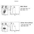
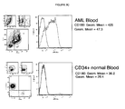
- the invention provides the use of anti-HAVCR2 antibodies.
- HAVCR2 also known as TIM3, KIM-3, TIMD3, and FLJ14428
- TIM3, KIM-3 is a T helper cell type 1-specific cell surface protein that regulates macrophage activation and enhances the severity of experimental autoimmune encephalomyelitis In mice.
- the present invention provides the use of anti-HAVCR2 antibodies, preferably monoclonal antibodies, that can specifically bind to HAVCR2 antigen, e.g., HAVCR2 polypeptide exposed on the surface of HTCs of myeloid origin.
- the anti-HAVCR2 antibodies preferably bind such myelogenous HTCs, thereby inhibiting their proliferation and/or mediating their destruction.
- anti-HAVCR2 antibodies commercially available from IMGENIX, Novus Biologicals, Lifespan Biosciences, and Atlas Antibodies, may also find use with respect to diagnostic and/or therapeutic applications taught herein.
- Monoclonal antibodies of the Instant disclosure specifically bind myelogenous HTCs by virtue of specifc binding to its target antigen.
- the monoclonal antibody (or a derivative thereof) is preferably specifically immunoreactive with cells of myeloid origin, such as granulocyte/macrophage progenitors (GMP), KG-1a, K-562, Jurkat, CML blasts, and AML blasts.
- GMP granulocyte/macrophage progenitors
- Antibodies can be produced readily by one skilled in the art.
- the general methodology for making monoclonal antibodies by hybridomas is now well known to the art. See, e.g., M. Schreier et al., Hybridoma Techniques (Cold Spring Harbor Laboratory ); Hammerling et al., Monoclonal Antibodies and T-Cell Hybridomas (Elsevier Biomedical Press ).
- the present disclosure provides methods of producing the monoclonal antibodies or derivatives thereof. In some embodiments, these methods comprise cultivating a hybridoma cell under suitable conditions, wherein the antibody is produced and obtaining the antibody and/or derivative thereof from the cell and/or from the cell culture medium.
- a specific example of making the monoclonal antibodies of the instant invention is also provided in the Examples below.
- the antibodies can be purified by methods known to the skilled artisan. Purification methods include, among others, selective precipitation, liquid chromatography, HPLC, electrophoresis, chromatofocusing, and various affinity techniques. Selective precipitation may use ammonium sulfate, ethanol (Cohn precipitation), polyethylene glycol, or others available in the art.
- Liquid chromatography mediums include, among others, ion exchange medium DEAE, polyaspartate), hydroxylapatite, size exclusion (e.g., those based on crosslinked agarose, acrylamide, dextran, etc.), hydrophobic matrixes (e.g., Blue Sepharose). Affinity techniques typically rely on proteins that interact with the immunoglobulin Fc domain.
- Protein A from Staphylococcus aureas can be used to purify antibodies that are based on human ⁇ 1, ⁇ 2, or ⁇ 4 heavy chains ( Lindmark et al., J. Immunol. Meth. 62:1-13 (1983 )).
- Protein L a Peptostreptococcus magnus cell-wall protein that binds Immunoglobulins (Ig) through k light-chain interactions (BD Bioscience/ ClonTech.
- Is useful for affinity purification of Ig subclasses IgM, IgA, IgD, IgG, IgE and IgY are also commercially available.
- the antibody contains metal binding residues, such as phage display antibodies constructed to contain histidine tags, metal affinity chromatography may be used.
- antigen affinity matrices may be made with the cells to provide an affinity method for purifying the antibodies.
- antibody fragment or "antigen-binding portion" of an antibody (or simply “antibody portion”) as used herein, refers to one or more fragments of an antibody that retain the ability to specifically bind to an antigen. It has been shown that the antigen-binding function of an antibody can be performed by fragments of a full-length antibody.
- binding fragments encompassed within the term "antibody fragment” or "antigen-binding portion” of an antibody include (i) a Fab fragment, a monovalent fragment consisting of the V L , V H , C L and C H1 domains; (ii) a F(ab') 2 fragment, a bivalent fragment comprising two Fab fragments linked by a disulfide bridge at the hinge region; (iii) a Fd fragment consisting of the V H and C H1 domains; (iv) a Fv fragment consisting of the V L and V H domains of a single arm of an antibody, (v) a dAb fragment ( Ward et al., (1989) Nature 341:544-546 ), which consists of a V H domain; and (vi) an isolated complementarity determining region (CDR), and (vii) bispecific single chain Fv dimers ( PCT/US92/09965 ).
- a Fab fragment a monovalent fragment consisting of the V L ,
- the two domains of the Fv fragment, V L and V H are coded for by separate genes, they can be joined, using recombinant methods, by a synthetic linker that enables them to be made as a single protein chain in which the V L and V H regions pair to form monovalent molecules (known as single chain Fv (scFv); see e.g., Bird et al. (1988) Science 242:423-426 ; and Huston et al. (1988) Proc. Natl. Acad. Sci. USA 85:5879-5883 ).
- single chain Fv single chain Fv
- Such single chain antibodies are also intended to be encompassed within the term "antigen-binding portion" of an antibody.
- antibody fragments are obtained using conventional techniques known to those with skill in the art, and the fragments are screened for utility In the same manner as are intact antibodies.
- the antibody fragments may be modified.
- the molecules may be stabilized by the incorporation of disulphide bridges linking the VH and VL domains ( Reiter et al., 1998, Nature Biotech. 14:1239-1245 ).
- the present disclosure further provides fragments of the antibodies disclosed herein.
- Immunoglobulin molecules can be cleaved into fragments.
- the antigen binding region of the molecule can be divided Into either F(ab') 2 or Fab fragments.
- the F(ab') 2 fragment is divalent and is useful when the Fc region is either undesirable or not a required feature.
- the Fab fragment is univalent and is useful when an antibody has a very high avidity for its antigen. Eliminating the Fc region from the antibody decreases non-specific binding between the Fc region and Fc receptor bearing cells.
- To generate Fab or F(ab) 2 fragments the antibodies are digested with an enzyme.
- Proteases that cleave at the hinge region of an immunoglobulin molecule preserve the disulfide bond(s) linking the F(ab) domain such that they remain together following cleavage.
- a suitable protease for this purpose is pepsin.
- proteases are chosen such that cleavage occurs above the hinge region containing the disulfide bonds that join the heavy chains but which leaves Intact the disulfide bond linking the heavy and light chain.
- a suitable protease for making F(ab) fragments is papain. The fragments are purified by the methods described above, with the exception of affinity techniques requiring the Intact Fc region (e.g., Protein A affinity chromatography).
- Antibody fragments can be produced by limited proteolysis of antibodies and are called proteolytic antibody fragments. These include, but are not limited to, the following: F(ab') 2 fragments, Fab' fragments, Fab'-SH fragments, and Fab fragments. "F(ab') 2 fragments" are released from an antibody by limited exposure of the antibody to a proteolytic enzyme, e.g., pepsin or ficin. A F(ab') 2 fragment comprises two "arms,” each of which comprises a variable region that is directed to and specifically binds a common antigen.
- Fab' fragments contain a single anti-binding domain comprising a Fab and an additional portion of the heavy chain through the hinge region.
- Fab'-SH fragments are typically produced from F(ab') 2 fragments, which are held together by disulfide bond(s) between the H chains in an F(ab') 2 fragment.
- Fab'-SH fragments are monovalent and monospecific.
- "Fab fragments" i.e., an antibody fragment that contains the antigen-binding domain and comprises a light chain and part of a heavy chain bridged by a disulfide bond
- a convenient method is to use papain immobilized on a resin so that the enzyme can be easily removed and the digestion terminated.
- Fab fragments do not have the disulfide bond(s) between the H chains present in a F(ab') 2 fragment.
- Single-chain antibodies are one type of antibody fragment.
- the term single chain antibody is often abbreviated as “scFv” or “sFv.” These antibody fragments are produced using molecular genetics and recombinant DNA technology.
- a single-chain antibody consists of a polypeptide chain that comprises both a V H and a V L domains which interact to form an antigen-binding site. The V H and V L domains are usually linked by a peptide of 10 to 25 amino acid residues.
- single-chain antibody further includes but is not limited to a disulfide-linked Fv (dsFv) In which two single-chain antibodies (each of which may be directed to a different epitope) linked together by a disulfide bond; a bispeciflc sFv in which two discrete scFvs of different specificity is connected with a peptide linker; a diabody (a dimerized sFv formed when the V H domain of a first sFv assembles with the V L domain of a second sFv and the V L domain of the first sFv assembles with the V H domain of the second sFv; the two antigen-binding regions of the diabody may be directed towards the same or different epitopes); and a triabody (a trimerized sFv, formed in a manner similar to a diabody, but in which three antigen-binding domains are created in a single complex; the three antigen binding
- CDR peptides are another form of an antibody fragment.
- a CDR peptide also known as “minimal recognition unit” is a peptide corresponding to a single complementarity-determining region (CDR), and can be prepared by constructing genes encoding the CDR of an antibody of interest. Such genes are prepared, for example, by using the polymerase chain reaction to synthesize the variable region from RNA of antibody-producing cells. See, for example, Larrick et al., Methods: A Companion to Methods in Enzymology 2:106, 1991 .
- cysteine-modified antibodies a cysteine amino acid is inserted or substituted on the surface of antibody by genetic manipulation and used to conjugate the antibody to another molecule via, e.g., a disulfide bridge. Cysteine substitutions or insertions for antibodies have been described (see U.S. Pat. No. 5,219,996 ). Methods for Introducing Cys residues into the constant region of the IgG antibodies for use In site-specific conjugation of antibodies are described byskyl et al. (J. Biol. Chem 275:330445-30450, 2000 ).
- the present disclosure further provides humanized and non-humanized antibodies.
- Humanized forms of non-human (e.g., mouse) antibodies are chimeric antibodies that contain minimal sequence derived from non-human immunoglobulin.
- humanized antibodies are non-human antibodies that have had the variable-domain framework regions swapped for sequences found In human antibodies.
- the humanized antibodies may be human immunoglobulins (recipient antibody) in which residues from a hypervariable region of the recipient are replaced by residues from a hypervariable region of a non-human species (donor antibody) such as mouse, rat, rabbit or nonhuman primate having the desired specificity, affinity, and capacity.
- donor antibody such as mouse, rat, rabbit or nonhuman primate having the desired specificity, affinity, and capacity.
- framework region (FR) residues of the human immunoglobulin are replaced by corresponding non-human residues.
- humanized antibodies may comprise residues that are not found in the recipient antibody or in the donor antibody. These modifications are made to further refine antibody performance.
- the humanized antibody will comprise substantially all of at least one, and typically two, variable domains, In which all or substantially all of the hypervariable loops correspond to those of a non-human immunoglobulin and all or substantially all of the FRs are those of a human Immunoglobulin sequence.
- the humanized antibody optionally also will comprise at least a portion of an immunoglobulin constant region (Fc), typically that of a human immunoglobulin.
- Fc immunoglobulin constant region
- a humanized antibody the entire antibody, except the CDRs, Is encoded by a polynucleotide of human origin or is identical to such an antibody except within its CDRs.
- the CDRs some or all of which are encoded by nucleic acids originating In a non-human organism, are grafted into the beta-sheet framework of a human antibody variable region to create an antibody, the specificity of which Is determined by the engrafted CDRs.
- the creation of such antibodies Is described In, e.g., WO 92/11018 , Jones, 1986, Nature 321:522-525 , Verhoeyen et al., 1988, Science 239:1534-1536 .
- Humanized antibodies can also be generated using mice with a genetically engineered immune system. Roque et al., 2004, Biotechnol. Prog. 20:639-654 .
- humanized and non-humanized antibodies Humanized forms of non-human (e.g. , mouse) antibodies are chimeric antibodies that contain minimal sequence derived from non-human immunoglobulin.
- humanized antibodies are human immunoglobulins (recipient antibody) In which residues from a hypervariable region of the recipient are replaced by residues from a hypervariable region of a non-human species (donor antibody) such as mouse, rat, rabbit or nonhuman primate having the desired specificity, affinity, and capacity.
- donor antibody such as mouse, rat, rabbit or nonhuman primate having the desired specificity, affinity, and capacity.
- framework region (FR) residues of the human immunoglobulin are replaced by corresponding non-human residues.
- humanized antibodies may comprise residues that are not found in the recipient antibody or in the donor antibody.
- the humanized antibody will comprise substantially all of at least one, and typically two, variable domains, in which all or substantially all of the hypervariable loops correspond to those of a non-human immunoglobulin and all or substantially all of the FRs are those of a human immunoglobulin sequence.
- the humanized antibody optionally also will comprise at least a portion of an immunoglobulin constant region (Fc), typically that of a human immunoglobulin.
- cysteine residue(s) can be introduced into the Fc region, thereby allowing interchain disulfide bond formation in this region.
- the homodimeric antibody thus generated can have improved internalization capability and/or increased complement-mediated cell killing and antibody-dependent cellular cytotoxicity (ADCC). See Caron et al., J. Exp Med., 176:1191-1195 (1992 ) and Shopes, J. Immunol., 148:2918-2922 (1992 ).
- Homodimeric antibodies with enhanced anti-tumor activity can also be prepared using heterobifunctional cross-linkers as described in Wolff et al. Cancer Research, 53:2560-2565 (1993 ).
- an antibody can be engineered that has dual Fc regions and can thereby have enhanced complement lysis and ADCC capabilities. See Stevenson et al., Anti-Cancer Drug Design, 3:219-230 (1989 ).
- the antibodies described herein specifically bind to an antigen corresponding to one or more of the disclosed members of the Ig superfamily or TLR superfamily present on the cell surface of HTCs that arose from progenitor cell populations in the myeloid lineage of the hematopoietic system.
- Differentiation in the myeloid lineage leads to formation of terminally differentiated cells that include, among others, megakaryocytes, erythroid cells, macrophages, basophils, eosinophils, neutrophils and myeloid dendritic cells.
- HSC hematopoietic stem cells
- the HSCs and the progenitor cell populations are identifiable from each other based on, among other distinguishing characteristics, their capacity to differentiate into specific cell subsets and the presence of a particular set of cellular markers that Is specific to the cell population.
- the monoclonal antibodies of the present disclosure are directed to progenitor cells of the myeloid lineage that are marker+ HTCs. In some cases the monoclonal antibodies in the present disclosure are directed to committed myeloid progenitor cells that are marker+ HTCs.
- modified antibodies that are derived from an antibody that specifically binds an antigen corresponding to an HTC marker disclosed herein.
- Modified antibodies also include recombinant antibodies as described herein.
- modified or recombinant antibodies include without limitation, engineered murine monoclonal antibodies (e.g. murine monoclonal antibodies, chimeric monoclonal antibodies, humanized monoclonal antibodies), domain antibodies (e.g. Fab, Fv, V H , scFV, and dsFv fragments), multivalent or multispecific antibodies (e.g. diabodies, minibodies, miniantibodies, (scFV) 2 , tribodies, and tetrabodies), and antibody conjugates as described herein.
- engineered murine monoclonal antibodies e.g. murine monoclonal antibodies, chimeric monoclonal antibodies, humanized monoclonal antibodies
- domain antibodies e.g. Fab, Fv, V H , scFV, and dsFv fragments
- multivalent or multispecific antibodies e.g. diabodies, minibodies, miniantibodies, (scFV) 2 , tribodies, and tetra
- the present disclosure includes domain antibodies.
- Domain antibodies are functional binding domains of antibodies, corresponding to the variable regions of either the heavy (VH) or light (VL) chains of human antibodies. Domain antibodies may have a molecular weight of approximately 13 kDa, or less than one-tenth the size of a full antibody. They are well expressed In a variety of hosts including bacterial, yeast, and mammalian cell systems. In addition, domain antibodies are highly stable and retain activity even after being subjected to harsh conditions, such as freeze-drying or heat denaturation. See, for example, US Patent 6,291,158 ; 6,582,915 ; 6,593,081 ; 6,172,197 ; US Serial No.
- the domain antibody of the present disclosure is a single domain.
- Single domain antibodies may be prepared, for example, as described in U.S. Patent No. 6,248,516 ,
- the present disclosure provides domain antibodies derived from an antibody that specifically binds to an antigen corresponding to one of the members of the Ig superfamily or TLR superfamily disclosed herein.
- the present disclosure includes multi-specific antibodies.
- Multi-specific antibodies include bispecific, trispecific, etc. antibodies.
- Bispecific antibodies can be produced via recombinant means, for example by using leucine zipper moieties (i.e., from the Fos and Jun proteins, which preferentially form heterodimers; Kostelny et al., 1992, J. Immnol. 148:1547 ) or other lock and key interactive domain structures as described in U.S. Pat. No. 5,582,996 . Additional useful techniques include those described in U.S. Pat. No. 5,959,083 ; and U.S. Pat. No. 5,807,706 .
- the present disclosure provides multi-specific antibodies that include an antibody that specifically binds an antigen corresponding to a member of the Ig superfamily or TLR superfamily disclosed herein.
- the multispecific antibody is bispecific.
- Bispecific antibodies are also sometimes referred to as "diabodies.” These are antibodies that bind to two (or more) different antigens. Also known in the art are triabodies (a trimerized sFv, formed in a manner similar to a diabody, but in which three antigen-binding domains are created in a single complex; the three antigen binding domains may be directed towards the same or different epltopes) or a tetrabodies (four antigen-binding domains created in a single complex where the four antigen binding domains may be directed towards the same or different epitopes), and the like.
- triabodies a trimerized sFv, formed in a manner similar to a diabody, but in which three antigen-binding domains are created in a single complex; the three antigen binding domains may be directed towards the same or different epltopes
- a tetrabodies four antigen-binding domains created in a single complex where the four antigen
- Dia-, tria- and tetrabodies can be manufactured in a variety of ways known in the art ( Holliger and Winter, 1993, Current Opinion Biotechnol. 4:446-449 ), e.g. , prepared chemically or from hybrid hybridomas.
- such antibodies and fragments thereof may be constructed by gene fusion ( Tomlinson et. al., 2000, Methods Enzymol. 326:461-479 ; WO94/13804 ; Holliger et al., 1993, Proc. Natl. Acad. Sci. U.S.A.
- the diabody, triabody, ortetrabody is derived from an antibody that specifically binds an antigen corresponding to a member of the Ig superfamily or TLR superfamily disclosed herein.
- the diabody, triabody, or tetrabody is derived from two or more monoclonal antibodies that specifically bind to different antigens, each antigen corresponding to different members of the Ig superfamily or TLR superfamily disclosed herein.
- minibodies which are minimized antibody-like proteins that include a scFV joined to a CH3 domain, that are derived from an antibody that specifically binds an antigen corresponding to a member of the Ig superfamily or TLR superfamily disclosed herein.
- Minibodies can be made as described in the art ( Hu et al., 1996, Cancer Res. 56:3055-3061 ).
- the present disclosure provides binding domain-immunglobulin fusion proteins, where the binding domain specifically binds an antigen corresponding to a member of the Ig superfamily or TLR superfamily disclosed herein.
- the fusion protein may include a marker specific binding domain polypeptide fused to an immunoglobulin hinge region polypeptide, which is fused to an immunoglobulin heavy chain CH2 constant region polypeptide fused to an immunoglobulin heavy chain CH3 constant region polypeptide.
- marker specific antibody fusion proteins can be made by methods appreciated by those of skill in the art (See published U.S. Patent Application Nos. 20050238646 , 20050202534 , 20050202028 , 2005020023 , 2005020212 , 200501866216 , 20050180970 , and 20050175614 ,
- the present disclosure provides a heavy-chain protein (V HH ) derived from a marker specific antibody.
- Naturally-occuring heavy chain antibodies e.g. camelidae antibodies having no light chains
- V HH heavy chain proteins
- Naturally-occuring heavy chain antibodies have been utiltized to develop antibody-derived therapeutic proteins that typically retain the structure and functional properties of naturally-occuring heavy-chain antibodies. They are known In the art as Nanobodies®.
- heavy chain proteins (V HH ) derived from a marker specific heavy chain antibody may be made by methods appreciated by those of skill in the art (See published U.S. Patent Application Nos. 20060246477 , 20060211088 , 20060149041 , 20060115470 , and 20050214857 .
- the present disclosure provides a modified antibody that is a human antibody.
- the marker specific antibodies described herein are fully human antibodies.
- "Fully human antibody” or “complete human antibody” refers to a human antibody having only the gene sequence of an antibody derived from a human chromosome.
- the anti-human marker specific complete human antibody can be obtained by a method using a human antibody-producing mouse having a human chromosome fragment containing the genes for a heavy chain and light chain of a human antibody [see Tomizuka, K. et al., Nature Genetics, 16, p.133-143, 1997 ; Kuroiwa, Y. et al., Nuc.
- the present disclosure provides a marker specific antibody that is an antibody analog, sometimes referred to as "synthetic antibodies.”
- synthetic antibodies include synthetic antibodies.
- a variety of recent work utilizes either alternative protein scaffolds or artificial scaffolds with grafted CDRs.
- Such scaffolds include, but are not limited to, mutations Introduced to stabilize the three-dimensional structure of the binding protein as well as wholly synthetic scaffolds consisting for example of biocompatible polymers. See, for example, Korndorfer et al., 2003, Proteins: Structure, Function, and Bioinformatics, Volume 53, Issue 1:121-129 . Roque et al., 2004, Biotechnol. Prog. 20:639-654 .
- PAMs peptide antibody mimetics
- the present disclosure provides cross-linked antibodies that include two or more antibodies described herein attached to each other to form antibody complexes.
- Cross-linked antibodies are also referred to as antibody multimers, homoconjugates, and heteroconjugates. It has been observed in the art that the multimerization of an antibody previously observed to have no signalling activity can result In a multimerized antibody with potent signalling activity. This has been particularly noted in the field of anti-tumor agents. For example, it has been reported that the IgG-IgG homodimerization of anti-CD19, anti-CD20, anti-CD21, anti-CD22, and anti-Her-2 monoclonal antibodies confers potent anti-tumor ability to such homodimers ( Ghetle, M.
- the antibody complexes provided herein include multimeric forms of antibodies directed to one or more of the antigens corresponding to one or more of the members of the Ig superfamily or TLR superfamily disclosed herein.
- antibodies complexes of the, disclosure may take the form of antibody dimers, trimers or higher-order multimers of monomeric Immunoglobulin molecules.
- Crosslinking of antibodies can be done through various methods know in the art. For example, crosslinking of antibodies may be accomplished through natural aggregation of antibodies, through chemical or recombinant linking techniques or other methods known in the art.
- purified antibody preparations can spontaneously form protein aggregates containing antibody homodimers, and other higher-order antibody multimers.
- the present disclosure provides homodimerized antibodies that specifically bind to an antigen corresponding to an HTC marker disclosed herein.
- Antibodies can be cross-linked or dimerized through linkage techniques known In the art (see Ghetie et al. (1997) supra ; Ghetle et al. (2001) supra ). Non-covalent methods of attachment may be utilized.
- crosslinking of antibodies can be achieved through the use of a secondary crosslinker antibody.
- the crosslinker antibody can be derived from a different animal compared to the antibody of interest. For example, a goat anti-mouse antibody (Fab specific) may be added to a mouse monoclonal antibody to form a heterodimer. This bivalent crosslinker antibody recognizes the Fab or Fc region of the two antibodies of interest forming a homodlmer.
- an antibody that specifically binds to an antigen corresponding to a member of the Ig superfamily or TLR superfamily disclosed herein is cross-linked using a goat anti-mouse antibody (GAM).
- GAM goat anti-mouse antibody
- the GAM crosslinker recognizes the Fab or Fc region of two antibodies, each of which specifically binds the same or two different antigens corresponding to the same or different members of the Ig superfamily or TLR superfamily disclosed herein.
- Chemical crosslinkers can be homo or heteroblfunctional and will covalently bind with two antibodies forming a homodimer.
- Cross-linking agents are well known In the art; for example, homo-or hetero-bifunctional linkers as are well known (see the 2006 Pierce Chemical Company Crosslinking Reagents Technical Handbook ; Hermanson, G.T., Bioconjugate Techniques, Academic Press, San Diego, CA (1996 ); Aslam M.
- Suitable examples of chemical crosslinkers used for antibody crosslinking include, but not limited to, SMCC [succinimidyl 4-(maleimldomethyl)cyclohexane-1-carboxylate], SATA [N-succinimldyl S-acethylthlo-acetate], hemi-succinate esters of N-hydroxysuccinimide; sulfo-N- hydroxy-succlnlmide; hydroxybenzotriazole, and p-nitrophenol; dicyclohexylcarbodiimide (DCC), 1-(3-dimethylaminopropyl)-3-ethylcarbodiimide (ECD), and 1-(3-dimethylaminopropyl)-3-ethylcarbodlimide methiodide (EDCI) (see, e.g., U.S.
- DCC dicyclohexylcarbodiimide
- ECD 1-(3-dimethylamin
- linking reagents include glutathione, 3-(diethoxyphosphoryloxy)-1,2,3- benzotriazin-4(3H)-one (DEPBT), onium salt-based coupling reagents, polyoxyethylene-based heterobifunctional cross-linking reagents, and other reagents ( Haltao, et al., Organ Lett 1:91-94 (1999 ); Albericio et al., J Organic Chemistry 63:9678-9683 (1998 ); Arpicco et al., Bloconjugate Chem. 8:327-337 (1997 ); Frisch et al., Bloconjugate Chem.
- the chemical cross-linker used is an SMCC or SATA crosslinker.
- the antibody-antibody conjugates of this disclosure can be covalently bound to each other by techniques known in the art such as the use of the heterobifunctional cross-linking reagents, GMBS (maleimidobutryloxy succinimide), and SPDP (N-succinimidyl 3-(2-pyridyldithio)propionate) [see, e.g., Hardy, "Purification And Coupling Of Fluorescent Proteins For Use In Flow Cytometry", Handbook Of Experimental Immunology, Volume 1, Immunochemistry, Weir et al. (eds.), pp. 31.4-31.12 4th Ed., (1986 ), and Ledbetter et al. U.S. Patent No. 6,010,902 .
- antibodies may be linked via a thloether cross-link as described in U.S. Patent Publication 20060216284 , U.S. Patent No. 6,368,596 ,
- antibodies can be crosslinked at the Fab region.
- the antibodies disclosed herein can be conjugated to inorganic or organic compounds, including, by way of example and not limitation, other proteins, nucleic acids, carbohydrates, steroids, and lipids (see for example Green, et al., Cancer Treatment Reviews, 26:269-286 (2000 ).
- the compound may be bioactive.
- Bioactive refers to a compound having a physiological effect on the cell as compared to a cell not exposed to the compound.
- a physiological effect is a change in a biological process, including, by way of example and not limitation, DNA replication and repair, recombination, transcription, translation, secretion, membrane turnover, cell adhesion, signal transduction, cell death, and the like.
- a bioactive compound includes pharmaceutical compounds.
- the antibodies are conjugated to or modified to carry a detectable compound.
- Conjugating antibodies to detectable enzymes, fluorochromes, or ligands provides a signal for visualization or quantitation of the target antigen.
- Antibodies may be labeled with various enzymes to provide highly specific probes that both visualize the target and amplify the signal by acting on a substrate to produce a colored or chemiluminescent product.
- Horseradish peroxidase, alkaline phosphatase, glucose oxidase, and ⁇ -galactosidase are the commonly used enzymes for this purpose.
- Fluorochromes such as fluorescein isothiocyanate, tetramethyirhodamine isothiocyanate (TRITC), phycoerythrin, and Cy5, provide a colored reagent for visualization and detection.
- Suitable fluorescent compounds are described in Haughland, R.P., Handbook of Fluorescent Probes and Research Chemicals Eugene, 9th Ed., Molecular Probes, OR (2003 ).
- the conjugated compounds are chelating ligands, or macrocyclic organic chelating compounds, particularly metal chelating compounds used to Image intracellular ion concentrations or used as contrast agents for medical imaging purposes.
- Chelating ligands are ligands that can bind with more than one donor atom to the same central metal ion. Chelators or their complexes have found applications as MRI contrast agents, radiopharmaceutical applications, and luminescent probes.
- Conjugates of chelating compounds useful for assessing intracellular ion concentrations may be voltage sensitive dyes and non-voltage sensitive dyes.
- Exemplary dye molecules for measuring intracellular ion levels include, by way of example and not limitation, Quin-2; Fluo-3; Fura-Red; Calcium Green; Calcium Orange 550 580; Calcium Crimson; Rhod-2 550 575; SPQ; SPA; MQAE; Fura-2; Mag-Fura-2; Mag-Fura-5; Di-4-ANEPPS; Di-8-ANEPPS; BCECF; SNAFL-1; SBFI; and SBFI.
- the ligands are chelating ligands that bind paramagnetic, superparamagnetic or ferromagnetic metals. These are useful as contrast agents for medical imaging and for delivery of radioactive metals to selected cells.
- Metal chelating ligands include, by way of example and not limitation, diethylenetriaminepenta acetic acid (DTPA); diethylenetriamlnepenta acetic acid bis(methylamide); macrocyclic tetraamine 1,4,7,10-tetraazacyclododecane-N,N',N',N"'-tetraacetic acid (DOTA); and porphyrins (see, e.g., The Chemistry of Contrast Agents in Medical Magnetic Resonance Imaging, Merbach A.E.
- Paramagnetic metal Ions which are detectable in their chelated form by magnetic resonance imaging, include, for example, iron(III), gadolinium(III), manganese (II and III), chromium(III), copper(II), dysprosium(III), terblum(III), holmium (III), erbium (III), and europium (III).
- Paramagnetic metal ions particularly useful as magnetic resonance imaging contrast agents comprise iron(III) and gadolinium(III) metal complexes. Other paramagnetic, superparamagnetic or ferromagnetic are well known to those skilled In the art.
- the metal-chelate comprises a radioactive metal.
- Radioactive metals may be used for diagnosis or as therapy based on delivery of small doses of radiation to a specific site in the body.
- Targeted metalloradiopharmaceuticals are constructed by attaching the radioactive metal ion to a metal chelating ligand, such as those used for magnetic imaging, and delivering the chelate-complex to cells.
- An exemplary radioactive metal chelate complex is DTPA (see, e.g., U.S. Patent No. 6,010,679 ).
- the conjugated compounds are peptide tags used for purposes of detection, particularly through the use of antibodies directed against the peptide.
- tag polypeptides and their respective antibodies are well known in the art. Examples include poly-histidine (poly-his) or poly-histidine-glycine (poly-his-gly) tags; the flu HA tag polypeptide and its antibody 12CA5 ( Field et al., Mol. Cell. Biol. 8:2159-2165 (1988 )); the c-myc tag and the 8F9, 3C7, 6E10, G4, B7 and 9E10 antibodies thereto ( Evan et al., Mol. Cell. Biol.
- tag polypeptides include the Flag-peptide ( Hopp et al., BioTechnology 6:1204-1210 (1988 )); the KT3 epitope peptide ( Martin et al., Science 255:192-194 (1992 )); tubulin epitope peptide ( Skinner et al., J. Biol. Chem. 266:15163-15166 (1991 )); and the T7 gene 10 protein peptide tag ( Lutz-Freyermuth et al., Proc. Natl. Acad. Sci. USA 87:6393-6397 (1990 )).
- conjugation of compounds to antibodies is well know to the skilled artisan, and typically takes advantage of functional groups present on or introduced onto the antibodies and compound.
- Functional groups include, among others, hydroxyl, amino, thio, imino, and carboxy moieties. Reaction between functional groups may be aided by coupling reagents and crosslinking agents.
- Crosslinking agents and linkers and corresponding methods for conjugation are described in Hermanson, G.T., Bioconjugate Techniques, Academic Press, San Diego, CA (1996 ); Aslam M.
- Exemplary coupling or linking reagents include, by way of example and not limitation, hemi-succinate esters of N-hydroxysuccinimide; sulfo-N-hydroxy-succinimide; hydroxybenzotriazole, and p-nitrophehol; dicyclohexylcarbodiimide (DCC), 1-(3-dimethylaminopropyl)-3-ethylcarbodlimide (ECD), and 1-(3-dimethylaminopropyl)-3-ethylcarbodiimide methiodide (EDCI) (see, e.g., U.S. Patent No.
- DCC dicyclohexylcarbodiimide
- ECD 1-(3-dimethylaminopropyl)-3-ethylcarbodlimide
- EDCI 1-(3-dimethylaminopropyl)-3-ethylcarbodiimide methiodide
- linking reagents include glutathione, 3-(diethoxyphosphoryloxy)-1,2,3- benzotriazin-4(3H)-one (DEPBT), onlum salt-based coupling reagents, polyoxyethylene-based heterobifunctional cross-linking reagents, and other reagents that facilitate the coupling of antibodies to organic drugs and peptides and other ligands ( Haitao, et al., Organ Lett 1:91-94 (1999 ); Albericio et al., J Organic Chemistry 63:9678-9683 (1998 ); Arpicco et al., Bloconjugate Chem.
- Such methods comprise Incubating a test sample with one or more of the marker specific antibodies of the present invention and assaying for antibodies that bind to components within the test sample. Conditions for incubating the antibody with a test sample may vary. Incubation conditions depend on the format employed, the detection methods employed, and the antibody used In the assay. One skilled In the art will recognize that any one of the commonly available Immunological assay formats can readily be adapted to employ antibodies of the present invention (see Chard, T., An introduction to Radioimmunoassay and Related Techniques, Elsevier Science Publishers, Amsterdam, The Netherlands (1986 ); Bullock, G. R.
- test sample generally refers to a sample obtained or derived from a patient afflicted with, or suspected of being afflicted with, a hematological proliferative disorder of myeloid origin.
- the test samples of the present invention include cells, protein or membrane extracts of cells, or biological fluids. Samples can comprise brain, blood, serum, plasma, lymphatic fluid, bone marrow, plasma, lymph, urine, tissue, mucus, sputum, saliva or other cell samples.
- the test sample used will vary based on the assay format, nature of the detection method and the tissues, cells or extracts used and the sample to be assayed. Methods for preparing protein extracts or membrane extracts of cells are well known In the art and can be readily adapted In order to obtain a sample which is compatible with the system utilized.
- the test sample Is obtained from peripheral blood, particularly from mobilized peripheral blood (MPB).
- the sample Initially obtained from a subject is generally enriched for stem cells.
- Hematopoletlc stem cells can be identified by the presence or absence of certain surface markers, as described above, Including, e.g., CD34 + ; CD38 - ; as well as Lin - and/or CD90 + .
- the present invention discloses diagnostic methods for hematological proliferative disorders of myeloid origin, where the level of an expression product of one or more of the disclosed members of the Ig superfamily or TLR superfamily Is detected.
- Detected refers to assessing, measuring, reading, or otherwise determining a value for the level or amount of expression product corresponding to one or more of the members of the Ig superfamily or TLR superfamily disclosed herein.
- An expression product "corresponds to" a designated Ig or TLR superfamily member when it is derived therefrom via transcription and/or translation of the gene encoding the designated member.
- Expression levels refer to the amount of expression of the expression product, as described herein.
- a value for an expression level is also referred to as a "signal."
- the values for expression levels can be absolute or relative values, e.g., values provided in comparison to control levels.
- the values for expression levels can be raw values, or values that are optionally rescaled, filtered and/or normalized. The approach used will depend, for example, on the nature of the expression product (e.g., RNA or polypeptide) as well as specific characteristics of the product, and the intended use for the data.
- the expression product is a transcription product, such as RNA.
- RNA includes, e.g, mRNA rRNA, tRNA, snRNA, and the like, inclulding any nucleic acid molecue that is transcribed from a gene.
- the nucleic acid molecule levels measured can be derived directly from the gene or, alternatively, from a corresponding regulatory gene or regulatory sequence element.
- variants of genes and gene expression products Including, for example, spliced variants and polymorphic alleles, can be detected.
- Methods of detecting the level of an RNA transcript include, for example, utilizing a specific hybridization probe or an array of such probes.
- the probe comprises a polynucleotide sequence that can hybridize to all or a portion of the transcribed RNA sequence.
- conditions of moderate stringency refer to those known to the ordinarily skilled artisian, e.g., as defined by Sambrook et al. Molecular Cloning: A Laboratory Manual, 2 ed. Vol.1, pp. 1.101-104, Cold Spring Harbor Laboratory Press, (1989 ).
- Morerate stringency conditions include use of a prewashing solution for nitrocellulose filters 5XSSC, 0.5% SDS, 1.0 mM EDTA (pH 8.0), hybridization conditions of 50% formamide, 6XSSC at 42°C (or other similar hybridization solution, such as Stark's solution, in 50% formamide at 42°C), and washing conditions of about 60°C, 0.5XSSC, 0.1 % SDS.
- High stringency conditions usually Involve, for example, hybridization conditions as above, with washing at 68°C, 0.2XSSC, 0.1% SDS.
- the skilled artisan will recognize that the temperature and wash solution salt concentration can be adjusted as necessary according to factors such as the length of the probe.
- the hybridization probe can be of any length and usually consists of at least about 5 nucleotides, at least about 10, at least about 15, at least about 20, or at least about 30 nucleotides. Longer lengths are suitable to lower stringency conditions.
- the probe may include natural (i.e. A, G, U, C, or T) or modified bases (7-deazaguanosine, inosine, etc.).
- the bases in probes may be joined by a linkage other than a phosphodiester bond, so long as the bond does not Interfere with hybridization.
- probes may be peptide nucleic acids In which the constituent bases are joined by peptide bonds rather than phosphodiester linkages.
- more than one hybridization probe is used, e.g., two, three, four, five, 10 or more probes, up to, including and beyond as many probes as members of the Ig superfamily or TLR superfamily disclosed herein.
- different probes are directed to different RNA products, e.g., RNA products corresponding to two or more different members of the Ig superfamily or TLR superfamily disclosed herein. For example, probes for detecting the expression level of two, three, four, five, or more of the members of the Ig superfamily or TLR superfamily may be used.
- the probes are immobilized, e.g., on an array, in different known locations.
- An array refers to a solid support with a surface to which a plurality of different nucleic acid sequences (probes) are attached.
- the array can be prepared either synthetically or blosynthetically, and can assume a variety of formats, e.g., libraries of compounds tethered to resin beads, silica chips, or other solid supports, as well as libraries of nucleic acids prepared by spotting nucleic acids onto a substrate.
- the solid support can be any material or group of materials having a rigid or semi-rigid surface or surfaces.
- At least one surface of the solid support will be substantially flat, although in some case the array will include wells, raised regions, pins, etched trenches, or the like. Although a planar array surface Is preferred, the array may be fabricated on a surface of virtually any shape or even a multiplicity of surfaces.
- Solid support(s) can also take the form of beads, resins, fibers such as fiber optics, gels, microspheres, or other geometric configurations. See U.S. Pat. Nos. 5,770,358 , 5,789,162 , 5,708,153 , 6,040,193 and 5,800,992 ,
- Such arrays are generally termed “microarrays", or colloquially “chips”, and have been described in the art, for example, U.S. Pat. Nos. 5,143,854 , 5,445,934 , 5,744,305 , 5,677,195 , 6,040,193 , 5,424,186 and Fodor et al., Science, 251:767-777 (1991 ).
- These arrays may generally be produced using mechanical synthesis methods or light directed synthesis methods, which can Incorporate a combination of photolithographic methods and solid phase synthesis methods. Techniques for the synthesis of these arrays using mechanical synthesis methods are described in, e.g., U.S. Pat. No. 5,384,261 , incorporated herein by reference In its entirety for all purposes.
- Commercially available probes and arrays can be used, including, for example, Affymetrix human U133 Plus 2.0 Array.
- the expression product is mRNA and the mRNA levels are obtained by contacting the sample with a suitable microarray, and determining the extent of hybridization of the nucleic acid in the sample to the probes on the microarray.
- mRNA levels can be obtained from a GeneChipTMprobe array or Microarray (Affymetrix, Inc.) ( U.S. Pat. Nos. 5,631,734 , 5,874,219 , 5,861,242 , 5,856,659 , 5,856,174 , 5,843,655 , 5,837,832 , 5,834,758 , 5,770,722 , 5,770,456 , 5,733,729 , 5,556,752 . , and the expression levels can be calculated with software (e.g., Affymetrix GENECHIPTM software).
- Affymetrix GENECHIPTM software e.g., Affymetrix GENECHIPTM software
- nucleic acids e.g., mRNA
- the nucleic acid to be analyzed e.g., mRNA corresponding to an HTC marker
- a detectable label e.g. 32 P or fluorescent label
- the arrays are inserted into a scanner which can detect patterns of hybridization.
- the hybridization data are collected as light emitted from the labeled groups, which are now bound to the probe array.
- the probes that perfectly match the mRNA corresponding to an HTC marker produce a stronger signal than those that have mismatches.
- Quantitation from the hybridization of labeled mRNA /DNA microarray can be performed by scanning the microarrays to measure the amount of hybridization at each position on the microarray. This can be performed with, for example, an Affymetrix scanner (Affymetrix, Santa Clara, Calif.). Mlcroarrays are only one method of detecting RNA levels. Other methods known in the art or developed In the future can be used with the present Invention.
- the expression product Is a translation product, e.g. one or more of the Ig superfamily or TLR superfamily proteins disclosed herein.
- the Ig superfamily or TLR superfamily proteins include any polypeptide or derivative thereof, including, e.g., peptides, glycoproteins, lipoproteins and nucleic acid-protein complexes.
- antibodies specific for the protein or polypeptide can be obtained using methods which are routine in the art, and the specific binding of such antibodies to protein or polypeptide expression products can be detected and measured.
- Methods of detecting the level of proteins preferably involve utilizing antibodies of the Instant disclosure, as discussed above.
- small molecules known to bind to a polypeptide corresponding to a disclosed member of the Immunoglobulin (Ig) superfamily or toll-like receptor (TLR) superfamily can similarly be detectably labeled and/or attached to a solid support.
- more than one antibody is used, e.g., two or more mAbs.
- different mAbs are directed to a proteinaceous product expressed from two or more different members of the Ig superfamily or TLR superfamily disclosed herein.
- antibodies for detecting the expression level of two, three, four, five, or more of the members of the Ig superfamily or TLR superfamily disclosed herein may be used.
- both RNA and protein levels are detected in a given sample or multiple samples from an individual.
- the RNA or antigen level can be compared to control levels, e.g., levels obtained using normal HSCs.
- a control sample comprising one or more normal HSCs can be obtained, e.g., from an individual not afflicted with a hematological proliferative disorder, e.g, not afflicted with a hematological proliferative disorder of myeloid origin.
- the control sample Is obtained in a manner similar to that used to obtain the test sample, e.g, being obtained from the same organs, tissues or fluids (as detailed above with respect to test samples); and sorted to enrich for corresponding cell types.
- control levels are provided by previously-obtained data, such as from control samples that have been detected in prior assays; from published data; and/or from accessible data bases.
- Diagnosis is based on a correlation between the level of a given expression product in a test sample compared to that in a control sample.
- mRNA levels of the members of the Ig superfamily or TLR superfamily disclosed herein are higher in AML HTC samples as comprared to normal HSC samples.
- the difference in expression levels is at least about 2 fold, at least about 3 fold, at least about 5 fold, at least about 7 fold, at least about 10 fold or at least about 15 fold.
- the difference in expression levels is at least about 20 fold, at least about 30 fold, at least about 40 fold or at least about 50 fold.
- the difference in expression levels Is at least about 70 fold, at least about 100 fold, at least about 200, fold or as much as nearly 300 fold, 400 fold or 500 fold.
- CD84 mRNA levels in AML HTCs is over 15 times that in normal HSCs (see Table 1).
- the Information provided by the present disclosure alone or in conjunction with other test results, aids in sample classification and diagnosis of hematological proliferative disorders of myeloid origin, such as AML.
- more than one test sample and/or more than one control sample are obtained and/or detected.
- all of 3 normal samples showed low levels of expression of each HTC marker, while at least 2 out of 3 AML samples showed high expression levels.
- AML samples over-expressed the cytokine receptor by at least about 5 fold compared to normal HSCs. Diagnosis may take Into account the difference In expression levels between more than one test sample and/or more than one control sample.
- repeat assays are performed using a given sample.
- expression of more than one HTC marker can be detected.
- the members of the Ig superfamily or TLR superfamily disclosed herein can be detected simultaneously. For example, in some embodiments, two, three, four, five, or more of the members of the Ig superfamily or TLR superfamily disclosed herein are detected.
- the detection of numerous genes can provide a more accurate evaluation of the sample.
- the correlation between expression product levels and a given disease, such as AML, can be determined using a variety of methods. Methods of classifying samples are described, for example, in U.S. patent application Ser. No. 09/544,627, filed Apr. 6, 2000 by Golub et al.
- the present invention also discloses prognostic methods for predicting the efficacy of treating a haematological proliferative disorder of myeloid origin, where the level of an expression product of one or more of the disclosed members of the Ig superfamily or TLR superfamily is detected and wherein the expression product level Is correlated with a treatment outcome.
- Treatment outcome refers to the efficacy of a treatment with respect to a disease, that Is, the response of the disease to a particular treatment.
- the levels of expression products can be used to detect the likelihood that a given disorder will respond well to a particular treatment, or to determine which of a number of treatment options is more preferable.
- the treatment is one disclosed herein, e.g., one or more of the therapeutic uses of the antibodies described herein. In some embodiments, the treatment is a treatment otherwise known or used or to be used in the art; or a combination of such treatments with one or more of those disclosed herein.
- test and control samples stem cell sorting; use of one or more members of the Ig superfamily or TLR superfamily; detection of RNA and/or antigen levels; correlation of data and so forth, as provided in the case of diagnostic methods, also applies to the prognostic methods disclosed herein.
- the expression product is RNA and RNA levels in test samples are compared to control levels.
- the expression product is mRNA and the mRNA levels are obtained by contacting the sample with a suitable microarray, and determining the extent of hybridization of the nucleic acid in the sample to the probes on the microarray.
- the expression product is mRNA and lower mRNA levels correlate with more favorable treatment outcomes.
- a CD84 expression level that is about 10 times higher than control levels Indicates a more favorable outcome than where the CD84 expression level is about 30 times higher.
- the expression levels of more than one member of the Ig superfamily or TLR superfamily is be detected; and lower expression levels of multiple markers is further indication of a more favorable treatment outcome.
- the present invention also provides methods for monitoring the efficacy of treating AML or CML where the level of HAVCR2 expression and optionally one or more of the disclosed members of the Ig superfamily or TLR superfamily is detected at various time points and correlated with treatment outcome.
- the various time points can include, for example, time of diagnosis, times prior to commencing a treatment regime; times at intervals during a treatment regime; and times after the conclusion of treatment regime.
- the time intervals can include several hours; a day; 2, 3, or 4 days; a week; 2, 3, 4, 5 or 6 weeks; a month, 2, 3, 4, 5, or 6 months, a year, or several years post- initiation of a treatment regime.
- the treatment is one or more therapeutic uses of the antibodies described herein.
- the treatment is a treatment otherwise known or used or to be used in the art; or a combination of such treatments with one or more of those disclosed herein.
- test and control samples stem cell sorting; use of one or more members of the Ig superfamily or TLR superfamily; detection of RNA and/or protein levels; correlation of data and so forth, as provided in the case of diagnostic methods, also applies to the monitoring methods disclosed herein.
- the expression level is RNA expression level and RNA levels in test samples are compared to control levels.
- the expression level is mRNA expression level and the mRNA levels are obtained by contacting the sample with a suitable microarray, and determining the extent of hybridization of the nucleic acid in the sample to the probes on the microarray.
- Monitoring the efficacy of a treatment is useful, e.g. in facilitating clinical management of the disease, e.g., where decisions must be made as to whether to continue a treatment course or advance to other treatment options.
- Whether a myeloid leukemia is responding positively to a treatment can be determined based on changes in the given expression level (or levels) in test samples taken at various time points, e.g., before and after administration of a particular treatment.
- a shift in expression levels from a level correlated with HTCs of myeloid origin towards a level correlated with nomal HSCs is evidence of an effective therapeutic regime.
- a reduction in the expression level corresponding to one or more of the members of the Ig superfamily or TLR superfamily disclosed herein indicates a positive response to treatment.
- the reduction indicates a trend towards levels seen in normal HSC samples.
- the expression level in AML HTCs is higher than that in normal HSCs for each of the members of the Ig superfamily or TLR superfamily disclosed herein.
- a reduction in expression level in one or more of the disclosed HTC markers e.g., in test samples obtained after the commencement of treatment, would indicate a positive response.
- the expression level for one or more HTC markers is reduced at least about 5 fold, at least about 7 fold, at least about 10 fold or at least about 15 fold, during the course of treatment. In particulary preferred embodiments, the expression level for one or more HTC markers is reduced at least about 20 fold, at least about 30 fold, at least about 40 fold or at least about 50 fold, during the course of treatment. In still more preferred embodiments, the expression level for one or more HTC markers is reduced at least about 70 fold, at least about 100 fold, at least about 200 fold, or as much as nearly about 300 fold, about 400 fold or about 500 fold, during the course of treatment.
- CD84 mRNA levels in AML HTCs is 15 times that in normal HSCs (see Table 1). A reduction to 10, or just 2 times the CFD mRNA levels in normal HSCs is evidence of an effective therapeutic regime.
- Example 1 Identification of members of the immunoglobulin (Ig) superfamily or toll-like receptor (TLR) superfamily associated with HTCs.
- Ig immunoglobulin
- TLR toll-like receptor
- HTC markers were identified by comparing RNA transcript levels in nomal HSCs and in AML CSCs for a variety of genes using micorarrays. Specifically, data was obtained for test samples comprising AML Lin - CD34 + CD38 - cells, where three AML samples were taken from peripheral blood; Lin - CD34 + CD38 - and Lin - CD34 + CD38 + cells were double sorted. The sorting strategy produced cells which were also over 90% CD90 - . The sorting strategy produced purities greater than 98%.
- Lineage (Lin) cocktail included antibodies against CD2, CD3, CD11b, CD15, CD19, CD41, and CD235a. Exemplary dot plots of control samples and AML samples are shown In Figures 1 and 2 , respectively.
- Transcript levels were detected using Affymetrix whole Human Genome U133 Plus 2.0 Array. Briefley, the gene array was hybridized and read out. Signal Intensities, probe set ID# and absence/presence score for each probe set was tabulated using MS Excel.
- HTC marker genes were then selected based on the following criteria: (1) significance of signal intensity difference between test and control samples cohorts p ⁇ 0.05; (2) genes not expressed in all 3 normal HSC samples (scored 'absent' by Affymetrix software); (3) genes expressed in at least 2 out of 3 AML samples (scored 'present' by Affymetrix software); or (4) AMUHSC signal rato ⁇ 5 and (5) genes whose expression was known to be extracellular, encode an Ig domain and/or be involved in Toll-like receptor signaling.
- Tables 1 and 2 below shows comparison for 8 genes that were differentially expressed in at least 2 out of the 3 of the CD38 - or CD38 + , respectively, AML CSC samples compared to all three of the normal HSC samples.
- "Pos. samples” refers to positive samples, scored as 'present' by Affymetrix software.
- Table 1 Gene Normal CD38 - HSC sample CD38 - AML CSC sample Pos. samples detected Average signal intensity Pos.
- Ly86, CD84 and CD180 expression by AML CSCs is detectable by flow cytometric analysis using clone 1 H3 antibody from Abnova (Walnut, CA) to detect CD84; clone CD84.1.21 from BloLegend (San Diego, CA) to detect CD84; or clone MHR73-11 from BioLegend to detect CD180.
- flow cytometric analysis using clone 1 H3 antibody from Abnova (Walnut, CA) to detect CD84; clone CD84.1.21 from BloLegend (San Diego, CA) to detect CD84; or clone MHR73-11 from BioLegend to detect CD180.
- expression of these markers by normal HSCs is undetectable by flow cytometric analysis ( Figure 3 ).
- Immunogens that may be employed include a purified polypeptide corresponding to CD84; lymphocyte antigen 86 (Ly86); CD180 (RP105); HAVCR2; LILRA1; LILRA2, NEGR1; or TLR2, as well as fusion proteins containing the same.
- cells expressing recombinant an AML-expressed Isoform of CD84; Ly86; CD180; HAVCR2; LIBRA1; LILRA2, NEGR1; or TLR2, on the cell surface may be used. Selection of the Immunogen can be made by the skilled artisan without undue experimentation.
- mice such as Balb/c are Immunized with the selected immunogen, emulsified in complete Freund's adjuvant and Injected subcutaneously or intraperitoneally in an amount from 1-100 micrograms.
- the immunogen is emulsified in MPL-TDM adjuvant (Ribi Immunochemical Research, Hamilton, Mont.) and injected into the animal's hind foot pads.
- the immunized mice are then boosted 10 to 12 days later with additional immunogen emulsified in the selected adjuvant. Thereafter, for several weeks, the mice may also be boosted with additional immunization injections. Serum samples may be periodically obtained from the mice by retro-orbital bleeding for testing in ELISA assays to detect antibodies directed to the HTC marker polypeptide.
- the animals "positive" for antibodies can be Injected with a final intravenous injection of the immunogen corresponding to an HTC marker. Three to four days later, the mice are sacrificed and the spleen cells harvested. The spleen cells are then fused (using 35% polyethylene glycol) to a selected murine myeloma cell line such as P3X63AgU.1, available from ATCC, No. CRL 1597.
- a selected murine myeloma cell line such as P3X63AgU.1, available from ATCC, No. CRL 1597.
- the fusions generate hybridoma cells which can then be plated in 96 well tissue culture plates containing HAT (hypoxanthlne, amininopterin, and thymidine) medium to inhibit proliferation of non-fused cells, myeloma hybrids, and spleen cell hybrids.
- HAT hyperxanthlne, amininopterin, and thymidine
- hybridoma cells then can be screened in an ELISA for reactivity against the immunogen corresponding to the HTC marker. Determination of "positive" hybridoma cells secreting the desired monoclonal antibodies directed against the HTC marker polypeptide Is within the skill in the art.
- the positive hybridoma cells can be injected intraperitoneally into syngeneic Balb/c mice to produce ascites containing the monoclonal antibodies directed to polypeptide corresonding to the HTC marker.
- the hybridoma cells can be grown In tissue culture flasks or roller bottles. Purification of the monoclonal antibodies produced in the ascites can be accomplished using ammonium sulfate precipitation, followed by gel exclusion chromatography, as known in the art. Alternatively, affinity chromatography based upon binding of antibody to protein A or protein G can be employed, also as known in the art.
- Xenograft animals can be generated as described by Dazzi et al. Briefly, NOD/SCID mice are bred in house or purchased from a commercial supplier (Jackson Laboratories) and housed under pathogen-free conditions. Prior to injection of cells, animals are irradiated (250 cGy, x-ray source). Cryopreserved cells from a CML or AML patient obtained from peripheral blood, mobilized peripheral blood, or bone marrow are analyzed by flow cytometry to determine the percentage of CD34 + cells in the sample. Samples containing 1 to 10 x 10 6 CD34 + cells are injected IV Into the conditioned mouse In a total volume of 1 mL. Alternatively, CD34 + cells can be sorted from the sample by FACS prior to transplantation. A subset of the animals is sacrificed weekly and bone marrow and spleen analyzed for human CD34 + cells.
- Patient samples with engraftment potential are selected for use in antibody efficacy studies.
- CML/AML cells are transplanted and the test monoclonal antibody or control antibody will be injected on a schedule.
- Alternate schedules include once to 3 times per week, 1-3 injections per week for 1-4 weeks, or 1-2 per week for 1-4 months. Injections can be intravenous by tail vein injection, intraperitoneal, subcutaneous, or Intramuscular. Following completion of the treatment schedule, animals are sacrificed and tissues collected for analysis.
- Peripheral blood, spleen and bone marrow can be evaluated by FACS analysis for the presence of human phenotypic CML cancer stem cells, CD34 + marker + HTCs, detectable In the bone marrow and spleen at the conclusion of the treatment.
- Philadelphia (Ph) chromosome can be assayed by PCR to determine whether the cells are CML or normal.
- mice can be transplanted with CML sample (MISIRB 31104 750), 5x10 6 cells/mouse.
- Mice can be conditioned with 250rad TBI (x-ray source, Faxitron CP160), and anti-asialo GM1.
- the anti-asialo GM1 Is injected by intraperitoneal injection on days 0, 5 and 11.
- the mice in each group will begin receiving intraperitoneal or intravenous injections of a clone of a marker specific mAb, described herein, or control antibody, 250-1000 mg/dose, two times a week for 4 weeks.
- a group of 40 mice are also transplanted with CML (MISIRB 31104 750) as above.
- Antibody administration begins at the time of transplant. Mice are Injected by intraperitoneal injection with 0.5-1 mg/dose of antibody, twice a week for 8 weeks. Alternatively, CML cells isolated from previously engrafted mice will be serially transplanted. Some of these secondary recipients will be treated with the marker specific mAb clone at time of transplantation by intraperitoneal injection with 0.5-1 mg/dose of antibody, twice a week for 8 weeks. Following the above treatments with the marker specific mAb clone, the mice are analyzed by flow cytometry for tumor burden, expression of CD34 and expression of the specific HTC marker gene. The number and frequency of human cells in the bone marrow and spleen will be determined for all mice surviving to the end of the study. Human cells (marker+) will be sorted by FACS from both groups of mice for serial transplantation to determine if cells with functional cancer stem cell potential are present.
- CD34 + marker + cells can be sorted from the bone marrow and spleen of several mice for transplantation. NOD/SCID analysis for the secondary transplant can be performed 8 or 10 weeks post transplant with the CD34 compartment of bone marrow.
- mice transplanted with primary human AML blast crisis cells Twenty-five mice can be transplanted with AML cells injected at 10x10 6 CD34 + marker + cells/mouse. Cells are injected intravenously into the tail vein or the post lateral aspect of the orbital cavity. Mice are conditioned with 250rad TBI (x-ray source, Faxitron CP180), and anti-asialo GM1. The anti-asialo GM1 is injected IP on days 0, 5 and 11. Beginning 4 weeks post transplant, mice are randomized into 2 groups, therapy and control. The group receiving the therapeutic is Injected I.P. with the marker specific mAb clone.
- Antibody will be administered by intraperitoneal injection 2 times a week, for 4 weeks. Antibody concentration will be 1 mg per injection (a total of 2 mg/mouse/week). Volume will vary depending on the antibody lot used.
- Mouse IgG will be used as the control article; it will be prepared in the same diluent and injected at the same concentration and volume as the monoclonal antibody. Mice will be sacrificed 2-3 days following the last injection of marker specific antibody clone. Bone marrow and spleen will be Isolated and counted. Tissues will be analyzed by FACS for expression of CD34 and the specific HTC marker gene. The number and frequency of human cells in the bone marrow and spleen will be determined for all mice surviving to the end of the study.
- mice will be sorted by FACS from both groups of mice for serial transplantation to determine if cells with functional cancer stem cell potential are present post-antibody treatment. Alternatively, mice will begin antibody treatment at the time of AML cell transplant. These mice will be injected with 0.5mg of antibody 2 times a week for 8 weeks. Analysis will proceed as described above.
- the efficacy of the maker specific mAb clone can be tested for efficacy in leukemia in vivo models using human cell lines expressing the corresponding antigen.
- Immunocompromised animals will be inoculated with human leukemia cell lines recognized by the marker specific mAb clone.
- the efficacy of the clone will be tested using multiple cell lines.
- the cell lines used should maintain a primitive phenotype in vivo with sustained expression of the epitope recognized by the marker specific mAb clone. Such cell lines will be identified and used as appropriate. Each cell line will be characterized using different routes of administration; intravenous, subcutaneous or intraperitoneal Injection.
- NOD/SCID mice will be tested for tumor engraftment at the site of injection and subsequent invasion of bone marrow and spleen.
- Animals will be treated with the marker specific mAb clone beginning at the time of tumor inoculation or following tumor engraftment with injections starting 1-4 weeks post cell administration.
- Antibody will be administered by intraperitoneal injection 2 times a week, for 4 weeks.
- Mouse IgG will be used as the control article, Injected at the same concentration and volume as the marker specific mAb clone.
- Test cells will be administered at a single site. Animals are weighed weekly and observed for clinical signs of toxicity and death for the duration of treatment. Animals are observed and palpated for the formation of nodules at the site of injection twice weekly.
- Detected nodules will be measured in two dimensions and findings recorded.
- the injection site Is exposed at the end of study and tumor removed and measured and weighed.
- a sample of bone marrow, spleen and tumor mass are to be removed for phenotyping by FACS.
- These tissues will be disassociated and prepared for analysis by flow cytometry. Tissues will be screened for expression of one or more of the human HTC markers and the marker specific mAb clone.
Claims (7)
- Procédé permettant de vérifier l'efficacité d'un traitement d'une leucémie myéloïde aiguë (LMA) ou d'une leucémie myéloïde chronique (LMC) chez un patient, lequel procédé comporte :- le fait de détecter, dans des échantillons de test prélevés chez ledit patient à deux moments ou plus au cours dudit traitement, le niveau de l'expression du récepteur cellulaire 2 du virus de l'hépatite A (récepteur HAVCR2),étant entendu qu'une diminution du niveau de cette expression au cours du temps indique une réponse positive au traitement.
- Procédé conforme à la revendication 1, dans lequel ledit niveau d'expression est un niveau d'expression d'ARNm.
- Procédé conforme à la revendication 1, dans lequel on détecte, à deux ou plus desdits moments, les niveaux d'au moins deux produits d'expression différents.
- Procédé conforme à la revendication 1, dans lequel, pour ladite détection, on utilise une micromatrice.
- Procédé conforme à la revendication 1, dans lequel on vérifie l'efficacité d'un traitement de la LMA.
- Procédé conforme à la revendication 1, dans lequel le niveau d'expression est un niveau d'expression de protéine.
- Procédé conforme à la revendication 6, dans lequel, pour la détection, on utilise un anticorps.
Applications Claiming Priority (2)
| Application Number | Priority Date | Filing Date | Title |
|---|---|---|---|
| US3970108P | 2008-03-26 | 2008-03-26 | |
| PCT/US2009/038462 WO2009120905A2 (fr) | 2008-03-26 | 2009-03-26 | Protéines de récepteurs d'immunoglobuline et/ou de récepteurs toll associées à des troubles prolifératifs hématologiques myélogènes et utilisations correspondantes |
Publications (3)
| Publication Number | Publication Date |
|---|---|
| EP2271672A2 EP2271672A2 (fr) | 2011-01-12 |
| EP2271672A4 EP2271672A4 (fr) | 2012-02-29 |
| EP2271672B1 true EP2271672B1 (fr) | 2015-11-11 |
Family
ID=41114735
Family Applications (2)
| Application Number | Title | Priority Date | Filing Date |
|---|---|---|---|
| EP09726329.7A Not-in-force EP2271673B1 (fr) | 2008-03-26 | 2009-03-26 | Récepteurs de cytokine associés aux troubles prolifératifs hématologiques myélogènes et utilisations correspondantes |
| EP09725022.9A Not-in-force EP2271672B1 (fr) | 2008-03-26 | 2009-03-26 | Protéines de récepteurs d'immunoglobuline et/ou de récepteurs toll associées à des troubles prolifératifs hématologiques myélogènes et utilisations correspondantes |
Family Applications Before (1)
| Application Number | Title | Priority Date | Filing Date |
|---|---|---|---|
| EP09726329.7A Not-in-force EP2271673B1 (fr) | 2008-03-26 | 2009-03-26 | Récepteurs de cytokine associés aux troubles prolifératifs hématologiques myélogènes et utilisations correspondantes |
Country Status (5)
| Country | Link |
|---|---|
| US (4) | US20110044894A1 (fr) |
| EP (2) | EP2271673B1 (fr) |
| JP (1) | JP2011515497A (fr) |
| CN (1) | CN102046660A (fr) |
| WO (3) | WO2009120905A2 (fr) |
Cited By (4)
| Publication number | Priority date | Publication date | Assignee | Title |
|---|---|---|---|---|
| US10093730B2 (en) | 2014-11-10 | 2018-10-09 | Genentech, Inc. | Anti-interleukin-33 antibodies and uses thereof |
| US10639368B2 (en) | 2016-05-27 | 2020-05-05 | Agenus Inc. | Anti-TIM-3 antibodies and methods of use thereof |
| US11708608B2 (en) | 2014-11-10 | 2023-07-25 | Genentech, Inc. | Therapeutic and diagnostic methods for IL-33-mediated disorders |
| US11760797B2 (en) | 2020-03-13 | 2023-09-19 | Genentech, Inc. | Anti-interleukin-33 antibodies and uses thereof |
Families Citing this family (39)
| Publication number | Priority date | Publication date | Assignee | Title |
|---|---|---|---|---|
| GB2472856B (en) | 2009-08-21 | 2012-07-11 | Cantargia Ab | IL1-RAP modulators and uses thereof |
| CN102791738B (zh) | 2009-12-10 | 2015-10-07 | 霍夫曼-拉罗奇有限公司 | 优先结合人csf1r胞外域4的抗体及其用途 |
| EP2542587A1 (fr) | 2010-03-05 | 2013-01-09 | F. Hoffmann-La Roche AG | Anticorps dirigés contre le csf-1r humain et utilisations associées |
| EP2542588A1 (fr) | 2010-03-05 | 2013-01-09 | F. Hoffmann-La Roche AG | Anticorps contre le csf-1r humain et leurs utilisations |
| MX354450B (es) | 2011-01-19 | 2018-03-05 | Cantargia Ab | Anticuerpos anti proteína accesoria del receptor de interleucina-1 y su uso para tratamiento en humanos. |
| US8841418B2 (en) | 2011-07-01 | 2014-09-23 | Cellerant Therapeutics, Inc. | Antibodies that specifically bind to TIM3 |
| US9873918B2 (en) | 2011-08-11 | 2018-01-23 | Albert Einstein College Of Medicine, Inc. | Treatment of acute myeloid leukemia and myelodysplastic syndromes |
| JP6242804B2 (ja) | 2011-12-15 | 2017-12-06 | エフ・ホフマン−ラ・ロシュ・アクチェンゲゼルシャフト | ヒトcsf−1rに対する抗体及びその使用 |
| EP2897641B1 (fr) | 2012-09-18 | 2018-07-18 | University of Washington through its Center for Commercialization | Compositions et procédés concernant des antigènes ciblant cd180 |
| US20150315279A1 (en) * | 2012-12-21 | 2015-11-05 | Cellerant Therapeutics, Inc. | Antibodies that bind membrane-bound il1rap |
| AR095882A1 (es) | 2013-04-22 | 2015-11-18 | Hoffmann La Roche | Terapia de combinación de anticuerpos contra csf-1r humano con un agonista de tlr9 |
| WO2015014376A1 (fr) * | 2013-07-31 | 2015-02-05 | Biontech Ag | Diagnostic et thérapie du cancer impliquant des cellules souches cancéreuses |
| AR097584A1 (es) | 2013-09-12 | 2016-03-23 | Hoffmann La Roche | Terapia de combinación de anticuerpos contra el csf-1r humano y anticuerpos contra el pd-l1 humano |
| EP3049442A4 (fr) | 2013-09-26 | 2017-06-28 | Costim Pharmaceuticals Inc. | Méthodes de traitement de cancers hématologiques |
| JOP20200094A1 (ar) | 2014-01-24 | 2017-06-16 | Dana Farber Cancer Inst Inc | جزيئات جسم مضاد لـ pd-1 واستخداماتها |
| JOP20200096A1 (ar) | 2014-01-31 | 2017-06-16 | Children’S Medical Center Corp | جزيئات جسم مضاد لـ tim-3 واستخداماتها |
| EP3102607B1 (fr) | 2014-02-06 | 2018-08-22 | Yeda Research and Development Co. Ltd. | Anticorps anti cd84, compositions les comprenant et leurs utilisations |
| GB201403875D0 (en) | 2014-03-05 | 2014-04-16 | Cantargia Ab | Novel antibodies and uses thereof |
| GB201413913D0 (en) | 2014-08-06 | 2014-09-17 | Cantargia Ab | Novel antibodies and uses thereof |
| EP3659621A1 (fr) | 2014-09-13 | 2020-06-03 | Novartis AG | Polythérapies contre le cancer |
| CA3218106A1 (fr) | 2015-03-06 | 2016-09-15 | The Board Of Regents Of The University Of Texas System | Anticorps anti-lilrb et leur utilisation pour detecter et traiter un cancer |
| PT3283527T (pt) | 2015-04-13 | 2021-03-03 | Five Prime Therapeutics Inc | Terapêutica de combinação para o cancro |
| JP7114460B2 (ja) | 2015-06-26 | 2022-08-08 | サノフィ・バイオテクノロジー | モノクローナル抗IL-1RAcP抗体 |
| CR20180250A (es) | 2015-11-02 | 2018-10-01 | Janssen Pharmaceutica Nv | Anticuerpos anti-il1rap, moléculas de unión a antígenos biespecíficas que se unen a il1rap y cd3, y uso de estas |
| WO2017191627A1 (fr) * | 2016-05-02 | 2017-11-09 | Ramot At Tel-Aviv University Ltd. | Anticorps dirigés contre le récepteur alpha-1 de l'interleukine 13 (il -13 r alpha 1) et utilisations associées |
| EP3241845A1 (fr) | 2016-05-06 | 2017-11-08 | MAB Discovery GmbH | Anticorps anti-il-1r3 humanisés |
| CN106831998B (zh) * | 2016-06-22 | 2020-09-15 | 徐州医科大学 | 一种人il1rap特异性car及其应用 |
| US10077306B2 (en) | 2016-07-14 | 2018-09-18 | Bristol-Myers Squibb Company | Antibodies against TIM3 and uses thereof |
| JP7138094B2 (ja) | 2016-08-25 | 2022-09-15 | エフ・ホフマン-ラ・ロシュ・アクチェンゲゼルシャフト | マクロファージ活性化剤と組み合わせた抗csf-1r抗体の間欠投与 |
| US10253371B2 (en) * | 2016-08-29 | 2019-04-09 | National Guard Health Affairs | Method of treating leukemia based on gene expression of clock genes |
| US11359025B2 (en) | 2016-10-16 | 2022-06-14 | Cantargia Ab | Anti-IL1-RAP antibodies |
| CN110072553B (zh) | 2016-12-22 | 2023-09-15 | 豪夫迈·罗氏有限公司 | 在抗pd-l1/pd1治疗失败之后抗csf-1r抗体与抗pd-l1抗体组合对肿瘤的治疗 |
| EP3401332A1 (fr) | 2017-05-08 | 2018-11-14 | MAB Discovery GmbH | Anticorps anti-il-1r3 destinés à être utilisés dans des conditions inflammatoires |
| JP2020527332A (ja) | 2017-06-12 | 2020-09-10 | ブルーフィン バイオメディシン, インコーポレイテッド | 抗−il1rap抗体および抗体薬物コンジュゲート |
| US20210309752A1 (en) * | 2018-08-03 | 2021-10-07 | The Trustees Of Indiana University | Methods for targeting, inhibiting, and treating a tumoral microenvironment |
| TW202021618A (zh) | 2018-08-17 | 2020-06-16 | 美商23與我有限公司 | 抗il1rap抗體及其使用方法 |
| EP3908601A4 (fr) * | 2019-01-11 | 2022-10-19 | Omeros Corporation | Procédés et compositions pour le traitement du cancer |
| CN113238061B (zh) * | 2021-07-09 | 2021-10-01 | 中南大学湘雅医院 | 以cd180阴性b细胞指示重症肌无力病情的试剂盒及应用 |
| CN113408480B (zh) * | 2021-07-13 | 2022-09-30 | 上海交通大学医学院附属瑞金医院 | 基于骨髓细胞形态学的血液疾病人工智能辅助诊断系统 |
Family Cites Families (29)
| Publication number | Priority date | Publication date | Assignee | Title |
|---|---|---|---|---|
| GB8720833D0 (en) | 1987-09-04 | 1987-10-14 | Celltech Ltd | Recombinant dna product |
| US5530101A (en) | 1988-12-28 | 1996-06-25 | Protein Design Labs, Inc. | Humanized immunoglobulins |
| EP0672068A4 (fr) | 1992-09-25 | 1997-02-26 | Commw Scient Ind Res Org | Polypeptide se fixant a une cible. |
| PE64396A1 (es) * | 1995-01-23 | 1997-01-28 | Hoffmann La Roche | Proteina accesoria del receptor de la interleucina 1 |
| US6974682B1 (en) | 1996-08-26 | 2005-12-13 | Human Genome Sciences, Inc. | Soluble interleukin-1 receptor accessory molecule |
| US6280955B1 (en) | 1997-12-16 | 2001-08-28 | Tularik Inc. | Interleukin-1 receptor accessory proteins, nucleic acids and methods |
| JP2003531588A (ja) | 2000-04-11 | 2003-10-28 | ジェネンテック・インコーポレーテッド | 多価抗体とその用途 |
| WO2002064630A2 (fr) | 2000-10-31 | 2002-08-22 | Immunex Corporation | Proteine accessoire de recepteur de l'il-1 |
| WO2005117986A2 (fr) | 2004-06-01 | 2005-12-15 | Genentech, Inc. | Conjugues de medicaments anticorps et procedes correspondants |
| WO2005019258A2 (fr) | 2003-08-11 | 2005-03-03 | Genentech, Inc. | Compositions et methodes de traitement de maladies relatives au systeme immunitaire |
| EP1682903A2 (fr) * | 2003-11-04 | 2006-07-26 | Roche Diagnostics GmbH | Methode pour distinguer des mutations de longueur flt3 specifiques a aml a partir de mutations tkd |
| EP1848994A2 (fr) * | 2005-02-16 | 2007-10-31 | Wyeth | Methodes et systemes de diagnostic, de pronostic et de selection du traitement d'une leucemie |
| WO2006110593A2 (fr) | 2005-04-07 | 2006-10-19 | Macrogenics, Inc. | Cibles biologiques pour le diagnostic, le traitement et la prevention du cancer |
| US20060252073A1 (en) | 2005-04-18 | 2006-11-09 | Regents Of The University Of Michigan | Compositions and methods for the treatment of cancer |
| EP1885855A4 (fr) | 2005-05-05 | 2009-03-04 | Valorisation Hsj Soc En Comman | Modulateurs du récepteur de la cytokine et leurs utilisations |
| US20090215992A1 (en) * | 2005-08-19 | 2009-08-27 | Chengbin Wu | Dual variable domain immunoglobulin and uses thereof |
| US7612181B2 (en) | 2005-08-19 | 2009-11-03 | Abbott Laboratories | Dual variable domain immunoglobulin and uses thereof |
| US7560530B1 (en) | 2006-07-20 | 2009-07-14 | Schering Corporation | IL-33 receptor |
| HUE050958T2 (hu) | 2008-01-15 | 2021-01-28 | Univ Leland Stanford Junior | Akut mieloid leukémia õssejtek markerei |
| WO2010088187A1 (fr) | 2009-01-27 | 2010-08-05 | Proteogenix, Inc. | Biomarqueurs pour détecter une sepsie néonatale dans un fluide biologique |
| US8647623B2 (en) * | 2009-04-10 | 2014-02-11 | Kyowa Hakko Kirin Co., Ltd | Method for treatment of blood tumor using anti-TIM-3 antibody |
| GB2472856B (en) | 2009-08-21 | 2012-07-11 | Cantargia Ab | IL1-RAP modulators and uses thereof |
| CA2814155C (fr) * | 2010-06-11 | 2019-10-22 | Kyowa Hakko Kirin Co., Ltd. | Anticorps anti-tim-3 |
| WO2012088290A2 (fr) | 2010-12-22 | 2012-06-28 | Abbott Laboratories | Protéines de liaison à trois domaines variables et leurs utilisations |
| EP2654792A4 (fr) | 2010-12-22 | 2016-05-11 | Abbvie Inc | Protéines de liaison à une demi-immunoglobuline et leurs utilisations |
| MX354450B (es) | 2011-01-19 | 2018-03-05 | Cantargia Ab | Anticuerpos anti proteína accesoria del receptor de interleucina-1 y su uso para tratamiento en humanos. |
| AU2012242666A1 (en) | 2011-04-15 | 2013-11-28 | Merck Patent Gmbh | Anti- IL-1R1 inhibitors for use in cancer |
| US20150030586A1 (en) | 2011-06-21 | 2015-01-29 | Sarah Ellen Warren | Compositions and methods for the therapy and diagnosis of cancer |
| US9873918B2 (en) | 2011-08-11 | 2018-01-23 | Albert Einstein College Of Medicine, Inc. | Treatment of acute myeloid leukemia and myelodysplastic syndromes |
-
2009
- 2009-03-26 US US12/810,006 patent/US20110044894A1/en not_active Abandoned
- 2009-03-26 CN CN2009801190867A patent/CN102046660A/zh active Pending
- 2009-03-26 WO PCT/US2009/038462 patent/WO2009120905A2/fr active Application Filing
- 2009-03-26 JP JP2011502076A patent/JP2011515497A/ja not_active Withdrawn
- 2009-03-26 EP EP09726329.7A patent/EP2271673B1/fr not_active Not-in-force
- 2009-03-26 WO PCT/US2009/038459 patent/WO2009120903A2/fr active Application Filing
- 2009-03-26 US US12/934,120 patent/US8709715B2/en active Active
- 2009-03-26 EP EP09725022.9A patent/EP2271672B1/fr not_active Not-in-force
- 2009-03-26 WO PCT/US2009/038455 patent/WO2009120899A2/fr active Application Filing
-
2013
- 2013-02-11 US US13/764,492 patent/US8715619B2/en active Active
-
2014
- 2014-04-03 US US14/244,698 patent/US9371390B2/en active Active
Cited By (7)
| Publication number | Priority date | Publication date | Assignee | Title |
|---|---|---|---|---|
| US10093730B2 (en) | 2014-11-10 | 2018-10-09 | Genentech, Inc. | Anti-interleukin-33 antibodies and uses thereof |
| US10723795B2 (en) | 2014-11-10 | 2020-07-28 | Genentech, Inc. | Anti-interleukin-33 antibodies and uses thereof |
| US11708608B2 (en) | 2014-11-10 | 2023-07-25 | Genentech, Inc. | Therapeutic and diagnostic methods for IL-33-mediated disorders |
| US11725050B2 (en) | 2014-11-10 | 2023-08-15 | Genentech, Inc. | Anti-interleukin-33 antibodies and uses thereof |
| US10639368B2 (en) | 2016-05-27 | 2020-05-05 | Agenus Inc. | Anti-TIM-3 antibodies and methods of use thereof |
| US10912828B2 (en) | 2016-05-27 | 2021-02-09 | Agenus Inc. | Anti-TIM-3 antibodies and methods of use thereof |
| US11760797B2 (en) | 2020-03-13 | 2023-09-19 | Genentech, Inc. | Anti-interleukin-33 antibodies and uses thereof |
Also Published As
| Publication number | Publication date |
|---|---|
| WO2009120903A3 (fr) | 2010-01-07 |
| EP2271673A4 (fr) | 2012-03-07 |
| US8709715B2 (en) | 2014-04-29 |
| EP2271672A2 (fr) | 2011-01-12 |
| US20110044894A1 (en) | 2011-02-24 |
| WO2009120903A9 (fr) | 2010-02-18 |
| CN102046660A (zh) | 2011-05-04 |
| WO2009120899A3 (fr) | 2010-01-07 |
| EP2271673B1 (fr) | 2016-02-24 |
| US20140335103A1 (en) | 2014-11-13 |
| US8715619B2 (en) | 2014-05-06 |
| US9371390B2 (en) | 2016-06-21 |
| WO2009120903A2 (fr) | 2009-10-01 |
| EP2271673A2 (fr) | 2011-01-12 |
| EP2271672A4 (fr) | 2012-02-29 |
| WO2009120899A2 (fr) | 2009-10-01 |
| US20110059852A1 (en) | 2011-03-10 |
| JP2011515497A (ja) | 2011-05-19 |
| WO2009120905A2 (fr) | 2009-10-01 |
| US20130216558A1 (en) | 2013-08-22 |
| WO2009120905A3 (fr) | 2010-04-22 |
Similar Documents
| Publication | Publication Date | Title |
|---|---|---|
| EP2271672B1 (fr) | Protéines de récepteurs d'immunoglobuline et/ou de récepteurs toll associées à des troubles prolifératifs hématologiques myélogènes et utilisations correspondantes | |
| US9862767B2 (en) | Therapeutic methods using anti-CD200 antibodies | |
| JP5727406B2 (ja) | 慢性リンパ性白血病細胞に由来するポリペプチドおよび抗体、ならびにそれらの使用 | |
| JP5940093B2 (ja) | 抗il−1rap抗体及びヒトを処置するためのそれらの使用 | |
| US9696312B2 (en) | Diagnosis and treatment of cancer expressing ILT3 or ILT3 ligand | |
| US20080003224A1 (en) | Compositions and methods for treating haematological proliferative disorders | |
| WO2011021014A2 (fr) | Nouveaux agents et leurs utilisations | |
| CN106573976B (zh) | 抗cd84抗体、包含所述抗体的组合物及其用途 | |
| US20230139592A1 (en) | Sirp-gamma targeted agents for use in the treatment of cancer | |
| US20220289840A1 (en) | Antibodies To Cell Adhesion Molecule-Related/Down-Regulated By Oncogenes (CDON) And Uses Thereof | |
| WO2010117059A1 (fr) | Agent thérapeutique pour des maladies dans lesquelles se produit une prolifération néoplasique de plasmocytes | |
| JP2022528238A (ja) | がん療法で使用するためのセマフォリン-4dアンタゴニスト | |
| WO2016149682A2 (fr) | Compositions et procédés de ciblage de cd99 dans les malignités lymphoïdes et hématopoïétiques | |
| CA3157048A1 (fr) | Anticorps anti-ca ix humanises et leurs procedes d'utilisation | |
| KR20180073670A (ko) | 암 줄기 세포를 표적화하여 공격성 암을 치료하기 위한 항체 | |
| WO2023198874A1 (fr) | Méthodes pour le diagnostic et le traitement de lymphomes t | |
| WO2024023283A1 (fr) | Lrrc33 en tant que biomarqueur et biocible dans des lymphomes t cutanés | |
| WO2024003310A1 (fr) | Méthodes de diagnostic et de traitement de la leucémie lymphoblastique aiguë | |
| CA3175527A1 (fr) | Traitement therapeutique de leucemies lymphoblastiques aigues a lymphocytes t avec un anticorps monoclonal reconnaissant le recepteur pre-tcr | |
| WO2024018046A1 (fr) | Garp utilisée en tant que biomarqueur et biocible dans des malignités de lymphocytes t | |
| JP2022512860A (ja) | 白血病幹細胞を根絶することによる急性骨髄性白血病の治療のための方法および医薬組成物 |
Legal Events
| Date | Code | Title | Description |
|---|---|---|---|
| PUAI | Public reference made under article 153(3) epc to a published international application that has entered the european phase |
Free format text: ORIGINAL CODE: 0009012 |
|
| 17P | Request for examination filed |
Effective date: 20101022 |
|
| AK | Designated contracting states |
Kind code of ref document: A2 Designated state(s): AT BE BG CH CY CZ DE DK EE ES FI FR GB GR HR HU IE IS IT LI LT LU LV MC MK MT NL NO PL PT RO SE SI SK TR |
|
| AX | Request for extension of the european patent |
Extension state: AL BA RS |
|
| DAX | Request for extension of the european patent (deleted) | ||
| A4 | Supplementary search report drawn up and despatched |
Effective date: 20120201 |
|
| RIC1 | Information provided on ipc code assigned before grant |
Ipc: C07K 16/28 20060101AFI20120126BHEP Ipc: G01N 33/574 20060101ALI20120126BHEP Ipc: C12Q 1/68 20060101ALI20120126BHEP Ipc: A61K 39/395 20060101ALI20120126BHEP Ipc: A61P 35/00 20060101ALI20120126BHEP |
|
| 17Q | First examination report despatched |
Effective date: 20121016 |
|
| GRAP | Despatch of communication of intention to grant a patent |
Free format text: ORIGINAL CODE: EPIDOSNIGR1 |
|
| INTG | Intention to grant announced |
Effective date: 20150528 |
|
| RAP1 | Party data changed (applicant data changed or rights of an application transferred) |
Owner name: CELLERANT THERAPEUTICS, INC. |
|
| GRAS | Grant fee paid |
Free format text: ORIGINAL CODE: EPIDOSNIGR3 |
|
| GRAA | (expected) grant |
Free format text: ORIGINAL CODE: 0009210 |
|
| AK | Designated contracting states |
Kind code of ref document: B1 Designated state(s): AT BE BG CH CY CZ DE DK EE ES FI FR GB GR HR HU IE IS IT LI LT LU LV MC MK MT NL NO PL PT RO SE SI SK TR |
|
| REG | Reference to a national code |
Ref country code: GB Ref legal event code: FG4D |
|
| REG | Reference to a national code |
Ref country code: CH Ref legal event code: EP |
|
| REG | Reference to a national code |
Ref country code: IE Ref legal event code: FG4D |
|
| REG | Reference to a national code |
Ref country code: AT Ref legal event code: REF Ref document number: 760408 Country of ref document: AT Kind code of ref document: T Effective date: 20151215 |
|
| REG | Reference to a national code |
Ref country code: DE Ref legal event code: R096 Ref document number: 602009034783 Country of ref document: DE |
|
| REG | Reference to a national code |
Ref country code: FR Ref legal event code: PLFP Year of fee payment: 8 |
|
| REG | Reference to a national code |
Ref country code: LT Ref legal event code: MG4D |
|
| REG | Reference to a national code |
Ref country code: NL Ref legal event code: MP Effective date: 20160211 |
|
| REG | Reference to a national code |
Ref country code: AT Ref legal event code: MK05 Ref document number: 760408 Country of ref document: AT Kind code of ref document: T Effective date: 20151111 |
|
| PG25 | Lapsed in a contracting state [announced via postgrant information from national office to epo] |
Ref country code: NO Free format text: LAPSE BECAUSE OF FAILURE TO SUBMIT A TRANSLATION OF THE DESCRIPTION OR TO PAY THE FEE WITHIN THE PRESCRIBED TIME-LIMIT Effective date: 20160211 Ref country code: NL Free format text: LAPSE BECAUSE OF FAILURE TO SUBMIT A TRANSLATION OF THE DESCRIPTION OR TO PAY THE FEE WITHIN THE PRESCRIBED TIME-LIMIT Effective date: 20151111 Ref country code: IS Free format text: LAPSE BECAUSE OF FAILURE TO SUBMIT A TRANSLATION OF THE DESCRIPTION OR TO PAY THE FEE WITHIN THE PRESCRIBED TIME-LIMIT Effective date: 20160311 Ref country code: HR Free format text: LAPSE BECAUSE OF FAILURE TO SUBMIT A TRANSLATION OF THE DESCRIPTION OR TO PAY THE FEE WITHIN THE PRESCRIBED TIME-LIMIT Effective date: 20151111 Ref country code: ES Free format text: LAPSE BECAUSE OF FAILURE TO SUBMIT A TRANSLATION OF THE DESCRIPTION OR TO PAY THE FEE WITHIN THE PRESCRIBED TIME-LIMIT Effective date: 20151111 Ref country code: LT Free format text: LAPSE BECAUSE OF FAILURE TO SUBMIT A TRANSLATION OF THE DESCRIPTION OR TO PAY THE FEE WITHIN THE PRESCRIBED TIME-LIMIT Effective date: 20151111 Ref country code: IT Free format text: LAPSE BECAUSE OF FAILURE TO SUBMIT A TRANSLATION OF THE DESCRIPTION OR TO PAY THE FEE WITHIN THE PRESCRIBED TIME-LIMIT Effective date: 20151111 |
|
| PG25 | Lapsed in a contracting state [announced via postgrant information from national office to epo] |
Ref country code: PT Free format text: LAPSE BECAUSE OF FAILURE TO SUBMIT A TRANSLATION OF THE DESCRIPTION OR TO PAY THE FEE WITHIN THE PRESCRIBED TIME-LIMIT Effective date: 20160311 Ref country code: GR Free format text: LAPSE BECAUSE OF FAILURE TO SUBMIT A TRANSLATION OF THE DESCRIPTION OR TO PAY THE FEE WITHIN THE PRESCRIBED TIME-LIMIT Effective date: 20160212 Ref country code: AT Free format text: LAPSE BECAUSE OF FAILURE TO SUBMIT A TRANSLATION OF THE DESCRIPTION OR TO PAY THE FEE WITHIN THE PRESCRIBED TIME-LIMIT Effective date: 20151111 Ref country code: SE Free format text: LAPSE BECAUSE OF FAILURE TO SUBMIT A TRANSLATION OF THE DESCRIPTION OR TO PAY THE FEE WITHIN THE PRESCRIBED TIME-LIMIT Effective date: 20151111 Ref country code: PL Free format text: LAPSE BECAUSE OF FAILURE TO SUBMIT A TRANSLATION OF THE DESCRIPTION OR TO PAY THE FEE WITHIN THE PRESCRIBED TIME-LIMIT Effective date: 20151111 Ref country code: FI Free format text: LAPSE BECAUSE OF FAILURE TO SUBMIT A TRANSLATION OF THE DESCRIPTION OR TO PAY THE FEE WITHIN THE PRESCRIBED TIME-LIMIT Effective date: 20151111 Ref country code: LV Free format text: LAPSE BECAUSE OF FAILURE TO SUBMIT A TRANSLATION OF THE DESCRIPTION OR TO PAY THE FEE WITHIN THE PRESCRIBED TIME-LIMIT Effective date: 20151111 |
|
| PG25 | Lapsed in a contracting state [announced via postgrant information from national office to epo] |
Ref country code: CZ Free format text: LAPSE BECAUSE OF FAILURE TO SUBMIT A TRANSLATION OF THE DESCRIPTION OR TO PAY THE FEE WITHIN THE PRESCRIBED TIME-LIMIT Effective date: 20151111 |
|
| REG | Reference to a national code |
Ref country code: DE Ref legal event code: R097 Ref document number: 602009034783 Country of ref document: DE |
|
| PG25 | Lapsed in a contracting state [announced via postgrant information from national office to epo] |
Ref country code: DK Free format text: LAPSE BECAUSE OF FAILURE TO SUBMIT A TRANSLATION OF THE DESCRIPTION OR TO PAY THE FEE WITHIN THE PRESCRIBED TIME-LIMIT Effective date: 20151111 Ref country code: BE Free format text: LAPSE BECAUSE OF NON-PAYMENT OF DUE FEES Effective date: 20160331 Ref country code: SK Free format text: LAPSE BECAUSE OF FAILURE TO SUBMIT A TRANSLATION OF THE DESCRIPTION OR TO PAY THE FEE WITHIN THE PRESCRIBED TIME-LIMIT Effective date: 20151111 Ref country code: RO Free format text: LAPSE BECAUSE OF FAILURE TO SUBMIT A TRANSLATION OF THE DESCRIPTION OR TO PAY THE FEE WITHIN THE PRESCRIBED TIME-LIMIT Effective date: 20151111 Ref country code: EE Free format text: LAPSE BECAUSE OF FAILURE TO SUBMIT A TRANSLATION OF THE DESCRIPTION OR TO PAY THE FEE WITHIN THE PRESCRIBED TIME-LIMIT Effective date: 20151111 |
|
| PLBE | No opposition filed within time limit |
Free format text: ORIGINAL CODE: 0009261 |
|
| STAA | Information on the status of an ep patent application or granted ep patent |
Free format text: STATUS: NO OPPOSITION FILED WITHIN TIME LIMIT |
|
| 26N | No opposition filed |
Effective date: 20160812 |
|
| PG25 | Lapsed in a contracting state [announced via postgrant information from national office to epo] |
Ref country code: LU Free format text: LAPSE BECAUSE OF FAILURE TO SUBMIT A TRANSLATION OF THE DESCRIPTION OR TO PAY THE FEE WITHIN THE PRESCRIBED TIME-LIMIT Effective date: 20160326 Ref country code: MC Free format text: LAPSE BECAUSE OF FAILURE TO SUBMIT A TRANSLATION OF THE DESCRIPTION OR TO PAY THE FEE WITHIN THE PRESCRIBED TIME-LIMIT Effective date: 20151111 |
|
| REG | Reference to a national code |
Ref country code: CH Ref legal event code: PL |
|
| PG25 | Lapsed in a contracting state [announced via postgrant information from national office to epo] |
Ref country code: SI Free format text: LAPSE BECAUSE OF FAILURE TO SUBMIT A TRANSLATION OF THE DESCRIPTION OR TO PAY THE FEE WITHIN THE PRESCRIBED TIME-LIMIT Effective date: 20151111 |
|
| REG | Reference to a national code |
Ref country code: IE Ref legal event code: MM4A |
|
| PG25 | Lapsed in a contracting state [announced via postgrant information from national office to epo] |
Ref country code: BE Free format text: LAPSE BECAUSE OF FAILURE TO SUBMIT A TRANSLATION OF THE DESCRIPTION OR TO PAY THE FEE WITHIN THE PRESCRIBED TIME-LIMIT Effective date: 20151111 |
|
| PG25 | Lapsed in a contracting state [announced via postgrant information from national office to epo] |
Ref country code: IE Free format text: LAPSE BECAUSE OF NON-PAYMENT OF DUE FEES Effective date: 20160326 Ref country code: CH Free format text: LAPSE BECAUSE OF NON-PAYMENT OF DUE FEES Effective date: 20160331 Ref country code: LI Free format text: LAPSE BECAUSE OF NON-PAYMENT OF DUE FEES Effective date: 20160331 |
|
| REG | Reference to a national code |
Ref country code: FR Ref legal event code: PLFP Year of fee payment: 9 |
|
| PG25 | Lapsed in a contracting state [announced via postgrant information from national office to epo] |
Ref country code: MT Free format text: LAPSE BECAUSE OF FAILURE TO SUBMIT A TRANSLATION OF THE DESCRIPTION OR TO PAY THE FEE WITHIN THE PRESCRIBED TIME-LIMIT Effective date: 20151111 |
|
| REG | Reference to a national code |
Ref country code: FR Ref legal event code: PLFP Year of fee payment: 10 |
|
| PG25 | Lapsed in a contracting state [announced via postgrant information from national office to epo] |
Ref country code: CY Free format text: LAPSE BECAUSE OF FAILURE TO SUBMIT A TRANSLATION OF THE DESCRIPTION OR TO PAY THE FEE WITHIN THE PRESCRIBED TIME-LIMIT Effective date: 20151111 Ref country code: HU Free format text: LAPSE BECAUSE OF FAILURE TO SUBMIT A TRANSLATION OF THE DESCRIPTION OR TO PAY THE FEE WITHIN THE PRESCRIBED TIME-LIMIT; INVALID AB INITIO Effective date: 20090326 |
|
| PG25 | Lapsed in a contracting state [announced via postgrant information from national office to epo] |
Ref country code: MK Free format text: LAPSE BECAUSE OF FAILURE TO SUBMIT A TRANSLATION OF THE DESCRIPTION OR TO PAY THE FEE WITHIN THE PRESCRIBED TIME-LIMIT Effective date: 20151111 Ref country code: MT Free format text: LAPSE BECAUSE OF FAILURE TO SUBMIT A TRANSLATION OF THE DESCRIPTION OR TO PAY THE FEE WITHIN THE PRESCRIBED TIME-LIMIT Effective date: 20160331 Ref country code: TR Free format text: LAPSE BECAUSE OF FAILURE TO SUBMIT A TRANSLATION OF THE DESCRIPTION OR TO PAY THE FEE WITHIN THE PRESCRIBED TIME-LIMIT Effective date: 20151111 |
|
| PG25 | Lapsed in a contracting state [announced via postgrant information from national office to epo] |
Ref country code: BG Free format text: LAPSE BECAUSE OF FAILURE TO SUBMIT A TRANSLATION OF THE DESCRIPTION OR TO PAY THE FEE WITHIN THE PRESCRIBED TIME-LIMIT Effective date: 20151111 |
|
| PGFP | Annual fee paid to national office [announced via postgrant information from national office to epo] |
Ref country code: FR Payment date: 20210210 Year of fee payment: 13 |
|
| PGFP | Annual fee paid to national office [announced via postgrant information from national office to epo] |
Ref country code: DE Payment date: 20210316 Year of fee payment: 13 Ref country code: GB Payment date: 20210317 Year of fee payment: 13 |
|
| REG | Reference to a national code |
Ref country code: DE Ref legal event code: R119 Ref document number: 602009034783 Country of ref document: DE |
|
| GBPC | Gb: european patent ceased through non-payment of renewal fee |
Effective date: 20220326 |
|
| PG25 | Lapsed in a contracting state [announced via postgrant information from national office to epo] |
Ref country code: GB Free format text: LAPSE BECAUSE OF NON-PAYMENT OF DUE FEES Effective date: 20220326 Ref country code: FR Free format text: LAPSE BECAUSE OF NON-PAYMENT OF DUE FEES Effective date: 20220331 Ref country code: DE Free format text: LAPSE BECAUSE OF NON-PAYMENT OF DUE FEES Effective date: 20221001 |