EP2249739B1 - Resorbierbares implantat zum einbringen in einen alvoeolarraum - Google Patents
Resorbierbares implantat zum einbringen in einen alvoeolarraum Download PDFInfo
- Publication number
- EP2249739B1 EP2249739B1 EP09713162A EP09713162A EP2249739B1 EP 2249739 B1 EP2249739 B1 EP 2249739B1 EP 09713162 A EP09713162 A EP 09713162A EP 09713162 A EP09713162 A EP 09713162A EP 2249739 B1 EP2249739 B1 EP 2249739B1
- Authority
- EP
- European Patent Office
- Prior art keywords
- collagen
- membrane
- implant according
- part body
- base face
- Prior art date
- Legal status (The legal status is an assumption and is not a legal conclusion. Google has not performed a legal analysis and makes no representation as to the accuracy of the status listed.)
- Active
Links
Images


Classifications
-
- A—HUMAN NECESSITIES
- A61—MEDICAL OR VETERINARY SCIENCE; HYGIENE
- A61C—DENTISTRY; APPARATUS OR METHODS FOR ORAL OR DENTAL HYGIENE
- A61C8/00—Means to be fixed to the jaw-bone for consolidating natural teeth or for fixing dental prostheses thereon; Dental implants; Implanting tools
- A61C8/008—Healing caps or the like
-
- A—HUMAN NECESSITIES
- A61—MEDICAL OR VETERINARY SCIENCE; HYGIENE
- A61C—DENTISTRY; APPARATUS OR METHODS FOR ORAL OR DENTAL HYGIENE
- A61C8/00—Means to be fixed to the jaw-bone for consolidating natural teeth or for fixing dental prostheses thereon; Dental implants; Implanting tools
- A61C8/0003—Not used, see subgroups
- A61C8/0004—Consolidating natural teeth
- A61C8/0006—Periodontal tissue or bone regeneration
Definitions
- the invention relates to an implant for introduction into an alveolar space.
- Another approach uses an implant immediately after tooth extraction.
- An example of such an implant is eg in DE 196 30 034 A1 described. It contains a hard titanium core, which is provided, inter alia, with a collagen-containing coating and which serves as the basis for a tooth crown after a waxing-in phase.
- a hard titanium core which is provided, inter alia, with a collagen-containing coating and which serves as the basis for a tooth crown after a waxing-in phase.
- the implant for insertion into an alveolar space is described.
- This implant is designed as a resorbable composite body of two partial bodies.
- the first partial body may be a body base having a cone, cube or cylinder.
- the second part body is formed as a cover membrane with a membrane surface which is larger than the body base surface, wherein the cover membrane protrudes in a variant on two sides laterally beyond the body base surface of the first part body.
- the two partial bodies are firmly connected to each other in the area of the adjoining body base area and membrane area.
- EP-A-2 104 518 which relates to a prior art under Article 54 (3) EPC, describes an implant for introduction into an alveolar space, which is designed as a resorbable composite body of at least a first and a second partial body.
- the first partial body is designed as a cuboid having a body base.
- the second part-body is designed as a cover membrane with a membrane area that is larger than the body base area.
- the cover membrane is placed on the body base so that it protrudes laterally everywhere over the body base.
- the first and the second part of the body are firmly connected to one another in the region of the adjoining body base surface and membrane surface.
- the object of the invention is to provide an implant of the type described, which is easy and reliable to use and allows a good healing of the alveolus.
- the implant according to the invention enables a complete regeneration of bone as well as connective and epithelial tissue. In particular, it preserves the bone substance of the alveolar crest and allows the ingrowth of additional bone tissue into the alveolus, so that sufficient bone substance is available for subsequent anchoring of dentures. After tooth removal, a stable coagulum forms within the alveolar space when using the implant according to the invention. This coagulum is replaced by bone during wound healing.
- the implant according to the invention is in particular completely absorbed during the wound healing process.
- the first part body of the implant according to the invention is due to its conical (blunt) shape adapted very well to the shape of the alveolus. He fills her well.
- the first part of the body hemostatic and favors the formation of a stable coagulum, so that a collapse of the alveolar space is reliably prevented. The ingrowth of bone tissue is thus promoted.
- the implant according to the invention thus also acts osteoconductively.
- the second partial body of the implant according to the invention prevents the ingrowth of connective and epithelial tissue into the alveolus.
- This cover membrane covers the alveolus with its over the body base protruding edge region and thus forms an effective barrier against uncontrolled connective and epithelial tissue proliferation in the alveolus. Since bone tissue proliferates much more slowly compared to connective and epithelial tissue, there would be a risk without the covering membrane that the connective and epithelial tissue fills the alveolar space faster and thus inhibits bone growth.
- the composite body of the implant according to the invention is therefore particularly simple and reliable in handling and shortens the treatment time.
- the covering membrane is slotted one or more times in an edge region beyond the body base area, the application for covering the alveolus is further simplified.
- the implant according to the invention provides a "regenerative space", due to which the physiological healing of wounds is promoted and a loss of bone mass is avoided.
- the first partial body consists at least partially of a porous collagen, for example of lyophilized, dried or felt-produced collagen, preferably of reconstituted type 1 collagen. Furthermore, it is preferably provided the collagen of the first partial body has a density of 1 to 25 mg / cm 3 , preferably of 5 to 12 mg / cm 3 . Collagen with these density values is particularly easy to produce. Collagen is very well tolerated as a bioresorbable material and is used for hemostasis, for filling bones and tissue defects and for covering wounds. Collagen promotes hemostasis by aggregating platelets on the collagen fibrils and forming a coagulum. Due to the action of immigrated macrophages and endogenous collagenase, the collagen is completely absorbed during wound healing.
- the first part-body has a particularly solid cylindrical or conical core of native, processed bone material, such as e.g. Cancellous bone, or of synthetic bone substitute material, such as e.g. Tricalcium phosphate, hydroxyapatite or a similar ceramic material.
- the first part body may also preferably consist of a mixed material with a collagen fraction and a bone component homogeneously distributed in the collagen fraction, the bone portion being replaced by bone substitute material or comminuted native, processed bone material, such as e.g. Spongiosa, is formed.
- the proportion of bone added homogeneously in the first variant by means of the separate core or homogenously mixed in the second variant varies in each case in particular in the range between 5% and 90% of the volume of the first partial body. Both variants require bone formation in the alveolar space during the wound healing process.
- the second partial body consists of a compacted collagen having in particular a smaller pore size than that of the collagen of the first partial body.
- the compacted collagen forms in particular a film, a film or a compacted sponge.
- the compacted collagen of the second partial body is a compacted lyophilisate of reconstituted collagen.
- the second partial body serves as a barrier against ingrowth of connective and epithelial tissue. A tissue formation which is not desirable at this point is prevented or at least made considerably more difficult by the use of compacted collagen.
- the collagen of the second partial body is denser by a factor of 0.5 to 5 than that of the first partial body.
- At least one of the two partial bodies can consist of a material which contains at least one wound-healing and / or bone-growth-promoting bioactive constituent.
- BMP-2 Bone Morphogenic Protein 2
- TGF beta transforming growth factor
- At least one of the two partial bodies can consist of a material which contains at least one antimicrobial constituent.
- This antimicrobial ingredient helps to prevent and / or combat infections. It is preferably a locally acceptable antiseptic, such as polihexanide, octenidine, silver ions, iodine derivatives or the like.
- a suitable for local application antibiotic substance such as gentamicin, metronidazole, vancomycin or the like, are used.
- the composite body at least partially, in particular the cone (dull) or the covering membrane proportionately or completely, consists of a gelatin or an oxidized cellulose.
- a gelatin or an oxidized cellulose these substances are completely absorbable and biologically well tolerated.
- the oxidized cellulose is another biopolymer.
- the composite body can also consist of a mixture of one of the abovementioned collagen materials and the gelatin and / or the oxidized cellulose.
- the composite body has the following particularly favorable geometric dimensions, which leads to a very good adaptation to the purpose, so the insertion into the socket and the covering of the alveolus.
- the one total height of the composite body is in particular at least 1.6 cm.
- the body base area of the first part body has a preferred diameter of 1 cm to 1.5 cm, preferably 1.3 cm.
- a cone height of the first part of the body is in particular between 1 cm and 3 cm, preferably between 1.3 cm and 1.7 cm.
- the second partial body preferably has a membrane thickness of between 0.05 cm and 0.5 cm and a membrane diameter which is larger by 0.2 cm to 2 cm than a diameter of the body base area and which in particular is at least 1.4 cm.
- the covering membrane comprises an approximately circular center section, on whose outer circumference at least one elongated, laterally projecting membrane wing is integrally attached.
- two such membrane wings are provided, which are opposite to each other. They serve to cover vestibular and palatal bone defects.
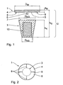
- Fig. 1 is an embodiment of a resorbable implant 1, which is designed as a composite body 2 of two partial bodies shown.
- the first partial body is a truncated cone 3
- the second partial body is a substantially round covering membrane 4.
- the truncated cone 3 and the covering membrane 4 are firmly connected to each other in the resorbable implant 1.
- the cover membrane 4 with a lower membrane surface 5 is, as in Fig. 1 indicated by the arrow 6, approximately concentric with a designed as an upper cone base surface 7 body base the truncated cone 3 is placed.
- the membrane surface 5 is larger than the cone base 7. It is in an edge region 8 on the cone base 7 addition. In the edge region 8, the covering membrane 4 is slotted.
- in the embodiment shown provided a total of four evenly distributed over the circumference arranged in the radial direction extending slots 9.
- the cone base 7 of the truncated cone 3 has a diameter D KG of about 1.3 cm.
- a diameter D KS at the narrow end of the truncated cone 3 is about 0.9 cm.
- Its height h K is about 1.5 cm.
- the covering membrane 4 has a membrane diameter D M of about 2 cm and a membrane thickness d M of about 0.1 cm, so that an overall height h G of the composite body 2 is about 1.6 cm.
- the truncated cone 3 and the cover membrane 4 are firmly connected to one another by means of the adhesive force of the collagen-containing materials from which the truncated cone 3 and the covering membrane 4 are each made.
- the two parts 3 and 4 of the composite body 2 are joined together, as long as at least one of the two collagen-containing material is not dried out, so that the natural adhesive force effect of the collagen-containing material is used to produce the solid compound. This is favorable, since no separate adhesive or connecting means are required.
- the truncated cone 3 is when in Fig. 1 shown embodiment of a porous collagen glyphylate with a density of 7 mg / cm 3 .
- a Fig. 1 with illustrated cylindrical or conical (blunt) shaped core 10 of a bone material, for example of native, treated cancellous bone material or of a synthetically produced material such as tricalcium phosphate or hydroxyapatite.
- the cover membrane 4 consists in Fig. 1 shown embodiment of a densified lyophilisate from reconstituted collagen.
- the collagen material of the covering membrane 4 has a smaller pore size than that of the truncated cone 3.
- the covering membrane 4 is formed, for example, as a foil or compacted sponge from said collagen. Their density is about a factor of two higher than that of the truncated cone 3.
- the implant 1 is e.g. prepared as follows. First, the cover membrane 4 is lyophilized, pressed and cut. Thereafter, a liquid collagen suspension is introduced into a truncated cone shape to form the truncated cone 3. The covering membrane 4 is applied to this collagen suspension, as long as the latter is still in a liquid state. By means of a final lyophilization step, the composite body 2 is formed with the porous collagen truncated cone 3 and the collagen covering membrane 4 as a firmly connected unit.
- a tooth 12 seated in an alveolus 11 is shown.
- the socket 11 is formed by the jaw bone 13, which is covered on its outside with connective and epithelial tissue 14.
- the alveolus 11 is immediately filled with a resorbable implant 15, which is substantially like the implant 1 according to FIG Fig. 1 and 2 is constructed, equipped (see Fig. 4 ).
- a truncated cone 16 of the implant 15 does not contain a core of bone (replacement) material. Otherwise, the implant 15, just like the implant 1, also has a covering membrane 17 which is likewise firmly connected to the truncated cone 16.
- the truncated cone 16 fills the alveolus 1 very well in the entire area, so that a stable coagulum promoting wound healing can form in the entire alveolus 11.
- the truncated cone 16 is plastically deformable and thus adapts well to the alveolus 11. Thus, a stabilization of the coagulum in the entire alveolus 11 is achieved.
- the truncated cone 16 is osteoconductive, i. it facilitates the ingrowth of new bone tissue into the alveolus 11.
- the truncated cone 16 is held in addition by its shape by the cover membrane 17, which rests with its laterally projecting edge portion 18 on the jaw bone 13, in the correct position. Due to the cover membrane 17 a very accurate vertical and horizontal positioning of the implant 15 is possible in total. The application simplifies and becomes safer. Because of the fixed combination, there can be no relative displacement between the cover membrane 17 and the truncated cone 16.
- the cover membrane 17 fulfills a function shielding the alveolar space, whereby the connective and epithelial tissue 14 is prevented from undesired ingrowth into the alveolus 11. Since bone tissue proliferates much more slowly compared to the connective and epithelial tissue 14, the connective and epithelial tissue 14 would be devoid of the barrier formed by the covering membrane 17 Filling the alveolar space faster and thus inhibiting bone growth.
- the implants 1 and 15 thus create a "regenerative space", by means of which the physiological wound healing promoted and a loss of bone mass is avoided.
- the thus-obtained or additional built-up bone mass of the alveolus 11 can then serve as the basis for a later intended implantation of a dental prosthesis.
- a further embodiment of a resorbable implant 19 is shown. It comprises a similar plastically deformable truncated cone 16 as the implant 15 according to Fig. 4 , Also in the case of the implant 19, a covering membrane 20 consisting of collagen is firmly connected to the truncated cone 16.
- the covering membrane 20 is configured somewhat differently than the covering membrane 4 or 17 according to FIG Fig. 1 and 2 although it also has an approximately circular central portion 21, which in turn is larger than the cone base surface 7 of the truncated cone 16 (see the view from below in accordance with Fig. 6 ).
- the two elongated membrane wings 22 are opposite each other.
- the membrane wings 22 are lateral extensions of the central portion 21.
- the effect of the membrane wings 22 goes out of the in Fig. 8 shown illustration of the implant 19 in its inserted state.
- the membrane wings 22 cover the area of the alveolus 11 vestibular and palatal. This is particularly advantageous if there are bone defects in this area. Then the membrane wings 22 prevent an uncontrolled penetration of connective and epithelial tissue at these sites, which would otherwise be at the expense of a regeneration of the bone tissue.
Landscapes
- Health & Medical Sciences (AREA)
- Life Sciences & Earth Sciences (AREA)
- Public Health (AREA)
- Veterinary Medicine (AREA)
- Epidemiology (AREA)
- Orthopedic Medicine & Surgery (AREA)
- Animal Behavior & Ethology (AREA)
- General Health & Medical Sciences (AREA)
- Oral & Maxillofacial Surgery (AREA)
- Dentistry (AREA)
- Engineering & Computer Science (AREA)
- Biomedical Technology (AREA)
- Developmental Biology & Embryology (AREA)
- Materials For Medical Uses (AREA)
- Prostheses (AREA)
- Dental Prosthetics (AREA)
Description
- Die Erfindung betrifft ein Implantat zum Einbringen in einen Alveolarraum.
- Nach einer Zahnextraktion sind in der Zahnmedizin verschiedene Behandlungsverfahren bekannt. Bei einem ersten Ansatz erfolgt zunächst eine Ausheilung der Zahntasche (= Alveole), um danach mit einer Zahnersatz-Behandlung für z.B. eine Zahnimplantation neu zu beginnen. Hierbei kann es während der Abheilzeit zu einem unerwünschten Schwund an Knochensubstanz kommen. Ursache hierfür ist z.B. eine Entzündung in der Alveole, die die fibrinolytische Aktivität des Blutes erhöht, weshalb sich kein stabiles Blutgerinnsel (= Koagulum) ausbilden kann. Es kommt zu einer gestörten Wundheilung und einem Verlust von Knochengewebe. Als Folge davon lässt sich der Zahnersatz entweder nicht mehr optimal verankern oder es muss sogar eine aufwändige Regenerationsbehandlung des Kieferknochens zwischengeschaltet werden.
- Bei einem anderen Ansatz wird unmittelbar nach der Zahnextraktion ein Implantat eingesetzt. Ein Beispiel für ein derartiges Implantat ist z.B. in der
DE 196 30 034 A1 beschrieben. Es enthält einen harten Titan-Kern, der unter anderem mit einer kollagenhaltigen Beschichtung versehen ist und der nach einer Einwachsphase als Basis für eine Zahnkrone dient. Bei diesen unmittelbar eingesetzten Implantaten kann es z.B. wegen Entzündungen in der Alveole zu Problemen beim Einwachsen kommen. - In der
EP1 433 489 A1 wird ein Implantat zum Einbringen in einen Alveolarraum beschrieben. Dieses Implantat ist als resorbierbarer Verbundkörper aus zwei Teilkörpern ausgeführt. Der erste Teilkörper kann ein eine Körpergrundfläche aufweisender Kegel, Würfel oder Zylinder sein. Der zweite Teilkörper ist als eine Abdeckmembran mit einer Membranfläche ausgebildet, die größer als die Körpergrundfläche ist, wobei die Abdeckmembran bei einer Ausführungsvariante an zwei Seiten seitlich über die Körpergrundfläche des ersten Teilkörpers hinausragt. Die beiden Teilkörper sind im Bereich der aneinander angrenzenden Körpergrundfläche und Membranfläche fest miteinander verbunden. - In der
US 2002/0013626 A1 ist die Verwendung von Kollagen als Material für ein Implantat beschrieben. -
EP-A-2 104 518 , das einen Stand der Technik gemäss Artikel 54 (3) EPÜ betrifft, beschreibt ein Implantat zum Einbringen in einen Alveolarraum, welches als resorbierbarer Verbundkörper aus mindestens einem ersten und einem zweiten Teilkörper ausgeführt ist. Der erste Teilkörper ist als ein eine Körpergrundfläche aufweisender Quader ausgebildet. Der zweite Teilkörper ist als eine Abdeckmembran mit einer Membranfläche, die grösser als die Körpergrundfläche ist, ausgebildet. Die Abdeckmembran ist so auf die Körpergrundfläche aufgesetzt, dass sie seitlich überall über die Körpergrundfläche hinausragt. Der erste und der zweite Teilkörper sind im Bereich der aneinander angrenzenden Körpergrundfläche und Membranfläche fest miteinander verbunden. - Die Aufgabe der Erfindung besteht darin, ein Implantat der eingangs bezeichneten Art anzugeben, das einfach und zuverlässig in der Handhabung ist sowie eine gute Ausheilung der Alveole ermöglicht.
- Zur Lösung dieser Aufgabe wird ein Implantat entsprechend den Merkmalen des Patentanspruchs 1 angegeben. Das erfindungsgemäße Implantat ist als resorbierbarer Verbundkörper aus mindestens einem ersten und einem zweiten Teilkörper ausgeführt, wobei
- a) der erste Teilkörper als ein eine Körpergrundfläche aufweisender Kegel, Kegelstumpf oder Zylinder ausgebildet ist,
- b) der zweite Teilkörper als eine Abdeckmembran mit einer Membranfläche, die größer als die Körpergrundfläche ist, ausgebildet ist,
- c) der erste und der zweite Teilkörper im Bereich der aneinander angrenzenden Körpergrundfläche und Membranfläche fest miteinander verbunden sind,
- d) die Abdeckmembran auf die Körpergrundfläche aufgesetzt ist, so dass sie seitlich überall über die Körpergrundfläche hinausragt, und
- e) die Abdeckmembran in einem über die Körpergrundfläche hinaus stehenden Randbereich ein- oder mehrfach geschlitzt ist.
- Das erfindungsgemäße Implantat ermöglicht eine vollständige Regeneration von Knochen sowie von Binde- und Epithelgewebe. Insbesondere erhält es die Knochensubstanz des Alveolarkamms und ermöglicht das Einwachsen von zusätzlichem Knochengewebe in die Alveole, so dass für eine spätere Verankerung von Zahnersatz, ausreichend Knochensubstanz vorhanden ist. Nach der Zahnentfernung bildet sich bei Verwendung des erfindungsgemäßen Implantats ein stabiles Koagulum innerhalb des Alveolarraums. Dieses Koagulum wird im Verlauf der Wundheilung durch Knochen ersetzt. Das erfindungsgemäße Implantat wird während des Wundheilungsprozesses insbesondere vollständig resorbiert.
- Der erste Teilkörper des erfindungsgemäßen Implantats ist aufgrund seiner Kegel(stumpf)form sehr gut an die Form der Alveole angepasst. Er füllt sie gut aus. Außerdem wirkt der erste Teilkörper blutstillend und begünstigt die Bildung eines stabilen Koagulums, so dass ein Kollaps des Alveolarraums sicher verhindert wird. Das Einwachsen von Knochengewebe wird somit gefördert. Das erfindungsgemäße Implantat wirkt also auch osteokonduktiv.
- Der zweite Teilkörper des erfindungsgemäßen Implantats verhindert dagegen das Einwachsen von Binde- und Epithelgewebe in die Alveole. Diese Abdeckmembran deckt die Alveole mit ihrem über die Körpergrundfläche hinausragenden Randbereich ab und bildet so eine wirksame Barriere gegen unkontrollierte Binde- und Epithelgewebeproliferation in die Alveole. Da Knochengewebe im Vergleich zum Binde- und Epithelgewebe wesentlich langsamer proliferiert, bestünde ohne die Abdeckmembran die Gefahr, dass das Binde- und Epithelgewebe den Alveolarraum schneller ausfüllt und damit das Knochenwachstum hemmt.
- Aufgrund der festen Verbindung zwischen den beiden Teilkörper ist stets gewährleistet, dass sich die Abdeckmembran in Relation zu dem Kegel-(stumpf) und der Alveole in der richtigen Position befindet und auch während der Wundheilung in dieser Position bleibt. Die feste Verbindung zwischen den beiden Teilkörpern verhindert außerdem, dass beim Einsetzen Fehler passieren, beispielsweise, dass die Abdeckmembran falsch platziert wird und die Alveole nicht vollständig abdeckt. Der Verbundkörper des erfindungsgemäßen Implantats ist also besonders einfach und zuverlässig in der Handhabung und verkürzt die Behandlungsdauer.
- Dadurch, dass die Abdeckmembran in einem über die Körpergrundfläche hinaus stehenden Randbereich ein- oder mehrfach geschlitzt ist, vereinfacht sich die Applikation zur Abdeckung der Alveole weiter.
- Insgesamt schafft das erfindungsgemäße Implantat einen "Regenerativen Raum", aufgrund dessen die physiologische Wundheilung gefördert und ein Verlust von Knochenmasse vermieden wird.
- Vorteilhafte Ausgestaltungen des erfindungsgemäßen resorbierbaren Implantats ergeben sich aus den Merkmalen der von Anspruch 1 abhängigen Ansprüche.
- Günstig ist eine Variante, bei der der erste Teilkörper zumindest teilweise aus einem porösen Kollagen, z.B. aus lyophilisiertem, getrocknetem oder mittels Verfilzen hergestelltem Kollagen, vorzugsweise aus rekonstituiertem Kollagen vom Typ1 besteht. Weiterhin ist es bevorzugt vorgesehen, dass das Kollagen des ersten Teilkörpers eine Dichte von 1 bis 25 mg/cm3, vorzugsweise von 5 bis 12 mg/cm3, hat. Kollagen mit diesen Dichtewerten lässt sich besonders gut herstellen. Kollagen ist als bioresorbierbares Material sehr gut verträglich und findet Anwendung zur Blutstillung, zur Füllung von Knochen und Gewebedefekten und zur Abdeckung von Wunden. Kollagen unterstützt die Blutstillung, indem Thrombozyten an den Kollagenfibrillen aggregieren und sich ein Koagulum bildet. Durch die Wirkung von eingewanderten Makrophagen und körpereigener Kollagenase wird das Kollagen im Rahmen der Wundheilung komplett resorbiert.
- Gemäß einer anderen vorteilhaften Variante hat der erste Teilkörper einen insbesondere soliden zylindrischen oder kegelförmigen Kern aus nativem, aufbereitetem Knochenmaterial, wie z.B. Spongiosa, oder aus synthetischem Knochenersatzmaterial, wie z.B. Tricalciumphosphat, Hydroxylapatit oder einem ähnlichen keramischen Material. Alternativ dazu kann der erste Teilkörper aber ebenso vorzugsweise aus einem Mischmaterial mit einem Kollagenanteil und einem in den Kollagenanteil homogen verteilt eingelagerten Knochenanteil bestehen, wobei der Knochenanteil durch Knochenersatzmaterial oder durch zerkleinertes natives, aufbereitetes Knochenmaterial, wie z.B. Spongiosa, gebildet ist. Der bei der ersten Variante mittels des gesonderten Kerns zugesetzte bzw. bei der zweiten Variante homogen zugemischte Knochenanteil bewegt sich jeweils insbesondere im Bereich zwischen 5 % und 90 % des Volumens des ersten Teilkörpers. Beide Varianten fordern die Knochenbildung in dem Alveolarraum während des Wundheilungsprozesses.
- Gemäß einer weiteren günstigen Ausgestaltung besteht der zweite Teilkörper aus einem verdichteten Kollagen mit insbesondere geringerer Porengröße als der des Kollagens des ersten Teilkörpers. Das verdichtete Kollagen bildet insbesondere einen Film, eine Folie oder einen verdichteten Schwamm. Vorzugsweise ist das verdichtete Kollagen des zweiten Teilkörpers ein verdichtetes Lyophilisat aus rekonstituiertem Kollagen. Im Gegensatz zu dem poröseren ersten Teilkörper, bei dem die Wundheilung und Gewebebildung im Vordergrund steht, dient der zweite Teilkörper als Barriere gegen ein Einwachsen von Binde- und Epithelgewebe. Eine an dieser Stelle gerade nicht erwünschte Gewebebildung wird durch die Verwendung von verdichtetem Kollagen verhindert oder zumindest deutlich erschwert. Insbesondere ist das Kollagen des zweiten Teilkörpers um den Faktor 0,5 bis 5 dichter als das des ersten Teilkörpers.
- Gemäß einer weiteren bevorzugten Ausgestaltung kann zumindest einer der beiden Teilkörper aus einem Material bestehen, das zumindest einen wundheilenden und/oder das Knochenwachstum fördernden bioaktiven Bestandteil enthält. Bei diesem bioaktiven Bestandteil handelt es sich vorzugsweise um ein natives isoliertes oder biotechnologisch gewonnenes Protein, nämlich BMP-2 (= Bone Morphogenic Protein 2). Hierunter fällt unter anderem auch TGF beta (TGF = Transforming Growth Factor) oder ähnliches, also insbesondere Wachstumsfaktoren ganz allgemein.
- Gemäß einer anderen ebenfalls günstigen Ausgestaltung kann zumindest einer der beiden Teilkörper aus einem Material bestehen, das zumindest einen antimikrobiellen Bestandteil enthält. Dieser antimikrobielle Bestandteil trägt dazu bei, Infektionen zu verhindern und/oder zu bekämpfen. Es handelt sich hierbei vorzugsweise um ein lokal verträgliches Antiseptikum, wie z.B. Polihexanid, Octenidin, Silberionen, Jodderivate oder ähnliches. Ebenso kann eine für die lokale Anwendung geeignete antibiotisch wirkende Substanz, wie z.B. Gentamicin, Metronidazol, Vancomycin oder ähnliches, zum Einsatz kommen.
- Weiterhin ist es vorzugsweise vorgesehen, dass der Verbundkörper zumindest teilweise, insbesondere der Kegel(stumpf) oder die Abdeckmembran anteilig oder vollständig, aus einer Gelatine oder einer oxidierten Cellulose besteht. Auch diese Stoffe sind vollständig resorbierbar und biologisch gut verträglich. Bei der Gelatine kann es dabei insbesondere auch um ein kollagenhaltiges Material handeln, dessen Kollagen stärker denaturiert ist als bei den zuvor genannten Kollagenmaterialien (= lyophilisiertes und/oder rekonstituiertes Kollagen vom Typ 1). Die oxidierte Cellulose ist ein anderes Biopolymer. Der Verbundkörper kann auch aus einer Mischung aus einem der zuvor genannten Kollagenmaterialien und der Gelatine und/oder der oxidierten Cellulose bestehen.
- Gemäß weiteren vorteilhaften Ausgestaltungen hat der Verbundkörper die im Folgenden genannten besonders günstigen geometrischen Abmessungen, die zu einer sehr guten Anpassung an den Einsatzzweck, also das Einsetzen in die Alveole und das Abdecken der Alveole, führt. Die eine Gesamthöhe des Verbundkörpers beträgt insbesondere mindestens 1,6 cm. Weiterhin hat die Körpergrundfläche des ersten Teilkörpers einen bevorzugten Durchmesser von 1 cm bis 1,5 cm, vorzugsweise von 1,3 cm. Eine Kegelhöhe des ersten Teilkörpers beträgt insbesondere zwischen 1 cm und 3 cm, vorzugsweise zwischen 1,3 cm und 1,7 cm. Der zweite Teilkörper hat bevorzugt eine Membrandicke von zwischen 0,05 cm und 0,5 cm und einen Membrandurchmesser, der um 0,2 cm bis 2 cm größer ist als ein Durchmesser der Körpergrundfläche und der insbesondere mindestens 1,4 cm beträgt.
- Gemäß einer weiteren vorteilhaften Ausgestaltung umfasst die Abdeckmembran einen in etwa kreisrunden Mittenabschnitt, an dessen Außenumfang mindestens ein länglicher, seitlich abstehender Membranflügel einstückig angebracht ist. Insbesondere sind zwei derartige Membranflügel vorgesehen, die einander gegenüber liegen. Sie dienen zur Abdeckung vestibulärer und palatinaler Knochendefekte.
- Weitere Merkmale, Vorteile und Einzelheiten der Erfindung ergeben sich aus der nachfolgenden Beschreibung von Ausführungsbeispielen anhand der Zeichnung. Es zeigt:
- Fig. 1
- ein Ausführungsbeispiel eines resorbierbaren Implantats als Verbundkörper aus einem Kegelstumpf und einer Abdeck- membran in einer Explosionsdarstellung,
- Fig. 2
- das Implantat gemäß
Fig. 1 in einer Draufsicht von oben auf die Abdeckmembran, - Fig. 3
- einen in einer Alveole sitzenden Zahn in einer Querschnitts- darstellung,
- Fig. 4
- ein Ausführungsbeispiel eines resorbierbaren aus einem Ke- gelstumpf und einer Abdeckmembran bestehenden Implan- tats, das nach einer Extraktion des Zahns in die Alveole ge- mäß
Fig. 3 eingesetzt ist, - Fig. 5 bis 7
- ein weiteres Ausführungsbeispiel eines resorbierbaren aus einem Kegelstumpf und einer seitlich verlängerten Abdeck- membran bestehenden Implantats in einer perspektivischen Ansicht, in einer Ansicht von unten und in einer Seitenan- sicht, und
- Fig. 8
- das Implantat gemäß
Fig. 5 bis 7 in einen eingesetzten Zu- stand. - Einander entsprechende Teile sind in den
Fig. 1 bis 8 mit denselben Bezugszeichen versehen. - In
Fig. 1 ist ein Ausführungsbeispiel eines resorbierbaren Implantats 1, das als Verbundkörper 2 aus zwei Teilkörpern ausgeführt ist, gezeigt. Bei dem ersten Teilkörper handelt es sich um einen Kegelstumpf 3, bei dem zweiten Teilkörper um eine im Wesentlichen runde Abdeckmembran 4. Der Kegelstumpf 3 und die Abdeckmembran 4 sind bei dem resorbierbaren Implantat 1 fest miteinander verbunden. Die Abdeckmembran 4 mit einer unteren Membranfläche 5 ist, wie inFig. 1 durch den Pfeil 6 angedeutet, in etwa konzentrisch auf eine als obere Kegelgrundfläche 7 ausgebildete Körpergrundfläche des Kegelstumpfs 3 aufgesetzt. Die Membranfläche 5 ist größer als die Kegelgrundfläche 7. Sie steht in einem Randbereich 8 über die Kegelgrundfläche 7 hinaus. Im Randbereich 8 ist die Abdeckmembran 4 geschlitzt. Um eine einfachere Applikation zu ermöglichen, sind bei dem gezeigten Ausführungsbeispiel (siehe Draufsicht gemäßFig. 2 ) insgesamt vier gleichmäßig über den Umfang verteilt angeordnete, in radiale Richtung verlaufende Schlitze 9 vorgesehen. - Beim Ausführungsbeispiel hat die Kegelgrundfläche 7 des Kegelstumpfs 3 einen Durchmesser DKG von etwa 1,3 cm. Ein Durchmesser DKS an dem schmalen Ende des Kegelstumpfs 3 beträgt etwa 0,9 cm. Seine Höhe hK liegt bei ungefähr 1,5 cm. Die Abdeckmembran 4 hat einen Membrandurchmesser DM von etwa 2 cm und eine Membrandicke dM von etwa 0,1 cm, so dass eine Gesamthöhe hG des Verbundkörpers 2 etwa 1,6 cm beträgt.
- In dem Bereich, in dem die Membranfläche 5 und die Kegelgrundfläche 7 aneinander angrenzen, sind der Kegelstumpf 3 und die Abdeckmembran 4 mittels der Klebekraft der kollagenhaltigen Materialien, aus denen der Kegelstumpf 3 und die Abdeckmembran 4 jeweils gefertigt sind, fest miteinander verbunden. Die beiden Teile 3 und 4 des Verbundkörpers 2 werden zusammengefügt, solange bei zumindest einem der beiden das kollagenhaltige Material noch nicht ausgetrocknet ist, so dass die natürliche Klebekraftwirkung des kollagenhaltigen Materials zur Herstellung der festen Verbindung genutzt wird. Dies ist günstig, da so keine gesonderten Klebe-oder Verbindungsmittel erforderlich sind.
- Der Kegelstumpf 3 besteht beim in
Fig. 1 gezeigten Ausführungsbeispiel aus einem porösen Kollagenlyphilisat mit einer Dichte von 7 mg/cm3. In ihm eingeschlossen ist optional ein inFig. 1 mit dargestellter zylindrischer oder kegel(stumpf)förmiger Kern 10 aus einem Knochenmaterial, beispielsweise aus nativem, aufbereitetem Spongiosa-Material oder aus einem synthetisch hergestellten Material wie Tricalciumphosphat oder Hydroxylapatit. - Die Abdeckmembran 4 besteht beim in
Fig. 1 gezeigten Ausführungsbeispiel aus einem verdichteten Lyophilisat aus rekonstituiertem Kollagen. Das Kollagenmaterial der Abdeckmembran 4 hat eine geringere Porengröße als das des Kegelstumpfs 3. Die Abdeckmembran 4 ist beispielsweise als Folie oder verdichteter Schwamm aus dem genannten Kollagen ausgebildet. Ihre Dichte liegt etwa um den Faktor zwei höher als diejenige des Kegelstumpfs 3. - Das Implantat 1 wird z.B. folgendermaßen hergestellt. Zunächst wird die Abdeckmembran 4 lyophilisiert, gepresst und zugeschnitten. Danach wird eine flüssige Kollagensuspension in eine Kegelstumpfform eingebracht, um den Kegelstumpf 3 zu bilden. Die Abdeckmembran 4 wird auf diese Kollagensuspension aufgebracht, solange sich letztere in noch flüssigem Zustand befindet. Mittels eines abschließenden Lyophilisationsschrittes entsteht der Verbundkörper 2 mit dem porösen Kollagen-Kegelstumpf 3 und der Kollagen-Abdeckmembran 4 als fest verbundene Einheit.
- In
Fig. 3 ist ein in einer Alveole 11 sitzender Zahn 12 gezeigt. Die Alveole 11 ist durch den Kieferknochen 13 gebildet, der an seiner Außenseite mit Binde- und Epithelgewebe 14 bedeckt ist. - Nach der Extraktion des Zahns 12 wird die Alveole 11 sofort mit einem resorbierbaren Implantat 15, das im Wesentlichen wie das Implantat 1 gemäß
Fig. 1 und 2 aufgebaut ist, bestückt (sieheFig. 4 ). Im Unterschied zu dem Implantat 1 enthält ein Kegelstumpf 16 des Implantats 15 keinen Kern aus Knochen(ersatz)material. Ansonsten hat das Implantat 15 genau wie das Implantat 1 eine ebenfalls fest mit dem Kegelstumpf 16 verbundene Abdeckmembran 17. - Aufgrund seiner an den Alveolarraum angepassten kegeligen Kontur füllt der Kegelstumpf 16 die Alveole 1 im gesamten Bereich sehr gut aus, so dass sich in der gesamten Alveole 11 ein stabiles, die Wundheilung begünstigendes Koagulum ausbilden kann. Der Kegelstumpf 16 ist plastisch verformbar und passt sich damit der Alveole 11 gut an. So wird eine Stabilisierung des Koagulums in der gesamten Alveole 11 erreicht. Der Kegelstumpf 16 wirkt osteokonduktiv, d.h. er erleichtert das Einwachsen von neuem Knochengewebe in die Alveole 11.
- Der Kegelstumpf 16 wird außer durch seine Form auch durch die Abdeckmembran 17, die mit ihrem seitlich überstehenden Randbereich 18 auf dem Kieferknochen 13 aufliegt, in der richtigen Position gehalten. Aufgrund der Abdeckmembran 17 ist eine sehr genaue vertikale und horizontale Positionierung des Implantats 15 insgesamt möglich. Die Anwendung vereinfacht sich und wird sicherer. Wegen der fixen Kombination kann es zu keiner Relativverschiebung zwischen der Abdeckmembran 17 und dem Kegelstumpf 16 kommen. Darüber hinaus erfüllt die Abdeckmembran 17 eine den Alveolarraum abschirmende Funktion, wodurch das Binde- und Epithelgewebe 14 an einem unerwünschten Einwachsen in die Alveole 11 gehindert wird. Da Knochengewebe im Vergleich zu dem Binde- und Epithelgewebe 14 wesentlich langsamer proliferiert, würde das Binde- und Epithelgewebe 14 ohne die durch die Abdeckmembran 17 gebildete Barriere den Alveolarraum schneller ausfüllen und damit das Knochenwachstum hemmen.
- Die Implantate 1 und 15 sind resorbierbar und insbesondere auch blutstillend. Eingesetzt in die Alveole 11 (Zahntasche) bewirken sie nach einer Zahnextraktion eine Blutstillung und verhindern vorteilhafterweise einen Kollaps der Alveole 11. Mit fortschreitendem entzündungsfreiem Heilungsprozess wird außerdem sogar eine Knochenbildung innerhalb der Alveole 11 begünstigt (= osteokonduktive Wirkung). Die Implantate 1 und 15 schaffen also einen "Regenerativen Raum", mittels dessen die physiologische Wundheilung gefördert und ein Verlust von Knochenmasse vermieden wird. Die so erhaltene bzw. zusätzliche aufgebaute Knochenmasse der Alveole 11 kann dann als Basis für eine später vorgesehene Implantation eines Zahnersatzes dienen.
- In
Fig. 5 bis 7 ist ein weiteres Ausführungsbeispiel eines resorbierbaren Implantats 19 gezeigt. Es umfasst einen ähnlichen plastisch verformbaren Kegelstumpf 16 wie das Implantat 15 gemäßFig. 4 . Auch bei dem Implantat 19 ist eine aus Kollagen bestehende Abdeckmembran 20 fest mit dem Kegelstumpf 16 verbunden. Die Abdeckmembran 20 ist etwas anders ausgestaltet als die Abdeckmembran 4 oder 17 gemäßFig. 1 und 2 bzw. 4. Sie hat zwar ebenfalls einen in etwa kreisrunden Mittenabschnitt 21, die wiederum größer ist als die Kegelgrundfläche 7 des Kegelstumpfs 16 (vgl. die Ansicht von unten gemäßFig. 6 ). An den Mittenabschnitt 21 sind aber zusätzlich zwei seitliche Membranflügel 22 jeweils einstückig angeformt. Die beiden länglichen Membranflügel 22 liegen einander gegenüber. Sie sind also in Umfangsrichtung des Mittenabschnitts 21 um etwa 180° gegen einander versetzt an dem Mittenabschnitt 21 angebracht. Ihre Ausdehnung in Längsrichtung ist jeweils vorzugsweise etwa genauso lang wie die Höhe hk des Kegelstumpfs 16. Gegebenenfalls kann ihre Länge aber auch größer sein als die Höhe hk des Kegelstumpfs 16. Die Membranflügel 22 stellen seitliche Verlängerungen des Mittenabschnitts 21 dar. - Die Wirkung der Membranflügel 22 geht aus der in
Fig. 8 gezeigten Abbildung des Implantats 19 in seinem eingesetzten Zustand hervor. Die Membranflügel 22 decken den Bereich der Alveole 11 vestibulär und palatinal ab. Dies ist insbesondere dann von Vorteil, wenn in diesem Bereich Knochendefekte bestehen. Dann verhindern die Membranflügel 22 auch an diesen Stellen ein unkontrolliertes Vordringen von Binde- und Epithelgewebe, das ansonsten zulasten einer Regeneration des Knochengewebes ginge.
Claims (14)
- Implantat zum Einbringen in einen Alveolarraum (11), welches als resorbierbarer Verbundkörper (2) aus mindestens einem ersten und einem zweiten Teilkörper (3, 4; 16, 17; 16, 20) ausgeführt ist, wobeia) der erste Teilkörper als ein eine Körpergrundfläche (7) aufweisender Kegel, Kegelstumpf (3; 16) oder Zylinder ausgebildet ist,b) der zweite Teilkörper als eine Abdeckmembran (4; 17; 20) mit einer Membranfläche (5), die größer als die Körpergrundfläche (7) ist, ausgebildet ist,c) der erste und der zweite Teilkörper (3, 4; 16, 17; 16, 20) im Bereich der aneinander angrenzenden Körpergrundfläche (7) und Membranfläche (5) fest miteinander verbunden sind,d) die Abdeckmembran (4; 17; 20) auf die Körpergrundfläche (7) aufgesetzt ist, so dass sie seitlich überall über die Körpergrundfläche (7) hinausragt, unde) die Abdeckmembran (4; 17) in einem über die Körpergrundfläche (7) hinaus stehenden Randbereich (8; 18) ein- oder mehrfach geschlitzt ist.
- Implantat nach Anspruch 1, bei dem der erste Teilkörper (3; 16) zumindest teilweise aus einem porösen Kollagen, vorzugsweise aus rekonstituiertem Kollagen vom Typ 1 besteht.
- Implantat nach Anspruch 2, bei dem das Kollagen des ersten Teilkörpers (3; 16) eine Dichte von 1 bis 25 mg/cm3, vorzugsweise von 5 bis 12 mg/cm3, hat ,
- Implantat nach Anspruch 1, bei dem der erste Teilkörper (3) einen Kern (10) aus nativem, aufbereitetem Knochenmaterial oder aus synthetischem Knochenersatzmaterial hat.
- Implantat nach Anspruch 1, bei dem der erste Teilkörper (16) aus einem Mischmaterial mit einem Kollagenanteil und einem in den Kollagenanteil homogen verteilt eingelagerten Knochenanteil besteht, wobei der Knochenanteil durch Knochenersatzmaterial oder durch zerkleinertes natives, aufbereitetes Knochenmaterial gebildet ist.
- Implantat nach Anspruch 1, bei dem der zweite Teilkörper (4; 17; 20) aus einem verdichteten Kollagen mit insbesondere geringerer Porengröße als der des Kollagens des ersten Teilkörpers (3; 16) besteht, und das verdichtete Kollagen insbesondere einen Film, eine Folie oder einen verdichteten Schwamm bildet.
- Implantat nach Anspruch 6, bei dem das ver-dichtete Kollagen des zweiten Teilkörpers (4; 17; 20) ein verdichtetes Lyophilisat aus rekonstituiertem Kollagen ist.
- Implantat nach Anspruch 1, bei dem zumindest einer der beiden Teilkörper (3, 4; 16, 17; 16, 20) aus einem Material besteht, das zumindest einen wundheilenden oder das Knochenwachs-tum fördernden bioaktiven Bestandteil enthält.
- Implantat nach Anspruch 1, bei dem zumindest einer der beiden Teilkörper (3, 4; 16, 17; 16, 20) aus einem Material besteht, das zumindest einen antimikrobiellen oder antibiotisch wir-kenden Bestandteil enthält.
- Implantat nach Anspruch 1, bei dem der Verbundkörper (2) zumindest teilweise aus einer Gelatine oder einer oxidierten Cellulose besteht.
- Implantat nach Anspruch 1, bei dem eine Gesamthöhe (hG) des Verbundkörpers (2) mindestens 1,6 cm beträgt.
- Implantat nach Anspruch 1, wobei bei dem ersten Teilkörper (3; 16) die Körpergrundfläche (7) einen Durchmesser (DKG) von 1 cm bis 1,5 cm, vorzugsweise von 1,3 cm, hat und eine Ke-gelhöhe (hK) zwischen 1 cm und 3 cm, vorzugsweise zwischen 1,3 cm und 1,7 cm, beträgt.
- Implantat nach Anspruch 1, bei dem der zweite Teilkörper (4; 17) eine Membrandicke (dM) von zwischen 0,05 cm und 0,5 cm und einen Membrandurchmesser (DM) hat, der um 0,2 cm bis 2 cm größer ist als ein Durchmesser (DKG) der Körpergrundfläche (7) und der insbesondere mindestens 1,4 cm beträgt.
- Implantat nach Anspruch 1, bei dem die Abdeckmembran (20) einen in etwa kreisrunden Mittenabschnitt (21) umfasst, an dessen Außenumfang mindestens ein länglicher, seitlich abstehender Membranflügel (22) einstückig angebracht ist.
Priority Applications (1)
| Application Number | Priority Date | Filing Date | Title |
|---|---|---|---|
| PL09713162T PL2249739T3 (pl) | 2008-02-23 | 2009-02-09 | Resorbowalny implant do umieszczania w zębodole |
Applications Claiming Priority (2)
| Application Number | Priority Date | Filing Date | Title |
|---|---|---|---|
| DE102008010893A DE102008010893B3 (de) | 2008-02-23 | 2008-02-23 | Implantat zum Einbringen in einen Alveolarraum |
| PCT/EP2009/000876 WO2009103437A1 (de) | 2008-02-23 | 2009-02-09 | Resorbierbares implantat zum einbringen in einen alvoeolarraum |
Publications (2)
| Publication Number | Publication Date |
|---|---|
| EP2249739A1 EP2249739A1 (de) | 2010-11-17 |
| EP2249739B1 true EP2249739B1 (de) | 2011-07-06 |
Family
ID=40551783
Family Applications (1)
| Application Number | Title | Priority Date | Filing Date |
|---|---|---|---|
| EP09713162A Active EP2249739B1 (de) | 2008-02-23 | 2009-02-09 | Resorbierbares implantat zum einbringen in einen alvoeolarraum |
Country Status (9)
| Country | Link |
|---|---|
| US (1) | US20100331997A1 (de) |
| EP (1) | EP2249739B1 (de) |
| AT (1) | ATE515242T1 (de) |
| DE (1) | DE102008010893B3 (de) |
| DK (1) | DK2249739T3 (de) |
| ES (1) | ES2367410T3 (de) |
| PL (1) | PL2249739T3 (de) |
| PT (1) | PT2249739E (de) |
| WO (1) | WO2009103437A1 (de) |
Cited By (1)
| Publication number | Priority date | Publication date | Assignee | Title |
|---|---|---|---|---|
| DE102011082960A1 (de) * | 2011-09-19 | 2013-03-21 | Resorba Wundversorgung Gmbh & Co. Kg | Formkörper mit kollagenhaltigem Kompositmaterial zum Einbringen in eine Knochendefektstelle |
Families Citing this family (10)
| Publication number | Priority date | Publication date | Assignee | Title |
|---|---|---|---|---|
| US20090038701A1 (en) | 2006-01-17 | 2009-02-12 | Baxter International Inc. | Device, system and method for mixing |
| DE202009018865U1 (de) * | 2009-08-21 | 2014-02-06 | Philipp Streckbein | Block aus Knochen - Knochenersatzmaterial |
| EP2594224A1 (de) * | 2011-11-15 | 2013-05-22 | A.B. Dental Devices Ltd. | Zahnplattform |
| KR101984691B1 (ko) * | 2012-05-10 | 2019-05-31 | 호메이욘 에이치. 자데흐 | 발치 위치 재건을 위한 치과용 장치 |
| PL225171B1 (pl) * | 2013-07-25 | 2017-02-28 | Bartłomiej Iwańczyk | Resorbowalny implant ekspansyjny siatkowy częściowo powleczony błoną zaporową |
| EP2886134A1 (de) | 2013-12-20 | 2015-06-24 | nolax AG | Resorbierbares Implantat |
| CN108495658B (zh) * | 2015-09-01 | 2021-09-03 | 巴克斯特国际公司 | 止血材料 |
| IT201700009013A1 (it) * | 2017-05-25 | 2018-11-25 | Gian Paolo Gennari | Un dispositivo di rigenerazione ossea mandibolare e/o mascellare ed un metodo per la realizzazione del dispositivo |
| BR202019022213U2 (pt) * | 2019-10-23 | 2021-05-04 | Marcio Baltazar Conz | Placa de proteção alveolar |
| DE102022209599A1 (de) * | 2022-09-14 | 2024-03-14 | Resorba Medical Gmbh | Knochenimplantat-Körper und Verfahren zu dessen Herstellung |
Family Cites Families (20)
| Publication number | Priority date | Publication date | Assignee | Title |
|---|---|---|---|---|
| US4531916A (en) * | 1983-07-08 | 1985-07-30 | W. L. Gore & Associates, Inc. | Dental implant with expanded PTFE gingival interface |
| US4872840A (en) * | 1987-07-15 | 1989-10-10 | Team Incorporated | Dental implant and method |
| US5819748A (en) * | 1988-11-30 | 1998-10-13 | Ed Geistlich Sohne Ag Fur Chemische Industrie | Implant for use in bone surgery |
| US5320844A (en) * | 1992-03-12 | 1994-06-14 | Liu Sung Tsuen | Composite materials for hard tissue replacement |
| DE4302709C1 (de) * | 1993-02-01 | 1994-07-28 | Kirsch Axel | Abdeckeinrichtung mit Abdeckmembran |
| US6019764A (en) * | 1993-08-02 | 2000-02-01 | Bartee; Barry K. | Method of treating alveolar bone defects |
| US5501706A (en) * | 1994-11-29 | 1996-03-26 | Wildflower Communications, Inc. | Medical implant structure and method for using the same |
| JP3471967B2 (ja) * | 1995-03-28 | 2003-12-02 | カネボウ株式会社 | ハイブリッド型人工歯根 |
| IT1283767B1 (it) * | 1995-05-19 | 1998-04-30 | Urbami Giacomo | Modello di chiodo utilizzato nella rigenerazione ossea guidata |
| DE19835251A1 (de) * | 1998-07-10 | 2000-03-23 | Eckhard Binder | Formteile, insbesondere Folien, zur Förderung der Neubildung von Knochenmaterial im Kiefer |
| US6379154B2 (en) * | 1999-10-19 | 2002-04-30 | John Devincenzo | Subperiosteal bone anchor |
| DK177997B1 (da) * | 2000-07-19 | 2015-02-23 | Ed Geistlich Söhne Ag Für Chemische Ind | Knoglemateriale og collagenkombination til opheling af beskadigede led |
| US6626950B2 (en) * | 2001-06-28 | 2003-09-30 | Ethicon, Inc. | Composite scaffold with post anchor for the repair and regeneration of tissue |
| EP1433489A1 (de) * | 2002-12-23 | 2004-06-30 | Degradable Solutions AG | Biologisch abbaubares, poröses Knochenimplantat mit integrierter Abdeckmembrane |
| DE102004022645A1 (de) * | 2004-05-07 | 2005-12-15 | Resorba Wundversorgung Gmbh & Co. Kg | Bioresorbierbares Material auf Kollagen-Basis |
| EP1819371A2 (de) * | 2004-11-23 | 2007-08-22 | Ossacur AG | Bioresorbierbares und oberflächen-mineralisiertes material zur füllung von knochendefekten |
| DE102005039382B4 (de) * | 2005-08-19 | 2008-03-13 | Detzer, Fritz, Dr. med. dent. | Hohlkörper aus biodegradierbarem Material, insbesondere zum Knochenaufbau im Kieferknochen eines Patienten und dessen Verwendung |
| US20080147197A1 (en) * | 2006-12-14 | 2008-06-19 | Mckay William F | Biodegradable osteogenic porous biomedical implant with impermeable membrane |
| US20090181347A1 (en) * | 2008-01-15 | 2009-07-16 | Fu-Yi Lin | Bone augmentation product for guiding bone tissue regeneration |
| US20100256758A1 (en) * | 2009-04-02 | 2010-10-07 | Synvasive Technology, Inc. | Monolithic orthopedic implant with an articular finished surface |
-
2008
- 2008-02-23 DE DE102008010893A patent/DE102008010893B3/de not_active Expired - Fee Related
-
2009
- 2009-02-09 US US12/918,430 patent/US20100331997A1/en not_active Abandoned
- 2009-02-09 EP EP09713162A patent/EP2249739B1/de active Active
- 2009-02-09 PL PL09713162T patent/PL2249739T3/pl unknown
- 2009-02-09 ES ES09713162T patent/ES2367410T3/es active Active
- 2009-02-09 DK DK09713162.7T patent/DK2249739T3/da active
- 2009-02-09 WO PCT/EP2009/000876 patent/WO2009103437A1/de not_active Ceased
- 2009-02-09 PT PT09713162T patent/PT2249739E/pt unknown
- 2009-02-09 AT AT09713162T patent/ATE515242T1/de active
Cited By (1)
| Publication number | Priority date | Publication date | Assignee | Title |
|---|---|---|---|---|
| DE102011082960A1 (de) * | 2011-09-19 | 2013-03-21 | Resorba Wundversorgung Gmbh & Co. Kg | Formkörper mit kollagenhaltigem Kompositmaterial zum Einbringen in eine Knochendefektstelle |
Also Published As
| Publication number | Publication date |
|---|---|
| ATE515242T1 (de) | 2011-07-15 |
| DK2249739T3 (da) | 2011-10-24 |
| EP2249739A1 (de) | 2010-11-17 |
| PL2249739T3 (pl) | 2011-11-30 |
| PT2249739E (pt) | 2011-09-05 |
| US20100331997A1 (en) | 2010-12-30 |
| ES2367410T3 (es) | 2011-11-03 |
| DE102008010893B3 (de) | 2009-07-09 |
| WO2009103437A1 (de) | 2009-08-27 |
Similar Documents
| Publication | Publication Date | Title |
|---|---|---|
| EP2249739B1 (de) | Resorbierbares implantat zum einbringen in einen alvoeolarraum | |
| DE68913556T2 (de) | Chirurgische grenzschicht. | |
| DE3905608C2 (de) | ||
| DE2733394A1 (de) | Kunstzahn mit implantierbarer zahnwurzel | |
| EP1691861A1 (de) | Implantat mit einem hautdurchtrittsabschnitt | |
| DE2540077A1 (de) | Implantierbare vorrichtung | |
| EP2741700B1 (de) | Körper aus knochenersatzmaterial und verfahren zur herstellung | |
| EP0864299B1 (de) | Dentale oder implantologische Dosiervorrichtung | |
| EP0501235B1 (de) | Zahnwurzelimplantat | |
| DE2522433A1 (de) | Implantat, insbesondere fuer die implantation in knochen | |
| EP3721831B1 (de) | Dentalimplantat und dentalimplantatsystem | |
| EP4274515A1 (de) | Mehrteiliges implantat mit stützelement und funktionselement | |
| WO2005000146A2 (de) | Grundkörper für ein zahnimplantat, implantatpfosten und verpackung für zahnimplantat | |
| EP3541438A1 (de) | Implantat und ein kit zum behandeln eines knochendefekts | |
| EP2572738B1 (de) | Formkörper mit kollagenhaltigem Kompositmaterial zum Einbringen in eine Knochendefektstelle | |
| CH715121A2 (de) | Enossales Dentalimplantat. | |
| DE10356920B4 (de) | In einen menschlichen Kiefer endostal einschraubbares Dentalimplantat zur Aufnahme und Fixierung eines Zahnersatzes | |
| DE102011016279A1 (de) | Zahnimplantat | |
| DE102011051713B4 (de) | Knochentransplantate zur Augmentation eines Kieferknochens | |
| DE19644333A1 (de) | Zahnmedizinisches Implantat | |
| EP1527749A2 (de) | In einen menschlichen Kiefer endostal einschraubbares Dentalimplantat zur Aufnahme und Fixierung eines Zahnersatzes | |
| EP0649296A1 (de) | Implantat und verfahren zu seiner herstellung | |
| DE102011001403A1 (de) | Zahnimplantat und Verfahren zur Herstellung oder zur Bearbeitung eines Zahnimplantats | |
| DE102019116244A1 (de) | Zahntechnisches Implantat und Set | |
| EP3087954B1 (de) | Kieferknochentransplantatanordnung |
Legal Events
| Date | Code | Title | Description |
|---|---|---|---|
| PUAI | Public reference made under article 153(3) epc to a published international application that has entered the european phase |
Free format text: ORIGINAL CODE: 0009012 |
|
| 17P | Request for examination filed |
Effective date: 20100809 |
|
| AK | Designated contracting states |
Kind code of ref document: A1 Designated state(s): AT BE BG CH CY CZ DE DK EE ES FI FR GB GR HR HU IE IS IT LI LT LU LV MC MK MT NL NO PL PT RO SE SI SK TR |
|
| AX | Request for extension of the european patent |
Extension state: AL BA RS |
|
| GRAP | Despatch of communication of intention to grant a patent |
Free format text: ORIGINAL CODE: EPIDOSNIGR1 |
|
| DAX | Request for extension of the european patent (deleted) | ||
| RTI1 | Title (correction) |
Free format text: RESORBABLE IMPLANT FOR INTRODUCING INTO AN ALVEOLAR SPACE |
|
| GRAS | Grant fee paid |
Free format text: ORIGINAL CODE: EPIDOSNIGR3 |
|
| GRAA | (expected) grant |
Free format text: ORIGINAL CODE: 0009210 |
|
| AK | Designated contracting states |
Kind code of ref document: B1 Designated state(s): AT BE BG CH CY CZ DE DK EE ES FI FR GB GR HR HU IE IS IT LI LT LU LV MC MK MT NL NO PL PT RO SE SI SK TR |
|
| REG | Reference to a national code |
Ref country code: GB Ref legal event code: FG4D Free format text: NOT ENGLISH |
|
| REG | Reference to a national code |
Ref country code: CH Ref legal event code: EP |
|
| REG | Reference to a national code |
Ref country code: IE Ref legal event code: FG4D Free format text: LANGUAGE OF EP DOCUMENT: GERMAN Ref country code: NL Ref legal event code: T3 |
|
| REG | Reference to a national code |
Ref country code: DE Ref legal event code: R096 Ref document number: 502009000910 Country of ref document: DE Effective date: 20110825 |
|
| REG | Reference to a national code |
Ref country code: PT Ref legal event code: SC4A Free format text: AVAILABILITY OF NATIONAL TRANSLATION Effective date: 20110829 |
|
| REG | Reference to a national code |
Ref country code: SE Ref legal event code: TRGR |
|
| REG | Reference to a national code |
Ref country code: DK Ref legal event code: T3 |
|
| REG | Reference to a national code |
Ref country code: ES Ref legal event code: FG2A Ref document number: 2367410 Country of ref document: ES Kind code of ref document: T3 Effective date: 20111103 |
|
| PG25 | Lapsed in a contracting state [announced via postgrant information from national office to epo] |
Ref country code: SI Free format text: LAPSE BECAUSE OF FAILURE TO SUBMIT A TRANSLATION OF THE DESCRIPTION OR TO PAY THE FEE WITHIN THE PRESCRIBED TIME-LIMIT Effective date: 20110706 |
|
| REG | Reference to a national code |
Ref country code: PL Ref legal event code: T3 |
|
| PG25 | Lapsed in a contracting state [announced via postgrant information from national office to epo] |
Ref country code: LT Free format text: LAPSE BECAUSE OF FAILURE TO SUBMIT A TRANSLATION OF THE DESCRIPTION OR TO PAY THE FEE WITHIN THE PRESCRIBED TIME-LIMIT Effective date: 20110706 Ref country code: FI Free format text: LAPSE BECAUSE OF FAILURE TO SUBMIT A TRANSLATION OF THE DESCRIPTION OR TO PAY THE FEE WITHIN THE PRESCRIBED TIME-LIMIT Effective date: 20110706 Ref country code: IS Free format text: LAPSE BECAUSE OF FAILURE TO SUBMIT A TRANSLATION OF THE DESCRIPTION OR TO PAY THE FEE WITHIN THE PRESCRIBED TIME-LIMIT Effective date: 20111106 Ref country code: NO Free format text: LAPSE BECAUSE OF FAILURE TO SUBMIT A TRANSLATION OF THE DESCRIPTION OR TO PAY THE FEE WITHIN THE PRESCRIBED TIME-LIMIT Effective date: 20111006 Ref country code: HR Free format text: LAPSE BECAUSE OF FAILURE TO SUBMIT A TRANSLATION OF THE DESCRIPTION OR TO PAY THE FEE WITHIN THE PRESCRIBED TIME-LIMIT Effective date: 20110706 |
|
| REG | Reference to a national code |
Ref country code: IE Ref legal event code: FD4D |
|
| PG25 | Lapsed in a contracting state [announced via postgrant information from national office to epo] |
Ref country code: GR Free format text: LAPSE BECAUSE OF FAILURE TO SUBMIT A TRANSLATION OF THE DESCRIPTION OR TO PAY THE FEE WITHIN THE PRESCRIBED TIME-LIMIT Effective date: 20111007 Ref country code: CY Free format text: LAPSE BECAUSE OF FAILURE TO SUBMIT A TRANSLATION OF THE DESCRIPTION OR TO PAY THE FEE WITHIN THE PRESCRIBED TIME-LIMIT Effective date: 20110706 Ref country code: LV Free format text: LAPSE BECAUSE OF FAILURE TO SUBMIT A TRANSLATION OF THE DESCRIPTION OR TO PAY THE FEE WITHIN THE PRESCRIBED TIME-LIMIT Effective date: 20110706 |
|
| REG | Reference to a national code |
Ref country code: HU Ref legal event code: AG4A Ref document number: E012413 Country of ref document: HU |
|
| PG25 | Lapsed in a contracting state [announced via postgrant information from national office to epo] |
Ref country code: SK Free format text: LAPSE BECAUSE OF FAILURE TO SUBMIT A TRANSLATION OF THE DESCRIPTION OR TO PAY THE FEE WITHIN THE PRESCRIBED TIME-LIMIT Effective date: 20110706 Ref country code: IE Free format text: LAPSE BECAUSE OF FAILURE TO SUBMIT A TRANSLATION OF THE DESCRIPTION OR TO PAY THE FEE WITHIN THE PRESCRIBED TIME-LIMIT Effective date: 20110706 |
|
| PLBE | No opposition filed within time limit |
Free format text: ORIGINAL CODE: 0009261 |
|
| STAA | Information on the status of an ep patent application or granted ep patent |
Free format text: STATUS: NO OPPOSITION FILED WITHIN TIME LIMIT |
|
| PG25 | Lapsed in a contracting state [announced via postgrant information from national office to epo] |
Ref country code: RO Free format text: LAPSE BECAUSE OF FAILURE TO SUBMIT A TRANSLATION OF THE DESCRIPTION OR TO PAY THE FEE WITHIN THE PRESCRIBED TIME-LIMIT Effective date: 20110706 Ref country code: EE Free format text: LAPSE BECAUSE OF FAILURE TO SUBMIT A TRANSLATION OF THE DESCRIPTION OR TO PAY THE FEE WITHIN THE PRESCRIBED TIME-LIMIT Effective date: 20110706 |
|
| 26N | No opposition filed |
Effective date: 20120411 |
|
| REG | Reference to a national code |
Ref country code: DE Ref legal event code: R097 Ref document number: 502009000910 Country of ref document: DE Effective date: 20120411 |
|
| BERE | Be: lapsed |
Owner name: RESORBA WUNDVERSORGUNG G.M.B.H. & CO. KG Effective date: 20120228 |
|
| PG25 | Lapsed in a contracting state [announced via postgrant information from national office to epo] |
Ref country code: MC Free format text: LAPSE BECAUSE OF NON-PAYMENT OF DUE FEES Effective date: 20120229 |
|
| PG25 | Lapsed in a contracting state [announced via postgrant information from national office to epo] |
Ref country code: BE Free format text: LAPSE BECAUSE OF NON-PAYMENT OF DUE FEES Effective date: 20120228 |
|
| PG25 | Lapsed in a contracting state [announced via postgrant information from national office to epo] |
Ref country code: MK Free format text: LAPSE BECAUSE OF FAILURE TO SUBMIT A TRANSLATION OF THE DESCRIPTION OR TO PAY THE FEE WITHIN THE PRESCRIBED TIME-LIMIT Effective date: 20110706 |
|
| PG25 | Lapsed in a contracting state [announced via postgrant information from national office to epo] |
Ref country code: BG Free format text: LAPSE BECAUSE OF FAILURE TO SUBMIT A TRANSLATION OF THE DESCRIPTION OR TO PAY THE FEE WITHIN THE PRESCRIBED TIME-LIMIT Effective date: 20111006 |
|
| PG25 | Lapsed in a contracting state [announced via postgrant information from national office to epo] |
Ref country code: MT Free format text: LAPSE BECAUSE OF FAILURE TO SUBMIT A TRANSLATION OF THE DESCRIPTION OR TO PAY THE FEE WITHIN THE PRESCRIBED TIME-LIMIT Effective date: 20110706 |
|
| PG25 | Lapsed in a contracting state [announced via postgrant information from national office to epo] |
Ref country code: LU Free format text: LAPSE BECAUSE OF NON-PAYMENT OF DUE FEES Effective date: 20120209 |
|
| REG | Reference to a national code |
Ref country code: DE Ref legal event code: R082 Ref document number: 502009000910 Country of ref document: DE Representative=s name: RAU, SCHNECK & HUEBNER PATENTANWAELTE RECHTSAN, DE Effective date: 20140710 Ref country code: DE Ref legal event code: R081 Ref document number: 502009000910 Country of ref document: DE Owner name: RESORBA MEDICAL GMBH, DE Free format text: FORMER OWNER: RESORBA WUNDVERSORGUNG GMBH & CO. KG, 90475 NUERNBERG, DE Effective date: 20140710 |
|
| REG | Reference to a national code |
Ref country code: GB Ref legal event code: 732E Free format text: REGISTERED BETWEEN 20140731 AND 20140806 |
|
| REG | Reference to a national code |
Ref country code: FR Ref legal event code: TP Owner name: RESORBA MEDICAL GMBH, DE Effective date: 20141007 |
|
| REG | Reference to a national code |
Ref country code: NL Ref legal event code: SD Effective date: 20150611 Ref country code: NL Ref legal event code: TD Effective date: 20150611 |
|
| REG | Reference to a national code |
Ref country code: PT Ref legal event code: PC4A Owner name: RESORBA MEDICAL GMBH, DE Effective date: 20150617 |
|
| REG | Reference to a national code |
Ref country code: CH Ref legal event code: PFUS Owner name: RESORBA MEDICAL GMBH, DE Free format text: FORMER OWNER: RESORBA WUNDVERSORGUNG GMBH AND CO. KG, DE |
|
| REG | Reference to a national code |
Ref country code: ES Ref legal event code: PC2A Owner name: RESORBA MEDICAL GMBH Effective date: 20150915 |
|
| REG | Reference to a national code |
Ref country code: HU Ref legal event code: HC9C Owner name: RESORBA MEDICAL GMBH, DE Free format text: FORMER OWNER(S): RESORBA WUNDVERSORGUNG GMBH & CO. KG, DE |
|
| REG | Reference to a national code |
Ref country code: FR Ref legal event code: PLFP Year of fee payment: 8 |
|
| REG | Reference to a national code |
Ref country code: AT Ref legal event code: PC Ref document number: 515242 Country of ref document: AT Kind code of ref document: T Owner name: RESORBA MEDICAL GMBH, DE Effective date: 20160315 |
|
| REG | Reference to a national code |
Ref country code: FR Ref legal event code: PLFP Year of fee payment: 9 |
|
| REG | Reference to a national code |
Ref country code: FR Ref legal event code: PLFP Year of fee payment: 10 |
|
| P01 | Opt-out of the competence of the unified patent court (upc) registered |
Effective date: 20231026 |
|
| PGFP | Annual fee paid to national office [announced via postgrant information from national office to epo] |
Ref country code: NL Payment date: 20240220 Year of fee payment: 16 Ref country code: ES Payment date: 20240319 Year of fee payment: 16 |
|
| PGFP | Annual fee paid to national office [announced via postgrant information from national office to epo] |
Ref country code: AT Payment date: 20240118 Year of fee payment: 16 |
|
| PGFP | Annual fee paid to national office [announced via postgrant information from national office to epo] |
Ref country code: HU Payment date: 20240206 Year of fee payment: 16 Ref country code: CZ Payment date: 20240129 Year of fee payment: 16 Ref country code: GB Payment date: 20240222 Year of fee payment: 16 Ref country code: CH Payment date: 20240301 Year of fee payment: 16 Ref country code: PT Payment date: 20240130 Year of fee payment: 16 |
|
| PGFP | Annual fee paid to national office [announced via postgrant information from national office to epo] |
Ref country code: TR Payment date: 20240201 Year of fee payment: 16 Ref country code: SE Payment date: 20240221 Year of fee payment: 16 Ref country code: PL Payment date: 20240116 Year of fee payment: 16 Ref country code: IT Payment date: 20240229 Year of fee payment: 16 Ref country code: FR Payment date: 20240222 Year of fee payment: 16 Ref country code: DK Payment date: 20240221 Year of fee payment: 16 |
|
| PGFP | Annual fee paid to national office [announced via postgrant information from national office to epo] |
Ref country code: DE Payment date: 20240426 Year of fee payment: 16 |
|
| REG | Reference to a national code |
Ref country code: DE Ref legal event code: R119 Ref document number: 502009000910 Country of ref document: DE |
|
| REG | Reference to a national code |
Ref country code: DK Ref legal event code: EBP Effective date: 20250228 |
|
| REG | Reference to a national code |
Ref country code: SE Ref legal event code: EUG Ref country code: CH Ref legal event code: PL |
|
| REG | Reference to a national code |
Ref country code: NL Ref legal event code: MM Effective date: 20250301 |
|
| PG25 | Lapsed in a contracting state [announced via postgrant information from national office to epo] |
Ref country code: PT Free format text: LAPSE BECAUSE OF NON-PAYMENT OF DUE FEES Effective date: 20250811 |
|
| REG | Reference to a national code |
Ref country code: AT Ref legal event code: MM01 Ref document number: 515242 Country of ref document: AT Kind code of ref document: T Effective date: 20250209 |
|
| PG25 | Lapsed in a contracting state [announced via postgrant information from national office to epo] |
Ref country code: HU Free format text: LAPSE BECAUSE OF NON-PAYMENT OF DUE FEES Effective date: 20250210 |
|
| PG25 | Lapsed in a contracting state [announced via postgrant information from national office to epo] |
Ref country code: AT Free format text: LAPSE BECAUSE OF NON-PAYMENT OF DUE FEES Effective date: 20250209 |
|
| PG25 | Lapsed in a contracting state [announced via postgrant information from national office to epo] |
Ref country code: CH Free format text: LAPSE BECAUSE OF NON-PAYMENT OF DUE FEES Effective date: 20250228 |
|
| PG25 | Lapsed in a contracting state [announced via postgrant information from national office to epo] |
Ref country code: CZ Free format text: LAPSE BECAUSE OF NON-PAYMENT OF DUE FEES Effective date: 20250209 |
|
| GBPC | Gb: european patent ceased through non-payment of renewal fee |
Effective date: 20250209 |
|
| PG25 | Lapsed in a contracting state [announced via postgrant information from national office to epo] |
Ref country code: NL Free format text: LAPSE BECAUSE OF NON-PAYMENT OF DUE FEES Effective date: 20250301 |