EP2144641B1 - Gefässstent - Google Patents
Gefässstent Download PDFInfo
- Publication number
- EP2144641B1 EP2144641B1 EP08749462A EP08749462A EP2144641B1 EP 2144641 B1 EP2144641 B1 EP 2144641B1 EP 08749462 A EP08749462 A EP 08749462A EP 08749462 A EP08749462 A EP 08749462A EP 2144641 B1 EP2144641 B1 EP 2144641B1
- Authority
- EP
- European Patent Office
- Prior art keywords
- gelatin
- layer
- layers
- crosslinking
- carrier
- Prior art date
- Legal status (The legal status is an assumption and is not a legal conclusion. Google has not performed a legal analysis and makes no representation as to the accuracy of the status listed.)
- Not-in-force
Links
- 229920000159 gelatin Polymers 0.000 claims abstract description 136
- 235000019322 gelatine Nutrition 0.000 claims abstract description 136
- 108010010803 Gelatin Proteins 0.000 claims abstract description 129
- 235000011852 gelatine desserts Nutrition 0.000 claims abstract description 129
- 239000008273 gelatin Substances 0.000 claims abstract description 127
- 239000000463 material Substances 0.000 claims abstract description 89
- 239000010410 layer Substances 0.000 claims description 213
- 238000004132 cross linking Methods 0.000 claims description 55
- 230000002792 vascular Effects 0.000 claims description 28
- 230000004962 physiological condition Effects 0.000 claims description 16
- 230000007423 decrease Effects 0.000 claims description 7
- 125000005313 fatty acid group Chemical group 0.000 claims description 5
- 239000012790 adhesive layer Substances 0.000 claims description 3
- 230000003247 decreasing effect Effects 0.000 claims description 2
- 239000004902 Softening Agent Substances 0.000 claims 1
- 208000037803 restenosis Diseases 0.000 abstract description 14
- 230000002829 reductive effect Effects 0.000 abstract description 8
- 230000001028 anti-proliverative effect Effects 0.000 abstract description 7
- WSFSSNUMVMOOMR-UHFFFAOYSA-N Formaldehyde Chemical compound O=C WSFSSNUMVMOOMR-UHFFFAOYSA-N 0.000 description 22
- 239000000243 solution Substances 0.000 description 19
- 230000000694 effects Effects 0.000 description 17
- PEDCQBHIVMGVHV-UHFFFAOYSA-N Glycerine Chemical compound OCC(O)CO PEDCQBHIVMGVHV-UHFFFAOYSA-N 0.000 description 16
- 230000015556 catabolic process Effects 0.000 description 13
- 238000006731 degradation reaction Methods 0.000 description 13
- 238000000034 method Methods 0.000 description 13
- 210000004204 blood vessel Anatomy 0.000 description 12
- 238000011534 incubation Methods 0.000 description 12
- 238000013459 approach Methods 0.000 description 10
- 238000004519 manufacturing process Methods 0.000 description 10
- 241001465754 Metazoa Species 0.000 description 9
- 238000002513 implantation Methods 0.000 description 9
- 230000008569 process Effects 0.000 description 9
- 210000001519 tissue Anatomy 0.000 description 9
- 210000004027 cell Anatomy 0.000 description 8
- 239000003431 cross linking reagent Substances 0.000 description 8
- 239000002158 endotoxin Substances 0.000 description 8
- 238000012360 testing method Methods 0.000 description 8
- 239000001828 Gelatine Substances 0.000 description 7
- 208000031481 Pathologic Constriction Diseases 0.000 description 7
- GWEVSGVZZGPLCZ-UHFFFAOYSA-N Titan oxide Chemical compound O=[Ti]=O GWEVSGVZZGPLCZ-UHFFFAOYSA-N 0.000 description 7
- 230000033115 angiogenesis Effects 0.000 description 7
- 239000008280 blood Substances 0.000 description 7
- 210000004369 blood Anatomy 0.000 description 7
- 239000003814 drug Substances 0.000 description 7
- 238000002474 experimental method Methods 0.000 description 7
- 125000003588 lysine group Chemical group [H]N([H])C([H])([H])C([H])([H])C([H])([H])C([H])([H])C([H])(N([H])[H])C(*)=O 0.000 description 7
- 239000004698 Polyethylene Substances 0.000 description 6
- 239000003795 chemical substances by application Substances 0.000 description 6
- 238000000151 deposition Methods 0.000 description 6
- -1 dodecenyl succinate Chemical compound 0.000 description 6
- 229940079593 drug Drugs 0.000 description 6
- 235000011187 glycerol Nutrition 0.000 description 6
- 230000002401 inhibitory effect Effects 0.000 description 6
- 239000000758 substrate Substances 0.000 description 6
- 230000008021 deposition Effects 0.000 description 5
- 238000001035 drying Methods 0.000 description 5
- 208000010125 myocardial infarction Diseases 0.000 description 5
- 229920000573 polyethylene Polymers 0.000 description 5
- 235000015277 pork Nutrition 0.000 description 5
- 238000000926 separation method Methods 0.000 description 5
- 241000283690 Bos taurus Species 0.000 description 4
- 238000010521 absorption reaction Methods 0.000 description 4
- 230000008901 benefit Effects 0.000 description 4
- 230000015572 biosynthetic process Effects 0.000 description 4
- 238000006243 chemical reaction Methods 0.000 description 4
- 210000004351 coronary vessel Anatomy 0.000 description 4
- 239000008098 formaldehyde solution Substances 0.000 description 4
- 229910052751 metal Inorganic materials 0.000 description 4
- 239000002184 metal Substances 0.000 description 4
- 239000000203 mixture Substances 0.000 description 4
- 239000004014 plasticizer Substances 0.000 description 4
- 208000037804 stenosis Diseases 0.000 description 4
- 230000036262 stenosis Effects 0.000 description 4
- 230000036962 time dependent Effects 0.000 description 4
- XLYOFNOQVPJJNP-UHFFFAOYSA-N water Chemical compound O XLYOFNOQVPJJNP-UHFFFAOYSA-N 0.000 description 4
- 241000699666 Mus <mouse, genus> Species 0.000 description 3
- 241000699670 Mus sp. Species 0.000 description 3
- 230000009471 action Effects 0.000 description 3
- 125000003277 amino group Chemical group 0.000 description 3
- 239000000969 carrier Substances 0.000 description 3
- 230000021164 cell adhesion Effects 0.000 description 3
- 210000001612 chondrocyte Anatomy 0.000 description 3
- 238000004140 cleaning Methods 0.000 description 3
- 239000011248 coating agent Substances 0.000 description 3
- 238000000576 coating method Methods 0.000 description 3
- 239000012153 distilled water Substances 0.000 description 3
- 239000011888 foil Substances 0.000 description 3
- 230000004048 modification Effects 0.000 description 3
- 238000012986 modification Methods 0.000 description 3
- 210000000056 organ Anatomy 0.000 description 3
- 239000004033 plastic Substances 0.000 description 3
- 229920003023 plastic Polymers 0.000 description 3
- 238000007920 subcutaneous administration Methods 0.000 description 3
- 239000004408 titanium dioxide Substances 0.000 description 3
- 238000011282 treatment Methods 0.000 description 3
- 230000007998 vessel formation Effects 0.000 description 3
- 102000015081 Blood Coagulation Factors Human genes 0.000 description 2
- 108010039209 Blood Coagulation Factors Proteins 0.000 description 2
- 208000007536 Thrombosis Diseases 0.000 description 2
- 230000004913 activation Effects 0.000 description 2
- 239000003114 blood coagulation factor Substances 0.000 description 2
- 230000003111 delayed effect Effects 0.000 description 2
- 230000002255 enzymatic effect Effects 0.000 description 2
- 239000012634 fragment Substances 0.000 description 2
- 238000012454 limulus amebocyte lysate test Methods 0.000 description 2
- 244000005700 microbiome Species 0.000 description 2
- 238000000465 moulding Methods 0.000 description 2
- 239000013642 negative control Substances 0.000 description 2
- 230000023578 negative regulation of cell adhesion Effects 0.000 description 2
- 206010033675 panniculitis Diseases 0.000 description 2
- 230000036961 partial effect Effects 0.000 description 2
- 239000000047 product Substances 0.000 description 2
- 239000002994 raw material Substances 0.000 description 2
- 239000007858 starting material Substances 0.000 description 2
- 210000004304 subcutaneous tissue Anatomy 0.000 description 2
- 230000009772 tissue formation Effects 0.000 description 2
- 238000002604 ultrasonography Methods 0.000 description 2
- FALRKNHUBBKYCC-UHFFFAOYSA-N 2-(chloromethyl)pyridine-3-carbonitrile Chemical compound ClCC1=NC=CC=C1C#N FALRKNHUBBKYCC-UHFFFAOYSA-N 0.000 description 1
- 235000008733 Citrus aurantifolia Nutrition 0.000 description 1
- 102000008186 Collagen Human genes 0.000 description 1
- 108010035532 Collagen Proteins 0.000 description 1
- FBPFZTCFMRRESA-FSIIMWSLSA-N D-Glucitol Natural products OC[C@H](O)[C@H](O)[C@@H](O)[C@H](O)CO FBPFZTCFMRRESA-FSIIMWSLSA-N 0.000 description 1
- FBPFZTCFMRRESA-KVTDHHQDSA-N D-Mannitol Chemical compound OC[C@@H](O)[C@@H](O)[C@H](O)[C@H](O)CO FBPFZTCFMRRESA-KVTDHHQDSA-N 0.000 description 1
- FBPFZTCFMRRESA-JGWLITMVSA-N D-glucitol Chemical compound OC[C@H](O)[C@@H](O)[C@H](O)[C@H](O)CO FBPFZTCFMRRESA-JGWLITMVSA-N 0.000 description 1
- 229930195725 Mannitol Natural products 0.000 description 1
- 208000012868 Overgrowth Diseases 0.000 description 1
- 241000519995 Stachys sylvatica Species 0.000 description 1
- 235000011941 Tilia x europaea Nutrition 0.000 description 1
- 108060008539 Transglutaminase Proteins 0.000 description 1
- 206010053648 Vascular occlusion Diseases 0.000 description 1
- 230000002411 adverse Effects 0.000 description 1
- 230000002491 angiogenic effect Effects 0.000 description 1
- 125000000129 anionic group Chemical group 0.000 description 1
- 238000005452 bending Methods 0.000 description 1
- 239000012620 biological material Substances 0.000 description 1
- 229920001222 biopolymer Polymers 0.000 description 1
- 230000037396 body weight Effects 0.000 description 1
- 238000010382 chemical cross-linking Methods 0.000 description 1
- 238000007385 chemical modification Methods 0.000 description 1
- 229920001436 collagen Polymers 0.000 description 1
- 230000001609 comparable effect Effects 0.000 description 1
- 210000002808 connective tissue Anatomy 0.000 description 1
- NKLPQNGYXWVELD-UHFFFAOYSA-M coomassie brilliant blue Chemical compound [Na+].C1=CC(OCC)=CC=C1NC1=CC=C(C(=C2C=CC(C=C2)=[N+](CC)CC=2C=C(C=CC=2)S([O-])(=O)=O)C=2C=CC(=CC=2)N(CC)CC=2C=C(C=CC=2)S([O-])(=O)=O)C=C1 NKLPQNGYXWVELD-UHFFFAOYSA-M 0.000 description 1
- 239000000824 cytostatic agent Substances 0.000 description 1
- 230000001085 cytostatic effect Effects 0.000 description 1
- 230000007812 deficiency Effects 0.000 description 1
- 238000007872 degassing Methods 0.000 description 1
- 230000001934 delay Effects 0.000 description 1
- 230000001419 dependent effect Effects 0.000 description 1
- 238000010586 diagram Methods 0.000 description 1
- 230000002526 effect on cardiovascular system Effects 0.000 description 1
- 230000008030 elimination Effects 0.000 description 1
- 238000003379 elimination reaction Methods 0.000 description 1
- 238000011156 evaluation Methods 0.000 description 1
- 239000004744 fabric Substances 0.000 description 1
- 238000001914 filtration Methods 0.000 description 1
- 239000000576 food coloring agent Substances 0.000 description 1
- 239000011521 glass Substances 0.000 description 1
- 238000010438 heat treatment Methods 0.000 description 1
- 229960003444 immunosuppressant agent Drugs 0.000 description 1
- 239000003018 immunosuppressive agent Substances 0.000 description 1
- 239000007943 implant Substances 0.000 description 1
- 238000000338 in vitro Methods 0.000 description 1
- 238000001727 in vivo Methods 0.000 description 1
- 238000010348 incorporation Methods 0.000 description 1
- 238000003780 insertion Methods 0.000 description 1
- 230000037431 insertion Effects 0.000 description 1
- 150000002500 ions Chemical class 0.000 description 1
- 239000004571 lime Substances 0.000 description 1
- 150000002632 lipids Chemical class 0.000 description 1
- 239000000594 mannitol Substances 0.000 description 1
- 235000010355 mannitol Nutrition 0.000 description 1
- 230000002503 metabolic effect Effects 0.000 description 1
- 238000000386 microscopy Methods 0.000 description 1
- 210000004165 myocardium Anatomy 0.000 description 1
- 230000003287 optical effect Effects 0.000 description 1
- 230000035515 penetration Effects 0.000 description 1
- 239000011148 porous material Substances 0.000 description 1
- 238000002360 preparation method Methods 0.000 description 1
- 230000002265 prevention Effects 0.000 description 1
- 230000001737 promoting effect Effects 0.000 description 1
- 230000035484 reaction time Effects 0.000 description 1
- 230000000717 retained effect Effects 0.000 description 1
- 239000000600 sorbitol Substances 0.000 description 1
- 230000001954 sterilising effect Effects 0.000 description 1
- 238000004659 sterilization and disinfection Methods 0.000 description 1
- 238000003860 storage Methods 0.000 description 1
- 239000000126 substance Substances 0.000 description 1
- 229940014800 succinic anhydride Drugs 0.000 description 1
- 230000035322 succinylation Effects 0.000 description 1
- 238000010613 succinylation reaction Methods 0.000 description 1
- 239000006228 supernatant Substances 0.000 description 1
- 102000003601 transglutaminase Human genes 0.000 description 1
- 208000021331 vascular occlusion disease Diseases 0.000 description 1
- 239000012463 white pigment Substances 0.000 description 1
Images




Classifications
-
- A—HUMAN NECESSITIES
- A61—MEDICAL OR VETERINARY SCIENCE; HYGIENE
- A61F—FILTERS IMPLANTABLE INTO BLOOD VESSELS; PROSTHESES; DEVICES PROVIDING PATENCY TO, OR PREVENTING COLLAPSING OF, TUBULAR STRUCTURES OF THE BODY, e.g. STENTS; ORTHOPAEDIC, NURSING OR CONTRACEPTIVE DEVICES; FOMENTATION; TREATMENT OR PROTECTION OF EYES OR EARS; BANDAGES, DRESSINGS OR ABSORBENT PADS; FIRST-AID KITS
- A61F2/00—Filters implantable into blood vessels; Prostheses, i.e. artificial substitutes or replacements for parts of the body; Appliances for connecting them with the body; Devices providing patency to, or preventing collapsing of, tubular structures of the body, e.g. stents
- A61F2/82—Devices providing patency to, or preventing collapsing of, tubular structures of the body, e.g. stents
- A61F2/86—Stents in a form characterised by the wire-like elements; Stents in the form characterised by a net-like or mesh-like structure
- A61F2/90—Stents in a form characterised by the wire-like elements; Stents in the form characterised by a net-like or mesh-like structure characterised by a net-like or mesh-like structure
- A61F2/91—Stents in a form characterised by the wire-like elements; Stents in the form characterised by a net-like or mesh-like structure characterised by a net-like or mesh-like structure made from perforated sheets or tubes, e.g. perforated by laser cuts or etched holes
-
- A—HUMAN NECESSITIES
- A61—MEDICAL OR VETERINARY SCIENCE; HYGIENE
- A61F—FILTERS IMPLANTABLE INTO BLOOD VESSELS; PROSTHESES; DEVICES PROVIDING PATENCY TO, OR PREVENTING COLLAPSING OF, TUBULAR STRUCTURES OF THE BODY, e.g. STENTS; ORTHOPAEDIC, NURSING OR CONTRACEPTIVE DEVICES; FOMENTATION; TREATMENT OR PROTECTION OF EYES OR EARS; BANDAGES, DRESSINGS OR ABSORBENT PADS; FIRST-AID KITS
- A61F2/00—Filters implantable into blood vessels; Prostheses, i.e. artificial substitutes or replacements for parts of the body; Appliances for connecting them with the body; Devices providing patency to, or preventing collapsing of, tubular structures of the body, e.g. stents
- A61F2/82—Devices providing patency to, or preventing collapsing of, tubular structures of the body, e.g. stents
- A61F2/86—Stents in a form characterised by the wire-like elements; Stents in the form characterised by a net-like or mesh-like structure
- A61F2/90—Stents in a form characterised by the wire-like elements; Stents in the form characterised by a net-like or mesh-like structure characterised by a net-like or mesh-like structure
- A61F2/91—Stents in a form characterised by the wire-like elements; Stents in the form characterised by a net-like or mesh-like structure characterised by a net-like or mesh-like structure made from perforated sheets or tubes, e.g. perforated by laser cuts or etched holes
- A61F2/915—Stents in a form characterised by the wire-like elements; Stents in the form characterised by a net-like or mesh-like structure characterised by a net-like or mesh-like structure made from perforated sheets or tubes, e.g. perforated by laser cuts or etched holes with bands having a meander structure, adjacent bands being connected to each other
-
- A—HUMAN NECESSITIES
- A61—MEDICAL OR VETERINARY SCIENCE; HYGIENE
- A61L—METHODS OR APPARATUS FOR STERILISING MATERIALS OR OBJECTS IN GENERAL; DISINFECTION, STERILISATION OR DEODORISATION OF AIR; CHEMICAL ASPECTS OF BANDAGES, DRESSINGS, ABSORBENT PADS OR SURGICAL ARTICLES; MATERIALS FOR BANDAGES, DRESSINGS, ABSORBENT PADS OR SURGICAL ARTICLES
- A61L31/00—Materials for other surgical articles, e.g. stents, stent-grafts, shunts, surgical drapes, guide wires, materials for adhesion prevention, occluding devices, surgical gloves, tissue fixation devices
- A61L31/08—Materials for coatings
- A61L31/10—Macromolecular materials
-
- A—HUMAN NECESSITIES
- A61—MEDICAL OR VETERINARY SCIENCE; HYGIENE
- A61L—METHODS OR APPARATUS FOR STERILISING MATERIALS OR OBJECTS IN GENERAL; DISINFECTION, STERILISATION OR DEODORISATION OF AIR; CHEMICAL ASPECTS OF BANDAGES, DRESSINGS, ABSORBENT PADS OR SURGICAL ARTICLES; MATERIALS FOR BANDAGES, DRESSINGS, ABSORBENT PADS OR SURGICAL ARTICLES
- A61L31/00—Materials for other surgical articles, e.g. stents, stent-grafts, shunts, surgical drapes, guide wires, materials for adhesion prevention, occluding devices, surgical gloves, tissue fixation devices
- A61L31/14—Materials characterised by their function or physical properties, e.g. injectable or lubricating compositions, shape-memory materials, surface modified materials
- A61L31/148—Materials at least partially resorbable by the body
-
- A—HUMAN NECESSITIES
- A61—MEDICAL OR VETERINARY SCIENCE; HYGIENE
- A61F—FILTERS IMPLANTABLE INTO BLOOD VESSELS; PROSTHESES; DEVICES PROVIDING PATENCY TO, OR PREVENTING COLLAPSING OF, TUBULAR STRUCTURES OF THE BODY, e.g. STENTS; ORTHOPAEDIC, NURSING OR CONTRACEPTIVE DEVICES; FOMENTATION; TREATMENT OR PROTECTION OF EYES OR EARS; BANDAGES, DRESSINGS OR ABSORBENT PADS; FIRST-AID KITS
- A61F2/00—Filters implantable into blood vessels; Prostheses, i.e. artificial substitutes or replacements for parts of the body; Appliances for connecting them with the body; Devices providing patency to, or preventing collapsing of, tubular structures of the body, e.g. stents
- A61F2/82—Devices providing patency to, or preventing collapsing of, tubular structures of the body, e.g. stents
- A61F2/86—Stents in a form characterised by the wire-like elements; Stents in the form characterised by a net-like or mesh-like structure
- A61F2/90—Stents in a form characterised by the wire-like elements; Stents in the form characterised by a net-like or mesh-like structure characterised by a net-like or mesh-like structure
- A61F2/91—Stents in a form characterised by the wire-like elements; Stents in the form characterised by a net-like or mesh-like structure characterised by a net-like or mesh-like structure made from perforated sheets or tubes, e.g. perforated by laser cuts or etched holes
- A61F2/915—Stents in a form characterised by the wire-like elements; Stents in the form characterised by a net-like or mesh-like structure characterised by a net-like or mesh-like structure made from perforated sheets or tubes, e.g. perforated by laser cuts or etched holes with bands having a meander structure, adjacent bands being connected to each other
- A61F2002/9155—Adjacent bands being connected to each other
- A61F2002/91575—Adjacent bands being connected to each other connected peak to trough
-
- A—HUMAN NECESSITIES
- A61—MEDICAL OR VETERINARY SCIENCE; HYGIENE
- A61F—FILTERS IMPLANTABLE INTO BLOOD VESSELS; PROSTHESES; DEVICES PROVIDING PATENCY TO, OR PREVENTING COLLAPSING OF, TUBULAR STRUCTURES OF THE BODY, e.g. STENTS; ORTHOPAEDIC, NURSING OR CONTRACEPTIVE DEVICES; FOMENTATION; TREATMENT OR PROTECTION OF EYES OR EARS; BANDAGES, DRESSINGS OR ABSORBENT PADS; FIRST-AID KITS
- A61F2210/00—Particular material properties of prostheses classified in groups A61F2/00 - A61F2/26 or A61F2/82 or A61F9/00 or A61F11/00 or subgroups thereof
- A61F2210/0004—Particular material properties of prostheses classified in groups A61F2/00 - A61F2/26 or A61F2/82 or A61F9/00 or A61F11/00 or subgroups thereof bioabsorbable
-
- A—HUMAN NECESSITIES
- A61—MEDICAL OR VETERINARY SCIENCE; HYGIENE
- A61F—FILTERS IMPLANTABLE INTO BLOOD VESSELS; PROSTHESES; DEVICES PROVIDING PATENCY TO, OR PREVENTING COLLAPSING OF, TUBULAR STRUCTURES OF THE BODY, e.g. STENTS; ORTHOPAEDIC, NURSING OR CONTRACEPTIVE DEVICES; FOMENTATION; TREATMENT OR PROTECTION OF EYES OR EARS; BANDAGES, DRESSINGS OR ABSORBENT PADS; FIRST-AID KITS
- A61F2210/00—Particular material properties of prostheses classified in groups A61F2/00 - A61F2/26 or A61F2/82 or A61F9/00 or A61F11/00 or subgroups thereof
- A61F2210/0076—Particular material properties of prostheses classified in groups A61F2/00 - A61F2/26 or A61F2/82 or A61F9/00 or A61F11/00 or subgroups thereof multilayered, e.g. laminated structures
-
- A—HUMAN NECESSITIES
- A61—MEDICAL OR VETERINARY SCIENCE; HYGIENE
- A61F—FILTERS IMPLANTABLE INTO BLOOD VESSELS; PROSTHESES; DEVICES PROVIDING PATENCY TO, OR PREVENTING COLLAPSING OF, TUBULAR STRUCTURES OF THE BODY, e.g. STENTS; ORTHOPAEDIC, NURSING OR CONTRACEPTIVE DEVICES; FOMENTATION; TREATMENT OR PROTECTION OF EYES OR EARS; BANDAGES, DRESSINGS OR ABSORBENT PADS; FIRST-AID KITS
- A61F2220/00—Fixations or connections for prostheses classified in groups A61F2/00 - A61F2/26 or A61F2/82 or A61F9/00 or A61F11/00 or subgroups thereof
- A61F2220/0025—Connections or couplings between prosthetic parts, e.g. between modular parts; Connecting elements
- A61F2220/005—Connections or couplings between prosthetic parts, e.g. between modular parts; Connecting elements using adhesives
-
- A—HUMAN NECESSITIES
- A61—MEDICAL OR VETERINARY SCIENCE; HYGIENE
- A61F—FILTERS IMPLANTABLE INTO BLOOD VESSELS; PROSTHESES; DEVICES PROVIDING PATENCY TO, OR PREVENTING COLLAPSING OF, TUBULAR STRUCTURES OF THE BODY, e.g. STENTS; ORTHOPAEDIC, NURSING OR CONTRACEPTIVE DEVICES; FOMENTATION; TREATMENT OR PROTECTION OF EYES OR EARS; BANDAGES, DRESSINGS OR ABSORBENT PADS; FIRST-AID KITS
- A61F2230/00—Geometry of prostheses classified in groups A61F2/00 - A61F2/26 or A61F2/82 or A61F9/00 or A61F11/00 or subgroups thereof
- A61F2230/0002—Two-dimensional shapes, e.g. cross-sections
- A61F2230/0004—Rounded shapes, e.g. with rounded corners
- A61F2230/0013—Horseshoe-shaped, e.g. crescent-shaped, C-shaped, U-shaped
-
- A—HUMAN NECESSITIES
- A61—MEDICAL OR VETERINARY SCIENCE; HYGIENE
- A61F—FILTERS IMPLANTABLE INTO BLOOD VESSELS; PROSTHESES; DEVICES PROVIDING PATENCY TO, OR PREVENTING COLLAPSING OF, TUBULAR STRUCTURES OF THE BODY, e.g. STENTS; ORTHOPAEDIC, NURSING OR CONTRACEPTIVE DEVICES; FOMENTATION; TREATMENT OR PROTECTION OF EYES OR EARS; BANDAGES, DRESSINGS OR ABSORBENT PADS; FIRST-AID KITS
- A61F2250/00—Special features of prostheses classified in groups A61F2/00 - A61F2/26 or A61F2/82 or A61F9/00 or A61F11/00 or subgroups thereof
- A61F2250/0058—Additional features; Implant or prostheses properties not otherwise provided for
- A61F2250/0067—Means for introducing or releasing pharmaceutical products into the body
Definitions
- the present invention relates to a vascular stent, in particular a coronary stent.
- Stents are used in medicine as implants to keep hollow structures of organs in the body open or to reopen after a closure (stenosis).
- a tubular stent is introduced in a compressed form into the relevant organ and then expanded to support the walls of the organ.
- Stents are of particular importance in the opening of blood vessels, in particular in the area of the coronary arteries (coronary stents).
- the constriction or occlusion of coronary vessels by the deposition of thrombi (blood clots), blood lipids or lime on the vessel walls is a common cause of deficiency of areas of the heart muscle with blood and thus a heart attack.
- the stents used to prevent or eliminate such stenoses often consist of a tubular lattice framework which is inserted into the affected vessel and radially expanded by means of a balloon catheter to re-expand the vessel to a sufficient size.
- restenosis refers to a re-narrowing of the vessel by deposits on the stent and / or the overgrowth of the stent with tissue, which is induced by an activation of platelets and coagulation factors on the "foreign material" of the stent (usually metal or plastic). Restenosis often requires re-treatment of the affected vessel.
- DES drug-eluting stents
- Medication-coated stents are disclosed, inter alia, in the published patent application US 2005/0019404 A1 disclosed.
- the object of the present invention is to provide a stent in which the risk of restenosis is reduced, without having to resort to anti-proliferative agents.
- a vascular stent of the type mentioned comprising a support of a dimensionally stable material, and one or more, at least partially arranged on the support layers of resorbable material under physiological conditions based on crosslinked gelatin, wherein the adhesion between The carrier and the layer and / or between individual layers can be canceled by (i) the degree of crosslinking of the gelatin within one or more layers in the direction of the carrier decreasing, (ii) the average degree of crosslinking of the gelatin in the individual layers in the direction of the gelatin the carrier adjacent to the carrier, and / or (iii) one or more layers containing gelatin modified with fatty acid residues.
- physiological conditions to which the vascular stent according to the invention is exposed when used in the human or animal body ie in particular the conditions prevailing in the blood, can be defined by so-called physiological standard conditions and readjusted in vitro .
- Standard physiological conditions are understood to mean incubation in PBS buffer (pH 7.2) at 37 ° C. within the scope of this invention.
- one or more layers of the resorbable material are individually removable.
- the detachment of a layer in the sense of the present invention is understood to mean detachment of the resorbable material forming the layer from the underlying layer or from the support, at least in regions. In other words, there is a detachment of areas or fragments of the situation that still have some structural integrity. This detachment process is in contrast to a continuous degradation or resorption of the layer at the molecular level, in which the layer is essentially completely absorbed before being removed in the manner described above.
- the advantageous effect of the stent according to the invention is based on the fact that by removing the adhesion and the detachment of one or more layers of the resorbable material from the vascular stent cells, tissues or thrombi, which have formed on the surface of the stent, again detached from the stent and removed with the blood stream from the affected vessel area.
- this process results in a cleaning of the stent and a renewal of its surface so that restenosis is prevented or at least delayed without the need for antiproliferative or similar agents.
- the initially existing adhesion between the layer and the carrier or the layers with one another is important for the present invention, so that the controllable abolition of the adhesion in a timely and predictable manner enables the detachment of planar parts of the layer and the stent has defined properties until the beginning of the detachment.
- adhesive layers of a material that is soluble under physiological conditions can be provided between individual layers of the resorbable material.
- various materials are conceivable, for example low molecular weight, soluble collagen hydrolyzate.
- Preferred supports have a microscopically smooth, closed surface. This prevents material from depositing on the substrate during application of the material into pores and thus results in adhesion due to form-fit effects. This also applies in particular to the use of carriers in the form of tube-like lattice structures, which are also particularly suitable in the present invention. In the case of carriers with a lattice structure, it is achieved, in the context of the particular advantage, that no physiologically too large and thus medically questionable patches are released into the circulating blood when detaching a layer in areal terms.
- separation layers can also be provided between individual layers of the resorbable material, as will be discussed in more detail below.
- the abolition of adhesion between individual layers of the resorbable material may in particular also be based on an at least partial degradation of the material under physiological conditions, degradation being to be understood as meaning primarily those processes which ultimately also lead to the absorption of the material.
- the degradation properties of the resorbable material can be specifically influenced by various measures, e.g. by the use of gelatins with different molecular weights and / or the addition of further biopolymers to the gelatin-based material.
- the adhesion can be canceled depending on the degree of crosslinking of the gelatin.
- a higher degree of crosslinking of the gelatin leads to a slower degradation (and resorption) of the gelatin-based material, so that this parameter can be used to easily adjust the detachment behavior of individual layers.
- the gelatin is crosslinked in such a way that the degree of crosslinking within one or more layers of the absorbable material decreases in the direction of the carrier.
- a lower degree of crosslinking of the gelatin on the wearer facing side of a layer is causing it in this area to a faster degradation of the material and thereby the adhesion to the carrier (or to the underlying layer) is canceled while on the outer side of the layer is retained by the higher degree of crosslinking of the gelatin a certain structural integrity and the above-described, at least partially planar detachment of the layer is achieved.
- the carrier In the dimensionally stable material of which the carrier is formed, it is preferably a material which is inert under physiological conditions, in particular metal and / or plastic.
- a carrier in particular expandable Grid structures or similar structures in tube or tube form are used.
- the vascular stent according to the invention is preferably used in the cardiovascular area, and here in particular for the treatment of coronary vessels (coronary stent), since vascular stenoses can lead to a myocardial infarction.
- the stent may also be used to treat stenoses in other areas of the body.
- the effect according to the invention of at least reducing the risk of restenosis can already be achieved with a partial arrangement of the layer (s) on the carrier.
- the at least one layer covers about 75% or more, in particular about 90% or more of the surface of the carrier.
- the at least one layer covers substantially the entire surface of the carrier.
- gelatin as the base material for the layer (s) of the resorbable material offers the advantage that it is a product which is very well biocompatible, substantially completely resorbable and which can be produced in reproducible purity and quality.
- gelatin in the context of the present invention has a particularly advantageous effect in that it promotes angiogenesis, ie the formation of new blood vessels.
- angiogenesis ie the formation of new blood vessels.
- Related studies have shown that incorporation of gelatin-containing shaped bodies into the human or animal body produces a local angiogenesis-promoting effect. This does not only apply to porous shaped articles in which capillary growth is observed in the porous structure (see German patent application 10 2005 054 937 ), but especially for non-porous shaped articles, such as films, in which the angiogenesis-promoting effect in the environment of the shaped body is observed.
- the vascular stent according to the invention thus reduces or delays the risk of restenosis on the one hand due to the self-cleaning effect of the stent surface, on the other hand it simultaneously promotes the formation of collateral blood vessels due to the angiogenesis-promoting action of the gelatin in the environment of the affected vessel. If restenosis nevertheless occurs, especially after all the layers of the stent have come off, collateral blood vessels are available as a natural bypass circulation.
- a waiver of anti-proliferative agents has, moreover, other benefits.
- these agents prevent not only the attachment of tissue, which can lead to restenosis, but also an endothelialization of the stent.
- endothelialization a connective tissue layer is formed around the stent that is compatible with the components of the blood, thereby preventing the activation of platelets and coagulation factors on the stent material, ie, this process counteracts thrombosis in the area of the stent.
- the resorbable material according to the invention is preferably formed to a predominant extent from crosslinked gelatin. This means that the gelatin makes the largest contribution to any other components of the material used.
- Particularly suitable gelatin types are pork rind gelatin, which is preferably of high molecular weight and has a bloom value of about 160 to about 320 g.
- a gelatin with a particularly low content of endotoxins is preferably used as starting material.
- Endotoxins are metabolic products or fragments of microorganisms found in the animal raw material.
- the endotoxin content of gelatin is given in international units per gram (I.U./g) and determined according to the LAL test described in the fourth edition of the European Pharmacopoeia (Ph. Eur. 4).
- the endotoxin content of gelatin can be drastically reduced by certain measures in the manufacturing process.
- measures include, first and foremost, the use of fresh raw materials (e.g., pork rind) to avoid storage times, the thorough cleaning of the entire production line just prior to the start of gelatine production, and, if necessary, the replacement of ion exchangers and filtration systems at the production line.
- the gelatin used in the present invention preferably has an endotoxin content of about 1200 IU / g or less, more preferably of about 200 IU / g or less. Optimally, the endotoxin content is about 50 IU / g or less, each determined according to the LAL test. In comparison, some commercially available gelatins have endotoxin levels of over 20,000 IU / g.
- the inventive crosslinking of the gelatin which is soluble in itself, it is converted into an insoluble but absorbable material under physiological conditions.
- the resorption or degradation rate of the material is dependent on the degree of crosslinking of the gelatin and can be adjusted over a relatively wide range, as already mentioned above.
- the time of detachment of individual layers can be specified via the respective degree of crosslinking of the gelatin.
- Shaped bodies based on crosslinked gelatin, their absorption properties and production are disclosed in the published patent application DE 10 2004 024 635 A1 described.
- Crosslinking of the gelatin can be accomplished by both chemical and enzymatic crosslinking agents.
- chemical crosslinking agents crosslinking using formaldehyde which simultaneously causes sterilization is preferable.
- a preferred enzymatic crosslinking agent is the enzyme transglutaminase.
- a two-stage crosslinking process is suitable in which, in a first stage, the gelatin is partially crosslinked in solution. From the partially crosslinked gelatin solution, a shaped body is then prepared and subjected to a second crosslinking step.
- the second crosslinking step can be carried out in particular by the action of a crosslinking agent in the gas phase, preferably formaldehyde.
- the vascular stent according to the invention can be prepared by applying a partially crosslinked solution of the gelatin-based material to the surface of the carrier, e.g. by immersing the carrier in the solution. After drying the solution, a carrier is obtained with a layer of resorbable material, which is then subjected to the second cross-linking step, e.g. with formaldehyde in the gas phase, is subjected.
- a partially crosslinked solution of the gelatin-based material e.g. by immersing the carrier in the solution. After drying the solution, a carrier is obtained with a layer of resorbable material, which is then subjected to the second cross-linking step, e.g. with formaldehyde in the gas phase, is subjected.
- the preferred gradient in the degree of crosslinking can be obtained in a simple manner, since the crosslinking agent can penetrate substantially only from the outside of the layer, resulting in greater crosslinking on the outside and less crosslinking on the inside , the wearer facing side leads.
- the resorbable material contains one or more plasticizers.
- This increases the flexibility of the material, which may be particularly advantageous in view of the expansion of the stent after implantation. By sufficient flexibility can be largely avoided that the at least one layer is damaged during expansion of the stent or peels off mechanically from the carrier.
- Preferred plasticizers are, for example, glycerol, oligoglycerols, oligoglycols, sorbitol and mannitol.
- the proportion of plasticizer in the resorbable material is preferably about 12 to about 40 wt .-%, more preferably about 16 to about 25 wt .-%.
- a plurality of layers of the resorbable material are arranged on the carrier.
- the surface of the stent according to the invention can be repeatedly freed from deposition and thus the period in which the risk of restenosis is reduced, be significantly extended.
- the vascular stent comprises two to five layers of resorbable material.
- Stents according to the invention having a plurality of layers can be produced in a relatively simple manner by carrying out the method steps described above (application of the optionally partially crosslinked gelatin solution, drying and second crosslinking) several times in succession.
- the degree of crosslinking of the gelatin within each layer decreases in the direction of the carrier. This promotes the elimination of adhesion in the replacement of each individual layer.
- the individual layers of the resorbable material are successively removable from the outside to the inside.
- Such a successive detachment is already favored by the fact that layers located further inside are to a certain extent protected from degradation by the layers lying above each other. This also applies if all layers have the same average degree of crosslinking of the gelatin.
- the average degree of crosslinking of the individual layers increases in the direction of the layer adjacent to the carrier. Due to a high degree of cross-linking of the inner layers, their degradation and detachment can additionally be delayed.
- the detachment behavior of the individual layers can be adapted specifically to the respective needs, ie in particular to the expected intensity of deposition or tissue formation on the stent.
- the time until detachment of the innermost layer is sufficiently long, so that by this time the formation of collateral Blood vessels, facilitated by the angiogenesis-promoting effect of the gelatin-based material, is made possible.
- the average degree of crosslinking of the individual layers can be selected, for example, so that the outermost layer separates after about 1 to 2 weeks and the innermost, the carrier adjacent layer after about 3 to 6 months.
- the said periods are related to the time at which the stent according to the invention is exposed to physiological conditions, i. is used in particular in a blood vessel.
- Different degrees of crosslinking of the individual layers can be realized in the above-described production method by different crosslinking agent concentrations in the gelatin solution and / or by different concentrations or reaction times of the crosslinking agent in the second crosslinking step.
- the thickness of the individual layers of the resorbable material is preferably in the range of about 5 to about 50 microns.
- one or more separating layers are arranged between a plurality of layers of the resorbable material and / or on the outside of the layer (s).
- the adhesion between several layers can be reduced by the separating layers.
- the release of the adhesion and the release of this layer by the release layer are accelerated, leaving the release layer itself on the outside of the underlying layer.
- an absorbable material is used for the separating layers.
- this material has a longer absorption time than that Gelatin-based material in the at least one layer, so that the release layer on the outside of a layer at the time of detachment of this layer is still present.
- the separating layers can be formed, for example, from a release wax.
- a separating layer arranged on the respective outermost layer it is additionally possible to reduce the deposition of cells or tissue on the surface of the stent in that the separating layer has an adhesion-inhibiting action against these deposits. This additionally counteracts the risk of restenosis. If the extent of the deposits is reduced, a later release time of the layer (s) of the resorbable material can also be selected.
- the at least one release layer contains modified gelatin. It has been found that by a chemical modification of gelatin an adhesion-inhibiting action against cells can be achieved.
- the modified gelatin is contained in one or more layers of the gelatin-based material. As a result, a comparable effect as with separation layers of modified gelatin can be achieved.
- release liners can be formed to a large extent or substantially entirely of modified gelatin, the possible level of modified gelatin in the sheets is limited insofar as adverse effects on the gelatin crosslinkability and on the degradation behavior should be avoided.
- the modified gelatin is a gelatin modified with fatty acid residues.
- An example of this is the modification of gelatin with dodecenyl succinate.
- the modification takes place here in particular on the free amino groups of the lysine residues of gelatin.
- Another possibility for achieving an adhesion-inhibiting effect is an anionic modification of the gelatin, for example the succinylation of side chains.
- the Figures 1 and 2 show an embodiment of a vascular stent according to the invention, which is used in particular as a coronary stent.
- the stent comprises a carrier in the form of a tubular grid made of metal or plastic, on the surface of which several layers of a resorbable material based on crosslinked gelatin are arranged.
- FIG. 1 shows the stent in a compressed state.
- the stent in this state occupies only a relatively small cross-section and can therefore be introduced into a region of the vessel affected by a stenosis.
- the stent is widened eg by means of a balloon catheter, ie radially expanded, so that the affected vessel is widened and supported by the stent.
- This expanded state of the stent is in the FIG. 2 shown. Both the grid of the carrier and the layers of the resorbable material have sufficient flexibility to perform this expansion process can.
- FIG. 3 shows an enlarged section of the framework structure of the stent according to the invention.
- the grid of the carrier is formed from a plurality of interconnected webs, wherein the layers are arranged on the surface of these webs. Preferably, the largest possible proportion of the total surface of the carrier is covered here.
- FIG. 3 shown geometry of the grid structure or arrangement of the webs is merely exemplary.
- the lattice structure should be like that be designed that an expansion of the stent is made possible by bending or deformation of the webs.
- FIG. 4 A schematic representation of the cross section through a web 10 of the carrier, for example along the line AA, is shown in FIG FIG. 4 shown.
- the web which for example has a diameter in the range of 100 to 200 microns, is surrounded by three layers, namely an inner, the web 10 adjacent layer 21, a middle layer 22 and an outer layer 23.
- Alternatively to this embodiment can also be provided one, two or more than three layers.
- All layers are formed from a resorbable under physiological conditions based on crosslinked gelatin material.
- the material may additionally comprise a plasticizer, e.g. Glycerin.
- the degree of crosslinking of the gelatin preferably decreases within each of the three layers 21, 22 and 23 in the direction of the web 10 of the carrier. This means that e.g. the outer layer 23 at its inner, i. the middle layer 22 facing side has a lower degree of crosslinking than on its outer side 20th
- the average degree of crosslinking of the gelatin in the individual layers 21, 22 and 23 preferably increases in the direction of the web 10, i. the layer 23 has the lowest and the layer 21 the highest average degree of crosslinking.
- the degrees of crosslinking in the individual layers are selected so that the layer 23 is about one to two weeks, the layer 22 about four to eight weeks and the layer 21 about three to six months after insertion of the vascular stent into a blood vessel replaces.
- the individual layers 21, 22 and 23 preferably have a thickness in the range of about 5 to about 50 microns.
- separating layers may be arranged. On the one hand, such separating layers can accelerate the abolition of the adhesion between the individual layers and / or counteract the adhesion of cells and tissue.
- the separating layers and / or the layers 21, 22 and 23 may in particular contain modified gelatin. This can reduce cell adhesion to unmodified gelatin, as shown in Example 1 below.
- the amino groups of the lysine residues in the gelatin can be converted into a succinated form by means of succinic anhydride, whereby the pK s value of the gelatin material of 8 to 9, as found for the unmodified gelatin, is lowered to about 4.
- Another way to modify the gelatin is to convert the amino groups of the lysine residues in dodecenyl-succinyl groups.
- the pK s value is lowered to about 5 and, at the same time, a slight hydrophobization of the gelatin by the fatty acid residue takes place.
- the degree of conversion of the lysine groups of the modified gelatin is preferably 30% or more. In the case of dodecenyl-succinated gelatin, conversion rates of 40 to 50% are often very good, whereas in the case of succinated gelatin, 80% to almost complete conversion of the lysine groups yields the best results.
- FIGS. 5 and 6 show cell adhesion results for test surfaces on glass surfaces applied to gelatine materials starting from pork rind gelatin (MW 119 kDa) and to about 95% of the lysine groups succinated gelatin ( FIG. 5 ) or about 45% dodecenyl-succinated gelatin ( FIG. 6 ) of the same type were produced. Blends of unmodified gelatin with modified gelatin were tested in ratios of 100: 0, 80:20, 50:50 and 0: 100, respectively.
- the percentages in the graphs represent the proportion of cells found on the film test panels versus the number used for incubation after the above procedure was performed.
- the separating layers preferably arranged between the individual layers may contain a very high proportion of modified gelatin of up to 100%. Since these separating layers each form the surface of the stent after detachment of the layer above it, a very strong adhesion-inhibiting effect of the stent according to the invention can be achieved.
- the following example is intended to demonstrate the local angiogenesis-promoting activity of gelatin-based materials.
- Gelatin films having three different degrees of crosslinking were prepared by a two-stage crosslinking process.
- the mixtures were homogenized and doctored at about 60 ° C in a thickness of about 250 microns on a polyethylene pad.
- the films were peeled from the PE pad and after-dried for about 12 h under the same conditions.
- the dried films (thickness about 50 microns) were exposed to the equilibrium vapor pressure of a 17 wt .-% aqueous formaldehyde solution at room temperature to perform the second crosslinking step in a desiccator.
- the exposure time of the formaldehyde vapor was 2 hours, in the case of film C 17 hours.
- the film A has a total of the lowest and the film C in total the highest degree of crosslinking, the film B is in between. This is reflected in the different degradation behavior of the films, wherein the absorption times of the films described under physiological conditions in animal experiments (see below) are between about 14 days (film A) and about 21 days (film C).
- the angiogenesis-promoting action of the gelatin films A, B and C in vivo was investigated in animal experiments. Ten weeks old mice of the strain Balb / C from Charles River (Sulzfeld) with a body weight of 20 g were used as test animals.
- the substrates used were 5 ⁇ 5 mm 2 pieces of the gelatin films described above.
- the mice were each implanted two pieces of film of a certain degree of crosslinking subcutaneously in the neck area. For this purpose, the animals were anesthetized and shaved the coat in the neck area. With a pair of tweezers, a piece of the neck skin was lifted and an incision of about 1 cm length was made. About this incision, a subcutaneous pocket was created with blunt scissors, in each of which two of the pieces of foil were inserted with tweezers. The wound closure took place by means of two single button booklets.
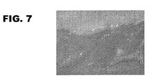
- FIG. 7 Figure 11 shows as a negative control the corresponding area of the subcutaneous tissue of a mouse in which no implantation of the angiogenesis promoting substrate was performed. There is very little blood vessel penetration seen as normal for the subcutaneous skin tissue of the mouse.
- FIGS. 8a to 8c show photographic images of the subcutaneous skin tissue in the area of the implanted pieces of foil A, B and C, respectively, after the corresponding mice were killed 12 days after implantation.
- the position of the pieces of film is indicated by black squares (reference numbers A, B and C, respectively, for the corresponding film) because the films themselves are poorly visible in the photograph.
- the films were partly dyed with Coomassie Brilliant Blue, as shown in the FIG. 8a can be seen.
- This local angiogenesis-promoting effect of materials based on crosslinked gelatin leads to a particularly advantageous effect in the vascular stent according to the invention.
- the layers of the gelatin-containing material By the layers of the gelatin-containing material, the formation of collateral blood vessels in the area of the stent-treated vessel is stimulated, whereby in the case of restenosis, for example after the complete detachment of all layers, the risk of heart attack can be significantly reduced.
- a lattice framework of a stent was used here as the support, rather than a flat support made of polyethylene, to which two layers of the resorbable material were applied over a large area.
- Resorbable materials of the same composition can be applied in the context of the present invention to the surface of a carrier of a vascular stent according to the invention.
- the first layer was colored with a white pigment (titanium dioxide) and the second layer with a red food colorant (Candurin Wine Red). For this reason, it has also been possible to produce layers with a greater thickness than the preferred thicknesses within the scope of the stent according to the invention.
- This gelatin solution was doctored to a flat polyethylene support in a thickness of about 550 microns to form the first layer of resorbable material on the support.
- the polyethylene carrier with the first layer was exposed to the equilibrium vapor pressure of a 10% strength by weight aqueous formaldehyde solution at room temperature for 17 hours in the desiccator.
- the first layer of the gelatin-based material was dried at 26 ° C and a relative humidity of 10% overnight.
- the dried layer had a thickness of about 100 ⁇ m.
- the support with the crosslinked first layer was cooled to about 4 ° C.
- a Boeson release wax was sprayed onto the layer and spread evenly with a cloth.
- the gelatin solution for the second layer of the resorbable material was prepared in the same manner as the solution for the first layer using as starting materials 20 g of gelatin, 4 g of glycerol, 73 g of distilled water and 1 g of Candurin Wine Red instead of titanium dioxide.
- the resulting gelatin solution was again applied in a thickness of about 550 microns to the provided with the release wax first layer of the resorbable material.
- the second layer was also subjected to crosslinking with formaldehyde vapor, as described for the first layer, but with the difference that the contact time of the crosslinking agent was only 2 hours instead of 17 hours.
- the second layer has a lower average degree of crosslinking than the first layer, wherein the degree of crosslinking of the gelatin within the second layer likewise decreases in the direction of the carrier.
- the drying was carried out as described for the first layer. After drying, the second layer had a thickness of about 70 microns.
- the time-dependent detachment behavior of the two layers of the gelatin-based material under physiological conditions was determined by incubation in PBS buffer (pH 7.2) at 37 ° C. By means of these physiological standard conditions, the conditions which prevail when the vascular stent according to the invention is used in the body can be adjusted.
- FIG. 9 Figure 4 shows photographic representations of the two-layer support according to Experimental Run 3-1 after 5, 6, 7, 10, 11, and 12 days of incubation in the PBS buffer.
- the adhesion of the second (outer) layer and its detachment from the underlying first layer is abolished.
- the second layer colored red in the original, appears in the upper three representations of the FIG. 9 as a dark area, while the white colored first layer can be seen as a much lighter area.
- After 5 days about 20% of the second layer is detached, there are individual white spots of the first layer visible.
- the detachment of the second layer is about 65%, wherein the second layer is substantially only present in the lower right area of the carrier.
- the second layer is finally substantially completely detached.
- the first layer is now visible, wherein this is already partially swollen and bubbles throws, but is still substantially completely present.
- the detachment of the first layer of gelatin-based material from the carrier occurs substantially between the 10th and 12th day of incubation, as in the lower three representations of FIG. 9 can be seen.
- the polyethylene support becomes visible as a dark background.
- 10 days are about 35%
- 11 days About 80% and after 12 days of incubation about 95% of the first layer detached from the carrier.
- the experiment also shows that, due to the removal of the adhesion between the layers of the gelatin-based material, an at least partially planar detachment of a layer from the underlying layer or from the carrier takes place. This is favored by a lower degree of crosslinking of the gelatin on the inside of the respective layer.
- a second test run 3-2 was carried out in the same manner as test run 3-1 described above, except that the release layer (Boeson release wax) was omitted between the two layers of the gelatin-based material.
- the release layer Boeson release wax
- the first layer has a thickness of about 80 microns and the second layer has a thickness of about 100 microns.
- the second layer of the preparation 3-2 was still essentially completely preserved after 13 days. After 18 days it was about 20% and after 25 days to about 70% replaced.
- a further test batch 3-3 was carried out in the same way as in batch 3-2, with the difference that 2 ml of a 1% strength by weight aqueous formaldehyde solution were added to each of the gelatin solutions for the first and second layer prior to doctoring.
- both layers of this approach have a higher average degree of crosslinking compared to approaches 3-1 and 3-2. This led to an even later separation time of the first layer of this approach, which was replaced after 25 days of incubation to less than about 5% of the carrier. Complete replacement took only 32 days.
Landscapes
- Health & Medical Sciences (AREA)
- Life Sciences & Earth Sciences (AREA)
- Veterinary Medicine (AREA)
- Engineering & Computer Science (AREA)
- Biomedical Technology (AREA)
- Public Health (AREA)
- General Health & Medical Sciences (AREA)
- Animal Behavior & Ethology (AREA)
- Heart & Thoracic Surgery (AREA)
- Vascular Medicine (AREA)
- Cardiology (AREA)
- Transplantation (AREA)
- Oral & Maxillofacial Surgery (AREA)
- Physics & Mathematics (AREA)
- Optics & Photonics (AREA)
- Surgery (AREA)
- Epidemiology (AREA)
- Materials For Medical Uses (AREA)
- Medicinal Preparation (AREA)
- Prostheses (AREA)
Abstract
Description
- Die vorliegende Erfindung betrifft einen Gefäßstent, insbesondere einen koronaren Stent.
- Stents werden in der Medizin als Implantate eingesetzt, um Hohlstrukturen von Organen im Körper offenzuhalten bzw. nach einem Verschluss (Stenose) wieder zu öffnen. In der Regel wird dabei ein röhrchenförmiger Stent in einer komprimierten Form in das betreffende Organ eingebracht und anschließend expandiert, um die Wände des Organs abzustützen.
- Besondere Bedeutung haben Stents bei der Öffnung von Blutgefäßen, und hier insbesondere im Bereich der Herzkranzgefäße (koronare Stents). Die Verengung oder der Verschluss von Herzkranzgefäßen durch die Ablagerung von Thromben (Blutgerinnseln), Blutfetten oder Kalk an den Gefäßwänden ist eine häufige Ursache für die Unterversorgung von Bereichen des Herzmuskels mit Blut und damit für einen Herzinfarkt. Die zur Vermeidung oder Beseitigung derartiger Stenosen eingesetzten Stents bestehen häufig aus einem röhrchenförmigen Gittergerüst, welches in das betroffene Gefäß eingeführt und mittels eines Ballon-Katheters radial expandiert wird, um das Gefäß wieder auf eine ausreichende Größe zu weiten.
- Bei derartigen Behandlungen mit Gefäßstents besteht jedoch regelmäßig das Problem der so genannten Restenose. Hierunter versteht man eine erneute Verengung des Gefäßes durch Ablagerungen auf dem Stent und/oder die Überwucherung des Stents mit Gewebe, die durch eine Aktivierung von Blutplättchen und Gerinnungsfaktoren am "Fremdmaterial" des Stents (in der Regel Metall oder Kunststoff) induziert wird. Eine Restenose macht häufig eine erneute Behandlung des betroffenen Gefäßes erforderlich.
- Zur Vermeidung oder Reduzierung dieses Problems werden seit einiger Zeit medikamentenbeschichtete Stents (drug-eluting stents, DES) eingesetzt. Diese Stents weisen eine Beschichtung auf, aus der bestimmte Wirkstoffe zur Hemmung der Gewebebildung freigesetzt werden (z.B. Antiproliferativa, Cytostatika oder Immunsupressiva). Durch die Verwendung derartiger Stents soll das Risiko einer Restenose des betreffenden Gefäßes deutlich gesenkt werden (siehe M.C. Morice et al., New England Journal of Medicine 2002 (346) 1773-1780).
- Medikamentenbeschichtete Stents werden unter anderem in der Offenlegungsschrift
US 2005/0019404 A1 offenbart. - In jüngeren Untersuchungen wurde allerdings festgestellt, dass bei Patienten, denen nach Stenose eines Herzkranzgefäßes ein medikamentenbeschichteter Stent implantiert wurde, die Mortalitätsrate höher ist als bei Patienten, die mit einem unbeschichteten Metallstent behandelt wurden. Quantitativ ergab diese Untersuchung, dass die Todeswahrscheinlichkeit durch Herzinfarkt in dem Zeitraum zwischen 6 Monaten und 3 Jahren nach der Stentimplantation bei der erstgenannten Patientengruppe um 32 % höher ist als bei der letztgenannten (siehe B. Lagerqvist et al., New England Journal of Medicine 2007 (356) 1009-1019).
- T. Shirota et al. beschreiben einen metallischen intravaskulären Stent, der mit einer photoreaktiven, vernetzten Gelatine beschichtet ist (Biomaterials 2003 (24) 2295-2302).
- Die Aufgabe der vorliegenden Erfindung ist es, einen Stent zur Verfügung zu stellen, bei dem das Risiko einer Restenose vermindert ist, ohne dabei auf anti-proliferative Wirkstoffe zurückgreifen zu müssen.
- Diese Aufgabe wird erfindungsgemäß gelöst durch einen Gefäßstent der eingangs genannten Art, umfassend einen Träger aus einem formstabilen Material, sowie eine oder mehrere, zumindest bereichsweise auf dem Träger angeordnete Lagen eines unter physiologischen Bedingungen resorbierbaren Materials auf der Basis von vernetzter Gelatine, wobei die Adhäsion zwischen dem Träger und der Lage und/oder zwischen einzelnen Lagen dadurch aufhebbar ist, dass (i) der Vernetzungsgrad der Gelatine innerhalb einer oder mehrerer Lagen in Richtung des Trägers abnimmt, dass (ii) der mittlere Vernetzungsgrad der Gelatine in den einzelnen Lagen in Richtung der dem Träger benachbarten Lage zunimmt, und/oder dass (iii) eine oder mehrere Lagen mit Fettsäureresten modifizierte Gelatine enthalten.
- Durch diese Aufhebung der Adhäsion wird eine Ablösung einer oder mehrerer Lagen von dem erfindungsgemäßen Stent unter physiologischen Bedingungen begünstigt. Die physiologischen Bedingungen, denen der erfindungsgemäße Gefäßstent bei seiner Verwendung im menschlichen oder tierischen Körper ausgesetzt ist, d.h. insbesondere die im Blut vorherrschenden Bedingungen, können dabei durch so genannte physiologische Standardbedingungen definiert und in vitro nachgestellt werden. Unter physiologischen Standardbedingungen wird im Rahmen dieser Erfindung die Inkubation in PBS-Puffer (pH 7,2) bei 37 °C verstanden.
- Bevorzugt sind eine oder mehrere Lagen des resorbierbaren Materials einzeln ablösbar. Unter der Ablösung einer Lage im Sinne der vorliegenden Erfindung wird dabei eine zumindest bereichsweise flächige Ablösung des die Lage bildenden resorbierbaren Materials von der darunter liegenden Lage bzw. von dem Träger verstanden. Mit anderen Worten kommt es zu einer Ablösung von Bereichen oder Bruchstücken der Lage, die noch eine gewisse strukturelle Integrität aufweisen. Dieser Ablösevorgang ist im Gegensatz zu einer kontinuierlichen Degradation bzw. Resorption der Lage auf molekularer Ebene zu sehen, bei dem die Lage im Wesentlichen vollständig resorbiert wird, bevor eine Ablösung im oben beschriebenen Sinne erfolgt.
- Die vorteilhafte Wirkung des erfindungsgemäßen Stents beruht darauf, dass durch die Aufhebung der Adhäsion und die Ablösung einer oder mehrerer Lagen des resorbierbaren Materials von dem Gefäßstent auch Zellen, Gewebe oder Thromben, die sich auf der Oberfläche des Stents gebildet haben, wieder von dem Stent abgelöst und mit dem Blutstrom aus dem betroffenen Gefäßbereich entfernt werden. Somit führt dieser Vorgang zu einer Reinigung des Stents und einer Erneuerung seiner Oberfläche, sodass eine Restenose verhindert oder zumindest verzögert wird, ohne dass ein Einsatz von Antiproliferativa oder ähnlichen Wirkstoffen erforderlich ist.
- Die zunächst vorhandene Adhäsion zwischen der Lage und dem Träger bzw. den Lagen untereinander ist für die vorliegende Erfindung von Bedeutung, damit über die steuerbare Aufhebung der Adhäsion zeitlich gezielt und vorherbestimmbar die Ablösung von flächigen Lagenteilen ermöglicht ist und der Stent bis zum Beginn des Ablösens definierte Eigenschaften aufweist.
- Die erfindungsgemäß geschaffene Möglichkeit der Aufhebung der Adhäsion zwischen einzelnen Lagen kann durch verschiedene Maßnahmen erreicht werden. Zum einen können zwischen einzelnen Lagen des resorbierbaren Materials Haftschichten aus einem unter physiologischen Bedingungen löslichen Material vorgesehen sein. Für solche Haftschichten sind verschiedene Materialien denkbar, beispielsweise niedermolekulares, lösliches Kollagenhydrolysat.
- Bevorzugte Träger weisen eine mikroskopisch glatte, geschlossene Oberfläche auf. Damit wird verhindert, dass sich beim Aufbringen der Lagen auf dem Träger Material in Poren ablagert und damit eine Haftung aufgrund von Formschlusseffekten ergibt. Dies gilt insbesondere auch bei der Verwendung von Trägern in der Form von röhrchenartigen Gittergerüsten, die sich auch bei der vorliegenden Erfindung besonders eignen. Bei Trägern mit Gittergerüststruktur wird im Rahmen der besondere Vorteil erzielt, dass beim flächenmäßigen Ablösen einer Lage keine physiologisch zu großen und damit medizinisch bedenklichen Lagenstücke in das zirkulierende Blut abgegeben werden.
- Bei Trägern mit Gitterstruktur wird bevorzugt eine Beschichtung von dessen Stegen angestrebt, die im Querschnitt von der bzw. den Lagen vorzugsweise vollständig umgeben sind. Auch nach der Beschichtung der Stege des Trägers bleiben die Zwischenräume zwischen den Stegen bevorzugt offen.
- Eine Expansion des Stents ist dann möglich ohne die Integrität der Lage(n) auf dem Träger, d.h. der Lage(n) auf den Stegen des Trägers zu gefährden.
- Des Weiteren können zwischen einzelnen Lagen des resorbierbaren Materials auch Trennschichten vorgesehen sein, worauf weiter unten noch im Detail eingegangen wird.
- Die Aufhebung der Adhäsion zwischen einzelnen Lagen des resorbierbaren Materials (bzw. zwischen dem Träger und der benachbarten Lage) kann insbesondere auch auf einer zumindest partiellen Degradation des Materials unter physiologischen Bedingungen basieren, wobei unter einer Degradation in erster Linie diejenigen Vorgänge zu verstehen sind, die letztlich auch zu der Resorption des Materials führen. Die Degradationseigenschaften des resorbierbaren Materials können dabei durch verschiedene Maßnahmen gezielt beeinflusst werden, z.B. durch den Einsatz von Gelatinen mit unterschiedlichen Molekulargewichten und/oder die Beimischung weiterer Biopolymere zu dem Gelatine basierenden Material.
- Gemäß einer bevorzugten Ausführungsform der Erfindung ist die Adhäsion in Abhängigkeit vom Vernetzungsgrad der Gelatine aufhebbar. Ein höherer Vernetzungsgrad der Gelatine führt zu einer langsameren Degradation (und Resorption) des Gelatine basierenden Materials, sodass durch diesen Parameter auf einfache Weise das Ablöseverhalten einzelner Lagen eingestellt werden kann.
- Besonders bevorzugt ist die Gelatine in der Weise vernetzt, dass der Vernetzungsgrad innerhalb einer oder mehrerer Lagen des resorbierbaren Materials in Richtung des Trägers abnimmt. Durch einen geringeren Vernetzungsgrad der Gelatine an der dem Träger zugewandten Seite einer Lage wird bewirkt, dass es in diesem Bereich zu einer schnelleren Degradation des Materials kommt und dadurch die Adhäsion an den Träger (bzw. an die darunter liegende Lage) aufgehoben wird, während an der Außenseite der Lage durch den höheren Vernetzungsgrad der Gelatine eine gewisse strukturelle Integrität erhalten bleibt und die oben beschriebene, zumindest bereichsweise flächige Ablösung der Lage erreicht wird.
- Bei dem formstabilen Material, aus dem der Träger gebildet ist, handelt es sich bevorzugt um ein unter physiologischen Bedingungen inertes Material, insbesondere um Metall und/oder Kunststoff. Als Träger können insbesondere expandierbare Gittergerüste oder ähnliche Strukturen in Röhrchen- oder Schlauchform zum Einsatz kommen.
- Der erfindungsgemäße Gefäßstent kommt bevorzugt im kardiovaskulären Bereich, und hier insbesondere zur Behandlung der Herzkranzgefäße (koronarer Stent) zum Einsatz, da hier Gefäßstenosen zu einem Herzinfarkt führen können. Ebenso kann der Stent jedoch zur Behandlung von Stenosen in anderen Bereichen des Körpers verwendet werden.
- Der erfindungsgemäße Effekt, die Gefahr von Restenosen zumindest zu reduzieren, kann bereits mit einer bereichsweisen Anordnung der Lage(n) auf dem Träger erreicht werden. Bevorzugt ist jedoch, wenn die mindestens eine Lage ca. 75 % oder mehr, insbesondere ca. 90 % oder mehr der Oberfläche des Trägers abdeckt. Besonders bevorzugt deckt die mindestens eine Lage im Wesentlichen die gesamte Oberfläche des Trägers ab.
- Der erfindungsgemäße Einsatz von Gelatine als Basismaterial für die Lage(n) des resorbierbaren Materials bietet den Vorteil, dass es sich hierbei um ein sehr gut körperverträgliches, im Wesentlichen vollständig resorbierbares Produkt handelt, welches in reproduzierbarer Reinheit und Qualität hergestellt werden kann.
- Darüber hinaus hat Gelatine im Rahmen der vorliegenden Erfindung insofern eine besonders vorteilhafte Wirkung, als sie die Angiogenese, d.h. die Neubildung von Blutgefäßen, fördert. Diesbezügliche Untersuchungen haben gezeigt, dass bei Einbringung von Gelatine enthaltenden Formkörpern in den menschlichen oder tierischen Körper ein lokaler Angiogenese fördernder Effekt auftritt. Dies gilt nicht nur für poröse Formkörper, bei denen ein Einwachsen von Kapillaren in die poröse Struktur beobachtet wird (siehe die deutsche Patentanmeldung
10 2005 054 937 ), sondern insbesondere auch für nicht-poröse Formkörper, wie z.B. Folien, bei denen die Angiogenese fördernde Wirkung in der Umgebung des Formkörpers beobachtet wird. - Es wurde festgestellt, dass die eingangs erwähnte höhere Mortalität bei medikamentenbeschichteten Stents ihre Ursache in erster Linie in einer unerwünschten Nebenwirkung der in den beschichteten Stents eingesetzten antiproliferativen Wirkstoffe hat, nämlich einer Verhinderung der Angiogenese im Umfeld des betroffenen Gefäßes. Im Fall einer Stenose besteht eine natürliche Reaktion des Körpers darin, durch die Neubildung von Blutgefäßen den Gefäßverschluss zu überbrücken. Dieser Vorgang wird durch Antiproliferativa o.ä. gehemmt, sodass im Fall einer dennoch eintretenden Restenose keine kollateralen Blutgefäße zur Verfügung stehen und ein Herzinfarkt die Folge ist (P. Meier et al., Journal of American Cardiology 2007 (49) 15 bis 20).
- Durch die Möglichkeit des Verzichts auf anti-proliferative Wirkstoffe bei dem erfindungsgemäßen Stent wird nicht nur eine Angiogenese hemmende Wirkung vermieden, sondern es kommt im Gegenteil durch das Gelatine basierende Material zu einer Stimulierung der Angiogenese.
- Der erfindungsgemäße Gefäßstent vermindert oder verzögert somit einerseits das Risiko einer Restenose durch den Selbstreinigungseffekt der Stentoberfläche, andererseits fördert er gleichzeitig die Bildung von kollateralen Blutgefäßen aufgrund der Angiogenese fördernden Wirkung der Gelatine in der Umgebung des betroffenen Gefäßes. Kommt es also dennoch zu einer Restenose, insbesondere nachdem sämtliche Lagen des Stents abgelöst sind, stehen kollaterale Blutgefäße als natürlicher Umgehungskreislauf zur Verfügung.
- Ein Verzicht auf anti-proliferative Agenzien hat im Übrigen noch weitere Vorteile. So verhindern diese Wirkstoffe nicht nur die Anlagerung von Gewebe, welches zu einer Restenose führen kann, sondern auch eine Endothelialisierung des Stents. Bei der Endothelialisierung entsteht um den Stent eine Bindegewebsschicht, die mit den Komponenten des Blutes verträglich ist, wodurch die Aktivierung von Blutplättchen und Gerinnungsfaktoren am Stentmaterial verhindert wird, d.h. dieser Vorgang wirkt einer Thrombose im Bereich des Stents entgegen.
- Bevorzugt ist das erfindungsgemäße resorbierbare Material zu einem überwiegenden Anteil aus vernetzter Gelatine gebildet. Dies bedeutet, dass die Gelatine den größten Anteil bei eventuell weiteren verwendeten Komponenten des Materials stellt.
- Besonders geeignete Gelatinetypen sind Schweineschwartengelatine, die vorzugsweise hochmolekular ist und einen Bloomwert von ca. 160 bis ca. 320 g aufweist.
- Um eine optimale Bioverträglichkeit des erfindungsgemäßen Stents zu gewährleisten, wird als Ausgangsmaterial bevorzugt eine Gelatine mit einem besonders geringen Gehalt an Endotoxinen eingesetzt. Bei Endotoxinen handelt sich um Stoffwechselprodukte oder Bruchstücke von Mikroorganismen, welche in dem tierischen Rohmaterial vorkommen. Der Endotoxingehalt von Gelatine wird in internationalen Einheiten pro Gramm (I.E./g) angegeben und gemäß dem LAL-Test bestimmt, dessen Durchführung in der vierten Ausgabe des Europäischen Arzneibuches (Ph. Eur. 4) beschrieben ist.
- Um den Gehalt an Endotoxinen möglichst gering zu halten, ist es vorteilhaft, die Mikroorganismen möglichst frühzeitig im Zuge der Gelatineherstellung abzutöten. Ferner sollten entsprechende Hygienestandards beim Herstellungsprozess eingehalten werden.
- Somit kann der Endotoxingehalt von Gelatine durch bestimmte Maßnahmen beim Herstellungsprozess drastisch gesenkt werden. Zu diesen Maßnahmen zählen in erster Linie die Verwendung frischer Rohmaterialien (z.B. Schweineschwarte) unter Vermeidung von Lagerzeiten, die sorgfältige Reinigung der gesamten Produktionsanlage unmittelbar vor Beginn der Gelatineherstellung sowie gegebenenfalls das Auswechseln von Ionenaustauschern und Filtersystemen in der Produktionsanlage.
- Die im Rahmen der vorliegenden Erfindung eingesetzte Gelatine weist bevorzugt einen Endotoxingehalt von ca. 1.200 I.E./g oder weniger, noch mehr bevorzugt von ca. 200 I.E/g oder weniger auf. Optimalerweise liegt der Endotoxingehalt bei ca. 50 I.E./g oder weniger, jeweils gemäß dem LAL-Test bestimmt. Im Vergleich hierzu weisen manche handelsübliche Gelatinen Endotoxingehalte von über 20.000 I.E./g auf.
- Durch die erfindungsgemäße Vernetzung der an sich löslichen Gelatine wird diese in ein unlösliches, jedoch unter physiologischen Bedingungen resorbierbares Material überführt. Dabei ist die Resorptions- bzw. Degradationsgeschwindigkeit des Materials vom Vernetzungsgrad der Gelatine abhängig und kann über einen relativ weiten Bereich eingestellt werden, wie dies bereits oben angesprochen wurde. Insbesondere kann der Zeitpunkt der Ablösung einzelner Lagen über den jeweiligen Vernetzungsgrad der Gelatine vorgegeben werden.
- Formkörper auf Basis von vernetzter Gelatine, deren Resorptionseigenschaften und Herstellung sind in der Offenlegungsschrift
DE 10 2004 024 635 A1 beschrieben. - Die Vernetzung der Gelatine kann sowohl durch chemische als auch durch enzymatische Vernetzungsmittel erfolgen. Unter den chemischen Vernetzungsmitteln ist eine Vernetzung unter Verwendung von Formaldehyd bevorzugt, welches gleichzeitig eine Sterilisierung bewirkt.
- Ein bevorzugtes enzymatisches Vernetzungsmittel ist das Enzym Transglutaminase.
- Bei der Herstellung von Formkörpern auf Basis von vernetzter Gelatine bietet sich ein zweistufiges Vernetzungsverfahren an, bei dem in einer ersten Stufe die Gelatine in Lösung partiell vernetzt wird. Aus der partiell vernetzten Gelatinelösung wird dann ein Formkörper hergestellt und einem zweiten Vernetzungsschritt unterworfen.
- Der zweite Vernetzungsschritt kann insbesondere durch Einwirkung eines Vernetzungsmittels in der Gasphase, vorzugsweise Formaldehyd, durchgeführt werden.
- Entsprechend diesem Verfahren kann der erfindungsgemäße Gefäßstent in der Weise hergestellt werden, dass eine partiell vernetzte Lösung des Gelatine basierenden Materials auf die Oberfläche des Trägers aufgebracht wird, z.B. durch Eintauchen des Trägers in die Lösung. Nach dem Trocknen der Lösung wird ein Träger mit einer Lage des resorbierbaren Materials erhalten, der dann dem zweiten Vernetzungsschritt, z.B. mit Formaldehyd in der Gasphase, unterworfen wird.
- Durch den zweiten Vernetzungsschritt kann hierbei auf einfache Weise der bevorzugte Gradient im Vernetzungsgrad erhalten werden, da das Vernetzungsmittel im Wesentlichen nur von der Außenseite der Lage her in diese eindringen kann, was zu einer stärkeren Vernetzung an der Außenseite und zu einer geringeren Vernetzung an der inneren, dem Träger zugewandten Seite, führt.
- Es ist im Rahmen der Erfindung ebenso denkbar, keine Vernetzung der Gelatine in der Lösung vorzusehen, sondern nur einen einzigen Vernetzungsschritt nach dem Aufbringen der Lage auf den Träger durchzuführen.
- Bevorzugt enthält das resorbierbare Material ein oder mehrer Weichmacher. Hierdurch wird die Flexibilität des Materials erhöht, was insbesondere im Hinblick auf die Expansion des Stents nach dem Implantieren von Vorteil sein kann. Durch eine ausreichende Flexibilität kann weitgehend vermieden werden, dass die mindestens eine Lage beim Expandieren des Stents beschädigt wird oder sich mechanisch von dem Träger ablöst.
- Bevorzugte Weichmacher sind beispielsweise Glycerin, Oligoglycerine, Oligoglycole, Sorbit und Mannit. Der Anteil an Weichmacher in dem resorbierbaren Material beträgt bevorzugt ca. 12 bis ca. 40 Gew.-%, weiter bevorzugt ca. 16 bis ca. 25 Gew.-%.
- Gemäß einer bevorzugten Ausführungsform der Erfindung sind auf dem Träger mehrere Lagen des resorbierbaren Materials angeordnet. Durch das Vorsehen mehrerer Lagen, die sich bevorzugt jeweils einzeln ablösen, kann die Oberfläche des erfindungsgemäßen Stents wiederholt von Ablagerung befreit werden und so der Zeitraum, in dem die Gefahr der Restenose verringert wird, deutlich verlängert werden. Bevorzugt umfasst der Gefäßstent zwei bis fünf Lagen des resorbierbaren Materials.
- Erfindungsgemäße Stents mit mehreren Lagen können auf relativ einfache Weise hergestellt werden, indem die oben dargestellten Verfahrensschritte (Aufbringen der ggf. partiell vernetzten Gelatinelösung, Trocknen und zweite Vernetzung) mehrmals nacheinander durchgeführt werden.
- Bevorzugt nimmt der Vernetzungsgrad der Gelatine innerhalb jeder Lage in Richtung des Trägers ab. Dadurch wird die Aufhebung der Adhäsion bei der Ablösung jeder einzelnen Lage begünstigt.
- Vorteilhafterweise sind die einzelnen Lagen des resorbierbaren Materials nacheinander von außen nach innen ablösbar. Eine solche sukzessive Ablösung wird bereits dadurch begünstigt, dass weiter innen befindliche Lagen durch die jeweils darüber liegenden Lagen in gewissem Ausmaß vor einer Degradation geschützt sind. Dies gilt auch dann, wenn sämtliche Lagen denselben mittleren Vernetzungsgrad der Gelatine aufweisen.
- Bevorzugt ist jedoch, dass der mittlere Vernetzungsgrad der einzelnen Lagen in Richtung der dem Träger benachbarten Lage zunimmt. Durch einen hohen Vernetzungsgrad der inneren Lagen kann deren Degradation und Ablösung zusätzlich verzögert werden. Darüber hinaus kann das Ablöseverhalten der einzelnen Lagen gezielt an die jeweiligen Bedürfnisse angepasst werden, d.h. insbesondere an die erwartete Intensität der Ablagerung bzw. Gewebebildung an dem Stent. Im Idealfall ist die Zeit bis zur Ablösung der innersten Lage ausreichend lang, sodass bis zu diesem Zeitpunkt die Ausbildung von kollateralen Blutgefäßen, begünstigt durch den Angiogenese fördernden Effekt des Gelatine basierenden Materials, ermöglicht wird.
- Die mittleren Vernetzungsgrade der einzelnen Lagen können beispielsweise so gewählt werden, dass sich die äußerste Lage nach ca. 1 bis 2 Wochen und die innerste, dem Träger benachbarte Lage nach ca. 3 bis 6 Monaten ablöst. Die genannten Zeiträume sind dabei auf den Zeitpunkt bezogen, an dem der erfindungsgemäße Stent physiologische Bedingungen ausgesetzt wird, d.h. insbesondere in ein Blutgefäß eingesetzt wird.
- Unterschiedliche Vernetzungsgrade der einzelnen Lagen können bei dem oben beschriebenen Herstellungsverfahren durch unterschiedliche Vernetzungsmittelkonzentrationen in der Gelatinelösung und/oder durch unterschiedliche Konzentrationen oder Einwirkzeiten des Vernetzungsmittels im zweiten Vernetzungsschritt realisiert werden.
- Die Dicke der einzelnen Lagen des resorbierbaren Materials liegt bevorzugt im Bereich von ca. 5 bis ca. 50 µm.
- Gemäß einer weiteren Ausführungsform der Erfindung sind zwischen mehreren Lagen des resorbierbaren Materials und/oder auf der Außenseite der Lage(n) ein oder mehrere Trennschichten angeordnet. Durch solche Trennschichten können verschiedene vorteilhafte Wirkungen erzielt werden.
- Zum einen kann durch die Trennschichten die Adhäsion zwischen mehreren Lagen reduziert werden. Bei fortschreitender Degradation des Gelatine basierenden Materials in der jeweils äußersten Lage wird somit die Aufhebung der Adhäsion und die Ablösung dieser Lage durch die Trennschicht beschleunigt, wobei die Trennschicht selbst auf der Außenseite der darunter liegenden Lage verbleibt.
- Bevorzugt wird für die Trennschichten ein resorbierbares Material eingesetzt. Vorzugsweise weist dieses Material eine längere Resorptionszeit auf als das Gelatine basierende Material in der mindestens einen Lage, sodass die Trennschicht auf der Außenseite einer Lage zum Zeitpunkt der Ablösung dieser Lage noch vorhanden ist.
- Die Trennschichten können beispielsweise aus einem Trennwachs gebildet sein.
- Durch eine auf der jeweils äußersten Lage angeordnete Trennschicht kann zudem erreicht werden, dass die Ablagerung von Zellen oder Gewebe auf der Oberfläche des Stents verringert wird, indem die Trennschicht eine adhäsionshemmende Wirkung gegenüber diesen Ablagerungen aufweist. Dies wirkt der Gefahr einer Restenose zusätzlich entgegen. Wenn das Ausmaß der Ablagerungen verringert ist, kann auch ein späterer Ablösezeitpunkt der Lage(n) des resorbierbaren Materials gewählt werden.
- Vorzugsweise enthält die mindestens eine Trennschicht modifizierte Gelatine. Es wurde gefunden, dass durch eine chemische Modifikation von Gelatine eine adhäsionshemmende Wirkung gegenüber Zellen erreicht werden kann.
- Gemäß einer weiteren Ausführungsform der Erfindung ist vorgesehen, dass die modifizierte Gelatine in einer oder mehreren Lagen des Gelatine basierenden Materials enthalten ist. Hierdurch kann ein vergleichbarer Effekt wie bei Trennschichten aus modifizierter Gelatine erzielt werden.
- Während Trennschichten zu einem hohen Anteil oder im Wesentlichen vollständig aus modifizierter Gelatine gebildet sein können, ist der mögliche Anteil an modifizierter Gelatine in den Lagen insofern limitiert, als nachteilige Wirkungen auf die Vernetzbarkeit der Gelatine und auf das Degradationsverhalten vermieden werden sollten.
- Bevorzugt handelt es sich bei der modifizierten Gelatine um eine mit Fettsäureresten modifizierte Gelatine. Ein Beispiel hierfür stellt die Modifikation von Gelatine mit Dodecenyl-succinat dar.
- Die Modifikation erfolgt hierbei insbesondere an den freien Aminogruppen der Lysinreste der Gelatine. Vorzugsweise sind ca. 10 bis ca. 80 % der Lysinreste der modifizierten Gelatine mit Fettsäureresten modifiziert.
- Eine weitere Möglichkeit, um eine adhäsionshemmende Wirkung zu erzielen, stellt eine anionische Modifikation der Gelatine dar, beispielsweise die Succinylierung von Seitenketten.
- Diese und weitere Vorteile der Erfindung werden anhand der folgenden Beispiele unter Bezugnahme auf die Zeichnung näher erläutert. Es zeigen im Einzelnen:
- Figur 1:
- Darstellung eines erfindungsgemäßen Stents in komprimiertem Zustand vor der Implantation;
- Figur 2:
- Darstellung eines erfindungsgemäßen Stents in expandiertem Zustand nach der Implantation;
- Figur 3:
- Darstellung eines Ausschnitts aus der Struktur eines erfindungsgemäßen Stents;
- Figur 4:
- schematische Querschnittsdarstellung der Lagen eines erfindungsgemäßen Stents;
- Figuren 5 und 6:
- Diagramme betreffend die adhäsionshemmende Wirkung von modifizierter Gelatine;
- Figur 7:
- fotografische Darstellung der Blutgefäßbildung im subkutanen Gewebe einer Maus;
- Figuren 8a bis c:
- fotografische Darstellungen der vermehrten Blutgefäßbildung bei Anwesenheit verschiedener Angiogenese fördernder Substrate auf der Basis von vernetzter Gelatine; und
- Figur 9:
- fotografische Darstellung des zeitabhängigen Ablösesverhalten zweier Lagen eines Gelatine basierenden Materials.
- Die
Figuren 1 und 2 zeigen ein Ausführungsbeispiel eines erfindungsgemäßen Gefäßstents, der insbesondere als koronarer Stent Verwendung findet. Der Stent umfasst einen Träger in Form eines röhrchenförmigen Gittergerüsts aus Metall oder Kunststoff, auf dessen Oberfläche mehrere Lagen eines resorbierbaren Material auf der Basis von vernetzter Gelatine angeordnet sind. -
Figur 1 zeigt den Stent in einem komprimierten Zustand. Der Stent nimmt in diesem Zustand nur einen relativ geringen Querschnitt ein und kann daher in einen von einer Stenose betroffenen Gefäßbereich eingeführt werden. - Nach dem Implantieren wird der Stent z.B. mittels eines Ballon-Katheters geweitet, d.h. radial expandiert, sodass das betroffene Gefäß aufgeweitet und durch den Stent abgestützt wird. Dieser expandierte Zustand des Stents ist in der
Figur 2 dargestellt. Sowohl das Gittergerüst des Trägers als auch die Lagen des resorbierbaren Materials weisen eine ausreichende Flexibilität auf, um diesen Expansionsvorgang durchführen zu können. - Die
Figur 3 zeigt einen vergrößerten Ausschnitt aus der Gerüststruktur des erfindungsgemäßen Stents. Das Gittergerüst des Trägers wird aus einer Vielzahl von miteinander verbundenen Stegen gebildet, wobei die Lagen auf der Oberfläche dieser Stege angeordnet sind. Vorzugsweise ist hierbei ein möglichst großer Anteil der Gesamtoberfläche des Trägers abgedeckt. - Die in der
Figur 3 gezeigte Geometrie der Gitterstruktur bzw. Anordnung der Stege ist hierbei lediglich beispielhaft. Die Gitterstruktur sollte jedoch so gestaltet sein, dass durch eine Verbiegung oder Verformung der Stege eine Expansion des Stents ermöglicht wird. - Eine schematische Darstellung des Querschnitts durch einen Steg 10 des Trägers, z.B. entlang der Linie A-A, ist in der
Figur 4 gezeigt. Der Steg, der z.B. einen Durchmesser im Bereich von 100 bis 200 µm aufweist, ist von drei Lagen umgeben, und zwar einer inneren, dem Steg 10 benachbarte Lage 21, einer mittleren Lage 22 sowie einer äußeren Lage 23. Alternativ zu dieser Ausführungsform können auch eine, zwei oder mehr als drei Lagen vorgesehen sein. - Sämtliche Lagen sind aus einem unter physiologischen Bedingungen resorbierbaren Material auf der Basis von vernetzter Gelatine gebildet. Zur Erhöhung der Flexibilität der Lagen kann das Material zusätzlich einen Weichmacher, z.B. Glycerin, umfassen.
- Der Vernetzungsgrad der Gelatine nimmt bevorzugt innerhalb jeder der drei Lagen 21, 22 und 23 in Richtung auf den Steg 10 des Trägers ab. Dies bedeutet, dass z.B. die äußere Lage 23 an ihrer inneren, d.h. der mittleren Lage 22 zugewandten Seite einen niedrigeren Vernetzungsgrad aufweist als an ihrer Außenseite 20.
- Durch diesen graduellen Vernetzungsgrad kommt es unter physiologischen Bedingungen an der Innenseite der Lage 23 zu einer schnelleren Degradation des Gelatine basierenden Materials und dadurch, nach einer vorgegebenen Zeit, zu einer Aufhebung der Adhäsion an die Lage 22 und einer zumindest bereichsweise flächigen Ablösung der Lage 23 von der Lage 22. Dabei werden Ablagerungen von Zellen oder Gewebe, die sich an der Außenseite 20 der Lage 23, d.h. an der Oberfläche des Stents, gebildet haben, mit dem Blutstrom aus dem betroffenen Gefäßbereich entfernt. Nach dieser Ablösung bildet die Lage 22 die äußere Lage, sodass sich der eben beschriebene Vorgang bei dieser Lage wiederholen kann.
- Der mittlere Vernetzungsgrad der Gelatine in den einzelnen Lagen 21, 22 und 23 nimmt bevorzugt in Richtung des Stegs 10 zu, d.h. die Lage 23 weist den geringsten und die Lage 21 den höchsten mittleren Vernetzungsgrad auf.
- Beispielsweise sind die Vernetzungsgrade in den einzelnen Lagen so gewählt, dass sich die Lage 23 ca. ein bis zwei Wochen, die Lage 22 ca. vier bis acht Wochen und die Lage 21 ca. drei bis sechs Monaten nach dem Einführen des Gefäßstents in ein Blutgefäß ablöst.
- Die einzelnen Lagen 21, 22 und 23 weisen vorzugsweise eine Dicke im Bereich von ca. 5 bis ca. 50 µm auf.
- Zwischen den einzelnen Lagen 21, 22 und 23 und/oder auf der Außenseite 20 der Lage 23 können Trennschichten angeordnet sein. Solche Trennschichten können zum einen die Aufhebung der Adhäsion zwischen den einzelnen Lagen beschleunigen und/oder der Adhäsion von Zellen und Gewebe entgegenwirken.
- Die Trennschichten und/oder die Lagen 21, 22 und 23 können insbesondere modifizierte Gelatine enthalten. Hierdurch kann die Zelladhäsion gegenüber nicht-modifizierter Gelatine herabgesetzt werden, wie in dem folgenden Beispiel 1 gezeigt wird.
- Die Aminogruppen der Lysinreste in der Gelatine lassen sich mittels Bernsteinsäureanhydrid in eine succinierte Form überführen, wodurch der pKs-Wert des Gelatinematerials von 8 bis 9, wie er für die unmodifizierte Gelatine gefunden wird, auf ca. 4 abgesenkt wird.
- Eine weitere Möglichkeit die Gelatine zu modifizieren besteht darin, die Aminogruppen der Lysinreste in Dodecenyl-succinyl-Gruppen zu überführen. Der pKs-Wert wird hierbei auf ca. 5 abgesenkt und gleichzeitig findet eine leichte Hydrophobierung der Gelatine durch den Fettsäurerest statt.
- In beiden Fällen nimmt die Zelladhäsion gegenüber einer solchermaßen behandelten Gelatine deutlich ab, was in den nachfolgend beschriebenen Versuchen am Beispiel von porcinen Chondrozyten gezeigt wird.
- Der Umsetzungsgrad der Lysingruppen der modifizierten Gelatine beträgt vorzugsweise 30 % oder mehr. Im Falle der Dodecenyl-succinierten Gelatine reichen oft Umsetzungsgrade von 40 bis 50 % sehr gut aus, während bei der succinierten Gelatine eher eine 80 %ige bis nahezu vollständige Umsetzung der Lysingruppen beste Resultate liefert.
- Die
Figuren 5 und 6 zeigen Zelladhäsionsergebnisse für zu Testzwecken auf Glasoberflächen aufgebrachten Prüfflächen aus Gelatinematerialien, die ausgehend von Schweineschwartengelatine (MW 119 kDa) und einer zu ca. 95 % an den Lysingruppen succinierten Gelatine (Figur 5 ) bzw. ca. 45 % Dodecenyl-succinierten Gelatine (Figur 6 ) gleichen Typs hergestellt wurden. Es wurden jeweils Abmischungen von unmodifizierter Gelatine mit modifizierter Gelatine in den Verhältnissen 100:0, 80:20, 50:50 und 0:100 geprüft. - In den Tests wurden jeweils 20.000 porcine Chondrozyten auf einer Prüffläche während 4 h bei 37 °C inkubiert. Der Überstand wurde entfernt, die Oberfläche gewaschen und die auf der Oberfläche verbleibenden Zellen fixiert, um sie anschließend lichtmikroskopisch auszuwerten. Vergleichbare Ergebnisse wurden mit humanen Chondrozyten erhalten.
- Die Prozentangaben in den Diagrammen stehen für den Anteil der auf den Folienprüfflächen gefunden Zellen gegenüber der zur Inkubation benutzten Anzahl, nachdem die vorstehend genannte Prozedur durchgeführt wurde.
- Für beide modifizierte Gelatinearten ergaben sich Besiedelungseffekte nahe Null bei der ausschließlichen Verwendung von modifizierter Gelatine.
- Diese deutliche Hemmung der Zelladhäsion durch chemisch modifizierte Gelatine kann im Rahmen der vorliegenden Erfindung ausgenutzt werden, um die Ablagerung von Zellen und Gewebe auf der jeweils äußeren Lage des Stents zu reduzieren.
- Die bevorzugt zwischen den einzelnen Lagen angeordneten Trennschichten können dabei einen sehr hohen Anteil an modifizierter Gelatine von bis zu 100 % enthalten. Da diese Trennschichten nach der Ablösung der darüber befindlichen Lage jeweils die Oberfläche des Stents bilden, kann dadurch eine sehr starke adhäsionshemmende Wirkung des erfindungsgemäßen Stents erzielt werden kann.
- Anhand des folgenden Beispiels soll die lokale Angiogenese fördernde Wirkung von Gelatine basierenden Materialien aufgezeigt werden.
- Es wurden Gelatinefolien mit drei unterschiedlichen Vernetzungsgraden (Folien A, B und C) mittels eines zweistufigen Vernetzungsverfahrens hergestellt.
- Für jeden der drei Ansätze wurden 25 g Schweineschwartengelatine (300 g Bloom), 9 g einer 85 Gew.-%igen Glycerinlösung sowie 66 g destilliertes Wasser gemischt und die Gelatine bei einer Temperatur von 60 °C gelöst. Nach dem Entgasen der Lösungen durch Ultraschall wurde zur Durchführung des ersten Vernetzungsschrittes eine wässrige Formaldehydlösung (2,0 Gew.-%ig, Raumtemperatur) zugegeben, und zwar 3,75 g dieser Lösung beim Ansatz A und jeweils 6,25 g der Lösung bei den Ansätzen B und C.
- Die Mischungen wurden homogenisiert und bei ca. 60 °C in einer Dicke von ca. 250 µm auf eine Polyethylenunterlage gerakelt.
- Nach dem Trocknen bei 30 °C und einer relativen Luftfeuchtigkeit von 30 % für etwa einen Tag wurden die Folien von der PE-Unterlage abgezogen und ca. 12 h unter denselben Bedingungen nachgetrocknet. Die getrockneten Folien (Dicke ca. 50 µm) wurden zur Durchführung des zweiten Vernetzungsschrittes in einem Exsikkator dem Gleichgewichtsdampfdruck einer 17 Gew.-%igen wässrigen Formaldehydlösung bei Raumtemperatur ausgesetzt. Im Falle der Folien A und B betrug die Einwirkzeit des Formaldehyddampfs 2 h, im Falle der Folie C 17 h.
- Von den auf diese Weise hergestellten Formkörpern weist die Folie A insgesamt den geringsten und die Folie C insgesamt den höchsten Vernetzungsgrad auf, die Folie B liegt dazwischen. Dies spiegelt sich im unterschiedlichen Abbauverhalten der Folien wider, wobei die Resorptionszeiten der beschriebenen Folien unter physiologischen Bedingungen im Tierversuch (siehe unten) zwischen ca. 14 Tagen (Folie A) und ca. 21 Tagen (Folie C) liegen.
- Die Angiogenese fördernde Wirkung der Gelatinefolien A, B und C in vivo wurde im Tierversuch untersucht. Als Versuchstiere wurden zehn Wochen alte Mäuse des Stammes Balb/C der Firma Charles River (Sulzfeld) mit einem Körpergewicht von 20 g verwendet.
- Als Substrate wurden jeweils 5 x 5 mm2 große Stücke der oben beschriebenen Gelatinefolien eingesetzt. Den Mäusen wurden jeweils zwei Folienstücke eines bestimmten Vernetzungsgrades subkutan im Nackenbereich implantiert. Hierzu wurden die Tiere narkotisiert und das Fell im Nackenbereich abrasiert. Mit einer Pinzette wurde ein Stück der Nackenhaut angehoben und ein Einschnitt von ca. 1 cm Länge durchgeführt. Über diesen Einschnitt wurde mit einer stumpfen Schere eine subkutane Tasche geschaffen, in die je zwei der Folienstücke mit einer Pinzette eingelegt wurden. Der Wundverschluss erfolgte mittels zweier Einzelknopfhefte.
- Nach 12 Tagen wurden die Tiere getötet und die angiogenetische Wirkung der implantierten Substrate optisch ausgewertet.
- Die
Figur 7 zeigt als Negativkontrolle den entsprechenden Bereich des subkutanen Gewebes einer Maus, bei der keine Implantation des Angiogenese fördernden Substrats durchgeführt wurde. Es ist eine nur sehr geringe Durchsetzung mit Blutgefäßen zu beobachten, wie es für das subkutane Hautgewebe der Maus normal ist. - Die
Figuren 8a bis 8c zeigen fotografische Aufnahmen des subkutanen Hautgewebes im Bereich der implantierten Folienstücke A, B bzw. C, nachdem die entsprechenden Mäuse 12 Tage nach der Implantation getötet wurden. Die Lage der Folienstücke ist durch schwarze Quadrate gekennzeichnet (Bezugszeichen A, B bzw. C für die entsprechende Folie), da die Folien selbst in der Fotografie schlecht sichtbar sind. Versuchsweise wurden die Folien z.T. mit Coomassie Brilliant Blue eingefärbt, wie dies in derFigur 8a zu erkennen ist. - In allen drei Abbildungen ist eine deutlich verstärkte Blutgefäßbildung in der Umgebung der implantierten Folienstücke zu erkennen. Sowohl die Anzahl als auch die Größe der Blutgefäße sind jeweils deutlich höher als bei der Negativkontrolle in
Figur 7 . Dieses Ergebnis belegt, dass durch ein unter physiologischen Bedingungen resorbierbares Material auf der Basis von vernetzter Gelatine die Angiogenese lokal stimuliert werden kann. - Dieser lokale Angiogenese fördernde Effekt von Materialien auf Basis von vernetzter Gelatine führt bei dem erfindungsgemäßen Gefäßstent zu einer besonders vorteilhaften Wirkung. Durch die Lagen des Gelatine enthaltende Materials wird die Bildung von kollateralen Blutgefäßen im Bereich des mit dem Stent behandelten Gefäßes stimuliert, wodurch im Fall einer Restenose, z.B. nach der vollständigen Ablösung sämtlicher Lagen, die Gefahr eines Herzinfarkts deutlich reduziert werden kann.
- Um das zeitabhängige Ablöseverhalten mehrerer Lagen eines Materials auf der Basis von vernetzter Gelatine qualitativ und quantitativ bestimmen zu können, wurde der im Folgenden beschriebene Versuch durchgeführt.
- Zugunsten einer einfachen optischen Auswertungsmöglichkeit wurde hier als Träger kein Gittergerüst eines Stents, sondern eine ebene Unterlage aus Polyethylen verwendet, auf die zwei Lagen des resorbierbaren Materials großflächig aufgebracht wurden. Resorbierbare Materialien derselben Zusammensetzung können im Rahmen der vorliegenden Erfindung auf die Oberfläche eines Trägers eines erfindungsgemäßen Gefäßstents aufgebracht werden.
- Um die zwei Lagen des resorbierbaren Materials in dem Versuch optisch unterscheiden zu können, wurde die erste Lage mit einem Weißpigment (Titandioxid) und die zweite Lage mit einem roten Lebensmittelfarbstoff (Candurin Wine Red) eingefärbt. Es wurden aus diesem Grund auch Lagen mit einer größeren Dicke hergestellt als die im Rahmen des erfindungsgemäßen Stents bevorzugten Dicken.
- Ein erster Versuchsansatz 3-1 wurde wie folgt durchgeführt:
- 20 g Schweineschwartengelatine (300 g Bloom), 8 g Glycerin, 1 g Titandioxid und 69 g destilliertes Wasser wurden gemischt und die Gelatine 30 Minuten bei Raumtemperatur gequollen. Anschließend wurde die Gelatine durch Erwärmen der Mischung auf 60 °C gelöst, die Lösung homogenisiert und durch Ultraschall entgast.
- Diese Gelatinelösung wurde in einer Dicke von ca. 550 µm auf einen ebenen Polyethylenträger gerakelt, um die erste Lage des resorbierbaren Materials auf dem Träger zu bilden.
- Für die Durchführung einer Vernetzung der Gelatine wurde der Polyethylenträger mit der ersten Lage für 17 h im Exsikkator dem Gleichgewichtsdampfdruck einer 10 Gew.-%igen wässrigen Formaldehydlösung bei Raumtemperatur ausgesetzt.
- Dadurch, dass der Formaldehyddampf im Wesentlichen nur von der dem Träger abgewandten Seite her in die Lage des resorbierbaren Materials eindringen kann, wird durch dieses Verfahren ein in Richtung des Trägers abnehmender Vernetzungsgrad der Gelatine erhalten.
- Anschließend wurde die erste Lage des Gelatine basierenden Materials bei 26 °C und einer relativen Luftfeuchtigkeit von 10 % über Nacht getrocknet. Die getrocknete Lage wies eine Dicke von ca. 100 µm auf.
- Der Träger mit der vernetzten ersten Lage wurde auf ca. 4 °C gekühlt. Zur Erzeugung einer Trennschicht wurde ein Boeson-Trennwachs auf die Lage aufgesprüht und mit einem Stofftuch gleichmäßig verteilt.
- Die Gelatinelösung für die zweite Lage des resorbierbaren Materials wurde in derselben Weise hergestellt wie die Lösung für die erste Lage, wobei als Ausgangsmaterialien 20 g Gelatine, 4 g Glycerin, 73 g destilliertes Wasser und anstelle von Titandioxid 1 g Candurin Wine Red verwendet wurden.
- Die resultierende Gelatinelösung wurde wiederum in einer Dicke von ca. 550 µm auf die mit dem Trennwachs versehene erste Lage des resorbierbaren Materials aufgetragen.
- Die zweite Lage wurde ebenfalls einer Vernetzung mit Formaldehyddampf unterzogen, wie dies für die erste Lage beschrieben wurde, jedoch mit dem Unterschied, dass die Einwirkzeit des Vernetzungsmittels nur 2 h statt 17 h betrug. Dadurch weist die zweite Lage einen geringeren mittleren Vernetzungsgrad auf als die erste Lage, wobei der Vernetzungsgrad der Gelatine innerhalb der zweiten Lage ebenfalls in Richtung auf den Träger abnimmt.
- Die Trocknung erfolgte wie für die erste Lage beschrieben. Nach dem Trocknen wies die zweite Lage eine Dicke von ca. 70 µm auf.
- Das zeitabhängige Ablöseverhalten der zwei Lagen des Gelatine basierenden Materials unter physiologischen Bedingungen wurde durch Inkubation in PBS-Puffer (pH 7,2) bei 37 °C ermittelt. Durch diese physiologischen Standardbedingungen können die Bedingungen, wie sie beim Einsatz des erfindungsgemäßen Gefäßstents im Körper herrschen, nachgestellt werden.
- Die
Figur 9 zeigt fotografische Darstellungen des Trägers mit den zwei Lagen gemäß dem Versuchsansatz 3-1 nach 5, 6, 7, 10, 11 und 12 Tagen Inkubation in dem PBS-Puffer. - Wie in den oberen drei Darstellungen zu erkennen ist, erfolgt zwischen dem 5. und 7. Tag der Inkubation die Aufhebung der Adhäsion der zweiten (äußeren) Lage und deren Ablösung von der darunter liegenden ersten Lage. Die im Original rot eingefärbte zweite Lage erscheint in den oberen drei Darstellungen der
Figur 9 als dunkler Bereich, während die weiß eingefärbte erste Lage als deutlich hellerer Bereich zu erkennen ist. Nach 5 Tagen sind ca. 20 % der zweiten Lage abgelöst, es sind einzelne weiße Flecken der ersten Lage sichtbar. Nach 6 Tagen beträgt die Ablösung der zweiten Lage ca. 65 %, wobei die zweite Lage im Wesentlichen nur noch im rechten unteren Bereich des Trägers vorhanden ist. Nach 7 Tagen Inkubation ist die zweite Lage schließlich im Wesentlichen vollständig abgelöst. Auf der gesamten Fläche des Versuchsansatzes ist nun die erste Lage sichtbar, wobei diese zum Teil bereits aufgequollen ist und Blasen wirft, aber im Wesentlichen noch vollständig vorhanden ist. - Die Ablösung der ersten Lage des Gelatine basierenden Materials von dem Träger erfolgt im Wesentlichen zwischen dem 10. und 12. Tag der Inkubation, wie in den unteren drei Darstellungen der
Figur 9 zu erkennen ist. Im Zuge der Ablösung der ersten Lage (weißer Bereich) wird der Polyethylenträger als dunkler Hintergrund sichtbar. Nach 10 Tagen sind ca. 35 %, nach 11 Tagen ca. 80 % und nach 12 Tagen Inkubation ca. 95 % der ersten Lage von dem Träger abgelöst. - Das beschriebene Ergebnis zeigt, dass es bei mehreren Lagen eines resorbierbaren Materials auf der Basis von vernetzter Gelatine möglich ist, durch unterschiedliche mittlere Vernetzungsgrade in den einzelnen Lagen eine Steuerung des Ablöseverhaltens dahingehend zu erreichen, dass die jeweils äußere Lage im Wesentlichen vollständig abgelöst ist, bevor die Ablösung der darunter liegenden Lage beginnt. Durch diesen Effekt kann bei dem erfindungsgemäßen Gefäßstent eine wiederholte Erneuerung der Stentoberfläche durch die sukzessive Ablösung mehrerer Lagen eines resorbierbaren Materials erzielt werden.
- Des Weiteren zeigt der Versuch auch, dass durch die Aufhebung der Adhäsion zwischen den Lagen des Gelatine basierenden Materials eine zumindest bereichsweise flächige Ablösung einer Lage von der darunter liegenden Lage bzw. von dem Träger erfolgt. Dies wird durch einen geringeren Vernetzungsgrad der Gelatine an der Innenseite der jeweiligen Lage begünstigt.
- Ein zweiter Versuchsansatz 3-2 wurde in derselben Weise durchgeführt wie der oben beschriebene Versuchsansatz 3-1, mit dem Unterschied, dass auf die Trennschicht (Boeson-Trennwachs) zwischen den beiden Lagen des Gelatine basierenden Materials verzichtet wurde. Bei diesem Ansatz wies die erste Lage eine Dicke von ca. 80 µm und die zweite Lage eine Dicke von ca. 100 µm auf.
- Auch bei dem Versuchsansatz 3-2 wurde ein sequenzielles Ablöseverhalten der beiden Lagen beobachtet. Allerdings war in diesem Fall die erste Lage nach 6 Tagen Inkubation zu ca. 20 %, nach 7 Tagen Inkubation zu ca. 55 % und erst nach 10 Tagen Inkubation zu nahezu 100 % abgelöst.
- Die zweite Lage des Ansatzes 3-2 war nach 13 Tagen noch im Wesentlichen vollständig erhalten. Nach 18 Tagen war sie zu ca. 20 % und nach 25 Tagen zu ca. 70 % abgelöst.
- Dieses Ergebnis zeigt insbesondere, dass durch den Einsatz von Trennschichten die Ablösung einzelner Lagen von der darunter liegenden Lage, bei gleichem Vernetzungsgrad der Gelatine, beschleunigt werden kann (zweite Lage des Ansatzes 3-1 gegenüber dem Ansatz 3-2). Die spätere Ablösung der ersten Lage des Ansatzes 3-2 ist auf deren größere Dicke und Abschirmung durch die Trennschicht zurückzuführen.
- Ein weiterer Versuchsansatz 3-3 wurde wie der Ansatz 3-2 durchgeführt, mit dem Unterschied, dass zu den Gelatinelösungen für die erste und zweite Lage vor dem Rakeln jeweils 2 ml einer 1 Gew.-%igen wässrigen Formaldehydlösung gegeben wurden. Durch diese zweistufige Vernetzung weisen beide Lagen dieses Ansatzes einen höheren mittleren Vernetzungsgrad auf im Vergleich zu den Ansätzen 3-1 und 3-2. Dies führte zu einem nochmals späteren Ablösezeitpunkt der ersten Lage dieses Ansatzes, die nach 25 Tagen Inkubation erst zu weniger als ca. 5 % von dem Träger abgelöst war. Eine vollständige Ablösung erfolgte erst nach 32 Tagen.
Claims (11)
- Gefäßstent, insbesondere koronarer Stent, umfassend einen Träger aus einem formstabilen Material, sowie eine oder mehrere, zumindest bereichsweise auf dem Träger angeordnete Lagen eines unter physiologischen Bedingungen resorbierbaren Materials auf der Basis von vernetzter Gelatine, wobei die Adhäsion zwischen dem Träger und der Lage und/oder zwischen einzelnen Lagen dadurch aufhebbar ist, dass (i) der Vernetzungsgrad der Gelatine innerhalb einer oder mehrerer Lagen in Richtung des Trägers abnimmt, dass (ii) der mittlere Vernetzungsgrad der Gelatine in den einzelnen Lagen in Richtung der dem Träger benachbarten Lage zunimmt, und/oder dass (iii) eine oder mehrere Lagen mit Fettsäureresten modifizierte Gelatine enthalten.
- Gefäßstent nach Anspruch 1, wobei eine oder mehrere Lagen des resorbierbaren Materials einzeln ablösbar sind.
- Gefäßstent nach Anspruch 1 oder 2, wobei zwischen einzelnen Lagen des resorbierbaren Materials Haftschichten aus einem unter physiologischen Bedingungen löslichen Material angeordnet sind.
- Gefäßstent nach einem der vorangehenden Ansprüche, wobei das resorbierbare Material zu einem überwiegenden Anteil aus vernetzter Gelatine gebildet ist.
- Gefäßstent nach einem der vorangehenden Ansprüche, wobei das resorbierbare Material ein oder mehrere Weichmacher enthält.
- Gefäßstent nach einem der vorangehenden Ansprüche, wobei der Gefäßstent mehrere Lagen, insbesondere zwei bis fünf Lagen, des resorbierbaren Materials umfasst.
- Gefäßstent nach Anspruch 6, wobei der Vernetzungsgrad der Gelatine innerhalb jeder Lage in Richtung des Trägers abnimmt.
- Gefäßstent nach einem der vorangehenden Ansprüche, wobei die Dicke der Lagen 5 bis ca. 50 µm beträgt.
- Gefäßstent nach einem der vorangehenden Ansprüche, wobei zwischen mehreren Lagen des resorbierbaren Materials und/oder auf der Außenseite der Lage(n) ein oder mehrere Trennschichten angeordnet sind.
- Gefäßstent nach Anspruch 9, wobei die mindestens eine Trennschicht eine längere Resorptionszeit aufweist als die Lage(n) des Gelatine basierenden Materials.
- Gefäßstent nach Anspruch 9 oder 10, wobei die mindestens eine Trennschicht modifizierte Gelatine enthält.
Priority Applications (1)
| Application Number | Priority Date | Filing Date | Title |
|---|---|---|---|
| PL08749462T PL2144641T3 (pl) | 2007-05-16 | 2008-05-15 | Stent naczyniowy |
Applications Claiming Priority (2)
| Application Number | Priority Date | Filing Date | Title |
|---|---|---|---|
| DE102007024256A DE102007024256A1 (de) | 2007-05-16 | 2007-05-16 | Gefäßstent |
| PCT/EP2008/003894 WO2008138611A2 (de) | 2007-05-16 | 2008-05-15 | Gefässstent |
Publications (2)
| Publication Number | Publication Date |
|---|---|
| EP2144641A2 EP2144641A2 (de) | 2010-01-20 |
| EP2144641B1 true EP2144641B1 (de) | 2012-07-11 |
Family
ID=39643070
Family Applications (1)
| Application Number | Title | Priority Date | Filing Date |
|---|---|---|---|
| EP08749462A Not-in-force EP2144641B1 (de) | 2007-05-16 | 2008-05-15 | Gefässstent |
Country Status (13)
| Country | Link |
|---|---|
| US (1) | US8133269B2 (de) |
| EP (1) | EP2144641B1 (de) |
| JP (1) | JP2010526610A (de) |
| CN (1) | CN101678153B (de) |
| AU (1) | AU2008250579A1 (de) |
| BR (1) | BRPI0811891A2 (de) |
| CA (1) | CA2687021A1 (de) |
| DE (1) | DE102007024256A1 (de) |
| ES (1) | ES2389616T3 (de) |
| IL (1) | IL202095A0 (de) |
| MX (1) | MX2009012325A (de) |
| PL (1) | PL2144641T3 (de) |
| WO (1) | WO2008138611A2 (de) |
Cited By (1)
| Publication number | Priority date | Publication date | Assignee | Title |
|---|---|---|---|---|
| US11998654B2 (en) | 2018-07-12 | 2024-06-04 | Bard Shannon Limited | Securing implants and medical devices |
Families Citing this family (55)
| Publication number | Priority date | Publication date | Assignee | Title |
|---|---|---|---|---|
| US9867530B2 (en) | 2006-08-14 | 2018-01-16 | Volcano Corporation | Telescopic side port catheter device with imaging system and method for accessing side branch occlusions |
| US10219780B2 (en) | 2007-07-12 | 2019-03-05 | Volcano Corporation | OCT-IVUS catheter for concurrent luminal imaging |
| WO2009009799A1 (en) | 2007-07-12 | 2009-01-15 | Volcano Corporation | Catheter for in vivo imaging |
| US9596993B2 (en) | 2007-07-12 | 2017-03-21 | Volcano Corporation | Automatic calibration systems and methods of use |
| EP2338534A2 (de) * | 2009-12-21 | 2011-06-29 | Biotronik VI Patent AG | Medizinisches Implantat, Beschichtungsverfahren sowie Implantationsverfahren |
| EP2532373A4 (de) * | 2010-02-03 | 2014-09-03 | Nat Inst For Materials Science | Biokompatible vorrichtung |
| JP4896245B2 (ja) * | 2010-03-31 | 2012-03-14 | 朝日インテック株式会社 | ガイドワイヤ |
| DE102010018541A1 (de) * | 2010-04-28 | 2011-11-03 | Acandis Gmbh & Co. Kg | Verfahren zur Herstellung einer medizinischen Vorrichtung |
| CN102309781A (zh) * | 2010-07-08 | 2012-01-11 | 乐普(北京)医疗器械股份有限公司 | 一种含重组hCREG糖蛋白的冠脉支架及其制备方法 |
| US11141063B2 (en) | 2010-12-23 | 2021-10-12 | Philips Image Guided Therapy Corporation | Integrated system architectures and methods of use |
| US11040140B2 (en) | 2010-12-31 | 2021-06-22 | Philips Image Guided Therapy Corporation | Deep vein thrombosis therapeutic methods |
| US10166128B2 (en) * | 2011-01-14 | 2019-01-01 | W. L. Gore & Associates. Inc. | Lattice |
| WO2013033592A1 (en) | 2011-08-31 | 2013-03-07 | Volcano Corporation | Optical-electrical rotary joint and methods of use |
| WO2014055880A2 (en) | 2012-10-05 | 2014-04-10 | David Welford | Systems and methods for amplifying light |
| US9307926B2 (en) | 2012-10-05 | 2016-04-12 | Volcano Corporation | Automatic stent detection |
| US10568586B2 (en) | 2012-10-05 | 2020-02-25 | Volcano Corporation | Systems for indicating parameters in an imaging data set and methods of use |
| US9286673B2 (en) | 2012-10-05 | 2016-03-15 | Volcano Corporation | Systems for correcting distortions in a medical image and methods of use thereof |
| US9367965B2 (en) | 2012-10-05 | 2016-06-14 | Volcano Corporation | Systems and methods for generating images of tissue |
| US20140100454A1 (en) | 2012-10-05 | 2014-04-10 | Volcano Corporation | Methods and systems for establishing parameters for three-dimensional imaging |
| US11272845B2 (en) | 2012-10-05 | 2022-03-15 | Philips Image Guided Therapy Corporation | System and method for instant and automatic border detection |
| US9858668B2 (en) | 2012-10-05 | 2018-01-02 | Volcano Corporation | Guidewire artifact removal in images |
| US10070827B2 (en) | 2012-10-05 | 2018-09-11 | Volcano Corporation | Automatic image playback |
| US9292918B2 (en) | 2012-10-05 | 2016-03-22 | Volcano Corporation | Methods and systems for transforming luminal images |
| US9324141B2 (en) | 2012-10-05 | 2016-04-26 | Volcano Corporation | Removal of A-scan streaking artifact |
| US9840734B2 (en) | 2012-10-22 | 2017-12-12 | Raindance Technologies, Inc. | Methods for analyzing DNA |
| CA2894403A1 (en) | 2012-12-13 | 2014-06-19 | Volcano Corporation | Devices, systems, and methods for targeted cannulation |
| US10939826B2 (en) | 2012-12-20 | 2021-03-09 | Philips Image Guided Therapy Corporation | Aspirating and removing biological material |
| CA2895770A1 (en) | 2012-12-20 | 2014-07-24 | Jeremy Stigall | Locating intravascular images |
| JP2016504589A (ja) | 2012-12-20 | 2016-02-12 | ナサニエル ジェイ. ケンプ, | 異なる撮像モード間で再構成可能な光コヒーレンストモグラフィシステム |
| US11406498B2 (en) | 2012-12-20 | 2022-08-09 | Philips Image Guided Therapy Corporation | Implant delivery system and implants |
| US10942022B2 (en) | 2012-12-20 | 2021-03-09 | Philips Image Guided Therapy Corporation | Manual calibration of imaging system |
| US10595820B2 (en) | 2012-12-20 | 2020-03-24 | Philips Image Guided Therapy Corporation | Smooth transition catheters |
| CA2895769A1 (en) | 2012-12-21 | 2014-06-26 | Douglas Meyer | Rotational ultrasound imaging catheter with extended catheter body telescope |
| US10058284B2 (en) | 2012-12-21 | 2018-08-28 | Volcano Corporation | Simultaneous imaging, monitoring, and therapy |
| US9612105B2 (en) | 2012-12-21 | 2017-04-04 | Volcano Corporation | Polarization sensitive optical coherence tomography system |
| US10191220B2 (en) | 2012-12-21 | 2019-01-29 | Volcano Corporation | Power-efficient optical circuit |
| EP2936626A4 (de) | 2012-12-21 | 2016-08-17 | David Welford | Systeme und verfahren zur verengung einer wellenlängenlichtemission |
| US9486143B2 (en) | 2012-12-21 | 2016-11-08 | Volcano Corporation | Intravascular forward imaging device |
| US10166003B2 (en) | 2012-12-21 | 2019-01-01 | Volcano Corporation | Ultrasound imaging with variable line density |
| EP2934323A4 (de) | 2012-12-21 | 2016-08-17 | Andrew Hancock | System und verfahren zur mehrpfad-verarbeitung von bildsignalen |
| JP2016508757A (ja) | 2012-12-21 | 2016-03-24 | ジェイソン スペンサー, | 医療データのグラフィカル処理のためのシステムおよび方法 |
| US10413317B2 (en) | 2012-12-21 | 2019-09-17 | Volcano Corporation | System and method for catheter steering and operation |
| US10226597B2 (en) | 2013-03-07 | 2019-03-12 | Volcano Corporation | Guidewire with centering mechanism |
| JP6243453B2 (ja) | 2013-03-07 | 2017-12-06 | ボルケーノ コーポレイション | 血管内画像におけるマルチモーダルセグメンテーション |
| EP3895604A1 (de) | 2013-03-12 | 2021-10-20 | Collins, Donna | Systeme und verfahren zur diagnose koronarer mikrovaskulärer erkrankungen |
| US11154313B2 (en) | 2013-03-12 | 2021-10-26 | The Volcano Corporation | Vibrating guidewire torquer and methods of use |
| EP2967488B1 (de) | 2013-03-13 | 2021-06-16 | Jinhyoung Park | System zur herstellung eines bildes aus einer rotierenden intravaskulären ultraschallvorrichtung |
| US11026591B2 (en) | 2013-03-13 | 2021-06-08 | Philips Image Guided Therapy Corporation | Intravascular pressure sensor calibration |
| US9301687B2 (en) | 2013-03-13 | 2016-04-05 | Volcano Corporation | System and method for OCT depth calibration |
| US10292677B2 (en) | 2013-03-14 | 2019-05-21 | Volcano Corporation | Endoluminal filter having enhanced echogenic properties |
| US10219887B2 (en) | 2013-03-14 | 2019-03-05 | Volcano Corporation | Filters with echogenic characteristics |
| US12343198B2 (en) | 2013-03-14 | 2025-07-01 | Philips Image Guided Therapy Corporation | Delivery catheter having imaging capabilities |
| WO2014152365A2 (en) | 2013-03-14 | 2014-09-25 | Volcano Corporation | Filters with echogenic characteristics |
| NL2013880B1 (en) | 2014-11-26 | 2016-10-11 | Rousselot B V | Gelatin purification. |
| US11523920B2 (en) | 2017-03-16 | 2022-12-13 | Keyvon Rashidi | Stent with a smooth surface in its expanded configuration |
Family Cites Families (25)
| Publication number | Priority date | Publication date | Assignee | Title |
|---|---|---|---|---|
| GB8430265D0 (en) * | 1984-11-30 | 1985-01-09 | Vascutek Ltd | Vascular graft |
| DE3608158A1 (de) * | 1986-03-12 | 1987-09-17 | Braun Melsungen Ag | Mit vernetzter gelatine impraegnierte gefaessprothese und verfahren zu ihrer herstellung |
| JP2619933B2 (ja) * | 1988-09-21 | 1997-06-11 | ニッピゼラチン工業株式会社 | 高重合度ゼラチンの製造方法 |
| JPH02277467A (ja) * | 1989-04-19 | 1990-11-14 | Olympus Optical Co Ltd | 生体内留置チューブ |
| US5632776A (en) * | 1990-11-22 | 1997-05-27 | Toray Industries, Inc. | Implantation materials |
| US20030086975A1 (en) * | 2001-11-08 | 2003-05-08 | Timothy Ringeisen | Method for making a porous Polymeric material |
| FR2738835B1 (fr) * | 1995-09-18 | 1997-10-17 | Oreal | Composition epaissie en milieu aqueux, procede d'epaississement d'un milieu aqueux et utilisations en cosmetique |
| US5854382A (en) * | 1997-08-18 | 1998-12-29 | Meadox Medicals, Inc. | Bioresorbable compositions for implantable prostheses |
| US6368347B1 (en) * | 1999-04-23 | 2002-04-09 | Sulzer Vascutek Ltd. | Expanded polytetrafluoroethylene vascular graft with coating |
| HK1046004B (en) * | 1999-11-12 | 2013-11-08 | Fibrogen, Inc. | Recombinant gelatins |
| IL137860A0 (en) * | 2000-08-15 | 2001-10-31 | Baruch David | Coronary membrane covered stent |
| US7771468B2 (en) * | 2001-03-16 | 2010-08-10 | Angiotech Biocoatings Corp. | Medicated stent having multi-layer polymer coating |
| CN1131074C (zh) * | 2001-08-10 | 2003-12-17 | 中国人民解放军总医院 | 一种用于微创快速血管吻合技术的速溶支架的制备方法 |
| US20050019404A1 (en) | 2003-06-30 | 2005-01-27 | Hsing-Wen Sung | Drug-eluting biodegradable stent |
| US20050163821A1 (en) | 2002-08-02 | 2005-07-28 | Hsing-Wen Sung | Drug-eluting Biodegradable Stent and Delivery Means |
| JP2004298294A (ja) * | 2003-03-31 | 2004-10-28 | Sumitomo Bakelite Co Ltd | 異物付着防止カテーテル |
| US20040215338A1 (en) * | 2003-04-24 | 2004-10-28 | Jeff Elkins | Method and system for drug delivery to abdominal aortic or thoracic aortic aneurysms |
| DE10320773A1 (de) * | 2003-05-09 | 2004-12-16 | B. Braun Melsungen Ag | Verfahren zur Abgabe von an einem Stent angebrachten Wirkstoffen an einen menschlichen oder tierischen Körper und Stent hierfür |
| DE10357281A1 (de) | 2003-12-05 | 2005-07-14 | Hassel, Thomas, Dipl.-Ing. | Gefäßstütze |
| JP2005211477A (ja) * | 2004-01-30 | 2005-08-11 | Gunze Ltd | 再生医療用支持体 |
| CA2554460A1 (en) | 2004-02-09 | 2005-08-25 | Cook Biotech Incorporated | Stent graft devices having collagen coating |
| JP2005312584A (ja) | 2004-04-28 | 2005-11-10 | National Cardiovascular Center | ステント及びその製造方法並びに該ステントを用いるドラッグデリバリー方法 |
| DE102004024635A1 (de) | 2004-05-12 | 2005-12-08 | Deutsche Gelatine-Fabriken Stoess Ag | Verfahren zur Herstellung von Formkörpern auf Basis von vernetzter Gelatine |
| EP1645907A1 (de) * | 2004-10-07 | 2006-04-12 | N-Zyme BioTec GmbH | Proteinbeschichtete Träger und deren Herstellung |
| DE102005054937A1 (de) | 2005-11-17 | 2007-05-24 | Gelita Ag | Angiogenese förderndes Substrat |
-
2007
- 2007-05-16 DE DE102007024256A patent/DE102007024256A1/de not_active Withdrawn
-
2008
- 2008-05-15 CA CA002687021A patent/CA2687021A1/en not_active Abandoned
- 2008-05-15 CN CN2008800161260A patent/CN101678153B/zh not_active Expired - Fee Related
- 2008-05-15 MX MX2009012325A patent/MX2009012325A/es unknown
- 2008-05-15 BR BRPI0811891-4A2A patent/BRPI0811891A2/pt not_active IP Right Cessation
- 2008-05-15 PL PL08749462T patent/PL2144641T3/pl unknown
- 2008-05-15 EP EP08749462A patent/EP2144641B1/de not_active Not-in-force
- 2008-05-15 JP JP2010507842A patent/JP2010526610A/ja not_active Ceased
- 2008-05-15 ES ES08749462T patent/ES2389616T3/es active Active
- 2008-05-15 WO PCT/EP2008/003894 patent/WO2008138611A2/de not_active Ceased
- 2008-05-15 AU AU2008250579A patent/AU2008250579A1/en not_active Abandoned
-
2009
- 2009-11-11 US US12/616,274 patent/US8133269B2/en not_active Expired - Fee Related
- 2009-11-12 IL IL202095A patent/IL202095A0/en unknown
Cited By (1)
| Publication number | Priority date | Publication date | Assignee | Title |
|---|---|---|---|---|
| US11998654B2 (en) | 2018-07-12 | 2024-06-04 | Bard Shannon Limited | Securing implants and medical devices |
Also Published As
| Publication number | Publication date |
|---|---|
| ES2389616T3 (es) | 2012-10-29 |
| EP2144641A2 (de) | 2010-01-20 |
| DE102007024256A1 (de) | 2008-11-20 |
| PL2144641T3 (pl) | 2012-12-31 |
| WO2008138611A3 (de) | 2009-11-19 |
| US8133269B2 (en) | 2012-03-13 |
| US20100057200A1 (en) | 2010-03-04 |
| CN101678153B (zh) | 2013-04-17 |
| JP2010526610A (ja) | 2010-08-05 |
| IL202095A0 (en) | 2010-06-16 |
| MX2009012325A (es) | 2009-12-01 |
| BRPI0811891A2 (pt) | 2014-10-21 |
| CA2687021A1 (en) | 2008-11-20 |
| AU2008250579A1 (en) | 2008-11-20 |
| WO2008138611A2 (de) | 2008-11-20 |
| CN101678153A (zh) | 2010-03-24 |
Similar Documents
| Publication | Publication Date | Title |
|---|---|---|
| EP2144641A2 (de) | Gefässstent | |
| EP1389471B1 (de) | Verfahren zur Herstellung eines implantationsfähigen Stents mit einer polymeren Beschichtung aus hochmolekularem Poly-L-Lactid | |
| DE10145714B4 (de) | Stent | |
| DE69531859T2 (de) | Bioumbaubare collagentransplantat prothese | |
| DE69304669T2 (de) | Endoluminale Prothese und ihre Herstellungsmethode | |
| EP1891992B1 (de) | Biodegradierbarer Stent mit einer aktiven Beschichtung | |
| DE69527141T2 (de) | Stent mit kollagen | |
| DE69629360T2 (de) | Chirurgische prothese | |
| DE69122369T2 (de) | Dreilagige Gefässprothese | |
| DE60113550T2 (de) | Biomaterial mit tierischem hornhautgewebe | |
| DE102018110591B4 (de) | Medizinische Vorrichtung mit Fibrinbeschichtung, System und Set mit einer derartigen Vorrichtung sowie Herstellverfahren | |
| EP1948263B1 (de) | Nervenleitschiene | |
| EP1419793A1 (de) | Endoprothese mit einer Trägerstruktur aus einer Magnesiumlegierung | |
| DE69915821T2 (de) | Intravaskulares implantat sowie herstellungsverfahren dafür | |
| EP2055328B1 (de) | Stent mit einem Grundkörper aus einem bioinerten metallischen Implantatwerkstoff | |
| DE29917306U1 (de) | Stent-Hülle | |
| DE3913926C2 (de) | ||
| EP2701757B1 (de) | Implantierbare gefässstütze | |
| DE20320682U1 (de) | Medizinisches Implantat, vorzugsweise einen Stent | |
| EP1523346B1 (de) | Beschichtungszusammensetzung für eine implantierbare medizinische vorrichtung und verfahren zur beschichtung einer solchen vorrichtung | |
| EP2465476B1 (de) | Stent und Verfahren zu seiner Herstellung | |
| EP1354515B1 (de) | Konservierte Gewebematrix eines Hohlorganes, insbesondere eines Blutgefässes, Verfahren zur Herstellung und Verwendung dieser | |
| EP1957125B1 (de) | Verfahren zur herstellung eines hohlprofils auf basis eines vernetzten, gelatine enthaltenden materials sowie implantate in form von hohlprofilen | |
| DE102007029672A1 (de) | Implantat und Verfahren zu dessen Herstellung | |
| DE102024130601A1 (de) | Chirurgisches Netzmaterial, Verfahren zur Herstellung des Netzmaterials und Verwendung eines Meshgraft-Geräts zur Herstellung des Netzmaterials, Operationsverfahren |
Legal Events
| Date | Code | Title | Description |
|---|---|---|---|
| PUAI | Public reference made under article 153(3) epc to a published international application that has entered the european phase |
Free format text: ORIGINAL CODE: 0009012 |
|
| 17P | Request for examination filed |
Effective date: 20091105 |
|
| AK | Designated contracting states |
Kind code of ref document: A2 Designated state(s): AT BE BG CH CY CZ DE DK EE ES FI FR GB GR HR HU IE IS IT LI LT LU LV MC MT NL NO PL PT RO SE SI SK TR |
|
| AX | Request for extension of the european patent |
Extension state: AL BA MK RS |
|
| 17Q | First examination report despatched |
Effective date: 20100525 |
|
| DAX | Request for extension of the european patent (deleted) | ||
| GRAP | Despatch of communication of intention to grant a patent |
Free format text: ORIGINAL CODE: EPIDOSNIGR1 |
|
| GRAS | Grant fee paid |
Free format text: ORIGINAL CODE: EPIDOSNIGR3 |
|
| GRAA | (expected) grant |
Free format text: ORIGINAL CODE: 0009210 |
|
| AK | Designated contracting states |
Kind code of ref document: B1 Designated state(s): AT BE BG CH CY CZ DE DK EE ES FI FR GB GR HR HU IE IS IT LI LT LU LV MC MT NL NO PL PT RO SE SI SK TR |
|
| REG | Reference to a national code |
Ref country code: GB Ref legal event code: FG4D Free format text: NOT ENGLISH |
|
| REG | Reference to a national code |
Ref country code: CH Ref legal event code: EP |
|
| REG | Reference to a national code |
Ref country code: AT Ref legal event code: REF Ref document number: 565761 Country of ref document: AT Kind code of ref document: T Effective date: 20120715 |
|
| REG | Reference to a national code |
Ref country code: IE Ref legal event code: FG4D Free format text: LANGUAGE OF EP DOCUMENT: GERMAN |
|
| REG | Reference to a national code |
Ref country code: DE Ref legal event code: R096 Ref document number: 502008007697 Country of ref document: DE Effective date: 20120906 |
|
| REG | Reference to a national code |
Ref country code: SE Ref legal event code: TRGR |
|
| REG | Reference to a national code |
Ref country code: ES Ref legal event code: FG2A Ref document number: 2389616 Country of ref document: ES Kind code of ref document: T3 Effective date: 20121029 |
|
| REG | Reference to a national code |
Ref country code: NL Ref legal event code: T3 |
|
| REG | Reference to a national code |
Ref country code: LT Ref legal event code: MG4D Effective date: 20120711 |
|
| REG | Reference to a national code |
Ref country code: PL Ref legal event code: T3 |
|
| PG25 | Lapsed in a contracting state [announced via postgrant information from national office to epo] |
Ref country code: LT Free format text: LAPSE BECAUSE OF FAILURE TO SUBMIT A TRANSLATION OF THE DESCRIPTION OR TO PAY THE FEE WITHIN THE PRESCRIBED TIME-LIMIT Effective date: 20120711 Ref country code: HR Free format text: LAPSE BECAUSE OF FAILURE TO SUBMIT A TRANSLATION OF THE DESCRIPTION OR TO PAY THE FEE WITHIN THE PRESCRIBED TIME-LIMIT Effective date: 20120711 Ref country code: CY Free format text: LAPSE BECAUSE OF FAILURE TO SUBMIT A TRANSLATION OF THE DESCRIPTION OR TO PAY THE FEE WITHIN THE PRESCRIBED TIME-LIMIT Effective date: 20120711 Ref country code: NO Free format text: LAPSE BECAUSE OF FAILURE TO SUBMIT A TRANSLATION OF THE DESCRIPTION OR TO PAY THE FEE WITHIN THE PRESCRIBED TIME-LIMIT Effective date: 20121011 Ref country code: FI Free format text: LAPSE BECAUSE OF FAILURE TO SUBMIT A TRANSLATION OF THE DESCRIPTION OR TO PAY THE FEE WITHIN THE PRESCRIBED TIME-LIMIT Effective date: 20120711 Ref country code: IS Free format text: LAPSE BECAUSE OF FAILURE TO SUBMIT A TRANSLATION OF THE DESCRIPTION OR TO PAY THE FEE WITHIN THE PRESCRIBED TIME-LIMIT Effective date: 20121111 |
|
| PG25 | Lapsed in a contracting state [announced via postgrant information from national office to epo] |
Ref country code: GR Free format text: LAPSE BECAUSE OF FAILURE TO SUBMIT A TRANSLATION OF THE DESCRIPTION OR TO PAY THE FEE WITHIN THE PRESCRIBED TIME-LIMIT Effective date: 20121012 Ref country code: LV Free format text: LAPSE BECAUSE OF FAILURE TO SUBMIT A TRANSLATION OF THE DESCRIPTION OR TO PAY THE FEE WITHIN THE PRESCRIBED TIME-LIMIT Effective date: 20120711 Ref country code: SI Free format text: LAPSE BECAUSE OF FAILURE TO SUBMIT A TRANSLATION OF THE DESCRIPTION OR TO PAY THE FEE WITHIN THE PRESCRIBED TIME-LIMIT Effective date: 20120711 Ref country code: PT Free format text: LAPSE BECAUSE OF FAILURE TO SUBMIT A TRANSLATION OF THE DESCRIPTION OR TO PAY THE FEE WITHIN THE PRESCRIBED TIME-LIMIT Effective date: 20121112 |
|
| PG25 | Lapsed in a contracting state [announced via postgrant information from national office to epo] |
Ref country code: DK Free format text: LAPSE BECAUSE OF FAILURE TO SUBMIT A TRANSLATION OF THE DESCRIPTION OR TO PAY THE FEE WITHIN THE PRESCRIBED TIME-LIMIT Effective date: 20120711 Ref country code: EE Free format text: LAPSE BECAUSE OF FAILURE TO SUBMIT A TRANSLATION OF THE DESCRIPTION OR TO PAY THE FEE WITHIN THE PRESCRIBED TIME-LIMIT Effective date: 20120711 Ref country code: RO Free format text: LAPSE BECAUSE OF FAILURE TO SUBMIT A TRANSLATION OF THE DESCRIPTION OR TO PAY THE FEE WITHIN THE PRESCRIBED TIME-LIMIT Effective date: 20120711 Ref country code: CZ Free format text: LAPSE BECAUSE OF FAILURE TO SUBMIT A TRANSLATION OF THE DESCRIPTION OR TO PAY THE FEE WITHIN THE PRESCRIBED TIME-LIMIT Effective date: 20120711 |
|
| PLBE | No opposition filed within time limit |
Free format text: ORIGINAL CODE: 0009261 |
|
| STAA | Information on the status of an ep patent application or granted ep patent |
Free format text: STATUS: NO OPPOSITION FILED WITHIN TIME LIMIT |
|
| PG25 | Lapsed in a contracting state [announced via postgrant information from national office to epo] |
Ref country code: SK Free format text: LAPSE BECAUSE OF FAILURE TO SUBMIT A TRANSLATION OF THE DESCRIPTION OR TO PAY THE FEE WITHIN THE PRESCRIBED TIME-LIMIT Effective date: 20120711 |
|
| 26N | No opposition filed |
Effective date: 20130412 |
|
| PG25 | Lapsed in a contracting state [announced via postgrant information from national office to epo] |
Ref country code: BG Free format text: LAPSE BECAUSE OF FAILURE TO SUBMIT A TRANSLATION OF THE DESCRIPTION OR TO PAY THE FEE WITHIN THE PRESCRIBED TIME-LIMIT Effective date: 20121011 |
|
| REG | Reference to a national code |
Ref country code: DE Ref legal event code: R097 Ref document number: 502008007697 Country of ref document: DE Effective date: 20130412 |
|
| BERE | Be: lapsed |
Owner name: GELITA A.G. Effective date: 20130531 |
|
| PG25 | Lapsed in a contracting state [announced via postgrant information from national office to epo] |
Ref country code: MC Free format text: LAPSE BECAUSE OF FAILURE TO SUBMIT A TRANSLATION OF THE DESCRIPTION OR TO PAY THE FEE WITHIN THE PRESCRIBED TIME-LIMIT Effective date: 20120711 |
|
| REG | Reference to a national code |
Ref country code: CH Ref legal event code: PL |
|
| PG25 | Lapsed in a contracting state [announced via postgrant information from national office to epo] |
Ref country code: CH Free format text: LAPSE BECAUSE OF NON-PAYMENT OF DUE FEES Effective date: 20130531 Ref country code: LI Free format text: LAPSE BECAUSE OF NON-PAYMENT OF DUE FEES Effective date: 20130531 |
|
| REG | Reference to a national code |
Ref country code: IE Ref legal event code: MM4A |
|
| PG25 | Lapsed in a contracting state [announced via postgrant information from national office to epo] |
Ref country code: BE Free format text: LAPSE BECAUSE OF NON-PAYMENT OF DUE FEES Effective date: 20130531 |
|
| PG25 | Lapsed in a contracting state [announced via postgrant information from national office to epo] |
Ref country code: IE Free format text: LAPSE BECAUSE OF NON-PAYMENT OF DUE FEES Effective date: 20130515 |
|
| REG | Reference to a national code |
Ref country code: AT Ref legal event code: MM01 Ref document number: 565761 Country of ref document: AT Kind code of ref document: T Effective date: 20130515 |
|
| PG25 | Lapsed in a contracting state [announced via postgrant information from national office to epo] |
Ref country code: AT Free format text: LAPSE BECAUSE OF NON-PAYMENT OF DUE FEES Effective date: 20130515 |
|
| PG25 | Lapsed in a contracting state [announced via postgrant information from national office to epo] |
Ref country code: MT Free format text: LAPSE BECAUSE OF FAILURE TO SUBMIT A TRANSLATION OF THE DESCRIPTION OR TO PAY THE FEE WITHIN THE PRESCRIBED TIME-LIMIT Effective date: 20120711 |
|
| REG | Reference to a national code |
Ref country code: FR Ref legal event code: PLFP Year of fee payment: 8 |
|
| REG | Reference to a national code |
Ref country code: DE Ref legal event code: R082 Ref document number: 502008007697 Country of ref document: DE Representative=s name: HOEGER, STELLRECHT & PARTNER PATENTANWAELTE MB, DE |
|
| PG25 | Lapsed in a contracting state [announced via postgrant information from national office to epo] |
Ref country code: LU Free format text: LAPSE BECAUSE OF NON-PAYMENT OF DUE FEES Effective date: 20130515 Ref country code: HU Free format text: LAPSE BECAUSE OF FAILURE TO SUBMIT A TRANSLATION OF THE DESCRIPTION OR TO PAY THE FEE WITHIN THE PRESCRIBED TIME-LIMIT; INVALID AB INITIO Effective date: 20080515 |
|
| PGFP | Annual fee paid to national office [announced via postgrant information from national office to epo] |
Ref country code: SE Payment date: 20150526 Year of fee payment: 8 Ref country code: TR Payment date: 20150609 Year of fee payment: 8 Ref country code: ES Payment date: 20150527 Year of fee payment: 8 Ref country code: GB Payment date: 20150520 Year of fee payment: 8 Ref country code: DE Payment date: 20150626 Year of fee payment: 8 |
|
| PGFP | Annual fee paid to national office [announced via postgrant information from national office to epo] |
Ref country code: NL Payment date: 20150525 Year of fee payment: 8 Ref country code: IT Payment date: 20150526 Year of fee payment: 8 Ref country code: PL Payment date: 20150527 Year of fee payment: 8 Ref country code: FR Payment date: 20150527 Year of fee payment: 8 |
|
| REG | Reference to a national code |
Ref country code: DE Ref legal event code: R119 Ref document number: 502008007697 Country of ref document: DE |
|
| REG | Reference to a national code |
Ref country code: NL Ref legal event code: MM Effective date: 20160601 |
|
| GBPC | Gb: european patent ceased through non-payment of renewal fee |
Effective date: 20160515 |
|
| PG25 | Lapsed in a contracting state [announced via postgrant information from national office to epo] |
Ref country code: SE Free format text: LAPSE BECAUSE OF NON-PAYMENT OF DUE FEES Effective date: 20160516 Ref country code: IT Free format text: LAPSE BECAUSE OF NON-PAYMENT OF DUE FEES Effective date: 20160515 Ref country code: NL Free format text: LAPSE BECAUSE OF NON-PAYMENT OF DUE FEES Effective date: 20160601 |
|
| REG | Reference to a national code |
Ref country code: FR Ref legal event code: ST Effective date: 20170131 |
|
| PG25 | Lapsed in a contracting state [announced via postgrant information from national office to epo] |
Ref country code: DE Free format text: LAPSE BECAUSE OF NON-PAYMENT OF DUE FEES Effective date: 20161201 Ref country code: FR Free format text: LAPSE BECAUSE OF NON-PAYMENT OF DUE FEES Effective date: 20160531 |
|
| PG25 | Lapsed in a contracting state [announced via postgrant information from national office to epo] |
Ref country code: GB Free format text: LAPSE BECAUSE OF NON-PAYMENT OF DUE FEES Effective date: 20160515 |
|
| PG25 | Lapsed in a contracting state [announced via postgrant information from national office to epo] |
Ref country code: PL Free format text: LAPSE BECAUSE OF NON-PAYMENT OF DUE FEES Effective date: 20160515 |
|
| PG25 | Lapsed in a contracting state [announced via postgrant information from national office to epo] |
Ref country code: ES Free format text: LAPSE BECAUSE OF NON-PAYMENT OF DUE FEES Effective date: 20160516 |
|
| REG | Reference to a national code |
Ref country code: ES Ref legal event code: FD2A Effective date: 20181204 |
|
| PG25 | Lapsed in a contracting state [announced via postgrant information from national office to epo] |
Ref country code: TR Free format text: LAPSE BECAUSE OF NON-PAYMENT OF DUE FEES Effective date: 20160515 |