EP2104682B1 - Diagnosis and treatment of alzheimer's and other neurodementing diseases - Google Patents
Diagnosis and treatment of alzheimer's and other neurodementing diseases Download PDFInfo
- Publication number
- EP2104682B1 EP2104682B1 EP08719192.0A EP08719192A EP2104682B1 EP 2104682 B1 EP2104682 B1 EP 2104682B1 EP 08719192 A EP08719192 A EP 08719192A EP 2104682 B1 EP2104682 B1 EP 2104682B1
- Authority
- EP
- European Patent Office
- Prior art keywords
- seq
- amino acid
- antibody
- sequence
- antibodies
- Prior art date
- Legal status (The legal status is an assumption and is not a legal conclusion. Google has not performed a legal analysis and makes no representation as to the accuracy of the status listed.)
- Active
Links
- 208000037265 diseases, disorders, signs and symptoms Diseases 0.000 title claims description 45
- 201000010099 disease Diseases 0.000 title claims description 43
- 238000011282 treatment Methods 0.000 title description 20
- 238000003745 diagnosis Methods 0.000 title description 2
- 108090000765 processed proteins & peptides Proteins 0.000 claims description 333
- 125000003275 alpha amino acid group Chemical group 0.000 claims description 303
- 241000282414 Homo sapiens Species 0.000 claims description 240
- 102000004196 processed proteins & peptides Human genes 0.000 claims description 224
- 229920001184 polypeptide Polymers 0.000 claims description 162
- 208000024827 Alzheimer disease Diseases 0.000 claims description 77
- 239000012634 fragment Substances 0.000 claims description 43
- 230000002797 proteolythic effect Effects 0.000 claims description 39
- 239000003814 drug Substances 0.000 claims description 36
- 230000029087 digestion Effects 0.000 claims description 21
- 238000004519 manufacturing process Methods 0.000 claims description 17
- 208000005145 Cerebral amyloid angiopathy Diseases 0.000 claims description 14
- 230000036961 partial effect Effects 0.000 claims description 13
- 206010002023 Amyloidoses Diseases 0.000 claims description 9
- 206010067889 Dementia with Lewy bodies Diseases 0.000 claims description 9
- 201000010374 Down Syndrome Diseases 0.000 claims description 9
- 201000011240 Frontotemporal dementia Diseases 0.000 claims description 9
- 201000002832 Lewy body dementia Diseases 0.000 claims description 9
- 206010044688 Trisomy 21 Diseases 0.000 claims description 9
- 206010002022 amyloidosis Diseases 0.000 claims description 9
- 239000008194 pharmaceutical composition Substances 0.000 claims description 7
- 229920002684 Sepharose Polymers 0.000 claims description 6
- SLXKOJJOQWFEFD-UHFFFAOYSA-N 6-aminohexanoic acid Chemical class NCCCCCC(O)=O SLXKOJJOQWFEFD-UHFFFAOYSA-N 0.000 claims description 3
- 235000001014 amino acid Nutrition 0.000 description 354
- 229940024606 amino acid Drugs 0.000 description 353
- 150000001413 amino acids Chemical class 0.000 description 185
- NFGXHKASABOEEW-UHFFFAOYSA-N 1-methylethyl 11-methoxy-3,7,11-trimethyl-2,4-dodecadienoate Chemical compound COC(C)(C)CCCC(C)CC=CC(C)=CC(=O)OC(C)C NFGXHKASABOEEW-UHFFFAOYSA-N 0.000 description 170
- 239000000523 sample Substances 0.000 description 140
- 102100035360 Cerebellar degeneration-related antigen 1 Human genes 0.000 description 137
- 230000027455 binding Effects 0.000 description 101
- 238000009739 binding Methods 0.000 description 101
- DHMQDGOQFOQNFH-UHFFFAOYSA-N Glycine Chemical compound NCC(O)=O DHMQDGOQFOQNFH-UHFFFAOYSA-N 0.000 description 95
- 210000004027 cell Anatomy 0.000 description 86
- 238000000034 method Methods 0.000 description 82
- MTCFGRXMJLQNBG-REOHCLBHSA-N (2S)-2-Amino-3-hydroxypropansäure Chemical compound OC[C@H](N)C(O)=O MTCFGRXMJLQNBG-REOHCLBHSA-N 0.000 description 72
- 108010029485 Protein Isoforms Proteins 0.000 description 71
- 102000001708 Protein Isoforms Human genes 0.000 description 71
- LOKCTEFSRHRXRJ-UHFFFAOYSA-I dipotassium trisodium dihydrogen phosphate hydrogen phosphate dichloride Chemical compound P(=O)(O)(O)[O-].[K+].P(=O)(O)([O-])[O-].[Na+].[Na+].[Cl-].[K+].[Cl-].[Na+] LOKCTEFSRHRXRJ-UHFFFAOYSA-I 0.000 description 67
- 239000002953 phosphate buffered saline Substances 0.000 description 67
- 125000003295 alanine group Chemical group N[C@@H](C)C(=O)* 0.000 description 64
- 108010064397 amyloid beta-protein (1-40) Proteins 0.000 description 64
- 239000000499 gel Substances 0.000 description 63
- 108090000623 proteins and genes Proteins 0.000 description 62
- 102000004169 proteins and genes Human genes 0.000 description 58
- 101000737793 Homo sapiens Cerebellar degeneration-related antigen 1 Proteins 0.000 description 57
- 101100112922 Candida albicans CDR3 gene Proteins 0.000 description 56
- 102100035361 Cerebellar degeneration-related protein 2 Human genes 0.000 description 56
- 101000737796 Homo sapiens Cerebellar degeneration-related protein 2 Proteins 0.000 description 56
- 239000000872 buffer Substances 0.000 description 55
- 235000018102 proteins Nutrition 0.000 description 54
- WEVYAHXRMPXWCK-UHFFFAOYSA-N Acetonitrile Chemical compound CC#N WEVYAHXRMPXWCK-UHFFFAOYSA-N 0.000 description 52
- 108091035707 Consensus sequence Proteins 0.000 description 48
- DTQVDTLACAAQTR-UHFFFAOYSA-N Trifluoroacetic acid Chemical compound OC(=O)C(F)(F)F DTQVDTLACAAQTR-UHFFFAOYSA-N 0.000 description 43
- 210000002966 serum Anatomy 0.000 description 42
- 239000000243 solution Substances 0.000 description 41
- 238000004458 analytical method Methods 0.000 description 37
- 239000000427 antigen Substances 0.000 description 36
- 239000000203 mixture Substances 0.000 description 36
- 108091007433 antigens Proteins 0.000 description 34
- 102000036639 antigens Human genes 0.000 description 34
- 150000002500 ions Chemical class 0.000 description 34
- DCXYFEDJOCDNAF-REOHCLBHSA-N L-asparagine Chemical compound OC(=O)[C@@H](N)CC(N)=O DCXYFEDJOCDNAF-REOHCLBHSA-N 0.000 description 33
- 238000005406 washing Methods 0.000 description 31
- OKKJLVBELUTLKV-UHFFFAOYSA-N Methanol Chemical compound OC OKKJLVBELUTLKV-UHFFFAOYSA-N 0.000 description 30
- 210000004556 brain Anatomy 0.000 description 30
- FAPWRFPIFSIZLT-UHFFFAOYSA-M Sodium chloride Chemical compound [Na+].[Cl-] FAPWRFPIFSIZLT-UHFFFAOYSA-M 0.000 description 29
- XLYOFNOQVPJJNP-UHFFFAOYSA-N water Substances O XLYOFNOQVPJJNP-UHFFFAOYSA-N 0.000 description 28
- 108060003951 Immunoglobulin Proteins 0.000 description 27
- 241001465754 Metazoa Species 0.000 description 27
- 239000012528 membrane Substances 0.000 description 27
- 102000018358 immunoglobulin Human genes 0.000 description 26
- 238000002955 isolation Methods 0.000 description 25
- XUJNEKJLAYXESH-REOHCLBHSA-N L-Cysteine Chemical compound SC[C@H](N)C(O)=O XUJNEKJLAYXESH-REOHCLBHSA-N 0.000 description 23
- 229920001213 Polysorbate 20 Polymers 0.000 description 23
- 238000004128 high performance liquid chromatography Methods 0.000 description 23
- 239000000256 polyoxyethylene sorbitan monolaurate Substances 0.000 description 23
- 235000010486 polyoxyethylene sorbitan monolaurate Nutrition 0.000 description 23
- 238000002360 preparation method Methods 0.000 description 22
- 229910001868 water Inorganic materials 0.000 description 22
- AYFVYJQAPQTCCC-GBXIJSLDSA-N L-threonine Chemical compound C[C@@H](O)[C@H](N)C(O)=O AYFVYJQAPQTCCC-GBXIJSLDSA-N 0.000 description 21
- 230000014509 gene expression Effects 0.000 description 21
- 238000002649 immunization Methods 0.000 description 21
- 238000000926 separation method Methods 0.000 description 21
- 238000012163 sequencing technique Methods 0.000 description 21
- WHUUTDBJXJRKMK-VKHMYHEASA-N L-glutamic acid Chemical compound OC(=O)[C@@H](N)CCC(O)=O WHUUTDBJXJRKMK-VKHMYHEASA-N 0.000 description 20
- 230000015572 biosynthetic process Effects 0.000 description 20
- 230000003053 immunization Effects 0.000 description 20
- 239000013604 expression vector Substances 0.000 description 19
- 238000009472 formulation Methods 0.000 description 19
- 210000004408 hybridoma Anatomy 0.000 description 19
- WHUUTDBJXJRKMK-UHFFFAOYSA-N Glutamic acid Natural products OC(=O)C(N)CCC(O)=O WHUUTDBJXJRKMK-UHFFFAOYSA-N 0.000 description 18
- 230000000903 blocking effect Effects 0.000 description 18
- 210000004899 c-terminal region Anatomy 0.000 description 18
- VHJLVAABSRFDPM-QWWZWVQMSA-N dithiothreitol Chemical compound SC[C@@H](O)[C@H](O)CS VHJLVAABSRFDPM-QWWZWVQMSA-N 0.000 description 18
- -1 sulfosuccinimide ester Chemical class 0.000 description 18
- KDXKERNSBIXSRK-YFKPBYRVSA-N L-lysine Chemical compound NCCCC[C@H](N)C(O)=O KDXKERNSBIXSRK-YFKPBYRVSA-N 0.000 description 17
- KDXKERNSBIXSRK-UHFFFAOYSA-N Lysine Natural products NCCCCC(N)C(O)=O KDXKERNSBIXSRK-UHFFFAOYSA-N 0.000 description 17
- 239000000178 monomer Substances 0.000 description 17
- 150000007523 nucleic acids Chemical class 0.000 description 17
- 101710137189 Amyloid-beta A4 protein Proteins 0.000 description 16
- 102100022704 Amyloid-beta precursor protein Human genes 0.000 description 16
- 101710151993 Amyloid-beta precursor protein Proteins 0.000 description 16
- ZDXPYRJPNDTMRX-VKHMYHEASA-N L-glutamine Chemical compound OC(=O)[C@@H](N)CCC(N)=O ZDXPYRJPNDTMRX-VKHMYHEASA-N 0.000 description 16
- 108010064539 amyloid beta-protein (1-42) Proteins 0.000 description 16
- DZHSAHHDTRWUTF-SIQRNXPUSA-N amyloid-beta polypeptide 42 Chemical compound C([C@@H](C(=O)N[C@@H](C)C(=O)N[C@@H](CCC(O)=O)C(=O)N[C@@H](CC(O)=O)C(=O)N[C@H](C(=O)NCC(=O)N[C@@H](CO)C(=O)N[C@@H](CC(N)=O)C(=O)N[C@@H](CCCCN)C(=O)NCC(=O)N[C@@H](C)C(=O)N[C@H](C(=O)N[C@@H]([C@@H](C)CC)C(=O)NCC(=O)N[C@@H](CC(C)C)C(=O)N[C@@H](CCSC)C(=O)N[C@@H](C(C)C)C(=O)NCC(=O)NCC(=O)N[C@@H](C(C)C)C(=O)N[C@@H](C(C)C)C(=O)N[C@@H]([C@@H](C)CC)C(=O)N[C@@H](C)C(O)=O)[C@@H](C)CC)C(C)C)NC(=O)[C@H](CC=1C=CC=CC=1)NC(=O)[C@@H](NC(=O)[C@H](CC(C)C)NC(=O)[C@H](CCCCN)NC(=O)[C@H](CCC(N)=O)NC(=O)[C@H](CC=1N=CNC=1)NC(=O)[C@H](CC=1N=CNC=1)NC(=O)[C@@H](NC(=O)[C@H](CCC(O)=O)NC(=O)[C@H](CC=1C=CC(O)=CC=1)NC(=O)CNC(=O)[C@H](CO)NC(=O)[C@H](CC(O)=O)NC(=O)[C@H](CC=1N=CNC=1)NC(=O)[C@H](CCCNC(N)=N)NC(=O)[C@H](CC=1C=CC=CC=1)NC(=O)[C@H](CCC(O)=O)NC(=O)[C@H](C)NC(=O)[C@@H](N)CC(O)=O)C(C)C)C(C)C)C1=CC=CC=C1 DZHSAHHDTRWUTF-SIQRNXPUSA-N 0.000 description 16
- 238000010828 elution Methods 0.000 description 16
- LFQSCWFLJHTTHZ-UHFFFAOYSA-N Ethanol Chemical compound CCO LFQSCWFLJHTTHZ-UHFFFAOYSA-N 0.000 description 15
- 239000004471 Glycine Substances 0.000 description 15
- 108010001336 Horseradish Peroxidase Proteins 0.000 description 15
- 230000008499 blood brain barrier function Effects 0.000 description 15
- 210000001218 blood-brain barrier Anatomy 0.000 description 15
- 238000002474 experimental method Methods 0.000 description 15
- 238000001294 liquid chromatography-tandem mass spectrometry Methods 0.000 description 15
- 238000004949 mass spectrometry Methods 0.000 description 15
- 230000035772 mutation Effects 0.000 description 15
- 239000011780 sodium chloride Substances 0.000 description 15
- 239000000758 substrate Substances 0.000 description 15
- 241000699666 Mus <mouse, genus> Species 0.000 description 14
- 239000000539 dimer Substances 0.000 description 14
- 238000001502 gel electrophoresis Methods 0.000 description 14
- 238000001840 matrix-assisted laser desorption--ionisation time-of-flight mass spectrometry Methods 0.000 description 14
- 108010090849 Amyloid beta-Peptides Proteins 0.000 description 13
- 239000004365 Protease Substances 0.000 description 13
- 238000011534 incubation Methods 0.000 description 13
- 239000000463 material Substances 0.000 description 13
- 239000002243 precursor Substances 0.000 description 13
- 239000013598 vector Substances 0.000 description 13
- 102000013455 Amyloid beta-Peptides Human genes 0.000 description 12
- IJGRMHOSHXDMSA-UHFFFAOYSA-N Atomic nitrogen Chemical compound N#N IJGRMHOSHXDMSA-UHFFFAOYSA-N 0.000 description 12
- PEDCQBHIVMGVHV-UHFFFAOYSA-N Glycerine Chemical compound OCC(O)CO PEDCQBHIVMGVHV-UHFFFAOYSA-N 0.000 description 12
- HRNLUBSXIHFDHP-UHFFFAOYSA-N N-(2-aminophenyl)-4-[[[4-(3-pyridinyl)-2-pyrimidinyl]amino]methyl]benzamide Chemical compound NC1=CC=CC=C1NC(=O)C(C=C1)=CC=C1CNC1=NC=CC(C=2C=NC=CC=2)=N1 HRNLUBSXIHFDHP-UHFFFAOYSA-N 0.000 description 12
- SEQKRHFRPICQDD-UHFFFAOYSA-N N-tris(hydroxymethyl)methylglycine Chemical compound OCC(CO)(CO)[NH2+]CC([O-])=O SEQKRHFRPICQDD-UHFFFAOYSA-N 0.000 description 12
- RIIWUGSYXOBDMC-UHFFFAOYSA-N benzene-1,2-diamine;hydron;dichloride Chemical compound Cl.Cl.NC1=CC=CC=C1N RIIWUGSYXOBDMC-UHFFFAOYSA-N 0.000 description 12
- 239000003795 chemical substances by application Substances 0.000 description 12
- 238000001514 detection method Methods 0.000 description 12
- 229940079593 drug Drugs 0.000 description 12
- 230000000694 effects Effects 0.000 description 12
- 239000002904 solvent Substances 0.000 description 12
- MHAJPDPJQMAIIY-UHFFFAOYSA-N Hydrogen peroxide Chemical compound OO MHAJPDPJQMAIIY-UHFFFAOYSA-N 0.000 description 11
- 108090000631 Trypsin Proteins 0.000 description 11
- 102000004142 Trypsin Human genes 0.000 description 11
- 239000003153 chemical reaction reagent Substances 0.000 description 11
- 238000005859 coupling reaction Methods 0.000 description 11
- 238000010790 dilution Methods 0.000 description 11
- 239000012895 dilution Substances 0.000 description 11
- 238000002347 injection Methods 0.000 description 11
- 239000007924 injection Substances 0.000 description 11
- 239000011159 matrix material Substances 0.000 description 11
- 238000001906 matrix-assisted laser desorption--ionisation mass spectrometry Methods 0.000 description 11
- 108020004707 nucleic acids Proteins 0.000 description 11
- 102000039446 nucleic acids Human genes 0.000 description 11
- 239000011347 resin Substances 0.000 description 11
- 229920005989 resin Polymers 0.000 description 11
- 238000004885 tandem mass spectrometry Methods 0.000 description 11
- 239000011534 wash buffer Substances 0.000 description 11
- OCKGFTQIICXDQW-ZEQRLZLVSA-N 5-[(1r)-1-hydroxy-2-[4-[(2r)-2-hydroxy-2-(4-methyl-1-oxo-3h-2-benzofuran-5-yl)ethyl]piperazin-1-yl]ethyl]-4-methyl-3h-2-benzofuran-1-one Chemical compound C1=C2C(=O)OCC2=C(C)C([C@@H](O)CN2CCN(CC2)C[C@H](O)C2=CC=C3C(=O)OCC3=C2C)=C1 OCKGFTQIICXDQW-ZEQRLZLVSA-N 0.000 description 10
- IAZDPXIOMUYVGZ-UHFFFAOYSA-N Dimethylsulphoxide Chemical compound CS(C)=O IAZDPXIOMUYVGZ-UHFFFAOYSA-N 0.000 description 10
- 238000002835 absorbance Methods 0.000 description 10
- 230000008878 coupling Effects 0.000 description 10
- 238000010168 coupling process Methods 0.000 description 10
- 230000002265 prevention Effects 0.000 description 10
- 238000002415 sodium dodecyl sulfate polyacrylamide gel electrophoresis Methods 0.000 description 10
- 238000001228 spectrum Methods 0.000 description 10
- 238000003786 synthesis reaction Methods 0.000 description 10
- 239000012588 trypsin Substances 0.000 description 10
- QTBSBXVTEAMEQO-UHFFFAOYSA-N Acetic acid Chemical compound CC(O)=O QTBSBXVTEAMEQO-UHFFFAOYSA-N 0.000 description 9
- 208000037259 Amyloid Plaque Diseases 0.000 description 9
- 241000699670 Mus sp. Species 0.000 description 9
- 102000007079 Peptide Fragments Human genes 0.000 description 9
- 108010033276 Peptide Fragments Proteins 0.000 description 9
- 206010035226 Plasma cell myeloma Diseases 0.000 description 9
- XSQUKJJJFZCRTK-UHFFFAOYSA-N Urea Chemical compound NC(N)=O XSQUKJJJFZCRTK-UHFFFAOYSA-N 0.000 description 9
- 238000001042 affinity chromatography Methods 0.000 description 9
- 238000003776 cleavage reaction Methods 0.000 description 9
- 239000002299 complementary DNA Substances 0.000 description 9
- 238000010494 dissociation reaction Methods 0.000 description 9
- 230000006870 function Effects 0.000 description 9
- 238000000816 matrix-assisted laser desorption--ionisation Methods 0.000 description 9
- 201000000050 myeloid neoplasm Diseases 0.000 description 9
- 238000000746 purification Methods 0.000 description 9
- 230000009467 reduction Effects 0.000 description 9
- 230000007017 scission Effects 0.000 description 9
- 238000010186 staining Methods 0.000 description 9
- 238000012546 transfer Methods 0.000 description 9
- ODKSFYDXXFIFQN-BYPYZUCNSA-N L-arginine Chemical compound OC(=O)[C@@H](N)CCCN=C(N)N ODKSFYDXXFIFQN-BYPYZUCNSA-N 0.000 description 8
- 108091028043 Nucleic acid sequence Proteins 0.000 description 8
- 239000002033 PVDF binder Substances 0.000 description 8
- 102000035195 Peptidases Human genes 0.000 description 8
- 108091005804 Peptidases Proteins 0.000 description 8
- 230000002776 aggregation Effects 0.000 description 8
- 238000004220 aggregation Methods 0.000 description 8
- 230000000875 corresponding effect Effects 0.000 description 8
- 238000011161 development Methods 0.000 description 8
- 230000018109 developmental process Effects 0.000 description 8
- 230000005593 dissociations Effects 0.000 description 8
- 238000005516 engineering process Methods 0.000 description 8
- 229920002981 polyvinylidene fluoride Polymers 0.000 description 8
- 230000002829 reductive effect Effects 0.000 description 8
- 230000009870 specific binding Effects 0.000 description 8
- 230000001225 therapeutic effect Effects 0.000 description 8
- 238000011830 transgenic mouse model Methods 0.000 description 8
- 238000001262 western blot Methods 0.000 description 8
- QKNYBSVHEMOAJP-UHFFFAOYSA-N 2-amino-2-(hydroxymethyl)propane-1,3-diol;hydron;chloride Chemical compound Cl.OCC(N)(CO)CO QKNYBSVHEMOAJP-UHFFFAOYSA-N 0.000 description 7
- 108091003079 Bovine Serum Albumin Proteins 0.000 description 7
- 108020004414 DNA Proteins 0.000 description 7
- 238000012300 Sequence Analysis Methods 0.000 description 7
- 238000003556 assay Methods 0.000 description 7
- 210000004369 blood Anatomy 0.000 description 7
- 239000008280 blood Substances 0.000 description 7
- 210000001175 cerebrospinal fluid Anatomy 0.000 description 7
- 238000006243 chemical reaction Methods 0.000 description 7
- 230000000295 complement effect Effects 0.000 description 7
- 238000001035 drying Methods 0.000 description 7
- 238000001962 electrophoresis Methods 0.000 description 7
- 238000004108 freeze drying Methods 0.000 description 7
- 239000001963 growth medium Substances 0.000 description 7
- 238000003364 immunohistochemistry Methods 0.000 description 7
- 238000012744 immunostaining Methods 0.000 description 7
- 239000007788 liquid Substances 0.000 description 7
- 239000003381 stabilizer Substances 0.000 description 7
- 208000024891 symptom Diseases 0.000 description 7
- JKMHFZQWWAIEOD-UHFFFAOYSA-N 2-[4-(2-hydroxyethyl)piperazin-1-yl]ethanesulfonic acid Chemical compound OCC[NH+]1CCN(CCS([O-])(=O)=O)CC1 JKMHFZQWWAIEOD-UHFFFAOYSA-N 0.000 description 6
- GHCZTIFQWKKGSB-UHFFFAOYSA-N 2-hydroxypropane-1,2,3-tricarboxylic acid;phosphoric acid Chemical compound OP(O)(O)=O.OC(=O)CC(O)(C(O)=O)CC(O)=O GHCZTIFQWKKGSB-UHFFFAOYSA-N 0.000 description 6
- 241000894006 Bacteria Species 0.000 description 6
- 208000031124 Dementia Alzheimer type Diseases 0.000 description 6
- 125000000510 L-tryptophano group Chemical group [H]C1=C([H])C([H])=C2N([H])C([H])=C(C([H])([H])[C@@]([H])(C(O[H])=O)N([H])[*])C2=C1[H] 0.000 description 6
- 241001529936 Murinae Species 0.000 description 6
- UZMAPBJVXOGOFT-UHFFFAOYSA-N Syringetin Natural products COC1=C(O)C(OC)=CC(C2=C(C(=O)C3=C(O)C=C(O)C=C3O2)O)=C1 UZMAPBJVXOGOFT-UHFFFAOYSA-N 0.000 description 6
- 239000007997 Tricine buffer Substances 0.000 description 6
- 239000007983 Tris buffer Substances 0.000 description 6
- 230000002411 adverse Effects 0.000 description 6
- 238000013459 approach Methods 0.000 description 6
- 239000000090 biomarker Substances 0.000 description 6
- 238000004113 cell culture Methods 0.000 description 6
- 238000010367 cloning Methods 0.000 description 6
- 230000008021 deposition Effects 0.000 description 6
- KCFYHBSOLOXZIF-UHFFFAOYSA-N dihydrochrysin Natural products COC1=C(O)C(OC)=CC(C2OC3=CC(O)=CC(O)=C3C(=O)C2)=C1 KCFYHBSOLOXZIF-UHFFFAOYSA-N 0.000 description 6
- 238000013467 fragmentation Methods 0.000 description 6
- 238000006062 fragmentation reaction Methods 0.000 description 6
- 230000036541 health Effects 0.000 description 6
- 238000000338 in vitro Methods 0.000 description 6
- PGLTVOMIXTUURA-UHFFFAOYSA-N iodoacetamide Chemical compound NC(=O)CI PGLTVOMIXTUURA-UHFFFAOYSA-N 0.000 description 6
- BDAGIHXWWSANSR-UHFFFAOYSA-N methanoic acid Natural products OC=O BDAGIHXWWSANSR-UHFFFAOYSA-N 0.000 description 6
- 239000013642 negative control Substances 0.000 description 6
- 229910052757 nitrogen Inorganic materials 0.000 description 6
- 239000000546 pharmaceutical excipient Substances 0.000 description 6
- 238000003752 polymerase chain reaction Methods 0.000 description 6
- 239000000126 substance Substances 0.000 description 6
- 239000004094 surface-active agent Substances 0.000 description 6
- LENZDBCJOHFCAS-UHFFFAOYSA-N tris Chemical compound OCC(N)(CO)CO LENZDBCJOHFCAS-UHFFFAOYSA-N 0.000 description 6
- 241000283707 Capra Species 0.000 description 5
- 108020004705 Codon Proteins 0.000 description 5
- BWGNESOTFCXPMA-UHFFFAOYSA-N Dihydrogen disulfide Chemical compound SS BWGNESOTFCXPMA-UHFFFAOYSA-N 0.000 description 5
- 230000004988 N-glycosylation Effects 0.000 description 5
- 239000000020 Nitrocellulose Substances 0.000 description 5
- 229910019142 PO4 Inorganic materials 0.000 description 5
- 239000002202 Polyethylene glycol Substances 0.000 description 5
- 108010090804 Streptavidin Proteins 0.000 description 5
- CZMRCDWAGMRECN-UGDNZRGBSA-N Sucrose Chemical compound O[C@H]1[C@H](O)[C@@H](CO)O[C@@]1(CO)O[C@@H]1[C@H](O)[C@@H](O)[C@H](O)[C@@H](CO)O1 CZMRCDWAGMRECN-UGDNZRGBSA-N 0.000 description 5
- 229930006000 Sucrose Natural products 0.000 description 5
- 101710120037 Toxin CcdB Proteins 0.000 description 5
- 125000000539 amino acid group Chemical group 0.000 description 5
- 210000003719 b-lymphocyte Anatomy 0.000 description 5
- 239000011230 binding agent Substances 0.000 description 5
- 229940098773 bovine serum albumin Drugs 0.000 description 5
- 239000000969 carrier Substances 0.000 description 5
- 229920001577 copolymer Polymers 0.000 description 5
- 239000008367 deionised water Substances 0.000 description 5
- 230000005284 excitation Effects 0.000 description 5
- 238000000605 extraction Methods 0.000 description 5
- 230000013595 glycosylation Effects 0.000 description 5
- 238000006206 glycosylation reaction Methods 0.000 description 5
- 229960002163 hydrogen peroxide Drugs 0.000 description 5
- 238000003780 insertion Methods 0.000 description 5
- 230000037431 insertion Effects 0.000 description 5
- 238000007912 intraperitoneal administration Methods 0.000 description 5
- 238000001990 intravenous administration Methods 0.000 description 5
- 238000001155 isoelectric focusing Methods 0.000 description 5
- 210000003292 kidney cell Anatomy 0.000 description 5
- 239000002609 medium Substances 0.000 description 5
- 229920001220 nitrocellulos Polymers 0.000 description 5
- 238000010647 peptide synthesis reaction Methods 0.000 description 5
- 102000013415 peroxidase activity proteins Human genes 0.000 description 5
- 108040007629 peroxidase activity proteins Proteins 0.000 description 5
- 229920001223 polyethylene glycol Polymers 0.000 description 5
- 230000008569 process Effects 0.000 description 5
- 239000000047 product Substances 0.000 description 5
- 235000019419 proteases Nutrition 0.000 description 5
- 238000011002 quantification Methods 0.000 description 5
- 102000005962 receptors Human genes 0.000 description 5
- 108020003175 receptors Proteins 0.000 description 5
- 238000013207 serial dilution Methods 0.000 description 5
- 241000894007 species Species 0.000 description 5
- 230000006641 stabilisation Effects 0.000 description 5
- 238000011105 stabilization Methods 0.000 description 5
- 238000010561 standard procedure Methods 0.000 description 5
- 238000003860 storage Methods 0.000 description 5
- 239000005720 sucrose Substances 0.000 description 5
- 235000000346 sugar Nutrition 0.000 description 5
- 150000005846 sugar alcohols Chemical class 0.000 description 5
- 150000008163 sugars Chemical class 0.000 description 5
- 239000000725 suspension Substances 0.000 description 5
- JADVWWSKYZXRGX-UHFFFAOYSA-M thioflavine T Chemical compound [Cl-].C1=CC(N(C)C)=CC=C1C1=[N+](C)C2=CC=C(C)C=C2S1 JADVWWSKYZXRGX-UHFFFAOYSA-M 0.000 description 5
- 238000001890 transfection Methods 0.000 description 5
- 230000032258 transport Effects 0.000 description 5
- YBJHBAHKTGYVGT-ZKWXMUAHSA-N (+)-Biotin Chemical compound N1C(=O)N[C@@H]2[C@H](CCCCC(=O)O)SC[C@@H]21 YBJHBAHKTGYVGT-ZKWXMUAHSA-N 0.000 description 4
- 125000003088 (fluoren-9-ylmethoxy)carbonyl group Chemical group 0.000 description 4
- WXTMDXOMEHJXQO-UHFFFAOYSA-N 2,5-dihydroxybenzoic acid Chemical compound OC(=O)C1=CC(O)=CC=C1O WXTMDXOMEHJXQO-UHFFFAOYSA-N 0.000 description 4
- HZAXFHJVJLSVMW-UHFFFAOYSA-N 2-Aminoethan-1-ol Chemical compound NCCO HZAXFHJVJLSVMW-UHFFFAOYSA-N 0.000 description 4
- AXAVXPMQTGXXJZ-UHFFFAOYSA-N 2-aminoacetic acid;2-amino-2-(hydroxymethyl)propane-1,3-diol Chemical compound NCC(O)=O.OCC(N)(CO)CO AXAVXPMQTGXXJZ-UHFFFAOYSA-N 0.000 description 4
- QFVHZQCOUORWEI-UHFFFAOYSA-N 4-[(4-anilino-5-sulfonaphthalen-1-yl)diazenyl]-5-hydroxynaphthalene-2,7-disulfonic acid Chemical compound C=12C(O)=CC(S(O)(=O)=O)=CC2=CC(S(O)(=O)=O)=CC=1N=NC(C1=CC=CC(=C11)S(O)(=O)=O)=CC=C1NC1=CC=CC=C1 QFVHZQCOUORWEI-UHFFFAOYSA-N 0.000 description 4
- 108010032595 Antibody Binding Sites Proteins 0.000 description 4
- XKRFYHLGVUSROY-UHFFFAOYSA-N Argon Chemical compound [Ar] XKRFYHLGVUSROY-UHFFFAOYSA-N 0.000 description 4
- CIWBSHSKHKDKBQ-JLAZNSOCSA-N Ascorbic acid Chemical compound OC[C@H](O)[C@H]1OC(=O)C(O)=C1O CIWBSHSKHKDKBQ-JLAZNSOCSA-N 0.000 description 4
- 108020004635 Complementary DNA Proteins 0.000 description 4
- FBPFZTCFMRRESA-FSIIMWSLSA-N D-Glucitol Natural products OC[C@H](O)[C@H](O)[C@@H](O)[C@H](O)CO FBPFZTCFMRRESA-FSIIMWSLSA-N 0.000 description 4
- FBPFZTCFMRRESA-KVTDHHQDSA-N D-Mannitol Chemical compound OC[C@@H](O)[C@@H](O)[C@H](O)[C@H](O)CO FBPFZTCFMRRESA-KVTDHHQDSA-N 0.000 description 4
- FBPFZTCFMRRESA-JGWLITMVSA-N D-glucitol Chemical compound OC[C@H](O)[C@@H](O)[C@H](O)[C@H](O)CO FBPFZTCFMRRESA-JGWLITMVSA-N 0.000 description 4
- 238000002965 ELISA Methods 0.000 description 4
- 102000004190 Enzymes Human genes 0.000 description 4
- 108090000790 Enzymes Proteins 0.000 description 4
- 108010091358 Hypoxanthine Phosphoribosyltransferase Proteins 0.000 description 4
- 108010021625 Immunoglobulin Fragments Proteins 0.000 description 4
- 102000008394 Immunoglobulin Fragments Human genes 0.000 description 4
- CKLJMWTZIZZHCS-REOHCLBHSA-N L-aspartic acid Chemical compound OC(=O)[C@@H](N)CC(O)=O CKLJMWTZIZZHCS-REOHCLBHSA-N 0.000 description 4
- COLNVLDHVKWLRT-QMMMGPOBSA-N L-phenylalanine Chemical compound OC(=O)[C@@H](N)CC1=CC=CC=C1 COLNVLDHVKWLRT-QMMMGPOBSA-N 0.000 description 4
- 229930195725 Mannitol Natural products 0.000 description 4
- 241000699660 Mus musculus Species 0.000 description 4
- 241001494479 Pecora Species 0.000 description 4
- 102000000447 Peptide-N4-(N-acetyl-beta-glucosaminyl) Asparagine Amidase Human genes 0.000 description 4
- 108010055817 Peptide-N4-(N-acetyl-beta-glucosaminyl) Asparagine Amidase Proteins 0.000 description 4
- ISWSIDIOOBJBQZ-UHFFFAOYSA-N Phenol Chemical compound OC1=CC=CC=C1 ISWSIDIOOBJBQZ-UHFFFAOYSA-N 0.000 description 4
- NQRYJNQNLNOLGT-UHFFFAOYSA-N Piperidine Chemical compound C1CCNCC1 NQRYJNQNLNOLGT-UHFFFAOYSA-N 0.000 description 4
- 240000004808 Saccharomyces cerevisiae Species 0.000 description 4
- 235000014680 Saccharomyces cerevisiae Nutrition 0.000 description 4
- IQFYYKKMVGJFEH-XLPZGREQSA-N Thymidine Chemical compound O=C1NC(=O)C(C)=CN1[C@@H]1O[C@H](CO)[C@@H](O)C1 IQFYYKKMVGJFEH-XLPZGREQSA-N 0.000 description 4
- KZSNJWFQEVHDMF-UHFFFAOYSA-N Valine Chemical compound CC(C)C(N)C(O)=O KZSNJWFQEVHDMF-UHFFFAOYSA-N 0.000 description 4
- 239000002671 adjuvant Substances 0.000 description 4
- 230000029936 alkylation Effects 0.000 description 4
- 238000005804 alkylation reaction Methods 0.000 description 4
- 230000008901 benefit Effects 0.000 description 4
- 239000007975 buffered saline Substances 0.000 description 4
- 239000004202 carbamide Substances 0.000 description 4
- 150000001720 carbohydrates Chemical group 0.000 description 4
- 150000001875 compounds Chemical class 0.000 description 4
- XUJNEKJLAYXESH-UHFFFAOYSA-N cysteine Natural products SCC(N)C(O)=O XUJNEKJLAYXESH-UHFFFAOYSA-N 0.000 description 4
- 235000018417 cysteine Nutrition 0.000 description 4
- 125000000151 cysteine group Chemical group N[C@@H](CS)C(=O)* 0.000 description 4
- 238000007405 data analysis Methods 0.000 description 4
- 230000003247 decreasing effect Effects 0.000 description 4
- 239000012154 double-distilled water Substances 0.000 description 4
- 229940088598 enzyme Drugs 0.000 description 4
- 239000003365 glass fiber Substances 0.000 description 4
- 238000001114 immunoprecipitation Methods 0.000 description 4
- 238000001727 in vivo Methods 0.000 description 4
- 239000003446 ligand Substances 0.000 description 4
- 239000002502 liposome Substances 0.000 description 4
- 238000004895 liquid chromatography mass spectrometry Methods 0.000 description 4
- RLSSMJSEOOYNOY-UHFFFAOYSA-N m-cresol Chemical compound CC1=CC=CC(O)=C1 RLSSMJSEOOYNOY-UHFFFAOYSA-N 0.000 description 4
- 239000000594 mannitol Substances 0.000 description 4
- 235000010355 mannitol Nutrition 0.000 description 4
- 239000003550 marker Substances 0.000 description 4
- 230000001404 mediated effect Effects 0.000 description 4
- 108020004999 messenger RNA Proteins 0.000 description 4
- 239000003094 microcapsule Substances 0.000 description 4
- 238000002156 mixing Methods 0.000 description 4
- 230000004048 modification Effects 0.000 description 4
- 238000012986 modification Methods 0.000 description 4
- 238000002823 phage display Methods 0.000 description 4
- NBIIXXVUZAFLBC-UHFFFAOYSA-K phosphate Chemical compound [O-]P([O-])([O-])=O NBIIXXVUZAFLBC-UHFFFAOYSA-K 0.000 description 4
- 239000010452 phosphate Substances 0.000 description 4
- 230000007505 plaque formation Effects 0.000 description 4
- 229920000642 polymer Polymers 0.000 description 4
- 239000013641 positive control Substances 0.000 description 4
- 150000003839 salts Chemical class 0.000 description 4
- 239000012723 sample buffer Substances 0.000 description 4
- 229910000162 sodium phosphate Inorganic materials 0.000 description 4
- 239000007790 solid phase Substances 0.000 description 4
- 239000000600 sorbitol Substances 0.000 description 4
- 238000001179 sorption measurement Methods 0.000 description 4
- UCSJYZPVAKXKNQ-HZYVHMACSA-N streptomycin Chemical compound CN[C@H]1[C@H](O)[C@@H](O)[C@H](CO)O[C@H]1O[C@@H]1[C@](C=O)(O)[C@H](C)O[C@H]1O[C@@H]1[C@@H](NC(N)=N)[C@H](O)[C@@H](NC(N)=N)[C@H](O)[C@H]1O UCSJYZPVAKXKNQ-HZYVHMACSA-N 0.000 description 4
- 238000007920 subcutaneous administration Methods 0.000 description 4
- 125000000999 tert-butyl group Chemical group [H]C([H])([H])C(*)(C([H])([H])[H])C([H])([H])[H] 0.000 description 4
- 238000012360 testing method Methods 0.000 description 4
- 229940124597 therapeutic agent Drugs 0.000 description 4
- 231100000331 toxic Toxicity 0.000 description 4
- 230000002588 toxic effect Effects 0.000 description 4
- 230000009261 transgenic effect Effects 0.000 description 4
- FUOOLUPWFVMBKG-UHFFFAOYSA-N 2-Aminoisobutyric acid Chemical compound CC(C)(N)C(O)=O FUOOLUPWFVMBKG-UHFFFAOYSA-N 0.000 description 3
- UMCMPZBLKLEWAF-BCTGSCMUSA-N 3-[(3-cholamidopropyl)dimethylammonio]propane-1-sulfonate Chemical compound C([C@H]1C[C@H]2O)[C@H](O)CC[C@]1(C)[C@@H]1[C@@H]2[C@@H]2CC[C@H]([C@@H](CCC(=O)NCCC[N+](C)(C)CCCS([O-])(=O)=O)C)[C@@]2(C)[C@@H](O)C1 UMCMPZBLKLEWAF-BCTGSCMUSA-N 0.000 description 3
- OSWFIVFLDKOXQC-UHFFFAOYSA-N 4-(3-methoxyphenyl)aniline Chemical compound COC1=CC=CC(C=2C=CC(N)=CC=2)=C1 OSWFIVFLDKOXQC-UHFFFAOYSA-N 0.000 description 3
- FWMNVWWHGCHHJJ-SKKKGAJSSA-N 4-amino-1-[(2r)-6-amino-2-[[(2r)-2-[[(2r)-2-[[(2r)-2-amino-3-phenylpropanoyl]amino]-3-phenylpropanoyl]amino]-4-methylpentanoyl]amino]hexanoyl]piperidine-4-carboxylic acid Chemical compound C([C@H](C(=O)N[C@H](CC(C)C)C(=O)N[C@H](CCCCN)C(=O)N1CCC(N)(CC1)C(O)=O)NC(=O)[C@H](N)CC=1C=CC=CC=1)C1=CC=CC=C1 FWMNVWWHGCHHJJ-SKKKGAJSSA-N 0.000 description 3
- 229920000936 Agarose Polymers 0.000 description 3
- 102000009027 Albumins Human genes 0.000 description 3
- 108010088751 Albumins Proteins 0.000 description 3
- ATRRKUHOCOJYRX-UHFFFAOYSA-N Ammonium bicarbonate Chemical compound [NH4+].OC([O-])=O ATRRKUHOCOJYRX-UHFFFAOYSA-N 0.000 description 3
- 229910000013 Ammonium bicarbonate Inorganic materials 0.000 description 3
- UHOVQNZJYSORNB-UHFFFAOYSA-N Benzene Chemical compound C1=CC=CC=C1 UHOVQNZJYSORNB-UHFFFAOYSA-N 0.000 description 3
- WVDDGKGOMKODPV-UHFFFAOYSA-N Benzyl alcohol Chemical compound OCC1=CC=CC=C1 WVDDGKGOMKODPV-UHFFFAOYSA-N 0.000 description 3
- 108010078791 Carrier Proteins Proteins 0.000 description 3
- 108090000317 Chymotrypsin Proteins 0.000 description 3
- 108091026890 Coding region Proteins 0.000 description 3
- RTZKZFJDLAIYFH-UHFFFAOYSA-N Diethyl ether Chemical compound CCOCC RTZKZFJDLAIYFH-UHFFFAOYSA-N 0.000 description 3
- 241000196324 Embryophyta Species 0.000 description 3
- WSFSSNUMVMOOMR-UHFFFAOYSA-N Formaldehyde Chemical compound O=C WSFSSNUMVMOOMR-UHFFFAOYSA-N 0.000 description 3
- WQZGKKKJIJFFOK-GASJEMHNSA-N Glucose Natural products OC[C@H]1OC(O)[C@H](O)[C@@H](O)[C@@H]1O WQZGKKKJIJFFOK-GASJEMHNSA-N 0.000 description 3
- 108010051815 Glutamyl endopeptidase Proteins 0.000 description 3
- 241000238631 Hexapoda Species 0.000 description 3
- 241000282412 Homo Species 0.000 description 3
- 102100029098 Hypoxanthine-guanine phosphoribosyltransferase Human genes 0.000 description 3
- 108700005091 Immunoglobulin Genes Proteins 0.000 description 3
- 206010029260 Neuroblastoma Diseases 0.000 description 3
- 239000012980 RPMI-1640 medium Substances 0.000 description 3
- 239000011542 SDS running buffer Substances 0.000 description 3
- MTCFGRXMJLQNBG-UHFFFAOYSA-N Serine Natural products OCC(N)C(O)=O MTCFGRXMJLQNBG-UHFFFAOYSA-N 0.000 description 3
- VMHLLURERBWHNL-UHFFFAOYSA-M Sodium acetate Chemical compound [Na+].CC([O-])=O VMHLLURERBWHNL-UHFFFAOYSA-M 0.000 description 3
- 241000700605 Viruses Species 0.000 description 3
- 230000004075 alteration Effects 0.000 description 3
- 235000012538 ammonium bicarbonate Nutrition 0.000 description 3
- 239000001099 ammonium carbonate Substances 0.000 description 3
- WQZGKKKJIJFFOK-VFUOTHLCSA-N beta-D-glucose Chemical compound OC[C@H]1O[C@@H](O)[C@H](O)[C@@H](O)[C@@H]1O WQZGKKKJIJFFOK-VFUOTHLCSA-N 0.000 description 3
- 230000004071 biological effect Effects 0.000 description 3
- 238000012512 characterization method Methods 0.000 description 3
- 210000004978 chinese hamster ovary cell Anatomy 0.000 description 3
- 229960002376 chymotrypsin Drugs 0.000 description 3
- 208000010877 cognitive disease Diseases 0.000 description 3
- 230000000052 comparative effect Effects 0.000 description 3
- 230000021615 conjugation Effects 0.000 description 3
- 230000022811 deglycosylation Effects 0.000 description 3
- 230000018044 dehydration Effects 0.000 description 3
- 238000006297 dehydration reaction Methods 0.000 description 3
- BNIILDVGGAEEIG-UHFFFAOYSA-L disodium hydrogen phosphate Chemical compound [Na+].[Na+].OP([O-])([O-])=O BNIILDVGGAEEIG-UHFFFAOYSA-L 0.000 description 3
- 229910000397 disodium phosphate Inorganic materials 0.000 description 3
- 238000000132 electrospray ionisation Methods 0.000 description 3
- 239000003623 enhancer Substances 0.000 description 3
- 230000002255 enzymatic effect Effects 0.000 description 3
- 235000019253 formic acid Nutrition 0.000 description 3
- 238000007710 freezing Methods 0.000 description 3
- 230000008014 freezing Effects 0.000 description 3
- 239000007789 gas Substances 0.000 description 3
- 239000008103 glucose Substances 0.000 description 3
- 230000012010 growth Effects 0.000 description 3
- 230000001900 immune effect Effects 0.000 description 3
- 230000002163 immunogen Effects 0.000 description 3
- 229940027941 immunoglobulin g Drugs 0.000 description 3
- 230000016784 immunoglobulin production Effects 0.000 description 3
- 238000001802 infusion Methods 0.000 description 3
- 230000005764 inhibitory process Effects 0.000 description 3
- 230000003993 interaction Effects 0.000 description 3
- 210000004698 lymphocyte Anatomy 0.000 description 3
- 238000005259 measurement Methods 0.000 description 3
- 230000007246 mechanism Effects 0.000 description 3
- 238000010369 molecular cloning Methods 0.000 description 3
- 210000002682 neurofibrillary tangle Anatomy 0.000 description 3
- 230000002887 neurotoxic effect Effects 0.000 description 3
- 230000003287 optical effect Effects 0.000 description 3
- 239000007981 phosphate-citrate buffer Substances 0.000 description 3
- 239000000843 powder Substances 0.000 description 3
- 239000003755 preservative agent Substances 0.000 description 3
- 238000012545 processing Methods 0.000 description 3
- 239000002510 pyrogen Substances 0.000 description 3
- 239000012488 sample solution Substances 0.000 description 3
- 230000035945 sensitivity Effects 0.000 description 3
- 239000011734 sodium Substances 0.000 description 3
- 239000001488 sodium phosphate Substances 0.000 description 3
- 229960003339 sodium phosphate Drugs 0.000 description 3
- 235000011008 sodium phosphates Nutrition 0.000 description 3
- FYKDNWHPKQOZOT-UHFFFAOYSA-M sodium;dihydrogen phosphate;2-hydroxypropane-1,2,3-tricarboxylic acid Chemical compound [Na+].OP(O)([O-])=O.OC(=O)CC(O)(C(O)=O)CC(O)=O FYKDNWHPKQOZOT-UHFFFAOYSA-M 0.000 description 3
- 210000000952 spleen Anatomy 0.000 description 3
- 229910001220 stainless steel Inorganic materials 0.000 description 3
- 239000010935 stainless steel Substances 0.000 description 3
- 239000011550 stock solution Substances 0.000 description 3
- 239000006228 supernatant Substances 0.000 description 3
- 238000002560 therapeutic procedure Methods 0.000 description 3
- 239000012929 tonicity agent Substances 0.000 description 3
- 231100000419 toxicity Toxicity 0.000 description 3
- 230000001988 toxicity Effects 0.000 description 3
- 238000003146 transient transfection Methods 0.000 description 3
- RYFMWSXOAZQYPI-UHFFFAOYSA-K trisodium phosphate Chemical compound [Na+].[Na+].[Na+].[O-]P([O-])([O-])=O RYFMWSXOAZQYPI-UHFFFAOYSA-K 0.000 description 3
- HDTRYLNUVZCQOY-UHFFFAOYSA-N α-D-glucopyranosyl-α-D-glucopyranoside Natural products OC1C(O)C(O)C(CO)OC1OC1C(O)C(O)C(O)C(CO)O1 HDTRYLNUVZCQOY-UHFFFAOYSA-N 0.000 description 2
- REITVGIIZHFVGU-IBGZPJMESA-N (2s)-2-(9h-fluoren-9-ylmethoxycarbonylamino)-3-[(2-methylpropan-2-yl)oxy]propanoic acid Chemical compound C1=CC=C2C(COC(=O)N[C@@H](COC(C)(C)C)C(O)=O)C3=CC=CC=C3C2=C1 REITVGIIZHFVGU-IBGZPJMESA-N 0.000 description 2
- FODJWPHPWBKDON-IBGZPJMESA-N (2s)-2-(9h-fluoren-9-ylmethoxycarbonylamino)-4-[(2-methylpropan-2-yl)oxy]-4-oxobutanoic acid Chemical compound C1=CC=C2C(COC(=O)N[C@@H](CC(=O)OC(C)(C)C)C(O)=O)C3=CC=CC=C3C2=C1 FODJWPHPWBKDON-IBGZPJMESA-N 0.000 description 2
- KJYAFJQCGPUXJY-UMSFTDKQSA-N (2s)-2-(9h-fluoren-9-ylmethoxycarbonylamino)-4-oxo-4-(tritylamino)butanoic acid Chemical compound C([C@@H](C(=O)O)NC(=O)OCC1C2=CC=CC=C2C2=CC=CC=C21)C(=O)NC(C=1C=CC=CC=1)(C=1C=CC=CC=1)C1=CC=CC=C1 KJYAFJQCGPUXJY-UMSFTDKQSA-N 0.000 description 2
- OTKXCALUHMPIGM-FQEVSTJZSA-N (2s)-2-(9h-fluoren-9-ylmethoxycarbonylamino)-5-[(2-methylpropan-2-yl)oxy]-5-oxopentanoic acid Chemical compound C1=CC=C2C(COC(=O)N[C@@H](CCC(=O)OC(C)(C)C)C(O)=O)C3=CC=CC=C3C2=C1 OTKXCALUHMPIGM-FQEVSTJZSA-N 0.000 description 2
- UMRUUWFGLGNQLI-QFIPXVFZSA-N (2s)-2-(9h-fluoren-9-ylmethoxycarbonylamino)-6-[(2-methylpropan-2-yl)oxycarbonylamino]hexanoic acid Chemical compound C1=CC=C2C(COC(=O)N[C@@H](CCCCNC(=O)OC(C)(C)C)C(O)=O)C3=CC=CC=C3C2=C1 UMRUUWFGLGNQLI-QFIPXVFZSA-N 0.000 description 2
- 108091032973 (ribonucleotides)n+m Proteins 0.000 description 2
- GEYOCULIXLDCMW-UHFFFAOYSA-N 1,2-phenylenediamine Chemical compound NC1=CC=CC=C1N GEYOCULIXLDCMW-UHFFFAOYSA-N 0.000 description 2
- QTBSBXVTEAMEQO-UHFFFAOYSA-M Acetate Chemical compound CC([O-])=O QTBSBXVTEAMEQO-UHFFFAOYSA-M 0.000 description 2
- 208000002109 Argyria Diseases 0.000 description 2
- 206010003445 Ascites Diseases 0.000 description 2
- 101150078806 BCAT2 gene Proteins 0.000 description 2
- DWRXFEITVBNRMK-UHFFFAOYSA-N Beta-D-1-Arabinofuranosylthymine Natural products O=C1NC(=O)C(C)=CN1C1C(O)C(O)C(CO)O1 DWRXFEITVBNRMK-UHFFFAOYSA-N 0.000 description 2
- 101001011741 Bos taurus Insulin Proteins 0.000 description 2
- 101500025097 Bos taurus Insulin B chain Proteins 0.000 description 2
- 102100026413 Branched-chain-amino-acid aminotransferase, mitochondrial Human genes 0.000 description 2
- 241000282693 Cercopithecidae Species 0.000 description 2
- 206010008111 Cerebral haemorrhage Diseases 0.000 description 2
- KRKNYBCHXYNGOX-UHFFFAOYSA-K Citrate Chemical compound [O-]C(=O)CC(O)(CC([O-])=O)C([O-])=O KRKNYBCHXYNGOX-UHFFFAOYSA-K 0.000 description 2
- 108700010070 Codon Usage Proteins 0.000 description 2
- 241000699800 Cricetinae Species 0.000 description 2
- SRBFZHDQGSBBOR-IOVATXLUSA-N D-xylopyranose Chemical compound O[C@@H]1COC(O)[C@H](O)[C@H]1O SRBFZHDQGSBBOR-IOVATXLUSA-N 0.000 description 2
- 206010012289 Dementia Diseases 0.000 description 2
- 241000255925 Diptera Species 0.000 description 2
- 239000006144 Dulbecco’s modified Eagle's medium Substances 0.000 description 2
- 241001269524 Dura Species 0.000 description 2
- KCXVZYZYPLLWCC-UHFFFAOYSA-N EDTA Chemical compound OC(=O)CN(CC(O)=O)CCN(CC(O)=O)CC(O)=O KCXVZYZYPLLWCC-UHFFFAOYSA-N 0.000 description 2
- 241000588724 Escherichia coli Species 0.000 description 2
- 238000004252 FT/ICR mass spectrometry Methods 0.000 description 2
- 241000233866 Fungi Species 0.000 description 2
- 206010018341 Gliosis Diseases 0.000 description 2
- 108010031186 Glycoside Hydrolases Proteins 0.000 description 2
- 102000005744 Glycoside Hydrolases Human genes 0.000 description 2
- 208000032843 Hemorrhage Diseases 0.000 description 2
- 101500024729 Homo sapiens Angiotensin-1 Proteins 0.000 description 2
- 101500024730 Homo sapiens Angiotensin-2 Proteins 0.000 description 2
- 101500026352 Homo sapiens Bradykinin Proteins 0.000 description 2
- 101500028867 Homo sapiens Neurotensin Proteins 0.000 description 2
- CZGUSIXMZVURDU-JZXHSEFVSA-N Ile(5)-angiotensin II Chemical compound C([C@@H](C(=O)N[C@@H]([C@@H](C)CC)C(=O)N[C@@H](CC=1NC=NC=1)C(=O)N1[C@@H](CCC1)C(=O)N[C@@H](CC=1C=CC=CC=1)C([O-])=O)NC(=O)[C@@H](NC(=O)[C@H](CCCNC(N)=[NH2+])NC(=O)[C@@H]([NH3+])CC([O-])=O)C(C)C)C1=CC=C(O)C=C1 CZGUSIXMZVURDU-JZXHSEFVSA-N 0.000 description 2
- DGAQECJNVWCQMB-PUAWFVPOSA-M Ilexoside XXIX Chemical compound C[C@@H]1CC[C@@]2(CC[C@@]3(C(=CC[C@H]4[C@]3(CC[C@@H]5[C@@]4(CC[C@@H](C5(C)C)OS(=O)(=O)[O-])C)C)[C@@H]2[C@]1(C)O)C)C(=O)O[C@H]6[C@@H]([C@H]([C@@H]([C@H](O6)CO)O)O)O.[Na+] DGAQECJNVWCQMB-PUAWFVPOSA-M 0.000 description 2
- KFZMGEQAYNKOFK-UHFFFAOYSA-N Isopropanol Chemical compound CC(C)O KFZMGEQAYNKOFK-UHFFFAOYSA-N 0.000 description 2
- 241000235058 Komagataella pastoris Species 0.000 description 2
- HNDVDQJCIGZPNO-YFKPBYRVSA-N L-histidine Chemical compound OC(=O)[C@@H](N)CC1=CN=CN1 HNDVDQJCIGZPNO-YFKPBYRVSA-N 0.000 description 2
- AGPKZVBTJJNPAG-WHFBIAKZSA-N L-isoleucine Chemical compound CC[C@H](C)[C@H](N)C(O)=O AGPKZVBTJJNPAG-WHFBIAKZSA-N 0.000 description 2
- ROHFNLRQFUQHCH-YFKPBYRVSA-N L-leucine Chemical compound CC(C)C[C@H](N)C(O)=O ROHFNLRQFUQHCH-YFKPBYRVSA-N 0.000 description 2
- FFEARJCKVFRZRR-BYPYZUCNSA-N L-methionine Chemical compound CSCC[C@H](N)C(O)=O FFEARJCKVFRZRR-BYPYZUCNSA-N 0.000 description 2
- 125000000174 L-prolyl group Chemical group [H]N1C([H])([H])C([H])([H])C([H])([H])[C@@]1([H])C(*)=O 0.000 description 2
- OUYCCCASQSFEME-QMMMGPOBSA-N L-tyrosine Chemical compound OC(=O)[C@@H](N)CC1=CC=C(O)C=C1 OUYCCCASQSFEME-QMMMGPOBSA-N 0.000 description 2
- 108010000817 Leuprolide Proteins 0.000 description 2
- TWRXJAOTZQYOKJ-UHFFFAOYSA-L Magnesium chloride Chemical compound [Mg+2].[Cl-].[Cl-] TWRXJAOTZQYOKJ-UHFFFAOYSA-L 0.000 description 2
- CSNNHWWHGAXBCP-UHFFFAOYSA-L Magnesium sulfate Chemical compound [Mg+2].[O-][S+2]([O-])([O-])[O-] CSNNHWWHGAXBCP-UHFFFAOYSA-L 0.000 description 2
- HOKKHZGPKSLGJE-GSVOUGTGSA-N N-Methyl-D-aspartic acid Chemical compound CN[C@@H](C(O)=O)CC(O)=O HOKKHZGPKSLGJE-GSVOUGTGSA-N 0.000 description 2
- 102400000108 N-terminal peptide Human genes 0.000 description 2
- 101800000597 N-terminal peptide Proteins 0.000 description 2
- 101800000135 N-terminal protein Proteins 0.000 description 2
- 206010028980 Neoplasm Diseases 0.000 description 2
- 206010029350 Neurotoxicity Diseases 0.000 description 2
- CTQNGGLPUBDAKN-UHFFFAOYSA-N O-Xylene Chemical compound CC1=CC=CC=C1C CTQNGGLPUBDAKN-UHFFFAOYSA-N 0.000 description 2
- 230000004989 O-glycosylation Effects 0.000 description 2
- 101800001452 P1 proteinase Proteins 0.000 description 2
- 108090000526 Papain Proteins 0.000 description 2
- 229930182555 Penicillin Natural products 0.000 description 2
- JGSARLDLIJGVTE-MBNYWOFBSA-N Penicillin G Chemical compound N([C@H]1[C@H]2SC([C@@H](N2C1=O)C(O)=O)(C)C)C(=O)CC1=CC=CC=C1 JGSARLDLIJGVTE-MBNYWOFBSA-N 0.000 description 2
- 102000057297 Pepsin A Human genes 0.000 description 2
- 108090000284 Pepsin A Proteins 0.000 description 2
- 108091093037 Peptide nucleic acid Proteins 0.000 description 2
- NBIIXXVUZAFLBC-UHFFFAOYSA-N Phosphoric acid Chemical compound OP(O)(O)=O NBIIXXVUZAFLBC-UHFFFAOYSA-N 0.000 description 2
- 108010059712 Pronase Proteins 0.000 description 2
- 108010076504 Protein Sorting Signals Proteins 0.000 description 2
- 241000607720 Serratia Species 0.000 description 2
- VYPSYNLAJGMNEJ-UHFFFAOYSA-N Silicium dioxide Chemical compound O=[Si]=O VYPSYNLAJGMNEJ-UHFFFAOYSA-N 0.000 description 2
- UIIMBOGNXHQVGW-UHFFFAOYSA-M Sodium bicarbonate Chemical compound [Na+].OC([O-])=O UIIMBOGNXHQVGW-UHFFFAOYSA-M 0.000 description 2
- 239000004473 Threonine Substances 0.000 description 2
- 206010044221 Toxic encephalopathy Diseases 0.000 description 2
- 108090000901 Transferrin Proteins 0.000 description 2
- 102000004338 Transferrin Human genes 0.000 description 2
- 108010033576 Transferrin Receptors Proteins 0.000 description 2
- 102100026144 Transferrin receptor protein 1 Human genes 0.000 description 2
- HDTRYLNUVZCQOY-WSWWMNSNSA-N Trehalose Natural products O[C@@H]1[C@@H](O)[C@@H](O)[C@@H](CO)O[C@@H]1O[C@@H]1[C@H](O)[C@@H](O)[C@@H](O)[C@@H](CO)O1 HDTRYLNUVZCQOY-WSWWMNSNSA-N 0.000 description 2
- 241000251539 Vertebrata <Metazoa> Species 0.000 description 2
- 238000009825 accumulation Methods 0.000 description 2
- 239000002253 acid Substances 0.000 description 2
- OIRDTQYFTABQOQ-KQYNXXCUSA-N adenosine Chemical compound C1=NC=2C(N)=NC=NC=2N1[C@@H]1O[C@H](CO)[C@@H](O)[C@H]1O OIRDTQYFTABQOQ-KQYNXXCUSA-N 0.000 description 2
- 238000001261 affinity purification Methods 0.000 description 2
- 238000013019 agitation Methods 0.000 description 2
- 229960003767 alanine Drugs 0.000 description 2
- HDTRYLNUVZCQOY-LIZSDCNHSA-N alpha,alpha-trehalose Chemical compound O[C@@H]1[C@@H](O)[C@H](O)[C@@H](CO)O[C@@H]1O[C@@H]1[C@H](O)[C@@H](O)[C@H](O)[C@@H](CO)O1 HDTRYLNUVZCQOY-LIZSDCNHSA-N 0.000 description 2
- 150000003862 amino acid derivatives Chemical class 0.000 description 2
- 238000003277 amino acid sequence analysis Methods 0.000 description 2
- ORWYRWWVDCYOMK-HBZPZAIKSA-N angiotensin I Chemical compound C([C@@H](C(=O)N[C@@H]([C@@H](C)CC)C(=O)N[C@@H](CC=1NC=NC=1)C(=O)N1[C@@H](CCC1)C(=O)N[C@@H](CC=1C=CC=CC=1)C(=O)N[C@@H](CC=1NC=NC=1)C(=O)N[C@@H](CC(C)C)C(O)=O)NC(=O)[C@@H](NC(=O)[C@H](CCCN=C(N)N)NC(=O)[C@@H](N)CC(O)=O)C(C)C)C1=CC=C(O)C=C1 ORWYRWWVDCYOMK-HBZPZAIKSA-N 0.000 description 2
- 238000010171 animal model Methods 0.000 description 2
- 239000003242 anti bacterial agent Substances 0.000 description 2
- 239000002260 anti-inflammatory agent Substances 0.000 description 2
- 229940121363 anti-inflammatory agent Drugs 0.000 description 2
- 230000003110 anti-inflammatory effect Effects 0.000 description 2
- 239000003963 antioxidant agent Substances 0.000 description 2
- 235000006708 antioxidants Nutrition 0.000 description 2
- 239000013011 aqueous formulation Substances 0.000 description 2
- 239000007864 aqueous solution Substances 0.000 description 2
- 229910052786 argon Inorganic materials 0.000 description 2
- 235000010323 ascorbic acid Nutrition 0.000 description 2
- 229960005070 ascorbic acid Drugs 0.000 description 2
- 239000011668 ascorbic acid Substances 0.000 description 2
- 208000037875 astrocytosis Diseases 0.000 description 2
- 230000007341 astrogliosis Effects 0.000 description 2
- 239000012298 atmosphere Substances 0.000 description 2
- IQFYYKKMVGJFEH-UHFFFAOYSA-N beta-L-thymidine Natural products O=C1NC(=O)C(C)=CN1C1OC(CO)C(O)C1 IQFYYKKMVGJFEH-UHFFFAOYSA-N 0.000 description 2
- 239000011616 biotin Substances 0.000 description 2
- 229960002685 biotin Drugs 0.000 description 2
- IXIBAKNTJSCKJM-BUBXBXGNSA-N bovine insulin Chemical compound C([C@@H](C(=O)N[C@@H](CC(C)C)C(=O)N[C@H]1CSSC[C@H]2C(=O)N[C@@H](C)C(=O)N[C@@H](CO)C(=O)N[C@H](C(=O)N[C@H](C(N[C@@H](CO)C(=O)N[C@@H](CC(C)C)C(=O)N[C@@H](CC=3C=CC(O)=CC=3)C(=O)N[C@@H](CCC(N)=O)C(=O)N[C@@H](CC(C)C)C(=O)N[C@@H](CCC(O)=O)C(=O)N[C@@H](CC(N)=O)C(=O)N[C@@H](CC=3C=CC(O)=CC=3)C(=O)N[C@@H](CSSC[C@H](NC(=O)[C@H](C(C)C)NC(=O)[C@H](CC(C)C)NC(=O)[C@H](CC=3C=CC(O)=CC=3)NC(=O)[C@H](CC(C)C)NC(=O)[C@H](C)NC(=O)[C@H](CCC(O)=O)NC(=O)[C@H](C(C)C)NC(=O)[C@H](CC(C)C)NC(=O)[C@H](CC=3NC=NC=3)NC(=O)[C@H](CO)NC(=O)CNC1=O)C(=O)NCC(=O)N[C@@H](CCC(O)=O)C(=O)N[C@@H](CCCNC(N)=N)C(=O)NCC(=O)N[C@@H](CC=1C=CC=CC=1)C(=O)N[C@@H](CC=1C=CC=CC=1)C(=O)N[C@@H](CC=1C=CC(O)=CC=1)C(=O)N[C@@H]([C@@H](C)O)C(=O)N1[C@@H](CCC1)C(=O)N[C@@H](CCCCN)C(=O)N[C@@H](C)C(O)=O)C(=O)N[C@@H](CC(N)=O)C(O)=O)=O)CSSC[C@@H](C(N2)=O)NC(=O)[C@H](CCC(N)=O)NC(=O)[C@H](CCC(O)=O)NC(=O)[C@H](C(C)C)NC(=O)[C@@H](NC(=O)CN)[C@@H](C)CC)C(C)C)NC(=O)[C@H](CCC(N)=O)NC(=O)[C@H](CC(N)=O)NC(=O)[C@@H](NC(=O)[C@@H](N)CC=1C=CC=CC=1)C(C)C)C1=CN=CN1 IXIBAKNTJSCKJM-BUBXBXGNSA-N 0.000 description 2
- UDSAIICHUKSCKT-UHFFFAOYSA-N bromophenol blue Chemical compound C1=C(Br)C(O)=C(Br)C=C1C1(C=2C=C(Br)C(O)=C(Br)C=2)C2=CC=CC=C2S(=O)(=O)O1 UDSAIICHUKSCKT-UHFFFAOYSA-N 0.000 description 2
- DQXBYHZEEUGOBF-UHFFFAOYSA-N but-3-enoic acid;ethene Chemical compound C=C.OC(=O)CC=C DQXBYHZEEUGOBF-UHFFFAOYSA-N 0.000 description 2
- YCIMNLLNPGFGHC-UHFFFAOYSA-N catechol Chemical compound OC1=CC=CC=C1O YCIMNLLNPGFGHC-UHFFFAOYSA-N 0.000 description 2
- 239000006143 cell culture medium Substances 0.000 description 2
- 210000003169 central nervous system Anatomy 0.000 description 2
- 238000005119 centrifugation Methods 0.000 description 2
- 239000002738 chelating agent Substances 0.000 description 2
- 108091006116 chimeric peptides Proteins 0.000 description 2
- OSASVXMJTNOKOY-UHFFFAOYSA-N chlorobutanol Chemical compound CC(C)(O)C(Cl)(Cl)Cl OSASVXMJTNOKOY-UHFFFAOYSA-N 0.000 description 2
- HVYWMOMLDIMFJA-DPAQBDIFSA-N cholesterol Chemical compound C1C=C2C[C@@H](O)CC[C@]2(C)[C@@H]2[C@@H]1[C@@H]1CC[C@H]([C@H](C)CCCC(C)C)[C@@]1(C)CC2 HVYWMOMLDIMFJA-DPAQBDIFSA-N 0.000 description 2
- 230000006999 cognitive decline Effects 0.000 description 2
- 238000012790 confirmation Methods 0.000 description 2
- 239000013068 control sample Substances 0.000 description 2
- 239000013078 crystal Substances 0.000 description 2
- 239000012228 culture supernatant Substances 0.000 description 2
- 238000012258 culturing Methods 0.000 description 2
- 229910021641 deionized water Inorganic materials 0.000 description 2
- 239000003405 delayed action preparation Substances 0.000 description 2
- 230000001419 dependent effect Effects 0.000 description 2
- 238000010511 deprotection reaction Methods 0.000 description 2
- 239000003085 diluting agent Substances 0.000 description 2
- 150000002016 disaccharides Chemical class 0.000 description 2
- 208000035475 disorder Diseases 0.000 description 2
- 239000012153 distilled water Substances 0.000 description 2
- 238000009826 distribution Methods 0.000 description 2
- 231100000673 dose–response relationship Toxicity 0.000 description 2
- 239000003937 drug carrier Substances 0.000 description 2
- 238000012377 drug delivery Methods 0.000 description 2
- 238000002330 electrospray ionisation mass spectrometry Methods 0.000 description 2
- 239000005038 ethylene vinyl acetate Substances 0.000 description 2
- 210000003527 eukaryotic cell Anatomy 0.000 description 2
- 238000011156 evaluation Methods 0.000 description 2
- 239000000284 extract Substances 0.000 description 2
- 239000000835 fiber Substances 0.000 description 2
- 230000004927 fusion Effects 0.000 description 2
- 108020001507 fusion proteins Proteins 0.000 description 2
- 230000002068 genetic effect Effects 0.000 description 2
- 238000010353 genetic engineering Methods 0.000 description 2
- 229960002989 glutamic acid Drugs 0.000 description 2
- ZDXPYRJPNDTMRX-UHFFFAOYSA-N glutamine Natural products OC(=O)C(N)CCC(N)=O ZDXPYRJPNDTMRX-UHFFFAOYSA-N 0.000 description 2
- 238000004896 high resolution mass spectrometry Methods 0.000 description 2
- 210000001320 hippocampus Anatomy 0.000 description 2
- HNDVDQJCIGZPNO-UHFFFAOYSA-N histidine Natural products OC(=O)C(N)CC1=CN=CN1 HNDVDQJCIGZPNO-UHFFFAOYSA-N 0.000 description 2
- 239000000017 hydrogel Substances 0.000 description 2
- 230000002209 hydrophobic effect Effects 0.000 description 2
- 238000012872 hydroxylapatite chromatography Methods 0.000 description 2
- FDGQSTZJBFJUBT-UHFFFAOYSA-N hypoxanthine Chemical compound O=C1NC=NC2=C1NC=N2 FDGQSTZJBFJUBT-UHFFFAOYSA-N 0.000 description 2
- 238000003384 imaging method Methods 0.000 description 2
- 230000005847 immunogenicity Effects 0.000 description 2
- 229940072221 immunoglobulins Drugs 0.000 description 2
- 239000012133 immunoprecipitate Substances 0.000 description 2
- 238000010348 incorporation Methods 0.000 description 2
- 230000002401 inhibitory effect Effects 0.000 description 2
- NOESYZHRGYRDHS-UHFFFAOYSA-N insulin Chemical compound N1C(=O)C(NC(=O)C(CCC(N)=O)NC(=O)C(CCC(O)=O)NC(=O)C(C(C)C)NC(=O)C(NC(=O)CN)C(C)CC)CSSCC(C(NC(CO)C(=O)NC(CC(C)C)C(=O)NC(CC=2C=CC(O)=CC=2)C(=O)NC(CCC(N)=O)C(=O)NC(CC(C)C)C(=O)NC(CCC(O)=O)C(=O)NC(CC(N)=O)C(=O)NC(CC=2C=CC(O)=CC=2)C(=O)NC(CSSCC(NC(=O)C(C(C)C)NC(=O)C(CC(C)C)NC(=O)C(CC=2C=CC(O)=CC=2)NC(=O)C(CC(C)C)NC(=O)C(C)NC(=O)C(CCC(O)=O)NC(=O)C(C(C)C)NC(=O)C(CC(C)C)NC(=O)C(CC=2NC=NC=2)NC(=O)C(CO)NC(=O)CNC2=O)C(=O)NCC(=O)NC(CCC(O)=O)C(=O)NC(CCCNC(N)=N)C(=O)NCC(=O)NC(CC=3C=CC=CC=3)C(=O)NC(CC=3C=CC=CC=3)C(=O)NC(CC=3C=CC(O)=CC=3)C(=O)NC(C(C)O)C(=O)N3C(CCC3)C(=O)NC(CCCCN)C(=O)NC(C)C(O)=O)C(=O)NC(CC(N)=O)C(O)=O)=O)NC(=O)C(C(C)CC)NC(=O)C(CO)NC(=O)C(C(C)O)NC(=O)C1CSSCC2NC(=O)C(CC(C)C)NC(=O)C(NC(=O)C(CCC(N)=O)NC(=O)C(CC(N)=O)NC(=O)C(NC(=O)C(N)CC=1C=CC=CC=1)C(C)C)CC1=CN=CN1 NOESYZHRGYRDHS-UHFFFAOYSA-N 0.000 description 2
- 238000007918 intramuscular administration Methods 0.000 description 2
- 238000005040 ion trap Methods 0.000 description 2
- 210000003734 kidney Anatomy 0.000 description 2
- RGLRXNKKBLIBQS-XNHQSDQCSA-N leuprolide acetate Chemical compound CC(O)=O.CCNC(=O)[C@@H]1CCCN1C(=O)[C@H](CCCNC(N)=N)NC(=O)[C@H](CC(C)C)NC(=O)[C@@H](CC(C)C)NC(=O)[C@@H](NC(=O)[C@H](CO)NC(=O)[C@H](CC=1C2=CC=CC=C2NC=1)NC(=O)[C@H](CC=1N=CNC=1)NC(=O)[C@H]1NC(=O)CC1)CC1=CC=C(O)C=C1 RGLRXNKKBLIBQS-XNHQSDQCSA-N 0.000 description 2
- 230000000670 limiting effect Effects 0.000 description 2
- 125000003588 lysine group Chemical group [H]N([H])C([H])([H])C([H])([H])C([H])([H])C([H])([H])C([H])(N([H])[H])C(*)=O 0.000 description 2
- 238000001819 mass spectrum Methods 0.000 description 2
- 229910052751 metal Inorganic materials 0.000 description 2
- 239000002184 metal Substances 0.000 description 2
- MYWUZJCMWCOHBA-VIFPVBQESA-N methamphetamine Chemical compound CN[C@@H](C)CC1=CC=CC=C1 MYWUZJCMWCOHBA-VIFPVBQESA-N 0.000 description 2
- 229930182817 methionine Natural products 0.000 description 2
- 125000002496 methyl group Chemical group [H]C([H])([H])* 0.000 description 2
- 239000004005 microsphere Substances 0.000 description 2
- 238000012544 monitoring process Methods 0.000 description 2
- 239000002105 nanoparticle Substances 0.000 description 2
- 210000000478 neocortex Anatomy 0.000 description 2
- 230000004770 neurodegeneration Effects 0.000 description 2
- 230000002981 neuropathic effect Effects 0.000 description 2
- 231100000228 neurotoxicity Toxicity 0.000 description 2
- 230000007135 neurotoxicity Effects 0.000 description 2
- 230000007935 neutral effect Effects 0.000 description 2
- 239000002736 nonionic surfactant Substances 0.000 description 2
- 231100000252 nontoxic Toxicity 0.000 description 2
- 230000003000 nontoxic effect Effects 0.000 description 2
- 150000007524 organic acids Chemical class 0.000 description 2
- 210000001672 ovary Anatomy 0.000 description 2
- 229940055729 papain Drugs 0.000 description 2
- 235000019834 papain Nutrition 0.000 description 2
- 239000012188 paraffin wax Substances 0.000 description 2
- 229940049954 penicillin Drugs 0.000 description 2
- AQIXEPGDORPWBJ-UHFFFAOYSA-N pentan-3-ol Chemical compound CCC(O)CC AQIXEPGDORPWBJ-UHFFFAOYSA-N 0.000 description 2
- 239000008191 permeabilizing agent Substances 0.000 description 2
- 230000000144 pharmacologic effect Effects 0.000 description 2
- 239000012071 phase Substances 0.000 description 2
- 239000008363 phosphate buffer Substances 0.000 description 2
- 229920001993 poloxamer 188 Polymers 0.000 description 2
- 229920001200 poly(ethylene-vinyl acetate) Polymers 0.000 description 2
- 229920000728 polyester Polymers 0.000 description 2
- 229920001296 polysiloxane Polymers 0.000 description 2
- 229920000036 polyvinylpyrrolidone Polymers 0.000 description 2
- 235000013855 polyvinylpyrrolidone Nutrition 0.000 description 2
- 230000000069 prophylactic effect Effects 0.000 description 2
- 238000011321 prophylaxis Methods 0.000 description 2
- QELSKZZBTMNZEB-UHFFFAOYSA-N propylparaben Chemical compound CCCOC(=O)C1=CC=C(O)C=C1 QELSKZZBTMNZEB-UHFFFAOYSA-N 0.000 description 2
- 238000000734 protein sequencing Methods 0.000 description 2
- 230000017854 proteolysis Effects 0.000 description 2
- 230000005180 public health Effects 0.000 description 2
- 238000003127 radioimmunoassay Methods 0.000 description 2
- 238000003259 recombinant expression Methods 0.000 description 2
- 238000010188 recombinant method Methods 0.000 description 2
- 238000011160 research Methods 0.000 description 2
- GHMLBKRAJCXXBS-UHFFFAOYSA-N resorcinol Chemical compound OC1=CC=CC(O)=C1 GHMLBKRAJCXXBS-UHFFFAOYSA-N 0.000 description 2
- 238000004007 reversed phase HPLC Methods 0.000 description 2
- 238000012216 screening Methods 0.000 description 2
- 230000003248 secreting effect Effects 0.000 description 2
- 230000028327 secretion Effects 0.000 description 2
- 235000020183 skimmed milk Nutrition 0.000 description 2
- 229910052708 sodium Inorganic materials 0.000 description 2
- 239000007787 solid Substances 0.000 description 2
- 230000001954 sterilising effect Effects 0.000 description 2
- 238000004659 sterilization and disinfection Methods 0.000 description 2
- 229960005322 streptomycin Drugs 0.000 description 2
- 238000010254 subcutaneous injection Methods 0.000 description 2
- 239000007929 subcutaneous injection Substances 0.000 description 2
- 125000003396 thiol group Chemical group [H]S* 0.000 description 2
- UMGDCJDMYOKAJW-UHFFFAOYSA-N thiourea Chemical compound NC(N)=S UMGDCJDMYOKAJW-UHFFFAOYSA-N 0.000 description 2
- 229940104230 thymidine Drugs 0.000 description 2
- 230000000699 topical effect Effects 0.000 description 2
- 239000012581 transferrin Substances 0.000 description 2
- 238000013519 translation Methods 0.000 description 2
- 239000008096 xylene Substances 0.000 description 2
- KLBPUVPNPAJWHZ-UMSFTDKQSA-N (2r)-2-(9h-fluoren-9-ylmethoxycarbonylamino)-3-tritylsulfanylpropanoic acid Chemical compound C([C@@H](C(=O)O)NC(=O)OCC1C2=CC=CC=C2C2=CC=CC=C21)SC(C=1C=CC=CC=1)(C=1C=CC=CC=1)C1=CC=CC=C1 KLBPUVPNPAJWHZ-UMSFTDKQSA-N 0.000 description 1
- XXMYDXUIZKNHDT-QNGWXLTQSA-N (2s)-2-(9h-fluoren-9-ylmethoxycarbonylamino)-3-(1-tritylimidazol-4-yl)propanoic acid Chemical compound C([C@@H](C(=O)O)NC(=O)OCC1C2=CC=CC=C2C2=CC=CC=C21)C(N=C1)=CN1C(C=1C=CC=CC=1)(C=1C=CC=CC=1)C1=CC=CC=C1 XXMYDXUIZKNHDT-QNGWXLTQSA-N 0.000 description 1
- WDGICUODAOGOMO-DHUJRADRSA-N (2s)-2-(9h-fluoren-9-ylmethoxycarbonylamino)-5-oxo-5-(tritylamino)pentanoic acid Chemical compound C([C@@H](C(=O)O)NC(=O)OCC1C2=CC=CC=C2C2=CC=CC=C21)CC(=O)NC(C=1C=CC=CC=1)(C=1C=CC=CC=1)C1=CC=CC=C1 WDGICUODAOGOMO-DHUJRADRSA-N 0.000 description 1
- XMQUEQJCYRFIQS-YFKPBYRVSA-N (2s)-2-amino-5-ethoxy-5-oxopentanoic acid Chemical compound CCOC(=O)CC[C@H](N)C(O)=O XMQUEQJCYRFIQS-YFKPBYRVSA-N 0.000 description 1
- KYBXNPIASYUWLN-WUCPZUCCSA-N (2s)-5-hydroxypyrrolidine-2-carboxylic acid Chemical compound OC1CC[C@@H](C(O)=O)N1 KYBXNPIASYUWLN-WUCPZUCCSA-N 0.000 description 1
- BYEAHWXPCBROCE-UHFFFAOYSA-N 1,1,1,3,3,3-hexafluoropropan-2-ol Chemical compound FC(F)(F)C(O)C(F)(F)F BYEAHWXPCBROCE-UHFFFAOYSA-N 0.000 description 1
- VHJLVAABSRFDPM-UHFFFAOYSA-N 1,4-dithiothreitol Chemical compound SCC(O)C(O)CS VHJLVAABSRFDPM-UHFFFAOYSA-N 0.000 description 1
- OWEGMIWEEQEYGQ-UHFFFAOYSA-N 100676-05-9 Natural products OC1C(O)C(O)C(CO)OC1OCC1C(O)C(O)C(O)C(OC2C(OC(O)C(O)C2O)CO)O1 OWEGMIWEEQEYGQ-UHFFFAOYSA-N 0.000 description 1
- 102100027831 14-3-3 protein theta Human genes 0.000 description 1
- IEJPPSMHUUQABK-UHFFFAOYSA-N 2,4-diphenyl-4h-1,3-oxazol-5-one Chemical compound O=C1OC(C=2C=CC=CC=2)=NC1C1=CC=CC=C1 IEJPPSMHUUQABK-UHFFFAOYSA-N 0.000 description 1
- FALRKNHUBBKYCC-UHFFFAOYSA-N 2-(chloromethyl)pyridine-3-carbonitrile Chemical compound ClCC1=NC=CC=C1C#N FALRKNHUBBKYCC-UHFFFAOYSA-N 0.000 description 1
- UPMGJEMWPQOACJ-UHFFFAOYSA-N 2-[4-[(2,4-dimethoxyphenyl)-(9h-fluoren-9-ylmethoxycarbonylamino)methyl]phenoxy]acetic acid Chemical compound COC1=CC(OC)=CC=C1C(C=1C=CC(OCC(O)=O)=CC=1)NC(=O)OCC1C2=CC=CC=C2C2=CC=CC=C21 UPMGJEMWPQOACJ-UHFFFAOYSA-N 0.000 description 1
- 125000000979 2-amino-2-oxoethyl group Chemical group [H]C([*])([H])C(=O)N([H])[H] 0.000 description 1
- XBBVURRQGJPTHH-UHFFFAOYSA-N 2-hydroxyacetic acid;2-hydroxypropanoic acid Chemical compound OCC(O)=O.CC(O)C(O)=O XBBVURRQGJPTHH-UHFFFAOYSA-N 0.000 description 1
- UAIUNKRWKOVEES-UHFFFAOYSA-N 3,3',5,5'-tetramethylbenzidine Chemical compound CC1=C(N)C(C)=CC(C=2C=C(C)C(N)=C(C)C=2)=C1 UAIUNKRWKOVEES-UHFFFAOYSA-N 0.000 description 1
- DGZSVBBLLGZHSF-UHFFFAOYSA-N 4,4-diethylpiperidine Chemical compound CCC1(CC)CCNCC1 DGZSVBBLLGZHSF-UHFFFAOYSA-N 0.000 description 1
- 229940117976 5-hydroxylysine Drugs 0.000 description 1
- 208000018282 ACys amyloidosis Diseases 0.000 description 1
- 206010069754 Acquired gene mutation Diseases 0.000 description 1
- HRPVXLWXLXDGHG-UHFFFAOYSA-N Acrylamide Chemical compound NC(=O)C=C HRPVXLWXLXDGHG-UHFFFAOYSA-N 0.000 description 1
- 241000256118 Aedes aegypti Species 0.000 description 1
- 241000256173 Aedes albopictus Species 0.000 description 1
- 108010011170 Ala-Trp-Arg-His-Pro-Gln-Phe-Gly-Gly Proteins 0.000 description 1
- 102000001049 Amyloid Human genes 0.000 description 1
- 108010094108 Amyloid Proteins 0.000 description 1
- 102000002659 Amyloid Precursor Protein Secretases Human genes 0.000 description 1
- 108010043324 Amyloid Precursor Protein Secretases Proteins 0.000 description 1
- 108010039627 Aprotinin Proteins 0.000 description 1
- 239000004475 Arginine Substances 0.000 description 1
- DCXYFEDJOCDNAF-UHFFFAOYSA-N Asparagine Natural products OC(=O)C(N)CC(N)=O DCXYFEDJOCDNAF-UHFFFAOYSA-N 0.000 description 1
- 241000228212 Aspergillus Species 0.000 description 1
- 241000351920 Aspergillus nidulans Species 0.000 description 1
- 241000228245 Aspergillus niger Species 0.000 description 1
- 206010003694 Atrophy Diseases 0.000 description 1
- 241001203868 Autographa californica Species 0.000 description 1
- 238000012935 Averaging Methods 0.000 description 1
- 230000007082 Aβ accumulation Effects 0.000 description 1
- 230000007351 Aβ plaque formation Effects 0.000 description 1
- 241000194108 Bacillus licheniformis Species 0.000 description 1
- 235000014469 Bacillus subtilis Nutrition 0.000 description 1
- 241000255789 Bombyx mori Species 0.000 description 1
- 241000409811 Bombyx mori nucleopolyhedrovirus Species 0.000 description 1
- 101800004538 Bradykinin Proteins 0.000 description 1
- 102400000967 Bradykinin Human genes 0.000 description 1
- 206010006187 Breast cancer Diseases 0.000 description 1
- 239000002126 C01EB10 - Adenosine Substances 0.000 description 1
- OYPRJOBELJOOCE-UHFFFAOYSA-N Calcium Chemical compound [Ca] OYPRJOBELJOOCE-UHFFFAOYSA-N 0.000 description 1
- 241000222120 Candida <Saccharomycetales> Species 0.000 description 1
- 241000282465 Canis Species 0.000 description 1
- 102000014914 Carrier Proteins Human genes 0.000 description 1
- 102000020313 Cell-Penetrating Peptides Human genes 0.000 description 1
- 108010051109 Cell-Penetrating Peptides Proteins 0.000 description 1
- 241000282552 Chlorocebus aethiops Species 0.000 description 1
- 108010047041 Complementarity Determining Regions Proteins 0.000 description 1
- 241000699802 Cricetulus griseus Species 0.000 description 1
- QNAYBMKLOCPYGJ-UWTATZPHSA-N D-alanine Chemical compound C[C@@H](N)C(O)=O QNAYBMKLOCPYGJ-UWTATZPHSA-N 0.000 description 1
- 150000008574 D-amino acids Chemical class 0.000 description 1
- 235000000638 D-biotin Nutrition 0.000 description 1
- 239000011665 D-biotin Substances 0.000 description 1
- RGHNJXZEOKUKBD-SQOUGZDYSA-M D-gluconate Chemical compound OC[C@@H](O)[C@@H](O)[C@H](O)[C@@H](O)C([O-])=O RGHNJXZEOKUKBD-SQOUGZDYSA-M 0.000 description 1
- KDXKERNSBIXSRK-RXMQYKEDSA-N D-lysine Chemical compound NCCCC[C@@H](N)C(O)=O KDXKERNSBIXSRK-RXMQYKEDSA-N 0.000 description 1
- WQZGKKKJIJFFOK-QTVWNMPRSA-N D-mannopyranose Chemical compound OC[C@H]1OC(O)[C@@H](O)[C@@H](O)[C@@H]1O WQZGKKKJIJFFOK-QTVWNMPRSA-N 0.000 description 1
- 108010041986 DNA Vaccines Proteins 0.000 description 1
- 229940021995 DNA vaccine Drugs 0.000 description 1
- YZCKVEUIGOORGS-OUBTZVSYSA-N Deuterium Chemical compound [2H] YZCKVEUIGOORGS-OUBTZVSYSA-N 0.000 description 1
- 239000004375 Dextrin Substances 0.000 description 1
- 229920001353 Dextrin Polymers 0.000 description 1
- 206010061818 Disease progression Diseases 0.000 description 1
- 230000010777 Disulfide Reduction Effects 0.000 description 1
- 241000255601 Drosophila melanogaster Species 0.000 description 1
- 206010059866 Drug resistance Diseases 0.000 description 1
- 108010067770 Endopeptidase K Proteins 0.000 description 1
- 241000588914 Enterobacter Species 0.000 description 1
- 102400001368 Epidermal growth factor Human genes 0.000 description 1
- 101800003838 Epidermal growth factor Proteins 0.000 description 1
- 241000588698 Erwinia Species 0.000 description 1
- 241000588722 Escherichia Species 0.000 description 1
- 108090000371 Esterases Proteins 0.000 description 1
- CTKXFMQHOOWWEB-UHFFFAOYSA-N Ethylene oxide/propylene oxide copolymer Chemical compound CCCOC(C)COCCO CTKXFMQHOOWWEB-UHFFFAOYSA-N 0.000 description 1
- 241000206602 Eukaryota Species 0.000 description 1
- 208000007487 Familial Cerebral Amyloid Angiopathy Diseases 0.000 description 1
- 108010010803 Gelatin Proteins 0.000 description 1
- 108700028146 Genetic Enhancer Elements Proteins 0.000 description 1
- 102000006395 Globulins Human genes 0.000 description 1
- 108010044091 Globulins Proteins 0.000 description 1
- SXRSQZLOMIGNAQ-UHFFFAOYSA-N Glutaraldehyde Chemical compound O=CCCCC=O SXRSQZLOMIGNAQ-UHFFFAOYSA-N 0.000 description 1
- 244000068988 Glycine max Species 0.000 description 1
- 235000010469 Glycine max Nutrition 0.000 description 1
- QXZGBUJJYSLZLT-UHFFFAOYSA-N H-Arg-Pro-Pro-Gly-Phe-Ser-Pro-Phe-Arg-OH Natural products NC(N)=NCCCC(N)C(=O)N1CCCC1C(=O)N1C(C(=O)NCC(=O)NC(CC=2C=CC=CC=2)C(=O)NC(CO)C(=O)N2C(CCC2)C(=O)NC(CC=2C=CC=CC=2)C(=O)NC(CCCN=C(N)N)C(O)=O)CCC1 QXZGBUJJYSLZLT-UHFFFAOYSA-N 0.000 description 1
- 239000007995 HEPES buffer Substances 0.000 description 1
- 208000032849 Hereditary cerebral hemorrhage with amyloidosis Diseases 0.000 description 1
- 241000701044 Human gammaherpesvirus 4 Species 0.000 description 1
- UGQMRVRMYYASKQ-UHFFFAOYSA-N Hypoxanthine nucleoside Natural products OC1C(O)C(CO)OC1N1C(NC=NC2=O)=C2N=C1 UGQMRVRMYYASKQ-UHFFFAOYSA-N 0.000 description 1
- 108010067060 Immunoglobulin Variable Region Proteins 0.000 description 1
- 102000017727 Immunoglobulin Variable Region Human genes 0.000 description 1
- 102000004877 Insulin Human genes 0.000 description 1
- 108090001061 Insulin Proteins 0.000 description 1
- 238000012695 Interfacial polymerization Methods 0.000 description 1
- 108091092195 Intron Proteins 0.000 description 1
- 241000588748 Klebsiella Species 0.000 description 1
- 241000235649 Kluyveromyces Species 0.000 description 1
- QNAYBMKLOCPYGJ-REOHCLBHSA-N L-alanine Chemical compound C[C@H](N)C(O)=O QNAYBMKLOCPYGJ-REOHCLBHSA-N 0.000 description 1
- ODKSFYDXXFIFQN-BYPYZUCNSA-P L-argininium(2+) Chemical compound NC(=[NH2+])NCCC[C@H]([NH3+])C(O)=O ODKSFYDXXFIFQN-BYPYZUCNSA-P 0.000 description 1
- 239000004201 L-cysteine Substances 0.000 description 1
- 235000013878 L-cysteine Nutrition 0.000 description 1
- 102000019298 Lipocalin Human genes 0.000 description 1
- 108050006654 Lipocalin Proteins 0.000 description 1
- 108090001030 Lipoproteins Proteins 0.000 description 1
- 102000004895 Lipoproteins Human genes 0.000 description 1
- 108010015340 Low Density Lipoprotein Receptor-Related Protein-1 Proteins 0.000 description 1
- 108010015372 Low Density Lipoprotein Receptor-Related Protein-2 Proteins 0.000 description 1
- 102100021922 Low-density lipoprotein receptor-related protein 2 Human genes 0.000 description 1
- 239000004472 Lysine Substances 0.000 description 1
- 101001018085 Lysobacter enzymogenes Lysyl endopeptidase Proteins 0.000 description 1
- 241000282553 Macaca Species 0.000 description 1
- 239000004907 Macro-emulsion Substances 0.000 description 1
- FYYHWMGAXLPEAU-UHFFFAOYSA-N Magnesium Chemical compound [Mg] FYYHWMGAXLPEAU-UHFFFAOYSA-N 0.000 description 1
- GUBGYTABKSRVRQ-PICCSMPSSA-N Maltose Natural products O[C@@H]1[C@@H](O)[C@H](O)[C@@H](CO)O[C@@H]1O[C@@H]1[C@@H](CO)OC(O)[C@H](O)[C@H]1O GUBGYTABKSRVRQ-PICCSMPSSA-N 0.000 description 1
- 241000124008 Mammalia Species 0.000 description 1
- 208000026139 Memory disease Diseases 0.000 description 1
- 241000699673 Mesocricetus auratus Species 0.000 description 1
- BZLVMXJERCGZMT-UHFFFAOYSA-N Methyl tert-butyl ether Chemical compound COC(C)(C)C BZLVMXJERCGZMT-UHFFFAOYSA-N 0.000 description 1
- NQTADLQHYWFPDB-UHFFFAOYSA-N N-Hydroxysuccinimide Chemical compound ON1C(=O)CCC1=O NQTADLQHYWFPDB-UHFFFAOYSA-N 0.000 description 1
- 125000000729 N-terminal amino-acid group Chemical group 0.000 description 1
- 229920002274 Nalgene Polymers 0.000 description 1
- 208000012902 Nervous system disease Diseases 0.000 description 1
- 208000025966 Neurological disease Diseases 0.000 description 1
- 241000221960 Neurospora Species 0.000 description 1
- 241000221961 Neurospora crassa Species 0.000 description 1
- 108091034117 Oligonucleotide Proteins 0.000 description 1
- 108020005187 Oligonucleotide Probes Proteins 0.000 description 1
- 102000015636 Oligopeptides Human genes 0.000 description 1
- 108010038807 Oligopeptides Proteins 0.000 description 1
- 241000283973 Oryctolagus cuniculus Species 0.000 description 1
- 102100035593 POU domain, class 2, transcription factor 1 Human genes 0.000 description 1
- 101710084414 POU domain, class 2, transcription factor 1 Proteins 0.000 description 1
- 241000228143 Penicillium Species 0.000 description 1
- BELBBZDIHDAJOR-UHFFFAOYSA-N Phenolsulfonephthalein Chemical compound C1=CC(O)=CC=C1C1(C=2C=CC(O)=CC=2)C2=CC=CC=C2S(=O)(=O)O1 BELBBZDIHDAJOR-UHFFFAOYSA-N 0.000 description 1
- 241000235648 Pichia Species 0.000 description 1
- 229920000361 Poly(styrene)-block-poly(ethylene glycol) Polymers 0.000 description 1
- 239000004743 Polypropylene Substances 0.000 description 1
- ONIBWKKTOPOVIA-UHFFFAOYSA-N Proline Natural products OC(=O)C1CCCN1 ONIBWKKTOPOVIA-UHFFFAOYSA-N 0.000 description 1
- 102100021923 Prolow-density lipoprotein receptor-related protein 1 Human genes 0.000 description 1
- 101710118538 Protease Proteins 0.000 description 1
- 241000588769 Proteus <enterobacteria> Species 0.000 description 1
- 241000589516 Pseudomonas Species 0.000 description 1
- 241000700159 Rattus Species 0.000 description 1
- 241000700157 Rattus norvegicus Species 0.000 description 1
- 108020004511 Recombinant DNA Proteins 0.000 description 1
- 241000607142 Salmonella Species 0.000 description 1
- 241000293869 Salmonella enterica subsp. enterica serovar Typhimurium Species 0.000 description 1
- 241000235347 Schizosaccharomyces pombe Species 0.000 description 1
- 241000311088 Schwanniomyces Species 0.000 description 1
- 241001123650 Schwanniomyces occidentalis Species 0.000 description 1
- 102000012479 Serine Proteases Human genes 0.000 description 1
- 108010022999 Serine Proteases Proteins 0.000 description 1
- 108010071390 Serum Albumin Proteins 0.000 description 1
- 102000007562 Serum Albumin Human genes 0.000 description 1
- 241000607768 Shigella Species 0.000 description 1
- 102100022433 Single-stranded DNA cytosine deaminase Human genes 0.000 description 1
- 101710143275 Single-stranded DNA cytosine deaminase Proteins 0.000 description 1
- 241000256251 Spodoptera frugiperda Species 0.000 description 1
- 108091081024 Start codon Proteins 0.000 description 1
- 241000187747 Streptomyces Species 0.000 description 1
- 108090000787 Subtilisin Proteins 0.000 description 1
- 239000004809 Teflon Substances 0.000 description 1
- 229920006362 Teflon® Polymers 0.000 description 1
- 241000255588 Tephritidae Species 0.000 description 1
- AYFVYJQAPQTCCC-UHFFFAOYSA-N Threonine Natural products CC(O)C(N)C(O)=O AYFVYJQAPQTCCC-UHFFFAOYSA-N 0.000 description 1
- 108010034949 Thyroglobulin Proteins 0.000 description 1
- 102000009843 Thyroglobulin Human genes 0.000 description 1
- 241001149964 Tolypocladium Species 0.000 description 1
- 241000223259 Trichoderma Species 0.000 description 1
- 101710162629 Trypsin inhibitor Proteins 0.000 description 1
- 229940122618 Trypsin inhibitor Drugs 0.000 description 1
- 244000000188 Vaccinium ovalifolium Species 0.000 description 1
- 241000235013 Yarrowia Species 0.000 description 1
- YVNQAIFQFWTPLQ-UHFFFAOYSA-O [4-[[4-(4-ethoxyanilino)phenyl]-[4-[ethyl-[(3-sulfophenyl)methyl]amino]-2-methylphenyl]methylidene]-3-methylcyclohexa-2,5-dien-1-ylidene]-ethyl-[(3-sulfophenyl)methyl]azanium Chemical compound C1=CC(OCC)=CC=C1NC1=CC=C(C(=C2C(=CC(C=C2)=[N+](CC)CC=2C=C(C=CC=2)S(O)(=O)=O)C)C=2C(=CC(=CC=2)N(CC)CC=2C=C(C=CC=2)S(O)(=O)=O)C)C=C1 YVNQAIFQFWTPLQ-UHFFFAOYSA-O 0.000 description 1
- 230000001133 acceleration Effects 0.000 description 1
- 239000008351 acetate buffer Substances 0.000 description 1
- OIPILFWXSMYKGL-UHFFFAOYSA-N acetylcholine Chemical compound CC(=O)OCC[N+](C)(C)C OIPILFWXSMYKGL-UHFFFAOYSA-N 0.000 description 1
- 229960004373 acetylcholine Drugs 0.000 description 1
- 239000003929 acidic solution Substances 0.000 description 1
- 230000002378 acidificating effect Effects 0.000 description 1
- 210000001642 activated microglia Anatomy 0.000 description 1
- 239000004480 active ingredient Substances 0.000 description 1
- 239000000654 additive Substances 0.000 description 1
- 229960005305 adenosine Drugs 0.000 description 1
- 230000004931 aggregating effect Effects 0.000 description 1
- 235000004279 alanine Nutrition 0.000 description 1
- 125000000217 alkyl group Chemical group 0.000 description 1
- WQZGKKKJIJFFOK-PHYPRBDBSA-N alpha-D-galactose Chemical compound OC[C@H]1O[C@H](O)[C@H](O)[C@@H](O)[C@H]1O WQZGKKKJIJFFOK-PHYPRBDBSA-N 0.000 description 1
- 229940037003 alum Drugs 0.000 description 1
- 239000004411 aluminium Substances 0.000 description 1
- 229910052782 aluminium Inorganic materials 0.000 description 1
- XAGFODPZIPBFFR-UHFFFAOYSA-N aluminium Chemical compound [Al] XAGFODPZIPBFFR-UHFFFAOYSA-N 0.000 description 1
- 229910000147 aluminium phosphate Inorganic materials 0.000 description 1
- 230000007792 alzheimer disease pathology Effects 0.000 description 1
- 125000003277 amino group Chemical group 0.000 description 1
- 238000012870 ammonium sulfate precipitation Methods 0.000 description 1
- APKFDSVGJQXUKY-INPOYWNPSA-N amphotericin B Chemical compound O[C@H]1[C@@H](N)[C@H](O)[C@@H](C)O[C@H]1O[C@H]1/C=C/C=C/C=C/C=C/C=C/C=C/C=C/[C@H](C)[C@@H](O)[C@@H](C)[C@H](C)OC(=O)C[C@H](O)C[C@H](O)CC[C@@H](O)[C@H](O)C[C@H](O)C[C@](O)(C[C@H](O)[C@H]2C(O)=O)O[C@H]2C1 APKFDSVGJQXUKY-INPOYWNPSA-N 0.000 description 1
- 230000006933 amyloid-beta aggregation Effects 0.000 description 1
- 230000003941 amyloidogenesis Effects 0.000 description 1
- 238000005571 anion exchange chromatography Methods 0.000 description 1
- 230000000692 anti-sense effect Effects 0.000 description 1
- 229940088710 antibiotic agent Drugs 0.000 description 1
- 238000011091 antibody purification Methods 0.000 description 1
- 230000005875 antibody response Effects 0.000 description 1
- 210000000628 antibody-producing cell Anatomy 0.000 description 1
- 230000000890 antigenic effect Effects 0.000 description 1
- 229960004405 aprotinin Drugs 0.000 description 1
- 239000008365 aqueous carrier Substances 0.000 description 1
- PYMYPHUHKUWMLA-UHFFFAOYSA-N arabinose Natural products OCC(O)C(O)C(O)C=O PYMYPHUHKUWMLA-UHFFFAOYSA-N 0.000 description 1
- ODKSFYDXXFIFQN-UHFFFAOYSA-N arginine Natural products OC(=O)C(N)CCCNC(N)=N ODKSFYDXXFIFQN-UHFFFAOYSA-N 0.000 description 1
- 210000004507 artificial chromosome Anatomy 0.000 description 1
- 229960001230 asparagine Drugs 0.000 description 1
- 235000009582 asparagine Nutrition 0.000 description 1
- 125000000613 asparagine group Chemical group N[C@@H](CC(N)=O)C(=O)* 0.000 description 1
- 210000001130 astrocyte Anatomy 0.000 description 1
- 238000004630 atomic force microscopy Methods 0.000 description 1
- 230000037444 atrophy Effects 0.000 description 1
- 230000002238 attenuated effect Effects 0.000 description 1
- 238000011888 autopsy Methods 0.000 description 1
- 208000021018 autosomal dominant inheritance Diseases 0.000 description 1
- 244000052616 bacterial pathogen Species 0.000 description 1
- 230000003385 bacteriostatic effect Effects 0.000 description 1
- 239000011324 bead Substances 0.000 description 1
- 230000003542 behavioural effect Effects 0.000 description 1
- 229960000686 benzalkonium chloride Drugs 0.000 description 1
- 229960003872 benzethonium Drugs 0.000 description 1
- 229960001950 benzethonium chloride Drugs 0.000 description 1
- UREZNYTWGJKWBI-UHFFFAOYSA-M benzethonium chloride Chemical compound [Cl-].C1=CC(C(C)(C)CC(C)(C)C)=CC=C1OCCOCC[N+](C)(C)CC1=CC=CC=C1 UREZNYTWGJKWBI-UHFFFAOYSA-M 0.000 description 1
- 235000019445 benzyl alcohol Nutrition 0.000 description 1
- CADWTSSKOVRVJC-UHFFFAOYSA-N benzyl(dimethyl)azanium;chloride Chemical compound [Cl-].C[NH+](C)CC1=CC=CC=C1 CADWTSSKOVRVJC-UHFFFAOYSA-N 0.000 description 1
- 239000002439 beta secretase inhibitor Substances 0.000 description 1
- SRBFZHDQGSBBOR-UHFFFAOYSA-N beta-D-Pyranose-Lyxose Natural products OC1COC(O)C(O)C1O SRBFZHDQGSBBOR-UHFFFAOYSA-N 0.000 description 1
- 229940000635 beta-alanine Drugs 0.000 description 1
- UCMIRNVEIXFBKS-UHFFFAOYSA-N beta-alanine Chemical compound NCCC(O)=O UCMIRNVEIXFBKS-UHFFFAOYSA-N 0.000 description 1
- GUBGYTABKSRVRQ-QUYVBRFLSA-N beta-maltose Chemical compound OC[C@H]1O[C@H](O[C@H]2[C@H](O)[C@@H](O)[C@H](O)O[C@@H]2CO)[C@H](O)[C@@H](O)[C@@H]1O GUBGYTABKSRVRQ-QUYVBRFLSA-N 0.000 description 1
- 230000001588 bifunctional effect Effects 0.000 description 1
- 108091008324 binding proteins Proteins 0.000 description 1
- 238000004166 bioassay Methods 0.000 description 1
- 230000003115 biocidal effect Effects 0.000 description 1
- 229920002988 biodegradable polymer Polymers 0.000 description 1
- 239000004621 biodegradable polymer Substances 0.000 description 1
- 235000020958 biotin Nutrition 0.000 description 1
- 238000007413 biotinylation Methods 0.000 description 1
- 230000006287 biotinylation Effects 0.000 description 1
- OWMVSZAMULFTJU-UHFFFAOYSA-N bis-tris Chemical compound OCCN(CCO)C(CO)(CO)CO OWMVSZAMULFTJU-UHFFFAOYSA-N 0.000 description 1
- HUTDDBSSHVOYJR-UHFFFAOYSA-H bis[(2-oxo-1,3,2$l^{5},4$l^{2}-dioxaphosphaplumbetan-2-yl)oxy]lead Chemical compound [Pb+2].[Pb+2].[Pb+2].[O-]P([O-])([O-])=O.[O-]P([O-])([O-])=O HUTDDBSSHVOYJR-UHFFFAOYSA-H 0.000 description 1
- 230000000740 bleeding effect Effects 0.000 description 1
- 230000037396 body weight Effects 0.000 description 1
- 238000006664 bond formation reaction Methods 0.000 description 1
- QXZGBUJJYSLZLT-FDISYFBBSA-N bradykinin Chemical compound NC(=N)NCCC[C@H](N)C(=O)N1CCC[C@H]1C(=O)N1[C@H](C(=O)NCC(=O)N[C@@H](CC=2C=CC=CC=2)C(=O)N[C@@H](CO)C(=O)N2[C@@H](CCC2)C(=O)N[C@@H](CC=2C=CC=CC=2)C(=O)N[C@@H](CCCNC(N)=N)C(O)=O)CCC1 QXZGBUJJYSLZLT-FDISYFBBSA-N 0.000 description 1
- 210000005013 brain tissue Anatomy 0.000 description 1
- 239000007853 buffer solution Substances 0.000 description 1
- 239000008366 buffered solution Substances 0.000 description 1
- 239000006172 buffering agent Substances 0.000 description 1
- LRHPLDYGYMQRHN-UHFFFAOYSA-N butyl alcohol Substances CCCCO LRHPLDYGYMQRHN-UHFFFAOYSA-N 0.000 description 1
- 125000000484 butyl group Chemical group [H]C([*])([H])C([H])([H])C([H])([H])C([H])([H])[H] 0.000 description 1
- 239000011575 calcium Substances 0.000 description 1
- 229910052791 calcium Inorganic materials 0.000 description 1
- 238000011088 calibration curve Methods 0.000 description 1
- 235000014633 carbohydrates Nutrition 0.000 description 1
- 125000003178 carboxy group Chemical group [H]OC(*)=O 0.000 description 1
- 206010061592 cardiac fibrillation Diseases 0.000 description 1
- 230000006652 catabolic pathway Effects 0.000 description 1
- 230000015556 catabolic process Effects 0.000 description 1
- 238000005277 cation exchange chromatography Methods 0.000 description 1
- 150000001768 cations Chemical class 0.000 description 1
- 230000004656 cell transport Effects 0.000 description 1
- 230000008614 cellular interaction Effects 0.000 description 1
- 208000019065 cervical carcinoma Diseases 0.000 description 1
- 230000008859 change Effects 0.000 description 1
- 150000001793 charged compounds Chemical class 0.000 description 1
- 238000007385 chemical modification Methods 0.000 description 1
- 229960004926 chlorobutanol Drugs 0.000 description 1
- 235000012000 cholesterol Nutrition 0.000 description 1
- 238000011098 chromatofocusing Methods 0.000 description 1
- 238000004587 chromatography analysis Methods 0.000 description 1
- 230000001684 chronic effect Effects 0.000 description 1
- 238000005354 coacervation Methods 0.000 description 1
- 239000011248 coating agent Substances 0.000 description 1
- 238000000576 coating method Methods 0.000 description 1
- 231100000876 cognitive deterioration Toxicity 0.000 description 1
- 238000001360 collision-induced dissociation Methods 0.000 description 1
- 238000004891 communication Methods 0.000 description 1
- 230000002860 competitive effect Effects 0.000 description 1
- 238000010668 complexation reaction Methods 0.000 description 1
- 230000001143 conditioned effect Effects 0.000 description 1
- 230000001268 conjugating effect Effects 0.000 description 1
- 239000000470 constituent Substances 0.000 description 1
- 238000010276 construction Methods 0.000 description 1
- 239000005289 controlled pore glass Substances 0.000 description 1
- 238000013270 controlled release Methods 0.000 description 1
- 230000001276 controlling effect Effects 0.000 description 1
- 238000001816 cooling Methods 0.000 description 1
- NKLPQNGYXWVELD-UHFFFAOYSA-M coomassie brilliant blue Chemical compound [Na+].C1=CC(OCC)=CC=C1NC1=CC=C(C(=C2C=CC(C=C2)=[N+](CC)CC=2C=C(C=CC=2)S([O-])(=O)=O)C=2C=CC(=CC=2)N(CC)CC=2C=C(C=CC=2)S([O-])(=O)=O)C=C1 NKLPQNGYXWVELD-UHFFFAOYSA-M 0.000 description 1
- 230000002596 correlated effect Effects 0.000 description 1
- 230000001054 cortical effect Effects 0.000 description 1
- 229940111134 coxibs Drugs 0.000 description 1
- 239000003431 cross linking reagent Substances 0.000 description 1
- 230000009260 cross reactivity Effects 0.000 description 1
- 239000012043 crude product Substances 0.000 description 1
- 238000002447 crystallographic data Methods 0.000 description 1
- HPXRVTGHNJAIIH-UHFFFAOYSA-N cyclohexanol Chemical compound OC1CCCCC1 HPXRVTGHNJAIIH-UHFFFAOYSA-N 0.000 description 1
- 239000003255 cyclooxygenase 2 inhibitor Substances 0.000 description 1
- 125000002470 cystinyl group Chemical group O=C([*])C(N([H])[H])([H])C([H])([H])SSC([H])([H])C([H])(C(O[H])=O)N([H])[H] 0.000 description 1
- 230000003436 cytoskeletal effect Effects 0.000 description 1
- 230000002354 daily effect Effects 0.000 description 1
- 230000006378 damage Effects 0.000 description 1
- 230000034994 death Effects 0.000 description 1
- 230000007423 decrease Effects 0.000 description 1
- 230000007812 deficiency Effects 0.000 description 1
- 230000002950 deficient Effects 0.000 description 1
- 230000006735 deficit Effects 0.000 description 1
- 230000007850 degeneration Effects 0.000 description 1
- 238000006731 degradation reaction Methods 0.000 description 1
- 230000001934 delay Effects 0.000 description 1
- 238000002716 delivery method Methods 0.000 description 1
- YSMODUONRAFBET-UHFFFAOYSA-N delta-DL-hydroxylysine Natural products NCC(O)CCC(N)C(O)=O YSMODUONRAFBET-UHFFFAOYSA-N 0.000 description 1
- 238000004925 denaturation Methods 0.000 description 1
- 230000036425 denaturation Effects 0.000 description 1
- 238000009795 derivation Methods 0.000 description 1
- 238000003795 desorption Methods 0.000 description 1
- 239000003599 detergent Substances 0.000 description 1
- 229910052805 deuterium Inorganic materials 0.000 description 1
- 235000019425 dextrin Nutrition 0.000 description 1
- 238000002405 diagnostic procedure Methods 0.000 description 1
- 238000000502 dialysis Methods 0.000 description 1
- 235000005911 diet Nutrition 0.000 description 1
- 230000037213 diet Effects 0.000 description 1
- 229960001760 dimethyl sulfoxide Drugs 0.000 description 1
- SIYLLGKDQZGJHK-UHFFFAOYSA-N dimethyl-(phenylmethyl)-[2-[2-[4-(2,4,4-trimethylpentan-2-yl)phenoxy]ethoxy]ethyl]ammonium Chemical compound C1=CC(C(C)(C)CC(C)(C)C)=CC=C1OCCOCC[N+](C)(C)CC1=CC=CC=C1 SIYLLGKDQZGJHK-UHFFFAOYSA-N 0.000 description 1
- 230000005750 disease progression Effects 0.000 description 1
- 235000019800 disodium phosphate Nutrition 0.000 description 1
- 230000006334 disulfide bridging Effects 0.000 description 1
- 239000002552 dosage form Substances 0.000 description 1
- 238000001647 drug administration Methods 0.000 description 1
- 238000009509 drug development Methods 0.000 description 1
- 230000005670 electromagnetic radiation Effects 0.000 description 1
- 238000001493 electron microscopy Methods 0.000 description 1
- 238000000119 electrospray ionisation mass spectrum Methods 0.000 description 1
- 239000012149 elution buffer Substances 0.000 description 1
- 206010014599 encephalitis Diseases 0.000 description 1
- 238000006911 enzymatic reaction Methods 0.000 description 1
- 229940116977 epidermal growth factor Drugs 0.000 description 1
- 238000011067 equilibration Methods 0.000 description 1
- YSMODUONRAFBET-UHNVWZDZSA-N erythro-5-hydroxy-L-lysine Chemical compound NC[C@H](O)CC[C@H](N)C(O)=O YSMODUONRAFBET-UHNVWZDZSA-N 0.000 description 1
- 238000012869 ethanol precipitation Methods 0.000 description 1
- 230000003203 everyday effect Effects 0.000 description 1
- 230000029142 excretion Effects 0.000 description 1
- 239000013613 expression plasmid Substances 0.000 description 1
- 239000012894 fetal calf serum Substances 0.000 description 1
- 230000003619 fibrillary effect Effects 0.000 description 1
- 230000002600 fibrillogenic effect Effects 0.000 description 1
- 238000001914 filtration Methods 0.000 description 1
- 239000012530 fluid Substances 0.000 description 1
- 238000012757 fluorescence staining Methods 0.000 description 1
- 239000012737 fresh medium Substances 0.000 description 1
- 210000005153 frontal cortex Anatomy 0.000 description 1
- 230000005714 functional activity Effects 0.000 description 1
- 102000037865 fusion proteins Human genes 0.000 description 1
- 229930182830 galactose Natural products 0.000 description 1
- 239000008273 gelatin Substances 0.000 description 1
- 229920000159 gelatin Polymers 0.000 description 1
- 235000019322 gelatine Nutrition 0.000 description 1
- 235000011852 gelatine desserts Nutrition 0.000 description 1
- 238000007429 general method Methods 0.000 description 1
- 210000004602 germ cell Anatomy 0.000 description 1
- 229940050410 gluconate Drugs 0.000 description 1
- 235000013922 glutamic acid Nutrition 0.000 description 1
- 239000004220 glutamic acid Substances 0.000 description 1
- 150000004676 glycans Chemical class 0.000 description 1
- 229960002449 glycine Drugs 0.000 description 1
- 239000008187 granular material Substances 0.000 description 1
- 230000005484 gravity Effects 0.000 description 1
- 239000003102 growth factor Substances 0.000 description 1
- 229940093915 gynecological organic acid Drugs 0.000 description 1
- 238000010438 heat treatment Methods 0.000 description 1
- 206010073071 hepatocellular carcinoma Diseases 0.000 description 1
- 229940088597 hormone Drugs 0.000 description 1
- 239000005556 hormone Substances 0.000 description 1
- 125000004435 hydrogen atom Chemical group [H]* 0.000 description 1
- 229920001477 hydrophilic polymer Polymers 0.000 description 1
- 229920001600 hydrophobic polymer Polymers 0.000 description 1
- 125000002349 hydroxyamino group Chemical group [H]ON([H])[*] 0.000 description 1
- 229920003063 hydroxymethyl cellulose Polymers 0.000 description 1
- 229940031574 hydroxymethyl cellulose Drugs 0.000 description 1
- 239000000819 hypertonic solution Substances 0.000 description 1
- 239000000815 hypotonic solution Substances 0.000 description 1
- 210000002865 immune cell Anatomy 0.000 description 1
- 230000028993 immune response Effects 0.000 description 1
- 210000000987 immune system Anatomy 0.000 description 1
- 230000036039 immunity Effects 0.000 description 1
- 239000003018 immunosuppressive agent Substances 0.000 description 1
- 229940125721 immunosuppressive agent Drugs 0.000 description 1
- 238000009169 immunotherapy Methods 0.000 description 1
- 239000007943 implant Substances 0.000 description 1
- 230000006872 improvement Effects 0.000 description 1
- 230000028709 inflammatory response Effects 0.000 description 1
- 239000003112 inhibitor Substances 0.000 description 1
- ZPNFWUPYTFPOJU-LPYSRVMUSA-N iniprol Chemical compound C([C@H]1C(=O)NCC(=O)NCC(=O)N[C@H]2CSSC[C@H]3C(=O)N[C@@H](CCCCN)C(=O)N[C@@H](C)C(=O)N[C@@H](CCCNC(N)=N)C(=O)N[C@H](C(N[C@H](C(=O)N[C@@H](CCCNC(N)=N)C(=O)N[C@@H](CC=4C=CC(O)=CC=4)C(=O)N[C@@H](CC=4C=CC=CC=4)C(=O)N[C@@H](CC=4C=CC(O)=CC=4)C(=O)N[C@@H](CC(N)=O)C(=O)N[C@@H](C)C(=O)N[C@@H](CCCCN)C(=O)N[C@@H](C)C(=O)NCC(=O)N[C@@H](CC(C)C)C(=O)N[C@@H](CSSC[C@H](NC(=O)[C@H](CC(O)=O)NC(=O)[C@H](CCC(O)=O)NC(=O)[C@H](C)NC(=O)[C@H](CO)NC(=O)[C@H](CCCCN)NC(=O)[C@H](CC=4C=CC=CC=4)NC(=O)[C@H](CC(N)=O)NC(=O)[C@H](CC(N)=O)NC(=O)[C@H](CCCNC(N)=N)NC(=O)[C@H](CCCCN)NC(=O)[C@H](C)NC(=O)[C@H](CCCNC(N)=N)NC2=O)C(=O)N[C@@H](CCSC)C(=O)N[C@@H](CCCNC(N)=N)C(=O)N[C@@H]([C@@H](C)O)C(=O)N[C@@H](CSSC[C@H](NC(=O)[C@H](CC=2C=CC=CC=2)NC(=O)[C@H](CC(O)=O)NC(=O)[C@H]2N(CCC2)C(=O)[C@@H](N)CCCNC(N)=N)C(=O)N[C@@H](CC(C)C)C(=O)N[C@@H](CCC(O)=O)C(=O)N2[C@@H](CCC2)C(=O)N2[C@@H](CCC2)C(=O)N[C@@H](CC=2C=CC(O)=CC=2)C(=O)N[C@@H]([C@@H](C)O)C(=O)NCC(=O)N2[C@@H](CCC2)C(=O)N3)C(=O)NCC(=O)NCC(=O)N[C@@H](C)C(O)=O)C(=O)N[C@@H](CCC(N)=O)C(=O)N[C@H](C(=O)N[C@@H](CC=2C=CC=CC=2)C(=O)N[C@H](C(=O)N1)C(C)C)[C@@H](C)O)[C@@H](C)CC)=O)[C@@H](C)CC)C1=CC=C(O)C=C1 ZPNFWUPYTFPOJU-LPYSRVMUSA-N 0.000 description 1
- 230000030214 innervation Effects 0.000 description 1
- 150000002484 inorganic compounds Chemical class 0.000 description 1
- 229910010272 inorganic material Inorganic materials 0.000 description 1
- 229940125396 insulin Drugs 0.000 description 1
- 238000001361 intraarterial administration Methods 0.000 description 1
- 238000007913 intrathecal administration Methods 0.000 description 1
- 238000010253 intravenous injection Methods 0.000 description 1
- 238000007914 intraventricular administration Methods 0.000 description 1
- 238000011835 investigation Methods 0.000 description 1
- 108010045069 keyhole-limpet hemocyanin Proteins 0.000 description 1
- 238000012933 kinetic analysis Methods 0.000 description 1
- 230000003902 lesion Effects 0.000 description 1
- 229960004338 leuprorelin Drugs 0.000 description 1
- 230000002197 limbic effect Effects 0.000 description 1
- 238000001972 liquid chromatography-electrospray ionisation mass spectrometry Methods 0.000 description 1
- 239000007791 liquid phase Substances 0.000 description 1
- 210000005229 liver cell Anatomy 0.000 description 1
- 238000011068 loading method Methods 0.000 description 1
- 230000033001 locomotion Effects 0.000 description 1
- 230000007774 longterm Effects 0.000 description 1
- 210000005265 lung cell Anatomy 0.000 description 1
- 229940087857 lupron Drugs 0.000 description 1
- 210000001165 lymph node Anatomy 0.000 description 1
- 239000012516 mab select resin Substances 0.000 description 1
- 239000011777 magnesium Substances 0.000 description 1
- 229910052749 magnesium Inorganic materials 0.000 description 1
- 229910001629 magnesium chloride Inorganic materials 0.000 description 1
- 229910052943 magnesium sulfate Inorganic materials 0.000 description 1
- 210000004962 mammalian cell Anatomy 0.000 description 1
- 238000013507 mapping Methods 0.000 description 1
- 206010027175 memory impairment Diseases 0.000 description 1
- 201000011475 meningoencephalitis Diseases 0.000 description 1
- 239000004292 methyl p-hydroxybenzoate Substances 0.000 description 1
- 235000010270 methyl p-hydroxybenzoate Nutrition 0.000 description 1
- 229960002216 methylparaben Drugs 0.000 description 1
- 239000004530 micro-emulsion Substances 0.000 description 1
- 244000005700 microbiome Species 0.000 description 1
- 230000007388 microgliosis Effects 0.000 description 1
- 102000035118 modified proteins Human genes 0.000 description 1
- 108091005573 modified proteins Proteins 0.000 description 1
- 239000003607 modifier Substances 0.000 description 1
- 238000001823 molecular biology technique Methods 0.000 description 1
- 150000002772 monosaccharides Chemical class 0.000 description 1
- 238000010172 mouse model Methods 0.000 description 1
- 238000002703 mutagenesis Methods 0.000 description 1
- 231100000350 mutagenesis Toxicity 0.000 description 1
- 239000002088 nanocapsule Substances 0.000 description 1
- 238000002610 neuroimaging Methods 0.000 description 1
- 230000007121 neuropathological change Effects 0.000 description 1
- 231100000189 neurotoxic Toxicity 0.000 description 1
- 239000000041 non-steroidal anti-inflammatory agent Substances 0.000 description 1
- 229940021182 non-steroidal anti-inflammatory drug Drugs 0.000 description 1
- 230000009871 nonspecific binding Effects 0.000 description 1
- 239000002773 nucleotide Substances 0.000 description 1
- 125000003729 nucleotide group Chemical group 0.000 description 1
- 235000015097 nutrients Nutrition 0.000 description 1
- QYSGYZVSCZSLHT-UHFFFAOYSA-N octafluoropropane Chemical compound FC(F)(F)C(F)(F)C(F)(F)F QYSGYZVSCZSLHT-UHFFFAOYSA-N 0.000 description 1
- 238000006384 oligomerization reaction Methods 0.000 description 1
- 239000002751 oligonucleotide probe Substances 0.000 description 1
- 235000005985 organic acids Nutrition 0.000 description 1
- 239000002357 osmotic agent Substances 0.000 description 1
- 238000007254 oxidation reaction Methods 0.000 description 1
- LXCFILQKKLGQFO-UHFFFAOYSA-N p-hydroxybenzoic acid methyl ester Natural products COC(=O)C1=CC=C(O)C=C1 LXCFILQKKLGQFO-UHFFFAOYSA-N 0.000 description 1
- 239000003002 pH adjusting agent Substances 0.000 description 1
- 238000010979 pH adjustment Methods 0.000 description 1
- 238000004091 panning Methods 0.000 description 1
- 230000001734 parasympathetic effect Effects 0.000 description 1
- 238000007911 parenteral administration Methods 0.000 description 1
- 230000001717 pathogenic effect Effects 0.000 description 1
- 231100000255 pathogenic effect Toxicity 0.000 description 1
- 230000003950 pathogenic mechanism Effects 0.000 description 1
- 230000001575 pathological effect Effects 0.000 description 1
- 230000007170 pathology Effects 0.000 description 1
- 230000037361 pathway Effects 0.000 description 1
- 108010043655 penetratin Proteins 0.000 description 1
- MCYTYTUNNNZWOK-LCLOTLQISA-N penetratin Chemical compound C([C@H](NC(=O)[C@H](CC=1C2=CC=CC=C2NC=1)NC(=O)[C@H]([C@@H](C)CC)NC(=O)[C@H](CCCCN)NC(=O)[C@@H](NC(=O)[C@H](CCC(N)=O)NC(=O)[C@@H](N)CCCNC(N)=N)[C@@H](C)CC)C(=O)N[C@@H](CCC(N)=O)C(=O)N[C@@H](CC(N)=O)C(=O)N[C@@H](CCCNC(N)=N)C(=O)N[C@@H](CCCNC(N)=N)C(=O)N[C@@H](CCSC)C(=O)N[C@@H](CCCCN)C(=O)N[C@@H](CC=1C2=CC=CC=C2NC=1)C(=O)N[C@@H](CCCCN)C(=O)N[C@@H](CCCCN)C(N)=O)C1=CC=CC=C1 MCYTYTUNNNZWOK-LCLOTLQISA-N 0.000 description 1
- 230000002093 peripheral effect Effects 0.000 description 1
- 229960003531 phenolsulfonphthalein Drugs 0.000 description 1
- WVDDGKGOMKODPV-ZQBYOMGUSA-N phenyl(114C)methanol Chemical compound O[14CH2]C1=CC=CC=C1 WVDDGKGOMKODPV-ZQBYOMGUSA-N 0.000 description 1
- 238000006303 photolysis reaction Methods 0.000 description 1
- 230000004962 physiological condition Effects 0.000 description 1
- 239000013612 plasmid Substances 0.000 description 1
- 229920001983 poloxamer Polymers 0.000 description 1
- 229940044519 poloxamer 188 Drugs 0.000 description 1
- 229920000729 poly(L-lysine) polymer Polymers 0.000 description 1
- 229920000747 poly(lactic acid) Polymers 0.000 description 1
- 229920003229 poly(methyl methacrylate) Polymers 0.000 description 1
- 229920002401 polyacrylamide Polymers 0.000 description 1
- 230000008488 polyadenylation Effects 0.000 description 1
- 108010054442 polyalanine Proteins 0.000 description 1
- 229920002338 polyhydroxyethylmethacrylate Polymers 0.000 description 1
- 238000006116 polymerization reaction Methods 0.000 description 1
- 239000004926 polymethyl methacrylate Substances 0.000 description 1
- 229920005862 polyol Polymers 0.000 description 1
- 150000003077 polyols Chemical class 0.000 description 1
- 239000000244 polyoxyethylene sorbitan monooleate Substances 0.000 description 1
- 235000010482 polyoxyethylene sorbitan monooleate Nutrition 0.000 description 1
- 229920001155 polypropylene Polymers 0.000 description 1
- 229920001282 polysaccharide Polymers 0.000 description 1
- 239000005017 polysaccharide Substances 0.000 description 1
- 229920000136 polysorbate Polymers 0.000 description 1
- 229940068977 polysorbate 20 Drugs 0.000 description 1
- 229940068968 polysorbate 80 Drugs 0.000 description 1
- 229920000053 polysorbate 80 Polymers 0.000 description 1
- 229940068965 polysorbates Drugs 0.000 description 1
- 229920002451 polyvinyl alcohol Polymers 0.000 description 1
- 239000001267 polyvinylpyrrolidone Substances 0.000 description 1
- 239000011148 porous material Substances 0.000 description 1
- 239000002244 precipitate Substances 0.000 description 1
- 230000002335 preservative effect Effects 0.000 description 1
- 238000002203 pretreatment Methods 0.000 description 1
- 125000002924 primary amino group Chemical group [H]N([H])* 0.000 description 1
- 230000002250 progressing effect Effects 0.000 description 1
- 239000004405 propyl p-hydroxybenzoate Substances 0.000 description 1
- 235000010232 propyl p-hydroxybenzoate Nutrition 0.000 description 1
- 229960003415 propylparaben Drugs 0.000 description 1
- 238000002731 protein assay Methods 0.000 description 1
- 238000000159 protein binding assay Methods 0.000 description 1
- 108020001580 protein domains Proteins 0.000 description 1
- 238000001814 protein method Methods 0.000 description 1
- 238000001742 protein purification Methods 0.000 description 1
- 230000012743 protein tagging Effects 0.000 description 1
- 230000006337 proteolytic cleavage Effects 0.000 description 1
- 238000005086 pumping Methods 0.000 description 1
- 230000035484 reaction time Effects 0.000 description 1
- 239000002464 receptor antagonist Substances 0.000 description 1
- 229940044551 receptor antagonist Drugs 0.000 description 1
- 230000001105 regulatory effect Effects 0.000 description 1
- 230000003252 repetitive effect Effects 0.000 description 1
- 230000010076 replication Effects 0.000 description 1
- 230000004044 response Effects 0.000 description 1
- 108091008146 restriction endonucleases Proteins 0.000 description 1
- 238000010839 reverse transcription Methods 0.000 description 1
- 238000012552 review Methods 0.000 description 1
- 239000011435 rock Substances 0.000 description 1
- 239000012047 saturated solution Substances 0.000 description 1
- 238000013391 scatchard analysis Methods 0.000 description 1
- 238000011894 semi-preparative HPLC Methods 0.000 description 1
- 238000011896 sensitive detection Methods 0.000 description 1
- 210000000717 sertoli cell Anatomy 0.000 description 1
- 238000007086 side reaction Methods 0.000 description 1
- 239000000377 silicon dioxide Substances 0.000 description 1
- 150000003384 small molecules Chemical class 0.000 description 1
- 239000001632 sodium acetate Substances 0.000 description 1
- 235000017281 sodium acetate Nutrition 0.000 description 1
- 229910000030 sodium bicarbonate Inorganic materials 0.000 description 1
- AJPJDKMHJJGVTQ-UHFFFAOYSA-M sodium dihydrogen phosphate Chemical compound [Na+].OP(O)([O-])=O AJPJDKMHJJGVTQ-UHFFFAOYSA-M 0.000 description 1
- 239000012064 sodium phosphate buffer Substances 0.000 description 1
- 229940074404 sodium succinate Drugs 0.000 description 1
- ZDQYSKICYIVCPN-UHFFFAOYSA-L sodium succinate (anhydrous) Chemical compound [Na+].[Na+].[O-]C(=O)CCC([O-])=O ZDQYSKICYIVCPN-UHFFFAOYSA-L 0.000 description 1
- TXBNDGDMWKVRQW-UHFFFAOYSA-M sodium;2-[[1,3-dihydroxy-2-(hydroxymethyl)propan-2-yl]azaniumyl]acetate;dodecyl sulfate Chemical compound [Na+].OCC(CO)(CO)NCC(O)=O.CCCCCCCCCCCCOS([O-])(=O)=O TXBNDGDMWKVRQW-UHFFFAOYSA-M 0.000 description 1
- 230000007928 solubilization Effects 0.000 description 1
- 238000005063 solubilization Methods 0.000 description 1
- 239000011877 solvent mixture Substances 0.000 description 1
- 230000037439 somatic mutation Effects 0.000 description 1
- 238000000527 sonication Methods 0.000 description 1
- 125000006850 spacer group Chemical group 0.000 description 1
- 230000006886 spatial memory Effects 0.000 description 1
- 239000007921 spray Substances 0.000 description 1
- 238000007447 staining method Methods 0.000 description 1
- 239000012192 staining solution Substances 0.000 description 1
- 238000007619 statistical method Methods 0.000 description 1
- SFVFIFLLYFPGHH-UHFFFAOYSA-M stearalkonium chloride Chemical compound [Cl-].CCCCCCCCCCCCCCCCCC[N+](C)(C)CC1=CC=CC=C1 SFVFIFLLYFPGHH-UHFFFAOYSA-M 0.000 description 1
- 238000011146 sterile filtration Methods 0.000 description 1
- 239000008174 sterile solution Substances 0.000 description 1
- 230000004936 stimulating effect Effects 0.000 description 1
- 238000003756 stirring Methods 0.000 description 1
- 238000000547 structure data Methods 0.000 description 1
- KDYFGRWQOYBRFD-UHFFFAOYSA-L succinate(2-) Chemical compound [O-]C(=O)CCC([O-])=O KDYFGRWQOYBRFD-UHFFFAOYSA-L 0.000 description 1
- 229940014800 succinic anhydride Drugs 0.000 description 1
- 239000012134 supernatant fraction Substances 0.000 description 1
- 239000013589 supplement Substances 0.000 description 1
- 238000002198 surface plasmon resonance spectroscopy Methods 0.000 description 1
- 230000004083 survival effect Effects 0.000 description 1
- 238000004114 suspension culture Methods 0.000 description 1
- 230000002459 sustained effect Effects 0.000 description 1
- 238000013268 sustained release Methods 0.000 description 1
- 239000012730 sustained-release form Substances 0.000 description 1
- 230000007470 synaptic degeneration Effects 0.000 description 1
- 230000002194 synthesizing effect Effects 0.000 description 1
- 239000008399 tap water Substances 0.000 description 1
- 235000020679 tap water Nutrition 0.000 description 1
- 230000008685 targeting Effects 0.000 description 1
- 102000013498 tau Proteins Human genes 0.000 description 1
- 108010026424 tau Proteins Proteins 0.000 description 1
- WROMPOXWARCANT-UHFFFAOYSA-N tfa trifluoroacetic acid Chemical compound OC(=O)C(F)(F)F.OC(=O)C(F)(F)F WROMPOXWARCANT-UHFFFAOYSA-N 0.000 description 1
- 150000003568 thioethers Chemical class 0.000 description 1
- 125000000341 threoninyl group Chemical group [H]OC([H])(C([H])([H])[H])C([H])(N([H])[H])C(*)=O 0.000 description 1
- 229960002175 thyroglobulin Drugs 0.000 description 1
- 238000001269 time-of-flight mass spectrometry Methods 0.000 description 1
- 210000001519 tissue Anatomy 0.000 description 1
- 235000013619 trace mineral Nutrition 0.000 description 1
- 239000011573 trace mineral Substances 0.000 description 1
- 238000013518 transcription Methods 0.000 description 1
- 230000035897 transcription Effects 0.000 description 1
- 230000005030 transcription termination Effects 0.000 description 1
- 230000031998 transcytosis Effects 0.000 description 1
- 238000010361 transduction Methods 0.000 description 1
- 230000026683 transduction Effects 0.000 description 1
- 239000012096 transfection reagent Substances 0.000 description 1
- 230000010474 transient expression Effects 0.000 description 1
- 230000014621 translational initiation Effects 0.000 description 1
- 239000013638 trimer Substances 0.000 description 1
- 239000002753 trypsin inhibitor Substances 0.000 description 1
- 239000012137 tryptone Substances 0.000 description 1
- 238000000539 two dimensional gel electrophoresis Methods 0.000 description 1
- 238000001419 two-dimensional polyacrylamide gel electrophoresis Methods 0.000 description 1
- 229910021642 ultra pure water Inorganic materials 0.000 description 1
- 239000012498 ultrapure water Substances 0.000 description 1
- 238000002604 ultrasonography Methods 0.000 description 1
- 241000701447 unidentified baculovirus Species 0.000 description 1
- 241001515965 unidentified phage Species 0.000 description 1
- 238000011144 upstream manufacturing Methods 0.000 description 1
- VBEQCZHXXJYVRD-GACYYNSASA-N uroanthelone Chemical compound C([C@@H](C(=O)N[C@H](C(=O)N[C@@H](CS)C(=O)N[C@@H](CC(N)=O)C(=O)N[C@@H](CS)C(=O)N[C@H](C(=O)N[C@@H]([C@@H](C)CC)C(=O)NCC(=O)N[C@@H](CC=1C=CC(O)=CC=1)C(=O)N[C@@H](CO)C(=O)NCC(=O)N[C@@H](CC(O)=O)C(=O)N[C@@H](CCCNC(N)=N)C(=O)N[C@@H](CS)C(=O)N[C@@H](CCC(N)=O)C(=O)N[C@@H]([C@@H](C)O)C(=O)N[C@@H](CCCNC(N)=N)C(=O)N[C@@H](CC(O)=O)C(=O)N[C@@H](CC(C)C)C(=O)N[C@@H](CCCNC(N)=N)C(=O)N[C@@H](CC=1C2=CC=CC=C2NC=1)C(=O)N[C@@H](CC=1C2=CC=CC=C2NC=1)C(=O)N[C@@H](CCC(O)=O)C(=O)N[C@@H](CC(C)C)C(=O)N[C@@H](CCCNC(N)=N)C(O)=O)C(C)C)[C@@H](C)O)NC(=O)[C@H](CO)NC(=O)[C@H](CC(O)=O)NC(=O)[C@H](CC(C)C)NC(=O)[C@H](CO)NC(=O)[C@H](CCC(O)=O)NC(=O)[C@@H](NC(=O)[C@H](CC=1NC=NC=1)NC(=O)[C@H](CCSC)NC(=O)[C@H](CS)NC(=O)[C@@H](NC(=O)CNC(=O)CNC(=O)[C@H](CC(N)=O)NC(=O)[C@H](CC(C)C)NC(=O)[C@H](CS)NC(=O)[C@H](CC=1C=CC(O)=CC=1)NC(=O)CNC(=O)[C@H](CC(O)=O)NC(=O)[C@H](CC=1C=CC(O)=CC=1)NC(=O)[C@H](CO)NC(=O)[C@H](CO)NC(=O)[C@H]1N(CCC1)C(=O)[C@H](CS)NC(=O)CNC(=O)[C@H]1N(CCC1)C(=O)[C@H](CC=1C=CC(O)=CC=1)NC(=O)[C@H](CO)NC(=O)[C@@H](N)CC(N)=O)C(C)C)[C@@H](C)CC)C1=CC=C(O)C=C1 VBEQCZHXXJYVRD-GACYYNSASA-N 0.000 description 1
- 210000005166 vasculature Anatomy 0.000 description 1
- 230000003612 virological effect Effects 0.000 description 1
- 238000012800 visualization Methods 0.000 description 1
- 238000003260 vortexing Methods 0.000 description 1
- 229920003169 water-soluble polymer Polymers 0.000 description 1
- 230000003442 weekly effect Effects 0.000 description 1
- 238000009736 wetting Methods 0.000 description 1
- AFVLVVWMAFSXCK-UHFFFAOYSA-N α-cyano-4-hydroxycinnamic acid Chemical compound OC(=O)C(C#N)=CC1=CC=C(O)C=C1 AFVLVVWMAFSXCK-UHFFFAOYSA-N 0.000 description 1
Images
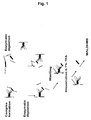

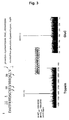


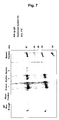
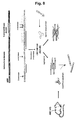

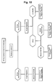
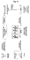


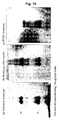
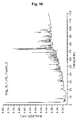

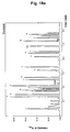
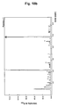


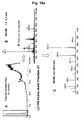
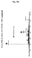


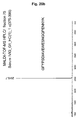
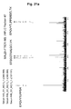
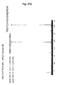


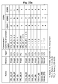

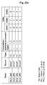
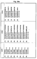
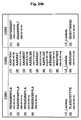

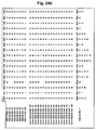
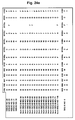
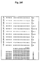
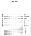
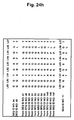
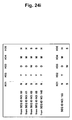
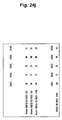
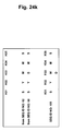
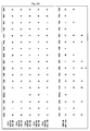

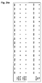

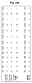
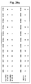


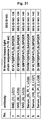


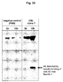


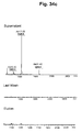
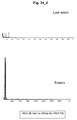
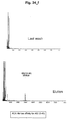


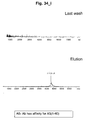


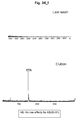
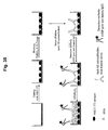
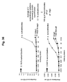

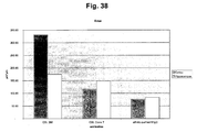
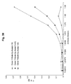

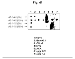


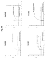
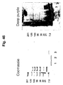
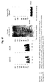
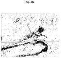

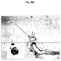
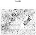


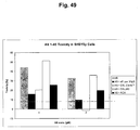
Classifications
-
- C—CHEMISTRY; METALLURGY
- C07—ORGANIC CHEMISTRY
- C07K—PEPTIDES
- C07K16/00—Immunoglobulins [IGs], e.g. monoclonal or polyclonal antibodies
- C07K16/18—Immunoglobulins [IGs], e.g. monoclonal or polyclonal antibodies against material from animals or humans
-
- A—HUMAN NECESSITIES
- A61—MEDICAL OR VETERINARY SCIENCE; HYGIENE
- A61P—SPECIFIC THERAPEUTIC ACTIVITY OF CHEMICAL COMPOUNDS OR MEDICINAL PREPARATIONS
- A61P25/00—Drugs for disorders of the nervous system
-
- A—HUMAN NECESSITIES
- A61—MEDICAL OR VETERINARY SCIENCE; HYGIENE
- A61P—SPECIFIC THERAPEUTIC ACTIVITY OF CHEMICAL COMPOUNDS OR MEDICINAL PREPARATIONS
- A61P25/00—Drugs for disorders of the nervous system
- A61P25/28—Drugs for disorders of the nervous system for treating neurodegenerative disorders of the central nervous system, e.g. nootropic agents, cognition enhancers, drugs for treating Alzheimer's disease or other forms of dementia
-
- A—HUMAN NECESSITIES
- A61—MEDICAL OR VETERINARY SCIENCE; HYGIENE
- A61P—SPECIFIC THERAPEUTIC ACTIVITY OF CHEMICAL COMPOUNDS OR MEDICINAL PREPARATIONS
- A61P9/00—Drugs for disorders of the cardiovascular system
-
- A—HUMAN NECESSITIES
- A61—MEDICAL OR VETERINARY SCIENCE; HYGIENE
- A61K—PREPARATIONS FOR MEDICAL, DENTAL OR TOILETRY PURPOSES
- A61K39/00—Medicinal preparations containing antigens or antibodies
- A61K2039/505—Medicinal preparations containing antigens or antibodies comprising antibodies
-
- C—CHEMISTRY; METALLURGY
- C07—ORGANIC CHEMISTRY
- C07K—PEPTIDES
- C07K2317/00—Immunoglobulins specific features
- C07K2317/20—Immunoglobulins specific features characterized by taxonomic origin
- C07K2317/21—Immunoglobulins specific features characterized by taxonomic origin from primates, e.g. man
-
- C—CHEMISTRY; METALLURGY
- C07—ORGANIC CHEMISTRY
- C07K—PEPTIDES
- C07K2317/00—Immunoglobulins specific features
- C07K2317/50—Immunoglobulins specific features characterized by immunoglobulin fragments
- C07K2317/56—Immunoglobulins specific features characterized by immunoglobulin fragments variable (Fv) region, i.e. VH and/or VL
- C07K2317/565—Complementarity determining region [CDR]
Definitions
- AD Alzheimer's disease
- AD the most common form of dementia among elderly population (prevalence: 1000/100,000; > 65 years)
- Cortical atrophy, neuronal loss, region-specific amyloid deposition, neuritic plaques, and neurofibrillary tangles are key neuropathological features in the AD brain. These alterations are thought to be linked to cognitive decline which clinically defines AD.
- neuritic plaques are amyloid immunoreactive, thioflavin positive, and accompanied by astrogliosis, microgliosis, cytoskeletal changes, and synaptic loss. The degree of neuritic degeneration within plaques correlates with clinical parameters of dementia.
- Neurotic plaques are spherical, multicellular lesions that are usually found in moderate to large numbers in limbic structures and associated neocortices of the AD brain. These plaques are comprised of extracellular deposits of amyloid- ⁇ peptide(s) (A ⁇ ) that include abundant amyloid fibrils intermixed with non-fibrillary forms of the peptide. Such plaques also contain variable numbers of activated microglia that are often situated very near the fibrillar amyloid core, as well as reactive astrocytes surrounding the core.
- a ⁇ amyloid- ⁇ peptide(s)
- Such plaques also contain variable numbers of activated microglia that are often situated very near the fibrillar amyloid core, as well as reactive astrocytes surrounding the core.
- a ⁇ The major constituent of the neuritic plaque, ⁇ -amyloid polypeptide (A ⁇ ), arises from a larger precursor protein, the amyloid precursor protein (APP) (Kang, et al., 1987; Tanzi, et al., 1987). A ⁇ is produced by normal cells and can be detected as a circulating peptide in the plasma and cerebrospinal fluid (CSF) of healthy humans.
- CSF cerebrospinal fluid
- APP amyloid precursor protein
- missense mutations in APP confer autosomal dominant inheritance of AD (FAD), and shed light on potentially important pathogenic mechanism(s).
- a ⁇ a 39-42 amino acid proteolytic product of APP
- a major A ⁇ cleavage product of APP is the A ⁇ (1-42) polypeptide, but A ⁇ peptides shorter at the C-terminus (39 to 41) are also produced by the proteolytic ( ⁇ -secretase) cleavage in the membrane.
- the N-terminal part of A ⁇ (1-42) is localized in the extracellular region of APP, and the major C-terminal part of the A ⁇ peptide is contained within the transmembrane domain.
- AD it has been postulated that increased synthesis and/or a decreased clearance of A ⁇ may lead to amyloid plaque deposition and subsequently to the neuropathological changes associated with the disease.
- synthetic A ⁇ peptide(s) have shown that neurotoxicity is dependent on A ⁇ being fibrilla and predominantly in a ⁇ -pleated sheet conformation.
- AD Alzheimer's disease
- a ⁇ neurotoxic amyloid peptide fragment
- APP ⁇ -amyloid precursor protein
- transgenic APPV717F mouse (TG mouse) is a well characterized model of AD-like plaque pathology with age- and region-dependent deposits of A ⁇ (1-40) and A ⁇ (1-42) (Games, et al., 1995). Recently, Schenk et al.
- the epitope was identified to consist of the residues (4-10) (FRHDSGY) of Aß(1-42).
- the selectivity of this recognition structure was ascertained by elucidation of the identical epitope from AD plaques, Aß(1-42) extracts from Aß-protofibrils, chemically synthesised Aß(1-42), and other (Aß-independent) polypeptides comprising the N-terminal Aß sequence (Przybylski et al. 2003).
- Naturally occurring anti-Aß autoantibodies were identified by Du et al. in both the blood and the CSF from non-immunized humans (Du, et al., 2001). These antibodies specifically recognize human Aß as has been shown by immunoprecipitation (Du, et al., 2001) and ELISA. Furthermore, the antibodies readily recognize synthetic A ⁇ (1-40) as well as human A ⁇ deposited in the brain of PDAPP transgenic mice. In addition, fibrillation/oligomerization and neurotoxicity of A ⁇ -peptides were reduced in the presence of A ⁇ -autoantibodies (Du, et al., 2003).
- IVIgG intracranial pressure
- a patient with AD is not convenient and associated with high costs, as the fraction of therapeutic A ⁇ autoantibodies is low.
- the vast majority of IgG in this preparation is not A ⁇ specific and may result in undesirable effects.
- the sources for IVIgG are limited, which is an unacceptable disadvantage in view of the prevalence of patients with Alzheimer's disease.
- AD diagnostics fall into three groups: (i) determinations for genetic risk factors or mutations (mainly for FAD cases, but not for sporadic AD diagnostics); (ii) neuroimaging methods; and (iii) diagnostics based on biochemical/biological markers.
- Present work on the development of diagnostic procedures based on biomarkers have been mainly focused on CSF, which has the principal disadvantage that such methods require elaborate, invasive material.
- a major problem associated with brain-derived biomarkers is that clinically examined controls often also include subjects with preclinical AD pathology. Further, current available biomarkers have the major disadvantage of low specificity. Similar disadvantages have been noted for a series of proteins expressed in the frontal cortex, identified by brain proteomics approaches, as potential brain biomarkers arising from presumed alterations of blood brain barrier in AD.
- AD neurodementing diseases
- the present invention provides isolated, monoclonal, human, anti- ⁇ -amyloid antibodies comprising
- Said antibody binds to dimeric forms of Aß with higher affinity than to monomeric forms of Aß.
- the antibody binds to a peptide comprising Aß(21-37), while in another the antibody shields residues of A ⁇ (21-37) from proteolytic digestion when being bound to an Aß polypeptide comprising Aß(21-37).
- the antibody binds specifically to A ⁇ (12-40) or Aß(20-37) when such antibody is coupled to NHS-activated 6-aminohexanoic acid-coupled sepharose, but does not bind specifically to Aß(17-28), Aß(25-35) or Aß(31-40).
- inventive anti- ⁇ -amyloid antibodies also can be formulated as pharmaceutical compositions.
- said antibodies are provided for use of preventing or treating a neurodementing disease in a patient comprising administering to the patient a therapeutically acceptable amount of the inventive anti- ⁇ -amyloid antibodies, in particular for use of preventing or treating neurodementing diseases selected from the group consisting of Alzheimer's disease, Down's syndrome, dementia with Lewy bodies, fronto-temporal dementia, cerebral amyloid angiopathy and amyloidoses.
- the neurodementing disease is Alzheimer's disease.
- inventive anti- ⁇ -amyloid antibodies can be used for the manufacture of a medicament in order to treat a neurodementing disease or to slow or prevent the progression of a neurodementing disease.
- This description further includes methods of detecting or measuring the progression of a neurodementing disease in a patient are provided that comprise (A) measuring in a sample from said patient an antibody titer against a first Aß peptide, wherein the first Aß peptide comprises at least the sequence according to Aß(30-37) and at most the sequence according to Aß(12-40); (B) measuring in a sample from said patient an antibody titer against a second Aß peptide wherein the second Aß peptide comprises at least the sequence according to Aß(4-10) and at most the sequence according to Aß(1-20); and (C) comparing the titers from steps (A) and (B).
- the first Aß peptide comprises at least the sequence according to A ⁇ (21-37).
- the methods further comprise comparing the patient titers with titers determined for healthy donors and AD patients whereby a higher titer against the first Aß peptide correlates with a lower risk of development and/or progression of Alzheimer's disease.
- the methods also can comprise comparing the patient titers with titers determined for healthy donors and AD patients whereby a higher titer against the first Aß peptide, relative to the titer against the second Aß peptide correlates with a lower risk of development and/or progression of Alzheimer's disease.
- the methods can comprise comparing the patient titers with titers determined for healthy donors and AD patients whereby a higher titer against the second Aß peptide correlates with a higher risk of development and/or progression of Alzheimer's disease.
- the methods further comprise comparing the patient titers with titers determined for healthy donors and AD patients whereby a higher titer against the second Aß peptide, relative to the titer against the first Aß peptide, correlates with a higher risk of development and/or progression of Alzheimer's disease.
- Methods of detecting or measuring the progression of a neurodementing disease in a patient also are disclosed in the description that comprise A) obtaining a first sample from said patient at a given time point; B) obtaining a second sample from said patient at later time point; C) measuring in said first and second samples the antibody titer against an epitope comprising at least Aß(30-37) and at most Aß(12-40); and D) comparing the titers of said first and second samples.
- Such methods comprise A) obtaining a first sample from said patient at a given time point; B) obtaining a second sample from said patient at later time point; C) measuring in said first and second samples the antibody titer against an epitope comprising at least Aß(4-10) and at most Aß(1-20); and D) comparing the titers of said first and second samples.
- such methods comprise A) obtaining a first sample from said patient at a given time point; B) obtaining a second sample from said patient at later time point; C) measuring in said first and second samples the antibody titer against an epitope comprising Aß(30-37); and D) comparing the titers of said first and second samples.
- the disclosure further relates to a kit comprising (A) a first Aß peptide comprising at least the sequence according to Aß(30-37) and at most the sequence according to Aß(12-40), and (B) a second Aß peptide wherein the second Aß peptide comprising at least the sequence according to Aß(4-10) and at most the sequence according to Aß(1-20).
- a ⁇ -autoantibodies were isolated from the serum of AD patients and healthy controls or pooled commercially obtainable serum immunoglobulin (IVIgG).
- the cDNA and amino acid sequences of the variable regions of the heavy and light chains were determined and all possible pairings of heavy and light chains were expressed in mammalian cells. A number of these pairings were found to bind with higher affinity to Aß dimers than to Aß monomers and one of these, CSL clone 7, was shown to possess biological activities potentially useful for the treatment of AD.
- the inventors further discovered that the CDRs of anti-A ⁇ (21-37) autoantibodies are highly homologous.
- consensus CDRs were determined and used to prepare human anti- ⁇ -amyloid antibodies useful for preventing or treating neurodementing diseases like AD as defined in claim 1.
- the disclosure also relates to the fact that AD patients, as compared to healthy controls, have an increased antibody titer against A ⁇ (4-10) and a decreased antibody titer against A ⁇ (21-37). According to the disclosure not only means for detecting and measuring the progression of a neurodementing disease like AD, but also methods for delaying the onset or progression of AD are described. Kits for detecting and measuring the progression of neurodementing diseases, like AD, also are described.
- a ⁇ polypeptide as used herein, defines a polypeptide having the amino acid sequence SEQ ID NO:01 or fragments thereof. Such fragments in particular comprise polypeptides having the sequence SEQ ID NO:02.
- antibody comprises also derivatives and/or fragments of antibodies.
- Such derivatives or fragments of antibodies retain the antigen binding properties of the intact antibody, but which lack some sequences of the intact antibody, for example the Fc-domain.
- Examples for such derivatives or fragments include, but are not limited to, Fab or F(ab') 2 fragments, which are obtainable via enzymatic digest of antibodies with papain or pepsin protease, respectively, single chain variable fragments (scFv), Fv fragments, minibodies and diabodies.
- autoantibody refers in general to antibodies which are directed against epitopes on proteins of the human body and which can be found in the blood or cerebrospinal fluid of a human subject without prior immunization with the respective antigen.
- anti-A ⁇ (21-37) autoantibody refers to autoantibodies that bind to an Aß peptide comprising Aß(21-37)(SEQ ID NO:2) and shield said SEQ ID NO 2 from proteolytic digestion.
- Such anti-A ⁇ (21-37) autoantibodies also bind with a higher affinity to dimers of Aß than to corresponding monomers of Aß.
- CDR Complementarity Determining Regions. Usually 3 of such CDR-regions (CDR1, CDR2, CDR3) can be found in the variable region on the light chain as well as on the heavy chain of an antibody. Each of these six hypervariable regions can contribute to the antigen specificity of the antibody.
- CDR does not imply that the molecule referred to is in fact an antibody. Rather, the term is considered to designate a sequence contributing to the specific binding of a polypeptide according to the invention to a C-terminal part of full length A ⁇ polypeptide (A ⁇ 1-40), in particular contributing to the binding to A ⁇ (21-37) polypeptide. Consequently, also derivatives of antibodies or other polypeptides engineered for binding to said epitope can exhibit CDRs.
- Consensus CDR refers to a single sequence derived by aligning two or more sequences for a given CDR according to the Kabat numbering system. (see Kabat, E. A., Wu, T. T., Perry, H. M., Gottesman, K. S.&Foeller, C. (1991) Sequences of Proteins of Immunological Interest (Department of Health and Human Services, Public Health Service, National Institutes of Health, Bethesda, MD) NIH Publ. No 91-3442 5th Ed . and R. Kontermann, S.
- diabodies refers to small antibody fragments with two antigen-binding sites, which fragments comprise a heavy chain variable domain (VH) connected to a light chain variable domain (VL) in the same polypeptide chain (VH VL).
- VH heavy chain variable domain
- VL light chain variable domain
- VH VL polypeptide chain
- epitope or “epitope peptide”, as used herein, generally refers to a polypeptide comprising the molecular recognition peptide sequence or structure derived from an antigen, that is bound by a specific antibody. It is an immunological determinant group of an antigen which is specifically recognized by the antibody.
- An epitope may comprise at least 5, preferably at least 8 amino acids in a spatial or discontinuous conformation.
- An epitope may also comprise a single segment of a polypeptide chain comprising a continuous linear amino acid sequence with a minimal length of approx. 5 amino acids.
- Fv is the minimum antibody fragment that contains a complete antigen recognition and binding site. This region consists of a dimer of one heavy and one light chain variable domain in tight, non-covalent association. It is in this configuration that the three CDRs of each variable domain interact to define an antigen binding site on the surface of the VH VL dimer. Collectively, the six CDRs confer antigen binding specificity to the antibody.
- moiety refers to a portion of a polypeptide with distinct function(s). Such moieties can provide for a structural or functional feature which is normally not present in the rest of the polypeptide or which feature is enhanced by this moiety. Such functional or structural moieties can for example provide binding, stabilization or detection of the polypeptide.
- the moiety can be a polypeptide on its own or can be any other compound, which provides the desired function(s) to the polypeptide. Said moiety is stably associated with the polypeptide, in particular covalently coupled to the polypeptide.
- moiety does not confer any information about the size of this portion in comparison to the polypeptide itself. The moiety can be smaller, equally sized or larger than the polypeptide it is coupled to.
- monoclonal antibody refers to an antibody obtained from a population of substantially homogeneous antibodies, i.e., the individual antibodies comprising the population are identical except for possible naturally occurring mutations that may be present in minor amounts. Monoclonal antibodies are highly specific, being directed against a specific antigenic site or epitope, in contrast to polyclonal antibody preparations that typically include different antibodies directed against different epitopes.
- neurodedementing diseases refer to diseases selected from Alzheimer's disease, Down's syndrome, Dementia with Lewy bodies, fronto-temporal dementia as well as to disorders such as cerebral amyloid angiopathy and amyloidoses.
- oligomerized refers to multimers of A ⁇ comprising also A ⁇ dimers, trimers, tetramers and higher oligomers but not to A ⁇ fibrils.
- plaque-specific antibody refers to an antibody directed against the A ⁇ (4-10) epitope of A ⁇ polypeptide.
- polypeptide refers to a polypeptide chain of at least 5 amino acid residues.
- the term also refers to an assembly of more than one polypeptide chain, e.g. an assembly of four polypeptide chains, such as an IgG antibody.
- paratope or “paratope peptide”, as used herein, generally defines a molecular recognition peptide sequence derived from a specific monoclonal or polyclonal antibody. This recognition peptide sequence exerts specific binding properties to an antigen epitope, and may comprise variable and/or constant region partial sequences of an antibody.
- Specifically binding to epitope X refers to the property of an antibody to bind to a particular epitope, e.g. epitope X, with a higher affinity than to other epitopes.
- Specifically binding to oligomeric structures of Aß means that the respective antibody binds to oligomeric structures of Aß with a higher affinity than to monomeric structures of Aß.
- a “therapeutically effective” amount of A ⁇ antibody refers to the dosage that provides the specific pharmacological response for which the antibody is administered in a significant number of subjects in need of such treatment.
- a therapeutically effective amount can be determined by prevention or amelioration of adverse conditions or symptoms of the neurodementing disease being treated.
- the appropriate dosage will vary depending, for example, on the type, stage, and severity of the disease, as well as on the mode of administration. It is emphasized that a "therapeutically effective amount" administered to a particular subject in a particular instance may not be effective for 100% of patients treated for a specific disease, even though such dosage is deemed a "therapeutically effective amount" by skilled practitioners.
- sample denotes a measurement of the amount or concentration of a particular antibody in a sample, typically blood.
- treatment refers to obtaining a desired pharmacological and/or physiologic effect.
- the effect can be prophylactic in terms of completely or partially preventing a disease or symptom thereof and/or can be therapeutic in terms of a partial or complete stabilization or cure for a disease and/or adverse effect attributable to the disease.
- Treatment covers any treatment of a disease in a mammal, particularly a human, and includes: (a) preventing the disease or symptom from occurring in a subject which can be predisposed to the disease or symptom but has not yet been diagnosed as having it; (b) inhibiting the disease symptom, i.e., arresting its development; or (c) relieving the disease symptom, i.e., causing regression of the disease or symptom.
- the antibodies according to the present invention as defined in claim 1 specifically bind to the epitope A ⁇ (21-37) (SEQ ID NO 2).
- An antibody or antibody fragment according to the present invention binds to a C-terminal part of full length A ⁇ polypeptide (A ⁇ 1-40), in particular to the epitope denoted in A ⁇ (21-37) (SEQ ID NO:2).
- the inventive antibodies are capable of binding specifically to a polypeptide comprising the epitope A ⁇ (21-37) (SEQ ID NO 2) of amyloid beta, in particular under physiological conditions, e.g. pH about 7.4, salt concentrations about 50 to about 150 mM in PBS.
- the antibodies according to the present invention have a relative dissociation constant, under in vitro conditions, (relative KD; reflecting in vitro results but not necessarily identical values under in vivo conditions) of at least about 10-5 M, about 10-6 M, about 10-7 M, about 10-8, about 10-9 M, about 10-10 M, about 10-11 M, about 10-12 M, or higher.
- the relative dissociation constant of the binding of an antibody according to the present invention to the epitope denoted in A ⁇ (21-37) can be between about 10-8 M to about 10-12 M, in particular around 1 to 50 x 10-9 M.
- Such dissociation rate constants can be determined readily using kinetic analysis techniques such as surface plasmon resonance (BIAcore or Biosensor), using general procedures outlined by the manufacturer or other methods known in the art.
- polypeptides specific for A ⁇ (21-37) are highly homologous.
- polypeptides are disclosed that comprise sequences selected from consensus CDR sequences.
- a polypeptide according to the present disclosure can comprise a CDR sequence having a sequence as denoted in any of the consensus sequences SEQ ID NOs: 6 to 8.
- SEQ ID NOs: 6 to 8 represent illustrative consensus sequences for CDR regions of the heavy chain of an antibody having the ability to bind to SEQ ID NO:2.
- the consensus sequences are derived from the sequence information derived from the antibodies, which have been identified by the inventors to bind specifically to SEQ ID NO:2.
- the identified consensus sequences for the CDR regions of both the heavy chain i.e. SEQ ID NOs: 6 to 8 and SEQ ID Nos 153 to 161
- the light chain i.e. SEQ ID NOs: 9 to 11
- SEQ ID NO:6 represents a CDR1 consensus sequence as described herein for the heavy chain, wherein the amino acid at position 1 of SEQ ID NO:6 which is at the Kabat position H31 can be Ser, Gly or Asn, the amino acid at position 2 of SEQ ID NO:6 which is the amino acid at the Kabat position 8 H32 is Tyr, the amino acid at position 3 of SEQ ID NO:6 which is the amino acid at the Kabat position H33 can be Trp or Asp, the amino acid at position 4 of SEQ ID NO:6 which is the amino acid at the Kabat position H34 is Met and the amino acid at position 5 of SEQ ID NO:6 which is the amino acid at the Kabat position H35 can be Ser or His.
- SEQ ID NO:153 represents a further CDR1 consensus sequence as described herein for the heavy chain, wherein the amino acid at position 1 of SEQ ID 153 which is at the Kabat position H31 can be Asn or Ser, the amino acid at position 2 of SEQ ID 153 which is the amino acid at the Kabat position 8 H32 is Tyr, the amino acid at position 3 of SEQ ID NO: 153 which is the amino acid at the Kabat position H33 can be Asp or Trp, the amino acid at position 4 of SEQ ID NO: 153 which is the amino acid at the Kabat position H34 is Met and the amino acid at position 5 of SEQ ID NO: 153 which is the amino acid at the Kabat position H35 can be His or Ser.
- SEQ ID NO: 154 represents an additional CDR1 consensus sequence as described herein for the heavy chain, wherein the amino acid at position 1 of SEQ ID NO: 154 which is at the Kabat position H31 of SEQ ID NO:6 is Asn, the amino acid at position 2 of SEQ ID NO: 154 which is the amino acid at the Kabat position 8 H32 is Tyr, the amino acid at position 3 of SEQ ID NO: 154 which is the amino acid at the Kabat position H33 is Asp, the amino acid at position 4 of SEQ ID NO: 154 which is the amino acid at the Kabat position H34 is Met and the amino acid at position 5 of SEQ ID NO: 154 which is the amino acid at the Kabat position H35 is His.
- SEQ ID NO:155 represents a further CDR1 consensus sequence as described herein for the heavy chain, wherein the amino acid at position 1 of SEQ ID NO: 155 which is at the Kabat position H31 is Ser, the amino acid at position 2 of SEQ ID NO: 155 which is the amino acid at the Kabat position 8 H32 is Tyr, the amino acid at position 3 of SEQ ID NO: 155 which is the amino acid at the Kabat position H33 can be Trp or Asp, the amino acid at position 4 of SEQ ID NO: 155 which is the amino acid at the Kabat position H34 is Met and the amino acid at position 5 of SEQ ID NO: 155 which is the amino acid at the Kabat position H35 is Ser.
- SEQ ID NO:7 represents a CDR2 consensus sequence as described herein for the heavy chain, wherein the amino acid at position 1 of SEQ ID NO:7 which is the amino acid at the Kabat position H50 can be Ser or Arg or Glu, the amino acid at position 2 of SEQ ID NO:7 which is the amino acid at the Kabat position H51 can be Val or Ile, the amino acid at position 3 of SEQ ID NO:7 which is the amino acid at the Kabat position H52 can be Lys or Gly or Asn, the amino acid at position 4 of SEQ ID NO:7 which is the amino acid at the Kabat position H52a can be Gln or no amino acid, the amino acid at position 5 of SEQ ID NO:7 which is the amino acid at the Kabat position H53 can be Asp or Phe or Thr or Arg, the amino acid at position 6 of SEQ ID NO:7 which is the amino acid at the Kabat position H54 can be Gly or Phe or Ala or Ser, the amino acid at position 7 of SEQ ID NO:7
- SEQ ID NO: 156 represents a further CDR2 consensus sequence as described herein for the heavy chain, wherein the amino acid at position 1 of SEQ ID NO: 156 which is the amino acid at the Kabat position H50 can be Arg or Ser or Glu, the amino acid at position 2 of SEQ ID NO: 156 which is the amino acid at the Kabat position H51 can be Ile or Val, the amino acid at position 3 of SEQ ID NO: 156 which is the amino acid at the Kabat position H52 can be Gly or Lys or Asn, the amino acid at position 4 of SEQ ID NO: 156 which is the amino acid at the Kabat position H52a can be Gln or no amino acid, the amino acid at position 5 of SEQ ID NO: 156 which is the amino acid at the Kabat position H53 can be Thr or Asp or Arg, the amino acid at position 6 of SEQ ID NO: 156 which is the amino acid at the Kabat position H54 can be or Ala or Gly or Ser, the amino acid at position 7 of SEQ ID
- SEQ ID NO: 157 represents an additional CDR2 consensus sequence as described herein for the heavy chain, wherein the amino acid at position 1 of SEQ ID NO: 157 which is the amino acid at the Kabat position H50 can be Arg or Glu, the amino acid at position 2 of SEQ ID NO: 157 which is the amino acid at the Kabat position H51 is Ile, the amino acid at position 3 of SEQ ID NO: 157 which is the amino acid at the Kabat position H52 can be Gly or Asn, the amino acid at position 4 of SEQ ID NO: 157 which is the amino acid at the Kabat position H53 can be Thr or Arg, the amino acid at position 5 of SEQ ID NO: 157 which is the amino acid at the Kabat position H54 can be Ala or Ser, the amino acid at position 6 of SEQ ID NO: 157 which is the amino acid at the Kabat position H55 is Gly, the amino acid at position 7 of SEQ ID NO: 157 which is the amino acid at the Kabat position H56 can be Arg or Asp
- SEQ ID NO: 158 represents a further CDR2 consensus sequence as described herein for the heavy chain, wherein the amino acid at position 1 of SEQ ID NO: 158 which is the amino acid at the Kabat position H50 is Ser, the amino acid at position 2 of SEQ ID NO: 158 which is the amino acid at the Kabat position H51 is Val, the amino acid at position 3 of SEQ ID NO: 158 which is the amino acid at the Kabat position H52 is Lys, the amino acid at position 4 of SEQ ID NO: 158 which is the amino acid at the Kabat position H52a is Gln, the amino acid at position 5 of SEQ ID NO: 158 which is the amino acid at the Kabat position H53 is Asp, the amino acid at position 6 of SEQ ID NO: 158 which is the amino acid at the Kabat position H54 is Gly, the amino acid at position 7 of SEQ ID NO: 158 which is the amino acid at the Kabat position H55 is Ser, the amino acid at position 8 of SEQ ID NO: 158 which is the amino
- SEQ ID NO:8 represents a CDR3 consensus sequence as described herein for the heavy chain, wherein the amino acid at position 1 of SEQ ID NO:8 which is the amino acid at the Kabat position H95 can be Asp or Gly, the amino acid at position 2 of SEQ ID NO:8 which is the amino acid at the Kabat position H 96 can be Ala or Gly, the amino acid at position 3 of SEQ ID NO:8 which is the amino acid at the Kabat position H97 can be Ser or Gly, the amino acid at position 4 of SEQ ID NO:8 which is the amino acid at the Kabat position H98 can be Ser or Arg, the amino acid at position 5 of SEQ ID NO:8 which is the amino acid at the Kabat position H99 is Trp, the amino acid at position 6 of SEQ ID NO:8 which is the amino acid at the Kabat position H100 can be Tyr or Ala, the amino acid at position 7 of SEQ ID NO:8 which is the amino acid at the Kabat position H100a can be Arg or Pro or Asp, the
- SEQ ID NO:159 represents an additional CDR3 consensus sequence as described herein for the heavy chain, wherein the amino acid at position 1 of SEQ ID NO: 159 which is the amino acid at the Kabat position H95 can be Gly or Asp, the amino acid at position 2 of SEQ ID NO: 159 which is the amino acid at the Kabat position H 96 can be Ala or Gly, the amino acid at position 3 of SEQ ID NO: 159 which is the amino acid at the Kabat position H97 can be Gly or Ser, the amino acid at position 4 of SEQ ID NO: 159 which is the amino acid at the Kabat position H98 can be Arg or Ser, the amino acid at position 5 of SEQ ID NO: 159 which is the amino acid at the Kabat position H99 is Trp, the amino acid at position 6 of SEQ ID NO: 159 which is the amino acid at the Kabat position H100 can be Ala or Tyr, the amino acid at position 7 of SEQ ID NO: 159 which is the amino acid at the Kabat position H100a can be Pro or
- SEQ ID NO:160 represents a further CDR3 consensus sequence as described herein for the heavy chain, wherein the amino acid at position 1 of SEQ ID NO:160 which is the amino acid at the Kabat position H95 is Gly, the amino acid at position 2 of SEQ ID NO:160 which is the amino acid at the Kabat position H 96 is Ala, the amino acid at position 3 of SEQ ID NO:160 which is the amino acid at the Kabat position H97 is Gly, the amino acid at position 4 of SEQ ID NO:160 which is the amino acid at the Kabat position H98 is Arg, the amino acid at position 5 of SEQ ID NO:160 which is the amino acid at the Kabat position H99 is Trp, the amino acid at position 6 of SEQ ID NO:160 which is the amino acid at the Kabat position H100 is Ala, the amino acid at position 7 of SEQ ID NO:160 which is the amino acid at the Kabat position H100a is Pro, the amino acid at position 8 of SEQ ID NO:160 which is the amino acid at the Kabat position H
- SEQ ID NO:161 represents a further CDR3 consensus sequence as described herein for the heavy chain, wherein the amino acid at position 1 of SEQ ID NO:161 which is the amino acid at the Kabat position H95 is Asp, the amino acid at position 2 of SEQ ID NO:161 which is the amino acid at the Kabat position H 96 can be Gly or Ala, the amino acid at position 3 of SEQ ID NO:161 which is the amino acid at the Kabat position H97 can be Ser or Gly, the amino acid at position 4 of SEQ ID NO:161 which is the amino acid at the Kabat position H98 can be Ser or Arg, the amino acid at position 5 of SEQ ID NO:161 which is the amino acid at the Kabat position H99 is Trp, the amino acid at position 6 of SEQ ID NO:161 which is the amino acid at the Kabat position H100 can be Tyr or Ala, the amino acid at position 7 of SEQ ID NO:161 which is the amino acid at the Kabat position H100a can be Arg or As
- a polypeptide according to the disclosure comprises as CDRs all three respective consensus CDR sequences as denoted in SEQ ID NO:6 to 8.
- a polypeptide according to the disclosure comprises at least two of the respective consensus CDR sequences as denoted in SEQ ID NO:6 to 8.
- the present disclosure further relates to a polypeptide comprising a CDR sequence having at least two of the respective consensus sequences as denoted in any of the consensus sequences SEQ ID NOs: 6 to 11.
- Illustrative SEQ ID NOs: 9 to 11 as disclosed herein represent consensus sequences for CDR regions of the light chain of an antibody having the ability to bind to SEQ ID NO:2.
- the consensus sequences are derived from the sequence information derived from the antibodies, which have been identified by the inventors to bind specifically to SEQ ID NO:2.
- the identified consensus sequences for the CDR regions of the light chain were derived from naturally occurring, human antibodies isolated as described in Example 2A.
- SEQ ID NO:9 represents a CDR1 consensus sequence as described herein for a kappa light chain immunoglobulin CDR1 region, wherein the amino acid at position 1 of SEQ ID NO:9 which is the amino acid at the Kabat position L24 can be an Arg, the amino acid at position 2 of SEQ ID NO:9 which is the amino acid at the Kabat position L25 can be Ala or Glu, the amino acid at position 3 of SEQ ID NO:9 which is the amino acid at the Kabat position L26 can be Ser, the amino acid at position 4 of SEQ ID NO:9 which is the amino acid at the Kabat position L27 can be Gln, the amino acid at position 5 of SEQ ID NO:9 which is the amino acid at the Kabat position L28 can be Ser or Gly, the amino acid at position 6 of SEQ ID NO:9 which is the amino acid at the Kabat position L29 can be Val or Ile, the amino acid at position 7 of SEQ ID NO:9 which is the amino acid at the Kabat position L30 can be As
- SEQ ID NO:10 represents a CDR2 consensus sequence as described herein for a kappa light chain immunoglobulin CDR2 region, wherein the amino acid at position 1 of SEQ ID NO:10 which is the amino acid at the Kabat position L50 can be Ala or Gly or Lys or Trp, the amino acid at position 2 of SEQ ID NO:10 which is the amino acid at the Kabat position L51 can be Val or Ala, the amino acid at position 3 of SEQ ID NO:10 which is the amino acid at the Kabat position L52 can be Ser or Ala, the amino acid at position 4 of SEQ ID NO:10 which is the amino acid at the Kabat position L53 can be Thr or Ser or Asn or Ile, the amino acid at position 5 of SEQ ID NO:10 which is the amino acid at the Kabat position L54 can be Arg or Leu, the amino acid at position 6 of SEQ ID NO:10 which is the amino acid at the Kabat position L55 can be Ala or Gln or Phe or Glu and the amino
- SEQ ID NO: 11 represents a CDR3 consensus sequence as described herein for a kappa light chain immunoglobulin CDR3 region, wherein the amino acid at position 1 of SEQ ID NO: 11 which is the amino acid at the Kabat position L89 can be Gln, the amino acid at position 2 of SEQ ID NO: 11 which is the amino acid at the Kabat position L90 can be Gln, the amino acid at position 3 of SEQ ID NO: 11 which is the amino acid at the Kabat position L91 can be Ala or Tyr, the amino acid at position 4 of SEQ ID NO: 11 which is the amino acid at the Kabat position L92 can be Gly or Asn, the amino acid at position 5 of SEQ ID NO: 11 which is the amino acid at the Kabat position L93 can be Ser, the amino acid at position 6 of SEQ ID NO: 11 which is the amino acid at the Kabat position L94 can be Ser or Phe, the amino acid at position 7 of SEQ ID NO: 11 which is the amino acid at the Kabat position L95 can be G
- polypeptide according to the disclosure comprises as CDRs for the light chain all three respective consensus CDR sequences as denoted in SEQ ID NO:6 to 11.
- polypeptide according to the disclosure comprises at least two CDRs for the light chain selected from all three respective consensus CDR sequences as denoted in SEQ ID NO:9 to 11.
- This description further includes a polypeptide comprising at least two CDR sequences selected from the consensus CDR sequences denoted for the light chain (SEQ ID NO:9 to 11) or at least two of the consensus CDR sequences denoted for the heavy chain (SEQ ID NO:6 to 8) or at least one CDR from the light chain (SEQ ID NO:9 to 11) and at least one CDR from the heavy chain (SEQ ID NO:6 to 8).
- polypeptide according to the disclosure comprises as CDRs for the light chain all three respective consensus CDR sequences as denoted in SEQ ID NO:9 to 11.
- the present disclosure further relates to a polypeptide comprising at least two CDR sequences selected from the consensus CDR sequences denoted for the light chain (SEQ ID NO:9 to 11) and/or at least two of the consensus CDR sequences denoted for the heavy chain (SEQ ID NO:6 to 8).
- isolated, monoclonal, anti- ⁇ -amyloid antibodies comprise more than one amino acid sequence selected from at least two consensus amino acid sequences of the group consisting of SEQ ID NO: 6, SEQ ID NO: 7, SEQ ID NO: 8, SEQ ID NO: 9, SEQ ID NO: 10 and SEQ ID NO: 11, wherein each of said more than one amino acid sequence is from a different SEQ ID NO, wherein said antibody binds to dimeric forms of A ⁇ with higher affinity than to monomeric forms of A ⁇ .
- the polypeptide comprises as CDR1 on the heavy chain one of the sequences as denoted in SEQ ID NOs: 13 to 20, as CDR2 on the heavy chain one of the sequence as denoted in SEQ ID NOs: 21 to 27 and 149, and/or as CDR3 on the heavy chain one of the sequences as denoted in SEQ ID NOs: 28 to 32.
- the polypeptide according to the disclosure comprises at least two consensus CDR sequences selected from consensus sequences for CDR 1, CDR 2 and CDR 3 of the heavy chain and CDR 1, CDR 2 and CDR 3 of the light chain, wherein the consensus sequences are derived by aligning the sequences of the following antibody variable regions according to the Kabat numbering:
- SEQ ID NOs 13 to 20 represent the CDR1 sequences
- SEQ ID NOs 21 to 27 and 149 represent the CDR2 sequences
- SEQ ID NOs 28 to 32 represent the CDR3 sequences found by the inventors to be present on the heavy chain of antibodies binding to a C-terminal part of A ⁇ (1-40), in particular to A ⁇ (21-37).
- polypeptide according to the present disclosure comprises as CDR1 on the light chain one of the sequences as denoted in SEQ ID NOs: 33 to 37, as CDR2 on the light chain one of the sequence as denoted in SEQ ID NOs: 38 to 43 and 150 to 152, and/or as CDR3 on the light chain one of the sequences as denoted in SEQ ID NOs: 44 to 46.
- SEQ ID NOs 33 to 37 represent the CDR1 sequences
- SEQ ID NOs 38 to 43 and 150 to 152 represent the CDR2 sequences
- SEQ ID NOs 44 to 46 represent the CDR3 sequences found by the inventors to be present on the light chain of antibodies binding to a C-terminal part of Aß(1-40), in particular to A ⁇ (21-37).
- a polypeptide according to the present disclosure comprises on the heavy chain a CDR1 sequence selected from SEQ ID NOs: 13 to 20, a CDR2 sequence selected from SEQ ID NOs: 21 to 27 and 148, and/or a CDR3 sequence selected from SEQ ID NOs: 28 to 32, and on the light chain a CDR1 sequence selected from SEQ ID NOs: 33 to 37, a CDR2 sequence selected from SEQ ID NOs: 38 to 43 and 150 to 152 and/or a CDR3 sequence selected from SEQ ID NOs: 44 to 46.
- polypeptide according to the present disclosure can comprise on the light chain:
- polypeptide according to the present disclosure can also comprise on the heavy chain:
- the present specification also relates to a polypeptide according to the present disclosure comprising:
- a polypeptide according to the present invention is comprising a light variable chain having the sequence of SEQ ID NO:53 and a variable heavy chain having the sequence of SEQ ID NO:60, or a polypeptide is comprising a light chain having the CDR sequences of SEQ ID NO:33, SEQ ID NO: 41 and SEQ ID NO:45 and a heavy chain having the CDR sequences of SEQ ID NO:15, SEQ ID NO:23 and SEQ ID NO:29.
- SEQ ID NOs: 47 to 55 and 145 to 147 denote variable regions of the light chain and SEQ ID NOs: 56 to 71 and 148 denote variable regions of the heavy chain of antibodies having the ability to bind specifically to a C-terminal part of A ⁇ (1-40), in particular to A ⁇ (21-37), as determined by the inventors.
- the variable regions of the heavy and light chain are responsible for antigen specificity.
- the polypeptide according to the present disclosure comprises a sequence selected from SEQ ID NOs: 47 to 55 and 145 to 147 and/or a sequence selected from SEQ ID NOs: 56 to 71 and 148.
- the polypeptide according to the present invention comprises SEQ ID NO:47 and SEQ ID NO:56 or SEQ ID NO:48 and SEQ ID NO:57.
- each of the light chains of SEQ ID NOs: 47 to 55 and 145 to 147 can be combined with any of the heavy chains of SEQ ID NOs: 56 to 71 and 148.
- anti- ⁇ -amyloid antibodies are disclosed that comprise SEQ ID NO: 47 and SEQ ID NO:56 ; SEQ ID NO: 47 and SEQ ID NO:57 ; SEQ ID NO: 47 and SEQ ID NO: 58; SEQ ID NO: 47 and SEQ ID NO: 59; SEQ ID NO: 47 and SEQ ID NO:60; SEQ ID NO: 47 and SEQ ID NO: 61; SEQ ID NO: 47 and SEQ ID NO: 62; SEQ ID NO: 47 and SEQ ID NO: 63; SEQ ID NO: 47 and SEQ ID NO: 64; SEQ ID NO: 47 and SEQ ID NO: 65; SEQ ID NO: 47 and SEQ ID NO: 66; SEQ ID NO: 47 and SEQ ID NO: 67; SEQ ID NO: 47 and SEQ ID NO:
- the anti- ⁇ -amyloid antibodies comprise SEQ ID NO: 145 and SEQ ID NO:60; SEQ ID NO: 53 and SEQ ID NO:60; SEQ ID NO: 51 and SEQ ID NO:60; SEQ ID NO: 52 and SEQ ID NO:60; SEQ ID NO: 146 and SEQ ID NO:60; SEQ ID NO: 53 and SEQ ID NO:148; SEQ ID NO: 55 and SEQ ID NO:61; SEQ ID NO: 145 and SEQ ID NO:62; SEQ ID NO: 54 and SEQ ID NO:60; SEQ ID NO: 54 and SEQ ID NO:61; SEQ ID NO: 54 and SEQ ID NO:148; SEQ ID NO: 145 and SEQ ID NO:148; or SEQ ID NO: 146 and SEQ ID NO:61, provided that only the antibodies as recited with claim 1 are in accordance with present invention.
- polypeptide according to the present disclosure can comprise a sequence selected from SEQ ID NOs: 72 to 74 and/or a sequence selected from SEQ ID NOs: 75 to 77.
- polypeptide according to the present disclosure can comprise a sequence selected from SEQ ID NO:72 or 73 and a sequence selected from SEQ ID NOs 75 to 77.
- polypeptide according to the present disclosure comprises a sequence selected from SEQ ID NOs: 78 to 93.
- polypeptide according to the present disclosure comprises a sequence selected from SEQ ID NOs: 94 to 137.
- the description further relates to a polypeptide according to the present disclosure comprising a sequence as denoted in SEQ ID NO:78 and a sequence as denoted in SEQ ID NO:94 or a sequence as denoted in SEQ ID NO:80 and a sequence as denoted in SEQ ID NO:97.
- a polypeptide according to the present disclosure comprising a sequence as denoted in SEQ ID NO:78 and a sequence as denoted in SEQ ID NO:94 or a sequence as denoted in SEQ ID NO:80 and a sequence as denoted in SEQ ID NO:97.
- Consensus sequences for framework regions also can be identified. If the amino acid sequences of the variable regions of the heavy and light chains are aligned respectively according to the Kabat rules, the so-called framework regions N-terminal to CDR1, between CDR1 and CDR2, between CDR2 and CDR3 and C-terminal to CDR3 can be compared in a way analogous to that described above for the CDR regions, and consensus sequences for the framework of the antibodies of the disclosure can be derived.
- the polypeptide according to the present disclosure comprises a sequence as denoted in the consensus sequence of SEQ ID NO: 44, in particular a sequence as denoted in SEQ ID NOs: 138 to 143.
- the inventive polypeptides bind specifically to oligomeric forms of ⁇ -amyloid polypeptide.
- the polypeptides can bind oligomeric forms of A ⁇ (1-40) or oligomeric forms of A ⁇ (12-20) or oligomeric forms of A ⁇ (21-37).
- the inventive polypeptides are capable of binding specifically to oligomeric forms of A ⁇ (1-40) when incubated overnight at 10 mM sodium phosphate, 150 mM NaCl, pH 7.4 at 4°C.
- the inventive autoantibodies bind with a higher affinity to dimers of Aß than to corresponding monomers of Aß.
- binding affinity is a measure of antibody-antigen combination and concerns the selectivity with which a given antibody binds to an epitope when compared with binding to any other epitope. This preferential or selective binding can be quantified as a binding affinity or titer.
- Methods of calculating antibody affinity are well-known in the field. See, e.g. Practical Immunology Ch. 3, Frank C. Hay & Olwyn M.R. Westwood, Blackwell Publishing (2002 ); Measuring Immunity: Basic Biology and Clinical Assessment, Ch. 16, Michael T. Lotze & Angus W. Thomson (eds.), Academic Press (2005 ).
- the polypeptide is a monoclonal antibody.
- Monoclonal antibodies have the advantage that they exhibit less cross reactivity.
- polypeptides according to the present invention comprises derivatives and/or fragments of antibodies binding to A ⁇ (21-37)(SEQ ID NO:2).
- antibodies can be treated enzymatically, e.g. with proteases in order to obtain fragments of antibodies which retain the antigen binding properties of the intact antibody, but which lack the Fc-domain.
- fragments are for example Fab or F(ab') 2 fragments, which are obtainable via enzymatic digest of antibodies with papain or pepsin protease, respectively.
- Another fragment of an antibody is a single chain variable fragment (scFv) of an antibody, i.e.
- variable regions of the heavy and light chains of an antibody via a short flexible linker (usually serine, glycine).
- a short flexible linker usually serine, glycine.
- Single chain variable fragments can be obtained by genetic engineering of a respective nucleic acid encoding for such a fusion protein. The advantage of such a fragment is that it consists only of a single polypeptide chain which is more easily expressed and properly folded in artificial expression systems than the whole antibody, which comprises at least 4 polypeptide chains which need a correct assembly in order to function adequately.
- the polypeptide according the invention is an antibody, namely a human antibody, having low immunogenicity in humans.
- the antibody or fragment thereof can be derived from any species suitable for antibody production.
- the antibody, derivatives or fragments thereof is a humanized antibody, derivative or fragment thereof, i.e. while the antigen binding domain or parts thereof is/are of non-human origin, the rest of the antibody, derivative or fragment thereof is of human origin.
- the antibody, derivative or fragment thereof is chimeric, i.e. while the variable domain or parts thereof is/are of non-human origin the constant domain is of human origin. Both serve the purpose to reduce negative side effects due to immunogenic properties of protein domains of non-human origin.
- polypeptide binding to epitope A ⁇ (21-37) (SEQ ID NO:2) of amyloid-beta peptide (1-40) (SEQ ID NO:1) is an antibody derivative or fragment which comprises only the paratope of an antibody binding to said epitope.
- Example for such a paratope is, for example, a polypeptide comprising the amino acid sequences for the respective CDR domains of heavy and light chain connected via the intervening sequences or via synthetic linkers.
- Altered glycosylation variants also can be produced that have a modified glycosylation pattern relative to the parent antibody, for example, by deleting one or more carbohydrate moieties found in the antibody, and/or adding one or more glycosylation sites that are not present in the antibody.
- Glycosylation of polypeptides including antibodies is typically either N-linked or O- linked.
- N-linked refers to the attachment of the carbohydrate moiety to the side chain of an asparagine residue.
- the tripeptide sequences asparagine-X-serine and asparagine-X-threonine, where X is any amino acid except proline, are the recognition sequences for enzymatic attachment of the carbohydrate moiety to the asparagine side chain.
- N-linked glycosylation sites may be added to an antibody by altering the amino acid sequence such that it contains one or more of these tripeptide sequences.
- O-linked glycosylation refers to the attachment of one of the sugars N- aceylgalactosamine, galactose, or xylose to a hydroxyamino acid, most commonly serine or threonine, although 5-hydroxyproline or 5-hydroxylysine also may be used.
- O-linked glycosylation sites may be added to a specific binding agent or antibody by inserting or substituting one or more serine or threonine residues to the sequence of the antibody.
- Modifications to increase serum half-life also may desirable, for example, by incorporation of or addition of a salvage receptor binding epitope (e.g., by mutation of the appropriate region or by incorporating the epitope into a peptide tag that is then fused to the antibody at either end or in the middle, e.g., by DNA or peptide synthesis; See, e.g., WO 96/32478 ) or adding molecules such as PEG or other water soluble polymers, including polysaccharide polymers.
- a salvage receptor binding epitope e.g., by mutation of the appropriate region or by incorporating the epitope into a peptide tag that is then fused to the antibody at either end or in the middle, e.g., by DNA or peptide synthesis; See, e.g., WO 96/32478
- adding molecules such as PEG or other water soluble polymers, including polysaccharide polymers.
- Polyclonal antibodies are preferably raised in animals by multiple subcutaneous (sc) or intraperitoneal (ip) injections of the relevant antigen and an adjuvant.
- antigen may be injected directly into the animal's lymph node (see Kilpatrick et al., Hybridoma, 16:381-389, 1997 ).
- An improved antibody response may be obtained by conjugating the relevant antigen to a protein that is immunogenic in the species to be immunized, e.g., keyhole limpet hemocyanin, serum albumin, bovine thyroglobulin, or soybean trypsin inhibitor using a bifunctional or derivatizing agent, for example, maleimidobenzoyl sulfosuccinimide ester (conjugation through cysteine residues), N-hydroxysuccinimide (through lysine residues), glutaraldehyde, succinic anhydride or other agents known in the art.
- a protein that is immunogenic in the species to be immunized e.g., keyhole limpet hemocyanin, serum albumin, bovine thyroglobulin, or soybean trypsin inhibitor
- a bifunctional or derivatizing agent for example, maleimidobenzoyl sulfosuccinimide ester (conjugation through cysteine residues
- Animals are immunized against the antigen, immunogenic conjugates or derivatives by combining, e.g., 100 ⁇ g of the protein or conjugate (for mice) with 3 volumes of Freund's complete adjuvant and injecting the solution intradermally at multiple sites.
- the animals are boosted with 1/5 to 1/10 the original amount of peptide or conjugate in Freund's complete adjuvant by subcutaneous injection at multiple sites.
- the animals are bled and the serum is assayed for antibody titer. Animals are boosted until the titer plateaus.
- the animal is boosted with the conjugate of the same antigen, but conjugated through a different cross-linking reagent.
- Conjugates also can be made in recombinant cell culture as protein fusions. Also, aggregating agents such as alum are suitably used to enhance the immune response.
- Monoclonal antibodies can be made using the hybridoma method first described by Kohler et al., Nature, 256:495 (1975 ), or by recombinant DNA methods.
- a mouse or other appropriate host animal such as rats, hamster or macaque monkey, is immunized to elicit lymphocytes that produce or are capable of producing antibodies that will specifically bind to the protein used for immunization.
- lymphocytes may be immunized in vitro. Lymphocytes then are fused with myeloma cells using a suitable fusing agent, such as polyethylene glycol, to form a hybridoma cell ( Goding, Monoclonal Antibodies: Principles and Practice, pp.
- the hybridoma cells thus prepared are seeded and grown in a suitable culture medium that preferably contains one or more substances that inhibit the growth or survival of the unfused, parental myeloma cells.
- a suitable culture medium that preferably contains one or more substances that inhibit the growth or survival of the unfused, parental myeloma cells.
- the culture medium for the hybridomas typically will include hypoxanthine, aminopteun, and thymidine (HAT medium), which substances prevent the growth of HGPRT-deficient cells.
- Preferred myeloma cells are those that fuse efficiently, support stable high-level production of antibody by the selected antibody-producing cells and are sensitive to a medium.
- Human myeloma and mouse-human heteromyeloma cell lines also have been described for the production of human monoclonal antibodies ( Kozbor, J. Immunol., 133: 3001 (1984 ); Brodeur et al., Monoclonal Antibody Production Techniques and Applications, pp. 51-63 (Marcel Dekker, Inc., New York, 1987 )).
- Exemplary marine myeloma lines include those derived from MOP-21 and M.C.-11 mouse tumors available from the Salk Institute Cell Distribution Center, San Diego, Calif.
- the binding specificity of monoclonal antibodies produced by hybridoma cells is determined by immunoprecipitation or by an in vitro binding assay, such as radioimmunoassay (RIA) or enzyme-linked immunoabsorbent assay (ELISA).
- RIA radioimmunoassay
- ELISA enzyme-linked immunoabsorbent assay
- the binding affinity of the monoclonal antibody can be determined, for example, by BIAcore or Scatchard analysis ( Munson et al., Anal. Biochem., 107:220 (1980 )).
- the clones can be subcloned by limiting dilution procedures and grown by standard methods ( Goding, Monoclonal Antibodies: Principles and Practice, pp. 59- 103 (Academic Press, 1986 )). Suitable culture media for this purpose include, for example, D-MEMO or RPMI 1640 medium.
- the hybridoma cells can be grown in vivo as ascites tumors in an animal.
- the monoclonal antibodies secreted by the subclones are suitably separated from the culture medium, ascites fluid, or serum by conventional immunoglobulin purification procedures such as protein A-Sepharose, hydroxylapatite chromatography, gel electrophoresis, dialysis, or affinity chromatography.
- amino acid sequence of an immunoglobulin of interest can be determined by direct protein sequencing, and suitable encoding nucleotide sequences can be designed according to a universal codon table.
- DNA encoding the monoclonal antibodies can be isolated and sequenced from the hybridoma cells using conventional procedures (e.g., by using oligonucleotide probes that are capable of binding specifically to genes encoding the heavy and light chains of the monoclonal antibodies). Sequence determination will generally require isolation of at least a portion of the gene or cDNA of interest. Usually this requires cloning the DNA or mRNA encoding the monoclonal antibodies. Cloning is carried out using standard techniques (see, e.g., Sambrook et al. (1989) Molecular Cloning: A Laboratory Guide, Vols 1-3, Cold Spring Harbor Press ).
- a cDNA library can be constructed by reverse transcription of polyA+ mRNA, preferably membrane-associated mRNA, and the library screened using probes specific for human immunoglobulin polypeptide gene sequences.
- the polymerase chain reaction PCR
- PCR polymerase chain reaction
- the amplified sequences can be cloned readily into any suitable vector, e.g., expression vectors, minigene vectors, or phage display vectors. It will be appreciated that the particular method of cloning used is not critical, so long as it is possible to determine the sequence of some portion of the immunoglobulin polypeptide of interest.
- RNA used for cloning and sequencing is a hybridomas produced by obtaining a B cell from the transgenic mouse and fusing the B cell to an immortal cell.
- An advantage of using hybridomas is that they can be easily screened, and a hybridoma that produces a human monoclonal antibody of interest selected.
- RNA can be isolated from B cells (or whole spleen) of the immunized animal.
- sources other than hybridomas it may be desirable to screen for sequences encoding immunoglobulins or immunoglobulin polypeptides with specific binding characteristics.
- One method for such screening is the use of phage display technology.
- Phage display is described in e.g., Dower et al., WO 91/17271 , McCafferty et al., WO 92/01047 , and Caton and Koprowski, Proc. Natl. Acad. Sci. USA, 87:6450-6454 (1990 ).
- cDNA from an immunized transgenic mouse e.g., total spleen cDNA
- PCR is used to amplify cDNA sequences that encode a portion of an immunoglobulin polypeptide, e.g., CDR regions, and the amplified sequences are inserted into a phage vector.
- cDNAs encoding peptides of interest e.g., variable region peptides with desired binding characteristics, are identified by standard techniques such as panning.
- sequence of the amplified or cloned nucleic acid is then determined.
- sequence encoding an entire variable region of the immunoglobulin polypeptide is determined, however, sometimes only a portion of a variable region need be sequenced, for example, the CDR-encoding portion.
- sequenced portion will be at least 30 bases in length, and more often bases coding for at least about one-third or at least about one-half of the length of the variable region will be sequenced.
- Sequencing can be carried out on clones isolated from a cDNA library or, when PCR is used, after subcloning the amplified sequence or by direct PCR sequencing of the amplified segment. Sequencing is carried out using standard techniques (see, e.g., Sambrook et al. (1989) Molecular Cloning: A Laboratory Guide, Vols 1-3, Cold Spring Harbor Press , and Sanger, F. et al. (1977) Proc. Natl. Acad. Sci. USA 74: 5463-5467 ).
- an artisan can determine readily, depending on the region sequenced, (i) the germline segment usage of the hybridoma immunoglobulin polypeptide (including the isotype of the heavy chain) and (ii) the sequence of the heavy and light chain variable regions, including sequences resulting from N-region addition and the process of somatic mutation.
- One source of immunoglobulin gene sequence information is the National Center for Biotechnology Information, National Library of Medicine, National Institutes of Health, Bethesda, MD .
- the DNA may be operably linked to expression control sequences or placed into expression vectors, which are then transfected into host cells such as E. coli cells, simian COS cells, Chinese hamster ovary (CHO) cells, or myeloma cells that do not otherwise produce immunoglobulin protein, to direct the synthesis of monoclonal antibodies in the recombinant host cells.
- host cells such as E. coli cells, simian COS cells, Chinese hamster ovary (CHO) cells, or myeloma cells that do not otherwise produce immunoglobulin protein, to direct the synthesis of monoclonal antibodies in the recombinant host cells.
- Expression control sequences denote DNA sequences necessary for the expression of an operably linked coding sequence in a particular host organism.
- the control sequences that are suitable for prokaryotes include a promoter, optionally an operator sequence, and a ribosome-binding site.
- Eukaryotic cells are known to utilize promoters, polyadenylation signals, and enhancers.
- Nucleic acid is operably linked when it is placed into a functional relationship with another nucleic acid sequence.
- DNA for a presequence or secretory leader is operably linked to DNA for a polypeptide if it is expressed as a preprotein that participates in the secretion of the polypeptide;
- a promoter or enhancer is operably linked to a coding sequence if it affects the transcription of the sequence; or a ribosome-binding site is operably linked to a coding sequence if it is positioned so as to facilitate translation.
- operably linked means that the DNA sequences being linked are contiguous, and, in the case of a secretory leader, contiguous and in reading phase. However, enhancers do not have to be contiguous. Linking can be accomplished by ligation at convenient restriction sites. If such sites do not exist, synthetic oligonucleotide adaptors or linkers can be used in accordance with conventional practice.
- Cell, cell line, and cell culture are often used interchangeably and all such designations include progeny.
- Transformants and transformed cells include the primary subject cell and cultures derived therefrom without regard for the number of transfers. It also is understood that all progeny may not be precisely identical in DNA content, due to deliberate or inadvertent mutations. Mutant progeny that have the same function or biological activity as screened for in the originally transformed cell are included.
- Isolated nucleic acids also are described that encode specific antibodies, optionally operably linked to control sequences recognized by a host cell, vectors and host cells comprising the nucleic acids, and recombinant techniques for the production of the antibodies, which may comprise culturing the host cell so that the nucleic acid is expressed and, optionally, recovering the antibody from the host cell culture or culture medium.
- Vector components can include one or more of the following: a signal sequence (that, for example, can direct secretion of the antibody), an origin of replication, one or more selective marker genes (that, for example, can confer antibiotic or other drug resistance, complement auxotrophic deficiencies, or supply critical nutrients not available in the media), an enhancer element, a promoter, and a transcription termination sequence, all of which are well known in the art.
- Suitable host cells include prokaryote, yeast, or higher eukaryote cells.
- Suitable prokaryotes include eubacteria, such as Gram-negative or Gram-positive organisms, for example, Enterohacteriaceae such as Escherichia, e.g., E. coli, Enterobacter, Erwinia, Klebsiella, Proteus, Salmonella, e.g., Salmonella typhimurium, Serratia, e.g., Serratia marcescans, and Shigella, as well as Bacilli such as B. subtilis and B. licheniformis, Pseudomonas, and Streptomyces.
- Enterohacteriaceae such as Escherichia, e.g., E. coli, Enterobacter, Erwinia, Klebsiella, Proteus
- Salmonella e.g., Salmonella typhimurium
- Serratia e.g.,
- eukaryotic microbes such as filamentous fungi or yeast are suitable cloning or expression hosts for antibody-encoding vectors.
- Saccharomyces cerevisiae, or common baker's yeast is the most commonly used among lower eukaryotic host microorganisms.
- a number of other genera, species, and strains are commonly available, such as Pichia, e.g. P.
- yeast pastoris Schizosaccharomyces pombe; Kluyveromyces, Yarrowia; Candida; Trichoderma reesia; Neurospora crassa; Schwanniomyces such as Schwanniomyces occidentalis; and filamentous fungi such as, e.g., Neurospora, Penicillium, Tolypocladium, and Aspergillus hosts such as A. nidulans and A. niger.
- Suitable host cells for the expression of glycosylated antibodies are derived from multicellular organisms.
- invertebrate cells include plant and insect cells.
- Numerous baculoviral strains and variants and corresponding permissive insect host cells from hosts such as Spodoptera frugiperda (caterpillar), Aedes aegypti (mosquito), Aedes albopictus (mosquito), Drosophila melanogaster (fruitfly), and Bombyx mori have been identified.
- a variety of viral strains for transfection of such cells are publicly available, e.g., the L-I variant of Autographa californica NPV and the Bm-5 strain of Bombyx mori NPV.
- ⁇ hamster ovary cells including CHOK1 cells (ATCC CCL61) and Chinese hamster ovary cells/-DHFR (DXB-11, DG-44; Urlaub et al, Proc. Natl. Acad. Sci. USA 77: 4216 (1980 )); monkey kidney CV1 line transformed by SV40 (COS-7, ATCC CRL 1651); human embryonic kidney line (293 or 293 cells subcloned for growth in suspension culture, [ Graham et al., J. Gen Virol.
- the host cells can be cultured in a variety of media.
- Commercially available media such as Ham's F10 (Sigma), Minimal Essential Medium ((MEM), (Sigma), RPMI- 1640 (Sigma), and Dulbecco's Modified Eagle's Medium ((DMEM), Sigma) are suitable for culturing the host cells.
- any of these media can be supplemented as necessary with hormones and/or other growth factors (such as insulin, transferrin, or epidermal growth factor), salts (such as sodium chloride, calcium, magnesium, and phosphate), buffers (such as HEPES), nucleotides (such as adenosine and thymidine), antibiotics (such as GentamycinTM drug), trace elements (defined as inorganic compounds usually present at final concentrations in the micromolar range), and glucose or an equivalent energy source. Any other necessary supplements also can be included at appropriate concentrations that would be known to those skilled in the art.
- the culture conditions such as temperature, pH, and the like, are those previously used with the host cell selected for expression, and will be apparent to the artisan.
- the antibody composition can be purified using, for example, hydroxylapatite chromatography, cation or anion exchange chromatography, or preferably affinity chromatography, using the antigen of interest or protein A or protein G as an affinity ligand.
- Protein A can be used to purify antibodies that are based on human ⁇ l, ⁇ 2, or ⁇ 4 heavy chains ( Lindmark et al., J. Immunol. Meth. 62: 1-13 (1983 )).
- Protein G is recommended for all mouse isotypes and for human ⁇ 3 ( Guss et al., 20 EMBO J. 5: 15671575 (1986 )).
- the matrix to which the affinity ligand is attached is most often agarose, but other matrices are available.
- Mechanically stable matrices such as controlled pore glass or poly(styrenedivinyl)benzene allow for faster flow rates and shorter processing times than can be achieved with agarose.
- the antibody comprises a CH3 domain
- the Bakerbond ABXTM resin J. T. Baker, Phillipsburg, 25 NJ.
- Other techniques for protein purification such as ethanol precipitation, Reverse Phase HPLC, chromatofocusing, SDS-PAGE, and ammonium sulfate precipitation are also possible depending on the specific binding agent or antibody to be recovered.
- One illustrative example would be to coat a sepharose column with a polypeptide having the sequence of SEQ ID NO: 2, followed by incubation with an immunoglobulin fraction (antibody fraction) derived from the sample, performing a washing step and subsequent elution of the A ⁇ (21-37) specific antibodies bound to the column.
- the polypeptide comprising an amino acid sequence of an A ⁇ polypeptide, wherein the sequence of the A ⁇ polypeptide has at least the sequence according to SEQ ID NO:2 and at most the sequence according to SEQ ID NO:4, additionally comprises moieties such as tags or markers, in particular biotin, streptavidin, GST, HIS, STREP-tag, Myc, HA, poly-L-lysine, poly-L-lysine-L-alanine copolymers, poly-Aib (alpha-aminoisobutyric acid), poly- ⁇ -alanine, poly-L-alanine, poly-D-lysine, poly-D-lysine-D-alanine copolymers, poly-D-alanine, or combinations of poly-L- and D-amino acids.
- moieties such as tags or markers, in particular biotin, streptavidin, GST, HIS, STREP-tag, Myc, HA, poly-L-lysine, poly-L-
- markers can provide for a facilitated binding of this polypeptide to a carrier.
- Elution i.e. separation of polypeptides according to the present invention from the carrier can, for instance, be achieved by applying a short-term pH change to pH 2, by adding excess of a polypeptide comprising the sequence according to SEQ ID NO:2 or by increasing the salt content of the elution buffer.
- non-limiting examples for isolation of polypeptides from the serum of a subject or from commercially available IVIgG preparations are given in more detail in the examples section of this application.
- Antibodies can not only be derived from a human subject but also from an animal. Besides isolating pre-existing anti-A ⁇ (21-37) autoantibodies from the blood of such an animal in analogy to the methods described above, such animals can be additionally immunized with a polypeptide comprising an amino acid sequence of an A ⁇ polypeptide, wherein the sequence of the A ⁇ polypeptide has at least the sequence according to SEQ ID NO:2 and at most the sequence according to SEQ ID NO:4. After several rounds of immunization the corresponding A ⁇ -specific antibody producing B-cells can be obtained from the blood of the animal by routine methods.
- B cell clones (of human or animal origin) can be converted into a cell line, for example by isolating cells from the spleen of an animal and immortalizing them by transfection with Epstein Barr Virus or by fusing the cells with myeloma cells (hybridoma technology). The latter is especially useful for producing antibodies in large quantities.
- polypeptides of the present invention can be, if suitable, directly synthesized, either by conventional polypeptide synthesis, by in vitro translation or by any other means for synthesizing polypeptides and proteins.
- a person skilled in the art will be familiar with a multitude of appropriate techniques and will be readily able to apply them to the subject-matter of the present invention.
- the polypeptide binding to the epitope A ⁇ (21-37) (SEQ ID NO:2) of amyloid-beta (1-40) (SEQ ID NO: 1) can originate from other proteins than antibodies.
- anticalins can be engineered to bind to certain epitopes i.e. to the epitope as denoted in SEQ ID NO: 2.
- Anticalins are a class of engineered ligand-binding proteins that are based on the lipocalin scaffold.
- variants with novel ligand specificities can be generated (see DE 199 26 068 ; Schlehuber et al., J. Mol. Biol. (2000),297 (5) p. 1105-1120 ; Expert Opin Biol Ther. 2005, 5(11), p. 1453-62 ). Binding of such a polypeptide to an A ⁇ C-terminal part, in particular to A ⁇ (21-37) can also provide for an inhibition of polymerization of A ⁇ peptide and thus provide an efficient means for treatment and or prophylaxis of Alzheimer's Disease and other neurodementing diseases.
- the antibodies according to the invention can also be obtained via a recombinant expression system.
- a respective nucleic acid expression construct has to be generated. Therefore, the present disclosure relates also to a nucleic acid having a sequence encoding for a polypeptide according to the present invention, in particular encoding the light chain or the heavy chain of one of the above mentioned antibodies, derivatives or fragments thereof or encoding for another protein according to the invention such as an anticalin binding to A ⁇ (21-37).
- nucleic acid can be for instance obtained by identifying the amino acid sequence of one of the peptides mentioned above, for instance via mass spectrometry means, via Edman sequencing or any other method for protein sequencing known to the skilled artisan.
- a nucleic acid sequence can be derived from the amino acid sequence.
- the generated nucleic acid sequence is optimized in regard to the codon usage of the respective expression system of interest.
- cells expressing such an antibody can be isolated (see above) and the genomic loci or mRNA encoding for the heavy and light chain of the antibody specific for the C-Terminus of A ⁇ are sequenced. For certain expression systems this nucleic acid sequence might need to be adapted in order to provide for an optimal codon usage.
- the present disclosure also relates to an expression construct providing for the expression of the polypeptides of the invention and to an isolated cell, which expresses a polypeptide or a fragment thereof according to the invention.
- Expression constructs as disclosed herein, i.e. vectors include plasmids, cosmids, viruses (bacteriophage, animal viruses, and plant viruses), episomes and artificial chromosomes (e.g., YACs).
- vectors include plasmids, cosmids, viruses (bacteriophage, animal viruses, and plant viruses), episomes and artificial chromosomes (e.g., YACs).
- Said cell can be for example a myeloma cell, a Chinese hamster ovary cell, a Syrian hamster ovary cell, a human embryonic kidney cell, insect cell (baculovirus system), or a transgenic plant cell, which is transformed with an expression vector according to the invention (see Schillberg et al., Cell Mol Life Sci. 2003 60(3): p. 433-445 ) or a cell, which endogenously expresses a polypeptide according to the invention (hybridoma).
- the expression of recombinant polypeptides of the invention is not restricted to eukaryotic cells but can also be expressed in prokaryotes. Recombinant polypeptides according to the invention can be obtained from any such cell by purification means which are well known in the art.
- the antibody according to the invention or a fragment thereof is optionally encoded by two expression vectors, i.e. one expression vector for the light chain and one for the heavy chain.
- an antibody or a fragment thereof requires primarily an intact antigen binding domain (variable domain).
- the constant region of such an antibody is usually not critical for antigen binding.
- this antibody/fragment may have any given isotype selected from the group consisting of IgG, IgM, IgA, IgD and IgE, including all respective subclasses of these isotypes.
- the isotype can be switched by additional expression or administration of activation induced cytidine deaminase and administration of stimulating factors known to the skilled artisan.
- the polypeptides according to the invention are chemically coupled or fused with substances which prevent plaque aggregation and/or lead to the disintegration of toxic A ⁇ oligomers.
- a substance could be for example a protease cleaving Amyloid beta polypeptide as used for the experimental characterization of the epitope according to the invention, i.e. Serine-proteases like Trypsin, Chymotrypsin; or Lys-C protease, Arg-C protease, Asp-N-proteases, also unspecific proteases such as proteinase-K, thermolysine, subtilisin.
- the present description also relates to a method of isolation and separation of a polypeptide according to the invention from a sample, the method comprising the following steps:
- a polypeptide according to the invention can be obtained by incubating the sample with the polypeptide comprising an amino acid sequence of an A ⁇ polypeptide, wherein the sequence of the A ⁇ polypeptide has at least the sequence according to SEQ ID NO:2 and at most the sequence according to SEQ ID NO:4 prior to incubation with the carrier.
- the present dsclosure also relates to a method of separation of a polypeptide according to the invention from a sample, the method comprising the following steps:
- Antibody fragments according to the invention do not have to be physically derived from an intact antibody but can also be genetically engineered by conventional means.
- polypeptides can be obtained for example by screening antibodies derived from the blood of a human subject for the capacity of binding to amino acid 21 to 37 of SEQ ID NO 1 (i.e. SEQ ID NO: 2).
- the present invention relates to above mentioned polypeptides according to present invention for use in human and veterinary medicine.
- polypeptides according to the present invention can be used for the manufacture of a medicament in order to treat and/or prevent the progression of Alzheimer's disease, Down's syndrome, Dementia with Lewy bodies, fronto-temporal dementia, cerebral amyloid angiopathy, and/or amyloidoses.
- the present disclosure relates to the use of a polypeptide comprising an amino acid sequence of an A ⁇ peptide, wherein the A ⁇ peptide has at least the sequence according to SEQ ID NO:2 and at most the sequence according to SEQ ID NO:4 for use in human and veterinary medicine, in particular for the manufacture of a medicament for the treatment and/or prevention of Alzheimer's disease, Down's syndrome, Dementia with Lewy bodies, fronto-temporal dementia, cerebral amyloid angiopathy, and amyloidoses.
- the inventors identified two A ⁇ epitope sequences recognized by A ⁇ autoantibodies isolated from serum of AD patients as well as of healthy control individuals.
- the A ⁇ autoantibodies of healthy control individuals were found to specifically recognize a C-terminal part of the A ⁇ sequence, namely A ⁇ (21-37) (SEQ ID NO:2).
- AD patients have an increased fraction of antibodies recognizing the A ⁇ (4-10) part of the A ⁇ polypeptide (SEQ ID NO:3), while having a decreased fraction of the antibodies recognizing the A ⁇ (21-37) part of A ⁇ polypeptide.
- the inventors found that in healthy individuals not suffering from AD, the ratio of the amount of A ⁇ autoantibodies directed against a specific C-terminal epitope of A ⁇ , namely A ⁇ (21-37) compared to the amount of A ⁇ autoantibodies binding to A ⁇ (4-10), is much higher than in AD patients.
- the binding to this epitope seems to inhibit the formation of plaques and therefore delay the onset or progression of AD.
- This provides the basis for a new, therapeutic approach by administration of agents to a human subject or animal, which agents bind to said epitope and thereby prevents the aggregation of A ⁇ peptide. This delays the onset and/or the progression of Alzheimer's disease and consequently the present description discloses a valuable therapeutic/prophylactic pharmaceutical.
- the inventors believe to have identified a class of natural human antibodies selected over time by evolution to target the toxic oligomers of Aß as a natural way the human body prevents neurodementing diseases. Such antibodies would be expected not to have pathological effects caused by the binding to Aß or related peptides to the brain vessels. Recently, it has been shown that passive immunization with antibodies directed against Aß's N-terminal part causes bleeding in an animal transgenic mouse model. In these experiments, following 5 months of passive immunization, a significant amyloid reduction was found in the neocortex of the immunized mice compared to sham-treated controls.
- the present invention also relates to the use of the above mentioned polypeptides for the manufacture of a medicament in order to treat and/or prevent the progression of a neurodementing disease, Alzheimer's disease, Down's syndrome, Dementia with Lewy bodies, fronto-temporal dementia, cerebral amyloid angiopathy, and amyloidoses.
- the treatment/prevention of the above mentioned diseases is provided by prevention of A ⁇ plaque formation. Depending on the stage of the respective disease this leads to a prevention of the disease (no onset yet) or to a treatment (after onset of the disease), e.g. by preventing further formation of plaques.
- the medicament is formulated for the treatment and/or prevention of plaques in the brain of a patient.
- the present disclosure relates also to the use of a polypeptide comprising an amino acid sequence of an A ⁇ polypeptide, wherein the A ⁇ polypeptide has at least the sequence according to SEQ ID NO:2 and at most the sequence according to SEQ ID NO:4, for the manufacture of a medicament for the treatment of Alzheimer's disease, Down's syndrome, Dementia with Lewy bodies, fronto-temporal dementia, cerebral amyloid angiopathy, and/or amyloidoses.
- this epitope serves for the active immunization of a human or animal subject in order to enhance endogenous antibody production against A ⁇ (21-37).
- the present description also relates to a nucleic acid molecule encoding for a polypeptide comprising an amino acid sequence of an A ⁇ polypeptide, wherein the A ⁇ polypeptide has at least the sequence according to SEQ ID NO:2 and at most the sequence according to SEQ ID NO:4. Consequently the present diclosure also relates to the use of this nucleic acid sequence for the manufacture of a medicament for the treatment and/or prevention of Alzheimer's disease, Down's syndrome, Dementia with Lewy bodies, fronto-temporal dementia, cerebral amyloid angiopathy or amyloidoses.
- the anti-A ⁇ antibodies according to the invention can be formulated into pharmaceutical compositions comprising a carrier suitable for the desired delivery method.
- Suitable carriers include any material which, when combined with the antibody, retains the high-affinity binding of A ⁇ and is nonreactive with the subject's immune system. Examples include, but are not limited to, any of a number of standard pharmaceutical carriers such as sterile phosphate buffered saline solutions, bacteriostatic water, and the like.
- aqueous carriers may be used, e.g., water, buffered water, 0.4% saline, 0.3% glycine and the like, and can include other proteins for enhanced stability, such as albumin, lipoprotein, globulin, etc., subjected to mild chemical modifications or the like.
- Exemplary antibody concentrations in the formulation can range from about 0.1 mg/ml to about 180 mg/ml or from about 0.1 mg/mL to about 50 mg/mL, or from about 0.5 mg/mL to about 25 mg/mL, or alternatively from about 2 mg/mL to about 10 mg/mL.
- An aqueous formulation of the antibody can be prepared in a pH-buffered solution, for example, at pH ranging from about 4.5 to about 6.5, or from about 4.8 to about 5.5, or alternatively about 5.0.
- buffers that are suitable for a pH within this range include, for example, acetate (e.g. sodium acetate), succinate (such as sodium succinate), gluconate, histidine, citrate and other organic acid buffers.
- the buffer concentration can be from about 1 mM to about 200 mM, or from about 10 mM to about 60 mM, depending, for example, on the buffer and the desired isotonicity of the formulation.
- a tonicity agent which also can stabilize the antibody, can be included in the formulation.
- exemplary tonicity agents include sugar alcohols, such as mannitol, sucrose or trehalose.
- sugar alcohols such as mannitol, sucrose or trehalose.
- the aqueous formulation is isotonic, although hypertonic or hypotonic solutions may be suitable.
- concentrations of the sugar alcohol in the formulation may range from about 1% to about 15% w/v.
- a surfactant also may be added to the antibody formulation to reduce aggregation of the formulated antibody and/or minimize the formation of particulates in the formulation and/or reduce adsorption.
- exemplary surfactants include nonionic surfactants such as polysorbates (e.g. polysorbate 20, or polysorbate 80) or poloxamers (e.g. poloxamer 188).
- Exemplary concentrations of surfactant may range from about 0.001% to about 0.5%, or from about 0.005% to about 0.2%, or alternatively from about 0.004% to about 0.01% w/v.
- the formulation contains the above-identified agents (i.e. antibody, buffer, polyol and surfactant) and is essentially free of one or more preservatives, such as benzyl alcohol, phenol, m-cresol, chlorobutanol and benzethonium Cl.
- a preservative may be included in the formulation, e.g., at concentrations ranging from about 0.1% to about 2%, or alternatively from about 0.5% to about 1%.
- One or more other pharmaceutically acceptable carriers, excipients or stabilizers such as those described in Remington's Pharmaceutical Sciences 16th edition, Osol, A. Ed. (1980 ) may be included in the formulation provided that they do not adversely affect the desired characteristics of the formulation.
- Acceptable carriers, excipients or stabilizers are nontoxic to recipients at the dosages and concentrations employed and include: additional buffering agents; co-solvents; antioxidants including ascorbic acid and methionine; chelating agents such as EDTA; metal complexes (e.g. Zn-protein complexes); biodegradable polymers such as polyesters; and/or salt-forming counter-ions such as sodium.
- Therapeutic formulations of the antibody are prepared for storage by mixing the antibody having the desired degree of purity with optional physiologically acceptable carriers, excipients or stabilizers ( Remington's Pharmaceutical Sciences 16th edition, Osol, A. Ed. (1980 )), in the form of lyophilized formulations or aqueous solutions.
- Acceptable carriers, excipients, or stabilizers are nontoxic to recipients at the dosages and concentrations employed, and include buffers such as phosphate, citrate, and other organic acids; antioxidants including ascorbic acid and methionine; preservatives (such as octadecyldimethylbenzyl ammonium chloride; hexamethonium chloride; benzalkonium chloride, benzethonium chloride; phenol, butyl or benzyl alcohol; alkyl parabens such as methyl or propyl paraben; catechol; resorcinol; cyclohexanol; 3-pentanol; and m-cresol); low molecular weight (less than about 10 residues) polypeptides; proteins, such as serum albumin, gelatin, or immunoglobulins; hydrophilic polymers such as polyvinylpyrrolidone; amino acids such as glycine, glutamine, asparagine, histidine,
- a suitable formulation contains an isotonic buffer such as a phosphate, acetate or TRIS buffer in combination with a tonicity agent such as a sugar alcohol, Sorbitol, sucrose or sodium chloride which tonicities and stabilizes.
- a tonicity agent such as a sugar alcohol, Sorbitol, sucrose or sodium chloride which tonicities and stabilizes.
- a tonicity agent is 5% Sorbitol or sucrose.
- the formulation optionally can include a surfactant such as to prevent aggregation and for stabilization at 0.01 to 0.02% w/v.
- the pH of the formulation can range from 4.5-6.5 or 4.5 to 5.5.
- Other exemplary descriptions of pharmaceutical formulations for antibodies can be found in US 2003/0113316 and US Patent No. 6,171,586 .
- the formulation herein also can contain more than one active compound as necessary for the particular indication being treated, preferably those with complementary activities that do not adversely affect each other.
- active compound preferably those with complementary activities that do not adversely affect each other.
- Such molecules are suitably present in combination in amounts that are effective for the purpose intended.
- the active ingredients also can be entrapped in microcapsule prepared, for example, by coacervation techniques or by interfacial polymerization, for example, hydroxymethylcellulose or gelatin-microcapsule and poly-(methylmethacrylate) microcapsule, respectively, in colloidal drug delivery systems (for example, liposomes, albumin microspheres, microemulsions, nano-particles and nanocapsules) or in macroemulsions or minicells.
- colloidal drug delivery systems for example, liposomes, albumin microspheres, microemulsions, nano-particles and nanocapsules
- macroemulsions or minicells minicells.
- Suspensions and crystal forms of antibodies are also contemplated. Methods to make suspensions and crystal forms are known to one of skill in the art.
- compositions to be used for in vivo administration must be sterile.
- the compositions of the invention can be sterilized by conventional, well known sterilization techniques. For example, sterilization is accomplished readily by filtration through sterile filtration membranes.
- the resulting solutions may be packaged for use or filtered under aseptic conditions and lyophilized, the lyophilized preparation being combined with a sterile solution prior to administration.
- the process of freeze-drying is often employed to stabilize polypeptides for long-term storage, particularly when the polypeptide is relatively unstable in liquid compositions.
- a lyophilization cycle is usually composed of three steps: freezing, primary drying, and secondary drying; Williams and Polli, Journal of Parenteral Science and Technology, Volume 38, Number 2, pages 48-59 (1984 ).
- the solution In the freezing step, the solution is cooled until it is adequately frozen. Bulk water in the solution forms ice at this stage. The ice sublimes in the primary drying stage, which is conducted by reducing chamber pressure below the vapor pressure of the ice, using a vacuum. Finally, sorbed or bound water is removed at the secondary drying stage under reduced chamber pressure and an elevated shelf temperature. The process produces a material known as a lyophilized cake. Thereafter the cake can be reconstituted prior to use.
- Excipients have been noted in some cases to act as stabilizers for freeze-dried products; Carpenter et al., Developments in Biological Standardization, Volume 74, pages 225-239 (1991 ).
- known excipients include sugar alcohols (including mannitol, sorbitol and glycerol); sugars (including glucose and sucrose); and amino acids (including alanine, glycine and glutamic acid).
- sugar alcohols and sugars are often used to protect polypeptides from freezing and drying-induced damage and to enhance the stability during storage in the dried state.
- sugars in particular disaccharides are effective in both the freeze-drying process and during storage.
- Other classes of molecules including mono- and di-saccharides and polymers such as PVP, also have been reported as stabilizers of lyophilized products.
- the pharmaceutical formulation and/or medicament can be a powder suitable for reconstitution with an appropriate solution as described above.
- these include, but are not limited to, freeze-dried, rotary-dried or spray-dried powders, amorphous powders, granules, precipitates, or particulates.
- the formulations may optionally contain stabilizers, pH modifiers, surfactants, bioavailability modifiers and combinations of these.
- sustained-release preparations can be prepared. Suitable examples of sustained-release preparations include semipermeable matrices of solid hydrophobic polymers containing the antibody, which matrices are in the form of shaped articles, e.g., films, or microcapsules. Examples of sustained-release matrices include polyesters, hydrogels (for example, poly(2-hydroxyethyl-methacrylate), or poly(vinylalcohol)), polylactides ( U.S. Patent No.
- copolymers of L-glutamic acid and ⁇ ethyl-L-glutamate copolymers of L-glutamic acid and ⁇ ethyl-L-glutamate, non-degradable ethylene-vinyl acetate, degradable lactic acid-glycolic acid copolymers such as the Lupron DepotTM (injectable microspheres composed of lactic acid-glycolic acid copolymer and leuprolide acetate), and poly-D-(-)-3-hydroxybutyric acid.
- Lupron DepotTM injectable microspheres composed of lactic acid-glycolic acid copolymer and leuprolide acetate
- poly-D-(-)-3-hydroxybutyric acid While polymers such as ethylene-vinyl acetate and lactic acid-glycolic acid enable release of molecules for over 100 days, certain hydrogels release proteins for shorter time periods.
- encapsulated antibodies When encapsulated antibodies remain in the body for a long time, they can denature or aggregate as a result of exposure to moisture at 3TC, resulting in a loss of biological activity and possible changes in immunogenicity or other functional properties. Rational strategies can be devised for stabilization depending on the mechanism involved. For example, if the aggregation mechanism is discovered to be intermolecular S-S bond formation through thio-disulfide interchange, stabilization may be achieved by modifying sulfhydryl residues, lyophilizing from acidic solutions, controlling moisture content, using appropriate additives, and developing specific polymer matrix compositions.
- the formulations of the invention can be designed to be short-acting, fast-releasing, long-acting, or sustained-releasing as described herein.
- the pharmaceutical formulations also can be formulated for controlled release or for slow release.
- Specific dosages can be adjusted depending on conditions of disease, age, body weight, general health conditions, sex, and diet of the subject, dose intervals, administration routes, excretion rate, and combinations of drugs. Any of the above dosage forms containing effective amounts are well within the bounds of routine experimentation and therefore, well within the scope of the instant invention.
- the specific binding agent or antibody is administered by any suitable means, including parenteral, subcutaneous, intraperitoneal, intrapulmonary, and intranasal, and, if desired for local treatment, intralesional administration.
- Parenteral infusions include intravenous, intraarterial, intraperitoneal, intramuscular, intradermal or subcutaneous administration.
- the specific binding agent or antibody is suitably administered by pulse infusion, particularly with declining doses of the specific binding agent or antibody.
- the dosing is given by injections, most preferably intravenous or subcutaneous injections, depending in part on whether the administration is brief or chronic.
- Other administration methods are contemplated, including topical, particularly transdermal, transmucosal, rectal, oral or local administration e.g.
- the antibody is administered intravenously in a physiological solution at a dose ranging between 0.01 mg/kg to 100 mg/kg at a frequency ranging from daily to weekly to monthly (e.g. every day, every other day, every third day, or 2, 3, 4, 5, or 6 times per week), preferably a dose ranging from 0.1 to 45 mg/kg, 0.1 to 15 mg/kg or 0.1 to 10 mg/kg at a frequency of 2 or 3 times per week, or up to 45mg/kg once a month.
- a dose ranging between 0.01 mg/kg to 100 mg/kg at a frequency ranging from daily to weekly to monthly (e.g. every day, every other day, every third day, or 2, 3, 4, 5, or 6 times per week), preferably a dose ranging from 0.1 to 45 mg/kg, 0.1 to 15 mg/kg or 0.1 to 10 mg/kg at a frequency of 2 or 3 times per week, or up to 45mg/kg once a month.
- a compound can be administered by direct intraventricular or intrathecal injection, preferably via slow infusion to minimize impact on brain parenchyma.
- the desired drug also can be delivered using a slow release implant in the brain, or (where the drug is a polypeptide) implanted recombinant cells that produce the drug.
- the blood brain barrier (BBB) can be permeabilized concomitant with drug administration, to permit movement of the drug across the BBB.
- Permeabilizing agents include osmotic agents, such as hypertonic mannitol, or another permeabilizing agent such as bradykinin, an alkylglycerol, ultrasound, electromagnetic radiation or parasympathetic innervation.
- receptor-mediated transport can be utilized to administer drug to the brain.
- peptides and proteins that directly cross the BBB may serve as carriers for selective therapeutic agents that allow the therapeutic agents to cross the BBB after delivery into the bloodstream ( Pan et al., Brain Research Reviews, 46:32-43, 2004 ; Misra et al., J. Pharm. Pharmaceut. Sci., 6:252-273, 2003 ; Begley, Pharmacol Ther. 2004 Oct;104(1):29-45 ; Poduslo, US App. Pub. No. 2003/0082191 ; Poduslo et al., Biochem., 43:6064-6075, 2004 ).
- Poduslo WO 03/020212 describes conjugation of antibodies to amyloid-beta protein fragments which are then taken up by low-density lipoprotein receptor related protein-1, a transporter at the BBB.
- Other examples of peptides which cross the BBB include transferrin which binds to the transferrin receptor, a transporter at the BBB; monoclonal antibodies to the transferrin receptor such as OX26; cell-penetrating peptides such as TAT transduction domain, penetratin, or Syn B1; and RAP which binds to low-density lipoprotein receptor related protein-2, another transporter at the BBB (see Pan et al., J Cell Sci. 2004 Oct 1;117(Pt 21):5071-8 ).
- Receptor-mediated drug delivery to the brain can employ chimeric peptide technology, wherein a non-transportable drug is conjugated to a BBB transport vector.
- BBB transport vector can be a modified protein or receptor-specific monoclonal antibody that undergoes receptor-mediated transcytosis through the BBB in vivo. Conjugation of drug to transport vector is facilitated with chemical linkers, avidin-biotin technology, polyethylene glycol linkers, or liposomes.
- Multiple classes of therapeutics have been delivered to the brain with the chimeric peptide technology, including peptide-based pharmaceuticals, anti-sense therapeutics including peptide nucleic acids (PNAs), and small molecules incorporated within liposomes.
- the drug can be encapsulated in a liposome or nanoparticle which is then linked to the BBB transport vector.
- the antibodies as claimed can be concurrently administered with other anti-amyloidgenic therapeutic agents.
- Concurrent administration includes administration of the two different therapeutic agents at different times and at different routes, as long as there is some overlap in the time during which the agents are exerting their therapeutic effects.
- anti-amyloidgenic agents known in the art include other anti-amyloid-beta antibodies, anti-inflammatories known in the art (e.g., NSAIDs and Cox-2 inhibitors) that reduce the pathogenic effects of amyloid accumulation, cholesterol lowering drugs, ⁇ -secretase inhibitors, or anti-inflammatories that reduce the inflammatory response due to the administration of A ⁇ antibody or that allow monitoring of the side effects of the anti-A ⁇ antibody.
- anti-inflammatories e.g., NSAIDs and Cox-2 inhibitors
- Administration of medicaments according to the present disclosure can be achieved via any common route.
- the intravenous route is preferred, other routes of administration are contemplated. This includes oral, nasal, buccal, rectal, vaginal or topical. Alternatively, administration may be by orthotopic, intradermal, subcutaneous, intramuscular or intraperitoneal.
- the medicament according to the present invention comprising a polypeptide according to the invention is formulated for a combined administration with a second medicament for the respective disease.
- examples for such therapies would be inhibitors of acetylcholine esterase or NMDA (N-methyl-D-aspartate) receptor antagonists.
- the administration of the medicament according to the invention can be prior to, simultaneously with or after administration of the second medicament.
- the present invention relates to one or more of the above mentioned polypeptides of the invention for use of treatment and/or prevention of Alzheimer's disease, Down's syndrome, Dementia with Lewy bodies, fronto-temporal dementia, cerebral amyloid angiopathy or amyloidoses comprising administering one or more of the above mentioned polypeptides of the invention to a subject in need thereof.
- the present description also relates to the use of a polypeptide comprising an amino acid sequence of an A ⁇ polypeptide, wherein the sequence of the A ⁇ polypeptide has at least the sequence according to SEQ ID NO:2, i.e. A ⁇ (21-37) and at most the sequence according to SEQ ID NO:4, i.e. A ⁇ (12-40) for isolation and separation of a polypeptide according to the present invention from a sample.
- Example 1 Isolation of A ⁇ - antibody from an AD patient
- Sepharose-G5A ⁇ (4-10) affinity matrix column Immuno-isolation of the serum A ⁇ -antibody from an AD patient by epitope-specific affinity-chromatography was performed on a Sepharose-G5A ⁇ (4-10) affinity matrix column.
- the Sepharose-G5A ⁇ (4-10) affinity matrix was washed with 10 ml of PBS (5 mmol L -1 Na 2 HPO 4, 150 mmol L -1 NaCl, pH 7.5) and transferred into a 1.7 ml vial using 300 ⁇ l of PBS. 800 ⁇ l (1 ⁇ g/ ⁇ l) of two different A ⁇ autoantibodies (isolated from the sera of Alzheimer patients) were added and the sample was slowly rotated overnight at 4°C.
- the suspension was transferred to a 0.8 ml micro-column (Mobitec, Gottingen, Germany) providing the possibility of extensive washing without significant loss of material.
- the first 2 ml were collected as flow through fraction.
- the column was washed with 20 ml of PBS and the last 1 ml was collected for one-dimensional electrophoresis.
- the affinity bound IgG was eluted with 6x 0.5 ml 0.1 % TFA; the column was shaken gently for 15' and the released antibody molecules collected in a microreaction cup.
- the samples were lyophilized and stored until 1D-SDS-PAGE analysis (shown in figure 5 ).
- Serum samples from healthy controls from all age ranges investigated were also tested for the presence of plaque-antibodies (N-terminal epitope), using the A ⁇ (4-10) epitope column. In all investigated samples, non-AD control samples were devoid of detectable plaque-specific antibody.
- Example 2 Isolation of anti - A ⁇ ( 21 - 37 )- autoantibodies from healthy individuals
- the anti-A ⁇ (21-37)-autoantibodies were isolated from (i), commercially obtainable serum immunoglobulin and (ii) from serum of healthy individuals (HI). Isolation of antibodies was performed by A ⁇ -epitope-specific affinity chromatography by a procedure that employed a N-cysteinyl- A ⁇ (12-40) column which was immobilized on Ultralink-iodoacetyl- solid phase carrier as described below.
- N-Cysteinyl-A ⁇ (12-40) H-CVHHQKLVFFAEDVGSNKGAIIGLMVGGVV-COOH was synthesized by solid phase peptide synthesis using 9-fluorenylmethoxycarbonyl/ t -butyl (Fmoc/ t Bu) chemistry on a NovaSyn TGR resin (0.23 mmole/g coupling capacity) on a semi-automated Peptide Synthesizer EPS-221 (INTAVIS, Langenfeld, Germany).
- the synthesis was performed according to the following protocol: (i) DMF washing; (ii) Fmoc deprotection with 2 % DBU, 2 % piperidine in DMF (5 + 10 min), (iii) DMF washing, (iv) coupling of 5 equiv of Fmoc amino acid : PyBOP : NMM in DMF (40 min), (v) coupling of 5 mol-equivalents of Fmoc amino acid : PyBOP : NMM in DMF (40 min) (vi) DMF washing (3 x 1 min). Due to the hydrophobic character of the C-terminal sequence of A ⁇ , double coupling of each amino acid was employed throughout the synthesis.
- the peptide was cleaved from the resin for 3 h using a mixture containing 95 % TFA, 2.5 % triisopropylsilan and 2.5 % deionized water.
- the crude product was precipitated with cold tert-butylmethylether, washed three times with diethyl ether and solubilized in 10% acetic acid (aqueous solution) prior to freeze-drying.
- Purification of the peptides was performed by semipreparative HPLC; subsequent characterization by HPLC and MALDI-TOF mass spectrometric analysis ensured molecular homogeneity of the peptide.
- a ⁇ (12-40) sequence contains two internal lysine residues which might lead to side reactions in immobilization procedures using amino groups
- a specific affinity column was prepared using a cysteine residue attached to the A ⁇ -N-terminus, to ensure homogeneous orientation of peptide molecules on the column support by immobilization through cysteinyl-S-thioether linkage.
- the azlactone-activated support contains an iodoacetyl group (UltraLink; Perbio, Bonn, Germany) at the end of a hexadecyl-spacer group, which was reacted with the cysteinyl-sulfhydryl group to yield a stable thioether linkage, in order to reduce steric hindrance and provide maximum binding capacity of the antibodies.
- UltraLink iodoacetyl group
- cysteinyl-sulfhydryl group for covalent attachment of the Cys-A ⁇ (12-40)
- 3.7 mg of peptide were dissolved in 50 mM Tris, 5 mM EDTA-Na coupling buffer (pH 8.5) to a final concentration of 0.37 mg/ml.
- the solution was added to 1 ml of drained Ultralink- Iodoacetyl gel and the coupling reaction was performed for 1 hr at 25°C under gentle mixing, followed by 30 min reaction time without mixing.
- An aliquot of 0.5 ml of the Cys-A ⁇ (12-40) coupled support was packed into a column (2.5 ml, MoBiTec, Göttingen, Germany) allowing the solution to drain.
- the column was washed with 3 ml of coupling buffer, and non-specific binding sites on the gel were blocked for 2 x 45 min by reaction with 1 ml of 50 mM L-Cysteine•HCl in coupling buffer.
- the column was washed with 5 ml of 1 M NaCl and 5 ml of 0.1 M Na-phosphate, 0.15 M NaCl (pH 7.2) and stored at 4°C.
- the gel support (0.5 ml) was transferred into a 15 ml Falcon vial using 5 ml PBS and mixed with 5 ml IVIgG. After gentle shaking overnight at 4 °C, the suspension was transferred to the column using the effluent to completely rinse the matrix back into the column.
- the column was washed eight times with 10 ml of PBS followed by 2 wash cycles with 10 ml ultrapure water.
- the affinity-bound antibodies were eluted from the column with 10 x 0.5 ml 0.1 % trifluoroacetic acid (TFA). Subsequent isolation and preparation of IgG for structural characterization and affinity studies was performed using two different protocols:
- Antibody concentrations in the elution fractions were determined by the Micro BCATM Protein Assay Kit method (Pierce; Perbio, Bonn, Germany). The stock solution of 2 mg/ml of bovine albumin supplied within the Micro BCATM Kit was used to prepare fresh standard dilutions within the range 40 - 0.5 ⁇ g/ml. The antibodies eluted between fractions 1 to 6, with highest concentrations in fractions 1 and 3. For quantification of each set of 10 elution fractions, fresh albumin standard dilutions were prepared. Results were read at 562 nm with the ELISA reader.
- Autoantibodies isolated as described in Example 2A from the serum of healthy individuals were immobilized using a solution of 100 ⁇ g Aß(,21-37) autoantibodies in 500 ⁇ l 0.2 M NaHCO 3 /0.5 M NaCl (pH 8.3), which was added to n-hydroxysuccinimidyl (NHS)-activated 6-aminohexanoic acid-coupled sepharose (Sigma, St Louis, USA) and allowed to bind for 60 min at 20°C before transferring onto a microcapillary (MoBiTec, Goettingen, Germany).
- NHS n-hydroxysuccinimidyl
- the peptides to be analyzed were applied in a molar ratio of about two peptides per coupled antibody. Then the columns were washed with 10 ml PBS wash and then 10 ml double desalted H 2 0 (MilliQ) to remove unspecifically bound peptides. Then elution was done by applying 500 ⁇ l 0.1% TFA and incubating it for 15 minutes under gentle agitation. Then the TFA solution containing specifically bound peptides is eluted and TFA solution is applied again 2 to 4 times. These eluates are pooled and lyophilized then measured by MS.
- Epitope excision was performed by application of 2-5 ⁇ g Aß- antigen in PBS in a molar ratio of about two peptides per coupled antibody to the antibody microcolumn produced as described in the paragraph above for 60 min at room temperature (20-25°C). After washing, digestion was performed on the column for 2 h at 37°C with 0.2 ⁇ g protease in 200 ⁇ l PBS. Unbound peptides were removed and the epitope was dissociated from the antibody using 500 ⁇ l 0.1 % trifluoroacetic acid.
- Epitope extraction was performed in an analogous manner, however, proteolytic digestion was performed first with the unbound antigen and the proteolytic digest was applied directly to the antibody column.
- the carboxy-terminal A ⁇ (21-37) sequence was found to be specifically recognized (proteolytically shielded), while N-terminal residues of A ⁇ were accessible for cleavage.
- the extracted Epitope bound by the A ⁇ -autoantibody isolated from the healthy individuals exhibited thus the amino acid sequence of A ⁇ (21-37). Therefore, the antibodies of the invention were called Aß(,21-37) autoantibodies.
- Figure 3 shows a similar experiment in which it was shown that the plaque specific antibodies directed against Aß(,4-10) were able to shield the amino acids F4, R5, D7 from proteolytic digestion.
- the specificity of the inventive antibodies was further investigated using Lab(21-37) autoantibodies purified as described in Example 2A from IVIgG.
- the anti-Aß antibody ACA (based on US patent 7,195,761 see example 5) also was evaluated.
- the antibodies were incubated with different partial Aß peptides as specified below and washed as described above in Example 2B.
- the elution profiles were analyzed via MS as above.
- Figures 34 (a-d) show that antibodies of the invention specifically bind in this experimental setting to Aß(12-40) and Aß(20-37) but do not bind Aß(,25-35), Aß((17-28) or Aß(31-40).
- FIG. 34 (b) shows that the Aß(21-37) autoantibodies specifically bound the Aß(12-40) polypeptide
- Figure 34(c) shows that Aß(21-37) antibodies did not bind to Aß-polypeptides Aß(25-35), Aß( 17-28) or Aß(31-40).
- Figures 34(d) to 34(1) show that both the immobilized ACA antibody and the immobilized Aß(21-37) autoantibodies bind to Aß(1-40) and to Aß(12-40) but that only the immobilized Aß(21-37) autoantibodies specifically bind to Aß(20-37) and the ACA antibody does not. Neither immobilized antibody bound Aß(17-28). In addition the immobilized antibody ACA did not bind to Aß(4-10).
- the antibodies of the invention are unique in that they are characterized by specifically binding to Aß(12-40) and Aß(20-37), whereas they do not bind to Aß(25-35), Aß(17-28) or Aß(31-40) under the experimental conditions specified above.
- the structure and conformational properties, binding affinity and specificity of the A ⁇ -autoantibody epitope were further characterized by investigation of synthetic peptides comprising the A ⁇ (21-37) epitope sequence, and by fine-structure mapping using Alanine sequence mutations, H-D exchange and high resolution mass spectrometry, ELISA studies and CD spectroscopic conformational analysis in different solvents.
- Biotinylated A ⁇ (21-37) peptides and peptides derivative flanked with oligo-Glycine and -(D-Ala) spacer groups were synthesized by solid-phase peptide synthesis according to previously described procedures for A ⁇ -peptides, and were purified by reversed-phase HPLC and characterized by MALDI- and ESI- mass spectrometry for molecular homogeneity (Manea et al., 2004; Mezo et al., 2004). Comparative binding studies were performed with A ⁇ -epitope peptides comprising different C-terminal sequence lengths, using an ELISA system (see below, 3).
- ELISA was performed using the flow through of the affinity-purification column (immobilized Aß12-40) as a control.
- the 96-well plate was incubated with 200 ng/well of A ⁇ (1-40) in PBS buffer for 2 hrs at 20°C.
- the plate was washed 4 times with 200 ⁇ l of PBS containing 0.05 % Tween-20 and blocked for 2 hours with 5 % BSA containing 0.05 % Tween-20 in PBS buffer.
- the plate was then incubated for 2 hrs at 20°C under gentle shaking with the affinity-purified antibodies obtained as described in example 2A in 5 % BSA, 0.05 % Tween-20 using the IVIgG flow through as a control.
- HRP horse-radish peroxidase
- Electrophoretic separation and isolation by isoelectric focusing of the anti-A ⁇ (21-37)-autoantibodies obtained as described in example 2A was carried out by 1D- and 2D-SDS-PAGE. Samples were equilibrated for 30 min in 6 M urea, 30 % glycerol, 2 % w/v SDS, 0.05 M Tris-HCl (pH 8.8), 1 % DTT and a trace of bromophenol blue, then for 30 min in the same solution except that DTT was replaced by 4.5 % (w/v) iodoacetamide.
- Isoelectric focusing was carried out with a Multiphor II horizontal electrophoresis system (Amersham Pharmacia Biotech) using 17 cm immobilized pH gradient (IPG) strips (pH range 3-10 linear).
- the second-dimensional separation was carried out with a Bio-Rad Protean II xi cell vertical electrophoresis system using 10 % SDS-PAGE gels of 1.5 mm thickness.
- the IPG strips were rehydrated overnight in a solution containing about 100 ⁇ g lyophilized anti-A ⁇ (21-37)-autoantibody for Coomassie and 30 ⁇ g for silver staining solubilised in 7 M urea, 2 M thiourea, 4 % CHAPS, 0.3 % DTT, 2 % Servalyt pH 3-10 and a trace of bromophenol blue.
- the samples were applied using the in-gel rehydration method. Rehydrated strips containing the sample were run in the first dimension for about 30 kVh at 20°C.
- Strips placed on the vertical gels were overlayed with 1 % agarose in SDS running buffer (25 mM Tris-HCl, 192 mM glycine and 0.1 % w/v SDS) and subjected to electrophoresis at 25 mA/gel for 30 min and 40 mA/gel until the tacking dye reached the anodic end of gels.
- SDS-PAGE gels After separation in SDS-PAGE gels, the proteins were visualized by silver staining or by sensitive colloidal Coomassie staining and scanned using a GS-710 Calibrated Imaging Densitometer (Bio-Rad) (see Fig. 12 and 13 ).
- Isolation of light chains and heavy chains of antibodies was made using 1D gel electrophoresis.
- the samples (50 - 200 ⁇ g) were dissolved in sample buffer (4 % SDS, 25 % glycerol, 50 mM Tris-buffer, 0.02 % Coomassie-blue, 6 M urea, pH 6.8) with repeated agitation, sonication and centrifugation to ensure maximum solubilization of the antibodies.
- Reduction of the disulfide bridges was performed by reaction with dithiothreitol (DTT) at a 1000-x molar excess for 90 min at 20 °C.
- DTT dithiothreitol
- the PDQuest software was then used to automatically detect the protein spots separated on 2-D gels, and for comparison of different gels. Approximately 20 bands were detected as discerned spots, of which 16 heavy chain and 15 light chain spots were analysed and identified by mass spectrometric analysis.
- Amino acid sequences were determined for anti-A ⁇ (21-37)-autoantibodies isolated by affinity-purification from a) serum- IVIgG, and b) serum- IgGs isolated from two healthy adult individuals (m, 30 yrs). Sequence determinations for complete antibodies, heavy and light chains, (identified with disulfide-linkages as described above) are shown with assignments of structural details in Figures 29 , 30 and 32 .
- disulfide linkages were confirmed by mass spectrometric molecular weight determinations from tryptic, non-reduced peptide mixtures (not shown), and subsequent DTT-reduced peptides, showing full agreement with homology comparison from predicted assignment of disulfide linkages from crystallographic data of a reference IgG1 structure (PDB 1IG, Brookhaven protein structure data base).
- sequences were identified for 12 heavy chain isoforms and 5 light chain isoforms of the respective variable regions; the light chain sequences comprised 4 kappa and 1 lambda chain sequences.
- sequence data were corresponding with, and ascertaining each other by at least two complementary, overlapping partial sequence determinations. All antibody sequences were identified as IgG 1 subclass molecules.
- Fig. 25 and 26 Variable sequences determined for light and heavy chains are summarized in Fig. 25 and 26 , comprising 12 sequence variants (heavy chain) and 5 (light chain). Sequence variations in the heavy chains were considerably more frequent than for light chains, except for the N-terminal sequences which showed several sequence variations for the light chains but complete homogeneity of the heavy chains. N-Terminal sequences were determined by a combination of direct Edman sequence analysis of proteins, Edman sequencing of tryptic N-terminal peptides, and LC-MS/MS sequencing of HPLC-isolated N-terminal peptides. A characteristic feature is the single uniform heavy chain N-terminal heavy chain (1-20), in contrast to the light chain N-terminal mutations. The N-terminal mutations of the light chains were confirmed by a blast homology search using the NCBI data base.
- CDR Sequence domains determined for both heavy and light chain regions are summarized in Fig. 24 a to d.
- a higher multiplicity was found for the heavy chain sequences with identifications of up to 7 CDR1, CDR2 and CDR3 sequences; while 4 and 5 sequences were determined for light chain CDR1 and CDR2 domains, and two CDR3 sequence variants.
- all CDR sequences were in agreement matching the Kabat rules ( Kabat, E. A., Wu, T. T., Perry, H. M., Gottesman, K. S.& Foeller, C.
- the antibodies were reduced, alkylated and separated by 1D- SDS-PAGE into heavy and light chain components as described in 4.).
- transfer buffer for 10 min.
- the PVDF membrane (10 x 10 cm, the size of the gel) was wetted in methanol (analytical grade, Normapur) for 1 min and then equilibrated for 20 min in the transfer buffer.
- Two sheets of filter paper were cut to the dimensions of the gel (10 x 10 cm), and filter papers were soaked in the transfer buffer.
- the blotting sandwich was assembled as follows: A wet filter paper was placed onto the anode (+) side of the blotting cassette. The equilibrated PVDF membrane was placed on top of the filter paper. The equilibrated gel was placed on top of the transfer membrane. The other wet filter paper was placed on top of the gel. Care was taken not to include air bubbles between the sandwich components. The cassette was then closed and immersed into the blotting tank.
- the blotting was carried out at constant current 1 mA/cm 2 for 4 hours.
- the PVDF membrane was washed twice for 15 min with MilliQ water to remove the SDS and glycine.
- the PVDF membrane was then washed with methanol for 1 min and then stained for 1 min.
- the stained membrane was washed with destaining solution until the protein spots were clearly visible from the background.
- the membrane was then allowed to dry in air.
- the protein spots were excised, placed in Eppendorf tubes and stored at 4 °C. Before sequence analysis the spots were washed with 100 % methanol until complete destaining.
- the protein spots were placed in the sequencer cartridge and sequenced using the standard PL PVDF protein method (pulsed liquid sequencing method for PVDF blotted proteins, Applied Biosystems).
- Heavy and light chains were separated by 1D-gel electrophoresis with 12 % separating gel and 5 % stacking gel and stained with colloidal Coomassie Blue as described in 4).
- the gel bands were cut out and destained by addition of 60 % acetonitrile in MilliQ water for 20 min at 25°C. After removal of the supernatant and lyophilization of the gel spot to dryness, 1 ml of a solution of 50 mM NH 4 HCO 3 was added for rehydration and incubated for 20 min at 25°C. This procedure was repeated two times and the final rehydration was performed with the protease solution (12.5 ng/ ⁇ l trypsin in 50 mM NH 4 HCO 3 ) at 4°C for 45 min.
- the gel spots were then incubated for 12 hrs at 37°C in 1 ml 50 mM NH 4 HCO 3 and protein fragments were eluted three times with 1 ml 60 % acetonitrile in water for 1 hr.
- the eluates were lyophilised to dryness and solubilised immediately prior to HPLC and MS analysis.
- the DTT-containing solution was replaced with the same volume of a solution of 55 mM iodoacetamide in 50 mM NH 4 HCO 3 .
- the gel pieces were washed for 10 min with 50-100 ⁇ L of 50 mM NH 4 HCO 3 , and dehydrated again by addition of the same volume of acetonitrile. The liquid phase was then removed and the gel pieces were completely dried in a vacuum centrifuge.
- Excised gel pieces were digested with trypsin either manually or automatically using a DigestPro 96 robot (Intavis Bioanalytical Instruments, Langenfeld, Germany) according to literature procedures.
- the spots were excised and destained by addition of 60 % acetonitrile in MilliQ water for 20 min at 25°C. After removal of supernatant and lyophilization of the gel spot, a solution of 50 mM NH 4 HCO 3 was added for rehydration and incubated for 20 min at 25°C.
- the DigestPro 96 is a commercial digest robot system consisting of a Gilson 221XL robot, equipped with a module containing a temperature-regulated aluminium reactor block. The block can hold up to 96 protein samples and is mounted on rails so that it can be moved by the robot arm to either a washing or a sample collection position. Protein gel pieces were excised from the 2D-PAGE gels and loaded into a clean 96 well PCR plate which contained small holes pierced into the well bottoms. The plate was covered by a silicone membrane held in place by a lid and four mounting screws.
- the holes in the silicone membrane allow for reagent delivery by a specially designed dispensing needle.
- This needle has a second, outer channel which delivers nitrogen pressure to the reaction wells. Needle positioning allows either the delivery of liquid to the vial or the ejection from the reactor by 2.6-bar nitrogen pressure.
- the entire in-gel digestion process was implemented on the robot platform, and was controlled by the DigestPro 96 software (version 4.02.; INTAVIS). Briefly, the gel pieces were washed four times with 50 ⁇ l of 50 mM NH 4 HCO 3 and after each step dehydrated with 100 ⁇ l acetonitrile.
- a ZipTip- cleanup procedure was then performed using ZipTip ® C18 pipette tips from Millipore (Eschborn, Germany).
- a ZipTip pipette tip is a microcolumn with a resin prepacked into the narrow end of a 10 ⁇ l pipette tip.
- ZipTip pipette tips contain C 18 or C 4 reversed-phase material for concentrating and purifying peptide and protein samples.
- ZipTip C18 pipette tips were applied for peptides and low molecular weight proteins, while ZipTip C4 pipette tips were applied for higher molecular weight proteins.
- the complete ZipTip procedure was carried out according to the instructions of the manufacturer. Briefly, it consists essentially of five steps: wetting; equilibration of the ZipTip pipette tip; binding of peptides and proteins to the pipette tip; washing; and elution.
- proteolytic peptide samples typically 50 ⁇ g-aliquots were dissolved in 200 ⁇ L of eluant A. Detection of peptides was generally performed at 220 nm using a BIO-RAD variable wavelength absorbance detector.
- Tryptic peptides were isolated by HPLC as described above, and lyophilized and stored at -20 °C prior to sequence analysis.
- the sample support used for sequence determinations consisted of a glass fibre filter (Applied Biosystems) which was treated with a 30 ⁇ L BioBreneTM Plus (Applied Biosystems) solution (100 ⁇ g/ ⁇ L Biobrene and 6,66 ⁇ g/ ⁇ L NaCl in water), and precycled (3 cycles) using the standard filter precycle method.
- the lyophilized HPLC fractions were reconstituted in 15 ⁇ L 0,1 % TFA, containing 20 % (v/v) acetonitrile in water.
- the reconstituted peptide solution was applied on the precycled glass fiber filter in aliquots of 5 ⁇ l to ensure a distribution as close to the centre of the glass fiber filter as possible, each application followed by drying under a stream of Ar for 1 min.
- the content of a sample was transferred into a 2 ml screw cap vial equipped with an internal microvial (0.1 ml) and placed in the LC/MS tray.
- a 3 ⁇ l aliquot of the sample was injected on the C-18 microcolumn by means of the automatic injection system, and an elution gradient listed in the table below (LC-MS gradient) was employed.
- the sample flows from the injection loop into the column and is then directed into the electrospray interface and through the ion optics into the ion trap.
- the total ion current (TIC) was recorded as a function of time, and is converted into the mass spectrum using the Data Analysis software (Bruker Daltonics).
- the most intensive ions to be used for MS/MS analysis were selected from the mass spectrum resulting from the first LC/MS run. For each precursor ion identified, a separate LC-MS/MS run was performed at identical gradient conditions (see Table). Following start of the pumping system, the isolation and fragmentation of parent ions was switched on in the Esquire Control window, using the specification of precursor mass, isolation width and fragmentation amplitude. The total ion current (TIC) corresponding to the ion fragments was recorded as a function of time; if a single parent ion was subjected to fragmentation during the run, the TIC contains a single peak. The MS/MS spectrum of the precursor ion was generated by the Data Analysis software by averaging the pulses at half peak width.
- the m/z values of the fragments contained in the MS/MS spectrum and their intensities were exported into a data analysis file type (wearing the extension *mgf).
- the file was uploaded into the MS/MS Mascot search engine for performing the NCBInr data base search, using the following search parameters: taxonomy, Homo sapiens; allowed missed cleavages, - 1; peptide search tolerance, 2 Da; MS/MS tolerance, 0.8 Da; fixed modification, carbamidomethyl (cysteine); variable modification, Met-oxidation.
- the results displayed contain the Mowse probability score in form of a chart, providing the peptide sequence and the protein originating for each hit result.
- HCCA ⁇ -cyano-4-hydroxy-cinnamic acid
- MALDI-FTICR mass spectrometric analyses were performed with a Bruker APEX II FTICR instrument (Bruker Daltonics, Bremen, Germany) equipped with an actively shielded 7T superconducting magnet (Magnex, Oxford, UK), a cylindrical infinity ICR analyzer cell, and an external Scout 100 fully automated X-Y target stage MALDI source with pulsed collision gas.
- the pulsed nitrogen laser was operated at 337 nm.
- MALDI-FTICR-MS/MS analyses were performed with the Bruker ApexII FTICR-MS instrument equipped with SORI-CID (sustained-off-resonance-collision-induced - dissociation) dissociation, IRMPD (Infrared Multiphoton Photodissociation) instrumentation for fragmentation of peptide and protein ions (Damoc et al., 2003). Ions formed by MALDI ionization were trapped in the analyzer cell, and isolation of a precursor ion was performed by ejecting from the ICR cell all ions of higher and lower masses through the application of suitable excitation pulses, using the appropriate frequencies and amplitudes.
- SORI-CID sustained-off-resonance-collision-induced - dissociation
- IRMPD Infrared Multiphoton Photodissociation
- correlated sweep attenuation 8-10 dB
- ejection safety belt 500-1000 Hz.
- SORI-CID a low-amplitude rf- excitation was applied for 250 msec to the precursor ion at a frequency that is slightly off-resonance (500-1000 Hz) from the cyclotron frequency.
- the amplitude of the excitation was kept low so that the ion never went too far from the center of the cell.
- the pressure was raised in the analyzer cell (10 -8 mbar) by admitting a collision gas (argon) through a pulse valve for 20-80 msec. Under these conditions, the precursor ion underwent many low-energy collisions, which slowly activated the ion until it reached its threshold for dissociation.
- IRMPD infrared-multiphoton-dissociation
- the K d values were determined by a modification of the method of Kim et al. (1990).
- the antibody concentrations employed were first derived from an initial calibration curve obtained by an indirect ELISA as described in example 9B.
- Human neuroblastoma cells (SH-Sy5y) were grown in RPMI 1640-Medium supplemented with 10 % fetal calf serum, 10 mM Hepes, 4 mM glutamine and penicillin (200 units/ml), streptomycin (200 ⁇ g/ml). Cells were incubated at a density of 30,000 cells/well over night in a 96-well microtiter plate. After removal of medium, cells were washed with PBS, and toxic A ⁇ -oligomers (2 ⁇ M final concentration) were added at a volume of 100 ⁇ l fresh medium to 7.5 ⁇ M or 15 ⁇ M of anti-A ⁇ (21-37)-autoantibody or without anti-A ⁇ (21-37)-autoantibody.
- the affinity purified IVIgG antibodies were obtained by purifying antibodies from commercially available IVIgG by affinity chromatography using Aß(1-40) coupled to a gel using the coupling chemistry described in Example 2A. MTT test was performed after 4-hrs incubation. Fig. 4 shows that the A ⁇ -mediated toxicity (grey bars) is almost completely antagonized by affinity purified IVIgGs (black bars).
- Soluble toxic Aß oligomers as used in Examples 4 can be prepared by dissolving 1.0 mg A ⁇ in 400 ⁇ L HFIP for 10-20 min at room temperature. 100 ⁇ l of the resulting seedless Aß solution are then added to 900 ⁇ L DD H2O in a siliconized Eppendorf tube. After 10-20 min incubation at room temperature, the samples are centrifuged for 15 min. at 14,000 x G and the supernatant fraction (pH 2.8-3.5) is transferred to a new siliconized tube and subjected to a gentle stream of N2 for 5-10 min to evaporate the HFIP. The samples are then stirred at 500 RPM using a Teflon coated micro stir bar for 24-48 hr at 22 °C. Aliquots (10 ⁇ l) are taken at 6-12 hr intervals for observation by atomic force microscopy or electron microscopy.
- Amino acid sequences for both the light chain variable region of CSL Clone 7 (SEQ ID NO: 53) and heavy chain variable region of CSL Clone 7 (SEQ ID NO: 60) were used to synthesize cDNA constructs encoding these sequences by GENEART AG (Regensburg, Germany).
- the light and heavy chain cDNA constructs were also designed to contain unique flanking restriction enzyme sites to allow cloning into a mammalian expression vector upstream of the human light and heavy chain constant regions respectively.
- the constructs were also engineered with a Kozak translation initiation sequence, an ATG start codon and signal peptides (MESQTQVLMSLLFWVSGTCG - light chains and MGWSWIFLFLVSGTGGVLS - heavy chains).
- the heavy chain variable region was cloned into the mammalian expression vector pcDNA3.1(+)-hIgG1, which is based on the pcDNA3.1(+) expression vector (Invitrogen) modified to include the human IgG1 constant region and a terminal stop codon downstream of the variable region insertion site.
- the light chain variable region was cloned into the expression vector pcDNA3.1(+)-h ⁇ , which is based on the pcDNA3.1(+) expression vector modified to include the human kappa constant region and a stop codon downstream of the variable region insertion site.
- CSL Clone 7 also was engineered as a "murinized" version to facilitate repetitive use in murine animal models.
- the heavy chain variable region was cloned into the mammalian expression vector pcDNA3.1(+)-mIgG2a, which is based on the pcDNA3.1(+) expression vector (Invitrogen) modified to include the murine IgG2a constant region and a terminal stop codon downstream of the variable region insertion site.
- the light chain variable region was cloned into the expression vector pcDNA3.1(+)-m ⁇ , which is based on the pcDNA3.1(+) expression vector modified to include the murine kappa constant region and a stop codon downstream of the variable region insertion site.
- Murinized CSL Clone 7 was expressed and purified as described below.
- Serum-free suspension adapted 293-T cells were obtained from Genechoice Inc. Cells were cultured in FreeStyleTM Expression Medium (Invitrogen) supplemented with penicillin/streptomycin/fungizone reagent (Invitrogen). Prior to transfection the cells were maintained at 37°C in humidified incubators with an atmosphere of 8% CO 2 .
- FreeStyleTM Expression Medium Invitrogen
- penicillin/streptomycin/fungizone reagent Invitrogen
- Transient transfection of the clone 7 expression plasmids using 293-T cells was performed using 293fectin transfection reagent (Invitrogen) according to the manufacturer's instructions.
- the light and heavy chain expression vectors were combined and co-transfected with the 293-T cells.
- Cells (1000 ml) were transfected at a final concentration of 1 x 10 6 viable cells/ml and incubated in a Cellbag 2L (Wave Biotech/GE Healthcare) for 5 days at 37°C with an atmosphere of 8% CO 2 on a 2/10 Wave Bioreactor system 2/10 or 20/50 (Wave Biotech/GE Healthcare).
- the culture conditions were 35 rocks per minute with an angle of 8°.
- Pluronic® F-68 (Invitrogen), to a final concentration of 0.1% v/v, was added 4 hours post-transfection. 24 hours post-transfection the cell cultures were supplemented with Tryptone N1 (Organotechnie, France) to a final concentration of 0.5 % v/v. The cell culture supernatants were harvested by centrifugation at 2500 rpm and were then passed through a 0.45 ⁇ M filter (Nalgene) prior to purification.
- the CSL Clone 7 monoclonal antibody was purified using protein A affinity chromatography at 4°C, where MabSelect resin (5 ml, GE Healthcare, UK) was packed into a 30 ml Poly-Prep empty column (Bio-Rad, CA). The resin was first washed with 10 column volumes of pyrogen free GIBCO Distilled Water (Invitrogen, CA) to remove storage ethanol and then equilibrated with 5 column volumes of pyrogen free phosphate buffered saline (PBS) (GIBCO PBS, Invitrogen, CA). The filtered conditioned cell culture media (1L) was loaded onto the resin by gravity feed.
- MabSelect resin 5 ml, GE Healthcare, UK
- PBS pyrogen free phosphate buffered saline
- PBS pyrogen free phosphate buffered saline
- the filtered conditioned cell culture media (1L) was loaded onto the resin by gravity feed.
- the resin was then washed with 5 column volumes of pyrogen free PBS to remove non-specific proteins.
- the bound antibody was eluted with 2 column volumes of 0.1M glycine pH 2.8 (Sigma, MO) into a fraction containing 0.2 column volumes of 2M Tris-HCl pH 8.0 (Sigma, MO) to neutralize the low pH.
- the eluted antibody was dialysed for 18 hrs at 4°C in a 12ml Slide-A-Lyzer cassette MW cutoff 3.5kD (Pierce, IL) against 5L PBS.
- the antibody concentration was determined by measuring the absorbance at 280 nm using an Ultraspec 3000 (GE Healthcare, UK) spectrophotometer.
- the purity of the antibody was analysed by SDS-PAGE, where 2 ⁇ g protein in reducing Sample Buffer (Invitrogen, CA) was loaded onto a Novex 10-20% Tris Glycine Gel (Invitrogen, CA) and a constant voltage of 150V was applied for 90 minutes in an XCell SureLock Mini-Cell (Invitrogen, CA) with Tris Glycine SDS running buffer before being visualized using Coomassie Stain, as per the manufacturer's instructions.
- the following 42 light and heavy chain antibody pairs expressed sufficient antibody for purification and analysis: 47/56, 50/60, 50/61, 50/62, 50/67, 50/68, 50/69, 50/148, 51/60, 51/61, 51/62, 51/68, 51/148, 52/60, 52/148, 53/60, 53/68, 53/148, 54/60, 54/61, 54/62, 54/67, 54/68, 54/69, 54/148, 55/60, 55/61, 55/62, 55/67, 55/68, 55/69, 55/148, 145/60, 145/61, 145/62, 145/68, 145/148, 146/60, 146/61, 146/62, 146/68 and 146/148.
- Such methods can also be employed to produce fully human anti- ⁇ amyloid antibodies comprising selected individual sequences based on the consensus sequences of the respective CDRs, such as SEQ ID NOs: 6 to 11 and SEQ ID NOs: 153 to 161, or the single sequences of the respective CDRs, such as (a) for CDR I of the heavy chain SEQ ID Nos: 13 to 20, b) for CDR2 of the heavy chain SEQ ID NOs: 21 to 27, c) for CDR3 of the heavy chain SEQ ID NOs: 28 to 32, d) for CDR1 of the light chain SEQ ID NOs: 33 to 37, e) for CDR2 of the light chain SEQ ID NOs: 38 to 43 and SEQ ID NO: 53, f) for CDR3 of the light chain SEQ ID Nos: 44 to 46, g) for the variable heavy chain SEQ ID NOs: 56 to 71 and h) for the variable light chain SEQ ID NOs: 47 to 55.
- the consensus sequences of the respective CDRs
- Example 6 Binding of a recombinantly expressed anti-A ⁇ (21-37)-autoantibody CSL Clone 7 and affinity purified IVIgG to oligomeric forms of A ⁇ .
- a synthetic amyloid beta 1-40 peptide (PSL GmbH Heidelberg) containing an additional cysteine at the amino terminal (A ⁇ 1-40.Cys) was analyzed in an immunoprecipitation assay against anti-A ⁇ (21-37) monoclonal antibody (mab) CSL Clone 7 and against affinity purified IVIgG as described in example 4.
- PBS was employed as a negative control.
- mab CSL Clone 7 would immunoprecipate the synthetic peptide in a monomer or an oligomer form.
- the peptide, resuspended (1mg/ml) in phosphate buffered saline (PBS: 10 mM sodium phosphate, 150 mM NaCl, pH 7.4) was used immediately (0h) or subjected to oligomerisation (15h) at 37°C, 900rpm and stored at -80°C in small aliquots until use.
- PBS phosphate buffered saline
- Protein separation was done by electrophoresis on NuPAGE 4-12% Bis-Tris Gels (Invitrogen) and western transfer on nitrocellulose membranes by wet blot according to the supplier (Invitrogen). Membranes were blocked with 1x Roti-Block (Roth) and then successively incubated with the first antibody, 1:6000 Bam90.1 (anti-A ⁇ ) (Sigma) and secondary antibody, goat anti-mouse HRP conjugated (Pierce). A SuperSignal West Dura Extended Duration Substrate (Thermo Scientific/Pierce) was used as chemiluminescent substrate.
- mab CSL Clone 7 see figure 33
- affinity purified IVIgG see figure 37
- mab CSL Clone 7 or affinity purified IVIgG co-incubated either with monomeric (0h) or oligomeric (15h) forms of A ⁇ 1-40,Cys precipitated oligomeric forms of the peptide. More results on binding to oligomeric forms of Aß can be found in Example 12D.
- Biotin-G 5 -FAEDVGSNKGA-NH 2 Biotin-G 5 -A ⁇ 20-30
- Biotin-G 5 -FAEDVGSNKGAIIGLMVG-NH 2 Biotin-G 5 -A ⁇ 20-37
- SPPS solid-phase peptide synthesis
- 9-Fluorenylmethoxycarbonyl/t-butyl (Fmoc/tBu) chemistry was used throughout for synthesis using a semi-automated Economy Peptide Synthesizer EPS-221 (ABIMED, Germany).
- the following side-chain protected amino acid derivatives were used: Fmoc-Lys(Boc)-OH, Fmoc-Asn(Trt)-OH, Fmoc-Ser(tBu)-OH, Fmoc-Asp(OtBu)-OH, Fmoc-Glu(OtBu)-OH.
- the synthesis was performed according to the following general protocol: (i) DMF washing (3x1 min), (ii) Fmoc deprotection with 2%DBU, 2% piperidine in DMF (15 min), (iii) DMF washing (6x1 min), (iv) coupling of 5 equiv of Fmoc amino acid : PyBOP : NMM in DMF (40-60 min), (v) DMF washing (3x1 min).
- a ⁇ (20-37) which has a hydrophobic C-terminal part
- double coupling of each amino acid was employed.
- the biotinylation of the N-terminus was carried out on the resin using D-(+)-Biotin.
- the peptides were cleaved from the resin using a TFA, tuethylsilane and deionized water mixture (95:2.5:2.5, V/V/V) for 3 h at room temperature.
- the synthetic peptides were used for example in ELISAs.
- OPD o-phenylenediamine dihydrochloride
- washing buffer PBS-T
- blocking buffer 5% BSA, 0.1% Tween-20 in PBS
- Aß-antibody quantifications were performed with a 1 ⁇ g/ ⁇ l stock solution, using a BSA reference curve for calibration using the commercial protein quantification kit Pierce micro-BCA. The results obtained are illustrated in Figure 8 .
- the percentage given illustrates the Aß-antibody concentrations in IVIgG from two separate ELISA determinations. Similar results were obtained with the Aß(21-37) affinity chromatography. To the contrary, affinity chromatography with Aß(4-10) peptide yielded no detectable amounts of polypeptides binding to the N-terminal epitope. Consequently, the results obtained with Aß(1-40) are equivalent to those obtained with Aß(21-37) for healthy individuals.
- washing buffer PBS-T; PBS with 0.05% Tween-20
- the 1 st antibody polyclonal anti-A ⁇ autoantibodies isolated from IVIgG or from individual human serum
- OPD o -phenylenediamine dihydrochloride
- substrate buffer phosphate-citrate
- 30% hydrogen-peroxide per 10 mL of substrate buffer were added.
- the absorbance at 450 nm was measured on a Wallac 1420 Victor 2 ELISA Plate. Background signals of the assay were measured with antibody dilutions incubated in wells lacking the biotinylated peptides.
- Aß1-40 PSL Heidelberg
- TSA trifluoroacetic acid 0.1 %
- the incubation was carried out overnight at 37 °C on a heating block.
- a 2,5 mM Thioflavin T (THT) solution in Glycine buffer pH 9.2 was prepared.
- the 100 ⁇ l Aß fiber formation sample was transferred into a black -96 well plate (Greiner), and 50 ⁇ M THT was added.
- the fluorescence (excitation 450 nm emission 490 nm) of the samples was measured after 24 hour with a Tecan InfiniTE M200 plate reader. One measurement represents the average of 25 flashes.
- Example 11 Assay binding of sera from an AD patient and an age matched healthy control
- the membranes were blocked with Roti-Block (Roth) for 1h at room temperature and then incubated with 10 ⁇ g primary antibody (6E10, Bam90.1, IVIG, CSL-7, ACA), an AD-serum (AD1) and a serum (K4) from a healthy human individual) in 20 ml blocking reagent (RotiBlock) over night at 4°C. Incubation with secondary antibodies (anti-human HRP: 1:100,000; anti-mouse HRP: 1:6,000) was done for 1h at room temperature. A SuperSignal West Dura Extended Duration Substrate (Thermo Scientific/Pierce) was used as chemiluminescence substrate according to the instructions of the manufacturer. The signal on X-ray films was recorded for 10s - 5 min.
- CSL Clone 7 and Aß(21-37) autoantibodies purified from IVIgG as described in Example 4 exhibited preferential binding to aggregated/oligomeric forms of Aß (Aß(1-40) 15h as opposed to Aß 1-40 0h).
- the control antibodies 6E10, Bam90.1 and ACA showed no such preference, binding instead equally to both species of Aß.
- Table 1 Summary data of the Dot Blot analysis Peptide 6E10 Bam90.1 CSL-Clone 7 Affinity purified IVIgG ACA serum AD1 serum K4 Aß 1-40, 0h ++ ++ (trace) - ++ - - Aß 1-40, 15h ++ ++ +++ ++ ++ +++ +++ Aß 1-15, 0h ++ - - - ++ - Aß 1-15, 15h ++ - - - - ++ -
- IgG from serum samples (AD1 and an age matched healthy human individual, see above in Example 11 A) after purification on Protein G (Pierce) according to the instructions of the manufacturer were loaded on an A ⁇ (1-16) affinity column (prepared according to Example 1), washed with PBS and 10 mM sodium phosphate pH 6.8 and eluted with 100 mM Glycine pH 2.8. The eluate was analyzed in an ELISA on Biotin-G 5 -A ⁇ (4-10) coated plates as described in Example 9D.
- the result shows a higher titer of Aß(4-10) antibodies in the serum of the AD patient as compared to the signal detected for the control sample from an age matched healthy human individual (see Figure 42 ).
- Results represent an early experiment which suggests, at least for the single AD1 serum tested, that Aß(4-10) autoantibodies are present but either in low amount, of low affinity, or possible both. If these results are verified, they indicate that ultra-sensitive assay procedures will be required to permit the procedure to become routine.
- Table 2 Binding characteristics of recombinantly expressed Aß(21-37) autoantibodies in ELISA and Biacore, whereby antibodies comprising the variable light chain region (VL) amino acid sequence and the variable heavy chain region (VH) amino acid sequence of SEQ ID NO: 53 and SEQ ID NO: 60, SEQ ID NO: 145 and SEQ ID NO: 60, SEQ ID NO: 51 and SEQ ID NO: 60, SEQ ID NO: 52 and SEQ ID NO: 60, SEQ ID NO: 146 and SEQ ID NO: 60, SEQ ID NO: 53 and SEQ ID NO: 148, SEQ ID NO: 55 and SEQ ID NO: 61, SEQ ID NO: 54 and SEQ ID NO: 61, or SEQ ID NO: 146 and SEQ ID NO: 61, respectively, are in accordance with present invention, whereas the remaining antibodies shown in table 2 represent comparative examples: ANTIBODY EIA BIACORE WESTERN BLOT ANALYSIS ACA (# 80) ++++ ++
- Antibodies which are expressed in high quantities and show binding in ELISA or Biacore or Western blot are preferred embodiments of the invention.
- a failure to express in high quantity or non-binding in any of the assays described below does not necessarily mean that these antibodies would not be functional if either expression would be improved or more sensitive detection methods were be employed.
- Lyophilized 1mg Aß(1-40) peptide (Sigma) was resuspended in 200 ⁇ l 1,1,1,3,3,3-hexafluoro-2-propanol (HFIP) (Sigma), aliquoted into 1.5ml Eppendorf tubes and lyophilized overnight. The aliquoted A ⁇ (1-40) peptide was resuspended at 1 mg/ml with dimethyl sulphoxide (DMSO, ICN) and stored at 4°C. This material is referred to as A ⁇ 1-40 monomer.
- DMSO dimethyl sulphoxide
- a ⁇ (1-40) oligomer To oligomerize the A ⁇ (1-40) peptide the A ⁇ (1-40) monomer was diluted to 0.1 mg/ml in 1X PBS (137 mM NaCl, 10mM phosphate, 2.7 mM KCl) and incubated for 3 to 6 days at 37°C and then stored at 4°C, this material is referred to as A ⁇ (1-40) oligomer.
- Aß(1-40) peptide with an N-terminal cysteine residue (A ⁇ (1-40 Cys)) was resuspended from a lyophilized state in ddH2O at 5.9 mg/ml and stored at -80°C.
- the A ⁇ (1-40 Cys) peptide was then diluted to 0.1 mg/ml in 1X PBS and stored at 4°C, this material is referred to as A ⁇ (1-40 Cys) oligomer.
- a ⁇ (1-40) monomer, A ⁇ (1-40) oligomer, A ⁇ (1-40 Cys) oligomer, and control plate using bovine serum albumin (Sigma) or Aprotinin (Sigma) were immobilized at 1 ⁇ g/ml in PBS (50 ⁇ l per well) on a Nunc Maxisorb 96-well plate overnight at 4°C. Wells were washed once with 350 ⁇ l Wash Buffer (1X PBS, 0.05% (v/v) Tween-20 (Sigma)). Wells were then blocked with Blocking Buffer (2% (w/v) Difco Skim milk (BD) in PBS, 50-150 ⁇ l per well) for 2 hours at room-temperature and washed once with 350 ⁇ l Wash Buffer.
- Blocking Buffer 2% (w/v) Difco Skim milk (BD) in PBS, 50-150 ⁇ l per well
- the primary antibody was serial diluted 1:2 in V-bottom well 96-well plates (Nunc) at starting concentration of 100 ⁇ g/ml in Antibody Buffer (1% (w/v) Bovine Serum Albumin (Sigma) in PBS, 0.05% Tween-20 (Sigma)).
- control antibodies eg. 6E10 mAb (Sigma) and ACA
- 6E10 mAb Sigma
- ACA ACA
- the secondary antibodies sheep anti-human IgG-HRP and sheep anti-mouse IgG-HRP were added at 1:1000 in Antibody Buffer (50 ⁇ l per well) and incubated for 30 minutes at room-temperature.
- An anti-human immunoglobulin biosensor chip was prepared using a human antibody capture kit (Biacore, Sweden) as per manufacturer's instructions at flow rate of 5 ⁇ l/min with a 6 minute contact time during NHS/ECD immobilisation. Approximately 10000 resonance units were achieved on all channels.
- the 3 remaining channels were used to capture either control antibody (ACA) or human monoclonal antibodies to test for peptide binding.
- Peptide binding (60 ⁇ l) to the captured antibodies was then analysed at a flow rate of 30 ⁇ l/min over all channels simultaneously and allowed to dissociate to for 300 -600 seconds prior to desorption from the sensor surface using a 5 ⁇ l injection of 3M MgCl 2 as per instructions. All samples were cooled prior to analysis at 12'C using a Multitemp (GE, Sweden) attached to the biosensor 2000 (GE, Sweden).
- a ⁇ (1-40 Cys) oligomer was analyzed at 20 - 50 ⁇ g/ml in 0.1mg/ml BSA, Hepes buffered saline.
- a ⁇ (1-40) monomer was diluted from a 1 mg/ml stock in 100% DMSO to 10 ⁇ g/ml in 0.1mg/ml BSA, Hepes buffered saline prior to analysis.
- Figures 43 to 45 show the results obtained in this analysis. Whereas the ACA control antibody show equal binding to both A ⁇ (1-40) monomer and A ⁇ (1-40 Cys) oligomer most antibodies of the invention show a preferential binding to A ⁇ (1-40 Cys) oligomer.
- 100 ⁇ l 1X TSB-NR loading samples were prepared as follows Peptides: Stock Dilution 100 ⁇ l A ⁇ 1-40 monomer 1.0 mg/ml 1/40 2.5 ⁇ l + 47.5 ⁇ l ddH 2 O + 50 ⁇ l 2X TSB-NR A ⁇ 1-40 oligomer 0.1 mg/ml 1/4 25 ⁇ l + 25 ⁇ l ddH 2 O + 50 ⁇ l 2X TSB-NR A ⁇ 1-40 Cys oligomer 0.1 mg/ml 1/4 25 ⁇ l + 25 ⁇ l ddH 2 O + 50 ⁇ l 2X TSB-NR
- Each gel was set up in a 10-lane format as below so that each test antivody was flanked by a molecular weight marker lane and a blank buffer lane. Molecular weight positions have been marked for simplicity.
- Tricine gels were run in XCell SureLock Mini-Cell (Invitrogen) with inner and outer buffer chambers containing Tricine SDS Running Buffer (0.1 M Tris Base, 0.1M Tricine, 0.1% SDS). The electrophoretic separation was completed applying 125V for 90 minutes.
- the gel was then transferred to nitrocellulose as per Membrane Filter Paper Sandwich (Invitrogen) using the XCell II Blot Module (Invitrogen) in the XCell SureLock Mini-Cell (Invitrogen).
- the inner XCell II Blot Module chamber was filled with chilled 1X Tris-Glycine Transfer Buffer (12 mM Tris Base, 96 mM glycine, 20% methanol)) and the outer chamber was filled with chilled distilled water.
- the primary antibody, control antibodies 6E10 and ACA were added to the membrane at 0.5 ⁇ g/ml and the recombinant Aß(21-37) antibodies at 10-20 ⁇ g/ml in 10ml, 1% (w/v) Bovine Serum Albumin (Sigma) in PBS, 0.05% Tween-20 (Sigma)) and were incubated with shaking for 2-3 hours. Membranes were washed 3 times for 10 minutes with 50 ml Wash Buffer (1X PBS, 0.05% (v/v) Tween-20 (Sigma)).
- the secondary antibody, sheep anti-human IgG-HRP and sheep anti-mouse IgG-HRP (Chemicon) was added at 0.5 ⁇ g/ml in 10 ml buffer (as above) and incubated with the membrane with shaking for 30 minutes.
- APP transgenic mice at 10 month of age received once a week an i.p. injection of 200 ⁇ l containing 200 ⁇ g (8g/kg) antibodies for 8 weeks.
- One animal received an affinity purified Aß autoantibodies from IVIgG, two animals received a murinized monoclonal antibody against Aß (CSL Clone 7), and two mice received a negative chimeric monoclonal control antibody (CSL360).
- CSL360 negative chimeric monoclonal control antibody
- animals were killed by decapitation.
- the brain was dissected and the hemispheres separated along the midline. One hemisphere was fixed in 4% buffered formaldehyde for 24h followed by dehydration and paraffin embedding. The other hemisphere was immediately snap-frozen in liquid nitrogen and kept at -80°C.
- Digital RGB-Pictures were taken using the above-mentioned camera in a 40x magnification.
- a macro was created by recording every step defining the threshold of intensity, minimum and maximum diameter, excluding e.g. vessels ("objects with holes in the middle") to ensure equal parameters for every analysis.
- the area of interest was defined by applying a measurement frame. Within this area, the aforementioned criteria were used to identify "objects" (cluster of pixels) fitting into the scheme provided by the macro.
- a binary picture was created by the software for background subtraction. Five independent fields per location were analyzed (Cortex and Hippocampal formation were evaluated separately). Data of the five analyses were summarized by the software and a small statistical analysis was provided, like number of plaques per measured area, the quotient of plaque area and measured area as percentage.
- Area fraction as it equals the percentage of plaque area (the marked area) from the measured area (total area).
- Area is a principal size criterion. In a non-calibrated system, it expresses the number of pixels; in a calibrated one, it expresses the real area (given in ⁇ m 2 ).
- 3 ⁇ m paraffin slices were cut from post-mortem brain material of a patient suffering from Alzheimer's disease and cerebral amyloid angiopathy (CAA) using the HM 355 S rotary microtome from Microm (MICROM International GmbH, Walldorf, Germany) and mounted on SuperFrost Plus microscope slides from Menzel-Glaeser (Menzel-Glaeser GmbH & Co. KG Braunschweig, Germany). All protocol steps were performed at room temperature if not stated otherwise.
- CAA Alzheimer's disease and cerebral amyloid angiopathy
- De-paraffining of the microscope slides was performed according to the following protocol: xylene (4 changes), 96% ethanol (3 changes), 70% ethanol (3 changes) followed by 2 changes of de-ionised water. Each step was performed for 3 minutes.
- slides were incubated in 70% (v/v) formic acid in PBS for 20 minutes, replaced by de-ionised water and two changes of PBS as washing steps.
- the endogenous peroxidase was blocked for 30 minutes using 1% (v/v) H 2 O 2 in Methanol.
- slides were incubated for 30 minutes with diluted goat serum (according to the Vectastain ® Elite ABC Kit instructions) or for one hour with diluted mouse IgG (according to the Vectastain ® M.O.M.-Kit instructions), respectively.
- the blocking solutions were removed, no washing step was used.
- the slides were incubated with the primary antibodies overnight (18hrs) at 4°C in a humid chamber. The day after, the slides were washed twice for 2 minutes with PBS. Afterwards, the slides were incubated for 30 minutes with biotinylated anti-mouse antibody (6F3D antibody) or biotinylated anti-human antibody (diluted according to the Vectastain ® Elite ABC Kit or the Vectastain ® M.O.M.-Kit instructions). The slides were washed twice for 2 minutes with PBS.
- the slides were incubated for 30 minutes with the Vectastain ® Elite ABC reagent according to the instructions of the Vectastain ® Elite ABC Kit and the Vectastain ® M.O.M.-Kit. This step was followed by two washing steps with PBS for 2 minutes. Afterwards, the DAB reagent was applied for 5 minutes (according to the manufacturer's instructions) as chromogen. This reaction was stopped by a washing step in de-ionised water for 5 minutes. A 10 second dip in Mayer's acid haemalaun-solution followed by blueing for 5 minutes in running tap-water served as a counterstain.
- Dehydration was performed by putting the slides into a sequence of 70% (v/v) ethanol (3 times), 96% ethanol (3 times), isopropanol (once), xylene (4 times) for 30 seconds each.
- the slides were air-dried and mounted with RotiHistokit ® and coverslips from Menzel-Glas. Images were taken using the Nikon Eclipse 80i microscope with a Nikon digitalsight 2 Megapixel camera and the Nikon NIS-Elements BR version 2.3 software.
Landscapes
- Health & Medical Sciences (AREA)
- Chemical & Material Sciences (AREA)
- Organic Chemistry (AREA)
- Medicinal Chemistry (AREA)
- General Health & Medical Sciences (AREA)
- Life Sciences & Earth Sciences (AREA)
- Engineering & Computer Science (AREA)
- Bioinformatics & Cheminformatics (AREA)
- General Chemical & Material Sciences (AREA)
- Neurology (AREA)
- Chemical Kinetics & Catalysis (AREA)
- Pharmacology & Pharmacy (AREA)
- Nuclear Medicine, Radiotherapy & Molecular Imaging (AREA)
- Animal Behavior & Ethology (AREA)
- Neurosurgery (AREA)
- Public Health (AREA)
- Veterinary Medicine (AREA)
- Biomedical Technology (AREA)
- Biophysics (AREA)
- Immunology (AREA)
- Biochemistry (AREA)
- Genetics & Genomics (AREA)
- Molecular Biology (AREA)
- Proteomics, Peptides & Aminoacids (AREA)
- Cardiology (AREA)
- Hospice & Palliative Care (AREA)
- Psychiatry (AREA)
- Heart & Thoracic Surgery (AREA)
- Peptides Or Proteins (AREA)
- Medicines Containing Antibodies Or Antigens For Use As Internal Diagnostic Agents (AREA)
- Preparation Of Compounds By Using Micro-Organisms (AREA)
Priority Applications (2)
| Application Number | Priority Date | Filing Date | Title |
|---|---|---|---|
| SI200831731A SI2104682T1 (sl) | 2007-01-11 | 2008-01-11 | Diagnostika in zdravljenje Alzheimerjeve bolezni in drugih nevrodementnih bolezni |
| EP08719192.0A EP2104682B1 (en) | 2007-01-11 | 2008-01-11 | Diagnosis and treatment of alzheimer's and other neurodementing diseases |
Applications Claiming Priority (10)
| Application Number | Priority Date | Filing Date | Title |
|---|---|---|---|
| US88451307P | 2007-01-11 | 2007-01-11 | |
| US88452607P | 2007-01-11 | 2007-01-11 | |
| EP07000507A EP1944314A1 (en) | 2007-01-11 | 2007-01-11 | Diagnosis of Alzheimer's disease and other neurodementing disorders |
| EP07000521A EP1944315A1 (en) | 2007-01-11 | 2007-01-11 | Prophylaxis and therapy of alzheimer's disease and other neurodementing diseases |
| US98166707P | 2007-10-22 | 2007-10-22 | |
| US98167507P | 2007-10-22 | 2007-10-22 | |
| EP07119026 | 2007-10-22 | ||
| EP07119002 | 2007-10-22 | ||
| EP08719192.0A EP2104682B1 (en) | 2007-01-11 | 2008-01-11 | Diagnosis and treatment of alzheimer's and other neurodementing diseases |
| PCT/IB2008/000456 WO2008084402A2 (en) | 2007-01-11 | 2008-01-11 | Diagnosis and treatment of alzheimer's and other neurodementing diseases |
Publications (2)
| Publication Number | Publication Date |
|---|---|
| EP2104682A2 EP2104682A2 (en) | 2009-09-30 |
| EP2104682B1 true EP2104682B1 (en) | 2016-09-21 |
Family
ID=39609117
Family Applications (1)
| Application Number | Title | Priority Date | Filing Date |
|---|---|---|---|
| EP08719192.0A Active EP2104682B1 (en) | 2007-01-11 | 2008-01-11 | Diagnosis and treatment of alzheimer's and other neurodementing diseases |
Country Status (11)
| Country | Link |
|---|---|
| US (3) | US7939075B2 (ru) |
| EP (1) | EP2104682B1 (ru) |
| JP (1) | JP2010515717A (ru) |
| AU (1) | AU2008204335B2 (ru) |
| CA (1) | CA2675340C (ru) |
| DK (1) | DK2104682T3 (ru) |
| ES (1) | ES2610474T3 (ru) |
| PL (1) | PL2104682T3 (ru) |
| SI (1) | SI2104682T1 (ru) |
| TW (1) | TW200844110A (ru) |
| WO (1) | WO2008084402A2 (ru) |
Families Citing this family (28)
| Publication number | Priority date | Publication date | Assignee | Title |
|---|---|---|---|---|
| US20090280114A1 (en) * | 2002-04-12 | 2009-11-12 | Ramot At Tel Aviv University Ltd. | Prevention of brain inflammation as a result of induced autoimmune response |
| DE10303974A1 (de) | 2003-01-31 | 2004-08-05 | Abbott Gmbh & Co. Kg | Amyloid-β(1-42)-Oligomere, Verfahren zu deren Herstellung und deren Verwendung |
| CN102898519B (zh) | 2005-11-30 | 2015-10-28 | Abbvie公司 | 抗淀粉样β蛋白的单克隆抗体及其用途 |
| PL2289909T3 (pl) | 2005-11-30 | 2015-04-30 | Abbvie Inc | Sposób przeszukiwania, proces oczyszczania niedyfundujących oligomerów Abeta, selektywne przeciwciała przeciw niedyfundującym oligomerom Abeta i sposób wytwarzania tych przeciwciał |
| US8455626B2 (en) | 2006-11-30 | 2013-06-04 | Abbott Laboratories | Aβ conformer selective anti-aβ globulomer monoclonal antibodies |
| US20100311767A1 (en) | 2007-02-27 | 2010-12-09 | Abbott Gmbh & Co. Kg | Method for the treatment of amyloidoses |
| EP2143447A1 (en) * | 2008-07-11 | 2010-01-13 | Universität Konstanz | Bioconjugates comprising Aß-autoantibody-specific epitopes for active immunotherapy and diagnosis of Alzheimer's disease |
| DK2398500T3 (da) * | 2009-02-20 | 2019-05-13 | 2 Bbb Medicines B V | Glutathion-baseret lægemiddelafgivelsessystem |
| KR101095630B1 (ko) * | 2009-06-17 | 2011-12-19 | 주식회사 이노파마스크린 | 베타 아밀로이드 펩타이드와 vegf의 상호작용을 억제하는 물질을 탐색 방법 및 그에 의해서 탐색된 저해제 |
| DE102009034119A1 (de) * | 2009-07-20 | 2011-01-27 | Philipps-Universität Marburg | Verfahren, insbesondere Enzyme-linked Immunosorbent Assay (ELISA), zum in vitro Nachweis von Amyloid beta Autoantikörpern, Mikrotiterplatte und Testkit |
| DK2462131T3 (en) | 2009-08-07 | 2015-01-05 | American Life Science Pharmaceuticals Inc | Preparations and Methods for Treating Beta-Amyloid-Related Diseases |
| EP2558494B1 (en) | 2010-04-15 | 2018-05-23 | AbbVie Inc. | Amyloid-beta binding proteins |
| US9062101B2 (en) | 2010-08-14 | 2015-06-23 | AbbVie Deutschland GmbH & Co. KG | Amyloid-beta binding proteins |
| GB201112056D0 (en) | 2011-07-14 | 2011-08-31 | Univ Leuven Kath | Antibodies |
| SI2579042T1 (sl) * | 2011-10-04 | 2014-09-30 | Affiris Ag | Postopek za detektiranje Ass-specifičnih protiteles v biološkem vzorcu |
| KR101190573B1 (ko) * | 2011-10-07 | 2012-10-18 | 대한민국 | 혈액 내 아밀로이드-베타 항체에 특이적으로 결합하는 신규 Aβ22(pE)-42 펩티드를 유효성분으로 포함하는 치매 진단용 키트 |
| JP2015511014A (ja) * | 2012-03-13 | 2015-04-13 | ヤンセン アルツハイマー イミュノセラピー | アルツハイマー病の診断、予後および監視におけるオリゴマー型Aβ |
| AU2013203043B2 (en) | 2013-03-15 | 2016-10-06 | Takeda Pharmaceutical Company Limited | Methods to produce a human plasma-derived igg preparation enriched in brain disease-related natural iggs |
| US10942190B2 (en) | 2014-01-21 | 2021-03-09 | Shimadzu Corporation | Measurement method for amyloid precursor protein cleavage peptides |
| CA2973508C (en) * | 2014-02-14 | 2022-05-17 | Ruhr-Universitat Bochum | Biosensor for conformation and secondary structure analysis |
| WO2016054114A1 (en) | 2014-09-29 | 2016-04-07 | The Regents Of The University Of California | Compositions for expanding regulatory t cells (treg), and treating autoimmune and inflammatory diseases and conditions |
| EP3043354B1 (en) * | 2015-01-12 | 2018-01-03 | Heraeus Deutschland GmbH & Co. KG | Use of an electrically conductive composition as an electrically conductive adhesive for mechanically and electrically connecting electrical conductors to electrical contacts of solar cells |
| EP3324186B1 (en) | 2016-11-21 | 2020-09-16 | Ruhr-Universität Bochum | Method for the preselection of drugs for protein misfolding diseases |
| CN111655729B (zh) * | 2017-12-19 | 2023-10-20 | 瑟罗泽恩奥普瑞汀公司 | 抗卷曲蛋白抗体和使用方法 |
| CN111727203B (zh) * | 2017-12-19 | 2024-04-26 | 瑟罗泽恩奥普瑞汀公司 | Wnt替代分子和其用途 |
| CN111699003B (zh) | 2017-12-19 | 2024-05-03 | 瑟罗泽恩奥普瑞汀公司 | 抗lrp5/6抗体和使用方法 |
| TW202300517A (zh) | 2021-03-12 | 2023-01-01 | 美商美國禮來大藥廠 | 抗類澱粉β抗體及其用途 |
| WO2022251048A1 (en) | 2021-05-24 | 2022-12-01 | Eli Lilly And Company | Anti-amyloid beta antibodies and uses thereof |
Family Cites Families (67)
| Publication number | Priority date | Publication date | Assignee | Title |
|---|---|---|---|---|
| US3773919A (en) | 1969-10-23 | 1973-11-20 | Du Pont | Polylactide-drug mixtures |
| USRE30985E (en) | 1978-01-01 | 1982-06-29 | Serum-free cell culture media | |
| US4560655A (en) | 1982-12-16 | 1985-12-24 | Immunex Corporation | Serum-free cell culture medium and process for making same |
| US4657866A (en) | 1982-12-21 | 1987-04-14 | Sudhir Kumar | Serum-free, synthetic, completely chemically defined tissue culture media |
| US4767704A (en) | 1983-10-07 | 1988-08-30 | Columbia University In The City Of New York | Protein-free culture medium |
| GB8516415D0 (en) | 1985-06-28 | 1985-07-31 | Celltech Ltd | Culture of animal cells |
| US4927762A (en) | 1986-04-01 | 1990-05-22 | Cell Enterprises, Inc. | Cell culture medium with antioxidant |
| CA1339014C (en) | 1987-10-08 | 1997-03-25 | Ronald E. Majocha | Antibodies to a4 amyloid peptide |
| AU632065B2 (en) | 1988-09-23 | 1992-12-17 | Novartis Vaccines And Diagnostics, Inc. | Cell culture medium for enhanced cell growth, culture longevity and product expression |
| DE3920358A1 (de) | 1989-06-22 | 1991-01-17 | Behringwerke Ag | Bispezifische und oligospezifische, mono- und oligovalente antikoerperkonstrukte, ihre herstellung und verwendung |
| US5427908A (en) | 1990-05-01 | 1995-06-27 | Affymax Technologies N.V. | Recombinant library screening methods |
| GB9015198D0 (en) | 1990-07-10 | 1990-08-29 | Brien Caroline J O | Binding substance |
| US5122469A (en) | 1990-10-03 | 1992-06-16 | Genentech, Inc. | Method for culturing Chinese hamster ovary cells to improve production of recombinant proteins |
| AU3178993A (en) | 1991-11-25 | 1993-06-28 | Enzon, Inc. | Multivalent antigen-binding proteins |
| EP1308461A3 (en) | 1993-01-25 | 2004-02-11 | Takeda Chemical Industries, Ltd. | Antibodies to beta-amyloids or their derivatives and use thereof |
| US7427392B1 (en) | 1994-11-14 | 2008-09-23 | Elan Pharmaceuticals, Inc. | Methods for aiding in the diagnosis of alzheimer's disease by measuring amyloid-β peptide (x-≧41) and tau |
| US6096871A (en) | 1995-04-14 | 2000-08-01 | Genentech, Inc. | Polypeptides altered to contain an epitope from the Fc region of an IgG molecule for increased half-life |
| AU743827B2 (en) | 1997-04-09 | 2002-02-07 | Intellect Neurosciences, Inc. | Recombinant antibodies specific for beta-amyloid ends, DNA encoding and methods of use thereof |
| US6171586B1 (en) | 1997-06-13 | 2001-01-09 | Genentech, Inc. | Antibody formulation |
| US6787523B1 (en) | 1997-12-02 | 2004-09-07 | Neuralab Limited | Prevention and treatment of amyloidogenic disease |
| TWI239847B (en) | 1997-12-02 | 2005-09-21 | Elan Pharm Inc | N-terminal fragment of Abeta peptide and an adjuvant for preventing and treating amyloidogenic disease |
| US7179892B2 (en) * | 2000-12-06 | 2007-02-20 | Neuralab Limited | Humanized antibodies that recognize beta amyloid peptide |
| DE19926068C1 (de) | 1999-06-08 | 2001-01-11 | Arne Skerra | Muteine des Bilin-Bindungsproteins |
| CA2393763A1 (en) | 1999-12-08 | 2001-06-14 | Mindset Biopharmaceuticals (Usa), Inc. | Chimeric peptides as immunogens, antibodies thereto, and methods for immunization using chimeric peptides or antibodies |
| US20020182660A1 (en) | 2000-02-18 | 2002-12-05 | Fong Kei-Lai L. | N- and C-terminus specific immunoassays for full length beta-amyloid peptide-Abeta(1-40), Abeta(1-39), Abeta(1-40), Abeta(1-42) , and Abeta(1-43) |
| AU4178601A (en) | 2000-02-24 | 2001-09-03 | Univ Washington | Humanized antibodies that sequester abeta peptide |
| US20030083277A1 (en) | 2000-02-24 | 2003-05-01 | Hersh Louis B. | Use of insulin degrading enzyme (IDE) for the treatment of alzheimer's disease in patients |
| PT2281843T (pt) * | 2000-06-16 | 2017-01-02 | Human Genome Sciences Inc | Anticorpos que se ligam imunoespecificamente a blys |
| US20020009445A1 (en) | 2000-07-12 | 2002-01-24 | Yansheng Du | Human beta-amyloid antibody and use thereof for treatment of alzheimer's disease |
| EP1172378A1 (en) | 2000-07-12 | 2002-01-16 | Richard Dr. Dodel | Human beta-amyloid antibody and use thereof for treatment of alzheimer's disease |
| US6815175B2 (en) | 2001-03-16 | 2004-11-09 | Cornell Research Foundation, Inc. | Anti-amyloid peptide antibody based diagnosis and treatment of a neurological disease or disorder |
| CA2454587C (en) | 2001-07-25 | 2012-11-13 | Protein Design Labs, Inc. | Stable lyophilized pharmaceutical formulation of igg antibodies |
| US20030082191A1 (en) | 2001-08-29 | 2003-05-01 | Poduslo Joseph F. | Treatment for central nervous system disorders |
| WO2003040183A2 (en) * | 2001-11-09 | 2003-05-15 | The Genetics Company, Inc | Compounds for the diagnosis/prevention/treatment of alzheimer's disease |
| WO2003051374A2 (en) | 2001-12-17 | 2003-06-26 | New York State Office Of Mental Health | SEQUESTRATION OF Aβ IN THE PERIPHERY IN THE ABSENCE OF IMMUNOMODULATING AGENT AS A THERAPEUTIC APPROACH FOR THE TREATMENT OR PREVENTION OF BETA-AMYLOID RELATED DISEASES |
| AR038568A1 (es) | 2002-02-20 | 2005-01-19 | Hoffmann La Roche | Anticuerpos anti-a beta y su uso |
| MY139983A (en) * | 2002-03-12 | 2009-11-30 | Janssen Alzheimer Immunotherap | Humanized antibodies that recognize beta amyloid peptide |
| EP1523499A2 (en) | 2002-07-24 | 2005-04-20 | Innogenetics N.V. | Fragments of beta-amyloid as targets for vaccination against alzheimer disease |
| AU2003270643A1 (en) | 2002-09-12 | 2004-04-30 | The Regents Of The University Of California | Immunogens and corresponding antibodies specific for high molecular weight aggregation intermediates common to amyloids formed from proteins of differing sequence |
| JP2006508072A (ja) | 2002-10-01 | 2006-03-09 | ノースウエスタン ユニバーシティ | アミロイドベータ由来拡散性リガンド(ADDLs)、ADDL代替物、ADDL結合性分子、およびそれらの使用 |
| PL377769A1 (pl) | 2002-10-09 | 2006-02-20 | Rinat Neuroscience Corp. | Sposób leczenia choroby Alzheimera z zastosowaniem przeciwciał skierowanych przeciw peptydowi beta amyloidu i ich kompozycje |
| US20050106626A1 (en) | 2002-10-20 | 2005-05-19 | George Pieczenik | Ligand binding of amyloid peptide protein epitope |
| KR20050086628A (ko) * | 2002-11-15 | 2005-08-30 | 젠맵 에이/에스 | Cd25에 대한 인간 모노클로날 항체 |
| WO2004056318A2 (en) | 2002-12-19 | 2004-07-08 | New York University | Method for treating amyloid disease |
| DE10303974A1 (de) | 2003-01-31 | 2004-08-05 | Abbott Gmbh & Co. Kg | Amyloid-β(1-42)-Oligomere, Verfahren zu deren Herstellung und deren Verwendung |
| US20070031416A1 (en) | 2003-09-09 | 2007-02-08 | Takeda Pharmaceutical Company Limited | Use of antibody |
| US20070110750A1 (en) | 2003-09-12 | 2007-05-17 | The Regents Of The University Of California | Monoclonal antibodies specific for high molecular weight aggregation intermediates common to amyloids formed from proteins of differing sequence |
| WO2005051998A2 (en) * | 2003-11-28 | 2005-06-09 | Astrazeneca Ab | Antibodies binding to a c-terminal fragment of apolipoprotein e |
| US20070190046A1 (en) | 2004-02-23 | 2007-08-16 | Eli Lilly And Company | Anti-abeta antibody |
| SE0401601D0 (sv) | 2004-06-21 | 2004-06-21 | Bioarctic Neuroscience Ab | Protofibril specific antibodies and uses thereof |
| AU2005271892A1 (en) | 2004-07-10 | 2006-02-16 | Alexion Pharmaceuticals, Inc. | Methods for discovering antibodies specific to cancer cells and antibodies discovered thereby |
| JP5113523B2 (ja) | 2004-10-13 | 2013-01-09 | アブリンクス ナームローゼ フェンノートシャップ | アミロイド−βに対するナノ抗体及びアルツハイマー病のような神経変性疾患の治療のためのナノ抗体TMを含むポリペプチド |
| WO2006047254A1 (en) | 2004-10-22 | 2006-05-04 | Regents Of The University Of Minnesota | Assemblies of oligomeric amyloid beta protein and uses thereof |
| EP2465872A3 (en) | 2004-10-25 | 2012-12-19 | Merck Sharp & Dohme Corporation | Anti-ADDL antibodies and uses thereof |
| PE20061401A1 (es) * | 2004-12-15 | 2006-12-23 | Neuralab Ltd | ANTICUERPOS Aß PARA MEJORAR LA COGNICION |
| US7973140B2 (en) * | 2004-12-21 | 2011-07-05 | Medimmune Limited | Antibodies directed to angiopoietin-2 and uses thereof |
| AU2006208226A1 (en) | 2005-01-24 | 2006-08-03 | Amgen Inc. | Humanized anti-amyloid antibody |
| TW200638943A (en) | 2005-01-28 | 2006-11-16 | Wyeth Corp | Stabilized liquid polypeptide formulations |
| EP1861422B1 (en) | 2005-03-05 | 2010-02-24 | Abbott GmbH & Co. KG | Screening method, process for purifying of non-diffusible a-beta oligomers, selective antibodies against said non-diffusible a-beta oligomers and a process for manufacturing of said antibodies |
| ES2318918B1 (es) | 2005-04-01 | 2010-02-16 | Biotherapix Molecular Medicines, S.L.U. | Anticuerpos humanos con capacidad de union al peptido beta-amiloide y sus aplicaciones. |
| MY148086A (en) | 2005-04-29 | 2013-02-28 | Rinat Neuroscience Corp | Antibodies directed against amyloid-beta peptide and methods using same |
| US9133267B2 (en) | 2005-11-22 | 2015-09-15 | The Trustees Of The University Of Pennsylvania | Antibody treatment of Alzheimer's and related diseases |
| CN102898519B (zh) | 2005-11-30 | 2015-10-28 | Abbvie公司 | 抗淀粉样β蛋白的单克隆抗体及其用途 |
| KR101505201B1 (ko) | 2005-12-12 | 2015-03-24 | 에이씨 이뮨 에스.에이. | 치료적 특성을 갖는 베타 1-42 특이적인 단일클론성 항체 |
| EP2325209A3 (en) | 2006-03-23 | 2011-08-03 | BioArtic Neuroscience AB | Improved protofibril selective antibodies and the use thereof |
| AU2008206923A1 (en) * | 2007-01-17 | 2008-07-24 | Merck Serono S.A. | Process for the purification of Fc-containing proteins |
| EP3121277B1 (en) * | 2008-02-08 | 2018-04-11 | Immunas Pharma, Inc. | Antibody capable of binding specifically to ab-oligomer, and use thereof |
-
2008
- 2008-01-11 AU AU2008204335A patent/AU2008204335B2/en not_active Ceased
- 2008-01-11 JP JP2009545252A patent/JP2010515717A/ja active Pending
- 2008-01-11 PL PL08719192T patent/PL2104682T3/pl unknown
- 2008-01-11 DK DK08719192.0T patent/DK2104682T3/en active
- 2008-01-11 ES ES08719192.0T patent/ES2610474T3/es active Active
- 2008-01-11 TW TW097101288A patent/TW200844110A/zh unknown
- 2008-01-11 EP EP08719192.0A patent/EP2104682B1/en active Active
- 2008-01-11 US US12/013,185 patent/US7939075B2/en active Active
- 2008-01-11 CA CA2675340A patent/CA2675340C/en active Active
- 2008-01-11 WO PCT/IB2008/000456 patent/WO2008084402A2/en active Application Filing
- 2008-01-11 SI SI200831731A patent/SI2104682T1/sl unknown
-
2011
- 2011-04-05 US US13/079,852 patent/US8491903B2/en active Active
- 2011-04-05 US US13/079,886 patent/US20120040382A1/en not_active Abandoned
Non-Patent Citations (2)
| Title |
|---|
| HOLT LUCY J ET AL: "Domain antibodies: Proteins for therapy.", TRENDS IN BIOTECHNOLOGY, vol. 21, no. 11, November 2003 (2003-11-01), pages 484 - 490, XP004467495, ISSN: 0167-7799 * |
| LITTLE M ET AL: "Of mice and men: Hybridoma and recombinant antibodies", 20000101; 20000000, vol. 21, no. 8, 1 January 2000 (2000-01-01), pages 364 - 370, XP004215163 * |
Also Published As
| Publication number | Publication date |
|---|---|
| DK2104682T3 (en) | 2017-01-16 |
| JP2010515717A (ja) | 2010-05-13 |
| AU2008204335A1 (en) | 2008-07-17 |
| US20120039897A1 (en) | 2012-02-16 |
| AU2008204335B2 (en) | 2013-06-13 |
| SI2104682T1 (sl) | 2017-05-31 |
| EP2104682A2 (en) | 2009-09-30 |
| US8491903B2 (en) | 2013-07-23 |
| PL2104682T3 (pl) | 2017-03-31 |
| US20090028869A1 (en) | 2009-01-29 |
| WO2008084402A8 (en) | 2009-08-27 |
| US20120040382A1 (en) | 2012-02-16 |
| CA2675340C (en) | 2018-07-31 |
| US7939075B2 (en) | 2011-05-10 |
| CA2675340A1 (en) | 2008-07-17 |
| WO2008084402A3 (en) | 2009-04-09 |
| WO2008084402A2 (en) | 2008-07-17 |
| TW200844110A (en) | 2008-11-16 |
| ES2610474T3 (es) | 2017-04-27 |
Similar Documents
| Publication | Publication Date | Title |
|---|---|---|
| EP2104682B1 (en) | Diagnosis and treatment of alzheimer's and other neurodementing diseases | |
| JP7028923B2 (ja) | 抗トランスサイレチン抗体 | |
| DK1954718T3 (en) | Anti-A-globulomer antibodies antigenbindingsgrupper thereof, corresponding hybridomas, nucleic acids, vectors, host cells, methods for producing said antibodies, | |
| KR102496162B1 (ko) | 트랜스티레틴(ttr) 아밀로이드증의 항체-기반의 치료 및 이를 위한 인간-유래의 항체 | |
| JP7219928B2 (ja) | 抗トランスサイレチン抗体 | |
| CN104744591B (zh) | β淀粉样蛋白结合蛋白 | |
| JP7017013B2 (ja) | 抗トランスサイレチン抗体 | |
| JP7101927B2 (ja) | 抗トランスサイレチン抗体 | |
| EP2261254A2 (en) | Anti-amyloid antibodies and uses thereof | |
| JP7016470B2 (ja) | 抗トランスサイレチン抗体 | |
| US10906967B2 (en) | Anti-transthyretin antibodies | |
| JP7076711B2 (ja) | 抗トランスサイレチン抗体 | |
| JP2003523764A (ja) | Aβペプチドを隔離するヒト化抗体 | |
| JP2016527260A (ja) | 治療用融合タンパク質 | |
| US9181332B2 (en) | Anti-A(β) oligomer humanized antibody | |
| CN113651887A (zh) | 人源抗亨廷顿蛋白(htt)抗体及其用途 | |
| DK2289909T3 (en) | The screening method, method of purification of non-diffusing alpha-beta oligomers selective antibodies to said non-diffunderingsdygtige alpha-beta oligomers and a method of producing said antibodies | |
| EP1944315A1 (en) | Prophylaxis and therapy of alzheimer's disease and other neurodementing diseases |
Legal Events
| Date | Code | Title | Description |
|---|---|---|---|
| PUAI | Public reference made under article 153(3) epc to a published international application that has entered the european phase |
Free format text: ORIGINAL CODE: 0009012 |
|
| 17P | Request for examination filed |
Effective date: 20090811 |
|
| AK | Designated contracting states |
Kind code of ref document: A2 Designated state(s): AT BE BG CH CY CZ DE DK EE ES FI FR GB GR HR HU IE IS IT LI LT LU LV MC MT NL NO PL PT RO SE SI SK TR |
|
| RIN1 | Information on inventor provided before grant (corrected) |
Inventor name: MANEA, MARILENA Inventor name: STEFANESCU, RALUCA Inventor name: BACHER, MICHAEL Inventor name: DODEL, RICHARD Inventor name: PRZYBYLSKI, MICHAEL |
|
| DAX | Request for extension of the european patent (deleted) | ||
| 17Q | First examination report despatched |
Effective date: 20100618 |
|
| RAP1 | Party data changed (applicant data changed or rights of an application transferred) |
Owner name: PHILIPPS-UNIVERSITAET MARBURG |
|
| RAP1 | Party data changed (applicant data changed or rights of an application transferred) |
Owner name: BACHER, MICHAEL Owner name: DODEL, RICHHARD |
|
| GRAP | Despatch of communication of intention to grant a patent |
Free format text: ORIGINAL CODE: EPIDOSNIGR1 |
|
| INTG | Intention to grant announced |
Effective date: 20160414 |
|
| RIN1 | Information on inventor provided before grant (corrected) |
Inventor name: DODEL, RICHARD Inventor name: MANEA, MARILENA Inventor name: STEFANESCU, RALUCA Inventor name: PRZYBYLSKI, MICHAEL Inventor name: BACHER, MICHAEL |
|
| GRAS | Grant fee paid |
Free format text: ORIGINAL CODE: EPIDOSNIGR3 |
|
| GRAA | (expected) grant |
Free format text: ORIGINAL CODE: 0009210 |
|
| AK | Designated contracting states |
Kind code of ref document: B1 Designated state(s): AT BE BG CH CY CZ DE DK EE ES FI FR GB GR HR HU IE IS IT LI LT LU LV MC MT NL NO PL PT RO SE SI SK TR |
|
| REG | Reference to a national code |
Ref country code: GB Ref legal event code: FG4D |
|
| REG | Reference to a national code |
Ref country code: CH Ref legal event code: EP |
|
| REG | Reference to a national code |
Ref country code: AT Ref legal event code: REF Ref document number: 830985 Country of ref document: AT Kind code of ref document: T Effective date: 20161015 |
|
| REG | Reference to a national code |
Ref country code: IE Ref legal event code: FG4D |
|
| REG | Reference to a national code |
Ref country code: DE Ref legal event code: R096 Ref document number: 602008046383 Country of ref document: DE |
|
| REG | Reference to a national code |
Ref country code: NL Ref legal event code: FP |
|
| REG | Reference to a national code |
Ref country code: SE Ref legal event code: TRGR |
|
| REG | Reference to a national code |
Ref country code: DK Ref legal event code: T3 Effective date: 20170109 |
|
| REG | Reference to a national code |
Ref country code: FR Ref legal event code: PLFP Year of fee payment: 10 |
|
| REG | Reference to a national code |
Ref country code: LT Ref legal event code: MG4D |
|
| REG | Reference to a national code |
Ref country code: NO Ref legal event code: T2 Effective date: 20160921 |
|
| PG25 | Lapsed in a contracting state [announced via postgrant information from national office to epo] |
Ref country code: LT Free format text: LAPSE BECAUSE OF FAILURE TO SUBMIT A TRANSLATION OF THE DESCRIPTION OR TO PAY THE FEE WITHIN THE PRESCRIBED TIME-LIMIT Effective date: 20160921 Ref country code: FI Free format text: LAPSE BECAUSE OF FAILURE TO SUBMIT A TRANSLATION OF THE DESCRIPTION OR TO PAY THE FEE WITHIN THE PRESCRIBED TIME-LIMIT Effective date: 20160921 |
|
| REG | Reference to a national code |
Ref country code: AT Ref legal event code: MK05 Ref document number: 830985 Country of ref document: AT Kind code of ref document: T Effective date: 20160921 |
|
| PG25 | Lapsed in a contracting state [announced via postgrant information from national office to epo] |
Ref country code: GR Free format text: LAPSE BECAUSE OF FAILURE TO SUBMIT A TRANSLATION OF THE DESCRIPTION OR TO PAY THE FEE WITHIN THE PRESCRIBED TIME-LIMIT Effective date: 20161222 Ref country code: LV Free format text: LAPSE BECAUSE OF FAILURE TO SUBMIT A TRANSLATION OF THE DESCRIPTION OR TO PAY THE FEE WITHIN THE PRESCRIBED TIME-LIMIT Effective date: 20160921 |
|
| REG | Reference to a national code |
Ref country code: ES Ref legal event code: FG2A Ref document number: 2610474 Country of ref document: ES Kind code of ref document: T3 Effective date: 20170427 |
|
| PG25 | Lapsed in a contracting state [announced via postgrant information from national office to epo] |
Ref country code: EE Free format text: LAPSE BECAUSE OF FAILURE TO SUBMIT A TRANSLATION OF THE DESCRIPTION OR TO PAY THE FEE WITHIN THE PRESCRIBED TIME-LIMIT Effective date: 20160921 Ref country code: RO Free format text: LAPSE BECAUSE OF FAILURE TO SUBMIT A TRANSLATION OF THE DESCRIPTION OR TO PAY THE FEE WITHIN THE PRESCRIBED TIME-LIMIT Effective date: 20160921 |
|
| PG25 | Lapsed in a contracting state [announced via postgrant information from national office to epo] |
Ref country code: BG Free format text: LAPSE BECAUSE OF FAILURE TO SUBMIT A TRANSLATION OF THE DESCRIPTION OR TO PAY THE FEE WITHIN THE PRESCRIBED TIME-LIMIT Effective date: 20161221 Ref country code: BE Free format text: LAPSE BECAUSE OF FAILURE TO SUBMIT A TRANSLATION OF THE DESCRIPTION OR TO PAY THE FEE WITHIN THE PRESCRIBED TIME-LIMIT Effective date: 20160921 Ref country code: IS Free format text: LAPSE BECAUSE OF FAILURE TO SUBMIT A TRANSLATION OF THE DESCRIPTION OR TO PAY THE FEE WITHIN THE PRESCRIBED TIME-LIMIT Effective date: 20170121 Ref country code: SK Free format text: LAPSE BECAUSE OF FAILURE TO SUBMIT A TRANSLATION OF THE DESCRIPTION OR TO PAY THE FEE WITHIN THE PRESCRIBED TIME-LIMIT Effective date: 20160921 Ref country code: CZ Free format text: LAPSE BECAUSE OF FAILURE TO SUBMIT A TRANSLATION OF THE DESCRIPTION OR TO PAY THE FEE WITHIN THE PRESCRIBED TIME-LIMIT Effective date: 20160921 Ref country code: PT Free format text: LAPSE BECAUSE OF FAILURE TO SUBMIT A TRANSLATION OF THE DESCRIPTION OR TO PAY THE FEE WITHIN THE PRESCRIBED TIME-LIMIT Effective date: 20170123 Ref country code: AT Free format text: LAPSE BECAUSE OF FAILURE TO SUBMIT A TRANSLATION OF THE DESCRIPTION OR TO PAY THE FEE WITHIN THE PRESCRIBED TIME-LIMIT Effective date: 20160921 |
|
| REG | Reference to a national code |
Ref country code: DE Ref legal event code: R097 Ref document number: 602008046383 Country of ref document: DE |
|
| PLBE | No opposition filed within time limit |
Free format text: ORIGINAL CODE: 0009261 |
|
| STAA | Information on the status of an ep patent application or granted ep patent |
Free format text: STATUS: NO OPPOSITION FILED WITHIN TIME LIMIT |
|
| 26N | No opposition filed |
Effective date: 20170622 |
|
| REG | Reference to a national code |
Ref country code: FR Ref legal event code: PLFP Year of fee payment: 11 |
|
| PG25 | Lapsed in a contracting state [announced via postgrant information from national office to epo] |
Ref country code: MT Free format text: LAPSE BECAUSE OF NON-PAYMENT OF DUE FEES Effective date: 20170111 |
|
| PG25 | Lapsed in a contracting state [announced via postgrant information from national office to epo] |
Ref country code: HU Free format text: LAPSE BECAUSE OF FAILURE TO SUBMIT A TRANSLATION OF THE DESCRIPTION OR TO PAY THE FEE WITHIN THE PRESCRIBED TIME-LIMIT; INVALID AB INITIO Effective date: 20080111 |
|
| PG25 | Lapsed in a contracting state [announced via postgrant information from national office to epo] |
Ref country code: CY Free format text: LAPSE BECAUSE OF NON-PAYMENT OF DUE FEES Effective date: 20160921 |
|
| PG25 | Lapsed in a contracting state [announced via postgrant information from national office to epo] |
Ref country code: HR Free format text: LAPSE BECAUSE OF FAILURE TO SUBMIT A TRANSLATION OF THE DESCRIPTION OR TO PAY THE FEE WITHIN THE PRESCRIBED TIME-LIMIT Effective date: 20160921 |
|
| PGFP | Annual fee paid to national office [announced via postgrant information from national office to epo] |
Ref country code: LU Payment date: 20220128 Year of fee payment: 15 |
|
| PGFP | Annual fee paid to national office [announced via postgrant information from national office to epo] |
Ref country code: IE Payment date: 20220128 Year of fee payment: 15 Ref country code: DK Payment date: 20220128 Year of fee payment: 15 |
|
| PGFP | Annual fee paid to national office [announced via postgrant information from national office to epo] |
Ref country code: TR Payment date: 20220121 Year of fee payment: 15 Ref country code: PL Payment date: 20220210 Year of fee payment: 15 Ref country code: NO Payment date: 20220201 Year of fee payment: 15 Ref country code: MC Payment date: 20220126 Year of fee payment: 15 Ref country code: IT Payment date: 20220128 Year of fee payment: 15 Ref country code: ES Payment date: 20220325 Year of fee payment: 15 |
|
| PGFP | Annual fee paid to national office [announced via postgrant information from national office to epo] |
Ref country code: SI Payment date: 20220120 Year of fee payment: 15 |
|
| REG | Reference to a national code |
Ref country code: DK Ref legal event code: EBP Effective date: 20230131 |
|
| REG | Reference to a national code |
Ref country code: NO Ref legal event code: MMEP |
|
| PG25 | Lapsed in a contracting state [announced via postgrant information from national office to epo] |
Ref country code: LU Free format text: LAPSE BECAUSE OF NON-PAYMENT OF DUE FEES Effective date: 20230111 |
|
| PG25 | Lapsed in a contracting state [announced via postgrant information from national office to epo] |
Ref country code: NO Free format text: LAPSE BECAUSE OF NON-PAYMENT OF DUE FEES Effective date: 20230131 |
|
| PG25 | Lapsed in a contracting state [announced via postgrant information from national office to epo] |
Ref country code: SI Free format text: LAPSE BECAUSE OF NON-PAYMENT OF DUE FEES Effective date: 20230112 |
|
| REG | Reference to a national code |
Ref country code: SI Ref legal event code: KO00 Effective date: 20231019 |
|
| PG25 | Lapsed in a contracting state [announced via postgrant information from national office to epo] |
Ref country code: IT Free format text: LAPSE BECAUSE OF NON-PAYMENT OF DUE FEES Effective date: 20230111 Ref country code: IE Free format text: LAPSE BECAUSE OF NON-PAYMENT OF DUE FEES Effective date: 20230111 Ref country code: DK Free format text: LAPSE BECAUSE OF NON-PAYMENT OF DUE FEES Effective date: 20230131 |
|
| PGFP | Annual fee paid to national office [announced via postgrant information from national office to epo] |
Ref country code: NL Payment date: 20240119 Year of fee payment: 17 |
|
| REG | Reference to a national code |
Ref country code: ES Ref legal event code: FD2A Effective date: 20240326 |
|
| PG25 | Lapsed in a contracting state [announced via postgrant information from national office to epo] |
Ref country code: ES Free format text: LAPSE BECAUSE OF NON-PAYMENT OF DUE FEES Effective date: 20230112 |
|
| PG25 | Lapsed in a contracting state [announced via postgrant information from national office to epo] |
Ref country code: ES Free format text: LAPSE BECAUSE OF NON-PAYMENT OF DUE FEES Effective date: 20230112 |
|
| PGFP | Annual fee paid to national office [announced via postgrant information from national office to epo] |
Ref country code: DE Payment date: 20240131 Year of fee payment: 17 Ref country code: GB Payment date: 20240119 Year of fee payment: 17 Ref country code: CH Payment date: 20240202 Year of fee payment: 17 |
|
| PG25 | Lapsed in a contracting state [announced via postgrant information from national office to epo] |
Ref country code: PL Free format text: LAPSE BECAUSE OF NON-PAYMENT OF DUE FEES Effective date: 20230111 |
|
| PGFP | Annual fee paid to national office [announced via postgrant information from national office to epo] |
Ref country code: SE Payment date: 20240119 Year of fee payment: 17 Ref country code: FR Payment date: 20240124 Year of fee payment: 17 |
|
| PG25 | Lapsed in a contracting state [announced via postgrant information from national office to epo] |
Ref country code: MC Free format text: LAPSE BECAUSE OF NON-PAYMENT OF DUE FEES Effective date: 20230131 |
|
| PG25 | Lapsed in a contracting state [announced via postgrant information from national office to epo] |
Ref country code: MC Free format text: LAPSE BECAUSE OF NON-PAYMENT OF DUE FEES Effective date: 20230131 |