EP2005975A2 - Zusammengesetzes implantatmaterial - Google Patents
Zusammengesetzes implantatmaterial Download PDFInfo
- Publication number
- EP2005975A2 EP2005975A2 EP07738054A EP07738054A EP2005975A2 EP 2005975 A2 EP2005975 A2 EP 2005975A2 EP 07738054 A EP07738054 A EP 07738054A EP 07738054 A EP07738054 A EP 07738054A EP 2005975 A2 EP2005975 A2 EP 2005975A2
- Authority
- EP
- European Patent Office
- Prior art keywords
- bone
- composite
- implant
- composite material
- porous
- Prior art date
- Legal status (The legal status is an assumption and is not a legal conclusion. Google has not performed a legal analysis and makes no representation as to the accuracy of the status listed.)
- Withdrawn
Links
Images


Classifications
-
- A—HUMAN NECESSITIES
- A61—MEDICAL OR VETERINARY SCIENCE; HYGIENE
- A61B—DIAGNOSIS; SURGERY; IDENTIFICATION
- A61B17/00—Surgical instruments, devices or methods, e.g. tourniquets
- A61B17/56—Surgical instruments or methods for treatment of bones or joints; Devices specially adapted therefor
- A61B17/58—Surgical instruments or methods for treatment of bones or joints; Devices specially adapted therefor for osteosynthesis, e.g. bone plates, screws, setting implements or the like
- A61B17/68—Internal fixation devices, including fasteners and spinal fixators, even if a part thereof projects from the skin
- A61B17/84—Fasteners therefor or fasteners being internal fixation devices
- A61B17/86—Pins or screws or threaded wires; nuts therefor
- A61B17/8625—Shanks, i.e. parts contacting bone tissue
-
- A—HUMAN NECESSITIES
- A61—MEDICAL OR VETERINARY SCIENCE; HYGIENE
- A61B—DIAGNOSIS; SURGERY; IDENTIFICATION
- A61B17/00—Surgical instruments, devices or methods, e.g. tourniquets
- A61B17/56—Surgical instruments or methods for treatment of bones or joints; Devices specially adapted therefor
- A61B17/58—Surgical instruments or methods for treatment of bones or joints; Devices specially adapted therefor for osteosynthesis, e.g. bone plates, screws, setting implements or the like
- A61B17/68—Internal fixation devices, including fasteners and spinal fixators, even if a part thereof projects from the skin
- A61B17/84—Fasteners therefor or fasteners being internal fixation devices
- A61B17/86—Pins or screws or threaded wires; nuts therefor
- A61B17/864—Pins or screws or threaded wires; nuts therefor hollow, e.g. with socket or cannulated
-
- A—HUMAN NECESSITIES
- A61—MEDICAL OR VETERINARY SCIENCE; HYGIENE
- A61B—DIAGNOSIS; SURGERY; IDENTIFICATION
- A61B17/00—Surgical instruments, devices or methods, e.g. tourniquets
- A61B17/56—Surgical instruments or methods for treatment of bones or joints; Devices specially adapted therefor
- A61B17/58—Surgical instruments or methods for treatment of bones or joints; Devices specially adapted therefor for osteosynthesis, e.g. bone plates, screws, setting implements or the like
- A61B17/68—Internal fixation devices, including fasteners and spinal fixators, even if a part thereof projects from the skin
- A61B17/84—Fasteners therefor or fasteners being internal fixation devices
- A61B17/86—Pins or screws or threaded wires; nuts therefor
- A61B17/866—Material or manufacture
-
- A—HUMAN NECESSITIES
- A61—MEDICAL OR VETERINARY SCIENCE; HYGIENE
- A61B—DIAGNOSIS; SURGERY; IDENTIFICATION
- A61B17/00—Surgical instruments, devices or methods, e.g. tourniquets
- A61B17/56—Surgical instruments or methods for treatment of bones or joints; Devices specially adapted therefor
- A61B17/58—Surgical instruments or methods for treatment of bones or joints; Devices specially adapted therefor for osteosynthesis, e.g. bone plates, screws, setting implements or the like
- A61B17/68—Internal fixation devices, including fasteners and spinal fixators, even if a part thereof projects from the skin
- A61B17/84—Fasteners therefor or fasteners being internal fixation devices
- A61B17/86—Pins or screws or threaded wires; nuts therefor
- A61B17/8685—Pins or screws or threaded wires; nuts therefor comprising multiple separate parts
-
- A—HUMAN NECESSITIES
- A61—MEDICAL OR VETERINARY SCIENCE; HYGIENE
- A61F—FILTERS IMPLANTABLE INTO BLOOD VESSELS; PROSTHESES; DEVICES PROVIDING PATENCY TO, OR PREVENTING COLLAPSING OF, TUBULAR STRUCTURES OF THE BODY, e.g. STENTS; ORTHOPAEDIC, NURSING OR CONTRACEPTIVE DEVICES; FOMENTATION; TREATMENT OR PROTECTION OF EYES OR EARS; BANDAGES, DRESSINGS OR ABSORBENT PADS; FIRST-AID KITS
- A61F2/00—Filters implantable into blood vessels; Prostheses, i.e. artificial substitutes or replacements for parts of the body; Appliances for connecting them with the body; Devices providing patency to, or preventing collapsing of, tubular structures of the body, e.g. stents
- A61F2/02—Prostheses implantable into the body
- A61F2/08—Muscles; Tendons; Ligaments
- A61F2/0811—Fixation devices for tendons or ligaments
-
- A—HUMAN NECESSITIES
- A61—MEDICAL OR VETERINARY SCIENCE; HYGIENE
- A61F—FILTERS IMPLANTABLE INTO BLOOD VESSELS; PROSTHESES; DEVICES PROVIDING PATENCY TO, OR PREVENTING COLLAPSING OF, TUBULAR STRUCTURES OF THE BODY, e.g. STENTS; ORTHOPAEDIC, NURSING OR CONTRACEPTIVE DEVICES; FOMENTATION; TREATMENT OR PROTECTION OF EYES OR EARS; BANDAGES, DRESSINGS OR ABSORBENT PADS; FIRST-AID KITS
- A61F2/00—Filters implantable into blood vessels; Prostheses, i.e. artificial substitutes or replacements for parts of the body; Appliances for connecting them with the body; Devices providing patency to, or preventing collapsing of, tubular structures of the body, e.g. stents
- A61F2/02—Prostheses implantable into the body
- A61F2/28—Bones
-
- A—HUMAN NECESSITIES
- A61—MEDICAL OR VETERINARY SCIENCE; HYGIENE
- A61F—FILTERS IMPLANTABLE INTO BLOOD VESSELS; PROSTHESES; DEVICES PROVIDING PATENCY TO, OR PREVENTING COLLAPSING OF, TUBULAR STRUCTURES OF THE BODY, e.g. STENTS; ORTHOPAEDIC, NURSING OR CONTRACEPTIVE DEVICES; FOMENTATION; TREATMENT OR PROTECTION OF EYES OR EARS; BANDAGES, DRESSINGS OR ABSORBENT PADS; FIRST-AID KITS
- A61F2/00—Filters implantable into blood vessels; Prostheses, i.e. artificial substitutes or replacements for parts of the body; Appliances for connecting them with the body; Devices providing patency to, or preventing collapsing of, tubular structures of the body, e.g. stents
- A61F2/02—Prostheses implantable into the body
- A61F2/30—Joints
- A61F2/30756—Cartilage endoprostheses
-
- A—HUMAN NECESSITIES
- A61—MEDICAL OR VETERINARY SCIENCE; HYGIENE
- A61L—METHODS OR APPARATUS FOR STERILISING MATERIALS OR OBJECTS IN GENERAL; DISINFECTION, STERILISATION OR DEODORISATION OF AIR; CHEMICAL ASPECTS OF BANDAGES, DRESSINGS, ABSORBENT PADS OR SURGICAL ARTICLES; MATERIALS FOR BANDAGES, DRESSINGS, ABSORBENT PADS OR SURGICAL ARTICLES
- A61L27/00—Materials for grafts or prostheses or for coating grafts or prostheses
- A61L27/40—Composite materials, i.e. containing one material dispersed in a matrix of the same or different material
- A61L27/44—Composite materials, i.e. containing one material dispersed in a matrix of the same or different material having a macromolecular matrix
- A61L27/446—Composite materials, i.e. containing one material dispersed in a matrix of the same or different material having a macromolecular matrix with other specific inorganic fillers other than those covered by A61L27/443 or A61L27/46
-
- A—HUMAN NECESSITIES
- A61—MEDICAL OR VETERINARY SCIENCE; HYGIENE
- A61L—METHODS OR APPARATUS FOR STERILISING MATERIALS OR OBJECTS IN GENERAL; DISINFECTION, STERILISATION OR DEODORISATION OF AIR; CHEMICAL ASPECTS OF BANDAGES, DRESSINGS, ABSORBENT PADS OR SURGICAL ARTICLES; MATERIALS FOR BANDAGES, DRESSINGS, ABSORBENT PADS OR SURGICAL ARTICLES
- A61L27/00—Materials for grafts or prostheses or for coating grafts or prostheses
- A61L27/50—Materials characterised by their function or physical properties, e.g. injectable or lubricating compositions, shape-memory materials, surface modified materials
- A61L27/56—Porous materials, e.g. foams or sponges
-
- A—HUMAN NECESSITIES
- A61—MEDICAL OR VETERINARY SCIENCE; HYGIENE
- A61L—METHODS OR APPARATUS FOR STERILISING MATERIALS OR OBJECTS IN GENERAL; DISINFECTION, STERILISATION OR DEODORISATION OF AIR; CHEMICAL ASPECTS OF BANDAGES, DRESSINGS, ABSORBENT PADS OR SURGICAL ARTICLES; MATERIALS FOR BANDAGES, DRESSINGS, ABSORBENT PADS OR SURGICAL ARTICLES
- A61L27/00—Materials for grafts or prostheses or for coating grafts or prostheses
- A61L27/50—Materials characterised by their function or physical properties, e.g. injectable or lubricating compositions, shape-memory materials, surface modified materials
- A61L27/58—Materials at least partially resorbable by the body
-
- A—HUMAN NECESSITIES
- A61—MEDICAL OR VETERINARY SCIENCE; HYGIENE
- A61B—DIAGNOSIS; SURGERY; IDENTIFICATION
- A61B17/00—Surgical instruments, devices or methods, e.g. tourniquets
- A61B2017/00004—(bio)absorbable, (bio)resorbable, resorptive
-
- A—HUMAN NECESSITIES
- A61—MEDICAL OR VETERINARY SCIENCE; HYGIENE
- A61F—FILTERS IMPLANTABLE INTO BLOOD VESSELS; PROSTHESES; DEVICES PROVIDING PATENCY TO, OR PREVENTING COLLAPSING OF, TUBULAR STRUCTURES OF THE BODY, e.g. STENTS; ORTHOPAEDIC, NURSING OR CONTRACEPTIVE DEVICES; FOMENTATION; TREATMENT OR PROTECTION OF EYES OR EARS; BANDAGES, DRESSINGS OR ABSORBENT PADS; FIRST-AID KITS
- A61F2/00—Filters implantable into blood vessels; Prostheses, i.e. artificial substitutes or replacements for parts of the body; Appliances for connecting them with the body; Devices providing patency to, or preventing collapsing of, tubular structures of the body, e.g. stents
- A61F2/02—Prostheses implantable into the body
- A61F2/30—Joints
- A61F2/30767—Special external or bone-contacting surface, e.g. coating for improving bone ingrowth
-
- A—HUMAN NECESSITIES
- A61—MEDICAL OR VETERINARY SCIENCE; HYGIENE
- A61F—FILTERS IMPLANTABLE INTO BLOOD VESSELS; PROSTHESES; DEVICES PROVIDING PATENCY TO, OR PREVENTING COLLAPSING OF, TUBULAR STRUCTURES OF THE BODY, e.g. STENTS; ORTHOPAEDIC, NURSING OR CONTRACEPTIVE DEVICES; FOMENTATION; TREATMENT OR PROTECTION OF EYES OR EARS; BANDAGES, DRESSINGS OR ABSORBENT PADS; FIRST-AID KITS
- A61F2/00—Filters implantable into blood vessels; Prostheses, i.e. artificial substitutes or replacements for parts of the body; Appliances for connecting them with the body; Devices providing patency to, or preventing collapsing of, tubular structures of the body, e.g. stents
- A61F2/02—Prostheses implantable into the body
- A61F2/30—Joints
- A61F2/38—Joints for elbows or knees
-
- A—HUMAN NECESSITIES
- A61—MEDICAL OR VETERINARY SCIENCE; HYGIENE
- A61F—FILTERS IMPLANTABLE INTO BLOOD VESSELS; PROSTHESES; DEVICES PROVIDING PATENCY TO, OR PREVENTING COLLAPSING OF, TUBULAR STRUCTURES OF THE BODY, e.g. STENTS; ORTHOPAEDIC, NURSING OR CONTRACEPTIVE DEVICES; FOMENTATION; TREATMENT OR PROTECTION OF EYES OR EARS; BANDAGES, DRESSINGS OR ABSORBENT PADS; FIRST-AID KITS
- A61F2/00—Filters implantable into blood vessels; Prostheses, i.e. artificial substitutes or replacements for parts of the body; Appliances for connecting them with the body; Devices providing patency to, or preventing collapsing of, tubular structures of the body, e.g. stents
- A61F2/02—Prostheses implantable into the body
- A61F2/30—Joints
- A61F2/44—Joints for the spine, e.g. vertebrae, spinal discs
- A61F2/4455—Joints for the spine, e.g. vertebrae, spinal discs for the fusion of spinal bodies, e.g. intervertebral fusion of adjacent spinal bodies, e.g. fusion cages
-
- A—HUMAN NECESSITIES
- A61—MEDICAL OR VETERINARY SCIENCE; HYGIENE
- A61F—FILTERS IMPLANTABLE INTO BLOOD VESSELS; PROSTHESES; DEVICES PROVIDING PATENCY TO, OR PREVENTING COLLAPSING OF, TUBULAR STRUCTURES OF THE BODY, e.g. STENTS; ORTHOPAEDIC, NURSING OR CONTRACEPTIVE DEVICES; FOMENTATION; TREATMENT OR PROTECTION OF EYES OR EARS; BANDAGES, DRESSINGS OR ABSORBENT PADS; FIRST-AID KITS
- A61F2/00—Filters implantable into blood vessels; Prostheses, i.e. artificial substitutes or replacements for parts of the body; Appliances for connecting them with the body; Devices providing patency to, or preventing collapsing of, tubular structures of the body, e.g. stents
- A61F2/02—Prostheses implantable into the body
- A61F2/08—Muscles; Tendons; Ligaments
- A61F2/0811—Fixation devices for tendons or ligaments
- A61F2002/0817—Structure of the anchor
- A61F2002/0841—Longitudinal channel for insertion tool running through the whole tendon anchor, e.g. for accommodating bone drill, guidewire
-
- A—HUMAN NECESSITIES
- A61—MEDICAL OR VETERINARY SCIENCE; HYGIENE
- A61F—FILTERS IMPLANTABLE INTO BLOOD VESSELS; PROSTHESES; DEVICES PROVIDING PATENCY TO, OR PREVENTING COLLAPSING OF, TUBULAR STRUCTURES OF THE BODY, e.g. STENTS; ORTHOPAEDIC, NURSING OR CONTRACEPTIVE DEVICES; FOMENTATION; TREATMENT OR PROTECTION OF EYES OR EARS; BANDAGES, DRESSINGS OR ABSORBENT PADS; FIRST-AID KITS
- A61F2/00—Filters implantable into blood vessels; Prostheses, i.e. artificial substitutes or replacements for parts of the body; Appliances for connecting them with the body; Devices providing patency to, or preventing collapsing of, tubular structures of the body, e.g. stents
- A61F2/02—Prostheses implantable into the body
- A61F2/08—Muscles; Tendons; Ligaments
- A61F2/0811—Fixation devices for tendons or ligaments
- A61F2002/0847—Mode of fixation of anchor to tendon or ligament
- A61F2002/0858—Fixation of tendon or ligament between anchor and bone, e.g. interference screws, wedges
-
- A—HUMAN NECESSITIES
- A61—MEDICAL OR VETERINARY SCIENCE; HYGIENE
- A61F—FILTERS IMPLANTABLE INTO BLOOD VESSELS; PROSTHESES; DEVICES PROVIDING PATENCY TO, OR PREVENTING COLLAPSING OF, TUBULAR STRUCTURES OF THE BODY, e.g. STENTS; ORTHOPAEDIC, NURSING OR CONTRACEPTIVE DEVICES; FOMENTATION; TREATMENT OR PROTECTION OF EYES OR EARS; BANDAGES, DRESSINGS OR ABSORBENT PADS; FIRST-AID KITS
- A61F2/00—Filters implantable into blood vessels; Prostheses, i.e. artificial substitutes or replacements for parts of the body; Appliances for connecting them with the body; Devices providing patency to, or preventing collapsing of, tubular structures of the body, e.g. stents
- A61F2/02—Prostheses implantable into the body
- A61F2/08—Muscles; Tendons; Ligaments
- A61F2/0811—Fixation devices for tendons or ligaments
- A61F2002/0847—Mode of fixation of anchor to tendon or ligament
- A61F2002/087—Anchor integrated into tendons, e.g. bone blocks, integrated rings
-
- A—HUMAN NECESSITIES
- A61—MEDICAL OR VETERINARY SCIENCE; HYGIENE
- A61F—FILTERS IMPLANTABLE INTO BLOOD VESSELS; PROSTHESES; DEVICES PROVIDING PATENCY TO, OR PREVENTING COLLAPSING OF, TUBULAR STRUCTURES OF THE BODY, e.g. STENTS; ORTHOPAEDIC, NURSING OR CONTRACEPTIVE DEVICES; FOMENTATION; TREATMENT OR PROTECTION OF EYES OR EARS; BANDAGES, DRESSINGS OR ABSORBENT PADS; FIRST-AID KITS
- A61F2/00—Filters implantable into blood vessels; Prostheses, i.e. artificial substitutes or replacements for parts of the body; Appliances for connecting them with the body; Devices providing patency to, or preventing collapsing of, tubular structures of the body, e.g. stents
- A61F2/02—Prostheses implantable into the body
- A61F2/28—Bones
- A61F2002/2825—Femur
- A61F2002/2828—Femoral head
-
- A—HUMAN NECESSITIES
- A61—MEDICAL OR VETERINARY SCIENCE; HYGIENE
- A61F—FILTERS IMPLANTABLE INTO BLOOD VESSELS; PROSTHESES; DEVICES PROVIDING PATENCY TO, OR PREVENTING COLLAPSING OF, TUBULAR STRUCTURES OF THE BODY, e.g. STENTS; ORTHOPAEDIC, NURSING OR CONTRACEPTIVE DEVICES; FOMENTATION; TREATMENT OR PROTECTION OF EYES OR EARS; BANDAGES, DRESSINGS OR ABSORBENT PADS; FIRST-AID KITS
- A61F2/00—Filters implantable into blood vessels; Prostheses, i.e. artificial substitutes or replacements for parts of the body; Appliances for connecting them with the body; Devices providing patency to, or preventing collapsing of, tubular structures of the body, e.g. stents
- A61F2/02—Prostheses implantable into the body
- A61F2/30—Joints
- A61F2002/30001—Additional features of subject-matter classified in A61F2/28, A61F2/30 and subgroups thereof
- A61F2002/30003—Material related properties of the prosthesis or of a coating on the prosthesis
- A61F2002/30004—Material related properties of the prosthesis or of a coating on the prosthesis the prosthesis being made from materials having different values of a given property at different locations within the same prosthesis
- A61F2002/30011—Material related properties of the prosthesis or of a coating on the prosthesis the prosthesis being made from materials having different values of a given property at different locations within the same prosthesis differing in porosity
-
- A—HUMAN NECESSITIES
- A61—MEDICAL OR VETERINARY SCIENCE; HYGIENE
- A61F—FILTERS IMPLANTABLE INTO BLOOD VESSELS; PROSTHESES; DEVICES PROVIDING PATENCY TO, OR PREVENTING COLLAPSING OF, TUBULAR STRUCTURES OF THE BODY, e.g. STENTS; ORTHOPAEDIC, NURSING OR CONTRACEPTIVE DEVICES; FOMENTATION; TREATMENT OR PROTECTION OF EYES OR EARS; BANDAGES, DRESSINGS OR ABSORBENT PADS; FIRST-AID KITS
- A61F2/00—Filters implantable into blood vessels; Prostheses, i.e. artificial substitutes or replacements for parts of the body; Appliances for connecting them with the body; Devices providing patency to, or preventing collapsing of, tubular structures of the body, e.g. stents
- A61F2/02—Prostheses implantable into the body
- A61F2/30—Joints
- A61F2002/30001—Additional features of subject-matter classified in A61F2/28, A61F2/30 and subgroups thereof
- A61F2002/30003—Material related properties of the prosthesis or of a coating on the prosthesis
- A61F2002/3006—Properties of materials and coating materials
- A61F2002/30062—(bio)absorbable, biodegradable, bioerodable, (bio)resorbable, resorptive
-
- A—HUMAN NECESSITIES
- A61—MEDICAL OR VETERINARY SCIENCE; HYGIENE
- A61F—FILTERS IMPLANTABLE INTO BLOOD VESSELS; PROSTHESES; DEVICES PROVIDING PATENCY TO, OR PREVENTING COLLAPSING OF, TUBULAR STRUCTURES OF THE BODY, e.g. STENTS; ORTHOPAEDIC, NURSING OR CONTRACEPTIVE DEVICES; FOMENTATION; TREATMENT OR PROTECTION OF EYES OR EARS; BANDAGES, DRESSINGS OR ABSORBENT PADS; FIRST-AID KITS
- A61F2/00—Filters implantable into blood vessels; Prostheses, i.e. artificial substitutes or replacements for parts of the body; Appliances for connecting them with the body; Devices providing patency to, or preventing collapsing of, tubular structures of the body, e.g. stents
- A61F2/02—Prostheses implantable into the body
- A61F2/30—Joints
- A61F2002/30001—Additional features of subject-matter classified in A61F2/28, A61F2/30 and subgroups thereof
- A61F2002/30108—Shapes
- A61F2002/3011—Cross-sections or two-dimensional shapes
- A61F2002/30112—Rounded shapes, e.g. with rounded corners
- A61F2002/30113—Rounded shapes, e.g. with rounded corners circular
-
- A—HUMAN NECESSITIES
- A61—MEDICAL OR VETERINARY SCIENCE; HYGIENE
- A61F—FILTERS IMPLANTABLE INTO BLOOD VESSELS; PROSTHESES; DEVICES PROVIDING PATENCY TO, OR PREVENTING COLLAPSING OF, TUBULAR STRUCTURES OF THE BODY, e.g. STENTS; ORTHOPAEDIC, NURSING OR CONTRACEPTIVE DEVICES; FOMENTATION; TREATMENT OR PROTECTION OF EYES OR EARS; BANDAGES, DRESSINGS OR ABSORBENT PADS; FIRST-AID KITS
- A61F2/00—Filters implantable into blood vessels; Prostheses, i.e. artificial substitutes or replacements for parts of the body; Appliances for connecting them with the body; Devices providing patency to, or preventing collapsing of, tubular structures of the body, e.g. stents
- A61F2/02—Prostheses implantable into the body
- A61F2/30—Joints
- A61F2002/30001—Additional features of subject-matter classified in A61F2/28, A61F2/30 and subgroups thereof
- A61F2002/30108—Shapes
- A61F2002/3011—Cross-sections or two-dimensional shapes
- A61F2002/30112—Rounded shapes, e.g. with rounded corners
- A61F2002/30125—Rounded shapes, e.g. with rounded corners elliptical or oval
- A61F2002/30128—Rounded shapes, e.g. with rounded corners elliptical or oval concentric ellipses
-
- A—HUMAN NECESSITIES
- A61—MEDICAL OR VETERINARY SCIENCE; HYGIENE
- A61F—FILTERS IMPLANTABLE INTO BLOOD VESSELS; PROSTHESES; DEVICES PROVIDING PATENCY TO, OR PREVENTING COLLAPSING OF, TUBULAR STRUCTURES OF THE BODY, e.g. STENTS; ORTHOPAEDIC, NURSING OR CONTRACEPTIVE DEVICES; FOMENTATION; TREATMENT OR PROTECTION OF EYES OR EARS; BANDAGES, DRESSINGS OR ABSORBENT PADS; FIRST-AID KITS
- A61F2/00—Filters implantable into blood vessels; Prostheses, i.e. artificial substitutes or replacements for parts of the body; Appliances for connecting them with the body; Devices providing patency to, or preventing collapsing of, tubular structures of the body, e.g. stents
- A61F2/02—Prostheses implantable into the body
- A61F2/30—Joints
- A61F2002/30001—Additional features of subject-matter classified in A61F2/28, A61F2/30 and subgroups thereof
- A61F2002/30108—Shapes
- A61F2002/3011—Cross-sections or two-dimensional shapes
- A61F2002/30138—Convex polygonal shapes
- A61F2002/30153—Convex polygonal shapes rectangular
-
- A—HUMAN NECESSITIES
- A61—MEDICAL OR VETERINARY SCIENCE; HYGIENE
- A61F—FILTERS IMPLANTABLE INTO BLOOD VESSELS; PROSTHESES; DEVICES PROVIDING PATENCY TO, OR PREVENTING COLLAPSING OF, TUBULAR STRUCTURES OF THE BODY, e.g. STENTS; ORTHOPAEDIC, NURSING OR CONTRACEPTIVE DEVICES; FOMENTATION; TREATMENT OR PROTECTION OF EYES OR EARS; BANDAGES, DRESSINGS OR ABSORBENT PADS; FIRST-AID KITS
- A61F2/00—Filters implantable into blood vessels; Prostheses, i.e. artificial substitutes or replacements for parts of the body; Appliances for connecting them with the body; Devices providing patency to, or preventing collapsing of, tubular structures of the body, e.g. stents
- A61F2/02—Prostheses implantable into the body
- A61F2/30—Joints
- A61F2002/30001—Additional features of subject-matter classified in A61F2/28, A61F2/30 and subgroups thereof
- A61F2002/30108—Shapes
- A61F2002/30199—Three-dimensional shapes
- A61F2002/30224—Three-dimensional shapes cylindrical
-
- A—HUMAN NECESSITIES
- A61—MEDICAL OR VETERINARY SCIENCE; HYGIENE
- A61F—FILTERS IMPLANTABLE INTO BLOOD VESSELS; PROSTHESES; DEVICES PROVIDING PATENCY TO, OR PREVENTING COLLAPSING OF, TUBULAR STRUCTURES OF THE BODY, e.g. STENTS; ORTHOPAEDIC, NURSING OR CONTRACEPTIVE DEVICES; FOMENTATION; TREATMENT OR PROTECTION OF EYES OR EARS; BANDAGES, DRESSINGS OR ABSORBENT PADS; FIRST-AID KITS
- A61F2/00—Filters implantable into blood vessels; Prostheses, i.e. artificial substitutes or replacements for parts of the body; Appliances for connecting them with the body; Devices providing patency to, or preventing collapsing of, tubular structures of the body, e.g. stents
- A61F2/02—Prostheses implantable into the body
- A61F2/30—Joints
- A61F2/30767—Special external or bone-contacting surface, e.g. coating for improving bone ingrowth
- A61F2/30771—Special external or bone-contacting surface, e.g. coating for improving bone ingrowth applied in original prostheses, e.g. holes or grooves
- A61F2002/30904—Special external or bone-contacting surface, e.g. coating for improving bone ingrowth applied in original prostheses, e.g. holes or grooves serrated profile, i.e. saw-toothed
-
- A—HUMAN NECESSITIES
- A61—MEDICAL OR VETERINARY SCIENCE; HYGIENE
- A61F—FILTERS IMPLANTABLE INTO BLOOD VESSELS; PROSTHESES; DEVICES PROVIDING PATENCY TO, OR PREVENTING COLLAPSING OF, TUBULAR STRUCTURES OF THE BODY, e.g. STENTS; ORTHOPAEDIC, NURSING OR CONTRACEPTIVE DEVICES; FOMENTATION; TREATMENT OR PROTECTION OF EYES OR EARS; BANDAGES, DRESSINGS OR ABSORBENT PADS; FIRST-AID KITS
- A61F2/00—Filters implantable into blood vessels; Prostheses, i.e. artificial substitutes or replacements for parts of the body; Appliances for connecting them with the body; Devices providing patency to, or preventing collapsing of, tubular structures of the body, e.g. stents
- A61F2/02—Prostheses implantable into the body
- A61F2/30—Joints
- A61F2/44—Joints for the spine, e.g. vertebrae, spinal discs
- A61F2002/448—Joints for the spine, e.g. vertebrae, spinal discs comprising multiple adjacent spinal implants within the same intervertebral space or within the same vertebra, e.g. comprising two adjacent spinal implants
-
- A—HUMAN NECESSITIES
- A61—MEDICAL OR VETERINARY SCIENCE; HYGIENE
- A61F—FILTERS IMPLANTABLE INTO BLOOD VESSELS; PROSTHESES; DEVICES PROVIDING PATENCY TO, OR PREVENTING COLLAPSING OF, TUBULAR STRUCTURES OF THE BODY, e.g. STENTS; ORTHOPAEDIC, NURSING OR CONTRACEPTIVE DEVICES; FOMENTATION; TREATMENT OR PROTECTION OF EYES OR EARS; BANDAGES, DRESSINGS OR ABSORBENT PADS; FIRST-AID KITS
- A61F2210/00—Particular material properties of prostheses classified in groups A61F2/00 - A61F2/26 or A61F2/82 or A61F9/00 or A61F11/00 or subgroups thereof
- A61F2210/0004—Particular material properties of prostheses classified in groups A61F2/00 - A61F2/26 or A61F2/82 or A61F9/00 or A61F11/00 or subgroups thereof bioabsorbable
-
- A—HUMAN NECESSITIES
- A61—MEDICAL OR VETERINARY SCIENCE; HYGIENE
- A61F—FILTERS IMPLANTABLE INTO BLOOD VESSELS; PROSTHESES; DEVICES PROVIDING PATENCY TO, OR PREVENTING COLLAPSING OF, TUBULAR STRUCTURES OF THE BODY, e.g. STENTS; ORTHOPAEDIC, NURSING OR CONTRACEPTIVE DEVICES; FOMENTATION; TREATMENT OR PROTECTION OF EYES OR EARS; BANDAGES, DRESSINGS OR ABSORBENT PADS; FIRST-AID KITS
- A61F2230/00—Geometry of prostheses classified in groups A61F2/00 - A61F2/26 or A61F2/82 or A61F9/00 or A61F11/00 or subgroups thereof
- A61F2230/0002—Two-dimensional shapes, e.g. cross-sections
- A61F2230/0004—Rounded shapes, e.g. with rounded corners
- A61F2230/0006—Rounded shapes, e.g. with rounded corners circular
-
- A—HUMAN NECESSITIES
- A61—MEDICAL OR VETERINARY SCIENCE; HYGIENE
- A61F—FILTERS IMPLANTABLE INTO BLOOD VESSELS; PROSTHESES; DEVICES PROVIDING PATENCY TO, OR PREVENTING COLLAPSING OF, TUBULAR STRUCTURES OF THE BODY, e.g. STENTS; ORTHOPAEDIC, NURSING OR CONTRACEPTIVE DEVICES; FOMENTATION; TREATMENT OR PROTECTION OF EYES OR EARS; BANDAGES, DRESSINGS OR ABSORBENT PADS; FIRST-AID KITS
- A61F2230/00—Geometry of prostheses classified in groups A61F2/00 - A61F2/26 or A61F2/82 or A61F9/00 or A61F11/00 or subgroups thereof
- A61F2230/0002—Two-dimensional shapes, e.g. cross-sections
- A61F2230/0004—Rounded shapes, e.g. with rounded corners
- A61F2230/0008—Rounded shapes, e.g. with rounded corners elliptical or oval
-
- A—HUMAN NECESSITIES
- A61—MEDICAL OR VETERINARY SCIENCE; HYGIENE
- A61F—FILTERS IMPLANTABLE INTO BLOOD VESSELS; PROSTHESES; DEVICES PROVIDING PATENCY TO, OR PREVENTING COLLAPSING OF, TUBULAR STRUCTURES OF THE BODY, e.g. STENTS; ORTHOPAEDIC, NURSING OR CONTRACEPTIVE DEVICES; FOMENTATION; TREATMENT OR PROTECTION OF EYES OR EARS; BANDAGES, DRESSINGS OR ABSORBENT PADS; FIRST-AID KITS
- A61F2230/00—Geometry of prostheses classified in groups A61F2/00 - A61F2/26 or A61F2/82 or A61F9/00 or A61F11/00 or subgroups thereof
- A61F2230/0002—Two-dimensional shapes, e.g. cross-sections
- A61F2230/0017—Angular shapes
- A61F2230/0019—Angular shapes rectangular
-
- A—HUMAN NECESSITIES
- A61—MEDICAL OR VETERINARY SCIENCE; HYGIENE
- A61F—FILTERS IMPLANTABLE INTO BLOOD VESSELS; PROSTHESES; DEVICES PROVIDING PATENCY TO, OR PREVENTING COLLAPSING OF, TUBULAR STRUCTURES OF THE BODY, e.g. STENTS; ORTHOPAEDIC, NURSING OR CONTRACEPTIVE DEVICES; FOMENTATION; TREATMENT OR PROTECTION OF EYES OR EARS; BANDAGES, DRESSINGS OR ABSORBENT PADS; FIRST-AID KITS
- A61F2230/00—Geometry of prostheses classified in groups A61F2/00 - A61F2/26 or A61F2/82 or A61F9/00 or A61F11/00 or subgroups thereof
- A61F2230/0063—Three-dimensional shapes
- A61F2230/0069—Three-dimensional shapes cylindrical
-
- A—HUMAN NECESSITIES
- A61—MEDICAL OR VETERINARY SCIENCE; HYGIENE
- A61F—FILTERS IMPLANTABLE INTO BLOOD VESSELS; PROSTHESES; DEVICES PROVIDING PATENCY TO, OR PREVENTING COLLAPSING OF, TUBULAR STRUCTURES OF THE BODY, e.g. STENTS; ORTHOPAEDIC, NURSING OR CONTRACEPTIVE DEVICES; FOMENTATION; TREATMENT OR PROTECTION OF EYES OR EARS; BANDAGES, DRESSINGS OR ABSORBENT PADS; FIRST-AID KITS
- A61F2250/00—Special features of prostheses classified in groups A61F2/00 - A61F2/26 or A61F2/82 or A61F9/00 or A61F11/00 or subgroups thereof
- A61F2250/0014—Special features of prostheses classified in groups A61F2/00 - A61F2/26 or A61F2/82 or A61F9/00 or A61F11/00 or subgroups thereof having different values of a given property or geometrical feature, e.g. mechanical property or material property, at different locations within the same prosthesis
- A61F2250/0023—Special features of prostheses classified in groups A61F2/00 - A61F2/26 or A61F2/82 or A61F9/00 or A61F11/00 or subgroups thereof having different values of a given property or geometrical feature, e.g. mechanical property or material property, at different locations within the same prosthesis differing in porosity
- A61F2250/0024—Special features of prostheses classified in groups A61F2/00 - A61F2/26 or A61F2/82 or A61F9/00 or A61F11/00 or subgroups thereof having different values of a given property or geometrical feature, e.g. mechanical property or material property, at different locations within the same prosthesis differing in porosity made from both porous and non-porous parts, e.g. adjacent parts
-
- A—HUMAN NECESSITIES
- A61—MEDICAL OR VETERINARY SCIENCE; HYGIENE
- A61F—FILTERS IMPLANTABLE INTO BLOOD VESSELS; PROSTHESES; DEVICES PROVIDING PATENCY TO, OR PREVENTING COLLAPSING OF, TUBULAR STRUCTURES OF THE BODY, e.g. STENTS; ORTHOPAEDIC, NURSING OR CONTRACEPTIVE DEVICES; FOMENTATION; TREATMENT OR PROTECTION OF EYES OR EARS; BANDAGES, DRESSINGS OR ABSORBENT PADS; FIRST-AID KITS
- A61F2310/00—Prostheses classified in A61F2/28 or A61F2/30 - A61F2/44 being constructed from or coated with a particular material
- A61F2310/00005—The prosthesis being constructed from a particular material
- A61F2310/00179—Ceramics or ceramic-like structures
Definitions
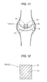
- the present invention relates to an implant composite material which is for use in the treatment of articular cartilage disorders such as hip joint femur head necrosis and knee joint bone head necrosis, the reconstruction/fixing of a bio-derived or artificial ligament or tendon, the uniting/fixing of a bone, etc.
- an interference screw for tendon or ligament fixing which is an interference screw comprising a biodegradable and bioabsorbable polymer and has a through-hole for Kirschner wire insertion formed along the center line therefor, an upper part of the through-hole (part on the screw head side) being a large elongated-circle hole part for rotating-tool fitting (patent document 3).
- This interference screw for tendon or ligament fixing is intended to be used in the following manner.
- a Kirschner wire guide wire for leading and screwing the screw in a desired direction with satisfactory accuracy
- the tip of a rotating tool is fitted into the elongated-circle hole part of the through-hole, and the tip is rotated to screw the screw in the proper direction into each of those holes formed in the bones of a joint (holes respectively formed in the upper and lower bones of a joint) into which the ends of a tendon or ligament to be transplanted/reconstructed have been inserted.
- the transplant bone flaps on both ends of the tendon or ligament are pressed against and fixed to the inner surfaces of the holes.
- the biodegradable and bioabsorbable polymer hydrolyzes due to contact with a body fluid and is assimilated by the living body. Consequently, this interference screw need not be taken out of the body through a reoperative surgery.
- the through-hole of that interference screw is less apt to undergo bone tissue invasion/growth (bone ingrowth). Because of this, there has been a problem that part of the through-hole remains vacant until the screw is mostly degraded/assimilated and replaced by a bone. Although a technique in which autobone particles taken out of another part are packed into the hole may be employed, the donor part remains as a defective part and, hence, should be filled with artificial bone particles. Complete repair with an autobone is not attained.
- the content of the bioceramic particles in the compact composite constituting the interference screw should be 30-60% by mass and the content of the bioceramic particles in the porous composite constituting the packing be 60-80% by mass. It is also preferred that the porous composite constituting the packing should be one which has a porosity of 60-90% and in which at least 50% of all pores are accounted for by interconnected pores and the interconnected pores have a pore diameter of 50-600 ⁇ m. Furthermore, the packing is preferably impregnated with at least one biological bone growth factor selected from a BMP, TGF- ⁇ , EP4, b-FGF, and PRP.
- a living bone conductively grows by the action of the bioactive bioceramic particles and the compact composite is thus replaced by bone tissues. Since the bioceramic particles contained in the porous composite and in the compact composite are bioabsorbable, they neither remain/accumulate in the (cartilage) bone tissues which have replaced and regenerated nor come into soft tissues or blood vessels.
- the implant composite material in which the porous composite has been superposed on and united until one side or all surfaces of the compact composite has the properties or functions required of scaffold materials and the like for use in, e.g., the treatment of articular cartilage disorders as stated above. Because of this, when the implant composite material of the first type in which the porous composite has been superposed on and united with one side of the compact composite is implanted in and fixed to, for example, a part where a necrotized part of an articular bone head has been excised, so that the porous composite is located on the cartilage side of the articular bone head surface, then it functions by the following mechanism.
- the porous composite is wholly replaced by cartilage tissues inductively grown in an early stage and disappearance, and the compact composite, which has strength, also is wholly replaced finally by conductively grown hard-bone tissues and disappears.
- the bioceramic particles also are completely assimilated.
- the hard-bone part and cartilage part of the necrotized articular bone head part are regenerated.
- the implant composite material of the first type in which the porous composite has been superposed on and united with the all surfaces of the compact composite is implanted in an excised part of an articular bone head, the following effect/advantage is brought about besides those described above.
- Hard-bone tissues rapidly grow inductively in the porous composite in contact with the hard bone in the excised part, whereby this implant composite material is bonded with and fixed to the excised part of the articular bone head in a short time period.
- the porous composite layer in which the content of the bioceramic particles gradually changes to have an inclination so that it increases from an inner-layer part to a surface-layer part of the porous composite layer in the range of 30-80% by mass has the following advantage. Since the surface-layer part has a high bioceramic-particle proportion and hence has higher bioactivity, the inductive growth of an osteoblast and bone tissues in the surface-layer part is especially enhanced, and replacement by and bonding with a living bone (inner surface of a hole) are further accelerated. Furthermore, the porous composite layer containing at least one biological bone growth factor selected from a BMP, TGF- ⁇ , EP4, b-FGF, and PRP and/or an osteoblast derived from a living organism has the following advantage.
- the implant composite material of the third type of the invention which is for tendon or ligament fixing (interference screw), maybe used, for example, in the following manner.
- the transplant bone flaps on end parts of a transplant tendon are inserted into holes respectively formed in the upper and lower bones of a joint, and this interference screw is screwed into the space between each transplant bone flap and the inner surface of the hole to thereby press the transplant bone flap against the inner surface of the hole and fix it.
- the interference screw itself, which comprises the compact composite comprising a biodegradable and bioabsorbable polymer containing bioceramic particles; has a sufficient mechanical strength, although it is a hollow object having a through-hole formed therein, and slowly undergoes hydrolysis by a body fluid.
- transplant bone flaps on end parts of the transplant tendon are fixed to the inner surfaces of the holes (i.e., to the living bones) in such an early stage. Thereafter, each interference screw and the packing further undergo degradation and assimilation and are finally replaced completely by a living bone formed by bone conduction or bone induction, whereby the joint is restored to the original state in which the through-hole of the screw does not remain vacant. Furthermore, since the biological bone growth factor contained in the packing comprising the porous composite has not undergone the heat history attributable to screw production, it has no fear of having undergone thermal alteration.
- the bioceramic particles contained in the packing and in the screw are bioabsorbable, they neither remain/accumulate in the living bones which have replaced nor come into/remain in soft tissues or blood vessels.
- This implant composite material is a composite comprising three components. Namely, it comprises a hollow object which, although hollow, has such a high mechanical strength and a long strength retention period that this hollow object is usable as a biodegradable bone-uniting material and in which the through-hole is filled with a porous material functioning as a bone substitute which itself has bone conductivity and bone inductivity and has a porous nature and mechanical strength similar to those of cancellous bones or as a scaffold which accelerates bone penetration and regeneration, the porous material containing a biological bone growth factor.
- This filler enables a body fluid and an osteoblast to penetrate into inner parts of the porous composite through interconnected pores, and is degraded and assimilated earlier than the bone-uniting material main body comprising the compact composite while exhibiting its bone conductivity and bone inductivity based on the bioactivity of the bioceramic particles.
- the biological bone growth factor supported such as a BMP, is gradually released. Because of this, the conductive formation of a living bone (autobone) is efficiently accelerated and bone adhesion is completed in about several weeks, which is considerably shorter than three months necessary for ordinary bone adhesion.
- Fig. 1 is a slant view of an implant composite material of the first type as one embodiment of the invention
- Fig. 2 is a view illustrating an example in which this implant composite material is used
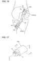
- Fig. 3 is an enlarged sectional view illustrating part of the implant composite material.
- the compact composite 1 is a compact block composite comprising a biodegradable and bioabsorbable polymer containing bioabsorbable and bioactive bioceramic particles.
- the compact composite 1 in this embodiment is in the form of a solid cylinder, it can have a quadrangular solid prism, elliptic solid cylinder, or flat plate shape or any of other various shapes according to the joint part into which the implant composite material is to be implanted.
- the size of the compact composite 1 also is not limited, and may be one suitable for the joint part into which the implant composite material is to be implanted.
- the bioceramic particles to be incorporated into this compact composite 1 preferably are particles which have bioactivity, are bioabsorbable and wholly assimilated by the living body and completely replaced by bone tissues, and have satisfactory bone conductivity (inductivity) and satisfactory biocompatibility.
- Example thereof include uncalcined and unsintered particles of hydroxyapatite, dicalcium phosphate, tricalcium phosphate, tetracalcium phosphate, octacalcium phosphate, calcite, Ceravital, diopside, and natural coral.
- the porous composite 2 is produced, for example, by the following process. First, a biodegradable and bioabsorbable polymer is dissolved in a volatile solvent and bioceramic particles are mixed with the solution to prepare a suspension. This suspension is formed into fibers by spraying or another technique to produce a fibrous mass composed of fibers intertwined with one another. This fibrous mass is immersed in a volatile solvent such as methanol, ethanol, isopropanol, dichloroethane(methane), or chloroform to bring it into a swollen or semi-fused state. The fibrous mass in this state is pressed to obtain a porous fusion-bonded fibrous mass in a disk form such as that shown in Fig. 1 .
- a volatile solvent such as methanol, ethanol, isopropanol, dichloroethane(methane), or chloroform
- This implant composite material 103 is inserted between vertebrae so that the inclined faces of the serrate projections 1a face the front side, whereby the following advantage is brought about besides the effects and advantages of the implant composite material 102 described above.
- the projections 1a of the compact composite 1 slightly bite into the upper and lower vertebrae and, hence, the implant composite material 103 can be prevented from coming out from the space between the vertebrae just after the insertion.
- Fig. 8 is a sectional viewof an implant compositematerial of the first type as still a further embodiment of the invention
- Fig. 9 is a view illustrating an example in which this implant composite material is used.
- interference screws 35 and 35 are ones comprising a biodegradable and bioabsorbable polymer containing bioactive bioceramic particles, then these screws 35 and 35 also are shortly replaced by bone tissues and bond with the inner surfaces of the holes 33 and 33 and with the two end parts 34a and 34a of the ligament 34, whereby the adherent parts of the ligament have a further improved fixing strength.
- the surface-layer parts having a high bioceramic-particle content have higher bioactivity. Because of this, the inductive growth of an osteoblast and (cartilage) bone tissues in the surface-layerparts is especially enhanced and replacement by (cartilage) bone tissues is further accelerated.
- the implant composite material can have a quadrangular solid prism, elliptic solid cylinder, or flat plate shape or any of other various shapes according to the joint part into which the implant composite material is to be implanted.
- the size thereof also may be one optimal for the joint part into which the implant composite material is to be implanted.
- the surface-layer part 2b having a high porosity and the inner-layer parts connected thereto having a relativelyhighporosity in this porous composite 2 are required to have a strength and flexibility such as those of cartilages and are further required to be rapidly degraded and undergo bonding with and complete replacement by a living (cartilage) bone in an early stage.
- porous composite 2 in which the content of bioceramic particles increases from a low-porosity surface-layer part 2a to a high-porosity surface-layer part 2b use may be made of a method which comprises preparing several suspensions differing in the amount of bioceramic particles incorporated, forming several fibrous masses differing in bioceramic-particle content, superposing these fibrous masses in order of increasing bioceramic-particle content, bringing this assemblage into a swollen or semi-fused stage, and pressing it.
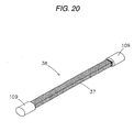
- the implant composite material of the second type of the invention which is to be attached as an anchor member to an end part of a ligamental member or tendinous member, will be explained next by reference to drawings.
- Organic fibers of the ligamental member 37 which will be described later, are passed through these small holes 1c and hitched on, whereby the implant composite material 109 can be attached to the end part of the artificial ligamental member 37 while preventing it from detaching.
- the shape of the implant composite material 109 is not limited to the elliptic solid cylinder, and the implant composite material 109 can have any shape as long as it can be easily inserted into the holes formed in the upper and lower living bones of a joint such as a knee joint and can be stably fixed with the interference screw to be screwed into the space between the inner surface of each hole and the implant composite material 109.
- This compact composite 1 is produced, for example, by a method in which a biodegradable and bioabsorbable polymer containing bioceramic particles is injection-molded into an elliptic solid cylinder having a projecting piece 1b on one edge face thereof and this projecting piece 1b is subjected to drilling or by a method in which a molded object of a biodegradable and bioabsorbable polymer containing bioceramic particles is cut into an elliptic solid cylinder having a projecting piece 1b on one edge face thereof and this projecting piece 1b is subjected to drilling.
- the layer of the porous composite 2 is a porous object which has interconnected pores inside and comprises a biodegradable and bioabsorbable polymer containing bioabsorbable and bioactive bioceramic particles. Part of the bioceramic particles are exposed in the surfaces of this porous composite 2 and in inner surfaces of the interconnected pores.
- the porous composite 2 in this implant composite material (anchor member) 109 as an embodiment has been superposed only on the peripheral surface of the compact composite 1 in an elliptic solid cylinder form, the porous composite 2 may be superposed on and united with all surfaces of the compact composite 1 except the projecting piece 1b, i.e., the peripheral surface and both edge faces of the compact composite 1.
- the layer of the porous composite 2 shouldbe one inwhich the porosity thereof is 50-90%, preferably 60-80%, interconnected pores account for 50% ormore, preferably 70-90%, of all pores, and the interconnected pores have a pore diameter of 50-600 ⁇ m, preferably 100-400 ⁇ m, when physical strength, osteoblastpenetration, stabilization, etc. are taken into account, as in the case of the porous composite 2 of the implant composite material 100 described above.
- the reasons for this are as described above with regard to the porous composite 2 of the implant composite material 100, and an explanation thereon is hence omitted.
- the porosity of the porous composite 2 may be even throughout the whole composite. However, when the property of bonding with a living bone and conductive/inductive growth are taken into account, it is preferred that the porosity thereof should gradually change to have an inclination so that it increases from an inner-layer part to a surface-layer part of the layer of the porous composite 2 as in the case of the porous composite 2 of the implant composite material 100.
- the porosity thereof should gradually increase continuously from an inner-layer part to a surface-layer part in the range of 50-90%, preferably in the range of 60-80%, and that the pore diameter of the interconnected pores should gradually increase from the inner-layer part to the surface-layer part in the range of 50-600 ⁇ m.
- hydrolysis proceeds rapidly on its surface-layer side and osteoblast penetration and the inductive growth of bone tissues are enhanced.
- This porous composite 2 bonds with a living bone in an early stage. Consequently, the strength of fixing of the implant composite material (anchor member) 109 to the upper or lower living bone of a knee joint or the like can be heightened in an early stage.
- the content of the bioceramic particles in the porous composite 2 may be even throughout the whole porous-composite layer 21d as in the case of the porous composite 2 of the implant composite material 100 described above, or may be uneven. In the former case, in which the content is even, it is preferred that the content of the bioceramic particles should be 60-80% by mass. The reasons for this are as described hereinabove with regard to the porous composite 2 of the implant composite material 100, and an explanation thereon is hence omitted. A more preferred range of the content of the bioceramic particles is 60-70% by mass.
- the content of the bioceramic particles in the porous composite 2 should be higher than the bioceramic-particle content in the compact composite 1 and gradually change to have an inclination so that it increases from an inner-layer part to a surface-layer part of the layer of the porous composite 2 in the range of 30-80% by mass as in the case of the porous composite 2 of the implant composite material 100.
- the bioceramic particle/biodegradable and bioabsorbable polymer proportion by mass in the porous composite 2 should be larger than that mass proportion in the compact composite 1 and gradually change to have an inclination so that it increases from the inner-layer part to the surface-layer part of the layer of the porous composite 2 in the range of from 30/70 to 80/20.
- the layer of the porous composite 2 having such an inclination of bioceramic-particle content bioactivity is high in the surface-layer side having a high content and the inductive growth of an osteoblast and bone tissues is enhanced especially in the surface-layer side.
- This porous composite 2 bonds with a living bone and is replaced thereby in an early stage. Consequently, the strength of fixing of the implant composite material (anchor member) 109 to the upper or lower living bone of a knee joint or the like can be heightened in an early stage.
- the content of the bioceramic particles in the compact composite 1 preferably is lower than the bioceramic-particle content in the porous-composite layer 2 and is in the range of 30-60% by mass as in the case of the compact composite 1 of the implant composite material 100 described above.
- the reasons for this are as described above with regard to the compact composite 1 of the implant composite material 100.
- the content of the bioceramic particles in the compact composite 1 may be even throughout the whole compact composite 1, provided that the content thereof is lower than in the porous composite 2 and in the range of 30-60% by mass as stated above.
- the content of the bioceramic particles in the compact composite 1 may gradually change to have an inclination so that it gradually increases from an axial core part toward a peripheral part of the compact composite 1, provided that the content thereof is lower than in the porous composite 2 and in the range of 30-60% by mass as stated above.
- the peripheral part having a high content undergoes the conductive growth of bone tissues while the axial core part having a low content retains a strength.
- this compact composite 1 is wholly replaced.
- this porous-composite layer 21d should be impregnated with at least one of the biological bone growth factors described above, i.e., a BMP, TGF- ⁇ , EP4, b-FGF, and PRP, and/or an osteoblast derived from a living organism.
- a BMP biological bone growth factors described above
- TGF- ⁇ a BMP, TGF- ⁇ , EP4, b-FGF, and PRP
- an osteoblast derived from a living organism i.e., a BMP, TGF- ⁇ , EP4, b-FGF, and PRP
- the surface of the layer of the porous composite 2 may be subjected to an oxidation treatment such as corona discharge, plasma treatment, or hydrogen peroxide treatment.
- an oxidation treatment such as corona discharge, plasma treatment, or hydrogen peroxide treatment.
- Such an oxidation treatment has an advantage that bonding with a living bone and replacement thereby are further accelerated because the wettability of the surface of the layer of the porous composite 2 is improved to enable an osteoblast to more effectively penetrate into and grow in interconnected pores of this composite 2.
- the fixing strength of this implant composite material (anchor member) 109 improves in an earlier stage.
- the surface of the compact composite 1 may, of course, be subjected to such an oxidation treatment.
- the implant composite material (anchor member) 109 shown in Fig. 17 to Fig. 19 is one obtained in the following manner.
- the compact composite 1 in an elliptic solid cylinder form is fitted into a layer of the porous composite 2 in an elliptic hollow cylinder form.
- two layers of the porous composite 2 in the form of a half of an elliptic hollow cylinder are united with each other and superposed on the periphery of the compact composite 1 in an elliptic solid cylinder form.
- These members superposed are united by, e.g., thermal fusion bonding to obtain the target implant composite material.
- Techniques for uniting the compact composite 1 with the layer(s) of the porous composite 2 are not limited to thermal fusion bonding.
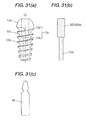
- the artificial ligament 38 shown in Fig. 20 comprises an artificial ligamental member 37 formed from organic fibers as a raw material and the implant composite material 109 attached as an anchor member to each end of the ligamental member 37 so as not to detach therefrom.
- the artificial ligamental member 37 comprising the structure described above preferably has an internal porosity in the range of 20-90%. In case where the internal porosity thereof is lower than 20%, this ligamental member 37 is too compact and is impaired in flexibility and deformability. This ligamental member is hence unsatisfactory as a substitute for a bio-derived ligament. On the other hand, in case where the internal porosity thereof exceeds 90%, this ligamental member 37 is reduced in shape retention and shows too high elongation. This ligamental member also is hence unsatisfactory as a substitute for a bio-derived ligament.
- the organic fibers to be used as a material for the artificial ligamental member 37 preferably are, for example, bioinert synthetic-resin fibers such as, e.g., fibers of polyethylene, polypropylene, and polytetrafluoroethylene, or coated fibers obtained by coating organic core fibers with any of these bioinert resins to impart bioinertness thereto.
- bioinert synthetic-resin fibers such as, e.g., fibers of polyethylene, polypropylene, and polytetrafluoroethylene
- coated fibers having a diameter of about 0.2-0.5 mm obtained by coating core fibers of ultrahigh-molecular polyethylene with linear low-density polyethylene are optimal fibers from the standpoints of strength, hardness, elasticity, suitability for weaving/knitting, etc.
- an interference screw 35 is screwed into the space between the inner surface of the hole 33 in the shinbone 40 and the other implant composite material 109 while keeping the artificial ligamental member 37 moderately loose to press this implant composite material 109 against that side of the inner surface of the hole 33 which is opposite to the screw 35 and thereby fix this implant composite material 109.
- the artificial ligamental 38 is implanted in and fixed to the knee joint.
- the implant composite materials 109 and 109 respectively on both ends of the artificial ligament 38 have a greatly improved fixing strength as compared with the conventional case where both ends of a ligament are fixed with interference screws only.
- the compact composite 1 of each implant composite material 109 is hard and strong and hydrolyzes far more slowly than the layer of the porous composite 2. It retains a sufficient strength until the hydrolysis proceeds to a certain degree.
- the compact composite 1 is wholly hydrolyzed and disappears through replacement by a living bone conductively formed by the action of the bioactive bioceramic particles. Consequently, the holes 33 and 33 formed in the thighbone 39 and shinbone 40 of the knee joint are almost completely filled with the living bones.
- This implant composite material (anchor member) 112 comprises: a compact composite 1 in the form of an elliptic solid cylinder which has ridges 1f each having a triangular sectional shape and formed on the peripheral surface thereof; and the porous composite 2 superposed on and united with the peripheral surface of the compact composite 1 so that the porous composite 2 fills the recesses between these ridges 1f.
- the other constitutions of this implant composite material 112 are the same as those of the implant composite material 109 in the artificial ligament 38 described above, and an explanation thereon is hence omitted.
- This implant composite material (anchor member) 114 is a composite material having a three-layer structure comprising a core layer comprising a compact composite 1 and a layer of a porous composite 2 superposed on and united with each of the upper and lower sides of the core layer.
- This implant composite material 114 as a whole is in the form of an elliptic solid cylinder. It has a projecting piece (not shown) having the small holes or projections for hitching organic fibers of an artificial ligamental member 37, the projecting piece being formed on one edge face of the core layer comprising a compact composite 1.
- implant composite material modifications are explained which eliminate the same problems as the implant composite materials of the second type described above, which are used as anchor members for artificial ligaments, etc., and can have the same effects and advantages as those implant composite materials.
- the implant composite materials as such modifications each have the following advantages when used for the reconstruction/fixing of a ligament, for example, in the following manner.
- the implant composite material is attached as an anchor member to each end of a ligamental member so as not to detach therefrom.
- These implant composite materials are inserted into holes respectively formed in the upper and lower living bones (thighbone and shinbone) of a knee joint.
- An interference screw is then screwed into the space between each implant composite material and the inner surface of the hole.
- the content of the bioceramic particles should gradually change to have an inclination so that it increases from the axial core part of the porous composite to the peripheral part to be in contact with a bone in the range of 30-80% by mass.
- the content of the bioceramic particles should gradually change to have an inclination so that it increases from that end part of the porous composite to which a ligamental member or tendinous member is to be attached to the end part on the opposite side in the range of 30-80% by mass.
- Those implant composite material modifications in which the content of the bioceramic particles inclines have the following advantage. Since the peripheral part or opposite-side end part has a high bioceramic-particle content and hence has higher bioactivity, the inductive growth of an osteoblast and bone tissues in the peripheral part or opposite-side end part is especially enhanced. Consequently, replacement by and bonding with a living bone are further accelerated.
- the porous composite should have been impregnated with at least one of the biological bone growth factors described above and/or an osteoblast derived from a living organism.
- Such implant composite materials have an advantage that osteoblast multiplication/growth is greatly accelerated and, hence, bone tissues grow vigorously to enable bonding with and replacement by a living bone to proceed more rapidly. It is also preferred that many small holes or small projections for the attachment of a ligamental member or tendinous member be formed in or on an end part.
- Such changes in porosity and pore diameter have the following advantages. That peripheral part 2i of the porous composite 2 which has a large pore diameter and a high porosity (hereinafter referred to as high-porosity peripheral part) is rapidly hydrolyzed because a body fluid easily penetrates thereinto. In addition, an osteoblast is apt to penetrate thereinto and this, coupled with the high content of the bioactive bioceramic particles as will be described later, enables bone tissues to inductively grow in an early stage. The peripheral part 2i thus bonds with a living bone and is replaced thereby in an early stage.
- this peripheral part 2i is undesirable because the penetration of a body fluid and an osteoblast thereinto is difficult and hydrolysis and the inductive growth of bone tissues are slow, resulting in a prolonged time period required for replacement by and bonding with a living bone.
- organic fibers in an end part of an artificial ligamental member 37 may be embedded in and fixed to the strong low-porosity axial core part 2h of the implant composite material 115 in such a manner than the organic fibers do not come out.
- the content thereof in the low-porosity axial core part 2h is lower than 30% by mass, this arouses a trouble that the inductive growth of bone tissues in the low-porosity axial core part 2h by the action of the bioceramic particles becomes slow and, hence, complete replacement by a living bone takes too much time.
- a more preferred upper limit of the content of the bioceramic particles is 70% by mass.
- the porous composite 2 constituting the implant composite material 115 should be impregnated with any of the biological bone growth factors described above and/or an osteoblast derived from a living organism.
- osteoblast multiplication and growth are greatly accelerated.
- bone tissues come to grow in the high-porosity peripheral part 2i of the porous composite 2 in an extremely short time period (about 1 week) to accelerate bonding with the inner surface of the hole 33.
- the time period required for the hole 33 to be mostly filled with a living bone through the complete replacement of the porous composite 2 can be reduced.
- the surface of this porous composite 2 may be subjected to an oxidation treatment such as corona discharge, plasma treatment, or hydrogen peroxide treatment to thereby improve the wettability of the surface of the porous composite 2 and further accelerate the penetration and growth of an osteoblast.
- an oxidation treatment such as corona discharge, plasma treatment, or hydrogen peroxide treatment to thereby improve the wettability of the surface of the porous composite 2 and further accelerate the penetration and growth of an osteoblast.
- the implant composite material 115 comprising the porous composite 2 may be produced, for example, by the following process.
- a biodegradable and bioabsorbable polymer is dissolved in a volatile solvent and bioceramic particles are mixed therewith to prepare a suspension.
- This suspension is formed into fibers by spraying or another technique to produce a fibrous mass composed of fibers intertwined with one another.
- This fibrous mass is packed into an elliptic hollow cylinder so that the fiber amount decreases from the center to the periphery.
- the fibrous mass packed is further immersed in a volatile solvent to bring it into a swollen or semi-fused state.
- the fibrous mass in this state is pressed in the axial direction for the elliptic cylinder to obtain a porous fusion-bonded fibrous mass in the form of an elliptic solid cylinder.
- the fibers in this fusion-bonded fibrous mass are shrunk and fused, and are thereby deprived substantially of their fibrous shape to form a matrix.
- the fibrous mass is changed in form into a porous composite in which the spaces among the fibers have been changed into rounded interconnected pores.
- One end of this porous composite in the form of an elliptic solid cylinder is cut to form a projecting piece and small holes.
- the implant composite material 115 is produced.
- the porous composite 2 in which the content of bioceramic particles increases from the low-porosity axial core part 2h to the high-porosity peripheral part 2i is to be produced.
- the following method may be used.
- Several suspensions differing in the amount of bioceramic particles incorporated are prepared, and several fibrous masses differing in bioceramic-particle content are formed therefrom.
- These fibrous masses are packed into an elliptic hollow cylinder in such a manner that the fibrous mass having a lowest content is disposed in the center and the other fibrous masses are disposed toward the periphery in order of increasing content.
- the fibrous masses thus packed are brought into a swollen or semi-fused state with a volatile solvent and then pressed.
- a projecting piece 2j projects from the end part the porosity of which is low or 0% and which is strong, and many small holes 2k have been formed in this projecting piece 2j.
- Organic fibers of an artificial ligamental member 37 are inserted into these small holes 2k, whereby the implant composite material 116 is attached as an anchor member to the end part of the ligamental member 37 so as not to detach therefrom.
- the other constitutions of this implant composite material 116 are the same as those of the anchor member 115, and an explanation thereon is hence omitted.
- This implant composite material 116 may be used in the following manner.
- the implant composite material 116 is attached as an anchor member to each end of a ligamental member 1.
- These composite materials 116 are inserted into the holes 33 respectively formed in the upper and lower living bones of a knee joint and are then fixed with interference screws.
- the upper side and peripheral surface of the opposite-side end part 2n, which has a high porosity, and the peripheral surface of the part which is located underneath the high-porosity opposite-side end part 2n and has a relatively high porosity are rapidly hydrolyzed and bond with the inner surface of each hole 33 while being replaced by a living bone in an early stage. Consequently, the fixing strength of each implant composite material 116 improves in an early stage.
- the end part 2m the porosity of which is low or 0%, is slow in hydrolysis and retains its strength over a certain time period.
- this end part 2m is wholly replaced by a living bone and disappears shortly and each hole 33 is mostly filled with the living bone which has replaced.
- implant composite materials (anchor members) 115 and 116 described above each are in the form of an elliptic solid cylinder, the shapes thereof are not limited to elliptic solid cylinders.
- the implant composite materials may have any shape as long as they can be easily inserted into holes 33 respectively formed in the upper and lower living bones of a knee joint and can be stably fixed with interference screws.
- implant composite materials (anchor members) 109, 110, 111, 112, 113, 114, 115, and 116 described above each can be used after having been attached to an end part of a bio-derived ligamental member or tendinous member, an artificial tendinous member, or the like so as not to detach therefrom.
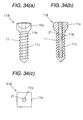
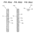
- This implant composite material 117 for tendon or ligament fixing comprises: an interference screw 10 (hereinafter referred to simply as screw) which comprises a compact composite of a biodegradable and bioabsorbable polymer containing bioabsorbable and bioactive bioceramic particles and has a through-hole 10c for Kirschner wire insertion formed along a center line CL therefor; and a packing 20 which comprises a porous composite of a biodegradable and bioabsorbable polymer containing bioabsorbable and bioactive bioceramic particles and with which the through-hole 10c is filled, the packing 20 having been impregnated with any of the biological bone growth factors described above.
- an interference screw 10 hereinafter referred to simply as screw
- screw which comprises a compact composite of a biodegradable and bioabsorbable polymer containing bioabsorbable and bioactive bioceramic particles and has a through-hole 10c for Kirschner wire insertion formed along a center line CL therefor
- a packing 20 which comprises a
- the screw 10 of this implant composite material 117 has a screw head 10a in a nearly roughly hemispherical form.
- the diameter of the screw head 10a is almost the same as the outer diameter of the screw thread 10b.
- the top of the screw thread 10b has been cut so as to have a flat surface. Inparticular, the top of the screw thread 10b in an area near to the screw tip has been considerably cut so as to result in a reduced outer diameter.
- This screw shape enables the screw to be smoothly screwed deeply into a hole formed in a bone of a joint while the flat surface of the screw thread top is being pressed against both of the inner surface of the hole and that end-part transplant bone flap of a transplant tendon or the like which is inserted into the hole.
- Small holes (not shown) connected to the through-hole 10c (10d and 10e) may be formed in this screw 10 as long as the screw 10 can retain a strength required of screws for tendon or ligament fixing. Formation of such small holes has the following advantages. A body fluid and an osteoblast are apt to penetrate through the small holes into the packing 20 packed in the through-hole, and the biological bone growth factor contained is apt to be released with the degradation and assimilation of the packing 20. Consequently, the growth of a living bone and bone adhesion can be further accelerated.
- the screw 10 of this implant composite material 117 comprises a compact composite of a biodegradable and bioabsorbable polymer containing bioabsorbable and bioactive bioceramic particles as described above, and is required to have a high strength which is equal to or higher than the upper and lower living bones (hard bones) of a joint.
- a biodegradable and bioabsorbable polymer suitable for use as a raw material therefor is a crystalline polymer such as poly(L-lactic acid) or poly(glycolic acid).
- poly(L-lactic acid) having a viscosity-average molecular weight of 150,000 or higher, desirably about 200,000-600,000.
- bioceramic particles to be incorporated into the compact composite constituting the screw 10 it is preferred to use the same bioceramic particles as those contained in the compact composite 1 of the implant composite material 100 described above.
- the content of the bioceramic particles is preferably in the range of 30-60% by mass. In case where the content thereof exceeds 60% by mass, there is a possibility that the compact composite becomes brittle, leading to a strength deficiency in the screw 10. Contents thereof lower than 30% by mass result in a trouble that the conductive bone formation by the action of the bioceramic particles becomes insufficient and the complete replacement of this screw 10 by bone tissues requires much time.
- the content of the bioceramic particles may be even throughout the whole compact composite constituting the screw 10 in the range of 30-60% by mass, or may change to have an inclination so that it gradually increases from a central part toward the periphery of the compact composite in the range of 30-60% by mass.
- the porous composite constituting this packing 20 need not have a high strength such as that of the screw 10 comprising the compact composite, and a strength and flexibility which prevent the packing 20 from breaking upon insertion into the through-hole 10c of the screw 10 suffice for the porous composite.
- This porous composite is required to be rapidly degraded and be wholly replaced by a living bone in an early stage. Because of this, the biodegradable and bioabsorbable polymer to be used as a raw material for the porous composite preferably is the same as the biodegradable and bioabsorbable polymer in the porous composite 2 of the implant composite material 100 described above.
- the porosity thereof is lower than 60%, the proportion of pores is lower than 50% based on all pores, and the pore diameter is smaller than 50 ⁇ m, then it is difficult to rapidly inject and infiltrate an appropriate amount of a biological bone growth factor and the penetration of a body fluid or osteoblast also becomes difficult.
- the hydrolysis of the porous composite and the inductive growth of bone tissues therein become slow and, hence, the time period required for the through-hole 1c of the screw 10 to be filled through complete replacement by a living bone is prolonged.
- bone inductivity is exhibited when fine interconnected pores on submicron order of 1-0.1 ⁇ m coexist with interconnected pores having that preferred pore diameter.
- the packing 20 comprising the porous composite can be produced, for example, by the following process.
- a biodegradable and bioabsorbable polymer is dissolved in a volatile solvent and bioceramic particles are mixed with the solution to prepare a suspension.
- This suspension is formed into fibers by spraying or another technique to produce a fibrous mass composed of fibers intertwined with one another.
- This fibrous mass is immersed in a volatile solvent such as methanol, ethanol, isopropanol, dichloroethane(methane), or chloroform to bring it into a swollen or semi-fused state.
- the implant composite material 117 for tendon or ligament fixing which is in the form of the interference screw described above may be used in the following manner.
- the interference screw is screwed into the space between the inner surface of each of holes respectively formed in the upper and lower bones of a joint and the transplant bone flap on each end of, e.g., a transplant tendon inserted into the holes to thereby press the transplant bone flap against the inner surface of the hole and fix it.
- the screw 10 itself; which comprises the compact composite comprising a biodegradable and bioabsorbable polymer containing bioceramic particles, has a sufficient mechanical strength, although it is a hollow object having a through-hole formed therein, and slowly undergoes hydrolysis by a body fluid.
- the biological bone growth factor contained in the packing 20 comprising the porous composite has not undergone the heat history attributable to screw production, it has no fear of having undergone thermal alteration.
- the bioceramic particles contained in the screw 10 and in the packing 20 are bioabsorbable, they neither remain/accumulate in the living bones which have replaced nor come into/remain in soft tissues or blood vessels.
- the surface-layer part of each screw 10 bonds in an early stage with the transplant bone flap on an end of the transplant tendon and with the inner surface of the hole in an early stage due to bone tissues conductively grown with hydrolysis, the screw 10 can be prevented from becoming loose.
- the content of the bioceramic particles in the compact composite constituting the interference screw is preferably 30-60% by mass, while the content of the bioceramic particles in the porous composite constituting the packing is preferably 60-80% by mass.
- the porosity thereof is preferably 60-90%, interconnected pores account for preferably 50% or more of all pores, and the interconnected pores have a pore diameter of preferably 50-600 ⁇ m. It is also preferred to infiltrate at least one of the biological bone growth factors described above into the packing. The reasons for these are as explained above with regard to the implant composite material 117 for tendon or ligament fixing.
- a Kirschner wire (not shown) and the tip of a rotating tool (not shown) are inserted into the through-hole 10c of a screw 10, and this screw 10 is screwed, while being rotated, in a proper direction into a proper position in the space between the inner surface of one hole 33 and the transplant bone flap 42a to thereby press this transplant bone flap 42a against the opposite-side inner surface of this hole 33 and fix it.
- the rotating tool and the Kirschner wire are drawn out and a packing 20 is packed into the through-hole 10c of the screw 10.
- a container 41 is opened and the biological bone growth factor is infiltrated into the packing 20.
- each screw 10 After the transplant tendon is thus fixed, each screw 10 retains a sufficient strength over a period of at least 3 months, which is usually necessary for the adhesion of the transplant bone flap 42a to the inner surface of the hole 33, to fix the osteosynthesis part without fail.
- the packing 20 releases the biological bone growth factor while being rapidly hydrolyzed. Consequently, the adhesion of the transplant bone flap 42a to the inner surface of the hole 33 is completed in about several weeks, although this period varies depending on the part and the bone growth factor.
- the packing 20 is replaced by a living bone due to the bioactivity of the bioceramic particles and the through-hole 10c is hence filled with the living bone in a relatively early stage.
- the screw 10 also is wholly replaced by a living bone and disappears, whereby the bone is restored to the original state in which the through-hole 10c does not remain vacant.
- This set for tendon or ligament fixing may be used in the following manner as for the fixing set shown in Fig. 31 described above.
- the screw 10 is screwed into the space between the inner surface of each of holes respectively formed in the upper and lower bones of a joint and a transplant bone flap of a transplant tendon (or transplant ligament) inserted into the hole to fix the bone flap.
- the Kirschner wire is drawn out.
- the packing 20 is packed into the through-hole 10c of the screw 1 and a biological bone growth factor separately prepared is infiltrated into the packing 20.
- a biological bone growth factor is infiltrated into the packing 20 before this packing 20 is packed into the through-hole 10c of the screw 10.
- Fig. 34 illustrates an implant composite material for osteosynthesis as still a further embodiment of the invention: (a), (b), and (c) are a front view, vertical sectional view, and plan view thereof, respectively.
- This implant composite material 118 for osteosynthesis comprises: a bone-uniting material main body which has been formed into a screw 11 and has a cylindrical deep hole 11b extending along the center line therefor from the upper end surface of the screw head 11a to a part near to the screw tip; and a filler 21 in a solid cylinder form which has a diameter and length conforming to the diameter and depth of the hole 11b and with which the hole 11b is filled, the filler 21 having been impregnated with any of the biological bone growth factors shown above.
- the filler 21 packed in the hole 11b which has been formed in the screw 11 (bone-uniting material main body) and at least one end of which is open is one comprising a porous composite of a biodegradable and bioabsorbable polymer containing bioabsorbable and bioactive bioceramic particles.
- the filler 21 has interconnected pores inside. Part of the bioceramic particles are exposed in inner surfaces of the interconnected pores and in the surfaces of the porous composite. This filler 21 has been impregnated with an appropriate amount of a biological bone growth factor.
- the porosity of the porous composite constituting the filler 21 may be even throughout the whole composite in the range of 60-90%, or may continuously increase toward the peripheral surface and toward the upper and lower ends in the range of 60-90%.
- the pore diameter of the interconnected pores may be even throughout the whole composite in the range of 50-600 ⁇ m, or may gradually increase toward the peripheral surface and toward the upper and lower ends in the range of 50-600 ⁇ m.
- the filler 21 comprising the porous composite having such inclinations of porosity and pore diameter has the following advantages. An appropriate amount of a biological bone growth factor can be rapidly infiltrated through the upper and lower ends and peripheral surface having a high porosity and a large pore diameter. Hydrolysis hence proceeds rapidly from the upper and lower ends and peripheral surface, and osteoblast penetration and the inductive growth of bone tissues are enhanced. Consequently, replacement by a living bone is further accelerated.
- the content of the bioceramic particles in the porous composite constituting the filler 21 is preferably 60-80% by mass.
- the content thereof may be even throughout the whole porous composite in that range or may continuously increase toward the peripheral surface and the upper and lower ends in that range.
- such a high bioceramic-particle content coupled with the high porosity of the porous composite results in a decrease in the physical strength of the filler 21 comprising the porous composite.
- Contents thereof lower than 60% by mass arouse the following trouble.
- This porous composite has reduced bioactivity and, hence, the inductive growth of bone tissues becomes slow. As a result, the time period required for the hole 11b of the screw 11 to be filled by complete replacement by a living bone is prolonged.
- the surface of the filler 21 comprising the porous composite may be subjected to an oxidation treatment such as corona discharge, plasma treatment, or hydrogen peroxide treatment.
- an oxidation treatment such as corona discharge, plasma treatment, or hydrogen peroxide treatment.
- Such an oxidation treatment has the following advantage.
- the wettability of the surface of the filler 21 is improved and an osteoblast more effectively penetrates into interconnected pores in the filler 21 through the minute gap between the filler 21 and the hole 1b of the screw 11 and grows therein. Because of this, complete replacement by a living bone is further accelerated and the filler-filled hole 11b of the screw 11 is filled with the living bone in an early stage.
- the surface of the screw 11 comprising the compact composite may, of course, be subjected to such an oxidation treatment.
- the biological bone growth factor to be infiltrated into the porous composite constituting the filler 21 may be any of the BMP, TGF- ⁇ , EP4, b-FGF, and PRP described above. These biological bone growth factors may be enclosed alone or as a mixture of two or more thereof in a container 3. It is also preferred that an osteoblast derived from a living organism should be added to any of those biological bone growth factors. Any of those biological bone growth factors or a mixture thereof with an osteoblast derived from a living organism may be infiltrated into the filler 21 as an injection or dripping preparation in a solution or suspension state.
- any of those biological bone growth factors and/or the osteoblast into the filler 21 has the following advantages.
- the biological bone growth factor, etc. Prior to or simultaneously with the hydrolysis of the filler 21, the biological bone growth factor, etc. exude to considerably accelerate osteoblast multiplication/growth. Because of this, bone adhesion is completed in about several weeks although this period depends on the part and the growth factor, and bone tissues come to grow in surface-layer parts of the porous composite constituting the filler 21.
- the porous composite is wholly replaced by a living bone rapidly thereafter and the hole 11b of the screw 11 is filled with the living bone.
- BMPs and EP4 are especially effective in hard-bone growth.
- PRP is a plasma having a highly elevated platelet concentration, and addition thereof accelerates the growth of a newly regenerated bone.
- another growth factor such as IL-1, TNF- ⁇ , TNF- ⁇ , or IFN- ⁇ may be mixed.
- the filler 21 which comprises the porous composite of a biodegradable and bioabsorbable polymer containing bioceramic particles and which has been inserted in the hole 11b of the screw 11 enables a body fluid and an osteoblast to penetrate into inner parts of the porous composite through interconnected pores threof, and is degraded and assimilated earlier than the screw 11 comprising the compact composite while exhibiting its bone conductivity and bone inductivity based on the bioactivity of the bioceramic particles.
- the biological bone growth factor e.g., a BMP
- the conductive formation of a living bone is efficiently accelerated and bone adhesion is completed in about several weeks, which period is considerably shorter than three months necessary for ordinary bone adhesion, although that period varies depending on the part and the biological bone growth factor.
- the screw 11 and the filler 21 further undergo degradation and assimilation and are finally replaced completely by a living bone formed by bone conduction or bone induction, whereby the bone is restored to the original state in which the hole 11b of the screw 11 does not remain vacant.
- the biological bone growth factor contained in the filler 21 comprising the porous composite has not undergone the heat history attributable to screw 11 production, it has not fear of having undergone thermal alteration and functions to accelerate bone growth.
- the bioceramic particles contained in the filler 21 and in the screw 11 are bioabsorbable, they neither remain/accumulate in the living bones which have replaced nor come into/remain in soft tissues or blood vessels.
- Fig. 35 illustrates an implant composite material for osteosynthesis as still a further embodiment of the invention: (a), (b), and (c) are a front view, vertical sectional view, and plan view thereof, respectively.
- This implant composite material 119 for osteosynthesis comprises a bone-uniting material main body which comprises a compact composite and has been formed into a pin 12.
- This pin 12 has a hole 12a which is open at each end and extends along the center line for the pin 12 from a one-end surface (upper end surface) to the other-end surface (lower end surface) of the pin 12.
- a filler 22 in a solid cylinder form having a diameter and length conforming to the diameter and length of the hole 12a and impregnated with a biological bone growth factor has been packed in the hole 12a.
- This pin 12 (bone-uniting material main body) comprises the same compact composite as the screw 11 described above, i.e., a compact composite of a biodegradable and bioabsorbable polymer containing bioabsorbable and bioactive bioceramic particles.
- the peripheral surface thereof has an alternation of a tapered surface 12b becoming gradually narrower toward an end (becoming gradually narrower downward) and a flange part 12c. This shape prevents the pin 12 which has been driven into a hole formed in the bone of a fractured part from coming out because the peripheral edges of the flange parts 12c bite into the living bone.
- the diameter of the through-hole 12a of this pin 12 is preferably regulated to about 1/3 to 2/3 the diameter of the pin 12 (diameter of the thinnest parts at the lower ends of the tapered surfaces). In case where the hole diameter is larger than that, this pin 12 has a reduced strength, resulting in a possibility that this pin 12 might break or be damaged when driven into a fractured part. In case where the hole diameter is smaller than that, the filler 22 is too thin and the proportion of the filler 22 is reduced, whereby the effect of inductively forming a living bone becomes insufficient.
- the shape of the pin 12 is not limited to that in this embodiment, and can be any desired shape.
- the pin 12 may be a pin of a mere hollow cylinder or hollow prism shape having a through-hole 12a extending along the center line or a pin of the hollow prism shape which has an alternation of an oblique surface and a flanged part on each of the four lateral sides for preventing the pin from coming out.
- the hole 12a is not limited to a through-hole which is open at each end. It may, of course, be a deep bottomed hole extending from a one-end surface (upper end surface) to a part close to the other-end surface (lower end surface) of the pin 12. Such a bottomed hole has an advantage that the lower end (tip) of the pin 12 has a high strength and is hence less apt to break when the pin 12 is driven.
- two or more short fillers 22 having a solid cylinder form with a diameter conforming to the diameter of this hole 12a should be prepared and successively inserted into the hole 12a of the pin 12 to thereby pack the fillers 2 so as to fill the hole 12a over the whole length thereof.
- small holes (not shown) connected to the hole 12a to be filled with the filler 22 may be formed in the pin 12 as long as a necessary strength can be maintained. Formation of such small holes has the following advantages.
- the small holes enable a body fluid and an osteoblast to easily come into contact with and penetrate into the filler 22 and facilitate the exudation of the biological bone growth factor. Consequently, the growth of a living bone and bone adhesion are further accelerated.
- implant composite material for osteosynthesis of the invention include one comprising: a bone-uniting material main body which comprises a compact composite of a biodegradable and bioabsorbable polymer containing bioabsorbable and bioactive bioceramic particles and has been formed into the screw 11 or pin 12 described above; and a filler (filler not impregnating with a biological bone growth factor) comprising the porous composite described above, which comprises a biodegradable and bioabsorbable polymer containing bioabsorbable and bioactive bioceramic particles, the filler having been inserted into the hole 11b or 12a of the bone-uniting material main body, the hole 11b or 12a being open at at least one end.
- a bone-uniting material main body which comprises a compact composite of a biodegradable and bioabsorbable polymer containing bioabsorbable and bioactive bioceramic particles and has been formed into the screw 11 or pin 12 described above
- a filler filler not impregnating with a biological bone growth
- a first osteosynthesis set among such sets is characterized by comprising a combination of (1) a filler-filled bone-uniting material main body obtained by forming a bone-uniting material main body comprising a compact composite of a biodegradable and bioabsorbable polymer containing bioabsorbable and bioactive bioceramic particles, forming in the main body a hole which is open at at least one end, and filling the hole with a filler comprising a porous composite of a biodegradable and bioabsorbable polymer containing bioabsorbable and bioactive bioceramic particles and (2) a biological bone growth factor to be infiltrated into the filler.
- a third osteosynthesis set is characterized by comprising a combination of (1) bone-uniting material main body which comprises a compact composite of a biodegradable and bioabsorbable polymer containing bioabsorbable and bioactive bioceramic particles and has a hole formed therein which is open at at least one end and (2) a filler which is to be packed into the hole of the bone-uniting material main body, comprises a porous composite of a biodegradable and bioabsorbable polymer containing bioabsorbable and bioactive bioceramic particles, and contains a biological bone growth factor.
- a fourth osteosynthesis set is characterized by comprising a combination of (1) bone-uniting material main body which comprises a compact composite of a biodegradable and bioabsorbable polymer containing bioabsorbable and bioactive bioceramic particles and has a hole formed therein which is open at at least one end and (2) a filler which is to be packed into the hole of the bone-uniting material main body and comprises a porous composite of a biodegradable and bioabsorbable polymer containing bioabsorbable and bioactive bioceramic particles.
- Typical examples of the bone-uniting material main bodies in those osteosynthesis sets are: a screw having a hole formed therein which is to be filled with the filler and extends along the center line from the upper end surface of the screw head toward the screw tip; and a pin having a hole formed therein which is to be filled with the filler and extends along the center line from one-end surface toward the other-end surface.
- the second osteosynthesis set is used in the following manner.
- the bone of a fractured part is united/fixed with the bone-uniting material main body.
- the filler is packed into the hole of the bone-uniting material main body, the hole being open at at least one end, and the biological bone growth factor, e.g., a BMP, is then injected/infiltrated into this filler.
- the biological bone growth factor is infiltrated into the filler and this filler is packed into the hole of the bone-uniting material main body.
- the third osteosynthesis set is used in the following manner.
- the bone of a fractured part is united/fixed with the bone-uniting material main body.
- the filler impregnated with a biological bone growth factor e.g., a BMP
- a biological bone growth factor e.g., a BMP
- the fourth osteosynthesis set is used in the following manner.
- the bone of a fractured part is united/fixed with the bone-uniting material main body.
- the filler is packed into the hole of the bone-uniting material main body, the hole being open at at least one end, and a biological bone growth factor separately prepared, e.g., a BMP, is then injected/infiltrated into this filler.
- a biological bone growth factor separately prepared e.g., a BMP
- a biological bone growth factor separately prepared is infiltrated into the filler and this filler is packed into the hole of the bone-uniting material main body.
- the content of the bioceramic particles in the compact composite constituting the bone-uniting material main body is preferably 30-60% by mass, and the content of the bioceramic particles in the porous composite constituting the filler is preferably 60-80% by mass.
- the osteosynthesis set having such contents exhibits satisfactory bone conductivity while retaining the intact strength required of the bone-uniting material main body, and can be replaced by a living bone.
- the filler also exhibits satisfactory bone inductivity and can be replaced by a living bone in an early stage.
- Fig. 36 illustrates one example of those bone-uniting material sets: (a) is a vertical sectional view of a filler-filled bone-uniting material main body in this set and (b) is a front view of a container in this set, the container containing a biological bone growth factor.
- This bone-uniting material set comprises a combination of: a filler-filled bone-uniting material main body comprising a screw 11 having a hole 11b formed therein which extends along the center line for the screw 11 and is open at at least one end and a filler 21 packed in the hole 11b; and a biological bone growth factor enclosed in a container 41 such as an ampule.
- This combination may be packed into, e.g., a bag or case.
- the screw 11 bone-uniting material main body
- the screw 11 bone-uniting material main body
- the filler 21 also is the same as the filler 21 in the implant composite material 118 in Fig. 34 described above. Explanations thereon are hence omitted.
- the biological bone growth factor also is the same as the biological bone growth factor infiltrated into the filler 21 in the implant composite material 118 in Fig. 34 described above, and is enclosed in the container 41 as an injection or dripping preparation in a solution or dispersion state. An explanation thereon is hence omitted.
- This bone-uniting material set may be used in the following manner.
- the screw 11 filled with the filler 21 (filler-filled bone-uniting material main body) is screwed into the bone of a fractured part to unite/fix the bone.
- the container 41 Prior to or before this uniting/fixing, the container 41 is opened and the biological bone growth factor is injected and infiltrated into the filler 21.
- This set when used in this manner, brings about the following advantages.
- the screw 11 retains a sufficient strength over a period of 3 months, which is necessary for bone adhesion, to fix the osteosynthesis part without fail.
- the filler 21 releases the biological bone growth factor while being rapidly hydrolyzed.
- the filler 21 is replaced by a living bone due to the bioactivity of the bioceramic particles and the hole 11b of the screw is hence filled with the living bone in a relatively early stage.
- the screw 11 also is wholly replaced by a living bone and disappears, whereby the fractured bone is restored to the original state in which the hole 11b does not remain vacant.
- Fig. 37 illustrates another example of the bone-uniting material sets: (a) is a front view of a bone-uniting material main body in this set, (b) is a front view of a filler in the set, and (c) is a front view of a container in the set, the container containing a biological bone growth factor.
- Fig. 38 is a vertical sectional view of the bone-uniting material main body in this bone-uniting material set.
- This bone-uniting material set comprises: a bone-uniting material main body which has been formed into a screw 11 and has a hole 11b extending along the center line for the screw 11; a filler 21 to be packed into the hole 11b; and a biological bone growth factor enclosed in a container 41 such as an ampule.
- This combination may be packed into, e.g., a bag or case.
- This screw 11 (bone-uniting material main body) comprises a compact composite of a biodegradable and bioabsorbable polymer containing bioabsorbable and bioactive bioceramic particles. As shown in Fig.
- the screw 11 has a hole 11d for Kirschner wire 43 insertion which penetrates the screw 11 so as to extend along the center line for the screw 11 from the upper end surface of the screw head 11a to the screw tip.
- This Kirschner wire insertion hole 11d is used also as the hole 11b to be filled with the filler 21.
- this screw 11 has been formed so that the screw head 11a has a square plane shape in which the four corners have been rounded.
- a screw thread 11c has not been formed throughout the whole length of the screw shaft as in the screw 11 described above but formed partly in an area ranging from a middle part of the screw shaft to the tip thereof.
- the filler 21 comprises a porous composite of a biodegradable and bioabsorbable polymer containing bioabsorbable and bioactive bioceramic particles, and is in a solid cylinder form having a diameter and length corresponding and conforming to the through-hole 11b (Kirschner wire insertion hole 11d) formed along the center line for the screw 11.
- the hole 11b to be filled with this filler is thin and long, it is preferred that short cylindrical fillers having a length of about 1/2, 1/3, or 1/4 the length of this hole 1b should be formed and two, three, or four such short fillers be included in a set together with the screw 11.
- Such short fillers have an advantage that even when the hole 11b of the screw 11 is long, the operation of filler insertion is easy because such fillers can be successively inserted into the hole 11b.
- the compact composite of a biodegradable and bioabsorbable polymer which constitutes the screw 11 (bone-uniting material main body), bioceramic particles contained therein, content thereof, and the like are the same as those in the screw 11 of the implant composite material 118 in Fig. 34 described above. Explanations thereon are hence omitted.
- the porous composite of a biodegradable and bioabsorbable polymer which constitutes the filler 21, porosity thereof, proportion of interconnected pores, pore diameter of the interconnected pores, bioceramic particles contained, content thereof, and the like also are the same as those in the filler 21 in the implant composite material 118 in Fig. 34 described above, and explanations thereon are hence omitted.
- the biological bone growth factor enclosed in the container 41 also is the same as the biological bone growth factor enclosed in the container 41 in the bone-uniting set in Fig. 36 described above, and an explanation thereon is hence omitted.
- Fig. 39 illustrates still another example of such bone-uniting material sets: (a) is a front view of a bone-uniting material main body in this set and (b) is a front view of a filler in the set.
- Fig. 40 (a) is a vertical sectional view of the bone-uniting material main body in this bone-uniting material set and (b) is a plan view thereof.
- This bone-uniting material set comprises a combination of: a bone-uniting material main body formed into a large screw 13 which is for greater-trochanter fracture osteosynthesis and has a hole 13b extending along the center line for this large screw 13; and a filler 23 to be packed into the hole 13b.
- This combination may be packed into, e.g., a bag or case.
- This large screw 13 (bone-uniting material main body) comprises a compact composite of a biodegradable and bioabsorbable polymer containing bioabsorbable and bioactive bioceramic particles. As shown in Fig.
- a large hole 13e having a female thread in the inner surface thereof and a hole13d for Kirschner wire insertion thereinto have been successively formed so as to penetrate the large screw 13 along the center line therefor from the upper end surface of the screw head 13a to the screw tip.
- the large hole 13e and the Kirschner wire insertion hole 13d are used also as the hole 13b to be filled with the filler 23.
- the head 13a of this large screw 13 is in the form of a solid hexagonal prism as shown in Fig. 40 (a) and (b) .
- a male thread 13c has been formed in which the male thread top is flat and the valley is a rounded groove.
- the peripheral surface of the screw shaft has a small external groove 13f extending in parallel with the center line.
- the filler 23 is packed into the large hole 13e and Kirschner wire insertion hole 13d of the large screw 13 which are used as a hole 13b to be filled, and a biological bone growth factor separately prepared is injected and infiltrated into this filler 23.
- a biological bone growth factor separately prepared is infiltrated into the filler 23, before this filler 23 impregnated with the biological bone growth factor is inserted into the large hole 13e and the Kirschner wire insertion hole 13d.
- This combination may be packed into, e.g., a bag or case.
- This bone-uniting material set may be used in the following manner.
- the screw (bone-uniting material main body) is used to unite/fix the bone of a fractured part. Thereafter, the filler impregnated with a biological bone growth factor is packed into the hole of the screw.
- the same effects and advantages as in the case of the bone-uniting material sets in Fig. 36, Fig. 37 , and Fig. 39 described above are obtained.
- the bone-uniting material sets described above each are one in which the bone-uniting material main body is a screw and this screw has a bored hole to be filled with a filler, the hole extending along the center line for the screw from the upper end surface of the screw head toward the screw tip.
- the bone-uniting material main body of such a bone-uniting material set may be a pin for osteosynthesis in which a hole to be filled with a filler has been formed so as to extend along the center line from a one-end surface toward the other-end surface of the pin.
- those bone-uniting material sets and the implant composite materials for osteosynthesis described hereinabove each are one in which the filler comprises a biodegradable and bioabsorbable polymer containing bioabsorbable and bioactive bioceramic particles.
- the filler comprises a biodegradable and bioabsorbable polymer containing bioabsorbable and bioactive bioceramic particles.
- use may be made of a constitution in which a filler is formed from a biodegradable and bioabsorbable polymer containing no bioceramic particles and a biological bone growth factor is infiltrated into this filler. It is not denied that such filler containing no bioceramic particles is slightly inferior to bioceramic-particle-containing fillers in living-bone conductivity or inductivity after impregnation with a biological bone growth factor.
- the biological bone growth factor supported on the filler exudes and, hence, bone adhesion in the osteosynthesis part is completed in about several weeks. Consequently, the time period required for the patient to leave his bed is significantly shortened, and all of the patient, doctor, and hospital make a large profit. Thus, one of the main objects of the invention can be sufficiently accomplished.
- the implant composite material of the invention can be advantageously used as a temporary prosthetic/scaffold material in the treatment or reconstruction of a necrotized part of an articular bone head or in the reinforcement of a ligament part adherent to a joint, or as an anchor member to be attached to an end part of a ligamental member or tendinous member, or as an interference screw for tendon or ligament fixing, or as a bone-uniting material for fractured parts.
- the porous composite is replaced by bone tissues in an early stage to attain bonding with and fixing to a living bone, while the compact composite retains a necessary strength over a necessary time period. Finally, the compact composite is wholly replaced by the living bone and disappears.
- This implant composite material can sufficiently meet desires in this medical field.
Applications Claiming Priority (6)
| Application Number | Priority Date | Filing Date | Title |
|---|---|---|---|
| JP2006066291A JP2007236802A (ja) | 2006-03-10 | 2006-03-10 | インプラント傾斜材料 |
| JP2006066292A JP2007236803A (ja) | 2006-03-10 | 2006-03-10 | インプラント複合材料 |
| JP2006207816A JP2008029680A (ja) | 2006-07-31 | 2006-07-31 | アンカー部材及び人工靱帯 |
| JP2006209013A JP2008029746A (ja) | 2006-07-31 | 2006-07-31 | 腱または靱帯固定用インターフェアランススクリュー、および、腱または靱帯固定セット |
| JP2006209012A JP2008029745A (ja) | 2006-07-31 | 2006-07-31 | 骨癒合を速める骨接合材および骨接合材セット |
| PCT/JP2007/054564 WO2007105600A1 (ja) | 2006-03-10 | 2007-03-08 | インプラント複合材料 |
Publications (2)
| Publication Number | Publication Date |
|---|---|
| EP2005975A2 true EP2005975A2 (de) | 2008-12-24 |
| EP2005975A4 EP2005975A4 (de) | 2012-09-12 |
Family
ID=38509425
Family Applications (1)
| Application Number | Title | Priority Date | Filing Date |
|---|---|---|---|
| EP07738054A Withdrawn EP2005975A4 (de) | 2006-03-10 | 2007-03-08 | Zusammengesetzes implantatmaterial |
Country Status (7)
| Country | Link |
|---|---|
| US (1) | US20090157194A1 (de) |
| EP (1) | EP2005975A4 (de) |
| KR (1) | KR20080108447A (de) |
| AU (1) | AU2007225892A1 (de) |
| CA (1) | CA2643586A1 (de) |
| TW (1) | TW200803803A (de) |
| WO (1) | WO2007105600A1 (de) |
Cited By (7)
| Publication number | Priority date | Publication date | Assignee | Title |
|---|---|---|---|---|
| WO2009113076A1 (en) * | 2008-03-13 | 2009-09-17 | Tavor [I.T.N] Ltd. | Ligament and tendon prosthesis |
| EP2275048A1 (de) * | 2009-07-16 | 2011-01-19 | Tyco Healthcare Group LP | Verbundstoffbefestigungsvorrichtung |
| US8002811B2 (en) | 2003-06-13 | 2011-08-23 | Tyco Healthcare Group Lp | Surgical fastener with predetermined resorption rate |
| US8114099B2 (en) | 2004-04-27 | 2012-02-14 | Tyco Healthcare Group Lp | Absorbable anchor for hernia mesh fixation |
| US9332983B2 (en) | 2004-04-27 | 2016-05-10 | Covidien Lp | Absorbable fastener for hernia mesh fixation |
| EP3552579A1 (de) * | 2018-04-06 | 2019-10-16 | Howmedica Osteonics Corp. | Weichgewebefixierungsmechanismus |
| RU2712021C2 (ru) * | 2010-01-27 | 2020-01-24 | СпортУэлдинг ГмбХ | Способ закрепления ткани или соответствующего протеза в отверстии, выполненном в кости человека или животного, и предназначенное для способа крепежное средство |
Families Citing this family (31)
| Publication number | Priority date | Publication date | Assignee | Title |
|---|---|---|---|---|
| CN102014800B (zh) * | 2008-02-28 | 2014-04-30 | 比奥波利公司 | 局部关节表面置换植入物、仪器和方法 |
| JP5433244B2 (ja) * | 2009-02-06 | 2014-03-05 | 日本特殊陶業株式会社 | 生体吸収性インプラント及びその製造方法 |
| FR2947444B1 (fr) | 2009-07-02 | 2012-08-03 | Tornier Sa | Implant de reconstruction osseuse et cartilagineuse |
| US8870876B2 (en) | 2009-02-13 | 2014-10-28 | Tarsus Medical Inc. | Methods and devices for treating hallux valgus |
| JP5404165B2 (ja) * | 2009-04-28 | 2014-01-29 | 日本特殊陶業株式会社 | 生体インプラント及び生体インプラントの製造方法 |
| US20120150309A1 (en) * | 2009-04-29 | 2012-06-14 | Roelof Marissen | Hinge structure |
| US8277459B2 (en) | 2009-09-25 | 2012-10-02 | Tarsus Medical Inc. | Methods and devices for treating a structural bone and joint deformity |
| WO2011041714A1 (en) * | 2009-10-02 | 2011-04-07 | Drexel University | Functionalized nanodiamond reinforced biopolymers |
| CA2776169A1 (en) * | 2009-10-07 | 2011-04-14 | Bio2 Technologies, Inc. | Devices and methods for tissue engineering |
| JP2011078624A (ja) * | 2009-10-08 | 2011-04-21 | Nagoya Univ | 傾斜構造リン酸カルシウム含有複合層をもつ樹脂複合体とその製造方法 |
| US8652141B2 (en) | 2010-01-21 | 2014-02-18 | Tarsus Medical Inc. | Methods and devices for treating hallux valgus |
| KR101031121B1 (ko) * | 2010-04-15 | 2011-04-27 | 주식회사 메타바이오메드 | 인공뼈 및 그 제조방법 |
| US8696719B2 (en) | 2010-06-03 | 2014-04-15 | Tarsus Medical Inc. | Methods and devices for treating hallux valgus |
| US8468673B2 (en) | 2010-09-10 | 2013-06-25 | Bio2 Technologies, Inc. | Method of fabricating a porous orthopedic implant |
| KR101236638B1 (ko) * | 2010-10-21 | 2013-02-22 | 서울대학교산학협력단 | 약물 전달용 임플란트 및 그 제조 방법 |
| US9138219B2 (en) | 2010-12-29 | 2015-09-22 | Tarsus Medical Inc. | Methods and devices for treating a syndesmosis injury |
| US9510940B2 (en) | 2011-02-17 | 2016-12-06 | Ethicon, Inc. | Bioabsorbable multilayer nasal valve spreader graft |
| JP5679889B2 (ja) * | 2011-04-20 | 2015-03-04 | 株式会社エム・エム・ティー | 骨孔用プラグ部材 |
| CN102151155B (zh) * | 2011-04-26 | 2013-02-27 | 卫小春 | 建立研究关节腔内环境及软骨下环境对关节软骨影响实验模型的工具 |
| JP5186050B1 (ja) * | 2012-02-24 | 2013-04-17 | 謙輔 山川 | 皮下組織および皮下脂肪組織増加促進用組成物 |
| US9498334B2 (en) * | 2012-03-27 | 2016-11-22 | DePuy Synthes Products, Inc. | Glenoid defect-filling component |
| US20130317540A1 (en) | 2012-05-22 | 2013-11-28 | Krasimira Hristov | Universal bioabsorbable nasal implant kit |
| EP2931326A1 (de) * | 2012-12-11 | 2015-10-21 | Dr.h.c. Robert Mathys Stiftung | Knochenersatz und verfahren zu dessen herstellung |
| US20160100934A1 (en) * | 2013-05-24 | 2016-04-14 | Northeastern University | Nanomaterials for the integration of soft into hard tissue |
| TW201522070A (zh) * | 2013-12-10 | 2015-06-16 | Geo Protector Tech Co Ltd | 多密度複合材料結構體及其製造方法 |
| JP5827357B2 (ja) * | 2014-03-04 | 2015-12-02 | 株式会社ビー・アイ・テック | リン酸カルシウム含有複合層をもつ樹脂複合体及びその製造方法 |
| AT516116B1 (de) | 2014-08-05 | 2016-05-15 | Dietmar Dr Sonnleitner | Verfahren zur Herstellung einer mehrschichtigen Folie |
| CN108186102B (zh) * | 2018-02-05 | 2023-12-05 | 上海锐植医疗器械有限公司 | 非金属植入物螺钉锁定结构 |
| US10729557B2 (en) * | 2018-08-21 | 2020-08-04 | Warsaw Orthopedic, Inc. | Porous surgical implant and method of making same |
| US20220000529A1 (en) * | 2018-11-16 | 2022-01-06 | Indian Institute Of Technology Delhi | Orthopedic screw |
| CN112107729B (zh) * | 2019-06-20 | 2022-05-31 | 北京博辉瑞进生物科技有限公司 | 一种复合肌腱修补材料及其制备方法 |
Citations (2)
| Publication number | Priority date | Publication date | Assignee | Title |
|---|---|---|---|---|
| WO1998046164A1 (en) * | 1997-04-11 | 1998-10-22 | Usbiomaterials Corporation | Biodegradable implant material comprising bioactive ceramic |
| WO1999016479A1 (en) * | 1997-10-01 | 1999-04-08 | Phillips-Origen Ceramic Technology, Llc. | Bone substitute materials |
Family Cites Families (21)
| Publication number | Priority date | Publication date | Assignee | Title |
|---|---|---|---|---|
| US5263984A (en) | 1987-07-20 | 1993-11-23 | Regen Biologics, Inc. | Prosthetic ligaments |
| JPH02271856A (ja) * | 1989-04-14 | 1990-11-06 | Mitsubishi Materials Corp | セラミックス製人工骨及びその製造方法 |
| JPH06254515A (ja) | 1993-02-15 | 1994-09-13 | Nippon Guriisu Nitsupuru Kk | 物品洗浄装置及び籠筒及び全自動ワーク加工システム |
| WO1995008354A1 (fr) | 1993-09-24 | 1995-03-30 | Takiron Co., Ltd. | Materiau d'implant |
| JPH07328109A (ja) * | 1994-06-08 | 1995-12-19 | Takiron Co Ltd | 骨接合用デバイス |
| JP3263806B2 (ja) | 1996-09-18 | 2002-03-11 | タキロン株式会社 | 骨接合ピン |
| JP3418323B2 (ja) | 1997-08-29 | 2003-06-23 | タキロン株式会社 | 骨接合用スクリュー |
| US20030114936A1 (en) * | 1998-10-12 | 2003-06-19 | Therics, Inc. | Complex three-dimensional composite scaffold resistant to delimination |
| JP3571560B2 (ja) * | 1998-11-27 | 2004-09-29 | タキロン株式会社 | 濃度傾斜材料 |
| JP2000166937A (ja) | 1998-11-30 | 2000-06-20 | Takiron Co Ltd | 生体内分解吸収性スクリュー |
| JP3897220B2 (ja) * | 2000-01-19 | 2007-03-22 | 独立行政法人物質・材料研究機構 | リン酸カルシウム系多孔質焼結体とその製造方法 |
| US20020062154A1 (en) * | 2000-09-22 | 2002-05-23 | Ayers Reed A. | Non-uniform porosity tissue implant |
| US20040153153A1 (en) * | 2001-05-31 | 2004-08-05 | Elson Robert J. | Anterior cruciate ligament reconstruction system and method of implementing same |
| JP4988996B2 (ja) * | 2001-07-05 | 2012-08-01 | タキロン株式会社 | インプラントと生体組織との間の結合用詰め物 |
| US8337537B2 (en) * | 2001-07-16 | 2012-12-25 | Depuy Products, Inc. | Device from naturally occurring biologically derived materials |
| EP1457214A4 (de) * | 2001-11-27 | 2010-04-14 | Takiron Co | Implantatmaterial und verfahren zu seiner herstellung |
| JP4327432B2 (ja) | 2002-09-30 | 2009-09-09 | タキロン株式会社 | インプラント材料 |
| GB0205868D0 (en) * | 2002-03-13 | 2002-04-24 | Univ Nottingham | Polymer composite with internally distributed deposition matter |
| US8016865B2 (en) * | 2003-09-29 | 2011-09-13 | Depuy Mitek, Inc. | Method of performing anterior cruciate ligament reconstruction using biodegradable interference screw |
| US7189263B2 (en) * | 2004-02-03 | 2007-03-13 | Vita Special Purpose Corporation | Biocompatible bone graft material |
| JP2006006822A (ja) * | 2004-06-29 | 2006-01-12 | Takiron Co Ltd | 生体骨用スクリュー |
-
2007
- 2007-03-08 EP EP07738054A patent/EP2005975A4/de not_active Withdrawn
- 2007-03-08 CA CA002643586A patent/CA2643586A1/en not_active Abandoned
- 2007-03-08 AU AU2007225892A patent/AU2007225892A1/en not_active Abandoned
- 2007-03-08 US US12/282,205 patent/US20090157194A1/en not_active Abandoned
- 2007-03-08 KR KR1020087022123A patent/KR20080108447A/ko not_active Application Discontinuation
- 2007-03-08 WO PCT/JP2007/054564 patent/WO2007105600A1/ja active Application Filing
- 2007-03-09 TW TW096108172A patent/TW200803803A/zh unknown
Patent Citations (2)
| Publication number | Priority date | Publication date | Assignee | Title |
|---|---|---|---|---|
| WO1998046164A1 (en) * | 1997-04-11 | 1998-10-22 | Usbiomaterials Corporation | Biodegradable implant material comprising bioactive ceramic |
| WO1999016479A1 (en) * | 1997-10-01 | 1999-04-08 | Phillips-Origen Ceramic Technology, Llc. | Bone substitute materials |
Non-Patent Citations (2)
| Title |
|---|
| SCHWARZ K ET AL: "HIERARCHICALLY STRUCTURED POLYGLYCOLIDE-A BIOMATERIAL MIMICKING NATURAL BONE", MACROMOLECULAR: RAPID COMMUNICATIONS, WILEY VCH VERLAG, WEINHEIM, DE, vol. 19, no. 12, 1 December 1998 (1998-12-01), pages 613-617, XP000803757, ISSN: 1022-1336, DOI: 10.1002/MARC.1998.030191204 * |
| See also references of WO2007105600A1 * |
Cited By (17)
| Publication number | Priority date | Publication date | Assignee | Title |
|---|---|---|---|---|
| US8414627B2 (en) | 2003-06-13 | 2013-04-09 | Covidien Lp | Surgical fastener with predetermined resorption rate |
| US9662106B2 (en) | 2003-06-13 | 2017-05-30 | Covidien Lp | Surgical fastener with predetermined resorption rate |
| US8002811B2 (en) | 2003-06-13 | 2011-08-23 | Tyco Healthcare Group Lp | Surgical fastener with predetermined resorption rate |
| US9186138B2 (en) | 2003-06-13 | 2015-11-17 | Covidien Lp | Surgical fastener with predetermined resorption rate |
| US8821557B2 (en) | 2003-06-13 | 2014-09-02 | Covidien Lp | Surgical fastener with predetermined resorption rate |
| US8216272B2 (en) | 2004-04-27 | 2012-07-10 | Tyco Healthcare Group Lp | Absorbable anchor for hernia mesh fixation |
| US8114099B2 (en) | 2004-04-27 | 2012-02-14 | Tyco Healthcare Group Lp | Absorbable anchor for hernia mesh fixation |
| US9332983B2 (en) | 2004-04-27 | 2016-05-10 | Covidien Lp | Absorbable fastener for hernia mesh fixation |
| US10478179B2 (en) | 2004-04-27 | 2019-11-19 | Covidien Lp | Absorbable fastener for hernia mesh fixation |
| WO2009113076A1 (en) * | 2008-03-13 | 2009-09-17 | Tavor [I.T.N] Ltd. | Ligament and tendon prosthesis |
| US8888828B2 (en) | 2009-07-16 | 2014-11-18 | Covidien Lp | Composite fixation device |
| AU2010202425B2 (en) * | 2009-07-16 | 2015-03-12 | Covidien Lp | Composite fixation device |
| US9414876B2 (en) | 2009-07-16 | 2016-08-16 | Covidien Lp | Composite fixation device |
| EP2275048A1 (de) * | 2009-07-16 | 2011-01-19 | Tyco Healthcare Group LP | Verbundstoffbefestigungsvorrichtung |
| RU2712021C2 (ru) * | 2010-01-27 | 2020-01-24 | СпортУэлдинг ГмбХ | Способ закрепления ткани или соответствующего протеза в отверстии, выполненном в кости человека или животного, и предназначенное для способа крепежное средство |
| EP3552579A1 (de) * | 2018-04-06 | 2019-10-16 | Howmedica Osteonics Corp. | Weichgewebefixierungsmechanismus |
| US11744695B2 (en) | 2018-04-06 | 2023-09-05 | Howmedica Osteonics Corp. | Soft tissue attachment device |
Also Published As
| Publication number | Publication date |
|---|---|
| AU2007225892A1 (en) | 2007-09-20 |
| EP2005975A4 (de) | 2012-09-12 |
| KR20080108447A (ko) | 2008-12-15 |
| US20090157194A1 (en) | 2009-06-18 |
| TW200803803A (en) | 2008-01-16 |
| CA2643586A1 (en) | 2007-09-20 |
| WO2007105600A1 (ja) | 2007-09-20 |
Similar Documents
| Publication | Publication Date | Title |
|---|---|---|
| EP2005975A2 (de) | Zusammengesetzes implantatmaterial | |
| CN101400381A (zh) | 植入复合材料 | |
| CN100506186C (zh) | 被植入骨组织或补充有骨代用材料的骨组织中的植入物 | |
| CA2480884C (en) | A surgical implant | |
| EP1883377B1 (de) | Synthetische lasttragende collagen-mineral-verbundstoffe für wirbelsäulenimplantate | |
| EP2428233B1 (de) | Medizinische Implantatfolien für Geweberegenerierung | |
| CA2327789C (en) | Membrane with tissue-guiding surface corrugations | |
| CA2477535C (en) | Pre-fabricated tissue-engineered plug | |
| KR20040062972A (ko) | 임플란트 재료 및 이의 제조방법 | |
| WO2010093955A9 (en) | Segmented delivery system | |
| JP2007236803A (ja) | インプラント複合材料 | |
| JP4313005B2 (ja) | インプラント材料 | |
| JP2008029745A (ja) | 骨癒合を速める骨接合材および骨接合材セット | |
| WO2018091524A1 (de) | Implantat und ein kit zum behandeln eines knochendefekts | |
| JP2008029746A (ja) | 腱または靱帯固定用インターフェアランススクリュー、および、腱または靱帯固定セット | |
| Zhang et al. | Porous hydroxyapatite reinforced with collagen protein | |
| CN103796688A (zh) | 生物材料及其实现方法 | |
| US20160151159A1 (en) | Collagen biomaterial for containment of biomaterials | |
| TWI252112B (en) | Implant material and process for producing the same | |
| US20120259425A1 (en) | Precision Shaped Compressed Demineralized Cancellous Bone Product and Method to Make Same | |
| JP2008029680A (ja) | アンカー部材及び人工靱帯 | |
| JP2004121301A (ja) | インプラント材料 | |
| Naderi et al. | Biomaterials and structural fat grafting |
Legal Events
| Date | Code | Title | Description |
|---|---|---|---|
| PUAI | Public reference made under article 153(3) epc to a published international application that has entered the european phase |
Free format text: ORIGINAL CODE: 0009012 |
|
| 17P | Request for examination filed |
Effective date: 20080909 |
|
| AK | Designated contracting states |
Kind code of ref document: A2 Designated state(s): AT BE BG CH CY CZ DE DK EE ES FI FR GB GR HU IE IS IT LI LT LU LV MC MT NL PL PT RO SE SI SK TR |
|
| AX | Request for extension of the european patent |
Extension state: AL BA HR MK RS |
|
| DAX | Request for extension of the european patent (deleted) | ||
| A4 | Supplementary search report drawn up and despatched |
Effective date: 20120813 |
|
| RIC1 | Information provided on ipc code assigned before grant |
Ipc: A61B 17/86 20060101ALI20120807BHEP Ipc: A61L 27/44 20060101AFI20120807BHEP |
|
| 17Q | First examination report despatched |
Effective date: 20140116 |
|
| STAA | Information on the status of an ep patent application or granted ep patent |
Free format text: STATUS: THE APPLICATION IS DEEMED TO BE WITHDRAWN |
|
| 18D | Application deemed to be withdrawn |
Effective date: 20140527 |