EP1881008B1 - Method for the in vitro diagnosis of alzheimer's disease using a monoclonal antibody - Google Patents
Method for the in vitro diagnosis of alzheimer's disease using a monoclonal antibody Download PDFInfo
- Publication number
- EP1881008B1 EP1881008B1 EP06725836A EP06725836A EP1881008B1 EP 1881008 B1 EP1881008 B1 EP 1881008B1 EP 06725836 A EP06725836 A EP 06725836A EP 06725836 A EP06725836 A EP 06725836A EP 1881008 B1 EP1881008 B1 EP 1881008B1
- Authority
- EP
- European Patent Office
- Prior art keywords
- antibody
- disease
- amyloid peptide
- monoclonal antibody
- sample
- Prior art date
- Legal status (The legal status is an assumption and is not a legal conclusion. Google has not performed a legal analysis and makes no representation as to the accuracy of the status listed.)
- Not-in-force
Links
Images


Classifications
-
- G—PHYSICS
- G01—MEASURING; TESTING
- G01N—INVESTIGATING OR ANALYSING MATERIALS BY DETERMINING THEIR CHEMICAL OR PHYSICAL PROPERTIES
- G01N33/00—Investigating or analysing materials by specific methods not covered by groups G01N1/00 - G01N31/00
- G01N33/48—Biological material, e.g. blood, urine; Haemocytometers
- G01N33/50—Chemical analysis of biological material, e.g. blood, urine; Testing involving biospecific ligand binding methods; Immunological testing
- G01N33/68—Chemical analysis of biological material, e.g. blood, urine; Testing involving biospecific ligand binding methods; Immunological testing involving proteins, peptides or amino acids
- G01N33/6893—Chemical analysis of biological material, e.g. blood, urine; Testing involving biospecific ligand binding methods; Immunological testing involving proteins, peptides or amino acids related to diseases not provided for elsewhere
- G01N33/6896—Neurological disorders, e.g. Alzheimer's disease
-
- A—HUMAN NECESSITIES
- A61—MEDICAL OR VETERINARY SCIENCE; HYGIENE
- A61P—SPECIFIC THERAPEUTIC ACTIVITY OF CHEMICAL COMPOUNDS OR MEDICINAL PREPARATIONS
- A61P25/00—Drugs for disorders of the nervous system
- A61P25/28—Drugs for disorders of the nervous system for treating neurodegenerative disorders of the central nervous system, e.g. nootropic agents, cognition enhancers, drugs for treating Alzheimer's disease or other forms of dementia
-
- C—CHEMISTRY; METALLURGY
- C07—ORGANIC CHEMISTRY
- C07K—PEPTIDES
- C07K14/00—Peptides having more than 20 amino acids; Gastrins; Somatostatins; Melanotropins; Derivatives thereof
- C07K14/435—Peptides having more than 20 amino acids; Gastrins; Somatostatins; Melanotropins; Derivatives thereof from animals; from humans
- C07K14/46—Peptides having more than 20 amino acids; Gastrins; Somatostatins; Melanotropins; Derivatives thereof from animals; from humans from vertebrates
- C07K14/47—Peptides having more than 20 amino acids; Gastrins; Somatostatins; Melanotropins; Derivatives thereof from animals; from humans from vertebrates from mammals
- C07K14/4701—Peptides having more than 20 amino acids; Gastrins; Somatostatins; Melanotropins; Derivatives thereof from animals; from humans from vertebrates from mammals not used
- C07K14/4711—Alzheimer's disease; Amyloid plaque core protein
-
- C—CHEMISTRY; METALLURGY
- C07—ORGANIC CHEMISTRY
- C07K—PEPTIDES
- C07K16/00—Immunoglobulins [IGs], e.g. monoclonal or polyclonal antibodies
- C07K16/18—Immunoglobulins [IGs], e.g. monoclonal or polyclonal antibodies against material from animals or humans
-
- C—CHEMISTRY; METALLURGY
- C07—ORGANIC CHEMISTRY
- C07K—PEPTIDES
- C07K7/00—Peptides having 5 to 20 amino acids in a fully defined sequence; Derivatives thereof
- C07K7/04—Linear peptides containing only normal peptide links
- C07K7/06—Linear peptides containing only normal peptide links having 5 to 11 amino acids
-
- G—PHYSICS
- G01—MEASURING; TESTING
- G01N—INVESTIGATING OR ANALYSING MATERIALS BY DETERMINING THEIR CHEMICAL OR PHYSICAL PROPERTIES
- G01N33/00—Investigating or analysing materials by specific methods not covered by groups G01N1/00 - G01N31/00
- G01N33/48—Biological material, e.g. blood, urine; Haemocytometers
- G01N33/50—Chemical analysis of biological material, e.g. blood, urine; Testing involving biospecific ligand binding methods; Immunological testing
- G01N33/53—Immunoassay; Biospecific binding assay; Materials therefor
-
- C—CHEMISTRY; METALLURGY
- C07—ORGANIC CHEMISTRY
- C07K—PEPTIDES
- C07K2317/00—Immunoglobulins specific features
- C07K2317/30—Immunoglobulins specific features characterized by aspects of specificity or valency
- C07K2317/34—Identification of a linear epitope shorter than 20 amino acid residues or of a conformational epitope defined by amino acid residues
-
- C—CHEMISTRY; METALLURGY
- C07—ORGANIC CHEMISTRY
- C07K—PEPTIDES
- C07K2317/00—Immunoglobulins specific features
- C07K2317/90—Immunoglobulins specific features characterized by (pharmaco)kinetic aspects or by stability of the immunoglobulin
- C07K2317/92—Affinity (KD), association rate (Ka), dissociation rate (Kd) or EC50 value
-
- G—PHYSICS
- G01—MEASURING; TESTING
- G01N—INVESTIGATING OR ANALYSING MATERIALS BY DETERMINING THEIR CHEMICAL OR PHYSICAL PROPERTIES
- G01N2800/00—Detection or diagnosis of diseases
- G01N2800/28—Neurological disorders
- G01N2800/2814—Dementia; Cognitive disorders
- G01N2800/2821—Alzheimer
Definitions
- the present invention relates to the diagnosis of neurological disorders, especially to the diagnosis of Alzheimer's disease by means of an antibody that is able to interact with specific peptides associated with said disease.
- AD Alzheimer's disease
- AD is a neurological disorder that causes the death of neurons in the brain. Generally it progresses slowly, commencing after age 50 years, and its first symptoms may be attributed to old age or to ordinary forgetfulness. As the disease progresses, there is gradual deterioration of cognitive abilities, including the ability to take decisions and carry out everyday tasks, and there may be changes in personality, as well as behavioral problems. In its advanced stages, AD leads to dementia and finally to death. At present the disease is incurable and constitutes a major cause of mortality.
- Alzheimer's disease is normally diagnosed from the clinical picture, since a definitive diagnosis can only be based on histological examination of brain samples (autopsy or biopsy), revealing the presence of its characteristic features in the brain tissue. In view of the risks associated with brain biopsy in live patients, this procedure is very rarely used, and accordingly it is estimated that the error rate in in-vivo diagnosis of this disease is around 20-30%.
- neurofibrillary degeneration which is identified by the presence of neurofibrillary tangles, dystrophic neurites and neuropil threads
- deposits of amyloid substance i.e. deposits of so-called ⁇ -amyloid peptide, generally abbreviated to A ⁇ , both in the form of plaques (diffuse plaques and neuritic plaques, these last-mentioned forms being characteristic of the disease, and being designated as such since they appear between and within the neurons
- vascular deposits which occur in the walls of the cerebral blood vessels.
- the A ⁇ peptide is mainly deposited in the form of diffuse plaques. This type of deposition is especially pronounced in some subjects with normal cognitive abilities, and for some authors constitutes a process of "pathologic ageing", which is regarded as being half-way between normal ageing of the brain and AD [1]. Particularly intensive, premature development of diffuse plaques years before neuritic plaques appear also takes place in Down's syndrome (DS), due to trisomy of chromosome 21 which leads to overexpression of the amyloid precursor protein ( ⁇ -APP).
- DS Down's syndrome
- ⁇ -APP amyloid precursor protein
- both the vascular amyloid deposits and the neuritic plaques contain A ⁇ peptide in fibrillary form and react with stains of amyloid substance such as Congo Red and Thioflavine T.
- AD cognitive decline in AD is correlated linearly with the progression of the neurofibrillary changes and cortical synapse loss [5]. Local synapse loss associated with diffuse plaques has not been observed [6]. In contrast, neuritic plaques are associated with synaptic density loss, neurofibrillary change and activation of the microglia [7]. Both the pure neurofibrillary change and the deposition of A ⁇ follow well established sequential patterns of progression in AD [8, 9].
- AD degree of cognitive decline has better correlation with a succession of stages based on neurofibrillary change [5]
- definitive neuropathologic diagnosis of AD is nevertheless based on histological demonstration of a significantly higher density of neuritic plaques in the associative neocortical regions, relative to that expected according to the patient's age group, within a clinical picture of dementia (consensus criteria of CERAD: Consortium to Establish a Registry for Alzheimer's Disease ) [10].
- the formation of neuritic plaques constitutes the central pathogenic process in AD, and the molecular composition of these ⁇ -amyloid deposits and the differences from the diffuse plaques in relation to said composition is accordingly one of the main fields of interest in current research into AD.
- a ⁇ X-40 is the predominant form in the vascular deposits and is the main form encountered in the CSF (cerebrospinal fluid)
- a ⁇ X-42 is the main form detected in the brain tissue deposits (diffuse plaques and neuritic plaques) [13].
- a ⁇ X-42 is the only component of the diffuse plaques and the principal component of the neuritic plaques. The latter may also contain A ⁇ X-40 , predominantly in the region of their central nucleus. It has also been established that A ⁇ X-42 is the form that is deposited in the initial stages.
- a ⁇ 17-42 may be the principal component of the diffuse plaques [16]
- others have found a relatively larger amount of longer forms (beginning at Asp1, or truncated at the amino end or other modified isoforms) at this level [15].
- a ⁇ 17-42 is produced by excision of ⁇ -APP by ⁇ -secretase (the so-called nonamyloidogenic pathway) and displays physicochemical properties quite different from the longer isoforms of A ⁇ (the latter being generated by excision of ⁇ -APP by ⁇ -secretase), its selective presence in diffuse plaques may be of crucial pathogenic significance in the development of the plaques.
- Gowing et al. [17] were the first to isolate the A ⁇ 17-42 peptide as the predominant form recovered from brains affected by AD rich in diffuse plaques. This deposit was not found in vascular amyloid deposits or in neuritic plaques.
- the commercial monoclonal antibody 6E10 which recognizes A ⁇ 1-17 , did not produce immunostaining of diffuse plaques, neither neocortical nor cerebellar, in a series of brains affected by AD and DS [18]. However, the corpus striatum, where the diffuse plaques are particularly abundant in the absence of neuritic plaques, showed some plaques that are positive for the 6E10 antibody. Using HPLC and immunohistochemical determinations, Lalowski et al.
- a ⁇ 17-42 represents 70% of the total amyloid content in diffuse plaques of the cerebellum, whereas A ⁇ 1-42 represents 12% and other truncated forms of A ⁇ X-42 represent 5% or less.
- Saido et al. [20] found greater staining of diffuse plaques with a specific anti-A ⁇ N3 (pyroGlu) antibody than with an anti-A ⁇ N(1) antibody.
- Parvathy et al. [22] found a subgroup of neuritic plaques that only react to the A ⁇ C40 antibody and a larger subgroup of plaques that react both to a A ⁇ C40 and to A ⁇ C42.
- the monoclonal antibody of the invention directed against the sequence constituted of amino acids 12-16 of ⁇ -amyloid peptide and with greater affinity for the A ⁇ 42 form than the A ⁇ 40 form, fulfils both characteristics.
- Another aspect in which the antibodies that are able to detect forms of ⁇ -amyloid peptide would be particularly useful would be in the diagnosis of Alzheimer's disease by analysis of the concentration of different forms of this peptide in biological fluids, an aspect that is being investigated both to facilitate diagnosis based on biochemical parameters, and even with a view to identifying preclinical cases or those at particular risk of developing the disease and for monitoring patients enrolled in clinical studies.
- many studies focus on the cerebrospinal fluid (CSF), with the aim of detecting whether there are variations in the concentrations of soluble forms of ⁇ -amyloid peptide present in this fluid that make it possible to differentiate between patients and healthy controls.
- CSF cerebrospinal fluid
- a ⁇ X-42 but not of A ⁇ X-40 , are raised in patients with Alzheimer's disease, and decrease as the disease develops [28, 29], although measurement of the concentration of these species in plasma cannot currently be applied in diagnosis.
- the presence of soluble forms of ⁇ -amyloid peptide has also been detected in urine [30], although a comparative study between patients and controls has not yet been carried out.
- the antibody of the invention which is able to interact with soluble forms of ⁇ -amyloid peptide and detect their presence in biological fluids such as urine, is one of the antibodies that may be useful for implementing these diagnostic techniques. Moreover, the fact that it is directed specifically against the region close to the amino end of ⁇ -amyloid peptide is a characteristic of interest in the differentiation of the forms of ⁇ -amyloid peptide that conserve the amino end from those forms in which said end is truncated.
- the epitope recognized by this antibody will be included in these amino acids 1 to 10, whereas the antibody claimed in the present invention recognizes at least the epitope that comprises the amino acids from 12 to 16.
- the design of the proposed diagnostic technique is also different, since what is determined is exclusively the binding of the antibodies used to what is considered in this patent to be the peptide that is characteristic of Alzheimer's disease, that constituted by amino acids 1 to 28 of the amyloid peptide, without considering binding to other forms of the peptide of different lengths and/or with variations in the amino and carboxyl ends.
- no experimental proof is given that demonstrates its capacity for detecting forms of the amyloid peptide present in biological fluids.
- patent application PCT WO 90/12871 describes the preparation of the monoclonal antibody designated SV17-6E10.
- This antibody is generated by immunization with the peptide sequence Asp-Ala-Glu-Phe-Arg-His-Asp-Ser-Gly-Tyr-Gln-Val-His-His-Gln-Lys-Leu, represented by SEQ ID NO:2, which might be considered equivalent to that of amino acids 1 to 17 of the amyloid peptide.
- Another monoclonal antibody directed against the region that includes amino acids 1 to 17 is the aforementioned commercial monoclonal antibody 6E10 which, as pointed out in the description of the data sheet corresponding to the product, http://www.alexis corp.com/monoclonal:antibodies-SIG-9320/opfa.1.1.SIG-9320.386.4.1.html , specifically, within amino acids 1 to 17 of ⁇ -amyloid peptide, recognizes the epitope comprised between amino acids 3 to 8. This epitope corresponds to a different region, closer to the amino end than that of the EM5 monoclonal antibody of the invention. Although it is capable of producing differential staining of neuritic plaques and vascular deposits, without staining the diffuse plaques, this antibody does not appear to display differences in affinity between peptides A ⁇ 42 and A ⁇ 40.
- EP 0683234 describes monoclonal antibodies raised against the region containing amino acids 1-16 of the ⁇ -amyloid peptide, their use in the diagnosis of Alzheimer's disease and their ability to bind to isoforms A ⁇ 40 and A ⁇ 42 of the ⁇ -amyloid peptide.
- WO 2004/029630 discloses monoclonal antibodies which bind to the epitope EVHHQKI of ⁇ -amyloid peptide, detect the ⁇ -amyloid peptide on brain slides and are used to treat or prevent Alzheimer's disease.
- the monoclonal antibody of the invention which has been designated EM5 constitutes a novel tool, because it recognizes at least one epitope that is not recognized by any of the antibodies described in the known state of the art, in the sequence of ⁇ -amyloid peptide.
- the antibody of the invention is, accordingly, useful for application in the diagnosis of Alzheimer's disease. It detects neuritic plaques specifically without detecting diffuse plaques, which are not specifically associated with the disease.
- the monoclonal antibody of the invention makes it possible to detect a subgroup of neuritic plaques differing in their molecular composition relative to the deposits of ⁇ -amyloid peptide.
- the monoclonal antibody of the invention is capable of binding to forms of ⁇ -amyloid peptide present in solution, permitting subsequent detection and quantification of said peptides, including if the solution is a biological fluid such as urine.
- the invention relates to a monoclonal antibody that recognizes, in ⁇ -amyloid peptide, the epitope corresponding to the sequence: Val-His-His-Gln-Lys (SEQ ID NO:3) and is capable of binding to isoforms of ⁇ -amyloid peptide that contain said sequence, regardless of whether the peptide is in soluble form, aggregated form or denatured by SDS, although displaying greater affinity for the A ⁇ 42 isoform than for the A ⁇ 40 isoform.
- the invention also relates to fragments of the aforesaid antibody that are also capable of binding to isoforms of ⁇ -amyloid peptide that contain said SEQ ID NO:3.
- the invention also relates to a hybridoma cell line capable of producing the monoclonal antibody previously described and to a method of producing said antibody by obtaining a hybridoma cell line and culture of said cell line in conditions that permit the production of the monoclonal antibody.
- the invention relates to a composition
- a composition comprising the antibody of the invention or at least one fragment thereof capable of binding to SEQ ID NO:3.
- a possible embodiment of a composition of the invention is that in which the antibody or the fragment thereof is coupled to a substance that enables it to be detected, said substance being a second antibody capable of binding to the antibody of the invention or to the fragment thereof and that is coupled to a substance that can enable it to be detected, such as an enzyme capable of catalyzing the conversion of a particular substance into another that can be detected, for example a chromogen.
- composition of the invention is that in which the antibody or a fragment thereof is coupled to some substance or particle that facilitates extraction of isoforms of ⁇ -amyloid peptide present in a solution, such as a magnetic particle that will permit separation of the solution of antigen-antibody or antigen-antibody fragment complexes formed if a magnetic field is applied, it thus being possible to concentrate the isoforms of ⁇ -Amyloid peptide present in said solution and facilitate their subsequent detection, identification and/or quantification.
- some substance or particle that facilitates extraction of isoforms of ⁇ -amyloid peptide present in a solution such as a magnetic particle that will permit separation of the solution of antigen-antibody or antigen-antibody fragment complexes formed if a magnetic field is applied, it thus being possible to concentrate the isoforms of ⁇ -Amyloid peptide present in said solution and facilitate their subsequent detection, identification and/or quantification.
- the invention also relates to the use of the monoclonal antibody of the invention or at least one fragment thereof capable of binding to SEQ ID NO:3 for detecting isoforms of ⁇ -amyloid peptide that contain SEQ ID NO:3.
- the isoforms of ⁇ -amyloid peptide to be detected can be present in a sample of brain tissue taken from an individual, in a solution such as a sample of a biological fluid or a solution derived therefrom or can even be contained in some other, non-biological type of sample, in which it is similarly desired to detect the possible presence of isoforms of ⁇ -amyloid peptide.
- the invention relates to a method of in-vitro diagnosis of Alzheimer's disease based on the detection of isoforms of ⁇ -amyloid peptide by using the antibody of the invention, or of at least one fragment thereof capable of binding to SEQ ID NO:3.
- the isoforms of ⁇ -amyloid peptide are detected in a sample of brain tissue taken from an individual.
- the compositions of the invention in which the antibody or the fragment thereof is coupled to a substance that is able to permit their detection, said substance possibly being a second antibody capable of binding to the antibody of the invention or to a fragment thereof and is coupled to another substance that is able to permit its detection can be particularly useful.
- the isoforms of ⁇ -amyloid peptide are detected in a solution, preferably a sample of a biological fluid such as cerebrospinal fluid, urine or blood, or even a solution derived from a biological fluid, such as plasma.
- a biological fluid such as cerebrospinal fluid, urine or blood
- a solution derived from a biological fluid such as plasma.
- compositions of the invention in which the antibody or the fragment thereof is coupled to some substance or particle that facilitates the extraction of isoforms of ⁇ -amyloid peptide present in solution, such as a magnetic particle that will permit the separation of the antigen-antibody or antigen-antibody fragment complexes formed from the solution, if a magnetic field is applied, it thus being possible to concentrate the isoforms of ⁇ -amyloid peptide present in the solution and facilitate their subsequent detection, identification and/or quantification.
- the method of diagnosis of the invention comprises the stages of:
- the solution from which the isoforms of ⁇ -Amyloid peptide are extracted is preferably a sample of blood or urine, as they are simpler to obtain than in the case of samples of cerebrospinal fluid, or alternatively a plasma sample derived from a blood sample.
- the sample of biological fluid is a urine sample, which can be obtained without the use of invasive methods.
- a very sensitive method for identifying and quantifying the isoforms of ⁇ -amyloid peptide such as MALDI-TOF mass spectrometry.
- polyclonal antibodies EM2 and EM3 which were also developed by the present inventors, can also be used as an additional tool in the method of diagnosis of the invention.
- the invention relates to a monoclonal antibody that recognizes, in ⁇ -amyloid peptide, the epitope corresponding to the sequence: Val-His-His-Gln-Lys (SEQ ID NO:3)
- This sequence corresponds to that of residues 12-16 of human ⁇ -amyloid peptide. Consequently, it is to be hoped that the aforesaid antibody would be capable of binding to isoforms of ⁇ -amyloid peptide that contained said sequence, without recognizing those that lacked it, for example the so-called p3 peptide (A ⁇ 17-42 ) and other forms with the amino end truncated (A ⁇ 17-X ).
- EM5 binds to any peptide containing residues 12 to 16 of the sequence of human A ⁇ , even though it has modifications outside of this region, and it does not recognize peptides which lack said region. If the region is modified, as is the case with peptide A ⁇ 1-28 (rodent), which has a change of amino acid in position 13, as well as in positions 5 and 10, the peptide is no longer recognized by EM5.
- the antibody is capable of binding to isoforms of the A ⁇ peptide that contain residues 12 to 16 regardless of whether they are in soluble form, in aggregated form, or denatured (in SDS).
- the tests described in the Examples that are presented below show that the antibody of the invention is capable of binding both to isoforms of ⁇ -amyloid peptide that have aggregated, forming a proportion of the plaques present in samples of brain tissue, and to isoforms of ⁇ -amyloid peptide that are in solution, including when the solution is a sample of biological fluid such as urine.
- another aspect of the invention relates to a composition that contains the antibody of the invention, or at least one fragment thereof capable of binding to SEQ ID NO:3, coupled to a substance that permits its detection and/or its concentration, and to its use in the diagnosis of Alzheimer's disease.
- the antibody of the invention or a fragment thereof is coupled to a substance that permits it to be detected, detection of the presence of the isoforms of ⁇ -amyloid peptide that have bound to the antibody or a fragment thereof and/or the quantification thereof will be possible by detection and/or quantification of the antibody or fragment thereof bound to isoforms of ⁇ -amyloid peptide that contain the specific sequence recognized by the antibody of the invention.
- the substance to which the antibody, or a fragment thereof capable of binding to SEQ ID NO:3, is coupled is a second antibody capable of binding to the monoclonal antibody of the invention, the second antibody being bound to an enzyme capable of catalyzing the conversion of a particular substance to another that possesses characteristics that facilitate detection of its presence.
- the second antibody bound to an enzyme is part of the Envision system, from Dako Laboratories, the substance whose conversion is catalyzed by the enzyme being a chromogen and the enzyme that catalyzes the reaction being either alkaline phosphatase (in that case using nitroblue tetrazolium as chromogen) or horseradish peroxidase (in that case using diaminobenzidine as chromogen).
- the antibody of the invention or a fragment thereof is bound to a substance or particle that facilitates concentration of the isoforms of ⁇ -amyloid peptide that have become bound to said antibody or a fragment thereof.
- This embodiment is preferred when the composition of the invention is to be used for the detection and/or quantification of isoforms of ⁇ -amyloid peptide that are present in solution, especially in a solution in which their concentration is low, such as blood or plasma, urine or cerebrospinal fluid.
- the substance or particle will be such as permits easy separation of the antigen-antibody complex from said solution, for example by immunoprecipitation.
- a preferred example of said particles are magnetic particles which, when coupled to the antibody of the invention or to a fragment thereof capable of binding to isoforms of ⁇ -amyloid peptide that contain the specific sequence recognized by the antibody of the invention, will permit the isoforms of ⁇ -amyloid peptide bound to the antibody or to a fragment thereof to be extracted from the solution if a suitable magnetic field is applied. Subsequently, the isoforms of ⁇ -amyloid peptide can be separated from the antibody or fragment thereof and can then be detected, identified and/or quantified.
- a method that is suitable is mass spectrometry, especially that known as MALDI-TOF ( Matrix Assisted Laser Desorption Ionization Time-of-flight ).
- the antibody of the invention or a fragment thereof capable of binding to isoforms of ⁇ -amyloid peptide that contain the specific sequence recognized by the antibody of the invention, as well as the compositions that contain at least one of them, can be used for the in-vitro diagnosis of Alzheimer's disease.
- the methods of in-vitro diagnosis of Alzheimer's disease that make use of the antibody of the invention or of a fragment thereof capable of binding to isoforms of ⁇ -amyloid peptide that contain the specific sequence recognized by the antibody of the invention, as well as those that make use of compositions that include said antibody or said fragment, are within the scope of the invention.
- Said diagnosis can be carried out on brain tissue, in which the presence of deposits in which isoforms of the amyloid peptide that contain the sequence recognized by the monoclonal antibody of the invention predominate (neuritic plaques and vascular deposits, deposits that are characteristic of the development of Alzheimer's disease) is revealed selectively, detecting the existence of binding, to said deposits, of the monoclonal antibody of the invention, or of a fragment thereof capable of binding to the specific sequence recognized by said antibody, in contrast to what will occur with the deposits in which the predominant isoforms are those that lack the sequence recognized by the monoclonal antibody of the invention (diffuse plaques), the isoforms A ⁇ 17-42 , which would not exhibit binding of the monoclonal antibody or of fragments thereof.
- Binding of the antibody to neuritic plaques and vascular deposits is revealed by means of a composition such as one of those that comprise, in addition to the antibody of the invention or a fragment thereof, a second antibody capable of binding to the monoclonal antibody of the invention, the second antibody being bound to an enzyme capable of catalysing the conversion of a defined substance to another that possesses characteristics that facilitate the detection of its presence, such as a chromogen.
- a composition such as one of those that comprise, in addition to the antibody of the invention or a fragment thereof, a second antibody capable of binding to the monoclonal antibody of the invention, the second antibody being bound to an enzyme capable of catalysing the conversion of a defined substance to another that possesses characteristics that facilitate the detection of its presence, such as a chromogen.
- the method of diagnosis is supplemented with the additional use of the polyclonal antibodies EM2 and EM3, which recognize either A ⁇ X-42 (EM3) or A ⁇ X-40 (EM2), both antibodies also having been
- Another embodiment of the method of diagnosis of the invention is carried out on a biological fluid that is known to contain isoforms of ⁇ -amyloid peptide, such as cerebrospinal fluid, urine or blood or, in this last-mentioned case, plasma derived therefrom. Since puncture is required to obtain cerebrospinal fluid, it is preferable to use plasma or urine, and especially the latter, which can be obtained without the use of invasive techniques. It is then preferred that the method of diagnosis of the invention should employ compositions that comprise the antibody of the invention or a fragment thereof bound to some substance or particle that facilitates the concentration of the isoforms of ⁇ -amyloid peptide that bind to said antibody or to a fragment thereof.
- the particles that facilitate the concentration of the isoforms of ⁇ -amyloid peptide are preferably magnetic particles coupled to the antibody or to a fragment thereof, so that when the solution that contains the complexes formed with the isoforms of ⁇ -amyloid peptide is submitted to the action of a magnetic field, the latter can be extracted from the solution, which can easily be discarded using a method such as aspiration.
- separation of said isoforms of the antibodies or of the fragments thereof is preferable prior to detection and/or quantification of the isoforms of ⁇ -amyloid peptide extracted, for which acetonitrile and trifluoroacetic acid can be used. Since the concentration of isoforms of ⁇ -amyloid peptide in urine is usually quite low, detection and/or quantification are preferably carried out using a very sensitive method such as MALDI-TOF mass spectrometry.
- Another aspect of the invention is a hybridoma cell line capable of producing the monoclonal antibody of the invention.
- said cell line is obtained by fusion, with the mouse myeloma line P3/X63-Ag.653, of spleen cells from BALB/c mice immunized with peptide A ⁇ 1-40 coupled to KLH (keyhole limpet haemocyanin).
- yet another aspect of the invention is a method of production and purification of the monoclonal antibody of the invention starting from hybridoma cells.
- the method comprises production of a hybridoma cell line as described previously and growing it as ascitic fluid in BALB/c mice treated beforehand with Pristane.
- the monoclonal antibody is purified from this ascitic fluid by affinity chromatography in a column of Protein A-Sepharose (Pharmacia).
- the hybridoma cells employed are from the line designated "EM5 clone A", deposition of which was requested on 1 March 2006 in the European Collection of Cell Cultures (ECACC), CAMR, Porton Down, Salisbury, Wiltshire, United Kingdom, and was given the access number 06030101.
- mice BALB/c mice were injected subcutaneously with 40 ⁇ g of peptide A ⁇ 1-40 coupled to KLH (keyhole limpet heamocyanin) and dissolved in phosphate-buffered saline (PBS) and emulsified with an equal volume of Freund complete adjuvant. The mice received three injections repeated every other week, in Freund incomplete adjuvant. Three days before fusion, the mice received an intraperitoneal injection of 25 ⁇ g of A ⁇ 1-40 -KLH in PBS. On the day of fusion, spleen cells from animals immunized with the mouse myeloma line P3/X63-Ag.653 were fused using polyethylene glycol 1400 (Sigma), following established procedures [25].
- PBS phosphate-buffered saline
- the fused cells were distributed in sterile 96-well plates at a density of 105 cells per well and were selected in media containing hypoxanthine, thymidine and aminopterin.
- the antibody-producing hybridomas were identified by ELISA, described later.
- Polystyrene microtitre plates (Maxisorb, Nunc) were covered overnight at 4°C with A ⁇ 1-40 , at 5 ⁇ g/ml in carbonate buffer 50 mM, pH 9.6 (covering buffer). The plates were washed with Tween 20 at 0.05% in PBS (PBS-T) and the nonspecific binding sites were blocked with bovine serum albumin (BSA) at 2% in PBS-T (blocking solution) for 1 hour at 37°C. After washing, the plates were incubated for 1 hour with tissue culture supernatant diluted twice in blocking solution. The plates were washed again and were incubated with goat anti-mouse IgG conjugated with horseradish peroxidase (Sigma) for 1 hour at 37°C.
- BSA bovine serum albumin
- the substrate (0.05% o-phenylenediamine and 0.015% hydrogen peroxide in citrate buffer 100 mM, pH 5.0) was added to the solution. The reaction was stopped 10 minutes later with 2.5 M sulfuric acid and the absorbance at 492 nm was determined in a microplate reader.
- the hybridomas whose supernatants produced an absorbance value at least twice that obtained with the supernatant from unrelated hybridomas (anti-gliadin antibodies) were selected.
- the hybridomas that produced specific antibodies were cloned repeatedly using limiting dilution, and the isotypes of the monoclonal antibodies were determined in cell culture supernatant concentrated by gel immunoprecipitation using a specific antiserum (Sigma). The selected hybridomas were grown as ascitic fluid in BALB/c mice previously treated with Pristane.
- the EM5 monoclonal antibody was purified from the ascitic fluid by affinity chromatography in a column of Protein A-Sepharose (Pharmacia). The purified antibodies were dialyzed profusely in PBS and were stored at -85°C until they were used.
- ELISA was used for investigating the affinity of EM5 and 6E10 monoclonal antibodies using recently dissolved, immobilized peptides A ⁇ 1-40 and A ⁇ 1-42 , which had been synthesized in the WM Keck plant of Yale University using the N-t-butyloxycarbonyl methodology.
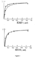
- Polystyrene microtitre plates (Immulon 2, Dynex Technology Inc., Chantilly, VA) were covered for 16 hours at 4°C with 0.5 ⁇ g of peptide A ⁇ 1-40 or A ⁇ 7-42 recently dissolved or aggregated in carbonate/bicarbonate buffer pH 9.6. After blocking with Superblock (Pierce Chemical Co.), increasing concentrations of purified EM5 (0-0.5 nM in TBS-T, 100 microliters per well) were added to the wells covered with A ⁇ , incubating for 3 hours at 37°C. The bound EM5 was detected with the F(ab')2 fragment of goat anti-mouse IgG conjugated with horseradish peroxidase (1:3000, Amersham).
- Fig. 1 shows saturation curves corresponding to binding, in ELISA, of EM5 and 6E10 monoclonal antibodies to recently dissolved and aggregated A ⁇ peptides. High affinity was observed in all cases, with apparent dissociation constants in the picomolar range. None of these antibodies displayed differential affinity for recently prepared or aggregated forms of A ⁇ . Interestingly, whereas 6E10 displayed equivalent affinity for A ⁇ 1-40 and A ⁇ 1-42 , EM5 displayed greater affinity for A ⁇ 42 than for A ⁇ 40 (p ⁇ 0.01) (13.7 pM and 15.5 pM for recently prepared and aggregated A ⁇ 42, respectively; and 37.5 pM and 37.0 pM for recently prepared and aggregated A ⁇ 40, respectively).
- a ⁇ 1-40 and A ⁇ 1-42 by EM5 was investigated and compared with EM2 and EM3 polyclonal antibodies by means of immunotransfer analysis.
- a ⁇ peptides (0.5 ⁇ g/lane) were submitted to PAGE electrophoresis in 16% acrylamide, tris-tricine-SDS.
- the peptides were transferred electrophoretically for 1 hour at 400 mA and 4°C to poly(vinylidene fluoride) membranes (Immobilon-P, Millipore) using 3-cyclohexylamino-1-propanesulfonic acid, pH 11, containing 10% methanol.
- the membranes were blocked for 16 h at 4°C with TBS-T containing 5% powdered skimmed milk and were then incubated for 1 hour at room temperature with 2 ⁇ g/ml of IgGs EM2, EM3 or EM5.
- IgGs EM2, EM3 or EM5 A goat anti-rabbit IgG (EM2 and EM3) or goat anti-mouse IgG (EM5) coupled to horseradish peroxidase (Amersham) and diluted 1:2000 was used as the second antibody. Immunotransfer was visualized by means of chemoluminescence (Amersham) following the manufacturer's specifications.
- Peptides A ⁇ 1-16 , A ⁇ 1-28 and A ⁇ 25-35 were obtained from Sigma (San Luis, MO); peptides A ⁇ 1-42 , A ⁇ 1-42 (E22Q), A ⁇ 1-40 , A ⁇ 1-40 (E22G), A ⁇ 1-40 (E22Q), A ⁇ 1-28 (rodent), A ⁇ 1-28 (E22Q), A ⁇ 17-40 , A ⁇ 16-42 and A ⁇ 25-35 were synthesized in the WM Keck plant of Yale University using the N-t-butyloxycarbonyl methodology; A ⁇ 21-28 , A ⁇ 21-28 (E22Q), A ⁇ 37-41 and A ⁇ 37-40 were synthesized in the Peptide Synthesis Unit (National Biotechnology Center, Madrid) using ordinary Fmoc methodology.
- peptides A ⁇ 37-42 and A ⁇ 37-49 included a tail at the amino end with a cysteine residue for coupling which had the sequence CSGGSGGG (SEQ ID NO:4). All the peptides were purified by high-performance liquid chromatography in inverted-phase mode and their purity was evaluated by MALDI-TOF mass spectrometry.
- Anti-mouse IgG coupled to peroxidase Sigma, San Luis, MO
- diluted 1:2000 was then applied for 30 minutes at 37°C.
- the reaction was developed with TMB (BioRad, CA), stopped with 2M sulfuric acid and quantified at 450 nm. Nonspecific binding was determined, omitting the first antibodies.
- Fig. 3 shows a bar chart corresponding to the absorbance values obtained corresponding to incubation of 20 ⁇ g/ml of the antibody with each of the peptides.
- EM5 was shown to bind to any peptide that contained residues 12 to 16 of the sequence of human A ⁇ and was unable to recognize peptides lacking this region.
- mutant variants with modifications outside of this region displayed similar binding when compared with the wild-type peptide, whereas the A ⁇ 1-28 (rodent) peptide, which shows three changes of amino acids in positions 5, 10 and 13, was not recognized by EM5.
- EM5 monoclonal antibody For immobilizing the EM5 monoclonal antibody on magnetic particles, a 5 ⁇ l aliquot (1.1 mg/ml) of EM5 was incubated with 50 ⁇ l of Dynabeads M450 coated with goat anti-mouse IgG at room temperature for 2 hours. The complex was washed 4 times with PBS with bidirectional stirring (in a rotor).
- ⁇ -amyloid peptide digested with trypsin (or ⁇ -chymotrypsin) with 50 ⁇ l of EM5 immobilized on magnetic particles was incubated in a rotor for 1 hour at 37°C.
- the magnetic-immunoprecipitate complex was washed 4 times with PBS in the same rotor and the supernatants were aspirated and discarded.
- the immunoprecipitated peptides contained in the magnetic complexes were released with 20 ⁇ l of 50% acetonitrile/0.3% trifluoroacetic acid. 5 ⁇ l of this solution was mixed with 5 ⁇ l of saturated ⁇ -cyano-4-hydroxycinnamic acid in 0.1% trifluoroacetic acid / acetonitrile (2:1). A volume of 0.5 ⁇ l of this solution was then placed in a pointed stainless steel probe and was left to dry at room temperature.
- the samples were measured in a MALDI-TOF mass spectrometer Reflex II from Bruker equipped with an ion source with visualization optics and an N 2 laser (337 nm).
- the mass spectra were recorded in positive linear mode at an accelerating voltage of 28.5 kV and 1.5 kV in the linear detector, accumulating 200 laser single-shot spectra below the irradiance threshold. Only mass signals with good resolution, of high intensity, emitted from 3-5 selected points of incidence, were taken into account. All the MALDI spectra were calibrated externally using a standardized mixture of peptides [angiotensin II (1047.2), 18-39 fragment of adrenocorticotropic hormone 2466.7) and insulin (5734.6); Sigma].
- Table I The characteristics of the peptides analyzed, as well as the data obtained, are summarized in the following Table I: Table I.- MALDI-TOF mass spectrometry data for fragments obtained by digestion of ⁇ -amyloid peptide Fragment N° Amino acids included Enzyme used Experimental mass (m/z) Expected value (m/z) 1 1-16 Trypsin 1956.57 1955.0 2 1-17 ⁇ -Chymotrypsin 2067.37 2068.2 3 5-17 ⁇ -Chymotrypsin 1606.62 1605.7 4 6-16 Trypsin 1337.12 1336.4 5 11-17 ⁇ -Chymotrypsin 888.66 890.0
- the brain tissue was supplied by the Tissue Bank for Neurological Investigations, Madrid. Six subjects, 3 men and 3 women, with well-defined AD were included in the study. Their ages ranged from 68 to 75 years. In all cases the diagnosis of AD was based on the clinical-pathological guidelines of CERAD ( Consortium to Establish a Registry for Alzheimer's Disease ) [10]. None of the patients had other relevant neuropathologic findings, for example Parkinson's disease or significant vascular changes. All the brains were processed for histological examination following the protocols for cutting, fixation and embedding of the Brain Bank. The periods post-mortem varied between 10 and 18 hours.
- tissue sections corresponding to the parieto-occipital lateral cortex, temporal lateral cortex (these last two areas as recommended by the CERAD guidelines [10]), hippocampus, caudatum - putamen (at the head of the caudate nucleus), and cortex of the cerebellar hemisphere, were obtained from the original paraffin blocks for immunohistochemical examination.
- modified methenamine - silver staining was carried out in sections consecutive to those processed for the amyloid immunohistochemistry.
- the primary antibodies employed were EM2, EM3, and EM5. Both EM2 and EM3 were used in an earlier study [24]. The sections were incubated with primary antibodies at room temperature for 30 minutes.
- the polyclonal antibodies were detected with the Envision system (Dako Laboratories) with alkaline phosphatase (APh) using nitroblue tetrazolium (NBT) as chromogen, and EM5 was detected either with the aforementioned system or with the Envision system (Dako Laboratories) with horseradish peroxidase (HRP) using diaminobenzidine (DAB) as chromogen.
- HRP horseradish peroxidase
- DAB diaminobenzidine
- Double immunostaining techniques were employed, using EM5 as first primary antibody and EM2 or EM3 as second primary antibody, as a preliminary operation for the purpose of establishing the degree of colocalization of each pair of antibodies, together with the optimum working dilution of each of them.
- EM5 as first primary antibody
- EM2 or EM3 as second primary antibody
- EM3 did not react with any diffuse plaques, neither in the cortical region nor in the subcortical region (Fig. (3E)).
- Double immunostaining with EM5 and EM3 reveals the colocalization of both antibodies in some neuritic plaques, but not in diffuse plaques ( Fig. (5C )), although some colocalization was found at this level when EM5 was incubated at a very low dilution (1:50). Only in one case, which displayed abundant EM3-positive diffuse plaques in sections of the corpus striatum, some of them were stained very slightly with EM5 at the working dilution (1:500). This same case did not show any reactivity of the diffuse plaques as EM5 in the cortex of the cerebellum.
- the panel of antibodies detected heterogeneity among the neuritic plaques, which proves relevant for indicating actual stages in the process of development of the diffuse plaques and their transformation to neuritic plaques.
- the EM5 antibody gave intense staining of all the structures (neuritic plaques and vessel walls) stained by EM2 and stained to a variable degree by EM3.
- a subset of neuritic plaques was found to have a pattern of staining identical to the vascular amyloid, with high colocalization of reactivity to EM2 and EM5.
- the invention shows that EM5 ought to react with all the structures that contain either A ⁇ ⁇ 11-40 or A ⁇ ⁇ 11-42 .
- the ELISA results presented here show that the EM5 antibody recognizes both recently dissolved and aggregated forms of the A ⁇ peptide.
- tissue sections we found greater variability of staining between the neuritic plaques with EM5 than with the polyclonal antibodies, together with high colocalization of intense reactivity to EM5 with EM2 (A ⁇ C40).
- Relatively more intense staining in these positive plaques may reveal a subgroup of neuritic plaques either with a particularly high content of long A ⁇ peptides, or selectively high content of A ⁇ C40, or even particular accessibility of the epitope recognized by EM5 in structures (vessels or plaques) that show codeposition of A ⁇ ⁇ 11-40 and A ⁇ ⁇ 11-42 .
- the antibody of the invention seems to detect the same subset of neuritic plaques A ⁇ C40 (+) detected previously by Parvathy et al. [22].
- This subset of neuritic plaques with particularly high contents of long A ⁇ peptides may represent important milestones in the progression of the amyloid lesions in AD that the monoclonal antibody of the invention is able to detect. Therefore, the use of EM5 can permit subsets of plaques to be defined that constitute a specific marker of the stage of progression of the disease.
- Example 6 Capacity of the antibodies for reacting with forms of ⁇ -amyloid peptide in urine
- the magnetic particles coupled to the EM5 antibody were prepared following the method described by Fuentes et al. [26], based on gentle oxidation of the glycoside residues of the immunoglobulins to generate aldehyde groups, which are reacted with magnetic particles, on the surface of which amino groups have been generated by modification with ethylenediamine. Briefly, oxidation of the EM5 antibody was induced by incubation with sodium periodate 10 mM for 2 hours, after which the oxidized antibody was dialysed in distilled water at 4°C.
- EM/100-30 magnetic particles (Merck Co, France), which have carboxyl groups on their surface, were modified by incubation, at a concentration of 10 mg/ml, with 1M ethylenediamine pH 4.75, for 90 minutes, after which solid EDCI (1-ethyl-3-(3'-dimethylaminopropyl)carbodiimide hydrochloride) was added to a final concentration of 10 mM and was left to react for 90 minutes, before washing profusely with distilled water.
- the antibody was immobilized on the magnetic particles after adding 10 mg of the oxidized EM5 antibody, dissolved in sodium phosphate buffer 150 mM of pH 7.5, to 2 ml of magnetic particles (10 mg/ml) with amino groups on their surface at 4°C and incubating overnight.
- the Schiff bases formed and the unreacted aldehyde groups were reduced by adding sodium borohydride until a concentration of 1 mg/ml was reached, at pH 8.5 and 4°C.
- the preparation was washed with copious amounts of distilled water.
- the amount of antibody immobilized was determined by quantifying the difference in concentration of proteins in the supernatant before and after immobilization, using Bradford's method [27].
- Fig. 6 Mass spectrometry analysis of the mixtures of peptides separated from the solution is shown in Fig. 6 , in which part A corresponds to analysis of the solution without prior treatment with antibodies (Ctrl.) and part B corresponds to the use of the antibody bound to magnetic particles (EM5+PM).
- the antibody is capable of binding to forms of ⁇ -Amyloid peptide in solution and of forming complexes with them, so that they can be separated from said solution.
- Synthetic peptides that corresponded to the forms of ⁇ -amyloid peptide A ⁇ 12-28 , A ⁇ 1-40 and A ⁇ 1-42 were mixed together in distilled water to give a mixture with final concentrations of 0.44 ⁇ g/ ⁇ l of A ⁇ 12-28 and A ⁇ 1-42 and 0.11 ⁇ g/ ⁇ l of A ⁇ 1-40 . 4 ⁇ l of this mixture of peptides was added to 981 ⁇ l of urine.
- the final concentration of forms of ⁇ -Amyloid peptide in urine was 1.76 ⁇ g/ml in the case of A ⁇ 12-18 and of A ⁇ 1-42 and 0.44 ⁇ g/ml in the case of A ⁇ 1-40 .
- the tube was placed in a magnetized separator of magnetic particles and the urine was extracted carefully using a pipette.
- the peptides bound to them were separated from the magnetic particles with 12 ⁇ l of a solution of a matrix of ⁇ -cyano-4-hydroxycinnamic acid in 30% (v/v) of aqueous acetonitrile containing 0.1% (v/v) of trifluoroacetic acid (TFA) and were analyzed by MALDI-TOF mass spectrometry.
- 1.5 ⁇ l of the sample mixture resulting from immunoprecipitation in the matrix of ⁇ -cyano-4-hydroxycinnamic acid was placed in a stainless steel probe with capacity for 100 samples and left to dry at room temperature for 5 minutes.
- the samples were measured in a workstation for MALDI-TOF mass spectrometry Voyager DE-PRO from PE Biosystems using the default configuration of the instrument.
- the mass spectra were recorded in positive reflector mode at an accelerating voltage of 20 kV and a collector voltage of 75%, 0.002% of guide filament and 150 nanoseconds of lag time, accumulating 200 spectra of individual laser firings below the threshold irradiation. Only mass signals with good resolution, of high intensity, from 3-5 selected points of incidence, were considered.
- the equipment was calibrated externally using calibration mixture 2, supplied by Applied Biosystems (Tres Cantos, Madrid, Spain), composed of angiotensin (1297 Da), ACTH 1-17 (2094 Da), ACTH 18-39 (2466 Da), ACTH 7-38 (3660 Da) and bovine insulin (2867 Da).
- calibration mixture 2 supplied by Applied Biosystems (Tres Cantos, Madrid, Spain), composed of angiotensin (1297 Da), ACTH 1-17 (2094 Da), ACTH 18-39 (2466 Da), ACTH 7-38 (3660 Da) and bovine insulin (2867 Da).
- Fig. 7 shows the graph obtained with one of the samples, which is representative of the others. Peaks can be seen that correspond to the peptides added to the urine sample, which demonstrates the capacity of the antibody of the invention for binding to them in urine samples. As the analysis had been carried out with a sample of a biological fluid, other peaks can also be seen, corresponding to other molecules naturally present in the sample and which also became bound to the antibody coupled to magnetic particles.
- the hybridoma that produces the EM5 antibody was deposited in the European Collection of Cell Cultures (ECACC), CAMR, Salisbury, Wiltshire, United Kingdom. The date of deposition and the access number are as follows: Designation of the hybridoma Date of deposition Access No. EM5 clone A 03.01.2006 06030101
- these hybridoma cells were obtained by fusion of two types of cells: a) BALB/c mouse spleen lymphocytes, obtained after immunization of the mice using, as immunogen, the form of ⁇ -amyloid peptide designated A ⁇ 1-40 , comprising amino acids 1 to 40 of said peptide, coupled to KLH (keyhole limpet heamocyanin); b) cells of the mouse myeloma line P3/X63-Ag653, which acted as the immortal part in the fusion.
- EM5 clone A which produces the monoclonal antibody designated "EM5", a type IgG1 antibody capable of specifically recognizing the antigen used for immunization, the peptide A ⁇ 1-40 , as verified by antibody capture tests of the ELISA type.
- This clone was grown in RPMI 1640 culture medium with 10% fetal calf serum, 10% DMSO, glutamine 2 mM and sodium pyruvate 1 mM, at 37°C and in an atmosphere with 5% CO 2 , conditions in which 95% of the cells grew in suspension and the remaining 5% adhered to the culture vessel.
- the cells were cloned twice by means of limiting dilutions, after which aliquots of 4x10 6 cells were taken, and were placed in vials. After controlling for absence of bacteria, absence of mycoplasmas and absence of fungi, several of these vials were sent to the European Collection of Cell Cultures (ECACC), requesting permission for their deposition.
- ECACC European Collection of Cell Cultures
Landscapes
- Health & Medical Sciences (AREA)
- Chemical & Material Sciences (AREA)
- Life Sciences & Earth Sciences (AREA)
- Engineering & Computer Science (AREA)
- Organic Chemistry (AREA)
- Molecular Biology (AREA)
- Biomedical Technology (AREA)
- Immunology (AREA)
- General Health & Medical Sciences (AREA)
- Medicinal Chemistry (AREA)
- Biochemistry (AREA)
- Proteomics, Peptides & Aminoacids (AREA)
- Biophysics (AREA)
- Urology & Nephrology (AREA)
- Genetics & Genomics (AREA)
- Hematology (AREA)
- Neurology (AREA)
- Neurosurgery (AREA)
- Cell Biology (AREA)
- Biotechnology (AREA)
- Microbiology (AREA)
- Food Science & Technology (AREA)
- Physics & Mathematics (AREA)
- Analytical Chemistry (AREA)
- General Physics & Mathematics (AREA)
- Pathology (AREA)
- Toxicology (AREA)
- Gastroenterology & Hepatology (AREA)
- Zoology (AREA)
- Bioinformatics & Cheminformatics (AREA)
- Chemical Kinetics & Catalysis (AREA)
- Animal Behavior & Ethology (AREA)
- Pharmacology & Pharmacy (AREA)
- Nuclear Medicine, Radiotherapy & Molecular Imaging (AREA)
- General Chemical & Material Sciences (AREA)
- Public Health (AREA)
- Psychiatry (AREA)
- Hospice & Palliative Care (AREA)
- Veterinary Medicine (AREA)
- Peptides Or Proteins (AREA)
Applications Claiming Priority (2)
| Application Number | Priority Date | Filing Date | Title |
|---|---|---|---|
| ES200500550A ES2259270B1 (es) | 2005-03-09 | 2005-03-09 | Metodo de diagnostico in vitro de la enfermedad de alzheimer mediante un anticuerpo monoclonal. |
| PCT/ES2006/070027 WO2006095041A1 (es) | 2005-03-09 | 2006-03-09 | Método de diagnóstico in vitro de la enfermedad de alzheimer mediante un anticuerpo monoclonal |
Publications (4)
| Publication Number | Publication Date |
|---|---|
| EP1881008A1 EP1881008A1 (en) | 2008-01-23 |
| EP1881008A9 EP1881008A9 (en) | 2009-10-14 |
| EP1881008A4 EP1881008A4 (en) | 2009-11-11 |
| EP1881008B1 true EP1881008B1 (en) | 2011-06-15 |
Family
ID=36952969
Family Applications (1)
| Application Number | Title | Priority Date | Filing Date |
|---|---|---|---|
| EP06725836A Not-in-force EP1881008B1 (en) | 2005-03-09 | 2006-03-09 | Method for the in vitro diagnosis of alzheimer's disease using a monoclonal antibody |
Country Status (11)
| Country | Link |
|---|---|
| US (1) | US7932048B2 (zh) |
| EP (1) | EP1881008B1 (zh) |
| JP (1) | JP5117373B2 (zh) |
| CN (1) | CN101137670B (zh) |
| AT (1) | ATE512987T1 (zh) |
| BR (1) | BRPI0609168A2 (zh) |
| CA (1) | CA2601550C (zh) |
| ES (2) | ES2259270B1 (zh) |
| MX (1) | MX2007010934A (zh) |
| RU (1) | RU2416619C2 (zh) |
| WO (1) | WO2006095041A1 (zh) |
Cited By (1)
| Publication number | Priority date | Publication date | Assignee | Title |
|---|---|---|---|---|
| DE112010000814B4 (de) | 2009-02-10 | 2021-09-09 | Hitachi High-Tech Corporation | Immunanalytisches Verfahren und immunanalytisches System mit Verwendung der Massenspektrometertechnologie |
Families Citing this family (46)
| Publication number | Priority date | Publication date | Assignee | Title |
|---|---|---|---|---|
| DE10303974A1 (de) | 2003-01-31 | 2004-08-05 | Abbott Gmbh & Co. Kg | Amyloid-β(1-42)-Oligomere, Verfahren zu deren Herstellung und deren Verwendung |
| US7732162B2 (en) | 2003-05-05 | 2010-06-08 | Probiodrug Ag | Inhibitors of glutaminyl cyclase for treating neurodegenerative diseases |
| RU2442793C2 (ru) | 2005-11-30 | 2012-02-20 | Эбботт Лэборетриз | АНТИТЕЛА ПРОТИВ ГЛОБУЛОМЕРА Аβ, ИХ АНТИГЕНСВЯЗЫВАЮЩИЕ ЧАСТИ, СООТВЕТСТВУЮЩИЕ ГИБРИДОМЫ, НУКЛЕИНОВЫЕ КИСЛОТЫ, ВЕКТОРЫ, КЛЕТКИ-ХОЗЯЕВА, СПОСОБЫ ПОЛУЧЕНИЯ УКАЗАННЫХ АНТИТЕЛ, КОМПОЗИЦИИ, СОДЕРЖАЩИЕ УКАЗАННЫЕ АНТИТЕЛА, ПРИМЕНЕНИЯ УКАЗАННЫХ АНТИТЕЛ И СПОСОБЫ ИСПОЛЬЗОВАНИЯ УКАЗАННЫХ АНТИТЕЛ |
| KR20180058863A (ko) | 2005-11-30 | 2018-06-01 | 애브비 인코포레이티드 | 아밀로이드 베타 단백질에 대한 모노클로날 항체 및 이의 용도 |
| EP2808032B1 (en) * | 2005-12-12 | 2018-08-01 | AC Immune S.A. | A beta 1-42 specific monoclonal antibodies with therapeutic properties |
| CL2007002070A1 (es) | 2006-07-14 | 2008-02-08 | Ac Immune S A Genentech Inc | Anticuerpo quimerico o humanizado, o fragmentos de ellos, que se adhieren especificamente a por lo menos un epitopo en la proteina beta-amiloide; molecula de acido nucleico que lo codifica; composicion que lo comprende; su uso para tratar enfermedade |
| US8455626B2 (en) | 2006-11-30 | 2013-06-04 | Abbott Laboratories | Aβ conformer selective anti-aβ globulomer monoclonal antibodies |
| WO2008104386A2 (en) * | 2007-02-27 | 2008-09-04 | Abbott Gmbh & Co. Kg | Method for the treatment of amyloidoses |
| CN101668525A (zh) | 2007-03-01 | 2010-03-10 | 前体生物药物股份公司 | 谷氨酰胺酰环化酶抑制剂的新用途 |
| EP2865670B1 (en) | 2007-04-18 | 2017-01-11 | Probiodrug AG | Thiourea derivatives as glutaminyl cyclase inhibitors |
| BRPI0812478A2 (pt) * | 2007-06-12 | 2017-05-16 | Ac Immune Sa | anticorpo quimérico ou um fragmento do mesmo, ou anticorpo humanizado ou um fragmento do mesmo, molécula de ácido nucleico, vetor de expressão, célula, composição, métodos de prevenir, tratar ou aliviar os efeitos d euma ou mais doenças, de diagnosticar uma doença ou condição associada com amilóide em um indivíduo, de determinar a extensão de carga de placa amiloidogênica em um tecido e/ou fluidos corporais de um indivíduo, de desagregar fibras beta-amilóides pré-formadas e de prevenir degradação de neurônios induzidas por abeta, kit de teste, região variável de cadeia, e, linhagem de célula |
| US8613923B2 (en) | 2007-06-12 | 2013-12-24 | Ac Immune S.A. | Monoclonal antibody |
| US8048420B2 (en) * | 2007-06-12 | 2011-11-01 | Ac Immune S.A. | Monoclonal antibody |
| CN101998863B (zh) * | 2007-10-05 | 2015-09-16 | 基因技术公司 | 抗淀粉状蛋白β抗体在眼病中的用途 |
| AU2008311367B2 (en) * | 2007-10-05 | 2014-11-13 | Ac Immune S.A. | Use of anti-amyloid beta antibody in ocular diseases |
| UA101167C2 (ru) * | 2007-10-05 | 2013-03-11 | Дженентек, Инк. | Фармацевтическая композиция, предназначенная для лечения глазной болезни |
| US20110104821A1 (en) * | 2008-05-08 | 2011-05-05 | Takahiko Tokuda | ABeta-OLIGOMER MEASUREMENT METHOD |
| EP2475428B1 (en) | 2009-09-11 | 2015-07-01 | Probiodrug AG | Heterocylcic derivatives as inhibitors of glutaminyl cyclase |
| DE102009054057A1 (de) * | 2009-11-20 | 2011-05-26 | Charité - Universitätsmedizin Berlin (Charité) | Screening-Verfahren für Wirkstoffe für die Prophylaxe und Therapie neurodegenerativer Erkrankungen |
| US9181233B2 (en) | 2010-03-03 | 2015-11-10 | Probiodrug Ag | Inhibitors of glutaminyl cyclase |
| AU2011226074B2 (en) | 2010-03-10 | 2015-01-22 | Vivoryon Therapeutics N.V. | Heterocyclic inhibitors of glutaminyl cyclase (QC, EC 2.3.2.5) |
| ES2684475T3 (es) | 2010-04-15 | 2018-10-03 | Abbvie Inc. | Proteínas que se unen a beta amiloide |
| WO2011131748A2 (en) | 2010-04-21 | 2011-10-27 | Probiodrug Ag | Novel inhibitors |
| WO2012016173A2 (en) | 2010-07-30 | 2012-02-02 | Ac Immune S.A. | Safe and functional humanized antibodies |
| US9062101B2 (en) | 2010-08-14 | 2015-06-23 | AbbVie Deutschland GmbH & Co. KG | Amyloid-beta binding proteins |
| DK2686313T3 (en) | 2011-03-16 | 2016-05-02 | Probiodrug Ag | Benzimidazole derivatives as inhibitors of glutaminyl cyclase |
| KR102020072B1 (ko) | 2011-03-16 | 2019-11-04 | 프로비오드룩 아게 | 진단 항체 시험 |
| EP2511296A1 (en) * | 2011-04-12 | 2012-10-17 | Araclón Biotech, S. L. | Antibody, kit and method for determination of amyloid peptides |
| ES2495266B8 (es) | 2013-02-13 | 2015-11-12 | Consejo Superior De Investigaciones Científicas (Csic) | Uso de igf-1 como reactivo de diagnóstico y/o pronóstico precoz de la enfermedad de alzheimer |
| MA38632B1 (fr) | 2013-05-20 | 2019-10-31 | Genentech Inc | Anticorps anti-récepteur de transferrine et procédés d'utilisation |
| EP3149040A1 (en) * | 2014-05-29 | 2017-04-05 | Spring Bioscience Corporation | Anti-b7-h3 antibodies and diagnostic uses thereof |
| CN107250158B (zh) | 2014-11-19 | 2022-03-25 | 基因泰克公司 | 抗转铁蛋白受体/抗bace1多特异性抗体和使用方法 |
| EP3221362B1 (en) | 2014-11-19 | 2019-07-24 | F.Hoffmann-La Roche Ag | Anti-transferrin receptor antibodies and methods of use |
| RU2017120039A (ru) | 2014-12-10 | 2019-01-10 | Дженентек, Инк. | Антитела к рецепторам гематоэнцефалического барьера и способы их применения |
| CN108350053A (zh) | 2015-11-09 | 2018-07-31 | 英属哥伦比亚大学 | 淀粉样蛋白β表位及其抗体 |
| JP7448174B2 (ja) | 2015-11-09 | 2024-03-12 | ザ・ユニバーシティ・オブ・ブリティッシュ・コロンビア | アミロイドベータ中間領域エピトープおよびそれに対する立体配座選択的抗体 |
| KR20180088828A (ko) | 2015-11-09 | 2018-08-07 | 더 유니버시티 오브 브리티쉬 콜롬비아 | 아밀로이드 베타에서의 n-말단 에피토프 및 이에 형태적으로-선택적인 항체 |
| US20180125920A1 (en) | 2016-11-09 | 2018-05-10 | The University Of British Columbia | Methods for preventing and treating A-beta oligomer-associated and/or -induced diseases and conditions |
| US11397188B2 (en) * | 2017-03-30 | 2022-07-26 | Board Of Regents, The University Of Texas System | Method of detecting an APP Alzheimer's disease marker peptide in patients with Alzheimer's disease |
| WO2018183703A1 (en) * | 2017-03-31 | 2018-10-04 | NeuroDiagnostics LLC | Lymphocyte-based morphometric test for alzheimer's disease |
| DK3461819T3 (da) | 2017-09-29 | 2020-08-10 | Probiodrug Ag | Inhibitorer af glutaminylcyklase |
| US11781183B2 (en) * | 2018-03-13 | 2023-10-10 | Yissum Research Development Company Of The Hebrew University Of Jerusalem Ltd. | Diagnostic use of cell free DNA chromatin immunoprecipitation |
| CN113226283A (zh) | 2018-10-04 | 2021-08-06 | 罗切斯特大学 | 通过操纵血浆重量摩尔渗透压浓度改善淋巴递送 |
| WO2020132230A2 (en) | 2018-12-20 | 2020-06-25 | Genentech, Inc. | Modified antibody fcs and methods of use |
| TW202300517A (zh) | 2021-03-12 | 2023-01-01 | 美商美國禮來大藥廠 | 抗類澱粉β抗體及其用途 |
| WO2022251048A1 (en) | 2021-05-24 | 2022-12-01 | Eli Lilly And Company | Anti-amyloid beta antibodies and uses thereof |
Family Cites Families (7)
| Publication number | Priority date | Publication date | Assignee | Title |
|---|---|---|---|---|
| US4666829A (en) * | 1985-05-15 | 1987-05-19 | University Of California | Polypeptide marker for Alzheimer's disease and its use for diagnosis |
| AU5439790A (en) * | 1989-04-14 | 1990-11-16 | Research Foundation For Mental Hygiene, Inc. | Cerebrovascular amyloid protein-specific monoclonal antibody sv17-6e10 |
| US5958883A (en) * | 1992-09-23 | 1999-09-28 | Board Of Regents Of The University Of Washington Office Of Technology | Animal models of human amyloidoses |
| DE69432629T3 (de) * | 1993-01-25 | 2008-01-17 | Takeda Pharmaceutical Co. Ltd. | Antikörper gegen beta-amyloid oder derivative davon und seine verwendung |
| US5688651A (en) * | 1994-12-16 | 1997-11-18 | Ramot University Authority For Applied Research And Development Ltd. | Prevention of protein aggregation |
| EP0866805A1 (en) * | 1995-12-12 | 1998-09-30 | Karolinska Innovations AB | PEPTIDE BINDING THE KLVFF-SEQUENCE OF AMYLOID $g(b) |
| WO2004029629A1 (en) * | 2002-09-27 | 2004-04-08 | Janssen Pharmaceutica N.V. | N-11 truncated amyloid-beta nomoclonal antibodies, compositions, methods and uses |
-
2005
- 2005-03-09 ES ES200500550A patent/ES2259270B1/es not_active Expired - Fee Related
-
2006
- 2006-03-09 CA CA2601550A patent/CA2601550C/en not_active Expired - Fee Related
- 2006-03-09 AT AT06725836T patent/ATE512987T1/de not_active IP Right Cessation
- 2006-03-09 JP JP2008500220A patent/JP5117373B2/ja not_active Expired - Fee Related
- 2006-03-09 US US11/886,022 patent/US7932048B2/en not_active Expired - Fee Related
- 2006-03-09 CN CN2006800076517A patent/CN101137670B/zh not_active Expired - Fee Related
- 2006-03-09 EP EP06725836A patent/EP1881008B1/en not_active Not-in-force
- 2006-03-09 MX MX2007010934A patent/MX2007010934A/es active IP Right Grant
- 2006-03-09 RU RU2007137125/10A patent/RU2416619C2/ru not_active IP Right Cessation
- 2006-03-09 WO PCT/ES2006/070027 patent/WO2006095041A1/es active Application Filing
- 2006-03-09 BR BRPI0609168-7A patent/BRPI0609168A2/pt not_active Application Discontinuation
- 2006-03-09 ES ES06725836T patent/ES2367837T3/es active Active
Non-Patent Citations (1)
| Title |
|---|
| Jiménez-Huete, A. et al (1998) Alzheimer's Reports 1:41-48 * |
Cited By (1)
| Publication number | Priority date | Publication date | Assignee | Title |
|---|---|---|---|---|
| DE112010000814B4 (de) | 2009-02-10 | 2021-09-09 | Hitachi High-Tech Corporation | Immunanalytisches Verfahren und immunanalytisches System mit Verwendung der Massenspektrometertechnologie |
Also Published As
| Publication number | Publication date |
|---|---|
| RU2416619C2 (ru) | 2011-04-20 |
| CN101137670A (zh) | 2008-03-05 |
| EP1881008A1 (en) | 2008-01-23 |
| WO2006095041A1 (es) | 2006-09-14 |
| BRPI0609168A2 (pt) | 2010-02-23 |
| JP2008532984A (ja) | 2008-08-21 |
| CA2601550C (en) | 2014-11-18 |
| JP5117373B2 (ja) | 2013-01-16 |
| MX2007010934A (es) | 2007-10-12 |
| RU2007137125A (ru) | 2009-04-20 |
| ES2259270B1 (es) | 2007-11-01 |
| EP1881008A9 (en) | 2009-10-14 |
| CN101137670B (zh) | 2012-08-08 |
| US20090023159A1 (en) | 2009-01-22 |
| ATE512987T1 (de) | 2011-07-15 |
| ES2259270A1 (es) | 2006-09-16 |
| ES2367837T3 (es) | 2011-11-08 |
| CA2601550A1 (en) | 2006-09-14 |
| US7932048B2 (en) | 2011-04-26 |
| EP1881008A4 (en) | 2009-11-11 |
Similar Documents
| Publication | Publication Date | Title |
|---|---|---|
| EP1881008B1 (en) | Method for the in vitro diagnosis of alzheimer's disease using a monoclonal antibody | |
| DK1546734T3 (en) | MONOCLONAL ANTIBODIES AGAINST N-11-TRUNCATED AMYLOID BETA, COMPOSITIONS, PROCEDURES AND APPLICATIONS | |
| JP3909084B2 (ja) | 微小管結合タンパク質タウに対するモノクローナル抗体 | |
| AU2005254928B2 (en) | Antibodies specific for soluble amyloid beta peptide protofibrils and uses thereof | |
| AU705907B2 (en) | Methods for aiding in the diagnosis of alzheimer's disease by measuring amyloid-beta peptide (X- LE 41) and tau | |
| JP2004121251A (ja) | 可溶性β−アミロイド・ペプチドの検出のための方法及び組成物 | |
| US8445649B2 (en) | Antibody and use thereof | |
| WO1996004309A1 (en) | Monoclonal antibodies specific for an epitope of a particular subclass or form of phosphorylated tau, hybridomas secreting them, antigen recognition of these antibodies and their applications | |
| EP3269736B1 (en) | Conformational-specific antibodies against oligomers of amyloid beta | |
| Altay et al. | Development and validation of an expanded antibody toolset that captures alpha-synuclein pathological diversity in Lewy body diseases | |
| JP7151985B2 (ja) | 抗プロパノイル化アミロイドβタンパク質抗体 | |
| Rábano et al. | Diversity of senile plaques in Alzheimer's disease as revealed by a new monoclonal antibody that recognizes an internal sequence of the Aβ peptide | |
| JP2000034300A (ja) | 抗リン酸化タウ蛋白質抗体及びそれを用いるアルツハイマー病の検出方法 | |
| AU2008200489B2 (en) | Methods and compositions for the detection of soluble beta-amyloid peptide |
Legal Events
| Date | Code | Title | Description |
|---|---|---|---|
| PUAI | Public reference made under article 153(3) epc to a published international application that has entered the european phase |
Free format text: ORIGINAL CODE: 0009012 |
|
| 17P | Request for examination filed |
Effective date: 20071009 |
|
| AK | Designated contracting states |
Kind code of ref document: A1 Designated state(s): AT BE BG CH CY CZ DE DK EE ES FI FR GB GR HU IE IS IT LI LT LU LV MC NL PL PT RO SE SI SK TR |
|
| DAX | Request for extension of the european patent (deleted) | ||
| A4 | Supplementary search report drawn up and despatched |
Effective date: 20091013 |
|
| 17Q | First examination report despatched |
Effective date: 20100726 |
|
| GRAP | Despatch of communication of intention to grant a patent |
Free format text: ORIGINAL CODE: EPIDOSNIGR1 |
|
| GRAS | Grant fee paid |
Free format text: ORIGINAL CODE: EPIDOSNIGR3 |
|
| GRAA | (expected) grant |
Free format text: ORIGINAL CODE: 0009210 |
|
| AK | Designated contracting states |
Kind code of ref document: B1 Designated state(s): AT BE BG CH CY CZ DE DK EE ES FI FR GB GR HU IE IS IT LI LT LU LV MC NL PL PT RO SE SI SK TR |
|
| REG | Reference to a national code |
Ref country code: GB Ref legal event code: FG4D Ref country code: CH Ref legal event code: EP |
|
| REG | Reference to a national code |
Ref country code: IE Ref legal event code: FG4D |
|
| REG | Reference to a national code |
Ref country code: DE Ref legal event code: R096 Ref document number: 602006022522 Country of ref document: DE Effective date: 20110728 |
|
| REG | Reference to a national code |
Ref country code: NL Ref legal event code: T3 |
|
| PG25 | Lapsed in a contracting state [announced via postgrant information from national office to epo] |
Ref country code: SE Free format text: LAPSE BECAUSE OF FAILURE TO SUBMIT A TRANSLATION OF THE DESCRIPTION OR TO PAY THE FEE WITHIN THE PRESCRIBED TIME-LIMIT Effective date: 20110615 Ref country code: LT Free format text: LAPSE BECAUSE OF FAILURE TO SUBMIT A TRANSLATION OF THE DESCRIPTION OR TO PAY THE FEE WITHIN THE PRESCRIBED TIME-LIMIT Effective date: 20110615 |
|
| REG | Reference to a national code |
Ref country code: ES Ref legal event code: FG2A Ref document number: 2367837 Country of ref document: ES Kind code of ref document: T3 Effective date: 20111108 |
|
| PG25 | Lapsed in a contracting state [announced via postgrant information from national office to epo] |
Ref country code: GR Free format text: LAPSE BECAUSE OF FAILURE TO SUBMIT A TRANSLATION OF THE DESCRIPTION OR TO PAY THE FEE WITHIN THE PRESCRIBED TIME-LIMIT Effective date: 20110916 Ref country code: LV Free format text: LAPSE BECAUSE OF FAILURE TO SUBMIT A TRANSLATION OF THE DESCRIPTION OR TO PAY THE FEE WITHIN THE PRESCRIBED TIME-LIMIT Effective date: 20110615 Ref country code: CY Free format text: LAPSE BECAUSE OF FAILURE TO SUBMIT A TRANSLATION OF THE DESCRIPTION OR TO PAY THE FEE WITHIN THE PRESCRIBED TIME-LIMIT Effective date: 20110615 Ref country code: FI Free format text: LAPSE BECAUSE OF FAILURE TO SUBMIT A TRANSLATION OF THE DESCRIPTION OR TO PAY THE FEE WITHIN THE PRESCRIBED TIME-LIMIT Effective date: 20110615 Ref country code: SI Free format text: LAPSE BECAUSE OF FAILURE TO SUBMIT A TRANSLATION OF THE DESCRIPTION OR TO PAY THE FEE WITHIN THE PRESCRIBED TIME-LIMIT Effective date: 20110615 Ref country code: AT Free format text: LAPSE BECAUSE OF FAILURE TO SUBMIT A TRANSLATION OF THE DESCRIPTION OR TO PAY THE FEE WITHIN THE PRESCRIBED TIME-LIMIT Effective date: 20110615 |
|
| PG25 | Lapsed in a contracting state [announced via postgrant information from national office to epo] |
Ref country code: BE Free format text: LAPSE BECAUSE OF FAILURE TO SUBMIT A TRANSLATION OF THE DESCRIPTION OR TO PAY THE FEE WITHIN THE PRESCRIBED TIME-LIMIT Effective date: 20110615 |
|
| PG25 | Lapsed in a contracting state [announced via postgrant information from national office to epo] |
Ref country code: EE Free format text: LAPSE BECAUSE OF FAILURE TO SUBMIT A TRANSLATION OF THE DESCRIPTION OR TO PAY THE FEE WITHIN THE PRESCRIBED TIME-LIMIT Effective date: 20110615 Ref country code: IS Free format text: LAPSE BECAUSE OF FAILURE TO SUBMIT A TRANSLATION OF THE DESCRIPTION OR TO PAY THE FEE WITHIN THE PRESCRIBED TIME-LIMIT Effective date: 20111015 Ref country code: CZ Free format text: LAPSE BECAUSE OF FAILURE TO SUBMIT A TRANSLATION OF THE DESCRIPTION OR TO PAY THE FEE WITHIN THE PRESCRIBED TIME-LIMIT Effective date: 20110615 Ref country code: PT Free format text: LAPSE BECAUSE OF FAILURE TO SUBMIT A TRANSLATION OF THE DESCRIPTION OR TO PAY THE FEE WITHIN THE PRESCRIBED TIME-LIMIT Effective date: 20111017 |
|
| PG25 | Lapsed in a contracting state [announced via postgrant information from national office to epo] |
Ref country code: RO Free format text: LAPSE BECAUSE OF FAILURE TO SUBMIT A TRANSLATION OF THE DESCRIPTION OR TO PAY THE FEE WITHIN THE PRESCRIBED TIME-LIMIT Effective date: 20110615 Ref country code: SK Free format text: LAPSE BECAUSE OF FAILURE TO SUBMIT A TRANSLATION OF THE DESCRIPTION OR TO PAY THE FEE WITHIN THE PRESCRIBED TIME-LIMIT Effective date: 20110615 Ref country code: PL Free format text: LAPSE BECAUSE OF FAILURE TO SUBMIT A TRANSLATION OF THE DESCRIPTION OR TO PAY THE FEE WITHIN THE PRESCRIBED TIME-LIMIT Effective date: 20110615 |
|
| PLBE | No opposition filed within time limit |
Free format text: ORIGINAL CODE: 0009261 |
|
| STAA | Information on the status of an ep patent application or granted ep patent |
Free format text: STATUS: NO OPPOSITION FILED WITHIN TIME LIMIT |
|
| 26N | No opposition filed |
Effective date: 20120316 |
|
| PG25 | Lapsed in a contracting state [announced via postgrant information from national office to epo] |
Ref country code: DK Free format text: LAPSE BECAUSE OF FAILURE TO SUBMIT A TRANSLATION OF THE DESCRIPTION OR TO PAY THE FEE WITHIN THE PRESCRIBED TIME-LIMIT Effective date: 20110615 |
|
| REG | Reference to a national code |
Ref country code: DE Ref legal event code: R097 Ref document number: 602006022522 Country of ref document: DE Effective date: 20120316 |
|
| PG25 | Lapsed in a contracting state [announced via postgrant information from national office to epo] |
Ref country code: MC Free format text: LAPSE BECAUSE OF NON-PAYMENT OF DUE FEES Effective date: 20120331 |
|
| REG | Reference to a national code |
Ref country code: CH Ref legal event code: PL |
|
| REG | Reference to a national code |
Ref country code: IE Ref legal event code: MM4A |
|
| PG25 | Lapsed in a contracting state [announced via postgrant information from national office to epo] |
Ref country code: IE Free format text: LAPSE BECAUSE OF NON-PAYMENT OF DUE FEES Effective date: 20120309 Ref country code: CH Free format text: LAPSE BECAUSE OF NON-PAYMENT OF DUE FEES Effective date: 20120331 Ref country code: LI Free format text: LAPSE BECAUSE OF NON-PAYMENT OF DUE FEES Effective date: 20120331 |
|
| PG25 | Lapsed in a contracting state [announced via postgrant information from national office to epo] |
Ref country code: BG Free format text: LAPSE BECAUSE OF FAILURE TO SUBMIT A TRANSLATION OF THE DESCRIPTION OR TO PAY THE FEE WITHIN THE PRESCRIBED TIME-LIMIT Effective date: 20110915 |
|
| PG25 | Lapsed in a contracting state [announced via postgrant information from national office to epo] |
Ref country code: TR Free format text: LAPSE BECAUSE OF FAILURE TO SUBMIT A TRANSLATION OF THE DESCRIPTION OR TO PAY THE FEE WITHIN THE PRESCRIBED TIME-LIMIT Effective date: 20110615 |
|
| PG25 | Lapsed in a contracting state [announced via postgrant information from national office to epo] |
Ref country code: LU Free format text: LAPSE BECAUSE OF NON-PAYMENT OF DUE FEES Effective date: 20120309 |
|
| PG25 | Lapsed in a contracting state [announced via postgrant information from national office to epo] |
Ref country code: HU Free format text: LAPSE BECAUSE OF FAILURE TO SUBMIT A TRANSLATION OF THE DESCRIPTION OR TO PAY THE FEE WITHIN THE PRESCRIBED TIME-LIMIT Effective date: 20060309 |
|
| REG | Reference to a national code |
Ref country code: FR Ref legal event code: PLFP Year of fee payment: 11 |
|
| REG | Reference to a national code |
Ref country code: FR Ref legal event code: PLFP Year of fee payment: 12 |
|
| REG | Reference to a national code |
Ref country code: FR Ref legal event code: PLFP Year of fee payment: 13 |
|
| PGFP | Annual fee paid to national office [announced via postgrant information from national office to epo] |
Ref country code: NL Payment date: 20180321 Year of fee payment: 13 Ref country code: GB Payment date: 20180321 Year of fee payment: 13 Ref country code: DE Payment date: 20180322 Year of fee payment: 13 |
|
| PGFP | Annual fee paid to national office [announced via postgrant information from national office to epo] |
Ref country code: FR Payment date: 20180323 Year of fee payment: 13 |
|
| PGFP | Annual fee paid to national office [announced via postgrant information from national office to epo] |
Ref country code: ES Payment date: 20180403 Year of fee payment: 13 |
|
| PGFP | Annual fee paid to national office [announced via postgrant information from national office to epo] |
Ref country code: IT Payment date: 20180327 Year of fee payment: 13 |
|
| REG | Reference to a national code |
Ref country code: DE Ref legal event code: R119 Ref document number: 602006022522 Country of ref document: DE |
|
| REG | Reference to a national code |
Ref country code: NL Ref legal event code: MM Effective date: 20190401 |
|
| GBPC | Gb: european patent ceased through non-payment of renewal fee |
Effective date: 20190309 |
|
| PG25 | Lapsed in a contracting state [announced via postgrant information from national office to epo] |
Ref country code: GB Free format text: LAPSE BECAUSE OF NON-PAYMENT OF DUE FEES Effective date: 20190309 Ref country code: NL Free format text: LAPSE BECAUSE OF NON-PAYMENT OF DUE FEES Effective date: 20190401 Ref country code: DE Free format text: LAPSE BECAUSE OF NON-PAYMENT OF DUE FEES Effective date: 20191001 |
|
| PG25 | Lapsed in a contracting state [announced via postgrant information from national office to epo] |
Ref country code: FR Free format text: LAPSE BECAUSE OF NON-PAYMENT OF DUE FEES Effective date: 20190331 Ref country code: IT Free format text: LAPSE BECAUSE OF NON-PAYMENT OF DUE FEES Effective date: 20190309 |
|
| REG | Reference to a national code |
Ref country code: ES Ref legal event code: FD2A Effective date: 20200724 |
|
| PG25 | Lapsed in a contracting state [announced via postgrant information from national office to epo] |
Ref country code: ES Free format text: LAPSE BECAUSE OF NON-PAYMENT OF DUE FEES Effective date: 20190310 |