EP1813943B1 - Novel diagnostic kit for malignant melanoma - Google Patents
Novel diagnostic kit for malignant melanoma Download PDFInfo
- Publication number
- EP1813943B1 EP1813943B1 EP05770440A EP05770440A EP1813943B1 EP 1813943 B1 EP1813943 B1 EP 1813943B1 EP 05770440 A EP05770440 A EP 05770440A EP 05770440 A EP05770440 A EP 05770440A EP 1813943 B1 EP1813943 B1 EP 1813943B1
- Authority
- EP
- European Patent Office
- Prior art keywords
- sparc
- melanoma
- gpc3
- protein
- sample
- Prior art date
- Legal status (The legal status is an assumption and is not a legal conclusion. Google has not performed a legal analysis and makes no representation as to the accuracy of the status listed.)
- Expired - Fee Related
Links
Images


Classifications
-
- G—PHYSICS
- G01—MEASURING; TESTING
- G01N—INVESTIGATING OR ANALYSING MATERIALS BY DETERMINING THEIR CHEMICAL OR PHYSICAL PROPERTIES
- G01N33/00—Investigating or analysing materials by specific methods not covered by groups G01N1/00 - G01N31/00
- G01N33/48—Biological material, e.g. blood, urine; Haemocytometers
- G01N33/50—Chemical analysis of biological material, e.g. blood, urine; Testing involving biospecific ligand binding methods; Immunological testing
- G01N33/53—Immunoassay; Biospecific binding assay; Materials therefor
- G01N33/574—Immunoassay; Biospecific binding assay; Materials therefor for cancer
- G01N33/57407—Specifically defined cancers
- G01N33/5743—Specifically defined cancers of skin, e.g. melanoma
-
- G—PHYSICS
- G01—MEASURING; TESTING
- G01N—INVESTIGATING OR ANALYSING MATERIALS BY DETERMINING THEIR CHEMICAL OR PHYSICAL PROPERTIES
- G01N2333/00—Assays involving biological materials from specific organisms or of a specific nature
- G01N2333/435—Assays involving biological materials from specific organisms or of a specific nature from animals; from humans
- G01N2333/46—Assays involving biological materials from specific organisms or of a specific nature from animals; from humans from vertebrates
- G01N2333/47—Assays involving proteins of known structure or function as defined in the subgroups
- G01N2333/4701—Details
- G01N2333/4722—Proteoglycans, e.g. aggreccan
-
- G—PHYSICS
- G01—MEASURING; TESTING
- G01N—INVESTIGATING OR ANALYSING MATERIALS BY DETERMINING THEIR CHEMICAL OR PHYSICAL PROPERTIES
- G01N2333/00—Assays involving biological materials from specific organisms or of a specific nature
- G01N2333/435—Assays involving biological materials from specific organisms or of a specific nature from animals; from humans
- G01N2333/46—Assays involving biological materials from specific organisms or of a specific nature from animals; from humans from vertebrates
- G01N2333/47—Assays involving proteins of known structure or function as defined in the subgroups
- G01N2333/4701—Details
- G01N2333/4727—Calcium binding proteins, e.g. calmodulin
Definitions
- the present invention relates to a novel diagnostic kit for malignant melanoma and a diagnostic method for malignant melanoma.
- Melanoma is a type of skin cancer referred to as malignant melanoma.
- malignant melanoma There are various types of skin cancer.
- Melanoma is the type of skin cancer that has the highest grade of malignancy and thus is very dreaded.
- melanin-pigment-producing cells are referred to as pigment cells (melanocytes). These cells become cancerous, and melanoma is developed.
- the frequency of the occurrence of melanoma in Japan is approximately 1.5 to 2 people per population of 100,000. It is inferred that melanoma annually occurs among approximately 1,500 to 2,000 people in Japan. In the Europe and the United States, the frequency of melanoma incidence is said to be over a dozen people per population of 100,000. The frequency of melanoma incidence in Australia is 20 or more people per population of 100,000, which is said to be the highest in the world. Accordingly, people in Europe, the United States and Australia are interested in melanoma and pay attention to its occurrence. Surprisingly, it has been confirmed that the frequency of the occurrence of melanoma is increasing yearly in both Japan and countries other than Japan.
- melanoma cases account for approximately 30% of all melanoma cases in Japan.
- Melanomas in Japanese people are characterized in that they are often developed in the toenails and fingernails. Furthermore, similar to melanomas in Western people, melanomas in Japanese people are developed at all skin locations, such as in the trunk, hands, feet, face, head, and the like.
- GPC3 glypican-3
- GPC3 is a secretory protein
- that GPC3 can be detected in the serum of 40% of hepatocellular carcinoma patients and 40% of melanoma patients using the ELISA method, and that GPC3 is useful as a novel tumor marker for hepatocellular carcinoma ( Nakatsura T. et al., Biochem. Biophys. Res. Commun. 306, 16-25 (2003 ); and Nakatsura, T. et al., Clin. Cancer Res. 10, 6612-6621, 2004 ).
- SPARC Secreted protein, acidic rich in cysteine, also known as osteonectin or BM-40
- BM-40 bone-derived protein, acidic rich in cysteine, also known as osteonectin or BM-40
- SPARC was reported in 1981 by Termine et al., as a protein constituting bone.
- SPARC was also reported in 1987 by Mann et al., as a constituent of the stroma of neoplasm of a basal membrane ( Termine JD et al., Cell, 26: 99-105, 981 ; and Mann K et al., FASEB J 218: 167-172, 1987 ).
- SPARC is an intercellular cement glycoprotein having various functions.
- the major functions of SPARC include inhibition of cell adhesion, inhibition of cell proliferation, and regulation of intercellular cement, for example ( Bradshaw and Sage, J. Clin. Invest, 107: 1049-1054, 2001 ; and Brekken and Sage, Matrix Biol., 2001; 19: 816-827 ).
- SPARC regulates interaction between intercellular cement and cells through binding of structural proteins of such intercellular cements such as collagen.
- SPARC is expressed at high levels in bone, blood platelets, wounded sites, or sites such as tumor sites where tissues are repeatedly reconstructed.
- Tumor cells or host interstitial cells and inflammatory cells in tumor tissues express SPARC.
- Ledda et al. have reported that SPARC expression in human melanoma correlates with tumor progress as determined by immunohistological tests ( Ledda et al., J. Invest. Delmatol., 108, 210-214, 1997 ). Furthermore, Ledda et al. have reported that suppression of SPARC expression in human melanoma cells using a SPARC antisense expression vector results in in vitro disappearance of adhesion properties and infiltration properties and in vivo disappearance of carcinogenicity ( Ledda et al., Nature Med., 3, 171-176, 1997 ).
- tumor markers for melanoma include serum LDH, 5-S-cysteinyldopa (5-S-CD) that is broadly used in Japan, and S-100 ⁇ protein and melanoma inhibitory activity (MIA) protein that have been reported as more sensitive markers in recent years.
- S-100 ⁇ protein and melanoma inhibitory activity (MIA) protein that have been reported as more sensitive markers in recent years.
- MIA melanoma inhibitory activity
- the object to be achieved by the present invention is to find out another tumor marker which is useful for early diagnosis of melanoma, and provide a diagnostic kit and diagnostic method for malignant melanoma using such marker.
- the present inventors previously detected a soluble GPC3 protein in the serum of hepatocellular carcinoma patients and melanoma patients, thus revealing that GPC3 can be a novel tumor marker for hepatocellular carcinoma and melanoma. Similar to the case of GPC3, the present inventors have detected a soluble SPARC protein in the serum and plasma of melanoma patients, thereby discovering that the SPARC protein can be a novel tumor marker for melanoma. Early melanoma could be detected by measuring the SPARC protein, as in the case of GPC3. The diagnostic yield could be increased to 60% by measuring the SPARC protein in combination with the measurement of GPC3.
- the present invention provides the following (1) to (4):
- the present inventors have discovered that a combination of SPARC and GPC3 is a serum tumor marker that is useful for early diagnosis of melanoma with the use of the method described in Nakatsura, T. et al., Clin. Cancer Res. 10, 6612-6621, 2004 .
- the amino acid sequence of the human SPARC protein is known.
- the amino acid sequence has been deposited with the GenBank protein database under accession No. NM 003118 and is easily obtained by persons skilled in the art.
- the amino acid sequence of human GPC3 protein is known and can be easily obtained by persons skilled in the art.
- the present invention provides a diagnostic kit for malignant melanoma which comprises an antibody against SPARC and an antibody against soluble GPC3.
- the antibody against SPARC and the antibody against soluble GPC3 that are used in the present invention may be either polyclonal or monoclonal antibodies and can be prepared by a method known by persons skilled in the art (e.g., see “ New Biochemical Experiment 1 (Shin-Seikagaku Jikken Ko-za 1)," Protein I, pp. 389-406, TOKYO KAGAKU DOZIN CO., LTD. ).
- the amino acid sequence of SPARC protein and that of the GPC3 protein are known as described above. These proteins can be produced based on such amino acid sequences using general protein expression techniques.
- SPARC or GPC3 can also be used.
- SPARC or GPC3 is preferably used after the removal of SDS with the use of SDS-OutTM (Sodium Dodecyl Sulfate Precipitation Reagent; purchased from PIERCE, Rockford, IL), if necessary.
- SDS-OutTM Sodium Dodecyl Sulfate Precipitation Reagent; purchased from PIERCE, Rockford, IL
- a partial peptide of SPARC or GPC3 can be produced by selecting an appropriate partial sequence from the amino acid sequence of SPARC or GPC3 and then using general peptide synthesis techniques.
- an appropriate amount of the SPARC protein, the GPC3 protein, or a partial peptide thereof is administered to an animal such as a rabbit, a guinea pig, a mouse, or a fowl.
- an adjuvant FAA or FCA
- Administration is generally performed every several weeks.
- the resulting antibody titer can be elevated.
- an anti-serum can be obtained by collecting blood from an immunized animal.
- anti-serum is subjected to fractionation by ammonium sulfate precipitation or anion chromatography, or affinity purification using protein A or an immobilized antigen, for example.
- a polyclonal antibody can be prepared.
- a monoclonal antibody against SPARC or soluble GPC3 can be prepared as follows.
- an animal is immunized with the SPARC protein, the GPC3 protein, or a partial peptide thereof in a manner similar to that of the above description.
- the spleen or the lymph node is collected from the immunized animal.
- Antibody-producing cells contained in the spleen or the lymph node are fused to myeloma cells using polyethylene glycol or the like, thereby preparing hybridomas.
- a hybridoma of interest is screened for and then the hybridoma is cultured.
- the monoclonal antibody can be prepared from the culture supernatant.
- Such monoclonal antibody can be purified through fractionation by ammonium sulfate precipitation or anion chromatography, or through affinity purification using protein A or an immobilized antigen, for example.
- an antibody that is used for the purpose of the present invention may be an antibody that recognizes any epitope of SPARC or GPC3.
- fragments of the above antibodies may also be used in the present invention.
- antibody fragments include a F(ab')2 fragment and a Fab' fragment.
- an antibody against SPARC and an antibody against soluble GPC3 are preferably human type antibodies or human antibodies.
- a mouse-human chimeric antibody that is an example of such human type antibody can be prepared by isolating an antibody gene from mouse cells that produce an antibody against the SPARC protein or the GPC3 protein, recombining the H chain constant region with a human IgE H chain constant region gene, and then introducing the resultant into mouse myeloma cells.
- a human antibody can be prepared by immunizing a mouse (in which the immune system has been replaced by a human immune system) with the SPARC protein or the GPC3 protein.
- an antibody against SPARC or an antibody against soluble GPC3 can be used at a concentration of (but not limited to) 0.5 ⁇ g/ml.
- the diagnostic kit of the present invention may appropriately contain a pharmaceutically acceptable carrier or the like, if necessary, in addition to the above antibodies against SPARC and GPC3.
- the present invention further provides a diagnostic method for malignant melanom, which comprises measuring SPARC and soluble GPC3 in a sample, wherein SPARC and GPC3 in a sample are be measured by the steps of causing the sample to come into contact with an antibody against SPARC and causing the sample to come into contact with an antibody against GPC3, wherein the sample in the present invention is selected from serum, saliva, and urine obtained from subjects who may be affected with melanoma; and wherein whether or not a measured value is melanoma-positive can be determined through comparison with a standard value that has been previously determined using normal samples not affected with melanoma or samples known to be affected with melanoma.
- a particularly preferable sample is a serum sample, for example.
- a sample may be caused to come into contact with the above antibody based on a method that is generally performed in the art, and the method therefor is not particularly limited.
- diagnosis can be made by causing a sample to come into contact with the above antibodies and then quantitatively detecting the specific binding between SPARC (that can be present in the sample) and the corresponding antibody and the specific binding between GPC3 (that can be present in the sample) and the corresponding antibody with the use of a fluorescent substance, a light-emitting substance, a secondary antibody labeled with an enzyme or the like.
- Reaction for diagnosis may also be performed in the liquid phase, such as in wells, or on solid-phase supports on which an antibody against SPARC or GPC3 has been immobilized.
- EIA enzyme immunoassay
- RIA radioimmunoassay
- sandwich ELISA 2 types of antibody having different antigen recognition sites are prepared and then one antibody type is adsorbed onto a plate in advance. A sample is caused to come into contact with the plate to perform a reaction. A reaction with the other antibody type having a different antigen recognition site and a reaction with an anti-immunoglobulin antibody labeled with an enzyme such as peroxidase or alkaline phosphatase are performed.
- a substrate that develops color because of the presence of such an enzyme is added to perform a color development reaction.
- the intensity of color developed is measured using a spectrophotometer.
- SPARC and soluble GPC3 in the sample can be detected or measured.
- radioimmunoassay procedures similar to those used in the above form of enzyme immunoassay using an antibody not labeled with an enzyme but labeled with a radioactive material such as 125 I are undertaken, and then radiation is measured using a scintillation counter.
- the diagnostic method of the present invention can be used for diagnosing whether or not a subject is affected with melanoma. Furthermore, the diagnostic method can also be performed over time so as to confirm therapeutic effects against melanoma.
- the kit of the present invention contains at least the above antibody against SPARC and the above antibody against soluble GPC3. Furthermore, the kit of the present invention can also appropriately contain reagents (e.g., a secondary antibody, a color reagent, and a buffer) required for detection of SPARC and soluble GPC3 in a sample.
- reagents e.g., a secondary antibody, a color reagent, and a buffer
- B16, B16F1, B16F10, EL4 MCA, and LLC mouse cell lines were obtained from the Cell Resource Center for Biomedical Research, Institute of Development, Aging and Cancer, Tohoku University.
- RT-PCR was performed according to a known method (e.g., Nakatsura T. et al., Biochem. Biophys. Res. Commun. 281, 936-944 (2001 )).
- Mouse SPARC gene-specific PCR primers capable of amplifying a 533-bp fragment were designed.
- RT-PCR reaction was performed using the primers, and it consisted of 5 minutes of initial denaturation at 94°C followed by 30 amplification cycles at an annealing temperature of 58°C.
- SPARC PCR primer sequences used herein were sense: 5'-GTCCCACACTGAGCTGGC-3' (SEQ ID NO: 1) and antisense: 5'-AAGCACAGAGTCTGGGTGAGTG-3' (SEQ ID NO: 2).
- Fig. 1A shows the results.
- lane 1 indicates B16
- lane 2 indicates B 16-F 1
- lane 3 indicates B16-F10
- lane 4 indicates MCA
- lane 5 indicates NIH-3T3
- lane 6 indicates LLC
- lane 7 indicates EL4.
- B16, B16F1, and B16F10 melanoma cell lines exhibited strong SPARC mRNA expression. Expression was also observed in MCA, LLC, and NIH/3T3, however, no such expression was observed in EL4 ( Fig. 1A ).
- RT-PCR reverse transcriptase-PCR
- G361, CRL1579, SK-MEL-28, and HMV-I melanoma cell lines were obtained from the Cell Resource Center for Biomedical Research, Institute of Development, Aging and Cancer, Tohoku University. 526mel and 888mel were donated by Dr. Y. Kawakami of Keio University.
- 164, SK-MEL-19, HM3KO, MEWO, and colo38 were donated by Dr. T. Kageshita of Kumamoto University.
- normal human epidermal melanocytes, NHEM were purchased from KURABO (KURABO INDUSTRIES LTD.).
- RT-PCR was performed according to a known method (e.g., Nakatsura T. et al., Biochem. Biophys. Res. Commun. 281, 936-944 (2001 )).
- Human SPARC gene-specific PCR primers capable of amplifying a 343-bp fragment were designed.
- RT-PCR reaction was performed using the primers, and it consisted of 5 minutes of initial denaturation at 94°C followed by 26 amplification cycles at an annealing temperature of 58°C.
- SPARC PCR primer sequences used herein were sense: 5'-CGAAGAGGAGGTGGTGGCGGAAAA-3' (SEQ ID NO: 3) and antisense: 5'-GGTTGTTGTCCTCATCCCTCTCATAC-3' (SEQ ID NO: 4).
- ⁇ -actin PCR primer sequences that were used for control experiments were sense: 5'-CCTCGCCTTTGCCGATCC-3' (SEQ ID NO: 5) and antisense: 5'-GGATCTTCATGAGGTAGTCAGTC-3' (SEQ ID NO: 6).
- melanoma cell lines were compared in terms of SPARC mRNA expression.
- Fig. 1B shows the results.
- lane 1 indicates 164
- lane 2 indicates 888
- lane 3 indicates HM3KO
- lane 4 indicates CRL1579
- lane 5 indicates 526mel
- lane 6 indicates G361
- lane 7 indicates MEWO
- lane 8 indicates SK-MEL-28
- lane 9 indicates SK-MEL-19
- lane 10 indicates Colo38
- lane 11 indicates HMV-I
- lane 12 indicates NHEM (melanocyte).
- SPARC protein expression in normal human skin, human pigmented nevus tissues, and human melanoma tissues was examined by the Western blotting method. Specimens used herein were donated by Dr. T. Kageshita, for which informed consent had been obtained from donors treated at the Department of Dermatology, Kumamoto University School of Medicine.
- Fig. 2 shows the results.
- lane 1 indicates nevi
- lane 2 indicates patient 1 (acral lentiginous melanoma) with normal skin
- lane 3 indicates patient 1 with patches
- lane 4 indicates patient 1 with melanoma
- lane 5 indicates patient 2 (acral lentiginous melanoma) with normal skin
- lane 6 indicates patient 2 with patches
- lane 7 indicates patient 2 with melanoma
- lane 8 indicates patient 3 with melanoma (superficial spreading melanoma)
- lane 9 indicates patient 4 with normal skin (superficial spreading melanoma)
- lane 10 indicates patient 4 with patches (superficial spreading melanoma)
- lane 11 patient 4 with melanoma (superficial spreading melanoma)
- lane 12 indicates patient 5 with melanoma (superficial spreading melanoma) metastasis.
- SPARC protein was expressed at extremely low levels in nevi, normal skin, and patches, and SPARC protein was expressed at high levels in all melanoma tissues.
- a 96-well ELISA plate (Nunc, Denmark) was coated with 0.05 ⁇ g/well anti-human SPARC monoclonal antibody (Zymed laboratories, San Francisco) in PBS (pH 7.4) at room temperature overnight. Subsequently, the plate was blocked using 100% Block Ace (Dainippon Pharmaceutical Co. Ltd.) at room temperature for 1 hour.
- SPARC is a secretory protein, as is indicated by the name. The present inventors attempted to detect if the SPARC protein is also secreted in melanoma.
- Detection was performed by Enzyme-Linked Immunosorbent Assay (ELISA) using an anti-human SPARC antibody and a biotinylated anti-human SPARC polyclonal antibody.
- ELISA Enzyme-Linked Immunosorbent Assay
- SPARC protein Haematologic Technologies, U.S.A.
- a standard curve for quantitative detection of the SPARC protein was evaluated based on OD data using the serial dilution of the SPARC protein.
- the SPARC protein was detected in the culture supernatants of 10 out of 11 types of melanoma cell lines ( Fig. 3 ).
- the soluble SPARC protein in the serum of melanoma patients was detected.
- Blood samples were collected from 87 preoperative melanoma patients.
- the patients' profiles were collected from the medical records and then clinical stages were determined based on the TNM classification.
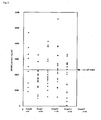
- SPARC protein levels in the serum of 87 melanoma patients and 60 healthy donors (HD) were evaluated by ELISA ( Figs. 4 and 5 ).
- the SPARC protein thought to be derived from blood platelets was also detected in HD serum. Even higher SPARC protein levels were detected in some of the melanoma patients.
- SPARC protein levels in the plasma of 11 melanoma patients and 21 healthy donors (HDs) were evaluated by ELISA ( Fig. 6 ).
- the mean value for melanoma patients was 606 ng/ml
- the mean value for healthy subjects was 140 ng/ml
- 1 out of 21 healthy subjects was found to be positive
- the degree of specificity was 95%.
- Table 1 stage MIA 5-S-CD GPC3 SPARC GPC3+SPARC 0 1/ 9 (11.1%) 0/ 9 ( 0.0%) 4/ 9 (44.4%) 5/ 9 (55.5%) 8/ 9 ( 88.9%) I 5/25 (20.0%) 2/25 ( 8.0%) 10/25 (40.0%) 3/23 ( 13.0%) 11/24 ( 48.0%) II 1/21 ( 4.8%) 2/20 (10.0%) 10/21 (47.6%) 5/ 19 (26.3%) 15/ 21 (71.4%) III 3/18 (16.7%) 5/18 (27.8%) 7/18 (38.9%) 8/18(44.4%) 12/18 ( 66.7%) IV 9/18 (50.0%) 15/18 (83.3%) 5/18 (27.8%) 8/18 (44.4%) 11/18(61.1%) Total 19/91 (20.9%) 24/90 (26.7%) 36/91 (39.6%) 29 /87 (33.3%) 57/90 (63.3%) Table 2: Stage Positive for GPC only Positive for SPARC only Positive for both GPC and SPARC Positive for GPC or SP
- a conventional tumor (melanoma) marker, 5-S-CD, and the recent subject of attention MIA were both useful only for advanced melanoma cases at stage III or IV. Similar to the case of GPC3, SPARC was effective in diagnosis of melanoma even at early stages I and II. However, few cases were positive for both SPARC and GPC3. SPARC is thought to be secreted in serum by an individual mechanism. Diagnostic yield can be elevated with the combined use of SPARC and GPC3. Specifically, it was revealed that the use of SPARC and GPC3 in combination enables screening of 60 percent or more of melanoma patients and that this combination is useful as a novel tumor marker for melanoma. A MIA ELISA kit (Roche Germany) was used for measurement of MIA.
- Table 3 shows the results. Increased SPARC protein levels were observed in various tissue types, regardless of differences in melanoma tissue types. Table 3 Type Positive for GPC only Positive for SPARC only Positive for both GPC and SPARC Positive for GPC or SPARC ALM 11 /44 9/41 4/41 24/41 SSM 7/16 5/16 1/16 13/16 LMM/LM 4/9 4/8 0/8 8/8 NM 1/5 0/5 1/5 2/5 Mucosa 4/12 4/12 0/12 8/12 Total 27/86 22/82 6/82 55/82
- the diagnostic kit and the diagnostic method according to the present invention are very useful in diagnosing whether or not a subject is affected with melanoma. It was revealed that the combination of SPARC and GPC3 is very useful in applications pertaining to cancer diagnosis for many melanoma patients throughout the world.
Applications Claiming Priority (2)
| Application Number | Priority Date | Filing Date | Title |
|---|---|---|---|
| JP2004303688 | 2004-10-19 | ||
| PCT/JP2005/014567 WO2006043362A1 (ja) | 2004-10-19 | 2005-08-09 | 悪性黒色腫(メラノーマ)の新規な診断キット |
Publications (3)
| Publication Number | Publication Date |
|---|---|
| EP1813943A1 EP1813943A1 (en) | 2007-08-01 |
| EP1813943A4 EP1813943A4 (en) | 2007-12-12 |
| EP1813943B1 true EP1813943B1 (en) | 2009-09-23 |
Family
ID=36202786
Family Applications (1)
| Application Number | Title | Priority Date | Filing Date |
|---|---|---|---|
| EP05770440A Expired - Fee Related EP1813943B1 (en) | 2004-10-19 | 2005-08-09 | Novel diagnostic kit for malignant melanoma |
Country Status (6)
| Country | Link |
|---|---|
| US (1) | US8017345B2 (ja) |
| EP (1) | EP1813943B1 (ja) |
| JP (1) | JP4834839B2 (ja) |
| AU (1) | AU2005297303B2 (ja) |
| DE (1) | DE602005016831D1 (ja) |
| WO (1) | WO2006043362A1 (ja) |
Families Citing this family (12)
| Publication number | Priority date | Publication date | Assignee | Title |
|---|---|---|---|---|
| PL1923463T3 (pl) | 2005-08-09 | 2012-02-29 | Oncotherapy Science Inc | Peptyd antygenowy do odrzucania raka pochodzący z glipikan-3 (GPC3) do zastosowania u HLA-A2-pozytywnego pacjenta i środek farmaceutyczny zawierający antygen |
| ATE509096T1 (de) * | 2006-06-16 | 2011-05-15 | Onco Therapy Science Inc | Von sparc abgeleitetes krebsabstossungsantigenpeptid und pharmazeutikum, das dieses enthält |
| JP2011520451A (ja) | 2008-05-14 | 2011-07-21 | ダームテック インターナショナル | 核酸解析による黒色腫および日光黒子の診断法 |
| NZ623273A (en) * | 2008-12-05 | 2015-09-25 | Abraxis Bioscience Llc | Sparc binding scfvs |
| BR112012008316A2 (pt) * | 2009-09-18 | 2017-06-06 | Abraxis Bioscience Llc | utilização da assinatura microambiente sparc no tratamento de câncer |
| EP3085387A1 (en) * | 2010-04-26 | 2016-10-26 | Abraxis BioScience, LLC | Sparc binding antibodies and uses thereof |
| CN103002905A (zh) | 2010-06-03 | 2013-03-27 | 阿布拉西斯生物科学有限责任公司 | Sparc微环境标签在癌症治疗中的应用 |
| EP2625525A4 (en) * | 2010-10-08 | 2014-04-02 | Abraxis Bioscience Llc | SPARC MICRO ENVIRONMENT SIGNATURE, PLASMA SPARC AND LDH AS PROGNOSTIC BIOMARKERS IN THE TREATMENT OF CANCER |
| WO2013191146A1 (ja) | 2012-06-18 | 2013-12-27 | 独立行政法人国立がん研究センター | 悪性黒色腫(メラノーマ)の診断キット |
| US20140314697A1 (en) * | 2013-04-18 | 2014-10-23 | Corum Inc. | Method for Inhibiting Inflammation and Reducing Melanophilin Expression with Glycine Derivatives And the Composition Thereof |
| US20150005184A1 (en) * | 2013-06-28 | 2015-01-01 | Dermtech International | Diagnosis of melanoma by nucleic acid analysis |
| AU2020247911A1 (en) | 2019-03-26 | 2021-11-11 | Dermtech, Inc. | Novel gene classifiers and uses thereof in skin cancers |
Family Cites Families (5)
| Publication number | Priority date | Publication date | Assignee | Title |
|---|---|---|---|---|
| EP1084273A1 (en) * | 1998-06-06 | 2001-03-21 | Genostic Pharma Limited | Probes used for genetic profiling |
| WO2004005883A2 (en) * | 2002-07-02 | 2004-01-15 | The Johns Hopkins University | Secreted and cytoplasmic tumor endothelial markers |
| DK1536006T3 (da) | 2002-08-30 | 2011-10-17 | Medinet Co Ltd | Cancerantigener og anvendelse deraf |
| ATE511550T1 (de) * | 2003-07-17 | 2011-06-15 | Pacific Edge Biotechnology Ltd | Marker zum nachweis von magenkrebs |
| DE602004018819D1 (de) | 2003-10-29 | 2009-02-12 | Kumamoto Tech & Ind Found | Diagnoseverfahren für malignes Melanom |
-
2005
- 2005-08-09 WO PCT/JP2005/014567 patent/WO2006043362A1/ja active Application Filing
- 2005-08-09 US US11/577,435 patent/US8017345B2/en not_active Expired - Fee Related
- 2005-08-09 AU AU2005297303A patent/AU2005297303B2/en not_active Ceased
- 2005-08-09 EP EP05770440A patent/EP1813943B1/en not_active Expired - Fee Related
- 2005-08-09 JP JP2006542257A patent/JP4834839B2/ja not_active Expired - Fee Related
- 2005-08-09 DE DE602005016831T patent/DE602005016831D1/de active Active
Also Published As
| Publication number | Publication date |
|---|---|
| JP4834839B2 (ja) | 2011-12-14 |
| AU2005297303B2 (en) | 2011-11-10 |
| US8017345B2 (en) | 2011-09-13 |
| JPWO2006043362A1 (ja) | 2008-05-22 |
| AU2005297303A1 (en) | 2006-04-27 |
| US20090111095A1 (en) | 2009-04-30 |
| EP1813943A1 (en) | 2007-08-01 |
| DE602005016831D1 (ja) | 2009-11-05 |
| WO2006043362A1 (ja) | 2006-04-27 |
| EP1813943A4 (en) | 2007-12-12 |
Similar Documents
| Publication | Publication Date | Title |
|---|---|---|
| EP1813943B1 (en) | Novel diagnostic kit for malignant melanoma | |
| Jou et al. | Salivary zinc finger protein 510 peptide as a novel biomarker for detection of oral squamous cell carcinoma in early stages | |
| JP5006802B2 (ja) | 上皮由来の癌診断および予後診断のためのバイオマーカーとしてのCyr61 | |
| JP4594575B2 (ja) | がんのマーカーとして使用されるアネキシン類及び自己抗体 | |
| US11959919B2 (en) | Predicting cancer progression | |
| CN102687011B (zh) | 癌症生物标志物及其应用 | |
| CN112345755A (zh) | 乳腺癌的生物标志物及其应用 | |
| US8642347B2 (en) | Urinary CA125 peptides as biomarkers of ovarian cancer | |
| KR20170029414A (ko) | 암을 스크리닝하고 검출하는 방법 및 조성물 | |
| EP1684076B1 (en) | Diagnostic method for malignant melanoma | |
| KR20150087580A (ko) | Del-1 단백질 양성 엑소좀을 포함하는 암 진단 또는 예후 예측용 조성물 | |
| KR20120021518A (ko) | 간암 진단용 조성물 및 진단방법 | |
| US7501255B2 (en) | Levels of Pin1 in normal and cancerous tissue | |
| US20130040325A1 (en) | Enzyme Linked Immunosorbent Assay (ELISA) Method and Kit for Detecting Soluble Programmed Cell Death Protein 5 (PDCD5) | |
| KR102141546B1 (ko) | 난청 예후 예측방법 및 이를 이용한 키트 | |
| EP4063845A1 (en) | Method for detecting cancer bone metastasis and detection reagent | |
| KR101431063B1 (ko) | 유방암 진단용 단백질 마커 아포리포단백질 c-1, 이의 검출 방법 및 이에 대한 항체를 포함하는 유방암 진단키트 | |
| Lu et al. | The expression of human epididymis protein 4 and cyclindependent kinase inhibitor p27Kip1 in human ovarian carcinoma | |
| KR101127225B1 (ko) | 요로상피암 표지자 및 그를 이용한 요로상피암 진단방법 | |
| JP2010139293A (ja) | 早期癌腫瘍マーカー | |
| KR20090058726A (ko) | 유방암 진단용 a2―HS 당단백질에 대한 자가항체마커및 진단키트 |
Legal Events
| Date | Code | Title | Description |
|---|---|---|---|
| PUAI | Public reference made under article 153(3) epc to a published international application that has entered the european phase |
Free format text: ORIGINAL CODE: 0009012 |
|
| 17P | Request for examination filed |
Effective date: 20070516 |
|
| AK | Designated contracting states |
Kind code of ref document: A1 Designated state(s): DE FR GB |
|
| A4 | Supplementary search report drawn up and despatched |
Effective date: 20071108 |
|
| DAX | Request for extension of the european patent (deleted) | ||
| RBV | Designated contracting states (corrected) |
Designated state(s): DE FR GB |
|
| 17Q | First examination report despatched |
Effective date: 20080226 |
|
| GRAP | Despatch of communication of intention to grant a patent |
Free format text: ORIGINAL CODE: EPIDOSNIGR1 |
|
| GRAS | Grant fee paid |
Free format text: ORIGINAL CODE: EPIDOSNIGR3 |
|
| GRAA | (expected) grant |
Free format text: ORIGINAL CODE: 0009210 |
|
| AK | Designated contracting states |
Kind code of ref document: B1 Designated state(s): DE FR GB |
|
| REG | Reference to a national code |
Ref country code: GB Ref legal event code: FG4D |
|
| REF | Corresponds to: |
Ref document number: 602005016831 Country of ref document: DE Date of ref document: 20091105 Kind code of ref document: P |
|
| PLBE | No opposition filed within time limit |
Free format text: ORIGINAL CODE: 0009261 |
|
| STAA | Information on the status of an ep patent application or granted ep patent |
Free format text: STATUS: NO OPPOSITION FILED WITHIN TIME LIMIT |
|
| 26N | No opposition filed |
Effective date: 20100624 |
|
| REG | Reference to a national code |
Ref country code: DE Ref legal event code: R082 Ref document number: 602005016831 Country of ref document: DE Representative=s name: GODEMEYER BLUM LENZE PATENTANWAELTE, PARTNERSC, DE Effective date: 20120202 Ref country code: DE Ref legal event code: R082 Ref document number: 602005016831 Country of ref document: DE Representative=s name: GODEMEYER BLUM LENZE PARTNERSCHAFT, PATENTANWA, DE Effective date: 20120202 Ref country code: DE Ref legal event code: R081 Ref document number: 602005016831 Country of ref document: DE Owner name: LSIP, LLC, TOKYO, JP Free format text: FORMER OWNER: KUMAMOTO UNIVERSITY, KUMAMOTO-SHI, JP Effective date: 20120202 |
|
| REG | Reference to a national code |
Ref country code: GB Ref legal event code: 732E Free format text: REGISTERED BETWEEN 20120322 AND 20120328 |
|
| PGFP | Annual fee paid to national office [announced via postgrant information from national office to epo] |
Ref country code: DE Payment date: 20130821 Year of fee payment: 9 |
|
| PGFP | Annual fee paid to national office [announced via postgrant information from national office to epo] |
Ref country code: FR Payment date: 20130823 Year of fee payment: 9 Ref country code: GB Payment date: 20130821 Year of fee payment: 9 |
|
| REG | Reference to a national code |
Ref country code: DE Ref legal event code: R082 Ref document number: 602005016831 Country of ref document: DE Representative=s name: GODEMEYER BLUM LENZE PATENTANWAELTE, PARTNERSC, DE Ref country code: DE Ref legal event code: R082 Ref document number: 602005016831 Country of ref document: DE Representative=s name: GODEMEYER BLUM LENZE PARTNERSCHAFT, PATENTANWA, DE |
|
| REG | Reference to a national code |
Ref country code: DE Ref legal event code: R119 Ref document number: 602005016831 Country of ref document: DE |
|
| GBPC | Gb: european patent ceased through non-payment of renewal fee |
Effective date: 20140809 |
|
| REG | Reference to a national code |
Ref country code: DE Ref legal event code: R119 Ref document number: 602005016831 Country of ref document: DE Effective date: 20150303 |
|
| REG | Reference to a national code |
Ref country code: FR Ref legal event code: ST Effective date: 20150430 |
|
| PG25 | Lapsed in a contracting state [announced via postgrant information from national office to epo] |
Ref country code: GB Free format text: LAPSE BECAUSE OF NON-PAYMENT OF DUE FEES Effective date: 20140809 Ref country code: DE Free format text: LAPSE BECAUSE OF NON-PAYMENT OF DUE FEES Effective date: 20150303 |
|
| PG25 | Lapsed in a contracting state [announced via postgrant information from national office to epo] |
Ref country code: FR Free format text: LAPSE BECAUSE OF NON-PAYMENT OF DUE FEES Effective date: 20140901 |