EP1754438B1 - Endoscope, notamment duodendoscope pour mère-enfant-cholangioscopy - Google Patents
Endoscope, notamment duodendoscope pour mère-enfant-cholangioscopy Download PDFInfo
- Publication number
- EP1754438B1 EP1754438B1 EP06017240.0A EP06017240A EP1754438B1 EP 1754438 B1 EP1754438 B1 EP 1754438B1 EP 06017240 A EP06017240 A EP 06017240A EP 1754438 B1 EP1754438 B1 EP 1754438B1
- Authority
- EP
- European Patent Office
- Prior art keywords
- endoscope
- shaft
- port
- working channel
- inlet opening
- Prior art date
- Legal status (The legal status is an assumption and is not a legal conclusion. Google has not performed a legal analysis and makes no representation as to the accuracy of the status listed.)
- Active
Links
- 238000003780 insertion Methods 0.000 claims description 20
- 230000037431 insertion Effects 0.000 claims description 20
- 238000007789 sealing Methods 0.000 claims description 8
- 230000001154 acute effect Effects 0.000 claims description 3
- 230000000694 effects Effects 0.000 claims 1
- 239000000523 sample Substances 0.000 description 7
- 210000000013 bile duct Anatomy 0.000 description 4
- 210000001198 duodenum Anatomy 0.000 description 4
- 239000004809 Teflon Substances 0.000 description 3
- 229920006362 Teflon® Polymers 0.000 description 3
- 238000000034 method Methods 0.000 description 3
- 210000002784 stomach Anatomy 0.000 description 3
- 238000004026 adhesive bonding Methods 0.000 description 2
- 210000003445 biliary tract Anatomy 0.000 description 2
- 210000003238 esophagus Anatomy 0.000 description 2
- 210000003800 pharynx Anatomy 0.000 description 2
- 239000004033 plastic Substances 0.000 description 2
- 206010004637 Bile duct stone Diseases 0.000 description 1
- 238000002583 angiography Methods 0.000 description 1
- 238000005452 bending Methods 0.000 description 1
- 238000001574 biopsy Methods 0.000 description 1
- -1 biopsy forceps Substances 0.000 description 1
- 238000004140 cleaning Methods 0.000 description 1
- 230000000295 complement effect Effects 0.000 description 1
- 230000002950 deficient Effects 0.000 description 1
- 238000003745 diagnosis Methods 0.000 description 1
- 238000001839 endoscopy Methods 0.000 description 1
- 239000012530 fluid Substances 0.000 description 1
- 238000013467 fragmentation Methods 0.000 description 1
- 238000006062 fragmentation reaction Methods 0.000 description 1
- 238000002347 injection Methods 0.000 description 1
- 239000007924 injection Substances 0.000 description 1
- 230000002452 interceptive effect Effects 0.000 description 1
- 239000000463 material Substances 0.000 description 1
- 230000008774 maternal effect Effects 0.000 description 1
- 230000010412 perfusion Effects 0.000 description 1
- 230000002093 peripheral effect Effects 0.000 description 1
- 239000004575 stone Substances 0.000 description 1
Images


Classifications
-
- A—HUMAN NECESSITIES
- A61—MEDICAL OR VETERINARY SCIENCE; HYGIENE
- A61B—DIAGNOSIS; SURGERY; IDENTIFICATION
- A61B1/00—Instruments for performing medical examinations of the interior of cavities or tubes of the body by visual or photographical inspection, e.g. endoscopes; Illuminating arrangements therefor
- A61B1/012—Instruments for performing medical examinations of the interior of cavities or tubes of the body by visual or photographical inspection, e.g. endoscopes; Illuminating arrangements therefor characterised by internal passages or accessories therefor
- A61B1/018—Instruments for performing medical examinations of the interior of cavities or tubes of the body by visual or photographical inspection, e.g. endoscopes; Illuminating arrangements therefor characterised by internal passages or accessories therefor for receiving instruments
Definitions
- Such endoscopes find in endoscopy numerous applications.
- an instrument for example a catheter, a small so-called baby endoscope with a smaller diameter, etc., is usually inserted within an instrument channel of an already placed large endoscope extending from the proximal to the distal endoscope end in order to carry out an endoscopic examination or an intervention.
- a flexible endoscope (large duodenoscope) is placed in the usual ERCP technique on the papilla major (referred to as the papilla).
- the papilla a large portion of the duodenoscopic shaft must be inserted into the patient (pharynx, esophagus, stomach, duodenum) because the endoscope must be advanced along the receding curvature of the stomach into the duodenum.
- a stretching maneuver in which a large part of the shaft length is extracted until finally the duodenoscope is stretched on the papilla. After stretching, only about 60 cm of the anterior endoscope shaft are inserted into the patient. In this stretched position, both ERCP and mother-baby cholangioscopy are performed.
- the usual standard maternal-mother (mother-baby) cholangioscopy requires a (large) jumbo or mother duodenoscope and a baby cholangioscope insertable into the maternal duodenoscope.
- the baby endoscope is in this case introduced into the bile ducts via an opening in the endoscope housing or operating part of the instrumentation or working channel of the mother endoscope.
- the method is mainly used for the fragmentation of large bile duct stones, for example with a laser probe and the diagnosis of unclear bile duct findings.
- the baby endoscope is difficult or hardly maneuverable.
- many years of practice with frequent use shows a high repair liability of the baby endoscope, which is often defective after only a few uses.
- the present invention is therefore an object of the invention to improve this state of the art and to provide a duodenoscope and an assembly of a mother and baby endoscope, which avoid these disadvantages and ensure improved repairability with less susceptibility to repair.
- the shaft port Since the first input port, hereinafter referred to as shaft port, is located in a region of the shaft remote from the proximal end of the endoscope, the shaft port is located outside of the patient after extension, for example a few centimeters in front of his mouth, and can now be used as access into the instrumentation channel or working channel of the endoscope are used. As a result, a considerably shortened baby endoscope can be used because it is introduced via the shaft port and no longer via the usual (proximal) input or the proximal port of the Instrumentierkanals on the control panel of the endoscope.
- instruments such as a baby endoscope, a catheter, etc.
- a length of, for example, about 200 cm with a diameter of 3 to 4 mm to project beyond the shaft length of the parent endoscope now advantageously significantly shortened instruments, such as a baby endoscope be used by a length of only 90 to 140 cm, in particular 100 to 120 cm.
- the shortened instrument especially the shortened baby endoscope is better maneuverable, better rotatable than conventional longer instruments, and also the cleaning such a shorter instrument is facilitated.
- the bendability improves, which also reduces the cost of repair as a cost advantage.
- shortened instruments also include, for example, rotationally stable catheter of the angiographic catheter type, which are inserted into the bile ducts via the first entry opening or the shaft port in order to reach previously poor or inaccessible peripheral bile duct branches. While angiographic catheters introduced via normal duodenoscopes can no longer be rotated due to the great length, this is now possible due to the shortened path as a result of the shaft port or its shortened design.
- the various segments of the tree-like biliary system which otherwise can only be reached by long patient "poking" or untargeted probing with guidewires and catheters more or less randomly or not at all, can be targeted.
- the shaft port is located in the middle region of the shaft, in particular at a distance from the distal end of the endoscope of 60 to 70 cm.
- An instrument to be introduced therefore requires only a minimum length of, for example, 80 to 140 cm, preferably 120 cm, in order to project beyond the part of the shaft located in the patient after placement.
- the endoscope additionally has a further input opening in the form of a customary proximal port in the region of the proximal end or on the endoscope housing.
- the endoscope is additionally applicable as a normal endoscope, for example a duodenoscope, so that the costs for a separate acquisition of such an endoscope can be saved.
- the first and the further inlet opening or the shaft port and the proximal port can lead to one and the same working channel, wherein the working channel in this case extends to the proximal port.
- the working channel in this case extends to the proximal port.
- the shaft port is sealingly closable with a cover.
- a cover in the form of a fixed externally attachable lid, which preferably seals against both the working channel and the endoscope inside in the patch, for example, locked state.
- other possibilities are also conceivable for sealing the first input opening or the shaft port, in particular during the phase of the placement of the endoscope.
- a probe inserted into the endoscope channel via the conventional access on the operating part of the endoscope in particular a hollow probe (to allow further insufflation) which sealingly closes the shaft port from inside (inner cover) or through a closure sleeve, the opening thereof is closed by turning or shifting with respect to the shaft port opening (outer cover).
- a thin-walled rubber, plastic tube for example of the "fingerling" type with a cut-off tip
- an attachment with an insertion tube to facilitate the insertion of instruments in the working channel can be arranged fixed and removable on the shaft port an attachment with an insertion tube to facilitate the insertion of instruments in the working channel.
- This port attachment preferably seals the first entry port and the shaft port, respectively, so that air may not escape using a termination cap or gasket.
- the attachment can in this case be arranged on the shaft port or in this region of the shaft, so that the insertion in the cross-sectional profile laterally offset, for example 90 ° to 180 °, on the other side of a common proximal port comes to rest.
- the region of the endoscope shaft in which the shaft port is as a short, for example less than 10 cm, in particular 4 to 6 cm long, rigid (shaft) sleeve, for example in the form of a rigid outer shaft segment and a also rigid inner channel segment, formed.
- rigid (shaft) sleeve for example in the form of a rigid outer shaft segment and a also rigid inner channel segment, formed.
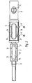
- first input port or the shaft port 1 of an endoscope is shown with an (outer) cover in the form of a lid 3.
- the shaft port 1 comprises a shaft segment in the form of an outer rigid sleeve 5 and a channel segment in Fonn a smaller diameter inner rigid sleeve 7, which serves for the passage of a working channel 9 of the endoscope and in cross-section ( Fig. 1c ) is not concentric, but is arranged near the inner wall of the shaft segment 5.
- both the shaft segment 5 and the channel segment 7 preferably have mutually aligned elongate openings 25, 27 (FIG. Fig. 3 ) on
- the access to the working channel realized in this way 9 can be like in Fig. 1b illustrated an insertion aid in the form of an outwardly widening chamfer 49 to facilitate the insertion of an instrument in the working channel 9.
- an inwardly widening slant or insertion aid 51 may be located to facilitate the oblique insertion of an instrument obliquely from the upper left into the working channel 9, which leads to the right towards the distal end. In this way, advantageously, even with a short shaft port 1 or first input port of, for example, 15-30 mm, an easy insertion of an instrument can be ensured.
- the channel segment 7 has two connecting pieces 11 and 13, on which the ends of the working channel 9 pushed and possibly additionally, for example by gluing, can be fixed.
- the shaft segment 5 is in the endoscope shaft 15, which in Fig. 1a and Fig. 1b dashed lines, fixed, for example, by gluing, aligned, so that despite the preferred rigid design of the entire shaft port 1 on the outer surface of the endoscope shaft 15 no undesirable attack sites, such as steps, edges, etc. form as slip obstacle.
- the thin lid 3 closes the shaft port 1 or the first input opening, with a projecting area 17 on the underside of the lid 3 into the elongated opening 25 of the shaft segment 5 and possibly even in FIG the hereby aligned opening 27 of the channel segment 7 preferably protrudes accurately.
- an actuating element for example in the form of a slotted screw 21 of a locking device is actuated, wherein the locking device consists for example of a locking element 19.
- This locking element 19 is oval-shaped on the underside of the screw 21, wherein only in the locked state opposite extensions 19a and 19b of the substantially oval disc 19 engage in corresponding recesses of the channel segment 7.
- the lid 3 alsklippbar, wherein it is formed in profile, for example, as a projecting beyond the shank circumference half, or encompassing half-shell.
- the protruding region 17 can in any case be designed to be flush in addition to the channel interior, so that, in the closed state, advantageously no unwanted resistances result for an instrument introduced via the proximal port 63.
- the shaft port 1 of a channel segment 7 and the shaft segment 5 is constructed such that, as out Fig. 1c can be seen, the channel segment is arranged with its opening 27 on the inside of the shaft segment 5 in the region of the opening 25.
- the channel segment 7 may have in cross-section a semicircular on the outer surface extension, in which threaded holes 35 are provided. In the assembled state, these holes 35 are aligned with holes 33 in the shaft segment 5, so that the channel segment 7 can be fixed to the shaft segment 5 by means of screws 31. In this way, the opening 25 of the shaft segment 5 go with their bevels 49 and 51 and the opening 27 of the channel segment 7 in alignment with each other.
- a seal (not shown in detail in the drawing) is provided between the outer surface of the channel segment 7, surrounding the opening 27, which sealingly bears against the inner side of the shaft segment 5. Furthermore, on the outer surface of the shaft segment 5, a circumferential around the opening 25 seal 29 is arranged in the region of the underside of the lid 3, so that the lid 3 in the locked state also sealingly closes.
- one or more elongated spring elements 23a and 23b may additionally be provided on both longitudinal sides of the cover 3 (or the corresponding complementary recess of the shaft port 1) be arranged, which are pre-bent so that they extend in the closing state oblong and their spring force acts against the placement of the lid 3.
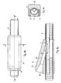
- Fig. 4a is shown as a port attachment 37, which is for example made as a milled part or injection molded part, is placed on the shaft port 1 to allow through an opening 39, the insertion of an instrument in the distal direction (in the drawing to the right) in the working channel 9.
- the port attachment 37 is in this case plugged through the lateral opening 25, 27 on the shaft port 1 of the mother endoscope shaft 15.
- a seal (“O" ring, rubber plate, etc.) (not shown in detail in the drawing) (not shown in detail in the drawing) can be provided between shaft port 1 and port attachment 37 in order to prevent air from escaping between port attachment 37 and shaft port 1.
- Fig. 4c is the port attachment 37 from a lower shell 41 and an upper shell 43, in which the funnel-shaped outwardly widening opening 39 and just adjoining Ein Industriesstutzen 40 are constructed.
- two fastening elements 45 and 47 for example in the form of locking pins or screws can be arranged, which of course also a hinge on one side and arranged on the opposite side lock, for example in the form of a hook, conceivable is.
- Fig. 4b shown leads the straight tubular inlet tube 40 from top left to obliquely bottom right at a shallow angle in the working channel 9 in the distal direction (right side in the drawing) into it, said guide through by on the short sides of the elongated openings 25 and 27 into one another transitional bevels 49 and 51 is supported.
- the insertion tube 40 in this case has a relation to the inner diameter of the working channel (for example, 4.2 mm) smaller or the same inner diameter.
- the port attachment 37 protrudes at this point with its corresponding to the opening 25 formed material 53, so that a spatially and rotationally fixed arrangement and centering of the port attachment 37 is ensured at the shaft port 1 by this intermeshing intervention.
- insertion aids 49, 51 even with a desired short length of the shaft port 1 and the shaft and channel segments 5 and 7 of 40 to 60 mm and a realized through the openings 25, 27 short shaft port opening of only 15 to 30 mm in length as possible flat or acute angle (as seen from the proximal end) between the longitudinal axis of the insertion and the longitudinal axis of the working channel 9 and the endoscope shaft 15, for example, 10 to 40 degrees, preferably from 10 to 20 degrees.
- the insertion tube 40 or the opening 39 can be closed with a sealing cap (not shown in detail in the drawing) (also for the sealing introduction of instruments).
- the shaft port 1 or the port attachment 37 is arranged in a mother or mother duodenoscope 61 shown here.
- the shaft port 1, which is 40 to 60 mm long and integrated in the shaft 15, is located approximately between 60 and 70 cm away from the distal head end of the endoscope 61.
- the elongate opening of the shaft port 1 has, for example, a diameter of 3.5 mm thick baby endoscope a length of about 15 to 30 mm.
- the shaft port 1 or port attachment 37 is arranged with its opening 39 in the longitudinal direction at the proximal end or the operating part of the endoscope from the left side, whereas a conventional, proximal port 63 is usually arranged on the right side of the control unit.
- a conventional, proximal port 63 is usually arranged on the right side of the control unit.
- Fig. 6 is also greatly simplified indicated that the port attachment instead of bivalve, for example, in the cross-sectional profile can be U-shaped. Moreover, in Fig. 6 another type of attachment, namely, for example by means of an insertable into the ends of the U-shaped leg screw or axle with an eccentric which clamps the port attachment to the shaft or the shaft port when actuated by rotation, indicated.
- the shaft port 1 must be closed, since in this case approximately 100 to 110 cm of the endoscope shaft and thus the shaft port itself are located inside a patient. This can be achieved, for example, by a cover 3 which can be inserted into the shaft port opening, as explained above.
- inner or outer covers such as, for example, a conventional proximal access or port 63 on the operating part of the endoscope into the endoscope channel 9 inserted hollow probe, which closes the shaft port 1 from the inside sealing, a closure sleeve, the opening is closed by turning or moving relative to the shaft port opening or a thin-walled rubber, plastic hose (type Fingerling with truncated dome) pushed onto the duodenoscope, on the shaft port opening is rolled, conceivable.
- a conventional proximal access or port 63 on the operating part of the endoscope into the endoscope channel 9 inserted hollow probe which closes the shaft port 1 from the inside sealing, a closure sleeve, the opening is closed by turning or moving relative to the shaft port opening or a thin-walled rubber, plastic hose (type Fingerling with truncated dome) pushed onto the duodenoscope, on the shaft port opening is rolled, conceivable.
- the shaft port 1 comes to lie outside the patient, as a result of the extension, only one shaft length of less than 60 cm remains in the patient.
- the shaft port 1 can then be used as access for instruments.
- the port attachment 37 is sealed against the respective instrument, for example an inserted baby endoscope (for example by means of a corresponding seal in the port attachment 37 or its opening, in order to prevent injected air from escaping during introduction as far as the duodenum and thus by collapsing the lumen
- a stronger air supply preferably has to take place in order to compensate for the possibly escaping air via the shaft port 1.
- the port attachment 37 is introduced via the shaft port 1 into the instrumentation channel or working channel 9 and seals it. At the outer end can also be attached a side nozzle for air insufflation. This nozzle may eventually be connected to the normal air supply.
- the port attachment 37 in principle as a very thin-walled (Teflon etc.) hose, which is inserted through the shaft port 1 in the working channel 9 substantially sealing.
- Teflon etc. very thin-walled
- the fixation of the port attachment 37 on the shaft 15 can by various ways, such as an eccentric (screw, lever o. ⁇ .) With the port attachment 37 is clamped done.
- the port attachment 37 is preferably equipped with centering means, such as by the above-mentioned projection, centering pins o. ⁇ ., And fixed on the shaft port 1, that the insertion tube 40 opens optimally in the Instrumentierkanal 9 and thus the baby endoscope optimally with the least possible friction can be inserted into the mother endoscope 61.
- Such a baby endoscope for example, has an outer diameter of about 3.5 mm and thus has about 0.5 mm clearance (for example for insufflation) to the instrumentation channel (diameter about 4 mm) of the parent endoscope.
- the outer skin of the baby endoscope is designed to be as slippery as possible (teflon, etc.) in order to provide low sliding resistance.
- the baby endoscope is preferably as stable as possible to rotation, so that the missing bending planes can be achieved by rotation.
- the baby endoscope has an instrumentation channel of at least 1 mm (preferably at least 1.4 mm), so that it is suitable for electrohydraulic probes, biopsy forceps, stone baskets, etc.
- an endoscope in particular a duodenoscope
- the endoscope can in principle also be used as a routine endoscope, in particular a duodenoscope, for example for conventional ERCP, so that not two duodenoscopes are used for the ERCP operation required are. This can be advantageously (especially in smaller clinics with limited budget) costs saved.
Landscapes
- Health & Medical Sciences (AREA)
- Life Sciences & Earth Sciences (AREA)
- Surgery (AREA)
- Nuclear Medicine, Radiotherapy & Molecular Imaging (AREA)
- Biomedical Technology (AREA)
- Optics & Photonics (AREA)
- Pathology (AREA)
- Radiology & Medical Imaging (AREA)
- Biophysics (AREA)
- Engineering & Computer Science (AREA)
- Physics & Mathematics (AREA)
- Heart & Thoracic Surgery (AREA)
- Medical Informatics (AREA)
- Molecular Biology (AREA)
- Animal Behavior & Ethology (AREA)
- General Health & Medical Sciences (AREA)
- Public Health (AREA)
- Veterinary Medicine (AREA)
- Endoscopes (AREA)
Claims (9)
- Duodénoscope pour cholangioscopie mère-enfant, avec un canal de travail (9) s'étendant dans ou sur une tige (15) de l'endoscope (61), qui comprend, du côté distal, sur la tête de l'endoscope, une ouverture de sortie,
caractérisé en ce que
le canal de travail (9) comprend au moins une première ouverture d'entrée (1), qui se trouve dans une zone de la tige (15) éloignée de l'extrémité proximale de l'endoscope (61),
la zone de la tige de l'endoscope (15), dans laquelle se trouve la première ouverture d'entrée (1), est conçue comme une gaine rigide (5, 7) et
la gaine rigide (5) est reliée de manière alignée dans la tige de l'endoscope (15), de façon à ce qu'aucun point d'attaque indésirable ne se forme à la surface externe de la tige de l'endoscope (15) et empêche tout glissement. - Endoscope selon la revendication 1, caractérisé en ce que l'endoscope (61) comprend, en outre, dans la zone de l'extrémité proximale ou sur le boîtier de l'endoscope, une ouverture d'entrée supplémentaire (63).
- Endoscope selon la revendication 2, caractérisé en ce que la première ouverture d'entrée et l'ouverture d'entrée supplémentaire (1, 63) mènent au même canal de travail (9), le canal de travail (9) s'étendant au moins jusqu'à l'ouverture d'entrée supplémentaire (63).
- Endoscope selon l'une des revendications précédentes, caractérisé en ce que la première ouverture d'entrée (1) peut être fermée de manière étanche avec un couvercle fixe (3).
- Endoscope selon la revendication 4, caractérisé en ce que le couvercle (3) comprend, sur son côté inférieur, une zone saillante (17) qui dépasse dans l'ouverture (27) du segment de canal (7), la zone saillante (17) étant alignée avec l'intérieur du canal, de façon à ce que, dans l'état fermé, aucune résistance indésirable n'apparaisse pour un instrument introduit par un orifice proximal (63).
- Endoscope selon l'une des revendications précédentes, caractérisé en ce que, au niveau de la première ouverture d'entrée (1), un élément (37), avec un embout d'introduction (40), peut être disposé de manière fixe et amovible afin de faciliter l'introduction d'instruments dans le canal de travail (9).
- Endoscope selon la revendication 6, caractérisé en ce que, dans l'état monté de l'élément (37), entre l'axe longitudinal de l'embout d'introduction (40) et l'axe longitudinal du canal de travail (9), il existe un angle aigu de 10 à 40 degrés, vu en direction de l'extrémité proximale de l'endoscope.
- Endoscope selon la revendication 6 ou 7, caractérisé en ce que, dans l'état monté, l'élément (37) est disposé de manière décalée latéralement dans le profil sur le côté opposé d'une ouverture d'entrée supplémentaire proximale (63).
- Dispositif constitué d'un endoscope selon l'une des revendications 1 à 8 et un endoscope pour bébé, qui peut être introduit dans le canal de travail (9) d'un endoscope (61) selon l'une des revendications 1 à 8 et qui présente une longueur de 80 à 140 cm, de préférence de 120 cm, afin de réaliser une image miroir des voies biliaires (cholangioscopie).
Applications Claiming Priority (1)
| Application Number | Priority Date | Filing Date | Title |
|---|---|---|---|
| DE102005039601A DE102005039601A1 (de) | 2005-08-19 | 2005-08-19 | Endoskop, insbesondere Duodenoskop für die Mutter-Baby Cholangioskopie |
Publications (2)
| Publication Number | Publication Date |
|---|---|
| EP1754438A1 EP1754438A1 (fr) | 2007-02-21 |
| EP1754438B1 true EP1754438B1 (fr) | 2015-05-27 |
Family
ID=37450906
Family Applications (1)
| Application Number | Title | Priority Date | Filing Date |
|---|---|---|---|
| EP06017240.0A Active EP1754438B1 (fr) | 2005-08-19 | 2006-08-18 | Endoscope, notamment duodendoscope pour mère-enfant-cholangioscopy |
Country Status (3)
| Country | Link |
|---|---|
| US (1) | US20070118016A1 (fr) |
| EP (1) | EP1754438B1 (fr) |
| DE (1) | DE102005039601A1 (fr) |
Cited By (1)
| Publication number | Priority date | Publication date | Assignee | Title |
|---|---|---|---|---|
| DE102017006214A1 (de) | 2017-06-30 | 2019-01-03 | Karl Storz Se & Co. Kg | Schaftport für ein Endoskop |
Families Citing this family (10)
| Publication number | Priority date | Publication date | Assignee | Title |
|---|---|---|---|---|
| USD668762S1 (en) * | 2011-03-31 | 2012-10-09 | Karl Storz Gmbh & Co. Kg | Handle for medical device |
| US20130035553A1 (en) | 2011-08-01 | 2013-02-07 | Gyrus Acmi, Inc. | Endoscope with multiple working channel ports |
| WO2016009690A1 (fr) * | 2014-07-17 | 2016-01-21 | オリンパス株式会社 | Endoscope |
| JP7082052B2 (ja) | 2015-09-03 | 2022-06-07 | ネプチューン メディカル インク. | 小腸内での内視鏡前進の為の器具 |
| CN110191667B (zh) | 2016-08-18 | 2022-06-03 | 海王星医疗公司 | 用于增强小肠视觉效果的装置和方法 |
| EP3801187B1 (fr) | 2018-05-31 | 2024-02-07 | Neptune Medical Inc. | Dispositif de visualisation améliorée de l'intestin grêle |
| CN112714658A (zh) | 2018-07-19 | 2021-04-27 | 海王星医疗公司 | 动态刚性化复合医疗结构 |
| US11793392B2 (en) | 2019-04-17 | 2023-10-24 | Neptune Medical Inc. | External working channels |
| EP4126095A4 (fr) | 2020-03-30 | 2024-04-24 | Neptune Medical Inc. | Parois stratifiées pour dispositifs de rigidification |
| US11937778B2 (en) | 2022-04-27 | 2024-03-26 | Neptune Medical Inc. | Apparatuses and methods for determining if an endoscope is contaminated |
Family Cites Families (17)
| Publication number | Priority date | Publication date | Assignee | Title |
|---|---|---|---|---|
| WO1980002499A1 (fr) * | 1979-05-21 | 1980-11-27 | American Cystoscope Makers Inc | Instrument chirurgical pour un endoscope |
| US4772093A (en) * | 1985-12-12 | 1988-09-20 | Microvasive, Inc. | Fiber-optic image-carrying device |
| US4979496A (en) * | 1988-04-05 | 1990-12-25 | Fuji Photo Optical Co., Ltd. | Endoscope for bile duct and pancreatic duct |
| JPH0724086Y2 (ja) * | 1989-05-01 | 1995-06-05 | 株式会社町田製作所 | 内視鏡用チャンネルチューブ |
| US5569161A (en) * | 1992-10-08 | 1996-10-29 | Wendell V. Ebling | Endoscope with sterile sleeve |
| US5575752A (en) * | 1993-02-19 | 1996-11-19 | Olympus Optical Co., Ltd. | Endoscope system, cover type endoscope unit, channeled cover type endoscope unit, holding tool in endoscope system, and housing member of cover type endoscope unit |
| US5573494A (en) * | 1993-02-23 | 1996-11-12 | Olympus Optical Co., Ltd. | Endoscope cover-sheathed endoscope in which an endoscope-cover coverable endoscope to be sheathed with an endoscope cover is structured to shut out water tightly |
| US5665064A (en) * | 1993-12-06 | 1997-09-09 | Sherwood Medical Company | Gastroenteric feeding tube for endoscopic placement and method of use |
| US6997931B2 (en) * | 2001-02-02 | 2006-02-14 | Lsi Solutions, Inc. | System for endoscopic suturing |
| AU2003284287A1 (en) * | 2002-10-18 | 2004-05-04 | Cook Ireland Ltd | Physician access system |
| JP4200731B2 (ja) * | 2002-10-23 | 2008-12-24 | フジノン株式会社 | 内視鏡の鉗子栓 |
| JP4477382B2 (ja) * | 2003-03-04 | 2010-06-09 | オリンパス株式会社 | 内視鏡的腹腔内処置システム |
| US20050245789A1 (en) * | 2003-04-01 | 2005-11-03 | Boston Scientific Scimed, Inc. | Fluid manifold for endoscope system |
| US20040199052A1 (en) * | 2003-04-01 | 2004-10-07 | Scimed Life Systems, Inc. | Endoscopic imaging system |
| JP4547184B2 (ja) * | 2003-07-29 | 2010-09-22 | オリンパス株式会社 | 内視鏡用アダプター及び内視鏡 |
| JP4778425B2 (ja) * | 2003-07-31 | 2011-09-21 | ウィルソン−クック・メディカル・インコーポレーテッド | 複数の医療装置を導入するためのシステム及び方法 |
| US7276045B2 (en) * | 2003-11-24 | 2007-10-02 | Medtronic Vascular, Inc. | Apparatus and method for wire exchange |
-
2005
- 2005-08-19 DE DE102005039601A patent/DE102005039601A1/de not_active Withdrawn
-
2006
- 2006-08-18 EP EP06017240.0A patent/EP1754438B1/fr active Active
- 2006-08-21 US US11/507,125 patent/US20070118016A1/en not_active Abandoned
Cited By (2)
| Publication number | Priority date | Publication date | Assignee | Title |
|---|---|---|---|---|
| DE102017006214A1 (de) | 2017-06-30 | 2019-01-03 | Karl Storz Se & Co. Kg | Schaftport für ein Endoskop |
| DE102017006214B4 (de) | 2017-06-30 | 2024-08-29 | Karl Storz Se & Co. Kg | Schaftport für ein Endoskop |
Also Published As
| Publication number | Publication date |
|---|---|
| DE102005039601A1 (de) | 2007-02-22 |
| US20070118016A1 (en) | 2007-05-24 |
| EP1754438A1 (fr) | 2007-02-21 |
Similar Documents
| Publication | Publication Date | Title |
|---|---|---|
| EP1754438B1 (fr) | Endoscope, notamment duodendoscope pour mère-enfant-cholangioscopy | |
| EP2124706B1 (fr) | Système tubulaire pour endoscope | |
| DE10126062B4 (de) | Haube für ein Endoskop | |
| DE4101472C2 (de) | Endoskop zur transurethralen Resektion | |
| DE4034143C2 (de) | Arthroskopierkanüle für die Druckmessung | |
| DE2033665A1 (de) | Medizinisches Instrument lnsbe sondere zur Fetal Blutentnahme | |
| EP1542579B1 (fr) | Instrument medical d'aspiration et de nettoyage et procede de fabrication associe | |
| DE69729323T2 (de) | Geschütztes bürstensystem zur mikrobiologischen probeentnahme | |
| DE10139153A1 (de) | Einweg-Endoskopmantel | |
| DE3615694A1 (de) | Perkutanes nephroskop mit sicherheitsdraht | |
| EP3851019B1 (fr) | Robinet d'arrêt pour un endoscope | |
| DE10358817B3 (de) | Endoskop | |
| DE19619065C2 (de) | Trokarhülse mit einem Ventil | |
| EP2903494B1 (fr) | Système d'endoscopie et de biopsie à usage unique | |
| DE19928289B4 (de) | Medizinisches Endoskop | |
| DE3942905C2 (de) | Endoskop, insbesondere Cystoskop-Urethroskop | |
| DE102004015291A1 (de) | Aufsetzkappe für Endoskop-Einführungsschläuche sowie Endoskope | |
| DE10145107B4 (de) | Füllstab für Endoskope | |
| DE60303586T2 (de) | Katheter zur Einführung in den menschlichen Körper | |
| DE19942929A1 (de) | Kathetereinrichtung für ein Endoskop | |
| DE68905983T2 (de) | Katheter, der beim Anschluss drehbar ist. | |
| DE202019102209U1 (de) | Endoskopiesystem umfassend ein Endoskop mit einem flexiblen Schaft | |
| DE19756629C2 (de) | Instrument, insbesondere Trokar oder Endoskop | |
| DE3237376A1 (de) | Vorrichtung zum reinigen und sterilisieren eines endoskops | |
| DE102007061142A1 (de) | Hysteroskop mit einem Schaft |
Legal Events
| Date | Code | Title | Description |
|---|---|---|---|
| PUAI | Public reference made under article 153(3) epc to a published international application that has entered the european phase |
Free format text: ORIGINAL CODE: 0009012 |
|
| AK | Designated contracting states |
Kind code of ref document: A1 Designated state(s): AT BE BG CH CY CZ DE DK EE ES FI FR GB GR HU IE IS IT LI LT LU LV MC NL PL PT RO SE SI SK TR |
|
| AX | Request for extension of the european patent |
Extension state: AL BA HR MK YU |
|
| 17P | Request for examination filed |
Effective date: 20070711 |
|
| 17Q | First examination report despatched |
Effective date: 20070810 |
|
| AKX | Designation fees paid |
Designated state(s): DE FR GB IT |
|
| GRAP | Despatch of communication of intention to grant a patent |
Free format text: ORIGINAL CODE: EPIDOSNIGR1 |
|
| INTG | Intention to grant announced |
Effective date: 20150129 |
|
| GRAS | Grant fee paid |
Free format text: ORIGINAL CODE: EPIDOSNIGR3 |
|
| GRAA | (expected) grant |
Free format text: ORIGINAL CODE: 0009210 |
|
| AK | Designated contracting states |
Kind code of ref document: B1 Designated state(s): DE FR GB IT |
|
| REG | Reference to a national code |
Ref country code: GB Ref legal event code: FG4D Free format text: NOT ENGLISH |
|
| REG | Reference to a national code |
Ref country code: DE Ref legal event code: R096 Ref document number: 502006014344 Country of ref document: DE |
|
| REG | Reference to a national code |
Ref country code: DE Ref legal event code: R097 Ref document number: 502006014344 Country of ref document: DE |
|
| PLBE | No opposition filed within time limit |
Free format text: ORIGINAL CODE: 0009261 |
|
| STAA | Information on the status of an ep patent application or granted ep patent |
Free format text: STATUS: NO OPPOSITION FILED WITHIN TIME LIMIT |
|
| 26N | No opposition filed |
Effective date: 20160301 |
|
| REG | Reference to a national code |
Ref country code: FR Ref legal event code: PLFP Year of fee payment: 11 |
|
| REG | Reference to a national code |
Ref country code: FR Ref legal event code: PLFP Year of fee payment: 12 |
|
| REG | Reference to a national code |
Ref country code: FR Ref legal event code: PLFP Year of fee payment: 13 |
|
| REG | Reference to a national code |
Ref country code: DE Ref legal event code: R082 Ref document number: 502006014344 Country of ref document: DE Representative=s name: EDER SCHIESCHKE & PARTNER MBB, PATENTANWAELTE, DE Ref country code: DE Ref legal event code: R081 Ref document number: 502006014344 Country of ref document: DE Owner name: STORZ ENDOSKOP PRODUKTIONS GMBH, CH Free format text: FORMER OWNER: KARL STORZ GMBH & CO. KG, 78532 TUTTLINGEN, DE |
|
| REG | Reference to a national code |
Ref country code: GB Ref legal event code: 732E Free format text: REGISTERED BETWEEN 20190314 AND 20190320 |
|
| PGFP | Annual fee paid to national office [announced via postgrant information from national office to epo] |
Ref country code: IT Payment date: 20190722 Year of fee payment: 14 |
|
| PGFP | Annual fee paid to national office [announced via postgrant information from national office to epo] |
Ref country code: GB Payment date: 20190722 Year of fee payment: 14 |
|
| GBPC | Gb: european patent ceased through non-payment of renewal fee |
Effective date: 20200818 |
|
| PG25 | Lapsed in a contracting state [announced via postgrant information from national office to epo] |
Ref country code: IT Free format text: LAPSE BECAUSE OF NON-PAYMENT OF DUE FEES Effective date: 20200818 |
|
| PG25 | Lapsed in a contracting state [announced via postgrant information from national office to epo] |
Ref country code: GB Free format text: LAPSE BECAUSE OF NON-PAYMENT OF DUE FEES Effective date: 20200818 |
|
| PGFP | Annual fee paid to national office [announced via postgrant information from national office to epo] |
Ref country code: FR Payment date: 20210722 Year of fee payment: 16 |
|
| P01 | Opt-out of the competence of the unified patent court (upc) registered |
Effective date: 20230527 |
|
| PG25 | Lapsed in a contracting state [announced via postgrant information from national office to epo] |
Ref country code: FR Free format text: LAPSE BECAUSE OF NON-PAYMENT OF DUE FEES Effective date: 20220831 |
|
| PGFP | Annual fee paid to national office [announced via postgrant information from national office to epo] |
Ref country code: DE Payment date: 20230720 Year of fee payment: 18 |