EP1732461B1 - System for vascular border detection - Google Patents
System for vascular border detection Download PDFInfo
- Publication number
- EP1732461B1 EP1732461B1 EP05723848A EP05723848A EP1732461B1 EP 1732461 B1 EP1732461 B1 EP 1732461B1 EP 05723848 A EP05723848 A EP 05723848A EP 05723848 A EP05723848 A EP 05723848A EP 1732461 B1 EP1732461 B1 EP 1732461B1
- Authority
- EP
- European Patent Office
- Prior art keywords
- border
- data
- image
- vascular
- application
- Prior art date
- Legal status (The legal status is an assumption and is not a legal conclusion. Google has not performed a legal analysis and makes no representation as to the accuracy of the status listed.)
- Expired - Lifetime
Links
- 230000002792 vascular Effects 0.000 title claims abstract description 82
- 238000001514 detection method Methods 0.000 title description 5
- 238000012512 characterization method Methods 0.000 claims abstract description 19
- 210000001519 tissue Anatomy 0.000 claims description 71
- 238000002608 intravascular ultrasound Methods 0.000 claims description 31
- 230000003595 spectral effect Effects 0.000 claims description 22
- 239000008280 blood Substances 0.000 claims description 10
- 210000004369 blood Anatomy 0.000 claims description 10
- 210000002808 connective tissue Anatomy 0.000 claims description 5
- 230000000063 preceeding effect Effects 0.000 claims 1
- 238000000034 method Methods 0.000 abstract description 29
- 238000013500 data storage Methods 0.000 abstract description 9
- 238000001228 spectrum Methods 0.000 abstract description 7
- 230000008569 process Effects 0.000 description 7
- 230000009466 transformation Effects 0.000 description 7
- 238000003384 imaging method Methods 0.000 description 6
- 230000007704 transition Effects 0.000 description 6
- 210000004204 blood vessel Anatomy 0.000 description 4
- 238000004458 analytical method Methods 0.000 description 3
- 238000002604 ultrasonography Methods 0.000 description 3
- 230000008859 change Effects 0.000 description 2
- 238000001914 filtration Methods 0.000 description 2
- 238000012014 optical coherence tomography Methods 0.000 description 2
- 230000003287 optical effect Effects 0.000 description 2
- 239000000523 sample Substances 0.000 description 2
- 238000010183 spectrum analysis Methods 0.000 description 2
- 241001270131 Agaricus moelleri Species 0.000 description 1
- 208000031481 Pathologic Constriction Diseases 0.000 description 1
- 230000006978 adaptation Effects 0.000 description 1
- 238000002399 angioplasty Methods 0.000 description 1
- 238000006243 chemical reaction Methods 0.000 description 1
- 238000005094 computer simulation Methods 0.000 description 1
- 239000000470 constituent Substances 0.000 description 1
- 210000004351 coronary vessel Anatomy 0.000 description 1
- 239000013078 crystal Substances 0.000 description 1
- 238000002059 diagnostic imaging Methods 0.000 description 1
- 201000010099 disease Diseases 0.000 description 1
- 208000037265 diseases, disorders, signs and symptoms Diseases 0.000 description 1
- 238000002592 echocardiography Methods 0.000 description 1
- 238000013213 extrapolation Methods 0.000 description 1
- 230000003176 fibrotic effect Effects 0.000 description 1
- 230000006870 function Effects 0.000 description 1
- 238000009499 grossing Methods 0.000 description 1
- 230000036541 health Effects 0.000 description 1
- 239000012528 membrane Substances 0.000 description 1
- 239000000203 mixture Substances 0.000 description 1
- 230000004048 modification Effects 0.000 description 1
- 238000012986 modification Methods 0.000 description 1
- 238000005457 optimization Methods 0.000 description 1
- 208000037804 stenosis Diseases 0.000 description 1
- 230000036262 stenosis Effects 0.000 description 1
- 230000009026 tissue transition Effects 0.000 description 1
- 238000000844 transformation Methods 0.000 description 1
- 238000012285 ultrasound imaging Methods 0.000 description 1
Images

Classifications
-
- A—HUMAN NECESSITIES
- A61—MEDICAL OR VETERINARY SCIENCE; HYGIENE
- A61B—DIAGNOSIS; SURGERY; IDENTIFICATION
- A61B8/00—Diagnosis using ultrasonic, sonic or infrasonic waves
- A61B8/12—Diagnosis using ultrasonic, sonic or infrasonic waves in body cavities or body tracts, e.g. by using catheters
-
- A—HUMAN NECESSITIES
- A61—MEDICAL OR VETERINARY SCIENCE; HYGIENE
- A61B—DIAGNOSIS; SURGERY; IDENTIFICATION
- A61B5/00—Measuring for diagnostic purposes; Identification of persons
- A61B5/02—Detecting, measuring or recording for evaluating the cardiovascular system, e.g. pulse, heart rate, blood pressure or blood flow
- A61B5/02007—Evaluating blood vessel condition, e.g. elasticity, compliance
-
- A—HUMAN NECESSITIES
- A61—MEDICAL OR VETERINARY SCIENCE; HYGIENE
- A61B—DIAGNOSIS; SURGERY; IDENTIFICATION
- A61B8/00—Diagnosis using ultrasonic, sonic or infrasonic waves
- A61B8/08—Clinical applications
- A61B8/0858—Clinical applications involving measuring tissue layers, e.g. skin, interfaces
Definitions
- the present invention relates to vascular images, or more particularly, to a system and method of using the frequency spectrum of a radio frequency (RF) signal backscattered from vascular tissue to identify at least one border on a corresponding vascular image.
- RF radio frequency
- the present invention relates to medical imaging arts. It finds particular application to a system and method of identifying a border on a vascular image (e. g. intra-vascular ultrasound (IVUS) image, Virtual Histology (VH) image, etc.).
- IVUS intra-vascular ultrasound
- VH Virtual Histology
- the present invention is described in terms of identifying a luminal and medial-adventitial border on an IVUS or VH image, the present invention is not so limited. Thus, for example, identifying any border (or boundary) on any vascular image is within the scope of the present invention.
- Ultrasonic imaging of portions of a patient's body provides a useful tool in various areas of medical practice for determining the best type and course of treatment.
- Imaging of the coronary vessels of a patient by ultrasonic techniques can provide physicians with valuable information.
- the image data may show the extent of a stenosis in a patient, reveal progression of disease, help determine whether procedures such as angioplasty or atherectomy are indicated or whether more invasive procedures may be warranted.
- an ultrasonic transducer is attached to the end of a catheter that is carefully manoeuvred through a patient's body to a point of interest such as within a blood vessel.
- the transducer may be a single-element crystal or probe that is mechanically scanned or rotated back and forth to cover a sector over a selected angular range.
- Acoustic signals are then transmitted and echoes (or backscatter) from these acoustic signals are received.
- the backscatter data can be used to identify the type of a scanned tissue.
- an image of the blood vessel e.g. an IVUS image
- This image is then visually analyzed by a cardiologist to assess the vessel components and plaque content.
- a typical analysis includes determining the size of the lumen and amount of plaque in the vessel. This is performed by generating an image of the vessel (e. g. an IVUS image) and manually drawing contoured boundaries on the image where the clinician believes the luminal and the medial-adventitial borders are located.
- the luminal border which demarcates the blood-intima interface
- the medial-adventitial border which demarcates the external elastic membrane or the boundary between the media and the adventitia, are manually drawn to identify the plaque-media complex that is located there between. This is a very time consuming process. Furthermore, this process is made more difficult when multiple images are being analyzed (e.
- WO 02/100249 describes a method for ultrasonically identifying vulnerable plaque that includes gathering an intravascular ultrasound data signal.
- the intra-vascular ultrasound data signal is characterized as a function of relative amplitude and frequency to define a spectral slope associated with fibrotic tissue.
- the intra-vascular ultrasound data signal is characterized as a mean power signal. Vulnerable plaque is then identified based upon the spectral slope and/or the mean power signal.
- US 6,264,609 relates generally to a medical device, and more particularly to ultrasound apparatus and methods for the identification and characterization of tissues, tissue transitions and tissue constituent structure.
- WO 2004/069027 is prior art under Article 54(3) EPC so is relevant only to the question of novelty, WO 2004/069027 is directed to the identification to tissues within a vascular object by analyzing ultrasound data collected from the vascular object by non-invasive scans. By identifying and characterizing types of tissue from ultrasound data, an assessment can be made about the health condition of a patient without an invasive procedure.
- WO 2004/069027 refers to the border detection system described in US 6,381,350 .
- the present invention provides a system and method of using the frequency spectrum of a radio frequency (RF) signal backscattered from vascular tissue to identify at least one border on a vascular image.
- a data gathering device e.g, an intra-vascular ultrasound (IVUS) device, etc.
- IVUS intra-vascular ultrasound
- the transducer is inserted into a blood vessel of a patient and used to gather radio frequency (RF) data backscattered from vascular tissue, The RF data is then provided to (or acquired by) the computing device via the data-gathering device.
- the computing device includes at least one data storage device (e. g. database, memory, etc.) and at least one application (e. g. a characterization application, a gradient-border application, a frequency-border application and/or an active-contour application).
- a data storage device is used (at least primarily) to store a plurality of tissue types and related parameters. Preferably, the information is stored so that each tissue type is linked to at least one corresponding parameter.
- the RF data (which is typically in the time domain) is provided to the characterization application, where it is converted (or transformed) into the frequency domain.
- the characterization application is then used to identify a plurality of parameters associated with the transformed RF data (or a portion thereof).
- the identified parameters are then compared to the parameters stored in the data storage device to identify the corresponding tissue type (or the type of tissue that backscattered the analyzed RF data).
- Such a process can be used, for example, to identify portions of RF data (or sets thereof) that are related to at least border-related tissue types (e. g., medial, adventitial, plaque, blood, etc.).
- the characterization application is further used to identify parameters from the RF data (which is typically in the time domain).
- Parameters associated with the RF data (or a portion thereof) can be used, for example, to spatially identify certain frequencies (or parameters related thereto).
- corresponding RF data can be used to identify the location of these tissues and the related frequency spectrum can be used to identify tissue types.
- the identified tissue types and corresponding RF data (or transformations thereof) are provided to the active-contour application.
- the data is then used to identify at least one border on an image of a vascular object (e. g. intra-vascular ultrasound (IVUS) image, Virtual Histology (VH) image, etc.).
- IVUS intra-vascular ultrasound
- VH Virtual Histology
- RF data corresponding to blood and plaque tissue can be used to identify (or substantially approximate) the luminal border on a vascular image.
- RF data corresponding to plaque, medial and/or adventitial tissue can be used to identify (or substantially approximate) the medial-adventitial border on a vascular image.
- the computing device further includes a frequency-border application.
- the characterization application is adapted to provide the identified information to the frequency-border application, where it is used to determine spectral information.
- the spectral information is then provided to the active-contour application and used to determine at least one border on a vascular image (i.e, image-border data).
- the spectral information comprises spectral-force data, or data representing a frequency-based force that is applicable to (or a component of) the image-border data.
- the spectral-information comprises spectral-border data, or data representing an estimation of at least one border on a vascular image.
- the computing device further includes a gradient-border application.
- the gradient-border application is adapted to use the acquired RF data to determine gradient information, which can then be used to identify a border or boundary. This is because a change in pixel colour (e.g., light-to- dark, dark-to-light, shade1-to-shade2, etc) can indicate the presence of a border.
- the gradient information comprises gradient-force data, or data that represents a gradient-based force that is applicable to (or a component of) the image-border data.
- the gradient information comprises gradient-border data, or data that represents an estimation of at least one border on a vascular image (e. g., the IVUS image, a VH image, etc.).
- the gradient-border data can be used, for example, either alone or together with other border-related information (e. g., spectral information, etc.), to determine at least one border on a vascular image.
- the active-contour application is further adapted to use other-border information to determine a border on a vascular image.
- information related to at least one border on another image(s) e. g. a previous image, a subsequent image, multiple images, etc.
- the active-contour application can be used to adjust the border to more closely match the actual border of the vascular object. This is done by considering, or taking into account continuity data, curvature data, and/or relatedness data.
- the frequency-border application is further adapted to filter the transformed RF data before it is used to generate spectral information and the gradient-border application is further adapted to process the acquired RF data using traditional IVUS imaging techniques.
- the present invention provides a system and method of using the frequency spectrum of a radio frequency (RF) signal backscattered from vascular tissue to identify at least one border on a vascular image.
- RF radio frequency
- Embodiments of the present invention operate in accordance with a data-gathering device and a computing device electrically connected thereto.
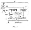
- Figure 1 illustrates a vascular-border-identification system 10 in accordance with one embodiment of the present invention.
- a data-gathering console 200 is electrically connected to a computing device 100 and a transducer 220 via a catheter 210.
- the transducer 220 is inserted into a blood vessel of a patient (not shown) and used to gather radio frequency (RF) data backscattered from vascular tissue.
- the RF data is then provided to (or acquired by) the data-gathering device 200, where it is used (or can be used) to produce an image of the vessel (e.g., intra-vascular ultrasound (IVUS) image, etc.).
- IVUS intra-vascular ultrasound
- RF data is, typically gathered in segments, either through a rotating transducer or an array of circumferentially positioned transducers, where each segment represents an angular portion of the resultant image.
- it takes a plurality of segments (or a set of RF data) to image an entire cross-section of a vascular object.
- multiple sets of RF data are typically gathered from multiple locations within a vascular object (e.g., by moving the transducer linearly through the vessel). These multiple sets of data can then be used to create a plurality of two-dimensional (2D) images or one three-dimensional (3D) image.
- the data-gathering device 200 includes, but is not limited to, an IVUS console, thermographic device, optical device (e.g., an optical coherence tomography (OCT) console), MRI device, or any vascular imaging device generally known to those skilled in the art.
- OCT optical coherence tomography
- the computing device 100 depicted in Figure 1 includes, but its not limited to, a personal computer or any other data-processing device (general purpose or application specific) that is generally known to those skilled in the art.
- the RF data (or multiple sets thereof) is provided to (or acquired by) the computing device 100.
- the computing device 100 includes at least one data storage device (e. g. , database 130, memory 150) and a plurality of applications (e. g. a characterization application 110, a gradient-border application 120, a frequency-border application 140 and/or an active-contour application 160).
- the RF data provided to (or acquired by) the computing device 100 is gated to electrocardiogram (ECG) information.
- ECG electrocardiogram
- a plurality of tissue types (e. g., medial, adventitial, plaque, blood, etc.) and related parameters are stored in a database 130.
- the information is stored so that each tissue type is linked to its corresponding parameters.
- each tissue type can be identified (or defined) by the parameters that are linked thereto.
- parameter includes, but is not limited to maximum power, minimum power, frequencies at maximum and/or minimum power, y intercepts (estimated or actual), slope, mid-band fit, integrated backscatter, tissue depth, and all parameters (either time or frequency based) generally known to (or discernable by) those skilled in the art.
- tissue type includes, but is not limited to, blood tissue, plaque tissue (e. g., calcified tissues, fibrous tissues, calcified-necrotic tissues and fibro-lipidic tissues), medial tissue, adventitial tissue, and all other vascular tissues, or combinations thereof (e. g. medial-adventitial tissue), generally known to those skilled in the art.
- plaque tissue e. g., calcified tissues, fibrous tissues, calcified-necrotic tissues and fibro-lipidic tissues
- medial tissue e. g., adventitial tissue, and all other vascular tissues, or combinations thereof (e. g. medial-adventitial tissue), generally known to those skilled in the art.
- data storage devices depicted herein e. g.
- database 130, memory 150 include, but are not limited to, RAM, cache memory, flash memory, magnetic disks, optical disks, removable disks, SCSI disks, IDE hard drives, tape drives and all other types of data storage devices (and combinations thereof, such as RAID devices) generally known to those skilled in the art.
- the RF data (which is typically in the time domain) is provided to the characterization application 110, where it is converted (or transformed) into the frequency domain.
- the characterization application 110 is then used to identify a plurality of parameters associated with the transformed RF data (or a portion thereof).
- the identified parameters are then compared to the parameters stored in the database 130 to identify the corresponding tissue type (or the type of tissue that backscattered the analyzed RF data).
- Such a process can be used (e. g. , once or repeatedly) to identify the portions of RF data (or sets thereof) that are associated with each stored tissue type (e. g., medial, adventitial, plaque, blood, etc.).
- the frequency. conversion (or transformation) discussed herein includes, but is not limited to, the use of a fast Fourier transformation (FFT), the Welch periodogram, autoregressive power spectrum (AR) analysis, or any other frequency transformation or spectral analysis generally known to those skilled in the art.
- FFT fast Fourier transformation
- AR autoregressive power spectrum
- the RF data may either be received in real-time (e. g., while the patient is in the operating room) or after a period of delay (e. g. via CD-ROM, etc.).
- the identified parameters should be related (generally) to the stored parameters.
- an estimated Y intercept parameter should be identified if data related to a signal's estimated Y intercept is stored in the database 130 and linked to at least one tissue type.
- the characterization application 110 is further used to identify parameters from the RF data (which is typically in the time domain). Parameters associated with the RF data (or a portion thereof) can be used, for example, to spatially identify certain frequencies (or parameters related thereto). Thus, for example, if a vascular wall comprises multiple tissue layers, corresponding RF data can be used to identify the location of these tissues and the related frequency spectrum can be used to identify tissue types. The use of parameters to identify tissue types is discussed in detail in U.S. Patent Number, 6,200, 268, which was issued on March 13, 2001 , and U.S. Patent Application Number 10/647,971, which was filed on August 25, 2003 .
- the RF data (or a transformation thereof) and the identified tissue types (including the association therebetween) are provided to the active-contour application 160. This data is then used to identify at least one border on an image of a vascular object (e. g., intra-vascular ultrasound (IVUS) image, Virtual Histology (VH) image, etc.).
- a vascular object e. g., intra-vascular ultrasound (IVUS) image, Virtual Histology (VH) image, etc.
- IVUS intra-vascular ultrasound
- VH Virtual Histology
- RF data corresponding to blood and plaque tissue can be used to identify (or substantially approximate) the luminal border on a vascular image.
- RF data corresponding to plaque, medial, and/or adventitial tissue can be used to identify (or substantially approximate) the medial-adventitial border on a vascular image.
- the present invention is not limited to the identification of any particular border (or boundary) on a vascular image, and includes all borders generally known to those skilled in the art.
- the applications depicted and discussed herein e. g. , characterization application 110, gradient-border application 120, frequency-border application 140, active-contour application 160
- the number and location of the components depicted in Figure 1 are not intended to limit the present invention, and are merely provided to illustrate the environment in which the present invention may operate.
- a computing device having a single data storage device and/or a remotely located characterization application is within the scope of the present invention.
- the computing device 100 further includes a frequency-border application 140.
- the characterization application 110 is adapted to provide the RF data (or a transformation thereof) and the identified tissue types (including the association therebetween) to the frequency-border application 140, where it is used to determine spectral information.
- the spectral information is then provided to the active-contour application 160 and used to determine at least one border on a vascular image (i. e. image-border data).
- the spectral information comprises spectral-force data.
- Spectral-force data is based (either directly or indirectly) on the information provided by the characterization application 110 and represents a frequency-based force that is applicable to (or a component of) the image-border data.
- the spectral-force data can be used, for example, to determine or refine the image-border data (e.g., as determined by the active-contour application 160).
- spectral-force data can be used together with other border-related information to determine a border on a vascular image, or applied to a border determined using border-related information (e.g., as an external force).
- the resultant border is based, at least in part, on the spectral-force data.
- spectral-force data can be analogized to a gravitation field, in which the force (or field) is used to attract a border in a given direction, or to have a given shape.
- the strength of the force (or gravitational pull) is directly proportional to the relatedness of the corresponding RF data to a border-related tissue type(s).
- the spectral information comprises spectral-border data.
- Spectral-border data is based (either directly or indirectly) on the information provided by the characterization application 110 and represents an estimation of at least one border on a vascular image.
- the spectral-border data can be used, for example, either alone or together with other border-related information, to determine image-border data (e.g., as determined by the active-contour application 160).
- image-border data e.g., as determined by the active-contour application 160.
- spectral information is not limited to spectral-force data and/or spectral-border data, but further includes any data resulting from a spectral analysis of the acquired RF data.
- the frequency-border application 140 is further adapted to filter the transformed RF data before it is used to generate spectral information. Specifically, in one embodiment of the present invention, a portion of the vascular object is selected. The corresponding transformed RF data and the information stored in the database 130 are then used to identify the tissue types that are related thereto. The minority tissue type is then filtered out. This can be accomplished, for example, by reassigning the RF data associated with the minority tissue type, so that it is associated with the majority tissue types.
- a first portion (or ninety-five percent) of the corresponding RF data is associate with blood tissue and a second portion (or fiver percent) of the corresponding RF data is associated with plaque tissue, then the second portion would be reassigned so that it is associate with blood tissue (i. e. making a homogenous composition).
- the present invention is not limited to such a filtering algorithm, and includes any other filtering algorithms generally known to those skilled in the art.
- the computing device 100 further includes a gradient-border application 120.
- the gradient- border application 120 is adapted to use the acquired RF data to determine gradient information, which can then be used to identify a border or boundary (e. g. , used by the gradient-border application 120 to estimate at least one border, provided to the active- contour application and used together with spectral information to determine image- border data, etc.). This is because a change in pixel color (e. g. light-to-dark, dark-to- light, shadel-to-shade2, etc) can indicate the presence of a border.
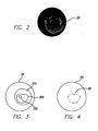
- Figure 2 illustrates an exemplary IVUS image 20 of a vascular object.
- the catheter can be identified by the first light-to-dark transition (or gradient).
- the catheter border is further identified in Figure 3 (i. e. , 330).
- the next dark-to-light transition or gradient
- the medial-adventitial border can then be identified by going outward from the luminal border until the next dark-to-light transition (or gradient) is found (see Figure 3 , 310).
- the IVUS image is constructed using gray-scales, it may be necessary to Utilize an algorithm and/or at least one threshold value to identify precisely where the image changes from light to dark (or vice versa).
- the present invention is not limited to any particular algorithm for identifying the aforementioned transitions, and includes all algorithms (and/or threshold values) generally known to those skilled in the art.
- the gradient-border application 120 is further adapted to process the acquired RF data using traditional IVUS imaging techniques.
- the gradient-border application 120 may be adapted to filter the RF data (e. g. , using a highpass filter, etc.), detect relevant portions (e. g. envelope detection, etc.), and/or modulate any portion thereof (e. g. smoothing, log compressing, etc.). Such techniques are all well known to those skilled in the art.
- the resultant data can then be used to produce an IVUS image or determine gradient information.
- the gradient information comprises gradient-force data.
- Gradient-force data is based (either directly or indirectly) on the gradients in an IVUS image and represents a gradient-based force that is applicable to (or a component of) the image-border data.
- the gradient-force data can be used, for example, to determine or refine the image-border data (e.g., as determined by the active-contour application 160).
- gradient-force data can be used together with other border-related information to determine a border on a vascular image, or applied to a border determined using border-related information (e.g., as an external force).
- the resultant border is based, at least in part, on the gradient-force data.
- gradient-force data can be analogized to a gravitation field, in which the force (or field) is used to attract a border in a given direction, or to have a given shape.
- the strength of the force is directly proportional to the gradients in the IVUS image (or transitions therein).
- the gradient information comprises gradient-border data.
- Gradient-border data is based (either directly or indirectly) on the gradients in an IVUS image and represents an estimation of at least one border on a vascular image (e.g., the IVUS image, a VH image, etc.).
- the gradient-border data can be used, for example, either alone or together with other border-related information (e.g., spectral information, etc.), to determine at least one border on a vascular image.
- border-related information e.g., spectral information, etc.
- the active-contour application 160 is further adapted to use other-border information to determine a border on a vascular images.
- information related to a border on another image(s) e.g., a previous image, a subsequent image, multiple images, etc.
- the active-contour application 160 is further adapted to use other-border information to determine a border on a vascular images.
- information related to a border on another image(s) e.g., a previous image, a subsequent image, multiple images, etc.
- the active-contour application 160 is further adapted to use other-border information to determine a border on a vascular images.
- information related to a border on another image(s) e.g., a previous image, a subsequent image, multiple images, etc.
- the border-detection algorithm 160 is adapted to identify at least one control point on the border of another image.
- the border-detection algorithm can be used, for example, to identify a plurality of control points 22 on the luminal border 320.
- the location and number of control points depicted in Figure 4 are not intended to limit the present invention, and are merely provided to illustrate the environment in which the present invention may operate.
- the active-contour application 160 is adapted to identify a border using user-identified control points. Such an embodiment is discussed in detail in U. S. Patent Number 6,381,350, which was issued on April 30,2002 .
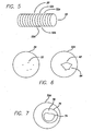
- the active-contour application 160 can be used to identify at least one control point on the current vascular image. In one embodiment of the present invention, this is done by extrapolating the previously identified control points to the current vascular image. By doing this, multiple 2D images (or at least one 3D image) can be produced. For example, as illustrated in Figure 5 , multiple 2D images (e. g. 20, 52a-52d, etc. ) are used to produce a 3D image of a tubular (e. g., vascular) object 50. It should be appreciated that a memory device 150 (see Figure 1 ) can be used to store information related to this embodiment (e. g. , a border on another image, control points on such a border, extrapolated control points, resulting image-border data, etc.).
- information related to this embodiment e. g. , a border on another image, control points on such a border, extrapolated control points, resulting image-border data, etc.
- Figure 6 illustrates one method as to how an identified control point can be extrapolated to the current vascular image.
- the control points that were illustrated in Figure 4 i. e. , 22
- the current image e.g. 52d
- the control points are extrapolated using Cartesian coordinates. It should be appreciated that, while Figure 6 illustrates control points being extrapolated to an adjacent image, the present invention is not so limited. Thus, extracting control points to (or from) additional images (e. g. 52c, 52b, etc.) is within the scope of the present invention.
- the active-contour application 160 is further adapted to identify (or approximate) a border based on the extrapolated points.
- the extrapolated points 62 may be connected using a plurality of lines 64, where the lines are either straight or curved (not shown).
- the extrapolating application is adapted to use an algorithm (e. g. , a cubic-interpolation algorithm, etc.) to identify line shape. Border extrapolation is discussed in detail in U. S. Patent Application Number 10/647,473, which was filed August 26,2003 .
- the active-contour application 160 is then used to adjust the border to more closely match the actual border of the vascular object.
- the active-contour application 160 may consider, or take into account, at least (i) the proximity of the border to each extrapolated point (i. e. , continuity or control-point factor), (ii) border curvature or smoothness (i. e. curvature or boundary factor), and/or (iii) the relationship between multiple borders (i. e. relatedness factor).
- continuity or control-point factor the border can be adjusted so that it passes through each extrapolated point.
- the border can be adjusted to prevent sharp transitions (e. g. corners, etc.).
- a relatedness factor multiple borders can be adjusted in relation to one another (e. g. , the luminal border can always be located inside the medial-adventitial border, etc.).
- these three factors are also used to determine related borders on adjacent images. It should be appreciated that if multiple factors are being considered, then individual factors may be weighted more heavily than others. This becomes important if the factors produce different results. It should further be appreciated that the present invention is not limited to the use of the aforementioned factors for border optimization, and that the use of additional factors to adjust (or optimize) a border is within the scope of the present invention.
- the adjusted borders are configured to be manually manipulated.
- at least one point on the border can be selected and manually moved to a new location:
- the active-contour application 160 is then used (as previously discussed) to reconstruct the border accordingly.
- the active-contour application is further adapted to adjust related borders in adjacent images. This is done by fitting a geometrical model (e.g., a tensor product B-spline, etc.) over the surface of a plurality of related borders (e.g., as identified on multiple IVUS images).
- a plurality of points on the geometrical model are then parameterized and formulated into a constrained least-squares system of equations. If a point on the border is manually moved, the active-contour application can utilize these equations to calculate a resulting surface (or mesh of control points).
- the affected borders e.g., adjacent borders
- the affected borders can then be adjusted accordingly.
- the active-contour application 160 can either be used (e.g., initiated) to identify a single border on a vascular image, thus requiring a subsequent use to identify another border on the vascular image, or used (e.g., initiated) to identify multiple borders on a vascular image.
- the multiple borders are identified at substantially the same time.
- the multiple borders can then be imaged (in either 2D or 3D) and analyzed by either a skilled practitioner or a computer algorithm.
- the luminal border 74 and the medial-adventitial border 76 can be used (by either a clinician or an algorithm) to identify the plaque-media complex 78 of a vascular object.
- RF data backscattered from vascular tissue is acquired.
- the RF data is then (i) transformed from the time domain into the frequency domain and (ii) processed using traditional IVUS imaging techniques, at steps 810 and 820, respectively.
- a plurality of parameters are identified at step 812, and compared to stored parameters, at step 814, to identify a plurality of tissue types.
- RF data (or a transformation thereof) that corresponds to at least relevant (e. g. border-related) tissue types is identified.
- the corresponding RF data is then used to determine spectral information (e. g. spectral-force data, spectral-border data, etc.) at step 818.
- spectral information e. g. spectral-force data, spectral-border data, etc.
- gradients are used to determine gradient information (e. g. , gradient-force data, gradient-border data, etc. ) at step 822.
- other-border data e. g. , data related to at least one border on at least one other image
- the gradient information and spectral information are used to modify the at least one border on the current vascular image, ending the method at step 828. It should be appreciated that the present invention is not limited to the method illustrated in Figure 8 .
Landscapes
- Health & Medical Sciences (AREA)
- Life Sciences & Earth Sciences (AREA)
- Physics & Mathematics (AREA)
- Biophysics (AREA)
- Veterinary Medicine (AREA)
- Pathology (AREA)
- Public Health (AREA)
- Engineering & Computer Science (AREA)
- Biomedical Technology (AREA)
- Heart & Thoracic Surgery (AREA)
- Medical Informatics (AREA)
- Molecular Biology (AREA)
- Surgery (AREA)
- Animal Behavior & Ethology (AREA)
- General Health & Medical Sciences (AREA)
- Radiology & Medical Imaging (AREA)
- Nuclear Medicine, Radiotherapy & Molecular Imaging (AREA)
- Vascular Medicine (AREA)
- Cardiology (AREA)
- Physiology (AREA)
- Ultra Sonic Daignosis Equipment (AREA)
- Supply Devices, Intensifiers, Converters, And Telemotors (AREA)
- Air Bags (AREA)
- Examining Or Testing Airtightness (AREA)
- Soil Working Implements (AREA)
- Holo Graphy (AREA)
Applications Claiming Priority (3)
| Application Number | Priority Date | Filing Date | Title |
|---|---|---|---|
| US55062004P | 2004-03-04 | 2004-03-04 | |
| US10/837,352 US7215802B2 (en) | 2004-03-04 | 2004-04-29 | System and method for vascular border detection |
| PCT/US2005/006159 WO2005091885A2 (en) | 2004-03-04 | 2005-02-25 | System and method for vascular border detection |
Publications (3)
| Publication Number | Publication Date |
|---|---|
| EP1732461A2 EP1732461A2 (en) | 2006-12-20 |
| EP1732461A4 EP1732461A4 (en) | 2008-05-21 |
| EP1732461B1 true EP1732461B1 (en) | 2011-04-06 |
Family
ID=34915696
Family Applications (1)
| Application Number | Title | Priority Date | Filing Date |
|---|---|---|---|
| EP05723848A Expired - Lifetime EP1732461B1 (en) | 2004-03-04 | 2005-02-25 | System for vascular border detection |
Country Status (6)
| Country | Link |
|---|---|
| US (3) | US7215802B2 (enExample) |
| EP (1) | EP1732461B1 (enExample) |
| JP (1) | JP4733107B2 (enExample) |
| AT (1) | ATE504239T1 (enExample) |
| DE (1) | DE602005027334D1 (enExample) |
| WO (1) | WO2005091885A2 (enExample) |
Families Citing this family (160)
| Publication number | Priority date | Publication date | Assignee | Title |
|---|---|---|---|---|
| US7727153B2 (en) * | 2003-04-07 | 2010-06-01 | Sonosite, Inc. | Ultrasonic blood vessel measurement apparatus and method |
| JP4489770B2 (ja) * | 2003-07-21 | 2010-06-23 | パイエオン インコーポレイテッド | 動く臓器を描出した画像シリーズ内の最適画像を識別する方法及びシステム |
| US7224356B2 (en) * | 2004-06-08 | 2007-05-29 | Microsoft Corporation | Stretch-driven mesh parameterization using spectral analysis |
| EP1615170A1 (en) * | 2004-07-10 | 2006-01-11 | Evotec Technologies GmbH | Image segmentation algorithms for applications in cellular biology |
| JP4575737B2 (ja) * | 2004-09-29 | 2010-11-04 | 富士フイルム株式会社 | 超音波撮像装置 |
| JP4585326B2 (ja) * | 2005-02-08 | 2010-11-24 | 富士フイルム株式会社 | 超音波撮像装置及び超音波撮像方法 |
| KR100856042B1 (ko) * | 2005-10-07 | 2008-09-03 | 주식회사 메디슨 | 웨이브렛 변환과 svm을 이용하여 초음파 영상에서대상체 볼륨을 추출하는 초음파 영상 시스템 및 방법 |
| US7988633B2 (en) * | 2005-10-12 | 2011-08-02 | Volcano Corporation | Apparatus and method for use of RFID catheter intelligence |
| US8135453B2 (en) * | 2005-12-07 | 2012-03-13 | Siemens Corporation | Method and apparatus for ear canal surface modeling using optical coherence tomography imaging |
| US20070160275A1 (en) * | 2006-01-11 | 2007-07-12 | Shashidhar Sathyanarayana | Medical image retrieval |
| US8249315B2 (en) * | 2006-05-22 | 2012-08-21 | Upmc | System and method for improved viewing and navigation of digital images |
| US9867530B2 (en) | 2006-08-14 | 2018-01-16 | Volcano Corporation | Telescopic side port catheter device with imaging system and method for accessing side branch occlusions |
| CN101662980B (zh) * | 2007-01-19 | 2013-02-27 | 桑尼布鲁克健康科学中心 | 用于成像探头的扫描机构 |
| US11197651B2 (en) | 2007-03-08 | 2021-12-14 | Sync-Rx, Ltd. | Identification and presentation of device-to-vessel relative motion |
| JP5639764B2 (ja) | 2007-03-08 | 2014-12-10 | シンク−アールエックス,リミティド | 運動する器官と共に使用するイメージング及びツール |
| US11064964B2 (en) | 2007-03-08 | 2021-07-20 | Sync-Rx, Ltd | Determining a characteristic of a lumen by measuring velocity of a contrast agent |
| WO2009153794A1 (en) | 2008-06-19 | 2009-12-23 | Sync-Rx, Ltd. | Stepwise advancement of a medical tool |
| US9375164B2 (en) | 2007-03-08 | 2016-06-28 | Sync-Rx, Ltd. | Co-use of endoluminal data and extraluminal imaging |
| US8781193B2 (en) | 2007-03-08 | 2014-07-15 | Sync-Rx, Ltd. | Automatic quantitative vessel analysis |
| US9629571B2 (en) | 2007-03-08 | 2017-04-25 | Sync-Rx, Ltd. | Co-use of endoluminal data and extraluminal imaging |
| US10716528B2 (en) | 2007-03-08 | 2020-07-21 | Sync-Rx, Ltd. | Automatic display of previously-acquired endoluminal images |
| US9968256B2 (en) | 2007-03-08 | 2018-05-15 | Sync-Rx Ltd. | Automatic identification of a tool |
| US9596993B2 (en) | 2007-07-12 | 2017-03-21 | Volcano Corporation | Automatic calibration systems and methods of use |
| WO2009009802A1 (en) | 2007-07-12 | 2009-01-15 | Volcano Corporation | Oct-ivus catheter for concurrent luminal imaging |
| EP2178442B1 (en) | 2007-07-12 | 2017-09-06 | Volcano Corporation | Catheter for in vivo imaging |
| WO2009143163A2 (en) * | 2008-05-21 | 2009-11-26 | University Of Florida Research Foundation, Inc. | Face relighting from a single image |
| US9974509B2 (en) | 2008-11-18 | 2018-05-22 | Sync-Rx Ltd. | Image super enhancement |
| US9144394B2 (en) | 2008-11-18 | 2015-09-29 | Sync-Rx, Ltd. | Apparatus and methods for determining a plurality of local calibration factors for an image |
| US8855744B2 (en) | 2008-11-18 | 2014-10-07 | Sync-Rx, Ltd. | Displaying a device within an endoluminal image stack |
| US9101286B2 (en) | 2008-11-18 | 2015-08-11 | Sync-Rx, Ltd. | Apparatus and methods for determining a dimension of a portion of a stack of endoluminal data points |
| US10362962B2 (en) | 2008-11-18 | 2019-07-30 | Synx-Rx, Ltd. | Accounting for skipped imaging locations during movement of an endoluminal imaging probe |
| US11064903B2 (en) | 2008-11-18 | 2021-07-20 | Sync-Rx, Ltd | Apparatus and methods for mapping a sequence of images to a roadmap image |
| US9095313B2 (en) | 2008-11-18 | 2015-08-04 | Sync-Rx, Ltd. | Accounting for non-uniform longitudinal motion during movement of an endoluminal imaging probe |
| US8478012B2 (en) * | 2009-09-14 | 2013-07-02 | General Electric Company | Methods, apparatus and articles of manufacture to process cardiac images to detect heart motion abnormalities |
| US12426789B2 (en) | 2009-09-23 | 2025-09-30 | Lightlab Imaging, Inc. | Blood vessel lumen morphology and minimum lumen area measurements data collection by intravascular imaging systems for stenosis or stent planning |
| ES2992724T3 (en) * | 2009-09-23 | 2024-12-17 | Light Lab Imaging Inc | Lumen morphology and vascular resistance measurements data collection systems, apparatus and methods |
| CA2777562C (en) | 2009-10-12 | 2016-08-16 | Silicon Valley Medical Instruments, Inc. | Intravascular ultrasound system for co-registered imaging |
| US8961420B2 (en) | 2010-04-01 | 2015-02-24 | Siemens Medical Solutions Usa, Inc. | System for cardiac condition detection and characterization |
| CN101964118B (zh) * | 2010-09-30 | 2012-09-19 | 华北电力大学(保定) | 一种血管内超声图像序列的三维分割方法 |
| US11141063B2 (en) | 2010-12-23 | 2021-10-12 | Philips Image Guided Therapy Corporation | Integrated system architectures and methods of use |
| US20120330146A1 (en) | 2010-12-31 | 2012-12-27 | Volcano Corporation | Pulmonary Embolism Therapeutic Devices and Associated Methods and Systems |
| US11040140B2 (en) | 2010-12-31 | 2021-06-22 | Philips Image Guided Therapy Corporation | Deep vein thrombosis therapeutic methods |
| EP2723231A4 (en) | 2011-06-23 | 2015-02-25 | Sync Rx Ltd | LUMINAL BACKGROUND CLEANING |
| US9295447B2 (en) | 2011-08-17 | 2016-03-29 | Volcano Corporation | Systems and methods for identifying vascular borders |
| WO2013033489A1 (en) | 2011-08-31 | 2013-03-07 | Volcano Corporation | Optical rotary joint and methods of use |
| CA2852233C (en) * | 2011-10-14 | 2023-08-08 | Jointvue, Llc | Real-time 3-d ultrasound reconstruction of knee and its implications for patient specific implants and 3-d joint injections |
| US10292676B2 (en) | 2011-12-21 | 2019-05-21 | Volcano Corporaton | Method for visualizing blood and blood-likelihood in vascualar images |
| US20130218014A1 (en) * | 2012-02-17 | 2013-08-22 | Samsung Electronics Co., Ltd. | Ultrasound apparatus and method of generating ultrasound image |
| EP2863802B1 (en) | 2012-06-26 | 2020-11-04 | Sync-RX, Ltd. | Flow-related image processing in luminal organs |
| US9858668B2 (en) | 2012-10-05 | 2018-01-02 | Volcano Corporation | Guidewire artifact removal in images |
| US11272845B2 (en) | 2012-10-05 | 2022-03-15 | Philips Image Guided Therapy Corporation | System and method for instant and automatic border detection |
| US10568586B2 (en) | 2012-10-05 | 2020-02-25 | Volcano Corporation | Systems for indicating parameters in an imaging data set and methods of use |
| WO2014055923A2 (en) * | 2012-10-05 | 2014-04-10 | Elizabeth Begin | System and method for instant and automatic border detection |
| US9292918B2 (en) | 2012-10-05 | 2016-03-22 | Volcano Corporation | Methods and systems for transforming luminal images |
| US9324141B2 (en) | 2012-10-05 | 2016-04-26 | Volcano Corporation | Removal of A-scan streaking artifact |
| US9478940B2 (en) | 2012-10-05 | 2016-10-25 | Volcano Corporation | Systems and methods for amplifying light |
| US20140100454A1 (en) | 2012-10-05 | 2014-04-10 | Volcano Corporation | Methods and systems for establishing parameters for three-dimensional imaging |
| US10070827B2 (en) | 2012-10-05 | 2018-09-11 | Volcano Corporation | Automatic image playback |
| US9307926B2 (en) | 2012-10-05 | 2016-04-12 | Volcano Corporation | Automatic stent detection |
| US9367965B2 (en) | 2012-10-05 | 2016-06-14 | Volcano Corporation | Systems and methods for generating images of tissue |
| US9286673B2 (en) | 2012-10-05 | 2016-03-15 | Volcano Corporation | Systems for correcting distortions in a medical image and methods of use thereof |
| US9840734B2 (en) | 2012-10-22 | 2017-12-12 | Raindance Technologies, Inc. | Methods for analyzing DNA |
| JP6322210B2 (ja) | 2012-12-13 | 2018-05-09 | ボルケーノ コーポレイション | 標的化された挿管のためのデバイス、システム、および方法 |
| US10942022B2 (en) | 2012-12-20 | 2021-03-09 | Philips Image Guided Therapy Corporation | Manual calibration of imaging system |
| CA2895502A1 (en) | 2012-12-20 | 2014-06-26 | Jeremy Stigall | Smooth transition catheters |
| EP2934310A4 (en) | 2012-12-20 | 2016-10-12 | Nathaniel J Kemp | RECONFIGURABLE OPTICAL COHERENCE TOMOGRAPHY SYSTEM BETWEEN DIFFERENT IMAGING MODES |
| CA2895770A1 (en) | 2012-12-20 | 2014-07-24 | Jeremy Stigall | Locating intravascular images |
| US11406498B2 (en) | 2012-12-20 | 2022-08-09 | Philips Image Guided Therapy Corporation | Implant delivery system and implants |
| US10939826B2 (en) | 2012-12-20 | 2021-03-09 | Philips Image Guided Therapy Corporation | Aspirating and removing biological material |
| US10413317B2 (en) | 2012-12-21 | 2019-09-17 | Volcano Corporation | System and method for catheter steering and operation |
| US10058284B2 (en) | 2012-12-21 | 2018-08-28 | Volcano Corporation | Simultaneous imaging, monitoring, and therapy |
| US9486143B2 (en) | 2012-12-21 | 2016-11-08 | Volcano Corporation | Intravascular forward imaging device |
| US10420530B2 (en) | 2012-12-21 | 2019-09-24 | Volcano Corporation | System and method for multipath processing of image signals |
| US10398413B2 (en) | 2012-12-21 | 2019-09-03 | Volcano Corporation | Method for multi-frequency imaging and composite image display using high-bandwidth transducer outputs |
| US10166003B2 (en) | 2012-12-21 | 2019-01-01 | Volcano Corporation | Ultrasound imaging with variable line density |
| EP2936626A4 (en) | 2012-12-21 | 2016-08-17 | David Welford | SYSTEMS AND METHOD FOR REDUCING A WAVELENGTH LIGHT EMISSION |
| US9612105B2 (en) | 2012-12-21 | 2017-04-04 | Volcano Corporation | Polarization sensitive optical coherence tomography system |
| US10993694B2 (en) | 2012-12-21 | 2021-05-04 | Philips Image Guided Therapy Corporation | Rotational ultrasound imaging catheter with extended catheter body telescope |
| CA2895993A1 (en) | 2012-12-21 | 2014-06-26 | Jason Spencer | System and method for graphical processing of medical data |
| US10191220B2 (en) | 2012-12-21 | 2019-01-29 | Volcano Corporation | Power-efficient optical circuit |
| US10368836B2 (en) * | 2012-12-26 | 2019-08-06 | Volcano Corporation | Gesture-based interface for a multi-modality medical imaging system |
| US10642953B2 (en) | 2012-12-26 | 2020-05-05 | Philips Image Guided Therapy Corporation | Data labeling and indexing in a multi-modality medical imaging system |
| US10799209B2 (en) | 2012-12-26 | 2020-10-13 | Philips Image Guided Therapy Corporation | Measurement navigation in a multi-modality medical imaging system |
| EP2965263B1 (en) | 2013-03-07 | 2022-07-20 | Bernhard Sturm | Multimodal segmentation in intravascular images |
| US10226597B2 (en) | 2013-03-07 | 2019-03-12 | Volcano Corporation | Guidewire with centering mechanism |
| US20140276923A1 (en) | 2013-03-12 | 2014-09-18 | Volcano Corporation | Vibrating catheter and methods of use |
| EP3895604A1 (en) | 2013-03-12 | 2021-10-20 | Collins, Donna | Systems and methods for diagnosing coronary microvascular disease |
| US9301687B2 (en) | 2013-03-13 | 2016-04-05 | Volcano Corporation | System and method for OCT depth calibration |
| US11026591B2 (en) | 2013-03-13 | 2021-06-08 | Philips Image Guided Therapy Corporation | Intravascular pressure sensor calibration |
| CN105120759B (zh) | 2013-03-13 | 2018-02-23 | 火山公司 | 用于从旋转血管内超声设备产生图像的系统和方法 |
| US20160030151A1 (en) | 2013-03-14 | 2016-02-04 | Volcano Corporation | Filters with echogenic characteristics |
| US9592027B2 (en) * | 2013-03-14 | 2017-03-14 | Volcano Corporation | System and method of adventitial tissue characterization |
| WO2014151808A1 (en) * | 2013-03-14 | 2014-09-25 | Volcano Corporation | Parallelized tree-based pattern recognition for tissue characterization |
| US10292677B2 (en) | 2013-03-14 | 2019-05-21 | Volcano Corporation | Endoluminal filter having enhanced echogenic properties |
| US12343198B2 (en) | 2013-03-14 | 2025-07-01 | Philips Image Guided Therapy Corporation | Delivery catheter having imaging capabilities |
| US10219887B2 (en) | 2013-03-14 | 2019-03-05 | Volcano Corporation | Filters with echogenic characteristics |
| US10154826B2 (en) | 2013-07-17 | 2018-12-18 | Tissue Differentiation Intelligence, Llc | Device and method for identifying anatomical structures |
| US10716536B2 (en) | 2013-07-17 | 2020-07-21 | Tissue Differentiation Intelligence, Llc | Identifying anatomical structures |
| US11260160B2 (en) | 2014-01-14 | 2022-03-01 | Philips Image Guided Therapy Corporation | Systems and methods for improving an AV access site |
| WO2015108973A1 (en) | 2014-01-14 | 2015-07-23 | Volcano Corporation | Methods and systems for clearing thrombus from a vascular access site |
| CN105916457A (zh) | 2014-01-14 | 2016-08-31 | 火山公司 | 用于形成血管通路的装置和方法 |
| WO2015108942A1 (en) | 2014-01-14 | 2015-07-23 | Volcano Corporation | Vascular access evaluation and treatment |
| CN106163386B (zh) | 2014-01-14 | 2019-08-06 | 火山公司 | 用于评估血液透析动静脉瘘管成熟的系统和方法 |
| US10098702B2 (en) | 2014-07-11 | 2018-10-16 | Volcano Corporation | Devices, systems, and methods for treatment of vessels |
| WO2016092390A1 (en) | 2014-12-08 | 2016-06-16 | Koninklijke Philips N.V. | Interactive physiologic data and intravascular imaging data and associated devices, systems, and methods |
| JP6751092B2 (ja) | 2014-12-10 | 2020-09-02 | コーニンクレッカ フィリップス エヌ ヴェKoninklijke Philips N.V. | ステント内再狭窄予測のためのデバイス、システム、及び方法 |
| CN107530049B (zh) | 2015-02-20 | 2021-06-04 | 皇家飞利浦有限公司 | 支持成像的斑块切除设备 |
| WO2016207762A1 (en) | 2015-06-25 | 2016-12-29 | Koninklijke Philips N.V. | Interactive intravascular procedure training and associated devices, systems, and methods |
| JP6516597B2 (ja) * | 2015-07-07 | 2019-05-22 | キヤノン株式会社 | 画像処理装置及び画像処理方法 |
| WO2017046628A1 (en) | 2015-09-15 | 2017-03-23 | Koninklijke Philips N.V. | Device and method for using ivus data to characterize and evaluate a vascular graft condition |
| US10653393B2 (en) | 2015-10-08 | 2020-05-19 | Acist Medical Systems, Inc. | Intravascular ultrasound imaging with frequency selective imaging methods and systems |
| US10909661B2 (en) * | 2015-10-08 | 2021-02-02 | Acist Medical Systems, Inc. | Systems and methods to reduce near-field artifacts |
| US11369337B2 (en) | 2015-12-11 | 2022-06-28 | Acist Medical Systems, Inc. | Detection of disturbed blood flow |
| KR101937018B1 (ko) * | 2016-03-24 | 2019-01-09 | 울산대학교 산학협력단 | 딥러닝을 이용한 혈관내 초음파 영상에서의 혈관내외경 자동 분할 방법 및 장치 |
| EP3459048B1 (en) | 2016-05-16 | 2023-05-03 | Acist Medical Systems, Inc. | Motion-based image segmentation systems and methods |
| US11986341B1 (en) | 2016-05-26 | 2024-05-21 | Tissue Differentiation Intelligence, Llc | Methods for accessing spinal column using B-mode imaging to determine a trajectory without penetrating the the patient's anatomy |
| US11701086B1 (en) | 2016-06-21 | 2023-07-18 | Tissue Differentiation Intelligence, Llc | Methods and systems for improved nerve detection |
| US11883235B2 (en) | 2017-08-15 | 2024-01-30 | Philips Image Guided Therapy Corporation | Phased array imaging and therapy intraluminal ultrasound device |
| EP3668411B1 (en) | 2017-08-15 | 2021-01-13 | Koninklijke Philips N.V. | Frequency-tunable intraluminal ultrasound device |
| EP3668409B1 (en) | 2017-08-15 | 2022-04-27 | Koninklijke Philips N.V. | Intraluminal ultrasound device for diagnostic imaging and therapy |
| US11819360B2 (en) | 2017-08-15 | 2023-11-21 | Koninklijke Philips N.V. | Intraluminal rotational ultrasound for diagnostic imaging and therapy |
| US12178643B2 (en) | 2017-08-15 | 2024-12-31 | Philips Image Guided Therapy Corporation | Intracardiac therapeutic and diagnostic ultrasound device |
| EP3668597A1 (en) | 2017-08-16 | 2020-06-24 | Koninklijke Philips N.V. | Disposable therapeutic ultrasound device |
| WO2019174971A1 (en) | 2018-03-14 | 2019-09-19 | Koninklijke Philips N.V. | Alternative anatomical borders of blood vessels and associated devices, systems, and methods |
| US11481905B2 (en) * | 2018-04-26 | 2022-10-25 | University Of Louisville Research Foundation, Inc. | Atlas for automatic segmentation of retina layers from OCT images |
| EP4272654B1 (en) | 2018-10-26 | 2025-12-10 | Koninklijke Philips N.V. | Speed determination for intraluminal ultrasound imaging and associated devices, systems, and methods |
| JP7378484B2 (ja) | 2018-10-26 | 2023-11-13 | コーニンクレッカ フィリップス エヌ ヴェ | 自動及び支援ラベル及びブックマークを用いた管腔内超音波イメージング |
| JP7493523B2 (ja) | 2018-10-26 | 2024-05-31 | コーニンクレッカ フィリップス エヌ ヴェ | 管腔内超音波方向性ガイダンス、並びに関連するデバイス、システム、及び方法 |
| EP3870057B1 (en) | 2018-10-26 | 2025-02-12 | Koninklijke Philips N.V. | Disease specific and treatment type specific control of intraluminal ultrasound imaging |
| EP3870063A1 (en) | 2018-10-26 | 2021-09-01 | Koninklijke Philips N.V. | Intraluminal ultrasound navigation guidance and associated devices, systems, and methods |
| US12440188B2 (en) | 2018-10-26 | 2025-10-14 | Philips Image Guided Therapy Corporation | Graphical longitudinal display for intraluminal ultrasound imaging and associated devices, systems, and methods |
| US11596384B2 (en) | 2018-10-26 | 2023-03-07 | Philips Image Guided Therapy Corporation | Intraluminal ultrasound vessel border selection and associated devices, systems, and methods |
| CN110051385B (zh) * | 2019-05-29 | 2023-01-24 | 深圳华声医疗技术股份有限公司 | 基于血管识别的全自动测量方法、装置、存储介质及系统 |
| CN110246136B (zh) * | 2019-05-29 | 2021-07-02 | 山东大学 | 一种基于混合算法的血管内超声参数提取方法及系统 |
| US11024034B2 (en) | 2019-07-02 | 2021-06-01 | Acist Medical Systems, Inc. | Image segmentation confidence determination |
| US12178640B2 (en) | 2019-10-08 | 2024-12-31 | Philips Image Guided Therapy Corporation | Visualization of reflectors in intraluminal ultrasound images and associated systems, methods, and devices |
| WO2021069216A1 (en) * | 2019-10-10 | 2021-04-15 | Koninklijke Philips N.V. | Vascular tissue characterization devices, systems, and methods |
| WO2021089810A1 (en) | 2019-11-06 | 2021-05-14 | Philips Image Guided Therapy Corporation | Co-registration of intravascular data and multi-segment vasculature, and associated devices, systems, and methods |
| US12394080B2 (en) | 2019-12-10 | 2025-08-19 | Philips Image Guided Therapy Corporation | Intraluminal image-based vessel diameter determination and associated devices, systems, and methods |
| EP4087492A1 (en) | 2020-01-06 | 2022-11-16 | Koninklijke Philips N.V. | Intraluminal imaging based detection and visualization of intraluminal treatment anomalies |
| WO2021180501A1 (en) | 2020-03-10 | 2021-09-16 | Koninklijke Philips N.V. | Intraluminal image visualization with adaptive scaling and associated systems, methods, and devices |
| EP4408297A1 (en) | 2021-09-30 | 2024-08-07 | Koninklijke Philips N.V. | Intraluminal ultrasound vessel segment identification and associated devices, systems, and methods |
| US20230181140A1 (en) | 2021-12-11 | 2023-06-15 | Philips Image Guided Therapy Corporation | Registration of intraluminal physiological data to longitudinal image body lumen using extraluminal imaging data |
| US20230190227A1 (en) | 2021-12-16 | 2023-06-22 | Philips Image Guided Therapy Corporation | Plaque burden indication on longitudinal intraluminal image and x-ray image |
| EP4449430A1 (en) | 2021-12-17 | 2024-10-23 | Philips Image Guided Therapy Corporation | Intravascular imaging assessment of stent deployment and associated systems, devices, and methods |
| CN118414127A (zh) | 2021-12-17 | 2024-07-30 | 皇家飞利浦有限公司 | 通过共同配准的血管内成像控制激光斑块切除术 |
| WO2023110555A1 (en) | 2021-12-17 | 2023-06-22 | Koninklijke Philips N.V. | Systems, devices, and methods for coregistration of intravascular data to enhanced stent deployment x-ray images |
| EP4201342A1 (en) | 2021-12-22 | 2023-06-28 | Koninklijke Philips N.V. | Intravascular ultrasound imaging for calcium detection and analysis |
| WO2023117721A1 (en) | 2021-12-22 | 2023-06-29 | Koninklijke Philips N.V. | Intraluminal imaging for reference image frame and target image frame confirmation with deep breathing |
| JP2025500957A (ja) | 2021-12-22 | 2025-01-15 | コーニンクレッカ フィリップス エヌ ヴェ | カルシウム検出及び分析のための血管内超音波イメージング |
| US20230190215A1 (en) | 2021-12-22 | 2023-06-22 | Philips Image Guided Therapy Corporation | Co-registration of intraluminal data to no contrast x-ray image frame and associated systems, device and methods |
| US20250160786A1 (en) | 2022-03-08 | 2025-05-22 | Philips Image Guided Therapy Corporation | Intravascular ultrasound imaging with contour generation and editing for circular and non-circular blood vessel borders |
| EP4543302A1 (en) | 2022-06-24 | 2025-04-30 | Koninklijke Philips N.V. | Intraluminal ultrasound imaging with automatic detection of target and reference regions |
| JP2025538852A (ja) | 2022-12-07 | 2025-12-02 | コーニンクレッカ フィリップス エヌ ヴェ | 管腔外画像データを使用した管腔内生理学的データと体腔の長手方向画像とのレジストレーション |
| WO2025103969A1 (en) | 2023-11-17 | 2025-05-22 | Koninklijke Philips N.V. | Intravascular image border detection responsive to location inside region and associated systems, devices, and methods |
| CN120324013A (zh) * | 2024-01-17 | 2025-07-18 | 开立生物医疗科技(武汉)有限公司 | 上消化道超声图像处理方法、装置、设备及介质 |
| WO2025168460A1 (en) | 2024-02-07 | 2025-08-14 | Koninklijke Philips N.V. | Intravascular imaging for stent planning with simultaneous landing zone adjustment and visualization |
| WO2025209855A1 (en) | 2024-04-02 | 2025-10-09 | Koninklijke Philips N.V. | Plaque shift and/or carina shift during stent placement and associated systems, devices, and methods |
| WO2025237795A1 (en) | 2024-05-15 | 2025-11-20 | Koninklijke Philips N.V. | Intravascular data-based treatment plan during delivery of treatment to blood vessel accompanying x-ray images without radiopaque contrast |
| WO2025252576A1 (en) | 2024-06-05 | 2025-12-11 | Koninklijke Philips N.V. | Catheter-based intravascular imaging with identification of vessel blockage and/or stent irregularity |
Family Cites Families (53)
| Publication number | Priority date | Publication date | Assignee | Title |
|---|---|---|---|---|
| US4228804A (en) | 1978-02-28 | 1980-10-21 | Case Western Reserve University | Diagnostic ultrasonography utilizing frequency spectrum analysis presented in terms of B-scan color patterns or X-Y graph displays |
| JPS5849140A (ja) | 1981-09-19 | 1983-03-23 | 株式会社東芝 | 超音波診断装置 |
| US4478219A (en) * | 1982-03-24 | 1984-10-23 | Manuel Dujovny | Temporary microvascular occluder |
| US4575799A (en) | 1983-03-23 | 1986-03-11 | Fujitsu Limited | Ultrasonic living body tissue characterization system |
| US4561019A (en) | 1983-05-16 | 1985-12-24 | Riverside Research Institute | Frequency diversity for image enhancement |
| US4858124A (en) | 1984-08-15 | 1989-08-15 | Riverside Research Institute | Method for enhancement of ultrasonic image data |
| US5445155A (en) | 1991-03-13 | 1995-08-29 | Scimed Life Systems Incorporated | Intravascular imaging apparatus and methods for use and manufacture |
| US5235984A (en) | 1992-03-30 | 1993-08-17 | Hewlett-Packard Company | On-line acoustic densitometry tool for use with an ultrasonic imaging system |
| JPH05337111A (ja) * | 1992-06-10 | 1993-12-21 | Nippon Koden Corp | 超音波診断装置 |
| WO1994023652A1 (en) * | 1993-04-19 | 1994-10-27 | Commonwealth Scientific And Industrial Research Organisation | Tissue characterisation using intravascular echoscopy |
| DE69429743T2 (de) | 1993-06-29 | 2002-10-02 | Koninklijke Philips Electronics N.V., Eindhoven | Verfahren und Gerät zur Bestimmung einer Kontur in einem durch eine Dichteverteilung gekennzeichneten Raum |
| US5363850A (en) | 1994-01-26 | 1994-11-15 | Cardiovascular Imaging Systems, Inc. | Method for recognition and reduction of blood speckle in blood vessel imaging system |
| US5417215A (en) | 1994-02-04 | 1995-05-23 | Long Island Jewish Medical Center | Method of tissue characterization by ultrasound |
| WO1995029737A1 (en) * | 1994-05-03 | 1995-11-09 | Board Of Regents, The University Of Texas System | Apparatus and method for noninvasive doppler ultrasound-guided real-time control of tissue damage in thermal therapy |
| US6615071B1 (en) * | 1995-09-20 | 2003-09-02 | Board Of Regents, The University Of Texas System | Method and apparatus for detecting vulnerable atherosclerotic plaque |
| US5724972A (en) | 1996-05-02 | 1998-03-10 | Acuson Corporation | Method and apparatus for distributed focus control with slope tracking |
| US5938607A (en) | 1996-09-25 | 1999-08-17 | Atl Ultrasound, Inc. | Ultrasonic diagnostic imaging system with access to reference image library |
| US6095976A (en) | 1997-06-19 | 2000-08-01 | Medinol Ltd. | Method for enhancing an image derived from reflected ultrasound signals produced by an ultrasound transmitter and detector inserted in a bodily lumen |
| JP2001511374A (ja) | 1997-07-25 | 2001-08-14 | アーチ・デベロップメント・コーポレーション | 側面胸部放射線像の肺領域を分割する方法とシステム |
| US6106465A (en) | 1997-08-22 | 2000-08-22 | Acuson Corporation | Ultrasonic method and system for boundary detection of an object of interest in an ultrasound image |
| US5957138A (en) | 1997-08-25 | 1999-09-28 | Diasonics Ultrasound, Inc. | Method and apparatus for three-dimensional flow lumen imaging |
| US6148095A (en) | 1997-09-08 | 2000-11-14 | University Of Iowa Research Foundation | Apparatus and method for determining three-dimensional representations of tortuous vessels |
| US5876343A (en) | 1997-09-23 | 1999-03-02 | Scimed Life Systems, Inc. | Methods and apparatus for blood speckle detection in an intravascular ultrasound imaging system |
| US5885218A (en) | 1997-11-07 | 1999-03-23 | Scimed Life Systems, Inc. | Method and apparatus for spatial filtering in an intravascular ultrasound imaging system |
| FR2772590B1 (fr) * | 1997-12-18 | 2000-04-14 | Michel Puech | Utilisation d'un transducteur ultrasonore pour l'exploration echographique du segment posterieur du globe oculaire |
| US6106460A (en) | 1998-03-26 | 2000-08-22 | Scimed Life Systems, Inc. | Interface for controlling the display of images of diagnostic or therapeutic instruments in interior body regions and related data |
| US6238342B1 (en) | 1998-05-26 | 2001-05-29 | Riverside Research Institute | Ultrasonic tissue-type classification and imaging methods and apparatus |
| US6063032A (en) | 1998-09-28 | 2000-05-16 | Scimed Systems, Inc. | Ultrasound imaging with zoom having independent processing channels |
| US6120445A (en) * | 1998-10-02 | 2000-09-19 | Scimed Life Systems, Inc. | Method and apparatus for adaptive cross-sectional area computation of IVUS objects using their statistical signatures |
| US7070595B2 (en) * | 1998-12-14 | 2006-07-04 | Medwaves, Inc. | Radio-frequency based catheter system and method for ablating biological tissues |
| US6398736B1 (en) | 1999-03-31 | 2002-06-04 | Mayo Foundation For Medical Education And Research | Parametric imaging ultrasound catheter |
| US6381350B1 (en) * | 1999-07-02 | 2002-04-30 | The Cleveland Clinic Foundation | Intravascular ultrasonic analysis using active contour method and system |
| US6200268B1 (en) | 1999-09-10 | 2001-03-13 | The Cleveland Clinic Foundation | Vascular plaque characterization |
| US6264609B1 (en) * | 1999-09-15 | 2001-07-24 | Wake Forest University | Ultrasound apparatus and method for tissue characterization |
| US6306089B1 (en) | 1999-09-24 | 2001-10-23 | Atl Ultrasound, Inc. | Ultrasonic diagnostic imaging system with customized measurements and calculations |
| US6454715B2 (en) | 2000-04-11 | 2002-09-24 | Scimed Life Systems, Inc. | Methods and apparatus for blood speckle detection in an intravascular ultrasound imaging system |
| US6561980B1 (en) | 2000-05-23 | 2003-05-13 | Alpha Intervention Technology, Inc | Automatic segmentation of prostate, rectum and urethra in ultrasound imaging |
| US6450960B1 (en) | 2000-08-29 | 2002-09-17 | Barbara Ann Karmanos Cancer Institute | Real-time three-dimensional acoustoelectronic imaging and characterization of objects |
| AU2001296873A1 (en) | 2000-09-14 | 2002-03-26 | Leland Stanford Junior University | Technique for manipulating medical images |
| US6785409B1 (en) | 2000-10-24 | 2004-08-31 | Koninklijke Philips Electronics, N.V. | Segmentation method and apparatus for medical images using diffusion propagation, pixel classification, and mathematical morphology |
| JP4614548B2 (ja) * | 2001-01-31 | 2011-01-19 | パナソニック株式会社 | 超音波診断装置 |
| WO2002100249A2 (en) * | 2001-06-13 | 2002-12-19 | Cardiovascular Innovations, Inc. | Apparatus and method for ultrasonically identifying vulnerable plaque |
| US6475149B1 (en) * | 2001-09-21 | 2002-11-05 | Acuson Corporation | Border detection method and system |
| EP1345154A1 (en) * | 2002-03-11 | 2003-09-17 | Bracco Imaging S.p.A. | A method for encoding image pixels and method for processing images aimed at qualitative recognition of the object reproduced by one more image pixels |
| DE10210648A1 (de) | 2002-03-11 | 2003-10-02 | Siemens Ag | Verfahren zur Erfassung und Darstellung eines in ein zu untersuchendes oder behandelndes Hohlraumorgan eines Patienten eingeführten medizinischen Instruments |
| US7927275B2 (en) | 2002-08-26 | 2011-04-19 | The Cleveland Clinic Foundation | System and method of aquiring blood-vessel data |
| US7074188B2 (en) | 2002-08-26 | 2006-07-11 | The Cleveland Clinic Foundation | System and method of characterizing vascular tissue |
| JP4933045B2 (ja) * | 2002-08-26 | 2012-05-16 | ザ クリーブランド クリニック ファウンデーション | 血管組織を特徴付けするシステムおよび方法 |
| US7359554B2 (en) | 2002-08-26 | 2008-04-15 | Cleveland Clinic Foundation | System and method for identifying a vascular border |
| US6824514B2 (en) | 2002-10-11 | 2004-11-30 | Koninklijke Philips Electronics N.V. | System and method for visualizing scene shift in ultrasound scan sequence |
| US6835177B2 (en) | 2002-11-06 | 2004-12-28 | Sonosite, Inc. | Ultrasonic blood vessel measurement apparatus and method |
| US7466848B2 (en) * | 2002-12-13 | 2008-12-16 | Rutgers, The State University Of New Jersey | Method and apparatus for automatically detecting breast lesions and tumors in images |
| US7175597B2 (en) | 2003-02-03 | 2007-02-13 | Cleveland Clinic Foundation | Non-invasive tissue characterization system and method |
-
2004
- 2004-04-29 US US10/837,352 patent/US7215802B2/en not_active Expired - Lifetime
-
2005
- 2005-02-25 EP EP05723848A patent/EP1732461B1/en not_active Expired - Lifetime
- 2005-02-25 JP JP2007501859A patent/JP4733107B2/ja not_active Expired - Lifetime
- 2005-02-25 DE DE602005027334T patent/DE602005027334D1/de not_active Expired - Lifetime
- 2005-02-25 WO PCT/US2005/006159 patent/WO2005091885A2/en not_active Ceased
- 2005-02-25 AT AT05723848T patent/ATE504239T1/de not_active IP Right Cessation
-
2006
- 2006-06-08 US US11/449,569 patent/US7463759B2/en not_active Expired - Lifetime
-
2007
- 2007-04-12 US US11/786,599 patent/US20070201736A1/en not_active Abandoned
Also Published As
| Publication number | Publication date |
|---|---|
| US20050196026A1 (en) | 2005-09-08 |
| US20070201736A1 (en) | 2007-08-30 |
| WO2005091885A2 (en) | 2005-10-06 |
| JP4733107B2 (ja) | 2011-07-27 |
| ATE504239T1 (de) | 2011-04-15 |
| EP1732461A2 (en) | 2006-12-20 |
| US7215802B2 (en) | 2007-05-08 |
| EP1732461A4 (en) | 2008-05-21 |
| US20070071326A1 (en) | 2007-03-29 |
| US7463759B2 (en) | 2008-12-09 |
| DE602005027334D1 (de) | 2011-05-19 |
| JP2007526083A (ja) | 2007-09-13 |
| WO2005091885A3 (en) | 2006-11-02 |
Similar Documents
| Publication | Publication Date | Title |
|---|---|---|
| EP1732461B1 (en) | System for vascular border detection | |
| US7359554B2 (en) | System and method for identifying a vascular border | |
| US8449465B2 (en) | System and method for characterizing vascular tissue | |
| JP2007526083A5 (enExample) | ||
| Golemati et al. | Using the Hough transform to segment ultrasound images of longitudinal and transverse sections of the carotid artery | |
| US7074188B2 (en) | System and method of characterizing vascular tissue | |
| EP1534137B1 (en) | System and method for identifying a vascular border | |
| CN105120764A (zh) | 用于组织特征化的基于并行树的样式辨识 | |
| Vonesh et al. | Regional vascular mechanical properties by 3-D intravascular ultrasound with finite-element analysis | |
| WO2005070300A1 (en) | System and method for determining a transfer function | |
| Ciompi et al. | Reconstruction and analysis of intravascular ultrasound sequences | |
| WO2025103969A1 (en) | Intravascular image border detection responsive to location inside region and associated systems, devices, and methods | |
| INTRAVASCULAR | Adithya G. Gangidi and Chi Hau Chen | |
| Abreu | Automatic location of atherosclerotic plaques by ultrasounds | |
| Kanber | Identifying the vulnerable carotid plaque by means of dynamic ultrasound image analysis | |
| Yue | Noise suppression and motion estimation in medical ultrasound imaging | |
| O'Malley | Computational methods for contrast-enhanced intravascular ultrasound sequence analysis | |
| Mehta | Coronary Plaque Characterization by Texture Analysis of Intravascular Ultrasound Images | |
| Ahanathapillai | Image processing techniques for automated diagnosis of wall motion abnormality in echocardiography sequences |
Legal Events
| Date | Code | Title | Description |
|---|---|---|---|
| PUAI | Public reference made under article 153(3) epc to a published international application that has entered the european phase |
Free format text: ORIGINAL CODE: 0009012 |
|
| 17P | Request for examination filed |
Effective date: 20060904 |
|
| AK | Designated contracting states |
Kind code of ref document: A2 Designated state(s): AT BE BG CH CY CZ DE DK EE ES FI FR GB GR HU IE IS IT LI LT LU MC NL PL PT RO SE SI SK TR |
|
| AX | Request for extension of the european patent |
Extension state: AL BA HR LV MK YU |
|
| RIN1 | Information on inventor provided before grant (corrected) |
Inventor name: KLINGENSMITH, JON, D. Inventor name: KUBAN, BARRY, D. Inventor name: NAIR, ANUJA Inventor name: VINCE, D., GEOFFREY |
|
| DAX | Request for extension of the european patent (deleted) | ||
| A4 | Supplementary search report drawn up and despatched |
Effective date: 20080422 |
|
| 17Q | First examination report despatched |
Effective date: 20080813 |
|
| GRAP | Despatch of communication of intention to grant a patent |
Free format text: ORIGINAL CODE: EPIDOSNIGR1 |
|
| RIC1 | Information provided on ipc code assigned before grant |
Ipc: A61B 8/08 20060101AFI20100720BHEP Ipc: A61B 6/12 20060101ALI20100720BHEP |
|
| RTI1 | Title (correction) |
Free format text: SYSTEM FOR VASCULAR BORDER DETECTION |
|
| GRAS | Grant fee paid |
Free format text: ORIGINAL CODE: EPIDOSNIGR3 |
|
| RAP1 | Party data changed (applicant data changed or rights of an application transferred) |
Owner name: THE CLEVELAND CLINIC FOUNDATION |
|
| GRAA | (expected) grant |
Free format text: ORIGINAL CODE: 0009210 |
|
| AK | Designated contracting states |
Kind code of ref document: B1 Designated state(s): AT BE BG CH CY CZ DE DK EE ES FI FR GB GR HU IE IS IT LI LT LU MC NL PL PT RO SE SI SK TR |
|
| REG | Reference to a national code |
Ref country code: GB Ref legal event code: FG4D |
|
| REG | Reference to a national code |
Ref country code: CH Ref legal event code: EP |
|
| REG | Reference to a national code |
Ref country code: IE Ref legal event code: FG4D |
|
| REF | Corresponds to: |
Ref document number: 602005027334 Country of ref document: DE Date of ref document: 20110519 Kind code of ref document: P |
|
| REG | Reference to a national code |
Ref country code: DE Ref legal event code: R096 Ref document number: 602005027334 Country of ref document: DE Effective date: 20110519 |
|
| REG | Reference to a national code |
Ref country code: NL Ref legal event code: VDEP Effective date: 20110406 |
|
| PG25 | Lapsed in a contracting state [announced via postgrant information from national office to epo] |
Ref country code: SI Free format text: LAPSE BECAUSE OF FAILURE TO SUBMIT A TRANSLATION OF THE DESCRIPTION OR TO PAY THE FEE WITHIN THE PRESCRIBED TIME-LIMIT Effective date: 20110406 |
|
| LTIE | Lt: invalidation of european patent or patent extension |
Effective date: 20110406 |
|
| PG25 | Lapsed in a contracting state [announced via postgrant information from national office to epo] |
Ref country code: PT Free format text: LAPSE BECAUSE OF FAILURE TO SUBMIT A TRANSLATION OF THE DESCRIPTION OR TO PAY THE FEE WITHIN THE PRESCRIBED TIME-LIMIT Effective date: 20110808 Ref country code: LT Free format text: LAPSE BECAUSE OF FAILURE TO SUBMIT A TRANSLATION OF THE DESCRIPTION OR TO PAY THE FEE WITHIN THE PRESCRIBED TIME-LIMIT Effective date: 20110406 Ref country code: SE Free format text: LAPSE BECAUSE OF FAILURE TO SUBMIT A TRANSLATION OF THE DESCRIPTION OR TO PAY THE FEE WITHIN THE PRESCRIBED TIME-LIMIT Effective date: 20110406 |
|
| PG25 | Lapsed in a contracting state [announced via postgrant information from national office to epo] |
Ref country code: ES Free format text: LAPSE BECAUSE OF FAILURE TO SUBMIT A TRANSLATION OF THE DESCRIPTION OR TO PAY THE FEE WITHIN THE PRESCRIBED TIME-LIMIT Effective date: 20110717 Ref country code: IS Free format text: LAPSE BECAUSE OF FAILURE TO SUBMIT A TRANSLATION OF THE DESCRIPTION OR TO PAY THE FEE WITHIN THE PRESCRIBED TIME-LIMIT Effective date: 20110806 Ref country code: FI Free format text: LAPSE BECAUSE OF FAILURE TO SUBMIT A TRANSLATION OF THE DESCRIPTION OR TO PAY THE FEE WITHIN THE PRESCRIBED TIME-LIMIT Effective date: 20110406 Ref country code: AT Free format text: LAPSE BECAUSE OF FAILURE TO SUBMIT A TRANSLATION OF THE DESCRIPTION OR TO PAY THE FEE WITHIN THE PRESCRIBED TIME-LIMIT Effective date: 20110406 Ref country code: CY Free format text: LAPSE BECAUSE OF FAILURE TO SUBMIT A TRANSLATION OF THE DESCRIPTION OR TO PAY THE FEE WITHIN THE PRESCRIBED TIME-LIMIT Effective date: 20110406 Ref country code: BE Free format text: LAPSE BECAUSE OF FAILURE TO SUBMIT A TRANSLATION OF THE DESCRIPTION OR TO PAY THE FEE WITHIN THE PRESCRIBED TIME-LIMIT Effective date: 20110406 Ref country code: GR Free format text: LAPSE BECAUSE OF FAILURE TO SUBMIT A TRANSLATION OF THE DESCRIPTION OR TO PAY THE FEE WITHIN THE PRESCRIBED TIME-LIMIT Effective date: 20110707 |
|
| PG25 | Lapsed in a contracting state [announced via postgrant information from national office to epo] |
Ref country code: NL Free format text: LAPSE BECAUSE OF FAILURE TO SUBMIT A TRANSLATION OF THE DESCRIPTION OR TO PAY THE FEE WITHIN THE PRESCRIBED TIME-LIMIT Effective date: 20110406 |
|
| PG25 | Lapsed in a contracting state [announced via postgrant information from national office to epo] |
Ref country code: EE Free format text: LAPSE BECAUSE OF FAILURE TO SUBMIT A TRANSLATION OF THE DESCRIPTION OR TO PAY THE FEE WITHIN THE PRESCRIBED TIME-LIMIT Effective date: 20110406 Ref country code: CZ Free format text: LAPSE BECAUSE OF FAILURE TO SUBMIT A TRANSLATION OF THE DESCRIPTION OR TO PAY THE FEE WITHIN THE PRESCRIBED TIME-LIMIT Effective date: 20110406 |
|
| PLBE | No opposition filed within time limit |
Free format text: ORIGINAL CODE: 0009261 |
|
| STAA | Information on the status of an ep patent application or granted ep patent |
Free format text: STATUS: NO OPPOSITION FILED WITHIN TIME LIMIT |
|
| PG25 | Lapsed in a contracting state [announced via postgrant information from national office to epo] |
Ref country code: RO Free format text: LAPSE BECAUSE OF FAILURE TO SUBMIT A TRANSLATION OF THE DESCRIPTION OR TO PAY THE FEE WITHIN THE PRESCRIBED TIME-LIMIT Effective date: 20110406 Ref country code: SK Free format text: LAPSE BECAUSE OF FAILURE TO SUBMIT A TRANSLATION OF THE DESCRIPTION OR TO PAY THE FEE WITHIN THE PRESCRIBED TIME-LIMIT Effective date: 20110406 Ref country code: PL Free format text: LAPSE BECAUSE OF FAILURE TO SUBMIT A TRANSLATION OF THE DESCRIPTION OR TO PAY THE FEE WITHIN THE PRESCRIBED TIME-LIMIT Effective date: 20110406 Ref country code: DK Free format text: LAPSE BECAUSE OF FAILURE TO SUBMIT A TRANSLATION OF THE DESCRIPTION OR TO PAY THE FEE WITHIN THE PRESCRIBED TIME-LIMIT Effective date: 20110406 |
|
| 26N | No opposition filed |
Effective date: 20120110 |
|
| REG | Reference to a national code |
Ref country code: DE Ref legal event code: R097 Ref document number: 602005027334 Country of ref document: DE Effective date: 20120110 |
|
| PG25 | Lapsed in a contracting state [announced via postgrant information from national office to epo] |
Ref country code: MC Free format text: LAPSE BECAUSE OF NON-PAYMENT OF DUE FEES Effective date: 20120229 |
|
| REG | Reference to a national code |
Ref country code: CH Ref legal event code: PL |
|
| PG25 | Lapsed in a contracting state [announced via postgrant information from national office to epo] |
Ref country code: CH Free format text: LAPSE BECAUSE OF NON-PAYMENT OF DUE FEES Effective date: 20120229 Ref country code: LI Free format text: LAPSE BECAUSE OF NON-PAYMENT OF DUE FEES Effective date: 20120229 |
|
| REG | Reference to a national code |
Ref country code: IE Ref legal event code: MM4A |
|
| PG25 | Lapsed in a contracting state [announced via postgrant information from national office to epo] |
Ref country code: IE Free format text: LAPSE BECAUSE OF NON-PAYMENT OF DUE FEES Effective date: 20120225 |
|
| PG25 | Lapsed in a contracting state [announced via postgrant information from national office to epo] |
Ref country code: BG Free format text: LAPSE BECAUSE OF FAILURE TO SUBMIT A TRANSLATION OF THE DESCRIPTION OR TO PAY THE FEE WITHIN THE PRESCRIBED TIME-LIMIT Effective date: 20110706 |
|
| PG25 | Lapsed in a contracting state [announced via postgrant information from national office to epo] |
Ref country code: TR Free format text: LAPSE BECAUSE OF FAILURE TO SUBMIT A TRANSLATION OF THE DESCRIPTION OR TO PAY THE FEE WITHIN THE PRESCRIBED TIME-LIMIT Effective date: 20110406 |
|
| PG25 | Lapsed in a contracting state [announced via postgrant information from national office to epo] |
Ref country code: LU Free format text: LAPSE BECAUSE OF NON-PAYMENT OF DUE FEES Effective date: 20120225 |
|
| PG25 | Lapsed in a contracting state [announced via postgrant information from national office to epo] |
Ref country code: HU Free format text: LAPSE BECAUSE OF FAILURE TO SUBMIT A TRANSLATION OF THE DESCRIPTION OR TO PAY THE FEE WITHIN THE PRESCRIBED TIME-LIMIT Effective date: 20050225 |
|
| REG | Reference to a national code |
Ref country code: FR Ref legal event code: PLFP Year of fee payment: 12 |
|
| PGFP | Annual fee paid to national office [announced via postgrant information from national office to epo] |
Ref country code: IT Payment date: 20160223 Year of fee payment: 12 |
|
| REG | Reference to a national code |
Ref country code: FR Ref legal event code: PLFP Year of fee payment: 13 |
|
| REG | Reference to a national code |
Ref country code: DE Ref legal event code: R082 Ref document number: 602005027334 Country of ref document: DE |
|
| REG | Reference to a national code |
Ref country code: FR Ref legal event code: PLFP Year of fee payment: 14 |
|
| PG25 | Lapsed in a contracting state [announced via postgrant information from national office to epo] |
Ref country code: IT Free format text: LAPSE BECAUSE OF NON-PAYMENT OF DUE FEES Effective date: 20170225 |
|
| PGFP | Annual fee paid to national office [announced via postgrant information from national office to epo] |
Ref country code: DE Payment date: 20240228 Year of fee payment: 20 Ref country code: GB Payment date: 20240220 Year of fee payment: 20 |
|
| PGFP | Annual fee paid to national office [announced via postgrant information from national office to epo] |
Ref country code: FR Payment date: 20240226 Year of fee payment: 20 |
|
| REG | Reference to a national code |
Ref country code: DE Ref legal event code: R071 Ref document number: 602005027334 Country of ref document: DE |
|
| REG | Reference to a national code |
Ref country code: GB Ref legal event code: PE20 Expiry date: 20250224 |
|
| PG25 | Lapsed in a contracting state [announced via postgrant information from national office to epo] |
Ref country code: GB Free format text: LAPSE BECAUSE OF EXPIRATION OF PROTECTION Effective date: 20250224 |