EP1668156B9 - Methode de diagnostic du cancer du sein - Google Patents
Methode de diagnostic du cancer du sein Download PDFInfo
- Publication number
- EP1668156B9 EP1668156B9 EP04773526A EP04773526A EP1668156B9 EP 1668156 B9 EP1668156 B9 EP 1668156B9 EP 04773526 A EP04773526 A EP 04773526A EP 04773526 A EP04773526 A EP 04773526A EP 1668156 B9 EP1668156 B9 EP 1668156B9
- Authority
- EP
- European Patent Office
- Prior art keywords
- brc
- expression
- genes
- protein
- gene
- Prior art date
- Legal status (The legal status is an assumption and is not a legal conclusion. Google has not performed a legal analysis and makes no representation as to the accuracy of the status listed.)
- Not-in-force
Links
Images


Classifications
-
- C—CHEMISTRY; METALLURGY
- C12—BIOCHEMISTRY; BEER; SPIRITS; WINE; VINEGAR; MICROBIOLOGY; ENZYMOLOGY; MUTATION OR GENETIC ENGINEERING
- C12Q—MEASURING OR TESTING PROCESSES INVOLVING ENZYMES, NUCLEIC ACIDS OR MICROORGANISMS; COMPOSITIONS OR TEST PAPERS THEREFOR; PROCESSES OF PREPARING SUCH COMPOSITIONS; CONDITION-RESPONSIVE CONTROL IN MICROBIOLOGICAL OR ENZYMOLOGICAL PROCESSES
- C12Q1/00—Measuring or testing processes involving enzymes, nucleic acids or microorganisms; Compositions therefor; Processes of preparing such compositions
- C12Q1/68—Measuring or testing processes involving enzymes, nucleic acids or microorganisms; Compositions therefor; Processes of preparing such compositions involving nucleic acids
- C12Q1/6876—Nucleic acid products used in the analysis of nucleic acids, e.g. primers or probes
- C12Q1/6883—Nucleic acid products used in the analysis of nucleic acids, e.g. primers or probes for diseases caused by alterations of genetic material
- C12Q1/6886—Nucleic acid products used in the analysis of nucleic acids, e.g. primers or probes for diseases caused by alterations of genetic material for cancer
-
- A—HUMAN NECESSITIES
- A61—MEDICAL OR VETERINARY SCIENCE; HYGIENE
- A61P—SPECIFIC THERAPEUTIC ACTIVITY OF CHEMICAL COMPOUNDS OR MEDICINAL PREPARATIONS
- A61P35/00—Antineoplastic agents
-
- A—HUMAN NECESSITIES
- A61—MEDICAL OR VETERINARY SCIENCE; HYGIENE
- A61P—SPECIFIC THERAPEUTIC ACTIVITY OF CHEMICAL COMPOUNDS OR MEDICINAL PREPARATIONS
- A61P35/00—Antineoplastic agents
- A61P35/04—Antineoplastic agents specific for metastasis
-
- C—CHEMISTRY; METALLURGY
- C07—ORGANIC CHEMISTRY
- C07K—PEPTIDES
- C07K14/00—Peptides having more than 20 amino acids; Gastrins; Somatostatins; Melanotropins; Derivatives thereof
- C07K14/435—Peptides having more than 20 amino acids; Gastrins; Somatostatins; Melanotropins; Derivatives thereof from animals; from humans
- C07K14/46—Peptides having more than 20 amino acids; Gastrins; Somatostatins; Melanotropins; Derivatives thereof from animals; from humans from vertebrates
- C07K14/47—Peptides having more than 20 amino acids; Gastrins; Somatostatins; Melanotropins; Derivatives thereof from animals; from humans from vertebrates from mammals
- C07K14/4701—Peptides having more than 20 amino acids; Gastrins; Somatostatins; Melanotropins; Derivatives thereof from animals; from humans from vertebrates from mammals not used
- C07K14/4702—Regulators; Modulating activity
-
- G—PHYSICS
- G01—MEASURING; TESTING
- G01N—INVESTIGATING OR ANALYSING MATERIALS BY DETERMINING THEIR CHEMICAL OR PHYSICAL PROPERTIES
- G01N33/00—Investigating or analysing materials by specific methods not covered by groups G01N1/00 - G01N31/00
- G01N33/48—Biological material, e.g. blood, urine; Haemocytometers
- G01N33/50—Chemical analysis of biological material, e.g. blood, urine; Testing involving biospecific ligand binding methods; Immunological testing
- G01N33/53—Immunoassay; Biospecific binding assay; Materials therefor
- G01N33/574—Immunoassay; Biospecific binding assay; Materials therefor for cancer
- G01N33/57407—Specifically defined cancers
- G01N33/57415—Specifically defined cancers of breast
-
- C—CHEMISTRY; METALLURGY
- C12—BIOCHEMISTRY; BEER; SPIRITS; WINE; VINEGAR; MICROBIOLOGY; ENZYMOLOGY; MUTATION OR GENETIC ENGINEERING
- C12Q—MEASURING OR TESTING PROCESSES INVOLVING ENZYMES, NUCLEIC ACIDS OR MICROORGANISMS; COMPOSITIONS OR TEST PAPERS THEREFOR; PROCESSES OF PREPARING SUCH COMPOSITIONS; CONDITION-RESPONSIVE CONTROL IN MICROBIOLOGICAL OR ENZYMOLOGICAL PROCESSES
- C12Q2600/00—Oligonucleotides characterized by their use
- C12Q2600/112—Disease subtyping, staging or classification
-
- C—CHEMISTRY; METALLURGY
- C12—BIOCHEMISTRY; BEER; SPIRITS; WINE; VINEGAR; MICROBIOLOGY; ENZYMOLOGY; MUTATION OR GENETIC ENGINEERING
- C12Q—MEASURING OR TESTING PROCESSES INVOLVING ENZYMES, NUCLEIC ACIDS OR MICROORGANISMS; COMPOSITIONS OR TEST PAPERS THEREFOR; PROCESSES OF PREPARING SUCH COMPOSITIONS; CONDITION-RESPONSIVE CONTROL IN MICROBIOLOGICAL OR ENZYMOLOGICAL PROCESSES
- C12Q2600/00—Oligonucleotides characterized by their use
- C12Q2600/118—Prognosis of disease development
-
- C—CHEMISTRY; METALLURGY
- C12—BIOCHEMISTRY; BEER; SPIRITS; WINE; VINEGAR; MICROBIOLOGY; ENZYMOLOGY; MUTATION OR GENETIC ENGINEERING
- C12Q—MEASURING OR TESTING PROCESSES INVOLVING ENZYMES, NUCLEIC ACIDS OR MICROORGANISMS; COMPOSITIONS OR TEST PAPERS THEREFOR; PROCESSES OF PREPARING SUCH COMPOSITIONS; CONDITION-RESPONSIVE CONTROL IN MICROBIOLOGICAL OR ENZYMOLOGICAL PROCESSES
- C12Q2600/00—Oligonucleotides characterized by their use
- C12Q2600/136—Screening for pharmacological compounds
-
- C—CHEMISTRY; METALLURGY
- C12—BIOCHEMISTRY; BEER; SPIRITS; WINE; VINEGAR; MICROBIOLOGY; ENZYMOLOGY; MUTATION OR GENETIC ENGINEERING
- C12Q—MEASURING OR TESTING PROCESSES INVOLVING ENZYMES, NUCLEIC ACIDS OR MICROORGANISMS; COMPOSITIONS OR TEST PAPERS THEREFOR; PROCESSES OF PREPARING SUCH COMPOSITIONS; CONDITION-RESPONSIVE CONTROL IN MICROBIOLOGICAL OR ENZYMOLOGICAL PROCESSES
- C12Q2600/00—Oligonucleotides characterized by their use
- C12Q2600/156—Polymorphic or mutational markers
-
- C—CHEMISTRY; METALLURGY
- C12—BIOCHEMISTRY; BEER; SPIRITS; WINE; VINEGAR; MICROBIOLOGY; ENZYMOLOGY; MUTATION OR GENETIC ENGINEERING
- C12Q—MEASURING OR TESTING PROCESSES INVOLVING ENZYMES, NUCLEIC ACIDS OR MICROORGANISMS; COMPOSITIONS OR TEST PAPERS THEREFOR; PROCESSES OF PREPARING SUCH COMPOSITIONS; CONDITION-RESPONSIVE CONTROL IN MICROBIOLOGICAL OR ENZYMOLOGICAL PROCESSES
- C12Q2600/00—Oligonucleotides characterized by their use
- C12Q2600/158—Expression markers
-
- G—PHYSICS
- G01—MEASURING; TESTING
- G01N—INVESTIGATING OR ANALYSING MATERIALS BY DETERMINING THEIR CHEMICAL OR PHYSICAL PROPERTIES
- G01N2500/00—Screening for compounds of potential therapeutic value
- G01N2500/04—Screening involving studying the effect of compounds C directly on molecule A (e.g. C are potential ligands for a receptor A, or potential substrates for an enzyme A)
Definitions
- the present invention relates to methods of detecting and diagnosing breast cancer as well as methods of treating and preventing breast cancer and breast cancer metastasis.
- the present inventors have analyzed the expression profiles of tumor or tumors from various tissues by cDNA microarrays ( Okabe, H.et al., Genome-wide analysis of gene expression in human hepatocellular carcinomas using cDNA microarray: identification of genes involved in viral carcinogenesis and tumor progression. Cancer Res, 61: 2129-2137,2001 .; Hasegawa, S.et al.,Genome-wide analysis of gene expression in intestinal-type gastric cancers using a complementary DNA microarray representing 23,040 genes. Cancer Res, 62: 7012-7017, 2002 .; Kaneta, Y.et al., and Ohno, R.
- FTIs farnesyltransferase
- a tyrosine kinase inhibitor which selectively inactivates bcr-abl fusion proteins, has been developed to treat chronic myelogenous leukemias wherein constitutive activation of bcr-abl tyrosine kinase plays a crucial role in the transformation of leukocytes.
- CTLs cytotoxic T lymphocytes
- TAAs tumor-associated antigens
- TAAs are currently undergoing clinical development as targets of immunotherapy. TAAs discovered so far include MAGE ( van der Bruggen et al., Science 254: 1643-7 (1991 )), gp100 ( Kawakami et al., J Exp Med 180: 347-52 (1994 )), SART ( Shichijo et al., J Exp Med 187: 277-88 (1998 )), and NY-ESO-1 ( Chen et al., Proc Natl Acad Sci USA 94: 1914-8 (1997 )). On the other hand, gene products demonstrated to be specifically over-expressed in tumor cells have been shown to be recognized as targets inducing cellular immune responses.
- Such gene products include p53 ( Umano et al., Brit J Cancer 84:1052-7 (2001 )), HER2/neu ( Tanaka et al., Brit J Cancer 84: 94-9 (2001 )), CEA ( Nukaya et al., Int J Cancer 80: 92-7 (1999 )), and so on.
- TAAs In spite of significant progress in basic and clinical research concerning TAAs ( Rosenberg et al., Nature Med 4: 321-7 (1998 ); Mukherji et al., Proc Natl Acad Sci USA 92: 8078-82 (1995 ); Hu et al., Cancer Res 56: 2479-83 (1996 )), only limited number of candidate TAAs for the treatment of adenocarcinomas, including colorectal cancer, are currently available. TAAs abundantly expressed in cancer cells yet whose expression is restricted to cancer cells would be promising candidates as immunotherapeutic targets.
- PBMCs peripheral blood mononuclear cells
- HLA-A24 and HLA-A0201 are popular HLA alleles in the Japanese, as well as the Caucasian populations ( Date et al., Tissue Antigens 47: 93-101 (1996 ); Kondo et al., J Immunol 155: 4307-12 (1995 ); Kubo et al., J Immunol 152:3913-24 (1994 ); Imanishi et al., Proceeding of the eleventh International Histocompatibility Workshop and Conference Oxford University Press, Oxford, 1065 (1992 ); Williams et al., Tissue Antigen 49: 129 (1997 )).
- antigenic peptides of carcinomas presented by these HLAs may be especially useful for the treatment of carcinomas among Japanese and Caucasians.
- the induction of low-affinity CTL in vitro usually results from the use of peptide at a high concentration; generating a high level of specific peptide/MHC complexes on antigen presenting cells (APCs), which will effectively activate these CTL ( Alexander-Miller et al., Proc Natl Acad Sci USA 93: 4102-7 (1996 )).
- PBK may show a higher expression in said samples ( WO02/29104 ).
- the present inventors performed large scale genome-wide analyses of gene expression profiles found in purified populations of breast cancer cells, including 12 ductal carcinomas in situ (DCIS) and 69 invasive ductal carcinomas (IDC), using a cDNA microarray representing 23.040 genes.
- DCIS 12 ductal carcinomas in situ
- IDC 69 invasive ductal carcinomas
- BRC breast cancer
- the present invention relates to the embodiments characterized in the claims. Thus, it relates to the following items:
- BRC-associated gene refers to a gene that is characterized by an expression level which differs in a BRC cell as compared to a normal cell.
- a normal cell is one obtained from breast tissue.
- the BRC-associated gene is the gene listed in tables 3-8 (i.e., gene of BRC No. 456).
- control level refers to a protein expression level detected in a control sample and includes both a normal control level and an breast cancer control level.
- a control level can be a single expression pattern derived from a single reference population or from a plurality of expression patterns.
- the control level can be a database of expression patterns from previously tested cells.
- a "normal control level” refers to a level of gene expression detected in a normal, healthy individual or in a population of individuals known not to be suffering from breast cancer. A normal individual is one with no clinical symptoms of breast cancer.
- a "BRC control level” refers to an expression profile of BRC-associated genes found in a population suffering from BRC.
- expression of a panel of BRC-associated genes in a sample can be compared to a BRC control level of the same panel of genes.
- a similarity between a sample expression and BRC control expression indicates that the subject (from which the sample was obtained) suffers from or is at risk of developing BRC.
- gene expression level is deemed “altered” when gene expression is increased or decreased 10%, 25%, 50% as compared to the control level.
- an expression level is deemed “increased” or “decreased” when gene expression is increased or decreased by at least 0.1, at least 0.2, at least 1, at least 2, at least 5, or at least 10 or more fold as compared to a control level.
- Expression is determined by detecting hybridization, e.g ., on an array, of a BRC-associated gene probe to a gene transcript of the patient-derived tissue sample.
- the patient-derived tissue sample is any tissue obtained from a test subject, e.g ., a patient known to or suspected of having BRC.
- the tissue may contains an epithelial cell.
- the tissue may be an epithelial cell from a breast ductal carcinoma.
- BRC reference expression profile comprising a gene expression level of two or more of BRC-associated genes listed in tables 3-8.
- the BRC reference expression profile may comprise the levels of expression of two or more of BRC-associated genes listed in tables 3, 5, and 7, or BRC-associated genes listed in tables 4, 6, and 8.
- the present invention further provides methods of identifying an agent that inhibits or enhances the expression or activity of an BRC-associated gene, i.e. the BRC-associated gene BRC No. 456, by contacting a test cell expressing a BRC-associated gene with a test compound and determining the expression level of the BRC-associated gene or the activity of its gene product
- the test cell may be an epithelial cell, such as an epithelial cell obtained from a breast carcinoma.
- a decrease in the expression level of an up-regulated BRC-associated gene or the activity of its gene product as compared to a normal control level or activity of the gene or gene product indicates that the test agent is an inhibitor of the BRC-associated gene and may be used to reduce a symptom of BRC, e.g .
- an increase in the expression level of a down-regulated BRC-associated gene or the activity of its gene product as compared to a normal control level or activity of the gene or gene product indicates that the test agent is an enhancer of expression or function of the BRC-associated gene and may be used to reduce a symptom of BRC, e.g., the under-expression of one or more BRC-associated genes listed in tables 4, 6, and 8.
- kits comprising a detection reagent which binds to one or more BRC nucleic acids or BRC polypeptides. Also provided is an array of nucleic acids that binds to one or more BRC nucleic acids.
- Therapeutic methods as disclosed herein include a method of treating or preventing BRC in a subject including the step of administering to the subject an antisense composition.
- the antisense composition reduces the expression of the specific target gene.
- the antisense composition may contain a nucleotide which is complementary to a BRC-associated gene sequence selected from the group consisting of the BRC-associated genes listed in tables 3, 5, and 7.
- the present method may include the steps of administering to a subject a small interfering RNA (siRNAs) composition.
- siRNA composition reduces the expression of a BRC nucleic acid selected from the group consisting of the BRC-associated genes listed in tables 3, 5, and 7.
- the treatment or prevention of BRC in a subject may be carried out by administering to a subject a ribozyme composition.
- the nucleic acid-specific ribozyme composition reduces the expression of a BRC nucleic acid selected from the group consisting of the BRC-associated genes listed in tables 3, 5, and 7.
- the inhibition effect of the siRNA for BRC-associated genes listed in the tables was confirmed.
- the siRNA for BRC-456 of table 7 (GenBank Accession No. AF237709, TOPK;T-LAK cell-originated protein kinase) inhibit cell proliferation of breast cancer cells in the examples section.
- BRC-associated genes listed in tables 3, 5, and 7, especially BRC-456 is preferable therapeutic target of the breaset cancer.
- Other therapeutic methods include those in which a subject is administered a compound that increases the expression of one or more of the BRC-associated genes listed in tables 4, 6, and 8 or the activity of a polypeptide encoded by one or more of the BRC-associated genes listed in tables 4, 6, and 8.
- a method of treating or preventing BRC in a subject may involve administering to the subject a vaccine containing a polypeptide encoded by a nucleic acid selected from the group consisting of BRC-associated genes listed in tables 3, 5, and 7 or an immunologically active fragment of such a polypeptide.
- an immunologically active fragment is a polypeptide that is shorter in length than the full-length naturally-occurring protein yet which induces an immune response analogous to that induced by the full-length protein.
- an immunologically active fragment should be at least 8 residues in length and capable of stimulating an immune cell such as a T cell or a B cell.
- Immune cell stimulation can be measured by detecting cell proliferation, elaboration of cytokines (e.g ., IL-2), or production of an antibody.
- genes listed in table 11 i.e., genes of BRC Nos. 719-752 were identified as genes having unique altered expression patterns in breast cancer cells with lymph-node metastasis.
- metastasis of breast cancer can be treated or prevented via the suppression of the expression or activity of up-regulated genes or their gene products selected from the group consisting of VAMP3, MGC11257, GSPT1, DNM2, CFL1, CLNS1A, SENP2, NDUFS3, NOP5/NOP58, PSMD13, SUOX, HRB2, LOC154467, THTPA, ZRF1, LOC51255, DEAF1, NEU1, UGCGL1, BRAF, TUFM, FLJ10726, DNAJB1, AP4S1, and MRPL40.
- genes or their gene products selected from the group consisting of VAMP3, MGC11257, GSPT1, DNM2, CFL1, CLNS1A, SENP2, NDUFS3, NOP5/NOP58, PSMD13, SUOX, HRB2, LOC154467, THTPA, ZRF1, LOC51255, DEAF1, NEU1, UGCGL1, BRAF, TUFM, FLJ10726, DNAJB1, AP4
- metastasis of breast cancer can be treated or prevented by enhancing the expression or activity of UBA52, GenBank Acc# AA634090, CEACAM3, C21orf97, KIAA1040, EEF1D, FUS, GenBank Acc# AW965200, and KIAA0475 in cancerous cells.
- the present method comprises the step of measuring the expression level of marker genes selected from the group consisting of genes listed in table 11. These marker genes are identified herein as genes having unique altered expression patterns in breast cancer cells of patients with lymph node metastasis. Therefore, metastasis of the breast cancer in a subject can be predicted by determining whether the expression level detected in a sample derived from the subject is closer to the mean expression level of lymph node metastasis positive cases or negative cases in reference samples.
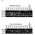
- A7870 designed T-LAK cell-originated protein kinase (TOPK), that was more than three-fold overexpressed in 30 of 39 (77%) breast cancer cases which were able to obtain expression data, especially in 29 of 36 (81 %) cases with invasive ductal carcinoma specimens.
- TOPK T-LAK cell-originated protein kinase
- Subsequent semi-quantitative RT-PCR also confirmed that A7870 were up-regulated in 7 of 12 clinical breast cancer samples and 17 of 20 breast cancer cell lines, compared to normal human organs including breast ductal cells or normal breast.
- Northern blot analyses revealed that the A7870 transcript was expressed only in breast cancer cell lines and normal human testis and thymus.
- breast cancer cells exist as a solid mass having a highly inflammatory reaction and containing various cellular components. Therefore, previous published microarray data are likely to reflect heterogenous profiles.
- the present inventors prepared purified populations of breast cancer cells and normal breast epithelial duct cells by a method of laser-microbeam microdissection (LMM), and analyzed genome-wide gene-expression profiles of 81 BRCs, including 12 ductal carcinomas in situ (DCIS) and 69 invasive ductal carcinomas (IDC), using a cDNA microarray representing 23,040 genes.
- LMM laser-microbeam microdissection
- DCIS ductal carcinomas in situ
- IDC 69 invasive ductal carcinomas
- the present invention is based, in part, on the discovery of changes in expression patterns of multiple nucleic acids between epithelial.cells and carcinomas of patients with BRC. The differences in gene expression were identified using a comprehensive DNA microarray system.
- the gene-expression profiles of cancer cells from 81 BRCs were analyzed using a cDNA microarray representing 23,040 genes coupled with laser microdissection.
- a cDNA microarray representing 23,040 genes coupled with laser microdissection.
- 102 genes shown in tables 3, 5 and 7 were identified as commonly up-regulated in BRC cells and among them 100 genes were selected as BRC-associated genes of the present disclosure
- 288 genes shown in tables 4, 6 and 8) were also identified as being commonly down-regulated in BRC cells.
- tables 3 and 4 provide a list of genes whose expression is altered between BRC, including DCIS and IDC, and normal tissue. Genes commonly up- or down- regulated in DCIS and IDC are shown in table 3 and table 4, respectively. Genes having elevated or decreased expression in transition from DCIS to IDC are listed in tables 5 and 6, respectively. Furthermore, genes commonly up- or down-regulated in IDC as compared with normal tissue are listed in tables 7 and 8, respectively.
- differentially expressed genes identified herein find diagnostic utility as markers of BRC and as BRC gene targets, the expression of which may be altered to treat or alleviate a symptom of BRC.
- genes differentially expressed between DCIS and IDC identified herein find diagnostic utility as markers for distinguishing IDC from DCIS and as BRC gene targets, the expression of which may be altered to treat or alleviate a symptom of IDC.
- BRC-associated genes The genes whose expression level is modulated (i.e ., increased or decreased) in BRC patients are summarized in tables 3-8 and are collectively referred to herein as "BRC-associated genes", “BRC nucleic acids” or “BRC polynucleotides” and the corresponding encoded polypeptides are referred to as “BRC polypeptides” or “BRC proteins.” Unless indicated otherwise, “BRC” refers to any of the sequences disclosed herein. ( e.g ., BRC-associated genes listed in tables 3-8). Genes that have been previously described are presented along with a database accession number.
- BRC By measuring expression of the various genes in a sample of cells, BRC can be diagnosed. Similarly, measuring the expression of these genes in response to various agents can identify agents for treating BRC.
- the present disclosure involves determining ( e.g ., measuring) the expression of at least one, and up to all the BRC-associated genes listed in tables 3-8.
- the BRC-associated genes can be detected and measured using techniques well known to one of ordinary skill in the art.
- sequences within the sequence database entries corresponding to BRC-associated genes can be used to construct probes for detecting RNA sequences corresponding to BRC-associated genes in, e.g ., Northern blot hybridization analyses. Probes typically include at least 10, at least 20, at least 50, at least 100, or at least 200 nucleotides of a reference sequence.
- the sequences can be used to construct primers for specifically amplifying the BRC nucleic acid in, e.g ., amplification-based detection methods, such as reverse-transcription based polymerase chain reaction.
- Expression level of one or more of BRC-associated genes in a test cell population is then compared to the expression level(s) of the same gene(s) in a reference population.
- the reference cell population includes one or more cells for which the compared parameter is known, i.e ., breast ductal carcinoma cells (e.g., BRC cells) or normal breast ductal epithelial cells (e.g., non-BRC cells).
- a pattern of gene expression in a test cell population as compared to a reference cell population indicates BRC or a predisposition thereto depends upon the composition of the reference cell population. For example, if the reference cell population is composed of non-BRC cells, a similarity in gene expression pattern between the test cell population and the reference cell population indicates the test cell population is non-BRC. Conversely, if the reference cell population is made up of BRC cells, a similarity in gene expression profile between the test cell population and the reference cell population indicates that the test cell population includes BRC cells.
- a level of expression of a BRC marker gene in a test cell population is considered "altered” if it varies from the expression level of the corresponding BRC marker gene in a reference cell population by more than 1.1, more than 1.5, more than 2.0, more than 5.0, more than 10.0 or more fold.
- Differential gene expression between a test cell population and a reference cell population can be normalized to a control nucleic acid, e.g . a housekeeping gene.
- a control nucleic acid is one which is known not to differ depending on the cancerous or non-cancerous state of the cell.
- the expression level of a control nucleic acid can be used to normalize signal levels in the test and reference populations.
- Exemplary control genes include, but are not limited to, e.g ., ⁇ -actin, glyceraldehyde 3- phosphate dehydrogenase and ribosomal protein P1.
- the test cell population can be compared to multiple reference cell populations. Each of the multiple reference populations may differ in the known parameter. Thus, a test cell population may be compared to a first reference cell population known to contain, e.g ., BRC cells, as well as a second reference population known to contain, e.g ., non-BRC cells (normal cells).
- the test cell may be included in a tissue type or cell sample from a subject known to contain, or suspected of containing, BRC cells.
- the test cell is obtained from a bodily tissue or a bodily fluid, e.g ., biological fluid (such as blood or sputum, for example).
- the test cell may be purified from breast tissue.
- the test cell population comprises an epithelial cell.
- the epithelial cell is preferably from a tissue known to be or suspected to be a breast ductal carcinoma.
- Cells in the reference cell population should be derived from a tissue type similar to that of the test cell.
- the reference cell population is a cell line, e.g . a BRC cell line (i.e., a positive control) or a normal non-BRC cell line (i.e., a negative control).
- the control cell population may be derived from a database of molecular information derived from cells for which the assayed parameter or condition is known.
- the subject is preferably a mammal.
- exemplary mammals include, but are not limited to, e.g ., a human, non-human primate, mouse, rat, dog, cat, horse, or cow.
- Expression of the genes disclosed herein can be determined at the protein or nucleic acid level, using methods known in the art. For example, Northern hybridization analysis, using probes which specifically recognize one or more of these nucleic acid sequences can be used to determine gene expression. Alternatively, gene expression may be measured using reverse-transcription-based PCR assays, e.g ., using primers specific for the differentially expressed gene sequences. Expression may also be determined at the protein level, i.e ., by measuring the level of a polypeptides encoded by a gene described herein, or the biological activity thereof. Such methods are well known in the art and include, but are not limited to, e.g ., immunoassays that utilize antibodies to proteins encoded by the genes. The biological activities of the proteins encoded by the genes are generally well known.
- BRC is diagnosed by measuring the expression level of one or more BRC nucleic acids from a test population of cells, (i.e ., a patient-derived biological sample).
- the test cell population contains an epithelial cell, e.g ., a cell obtained from breast tissue.
- Gene expression can also be measured from blood or other bodily fluids such as urine.
- Other biological samples can be used for measuring protein levels.
- the protein level in blood or serum derived from a subject to be diagnosed can be measured by immunoassay or other conventional biological assay.
- Expression of one or more BRC-associated genes is determined in the test cell or biological sample and compared to the normal control expression level associated with the one or more BRC-associated gene(s) assayed.
- a normal control level is an expression profile of a BRC-associated gene typically found in a population known not to be suffering from BRC.
- An alteration e.g ., an increase or decrease
- in the level of expression in the patient-derived tissue sample of one or more BRC-associaied gene indicates that the subject is suffering from or is at risk of developing BRC.
- an increase in the expression of one or more up-regulated BRC-associated genes listed in tables 3, 5, and 7 in the test population as compared to the normal control level indicates that the subject is suffering from or is at risk of developing BRC.
- a decrease in expression of one or more down-regulated BRC-associated genes listed in tables 4, 6, and 8 in the test population as compared to the normal control level indicates that the subject is suffering from or is at risk of developing BRC.
- Alteration of one or more of the BRC-associated genes in the test population as compared to the normal control level indicates that the subject suffers from or is at risk of developing BRC. For example, alteration of at least 1%, at least 5%, at least 25%, at least 50%, at least 60%, at least 80%, at least 90% or more of the panel of BRC-associated genes (genes listed in tables 3-8) indicates that the subject suffers from or is at risk of developing BRC.
- marker gene(s) for identifying histopathological differentiation of BRC may be at least one gene selected from the group consisting of 231 genes shown in Tables 1 and 10.
- the nucleotide sequences of the genes and amino acid sequences encoded thereby are known in the art. See Tables and 10 for the Accession Numbers of the genes.
- An agent that inhibits the expression of a BRC-associated gene or the activity of its gene product can be identified by contacting a test cell population expressing a BRC-associated up-regulated gene with a test agent and then determining the expression level of the BRC-associated gene or the activity of its gene product.
- a decrease in the level of expression of the BRC-associated gene or in the level of activity of its gene product in the presence of the agent as compared to the expression or activity level in the absence of the test agent indicates that the agent is an inhibitor of a BRC-associated up-regulated gene and useful in inhibiting BRC.
- an agent that enhances the expression of a BRC-associated down-regulated gene or the activity of its gene product can be identified by contacting a test cell population expressing a BRC-associated gene with a test agent and then determining the expression level or activity of the BRC-associated down-regulated gene.
- An increase in the level of expression of the BRC-associated gene or in the level of activity of its gene product as compared to the expression or activity level in the absence of the test agent indicates that the test agent augments expression of the BRC-associated down-regulated gene or the activity of its gene product.
- the test cell population may be any cell expressing the BRC-associated genes.
- the test cell population may contain an epithelial cell, such as a cell derived from breast tissue.
- the test cell may be an immortalized cell line derived from an carcinoma cell.
- the test cell may be a cell which has been transfected with a BRC-associated gene or which has been transfected with a regulatory sequence (e.g . promoter sequence) from a BRC-associated gene operably linked to a reporter gene.
- the differentially expressed BRC-associated genes identified herein also allow for the course of treatment of BRC to be monitored.
- a test cell population is provided from a subject undergoing treatment for BRC. If desired, test cell populations are obtained from the subject at various time points, before, during, and/or after treatments. Expression of one or more of the BRC-associated genes in the cell population is then determined and compared to a reference cell population which includes cells whose BRC state is known. In the context of the present disclosure, the reference cells should have not been exposed to the treatment of interest.
- the reference cell population contains no BRC cells, a similarity in the expression of a BRC-associated gene in the test cell population and the reference cell population indicates that the treatment of interest is efficacious. However, a difference in the expression of a BRC-associated gene in the test population and a normal control reference cell population indicates a less favorable clinical outcome or prognosis. Similarly, if the reference cell population contains BRC cells, a difference between the expression of a BRC-associated gene in the test cell population and the reference cell population indicates that the treatment of interest is efficacious, while a similarity in the expression of a BRC-associated gene in the test population and a cancer control reference cell population indicates a less favorable clinical outcome or prognosis.
- the expression level of one or more BRC-associated genes determined in a subject-derived biological sample obtained after treatment can be compared to the expression level of the one or more BRC-associated genes determined in a subject-derived biological sample obtained prior to treatment onset (i.e., pre-treatment levels). If the BRC-associated gene is an up-regulated gene, a decrease in the expression level in a post-treatment sample indicates that the treatment of interest is efficacious while an increase or maintenance in the expression level in the post-treatment sample indicates a less favorable clinical outcome or prognosis.
- an increase in the expression level in a post-treatment sample may indicate that the treatment of interest is efficacious while an decrease or maintenance in the expression level in the post-treatment sample indicates a less favorable clinical outcome or prognosis.
- the term “efficacious” indicates that the treatment leads to a reduction in the expression of a pathologically up-regulated gene, an increase in the expression of a pathologically down-regulated gene or a decrease in size, prevalence, or metastatic potential of breast ductal carcinoma in a subject.
- the term “efficacious” means that the treatment retards or prevents a breast tumor from forming or retards, prevents, or alleviates a symptom of clinical BRC. Assessment of breast tumors can be made using standard clinical protocols.
- efficaciousness can be determined in association with any known method for diagnosing or treating BRC.
- BRC can be diagnosed, for example, by identifying symptomatic anomalies, e.g ., weight loss, abdominal pain, back pain, anorexia, nausea, vomiting and generalized malaise, weakness, and jaundice.
- differentially expressed BRC-associated genes disclosed herein allow for a putative therapeutic or prophylactic inhibitor of BRC to be tested in a test cell population from a selected subject in order to determine if the agent is a suitable inhibitor of BRC in the subject.
- a test cell population from the subject is exposed to a therapeutic agent, and the expression of one or more of BRC-associated genes listed in table 3-8 is determined.
- the test cell population contains a BRC cell expressing a BRC-associated gene.
- the test cell is an epithelial cell.
- a test cell population may be incubated in the presence of a candidate agent and the pattern of gene expression of the test cell population may be measured and compared to one or more reference profiles, e.g ., a BRC reference expression profile or a non-BRC reference expression profile.
- test agent can be any compound or composition.
- exemplary test agents include, but are not limited to, immunomodulatory agents.
- differentially expressed BRC-associated genes disclosed herein can also be used to identify candidate therapeutic agents for treating BRC.
- the method as disclosed herein can also be used to identify candidate therapeutic agents for treating BRC.
- a cell is exposed to a test agent or a plurality of test agents (sequentially or in combination) and the expression of one or more of the BRC-associated genes listed in tables 3-8 in the cell is measured.
- the expression profile of the BRC-associated gene(s) assayed in the test population is compared to expression level of the same BRC-associated gene(s) in a reference cell population that is not exposed to the test agent
- An agent capable of stimulating the expression of an under-expressed gene or suppressing the expression of an over-expressed genes has potential clinical benefit. Such agents may be further tested for the ability to prevent breast ductal carcinomal growth in animals or test subjects.
- candidate agents which act on the potential targets in the treatment of BRC.
- BRC cancerous or non-cancerous state.
- candidate agents which act on the potential targets in the treatment of BRC, can be identified through screening methods that use such expression levels and activities as indices of the cancerous or non-cancerous state.
- such screening may comprise, for example, the following steps:
- the screening method of the present invention may comprise the following steps:
- Cells expressing a marker gene include, for example, cell lines established from BRC; such cells can be used for the above screening of the present invention.
- the screening method of the present invention may comprise the following steps:
- a protein for use in the screening method of the present invention can be obtained as a recombinant protein using the nucleotide sequence of the marker gene. Based on the information regarding the marker gene and its encoded protein, one skilled in the art can select any biological activity of the protein as an index for screening and any suitable measurement method to assay for the selected biological activity.
- the screening method of the present invention may comprise the following steps:
- a reporter construct suitable for the screening method of the present invention can be prepared by using the transcriptional regulatory region of a marker gene.
- a reporter construct can be prepared by using the previous sequence information.
- a nucleotide segment containing the transcriptional regulatory region can be isolated from a genome library based on the nucleotide sequence information of the marker gene.
- a compound isolated by the screening serves as a candidate for the development of drugs that inhibit the expression of the marker gene or the activity of the protein encoded by the marker gene and can be applied to the treatment or prevention of breast cancer.
- compounds in which a part of the structure of the compound inhibiting the activity of proteins encoded by marker genes is converted by addition, deletion and/or replacement are also included as the compounds obtainable by the screening method of the present invention.
- the isolated compound When administrating a compound isolated by the method as disclosed herein as a pharmaceutical for humans and other mammals, such as mice, rats, guinea-pigs, rabbits, cats, dogs, sheep, pigs, cattle, monkeys, baboons, and chimpanzees, the isolated compound can be directly administered or can be formulated into a dosage form using known pharmaceutical preparation methods.
- the drugs can be taken orally, as sugar-coated tablets, capsules, elixirs and microcapsules, or non-orally, in the form of injections of sterile solutions or suspensions with water or any other pharmaceutically acceptable liquid.
- the compounds can be mixed with pharmaceutically acceptable carriers or media, specifically, sterilized water, physiological saline, plant-oils, emulsifiers, suspending agents, surfactants, stabilizers, flavoring agents, excipients, vehicles, preservatives, binders, and such, in a unit dose form required for generally accepted drug implementation.
- pharmaceutically acceptable carriers or media specifically, sterilized water, physiological saline, plant-oils, emulsifiers, suspending agents, surfactants, stabilizers, flavoring agents, excipients, vehicles, preservatives, binders, and such, in a unit dose form required for generally accepted drug implementation.
- the amount of active ingredient contained in such a preparation makes a suitable dosage within the indicated range acquirable.
- additives that can be admixed into tablets and capsules include, but are not limited to, binders, such as gelatin, corn starch, tragacanth gum and arabic gum; excipients, such as crystalline cellulose; swelling agents, such as corn starch, gelatin and alginic acid; lubricants, such as magnesium stearate; sweeteners, such as sucrose, lactose or saccharin; and flavoring agents, such as peppermint, Gaultheria adenothrix oil and cherry.
- binders such as gelatin, corn starch, tragacanth gum and arabic gum
- excipients such as crystalline cellulose
- swelling agents such as corn starch, gelatin and alginic acid
- lubricants such as magnesium stearate
- sweeteners such as sucrose, lactose or saccharin
- flavoring agents such as peppermint, Gaultheria adenothrix oil and cherry.
- a liquid carrier such as an oil
- Sterile composites for injection can be formulated following normal drug implementations using vehicles, such as distilled water, suitable for injection.
- Physiological saline, glucose, and other isotonic liquids including adjuvants, such as D-sorbitol, D-mannnose, D-mannitol, and sodium chloride, can be used as aqueous solutions for injection.
- adjuvants such as D-sorbitol, D-mannnose, D-mannitol, and sodium chloride
- suitable solubilizers such as alcohol, for example, ethanol
- polyalcohols such as propylene glycol and polyethylene glycol
- nonionic surfactants such as Polysorbate 80 (TM) and HCO-50.
- Sesame oil or soy-bean oil can be used as an oleaginous liquid, may be used in conjunction with benzyl benzoate or benzyl alcohol as a solubilizer, and may be formulated with a buffer, such as phosphate buffer and sodium acetate buffer; a pain-killer, such as procaine hydrochloride; a stabilizer, such as benzyl alcohol and phenol; and/or an anti-oxidant.
- a prepared injection may be filled into a suitable ampoule.
- Methods well known to those skilled in the art may be used to administer the pharmaceutical composition of the present invention to patients, for example as an intraarterial, intravenous, or percutaneous injection or as an intranasal, transbronchial, intramuscular or oral administration.
- the dosage and method of administration vary according to the body-weight and age of a patient and the administration method; however, one skilled in the art can routinely select a suitable method of administration. If said compound is encodable by a DNA, the DNA can be inserted into a vector for gene therapy and the vector administered to a patient to perform the therapy.
- the dosage and method of administration vary according to the body-weight, age, and symptoms of the patient; however, one skilled in the art can suitably select them.
- the dose of a compound that binds to a protein of the present invention and regulates its activity depends on the symptoms, the dose is generally about 0.1 mg to about 100 mg per day, preferably about 1.0 mg to about 50 mg per day and more preferably about 1.0 mg to about 20 mg per day, when administered orally to a normal adult human (weight 60 kg).
- the compound parenterally in the form of an injection to a normal adult human (weight 60 kg), although there are some differences according to the patient, target organ, symptoms and method of administration, it is convenient to intravenously inject a dose of about 0.01 mg to about 30 mg per day, preferably about 0.1 to about 20 mg per day and more preferably about 0.1 to about 10 mg per day.
- the appropriate dosage amount may be routinely calculated by converting to 60 kgs of body-weight.
- Screening assays for BRC metastasis as disclosed herein can be performed according to the method for BRC described above, using marker genes associated with BRC metastasis.
- marker genes selected from the group consisting of genes listed in table 11 are useful for the screening.
- 34 genes shown in the Table are associated with lymph node metastasis.
- 25 genes (+) were relatively up-regulated and 9 genes (-) were down-regulated in node-positive tumors (Table 11 and Figure 10 ).
- An agent that suppresses the expression of one or more of up-regulated genes or the activity of their gene products obtained by the present disclosure are useful for treating or preventing BRC with lymph-node metastasis.
- an agent that enhances the expression of one or more down-regulated genes or the activity of their gene products obtained by the present disclosure are also useful for treating or preventing BRC with lymph-node metastasis.
- the agent regulating an expression level of genes listed in table 11 can be identified by the same manner for identifying agents that inhibit or enhance BRC-associated gene expression.
- the agent regulating the activity of their gene products can be also identified by the same manner for identifing agents that inhibit or enhance BRC-associated gene product
- a method of assessing the prognosis of a subject with BRC including the step of comparing the expression of one or more BRC-associated genes in a test cell population to the expression of the same BRC-associated genes in a reference cell population derived from patients over a spectrum of disease stages.
- an increase in the expression of one or more of up-regulated BRC-associated genes, such as those listed in table 3, 5 or 7, as compared to a normal control or a decrease in the expression of one or more of down-regulated BRC-associated genes, such as those listed in table 4, 6 or 8, as compared to a normal control indicates less favorable prognosis.
- a similarity in the expression of one or more of BRC-associated genes listed in tables 3-8 as compared to normal control indicates a more favorable prognosis for the subject
- the prognosis of a subject can be assessed by comparing the expression profile of the gene selected from the group consisting of genes listed in table 3, 4, 5, 6, 7 and 8.
- the classification score (CS) may be used for comparing the expression profile.
- BRC-detection reagent e.g. , a nucleic acid that specifically binds to or identifies one or more BRC nucleic acids, such as oligonucleotide sequences which are complementary to a portion of a BRC nucleic acid, or an antibody that bind to one or more proteins encoded by a BRC nucleic acid.
- the detection reagents may be packaged together in the form of a kit
- the detection reagents may be packaged in separate containers, e.g.
- a nucleic acid or antibody either bound to a solid matrix or packaged separately with reagents for binding them to the matrix
- a control reagent positive and/or negative
- a detectable label e.g. , a detectable label
- Instructions e.g. , written, tape, VCR, CD-ROM, etc.
- the assay format of the kit may be a Northern hybridization or a sandwich ELISA, both of which are known in the art.
- a BRC detection reagent may be immobilized on a solid matrix, such as a porous strip, to form at least one BRC detection site.
- the measurement or detection region of the porous strip may include a plurality of sites, each containing a nucleic acid.
- a test strip may also contain sites for negative and/or positive controls. Alternatively, control sites may be located on a separate strip from the test strip.
- the different detection sites may contain different amounts of immobilized nucleic acids, i.e., a higher amount in the first detection site and lesser amounts in subsequent sites.
- the number of sites displaying a detectable signal provides a quantitative indication of the amount of BRC present in the sample.
- the detection sites may be configured in any suitably detectable shape and are typically in the shape of a bar or dot spanning the width of a test strip.
- the kit may contain a nucleic acid substrate array comprising one or more nucleic acids.
- the nucleic acids on the array specifically identify one or more nucleic acid sequences represented by the BRC-associated genes listed in tables 3-8.
- the expression of 2, 3, 4, 5, 6, 7, 8, 9, 10, 15, 20, 25, 40 or 50 or more of the nucleic acids represented by the BRC-associated genes listed in tables 3-8 may be identified by virtue of the level of binding to an array test strip or chip.
- the substrate array can be on, e.g. , a solid substrate, such as a "chip" described in U.S. Patent No.5,744,305 , the contents of which are incorporated by reference herein in its entirety.
- nucleic acid substrate array comprising one or more nucleic acids.
- the nucleic acids on the array specifically correspond to one or more nucleic acid sequences represented by the BRC-associated genes listed in tables 3-8.
- the level of expression of 2, 3, 4, 5, 6, 7, 8, 9, 10, 15, 20, 25, 40 or 50 or more of the nucleic acids represented by the BRC-associated genes listed in tables 3-8 may be identified by detecting nucleic acid binding to the array.
- an isolated plurality i.e., a mixture of two or more nucleic acids
- the nucleic acids may be in a liquid phase or a solid phase, e.g. , immobilized on a solid support such as a nitrocellulose membrane.
- the plurality includes one or more of the nucleic acids represented by the BRC-associated genes listed in tables 3-8. In various embodiments, the plurality includes 2, 3, 4, 5, 6, 7, 8, 9, 10, 15, 20, 25, 40 or 50 or more of the nucleic acids represented by the BRC-associated genes listed in tables 3-8.
- Suitable therapeutic compounds can be administered prophylactically or therapeutically to a subject suffering from or at risk of (or susceptible to) developing BRC. Such subjects can be identified using standard clinical methods or by detecting an aberrant level of expression of one or more of the BRC-associated genes listed in tables 3-8 or aberrant activity of its gene product.
- suitable therapeutic agents include, for example, inhibitors of cell cycle regulation, cell proliferation, and protein kinase activity.
- the therapeutic method as disclosed herein includes the step of increasing the expression, function, or both of one or more gene products of genes whose expression is decreased ("down-regulated” or "under-expressed” genes) in a BRC cell relative to normal cells of the same tissue type from which the BRC cells are derived.
- the subject is treated with an effective amount of a compound that increases the amount of one or more of the under-expressed (down-regulated) genes in the subject.
- Administration can be systemic or local.
- Suitable therapeutic compounds include a polypeptide product of an under-expressed gene, a biologically active fragment thereof, and a nucleic acid encoding an under-expressed gene and having expression control elements permitting expression in the BRC cells; for example, an agent that increases the level of expression of such a gene endogenous to the BRC cells (i.e., which up-regulates the expression of the under-expressed gene or genes).
- an agent that increases the level of expression of such a gene endogenous to the BRC cells i.e., which up-regulates the expression of the under-expressed gene or genes.
- Administration of such compounds counters the effects of aberrantly under-expressed gene or genes in the subject's breast cells and improves the clinical condition of the subject.
- the therapeutic method as disclosed herein may include the step of decreasing the expression, function, or both, of one or more gene products of genes whose expression is aberrantly increased (“up-regulated” or "over-expressed” gene) in breast cells.
- Expression may be inhibited in any of several ways known in the art For example, expression can be inhibited by administering to the subject a nucleic acid that inhibits, or antagonizes the expression of the over-expressed gene or genes, e.g. , an antisense oligonucleotide or small interfering RNA which disrupts expression of the over-expressed gene or genes.
- antisense nucleic acids corresponding to the nucleotide sequence of the BRC-associated genes listed in tables 3, 5, and 7 can be used to reduce the expression level of the genes.
- Antisense nucleic acids corresponding to the BRC-associated genes listed in tables 3, 5, and 7 that are up-regulated in breast cancer are useful for the treatment of breast cancer.
- the antisense nucleic acids as disclosed herein may act by binding to the BRC-associated genes listed in tables 3, 5, and 7, or mRNAs corresponding thereto, thereby inhibiting the transcription or translation of the genes, promoting the degradation of the mRNAs, and/or inhibiting the expression of proteins encoded by the BRC-associated genes listed in tables 3, 5, and 7, thereby, inhibiting the function of the proteins.
- antisense nucleic acids encompasses both nucleotides that are entirely complementary to the target sequence and those having a mismatch of one or more nucleotides, so long as the antisense nucleic acids can specifically hybridize to the target sequences.
- the antisense nucleic acids of the present invention include polynucleotides that have a homology of at least 70% or higher, preferably at least 80% or higher, more preferably at least 90% or higher, even more preferably at least 95% or higher over a span of at least 15 continuous nucleotides. Algorithms known in the art can be used to determine the homology.
- the antisense nucleic acid as disclosed herein act on cells producing the proteins encoded by BRC-associated marker genes by binding to the DNAs or mRNAs encoding the proteins, inhibiting their transcription or translation, promoting the degradation of the mRNAs, and inhibiting the expression of the proteins, thereby resulting in the inhibition of the protein function.
- An antisense nucleic acid as disclosed herein can be made into an external preparation, such as a liniment or a poultice, by admixing it with a suitable base material which is inactive against the nucleic acid.
- the antisense nucleic acids as disclosed herein can be formulated into tablets, powders, granules, capsules, liposome capsules, injections, solutions, nose-drops and freeze-drying agents by adding excipients, isotonic agents, solubilizers, stabilizers, preservatives, pain-killers, and such. These can beprepared by following known methods.
- the antisense nucleic acids as disclosed herein can be given to the patient by direct application onto the ailing site or by injection into a blood vessel so that it will reach the site of ailment.
- An antisense-mounting medium can also be used to increase durability and membrane-permeability. Examples include, but are not limited to, liposomes, poly-L-lysine, lipids, cholesterol, lipofectin or derivatives of these.
- the dosage of the antisense nucleic acid derivative as disclosed herein 1 can be adjusted suitably according to the patient's condition and used in desired amounts. For example, a dose range of 0.1 to 100 mg/kg, preferably 0.1 to 50 mg/kg can be administered.
- antisense nucleic acids as disclosed herein inhibit the expression of a protein as disclosed herein and are thereby useful for suppressing the biological activity of the protein as disclosed herein
- expression-inhibitors comprising antisense nucleic acids of the present invention, are useful in that they can inhibit the biological activity of a protein of the present invention.
- the method as disclosed herein can be used to alter the expression in a cell of an up-regulated BRC-associated gene, e.g. , up-regulation resulting from the malignant transformation of the cells. Binding of the siRNA to a transcript corresponding to one of the BRC-associated genes listed in tables 3, 5, and 7 in the target cell results in a reduction in the protein production by the cell.
- the length of the oligonucleotide is at least 10 nucleotides and may be as long as the naturally-occurring transcript.
- the oligonucleotide is 19-25 nucleotides in length.
- the oligonucleotide is less than 75, 50, 25 nucleotides in length.
- the antisense nucleic acids as disclosed herein include modified oligonucleotides.
- thioated oligonucleotides may be used to confer nuclease resistance to an oligonucleotide.
- siRNA against a marker gene can be used to reduce the expression level of the marker gene.
- siRNA refers to a double stranded RNA molecule which prevents translation of a target mRNA. Standard techniques for introducing siRNA into the cell may be used, including those in which DNA is a template from which RNA is transcribed.
- the siRNA comprises a sense nucleic acid sequence and an anti-sense nucleic acid sequence against an up-regulated marker gene, such as a BRC-associated gene listed in tables 3, 5, and 7.
- the siRNA is constructed such that a single transcript has both the sense and complementary antisense sequences from the target gene, e.g. , a hairpin.
- siRNA of a BRC-associated gene hybridizes to target mRNA and thereby decreases or inhibits production of the polypeptides encoded by BRC-associated gene listed in tables 3, 5, and 7 by associating with the normally single-stranded mRNA transcript, thereby interfering with translation and thus, expression of the protein.
- an siRNA is preferably less than 500, 200, 100, 50, or 25 nucleotides in length. More preferably an siRNA is 19-25 nucleotides in length.
- Exemplary nucleic acid sequence for the production of TOPK siRNA includes the sequences of nucleotides of SEQ ID NOs: 25, 28 and 31 as the target sequence.
- nucleotide "u” can be added to 3'end of the antisense strand of the target sequence.
- the number of "u”s to be added is at least 2, generally 2 to 10, preferably 2 to 5.
- the added "u”s form single strand at the 3'end of the antisense strand of the siRNA.
- siRNA of a BRC-associated gene can be directly introduced into the cells in a form that is capable of binding to the mRNA transcripts.
- a DNA encoding the siRNA may be carried in a vector.
- Vectors may be produced, for example, by cloning a BRC-associated gene target sequence into an expression vector having operatively-linked regulatory sequences flanking the sequence in a manner that allows for expression (by transcription of the DNA molecule) of both strands ( Lee, N.S., Dohjima, T., Bauer, G., Li, H., Li, M.-J., Ehsani, A.,Salvaterra, P., and Rossi, J. (2002) Expression of small interfering RNAs targeted against HIV-1 rev transcripts in human cells. Nature Biotechnology 20 : 500-505 .).
- An RNA molecule that is antisense to mRNA of a BRC-associated gene is transcribed by a first promoter (e.g.
- a promoter sequence 3' of the cloned DNA and an RNA molecule that is the sense strand for the mRNA of a BRC-associated gene is transcribed by a second promoter (e.g. , a promoter sequence 5' of the cloned DNA).
- the sense and antisense strands hybridize in vivo to generate siRNA constructs for silencing of the BRC-associated gene.
- the two constructs can be utilized to create the sense and anti-sense strands of a siRNA construct.
- Cloned BRC-associated genes can encode a construct having secondary structure, e.g., hairpins, wherein a single transcript has both the sense and complementary antisense sequences from the target gene.
- a loop sequence consisting of an arbitrary nucleotide sequence can be located between the sense and antisense sequence in order to form the hairpin loop structure.
- the present invention also provides siRNA having the general formula 5'-[A]-[B]-[A']-3', wherein [A] is a ribonucleotide sequence corresponding to a sequence of gene selected from table 3, 5 or 7,
- [B] is a ribonucleotide sequence consisting of 3 to 23 nucleotides
- [A'] is a ribonucleotide sequence consisting of the complementary sequence of [A].
- the region [A] hybridizes to [A'], and then a loop consisting of region [B] is formed.
- the loop sequence may be preferably 3 to 23 nucleotide in length.
- the loop sequence for example, can be selected from group consisting of following sequences (http://www.ambion.com/techlib/tb/tb_506.html).
- loop sequence consisting of 23 nucleotides also provides active siRNA ( Jacque, J.-M., Triques, K., and Stevenson, M. (2002) Modulation of HIV-1 replication by RNA interference. Nature 418 : 435-438 .).
- CCC, CCACC or CCACACC Jacque, J. M, Triques, K., and Stevenson, M (2002) Modulation of HIV-1 replication by RNA interference. Nature, Vol. 418: 435-438 .
- UUCG Lee, N.S., Dohjima, T., Bauer, G., Li, H., Li, M.-J., Ehsani, A., Salvaterra, P., and Rossi, J. (2002) Expression of small interfering RNAs targeted against HIV-1 rev transcripts in human cells. Nature Biotechnology 20 : 500-505 . Fruscoloni, P., Zamboni, M., and Tocchini-Valentini, G. P. (2003) Exonucleolytic degradation of double-stranded RNA by an activity in Xenopus laevis germinal vesicles. Proc. Natl. Acad. Sci. USA 100(4): 1639-1644 .
- UUCAAGAGA Dykxhoorn, D. M., Novina, C. D., and Sharp, P. A. (2002) Killing the messenger: Short RNAs that silence gene expression. Nature Reviews Molecular Cell Biology 4: 457-467 .
- the loop sequence can be selected from group consisting of, CCC, UUCG, CCACC, CCACACC, and UUCAAGAGA.
- Preferable loop sequence is UUCAAGAGA ("ttcaagaga" in DNA).
- Exemplary hairpin siRNA suitable for use in the context of the present invention include:
- nucleotide sequence of suitable siRNAs can be designed using an siRNA design computer program available from the Ambion website (http://www.ambion.com/techlib/misc/siRNA_finder.html).
- the computer program selects nucleotide sequences for siRNA synthesis based on the following protocol.
- the regulatory sequences flanking the BRC-associated gene sequences can be identical or different, such that their expression can be modulated independently, or in a temporal or spatial manner.
- siRNAs are transcribed intracellularly by cloning the BRC-associated gene templates, respectively, into a vector containing, e.g. , a RNA pol III transcription unit from the small nuclear RNA (snRNA) U6 or the human H1 RNA promoter.
- transfection-enhancing agent can be used for introducing the vector into the cell. FuGENE (Rochediagnostices), Lipofectamin 2000 (Invitrogen), Oligofectamin (Invitrogen), and Nucleofactor (Wako pure Chemical) are useful as the transfection-enhancing agent.
- the antisense oligonucleotide or siRNA of the present invention inhibits the expression of a polypeptide of the present invention and is thereby useful for suppressing the biological activity of a polypeptide of the invention.
- expression-inhibitors comprising the antisense oligonucleotide or siRNA of the invention, are useful in the point that they can inhibit the biological activity of the polypeptide of the invention. Therefore, a composition comprising an antisense oligonucleotide or siRNA of the present invention is useful for treating a breast cancer.
- function of one or more gene products of the genes over-expressed in BRC can be inhibited by administering a compound that binds to or otherwise inhibits the function of the gene products.
- the compound is an antibody which binds to the over-expressed gene product or gene products.
- antibodies particularly antibodies against a protein encoded by an up-regulated marker gene, or a fragment of such an antibody.
- antibody refers to an immunoglobulin molecule having a specific structure, that interacts (i.e., binds) only with the antigen that was used for synthesizing the antibody (i.e., the gene product of an up-regulated marker) or with an antigen closely related thereto.
- an antibody may be a fragment of an antibody or a modified antibody, so long as it binds to one or more of the proteins encoded by the marker genes.
- the antibody fragment may be Fab, F(ab') 2 , Fv, or single chain Fv (scFv), in which Fv fragments from H and L chains are ligated by an appropriate linker ( Huston J. S. et al. Proc. Natl. Acad. Sci. U.S.A. 85:5879-5883 (1988 )). More specifically, an antibody fragment may be generated by treating an antibody with an enzyme, such as papain or pepsin. Alternatively, a gene encoding the antibody fragment may be constructed, inserted into an expression vector, and expressed in an appropriate host cell (see, for example, Co M. S. et al. J. Immunol. 152:2968-2976 (1994 ); Better M.
- An antibody may be modified by conjugation with a variety of molecules, such as polyethylene glycol (PEG). There are disclosed such modified antibodies.
- the modified antibody can be obtained by chemically modifying an antibody. Such modification methods are conventional in the field.
- an antibody may comprise a chimeric antibody having a variable region derived from a nonhuman antibody and a constant region derived from a human antibody, or a humanized antibody, comprising a complementarity determining region (CDR) derived from a nonhuman antibody, a frame work region (FR) and a constant region derived from a human antibody.
- CDR complementarity determining region
- FR frame work region
- Cancer therapies directed at specific molecular alterations that occur in cancer cells have been validated through clinical development and regulatory approval of anti-cancer drugs such as trastuzumab (Herceptin) for the treatment of advanced breast cancer, imatinib methylate (Gleevec) for chronic myeloid leukemia, gefitinib (Iressa) for non-small cell lung cancer (NSCLC), and rituximab (anti-CD20 mAb) for B-cell lymphoma and mantle cell lymphoma ( Ciardiello F, Tortora G. A novel approach in the treatment of cancer: targeting the epidermal growth factor receptor. Clin Cancer Res. 2001 Oct;7(10):2958-70 .
- targeted drugs can enhance the efficacy of standard chemotherapy when used in combination with it ( Gianni L. (2002). Oncology, 63 Suppl 1, 47-56 .; Klejman A, Rushen L, Morrione A, Slupianek A and Skorski T. (2002). Oncogene, 21, 5868-5876 .). Therefore, future cancer treatments will probably involve combining conventional drugs with target-specific agents aimed at different characteristics of tumor cells such as angiogenesis and invasiveness.
- modulatory methods can be performed ex vivo or in vitro (e.g., by culturing the cell with the agent) or, alternatively, in vivo ( e.g., by administering the agent to a subject).
- the methods involve administering a protein or combination of proteins or a nucleic acid molecule or combination of nucleic acid molecules as therapy to counteract aberrant expression of the differentially expressed genes or aberrant activity of their gene products.
- Diseases and disorders that are characterized by increased (relative to a subject not suffering from the disease or disorder) expression levels or biological activities of genes and gene products, respectively, may be treated with therapeutics that antagonize (i.e. , reduce or inhibit) activity of the over-expressed gene or genes.
- Therapeutics that antagonize activity can be administered therapeutically or prophylactically.
- therapeutics that may be utilized in the context as disclosed herein include, e.g. , (i) a polypeptide of the over-expressed or under-expressed gene or genes, or analogs, derivatives, fragments or homologs thereof; (ii) antibodies to the over-expressed gene or gene products; (iii) nucleic acids encoding the over-expressed or under-expressed gene or genes; (iv) antisense nucleic acids or nucleic acids that are "dysfunctional" ( i.e.
- RNA small interfering RNA
- modulators i.e., inhibitors, agonists and antagonists that alter the interaction between an over-expressed or under-expressed polypeptide and its binding partner.
- the dysfunctional antisense molecules are utilized to "knockout" endogenous function of a polypeptide by homologous recombination (see, e.g. , Capecchi, Science 244: 1288-1292 1989 ).
- Therapeutics that are characterized by decreased (relative to a subject not suffering from the disease or disorder) biological activity may be treated with therapeutics that increase (i.e., are agonists to) activity.
- Therapeutics that up-regulate activity may be administered in a therapeutic or prophylactic manner.
- Therapeutics that may be utilized include, but are not limited to, a polypeptide (or analogs, derivatives, fragments or homologs thereof) or an agonist that increases bioavailability.
- Increased or decreased levels can be readily detected by quantifying peptide and/or RNA, by obtaining a patient tissue sample (e.g. , from biopsy tissue) and assaying it in vitro for RNA or peptide levels, structure and/or activity of the expressed peptides (or mRNAs of a gene whose expression is altered).
- Methods that are well-known within the art include, but are not limited to, immunoassays (e.g. , by Western blot analysis, immunoprecipitation followed by sodium dodecyl sulfate (SDS) polyacrylamide gel electrophoresis, immunocytochemistry, etc.) and/or hybridization assays to detect expression of mRNAs (e.g. , Northern assays, dot blots, in situ hybridization, etc.).
- immunoassays e.g. , by Western blot analysis, immunoprecipitation followed by sodium dodecyl sulfate (SDS) polyacrylamide gel electro
- Prophylactic administration occurs prior to the manifestation of overt clinical symptoms of disease, such that a disease or disorder is prevented or, alternatively, delayed in its progression.
- Therapeutic methods as disclosed herein may include the step of contacting a cell with an agent that modulates one or more of the activities of the gene products of the differentially expressed genes.
- agent that modulates protein activity include, but are not limited to, nucleic acids, proteins, naturally-occurring cognate ligands of such proteins, peptides, peptidomimetics, and other small molecule.
- a suitable agent may stimulate one or more protein activities of one or more differentially under-expressed genes.
- a method of treating or preventing breast cancer in a subject comprising the step of administering to said subject a vaccine comprising a polypeptide encoded by a nucleic acid selected from the group consisting of the BRC-associated genes listed in tables 3, 5, and 7 (i.e., up-regulated genes), an immunologically active fragment of said polypeptide, or a polynucleotide encoding such a polypeptide or fragment thereof.
- Administration of the polypeptide induces an anti-tumor immunity in a subject.
- a polypeptide encoded by a nucleic acid selected from the group consisting of the BRC-associated genes listed in tables 3, 5, and 7, an immunologically active fragment of said polypeptide, or a polynucleotide encoding such a polypeptide or fragment thereof is administered to subject in need thereof.
- the polypeptide encoded by a nucleic acid selected from the group consisting of the BRC-associated genes listed in tables 5 and 7 may induce antitumor immunity against invasion of breast cancer and IDC, respectively.
- the polypeptide or the immunologically active fragments thereof are useful as vaccines against BRC.
- the proteins or fragments thereof may be administered in a form bound to the T cell receptor (TCR) or presented by an antigen presenting cell (APC), such as macrophage, dendritic cell (DC), or B-cells. Due to the strong antigen presenting ability of DC, the use of DC is most preferable among the APCs.
- TCR T cell receptor
- APC antigen presenting cell
- DC dendritic cell
- B-cells B-cells. Due to the strong antigen presenting ability of DC, the use of DC is most preferable among the APCs.
- a vaccine against BRC refers to a substance that has the ability to induce anti-tumor immunity upon inoculation into animals.
- polypeptides encoded by the BRC-associated genes listed in tables 3, 5, and 7, or fragments thereof were suggested to be HLA-A24 or HLA-A*0201 restricted epitopes peptides that may induce potent and specific immune response against BRC cells expressing the BRC-associated genes listed in tables 3, 5, and 7.
- anti-tumor immunity includes immune responses such as follows:
- the protein when a certain protein induces any one of these immune responses upon inoculation into an animal, the protein is determined to have anti-tumor immunity inducing effect.
- the induction of the anti-tumor immunity by a protein can be detected by observing in vivo or in vitro the response of the immune system in the host against the protein.
- cytotoxic T lymphocytes For example, a method for detecting the induction of cytotoxic T lymphocytes is well known. Specifically, a foreign substance that enters the living body is presented to T cells and B cells by the action of antigen presenting cells (APCs). T cells that respond to the antigen presented by the APCs in an antigen specific manner differentiate into cytotoxic T cells (or cytotoxic T lymphocytes; CTLs) due to stimulation by the antigen, and then proliferate (this is referred to as activation of T cells). Therefore, CTL induction by a certain peptide can be evaluated by presenting the peptide to a T cell via an APC, and detecting the induction of CTLs.
- APCs antigen presenting cells
- APCs have the effect of activating CD4+ T cells, CD8+ T cells, macrophages, eosinophils, and NK cells. Since CD4+ T cells and CD8+ T cells are also important in anti-tumor immunity, the anti-tumor immunity-inducing action of the peptide can be evaluated using the activation effect of these cells as indicators.
- DCs dendritic cells
- a method for evaluating the inducing action of CTLs using dendritic cells (DCs) as the APC is well known in the art.
- DCs are a representative APCs having the strongest CTL-inducing action among APCs.
- the test polypeptide is initially contacted with DCs, and then the DCs are contacted with T cells.
- Detection of T cells having cytotoxic effects against the cells of interest after the contact with DC shows that the test polypeptide has an activity of inducing the cytotoxic T cells.
- Activity of CTLs against tumors can be detected, for example, using the lysis of 51 Cr-labeled tumor cells as the indicator.
- the method of evaluating the degree of tumor cell damage using 3 H-thymidine uptake activity or LDH (lactose dehydrogenase)-release as the indicator is also well known.
- peripheral blood mononuclear cells may also be used as the APC.
- the induction of CTLs has been reported to be enhanced by culturing PBMCs in the presence of GM-CSF and IL-4.
- CTLs have been shown to be induced by culturing PBMCs in the presence of keyhole limpet hemocyanin (KLH) and IL-7.
- KLH keyhole limpet hemocyanin
- Test polypeptides confirmed to possess CTL-inducing activity by these methods are deemed to be polypeptides having DC activation effect and subsequent CTL-inducing activity. Therefore, polypeptides that induce CTLs against tumor cells are useful as vaccines against tumors. Furthermore, APCs that have acquired the ability to induce CTLs against tumors through contact with the polypeptides are also useful as vaccines against tumors. Furthermore, CTLs that have acquired cytotoxicity due to presentation of the polypeptide antigens by APCs can be also be used as vaccines against tumors. Such therapeutic methods for tumors, using anti-tumor immunity due to APCs and CTLs, are referred to as cellular immunotherapy.
- the induction of anti-tumor immunity by a polypeptide can be confirmed by observing the induction of antibody production against tumors. For example, when antibodies against a polypeptide are induced in a laboratory animal immunized with the polypeptide, and when growth of tumor cells is suppressed by those antibodies, the polypeptide is deemed to have the ability to induce anti-tumor immunity.
- Anti-tumor immunity is induced by administering the vaccine as disclosed herein, and the induction of anti-tumor immunity enables treatment and prevention of BRC.
- Therapy against cancer or prevention of the onset of cancer includes any of the following steps, such as inhibition of the growth of cancerous cells, involution of cancer, and suppression of the occurrence of cancer.
- a decrease in mortality and morbidity of individuals having cancer, decrease in the levels of tumor markers in the blood, alleviation of detectable symptoms accompanying cancer, and such are also included in the therapy or prevention of cancer.
- Such therapeutic and preventive effects are preferably statistically significant. For example, in observation, at a significance level of 5% or less, wherein the therapeutic or preventive effect of a vaccine against cell proliferative diseases is compared to a control without vaccine administration.
- Student's t-test, the Mann-Whitney U-test, or ANOVA may be used for statistical analysis.
- the above-mentioned protein having immunological activity or a vector encoding the protein may be combined with an adjuvant.
- An adjuvant refers to a compound that enhances the immune response against the protein when administered together (or successively) with the protein having immunological activity.
- Exemplary adjuvants include, but are not limited to, cholera toxin, salmonella toxin, alum, and such, but are not limited thereto.
- the vaccine as disclosed herein may be combined appropriately with a pharmaceutically acceptable carrier. Examples of such carriers include sterilized water, physiological saline, phosphate buffer, culture fluid, and such.
- the vaccine may contain as necessary, stabilizers, suspensions, preservatives, surfactants, and such.
- the vaccine can be administered systemically or locally. Vaccine administration can be performed by single administration, or boosted by multiple administrations.
- tumors can be treated or prevented, for example, by the ex vivo method. More specifically, PBMCs of the subject receiving treatment or prevention are collected, the cells are contacted with the polypeptide ex vivo , and following the induction of APCs or CTLs, the cells may be administered to the subject.
- APCs can be also induced by introducing a vector encoding the polypeptide into PBMCs ex vivo.
- APCs or CTLs induced in vitro can be cloned prior to administration. By cloning and growing cells having high activity of damaging target cells, cellular immunotherapy can be performed more effectively.
- APCs and CTLs isolated in this manner may be used for cellular immunotherapy not only against individuals from whom the cells are derived, but also against similar types of tumors from other individuals.
- a pharmaceutical composition for treating or preventing a cell proliferative disease, such as cancer comprising a pharmaceutically effective amount of the polypeptide is disclosed.
- the pharmaceutical composition may be used for raising anti tumor immunity.
- compositions for inhibiting BRC or malignant BRC are provided.
- suitable pharmaceutical formulations include those suitable for oral, rectal, nasal, topical (including buccal and sub-lingual), vaginal or parenteral (including intramuscular, sub-cutaneous and intravenous) administration, or for administration by inhalation or insufflation. Preferably, administration is intravenous.
- the formulations are optionally packaged in discrete dosage units.
- compositions suitable for oral administration include capsules, cachets or tablets, each containing a predetermined amount of active ingredient. Suitable formulations also include powders, granules, solutions, suspensions and emulsions. The active ingredient is optionally administered as a bolus electuary or paste. Tablets and capsules for oral administration may contain conventional excipients, such as binding agents, fillers, lubricants, disintegrant and/or wetting agents. A tablet may be made by compression or molding, optionally with one or more formulational ingredients.
- Compressed tablets may be prepared by compressing in a suitable machine the active ingredients in a free-flowing form, such as a powder or granules, optionally mixed with a binder, lubricant, inert diluent, lubricating, surface active and/or dispersing agent. Molded tablets may be made by molding in a suitable machine a mixture of the powdered compound moistened with an inert liquid diluent. The tablets may be coated according to methods well known in the art. Oral fluid preparations may be in the form of, for example, aqueous or oily suspensions, solutions, emulsions, syrups or elixirs, or may be presented as a dry product for constitution with water or other suitable vehicle before use.
- Such liquid preparations may contain conventional additives, such as suspending agents, emulsifying agents, non-aqueous vehicles (which may include edible oils), and/or preservatives.
- the tablets may optionally be formulated so as to provide slow or controlled release of the active ingredient therein.
- a package of tablets may contain one tablet to be taken on each of the month.
- Formulations suitable for parenteral administration include aqueous and non-aqueous sterile injection solutions, optionally contain anti-oxidants, buffers, bacteriostats and solutes which render the formulation isotonic with the blood of the intended recipient; as well as aqueous and non-aqueous sterile suspensions including suspending agents and/or thickening agents.
- the formulations may be presented in unit dose or multi-dose containers, for example as sealed ampoules and vials, and may be stored in a freeze-dried (lyophilized) condition, requiring only the addition of the sterile liquid carrier, for example, saline, water-for-injection, immediately prior to use. Alternatively, the formulations may be presented for continuous infusion.
- Extemporaneous injection solutions and suspensions may be prepared from sterile powders, granules and tablets of the kind previously described.
- Formulations suitable for rectal administration include suppositories with standard carriers such as cocoa butter or polyethylene glycol.
- Formulations suitable for topical administration in the mouth include lozenges, containing the active ingredient in a flavored base such as sucrose and acacia or tragacanth, and pastilles, comprising the active ingredient in a base such as gelatin and glycerin or sucrose and acacia.
- the compounds of the invention may be used as a liquid spray, a dispersible powder, or in the form of drops. Drops may be formulated with an aqueous or non-aqueous base also comprising one or more dispersing agents, solubilizing agents and/or suspending agents.
- the compounds can be conveniently delivered from an insufflator, nebulizer, pressurized packs or other convenient means of delivering an aerosol spray.
- Pressurized packs may comprise a suitable propellant such as dichlorodifluoromethane, trichlorofluoromethane, dichiorotetrafluoroethane, carbon dioxide or other suitable gas.
- the dosage unit may be determined by providing a valve to deliver a metered amount.
- the compounds may take the form of a dry powder composition, for example a powder mix of the compound and a suitable powder base, such as lactose or starch.
- a powder composition may be presented in unit dosage form, for example, as capsules, cartridges, gelatin or blister packs, from which the powder may be administered with the aid of an inhalator or insufflators.
- formulations include implantable devices and adhesive patches which release a therapeutic agent.
- compositions adapted to give sustained release of the active ingredient, may be employed.
- the pharmaceutical compositions may also contain other active ingredients, such as antimicrobial agents, immunosuppressants and/or preservatives.
- formulations of this invention may include other agents conventional in the art with regard to the type of formulation in question.
- formulations suitable for oral administration may include flavoring agents.
- Preferred unit dosage formulations contain an effective dose, as recited below, or an appropriate fraction thereof, of the active ingredient.
- the compositions e.g. , polypeptides and organic compounds
- the dose range for adult humans is generally from about 5 mg to about 17.5 g/day, preferably about 5 mg to about 10 g/day, and most preferably about 100 mg to about 3 g/day.
- Tablets or other unit dosage forms of presentation provided in discrete units may conveniently contain an amount which is effective at such dosage or as a multiple of the same, for instance, units containing about 5 mg to about 500 mg, usually from about 100 mg to about 500 mg.
- the dose employed will depend upon a number of factors, including the age and sex of the subject, the precise disorder being treated, and its severity. Also the route of administration may vary depending upon the condition and its severity. In any event, appropriate and optimum dosages may be routinely calculated by those skilled in the art, taking into consideration the above-mentioned factors.
- Tissue obtained from diseased tissue e.g. , epithelial cells from BRC
- normal tissues was evaluated to identify genes which are differently expressed or a disease state, e.g. , BRC.
- the assays were carried out as follows.
- EIA ER negative when less than 13fmol/mg protein, BML.
- RNAs amplified RNAs
- aRNAs amplified RNAs
- RNAs from normal samples from 15 premenopausal patients or 12 postmenopausal patients were amplified total of 2240.2 ⁇ g and 2023.8 ⁇ g were yielded, respectively.
- 2.5 ⁇ g aliquots of aRNA from each cancerous cells and noncancerous breast ductal cells were reverse-transcribed in the presence of Cy5-dCTP and Cy3-dCTP (Amersham Biosciences), respectively.
- a "genome-wide" cDNA microarray system was established containing 23,040 cDNAs selected from the UniGene database (build #131) the National Center for Biotechnology Information (NCBI). Fabrication of cDNA microarray slides has been described elsewhere ( Ono K, Tanaka T, Tsunoda T, Kitahara O, Kihara C, Okamoto A, Ochiai K, Katagiri T and Nakamura Y. Identification by cDNA Microarray of Genes Involved in Ovarian Carcinogenesis. Cancer Res., 60, 5007-11, 2000 .).
- the cDNAs were amplified by reverse transcription-PCR using poly(A)+RNA isolated from various human organs as templates; lengths of the amplicons ranged from 200 to 1100 bp without repetitive or poly(A) sequences.
- the PCR products were spotted in duplicate on type-7 glass slides (Amersham Bioscience) using a Lucidea Array Spotter (Amersham Biosciences); 4,608 or 9,216 genes were spotted in duplicate on a single slide.
- Three different sets of slides (total 23,040 genes) were prepared, each of which were spotted with the same 52 housekeeping genes and two kinds of negative-control genes as well.
- Hybridization and washing were performed according to protocols described previously except that all processes were carried out with an Automated Slide Processor (Amersham Biosciences) ( Giuliani, N., et al., V. Human myeloma cells stimulate the receptor activator of nuclear factor-kappa B ligand (RANKL) in T lymphocytes: a potential role in multiple myeloma bone disease. Blood, 100: 4615-4621, 2002 .). The intensity of each hybridization signal was calculated photometrically by the Array Vision computer program (Amersham Biosciences) and background intensity was subtracted.
- Array Vision computer program Amersham Biosciences
- the fluorescence intensities of Cy5 (tumor) and Cy3 (control) for each target spot were adjusted so the mean Cy5/Cy3 ratio was performed using averaged signals from the 52 housekeeping genes. Because data derived from low signal intensities are less reliable, a cut-off value for signal intensities on each slide was determined and excluded genes from further analysis when both Cy3 and Cy5 dyes gave signal intensities lower than the cut-off. A cut-off value for each expression level was automatically calculated according to background fluctuation. When both Cy5 and Cy3 signal intensities were lower than the cut-off values, expression of the corresponding gene in that sample was assessed as absent. The Cy5/Cy3 ratio was calculated as the relative expression ratio. For other genes, the Cy5/Cy3 ratio was calculated using the raw data for each sample.
- Perilipin PLIN
- FABP4 fatty acid binding protein 4
- RNA of poly A + RNA isolated from normal whole-mammary gland (Clontech) and of microdissected normal breast ductal epithelial cells were reverse-transcribed in the presence of Cy5-dCTP and Cy3-dCTP, respectively. After hybridization on microarray slides, the Cy5/Cy3 ratio was calculated. The average of each ratio was decided by the result used mammary gland tissue and microdissected normal breast ductal cells in premenopausal patients and postmenopausal patients.
- the relative expression ratio of each gene was classified into one of four categories: (A) up-regulated (expression ratio >2.0); (B) down-regulated (expression ratio ⁇ 0.5); (C) unchanged (expression ratio between 0.5 and 2.0); and (D) not expressed (or slight expression but under the cutoff level for detection). These categories were used to detect a set of genes for which changes in the expression ratios were common among samples. To detect candidate genes that were commonly up- or down-regulated in each group, the overall expression patterns of 23,040 genes were first screened to select genes with expression ratios >3.0 or ⁇ 1/3 that were present in >50% of the groups categorized.
- the discriminating genes were selected using the following two criteria: (1) signal intensities higher than the cut-off level in at least 70% (ER status) or 50% (Histopathological status and lymph-node metastasis) of the cases; (2)
- a random permutation test was applied to identify genes that were expressed differently between one group (group A) and another (group B).
- ⁇ and ⁇ n Mean ( ⁇ ) and standard ( ⁇ ) deviations were calculated from the log-transformed relative expression ratios of each gene in group A (r) and group B (n) cases.
- Prediction scores were calculated according to procedures described previously ( Golub, T. et al., Molecular classification of cancer: class discovery and class prediction by gene expression monitoring. Science, 286: 531-537, 1999 .).
- Each gene (gi) votes for either lymph node-negative or lymph node-positive depending on whether the expression level (xi) in the sample is closer to the mean expression level of node-negative or -positive in reference samples.
- a larger value of CS indicates better separation of the two groups by the predictive-scoring system.
- the leave-one-out test one sample is withheld, the permutation p-value and mean expression levels are calculated using remaining samples, and the class of the withheld sample is subsequently evaluated by calculating its prediction score. This procedure was repeated for each of the 20 samples.
- Human-breast cancer cell lines HBL-100, HCC1937, MCF-7, MDA-MB-435s, YMB1, SKBR3, T47D, BT-20, BT-474, BT-549, HCC1143, HCC1500, HCC1599, MDA-MB-157, MDA-MB453, OUCB-F, ZR-75-1, COS-7 cell lines are purchased from American Type Culture Collection (ATCC) and are cultured under their respective depositors' recommendation.
- ATCC4 HBC5 and MDA-MB-231 cells lines are kind gifts from Dr. Yamori of Molecular Pharmacology, Cancer Chemotherapy Centre of the Japanese Foundation for Cancer Research. All cells were cultured in appropriate media; i.e.
- RPMI-1640 (Sigma, St. Louis, MO) for HBC4, HBC5, T47D, YMB1, OUCB-F, ZR-75-1, BT-549, HCC1143, HCC1500, HCC1599 and HCC1937 (with 2mM L-glutamine); Dulbecco's modified Eagle's medium (Invitrogen, Carlsbad, CA) for BT474, HBL100, COS7; EMEM (Sigma) with 0.1mM essential amino acid (Roche), 1mM sodium pyruvate (Roche), 0.01mg/ml Insulin (Sigma) for BT-20 and MCF-7; McCoy (Sigma) for SKBR3 (with 1.5mM L-glutamine); L-15 (Roche) for MDA-MB-231, MDA-MB-157, MDA-MB453 and MDA-MB-435S.
- MDA-MB-231 and MDA-MB-435S cells were maintained at 37°C an atmosphere of humidified air without CO 2 .
- Other cell lines were maintained at 37°C an atmosphere of humidified air with 5% CO 2 .
- Clinical samples (breast cancer and normal breast duct) were obtained from surgical specimens, concerning which all patients had given informed consent.
- RNAs were extracted from all breast cancer cell lines using RNeasy kit (QIAGEN) according to the manufacturer's instructions. After treatment with DNase I (Nippon Gene, Osaka, Japan), mRNA was isolated with mRNA purification kit (Amersham Biosciences) following the manufacturer's instructions. A 1- ⁇ g aliquot of each mRNA, along with polyA(+) RNAs isolated from normal adult human breast (Biochain), lung, heart, liver, kidney, bone marrow (BD, Clontech, Palo Alto, CA), were separated on 1% denaturing agarose gels and transferred to nylon membranes (Breast cancer-Northern blots).
- A7870 expression vectors For constructing of A7870 expression vectors, the entire coding sequence of A7870 cDNA was amplified by the PCR using KOD-Plus DNA polymerase (Toyobo, Osaka, Japan). The PCR products were inserted into the EocRI and Xho I sites of pCAGGSn3FH-HA expression vector. This construct (pCAGGS-A7870-HA) was confirmed by DNA sequencing. Next, to initially examine the sub-cellular localization of exogenous A7870, we seeded COS7 cells at 1x10 5 per well for exogenous expression.
- A7870-HA-transfected COS7 cells were incubated with a mouse anti-HA antibody (SANTA CRUZ) at 1:1000 dilution and anti-TOPK polyclonal antibody (Cell Signaling) at 1:1000 dilution. After washing with PBS, both transfected-cells were stained by an Alexa594-conjugated anti-mouse secondary antibody (Molecular Probe) at 1:5000 dilution.
- SANTA CRUZ mouse anti-HA antibody
- CD34 Cell Signaling
- siRNA expression vector against A7870 was prepared by cloning of double-stranded oligonucleotides in Table 13 into the Bbs I site in the psiH1BX3.0 vector.
- Control plasmids, psiU6BX-SC and psiU6BX-LUC was prepared by cloning double-stranded oligonucleotides of 5'-TCCCGCGCTTTGTAGGATTCGTTCAAGAGACGAATCCTACAAAGCGCGC-3'(SEQ ID NO:15) and 5'-AAAAGCGCTTTGTAGGATTCGTCTCTTGAACGAATCCTACAAAGCGCGC-3'(SEQ ID NO:16) for SC (scrambled control); 5'-TCCCCGTACGCGGAATACTTCGATTCAAGAGATCGAAGTATTCCGCGTACG-3'(SEQ ID NO:17) and 5'-AAAACGTACGCGGAATACTTCGATCTCTTGAATCGAAGTATTCCGCGTACG-3' (SEQ ID NO:18) for LUC (luciferase control) into the BbsI site in the psiU6BX3.0 vector, respectively.
- Human breast cancer cells lines T47D or BT-20 was plated onto 15-cm dishes (4x10 6 cells/dish) and transfected with 16 ⁇ g of each psiU6BX-LUC (luciferase control), psiU6BX-SC (scrambled control) as negative controls and psiU6BX-A7870 using FuGENE6 reagent according to the supplier's recommendations (Roche). 24 hour after transfection, cells are reseeded again for colony formation assay (2x10 6 cells/10 cm dish), RT-PCR (2x10 6 cells /10 cm dish) and MTT assay (2x10 6 cells / well).
- transfectants expressing siRNAs using T47D or BT-20 cell lines were grown for 23 days in selective media containing neomycin, respectively. After fixation with 4% paraformaldehyde, transfected cells were stained with Giemsa solution to assess colony formation. MTT assays were performed to quantify cell viability. After 10 days of culture in the neomycin-containing medium, MTT solution (3-(4,5-dimethylthiazol-2-yl)-2,5-diphenyl tetrazolium bromide) (Sigma) was added at a concentration of 0.5 mg/ml.
- breast cancer contains a low population of cancer cells in tumor mass and originates from normal epithelial duct cells
- microdissection was carried out to avoid contamination of the surrounding non-cancerous cells or non-normal ductal epithelial cells.
- the great majority of cells in breast tissue are adipocytes, it was considered to not be suitable to use the whole breast tissue to analyze cancer-specific expression profiles in that organ.
- the representative examples of DCIS (case 10326T), IDC (10502T), and normal ductal epithelium (10341N) were microdissected from each clinical specimen. This allows the subsequent gene expression profiles to be obtained more precisely.
- adipocytes that contaminated the microdissected population of normal breast ductal epithelial cells serving as a universal control were examined by measuring the signal intensities of two genes (i.e., PLIN and FABP4 ) that are highly expressed in adipose and mammary gland tissues as described previously ( Saito-Hisaminato, A., et al.,. Genome-wide profiling of gene expression in 29 normal human tissues with a cDNA microarray. DNA Res, 9: 35-45, 2002 .).
- an unsupervised two-dimensional hierarchical clustering algorithm was applied to group genes on the basis of similarity in their expression pattern over 102 clinical samples: 81 microdissected different clinical breast cancer specimens, 11 microdissected different histological types in 10 individuals, 2 whole breast cancer tissues, 6 microdissected normal breast ductal cells and two whole mammary gland tissues. Reproducible clusters were obtained with 710 genes (see Material and methods); their expression patterns across the 102 samples are shown in Figure 2A . In the sample axis, the 102 samples were clustered into three major groups (Group A, B and C) on the basis of their expression profiles. Then, this classification was associated with clinical parameters, especially estrogen receptor (ER) as determined with EIA.
- ER estrogen receptor
- Group B Out of 55 ER-positive tumors, 45 cases clustered into same branch (Group B) of the tumor dendrogram, suggesting a tendency with ER status. Moreover, 7 of 10 cases with different histological type (sample# 10864, 10149, 10818, 10138, 10005, 10646 and 10435) were labeled and hybridized in independent experiments were clustered most closely within same group. In particular, among them, the one duplicated case (10149al and 10149a1T) was also clustered into the shortest branch, supporting the reproducibility and reliability of the microarray data.
- Group C contained microdissected non-cancerous cells and breast cancer whole tissues, with the exception of one microdissected tumor case, suggesting this data represents accurate breast cancer specific-expression profiles.
- TNFSF11 TNFSF11
- ITGA5 nuclear factor-kappa B ligand
- NFAT5 Giuliani, N., et al., Human myeloma cells stimulate the receptor activator of nuclear factor-kappa B ligand (RANKL) in T lymphocytes: a potential role in multiple myeloma bone disease.
- RNKL nuclear factor-kappa B ligand
- Sebastien J. et al. The role of NFAT transcription factors in integrin-mediated carcinoma invasion. Nature cell biology, 4: 540-544, 2002 .
- GATA binding protein 3 GATA3
- TFF3 trefoil factor 3
- CCND1 cyclin D1
- MAPKK homolog MAP2K4
- TIMP1 tissue inhibitor of metalloprotease 1
- IRS1 insulin receptor substrate 1
- XBP1 X-box binding protein 1
- GLI-Kruppel family memberGLI3( GL13 ) were over-expressed in the ER-positives (Table 2).
- ESR1 estrogen receptor
- fibronectin FN1
- Mackay, A. et al. cDNA microarray analysis of genes associated with ERBB2 (HER2/neu) overexpression in human mammary luminal epithelial cells.
- AXUD1 a gene induced by AXIN , which was frequently down-regulated in lung, liver, colon and kidney cancers ( Ishiguro, H., et al., Identification of AXUD1, a novel human gene induced by AKIN1 and its reduced expression in human carcinomas of the lung, liver, colon and kidney. Oncogene, 20: 5062-5066, 2001 .) was included, suggesting that AXUD1 may also be involved in breast cancer carcinogenesis.
- A7870 designed to T-LAK cell originated protein kinase, TOPK (Genbank Accession, NM_018492) is located at chromosome 8p21.2 with a mRNA transcript 1899 bases in length consisting of 8 exons.
- TOPK Genebank Accession, NM_018492
- Expression of A7870 was elevated in 30 of 39 (77%) breast cancer cases which were able to obtain expression data, especially in 29 of 36 (81%) cases with invasive ductal carcinoma specimens.
- To confirm the expression pattern of this gene in breast cancers we performed semi-quantitative RT-PCR analysis using breast cancer cell lines and normal human tissues including normal breast cells.
- TOPK (Genbank accession number NM_018492)
- MAPKK mitogen-activated protein kinase kinase family
- A7870 we examined the sub-cellular localization of these gene products in mammalian cells. Firstly, when we transiently transfected plasmids expressing A7870 protein (pCAGGS-A7870-HA) into COS7 cells, immunocytochemical analysis with anti-HAtag antibody and TOPK polyclonal antibody reveals that exogenous A7870 protein localized to the cytoplasm and especially, strong signal around the nucleus membrane in all transfected-COS7 cells ( Fig. 8a ). Moreover, we examined the sub-cellular localization of endogenous protein with immunocytochemical staining using an anti-TOPK polyclonal antibody. Similarly, A7870 protein was also observed to be cytoplasmic apparatus and around nucleus in T47D, BT-20 and HBC5 cells ( Figure 8b ).
- RNAi mammalian vector-based RNA interference
- One goal of the present invention was to discover consistently up- or down-regulated genes at different phenotype in some patients.
- breast cancer shows heterogeneous and various phenotypes
- histopathological differentiation by microscopy was not clearly discerned using unsupervised classification by gene expression patterns as shown in Figure 2 .
- a random-permutation test was performed and 206 genes that can distinguish between well-differentiated and poorly-differentiated cases were extracted. These 206 discriminating genes were all significant at the level of P ⁇ 0.01 between 31 well- and 24 poorly-differentiated cancers ( Figure 9 , Table 10).
- Group A cluster contained genes with markedly increased expression in poorly-differentiated samples (branch 1 in the horizontal row); extracellular matrix structure ( COL1A2, COL3A1 and P4HA2 ) , cell adhesion ( LOXL2, THBS2 and TAGLN2 ) , whereas group B cluster contained the genes with increased expression primarily in well-differentiated and moderately-differentiated samples (branch 2 in the horizontal row); regulation of transcription ( BTF, WTAP, HTATSF1 ) , cell cycle regulator ( CDC5L, CCT7 ).
- Breast cancer is a multifactor disease that develops as a result of interactions among genetic, environmental, and hormonal factors. Although distinct pathological stages of breast cancer have been described, the molecular differences among these stages are largely unknown ( McGuire, W. L. Breast cancer prognostic factors: evaluation guidelines. J Natl Cancer Inst, 83: 154-155, 1991 .; Eifel, P., et al., National Institutes of Health Consensus Development Conference Statement: adjuvant therapy for breast cancer, November 1-3, 2000.
- LMM laser microbeam microdissection
- adipocytes account for more than 90% of mammary gland tissue, and epithelial cells in the organ, from which the carcinoma originates, correspond to a very small percentage
- an analysis of gene-expression profiles using whole cancer tissues and normal whole mammary gland is significantly influenced by the particular mixture of cells in the tissues examined; proportional differences of adipocytes, fibroblasts, and inflammatory cells can mask significantly specific-expression of genes involved in breast carcinogenesis.
- an LMM system was used to purify as much as possible the populations of cancerous cells and normal epithelial cells obtained from surgical specimens ( Hasegawa, S., et al. Genome-wide analysis of gene expression in intestinal-type gastric cancers using a complementary DNA microarray representing 23,040 genes.
- the proportion of contaminating adipocytes among the normal breast ductal epithelial cells was estimated to be smaller than 0.6%.
- expression levels of PLIN were examined ( Nishiu, J., et al., Isolation and chromosomal mapping of the human homolog of perilipin (PLIN), a rat adipose tissue-specific gene, by differential display method. Genomics, 48: 254-257, 1998 .), the purity of cell populations subjected to the LMM technique could therefore be approximately 100%.
- Estrogens are important regulators of growth and differentiation in the normal mammary gland and are also important in the development and progression of breast carcinoma ( Shek, L. L. and Godolphin, W. Model for breast cancer survival: relative prognostic roles of axillary nodal status, TNM stage, estrogen receptor concentration, and tumor necrosis. Cancer Res, 48: 5565-5569, 1988 .). Estrogens regulate gene expression via ER; however, the details of the estrogen effect on downstream gene targets, the role of cofactors, and cross-talk between other signaling pathways are far from fully understood.
- SERMs selective estrogen receptor modulators
- Neoplasma 48: 108-111,2001 .; Chen, Y., et al., HEC, a novel nuclear protein rich in leucine heptad repeats specifically involved in mitosis. Mol Cell Biol, 17: 6049-6056, 1997 .; Bertucci, F., et al., Gene expression profiling of primary breast carcinomas using arrays of candidate genes. Hum Mol Genet, 9: 2981-2991, 2000 .; Cheng, Y. and Lotan, R. Molecular cloning and characterization of a novel retinoic acid-inducible gene that encodes a putative G protein-coupled receptor.
- TGFBR2 included as a down-regulated gene in the present invention, is known to lead to reduced malignancy ( Sun, L., et al., Expression of transforming growth factor beta type II receptor leads to reduced malignancy in human breast cancer MCF-7 cells. J Biol Chem, 269: 26449-26455, 1994 .). These findings suggest that these genes may be involved in transition from DCIS to IDC.
- up-regulated genes (Table 5) and 49 down-regulated genes (Table 6) were identified with elevated or decreased expression according to transition from DCIS to IDC.
- the list of up-regulated elements included genes encoding transcriptional factors and proteins involved in the signal transduction pathway, and in the cell cycle, and that play an important role in invasive tumorigenesis.
- Over-expression of FoxM1 and cyclin B1 have been reported in various tumour types. Over-expression of FoxM1 stimulates cyclin B1 expression (Leung TW, 2001).
- CCNB1 is a cell cycle control protein that is required for passage through G2 and mitosis ( Pines, J. and Hunter, T. Cyclins A and B1 in the human cell cycle.
- TOP2A inhibitors are widely used as chemotherapeutic agents in lung cancer treatment ( Miettinen, H. E., et al., High topoisomerase II alpha expression associates with high proliferation rate and and poor prognosis in oligodendrogliomas. Neuropathol Appl Neurobiol, 26: 504-512, 2000 .).
- BUB1B may be responsible for a chromosomal instability phenotype contributing to tumor progression in mitotic checkpoint and genetic instability ( Bardelli, A., et al. Carcinogen-specific induction of genetic instability.
- ECM1 Extracellular matrix protein 1 (ECM1) has angiogenic properties and is expressed by breast tumor cells. Faseb J, 15: 988-994, 2001 .). Although the most of these functions are still unknown, evaluation of the functional analysis of these genes may indicate that these play a role in mediating invasive activity.
- A7870 designed to TOPK, a new member of the MAPKK family, is selected for study as its significant elevated-expression in breast cancer.
- siRNA effectively inhibited expression of A7870 and significantly suppressed cell/tumor growth of breast cancer.
- NFAT5 nuclear factor of activated T-cells 5
- THSB2 Thrombospondin 2
- lymph-node metastasis is a critical step in tumor progression and one of the major component of poor prognosis in breast cancer patients ( Shek, L. L. and Godolphin, W. Model for breast cancer survival: relative prognostic roles of axillary nodal status, TNM stage, estrogen receptor concentration, and tumor necrosis. Cancer Res, 48: 5565-5569, 1988 .), but only a minority of patients exhibits clinically detectable metastases at diagnosis. Lymph-node status at diagnosis is the most important measure for future recurrence and overall survival, it is a surrogate that is imperfect at best.
- Sentinel lymph node biopsy was shown to be an accurate procedure in the study of axillary lymph nodes; it allowed a marked decrease in surgery-related morbidity of breast cancer and axillary dissection could be avoided.
- Other parameters such as nuclear grading, patient age, tumor size, are not able to predict the axillary lymph node status, and it is not possible to effectively diagnose lymph node status by sentinel lymph node biopsy.
- Cluster analysis suggested to separate cases with lymph-node metastasis from those without metastasis.
- the genes that contributed to separation of the two patient groups according to the status of lymph-node metastasis may serve as molecular markers for metastasis ( Ramaswamy, S., et al., A molecular signature of metastasis in primary solid tumors. Nat Genet, 33: 49-54, 2003 .).
- FUS which is known as TLS for translocated in liposarcoma
- TLS for translocated in liposarcoma
- ERG-1 in human myeloid leukaemias.
- One of the important functions of wild-type FUS is genome maintenance, particularly the maintenance of genomic stability ( Hicks, G. G., et al., Fus deficiency in mice results in defective B-lymphocyte development and activation, high levels of chromosomal instability and perinatal death. Nat Genet, 24: 175-179, 2000 .). Expression levels were increased for some of the genes in the metastasis-positive group as compared to the negative group.
- BRAF the B-Raf kinase
- the causes and clinical course of recurrence are presently unknown. Furthermore, it is not possible to predict outcome reliably on the basis of available clinical, pathological, and genetic markers. Although it is believed that the predicting score system of the present invention, using the expression profiles of these 34 genes, may be useful for improvement of prognosis, verification using a larger number of cases may be needed for introduction into clinical stages. In any event, the present invention appears to provide precise information about the biological nature of cancer cells that have been misunderstood by conventional histological diagnosis.
- Cancer therapies directed at specific molecular alterations that occur in cancer cells have been validated through clinical development and regulatory approval of anti-cancer drugs such as trastuzumab (Herceptin) for the treatment of advanced breast cancer ( Coussens, L., et al. Tyrosine kinase receptor with extensive homology to EGF receptor shares chromosomal location with neu oncogene. Science, 230: 1132-1139, 1985 .).
- This drug is clinically effective and better tolerated than traditional anti-cancer agents because it targets only transformed cells.
- this drug not only improves survival and quality of life for cancer patients, but also validates the concept of molecularly targeted cancer therapy.
- targeted drugs can enhance the efficacy of standard chemotherapy when used in combination therewith ( Gianni, L.
- the cancer-specific expression profiles of the present invention should provide useful information for identifying molecular targets for the treatment of patents.
- Table 1 List of genes with altered expression between well and poorly differentiated type in histological phenotype BRC NO. ACCESSION NO.
- TITLE p-value 1 AF053712 TNFSF11 tumor necrosis factor (ligand) superfamily, member 11 1.2E-06 2 BF973104 LOC201725 hypothetical protein LOC201725 3.2E-05 3 AV752313 KPNA6 karyopherin alpha 6 (importin alpha 7) 1.1E-04 4 AK026898 FOXP1 forkhead box P1 7.4E-04 5 AA148107 ITGA5 integrin, alpha 5 (fibronectin receptor, alpha polypeptide) 7.9E-04 6 AK001067 NFAT5 nuclear factor of activated T-cells 5, tonicity-responsive 8.2E-04 7 AB007919 KIAA0450 KIAA0450 gene product 1.8E-03 8 BG026429 SFRS2 splicing factor, arginine/serine-rich 2 2.0E-03 9 M87770 FGFR2 fibroblast growth factor receptor 2 (bacteria-expressed kinase, keratin
- AI189414 RNPC2 RNA-binding region (RNP1, RRM) containing 2 206 AV705636 EIF3S6IP eukaryotic translation initiation factor 3, subunit 6 interacting protein 207 U28977 CASP4 caspase 4, apoptosis-related cysteine protease 208 AV708528 NM_018579 MSCP mitochondrial solute carrier protein 209 AA022956 NM_024667 FLJ12750 hypothetical protein FLJ12750 210 AI928443 EST Homo sapiens cDNA FLJ38855 fis, clone MESAN2010681.
- 318 W67951 EST Human S6 A-5 mRNA expressed in chromosome 6-suppressed melanoma cells. 319 ALI11605 EST Homo sapiens mRNA; cDNA DILFZp564N1063 (from clone DKFZp564N1063) 320 AI376418 EST Homo sapiens cDNA FLJ35169 fis, clone PLACE6012908. 321 AA683373 EST EST 322 AK022877 EST Homo sapiens cDNA FLJ12815 fis, clone NT2RP2002546.
- NM_0022 58 KLRB1 killer cell lectin-like receptor subfamily B member 1 324 M69225 BPAG1 bullous pemphigoid antigen 1,230/240kDa 325 AW29957 2 NM_015461 EHZF early hematopoietic zinc finger 326 BE044467 NM_005737 ARL7 ADP-ribosylation factor-like 7 327 AA938297 NM_017938 FLJ20716 hypothetical protein FLJ20716 328 AA706316 NM_033317 ZD52F10 hypothetical gene ZD52F10 329 AI827230 NM_153000 APCDD1 adenomatosis polyposis coli down-regulated 1 330 AK000251 FLJ20244 hypothetical protein FLJ20244 331 N62352 NM_020925 KIAA1573 KIAA1573 protein 332 H53164 ICSBP1 interferon consensus sequence binding protein 1 333 BE394824 WFDC2 WAP
- TITLE 448 X14420 COL3A1 collagen, type III, alpha 1 (Ehlers-Danlos syndrome type IV, autosomal dominant) 449 AF044588 PRC1 protein regulator of cytokinesis 1 AF161499 HSPC150 HSPC150 protein similar to ubiquitin-conjugating enzyme 451 AA78923 3 NM_000088 COL1A1 collagen, type I, alpha 1 452 U16306 CSPG2 chondroitin sulfate proteoglycan 2 (versican) 453 NM_0044 25 ECM1 extracellular matrix protein 1 454 NM_0068 55 KDELR3 KDEL (Lys-Asp-Glu-Leu) endoplasmic reticulum protein retention receptor 3 455 AI972071 NM_031966 CCNB1 cyclin B1 456 AF237709 NM_018492 TOPK T-LAK cell-originated protein kinase 457 BE74
- the gene-expression analysis of breast cancer described herein obtained through a combination of laser-capture dissection and genome-wide cDNA microarray, has identified specific genes as targets for cancer prevention and therapy. Based on the expression of a subset of these differentially expressed genes, the present invention provides molecular diagnostic markers for identifying and detecting breast cancer.
- the methods described herein are also useful in the identification of additional molecular targets for prevention, diagnosis and treatment of breast cancer.
- the data reported herein add to a comprehensive understanding of breast cancer, facilitate development of novel diagnostic strategies, and provide clues for identification of molecular targets for therapeutic drugs and preventative agents. Such information contributes to a more profound understanding of breast tumorigenesis, and provide indicators for developing novel strategies for diagnosis, treatment, and ultimately prevention of breast cancer.
Landscapes
- Health & Medical Sciences (AREA)
- Chemical & Material Sciences (AREA)
- Life Sciences & Earth Sciences (AREA)
- Organic Chemistry (AREA)
- Engineering & Computer Science (AREA)
- Immunology (AREA)
- Molecular Biology (AREA)
- Proteomics, Peptides & Aminoacids (AREA)
- General Health & Medical Sciences (AREA)
- Zoology (AREA)
- Medicinal Chemistry (AREA)
- Genetics & Genomics (AREA)
- Pathology (AREA)
- Biochemistry (AREA)
- Analytical Chemistry (AREA)
- Oncology (AREA)
- Biomedical Technology (AREA)
- Physics & Mathematics (AREA)
- Biotechnology (AREA)
- Wood Science & Technology (AREA)
- Hematology (AREA)
- Microbiology (AREA)
- Biophysics (AREA)
- Urology & Nephrology (AREA)
- Hospice & Palliative Care (AREA)
- Animal Behavior & Ethology (AREA)
- General Engineering & Computer Science (AREA)
- Food Science & Technology (AREA)
- General Physics & Mathematics (AREA)
- Veterinary Medicine (AREA)
- Public Health (AREA)
- Nuclear Medicine, Radiotherapy & Molecular Imaging (AREA)
- Chemical Kinetics & Catalysis (AREA)
- Cell Biology (AREA)
- Pharmacology & Pharmacy (AREA)
- Bioinformatics & Cheminformatics (AREA)
- General Chemical & Material Sciences (AREA)
- Toxicology (AREA)
- Gastroenterology & Hepatology (AREA)
- Measuring Or Testing Involving Enzymes Or Micro-Organisms (AREA)
Claims (15)
- Procédé de diagnostic in vitro du carcinome canalaire invasif (IDC) ou d'une prédisposition pour développer un IDC chez un sujet, consistant à déterminer un niveau d'expression d'un gène associé au cancer du sein dans un échantillon biologique prélevé chez un patient, où une augmentation dudit niveau d'expression de l'échantillon comparé à un niveau témoin normal dudit gène indique que ledit sujet souffre de ou risque de développer un IDC, où ledit gène associé au cancer du sein est BRC N° 456.
- Procédé selon la revendication 1, où ledit niveau d'expression de l'échantillon est d'au moins 10 % plus élevé que ledit niveau témoin normal.
- Procédé selon la revendication 1, où le niveau d'expression du gène est déterminé par un procédé choisi parmi le groupe consistant à :(a) détecter un ARNm de BRC N° 456,(b) détecter la protéine codée par BRC N°456, et(c) détecter une activité kinase de la protéine codée par BRC N° 456.
- Procédé selon la revendication 3, où ladite détection est réalisée sur une puce à ADN.
- Procédé selon la revendication 1, où ledit échantillon biologique prélevé chez un patient comprend(a) une cellule épithéliale ;(b) une cellule de IDC ; ou(c) une cellule épithéliale provenant d'un tissu connu pour être ou supposé être un IDC.
- Procédé de criblage d'un composé pour traiter ou prévenir un IDC, ledit procédé comprenant les étapes consistant à :(a) mettre en contact un composé d'essai avec le polypeptide codé par BRC N°456 ; et(b) détecter l'activité de liaison entre ledit polypeptide et ledit composé d'essai.
- Procédé de criblage d'un composé pour traiter ou prévenir un IDC, ledit procédé comprenant l'étape consistant à :mettre en contact un composé candidat avec une cellule exprimant BRC N° 456 ; et déterminer le niveau d'expression de BRC N° 456.
- Procédé selon la revendication 7, où ladite cellule est une cellule de IDC.
- Procédé de criblage d'un composé pour traiter ou prévenir un IDC, ledit procédé comprenant les étapes consistant à :(a) mettre en contact un composé d'essai avec le polypeptide codé par BRC N° 456 ; et(b) détecter l'activité kinase du polypeptide de l'étape (a).
- Utilisation d'une séquence nucléotidique complémentaire de la séquence codante de BRC N° 456 pour la préparation d'une composition pharmaceutique pour traiter ou prévenir un IDC chez un sujet.
- Utilisation d'une composition d' ARNsi qui réduit l'expression de BRC N° 456 pour la préparation d'une composition pharmaceutique pour traiter ou prévenir un IDC chez un sujet.
- Utilisation selon la revendication 11, où l'ARNsi comprend le brin sens comprenant une séquence nucléotidique choisie parmi le groupe consistant en séquences nucléotidiques de SEQ ID NO: 25, 28 et 31 en tant que la séquence cible.
- Composition pharmaceutique comprenant l'ARNsi contre BRC N° 456, où ledit ARNsi comprend le brin sens comprenant une séquence nucléotidique choisie parmi le groupe consistant en séquences nucléotidiques de SEQ ID NO: 25, 28 et 31 en tant que la séquence cible destinée à une utilisation pour traiter ou prévenir un IDC.
- Molécule double brin comprenant un brin sens et un brin antisens, où le brin sens comprend une séquence ribonucléotidique correspondant à une séquence cible consistant en SEQ ID N°: 25, 28 et 31, et où le brin antisens comprend une séquence ribonucléotidique qui est complémentaire dudit brin sens, où ledit brin sens et ledit brin antisens s'hybrident l'un à l'autre pour former ladite molécule double brin destinée à une utilisation pour traiter ou prévenir un IDC.
- Molécule double brin selon la revendication 14, ayant la formule générale choisie parmi le groupe consistant en :(a) gaacgauauaaagccagcc-[B]-ggcuggcuuuauaucguuc,(b) cuggaugaaucauaccaga-[B]-ucugguaugauucauccag, et(c) guguggcuugcguaaauaa-[B]-uuauuuacgcaagccacac,où [B] est une séquence de boucle ribonucléotidique consistant en 3 à 23 nucléotides.
Applications Claiming Priority (2)
| Application Number | Priority Date | Filing Date | Title |
|---|---|---|---|
| US50557103P | 2003-09-24 | 2003-09-24 | |
| PCT/JP2004/014438 WO2005028676A2 (fr) | 2003-09-24 | 2004-09-24 | Methode de diagnostic du cancer du sein |
Publications (3)
| Publication Number | Publication Date |
|---|---|
| EP1668156A2 EP1668156A2 (fr) | 2006-06-14 |
| EP1668156B1 EP1668156B1 (fr) | 2011-05-04 |
| EP1668156B9 true EP1668156B9 (fr) | 2012-02-22 |
Family
ID=34375580
Family Applications (2)
| Application Number | Title | Priority Date | Filing Date |
|---|---|---|---|
| EP04771702A Withdrawn EP1668354A2 (fr) | 2003-09-24 | 2004-08-10 | Methode de diagnostic du cancer du sein |
| EP04773526A Not-in-force EP1668156B9 (fr) | 2003-09-24 | 2004-09-24 | Methode de diagnostic du cancer du sein |
Family Applications Before (1)
| Application Number | Title | Priority Date | Filing Date |
|---|---|---|---|
| EP04771702A Withdrawn EP1668354A2 (fr) | 2003-09-24 | 2004-08-10 | Methode de diagnostic du cancer du sein |
Country Status (9)
| Country | Link |
|---|---|
| US (3) | US7531300B2 (fr) |
| EP (2) | EP1668354A2 (fr) |
| JP (3) | JP4579246B2 (fr) |
| CN (2) | CN1898563B (fr) |
| AT (1) | ATE508202T1 (fr) |
| DE (1) | DE602004032556D1 (fr) |
| ES (1) | ES2365732T3 (fr) |
| TW (2) | TW200512298A (fr) |
| WO (2) | WO2005029067A2 (fr) |
Families Citing this family (75)
| Publication number | Priority date | Publication date | Assignee | Title |
|---|---|---|---|---|
| NZ547955A (en) * | 2003-12-05 | 2010-03-26 | Erasmus Mc | Predicting response and outcome of metastatic breast cancer anti-estrogen therapy |
| FR2875512B1 (fr) * | 2004-09-17 | 2011-02-04 | Diagnogene S A | Biopuce de diagnostic du caractere metastatique ou localise d'une tumeur mammaire, procede et kit d'utilisation |
| EP1854473A1 (fr) * | 2005-03-03 | 2007-11-14 | Takeda Pharmaceutical Company Limited | Agent préventif/thérapeutique pour le cancer |
| EP1863530B1 (fr) * | 2005-03-11 | 2010-07-14 | Oncotherapy Science, Inc. | Méthodes d'endomagement des cellules en utilisant les fonctions effectrices des anticorps anti-gfra1 |
| JP2009505631A (ja) * | 2005-07-28 | 2009-02-12 | オンコセラピー・サイエンス株式会社 | 癌関連遺伝子rasgef1a |
| DE602006021315D1 (de) * | 2005-07-29 | 2011-05-26 | Oncotherapy Science Inc | Gen und polypeptid in zusammenhang mit brustkrebs |
| JP2007037421A (ja) * | 2005-08-01 | 2007-02-15 | Osaka Univ | 大腸癌リンパ節転移の有無を予測するための遺伝子セット |
| WO2007141595A2 (fr) | 2005-11-10 | 2007-12-13 | Aurelium Biopharma Inc. | Diagnostic tissulaire du cancer du sein |
| WO2007061770A2 (fr) * | 2005-11-17 | 2007-05-31 | University Of Maryland | Procede et systeme d’analyse de quantites moleculaires en series temporelles |
| EP2380977A3 (fr) * | 2006-02-03 | 2012-02-15 | MessengerScape Co. Ltd. | Groupe de gènes applicable au pronostic d'un cancer |
| JP5321780B2 (ja) | 2006-02-10 | 2013-10-23 | オンコセラピー・サイエンス株式会社 | 肺癌の治療方法 |
| US20080108065A1 (en) * | 2006-04-28 | 2008-05-08 | Indra Poola | Systems and diagnostic methods for breast cancer using mmp-1 markers |
| BRPI0716498A2 (pt) | 2006-08-10 | 2015-01-27 | Oncotherapy Science Inc | Genes e polipeptídeos relacionados a cânceres de mama |
| US20090311700A1 (en) * | 2006-09-27 | 2009-12-17 | Siemens Healthcare Diagnositcs Inc. | Methods for Breast Cancer Prognosis |
| WO2008093427A1 (fr) * | 2007-02-01 | 2008-08-07 | Oriental Yeast Co., Ltd. | Procédé de détection de cellules du cancer du sein |
| EP2118663B1 (fr) * | 2007-02-15 | 2014-04-02 | Universita' Degli Studi di Torino | Régulation de l'expression de la protéine pi3kb dans les tumeurs |
| TW200908998A (en) * | 2007-06-27 | 2009-03-01 | Oncotherapy Science Inc | Compositions and methods of treating cancer |
| WO2009025196A1 (fr) | 2007-08-20 | 2009-02-26 | Oncotherapy Science, Inc. | Peptide foxm1 et agent médicinal le comprenant |
| PT2186889E (pt) * | 2007-08-20 | 2015-06-17 | Oncotherapy Science Inc | Péptido cdca1 e agente farmacêutico que o compreende |
| US8486903B2 (en) * | 2007-10-04 | 2013-07-16 | Agency For Science, Technology And Research (A*Star) | TAZ/WWTR1 for diagnosis and treatment of cancer |
| WO2009064933A2 (fr) * | 2007-11-13 | 2009-05-22 | Ikonisys, Inc. | Détection de cellules tumorales circulantes dans du sang périphérique à l'aide d'un microscope à fluorescence et à balayage automatisé |
| US9195796B2 (en) * | 2008-01-11 | 2015-11-24 | H. Lee Moffitt Cancer Center And Research Institute, Inc. | Malignancy-risk signature from histologically normal breast tissue |
| WO2009116860A1 (fr) * | 2008-03-17 | 2009-09-24 | Vereniging Het Nederlands Kanker Instituut | Marqueurs utilisés dans le pronostic de métastases chez les patients cancéreux |
| TWI526219B (zh) | 2008-06-19 | 2016-03-21 | 腫瘤療法 科學股份有限公司 | Cdca1抗原決定位胜肽及含此胜肽的疫苗 |
| TW201008574A (en) | 2008-08-19 | 2010-03-01 | Oncotherapy Science Inc | INHBB epitope peptides and vaccines containing the same |
| US20100055705A1 (en) * | 2008-08-29 | 2010-03-04 | Wilson Gerald M | Compositions and methods for diagnosing and treating cancer |
| TWI469791B (zh) | 2009-02-18 | 2015-01-21 | Oncotherapy Science Inc | Foxm1胜肽以及含此胜肽之疫苗 |
| KR101174369B1 (ko) | 2009-05-01 | 2012-08-16 | 한국생명공학연구원 | 유방암 진단용 바이오마커 및 유방암 진단제 |
| US20120195488A1 (en) * | 2009-05-29 | 2012-08-02 | University Of Utah Research Foundation | Methods and Compositions Related to Cellular Assays |
| US20110217701A1 (en) * | 2010-03-03 | 2011-09-08 | Carter Scott L | Prognostic Marker for Endometrial Carcinoma |
| EP2558598A4 (fr) * | 2010-04-13 | 2013-12-04 | Univ Columbia | Biomarqueurs fondés sur mécanisme associé à de multiples cancers envahissants |
| EP2426216A1 (fr) * | 2010-09-01 | 2012-03-07 | Institut Gustave Roussy (IGR) | Biomarqueurs de pronostic et/ou de prédiction et applications biologiques correspondantes |
| CN108192972B (zh) | 2010-10-06 | 2022-09-09 | 生物医学研究机构基金会 | 用于乳腺癌转移的诊断、预后和治疗的方法 |
| WO2012056694A1 (fr) | 2010-10-26 | 2012-05-03 | 国立大学法人山口大学 | Procédé d'estimation de la susceptibilité au cancer du sein |
| WO2012066093A1 (fr) * | 2010-11-19 | 2012-05-24 | Santaris Pharma A/S | Composés pour la modulation de l'expression de la protéine kinase pbk à liaison avec pdz |
| CA2823334A1 (fr) * | 2010-12-29 | 2012-07-05 | Expression Pathology, Inc. | Biomarqueurs proteiques de cancer du sein recurrent |
| CN102312002A (zh) * | 2011-09-20 | 2012-01-11 | 李艳 | 一种快速检测BRCA1基因mRNA表达量的试剂盒 |
| EP2771464B1 (fr) | 2011-10-27 | 2018-03-21 | Yeda Research and Development Co. Ltd. | Procédés de traitement du cancer |
| BR122021015246B1 (pt) * | 2011-10-28 | 2023-03-28 | Oncotherapy Science, Inc | Composição para indução de linfócito t citotóxico (ctl), uso no tratamento de câncer e indução de resposta imune contra câncer, bem como métodos in vitro para indução de ctl e célula apresentadora de antígeno |
| RU2619739C2 (ru) * | 2012-05-30 | 2017-05-17 | Федеральное государственное бюджетное учреждение "Российский научный центр рентгенорадиологии" Министерства здравоохранения российской федерации (ФГБУ "РНЦРР" Минздрава России) | Способ диагностики и прогноза при гиперпролиферативных заболеваниях молочной железы |
| EP2872532A4 (fr) | 2012-07-10 | 2016-04-13 | Oncotherapy Science Inc | Peptides épitopes du cdca1 pour cellules th1 et vaccins les contenant |
| RU2663350C2 (ru) | 2012-09-11 | 2018-08-03 | Онкотерапи Сайенс, Инк. | Пептиды ube2t и содержащие их вакцины |
| WO2014102668A2 (fr) * | 2012-12-26 | 2014-07-03 | Koninklijke Philips N.V. | Évaluation de l'activité d'une voie de signalisation cellulaire faisant appel à une ou des combinaisons linéaires d'expressions de gènes cibles |
| TWI658049B (zh) | 2013-03-12 | 2019-05-01 | 腫瘤療法 科學股份有限公司 | Kntc2胜肽及含此胜肽之疫苗 |
| CA2926894A1 (fr) * | 2013-10-09 | 2015-04-16 | Fundacio Institut De Recerca Biomedica (Irb Barcelona) | Methode de pronostic et de traitement d'une metastase cancereuse |
| CN107073038A (zh) * | 2014-03-14 | 2017-08-18 | 宾夕法尼亚大学理事会 | 在癌症和免疫恢复中监测cd4+1型t辅助细胞应答的方法 |
| EP4282883A3 (fr) | 2014-08-04 | 2024-04-24 | OncoTherapy Science, Inc. | Peptide dérivé de cdca1 et vaccin le contenant |
| US10380834B2 (en) | 2015-07-22 | 2019-08-13 | Mark A. Litman | Replaceable flexible electronic table top with display function for gaming tables |
| US10311681B2 (en) | 2015-08-19 | 2019-06-04 | Mark A. Litman | Electronic table game platform with secondary random event displays |
| SG11201802881YA (en) | 2015-10-08 | 2018-05-30 | Oncotherapy Science Inc | Foxm1-derived peptide, and vaccine including same |
| US11609427B2 (en) | 2015-10-16 | 2023-03-21 | Ostendo Technologies, Inc. | Dual-mode augmented/virtual reality (AR/VR) near-eye wearable displays |
| US11106273B2 (en) * | 2015-10-30 | 2021-08-31 | Ostendo Technologies, Inc. | System and methods for on-body gestural interfaces and projection displays |
| KR102541256B1 (ko) | 2015-11-30 | 2023-06-12 | (주)아모레퍼시픽 | Lipa 억제를 통한 흑색종 예방 또는 치료제, 및 그 스크리닝 방법 |
| KR102475128B1 (ko) | 2015-11-30 | 2022-12-07 | (주)아모레퍼시픽 | Entpd4 억제를 통한 흑색종 예방 또는 치료제, 및 그 스크리닝 방법 |
| US10345594B2 (en) | 2015-12-18 | 2019-07-09 | Ostendo Technologies, Inc. | Systems and methods for augmented near-eye wearable displays |
| US10578882B2 (en) | 2015-12-28 | 2020-03-03 | Ostendo Technologies, Inc. | Non-telecentric emissive micro-pixel array light modulators and methods of fabrication thereof |
| US10353203B2 (en) | 2016-04-05 | 2019-07-16 | Ostendo Technologies, Inc. | Augmented/virtual reality near-eye displays with edge imaging lens comprising a plurality of display devices |
| US10453431B2 (en) | 2016-04-28 | 2019-10-22 | Ostendo Technologies, Inc. | Integrated near-far light field display systems |
| US10522106B2 (en) | 2016-05-05 | 2019-12-31 | Ostendo Technologies, Inc. | Methods and apparatus for active transparency modulation |
| CN117230193A (zh) | 2016-05-25 | 2023-12-15 | 生物运动有限公司 | 基于c-maf状态的乳腺癌治疗性治疗 |
| JP6928386B2 (ja) * | 2016-06-03 | 2021-09-01 | 国立大学法人九州大学 | 標的mRNAからのタンパク質発現量を向上させるための融合タンパク質 |
| US11690847B2 (en) | 2016-11-30 | 2023-07-04 | Case Western Reserve University | Combinations of 15-PGDH inhibitors with corticosteroids and/or TNF inhibitors and uses thereof |
| WO2018145080A1 (fr) | 2017-02-06 | 2018-08-09 | Case Western Reserve University | Compositions et procédés de modulation de l'activité de la déshydrogénase à chaîne courte |
| KR102091747B1 (ko) * | 2017-11-15 | 2020-03-20 | 주식회사 프로탄바이오 | 유방암 진단용 바이오마커 및 이의 용도 |
| CA3082728A1 (fr) | 2017-11-22 | 2019-05-31 | Inbiomotion S.L. | Traitement therapeutique du cancer du sein sur la base de c-maf |
| CN108441559B (zh) * | 2018-02-27 | 2021-01-05 | 海门善准生物科技有限公司 | 一种免疫相关基因群作为标志物在制备评估高增殖性乳腺癌远处转移风险的产品中的应用 |
| CN109234384A (zh) * | 2018-10-31 | 2019-01-18 | 中国医学科学院阜外医院 | Hmgcl作为诊断标志物和治疗靶点在致密化不全型心肌病的应用 |
| US20220002815A1 (en) * | 2018-11-04 | 2022-01-06 | Pfs Genomics, Inc. | Methods and genomic classifiers for prognosis of breast cancer and predicting benefit from adjuvant radiotherapy |
| CN109811059B (zh) * | 2019-03-31 | 2021-10-19 | 青岛泱深生物医药有限公司 | 生物标志物uggt1在宫颈疾病中的应用 |
| US20230332232A1 (en) * | 2020-09-14 | 2023-10-19 | Duke University | Compositions and methods for diagnosing and treating a dystonia |
| CN113025720B (zh) * | 2021-04-28 | 2022-12-27 | 深圳市人民医院 | 一种用于检测原发性乳腺弥漫性大b细胞淋巴瘤的标志物及其试剂盒和应用 |
| CN113466447B (zh) * | 2021-05-28 | 2024-03-08 | 天津市肿瘤医院(天津医科大学肿瘤医院) | 诊断胰腺癌预后的肿瘤标志物bzw1及检测试剂盒 |
| CN114931665A (zh) * | 2021-12-14 | 2022-08-23 | 广州医科大学 | 六型胶原α2亚基在神经修复产品中的应用 |
| CN114561358B (zh) * | 2022-03-09 | 2023-10-03 | 广州源井生物科技有限公司 | 一种提高细胞慢病毒感染率的增强感染培养基及方法 |
| CN114561470B (zh) * | 2022-03-19 | 2024-03-01 | 新乡医学院 | 三阴性乳腺癌分子标志物及其应用 |
Family Cites Families (12)
| Publication number | Priority date | Publication date | Assignee | Title |
|---|---|---|---|---|
| US5744101A (en) | 1989-06-07 | 1998-04-28 | Affymax Technologies N.V. | Photolabile nucleoside protecting groups |
| KR19990077146A (ko) * | 1996-01-11 | 1999-10-25 | 길리스 스티브 | 유방암의 치료 및 진단용 조성물 및 방법 |
| US5968747A (en) * | 1997-12-12 | 1999-10-19 | Incyte Pharmaceuticals, Inc. | Ubiquitin-like conjugating protein |
| AU2002218770A1 (en) * | 2000-07-11 | 2002-01-21 | Corixa Corporaton | Compositions and methods for the therapy and diagnosis of lung cancer |
| EP1425410A2 (fr) | 2000-10-03 | 2004-06-09 | Glaxo Group Limited | Marqueur tumoral et procedes d'utilisation de ce dernier |
| US20030157544A1 (en) | 2000-10-30 | 2003-08-21 | Eos Biotechnology, Inc. | Novel methods of diagnosing breast cancer, compositions, and methods of screening for breast cancer modulators |
| US20030104418A1 (en) | 2001-04-27 | 2003-06-05 | Chao Zhang | Diagnostic markers for breast cancer |
| US7189507B2 (en) * | 2001-06-18 | 2007-03-13 | Pdl Biopharma, Inc. | Methods of diagnosis of ovarian cancer, compositions and methods of screening for modulators of ovarian cancer |
| EP1546410A2 (fr) | 2002-09-30 | 2005-06-29 | Oncotherapy Science, Inc. | Methode permettant de diagnostiquer des seminomes du testicule |
| JP5109131B2 (ja) | 2005-02-10 | 2012-12-26 | オンコセラピー・サイエンス株式会社 | 膀胱癌を診断する方法 |
| BRPI0716498A2 (pt) | 2006-08-10 | 2015-01-27 | Oncotherapy Science Inc | Genes e polipeptídeos relacionados a cânceres de mama |
| TW200908998A (en) * | 2007-06-27 | 2009-03-01 | Oncotherapy Science Inc | Compositions and methods of treating cancer |
-
2004
- 2004-08-10 EP EP04771702A patent/EP1668354A2/fr not_active Withdrawn
- 2004-08-10 JP JP2006527766A patent/JP4579246B2/ja not_active Expired - Fee Related
- 2004-08-10 TW TW093123896A patent/TW200512298A/zh unknown
- 2004-08-10 WO PCT/JP2004/011741 patent/WO2005029067A2/fr active Application Filing
- 2004-08-10 CN CN2004800347098A patent/CN1898563B/zh not_active Expired - Fee Related
- 2004-09-22 TW TW093128694A patent/TW200521243A/zh unknown
- 2004-09-24 ES ES04773526T patent/ES2365732T3/es active Active
- 2004-09-24 WO PCT/JP2004/014438 patent/WO2005028676A2/fr active Application Filing
- 2004-09-24 JP JP2006527773A patent/JP4620670B2/ja not_active Expired - Fee Related
- 2004-09-24 US US10/573,297 patent/US7531300B2/en not_active Expired - Fee Related
- 2004-09-24 AT AT04773526T patent/ATE508202T1/de not_active IP Right Cessation
- 2004-09-24 CN CN2004800347011A patent/CN1890381B/zh not_active Expired - Fee Related
- 2004-09-24 DE DE602004032556T patent/DE602004032556D1/de active Active
- 2004-09-24 EP EP04773526A patent/EP1668156B9/fr not_active Not-in-force
-
2007
- 2007-09-13 JP JP2007237927A patent/JP2008092949A/ja not_active Withdrawn
-
2009
- 2009-04-01 US US12/416,900 patent/US8044193B2/en not_active Expired - Fee Related
-
2011
- 2011-09-21 US US13/238,273 patent/US20120010090A1/en not_active Abandoned
Also Published As
| Publication number | Publication date |
|---|---|
| EP1668156B1 (fr) | 2011-05-04 |
| ES2365732T3 (es) | 2011-10-10 |
| TW200512298A (en) | 2005-04-01 |
| ATE508202T1 (de) | 2011-05-15 |
| JP2007506426A (ja) | 2007-03-22 |
| DE602004032556D1 (fr) | 2011-06-16 |
| JP4620670B2 (ja) | 2011-01-26 |
| EP1668354A2 (fr) | 2006-06-14 |
| JP2007506424A (ja) | 2007-03-22 |
| TW200521243A (en) | 2005-07-01 |
| US20120010090A1 (en) | 2012-01-12 |
| CN1898563A (zh) | 2007-01-17 |
| WO2005028676A2 (fr) | 2005-03-31 |
| US20090286856A1 (en) | 2009-11-19 |
| WO2005029067A2 (fr) | 2005-03-31 |
| WO2005028676A3 (fr) | 2005-09-15 |
| JP4579246B2 (ja) | 2010-11-10 |
| CN1890381B (zh) | 2011-06-22 |
| US7531300B2 (en) | 2009-05-12 |
| JP2008092949A (ja) | 2008-04-24 |
| EP1668156A2 (fr) | 2006-06-14 |
| US8044193B2 (en) | 2011-10-25 |
| US20070269432A1 (en) | 2007-11-22 |
| CN1890381A (zh) | 2007-01-03 |
| CN1898563B (zh) | 2011-11-23 |
| WO2005029067A3 (fr) | 2005-08-18 |
Similar Documents
| Publication | Publication Date | Title |
|---|---|---|
| EP1668156B9 (fr) | Methode de diagnostic du cancer du sein | |
| EP1549771B1 (fr) | Methode de diagnostic du cancer du pancreas | |
| Nishidate et al. | Genome-wide gene-expression profiles of breast-cancer cells purified with laser microbeam microdissection: identification of genes associated with progression and metastasis | |
| US20050260639A1 (en) | Method for diagnosing pancreatic cancer | |
| US20050259483A1 (en) | Genes and polypeptides relating to prostate cancers | |
| US20070092519A1 (en) | Method for diagnosing chronic myeloid leukemia | |
| US20060194199A1 (en) | Method for diagnosing testicular seminomas | |
| US8029981B2 (en) | Hypoxia-inducible protein 2 (HIG2), a diagnostic marker for clear cell renal cell carcinoma | |
| WO2005028675A2 (fr) | Procede pour diagnostiquer des carcinomes hepatocellulaires | |
| EP1907580A2 (fr) | Procede pour le diagnostic et le traitement d'hypernephrome | |
| EP1546411A2 (fr) | Technique de diagnostic du cancer de la prostate | |
| US20080063640A1 (en) | Pin-Prc Transition Genes | |
| US20070202109A1 (en) | Method Of Diagnosing Breast Cancer | |
| WO2007013359A2 (fr) | Gene rasgef1a associe au cancer |
Legal Events
| Date | Code | Title | Description |
|---|---|---|---|
| PUAI | Public reference made under article 153(3) epc to a published international application that has entered the european phase |
Free format text: ORIGINAL CODE: 0009012 |
|
| 17P | Request for examination filed |
Effective date: 20060421 |
|
| AK | Designated contracting states |
Kind code of ref document: A2 Designated state(s): AT BE BG CH CY CZ DE DK EE ES FI FR GB GR HU IE IT LI LU MC NL PL PT RO SE SI SK TR |
|
| RAP1 | Party data changed (applicant data changed or rights of an application transferred) |
Owner name: ONCOTHERAPY SCIENCE, INC. |
|
| DAX | Request for extension of the european patent (deleted) | ||
| 17Q | First examination report despatched |
Effective date: 20061214 |
|
| 17Q | First examination report despatched |
Effective date: 20061214 |
|
| GRAP | Despatch of communication of intention to grant a patent |
Free format text: ORIGINAL CODE: EPIDOSNIGR1 |
|
| GRAS | Grant fee paid |
Free format text: ORIGINAL CODE: EPIDOSNIGR3 |
|
| GRAA | (expected) grant |
Free format text: ORIGINAL CODE: 0009210 |
|
| RIN1 | Information on inventor provided before grant (corrected) |
Inventor name: NAKATSURU, SHUICHI Inventor name: NAKAMURA, YUSUKE Inventor name: KATAGIRI, TOYOMASA |
|
| AK | Designated contracting states |
Kind code of ref document: B1 Designated state(s): AT BE BG CH CY CZ DE DK EE ES FI FR GB GR HU IE IT LI LU MC NL PL PT RO SE SI SK TR |
|
| REG | Reference to a national code |
Ref country code: GB Ref legal event code: FG4D |
|
| REG | Reference to a national code |
Ref country code: CH Ref legal event code: EP |
|
| REG | Reference to a national code |
Ref country code: IE Ref legal event code: FG4D |
|
| REF | Corresponds to: |
Ref document number: 602004032556 Country of ref document: DE Date of ref document: 20110616 Kind code of ref document: P |
|
| REG | Reference to a national code |
Ref country code: DE Ref legal event code: R096 Ref document number: 602004032556 Country of ref document: DE Effective date: 20110616 |
|
| REG | Reference to a national code |
Ref country code: NL Ref legal event code: VDEP Effective date: 20110504 |
|
| REG | Reference to a national code |
Ref country code: ES Ref legal event code: FG2A Ref document number: 2365732 Country of ref document: ES Kind code of ref document: T3 Effective date: 20111010 |
|
| PG25 | Lapsed in a contracting state [announced via postgrant information from national office to epo] |
Ref country code: SE Free format text: LAPSE BECAUSE OF FAILURE TO SUBMIT A TRANSLATION OF THE DESCRIPTION OR TO PAY THE FEE WITHIN THE PRESCRIBED TIME-LIMIT Effective date: 20110504 |
|
| PG25 | Lapsed in a contracting state [announced via postgrant information from national office to epo] |
Ref country code: AT Free format text: LAPSE BECAUSE OF FAILURE TO SUBMIT A TRANSLATION OF THE DESCRIPTION OR TO PAY THE FEE WITHIN THE PRESCRIBED TIME-LIMIT Effective date: 20110504 Ref country code: FI Free format text: LAPSE BECAUSE OF FAILURE TO SUBMIT A TRANSLATION OF THE DESCRIPTION OR TO PAY THE FEE WITHIN THE PRESCRIBED TIME-LIMIT Effective date: 20110504 Ref country code: SI Free format text: LAPSE BECAUSE OF FAILURE TO SUBMIT A TRANSLATION OF THE DESCRIPTION OR TO PAY THE FEE WITHIN THE PRESCRIBED TIME-LIMIT Effective date: 20110504 Ref country code: CY Free format text: LAPSE BECAUSE OF FAILURE TO SUBMIT A TRANSLATION OF THE DESCRIPTION OR TO PAY THE FEE WITHIN THE PRESCRIBED TIME-LIMIT Effective date: 20110504 Ref country code: GR Free format text: LAPSE BECAUSE OF FAILURE TO SUBMIT A TRANSLATION OF THE DESCRIPTION OR TO PAY THE FEE WITHIN THE PRESCRIBED TIME-LIMIT Effective date: 20110805 Ref country code: BE Free format text: LAPSE BECAUSE OF FAILURE TO SUBMIT A TRANSLATION OF THE DESCRIPTION OR TO PAY THE FEE WITHIN THE PRESCRIBED TIME-LIMIT Effective date: 20110504 |
|
| PG25 | Lapsed in a contracting state [announced via postgrant information from national office to epo] |
Ref country code: NL Free format text: LAPSE BECAUSE OF FAILURE TO SUBMIT A TRANSLATION OF THE DESCRIPTION OR TO PAY THE FEE WITHIN THE PRESCRIBED TIME-LIMIT Effective date: 20110504 |
|
| PG25 | Lapsed in a contracting state [announced via postgrant information from national office to epo] |
Ref country code: EE Free format text: LAPSE BECAUSE OF FAILURE TO SUBMIT A TRANSLATION OF THE DESCRIPTION OR TO PAY THE FEE WITHIN THE PRESCRIBED TIME-LIMIT Effective date: 20110504 Ref country code: CZ Free format text: LAPSE BECAUSE OF FAILURE TO SUBMIT A TRANSLATION OF THE DESCRIPTION OR TO PAY THE FEE WITHIN THE PRESCRIBED TIME-LIMIT Effective date: 20110504 |
|
| PG25 | Lapsed in a contracting state [announced via postgrant information from national office to epo] |
Ref country code: SK Free format text: LAPSE BECAUSE OF FAILURE TO SUBMIT A TRANSLATION OF THE DESCRIPTION OR TO PAY THE FEE WITHIN THE PRESCRIBED TIME-LIMIT Effective date: 20110504 Ref country code: DK Free format text: LAPSE BECAUSE OF FAILURE TO SUBMIT A TRANSLATION OF THE DESCRIPTION OR TO PAY THE FEE WITHIN THE PRESCRIBED TIME-LIMIT Effective date: 20110504 Ref country code: PL Free format text: LAPSE BECAUSE OF FAILURE TO SUBMIT A TRANSLATION OF THE DESCRIPTION OR TO PAY THE FEE WITHIN THE PRESCRIBED TIME-LIMIT Effective date: 20110504 Ref country code: RO Free format text: LAPSE BECAUSE OF FAILURE TO SUBMIT A TRANSLATION OF THE DESCRIPTION OR TO PAY THE FEE WITHIN THE PRESCRIBED TIME-LIMIT Effective date: 20110504 |
|
| PLBE | No opposition filed within time limit |
Free format text: ORIGINAL CODE: 0009261 |
|
| STAA | Information on the status of an ep patent application or granted ep patent |
Free format text: STATUS: NO OPPOSITION FILED WITHIN TIME LIMIT |
|
| 26N | No opposition filed |
Effective date: 20120207 |
|
| PG25 | Lapsed in a contracting state [announced via postgrant information from national office to epo] |
Ref country code: MC Free format text: LAPSE BECAUSE OF NON-PAYMENT OF DUE FEES Effective date: 20110930 |
|
| REG | Reference to a national code |
Ref country code: CH Ref legal event code: PL |
|
| REG | Reference to a national code |
Ref country code: DE Ref legal event code: R097 Ref document number: 602004032556 Country of ref document: DE Effective date: 20120207 |
|
| REG | Reference to a national code |
Ref country code: IE Ref legal event code: MM4A |
|
| PG25 | Lapsed in a contracting state [announced via postgrant information from national office to epo] |
Ref country code: LI Free format text: LAPSE BECAUSE OF NON-PAYMENT OF DUE FEES Effective date: 20110930 Ref country code: IE Free format text: LAPSE BECAUSE OF NON-PAYMENT OF DUE FEES Effective date: 20110924 Ref country code: CH Free format text: LAPSE BECAUSE OF NON-PAYMENT OF DUE FEES Effective date: 20110930 |
|
| PG25 | Lapsed in a contracting state [announced via postgrant information from national office to epo] |
Ref country code: LU Free format text: LAPSE BECAUSE OF NON-PAYMENT OF DUE FEES Effective date: 20110924 |
|
| PG25 | Lapsed in a contracting state [announced via postgrant information from national office to epo] |
Ref country code: BG Free format text: LAPSE BECAUSE OF FAILURE TO SUBMIT A TRANSLATION OF THE DESCRIPTION OR TO PAY THE FEE WITHIN THE PRESCRIBED TIME-LIMIT Effective date: 20110804 |
|
| PG25 | Lapsed in a contracting state [announced via postgrant information from national office to epo] |
Ref country code: TR Free format text: LAPSE BECAUSE OF FAILURE TO SUBMIT A TRANSLATION OF THE DESCRIPTION OR TO PAY THE FEE WITHIN THE PRESCRIBED TIME-LIMIT Effective date: 20110504 |
|
| PG25 | Lapsed in a contracting state [announced via postgrant information from national office to epo] |
Ref country code: HU Free format text: LAPSE BECAUSE OF FAILURE TO SUBMIT A TRANSLATION OF THE DESCRIPTION OR TO PAY THE FEE WITHIN THE PRESCRIBED TIME-LIMIT Effective date: 20110504 |
|
| PG25 | Lapsed in a contracting state [announced via postgrant information from national office to epo] |
Ref country code: PT Free format text: LAPSE BECAUSE OF FAILURE TO SUBMIT A TRANSLATION OF THE DESCRIPTION OR TO PAY THE FEE WITHIN THE PRESCRIBED TIME-LIMIT Effective date: 20110504 |
|
| REG | Reference to a national code |
Ref country code: FR Ref legal event code: PLFP Year of fee payment: 13 |
|
| REG | Reference to a national code |
Ref country code: FR Ref legal event code: PLFP Year of fee payment: 14 |
|
| REG | Reference to a national code |
Ref country code: FR Ref legal event code: PLFP Year of fee payment: 15 |
|
| PGFP | Annual fee paid to national office [announced via postgrant information from national office to epo] |
Ref country code: DE Payment date: 20180920 Year of fee payment: 15 Ref country code: IT Payment date: 20180925 Year of fee payment: 15 Ref country code: FR Payment date: 20180924 Year of fee payment: 15 |
|
| PGFP | Annual fee paid to national office [announced via postgrant information from national office to epo] |
Ref country code: GB Payment date: 20180919 Year of fee payment: 15 |
|
| PGFP | Annual fee paid to national office [announced via postgrant information from national office to epo] |
Ref country code: ES Payment date: 20181022 Year of fee payment: 15 |
|
| REG | Reference to a national code |
Ref country code: DE Ref legal event code: R119 Ref document number: 602004032556 Country of ref document: DE |
|
| PG25 | Lapsed in a contracting state [announced via postgrant information from national office to epo] |
Ref country code: DE Free format text: LAPSE BECAUSE OF NON-PAYMENT OF DUE FEES Effective date: 20200401 |
|
| PG25 | Lapsed in a contracting state [announced via postgrant information from national office to epo] |
Ref country code: IT Free format text: LAPSE BECAUSE OF NON-PAYMENT OF DUE FEES Effective date: 20190924 |
|
| GBPC | Gb: european patent ceased through non-payment of renewal fee |
Effective date: 20190924 |
|
| PG25 | Lapsed in a contracting state [announced via postgrant information from national office to epo] |
Ref country code: FR Free format text: LAPSE BECAUSE OF NON-PAYMENT OF DUE FEES Effective date: 20190930 Ref country code: GB Free format text: LAPSE BECAUSE OF NON-PAYMENT OF DUE FEES Effective date: 20190924 |
|
| REG | Reference to a national code |
Ref country code: ES Ref legal event code: FD2A Effective date: 20210129 |
|
| PG25 | Lapsed in a contracting state [announced via postgrant information from national office to epo] |
Ref country code: ES Free format text: LAPSE BECAUSE OF NON-PAYMENT OF DUE FEES Effective date: 20190925 |