EP1476469B1 - Monoklonale antikörper gegen extrazelluläre schleifen von c5ar - Google Patents
Monoklonale antikörper gegen extrazelluläre schleifen von c5ar Download PDFInfo
- Publication number
- EP1476469B1 EP1476469B1 EP03700693.9A EP03700693A EP1476469B1 EP 1476469 B1 EP1476469 B1 EP 1476469B1 EP 03700693 A EP03700693 A EP 03700693A EP 1476469 B1 EP1476469 B1 EP 1476469B1
- Authority
- EP
- European Patent Office
- Prior art keywords
- antibody
- seq
- sequence
- c5ar
- binding
- Prior art date
- Legal status (The legal status is an assumption and is not a legal conclusion. Google has not performed a legal analysis and makes no representation as to the accuracy of the status listed.)
- Expired - Lifetime
Links
Images


Classifications
-
- C—CHEMISTRY; METALLURGY
- C07—ORGANIC CHEMISTRY
- C07K—PEPTIDES
- C07K16/00—Immunoglobulins [IGs], e.g. monoclonal or polyclonal antibodies
- C07K16/18—Immunoglobulins [IGs], e.g. monoclonal or polyclonal antibodies against material from animals or humans
- C07K16/28—Immunoglobulins [IGs], e.g. monoclonal or polyclonal antibodies against material from animals or humans against receptors, cell surface antigens or cell surface determinants
- C07K16/2896—Immunoglobulins [IGs], e.g. monoclonal or polyclonal antibodies against material from animals or humans against receptors, cell surface antigens or cell surface determinants against molecules with a "CD"-designation, not provided for elsewhere
-
- A—HUMAN NECESSITIES
- A61—MEDICAL OR VETERINARY SCIENCE; HYGIENE
- A61P—SPECIFIC THERAPEUTIC ACTIVITY OF CHEMICAL COMPOUNDS OR MEDICINAL PREPARATIONS
- A61P1/00—Drugs for disorders of the alimentary tract or the digestive system
- A61P1/04—Drugs for disorders of the alimentary tract or the digestive system for ulcers, gastritis or reflux esophagitis, e.g. antacids, inhibitors of acid secretion, mucosal protectants
-
- A—HUMAN NECESSITIES
- A61—MEDICAL OR VETERINARY SCIENCE; HYGIENE
- A61P—SPECIFIC THERAPEUTIC ACTIVITY OF CHEMICAL COMPOUNDS OR MEDICINAL PREPARATIONS
- A61P11/00—Drugs for disorders of the respiratory system
-
- A—HUMAN NECESSITIES
- A61—MEDICAL OR VETERINARY SCIENCE; HYGIENE
- A61P—SPECIFIC THERAPEUTIC ACTIVITY OF CHEMICAL COMPOUNDS OR MEDICINAL PREPARATIONS
- A61P11/00—Drugs for disorders of the respiratory system
- A61P11/06—Antiasthmatics
-
- A—HUMAN NECESSITIES
- A61—MEDICAL OR VETERINARY SCIENCE; HYGIENE
- A61P—SPECIFIC THERAPEUTIC ACTIVITY OF CHEMICAL COMPOUNDS OR MEDICINAL PREPARATIONS
- A61P13/00—Drugs for disorders of the urinary system
- A61P13/12—Drugs for disorders of the urinary system of the kidneys
-
- A—HUMAN NECESSITIES
- A61—MEDICAL OR VETERINARY SCIENCE; HYGIENE
- A61P—SPECIFIC THERAPEUTIC ACTIVITY OF CHEMICAL COMPOUNDS OR MEDICINAL PREPARATIONS
- A61P17/00—Drugs for dermatological disorders
-
- A—HUMAN NECESSITIES
- A61—MEDICAL OR VETERINARY SCIENCE; HYGIENE
- A61P—SPECIFIC THERAPEUTIC ACTIVITY OF CHEMICAL COMPOUNDS OR MEDICINAL PREPARATIONS
- A61P17/00—Drugs for dermatological disorders
- A61P17/06—Antipsoriatics
-
- A—HUMAN NECESSITIES
- A61—MEDICAL OR VETERINARY SCIENCE; HYGIENE
- A61P—SPECIFIC THERAPEUTIC ACTIVITY OF CHEMICAL COMPOUNDS OR MEDICINAL PREPARATIONS
- A61P19/00—Drugs for skeletal disorders
- A61P19/02—Drugs for skeletal disorders for joint disorders, e.g. arthritis, arthrosis
-
- A—HUMAN NECESSITIES
- A61—MEDICAL OR VETERINARY SCIENCE; HYGIENE
- A61P—SPECIFIC THERAPEUTIC ACTIVITY OF CHEMICAL COMPOUNDS OR MEDICINAL PREPARATIONS
- A61P21/00—Drugs for disorders of the muscular or neuromuscular system
-
- A—HUMAN NECESSITIES
- A61—MEDICAL OR VETERINARY SCIENCE; HYGIENE
- A61P—SPECIFIC THERAPEUTIC ACTIVITY OF CHEMICAL COMPOUNDS OR MEDICINAL PREPARATIONS
- A61P21/00—Drugs for disorders of the muscular or neuromuscular system
- A61P21/04—Drugs for disorders of the muscular or neuromuscular system for myasthenia gravis
-
- A—HUMAN NECESSITIES
- A61—MEDICAL OR VETERINARY SCIENCE; HYGIENE
- A61P—SPECIFIC THERAPEUTIC ACTIVITY OF CHEMICAL COMPOUNDS OR MEDICINAL PREPARATIONS
- A61P25/00—Drugs for disorders of the nervous system
-
- A—HUMAN NECESSITIES
- A61—MEDICAL OR VETERINARY SCIENCE; HYGIENE
- A61P—SPECIFIC THERAPEUTIC ACTIVITY OF CHEMICAL COMPOUNDS OR MEDICINAL PREPARATIONS
- A61P25/00—Drugs for disorders of the nervous system
- A61P25/28—Drugs for disorders of the nervous system for treating neurodegenerative disorders of the central nervous system, e.g. nootropic agents, cognition enhancers, drugs for treating Alzheimer's disease or other forms of dementia
-
- A—HUMAN NECESSITIES
- A61—MEDICAL OR VETERINARY SCIENCE; HYGIENE
- A61P—SPECIFIC THERAPEUTIC ACTIVITY OF CHEMICAL COMPOUNDS OR MEDICINAL PREPARATIONS
- A61P27/00—Drugs for disorders of the senses
- A61P27/02—Ophthalmic agents
-
- A—HUMAN NECESSITIES
- A61—MEDICAL OR VETERINARY SCIENCE; HYGIENE
- A61P—SPECIFIC THERAPEUTIC ACTIVITY OF CHEMICAL COMPOUNDS OR MEDICINAL PREPARATIONS
- A61P27/00—Drugs for disorders of the senses
- A61P27/16—Otologicals
-
- A—HUMAN NECESSITIES
- A61—MEDICAL OR VETERINARY SCIENCE; HYGIENE
- A61P—SPECIFIC THERAPEUTIC ACTIVITY OF CHEMICAL COMPOUNDS OR MEDICINAL PREPARATIONS
- A61P29/00—Non-central analgesic, antipyretic or antiinflammatory agents, e.g. antirheumatic agents; Non-steroidal antiinflammatory drugs [NSAID]
-
- A—HUMAN NECESSITIES
- A61—MEDICAL OR VETERINARY SCIENCE; HYGIENE
- A61P—SPECIFIC THERAPEUTIC ACTIVITY OF CHEMICAL COMPOUNDS OR MEDICINAL PREPARATIONS
- A61P3/00—Drugs for disorders of the metabolism
- A61P3/08—Drugs for disorders of the metabolism for glucose homeostasis
- A61P3/10—Drugs for disorders of the metabolism for glucose homeostasis for hyperglycaemia, e.g. antidiabetics
-
- A—HUMAN NECESSITIES
- A61—MEDICAL OR VETERINARY SCIENCE; HYGIENE
- A61P—SPECIFIC THERAPEUTIC ACTIVITY OF CHEMICAL COMPOUNDS OR MEDICINAL PREPARATIONS
- A61P35/00—Antineoplastic agents
-
- A—HUMAN NECESSITIES
- A61—MEDICAL OR VETERINARY SCIENCE; HYGIENE
- A61P—SPECIFIC THERAPEUTIC ACTIVITY OF CHEMICAL COMPOUNDS OR MEDICINAL PREPARATIONS
- A61P37/00—Drugs for immunological or allergic disorders
-
- A—HUMAN NECESSITIES
- A61—MEDICAL OR VETERINARY SCIENCE; HYGIENE
- A61P—SPECIFIC THERAPEUTIC ACTIVITY OF CHEMICAL COMPOUNDS OR MEDICINAL PREPARATIONS
- A61P37/00—Drugs for immunological or allergic disorders
- A61P37/02—Immunomodulators
-
- A—HUMAN NECESSITIES
- A61—MEDICAL OR VETERINARY SCIENCE; HYGIENE
- A61P—SPECIFIC THERAPEUTIC ACTIVITY OF CHEMICAL COMPOUNDS OR MEDICINAL PREPARATIONS
- A61P37/00—Drugs for immunological or allergic disorders
- A61P37/02—Immunomodulators
- A61P37/06—Immunosuppressants, e.g. drugs for graft rejection
-
- A—HUMAN NECESSITIES
- A61—MEDICAL OR VETERINARY SCIENCE; HYGIENE
- A61P—SPECIFIC THERAPEUTIC ACTIVITY OF CHEMICAL COMPOUNDS OR MEDICINAL PREPARATIONS
- A61P37/00—Drugs for immunological or allergic disorders
- A61P37/08—Antiallergic agents
-
- A—HUMAN NECESSITIES
- A61—MEDICAL OR VETERINARY SCIENCE; HYGIENE
- A61P—SPECIFIC THERAPEUTIC ACTIVITY OF CHEMICAL COMPOUNDS OR MEDICINAL PREPARATIONS
- A61P43/00—Drugs for specific purposes, not provided for in groups A61P1/00-A61P41/00
-
- A—HUMAN NECESSITIES
- A61—MEDICAL OR VETERINARY SCIENCE; HYGIENE
- A61P—SPECIFIC THERAPEUTIC ACTIVITY OF CHEMICAL COMPOUNDS OR MEDICINAL PREPARATIONS
- A61P9/00—Drugs for disorders of the cardiovascular system
-
- A—HUMAN NECESSITIES
- A61—MEDICAL OR VETERINARY SCIENCE; HYGIENE
- A61P—SPECIFIC THERAPEUTIC ACTIVITY OF CHEMICAL COMPOUNDS OR MEDICINAL PREPARATIONS
- A61P9/00—Drugs for disorders of the cardiovascular system
- A61P9/10—Drugs for disorders of the cardiovascular system for treating ischaemic or atherosclerotic diseases, e.g. antianginal drugs, coronary vasodilators, drugs for myocardial infarction, retinopathy, cerebrovascula insufficiency, renal arteriosclerosis
-
- A—HUMAN NECESSITIES
- A61—MEDICAL OR VETERINARY SCIENCE; HYGIENE
- A61K—PREPARATIONS FOR MEDICAL, DENTAL OR TOILETRY PURPOSES
- A61K39/00—Medicinal preparations containing antigens or antibodies
- A61K2039/505—Medicinal preparations containing antigens or antibodies comprising antibodies
-
- C—CHEMISTRY; METALLURGY
- C07—ORGANIC CHEMISTRY
- C07K—PEPTIDES
- C07K2317/00—Immunoglobulins specific features
- C07K2317/30—Immunoglobulins specific features characterized by aspects of specificity or valency
- C07K2317/34—Identification of a linear epitope shorter than 20 amino acid residues or of a conformational epitope defined by amino acid residues
-
- C—CHEMISTRY; METALLURGY
- C07—ORGANIC CHEMISTRY
- C07K—PEPTIDES
- C07K2317/00—Immunoglobulins specific features
- C07K2317/50—Immunoglobulins specific features characterized by immunoglobulin fragments
- C07K2317/56—Immunoglobulins specific features characterized by immunoglobulin fragments variable (Fv) region, i.e. VH and/or VL
Definitions
- the present invention relates to antibodies which bind to C5aR and which are useful in diagnostic and therapeutic methods.
- C5a Proteolysis of each of the complement proteins C3-C5 gives rise to aminoterminal cationic fragments with signalling molecules called anaphylatoxins (6-9). The most potent of these, C5a, elicits the broadest responses. Considering the components of the inflammatory response as margination and infiltration of leukocytes, release of granule-bound proteolytic enzymes, production of activated oxygen and nitrogen-derived radicals, changes in blood flow and capillary leakage, along with the ability to contract smooth muscle, the C5a molecule is the "complete" pro-inflammatory mediator.
- C5a has been implicated in the pathogenesis of rheumatoid arthritis, psoriasis, sepsis, reperfusion injury, and adult respiratory distress syndrome [1,2].
- C5aR The activities of C5a are mediated by the binding of the C5a to its receptor (C5aR).
- C5aR belongs to the family of seven transmembrane G-protein-coupled receptors.
- C5aR is a high affinity receptor for C5a, with a Kd of ⁇ 1nM, and is located on a number of different cell types including leukocytes. The number of receptors per cell is extremely high, up to 200,000 sites per leukocyte. Biological activation of the receptor occurs over the range that saturates binding.
- C5aR comprises an extended N-terminal extracellular domain. This large N-terminal domain is typical of G-protein coupled receptors which bind peptides including the IL-8 and fMet-Leu-Phe (FMLP) receptor families.
- the C5aR structure conforms to the seven transmembrane receptor family, with the extracellular N-terminus being followed by seven transmembrane helices connected by interhelical domains alternating as intracellular and extracellular loops, and ending with an intracellular C-terminal domain.
- C5aR peptide antagonists and anti-C5a receptor antibodies have been previously described [3-7].
- WO95/00164 describes antibodies directed against an N-terminal peptide (residues 9-29) of the C5a receptor.
- alternative and/or improved C5aR antagonists are desirable.
- the present inventors have now developed novel monoclonal antibodies which are reactive with regions of C5aR other than the N-terminal domain and which are highly effective in inhibiting C5a binding to C5aR. These monoclonal antibodies have been designated 7F3, 6C12 and 12D4.
- the present invention provides an antibody that is reactive with the second extracellular loop, residues 175 to 206, of C5aR, wherein the antibody substantially inhibits the binding of C5a to C5aR.
- the antibody may be reactive with the same epitope of C5aR as MAb 7F3.
- the antibody may be reactive with the same epitope of C5aR as MAb 6C12.
- the antibody may be reactive with the same epitope of C5aR as MAb 12D4.
- the antibody may competitively inhibit the binding of MAb 7F3 to C5aR.
- the antibody may competitively inhibit the binding of MAb 6C12 to C5aR.
- the antibody may competitively inhibit the binding of MAb 12D4 to C5aR.
- the comparative binding specificity is determined by antibody-antibody competition assays in the presence of C5aR or a polypeptide comprising an extracellular loop of C5aR.
- the antibody may comprise substantially the same light and/or heavy chain sequences as shown in SEQ ID NO: 19 and SEQ ID NO:21 respectively.
- the antibody may comprise at least one CDR loop sequence which is substantially the same as a variable heavy chain CDR1, CDR2 or CDR3 loop sequence as shown in SEQ ID NO:26, SEQ ID NO:27 or SEQ ID NO:28 respectively.
- the antibody comprises at least two, more preferably at least three CDR loop sequences which are substantially the same as the variable heavy chain CDR1, CDR2 or CDR3 loop sequences shown in SEQ ID NO:26, SEQ ID NO:27 and SEQ ID NO:28 respectively.
- the antibody comprises at least one CDR loop sequence substantially as defined by amino acid residues 24 to 39, 55 to 61 or 94 to 102 of the variable light chain sequence shown in SEQ ID NO:19.
- the antibody comprises at least two, more preferably at least three CDR loop sequences substantially as defined by amino acid residues 24 to 39, 55 to 61 and 94 to 102 of the variable light chain sequence shown in SEQ ID NO:19.
- the antibody may comprise substantially the same light and/or heavy chain sequences as shown in SEQ ID NO: 15 and SEQ ID NO: 17 respectively.
- the antibody may comprise at least one CDR loop sequence which is substantially the same as a variable heavy chain CDR1, CDR2 or CDR3 loop sequence as shown in SEQ ID NO:29, SEQ ID NO:30 or SEQ ID NO:31 respectively.
- the antibody comprises at least two, more preferably at least three CDR loop sequences which are substantially the same as the variable heavy chain CDR1, CDR2 or CDR3 loop sequences shown in SEQ ID NO:29, SEQ ID NO:30 and SEQ ID NO:31 respectively.
- the antibody comprises at least one CDR loop sequence substantially as defined by amino acid residues 24 to 39, 55 to 61 or 94 to 102 of the variable light chain sequence shown in SEQ ID NO:15.
- the antibody comprises at least two, more preferably at least three CDR loop sequences substantially as defined by amino acid residues 24 to 39, 55 to 61 and 94 to 102 of the variable light chain sequence shown in SEQ ID NO:15.
- the antibody may comprise substantially the same light and/or heavy chain sequences as shown in SEQ ID NO:23 and SEQ ID NO:25 respectively.
- the antibody may comprise at least one CDR loop sequence which is substantially the same as a variable heavy chain CDR1, CDR2 or CDR3 loop sequence as shown in SEQ ID NO:32, SEQ ID NO:33 or SEQ ID NO:34 respectively.
- the antibody comprises at least two, more preferably at least three CDR loop sequences which are substantially the same as the variable heavy chain CDR1, CDR2 or CDR3 loop sequences shown in SEQ ID NO:32, SEQ ID NO:33 and SEQ ID NO:34 respectively.
- the antibody comprises at least one CDR loop sequence substantially as defined by amino acid residues 24 to 39, 55 to 61 or 94 to 102 of the variable light chain sequence shown in SEQ ID NO:23.
- the antibody comprises at least two, more preferably at least three CDR loop sequences substantially as defined by amino acid residues 24 to 39, 55 to 61 and 94 to 102 of the variable light chain sequence shown in SEQ ID NO:23.
- the C5aR is human C5aR.
- the antibody also inhibits neutrophil activation by other neutrophil chemoattractants, particularly CXCR1 and CXCR2 ligands such as IL-8.
- the antibody is a monoclonal or recombinant antibody.
- the monoclonal or recombinant antibody is a chimeric antibody or a humanized antibody.
- the antibody may be of any isotype. In a further preferred embodiment of the present invention, however, the antibody is a class IgG2a or class IgG3 antibody.
- the antibody is a monoclonal antibody selected from the group consisting of MAb 7F3, MAb 6C12 and MAb 12D4.
- immunoconjugates consisting of an antibody of the present invention bound to a label such as a radioisotope or other tracer molecule can be made by techniques known in the art.
- the antibody may be bound to a therapeutically useful molecule which is targeted to its desired site of action by virtue of the antibody's binding specificity.
- the present invention provides a conjugate comprising an antibody of the present invention and a therapeutic agent.
- therapeutic agents include agents that mediate cell death or protein inactivation.
- the therapeutic agent may be any of a large number of toxins known in the art.
- the toxin may be Pseudomonas exotoxin or a derivative thereof. In a preferred embodiment, the toxin is PE40.
- the present invention provides a conjugate comprising an antibody of the present invention and a detectable label.
- the detectable label may be any suitable label known in the art.
- the label may be a radiolabel, a fluorescent label, an enzymatic label or contrast media.
- the present invention provides an isolated nucleic acid molecule, the nucleic acid molecule comprising a sequence encoding an antibody of the present invention.
- the present invention provides a composition comprising a antibody of the present invention and a pharmaceutically acceptable carrier.
- the present invention provides an antibody of the present invention for use as a medicament.
- antibodies of the present invention may also be used to detect, quantitate and/or localise cells expressing C5aR.
- the present invention provides a method for diagnosing a disorder involving neutrophil migration in a subject, the method comprising contacting a sample obtained from the subject with a conjugate of the present invention, and detecting immunospecific binding between the conjugate and the sample.
- immunoassays may be used in the methods of diagnosis.
- Such immunoassays include competitive and non-competitive assay systems using techniques such as radioimmunoassays, ELISA, "sandwich” immunoassays, precipitin reactions, gel diffusion precipitin reactions, immunodiffusion assays, agglutination assays, complement fixation assays, immunoradiometric assays, fluorescent immunoassays and the like. Both in vitro and in vivo assays can be used.
- the sample obtained from the subject may comprise any bodily fluid, such as peripheral blood, plasma, lymphatic fluid, peritoneal fluid, cerebrospinal fluid, or pleural fluid, or any body tissue.
- In vitro binding may be performed using histological specimens or subfractions of tissue or fluid.
- In vivo binding may be achieved by administering the conjugate by any means known in the art (such as intravenous, intraperitoneal, intrasarterial, etc.) such that immunospecific binding may be detected.
- imaging techniques may be used, in which an antibody of the first aspect is bound to a suitable imaging label.
- the labeled antibody may be administered in vivo to determine the localisation of C5aR in a subject.
- the a disorder involving neutrophil migration is a C5aR mediated disorder.
- the disorder is an immunopathological disorder.
- cells presenting C5aR are selected from the group consisting of granulocytes, leukocytes, such as monocytes, macrophages, basophils and eosinophils, mast cells and lymphocytes including T cells, dendritic cells, and non-myeloid cells such as endothelial cells and smooth muscle cells.
- leukocytes such as monocytes, macrophages, basophils and eosinophils
- mast cells and lymphocytes including T cells, dendritic cells, and non-myeloid cells such as endothelial cells and smooth muscle cells.
- the antibodies of the present invention may be used in methods of identifying additional ligands or other substances which bind C5aR, including inhibitors and/or promoters of mammalian C5aR function.
- agents having the same or a similar binding specificity as that of an antibody of the present invention or functional fragment thereof can be identified by a competition assay with said antibody or fragment.
- the method may be used to identify ligands or other substances which bind C5aR, including inhibitors (e.g., antagonists) or promoters (e.g., agonists) of receptor function.
- cells which naturally express C5aR or suitable host cells which have been engineered to express C5aR or variant encoded by a nucleic acid introduced into said cells are used in an assay to identify and assess the efficacy of ligands, inhibitors or promoters of receptor function. Such cells are also useful in assessing the function of the expressed receptor protein or polypeptide.
- amino acid sequence of human C5aRis provided in SEQ ID NO: 1.
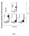
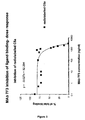
- Murine monoclonal antibodies specific for C5aR designated 7F3, 6C12 and 12D4, have been produced by the present inventors as described herein. Surprisingly, these monoclonal antibodies (MAbs) are able to substantially or completely block C5a binding to C5aR. In particular, MAb 7F3 is fully neutralising.
- MAbs 7F3, 6C12 and 12D4 are reactive with regions of C5aR other than the N-terminal region. It is believed that MAbs 7F3, 6C12 and 12D4 are primarily reactive with the second extracellular loop (residues 175 to 206) of C5aR. For example, MAb 12D4 reactivity with C5aR is almost completely abolished by mutation of the 2nd extracellular loop residues 181 and 192 from tyrosine to phenylalanine. This inhibition was observed in binding studies involving the C5aR mutant L2-FF ( Farzan et al., J. Exp. Med., 193:1059-1065, 2001 ).
- the MAbs may also simultaneously bind to a region of one of the other extracellular loops or the N-terminal domain.
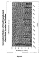
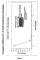
- MAbs 7F3, 6C12 and 12D4 are also capable of inhibiting activation of neutrophils by other chemoattractant ligands.
- these other chemoattractant ligands include the CXCR1 and CXCR2 ligands IL-8, ENA-78 and GPC-2.
- This ability to inhibit the function of different chemoattractant receptors provides an unusual and unexpected advantage over other known anti-C5aR molecules.
- anti-C5aR molecules that are able to inhibit the function of multiple neutrophil chemoattractant receptors are likely to be highly efficient therapeutic agents in the treatment of immunopathological disorders.
- the present invention provides antibodies that bind to the second extracellular loop, residues 175 to 206, of C5aR, either alone or in conjunction with other loops or domains.
- the invention provides antibodies that bind to C5aR and have epitopic specificity the same or similar to that of any one of MAbs 7F3, 6C12 or 12D4.
- antibody as used in this invention includes intact molecules as well as fragments thereof, such as Fab, F(ab')2, and Fv which are capable of binding the epitopic determinant. These antibody fragments retain some ability to selectively bind with its antigen or receptor and are defined as follows:
- epitopic determinants means any antigenic determinant on an antigen to which the paratope of an antibody binds.
- Epitopic determinants usually consist of chemically active surface groupings of molecules such as amino acids or sugar side chains and usually have specific three dimensional structural characteristics, as well as specific charge characteristics.
- Antibodies against C5aR can be prepared using cells expressing C5sR, intact C5aR or fragments containing one or more extracellular loops as the immunizing antigen.
- a peptide used to immunize an animal can be derived from translated cDNA or chemical synthesis and is purified and conjugated to a carrier protein, if desired.
- carrier protein e.g., a carrier protein
- Such commonly used carriers which are chemically coupled to the peptide include keyhole limpet hemocyanin (KLH), thyroglobulin, bovine serum albumin (BSA), and tetanus toxoid.
- KLH keyhole limpet hemocyanin
- BSA bovine serum albumin
- the coupled peptide may then be used to immunize the animal (e.g., a mouse or a rabbit).
- polyclonal antibodies can be further purified, for example, by binding to and elution from a matrix to which the peptide to which the antibodies were raised is bound.
- a matrix to which the peptide to which the antibodies were raised is bound.
- Those of skill in the art will know of various techniques common in the immunology arts for purification and/or concentration of polyclonal antibodies, as well as monoclonal antibodies (See for example, Coligan, et al., Unit 9, Current Protocols in Immunology, Wiley Interscience, 1991 ).
- Monoclonal antibodies may be prepared using any technique which provides for the production of antibody molecules by continuous cell lines in culture, such as, for example, the hybridoma technique, the human B-cell hybridoma technique, and the EBV-hybridoma technique ( Kohler et al. Nature 256, 495-497, 1975 ; Kozbor et al., J. Immunol. Methods 81, 31-42, 1985 ; Cote et al., Proc. Natl. Acad. Sci. USA 80, 2026-2030, 1983 ; Cole et al., Mol. Cell Biol. 62, 109-120, 1984 ).
- the hybridoma technique the human B-cell hybridoma technique
- EBV-hybridoma technique Kohler et al. Nature 256, 495-497, 1975 ; Kozbor et al., J. Immunol. Methods 81, 31-42, 1985 ; Cote et al., Proc. Natl.
- a method for the identification and isolation of an antibody binding domain which exhibits binding to a C5aR extracellular loop is the bacterio-phage a vector system.
- This vector system has been used to express a combinatorial library of Fab fragments from the mouse antibody repertoire in Escherichia coli ( Huse, et al., Science, 246:1275-1281, 1989 ) and from the human antibody repertoire ( Mullinax, et al., Proc. Nat. Acad. Sci., 87:8095-8099, 1990 ).
- Hybridomas which secrete a desired monoclonal antibody can be produced in various ways using techniques well understood by those having ordinary skill in the art and will not be repeated here. Details of these techniques are described in such references as Monoclonal Antibodies-Hybridomas: A New Dimension in Biological Analysis, Edited by Roger H. Kennett, et al., Plenum Press, 1980 ; and U.S. 4,172,124 .
- chimeric antibody molecules with various combinations of "humanized” antibodies include combining murine variable regions with human constant regions ( Cabily, et al. Proc. Natl. Acad. Sci. USA, 81:3273, 1984 ), or by grafting the murine-antibody complementarity determining regions (CDRs) onto the human framework ( Riechmann, et al., Nature 332:323, 1988 ).
- murine variable regions with human constant regions
- CDRs murine-antibody complementarity determining regions
- chimeric antibodies of the anti-C5aR antibodies of the present invention or biologically active fragments thereof.
- the term "chimeric antibody” refers to an antibody in which the variable regions of antibodies derived from one species are combined with the constant regions of antibodies derived from a different species or alternatively refers to CDR grafted antibodies. Chimeric antibodies are constructed by recombinant DNA technology, and are described in Shaw, et al., J. Immun., 138:4534 (1987 ), Sun, LK., et al., Proc. Natl. Acad. Sci. USA, 84:214-218 (1987 ), for example.
- CDR complementary metal-oxide-semiconductor
- complementarity determining region or “hypervariable region” is defined as the amino acid sequences on the light and heavy chains of an antibody which form the three-dimensional loop structure that contributes to the formation of the antigen binding site.
- CDR grafted antibody refers to an antibody having an amino acid sequence in which at least parts of one or more CDR sequences in the light and/or variable domain have been replaced by analogous parts of CDR sequences from an antibody having a different binding specificity for a given antigen or receptor.
- variable region and “heavy chain variable region” refer to the regions or domains at the N-terminal portion of the light and heavy chains respectively which have a varied primary amino acid sequence for each antibody.
- the variable region of the antibody consists of the amino terminal domain of the light and heavy chains as they fold together to form a three-dimensional binding site for an antibody.
- the analogous CDR sequences are said to be “grafted” onto the substrate or recipient antibody.
- the "donor” antibody is the antibody providing the CDR sequence, and the antibody receiving the substituted sequences is the "substrate” antibody.
- the invention also provides cell lines which produce monoclonal antibodies of the invention.
- the isolation of cell lines producing monoclonal antibodies of the invention can be accomplished using routine screening techniques which permit determination of the elementary reaction pattern of the monoclonal antibody of interest. Thus, if a monoclonal antibody being tested binds the second extracellular loop of C5aR and blocks C5a-mediated biological activity, then the monoclonal antibody being tested and the monoclonal antibody produced by the cell lines of the invention are equivalent.
- Antibodies with an epitopic specificity which is the same as or similar to that of MAbs 7F3, 6C12 or 12D4 can be identified by their ability to compete with that particular MAb for binding to C5aR (e.g. to cells bearing C5aR, such as transfectants bearing C5aR, monocytes, dendritic cells, macrophages and basophils). Using receptor chimeras ( Rucker et al., Cell 87:437-446 (1996 )) or other techniques known to those skilled in the art, the binding site of any one of MAbs 7F3, 6C12 or 12D4 may be mapped.
- a monoclonal antibody has the same specificity as a monoclonal antibody of the invention by ascertaining whether the former prevents the latter from binding to a peptide comprising the second extracellular loop of C5aR. If the monoclonal antibody being tested competes with the monoclonal antibody of the invention, as shown by a decrease in binding by the monoclonal antibody of the invention, then the two monoclonal antibodies bind to the same, or a closely related, epitope.
- Still another way to determine whether a monoclonal antibody has the specificity of a monoclonal antibody of the invention is to pre-incubate the monoclonal antibody being tested with a peptide to which the antibody is presumed to be reactive, and then add the monoclonal antibody of the invention to determine if the monoclonal antibody of the invention is inhibited in its ability to bind the peptide. If the monoclonal antibody of the invention is inhibited then, in all likelihood, the monoclonal antibody being tested has the same, or functionally equivalent, epitopic specificity as the monoclonal antibody of the invention. Screening of monoclonal antibodies of the invention, can also be carried out utilizing suitable peptides and determining whether the monoclonal antibody blocks C5a from binding to C5aR.
- anti-idiotypic antibodies which can be used to screen monoclonal antibodies to identify whether the antibody has the same binding specificity as a monoclonal antibody of the invention.
- These antibodies can also be used for immunization purposes ( Herlyn, et al., Science, 232:100, 1986 ).
- anti-idiotypic antibodies can be produced using well-known hybridoma techniques ( Kohler and Milstein, Nature, 256:495, 1975 ).
- An anti-idiotypic antibody is an antibody which recognizes unique determinants present on the monoclonal antibody produced by the cell line of interest. These determinants are located in the hypervariable region of the antibody.
- An anti-idiotypic antibody can be prepared by immunizing an animal with the monoclonal antibody of interest. The immunized animal will recognize and respond to the idiotypic determinants of the immunizing antibody and produce an antibody to these idiotypic determinants.
- the anti-idiotypic antibodies of the immunized animal which are specific for a monoclonal antibody of the invention produced by a cell line which was used to immunize the second animal, it is possible to identify other clones with the same idiotype as the antibody of the hybridoma used for immunization.
- Idiotypic identity between monoclonal antibodies of two cell lines demonstrates that the two monoclonal antibodies are the same with respect to their recogniition of the same epitopic determinant.
- anti-idiotypic antibodies it is possible to identify other hybridomas expressing monoclonal antibodies having the same epitopic specificity.
- an anti-idiotypic monoclonal antibody made to a first monoclonal antibody will have a binding domain in the hypervariable region which is the "image" of the epitope bound by the first monoclonal antibody.
- the anti-idiotypic monoclonal antibody can be used for immunization, since the anti-idiotype monoclonal antibody binding domain effectively acts as an antigen.
- Antibody fragments which contain epitopic binding sites of any one of the MAbs of the present invention can be generated by known techniques. For example, the DNA encoding the hypervariable region may be cloned, using standard recombinant DNA procedures such as those described herein, in a suitable host.
- variable antibodies of the present invention comprise variable regions or one or more CDR loops that are substantially the same as those of MAbs 7F3, 6C12 or 12D4. It will be understood that the variable regions or CDR loops shown in the sequence listings may be modified for use in the present invention. Typically, modifications are made that maintain the binding specificity of the sequence. Conservative substitutions may be made, for example, without affecting the binding specificity of the antibody. Thus, in one embodiment, amino acid substitutions may be made, for example from 1, 2 or 3 to 10, 20 or 30 substitutions provided that the modified sequence retains substantially the same binding specificity. However, in an alternative embodiment, modifications to the amino acid sequences of an antibody of the invention may be made intentionally to reduce the biological activity of the antibody. For example modified antibodies that remain capable of binding to C5aR but lack functional effector domains may be useful as inhibitors of the biological activity of C5aR.
- Amino acid substitutions may also include the use of non-naturally occurring analogues, for example to increase blood plasma half-life of a therapeutically administered antibody.
- amino acid residues of a variant or derivative are altered as compared with the corresponding variable regions or CDR loops depicted in the sequence listings.
- a sequence "substantially the same" as one of the variable regions shown is the sequence listing may include an amino acid sequence which is at least 80%, 85% or 90% identical, preferably at least 95 or 98% identical at the amino acid level over at least 20, preferably at least 50 amino acids with that variable region. Homology should typically be considered with respect to those regions of the sequence known to be essential for binding specificity rather than non-essential neighbouring sequences.
- Homology comparisons can be conducted by eye, or more usually, with the aid of readily available sequence comparison programs. These commercially available computer programs can calculate % homology between two or more sequences.
- Percentage homology may be calculated over contiguous sequences, i.e. one sequence is aligned with the other sequence and each amino acid in one sequence directly compared with the corresponding amino acid in the other sequence, one residue at a time. This is called an "ungapped" alignment. Typically, such ungapped alignments are performed only over a relatively short number of residues (for example less than 50 contiguous amino acids).
- Calculation of maximum % homology therefore firstly requires the production of an optimal alignment, taking into consideration gap penalties.
- a suitable computer program for carrying out such an alignment is the GCG Wisconsin Bestfit package (University of Wisconsin, U.S.A.; Devereux et al., 1984, Nucleic Acids Research 12:387 ).
- Examples of other software than can perform sequence comparisons include, but are not limited to, the BLAST package (see Ausubel et al., 1999 ibid - Chapter 18), FASTA ( Atschul et al., 1990, J. Mol. Biol., 403-410 ) and the GENEWORKS suite of comparison tools. Both BLAST and FASTA are available for offline and online searching (see Ausubel et al., 1999 ibid, pages 7-58 to 7-60). However it is preferred to use the GCG Bestfit program.
- a scaled similarity score matrix is generally used that assigns scores to each pairwise comparison based on chemical similarity or evolutionary distance.
- An example of such a matrix commonly used is the BLOSUM62 matrix - the default matrix for the BLAST suite of programs.
- GCG Wisconsin programs generally use either the public default values or a custom symbol comparison table if supplied (see user manual for further details). It is preferred to use the public default values for the GCG package, or in the case of other software, the default matrix, such as BLOSUM62.
- % homology preferably % sequence identity.
- the software typically does this as part of the sequence comparison and generates a numerical result.
- an antibody of the present invention is humanized, that is, an antibody produced by molecular modelling techniques wherein the human content of the antibody is maximised while causing little or no loss of binding affinity attributable to the variable region of the murine antibody.
- the invention provides a chimeric antibody comprising the amino acid sequence of a human framework region and of a constant region from a human antibody so as to humanise or render nonimmunogenic the hypervariable region from a mouse monoclonal antibody such as 7F3, C612 or 12D4.
- a two-step approach may be used which involves (a) selecting human antibody sequences that are used as human frameworks for humanization, and (b) determining which variable region residues of the animal monoclonal antibody should be selected for insertion into the human framework chosen.
- the first step involves selection of the best available human framework sequences for which sequence information is available. This selection process is based upon the following selection criteria.
- sequences of the heavy and light chain variable regions of an animal monoclonal antibody that is to be humanized are optimally aligned and compared preferably with all known human antibody heavy and light chain variable region sequences.
- the known human antibody chain sequences are then evaluated for the presence of unidentified residues and/or ambiguities, which are sequence uncertainties.
- the most common of such uncertainties are mistaken identification of an acidic amino acid for an amide amino acid due to loss of ammonia during the sequencing procedure, eg., incorrect identification of a glutamic acid residue, when the residue actually present in the protein was a glutamine residue. All other factors being equal, it is desirable to select a human antibody chain having as few such ambiguities as possible.
- Antibody chain variable regions contain intra-domain disulfide bridges.
- the distance (number of residues) between the cysteine residues comprising these bridges is referred to as the Pin-region spacing [ Chothia et al, J. Mol. Biol. 196:901 (1987 )]. All other factors being equal, it is most desirable that the Pin-region spacing of a human antibody selected be similar or identical to that of the animal antibody. It is also desirable that the human sequence Pin-region spacing be similar to that of a known antibody 3-dimensional structure, to facilitate computer modeling.
- the human antibody having the best overall combination of desirable characteristics is selected as the framework for humanization of the animal antibody.
- the heavy and light chains selected may be from the same or different human antibodies.
- the second step in the methods of this invention involves determination of which of the animal antibody variable region sequences should be selected for grafting into the human framework. This selection process is based upon the following selection criteria:
- minimal residues Two types of potential variable region residues are evaluated in the animal antibody sequences, the first of which are called “minimal residues.” These minimal residues comprise CDR structural loops plus any additional residues required, as shown by computer modeling, to support and/or orient the CDR structural loops.
- maximal residues comprise the minimal residues plus any additional residues which, as determined by computer modeling, fall within about 10 / of CDR structural loop residues and possess a water solvent accessible surface [ Lee et al, J. Biol. Chem. 55:379 (1971 )].
- variable region sequences of the animal antibody that is to be humanized are identified by computer modeling.
- computer modeling is carried out on (a) the variable region sequences of the animal antibody that is to be humanized, (b) the selected human antibody framework sequences, and (c) all possible recombinant antibodies comprising the human antibody framework sequences into which the various minimal and maximal animal antibody residues have been grafted.
- the computer modeling is performed using software suitable for protein modeling and structural information obtained from an antibody that (a) has variable region amino acid sequences most nearly identical to those of the animal antibody and (b) has a known 3-dimensional structure.
- An example of software that can be used is the SYBYL Biopolymer Module software (Tripos Associates).
- the antibody from which the structural information can be obtained may be but need not necessarily be a human antibody.
- monoclonal antibodies of one isotype might be more preferable than those of another in terms of their diagnostic or therapeutic efficacy.
- unmodified mouse monoclonal antibodies of isotype gamma-2a and gamma-3 are generally more effective in lysing target cells than are antibodies of the gamma-1 isotype.
- This differential efficacy is thought to be due to the ability of the gamma-2a and gamma-3 isotypes to more actively participate in the cytolytic destruction of the target cells.
- Particular isotypes of a monoclonal antibody can be prepared secondarily, from a parental hybridoma secreting monoclonal antibody of different isotype, by using the sib selection technique to isolate class-switch variants ( Steplewski, et al., Proc. Natl. Acad. Sci. U.S.A., 82:8653,1985 ; Spira, et al., J. Immunol. Methods, 74:307, 1984 ).
- the monoclonal antibodies of the invention would include class-switch variants having the specificity of any one of MAbs 7F3, 6C 12 and 12D4.
- the monoclonal antibodies of the invention are suited for use in vitro, for example, in immunoassays in which they can be utilized in liquid phase or bound to a solid phase carrier.
- the antibodies may be useful for monitoring the level of C5aR in a sample.
- anti-idiotype antibodies are useful for measuring the level of C5a in a sample.
- the monoclonal antibodies in these immunoassays can be detectably labeled in various ways. Examples of types of immunoassays which can utilize monoclonal antibodies of the invention are competitive and non-competitive immunoassays in either a direct or indirect format. Examples of such immunoassays are the radioimmunoassay (RIA) and the sandwich (immunometric) assay.
- RIA radioimmunoassay
- sandwich immunometric
- Detection of the antigens using the monoclonal antibodies of the invention can be done utilizing immunoassays which are run in either the forward, reverse, or simultaneous modes, including immunohistochemical assays on physiological samples. Those of skill in the art will know, or can readily discern, other immunoassay formats without undue experimentation.
- the antibodies of the invention can be bound to many different carriers and used to detect the presence of C5aR.
- carriers include glass, polystyrene, polypropylene, polyethylene, dextran, nylon, amylases, natural and modified celluloses, polyacrylamides, agaroses and magnetite.
- the nature of the carrier can be either soluble or insoluble for purposes of the invention. Those skilled in the art will know of other suitable carriers for binding monoclonal antibodies, or will be able to ascertain such, using routine experimentation.
- Cells which naturally express C5aR or cells comprising a recombinant nucleic acid sequence which encodes a C5aR or variant thereof may be used in binding assays.
- the cells are maintained under conditions appropriate for expression of receptor.
- the cells are contacted with an antibody or fragment under conditions suitable for binding (e.g., in a suitable binding buffer), and binding is detected by standard techniques.
- the extent of binding can be determined relative to a suitable control (e.g., compared with background determined in the absence of antibody, compared with binding of a second antibody (i.e., a standard), compared with binding of antibody to untransfected cells).
- a cellular fraction, such as a membrane fraction, containing receptor or liposomes comprising receptor can be used in lieu of whole cells.
- Binding inhibition assays can also be used to identify antibodies or fragments thereof which bind C5aR and inhibit binding of C5a to C5aR or a functional variant.
- a binding assay can be conducted in which a reduction in the binding of C5a (in the presence of the antibody), as compared to binding of C5a in the absence of the antibody, is detected or measured.
- a composition comprising an isolated and/or recombinant mammalian C5aR or functional variant thereof can be contacted with C5a and antibody simultaneously, or one after the other, in either order.
- a reduction in the extent of binding of the ligand in the presence of the antibody is indicative of inhibition of binding by the antibody. For example, binding of the ligand could be decreased or abolished.
- Antibodies which are identified in this manner can be further assessed to determine whether, subsequent to binding, they act to inhibit other functions of C5aR and/or to assess their therapeutic utility.
- the binding of a ligand or promoter, such as an agonist, to C5aR can result in signaling by this G protein-coupled receptor, and the activity of G proteins as well as other intracellular signaling molecules is stimulated.
- the induction of signaling function by a compound e.g., an antibody or fragment thereof
- Such an assay can be used to identify antibody agonists of C5aR.
- the inhibitory activity of an antibody or functional fragment thereof can be determined using a ligand or promoter in the assay, and assessing the ability of the antibody to inhibit the activity induced by ligand or promoter.
- G protein activity such as hydrolysis of GTP to GDP, or later signaling events triggered by receptor binding, such as induction of rapid and transient increase in the concentration of intracellular (cytosolic) free calcium
- G protein activity such as hydrolysis of GTP to GDP, or later signaling events triggered by receptor binding, such as induction of rapid and transient increase in the concentration of intracellular (cytosolic) free calcium
- cytosolic intracellular free calcium
- the functional assay of Sledziewski et al. using hybrid G protein coupled receptors can be used to monitor the ability of a ligand or promoter to bind receptor and activate a G protein ( Sledziewski et al., U.S. Pat. No. 5,284,746 ).
- Such assays can be performed in the presence of the antibody or fragment thereof to be assessed, and the ability of the antibody or fragment to inhibit the activity induced by the ligand or promoter is determined using known methods and/or methods described herein.
- Chemotaxis assays can also be used to assess the ability of an antibody or functional fragment thereof to block binding of a ligand to C5aR and/or inhibit function associated with binding of the ligand to the receptor. These assays are based on the functional migration of cells in vitro or in vivo induced by a compound. Chemotaxis can be assessed by any suitable means, for example, in an assay utilizing a 96-well chemotaxis plate, or using other art-recognized methods for assessing chemotaxis. For example, the use of an in vitro transendothelial chemotaxis assay is described by Springer et al. ( Springer et al., WO 94/20142, published Sep.
- chemotaxis assays monitor the directional movement or migration of a suitable cell (such as a leukocyte (e.g., lymphocyte, eosinophil, basophil)) into or through a barrier (e.g., endothelium, a filter), toward increased levels of a compound, from a first surface of the barrier toward an opposite second surface.
- a suitable cell such as a leukocyte (e.g., lymphocyte, eosinophil, basophil)
- a barrier e.g., endothelium, a filter
- Membranes or filters provide convenient barriers, such that the directional movement or migration of a suitable cell into or through a filter, toward increased levels of a compound, from a first surface of the filter toward an opposite second surface of the filter, is monitored.
- the membrane is coated with a substance to facilitate adhesion, such as ICAM-1, fibronectin or collagen.
- ICAM-1 interleukin-1
- a suitable membrane having a suitable pore size for monitoring specific migration in response to compound, including, for example, nitrocellulose, polycarbonate, is selected.
- pore sizes of about 3-8 microns, and preferably about 5-8 microns can be used. Pore size can be uniform on a filter or within a range of suitable pore sizes.
- the distance of migration into the filter, the number of cells crossing the filter that remain adherent to the second surface of the filter, and/or the number of cells that accumulate in the second chamber can be determined using standard techniques (e.g., microscopy).
- the cells are labeled with a detectable label (e.g., radioisotope, fluorescent label, antigen or epitope label), and migration can be assessed in the presence and absence of the antibody or fragment by determining the presence of the label adherent to the membrane and/or present in the second chamber using an appropriate method (e.g., by detecting radioactivity, fluorescence, immunoassay).
- a detectable label e.g., radioisotope, fluorescent label, antigen or epitope label
- the extent of migration induced by an antibody agonist can be determined relative to a suitable control (e.g., compared to background migration determined in the absence of the antibody, compared to the extent of migration induced by a second compound (i.e., a standard), compared with migration of untransfected cells induced by the antibody).
- a suitable control e.g., compared to background migration determined in the absence of the antibody, compared to the extent of migration induced by a second compound (i.e., a standard), compared with migration of untransfected cells induced by the antibody.
- a second compound i.e., a standard
- transendothelial migration can be monitored.
- transmigration through an endothelial cell layer is assessed.
- endothelial cells can be cultured on a microporous filter or membrane, optionally coated with a substance such as collagen, fibronectin, or other extracellular matrix proteins, to facilitate the attachment of endothelial cells.
- endothelial cells are cultured until a confluent monolayer is formed.
- mammalian endothelial cells can are available for monolayer formation, including for example, vein, artery or microvascular endothelium, such as human umbilical vein endothelial cells (Clonetics Corp, San Diego, Calif.).
- endothelial cells of the same mammal are preferred; however endothelial cells from a heterologous mammalian species or genus can also be used.
- the assay is performed by detecting the directional migration of cells into or through a membrane or filter, in a direction toward increased levels of a compound, from a first surface of the filter toward an opposite second surface of the filter, wherein the filter contains an endothelial cell layer on a first surface.
- Directional migration occurs from the area adjacent to the first surface, into or through the membrane, towards a compound situated on the opposite side of the filter.
- concentration of compound present in the area adjacent to the second surface is greater than that in the area adjacent to the first surface.
- a composition comprising cells capable of migration and expressing C5aR can be placed in the first chamber.
- a composition comprising one or more ligands or promoters capable of inducing chemotaxis of the cells in the first chamber (having chemoattractant function) is placed in the second chamber.
- a composition comprising the antibody to be tested is placed, preferably, in the first chamber.
- Antibodies or functional fragments thereof which can bind receptor and inhibit the induction of chemotaxis, by a ligand or promoter, of the cells expressing C5aR in this assay are inhibitors of receptor function (e.g., inhibitors of stimulatory function).
- a reduction in the extent of migration induced by the ligand or promoter in the presence of the antibody or fragment is indicative of inhibitory activity.
- In vivo assays which monitor leukocyte infiltration of a tissue, in response to injection of a compound (e.g., chemokine or antibody) in the tissue, are described below (see Models of Inflammation). These models of in vivo homing measure the ability of cells to respond to a ligand or promoter by emigration and chemotaxis to a site of inflammation and to assess the ability of an antibody or fragment thereof to block this emigration.
- a compound e.g., chemokine or antibody
- the effects of an antibody or fragment on the stimulatory function of C5aR can be assessed by monitoring cellular responses induced by active receptor, using suitable host cells containing receptor.
- the assays described above which can be used to assess binding and function of the antibodies and fragments of the present invention, can be adapted to identify additional ligands or other substances which bind C5aR or functional variant thereof, as well as inhibitors and/or promoters of C5aR function.
- agents having the same or a similar binding specificity as that of an antibody of the present invention or functional portion thereof can be identified by a competition assay with said antibody or portion thereof.
- antibodies of the present invention may be used in methods of identifying ligands of the receptor or other substances which bind C5aR, as well as inhibitors (e.g., antagonists) or promoters (e.g., agonists) of receptor function.
- cells bearing a C5aR protein or functional variant thereof are used in an assay to identify and assess the efficacy of ligands or other substances which bind receptor, including inhibitors or promoters of receptor function.
- Such cells are also useful in assessing the function of the expressed receptor protein or polypeptide.
- Ligands and other substances which bind receptor, inhibitors and promoters of receptor function can be identified in a suitable assay, and further assessed for therapeutic effect.
- Antogonists of receptor function can be used to inhibit (reduce or prevent) receptor activity, and ligands and/or agonists can be used to induce (trigger or enhance) normal receptor function where indicated.
- an antagonist of receptor function identified in a method using antibodies of the present invention, may be used in the treatment of inflammatory diseases, including autoimmune disease and graft rejection.
- a novel ligand or agonist of receptor function identified in a method utilising antibodies of the current invention, may provide a new approach to selective stimulation of leukocyte function, which is useful, for example, in the treatment of infectious diseases and cancer.
- a "ligand" of a C5aR protein refers to a particular class of substances which bind to a mammalian C5aR protein, including natural ligands and synthetic and/or recombinant forms of natural ligands. In a preferred embodiment, ligand binding of a C5aR protein occurs with high affinity.
- an "antagonist” is a substance which inhibits (decreases or prevents) at least one function characteristic of a C5aR protein such as a binding activity (e.g., ligand binding, promoter binding, antibody binding), a signaling activity (e.g., activation of a mammalian G protein, induction of rapid and transient increase in the concentration of cytosolic free calcium) and/or cellular response function (e.g., stimulation of chemotaxis, exocytosis or inflammatory mediator release by leukocytes).
- a binding activity e.g., ligand binding, promoter binding, antibody binding
- a signaling activity e.g., activation of a mammalian G protein, induction of rapid and transient increase in the concentration of cytosolic free calcium
- cellular response function e.g., stimulation of chemotaxis, exocytosis or inflammatory mediator release by leukocytes.
- the term antagonist encompasses substances which bind receptor (e.g., an antibody, a mutant of a natural ligand, small molecular weight organic molecules, other competitive inhibitors of ligand binding), and substances which inhibit receptor function without binding thereto (e.g., an anti-idiotypic antibody).
- substances which bind receptor e.g., an antibody, a mutant of a natural ligand, small molecular weight organic molecules, other competitive inhibitors of ligand binding
- substances which inhibit receptor function without binding thereto e.g., an anti-idiotypic antibody.
- an "agonist” is a substance which promotes (induces, causes, enhances or increases) at least one function characteristic of a C5aR protein such as a binding activity (e.g., ligand, inhibitor and/or promoter binding), a signaling activity (e.g., activation of a mammalian G protein, induction of rapid and transient increase in the concentration of cytosolic free calcium) and/or a cellular response function (e.g., stimulation of chemotaxis, exocytosis or inflammatory mediator release by leukocytes).
- a binding activity e.g., ligand, inhibitor and/or promoter binding
- a signaling activity e.g., activation of a mammalian G protein, induction of rapid and transient increase in the concentration of cytosolic free calcium
- a cellular response function e.g., stimulation of chemotaxis, exocytosis or inflammatory mediator release by leukocytes.
- agonist encompasses substances which bind receptor (e.g., an antibody, a homolog of a natural ligand from another species), and substances which promote receptor function without binding thereto (e.g., by activating an associated protein).
- the agonist is other than a homolog of a natural ligand.
- antibodies of the invention may also be used in a method of detecting or identifying an agent which binds C5aR or ligand binding variant thereof, including ligands, antagonists, agonists, and other substances which bind C5aR or functional variant.
- an agent to be tested, an antibody or antigen-binding fragment of the present invention e.g. an antibody having an epitopic specificity which is the same as or similar to that of 7F3, and antigen-binding fragments thereof
- a composition comprising a C5aR or a ligand binding variant thereof.
- the foregoing components are combined under conditions suitable for binding of the antibody or antigen-binding fragment to C5aR, and binding of the antibody or fragment to the C5aR is detected or measured, either directly or indirectly, according to methods described herein or other suitable methods.
- a decrease in the amount of complex formed relative to a suitable control e.g., in the absence of the agent to be tested
- the composition comprising C5aR can be a membrane fraction of a cell bearing recombinant C5aR protein or ligand binding variant thereof.
- the antibody or fragment thereof can be labeled with a label such as a radioisotope, spin label, antigen or epitope label, enzyme label, fluorescent group and chemiluminescent group.
- In vivo models of inflammation are available which can be used to assess the effects of antibodies and fragments of the invention in vivo as therapeutic agents.
- leukocyte infiltration upon intradermal injection of a chemokine and an antibody or fragment thereof reactive with C5aR into a suitable animal such as rabbit, mouse, rat, guinea pig or rhesus macaque
- a suitable animal such as rabbit, mouse, rat, guinea pig or rhesus macaque
- a suitable animal such as rabbit, mouse, rat, guinea pig or rhesus macaque
- skin biopsies are assessed histologically for infiltration of leukocytes (e.g., eosinophils, granulocytes).
- labeled cells e.g., stably transfected cells expressing C5aR
- An antibody or fragment to be assessed can be administered, either before, simultaneously with or after the labeled cells are administered to the test animal.
- a decrease of the extent of infiltration in the presence of antibody as compared with the extent of infiltration in the absence of inhibitor is indicative of inhibition.
- the antibodies and fragments of the present invention are useful in a variety of applications, including research, diagnostic and therapeutic applications.
- the antibodies are labeled with a suitable label (e.g., fluorescent label, chemiluminescent label, isotope label, antigen or epitope label or enzyme label).
- a suitable label e.g., fluorescent label, chemiluminescent label, isotope label, antigen or epitope label or enzyme label.
- they can be used to isolate and/or purify receptor or portions thereof, and to study receptor structure (e.g., conformation) and function.
- the various antibodies of the present invention can be used to detect C5aR or to measure the expression of receptor, for example, on T cells (e.g., CD8+ cells, CD45RO+ cells), monocytes and/or on cells transfected with a receptor gene.
- T cells e.g., CD8+ cells, CD45RO+ cells
- monocytes e.g., CD8+ cells, CD45RO+ cells
- monocytes e.g., CD45RO+ cells
- cells transfected with a receptor gene e.g., monocytes and/or on cells transfected with a receptor gene.
- cell sorting e.g., flow cytometry, fluorescence activated cell sorting
- the anti-C5aR antibodies of the present invention have value in diagnostic applications.
- diagnostic assays entail detecting the formation of a complex resulting from the binding of an antibody or fragment thereof to C5aR.
- the antibodies or antigen-binding fragments can be labeled or unlabeled.
- the antibodies or fragments can be directly labeled.
- labels can be employed, including, but not limited to, radionuclides, fluorescers, enzymes, enzyme substrates, enzyme cofactors, enzyme inhibitors and ligands (e.g., biotin, haptens). Numerous appropriate immunoassays are known to the skilled artisan (see, for example, U.S. Pat. Nos.
- Immunohistochemistry of tissue samples may also be used in the diagnostic methods of the present invention.
- the antibodies or fragments can be detected using suitable means, as in agglutination assays, for example.
- Unlabeled antibodies or fragments can also be used in combination with another (i.e., one or more) suitable reagent which can be used to detect antibody, such as a labeled antibody (e.g., a second antibody) reactive with the first antibody (e.g., anti-idiotype antibodies or other antibodies that are specific for the unlabeled immunoglobulin) or other suitable reagent (e.g., labeled protein A).
- a labeled antibody e.g., a second antibody
- suitable reagent e.g., labeled protein A
- Kits for use in detecting the presence of a C5aR protein in a biological sample can also be prepared.
- Such kits will include an antibody or functional fragment thereof which binds to C5aR, as well as one or more ancillary reagents suitable for detecting the presence of a complex between the antibody or fragment and C5aR.
- the antibody compositions of the present invention can be provided in lyophilized form, either alone or in combination with additional antibodies specific for other epitopes.
- the antibodies which can be labeled or unlabeled, can be included in the kits with adjunct ingredients (e.g., buffers, such as Tris, phosphate and carbonate, stabilizers, excipients, biocides and/or inert proteins, e.g., bovine serum albumin).
- adjunct ingredients e.g., buffers, such as Tris, phosphate and carbonate, stabilizers, excipients, biocides and/or inert proteins, e.g., bovine serum albumin.
- the antibodies can be provided as a lyophilized mixture with the adjunct ingredients, or the adjunct ingredients can be separately provided for combination by the user.
- these adjunct materials will be present in less than about 5% weight based on the amount of active antibody, and usually will be present in a total amount of at least about 0.001 % weight based on antibody concentration.
- a second antibody capable of binding to the monoclonal antibody can be provided in the kit, for instance in a separate vial or container.
- the second antibody if present, is typically labeled, and can be formulated in an analogous manner with the antibody formulations described above.
- the antibodies of the present invention may be used in a method of detecting and/or quantitating expression of C5aR by a cell, in which a composition comprising a cell or fraction thereof (e.g., membrane fraction) is contacted with an antibody or functional fragment thereof which binds to C5aR under conditions appropriate for binding of the antibody or fragment thereto, and binding is monitored. Detection of the antibody, indicative of the formation of a complex between antibody and C5aR, indicates the presence of the receptor. Binding of antibody to the cell can be determined as described above under the heading "Binding Assays", for example.
- the method can be used to detect expression of C5aR on cells from an individual (e.g., in a sample, such as a body fluid, such as blood, saliva or other suitable sample).
- a sample such as a body fluid, such as blood, saliva or other suitable sample.
- the level of expression of C5aR on the surface of T cells or monocytes can also be determined, for instance, by flow cytometry, and the level of expression (e.g., staining intensity) can be correlated with disease susceptibility, progression or risk.
- Chemoattractant receptors function in the migration of leukocytes throughout the body, particularly to inflammatory sites. Inflammatory cell emigration from the vasculature is regulated by a three-step process involving interactions of leukocyte and endothelial cell adhesion proteins and cell specific chemoattractants and activating factors ( Springer, T. A., Cell, 76:301-314 (1994 ); Butcher, E. C., Cell, 67:1033-1036 (1991 ); Butcher, E. C. and Picker, L. J., Science (Wash. D.C.), 272:60-66 (1996 )).
- the second step is crucial in that the activation of the leukocyte chemoattractant receptors is thought to cause the transition from the selectin-mediated cell rolling to the integrin-mediated tight binding. This results in the leukocyte being ready to transmigrate to perivascular sites.
- the chemoattractant/chemoattractant receptor interaction is also crucial for transendothelial migration and localization within a tissue ( Campbell, J. J., et al., J. Cell Biol., 134:255-266 (1996 ); Carr, M. W., et al., Immunity, 4:179 187 (1996 )). This migration is directed by a concentration gradient of chemoattractant leading towards the inflammatory focus.
- C5aR has an important role in leukocyte trafficking. It is likely that C5aR is a key chemoattractant receptor for neutrophil, eosinophil, T cell or T cell subset or monocyte migration to certain inflammatory sites, and so anti-C5aR mAbs can be used to inhibit (reduce or prevent) leukocyte migration, particularly that associated with neutrophil tissue injury such as reperfusion injury and stroke, T cell dysfunction, such as autoimmune disease, or allergic reactions or with monocyte-mediated disorders such as atherosclerosis.
- neutrophil tissue injury such as reperfusion injury and stroke
- T cell dysfunction such as autoimmune disease
- monocyte-mediated disorders such as atherosclerosis.
- the antibodies and fragments thereof of the present invention can also be used to modulate receptor function in research and therapeutic applications.
- the antibodies and functional fragments described herein can act as inhibitors to inhibit (reduce or prevent) (a) binding (e.g., of a ligand, an inhibitor or a promoter) to the receptor, (b) a receptor signaling function, and/or (c) a stimulatory function.
- Antibodies which act as inhibitors of receptor function can block ligand or promoter binding directly or indirectly (e.g., by causing a conformational change).
- antibodies can inhibit receptor function by inhibiting binding of a ligand, or by desensitization (with or without inhibition of binding of a ligand).
- Antibodies which bind receptor can also act as agonists of receptor function, triggering or stimulating a receptor function, such as a signaling and/or a stimulatory function of a receptor (e.g., leukocyte trafficking) upon binding to receptor.
- an antibody or functional fragment of the present invention may be used in a method of inhibiting leukocyte trafficking in a mammal (e.g., a human patient).
- the antibodies of the present invention may also be used in a method of inhibiting other effects associated with C5aR activity such as histamine release from basophils and granule release from eosinophils, basophils and neutrophils.
- Administration of an antibody or fragment of the present invention can result in amelioration or elimination of the disease state.
- the monoclonal antibodies can also be used immunotherapeutically for immunopathological associated disease.
- immunotherapeutically or “immunotherapy” as used herein in conjunction with the monoclonal antibodies of the invention denotes both prophylactic as well as therapeutic administration.
- the monoclonal antibodies can be administered to high-risk patients in order to lessen the likelihood and/or severity of immunopathological disease or administered to patients already evidencing active disease, for example sepsis due to gram-negative bacterial infection.
- the antibodies or functional fragments thereof can be used to treat allergy, atherogenesis, anaphylaxis, malignancy, chronic and acute inflammation, histamine and IgE-mediated allergic reactions, shock, and rheumatoid arthritis, atherosclerosis, multiple sclerosis, allograft rejection, fibrotic disease, asthma, inflammatory glomerulopathies or any immune complex related disorder.
- Anti-C5aR antibodies of the present invention can block the binding of one or more ligands, thereby blocking the downstream cascade of one or more events leading to the above disorders.
- the antibodies of the present invention may be used in the treatment of sepsis, stroke or adult respiratory distress syndrome.
- Diseases or conditions of humans or other species which can be treated with promoters of C5aR function include, but are not limited to immunosuppression, such as that in individuals with immunodeficiency syndromes such as AIDS, individuals undergoing radiation therapy, chemotherapy, therapy for autoimmune disease or other drug therapy (e.g., corticosteroid therapy), which causes immunosuppression; and immunosuppression due congenital deficiency in receptor function or other causes.
- immunosuppression such as that in individuals with immunodeficiency syndromes such as AIDS, individuals undergoing radiation therapy, chemotherapy, therapy for autoimmune disease or other drug therapy (e.g., corticosteroid therapy), which causes immunosuppression; and immunosuppression due congenital deficiency in receptor function or other causes.
- a immunotherapeutic method may entail the administration of a therapeutic agent of the invention by injection or infusion prior to (prophylaxis) or following (therapy) the onset of the immunopathological disease.
- One or more antibodies or fragments of the present invention can be administered to an individual by an appropriate route, either alone or in combination with (before, simultaneous with, or after) another drug or agent.
- the antibodies of the present invention can also be used in combination with other monoclonal or polyclonal antibodies (e.g., in combination with antibodies which bind chemokine receptors, including, but not limited to, CCR2 and CCR3) or with anti-TNF or other antiinflammatory agents or with existing blood plasma products, such as commercially available gamma globulin and immune globulin products used in prophylactic or therapeutic treatments.
- the antibodies or fragments of the present invention can be used as separately administered compositions given in conjunction with antibiotics and/or antimicrobial agents.
- An effective amount of an antibody or fragment i.e., one or more antibodies or fragments is administered.
- An effective amount is an amount sufficient to achieve the desired therapeutic (including prophylactic) effect, under the conditions of administration, such as an amount sufficient for inhibition of a C5aR function, and thereby, inhibition of an inflammatory response.
- routes of administration are possible including, but not necessarily limited to, oral, dietary, topical, parenteral (e.g., intravenous, intraarterial, intramuscular, subcutaneous injection), inhalation (e.g., intrabronchial, intraocular, intranasal or oral inhalation, intranasal drops), depending on the disease or condition to be treated.
- parenteral e.g., intravenous, intraarterial, intramuscular, subcutaneous injection
- inhalation e.g., intrabronchial, intraocular, intranasal or oral inhalation, intranasal drops
- Other suitable methods of administration can also include rechargeable or biodegradable devices and slow release polymeric devices.
- the pharmaceutical compositions of this invention can also be administered as part of a combinatorial therapy with other agents.
- Formulation of an antibody or fragment to be administered will vary according to the route of administration and formulation (e.g., solution, emulsion, capsule) selected.
- An appropriate pharmaceutical composition comprising an antibody or functional fragment thereof to be administered can be prepared in a physiologically acceptable vehicle or carrier.
- a physiologically acceptable vehicle or carrier A mixture of antibodies and/or fragments can also be used.
- suitable carriers include, for example, aqueous or alcoholic/aqueous solutions, emulsions or suspensions, including saline and buffered media.
- Parenteral vehicles can include sodium chloride solution, Ringer's dextrose, dextrose and sodium chloride, lactated Ringer's or fixed oils.
- aqueous carriers include water, buffered water, buffered saline, polyols (e.g., glycerol, propylene glycol, liquid polyethylene glycol), dextrose solution and glycine.
- Intravenous vehicles can include various additives, preservatives, or fluid, nutrient or electrolyte replenishers (See, generally, Remington's Pharmaceutical Science, 16th Edition, Mack, Ed. 1980 ).
- the compositions can optionally contain pharmaceutically acceptable auxiliary substances as required to approximate physiological conditions such as pH adjusting and buffering agents and toxicity adjusting agents, for example, sodium acetate, sodium chloride, potassium chloride, calcium chloride and sodium lactate.
- the antibodies and fragments of this invention can be lyophilized for storage and reconstituted in a suitable carrier prior to use according to art-known lyophilization and reconstitution techniques.
- the optimum concentration of the active ingredient(s) in the chosen medium can be determined empirically, according to procedures well known to the skilled artisan, and will depend on the ultimate pharmaceutical formulation desired.
- the antibody or fragment can be solubilized and loaded into a suitable dispenser for administration (e.g., an atomizer, nebulizer or pressurized aerosol dispenser).
- the dosage ranges for the administration of the monoclonal antibodies of the invention are those large enough to produce the desired effect in which the symptoms of the immunopathological disease are ameliorated or the likelihood of infection or over stimulation of the immune system decreased.
- the dosage should not be so large as to cause adverse side effects, such as hyper-viscosity syndromes, pulmonary edema, conjestive heart failure, and the like.
- the dosage will vary with the age, condition, sex and extent of the disease in the patient and can be determined by one of skill in the art.
- the dosage can be adjusted by the individual physician in the event of anycomplication.
- Dosage can vary from about 0.1 mg/kg to about 300 mg/kg, preferably from about 0.2 mg/kg to about 200 mg/kg, most preferably from about 0.5 mg/kg to about 20 mg/kg, in one or more dose administrations daily, for one or several days.
- the antibodies of the present invention may be introduced into a subject by administering a nucleic acid molecule comprising a sequence encoding the antibody.
- the nucleic acid molecule may be in the form of DNA or RNA or a chimeric molecule comprising both DNA or RNA.
- a nucleotide sequence encoding the antibody may be cloned into an expression vector where the sequence encoding the agent is operably linked with expression control elements.
- Expression control elements are well known in the art and include, for example, promoters, enhancers and appropriate start and stop codons.
- a variety of methods can be used for introducing a nucleic acid encoding the antibody into a target cell in vivo.
- the naked nucleic acid may be injected at the target site, may be encapsulated into liposomes, or may be introduced by way of a viral vector.
- Direct injection of a nucleic acid molecule alone or encapsulated, for example, in cationic liposomes may be used for stable gene transfer of a nucleic acid encoding TSP-1 into non-dividing or dividing cells in vivo ( Ulmer et al., Science 259:1745-1748 (1993 )).
- the nucleic acid can be transferred into a variety of tissues in vivo using the particle bombardment method ( Williams et al., Proc. Natl. Acad. Sci. USA 88:2726-2730 (1991 )).
- Viral vectors are useful for gene transfer of a nucleic acid molecules encoding the antibody into a specific cell type in vivo.
- Viruses are specialized infectious agents that can infect and propagate in specific cell types. This specificity for infecting particular cell types is especially suitable for targeting the antibody to selected cells in vivo. The selection of a viral vector will depend, in part, on the cell type to be targeted.
- Specialized viral vectors are well known in the art that can target to specific cell types.
- Such vectors include, for example, recombinant adeno-associated viral vectors having general or tissue-specific promoters ( Lebkowski et al. U.S Pat. No. 5,354,678 ).
- Recombinant adeno-associated viral vectors have the added advantage that the recombinant virus can stably integrate into the chromatin of even quiescent non-proliferating cells ( Lebkowski et al., Mol. Cell. Biol. 8:3988-3996 (1988 )).
- Viral vectors can be constructed to further control the type of cell that expresses the encoded antibody by incorporating a tissue-specific promoter or enhancer into the vector ( Dai et al., Proc. Natl. Acad. Sci. USA 89:10892-10895 (1992 )).
- Retroviral vectors are also suitable for the methods for delivering nucleic acid molecules encoding the antibody in vivo.
- Such vectors can be constructed either to function as infectious particles or as non-infectious particles that undergo only a single initial round of infection.
- Receptor-mediated DNA delivery approaches also can be used to deliver a nucleic acid molecule encoding the antibody into a cell in a tissue-specific manner using a tissue-specific ligand or an antibody that is non-covalently complexed with the nucleic acid molecule via a bridging molecule ( Curiel et al., Hum. Gene Ther. 3:147-154 (1992 ); Wu and Wu, J. Biol. Chem. 262:4429-4432 (1987 )).
- Gene transfer to obtain expression of the antibody in a subject also can be performed by, for example, ex vivo transfection of autologous cells.
- Suitable cells for such ex vivo transfection include blood cells since these cells are readily accessible for manipulation and reintroduction back into the subject by methods well known in the art.
- Gene transfer through transfection of cells ex vivo can be performed by a variety of methods, including, for example, calcium phosphate precipitation, diethyaminoethyl dextran, electroporation, lipofection, or viral infection. Such methods are well known in the art (see, for example, Sambrook et al., Molecular Cloning: A Laboratory Manual, Cold Springs Harbour Laboratory Press (1989 )).
- the cells are then transplanted or grafted back into a subject to be treated.
- the cells once introduced into the body can produce the antibody, which can enter the circulation and inhibit platelet aggregation at the site of the disease or condition.
- Monoclonal antibodies (MAbs) reactive with C5aR were generated by immunising C57BL/6 mice with 10 7 L1.2 C5aR transfected cells [8], intraperitoneally, five to six times at 2-wk intervals. The final immunisation was injected intravenously. Four days later, the spleen was removed and cells were fused with the SP2/0 cell line as described [9].
- MAbs reactive with C5aR were identified using C5aR transfected L1.2 cells, and untransfected L1.2 cells, or L1.2 cells transfected with unrelated receptors such as CXCR2 or CX3CR1 (V28) using immunofluorescent staining and analysis using a FACScan® (Becton Dickinson & Co., Mountain View, CA). MAb staining of cells was performed using standard procedures, as described previously [10].
- C5a binding to L1.2 C5aR transfectants was carried out as described previously [9, 11]. Briefly, cells were washed once in PBS and resuspended in binding buffer (50mM Hepes, pH 7.5, 1 mM CaCl, 5mM MgCl 2 , 0.5% BSA and 0.05% azide) at a concentration of 10 7 /ml.
- binding buffer 50mM Hepes, pH 7.5, 1 mM CaCl, 5mM MgCl 2 , 0.5% BSA and 0.05% azide
- Tissue culture inserts (Becton Dickinson & Co., Mountain View, CA) were placed in each of the wells of 24-well tissue-culture plates, forming an upper and lower chamber separated by a polyethylene terepthalate membrane bearing 3-mm-diameter pores.
- Chemotactic C5a (diluted in assay medium) was added to 600 ⁇ l of assay medium in the 24-well tissue culture plates for a final concentration of 1nM.
- One million cells in 100 Tl were preincubated for 30 mins with the supernatants from the hybridomas containing the antibody.
- the cell-supernatant mixture or purified mAb was added to the upper chamber in the wells and the cells were allowed to migrate through to the lower chamber in an 5% CO 2 , 37°C incubator for 18 h.
- the inserts were removed after migration and the cells were counted by the FACScan®. Relative cell counts were obtained by acquiring events for a set time period of 30 seconds. This method was found to be highly reproducible, and enabled gating on the leukocytes and the exclusion of debris.
- Neutrophils were isolated from peripheral blood by first obtaining the leucocyte fraction via a dextran sedimentation step for 40 min at room temperature. The cells were then layered onto Ficoll-Paque (Amersham Biosciences) for density gradient centrifugation at 2500rpm for 15min at room temperature. After hypotonic lysis of residual red blood cells, neutrophils were resuspended in equal volumes of RPMI 1640 (Invitrogen Inc.), M199 (Invitrogen Inc.) and 2% FCS (HyClone).
- RPMI 1640 Invitrogen Inc.
- M199 Invitrogen Inc.
- FCS HyClone
- Chemotaxis Assay Anti-C5aR MAbs, 6C12, 7F3 and 12D4 were added to neutrophils (1x 10 7 /ml) at concentrations ranging from 0.5 to 10ug/ml. The cells were then loaded into the upper chamber of 24-well inserts (Corning Inc., NY) with a polycarbonate membrane of 3.0 ⁇ m porosity and incubated for 10min at room temperature. The inserts were then placed onto lower chambers containing human neutrophil chemoattractants such as C5a (0.1 to 100nM) and IL-8 (both 1.12 ng/ml to 11.2 ⁇ g/ml). The neutrophils were then incubated for 30min at 37°C. The number of neutrophils migrating through the membrane to the lower chamber were quantified by flow cytometry (FACSCalibur; BD Biosciences).
- Anti-C5aR MAbs were added at 50ug/ml, to a C5aR N-terminal synthetically produced peptide (residues 9-29) known as "PEPI" (Biosource; Eldridge) at concentrations ranging from 1 to 100 ⁇ M.
- PEPI synthetically produced peptide
- Mouse L1.2 cells transfected with human C5a receptor and resuspended in 1% bovine serine albumin (BSA; GibcoBRL) (1x 10⁷/ml) were then added to give a total volume of 100 ⁇ l. The cells were incubated for 30min at 4°C and washed once with 0.1% BSA.
- ELISAs were performed as described in Current Protocols in Immunology (Unit 2.1) (Edited by J.F. Coligan, A.M. Kruisbeek, D.B. Margulies, E.M. Shevach and W. Strober), John Wiley and Sons, New York . Briefly, 96-well flat bottom ELISA plates (Maxisorp; Nunc) were coated with 1Tg/ml protein (PEPI or OPG) in PBS at 37°C for 1hr, then blocked with BSA at 4°C overnight. The plates were then washed, incubated with antibodies, washed and incubated with peroxidase-conjugated sheep anti-mouse IgG antibody. The substrate used was TMB substrate reagent (PharMingen).
- L1.2 transfectants expressing high levels of C5aR [8] were used to immunize mice, and ten MAbs were identified via flow cytometry that reacted specifically with L1.2 cells transfected with C5aR, but not with L1.2 cells transfected with CX3CR1 (V28) or CXCR2. These ten MAbs were designated 12D4, 10G1, 5H11, 6C12, 10D4, 5F3, 7F3, 8D6, 11B9 and 1D12.
- Figure 1 is a set of histograms showing that MAb 7F3 reacts with C5aR transfectants (L1.2C5aR) and with human neutrophils but not with cells transfected with CX3CR1 (L1.2 V28) or with cells transfected with CXCR2 (L1.2CXCR2). These MAb 7F3 results are representative of the ten mAbs identified.
- Anti-C5aR MAbs were dialysed in 1x PBS (GibcoBRL), and both the dialysed and non-dialysed 7F3 MAbs were added to neutrophils (1x 10⁷/ml) at 5ug/ml. Negative controls (no Ab addition, and 1x PBS added) were included.
- the cells were then loaded into the upper chamber of 24-well inserts (Corning Inc., NY) with a polycarbonate membrane of 3.0 ⁇ m porosity and incubated for 10min at room temperature.
- the inserts were then placed onto lower chambers containing the human neutrophil chemoattractant C5a (0.1 to 100nM).
- the neutrophils were then incubated for 30min at 37°C.
- the number of neutrophils migrating through the membrane to the lower chamber were quantified by flow cytometry (FACSCalibur; BD Biosciences).
- Figure 6 shows that addition of MAb 7F3 (whether dialysed or non-dialysed) resulted in inhibition of neutrophil migration compared to the two negative controls.
- EXAMPLE 5 Inhibition of human C5a-directed neutrophil migration by MAbs 7F3,6C12 and 12D4
- the three anti-C5aR MAbs, 7F3, 12D4 and 6C12 were added to neutrophils (1x 10 7 /ml) at 5ug/ml. Negative controls (no Ab addition, and 1x PBS added) were included.
- the cells were then loaded into the upper chamber of 24-well inserts (Corning Inc., NY) with a polycarbonate membrane of 3.0 ⁇ m porosity and incubated for 10min at room temperature.
- the inserts were then placed onto lower chambers containing the human neutrophil chemoattractant C5a (1.12 to 1120ng/ml).
- the neutrophils were then incubated for 30min at 37°C.
- the number of neutrophils migrating through the membrane to the lower chamber were quantified by flow cytometry (FACSCalibur; BD Biosciences).
- EXAMPLE 6 Inhibition of human IL-8-directed neutrophil migration by MAbs 7F3,12D4 and 6C12
- the three anti-C5aR MAbs, 7F3, 12D4 and 6C12; and the dialysed sample of 7F3 were added to purified neutrophils (1x10 7 /ml) at 5ug/ml and loaded into the upper chamber of 24-well inserts. Negative controls (no Ab addition, and 1x PBS added) were again included. After 10min incubation at room temperature. The inserts were then placed onto lower chambers containing IL-8 (1.12 to 1120 ng/ml), a human neutrophil chemoattractant that binds the CXCR1 and CXCR2 receptors expressed on the surface of neutrophils. The neutrophils were then incubated for 30min at 37°C. The number of neutrophils migrating through the membrane to the lower chamber were quantified by flow cytometry (FACSCalibur; BD Biosciences).
- Figure 8 (a) shows that all three MAbs exhibited inhibition of neutrophil migration towards IL-8.
- 7F3 MAb both dialysed and non-dialysed was the most effective inhibitor resulting in a 5-fold reduction in neutrophil migration numbers.
- MAB 7F3 was also tested for its ability to inhibit other neutrophil chemoattractants, particularly CXCR1 and CXCR2 ligands.
- Table 1 shows substantial inhibition of neutrophil migration to a number of neutrophil chemoattractants, particularly CXCR1 and CXCR2 ligands, in neutrophil chemotaxis assays.
- EXAMPLE 7 Competitive inhibition of binding of MAbs 7F3, 12D4 and 6C12 to C5aR transfectants by a C5aR N-terminal peptide (9-29)
- Binding of the MAbs 7F3, 12D4 and 6C12 to cells transfected with C5aR was measured by staining with fluorescein (FITC) conjugated sheep anti-mouse IgG.
- FITC fluorescein conjugated sheep anti-mouse IgG.
- the ability of a C5aR N-terminal peptide (residues 9-29) to inhibit this binding was then assessed according to the methodology described above.
- This C5aR N-terminal peptide has the sequence PDYGHYDDKDTLDLNTPVDKT and is referred to herein as "PEPI".
- Figure 9(a) shows that increasing concentrations of PEPI did not inhibit the fluorescence staining of the three anti-C5aR MAbs. The fluorescence staining remained stable, even at concentrations of PEPI of 100 TM.
- Figure 9(b) shows that PEPI (at a concentration of 50TM) failed to inhibit FACS staining of purified neutrophils with MAb 7F3.
- EXAMPLE 8 Reactivity of Mkbs 7F3,12D4 and 6C12 with C5aR N-terminal peptide 9-29 (“PEPI”) and OPG
- OPG is a member of the TNF-receptor superfamily which binds specifically to its ligand TNFSF11/OPGL. More specifically, OPG is an osteoblast-secreted decoy receptor that functions as a negative regulator of bone resorption.
- MAbs 6C12, 12D4 and 7F3 were used in the ELISA as purified proteins at a concentration of 1 Tg/mL.
- MAb 9C1 which is specific for OPG
- MAb 11B9 which recognises PEPI were used as positive controls. These control MAbs were used in the form of undiluted tissue culture supernatant.
- FIG. 10 shows that MAbs 6C12, 12D4 and 7F3 were unreactive with PEPI.
- MAb 7F3 showed a small degree of cross-reactivity with OPG.
- the nucleotide sequence of anti-C5aR antibodies 7F3, 12D4 and 6C12 was determined from RNA extracted from antibody expressing hybridoma cells.
- the protein sequence of the variable region of the three antibodies was determined by Biogen Inc. and the isotype of the antibodies was determined using the Mouse Monoclonal Antibody isotyping kit - IsoStrip (Roche Cat. No. 1 493 027). Therefore, the 5' Framework 1 primer was derived from the Biogen Inc. protein sequence and the 3' primer was based on the isotype of the antibodies.
- the isotype of each of the anti-C5aR antibodies is as follows:
- Total RNA (2Tg) was used to make cDNA using the AMV reverse transcriptase (Promega Cat. No. M5101). The cDNA was then used as template to amplify the variable region coding sequence using the following primers:
- PCR Polymerase chain reaction
- EXAMPLE 10 Analysis of DNA and protein sequence identity and similarity for MAbs 7F3,12D4 and 6C12
- the three anti-C5aR antibodies (7F3, 12D4 and 6C12) DNA and protein sequence were compared using MacVector 6.5.3.
- the ClustalW(1.4) multiple alignment program was utilized for this analysis.
Landscapes
- Health & Medical Sciences (AREA)
- Chemical & Material Sciences (AREA)
- Organic Chemistry (AREA)
- Life Sciences & Earth Sciences (AREA)
- Medicinal Chemistry (AREA)
- General Health & Medical Sciences (AREA)
- Bioinformatics & Cheminformatics (AREA)
- Engineering & Computer Science (AREA)
- General Chemical & Material Sciences (AREA)
- Nuclear Medicine, Radiotherapy & Molecular Imaging (AREA)
- Pharmacology & Pharmacy (AREA)
- Animal Behavior & Ethology (AREA)
- Chemical Kinetics & Catalysis (AREA)
- Public Health (AREA)
- Veterinary Medicine (AREA)
- Immunology (AREA)
- Neurology (AREA)
- Pulmonology (AREA)
- Neurosurgery (AREA)
- Diabetes (AREA)
- Biomedical Technology (AREA)
- Physical Education & Sports Medicine (AREA)
- Orthopedic Medicine & Surgery (AREA)
- Proteomics, Peptides & Aminoacids (AREA)
- Molecular Biology (AREA)
- Genetics & Genomics (AREA)
- Biophysics (AREA)
- Biochemistry (AREA)
- Rheumatology (AREA)
- Dermatology (AREA)
- Urology & Nephrology (AREA)
- Heart & Thoracic Surgery (AREA)
- Cardiology (AREA)
- Obesity (AREA)
- Emergency Medicine (AREA)
- Hospice & Palliative Care (AREA)
- Transplantation (AREA)
- Hematology (AREA)
- Pain & Pain Management (AREA)
- Endocrinology (AREA)
Claims (13)
- Antikörper, der mit dem zweiten extrazellulären Loop, den Resten 175 bis 206, von C5aR reagiert, wobei der Antikörper die Bindung von C5a an C5aR hemmt.
- Antikörper nach Anspruch 1, der mit demselben Epitop von C5aR reagiert wie ein Antikörper, der Folgendes umfasst:(i) die Sequenz der variablen leichten Kette gemäß SEQ ID NO: 19 und die Sequenz der variablen schweren Kette gemäß SEQ ID NO: 21;(ii) die Sequenz der variablen leichten Kette gemäß SEQ ID NO: 15 und die Sequenz der variablen schweren Kette gemäß SEQ ID NO: 17 oder(iii) die Sequenz der variablen leichten Kette gemäß SEQ ID NO: 23 und die Sequenz der variablen schweren Kette gemäß SEQ ID NO: 25.
- Antikörper nach Anspruch 1, wobei der Antikörper Folgendes umfasst:(i) die Sequenzen der CDR1-, CDR2- und CDR3-Loops der variablen schweren Kette gemäß SEQ ID NO: 26, SEQ ID NO: 27 bzw. SEQ ID NO: 28 und die Sequenzen der CDR1-, CDR2- und CDR3-Loops der variablen leichten Kette mit Aminosäureresten 24 bis 39, 55 bis 61 bzw. 94 bis 102 der in SEQ ID NO: 19 gezeigten Sequenz der variablen leichten Kette;(ii) die Sequenzen der CDR1-, CDR2- und CDR3-Loops der variablen schweren Kette gemäß SEQ ID NO: 29, SEQ ID NO: 30 bzw. SEQ ID NO: 31 und die Sequenzen der CDR1-, CDR2- und CDR3-Loops der variablen leichten Kette mit Aminosäureresten 24 bis 39, 55 bis 61 bzw. 94 bis 102 der in SEQ ID NO: 15 gezeigten Sequenz der variablen leichten Kette; oder(iii) die Sequenzen der CDR1-, CDR2- und CDR3-Loops der variablen schweren Kette gemäß SEQ ID NO: 32, SEQ ID NO: 33 bzw. SEQ ID NO: 34 und die Sequenzen der CDR1-, CDR2- und CDR3-Loops der variablen leichten Kette mit Aminosäureresten 24 bis 39, 55 bis 61 bzw. 94 bis 102 der in SEQ ID NO: 23 gezeigten Sequenz der variablen leichten Kette.
- Antikörper nach Anspruch 1, der Folgendes umfasst:(i) eine Sequenz der leichten Kette die mindestens 80% Identität zu der Sequenz gemäß SEQ ID NO: 19 aufweist und eine Sequenz der schweren Kette, die mindestens 80% Identität zu der Sequenz gemäß SEQ ID NO: 21 aufweist;(ii) eine Sequenz der leichten Kette die mindestens 80% Identität zu der Sequenz gemäß SEQ ID NO: 15 aufweist und eine Sequenz der schweren Kette, die mindestens 80% Identität zu der Sequenz gemäß SEQ ID NO: 17 aufweist; oder(iii) eine Sequenz der leichten Kette die mindestens 80% Identität zu der Sequenz gemäß SEQ ID NO: 23 aufweist und eine Sequenz der schweren Kette, die mindestens 80% Identität zu der Sequenz gemäß SEQ ID NO: 25 aufweist.
- Antikörper nach einem vorhergehenden Anspruch, wobei es sich bei dem Antikörper um einen monoklonalen oder rekombinanten Antikörper handelt.
- Antikörper nach einem der Ansprüche 1 bis 4, wobei es sich bei dem Antikörper um einen chimären Antikörper oder einen humanisierten Antikörper handelt.
- Konjugat, das einen Antikörper nach einem der Ansprüche 1 bis 6 und ein Therapeutikum umfasst.
- Konjugat, das einen Antikörper nach einem der Ansprüche 1 bis 6 und einen nachweisbaren Marker umfasst.
- Isoliertes Nukleinsäuremolekül, wobei das Nukleinsäuremolekül eine Sequenz, die für einen Antikörper nach einem der Ansprüche 1 bis 6 codiert, umfasst.
- Zusammensetzung, die einen Antikörper nach einem der Ansprüche 1 bis 6 und einen pharmazeutisch unbedenklichen Träger umfasst.
- Antikörper nach einem der Ansprüche 1 bis 6 zur Verwendung als Arzneimittel.
- Verfahren zum Diagnostizieren einer Erkrankung, an der die Neutrophilenwanderung beteiligt ist, in einem Individuum, wobei das Verfahren das Inkontaktbringen einer von dem Individuum in vitro erhaltenen Probe mit einem Konjugat nach Anspruch 8 und das Nachweisen einer immunspezifischen Bindung zwischen dem Konjugat und der Probe umfasst.
- Antikörper nach einem der Ansprüche 1 bis 4 zur Verwendung in der Behandlung einer entzündlichen oder allergischen Erkrankung, einer Autoimmunerkrankung, Transplantatabstoßung, Arteriosklerose oder Krebsarten mit Leukozyteninfiltration der Haut oder Organe.
Priority Applications (2)
| Application Number | Priority Date | Filing Date | Title |
|---|---|---|---|
| EP10009060.4A EP2371861B1 (de) | 2002-01-25 | 2003-01-24 | Monoklonale Antikörper gegen extrazelluläre Loops von C5aR |
| DK10009060.4T DK2371861T3 (da) | 2002-01-25 | 2003-01-24 | Monoklonale antistoffer mod ekstracellulære løkker af C5aR |
Applications Claiming Priority (3)
| Application Number | Priority Date | Filing Date | Title |
|---|---|---|---|
| US35096102P | 2002-01-25 | 2002-01-25 | |
| US350961P | 2002-01-25 | ||
| PCT/AU2003/000084 WO2003062278A1 (en) | 2002-01-25 | 2003-01-24 | MONOCLONAL ANTIBODIES AGAINST EXTRACELLULAR LOOPS OF C5aR |
Related Child Applications (2)
| Application Number | Title | Priority Date | Filing Date |
|---|---|---|---|
| EP10009060.4A Division-Into EP2371861B1 (de) | 2002-01-25 | 2003-01-24 | Monoklonale Antikörper gegen extrazelluläre Loops von C5aR |
| EP10009060.4A Division EP2371861B1 (de) | 2002-01-25 | 2003-01-24 | Monoklonale Antikörper gegen extrazelluläre Loops von C5aR |
Publications (3)
| Publication Number | Publication Date |
|---|---|
| EP1476469A1 EP1476469A1 (de) | 2004-11-17 |
| EP1476469A4 EP1476469A4 (de) | 2005-11-23 |
| EP1476469B1 true EP1476469B1 (de) | 2015-11-18 |
Family
ID=27613447
Family Applications (2)
| Application Number | Title | Priority Date | Filing Date |
|---|---|---|---|
| EP10009060.4A Expired - Lifetime EP2371861B1 (de) | 2002-01-25 | 2003-01-24 | Monoklonale Antikörper gegen extrazelluläre Loops von C5aR |
| EP03700693.9A Expired - Lifetime EP1476469B1 (de) | 2002-01-25 | 2003-01-24 | Monoklonale antikörper gegen extrazelluläre schleifen von c5ar |
Family Applications Before (1)
| Application Number | Title | Priority Date | Filing Date |
|---|---|---|---|
| EP10009060.4A Expired - Lifetime EP2371861B1 (de) | 2002-01-25 | 2003-01-24 | Monoklonale Antikörper gegen extrazelluläre Loops von C5aR |
Country Status (11)
| Country | Link |
|---|---|
| US (3) | US8071096B2 (de) |
| EP (2) | EP2371861B1 (de) |
| JP (2) | JP4559081B2 (de) |
| CN (2) | CN102659945B (de) |
| AU (3) | AU2003202297C1 (de) |
| CA (1) | CA2476773C (de) |
| DK (1) | DK2371861T3 (de) |
| ES (2) | ES2561828T3 (de) |
| NZ (1) | NZ534399A (de) |
| WO (1) | WO2003062278A1 (de) |
| ZA (1) | ZA200406347B (de) |
Cited By (2)
| Publication number | Priority date | Publication date | Assignee | Title |
|---|---|---|---|---|
| US11773179B2 (en) | 2020-01-13 | 2023-10-03 | Visterra, Inc. | Antibody molecules to C5aR1 and uses thereof |
| US11912781B2 (en) | 2021-01-13 | 2024-02-27 | Visterra, Inc. | Humanized complement 5A receptor 1 antibodies and methods of use thereof |
Families Citing this family (18)
| Publication number | Priority date | Publication date | Assignee | Title |
|---|---|---|---|---|
| NZ534399A (en) | 2002-01-25 | 2008-08-29 | G2 Therapies Ltd | Monoclonal antibodies against extracellular loops of C5aR |
| WO2004043223A2 (en) * | 2002-11-05 | 2004-05-27 | The Regents Of The University Of Michigan | Compositions and methods for the diagnosis and treatment of sepsis |
| US8337852B2 (en) * | 2006-08-22 | 2012-12-25 | G2 Inflammation Pty Ltd | Anti-C5aR antibodies with improved properties |
| GB0617734D0 (en) * | 2006-09-08 | 2006-10-18 | Evolutec Ltd | Method of treating peripheral nerve disorders |
| US20100092493A1 (en) * | 2007-03-06 | 2010-04-15 | Novo Nordisk A/S | Modulation of Complement System Activation for Treatment of Bleeding-Related Inflammation |
| CA2718588A1 (en) * | 2008-02-19 | 2009-08-27 | The Trustees Of The University Of Pennsylvania | Complement inhibitors as therapeutic agents for treatment of cancer |
| RU2010138612A (ru) | 2008-02-20 | 2012-03-27 | Джи2 ИНФЛЕММЕЙШН ПТИ ЛТД (AU) | ГУМАНИЗИРОВАННЫЕ АНТИТЕЛА ПРОТИВ C5аR |
| CA2766565A1 (en) | 2009-06-23 | 2010-12-29 | Alexion Pharmaceuticals, Inc. | Bispecific antibodies that bind to complement proteins |
| WO2011100477A2 (en) * | 2010-02-10 | 2011-08-18 | Taiga Biotechnologies, Inc. | Antibodies and processes for preparing the same |
| EP2468295A1 (de) | 2010-12-21 | 2012-06-27 | Affiris AG | Impfstoffe basierend auf Komplementprotein C5a Peptide |
| AU2012266487B2 (en) | 2011-06-06 | 2016-12-08 | Novo Nordisk A/S | Therapeutic antibodies |
| PT2888593T (pt) | 2012-08-21 | 2018-12-12 | Janssen Pharmaceutica Nv | Anticorpos contra risperidona e utilizações dos mesmos |
| JP2016523829A (ja) * | 2013-05-08 | 2016-08-12 | ノヴォ ノルディスク アー/エス | C5arアンタゴニストの使用 |
| US10465153B2 (en) * | 2013-11-28 | 2019-11-05 | Ge Healthcare Bioprocess R&D Ab | Stabilization of fermented beverages |
| KR20160127042A (ko) * | 2014-03-20 | 2016-11-02 | 인플라알엑스 게엠베하 | 바이러스성 폐렴의 치료를 위한 c5a의 억제제 |
| CN106957366B (zh) * | 2016-01-12 | 2022-02-01 | 上海昀怡健康科技发展有限公司 | 一种C5aR抗体及其制备方法和应用 |
| SG11202108761YA (en) * | 2019-03-14 | 2021-09-29 | Morphosys Ag | Antibodies targeting c5ar |
| US20230340140A1 (en) | 2020-08-12 | 2023-10-26 | Innate Pharma | Subcutaneous anti-c5ar antagonist treatment regimen with avdoralimab |
Family Cites Families (50)
| Publication number | Priority date | Publication date | Assignee | Title |
|---|---|---|---|---|
| NL154598B (nl) | 1970-11-10 | 1977-09-15 | Organon Nv | Werkwijze voor het aantonen en bepalen van laagmoleculire verbindingen en van eiwitten die deze verbindingen specifiek kunnen binden, alsmede testverpakking. |
| US3901654A (en) | 1971-06-21 | 1975-08-26 | Biological Developments | Receptor assays of biologically active compounds employing biologically specific receptors |
| US3817827A (en) | 1972-03-30 | 1974-06-18 | Scott Paper Co | Soft absorbent fibrous webs containing elastomeric bonding material and formed by creping and embossing |
| US4098876A (en) | 1976-10-26 | 1978-07-04 | Corning Glass Works | Reverse sandwich immunoassay |
| US4704362A (en) | 1977-11-08 | 1987-11-03 | Genentech, Inc. | Recombinant cloning vehicle microbial polypeptide expression |
| US4172124A (en) | 1978-04-28 | 1979-10-23 | The Wistar Institute | Method of producing tumor antibodies |
| US4568649A (en) | 1983-02-22 | 1986-02-04 | Immunex Corporation | Immediate ligand detection assay |
| US4676980A (en) | 1985-09-23 | 1987-06-30 | The United States Of America As Represented By The Secretary Of The Department Of Health And Human Services | Target specific cross-linked heteroantibodies |
| EP0279582A3 (de) | 1987-02-17 | 1989-10-18 | Pharming B.V. | DNA-Sequenzen die Proteine zwecks effizienter Abscheidung zur Milchdrüse leiten |
| JPH02262599A (ja) * | 1989-01-03 | 1990-10-25 | Merck & Co Inc | ヒト多形核白血球からの精製C5aレセプター |
| WO1991000360A1 (en) | 1989-06-29 | 1991-01-10 | Medarex, Inc. | Bispecific reagents for aids therapy |
| US5633076A (en) | 1989-12-01 | 1997-05-27 | Pharming Bv | Method of producing a transgenic bovine or transgenic bovine embryo |
| US5284746A (en) | 1990-02-08 | 1994-02-08 | Zymogenetics, Inc. | Methods of producing hybrid G protein-coupled receptors |
| US5194594A (en) | 1990-09-07 | 1993-03-16 | Techniclone, Inc. | Modified antibodies |
| US5173414A (en) | 1990-10-30 | 1992-12-22 | Applied Immune Sciences, Inc. | Production of recombinant adeno-associated virus vectors |
| WO1992020373A1 (en) | 1991-05-14 | 1992-11-26 | Repligen Corporation | Heteroconjugate antibodies for treatment of hiv infection |
| DE69334287D1 (de) | 1992-09-25 | 2009-07-09 | Avipep Pty Ltd | Zielmoleküle-bindende Polypeptide bestehend aus einer IG-artigen VL Domäne die an eine IG-artige VH Domäne gebunden ist |
| PT752248E (pt) | 1992-11-13 | 2001-01-31 | Idec Pharma Corp | Aplicacao terapeutica de anticorpos quimericos e marcados radioactivamente contra antigenios de diferenciacao restrita de linfocitos b humanos para o tratamento do linfoma de celulas b |
| US5514555A (en) | 1993-03-12 | 1996-05-07 | Center For Blood Research, Inc. | Assays and therapeutic methods based on lymphocyte chemoattractants |
| US5480974A (en) * | 1993-06-18 | 1996-01-02 | The Scripps Research Institute | Antibodies to human C5a receptor |
| US5827690A (en) | 1993-12-20 | 1998-10-27 | Genzyme Transgenics Corporatiion | Transgenic production of antibodies in milk |
| US5861272A (en) | 1995-06-02 | 1999-01-19 | Human Genome Sciences, Inc. | C5A receptor |
| JPH08109200A (ja) | 1994-08-19 | 1996-04-30 | Takeda Chem Ind Ltd | 抗C5aレセプター抗体、その製造法および用途 |
| AUPO591797A0 (en) | 1997-03-27 | 1997-04-24 | Commonwealth Scientific And Industrial Research Organisation | High avidity polyvalent and polyspecific reagents |
| EP0832231A2 (de) | 1995-06-05 | 1998-04-01 | Incyte Pharmaceuticals, Inc. | C5a-ÄHNLICHER SIEBEN TRANSMEMBRAN-REZEPTOR |
| CA2722378C (en) | 1996-12-03 | 2015-02-03 | Amgen Fremont Inc. | Human antibodies that bind tnf.alpha. |
| JP2001511645A (ja) | 1997-01-31 | 2001-08-14 | インサイト・ファーマスーティカルズ・インコーポレイテッド | ヒトC5a様受容体 |
| AU7266698A (en) | 1997-04-30 | 1998-11-24 | Enzon, Inc. | Polyalkylene oxide-modified single chain polypeptides |
| US8007798B2 (en) | 1997-11-21 | 2011-08-30 | Genentech, Inc. | Treatment of complement-associated disorders |
| WO2002038767A2 (en) | 2000-11-07 | 2002-05-16 | Genetics Institute, Llc. | G protein-coupled receptor protein and nucleic acid molecules and uses therefor |
| EP1409646B1 (de) | 2000-12-19 | 2012-06-13 | Altor BioScience Corporation | Transgene tiere mit humanisiertem immunsystem |
| WO2002061087A2 (en) | 2000-12-19 | 2002-08-08 | Lifespan Biosciences, Inc. | Antigenic peptides, such as for g protein-coupled receptors (gpcrs), antibodies thereto, and systems for identifying such antigenic peptides |
| US20030133939A1 (en) | 2001-01-17 | 2003-07-17 | Genecraft, Inc. | Binding domain-immunoglobulin fusion proteins |
| TWI334439B (en) | 2001-08-01 | 2010-12-11 | Centocor Inc | Anti-tnf antibodies, compositions, methods and uses |
| ATE453661T1 (de) | 2001-09-27 | 2010-01-15 | Merck & Co Inc | Isolierte dna-moleküle, die für einen humanisierten calcitonin gene-related peptide receptor kodieren, damit verwandte, nichthumane transgene tiere und assayverfahren |
| US7241444B2 (en) | 2002-01-18 | 2007-07-10 | Pierre Fabre Medicament | Anti-IGF-IR antibodies and uses thereof |
| NZ534399A (en) * | 2002-01-25 | 2008-08-29 | G2 Therapies Ltd | Monoclonal antibodies against extracellular loops of C5aR |
| AU2003300776A1 (en) | 2002-09-09 | 2004-05-25 | Omeros Corporation | G protein coupled receptors and uses thereof |
| RU2377253C2 (ru) | 2002-12-02 | 2009-12-27 | Амген Фремонт,Инк. | Антитела, специфичные к фактору некроза опухолей, и их применение |
| JP2007534631A (ja) | 2003-10-28 | 2007-11-29 | ノボ ノルディスク アクティーゼルスカブ | ラミニン−5γ2結合性ペプチド、その関連組成物およびその使用 |
| EP1701611B1 (de) | 2003-12-24 | 2011-05-18 | G2 Inflammation Pty Ltd | Transgenes nichtmenschliches säugetier mit einem polynukleotid, das für human-c5ar oder humanisiertes c5ar codiert |
| NZ561883A (en) | 2005-03-23 | 2010-11-26 | Genmab As | Antibodies against CD38 for treatment of multiple myeloma |
| WO2008022391A1 (en) | 2006-08-22 | 2008-02-28 | G2 Inflammation Pty Ltd | Method for producing antibodies |
| US8337852B2 (en) | 2006-08-22 | 2012-12-25 | G2 Inflammation Pty Ltd | Anti-C5aR antibodies with improved properties |
| WO2008030564A2 (en) | 2006-09-08 | 2008-03-13 | Verenium Corporation | Aglycosylated antibodies and methods of making and using those antibodies |
| AU2008314697B2 (en) | 2007-10-22 | 2013-08-29 | Merck Serono S.A. | Single IFN-beta fused to a mutated lgG Fc fragment |
| RU2010138612A (ru) | 2008-02-20 | 2012-03-27 | Джи2 ИНФЛЕММЕЙШН ПТИ ЛТД (AU) | ГУМАНИЗИРОВАННЫЕ АНТИТЕЛА ПРОТИВ C5аR |
| WO2010000864A1 (en) | 2008-07-04 | 2010-01-07 | Novo Nordisk A/S | Method for producing monoclonal antibodies |
| CN107496917B (zh) | 2010-02-26 | 2021-06-11 | 诺沃—诺迪斯克有限公司 | 包含稳定抗体的组合物 |
| CN105055306B (zh) | 2010-05-28 | 2019-10-01 | 诺沃—诺迪斯克有限公司 | 包含抗体和防腐剂的稳定的多剂量组合物 |
-
2003
- 2003-01-24 NZ NZ534399A patent/NZ534399A/en not_active IP Right Cessation
- 2003-01-24 WO PCT/AU2003/000084 patent/WO2003062278A1/en active IP Right Grant
- 2003-01-24 ES ES03700693.9T patent/ES2561828T3/es not_active Expired - Lifetime
- 2003-01-24 EP EP10009060.4A patent/EP2371861B1/de not_active Expired - Lifetime
- 2003-01-24 JP JP2003562155A patent/JP4559081B2/ja not_active Expired - Lifetime
- 2003-01-24 ES ES10009060.4T patent/ES2646792T3/es not_active Expired - Lifetime
- 2003-01-24 CN CN201110205361.6A patent/CN102659945B/zh not_active Expired - Lifetime
- 2003-01-24 EP EP03700693.9A patent/EP1476469B1/de not_active Expired - Lifetime
- 2003-01-24 CA CA2476773A patent/CA2476773C/en not_active Expired - Fee Related
- 2003-01-24 DK DK10009060.4T patent/DK2371861T3/da active
- 2003-01-24 CN CNA038069482A patent/CN1642983A/zh active Pending
- 2003-01-24 AU AU2003202297A patent/AU2003202297C1/en not_active Ceased
- 2003-01-24 US US10/502,145 patent/US8071096B2/en active Active
-
2004
- 2004-08-11 ZA ZA200406347A patent/ZA200406347B/en unknown
-
2006
- 2006-01-24 AU AU2006200320A patent/AU2006200320A1/en not_active Abandoned
-
2009
- 2009-11-19 AU AU2009238340A patent/AU2009238340A1/en not_active Withdrawn
- 2009-11-30 JP JP2009271989A patent/JP5202501B2/ja not_active Expired - Lifetime
-
2011
- 2011-10-24 US US13/280,250 patent/US8221757B2/en not_active Expired - Fee Related
-
2012
- 2012-06-15 US US13/525,092 patent/US8673305B2/en not_active Expired - Lifetime
Non-Patent Citations (2)
| Title |
|---|
| LEE H ET AL: "Human C5aR knock-in mice facilitate the production and assessment of anti-inflammatory monoclonal antibodies", NATURE BIOTECHNOLOGY, NATURE PUBLISHING GROUP, NEW YORK, NY, US, vol. 24, no. 10, 1 October 2006 (2006-10-01), pages 1279 - 1284, XP002481696, ISSN: 1087-0156, [retrieved on 20060917], DOI: 10.1038/NBT1248 * |
| OPPERMANN M ET AL: "Probing the human receptor for C5a anaphylatoxin with site-directed antibodies. identification of a potential ligand binding site on the NH2-terminal domain", THE JOURNAL OF IMMUNOLOGY, THE AMERICAN ASSOCIATION OF IMMUNOLOGISTS, US, vol. 151, no. 7, 1 October 1993 (1993-10-01), pages 3785 - 3794, XP002992811, ISSN: 0022-1767 * |
Cited By (2)
| Publication number | Priority date | Publication date | Assignee | Title |
|---|---|---|---|---|
| US11773179B2 (en) | 2020-01-13 | 2023-10-03 | Visterra, Inc. | Antibody molecules to C5aR1 and uses thereof |
| US11912781B2 (en) | 2021-01-13 | 2024-02-27 | Visterra, Inc. | Humanized complement 5A receptor 1 antibodies and methods of use thereof |
Also Published As
| Publication number | Publication date |
|---|---|
| ES2646792T3 (es) | 2017-12-18 |
| ZA200406347B (en) | 2006-06-28 |
| US8673305B2 (en) | 2014-03-18 |
| AU2009238340A1 (en) | 2009-12-10 |
| CN102659945B (zh) | 2016-12-21 |
| AU2003202297B2 (en) | 2005-11-17 |
| EP1476469A4 (de) | 2005-11-23 |
| CA2476773A1 (en) | 2003-07-31 |
| JP2005535562A (ja) | 2005-11-24 |
| EP2371861A1 (de) | 2011-10-05 |
| JP5202501B2 (ja) | 2013-06-05 |
| AU2006200320A1 (en) | 2006-02-16 |
| JP2010051323A (ja) | 2010-03-11 |
| CA2476773C (en) | 2013-05-28 |
| US20120121584A1 (en) | 2012-05-17 |
| AU2003202297C1 (en) | 2006-05-18 |
| US8221757B2 (en) | 2012-07-17 |
| WO2003062278A1 (en) | 2003-07-31 |
| JP4559081B2 (ja) | 2010-10-06 |
| US8071096B2 (en) | 2011-12-06 |
| DK2371861T3 (da) | 2017-11-20 |
| CN102659945A (zh) | 2012-09-12 |
| EP2371861B1 (de) | 2017-08-09 |
| NZ534399A (en) | 2008-08-29 |
| US20130129717A1 (en) | 2013-05-23 |
| EP1476469A1 (de) | 2004-11-17 |
| US20050244406A1 (en) | 2005-11-03 |
| ES2561828T3 (es) | 2016-03-01 |
| CN1642983A (zh) | 2005-07-20 |
Similar Documents
| Publication | Publication Date | Title |
|---|---|---|
| US8673305B2 (en) | Methods of treatment with antibodies against the extracellular loops of C5aR | |
| EP2254911B1 (de) | Humanisierte anti-c5ar-antikörper | |
| AU2003202297A1 (en) | Anti-C5aR antibodies and uses thereof | |
| JP5162587B2 (ja) | 改良された特性を持つ坑−C5aR抗体 | |
| KR20180042412A (ko) | 항-lag-3 항체 | |
| WO2021227940A1 (zh) | 抗tigit的抗体、其制备方法和应用 | |
| US5480974A (en) | Antibodies to human C5a receptor | |
| CN114685666B (zh) | 抗间皮素纳米抗体及其应用 | |
| WO2022143550A1 (zh) | 间皮素结合分子及其应用 | |
| CN114685667B (zh) | 间皮素结合分子及其应用 | |
| RU2741802C2 (ru) | АНТИТЕЛО К Myl9 | |
| CN114729013A (zh) | 抗cd22抗体及其用途 | |
| CN114685655B (zh) | Pd-1结合分子及其应用 |
Legal Events
| Date | Code | Title | Description |
|---|---|---|---|
| PUAI | Public reference made under article 153(3) epc to a published international application that has entered the european phase |
Free format text: ORIGINAL CODE: 0009012 |
|
| 17P | Request for examination filed |
Effective date: 20040820 |
|
| AK | Designated contracting states |
Kind code of ref document: A1 Designated state(s): AT BE BG CH CY CZ DE DK EE ES FI FR GB GR HU IE IT LI LU MC NL PT SE SI SK TR |
|
| AX | Request for extension of the european patent |
Extension state: AL LT LV MK RO |
|
| A4 | Supplementary search report drawn up and despatched |
Effective date: 20051011 |
|
| GRAP | Despatch of communication of intention to grant a patent |
Free format text: ORIGINAL CODE: EPIDOSNIGR1 |
|
| INTG | Intention to grant announced |
Effective date: 20150602 |
|
| RAP1 | Party data changed (applicant data changed or rights of an application transferred) |
Owner name: NOVO NORDISK A/S |
|
| GRAS | Grant fee paid |
Free format text: ORIGINAL CODE: EPIDOSNIGR3 |
|
| GRAA | (expected) grant |
Free format text: ORIGINAL CODE: 0009210 |
|
| AK | Designated contracting states |
Kind code of ref document: B1 Designated state(s): AT BE BG CH CY CZ DE DK EE ES FI FR GB GR HU IE IT LI LU MC NL PT SE SI SK TR |
|
| REG | Reference to a national code |
Ref country code: GB Ref legal event code: FG4D |
|
| REG | Reference to a national code |
Ref country code: CH Ref legal event code: EP |
|
| REG | Reference to a national code |
Ref country code: FR Ref legal event code: PLFP Year of fee payment: 14 |
|
| REG | Reference to a national code |
Ref country code: AT Ref legal event code: REF Ref document number: 761526 Country of ref document: AT Kind code of ref document: T Effective date: 20151215 |
|
| REG | Reference to a national code |
Ref country code: IE Ref legal event code: FG4D |
|
| REG | Reference to a national code |
Ref country code: DE Ref legal event code: R096 Ref document number: 60348272 Country of ref document: DE |
|
| REG | Reference to a national code |
Ref country code: ES Ref legal event code: FG2A Ref document number: 2561828 Country of ref document: ES Kind code of ref document: T3 Effective date: 20160301 |
|
| REG | Reference to a national code |
Ref country code: NL Ref legal event code: MP Effective date: 20160218 |
|
| REG | Reference to a national code |
Ref country code: AT Ref legal event code: MK05 Ref document number: 761526 Country of ref document: AT Kind code of ref document: T Effective date: 20151118 |
|
| PG25 | Lapsed in a contracting state [announced via postgrant information from national office to epo] |
Ref country code: NL Free format text: LAPSE BECAUSE OF FAILURE TO SUBMIT A TRANSLATION OF THE DESCRIPTION OR TO PAY THE FEE WITHIN THE PRESCRIBED TIME-LIMIT Effective date: 20151118 |
|
| PG25 | Lapsed in a contracting state [announced via postgrant information from national office to epo] |
Ref country code: GR Free format text: LAPSE BECAUSE OF FAILURE TO SUBMIT A TRANSLATION OF THE DESCRIPTION OR TO PAY THE FEE WITHIN THE PRESCRIBED TIME-LIMIT Effective date: 20160219 Ref country code: AT Free format text: LAPSE BECAUSE OF FAILURE TO SUBMIT A TRANSLATION OF THE DESCRIPTION OR TO PAY THE FEE WITHIN THE PRESCRIBED TIME-LIMIT Effective date: 20151118 Ref country code: BE Free format text: LAPSE BECAUSE OF NON-PAYMENT OF DUE FEES Effective date: 20160131 Ref country code: PT Free format text: LAPSE BECAUSE OF FAILURE TO SUBMIT A TRANSLATION OF THE DESCRIPTION OR TO PAY THE FEE WITHIN THE PRESCRIBED TIME-LIMIT Effective date: 20160318 Ref country code: FI Free format text: LAPSE BECAUSE OF FAILURE TO SUBMIT A TRANSLATION OF THE DESCRIPTION OR TO PAY THE FEE WITHIN THE PRESCRIBED TIME-LIMIT Effective date: 20151118 Ref country code: SE Free format text: LAPSE BECAUSE OF FAILURE TO SUBMIT A TRANSLATION OF THE DESCRIPTION OR TO PAY THE FEE WITHIN THE PRESCRIBED TIME-LIMIT Effective date: 20151118 |
|
| PG25 | Lapsed in a contracting state [announced via postgrant information from national office to epo] |
Ref country code: CZ Free format text: LAPSE BECAUSE OF FAILURE TO SUBMIT A TRANSLATION OF THE DESCRIPTION OR TO PAY THE FEE WITHIN THE PRESCRIBED TIME-LIMIT Effective date: 20151118 |
|
| REG | Reference to a national code |
Ref country code: DE Ref legal event code: R097 Ref document number: 60348272 Country of ref document: DE |
|
| PG25 | Lapsed in a contracting state [announced via postgrant information from national office to epo] |
Ref country code: EE Free format text: LAPSE BECAUSE OF FAILURE TO SUBMIT A TRANSLATION OF THE DESCRIPTION OR TO PAY THE FEE WITHIN THE PRESCRIBED TIME-LIMIT Effective date: 20151118 Ref country code: DK Free format text: LAPSE BECAUSE OF FAILURE TO SUBMIT A TRANSLATION OF THE DESCRIPTION OR TO PAY THE FEE WITHIN THE PRESCRIBED TIME-LIMIT Effective date: 20151118 Ref country code: SK Free format text: LAPSE BECAUSE OF FAILURE TO SUBMIT A TRANSLATION OF THE DESCRIPTION OR TO PAY THE FEE WITHIN THE PRESCRIBED TIME-LIMIT Effective date: 20151118 Ref country code: LU Free format text: LAPSE BECAUSE OF FAILURE TO SUBMIT A TRANSLATION OF THE DESCRIPTION OR TO PAY THE FEE WITHIN THE PRESCRIBED TIME-LIMIT Effective date: 20160124 |
|
| REG | Reference to a national code |
Ref country code: CH Ref legal event code: PL |
|
| PLBE | No opposition filed within time limit |
Free format text: ORIGINAL CODE: 0009261 |
|
| STAA | Information on the status of an ep patent application or granted ep patent |
Free format text: STATUS: NO OPPOSITION FILED WITHIN TIME LIMIT |
|
| PG25 | Lapsed in a contracting state [announced via postgrant information from national office to epo] |
Ref country code: MC Free format text: LAPSE BECAUSE OF FAILURE TO SUBMIT A TRANSLATION OF THE DESCRIPTION OR TO PAY THE FEE WITHIN THE PRESCRIBED TIME-LIMIT Effective date: 20151118 |
|
| 26N | No opposition filed |
Effective date: 20160819 |
|
| PG25 | Lapsed in a contracting state [announced via postgrant information from national office to epo] |
Ref country code: CH Free format text: LAPSE BECAUSE OF NON-PAYMENT OF DUE FEES Effective date: 20160131 Ref country code: LI Free format text: LAPSE BECAUSE OF NON-PAYMENT OF DUE FEES Effective date: 20160131 |
|
| REG | Reference to a national code |
Ref country code: IE Ref legal event code: MM4A |
|
| PG25 | Lapsed in a contracting state [announced via postgrant information from national office to epo] |
Ref country code: SI Free format text: LAPSE BECAUSE OF FAILURE TO SUBMIT A TRANSLATION OF THE DESCRIPTION OR TO PAY THE FEE WITHIN THE PRESCRIBED TIME-LIMIT Effective date: 20151118 |
|
| REG | Reference to a national code |
Ref country code: FR Ref legal event code: PLFP Year of fee payment: 15 |
|
| PG25 | Lapsed in a contracting state [announced via postgrant information from national office to epo] |
Ref country code: BE Free format text: LAPSE BECAUSE OF FAILURE TO SUBMIT A TRANSLATION OF THE DESCRIPTION OR TO PAY THE FEE WITHIN THE PRESCRIBED TIME-LIMIT Effective date: 20151118 |
|
| PG25 | Lapsed in a contracting state [announced via postgrant information from national office to epo] |
Ref country code: IE Free format text: LAPSE BECAUSE OF NON-PAYMENT OF DUE FEES Effective date: 20160124 |
|
| REG | Reference to a national code |
Ref country code: FR Ref legal event code: PLFP Year of fee payment: 16 |
|
| PG25 | Lapsed in a contracting state [announced via postgrant information from national office to epo] |
Ref country code: HU Free format text: LAPSE BECAUSE OF FAILURE TO SUBMIT A TRANSLATION OF THE DESCRIPTION OR TO PAY THE FEE WITHIN THE PRESCRIBED TIME-LIMIT; INVALID AB INITIO Effective date: 20030124 Ref country code: CY Free format text: LAPSE BECAUSE OF FAILURE TO SUBMIT A TRANSLATION OF THE DESCRIPTION OR TO PAY THE FEE WITHIN THE PRESCRIBED TIME-LIMIT Effective date: 20151118 |
|
| PG25 | Lapsed in a contracting state [announced via postgrant information from national office to epo] |
Ref country code: TR Free format text: LAPSE BECAUSE OF FAILURE TO SUBMIT A TRANSLATION OF THE DESCRIPTION OR TO PAY THE FEE WITHIN THE PRESCRIBED TIME-LIMIT Effective date: 20151118 |
|
| PG25 | Lapsed in a contracting state [announced via postgrant information from national office to epo] |
Ref country code: BG Free format text: LAPSE BECAUSE OF FAILURE TO SUBMIT A TRANSLATION OF THE DESCRIPTION OR TO PAY THE FEE WITHIN THE PRESCRIBED TIME-LIMIT Effective date: 20151118 |
|
| PGFP | Annual fee paid to national office [announced via postgrant information from national office to epo] |
Ref country code: GB Payment date: 20220127 Year of fee payment: 20 Ref country code: DE Payment date: 20220127 Year of fee payment: 20 |
|
| PGFP | Annual fee paid to national office [announced via postgrant information from national office to epo] |
Ref country code: IT Payment date: 20220119 Year of fee payment: 20 Ref country code: FR Payment date: 20220125 Year of fee payment: 20 Ref country code: ES Payment date: 20220201 Year of fee payment: 20 |
|
| REG | Reference to a national code |
Ref country code: DE Ref legal event code: R071 Ref document number: 60348272 Country of ref document: DE |
|
| REG | Reference to a national code |
Ref country code: GB Ref legal event code: PE20 Expiry date: 20230123 |
|
| PG25 | Lapsed in a contracting state [announced via postgrant information from national office to epo] |
Ref country code: GB Free format text: LAPSE BECAUSE OF EXPIRATION OF PROTECTION Effective date: 20230123 |
|
| REG | Reference to a national code |
Ref country code: ES Ref legal event code: FD2A Effective date: 20230602 |
|
| PG25 | Lapsed in a contracting state [announced via postgrant information from national office to epo] |
Ref country code: ES Free format text: LAPSE BECAUSE OF EXPIRATION OF PROTECTION Effective date: 20230125 |