EP1400599B1 - Sondes d'hybridation couplées à des marqueurs de masse - Google Patents
Sondes d'hybridation couplées à des marqueurs de masse Download PDFInfo
- Publication number
- EP1400599B1 EP1400599B1 EP03076965A EP03076965A EP1400599B1 EP 1400599 B1 EP1400599 B1 EP 1400599B1 EP 03076965 A EP03076965 A EP 03076965A EP 03076965 A EP03076965 A EP 03076965A EP 1400599 B1 EP1400599 B1 EP 1400599B1
- Authority
- EP
- European Patent Office
- Prior art keywords
- mass
- base sequence
- label
- array
- mass label
- Prior art date
- Legal status (The legal status is an assumption and is not a legal conclusion. Google has not performed a legal analysis and makes no representation as to the accuracy of the status listed.)
- Expired - Lifetime
Links
- 0 Cc(c(*)c1I)c(*)c(*)c1OC Chemical compound Cc(c(*)c1I)c(*)c(*)c1OC 0.000 description 1
Images


Classifications
-
- C—CHEMISTRY; METALLURGY
- C12—BIOCHEMISTRY; BEER; SPIRITS; WINE; VINEGAR; MICROBIOLOGY; ENZYMOLOGY; MUTATION OR GENETIC ENGINEERING
- C12Q—MEASURING OR TESTING PROCESSES INVOLVING ENZYMES, NUCLEIC ACIDS OR MICROORGANISMS; COMPOSITIONS OR TEST PAPERS THEREFOR; PROCESSES OF PREPARING SUCH COMPOSITIONS; CONDITION-RESPONSIVE CONTROL IN MICROBIOLOGICAL OR ENZYMOLOGICAL PROCESSES
- C12Q1/00—Measuring or testing processes involving enzymes, nucleic acids or microorganisms; Compositions therefor; Processes of preparing such compositions
- C12Q1/68—Measuring or testing processes involving enzymes, nucleic acids or microorganisms; Compositions therefor; Processes of preparing such compositions involving nucleic acids
- C12Q1/6813—Hybridisation assays
- C12Q1/6834—Enzymatic or biochemical coupling of nucleic acids to a solid phase
- C12Q1/6837—Enzymatic or biochemical coupling of nucleic acids to a solid phase using probe arrays or probe chips
-
- C—CHEMISTRY; METALLURGY
- C12—BIOCHEMISTRY; BEER; SPIRITS; WINE; VINEGAR; MICROBIOLOGY; ENZYMOLOGY; MUTATION OR GENETIC ENGINEERING
- C12Q—MEASURING OR TESTING PROCESSES INVOLVING ENZYMES, NUCLEIC ACIDS OR MICROORGANISMS; COMPOSITIONS OR TEST PAPERS THEREFOR; PROCESSES OF PREPARING SUCH COMPOSITIONS; CONDITION-RESPONSIVE CONTROL IN MICROBIOLOGICAL OR ENZYMOLOGICAL PROCESSES
- C12Q1/00—Measuring or testing processes involving enzymes, nucleic acids or microorganisms; Compositions therefor; Processes of preparing such compositions
- C12Q1/68—Measuring or testing processes involving enzymes, nucleic acids or microorganisms; Compositions therefor; Processes of preparing such compositions involving nucleic acids
- C12Q1/6813—Hybridisation assays
- C12Q1/6816—Hybridisation assays characterised by the detection means
-
- C—CHEMISTRY; METALLURGY
- C12—BIOCHEMISTRY; BEER; SPIRITS; WINE; VINEGAR; MICROBIOLOGY; ENZYMOLOGY; MUTATION OR GENETIC ENGINEERING
- C12Q—MEASURING OR TESTING PROCESSES INVOLVING ENZYMES, NUCLEIC ACIDS OR MICROORGANISMS; COMPOSITIONS OR TEST PAPERS THEREFOR; PROCESSES OF PREPARING SUCH COMPOSITIONS; CONDITION-RESPONSIVE CONTROL IN MICROBIOLOGICAL OR ENZYMOLOGICAL PROCESSES
- C12Q1/00—Measuring or testing processes involving enzymes, nucleic acids or microorganisms; Compositions therefor; Processes of preparing such compositions
- C12Q1/68—Measuring or testing processes involving enzymes, nucleic acids or microorganisms; Compositions therefor; Processes of preparing such compositions involving nucleic acids
- C12Q1/6869—Methods for sequencing
- C12Q1/6872—Methods for sequencing involving mass spectrometry
-
- C—CHEMISTRY; METALLURGY
- C12—BIOCHEMISTRY; BEER; SPIRITS; WINE; VINEGAR; MICROBIOLOGY; ENZYMOLOGY; MUTATION OR GENETIC ENGINEERING
- C12Q—MEASURING OR TESTING PROCESSES INVOLVING ENZYMES, NUCLEIC ACIDS OR MICROORGANISMS; COMPOSITIONS OR TEST PAPERS THEREFOR; PROCESSES OF PREPARING SUCH COMPOSITIONS; CONDITION-RESPONSIVE CONTROL IN MICROBIOLOGICAL OR ENZYMOLOGICAL PROCESSES
- C12Q1/00—Measuring or testing processes involving enzymes, nucleic acids or microorganisms; Compositions therefor; Processes of preparing such compositions
- C12Q1/68—Measuring or testing processes involving enzymes, nucleic acids or microorganisms; Compositions therefor; Processes of preparing such compositions involving nucleic acids
- C12Q1/6869—Methods for sequencing
- C12Q1/6874—Methods for sequencing involving nucleic acid arrays, e.g. sequencing by hybridisation
Definitions
- the present invention relates to an array of hybridisation probes, use of hybridisation probes, a method of determining hybridisation of an array of such probes and methods for characterising cDNA and sequencing nucleic acid.
- Mass spectrometry is a highly sensitive technique for determining molecular masses, so sensitive that it can be used to give detailed structural information as well.
- the molecule(s) to be analysed is vaporised and ionised into a vacuum.
- the vapor phase ions are accelerated through electromagnetic fields and their mass/charge ratio is determined by analysis of the molecules behaviour in the electromagnetic fields.
- Various mass spectrometry technologies exist determined by the main targets of the systems or on the various ionisation techniques that they employ. On the whole mass spectrometry is used for direct analysis of molecules in order to determine their mass; identify them or acquire structural information. (For a textbook on mass spectrometry see reference 1)
- Combinatorial chemistry has lead to more specific requirements for indirect analysis of molecules.
- An alternative approach uses a variety of combinatorial monomers that can be enriched in particular isotopes to generate labels that give unique isotope signatures in a mass spectrum.
- This approach allows the generation of large numbers of labels that have distinct patterns of isotope peaks in restricted regions of the mass spectrum. This method is ideal for uniquely identifying a single, compound whose bead has been isolated from a large combinatorial library, for example but would almost certainly have problems resolving large numbers of molecules simultaneously.
- References 15 to 17 disclose applications of mass spectrometry to detect binding of various ligands.
- the present invention provides an array of hybridisation probes, each of which comprises a mass label linked to a known base sequence of predetermined length, wherein each mass label of the array, optionally together with the known base sequence, is relatable to that base sequence by mass spectrometry.
- each of the hybridisation probes comprises a mass label cleavably linked to a known base sequence of predetermined length, wherein each mass label of the array, when released from its respective base sequence, is relatable to that base sequence by mass spectrometry, typically by its mass/charge ratio which is preferably uniquely identifiable in relation to every other mass label in the array.
- the present invention further provides use of a hybridisation probe, comprising a mass label linked to a known base sequence of predetermined length, in a method for determining hybridisation of the probe by mass spectrometry of the mass label optionally together with the known base sequence.
- the hybridisation probe comprises a mass label cleavably linked to a known base sequence of predetermined length.
- the present invention further provides a method for determining hybridisation of a probe with a target nucleic acid, which method comprises
- the present invention further provides a method for determining hybridisation of an array of probes with a target nucleic acid, which method comprises
- the or each mass label is cleavably linked to its respective known base sequence and each hybridised probe is cleaved to release the mass label, which released label is identified by mass spectrometry.
- the predetermined length of the base sequence is usually from 2 to 25.
- Each mass label may be cleavably linked to the known base sequence by a link which may be a photocleavable link, a chemically cleavable link or a thermally cleavable link.
- the link cleaves when in a mass spectrometer, for example in the ionisation chamber of the mass spectrometer. This has the advantage that no cleavage of the link need take place outside of the mass spectrometer.
- cleavage is effected in the mass spectrometer so as to afford a rapid separation of the known base sequence from the mass label so that the mass label can be readily identified.
- the link is preferably less stable to electron ionisation than the mass label. This allows cleavage of the link without fragmentation of any part of the mass label inside the mass spectrometer.
- the mass label is stable-to electron ionisation at 50 volts, preferably at 100 volts.
- Conditions of electron ionisation occurring in mass spectrometers can cause fragmentation of molecules and so it is convenient to measure stability of a mass label in terms of its ability to withstand electron ionisation at a particular voltage.
- Stability to electron ionisation is also a useful guide as to stability of the molecule under collision induced dissociation conditions experienced in a mass spectrometer.
- the mass labels are resolvable in mass spectrometry from the known base sequences. This is advantageous because the need to separate or purify each mass label from their respective base sequences is avoided. Accordingly, in a preferred embodiment, the mass label and the known base sequences are not separated before entry into the mass spectrometer.
- the method is exclusively on-line.
- on-line is meant that at no stage in the method is there a step which is performed off-line. This is advantageous because the method can be performed as a continuous method and may be readily automatable.
- each mass label is designed to be negatively charged under ionisation conditions. This has the advantage that buffer conditions can be arranged whereby nucleic acid accompanying the mass label is positively charged. When in a mass spectrometer, this enables ready separation of the mass label from the DNA and results in less background noise in the mass spectrum.
- the known base sequence has linked thereto a plurality of identical mass labels.
- a plurality of identical mass labels has the advantage that simultaneous cleavage of the plurality of mass labels gives rise to a higher signal because a higher concentration of mass labels may be measured.
- the known base sequence comprises a sticky end of an adaptor oligonucleotide containing a recognition site for a restriction endonuclease which cuts at a predetermined displacement from the recognition site.
- This invention advocates the use of labels with well-behaved mass spectrometry properties, to allow relatively large numbers of olecules to be identified in a single mass spectrum.
- Several decades of mass spectrometry in organic chemistry has identified certain molecular features that are favorable for such use and certain features to be avoided.
- This invention describes the use of libraries of mass labels which identify the sequence of a covalently linked nucleic acid probe.
- the construction of mass labels is relatively simple for a qualified organic chemist. This makes it easy to produce labels that are controllably removable from their respective probe and which have beneficial physical properties that aid ionisation into a mass spectrometer and that aid detection and resolution of multiple labels over a large range of relative quantities of those labels.
- ESMS electrospray mass spectrometry
- MALDI TOF mass spectrometry a technique that allows ionisation from the liquid phase to the vapour phase
- MALDI techniques essentially allow ionisation from solid phase to vapour phase.
- Much molecular biology is carried out in the liquid phase or uses solid phase chemistry in a liquid medium through which reagents can be added and removed from molecules immobilised on solid phase supports. In a sense these two techniques are complementary allowing analysis of both solid phase and liquid phase elements.
- the Gene Profiling technology described in reference 8 provides a method for the analysis of patterns of gene expression in a cell by sampling each cDNA within the population of that cell. According to this patent application, a method is provided for characterising cDNA. The method comprises:
- the sample cut with the first sampling endonuclease preferably comprises isolated fragments of the cDNAs produced by cutting a sample comprising a population of one or more cDNAs with a restriction endonuclease and isolating fragments whose restriction site is at the reference site.
- the first sampling endonuclease preferably binds to a first recognition site and cuts at the first sampling site at a predetermined displacement from the restriction site of the restriction endonuclease.
- the first recognition site is provided in a first mass labelled adaptor oligonucleotide as described above, which is hybridised to the restriction site of the isolated fragments.
- the aggregate length of the first and second sticky end sequences of each sub-fragment is preferably 8.
- the sampling system takes two samples of 4 bp from each cDNA in a population and determines their sequence with respect to a defined reference point.
- each cDNA in a population is immobilised and may be cleaved with a restriction endonuclease.
- An adaptor is ligated to the resulting known sticky-end.
- the adaptor is designed to carry the binding site for a type IIs restriction endonuclease.
- An ambiguous 4 bp sticky-end is exposed at the adaptored terminals of each cDNA in the population using the type IIs restriction endonuclease.
- a family of adaptor molecules is used to probe those 4 exposed bases.
- the cDNA population is sorted into 256 subsets on the basis of sequence exposed by a type IIs restriction endonuclease. This sorting produces 256 populations of cDNA in 256 wells. A second 4 bp of sequence can be exposed for each cDNA by a second cleavage with a type IIs restriction endonuclease and these 4 bases can then be determined by ligation of mass-labelled adaptors
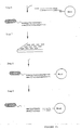
- nucleic acid assays can be performed using arrays of oligonucleotide synthesised on a planar solid phase substrate like a glass slide. Such arrays are generally constructed such that the slide is divided into distinct zones or fields and each field bears only a single oligonucleotide. Hybridisation of a labelled nucleic acid to the array is determined by measuring the signal from the labelled nucleic acid from each field of the array. Determination of mRNA levels can be effected in a number of ways. One can readily convert poly-A bearing mRNA to cDNA using reverse transcription. Reverse Transcriptase PCR (RTPCR) methods allow the quantity of single RNAs to be determined, but with a relatively low level of accuracy.
- RTPCR Reverse Transcriptase PCR
- Arrays of oligonucleotides are a relatively novel approach to nucleic acid analysis, allowing mutation analysis, sequencing by hybridisation and mRNA expression analysis. Methods of construction of such arrays have been developed, (see for example: references 9, 10, 11) and further methods are envisaged.
- Hybridisation of labelled nucleic acids to oligonucleotide arrays of the sort described above is typically detected using fluorescent labels.
- Arrays of oligonucleotides or cDNAs can be probed with nucleic acids labelled with fluorescent markers.
- fluorescent markers For an oligonucleotide chip this would reveal to which oligonucleotides a labelled nucleic is complementary by the appearance of fluorescence in the fields of the array containing oligonucleotides to which the labelled nucleic acid hybridises.
- Such oligonucleotide arrays could be read using MALDI mass spectrometry if nucleic acids that are hybridised to the oligonucleotide array were labelled with mass labels.
- the mass labels would preferrably be linked to their corresponding nucleic acid using a photo-cleavable linker.
- These mass labels could incorporate laser excitable agents into their structure or the oligonucleotide array could be treated with appropriate desorption agents after a hybridisation reaction has been performed, such as 3-hydroxypicolinic acid.
- the linker between mass label and nucleic acid can be cleaved by application of laser light of the appropriate frequency.
- the labels can then be desorbed from specific regions of an oligonucleotide array by scanning those regions with laser light of the appropriate frequency.
- the identity of the hybridised nucleic acid at a particular field of the oligonucleotide array can then be determined from the mass of the label that is desorbed from that field of the array.
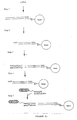
- Oligonucleotide arrays can be directly adapted for use with the gene-profiling technology disclosed in reference 12.
- An array that bears all 256 possible 4 base oligonucleotides at defined points on its surface can be used to effect the sorting step required by that invention, discussed above.
- this chip-based embodiment of the profiling system be compatible with mass-spectrometric analysis one requires that the labels used on the adaptors for determining the second 4 base sample of sequence be MALDI compatible so that the oligonucleotide chip can be scanned by an Ultra-Violet laser in a MALDI spectrometer.
- the region of the chip from which a set of labels is desorbed from identifies the first 4 bp of the signature while the composition of the labels identifies the second 4 bases of the signature and the relative quantities of each cDNA.
- the gene profiling process operates in a two stage process, molecular sorting of signatures followed by analysis of probe molecules ligated to the sorted signatures.
- the MALDI approach uses an oligonucleotide array to effect sorting of the signatures.
- An alternative to the use of an array is affinity chromatography.
- To sort signatures with an ambiguous sticky-end of 4 bp one can derivitise beads appropriate for use in an HPLC format with the 256 possible 4-mers at the sticky-end. Such a column may be loaded with the signatures dissolved in a buffer favouring hybridisation to the 4 mers on the derivitised beads.
- the column may then be washed with gradually increasing concentrations of a buffer that inhibits hybridisation.
- Signatures terminating with AAAA or TTTT sticky ends will be released first while GGGG and CCCC signatures will be released last.
- To ensure separation of signatures that are the complement of each other one can derivitise beads with base analogs so that the hybridisation affinity of a guanine in a signature to a cytosine on a bead is different to the hybridisation of a cytosine in a signature sticky-end to a guanosine on a bead.
- each 4-mer is present in a different relative concentration on the beads to any other.
- Such an affinity column should allow a population of signatures to be sorted into 256 fractions according to the sequence of its ambiguous sticky-end. Such fractions can then be loaded directly into an Electrospray Mass Spectrometer for analysis.
- a method of sequencing DNA using a reagent comprising an analyte moiety linked to one or more reporter groups is described in reference 33.
- a sequencing technology is described in reference 13, in which a method for sequencing nucleic acid is provided, which comprises:
- Reference 14 discloses a method to identify sites in the tertiary structure of the RNA that are accessible to oligonucleotides that does not require amplification of oligonucleotides or any form of electrophoresis.
- the binding of short oligonucleotide probes, preferrably 4-mers, to an mRNA is detected and the pattern of binding is correlated to the primary structure of the mRNA.
- An accessible region will have a number of probes binding to it with a high affinity and the sequences of those probes should be complementary to the primary sequence at that accessible region. The sequences of the probes should also overlap.
- the mRNA or the probes are immobilised onto a solid phase substrate and labelled probes or mRNA, respectively, are hybridised to the captured nucleic acids.
- the preferred method of labelling disclosed in reference 14 is fluorescent labelling, but it is clear that mass-labelled nucleic acids could be used instead.
- Reference 15 discusses a variety of hybridisation assays compatible with mass-labelled nucleic acid probes.
- the essential features of a mass spectrometer are as follows: Inlet System -> Ion Source -> Mass Analyser -> Ion Detector -> Data Capture System.
- the critical feature is the the inlet system and ion source.
- Other features of importance for the purposes of biological analysis are the sensitivity of the mass analyser/detector arrangements and their ability to quantify analyte molecules.
- 'soft' ionisation techniques are used for many biological mass spectrometry applications. These allow large molecules such as proteins and nucleic acids to be ionised essentially without fragmentation.
- the liquid phase techniques allow large biomolecules to enter the mass spectrometer in solutions with mild pH and at low concentrations.
- a number of techniques are ideal for use with this invention, including but not limited to Electrospray Ionisation, Fast Atom Bombardment and Matrix Assisted Laser Desorption Ionisation (MALDI).
- Electrospray ionisation requires that a dilute solution of a biomolecule be nebulised into the spectrometer, i.e., injected as a fine spray.
- solution may be sprayed from the tip of a capillary tube by a stream of dry nitrogen and under the influence of an electrostatic field.
- the mechanism of ionisation is not fully understood but is thought to be broadly as follows. In a stream of nitrogen the solvent evaporates. As the droplets become smaller, the concentration of the biomolecule increases. Under the spraying conditions, most biomolecules carry a net positive or negative charge, which increases electrostatic repulsion between the dissolved biomolecules.
- the strength of the electric field adds to their kinetic energies. This in turn leads to more or less energy transfer during collisions of ions and neutral molecules, which may then give rise to fragmentation. This is of significance when considering fragmentation of ions in the mass spectrometer. The more energy imparted to a population of ions the more likely it is that fragmentation will occur through collision of analyte molecules with the bath gas or solvent vapour present in the source. By adjusting the voltage used to accelerate ions in the ionisation chamber one can control the fragmentation of ions. This phenomenon is advantageous when fragmentation of ions is to be used as a means of cleaving a label from a mass labelled nucleic acid.
- MALDI Matrix Assisted Laser Desorption Ionisation
- MALDI requires that the biomolecule be embedded in a large molar excess of a photo-active 'matrix'.
- the application of laser light of the appropriate frequency (266 nm for nicotinic acid ) results in the excitation of the matrix which in turn leads to excitation and ionisation of the embedded biomolecule
- This technique imparts a significant quantity of translational energy to ions but tends not to induce excessive fragmentation. Electric fields can again be used to control fragmentation with this technique.
- MALDI techniques can be used in two ways. Mass-labelled DNA may be embedded in a matrix, so that the labels themselves are not specifically excitable by the laser or labels could be constructed so as to contain the necessary groups that would allow laser excitation.
- Such groups include nicotinic, sinapinic or cinnamic acid moieties.
- MALDI-based cleavage of labels would probably be most effective with a photocleavable linker as this would avoid a cleavage step prior to performing MALDI mass spectrometry.
- the various excitable ionisation agents have different excitation frequencies so that a different frequency can be chosen to trigger ionisation from that used to cleave the photolysable linker.
- These excitable moieties could derivitised using standard synthetic techniques in organic chemistry to give a variety of labels having a range of masses. The range could be constructed in a combinatorial manner.
- Fast Atom Bombardment has come to describe a number of techniques for vaporising and ionising relatively involatile molecules.
- the essential principal of these techniques is that samples are desorbed from surfaces by collision of the sample with accelerated atoms or ions, usually xenon atoms or caesium ions.
- the samples may be coated onto a solid surface as for MALDI but without the requirement of complex matrices.
- These techniques are also compatible with liquid phase inlet systems - the liquid eluting from a capillary electrophoresis inlet or a high pressure liquid chromatograph passes through a frit, essentially coating the surface of the frit with analyte solution which can be ionised from the frit surface by atom bombardment.
- a mass spectrometer is not a simple device for quantification but use of appropriate instrumentation can lead to great sensitivity.
- the number of ions reaching a mass spectrometer detector is not a direct measure of the number of molecules actually in the ion source.
- the relationship between numbers of ions and the initial concentration of biomolecules is a complex function of ionisation behaviour.
- Quantification may be effected by scanning the mass spectrum and counting ions at each mass/charge ratio scanned. The count is integrated to give the total count at each point in the spectrum over a given time. These counts can be related back to the original qunatities of source molecules in a sample.
- the configuration of the mass spectrometer is critical to determining the actual ion count.
- the ionisation and mass separation methods are particularly sensitive in this regard.
- Certain mass separation methods act as "mass filters".
- the quadrupole mass spectrometer only permits ions with a particular mass charge ratio to pass through at any one time. This means that a considerable proportion of ions never reaches the detector.
- Most mass spectrometers detect only one part of the mass spectrum at a time. Given that a large proportion of the mass spectrum may be empty or irrelevant but is usually scanned anyway, this means a further large proportion of the sample is wasted. These factors may be a problem in detecting very low abundances of ions but these problems can be overcome in large part by correct configuration of the instrumentation.
- Mattauch-Herzog geometry sector instruments permit this but have a number of limitations.
- Sector instruments are organised into distinct regions (sectors) that perform certain functions.
- ions generated in an ion source from a divergent beam which is narrowed by passage through adjustable slits.
- This defined beam then passes through a field free region into an electric sector, which focusses it.
- the passage through the slits results in some loss of ions and therefore results in a reduction in sensitivity to the sample.
- the focussed ion beam passes through a second field-free region and on into a magnetic sector. This last sector focusses the beam on the basis of the mass-to-charge ratios of the ions.
- a photographic plate can be placed across the mass-separated beam split can be used to measure the abundancies of ions and their mass-to-charge ratios.
- the photograph plate has only a small dynamic range of sensitivity before becoming saturated and is cumbersome. Better dynamic range is achievable by use of electron multiplier arrays but at a cost of some loss in resolution.
- array detectors would allow the simultaneous and continuous monitoring of a number of regions of the mass spectrum. The array limit on the resolution of closely spaced regions of the spectrum might restrict the number of labels one might use.
- the quadrupole assembly has an advantage over many configurations in that the electric fields that separate ions of different mass-to-charge ratios can be changed with extreme rapidity, allowing a very high sampling rate over a small number of peaks of interest.
- Mass spectrometry is a highly diverse discipline and numerous mass analyser configurations exist and which can often be combined in a variety of geometries to permit analysis of complex organic molecules.
- Typical single stage mass analysers are quadrupoles or time-of-flight instruments, which are both compatible with this present invention. Sector instruments are also applicable.
- the flight time of the deflected ions is recorded and this is sufficient to determine their mass-to-charge ratios.
- the gate generally only sends a short pulse of ions into the TOF analyser at any one time. Since the arrival of all ions is recorded and since the TOF separation is extremely fast, the entire mass spectrum is measured effectively simultaneously. Furthermore, the gate electrode can sample the ion beam at extremely high frequencies so that multiple spectra can be accummulated in a very short time interval. This is important where the sample concentration in the ion source is low or lasts for only a short time.
- the orthogonal TOF geometry is very sensitve.
- Tandem mass spectrometry describes a number of techniques in which ions from a sample are selected by a first mass analyser on the basis of their mass-to-charge ratios for further analysis by induced fragmentation of those selected ions.
- the fragmentation products are analysed by a second mass analyser.
- the first mass analyser in a tandem instrument acts as a filter in selecting ions that are to be investigated.
- the selected ions pass through a collision chamber containing a neutral gas, resulting in some of them fragmenting.
- a more active approach to fragmentation entails inducing decomposition of molecular ions as, for example, by collision induced decomposition (CID).
- CID uses mass spectrometer constructions to separate out a selected set of ions and then to induce their fragmentation by collision with a neutral gas; the resulting fragment ions are analysed by a second mass spectrometer.
- induced cleavage techniques are compatible with mass labelling methodologies.
- a typical geometry uses a tandem mass analyser configuration similar to those used in CID, but the collision cell is replaced by a photo-excitation chamber in which the ion stream leaving the first mass analyser is irradiated by laser light.
- High intensity lasers are required to ensure that a significant proportion of a fast moving ion stream interacts with a photon appropriately to induce cleavage.
- the positioning of the laser is extremely important to ensure exposure of the stream for a significant period of time. Tuning the laser to a specific frequency allows for precise control over the bonds that are induced to cleave.
- mass labels linked with an appropriate photocleavable linker to their probes can be cleaved within the mass spectrometer.
- the photocleavage stage does not require a tandem geometry, the photocleavage chamber could be within or immediately following the ion source.
- a further possible technique for fragmenting molecular ions is surface induced decomposition.
- Surface induced decomposition is a tandem analyser technique that involves generating an ion beam which is separated in a first analyser into selected m/z ratios. Any selected ions are collided with a solid surface at a glancing angle. The resulting collision fragments can then be analysed by a second mass spectrometer.
- tandem mass spectrometer utilises a triple quadrupole assembly, which comprises three quadrupole mass analysers, one of which acts as a collision chamber.
- the collision chamber quadrupole acts both as a collison chamber and as an ion guide between the two other mass analyser quadrupoles.
- Gas can be introduced into the middle quadrupole to allow so that its molecules collide with the ions entering from the first mass analyser. Fragment ions are separated in the third quadrupole. Induced cleavage can be performed with geometries other than those utilising tandem sector or quadrupole analysers.
- Ion trap mass spectrometers can be used to promote fragmentation through introduction of a buffer or 'bath' gas into the trap.
- the energy of collision may be increasd by speeding up the trapped ions.
- Helium or neon may be used as the bath gas in ion traps.
- photon induced fragmentation could be applied to trapped ions.
- Another favorable geometry is a Quadrupole/Orthogonal Time-of-Flight instrument, in which the high scanning rate of a quadrupole is coupled to the greater sensitivity of a TOF mass analyser to identify products of fragmentation.
- a sector mass analyser comprises two separate 'sectors' an electric sector which focusses an ion beam leaving a source into a stream of ions with the same kinetic. energy using electric fields.
- the magnetic sector separates the ions on the basis of their mass to generate a spectrum at a detector.
- a two sector mass analyser of this kind can be used where the electric sector provide the first mass analyser stage, the magnetic sector provides the second mass analyser, with a collision cell placed between the two sectors.
- This geometry might be quite effective for cleaving labels from a mass labelled nucleic acid.
- Two complete sector mass analysers separated by a collision cell can also be used for analysis of mass labelled nucleic acids.
- Ion Trap mass spectrometers are a relative of the quadrupole spectrometer.
- the ion trap generally has a 3 electrode construction - a "torroidal" electrode and 'cap' electrodes at each end forming a cavity (the ion trap).
- a sinusoidal radio frequency potential is applied to the cylindrical electrode while the cap electrodes are biased with DC or AC potentials.
- Ions injected into the cavity are constrained into a stable circular trajectory by the oscillating electric field of the cylindrical electrode. However, for a given amplitude of the oscillating potential, certain ions will have an unstable trajectory and will be ejected from the trap.
- a sample of ions injected into the trap can be sequentially ejected from the trap according to their mass-to-charge ratio by altering the oscillating radio frequency potential. The ejected ions can then be detected allowing a mass spectrum to be produced.
- Ion traps are generally operated with a small quantity of a 'bath gas, such as helium, present in the ion trap cavity. This increases both the resolution and the sensitivity of the device as the ions entering the trap are essentially cooled to the ambient temperature of the bath gas through collision with its molecules. Collisions dampen the amplitude and velocity of ion trajectories keeping them nearer the centre of the trap. This means that when the oscillating potential is changed, ions whose trajectories become unstable gain energy more rapidly, relative to the damped circulating ions and exit the trap in a tighter bunch giving greater resolution.
- a 'bath gas such as helium
- Ion traps can mimic tandem sector mass spectrometer geometries. In fact, they can mimic multiple mass spectrometer geometries thereby allowing complex analyses of trapped ions.
- a single mass species from a sample can be retained in a trap, viz., all other species can be ejected. Then, the retained species can be carefully excited by super-imposing a second oscillating frequency on the first. The kinetically-excited ions collide with bath gas molecules and will fragment if sufficiently excited. The fragments can be analysed further. This is MS/MS or MS 2 .
- a fragment ion can be further analysed by ejecting all other ions and then kinetically exciting the fragment so that it fragments after collison with bath gas molecules (MS/MS/MS or MS 3 ). This process can be repeated for as long as sufficient sample exists to permit further analysis (MS n ) . It should be noted that ion traps generally retain a high proportion of fragment ions after induced fragmentation.
- FTICR mass spectrometers discussed below represent a form of temporally resolved tandem mass spectrometry rather than spatially resolved tandem mass spectrometry which is found in linear mass spectrometers.
- FTICR mass spectrometry has similar features to ion traps in that a sample of ions is retained within a cavity but, in FTICR MS, the ions are trapped in a high vacuum chamber (ICR cell) by crossed electric and magnetic fields.
- the electric field is generated by a pair of plate electrodes that form two sides of a box.
- the box is contained in the field of a magnet, which in conjunction with the two plates (the trapping plates), constrain injected ions to have a cycloidal trajectory.
- the ions may be kinetically excited into larger cycloidal orbits by applying a radiofrequency pulse to two 'transmitter plates'.
- the cycloidal motions of the ions generate corresponding electric fields in the remaining two opposing sides (plates) of the box, which comprise the 'receiver plates'.
- the excitation pulses kinetically excite ions into larger orbits, which decay as the coherent motions of the ions is lost through collision with neutral gas molecules.
- the corresponding signals detected by the receiver plates are converted to a mass spectrum by Fourier transform analysis.
- these instruments can act in a similar manner to an ion trap - all ions except a single species of interest can be ejected from the ICR cell.
- a collision gas can be introduced into the trap and fragmentation can be induced.
- the fragment ions can be analysed subsequently.
- fragmentation products and bath gas combine to give poor resolution if analysed by FT of signals detected by the 'receiver plates' .
- the fragment ions can be ejected from the cell and then analysed in a tandem configuration with, for example, quadrupole.
- Mass labelled hybridisation probes may have the following basic structures. Nu - M Nu - L - M Where Nu is a nucleic acid probe and L is a linker group connecting the nucleic acid probe to the mass label, M.
- the linker group (L) is optional and the mass label may have the necessary linker features incorporated into it. The linker group is not necessary when a non-cleavable mass-labelled hybridisation probe is required.
- Nucleic acids are linear polymers of nucleotides, of which there is a relatively small number of naturally occurring species but a growing number of chemically synthesised analogues, which can be coupled to the linker group at numerous positions. Such possibilities are discussed later.
- Linker groups may have the following structural features:
- a mass-to-charge ratio of up to 2000 to 3000 units is a suitable range for such mass labels as this corresponds to the range over which singly charged ions can be detected reliably at greatest sensitivity.
- labels of mass less than 200 to 300 daltons are not ideal because the low mass end of any mass spectrum tends to be populated by solvent molecules, small molecule impurities, multiple ionisation peaks and fragmentation peaks.
- each label should be separated by a minimum of about 4 daltons from its neighbours to avoid overlap caused by carbon, nitrogen and oxygen isotope peaks.
- the mass label should ionise and separate so as to form predominantly one species (without fragmentation).
- the mass label should be easily ionised to ensure that as much of the cleaved mass label as possible is detected.
- the labels need to have a net electric charge, but preferably should not be multiply ionised, i.e. they should have a single electric charge.
- the labels should be resistant to fragmentation so that each peak in a mass spectrol scan corresponds only or uniquely to a single label; this simplifies analysis of the data and reduces any ambiguity in the determination of the quantity of the label, a criterion which is very important for some of the applications for which this invention has been developed.
- DNA and other nucleic acids tend to fragment to extensively in a mass spectrometer. It is desirable to ensure DNA fragment peaks in the resulting mass spectrum do not obscure those arising from mass labels. It is preferable to ensure that nucleic acid probe fragments are separated from mass labels after cleavage. To this end, one can use mass labels that form negative ions on ionisation and which can be separated by negative ion spectrometry. Nucleic acids, despite having a negatively charged backbone, have a tendency to be protonated on ionisation, particularly by electrospray and related liquid-to-gas phase ionisation techniques. This means that, if the mass spectrometer is configured for negative ion spectrometry, only negatively charged mass labels should appear in the mass spectrum. Most nucleic acid fragments will not reach the detector.
- nucleic acid probes can be promoted through the use of appropriate buffer solutions, thus ensuring that nucleic acids are extensively present with a pre-existing positive charge.
- Fragmentation is a highly significant feature of mass spectrometry. With respect to this invention it is important to consider how a mass label is to be identified. At the one extreme mass labels may be designed such that they are highly resistant to fragmentation and the label is identified by the appearance of the label's molecular ion in the mass spectrum. In this situation, families of labels having unique molecular ions would need to be designed. At the other extreme, a mass label having a highly characteristic fragmentation pattern could be designed such that this pattern would identify it. In this case, families of labels having non-overlapping patterns or with at least one unique fragmentation species for each label must be designed. Fragmentation is a property of the initial molecule and of the ionisation technique used to generate the ions from it.
- bond strengths may increase or decrease in ways that are difficult to predict a priori.
- D(A-B) refers to bond dissociation energy of the species in parentheses
- I (N) refers to the ionisation energy of the species in parentheses
- ⁇ H is the enthalpy of formation of the species in parentheses
- ⁇ S ⁇ 0 and therefore, ⁇ G ⁇ H the upshot of the equations above is that in order to predict whether a bond is likely to be stable under a given set of ionisation conditions it is necessary to know the ionisation energy of the molecule and the ionisation energy of the neutral fragment that results from fragmentation of the bond in question.
- ionisation energies of molecules and neutral fragments is a general working principle, which can be used to predict likely ionic bond strengths. If the energy added during ionisation is less than the ionic bond strength then fragmentation will not be observed.
- Aryl or Aryl-F bonds are also strong in ions which is attractive for mass labelling as fluorocarbons are cheap to manufacture, are chemically inert, have a detectable mass defect with respect to hydrocarbon molecules and fluorine has only the single naturally-occurring isoptope, 19 F.
- Photo-cleavable and chemically-cleavable linkers can be easily developed for the applications described.
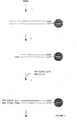
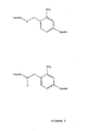
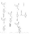
- Figure 5 shows a series of exemplary photocleavable linkers.
- Ortho-nitrobenzyl groups are well known in the art as photocleavable linkers, cleaving at the benzylamine bond.
- cleavable linkers see reference 18, which discusses a variety of photocleavable and chemically-cleavable linkers.
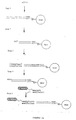
- Thermal cleavage operates by thermally induced rearrangements.
- Figure 6 shows the synthesis of one example of a mass label linked via a thermally cleavable linker to the 3'-OH position of a thymidine residue.
- Figure 6 also shows the thermally induced rearrangement that would cleave the label from its associated nucleotide.
- the group X in this example could be an aryl ether polymer, as discussed later.
- S could be replaced with N or C, and O be replaced by S.
- S see reference 28.
- a preferred method of cleavage is through the use of the ionisation process to induce fragmentation of labels.
- a linker may be designed to be highly labile in the ionisation process, such that it will cleave when the molecule to which it is attached is ionised in a mass spectrometer. There are two factors to consider in controlling cleavage using this method: (1) how much excess of energy is deposited in the ion during the ionisation process and, (2) whether this excess is sufficient to overcome any one bond energy in the ion. The excess of energy deposited is strongly determined by the ionisation technique used.
- the energy In order for the deposited energy to effect cleavage of a bond the energy must be in a vibrational/rotational mode and must be sufficient to overcome the dissociation energy of the bend.
- the bond energy is obviously determined by the chemical structure of the molecule being analysed. Bond energies are discussed later. Generally speaking, energy is imparted as electronic, vibrational, rotational and translational energy in the ionisation process. Within a very short time of ionisation, most of this excess of internal energy will have transformed into vibrational and rotational energy by intersystem and interstate crossing. The excess of internal revibrational energy may or may not lead to bond scission.
- ions In order to impart more internal vibrational energy into the moving ions, they can be collided with a bath gas to give fragmentation of the ion.
- a bath gas In an electrospray source there is a bath gas and volatised solvent. Ions can be accelerated through an electric field to increase the energy of collision with a bath gas. The acceleration kinetic energy to the ions. If sufficient kinetic energy is imparted to the ions then collisions with the bath gas will result in fragmentation of the ions. The amount of kinetic energy required depends on the strength of the bonds in the ion but the amount of energy imparted can be controlled by regulating the accelerating potential.
- Linear alkanes fragment relatively randomly while molecules containing secondary and tertiary alkyl groups cleave most commonly at the branching points of the molecule due to the increased stabilisation of secondary and tertiary carbocations.
- double bonds stabilise adjacent positive or negative charges through resonance or delocalisation effects. Similar effects are noted in bonds adjacent to aryl groups.
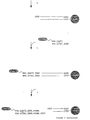
- Some cleavable linkers that can be induced to fragment by collision or otherwise are shown in Figure 7. These are numbered in order of their increasing lability.
- the groups on the left of the cleavable bond are well known as good leaving groups and are used to protect reactive positions in a molecule. As such they will be susceptible to chemical cleavage under certain conditions.
- Linker (4) in Figure 7 is highly susceptible to protic chemical attack and so would only be usable as a fragmentable linker if the probing reaction reaction was not acidic.
- Linker (1) is considerably less photolytically cleavable. Obviously, these groups could be chosen intentionally to cleave chemically as required. It is easy to see from Figure 7 that these linkers can also form part of a delocalised aryl-ether polymer system.
- the group to the right of the cleavable bond essentially stabilises a negative charge, which is advantageous in that it promotes bond breakage at this site and can provide a detectable negative ion. Other charge stabilising groups could be used at this position.
- the 'handles' on this and other Figures generally represents a reactive group useful in the synthesis of the mass labelled base sequence, which may not be present in the mass labelled molecule as synthesised.
- Mass labels and their linkers can be attached to a nucleic acid at a number of locations.
- the 5' hydroxyl of the ribose sugar is the easiest to derivitise.
- Other favoured positions for modifications are on the base at the 5' position in pyrimidines and the 7' and 8' positions in purines. These would be the preferred positions to attach cleavable mass labels and non-cleavable mass labels.
- the 2' position on the sugar is accessible for mass modifications but is more appropriate for small mass modifications that are not to be removed.
- phosphate linkage in natural nucleic acids can be modified to a considerable degree as well, including derivitisation with mass labels.
- modified nucleic acids might want to be used, which contain a number of different analogues for which hybridisation behaviour is modified. This is particulary important when groups of hybridisation probes are used simultaneously. It may be desirable to modify the hybridisation behaviour of a group of probes so that the melting temperatures of the correctly hybridised probes are very close to or at least above some threshold. Preferably the melting temperature of incorrectly hybridised prcbes will fall below this threshold. This allows groups of probes to be used simultaneously whilst ensuring the stringency of hybridisation reactions.
- duplexes comprising only adenine and thymine are unstable relative to duplexes containing only guanine and cytosine.
- These differences in stability can present problems when trying to hybridise mixtures of short oligonucleotides to a target RNA.
- Low temperatures are needed to hybridise A-T rich sequences but at these temperatures G-C rich sequences hybridise to sequences that are not fully complementary. This means that some mismatches may happen and specificity can be lost for the G-C rich sequences.
- G-C rich sequences hybridise specifically but A-T rich sequences do not hybridise.
- nucleic acids In order to normalise these effects modifications can be made to nucleic acids. These modifications fall into three broad categories: base modifications, backbone modifications and sugar modifications.
- Nucleotides may be readily modified in the phosphate moiety. Under certain conditions, such as low salt concentration, analogues such as methylphosphonates, triesters and phosphoramidates have been shown to increase duplex stability. Such modifications may also have increased nuclease resistance. Further phosphate modifications include phosphodithirates and boranophosphates, each of which increases the stability of oligonucleotide against exonucleases.
- the sugar may be replaced by a different sugar such as hexose or the entire sugar phosphate backbone can be entirely replaced by a novel structure such as in peptide nucleic acids (PNA) .
- PNA peptide nucleic acids
- hydrophobic groups to the 3' and 5' termini of an oligonucleotide also increase duplex stability by excluding water from the bases, thus reducing 'fraying' of the complex, i.e. hydrophobic groups reduce solvation of the terminal bases.
- Amine derivatives, quaternary ammonium ions or positive sulphur centres are good charge carriers if positive ion mass spectrometry is used. These have extremely good detection properties that generate clean sharp signals.
- negatively charged ions can be used, so molecules with carboxylic acid, sulphonic acid and other moieties are appropriate for negative ion spectrometry.
- Labels for MALDI mass spectrometry can be generated by derivitising known molecules that are excitable by UV visible laser light, such as sinapinnic acid or cinnamic acid, of which a number of derivatives are already commercially available. Fragmentation resistant groups are discussed above.
- polyaryl ether structures are very resistant to fragmentation and produce good negative ions since the delocalisation of electrons over the molecule can effectively stabilise a negative charge.
- These molecules are also thermally stable and so are particularly compatible with thermally cleaved linkers and with linkers cleaved by collision processes within the mass spectrometer.
- the 'Variable Groups' at either end of the polyaryl ethers are preferrably substituted aryl ethers which modify the properties of the mass label (Figure 9).
- Such modifying groups include 'mass series modifying' groups (see Figure 9) , solubilising groups, charge carrying groups (see Figure 10) and mass defect groups (see Figure 8).
- a linear polymer of polyaryl ethers increases in mass by 92 mass units with each additional "phenoxy" residue in the molecule.
- mass labels need only be about 4 daltons apart.
- each mass label preferably contains a group that shifts the mass of each series of aryl ethers.
- This Mass Series Modifying group acts to offset each series of aryl-ether polymers from the others.
- linear polymers of aryl ethers each monomer of which adds 92 daltons, there will be no coincidence in mass for a maximum of 23 series If each series of mass markers is 4 mass units apart.
- In order to generate 256 mass labels for example, one then needs to generate the 23 MSM groups, to link to polymers of aryl ethers with up to 12 consecutive phenoxy repeats. This would give a total of 276 mass labels.
- a polymer comprising a number of different subunits can be generated with those sub-units appearing in different sequences.
- branched structures are also possible but only linear polymers are shown for convenience of illustration. The preferred structures shown are chosen for convenience of synthesis. Different sequences of the same subunits are not significantly more difficult to produce but it is preferable to generate as many labels as possible in as few synthetic steps as possible.
- a prefered synthesis strategy is to generate polyaryl ethers of up to twelve repeats and then derivitise these with a number of different MSM groups, whose masses differ ideally by about 4 daltons to avoid overlap of isotope peaks. Variation in the MSM group can be fine-tuned by using isotopic substitutions; for example, replacement of 4 hydrogens in a molecule with 4 deuterium atoms gives a mass difference of 4 daltons.
- aromatics or heteroaromatics are used, they may be substituted or unsubstituted. If substituted, the substituents must also be resistant to fragmentation and may be selected from any of the categories set out above.
- any mass label be resistant to fragmentation and should preferably have a stability to electron ionisation conditions at 50 volts.
- An advantageous embodiment of this technology is the use of fluorinated mass labels when high resolution mass analysis of labels is employed after cleavage from their nucleic acid.
- a hydrocarbon molecule whose integral mass is 100 will have a fractionally higher accurate mass.
- a fluorinated molecule whose integral mass is 100 has a fractionally lower accurate mass.
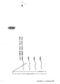
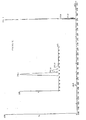
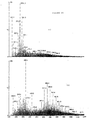
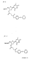
- a negative ion mass spectrum of the previously synthesised molecule, AG/1/75 is shown in Figure 11. This spectrum was generated with the molecule present at 10 ng/ ⁇ l. The solvent was methanol and water in a 1:1 ratio. The spectrum was generated with an electrospray inlet system coupled to a scanning quadrupole mass spectrometer. The inset shows the mass peaks corresponding to the anion of AG/1/75 molecule, a singly charged negative ion at m/z 291 daltons [M - Na] - . Note that the isotope peaks are significant over about three daltons from the quasi molecular ion peak.
- Figure 12 shows a positive ion spectrum of AG/1/75. There is no detectable molecular ion in this spectrum, hence this molecule is best used as a negative ion mode marker. Both of the above spectra were generated with a cone voltage in the electrospray source of 45 V.
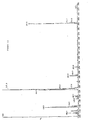
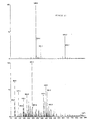
- Figure 13 shows a negative ion spectrum of AG/1/75 in the same solution as for the previous spectra but with a cone voltage of 75 V. This voltage is sufficient to cause significant fragmentation in the molecule generating a major negative fragment ion peak at m/z 156 daltons, corresponding to the cleavage at the position shown in the inset structure in Figure 13.
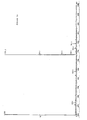
- Figures 14 and 15 show mass spectra of an 'unconditioned' PCR product in various buffers, in positive and negative modes.
- the PCR product was 'unconditioned' in that no effort had been made to separate the DNA from the buffer and reaction material beyond what is normally done for gel electrophoresis. No attempt was made to exchange metal ion adducts for ammonium ions or to generate pure DNA as is usual practice for mass spectrometry purposes.
- Figures 16 and 17 show the same PCR product with AG/1/75 which can clearly be detected in the negative ion mode but not in the positive mode.
- Figures 18 and 19 show the same spectra after signal processing to subtract background noise and it is clear that AG/1/75 can be easily detected in the negative ion mode.
- the reaction mixture was diluted with dichloromethane and washed with aqueous NaHCO 3 (5% w/v) and twice with water.
- the organic phase was dried (Na 2 SO 4 ) and the solvent was removed under reduced pressure.
- the residue was purified by flash chromatography using ethyl acetate/n-hexane (1:1) as eluant to give 111 mg (35 % yield) of FT 18/1 as a colurless oil.
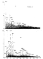
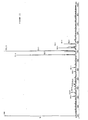
- FIG. 20 shows a mass spectrum generated by using an electrospray ion source, with a cone voltage of 45v, in a Platform-LC quadrupole scanning mass spectrometer (Micromass UK). In each case, FT23 was present at 4pmol/ ⁇ l.
- Figure 20 shows the mass spectrum in negative ion mode with a prominent peak at 729.3 corresponding to the [M - H] - ion.
- Figure 21 shows the corresponding mass spectrum in positive ion mode with a number of prominent peaks.
- Figures 22 and 23 show respectively negative ion and positive ion mode mass spectra generated under the same conditions as those shown in Figures 20 and 21 with the exception that an oligonucleotide sample of approximate molecular weight 3000 is additionally present in each case at 4pmol/ ⁇ l.
Landscapes
- Life Sciences & Earth Sciences (AREA)
- Chemical & Material Sciences (AREA)
- Organic Chemistry (AREA)
- Proteomics, Peptides & Aminoacids (AREA)
- Zoology (AREA)
- Wood Science & Technology (AREA)
- Health & Medical Sciences (AREA)
- Engineering & Computer Science (AREA)
- Physics & Mathematics (AREA)
- General Engineering & Computer Science (AREA)
- Microbiology (AREA)
- Molecular Biology (AREA)
- Analytical Chemistry (AREA)
- Immunology (AREA)
- Biotechnology (AREA)
- Biochemistry (AREA)
- Bioinformatics & Cheminformatics (AREA)
- Biophysics (AREA)
- General Health & Medical Sciences (AREA)
- Genetics & Genomics (AREA)
- Spectroscopy & Molecular Physics (AREA)
- Other Investigation Or Analysis Of Materials By Electrical Means (AREA)
- Measuring Or Testing Involving Enzymes Or Micro-Organisms (AREA)
- Investigating Or Analysing Biological Materials (AREA)
- Discharge Heating (AREA)
- Adhesives Or Adhesive Processes (AREA)
- Surgical Instruments (AREA)
- Apparatus Associated With Microorganisms And Enzymes (AREA)
- Pharmaceuticals Containing Other Organic And Inorganic Compounds (AREA)
- Ultra Sonic Daignosis Equipment (AREA)
Claims (44)
- Série de sondes d'hybridation, dont chacune comprend un marqueur de masse relié à une séquence de bases connue de longueur prédéterminée, caractérisée en ce que chaque marqueur de masse de la série, éventuellement avec la séquence de bases connue, peut être associé à cette séquence de bases par spectrométrie de masse, et en ce que chaque marqueur de masse comprend un groupe de photoexcitation choisi entre l'acide nicotinique, l'acide sinapique et l'acide cinnamique.
- Série selon la revendication 1, caractérisée en ce que chaque marqueur de masse peut être identifié de façon unique par rapport à tous les autres marqueurs de masse de la série.
- Série selon la revendication 1 ou 2, caractérisée en ce que la longueur prédéterminée de la séquence de bases est comprise entre 2 et 25.
- Série selon l'une des revendications 1 à 3, caractérisée en ce que chaque marqueur de masse est relié à la séquence de bases connue correspondante par une liaison clivable, et peut être associé à sa séquence de bases par spectrométrie de masse lorsqu'il est libéré de celle-ci.
- Série selon la revendication 4, caractérisée en ce que chaque marqueur de masse est relié à la séquence de bases connue par une liaison clivable pouvant être coupée par collision, photoclivage, clivage chimique ou clivage thermique.
- Série selon la revendication 4 ou 5, caractérisée en ce que chaque marqueur de masse est relié à la séquence de bases connue par une liaison clivable pouvant être coupée lorsqu'il se trouve dans un spectromètre de masse.
- Série selon l'une des revendications 4 à 6, caractérisée en ce que chaque marqueur de masse a une charge négative dans des conditions d'ionisation.
- Série selon l'une des revendications précédentes, caractérisée en ce que la séquence de bases connue comprend une extrémité cohésive d'un oligonucléotide adaptateur contenant un site de reconnaissance pour une endonucléase de restriction qui coupe à une distance prédéterminée du site de reconnaissance.
- Série selon l'une des revendications précédentes, caractérisée en ce que la séquence de bases connue comporte reliée à elle une pluralité de marqueurs de masse identiques.
- Série selon l'une des revendications précédentes, caractérisée en ce que le groupe de photoexcitation est un agent d'ionisation excitable et convient pour réaliser une désorption-ionisation par laser assistée par matrice.
- Utilisation d'une série de sondes d'hybridation selon l'une des revendications précédentes dans un procédé pour déterminer l'hybridation des sondes par spectrométrie de masse des marqueurs de masse, éventuellement avec les séquences de bases connues correspondantes.
- Utilisation d'une sonde d'hybridation, comprenant un marqueur de masse relié à une séquence de bases connue de longueur prédéterminée, dans un procédé pour déterminer l'hybridation des sondes par spectrométrie de masse des marqueurs de masse, éventuellement avec les séquences de bases connues, caractérisée en ce que le marqueur de masse comprend un groupe de photoexcitation choisi entre l'acide nicotinique, l'acide sinapique et l'acide cinnamique.
- Utilisation selon la revendication 12, caractérisée en ce que la longueur prédéterminée de la séquence de bases est comprise entre 2 et 25.
- Utilisation selon la revendication 12 ou 13, caractérisée en ce que le marqueur de masse est relié à la séquence de bases connue par une liaison clivable .
- Utilisation selon la revendication 14, caractérisée en ce que le marqueur de masse est relié à la séquence de bases connue par une liaison clivable pouvant être coupée par collision, photo-clivage, clivage chimique ou clivage thermique.
- Utilisation selon la revendication 14 ou 15, caractérisée en ce que le marqueur de masse est relié à la séquence de bases connue par une liaison clivable pouvant être coupée lorsqu'il se trouve dans un spectromètre de masse.
- Utilisation selon l'une des revendications 14 à 16, caractérisée en ce que le marqueur de masse a une charge négative dans des conditions d'ionisation.
- Utilisation selon l'une des revendications 14 à 17, caractérisée en ce que le marqueur de masse peut être séparé de la séquence de bases connue en spectrométrie de masse.
- Utilisation selon l'une des revendications 12 à 18, caractérisée en ce que la séquence de bases connue comprend une extrémité cohésive d'un oligonucléotide adaptateur contenant un site de reconnaissance pour une endonucléase de restriction qui coupe à une distance prédéterminée du site de reconnaissance.
- Utilisation selon l'une des revendications 12 à 19, en vue de déterminer l'hybridation de la sonde dans une réaction en chaîne de polymérase ou une réaction en chaîne de ligase.
- Utilisation selon l'une des revendications 12 à 20, caractérisée en ce que la séquence de bases connue porte liée à elle une pluralité de marqueurs de masse identiques.
- Utilisation selon l'une des revendications 12 à 21, caractérisée en ce que le groupe de photoexcitation est un agent d'ionisation excitable et convient pour réaliser désorption-ionisation par laser assistée par matrice.
- Utilisation selon l'une des revendications 11 à 22, caractérisée en ce que la méthode est réalisée exclusivement en ligne.
- Méthode pour déterminer l'hybridation d'une série de sondes avec un acide nucléique cible, laquelle méthode comprend :(a) la mise en contact de l'acide nucléique cible avec chaque sonde d'hybridation de la série dans des conditions propres à hybrider la sonde à l'acide nucléique cible, et éventuellement l'élimination du matériau non hybridé, chaque sonde comportant un marqueur de masse lié à une séquence de bases connue de longueur prédéterminée et chaque marqueur de masse comprenant un groupe de photoexcitation choisi entre l'acide: nicotinique, l'acide sinapique et l'acide cinnamique ; et(b) l'identification de la sonde hybridée par spectrométrie de masse.
- Méthode selon la revendication 24, caractérisée en ce que chaque marqueur de masse est relié à la séquence de bases connue correspondante par une liaison clivable et chaque sonde hybridée est clivée afin de libérer le marqueur de masse, lequel marqueur libéré est identifié à l'aide d'un spectromètre de masse.
- Méthode pour déterminer l'hybridation d'une sonde avec un acide nucléique cible, laquelle méthode comprend :(a) la mise en contact de l'acide nucléique cible avec une sonde d'hybridation comprenant un marqueur de masse relié à une séquence de bases connue de longueur prédéterminée, dans des conditions propres à hybrider la sonde à l'acide nucléique cible, et éventuellement l'élimination du matériau non hybridé, le marqueur de masse comprenant un groupe de photoexcitation choisi entre l'acide nicotinique, l'acide sinapique et l'acide cinnamique ; et(b) l'identification de la sonde hybridée par spectrométrie de masse.
- Méthode selon la revendication 26, caractérisée en ce que le marqueur de masse est relié à la séquence de bases connue par une liaison clivable et la sonde hybridée est clivée pour libérer le marqueur de masse, lequel marqueur libéré est identifié à l'aide d'un spectromètre de masse.
- Méthode selon l'une des revendications 24 à 27, caractérisée en ce que l'échantillon ou chaque échantillon est analysé par spectrométrie de masse à désorption-ionisatien par laser assistée par matrice.
- Méthode selon l'une des revendications 24 à 28, caractérisée en ce que la longueur prédéterminée de la séquence de bases est comprise entre 2 et 25.
- Méthode selon l'une des revendications 24 to 29, caractérisée en ce que le ou chaque marqueur de masse est relié à la séquence de bases connue par une liaison clivable pouvant être coupée par collision, photoclivage, clivage chimique ou clivage thermique.
- Méthode selon l'une des revendications 25 ou 27 à 30, caractérisée en ce que la liaison est coupée dans le spectromètre de masse.
- Méthode selon la revendication 31, caractérisée en ce que le clivage de la liaison est induit par un photoclivage par laser.
- Méthode selon la revendication 31, caractérisée en ce que le clivage de la liaison est induit par collision.
- Méthode selon l'une des revendications 25 ou 27 à 33, caractérisée en ce que chaque marqueur de masse a une charge négative dans des conditions d'ionisation.
- Méthode selon l'une des revendications 25 ou 27 à 34, caractérisée en ce que les marqueurs de masse et les séquences de bases connues ne sont pas séparés avant l'entrée dans le spectromètre de masse.
- Méthode selon l'une des revendications 24 à 35, caractérisée en ce que la séquence de bases connue comprend une extrémité cohésive d'un oligonucléotide adaptateur contenant un site de reconnaissance pour une endonucléase de restriction qui coupe à une distance prédéterminée du site de reconnaissance.
- Méthode selon l'une des revendications 24 à 36, caractérisée en ce que la séquence de bases connue comporte liée à elle une pluralité de marqueurs de masse identiques.
- Méthode selon l'une des revendications 24 à 37, caractérisée en ce que le groupe de photoexcitation est un agent d'ionisation excitable et convient pour réaliser une désorption-ionisation par laser assistée par matrice.
- Méthode selon l'une des revendications 24 à 38, qui est exécutée en ligne.
- Utilisation d'une série selon l'une des revendications 1 à 10 pour la lecture d'une puce à oligonucléotides.
- Utilisation d'une série selon l'une des revendications 1 à 10 dans un essai par liaison compétitive destiné à identifier un agent de liaison d'oligonucléotide.
- Utilisation d'une série selon l'une des revendications 1 à 10. dans une réaction en chaîne à la polymérase ou une réaction en chaîne à la ligase pour rechercher des séquences prédéterminées.
- Méthode de caractérisation de l'ADNc, laquelle méthode comprend :(a) la découpe d'un échantillon contenant une population d'un ou plusieurs ADNc avec une endonucléase de restriction et l'isolement de fragments portant une extrémité de l'ADNc dont le site de restriction se trouve dans un site de référence proximal par rapport à l'extrémité de l'ADNc,(b) la découpe des fragments isolés avec une première endonucléase d'échantillonnage sur un premier site d'échantillonnage à une distance connue du site de référence pour produire un premier sous-fragment et un deuxième, comprenant chacun une séquence d'extrémité cohésive de longueur prédéterminée et de séquence inconnue, le premier sous-fragment portant l'extrémité de l'ADNc,(c) le tri des premiers ou deuxièmes sous-fragments en sous-populations selon leur séquence d'extrémité cohésive et enregistrement de la séquence d'extrémité cohésive de chaque sous-population comme première extrémité cohésive,(d) la découpe des sous-fragments de chaque sous-population avec une deuxième endonucléase d'échantillonnage, qui est la même que la première endonucléase d'échantillonnage ou est différente de celle-ci, dans un deuxième site d'échantillonnage à une distance connue du premier site d'échantillonnage afin de produire, à partir de chaque sous-fragment, un nouveau sous-fragment contenant une deuxième séquence d'extrémité cohésive de longueur prédéterminée et de séquence inconnue,
et(e) la détermination de chaque deuxième séquence d'extrémité cohésive, caractérisée en ce que la longueur agrégée des première et seconde séquences d'extrémité cohésives de chaque sous-fragment est comprise entre 6 et 10, les séquences et les positions relatives du site de référence et des première et deuxième extrémités cohésives caractérisant l'ADNc ou chaque ADNc, la première endonucléase d'échantillonnage se liant à un premier site de reconnaissance et coupant au niveau du premier site d'échantillonnage à une distance prédéterminée du site de restriction de l'endonucléase de restriction et le premier et/ou le deuxième sites de reconnaissance étant présentés dans les premier et/ou second oligonucléotides adaptateurs de la série selon la revendication 8 et hybrides au site de restriction des fragments isolés. - Méthode pour le séquençage d'un acide nucléique, comprenant :(a) l'obtention d'une population d'acides nucléiques cibles comprenant des fragments d'acide nucléique dans lesquels chaque fragment est présent en quantité unique et porte à une extrémité une séquence d'extrémité cohésive de longueur prédéterminée et de séquence inconnue,(b) la protection de l'autre extrémité de chaque fragment, et(c) le séquençage de chacun des fragments par(i) mise en contact des fragments dans les conditions d'hybridation en présence d'une ligase avec une série selon la revendication 8, dont la séquence de bases a la même longueur prédéterminée que la séquence d'extrémité cohésive, laquelle série contient toutes les séquences de bases possibles ayant cette longueur prédéterminée; élimination de tout oligonucléotide adaptateur lié et enregistrement de la quantité d'oligonucléotide adaptateur lié par libération du marqueur de masse et identification du marqueur de masse libéré par spectrométrie de masse,(ii) mise en contact des oligonucléotides adaptateurs liés à la ligase avec une enzyme de séquençage qui se lie au site de reconnaissance et coupe le fragment pour exposer une nouvelle séquence d'extrémité cohésive contiguë à la précédente séquence d'extrémité cohésive ou chevauchant celle-ci, et(iii) répétition des étapes (i) et (ii) un nombre de fois suffisant et détermination de la séquence du fragment par comparaison des quantités enregistrées pour chaque séquence d'extrémité cohésive.
Applications Claiming Priority (7)
| Application Number | Priority Date | Filing Date | Title |
|---|---|---|---|
| GB9700746 | 1997-01-15 | ||
| GBGB9700746.2A GB9700746D0 (en) | 1996-10-04 | 1997-01-15 | Hybridisation probes |
| GBGB9718255.4A GB9718255D0 (en) | 1996-10-04 | 1997-08-28 | Hybridisation probes |
| GB9718255 | 1997-08-28 | ||
| GB9726953 | 1997-12-19 | ||
| GBGB9726953.4A GB9726953D0 (en) | 1997-12-19 | 1997-12-19 | Hybridisation probes |
| EP98900611A EP0979305B1 (fr) | 1997-01-15 | 1998-01-15 | Sondes d'hybridation couplees a des marqueurs de masse |
Related Parent Applications (1)
| Application Number | Title | Priority Date | Filing Date |
|---|---|---|---|
| EP98900611A Division EP0979305B1 (fr) | 1997-01-15 | 1998-01-15 | Sondes d'hybridation couplees a des marqueurs de masse |
Publications (2)
| Publication Number | Publication Date |
|---|---|
| EP1400599A1 EP1400599A1 (fr) | 2004-03-24 |
| EP1400599B1 true EP1400599B1 (fr) | 2007-10-03 |
Family
ID=27268674
Family Applications (2)
| Application Number | Title | Priority Date | Filing Date |
|---|---|---|---|
| EP98900611A Expired - Lifetime EP0979305B1 (fr) | 1997-01-15 | 1998-01-15 | Sondes d'hybridation couplees a des marqueurs de masse |
| EP03076965A Expired - Lifetime EP1400599B1 (fr) | 1997-01-15 | 1998-01-15 | Sondes d'hybridation couplées à des marqueurs de masse |
Family Applications Before (1)
| Application Number | Title | Priority Date | Filing Date |
|---|---|---|---|
| EP98900611A Expired - Lifetime EP0979305B1 (fr) | 1997-01-15 | 1998-01-15 | Sondes d'hybridation couplees a des marqueurs de masse |
Country Status (10)
| Country | Link |
|---|---|
| EP (2) | EP0979305B1 (fr) |
| JP (3) | JP3884087B2 (fr) |
| CN (1) | CN100434531C (fr) |
| AT (2) | ATE374836T1 (fr) |
| AU (1) | AU725966B2 (fr) |
| CA (2) | CA2277786A1 (fr) |
| DE (2) | DE69838519T2 (fr) |
| IL (1) | IL130885A (fr) |
| NZ (1) | NZ336769A (fr) |
| WO (1) | WO1998031830A1 (fr) |
Families Citing this family (91)
| Publication number | Priority date | Publication date | Assignee | Title |
|---|---|---|---|---|
| GB9707980D0 (en) | 1997-04-21 | 1997-06-11 | Brax Genomics Ltd | Characterising DNA |
| AU738237B2 (en) * | 1997-07-22 | 2001-09-13 | Qiagen Genomics, Inc. | Methods and compositions for analyzing nucleic acid molecules utilizing sizing techniques |
| GB9718921D0 (en) * | 1997-09-05 | 1997-11-12 | Brax Genomics Ltd | Catalytically generated mass labels |
| CA2303790A1 (fr) * | 1997-09-15 | 1999-03-25 | Brax Group Limited | Caracterisation d'acide nucleique par spectrometrie de masse |
| DE59804008D1 (de) * | 1997-12-05 | 2002-06-06 | Max Planck Gesellschaft | Verfahren zur identifikation von nucleinsäuren durch matrix-assistierte laser desorptions/ionisations massenspektrometrie |
| GB9823646D0 (en) | 1997-12-19 | 1998-12-23 | Brax Genomics Ltd | Compounds for mass spectrometry |
| GB9815163D0 (en) * | 1998-07-13 | 1998-09-09 | Brax Genomics Ltd | Compounds |
| GB9815164D0 (en) * | 1998-07-13 | 1998-09-09 | Brax Genomics Ltd | Compounds for mass spectrometry |
| GB9815166D0 (en) * | 1998-07-13 | 1998-09-09 | Brax Genomics Ltd | Compounds for mass spectrometry |
| DK1068216T3 (da) | 1998-05-15 | 2004-04-13 | Oxford Gene Tech Ip Ltd | Bibliotek af oligomerer, der er mærket med forskellige markører |
| US7399844B2 (en) | 1998-07-09 | 2008-07-15 | Agilent Technologies, Inc. | Method and reagents for analyzing the nucleotide sequence of nucleic acids |
| US6270976B1 (en) | 1998-09-15 | 2001-08-07 | Brax Group Limited | Characterizing nucleic acid by mass spectrometry |
| JP3668075B2 (ja) * | 1999-10-12 | 2005-07-06 | 光夫 板倉 | 遺伝物質シーケンス決定用懸濁系、その懸濁系を用いた遺伝物質シーケンス決定方法およびその懸濁系を用いたSNPs高速スコアリング方法 |
| US6509157B1 (en) * | 1999-11-05 | 2003-01-21 | Roche Molecular Systems, Inc | 3 blocked nucleic acid amplification primers |
| DE19963536C2 (de) * | 1999-12-20 | 2003-04-10 | Epigenomics Ag | Verfahren zur Analyse von Nukleinsäuresequenzen |
| JP2001204463A (ja) * | 2000-01-27 | 2001-07-31 | Toyo Kohan Co Ltd | ヌクレオチド固定用担体 |
| GB0006141D0 (en) | 2000-03-14 | 2000-05-03 | Brax Group Ltd | Mass labels |
| CA2408291C (fr) | 2000-05-04 | 2014-07-15 | Yale University | Reseaux de proteines a haute densite destines au criblage de l'activite de proteines |
| AU8356201A (en) | 2000-08-11 | 2002-02-25 | Agilix Corp | Ultra-sensitive detection systems |
| WO2002061661A2 (fr) | 2000-10-19 | 2002-08-08 | Target Discovery, Inc. | Procedes servant a determiner des sequences terminales de proteines et de peptides |
| JPWO2002050307A1 (ja) * | 2000-12-12 | 2004-04-22 | 中外製薬株式会社 | 質量分析を利用してdnaの多型を検出する方法 |
| US7226739B2 (en) | 2001-03-02 | 2007-06-05 | Isis Pharmaceuticals, Inc | Methods for rapid detection and identification of bioagents in epidemiological and forensic investigations |
| US20040121335A1 (en) | 2002-12-06 | 2004-06-24 | Ecker David J. | Methods for rapid detection and identification of bioagents associated with host versus graft and graft versus host rejections |
| US20030027135A1 (en) | 2001-03-02 | 2003-02-06 | Ecker David J. | Method for rapid detection and identification of bioagents |
| US7666588B2 (en) | 2001-03-02 | 2010-02-23 | Ibis Biosciences, Inc. | Methods for rapid forensic analysis of mitochondrial DNA and characterization of mitochondrial DNA heteroplasmy |
| US7217510B2 (en) | 2001-06-26 | 2007-05-15 | Isis Pharmaceuticals, Inc. | Methods for providing bacterial bioagent characterizing information |
| ES2296996T3 (es) | 2001-09-14 | 2008-05-01 | Electrophoretics Limited | Marcaje de moleculas. |
| JP4606028B2 (ja) * | 2002-04-04 | 2011-01-05 | エレクトロフォレティクス リミテッド | 被分析物の分析方法 |
| JP2004037128A (ja) * | 2002-06-28 | 2004-02-05 | Canon Inc | マトリクス援助レーザー脱着/イオン化飛行時間型質量分析法による基板上の物質の解析方法 |
| CA2800040C (fr) | 2002-10-29 | 2015-12-29 | Target Discovery, Inc. | Procede permettant d'accroitre l'efficacite d'ionisation en spectroscopie de masse |
| EP1578399A4 (fr) | 2002-12-06 | 2007-11-28 | Isis Pharmaceuticals Inc | Procedes d'identification rapide de pathogenes chez l'homme et les betes |
| US7195751B2 (en) | 2003-01-30 | 2007-03-27 | Applera Corporation | Compositions and kits pertaining to analyte determination |
| US8158354B2 (en) | 2003-05-13 | 2012-04-17 | Ibis Biosciences, Inc. | Methods for rapid purification of nucleic acids for subsequent analysis by mass spectrometry by solution capture |
| US7964343B2 (en) | 2003-05-13 | 2011-06-21 | Ibis Biosciences, Inc. | Method for rapid purification of nucleic acids for subsequent analysis by mass spectrometry by solution capture |
| US8546082B2 (en) | 2003-09-11 | 2013-10-01 | Ibis Biosciences, Inc. | Methods for identification of sepsis-causing bacteria |
| US8288523B2 (en) | 2003-09-11 | 2012-10-16 | Ibis Biosciences, Inc. | Compositions for use in identification of bacteria |
| US8097416B2 (en) | 2003-09-11 | 2012-01-17 | Ibis Biosciences, Inc. | Methods for identification of sepsis-causing bacteria |
| JP2005140755A (ja) * | 2003-11-10 | 2005-06-02 | Japan Science & Technology Agency | 質量分析用プローブ及びそれを用いた質量分析方法 |
| US7355045B2 (en) | 2004-01-05 | 2008-04-08 | Applera Corporation | Isotopically enriched N-substituted piperazine acetic acids and methods for the preparation thereof |
| US20050148087A1 (en) | 2004-01-05 | 2005-07-07 | Applera Corporation | Isobarically labeled analytes and fragment ions derived therefrom |
| US20050148771A1 (en) | 2004-01-05 | 2005-07-07 | Applera Corporation. | Active esters of N-substituted piperazine acetic acids, including isotopically enriched versions thereof |
| US7666592B2 (en) | 2004-02-18 | 2010-02-23 | Ibis Biosciences, Inc. | Methods for concurrent identification and quantification of an unknown bioagent |
| CA2557997C (fr) | 2004-03-01 | 2012-01-31 | Applera Corporation | Determination de caracteristiques de substances a analyser en fonction de proprietes de liaison |
| WO2005117270A2 (fr) | 2004-05-24 | 2005-12-08 | Isis Pharmaceuticals, Inc. | Spectrometrie de masse a filtration ionique selective par seuillage numerique |
| US20050266411A1 (en) | 2004-05-25 | 2005-12-01 | Hofstadler Steven A | Methods for rapid forensic analysis of mitochondrial DNA |
| US20070048752A1 (en) | 2004-07-12 | 2007-03-01 | Applera Corporation | Mass tags for quantitative analyses |
| US7811753B2 (en) | 2004-07-14 | 2010-10-12 | Ibis Biosciences, Inc. | Methods for repairing degraded DNA |
| US7943294B2 (en) | 2004-07-30 | 2011-05-17 | Hologic, Inc. | Methods for detecting oncofetal fibronectin |
| WO2006135400A2 (fr) | 2004-08-24 | 2006-12-21 | Isis Pharmaceuticals, Inc. | Procedes pour l'identification rapide d'organismes recombinants |
| US20060183238A1 (en) | 2005-02-09 | 2006-08-17 | Applera Corporation | Amine-containing compound analysis methods |
| EP1869180B1 (fr) | 2005-03-03 | 2013-02-20 | Ibis Biosciences, Inc. | Compositions utilisées pour identifier des virus polyoma |
| US8084207B2 (en) | 2005-03-03 | 2011-12-27 | Ibis Bioscience, Inc. | Compositions for use in identification of papillomavirus |
| US8026084B2 (en) | 2005-07-21 | 2011-09-27 | Ibis Biosciences, Inc. | Methods for rapid identification and quantitation of nucleic acid variants |
| GB0515323D0 (en) | 2005-07-26 | 2005-08-31 | Electrophoretics Ltd | Mass labels |
| GB0518585D0 (en) * | 2005-09-12 | 2005-10-19 | Electrophoretics Ltd | Mass labels |
| US8975404B2 (en) | 2006-01-24 | 2015-03-10 | Dh Technologies Development Pte. Ltd. | Labeling reagents for analyte determination and methods and compounds used in making the same |
| US8088582B2 (en) | 2006-04-06 | 2012-01-03 | Ibis Biosciences, Inc. | Compositions for the use in identification of fungi |
| CA2667854C (fr) | 2006-05-26 | 2015-04-07 | Applied Biosystems, Llc | Reactifs de marquage et procedes destines a determiner des composes hydroxyles |
| US11098345B2 (en) | 2006-06-05 | 2021-08-24 | California Institute Of Technology | Methods for detecting target analytes |
| US7906341B2 (en) | 2006-06-30 | 2011-03-15 | Dh Technologies Development Pte, Ltd. | Methods, mixtures, kits and compositions pertaining to analyte determination |
| US8362242B2 (en) | 2006-06-30 | 2013-01-29 | Dh Technologies Development Pte. Ltd. | Analyte determination utilizing mass tagging reagents comprising a non-encoded detectable label |
| WO2008014485A2 (fr) | 2006-07-28 | 2008-01-31 | California Institute Of Technology | Ensembles de pcr-q multiplex |
| US11525156B2 (en) | 2006-07-28 | 2022-12-13 | California Institute Of Technology | Multiplex Q-PCR arrays |
| US11560588B2 (en) | 2006-08-24 | 2023-01-24 | California Institute Of Technology | Multiplex Q-PCR arrays |
| US9149473B2 (en) | 2006-09-14 | 2015-10-06 | Ibis Biosciences, Inc. | Targeted whole genome amplification method for identification of pathogens |
| WO2008104002A2 (fr) | 2007-02-23 | 2008-08-28 | Ibis Biosciences, Inc. | Procédé d'analyse d'adn médico-légale rapide |
| GB0704764D0 (en) | 2007-03-12 | 2007-04-18 | Electrophoretics Ltd | Isobarically labelled reagents and methods of their use |
| WO2008151023A2 (fr) | 2007-06-01 | 2008-12-11 | Ibis Biosciences, Inc. | Procédés et compositions pour l'amplification par déplacement multiple d'acides nucléiques |
| CN101519698B (zh) * | 2008-03-10 | 2013-05-22 | 周国华 | 一种利用序列标签定量测定核酸的方法 |
| US8534447B2 (en) | 2008-09-16 | 2013-09-17 | Ibis Biosciences, Inc. | Microplate handling systems and related computer program products and methods |
| WO2010033627A2 (fr) | 2008-09-16 | 2010-03-25 | Ibis Biosciences, Inc. | Unités de traitement d'échantillons, systèmes et procédés associés |
| US8550694B2 (en) | 2008-09-16 | 2013-10-08 | Ibis Biosciences, Inc. | Mixing cartridges, mixing stations, and related kits, systems, and methods |
| WO2010093943A1 (fr) | 2009-02-12 | 2010-08-19 | Ibis Biosciences, Inc. | Ensembles sonde d'ionisation |
| US9719083B2 (en) | 2009-03-08 | 2017-08-01 | Ibis Biosciences, Inc. | Bioagent detection methods |
| WO2010104981A2 (fr) | 2009-03-10 | 2010-09-16 | University Of Maryland Biotechnology Institute | Marqueurs réactifs au deutérium isobare utilisés pour des analyses quantitatives |
| WO2010114842A1 (fr) | 2009-03-30 | 2010-10-07 | Ibis Biosciences, Inc. | Systèmes, dispositifs et procédés de détection d'agent biologique |
| US9194877B2 (en) | 2009-07-17 | 2015-11-24 | Ibis Biosciences, Inc. | Systems for bioagent indentification |
| WO2011008971A1 (fr) | 2009-07-17 | 2011-01-20 | Ibis Biosciences, Inc. | Appareil de levage et de montage |
| US9416409B2 (en) | 2009-07-31 | 2016-08-16 | Ibis Biosciences, Inc. | Capture primers and capture sequence linked solid supports for molecular diagnostic tests |
| US9080209B2 (en) | 2009-08-06 | 2015-07-14 | Ibis Biosciences, Inc. | Non-mass determined base compositions for nucleic acid detection |
| EP2488656B1 (fr) | 2009-10-15 | 2015-06-03 | Ibis Biosciences, Inc. | Amplification par déplacement multiple |
| WO2011115840A2 (fr) | 2010-03-14 | 2011-09-22 | Ibis Biosciences, Inc. | Recherche de parasites par le biais de la recherche d'endosymbiotes |
| ES2676183T3 (es) | 2010-07-02 | 2018-07-17 | Ventana Medical Systems, Inc. | Detección de dianas usando marcas de masa y espectrometría de masas |
| US8492163B2 (en) | 2011-01-31 | 2013-07-23 | Dh Technologies Development Pte. Ltd. | Methods, mixtures, kits and compositions pertaining to analyte determination |
| US9708647B2 (en) * | 2015-03-23 | 2017-07-18 | Insilixa, Inc. | Multiplexed analysis of nucleic acid hybridization thermodynamics using integrated arrays |
| US20170089915A1 (en) * | 2015-09-30 | 2017-03-30 | Agilent Technologies, Inc. | Methods of analyte derivatization and enhanced soft ionization |
| JP2019503174A (ja) * | 2016-01-04 | 2019-02-07 | クァンタムディーエックス グループ リミテッドQuantumdx Group Limited | 電荷量タグを含む合成ヌクレオチドのデザイン、合成、および使用 |
| WO2017155858A1 (fr) | 2016-03-07 | 2017-09-14 | Insilixa, Inc. | Identification de séquence d'acide nucléique à l'aide d'une extension de base unique cyclique en phase solide |
| EP3937780A4 (fr) | 2019-03-14 | 2022-12-07 | InSilixa, Inc. | Procédés et systèmes pour une détection à base de fluorescence résolue en temps |
| CN116490620A (zh) * | 2020-11-06 | 2023-07-25 | 沃特世科技公司 | 可用于核酸分析的方法和组合物 |
| CN116496335B (zh) * | 2023-03-21 | 2024-03-08 | 中国药科大学 | 一种修饰核苷酸及其应用 |
Family Cites Families (6)
| Publication number | Priority date | Publication date | Assignee | Title |
|---|---|---|---|---|
| US5547835A (en) * | 1993-01-07 | 1996-08-20 | Sequenom, Inc. | DNA sequencing by mass spectrometry |
| GB9315847D0 (en) * | 1993-07-30 | 1993-09-15 | Isis Innovation | Tag reagent and assay method |
| DE69621507T2 (de) * | 1995-03-28 | 2003-01-09 | Japan Science And Technology Corp., Kawaguchi | Verfahren zur molekularen Indexierung von Genen unter Verwendung von Restriktionsenzymen |
| EP0830460A1 (fr) * | 1995-04-11 | 1998-03-25 | Trustees Of Boston University | Sequen age de biopolymeres en phase solide |
| JP2000503845A (ja) * | 1996-01-23 | 2000-04-04 | ラピジーン,インコーポレイテッド | サイジング技術を用いる核酸分子の分析のための方法および組成物 |
| GB9618544D0 (en) * | 1996-09-05 | 1996-10-16 | Brax Genomics Ltd | Characterising DNA |
-
1998
- 1998-01-15 AU AU55700/98A patent/AU725966B2/en not_active Ceased
- 1998-01-15 AT AT03076965T patent/ATE374836T1/de not_active IP Right Cessation
- 1998-01-15 EP EP98900611A patent/EP0979305B1/fr not_active Expired - Lifetime
- 1998-01-15 DE DE69838519T patent/DE69838519T2/de not_active Expired - Lifetime
- 1998-01-15 WO PCT/GB1998/000127 patent/WO1998031830A1/fr active IP Right Grant
- 1998-01-15 EP EP03076965A patent/EP1400599B1/fr not_active Expired - Lifetime
- 1998-01-15 CA CA002277786A patent/CA2277786A1/fr active Pending
- 1998-01-15 JP JP53394298A patent/JP3884087B2/ja not_active Expired - Fee Related
- 1998-01-15 AT AT98900611T patent/ATE340869T1/de not_active IP Right Cessation
- 1998-01-15 IL IL13088598A patent/IL130885A/xx not_active IP Right Cessation
- 1998-01-15 DE DE69836013T patent/DE69836013T2/de not_active Expired - Lifetime
- 1998-01-15 NZ NZ336769A patent/NZ336769A/xx unknown
- 1998-01-15 CN CNB988032872A patent/CN100434531C/zh not_active Expired - Fee Related
- 1998-01-15 CA CA002441655A patent/CA2441655A1/fr not_active Abandoned
-
2004
- 2004-02-13 JP JP2004036206A patent/JP2004357697A/ja not_active Withdrawn
-
2006
- 2006-04-19 JP JP2006115777A patent/JP2006208400A/ja active Pending
Also Published As
| Publication number | Publication date |
|---|---|
| EP0979305B1 (fr) | 2006-09-27 |
| EP1400599A1 (fr) | 2004-03-24 |
| JP3884087B2 (ja) | 2007-02-21 |
| AU725966B2 (en) | 2000-10-26 |
| NZ336769A (en) | 2001-01-26 |
| DE69838519T2 (de) | 2008-07-03 |
| JP2001508307A (ja) | 2001-06-26 |
| EP0979305A1 (fr) | 2000-02-16 |
| AU5570098A (en) | 1998-08-07 |
| JP2004357697A (ja) | 2004-12-24 |
| DE69836013D1 (de) | 2006-11-09 |
| ATE340869T1 (de) | 2006-10-15 |
| CA2277786A1 (fr) | 1998-07-23 |
| IL130885A0 (en) | 2001-01-28 |
| CN100434531C (zh) | 2008-11-19 |
| IL130885A (en) | 2005-05-17 |
| JP2006208400A (ja) | 2006-08-10 |
| ATE374836T1 (de) | 2007-10-15 |
| DE69838519D1 (de) | 2007-11-15 |
| DE69836013T2 (de) | 2007-05-24 |
| CN1250483A (zh) | 2000-04-12 |
| CA2441655A1 (fr) | 1998-07-23 |
| WO1998031830A1 (fr) | 1998-07-23 |
Similar Documents
| Publication | Publication Date | Title |
|---|---|---|
| EP1400599B1 (fr) | Sondes d'hybridation couplées à des marqueurs de masse | |
| US20050042625A1 (en) | Mass label linked hybridisation probes | |
| EP1251136B1 (fr) | Méthode et réactifs pour l'analyse de séquences d'acides nucléiques | |
| US6194144B1 (en) | DNA sequencing by mass spectrometry | |
| AU734636B2 (en) | Characterising nucleic acids | |
| WO1997037041A9 (fr) | Sequençage d'adn par spectrometrie de masse | |
| WO1997037041A2 (fr) | Sequençage d'adn par spectrometrie de masse | |
| US6270976B1 (en) | Characterizing nucleic acid by mass spectrometry | |
| US6699668B1 (en) | Mass label linked hybridisation probes | |
| US6649354B1 (en) | Catalytically generated mass labels | |
| AU746443B2 (en) | Characterising nucleic acid by mass spectrometry | |
| CA2397844A1 (fr) | Procede d'analyse de sequences d'acide nucleique | |
| WO2000057180A2 (fr) | Utilisation d'analogues nucleosidiques bloques en spectrometrie de masse |
Legal Events
| Date | Code | Title | Description |
|---|---|---|---|
| PUAI | Public reference made under article 153(3) epc to a published international application that has entered the european phase |
Free format text: ORIGINAL CODE: 0009012 |
|
| 17P | Request for examination filed |
Effective date: 20030625 |
|
| AC | Divisional application: reference to earlier application |
Ref document number: 0979305 Country of ref document: EP Kind code of ref document: P |
|
| AK | Designated contracting states |
Kind code of ref document: A1 Designated state(s): AT BE CH DE DK ES FI FR GB GR IE IT LI LU MC NL PT SE |
|
| AKX | Designation fees paid |
Designated state(s): AT BE CH DE DK ES FI FR GB GR IE IT LI LU MC NL PT SE |
|
| 17Q | First examination report despatched |
Effective date: 20050404 |
|
| GRAP | Despatch of communication of intention to grant a patent |
Free format text: ORIGINAL CODE: EPIDOSNIGR1 |
|
| GRAS | Grant fee paid |
Free format text: ORIGINAL CODE: EPIDOSNIGR3 |
|
| GRAA | (expected) grant |
Free format text: ORIGINAL CODE: 0009210 |
|
| AC | Divisional application: reference to earlier application |
Ref document number: 0979305 Country of ref document: EP Kind code of ref document: P |
|
| AK | Designated contracting states |
Kind code of ref document: B1 Designated state(s): AT BE CH DE DK ES FI FR GB GR IE IT LI LU MC NL PT SE |
|
| REG | Reference to a national code |
Ref country code: GB Ref legal event code: FG4D |
|
| REG | Reference to a national code |
Ref country code: CH Ref legal event code: EP |
|
| REG | Reference to a national code |
Ref country code: IE Ref legal event code: FG4D |
|
| REF | Corresponds to: |
Ref document number: 69838519 Country of ref document: DE Date of ref document: 20071115 Kind code of ref document: P |
|
| NLV1 | Nl: lapsed or annulled due to failure to fulfill the requirements of art. 29p and 29m of the patents act | ||
| REG | Reference to a national code |
Ref country code: CH Ref legal event code: PL |
|
| PG25 | Lapsed in a contracting state [announced via postgrant information from national office to epo] |
Ref country code: ES Free format text: LAPSE BECAUSE OF FAILURE TO SUBMIT A TRANSLATION OF THE DESCRIPTION OR TO PAY THE FEE WITHIN THE PRESCRIBED TIME-LIMIT Effective date: 20080114 Ref country code: SE Free format text: LAPSE BECAUSE OF FAILURE TO SUBMIT A TRANSLATION OF THE DESCRIPTION OR TO PAY THE FEE WITHIN THE PRESCRIBED TIME-LIMIT Effective date: 20080103 Ref country code: CH Free format text: LAPSE BECAUSE OF FAILURE TO SUBMIT A TRANSLATION OF THE DESCRIPTION OR TO PAY THE FEE WITHIN THE PRESCRIBED TIME-LIMIT Effective date: 20071003 Ref country code: LI Free format text: LAPSE BECAUSE OF FAILURE TO SUBMIT A TRANSLATION OF THE DESCRIPTION OR TO PAY THE FEE WITHIN THE PRESCRIBED TIME-LIMIT Effective date: 20071003 Ref country code: NL Free format text: LAPSE BECAUSE OF FAILURE TO SUBMIT A TRANSLATION OF THE DESCRIPTION OR TO PAY THE FEE WITHIN THE PRESCRIBED TIME-LIMIT Effective date: 20071003 |
|
| PG25 | Lapsed in a contracting state [announced via postgrant information from national office to epo] |
Ref country code: PT Free format text: LAPSE BECAUSE OF FAILURE TO SUBMIT A TRANSLATION OF THE DESCRIPTION OR TO PAY THE FEE WITHIN THE PRESCRIBED TIME-LIMIT Effective date: 20080303 |
|
| ET | Fr: translation filed | ||
| PG25 | Lapsed in a contracting state [announced via postgrant information from national office to epo] |
Ref country code: AT Free format text: LAPSE BECAUSE OF FAILURE TO SUBMIT A TRANSLATION OF THE DESCRIPTION OR TO PAY THE FEE WITHIN THE PRESCRIBED TIME-LIMIT Effective date: 20071003 |
|
| PG25 | Lapsed in a contracting state [announced via postgrant information from national office to epo] |
Ref country code: DK Free format text: LAPSE BECAUSE OF FAILURE TO SUBMIT A TRANSLATION OF THE DESCRIPTION OR TO PAY THE FEE WITHIN THE PRESCRIBED TIME-LIMIT Effective date: 20071003 |
|
| PLBE | No opposition filed within time limit |
Free format text: ORIGINAL CODE: 0009261 |
|
| STAA | Information on the status of an ep patent application or granted ep patent |
Free format text: STATUS: NO OPPOSITION FILED WITHIN TIME LIMIT |
|
| PG25 | Lapsed in a contracting state [announced via postgrant information from national office to epo] |
Ref country code: BE Free format text: LAPSE BECAUSE OF FAILURE TO SUBMIT A TRANSLATION OF THE DESCRIPTION OR TO PAY THE FEE WITHIN THE PRESCRIBED TIME-LIMIT Effective date: 20071003 Ref country code: MC Free format text: LAPSE BECAUSE OF NON-PAYMENT OF DUE FEES Effective date: 20080131 |
|
| 26N | No opposition filed |
Effective date: 20080704 |
|
| PG25 | Lapsed in a contracting state [announced via postgrant information from national office to epo] |
Ref country code: IE Free format text: LAPSE BECAUSE OF NON-PAYMENT OF DUE FEES Effective date: 20080115 Ref country code: GR Free format text: LAPSE BECAUSE OF FAILURE TO SUBMIT A TRANSLATION OF THE DESCRIPTION OR TO PAY THE FEE WITHIN THE PRESCRIBED TIME-LIMIT Effective date: 20080104 |
|
| PG25 | Lapsed in a contracting state [announced via postgrant information from national office to epo] |
Ref country code: FI Free format text: LAPSE BECAUSE OF FAILURE TO SUBMIT A TRANSLATION OF THE DESCRIPTION OR TO PAY THE FEE WITHIN THE PRESCRIBED TIME-LIMIT Effective date: 20071003 |
|
| PG25 | Lapsed in a contracting state [announced via postgrant information from national office to epo] |
Ref country code: LU Free format text: LAPSE BECAUSE OF NON-PAYMENT OF DUE FEES Effective date: 20080115 |
|
| PG25 | Lapsed in a contracting state [announced via postgrant information from national office to epo] |
Ref country code: IT Free format text: LAPSE BECAUSE OF NON-PAYMENT OF DUE FEES Effective date: 20080131 |
|
| PGFP | Annual fee paid to national office [announced via postgrant information from national office to epo] |
Ref country code: FR Payment date: 20110818 Year of fee payment: 14 Ref country code: GB Payment date: 20110728 Year of fee payment: 14 Ref country code: DE Payment date: 20110729 Year of fee payment: 14 |
|
| GBPC | Gb: european patent ceased through non-payment of renewal fee |
Effective date: 20120115 |
|
| REG | Reference to a national code |
Ref country code: FR Ref legal event code: ST Effective date: 20120928 |
|
| PG25 | Lapsed in a contracting state [announced via postgrant information from national office to epo] |
Ref country code: DE Free format text: LAPSE BECAUSE OF NON-PAYMENT OF DUE FEES Effective date: 20120801 Ref country code: GB Free format text: LAPSE BECAUSE OF NON-PAYMENT OF DUE FEES Effective date: 20120115 |
|
| REG | Reference to a national code |
Ref country code: DE Ref legal event code: R119 Ref document number: 69838519 Country of ref document: DE Effective date: 20120801 |
|
| PG25 | Lapsed in a contracting state [announced via postgrant information from national office to epo] |
Ref country code: FR Free format text: LAPSE BECAUSE OF NON-PAYMENT OF DUE FEES Effective date: 20120131 |