EP1307482B1 - Zweifarbiges fluorimetrisches proteaseassay - Google Patents
Zweifarbiges fluorimetrisches proteaseassay Download PDFInfo
- Publication number
- EP1307482B1 EP1307482B1 EP01962914A EP01962914A EP1307482B1 EP 1307482 B1 EP1307482 B1 EP 1307482B1 EP 01962914 A EP01962914 A EP 01962914A EP 01962914 A EP01962914 A EP 01962914A EP 1307482 B1 EP1307482 B1 EP 1307482B1
- Authority
- EP
- European Patent Office
- Prior art keywords
- protease
- fusion protein
- autofluorescent
- protein
- confocal
- Prior art date
- Legal status (The legal status is an assumption and is not a legal conclusion. Google has not performed a legal analysis and makes no representation as to the accuracy of the status listed.)
- Expired - Lifetime
Links
- 108091005804 Peptidases Proteins 0.000 title claims abstract description 97
- 239000004365 Protease Substances 0.000 title claims abstract description 97
- 102100037486 Reverse transcriptase/ribonuclease H Human genes 0.000 title claims abstract description 91
- 238000003556 assay Methods 0.000 title description 16
- 238000000034 method Methods 0.000 claims abstract description 67
- 108020001507 fusion proteins Proteins 0.000 claims abstract description 63
- 102000037865 fusion proteins Human genes 0.000 claims abstract description 63
- 150000007523 nucleic acids Chemical group 0.000 claims abstract description 23
- 108091028043 Nucleic acid sequence Proteins 0.000 claims abstract description 22
- 230000002797 proteolythic effect Effects 0.000 claims abstract description 21
- 230000002401 inhibitory effect Effects 0.000 claims abstract description 14
- 238000001514 detection method Methods 0.000 claims abstract description 6
- 108090000623 proteins and genes Proteins 0.000 claims description 45
- 102000004169 proteins and genes Human genes 0.000 claims description 39
- 238000004458 analytical method Methods 0.000 claims description 26
- 108090000765 processed proteins & peptides Proteins 0.000 claims description 26
- 238000005259 measurement Methods 0.000 claims description 22
- 238000003776 cleavage reaction Methods 0.000 claims description 21
- 230000007017 scission Effects 0.000 claims description 20
- 230000003834 intracellular effect Effects 0.000 claims description 15
- 238000012546 transfer Methods 0.000 claims description 14
- 239000013598 vector Substances 0.000 claims description 14
- 102000004196 processed proteins & peptides Human genes 0.000 claims description 13
- 230000000694 effects Effects 0.000 claims description 10
- 230000006337 proteolytic cleavage Effects 0.000 claims description 10
- 238000011534 incubation Methods 0.000 claims description 9
- 241000723792 Tobacco etch virus Species 0.000 claims description 7
- 125000000539 amino acid group Chemical group 0.000 claims description 7
- 230000001413 cellular effect Effects 0.000 claims description 7
- 238000002060 fluorescence correlation spectroscopy Methods 0.000 claims description 7
- 239000005090 green fluorescent protein Substances 0.000 claims description 7
- 229920001184 polypeptide Polymers 0.000 claims description 6
- 239000012085 test solution Substances 0.000 claims description 6
- 241000725303 Human immunodeficiency virus Species 0.000 claims description 5
- 238000003780 insertion Methods 0.000 claims description 5
- 230000037431 insertion Effects 0.000 claims description 5
- 238000000475 fluorescence cross-correlation spectroscopy Methods 0.000 claims description 4
- 238000012216 screening Methods 0.000 claims description 4
- 230000003595 spectral effect Effects 0.000 claims description 4
- 241000242764 Aequorea victoria Species 0.000 claims description 3
- 108010043121 Green Fluorescent Proteins Proteins 0.000 claims description 3
- 102000004144 Green Fluorescent Proteins Human genes 0.000 claims description 3
- 238000009826 distribution Methods 0.000 claims description 3
- 238000004519 manufacturing process Methods 0.000 claims description 3
- 241000006271 Discosoma sp. Species 0.000 claims description 2
- 241000711549 Hepacivirus C Species 0.000 claims description 2
- 241000701024 Human betaherpesvirus 5 Species 0.000 claims description 2
- 108090000882 Peptidyl-Dipeptidase A Proteins 0.000 claims description 2
- 241000700584 Simplexvirus Species 0.000 claims description 2
- 108090000190 Thrombin Proteins 0.000 claims description 2
- 238000005457 optimization Methods 0.000 claims description 2
- 229960004072 thrombin Drugs 0.000 claims description 2
- 230000009261 transgenic effect Effects 0.000 claims description 2
- 238000000691 measurement method Methods 0.000 claims 3
- 239000003550 marker Substances 0.000 claims 2
- 101800004937 Protein C Proteins 0.000 claims 1
- 108010076504 Protein Sorting Signals Proteins 0.000 claims 1
- 101800001700 Saposin-D Proteins 0.000 claims 1
- 102400000827 Saposin-D Human genes 0.000 claims 1
- 239000000758 substrate Substances 0.000 abstract description 40
- 238000011002 quantification Methods 0.000 abstract description 3
- 230000009977 dual effect Effects 0.000 abstract 1
- 238000007421 fluorometric assay Methods 0.000 abstract 1
- 239000000523 sample Substances 0.000 description 18
- 210000004027 cell Anatomy 0.000 description 13
- 108010076818 TEV protease Proteins 0.000 description 9
- 108010021843 fluorescent protein 583 Proteins 0.000 description 9
- RAXXELZNTBOGNW-UHFFFAOYSA-N imidazole Natural products C1=CNC=N1 RAXXELZNTBOGNW-UHFFFAOYSA-N 0.000 description 9
- 239000000126 substance Substances 0.000 description 9
- 239000011534 wash buffer Substances 0.000 description 8
- FWMNVWWHGCHHJJ-SKKKGAJSSA-N 4-amino-1-[(2r)-6-amino-2-[[(2r)-2-[[(2r)-2-[[(2r)-2-amino-3-phenylpropanoyl]amino]-3-phenylpropanoyl]amino]-4-methylpentanoyl]amino]hexanoyl]piperidine-4-carboxylic acid Chemical compound C([C@H](C(=O)N[C@H](CC(C)C)C(=O)N[C@H](CCCCN)C(=O)N1CCC(N)(CC1)C(O)=O)NC(=O)[C@H](N)CC=1C=CC=CC=1)C1=CC=CC=C1 FWMNVWWHGCHHJJ-SKKKGAJSSA-N 0.000 description 7
- PXHVJJICTQNCMI-UHFFFAOYSA-N Nickel Chemical compound [Ni] PXHVJJICTQNCMI-UHFFFAOYSA-N 0.000 description 7
- 150000001413 amino acids Chemical class 0.000 description 7
- 239000000872 buffer Substances 0.000 description 7
- 102000035195 Peptidases Human genes 0.000 description 6
- 230000005284 excitation Effects 0.000 description 6
- 239000013612 plasmid Substances 0.000 description 6
- 241000894006 Bacteria Species 0.000 description 5
- 210000004899 c-terminal region Anatomy 0.000 description 5
- 238000000295 emission spectrum Methods 0.000 description 5
- 239000000243 solution Substances 0.000 description 5
- 102000004190 Enzymes Human genes 0.000 description 4
- 108090000790 Enzymes Proteins 0.000 description 4
- FAPWRFPIFSIZLT-UHFFFAOYSA-M Sodium chloride Chemical compound [Na+].[Cl-] FAPWRFPIFSIZLT-UHFFFAOYSA-M 0.000 description 4
- 230000015572 biosynthetic process Effects 0.000 description 4
- 238000006243 chemical reaction Methods 0.000 description 4
- 238000004587 chromatography analysis Methods 0.000 description 4
- 238000010367 cloning Methods 0.000 description 4
- 229940088598 enzyme Drugs 0.000 description 4
- 238000002866 fluorescence resonance energy transfer Methods 0.000 description 4
- 108091006047 fluorescent proteins Proteins 0.000 description 4
- 102000034287 fluorescent proteins Human genes 0.000 description 4
- 239000002773 nucleotide Substances 0.000 description 4
- 125000003729 nucleotide group Chemical group 0.000 description 4
- 108091008146 restriction endonucleases Proteins 0.000 description 4
- 241000588724 Escherichia coli Species 0.000 description 3
- 108010093488 His-His-His-His-His-His Proteins 0.000 description 3
- 230000009471 action Effects 0.000 description 3
- 230000008901 benefit Effects 0.000 description 3
- 239000013522 chelant Substances 0.000 description 3
- 239000003593 chromogenic compound Substances 0.000 description 3
- 238000013461 design Methods 0.000 description 3
- 239000007850 fluorescent dye Substances 0.000 description 3
- 230000007062 hydrolysis Effects 0.000 description 3
- 238000006460 hydrolysis reaction Methods 0.000 description 3
- 239000006166 lysate Substances 0.000 description 3
- 229910052759 nickel Inorganic materials 0.000 description 3
- 239000000047 product Substances 0.000 description 3
- 238000000746 purification Methods 0.000 description 3
- 238000004611 spectroscopical analysis Methods 0.000 description 3
- 239000006228 supernatant Substances 0.000 description 3
- 238000003786 synthesis reaction Methods 0.000 description 3
- 102100030988 Angiotensin-converting enzyme Human genes 0.000 description 2
- LFQSCWFLJHTTHZ-UHFFFAOYSA-N Ethanol Chemical compound CCO LFQSCWFLJHTTHZ-UHFFFAOYSA-N 0.000 description 2
- 108010014173 Factor X Proteins 0.000 description 2
- 108010076039 Polyproteins Proteins 0.000 description 2
- 101800001707 Spacer peptide Proteins 0.000 description 2
- 125000003275 alpha amino acid group Chemical group 0.000 description 2
- 238000005571 anion exchange chromatography Methods 0.000 description 2
- 230000008878 coupling Effects 0.000 description 2
- 238000010168 coupling process Methods 0.000 description 2
- 238000005859 coupling reaction Methods 0.000 description 2
- 210000003527 eukaryotic cell Anatomy 0.000 description 2
- 238000000695 excitation spectrum Methods 0.000 description 2
- 238000002189 fluorescence spectrum Methods 0.000 description 2
- 238000011068 loading method Methods 0.000 description 2
- 239000000463 material Substances 0.000 description 2
- YBYRMVIVWMBXKQ-UHFFFAOYSA-N phenylmethanesulfonyl fluoride Chemical compound FS(=O)(=O)CC1=CC=CC=C1 YBYRMVIVWMBXKQ-UHFFFAOYSA-N 0.000 description 2
- 238000001742 protein purification Methods 0.000 description 2
- 230000009467 reduction Effects 0.000 description 2
- 239000006152 selective media Substances 0.000 description 2
- 230000035945 sensitivity Effects 0.000 description 2
- 238000000926 separation method Methods 0.000 description 2
- 238000012163 sequencing technique Methods 0.000 description 2
- 239000011780 sodium chloride Substances 0.000 description 2
- 238000001228 spectrum Methods 0.000 description 2
- 230000014616 translation Effects 0.000 description 2
- UUUHXMGGBIUAPW-UHFFFAOYSA-N 1-[1-[2-[[5-amino-2-[[1-[5-(diaminomethylideneamino)-2-[[1-[3-(1h-indol-3-yl)-2-[(5-oxopyrrolidine-2-carbonyl)amino]propanoyl]pyrrolidine-2-carbonyl]amino]pentanoyl]pyrrolidine-2-carbonyl]amino]-5-oxopentanoyl]amino]-3-methylpentanoyl]pyrrolidine-2-carbon Chemical compound C1CCC(C(=O)N2C(CCC2)C(O)=O)N1C(=O)C(C(C)CC)NC(=O)C(CCC(N)=O)NC(=O)C1CCCN1C(=O)C(CCCN=C(N)N)NC(=O)C1CCCN1C(=O)C(CC=1C2=CC=CC=C2NC=1)NC(=O)C1CCC(=O)N1 UUUHXMGGBIUAPW-UHFFFAOYSA-N 0.000 description 1
- JBIJLHTVPXGSAM-UHFFFAOYSA-N 2-naphthylamine Chemical class C1=CC=CC2=CC(N)=CC=C21 JBIJLHTVPXGSAM-UHFFFAOYSA-N 0.000 description 1
- WCKQPPQRFNHPRJ-UHFFFAOYSA-N 4-[[4-(dimethylamino)phenyl]diazenyl]benzoic acid Chemical compound C1=CC(N(C)C)=CC=C1N=NC1=CC=C(C(O)=O)C=C1 WCKQPPQRFNHPRJ-UHFFFAOYSA-N 0.000 description 1
- BTJIUGUIPKRLHP-UHFFFAOYSA-N 4-nitrophenol Chemical compound OC1=CC=C([N+]([O-])=O)C=C1 BTJIUGUIPKRLHP-UHFFFAOYSA-N 0.000 description 1
- 102000002260 Alkaline Phosphatase Human genes 0.000 description 1
- 108020004774 Alkaline Phosphatase Proteins 0.000 description 1
- 108090000565 Capsid Proteins Proteins 0.000 description 1
- 102100023321 Ceruloplasmin Human genes 0.000 description 1
- 108020004414 DNA Proteins 0.000 description 1
- 238000002965 ELISA Methods 0.000 description 1
- 101150066002 GFP gene Proteins 0.000 description 1
- 108010010369 HIV Protease Proteins 0.000 description 1
- 229910021586 Nickel(II) chloride Inorganic materials 0.000 description 1
- 108091034117 Oligonucleotide Proteins 0.000 description 1
- 102000002278 Ribosomal Proteins Human genes 0.000 description 1
- 108010000605 Ribosomal Proteins Proteins 0.000 description 1
- 101001044869 Shewanella frigidimarina (strain NCIMB 400) Ice-binding protein 1 Proteins 0.000 description 1
- 208000036142 Viral infection Diseases 0.000 description 1
- 241000700605 Viruses Species 0.000 description 1
- JLCPHMBAVCMARE-UHFFFAOYSA-N [3-[[3-[[3-[[3-[[3-[[3-[[3-[[3-[[3-[[3-[[3-[[5-(2-amino-6-oxo-1H-purin-9-yl)-3-[[3-[[3-[[3-[[3-[[3-[[5-(2-amino-6-oxo-1H-purin-9-yl)-3-[[5-(2-amino-6-oxo-1H-purin-9-yl)-3-hydroxyoxolan-2-yl]methoxy-hydroxyphosphoryl]oxyoxolan-2-yl]methoxy-hydroxyphosphoryl]oxy-5-(5-methyl-2,4-dioxopyrimidin-1-yl)oxolan-2-yl]methoxy-hydroxyphosphoryl]oxy-5-(6-aminopurin-9-yl)oxolan-2-yl]methoxy-hydroxyphosphoryl]oxy-5-(6-aminopurin-9-yl)oxolan-2-yl]methoxy-hydroxyphosphoryl]oxy-5-(6-aminopurin-9-yl)oxolan-2-yl]methoxy-hydroxyphosphoryl]oxy-5-(6-aminopurin-9-yl)oxolan-2-yl]methoxy-hydroxyphosphoryl]oxyoxolan-2-yl]methoxy-hydroxyphosphoryl]oxy-5-(5-methyl-2,4-dioxopyrimidin-1-yl)oxolan-2-yl]methoxy-hydroxyphosphoryl]oxy-5-(4-amino-2-oxopyrimidin-1-yl)oxolan-2-yl]methoxy-hydroxyphosphoryl]oxy-5-(5-methyl-2,4-dioxopyrimidin-1-yl)oxolan-2-yl]methoxy-hydroxyphosphoryl]oxy-5-(5-methyl-2,4-dioxopyrimidin-1-yl)oxolan-2-yl]methoxy-hydroxyphosphoryl]oxy-5-(6-aminopurin-9-yl)oxolan-2-yl]methoxy-hydroxyphosphoryl]oxy-5-(6-aminopurin-9-yl)oxolan-2-yl]methoxy-hydroxyphosphoryl]oxy-5-(4-amino-2-oxopyrimidin-1-yl)oxolan-2-yl]methoxy-hydroxyphosphoryl]oxy-5-(4-amino-2-oxopyrimidin-1-yl)oxolan-2-yl]methoxy-hydroxyphosphoryl]oxy-5-(4-amino-2-oxopyrimidin-1-yl)oxolan-2-yl]methoxy-hydroxyphosphoryl]oxy-5-(6-aminopurin-9-yl)oxolan-2-yl]methoxy-hydroxyphosphoryl]oxy-5-(4-amino-2-oxopyrimidin-1-yl)oxolan-2-yl]methyl [5-(6-aminopurin-9-yl)-2-(hydroxymethyl)oxolan-3-yl] hydrogen phosphate Polymers Cc1cn(C2CC(OP(O)(=O)OCC3OC(CC3OP(O)(=O)OCC3OC(CC3O)n3cnc4c3nc(N)[nH]c4=O)n3cnc4c3nc(N)[nH]c4=O)C(COP(O)(=O)OC3CC(OC3COP(O)(=O)OC3CC(OC3COP(O)(=O)OC3CC(OC3COP(O)(=O)OC3CC(OC3COP(O)(=O)OC3CC(OC3COP(O)(=O)OC3CC(OC3COP(O)(=O)OC3CC(OC3COP(O)(=O)OC3CC(OC3COP(O)(=O)OC3CC(OC3COP(O)(=O)OC3CC(OC3COP(O)(=O)OC3CC(OC3COP(O)(=O)OC3CC(OC3COP(O)(=O)OC3CC(OC3COP(O)(=O)OC3CC(OC3COP(O)(=O)OC3CC(OC3COP(O)(=O)OC3CC(OC3COP(O)(=O)OC3CC(OC3CO)n3cnc4c(N)ncnc34)n3ccc(N)nc3=O)n3cnc4c(N)ncnc34)n3ccc(N)nc3=O)n3ccc(N)nc3=O)n3ccc(N)nc3=O)n3cnc4c(N)ncnc34)n3cnc4c(N)ncnc34)n3cc(C)c(=O)[nH]c3=O)n3cc(C)c(=O)[nH]c3=O)n3ccc(N)nc3=O)n3cc(C)c(=O)[nH]c3=O)n3cnc4c3nc(N)[nH]c4=O)n3cnc4c(N)ncnc34)n3cnc4c(N)ncnc34)n3cnc4c(N)ncnc34)n3cnc4c(N)ncnc34)O2)c(=O)[nH]c1=O JLCPHMBAVCMARE-UHFFFAOYSA-N 0.000 description 1
- 238000010521 absorption reaction Methods 0.000 description 1
- 238000000862 absorption spectrum Methods 0.000 description 1
- 230000003321 amplification Effects 0.000 description 1
- 125000003118 aryl group Chemical group 0.000 description 1
- 230000001580 bacterial effect Effects 0.000 description 1
- 210000004671 cell-free system Anatomy 0.000 description 1
- 230000008859 change Effects 0.000 description 1
- 238000012512 characterization method Methods 0.000 description 1
- 238000012412 chemical coupling Methods 0.000 description 1
- 125000003636 chemical group Chemical group 0.000 description 1
- 239000003795 chemical substances by application Substances 0.000 description 1
- 238000004140 cleaning Methods 0.000 description 1
- 238000010276 construction Methods 0.000 description 1
- 238000011109 contamination Methods 0.000 description 1
- 238000012937 correction Methods 0.000 description 1
- 238000010219 correlation analysis Methods 0.000 description 1
- 230000003247 decreasing effect Effects 0.000 description 1
- 230000029087 digestion Effects 0.000 description 1
- 229940042399 direct acting antivirals protease inhibitors Drugs 0.000 description 1
- 229940079593 drug Drugs 0.000 description 1
- 239000003814 drug Substances 0.000 description 1
- 238000001962 electrophoresis Methods 0.000 description 1
- 238000004520 electroporation Methods 0.000 description 1
- 238000011156 evaluation Methods 0.000 description 1
- 238000000855 fermentation Methods 0.000 description 1
- 230000004151 fermentation Effects 0.000 description 1
- 238000011049 filling Methods 0.000 description 1
- 238000002795 fluorescence method Methods 0.000 description 1
- 238000010353 genetic engineering Methods 0.000 description 1
- 238000004128 high performance liquid chromatography Methods 0.000 description 1
- 238000013537 high throughput screening Methods 0.000 description 1
- 125000000487 histidyl group Chemical group [H]N([H])C(C(=O)O*)C([H])([H])C1=C([H])N([H])C([H])=N1 0.000 description 1
- 230000003301 hydrolyzing effect Effects 0.000 description 1
- 210000003000 inclusion body Anatomy 0.000 description 1
- 230000005764 inhibitory process Effects 0.000 description 1
- 239000002054 inoculum Substances 0.000 description 1
- 230000003993 interaction Effects 0.000 description 1
- 150000002500 ions Chemical class 0.000 description 1
- BPHPUYQFMNQIOC-NXRLNHOXSA-N isopropyl beta-D-thiogalactopyranoside Chemical compound CC(C)S[C@@H]1O[C@H](CO)[C@H](O)[C@H](O)[C@H]1O BPHPUYQFMNQIOC-NXRLNHOXSA-N 0.000 description 1
- 230000000670 limiting effect Effects 0.000 description 1
- 239000007788 liquid Substances 0.000 description 1
- 239000002609 medium Substances 0.000 description 1
- 238000000520 microinjection Methods 0.000 description 1
- 230000004048 modification Effects 0.000 description 1
- 238000012986 modification Methods 0.000 description 1
- 108091005601 modified peptides Proteins 0.000 description 1
- 230000035772 mutation Effects 0.000 description 1
- 239000013642 negative control Substances 0.000 description 1
- QMMRZOWCJAIUJA-UHFFFAOYSA-L nickel dichloride Chemical compound Cl[Ni]Cl QMMRZOWCJAIUJA-UHFFFAOYSA-L 0.000 description 1
- 238000010606 normalization Methods 0.000 description 1
- 238000003199 nucleic acid amplification method Methods 0.000 description 1
- 239000006916 nutrient agar Substances 0.000 description 1
- 230000003287 optical effect Effects 0.000 description 1
- 230000036961 partial effect Effects 0.000 description 1
- 239000002245 particle Substances 0.000 description 1
- 239000000137 peptide hydrolase inhibitor Substances 0.000 description 1
- 238000010647 peptide synthesis reaction Methods 0.000 description 1
- 125000002467 phosphate group Chemical group [H]OP(=O)(O[H])O[*] 0.000 description 1
- 238000002360 preparation method Methods 0.000 description 1
- 229960000856 protein c Drugs 0.000 description 1
- 108020001580 protein domains Proteins 0.000 description 1
- 238000001243 protein synthesis Methods 0.000 description 1
- 230000002829 reductive effect Effects 0.000 description 1
- 238000011160 research Methods 0.000 description 1
- 108010064927 seryl-glutaminyl-asparaginyl-tyrosyl-prolyl-isoleucyl-valyl-glutamine Proteins 0.000 description 1
- 238000002415 sodium dodecyl sulfate polyacrylamide gel electrophoresis Methods 0.000 description 1
- 238000010532 solid phase synthesis reaction Methods 0.000 description 1
- 125000006850 spacer group Chemical group 0.000 description 1
- 230000002123 temporal effect Effects 0.000 description 1
- 238000010998 test method Methods 0.000 description 1
- 238000002560 therapeutic procedure Methods 0.000 description 1
- 230000007704 transition Effects 0.000 description 1
- 230000007306 turnover Effects 0.000 description 1
- 230000009385 viral infection Effects 0.000 description 1
- 238000005406 washing Methods 0.000 description 1
- XLYOFNOQVPJJNP-UHFFFAOYSA-N water Substances O XLYOFNOQVPJJNP-UHFFFAOYSA-N 0.000 description 1
- 238000001262 western blot Methods 0.000 description 1
Images


Classifications
-
- C—CHEMISTRY; METALLURGY
- C12—BIOCHEMISTRY; BEER; SPIRITS; WINE; VINEGAR; MICROBIOLOGY; ENZYMOLOGY; MUTATION OR GENETIC ENGINEERING
- C12Q—MEASURING OR TESTING PROCESSES INVOLVING ENZYMES, NUCLEIC ACIDS OR MICROORGANISMS; COMPOSITIONS OR TEST PAPERS THEREFOR; PROCESSES OF PREPARING SUCH COMPOSITIONS; CONDITION-RESPONSIVE CONTROL IN MICROBIOLOGICAL OR ENZYMOLOGICAL PROCESSES
- C12Q1/00—Measuring or testing processes involving enzymes, nucleic acids or microorganisms; Compositions therefor; Processes of preparing such compositions
- C12Q1/34—Measuring or testing processes involving enzymes, nucleic acids or microorganisms; Compositions therefor; Processes of preparing such compositions involving hydrolase
- C12Q1/37—Measuring or testing processes involving enzymes, nucleic acids or microorganisms; Compositions therefor; Processes of preparing such compositions involving hydrolase involving peptidase or proteinase
-
- G—PHYSICS
- G01—MEASURING; TESTING
- G01N—INVESTIGATING OR ANALYSING MATERIALS BY DETERMINING THEIR CHEMICAL OR PHYSICAL PROPERTIES
- G01N33/00—Investigating or analysing materials by specific methods not covered by groups G01N1/00 - G01N31/00
- G01N33/48—Biological material, e.g. blood, urine; Haemocytometers
- G01N33/50—Chemical analysis of biological material, e.g. blood, urine; Testing involving biospecific ligand binding methods; Immunological testing
- G01N33/58—Chemical analysis of biological material, e.g. blood, urine; Testing involving biospecific ligand binding methods; Immunological testing involving labelled substances
- G01N33/582—Chemical analysis of biological material, e.g. blood, urine; Testing involving biospecific ligand binding methods; Immunological testing involving labelled substances with fluorescent label
Definitions
- the present invention relates to an autofluorescent fusion protein which is suitable as a protease substrate, a nucleic acid sequence coding for this fusion protein and a method using the fusion protein and / or the nucleic acid sequence in a bi-color, confocal-fluorimetric Assay for the detection and quantification of proteolytic activity in liquid samples or cells.
- Proteases are enzymes that catalyze the hydrolytic cleavage of peptide molecules.
- the detection and quantification of proteolytic activity is for various branches of research as well as for the pharmaceutical and biotechnology industries. Use find appropriate, as protease assays called test method in the search for new Enzymes with proteolytic activity whose biochemical characterization, for contamination control in production plants and in the search for substances with activity-modifying properties.
- test method in the search for new Enzymes with proteolytic activity whose biochemical characterization, for contamination control in production plants and in the search for substances with activity-modifying properties.
- a special one The focus here is the high-throughput screening for drugs with protease-inhibiting Effect (protease inhibitors), e.g. for use in the Therapy of viral infections.
- protease assays mainly peptides are used as substrates. Their amino acid sequences allow optimal interaction with the active site of the proteases.
- the literature describes a number of suitable methods for measuring the hydrolysis of these substrates, eg electrophoretic, chromatographic or spectroscopic methods (Methods Enzymol. (1981) 80: 341-361, Methods Enzymol. (1994) 241: 70-86, Peptides ( 1991) 787-789).
- the known methods for the analysis of protease activity can in principle be subdivided into heterogeneous and homogeneous methods.
- heterogeneous methods the analysis of the hydrolysis products is carried out separately from the reaction ( off-line ) using methods such as SDS-PAGE, Western blot, ELISA or HPLC (J. Immunol. Methods (1993) 161: 151-155 ).
- these methods are characterized in particular by their complex handling. It is advantageous to be able to use completely unmodified substrates. For this reason, heterogeneous methods are used primarily for individual analyzes and evaluations.
- homogeneous analysis methods is that the timing of the reaction can be viewed directly in real time ( on-line ).
- Another advantage of spectroscopic methods over other methods lies in the comparatively small amount of substance that is necessary for the measurement.
- Homogeneous methods of the first generation include spectroscopic methods with chromogenic or fluorogenic substrates. The measurement here is based on a change in the absorption spectrum of a chromophore (chromogenic substrates) or the fluorescence emission of a fluorophore (fluorogenic substrates) as a result of proteolytic cleavage (Biochemistry (1967) 67 (6): 1765-1777, Anal. Biochem. 1979) 99 (1): 53-64).
- Fluorogenic substrates generally have the advantage over chromogenic substrates of a higher sensitivity and thus the ability to detect much lower concentrations. Chromophores and fluorophores in these substrates are usually coupled C-terminal via an amide bond to a peptide having an amino acid sequence specific for the protease to be determined.
- the chromophores and fluorophores are usually comparatively large, aromatic residues (eg, p-nitrophenol, ⁇ -naphthylamides, etc.), which differ greatly in their chemical nature from the residues of the twenty naturally occurring amino acids.
- aromatic residues eg, p-nitrophenol, ⁇ -naphthylamides, etc.
- the amide bond commonly used for the coupling of the chromophore or the fluorophore clearly differs from a peptide bond. This drastically reduces enzyme selectivity and turnover rate.
- the chromophore or fluorophore generally needs to be directly C-terminal bound to the protease site for release in protease-catalyzed hydrolysis, thus altering its spectral or fluorescent properties.
- the protease requires HIV (Human Immunodeficiency Virus) for substrate recognition three or four amino acid residues on both sides of the interface.
- HIV Human Immunodeficiency Virus
- fluorescent protease substrates were used introduced, which have an intramolecular energy transfer (Castillo, M.J., et al., Anal Biochem 95: 228-235 (1979); Gershkovich, A.A. and Kholodovych, V.V., J.
- the cleavage reaction becomes a quencher and a fluorophore but separated, whereby the fluorescence emission measurable upon excitation of the fluorophore rises sharply.
- the quencher molecule can also be second fluorophore occur whose excitation spectrum with the emission spectrum the first overlaps, causing a fluorescence resonance energy transfer (FRET, Förster energy transfer) comes.
- FRET fluorescence resonance energy transfer
- confocal fluorimetric methods such as two-color fluorescence cross-correlation spectroscopy (dual-color FCS, DE 197 57 740) or the confocal fluorescence coincidence analysis (CFCA, WO 99/34195), it is possible to determine the proportion of molecules in a test solution to which both fluorophores are attached. Since the only condition for this is the linking of the fluorophores via a chemical bond, so the determination of proteolytic activity is possible without limitation by the above boundary conditions: the distance and the orientation between the fluorophores is freely selectable depending on the assay requirement.
- this assay principle makes it possible to monitor protease assays on-line under almost natural conditions, since entire protein domains can be used as an interface and the spatial orientation of the two externally attached fluorophores is insignificant to one another. In addition, even the smallest sample volumes (up to a few picoliters) can be used to detect subnanomolar concentrations of bicoloured fluorophores.
- protease substrates with chemical Coupled fluorophores after all, is the expensive procedure to synthesize them.
- peptides can be synthesized by solid phase synthesis up to a length of about 50 amino acids are synthesized relatively efficiently, the twofold,
- site-specific coupling of fluorophores requires a considerable amount Effort, since the use of finding fluorophores usually not with the Peptide synthesis chemistry are compatible.
- slightly modified peptide sequences for example, to analyze a slightly altered substrate specificity, each require the same, high synthesis effort. Much easier is the production of polypeptides by genetic engineering, i. by constructing a nucleic acid sequence which is the expression of one of them coded polypeptide using cellular or cell-free expression systems allows.
- the present invention is therefore based on the technical problem, a Protease substrate to design and a method for measuring proteolytic Activity using this protease substrate, which taken together the above-described disadvantages of known protease substrates and to avoid known methods of measuring proteolytic activity.
- the substrate is intended for synthesis by means of cellular or cell-free Expression systems and the intracellular analysis of protease activities enable. It's supposed to be the need for elaborate, regiospecific avoid chemical coupling of fluorophores to polypeptides. Finally, it should allow an unrestricted design of the protease interface.
- the fusion protein according to embodiment (1) of the invention consists of several Partial sequences that together result in a continuous protein strand.
- the protein strand contains according to the invention a first auto-fluorescent Protein, an interface segment with a protease interface and at least another, distinguishable from the first, autofluorescent protein, with no significant fluorescence energy transfer between the two autofluorescent proteins takes place.
- the interface segment in the protein strand between the two autofluorescent distinguishable by their spectral properties Polypeptides arranged.
- No significant fluorescence energy transfer within the meaning of the present invention means that the emitted fluorescence of the two (or more) autofluorescent Proteins not or only to less than 50%, preferably to less than 30%, more preferably less than 10% of the other stimulates autofluorescent protein. That is due to the distance the two fluorescent proteins to each other, d. H. through the length of the spacer peptide between the autofluorescent proteins (here interface segment) influence. On the other hand, however, the nature of the spacer protein, d. H., the extent to which the spacer peptide tends to secondary structure formation (folding), for the fluorescence energy transfer relevant.
- interface segments with protease interface
- which do not form a secondary structure and are relatively rigid are relatively rigid.
- interface segments according to the present invention a length of at least 10, preferably more than 20, more preferably of at least 30 amino acid residues.
- the autofluorescent proteins are um Proteins which, after expression by means of a cellular protein biosynthesis apparatus or by means of a cell-free system for ribosomal protein synthesis and optionally after subsequent modification by cellular components or added enzymes have fluorescent properties.
- “Spectrally distinguishable” in the sense of the present invention are fluorophores According to the invention in particular when their emission spectra from each other are distinguishable.
- a fusion protein with rsGFP see SEQ ID NO: 1, AS 11 to 249) and DsRed (SEQ ID NO: 1, AS 263 to 487).
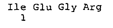
- protease cleavage sites are those which specifically recognize the protease from HIV (human immunodeficiency virus, recognizes the AS sequence SQNYPIVQ), the protease from the hepatitis C virus, the protease from TEV (Tobacco etch virus; AS sequence ENLYFQS), from the protease from hCMV (human cytomegalovirus, recognizes the AS sequence RGVVNASSRLA), from the protease from HSV (herpes simplex virus, recognizes the AS sequence LVLASSSF), from the protease plasmin (recognizes the AS-sequence Sequence KXYK), from the protease ACE (angiotensin converting enzyme, recognizes the AS sequence GKYAPWV), from the protease tPA, from factor X a (recognizes the AS sequence IEGR), thrombin (recognizes the AS sequence VGPRSFLLK), etc.
- fusion proteins are the following fusion proteins A - C (the interface segment is underlined, the protease cleavage site is bold, ⁇ denotes the interface for the TEV protease).
- a sample to be examined can be a natural isolate, a chemical component from a substance bank, a fraction from a fermentation broth or a protease library generated by variation techniques be.
- Confocal fluorimetric methods are the methods of passage single fluorophores by an excitation focus based fluorescence signals evaluate.
- the confocal-fluorimetric methods include in particular fluorescence correlation spectroscopy (FCS), two-color fluorescence cross-correlation spectroscopy (KK-FCS), the Confocal Fluorescence Coincidence Analysis (CFCA) and the 2D fluorescence intensity distribution analysis (2D-FIDA).
- FCS fluorescence correlation spectroscopy
- KK-FCS two-color fluorescence cross-correlation spectroscopy
- CFCA Confocal Fluorescence Coincidence Analysis
- 2D-FIDA 2D fluorescence intensity distribution analysis
- the invention relates to a method for analysis a sample for protease-inhibiting activity.
- This procedure is analogous to the above method of the embodiment (6), wherein instead of the sample to be assayed for proteolytic activity has a protease-inhibiting Activity towards sample to be examined and the corresponding protease is added.
- one of those proteases is the corresponding protease to understand the sequence specificity consistent with the protease substrate exhibit.
- the invention relates to a method for analysis intracellular protease activity.
- This method is analogous to that above-mentioned method of embodiment (6), wherein one for the fusion protein coding nucleic acid sequence is introduced into cells such that it is expressed intracellularly, and - by the confocal fluorimetric measurement the intracellular protease activity can be analyzed.
- the invention relates to a method for analysis intracellular protease-inhibiting activity analogous to those mentioned above Methods (6) to (8), wherein the for the fusion protein and for the corresponding Protease encoding nucleic acid sequences introduced into such cells be expressed intracellularly, and confocal fluorometric Measurement of the intracellular protease-inhibiting activity can be analyzed.
- the introduction of the nucleic acid sequence takes place in eukaryotic cells using a vector that is local and temporal controlled expression allows.
- the expression occurs only in selected cell compartments.
- KK-FCS konfokal-fluorimetric Measuring method two-color fluorescence cross-correlation spectroscopy
- CFCA Confocal Fluorescence Coincidence Analysis
- 2D-FIDA 2D fluorescence intensity distribution analysis
- Cloning strategy As the smallest amounts of substance are sufficient for FCS measurements, a vector with a lac promoter was used to express the fusion proteins, thereby minimizing the risk of inclusion in the inclusion bodies . To allow for alternative purification by means of nickel chelate chromatography, all fusion proteins intended for expression in E. coli were additionally provided with a C-terminal hexahistidine sequence. Since initially it was not known to what extent the amino acids in the broader environment of the recognition sequence of the protease influence the reaction, a peptide sequence derived from the polyprotein of the Tobacco-Etch virus was chosen for insertion between the two fluorophores. This lies in the region of the cleavage site between capsid protein and polymerase of the virus.
- the accessibility of the interface for the TEV protease depends on the length of the peptide sequence lying between the two fluorescent protein parts. Therefore, a short variant was prepared with a 32 amino acid residue (variant 2; STEV; SEQ ID NO: 2) interface segment and a long variant with a 73 amino acid residue (variant 3; LTEV; SEQ ID NO: 3) interface segment in the middle each interface is located. From the TEV polyprotein, the amino acids were selected from 2761 to 2819 for the long and from 2781 to 2797 for the short insert. In the first step, two constructs were prepared in which the gene for rsGFP is inserted in front of the one for DsRed and is in the same reading frame with it. For expression in E.
- the GFP gene from the vector pQBI63 (Qbiogene) was amplified.
- the PCR product was digested with the restriction endonucleases Sal I and Sph I and the insert subsequently inserted into the similarly cut plasmid pDsRed (Clontech).
- the result of the cloning was checked by digesting the plasmids with the previously used restriction enzymes.
- the final sequencing revealed that the cloning was successful and without mutations ("pGFRed", variant 1, SEQ ID NO: 1).
- the gene for the fusion protein was extended at its 3 'end by a nucleotide sequence coding for six consecutive histidine residues in order to enable later purification of the proteins by nickel chelate chromatography.
- constructs described above ( pGFRed, pGFRed-CH ) still have a short nucleotide sequence derived from the remainder of the 5'-MCS between the genes of the two fluorescent protein parts . These sections were subsequently expanded by the insertion of synthetically produced sequences. In this way, the length of the peptide chain lying between the two fluorescent protein parts could be varied and a specific protease cleavage site inserted into the later formed protein.
- Two peptide sequences of different lengths were designed, centered on a single-protein TEV protease cleavage site, and integrated into both the original and polyhistidine-sequenced GFRed protein.
- the sequences were generated by direct insertion of a synthetic nucleotide sequence.
- the vectors were cut with BamH I and the phosphate groups of the free ends were removed with alkaline phosphatase.
- the two synthetic oligonucleotides were phosphorylated so that they could be inserted as a double strand into the vectors.
- the plasmids thus prepared are pSTev (variant 2, SEQ ID NO: 2, short insert), pSTev-CH (short insert with C-terminal hexahistidine sequence ), pLTev (variant 3, SEQ ID NO: 3, long insert) and pLTV, respectively -CH (long insert with C-terminal hexahistidine sequence).
- the gene sections may be due to the use of only one interface each be inserted in two different orientations.
- the orientation of the insert was checked by analytical PCR. A PCR product was only then present when the gene segment inserted in the correct orientation had been.
- the procedure itself can also be extended to other restriction sites apply.
- type and position of the protease interface can be within the subsequent protein by appropriate choice of the inserted nucleotide sequence be chosen freely.
- Protein expression in E. coli Clones with appropriate vector construct were plated from permanent culture on a selective nutrient agar plate and incubated overnight at 37 ° C. One colony was used to inoculate a preculture of 10 ml of selective medium. This was also shaken overnight at 37 ° C and served the next morning as inoculum for two batches of 200 ml of selective medium, which were inoculated with 5 ml of preculture. When the cultures reached an optical density of 0.7-0.8 at 600 nm, protein expression was induced by addition of 100 ⁇ l each of IPTG solution (0.5 mM final). The bacteria were centrifuged off after four hours in the refrigerated centrifuge at 4 ° C and 5000 g for 15 minutes and discarded the supernatant.
- the bacteria were resuspended in 20 ml of buffer, then forced through a 0.25 mm diameter needle with a syringe and then lysed in the French Press.
- the lysate was collected in an ice-cooled centrifuge tube in which 100 ⁇ l of a solution of 100 mg / ml PMSF in ethanol had already been introduced for the inhibition of proteases.
- the cell debris was immediately centrifuged in the refrigerated centrifuge at 4 ° C and 20,000 xg for 20 minutes.
- the bacterial lysate was forced through a syringe filter and an aliquot of 100 ⁇ l was removed prior to application to the column.
- Protein purification by anion exchange chromatography The column material used was DEAE-Sephacel, the column diameter was 1 cm and the filling height 5 cm. The column was packed bubble-free and equilibrated before loading with at least 50 ml wash buffer; the flow rate was 5 ml / min. The bacteria were disrupted in the wash buffer and the column loaded with the filtered supernatant; the run was collected for later analysis.
- Protein purification by nickel chelate chromatography A HiTrap chelating column (Pharmacia) with 1 ml column volume was used; the flow rate during application of the lysate was 0.5 ml / min and otherwise 1 ml / min.
- the column was first washed with 10 ml of water and then loaded with 2 ml of nickel chloride solution (100 mM). The removal of nonspecifically bound Ni 2+ ions was carried out by 10 ml of a solution of 300 mM imidazole in washing buffer; then the column was equilibrated with 10 ml of washing buffer.
- the filtered supernatant of the digested bacteria was applied to the column, which was then washed with 10 ml of buffer, and the protein was then eluted with 8 ml of 100 mM imidazole in washing buffer.
- the collected fractions of 1 ml each were examined for fluorescence and the column was cleaned with 10 ml of 500 mM imidazole in buffer. The run of loading, washing and cleaning steps were each collected and stored for later analysis.
- the samples to be examined were analyzed with the aid of a Centricon column at 1000 ⁇ g concentrated, mixed with 100 ul PBS buffer and again at 1000 x g centrifuged. For the measurement, they were diluted with PBS buffer such that the Measured values are within a reasonable range.
- Fluorescence Spectra Emission spectra were recorded from each sample at an excitation wavelength of 488 nm.
- the spectra are in each case normalized to the sum of the fluorescence intensities at 540 nm and 580 nm in order to be able to compare the intensities relative to one another.
- the two wavelengths were chosen for the normalization, because here are about the maxima of DsRed and the rsGFP , the sum of which represents an approximate measure of the total intensity.
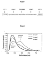
- the graphs of the preparations GFRed and STev have, in addition to the expected maximum of GFP emission at about 540 nm, yet another maximum, or a shoulder at about 580 nm, which is not recognizable in the spectrum of the protein LTev .
- This phenomenon was caused by energy transfer (FRET) within the protein. This energy transfer is due to the overlap of the emission spectrum of the rsGFP with the excitation spectrum of the DsRed . With increasing linker length, the proportion of energy transfer is reduced.
- Fluorescence correlation spectroscopic measurements The measurement of the TEV protease proteolytic activity with the E. coli- prepared substrate proteins was carried out in a Nunc sample chamber with a self-developed FCS device. The substrate concentrations were 50 nM. In each case two batches of 100 ⁇ l of substrate in PBS buffer were prepared, one of which was mixed with the TEV protease (various concentrations) and the other served as a negative control. The measurement was carried out in two-photon excitation with a wavelength of 950 nm.
Landscapes
- Life Sciences & Earth Sciences (AREA)
- Chemical & Material Sciences (AREA)
- Health & Medical Sciences (AREA)
- Engineering & Computer Science (AREA)
- Organic Chemistry (AREA)
- Molecular Biology (AREA)
- Immunology (AREA)
- Urology & Nephrology (AREA)
- Biotechnology (AREA)
- Proteomics, Peptides & Aminoacids (AREA)
- Microbiology (AREA)
- Biomedical Technology (AREA)
- Analytical Chemistry (AREA)
- Wood Science & Technology (AREA)
- Zoology (AREA)
- Biochemistry (AREA)
- Physics & Mathematics (AREA)
- Hematology (AREA)
- General Health & Medical Sciences (AREA)
- Bioinformatics & Cheminformatics (AREA)
- General Physics & Mathematics (AREA)
- Cell Biology (AREA)
- General Engineering & Computer Science (AREA)
- Biophysics (AREA)
- Food Science & Technology (AREA)
- Medicinal Chemistry (AREA)
- Genetics & Genomics (AREA)
- Pathology (AREA)
- Measuring Or Testing Involving Enzymes Or Micro-Organisms (AREA)
- Micro-Organisms Or Cultivation Processes Thereof (AREA)
- Organic Low-Molecular-Weight Compounds And Preparation Thereof (AREA)
- Investigating Or Analysing Materials By The Use Of Chemical Reactions (AREA)
- Preparation Of Compounds By Using Micro-Organisms (AREA)
- Peptides Or Proteins (AREA)
- Investigating, Analyzing Materials By Fluorescence Or Luminescence (AREA)
Description
Schließlich muss das Chromophor bzw. das Fluorophor in der Regel direkt C-terminal an die Protease-Schnittstelle gebunden werden, damit es bei der Proteasekatalysierten Hydrolyse freigesetzt wird und so seine spektralen bzw. fluoreszenten Eigenschaften ändert.


Im ersten Schritt wurden zwei Konstrukte hergestellt, bei denen jeweils das Gen für rsGFP vor dem für DsRed eingefügt ist und sich mit diesem im gleichen Leseraster befindet. Für die Expression in E. coli wurde zunächst das GFP-Gen vom Vektor pQBI63 (Qbiogene) amplifiziert. Das PCR-Produkt wurde mit den Restriktionsendonukleasen Sal I und Sph I verdaut und das Insert anschließend in das ebenso geschnittene Plasmid pDsRed (Clontech) eingefügt. Das Ergebnis der Klonierung wurde durch Verdau der Plasmide mit den zuvor verwendeten Restriktionsenzymen überprüft. Die abschließende Sequenzierung ergab, dass die Klonierung erfolgreich und ohne Mutationen verlaufen war ("pGFRed"; Variante 1; SEQ ID NO:1). Im nächsten Schritt wurde das Gen für das Fusionsprotein an seinem 3'-Ende durch eine für sechs aufeinander folgende Histidinreste kodierende Nukleotidsequenz erweitert, um eine spätere Aufreinigung der Proteine durch Nickelchelatchromatographie zu ermöglichen.
<120> Zweifarbiges fluorimetrisches Proteaseassay
<130> 011742wo/JH/ml
<141>
<211> 487
<212> PRT
<213> Künstliche Sequenz
<223> Beschreibung der künstlichen Sequenz: Autofluoreszierendes Fusionsprotein


<211> 506
<212> PRT
<213> Künstliche Sequenz
<223> Beschreibung der künstlichen Sequenz: Autofluoreszierendes Fusionsprotein


<211> 547
<212> PRT
<213> Künstliche Sequenz
<223> Beschreibung der künstlichen Sequenz: Autofluoreszierendes Fusionsprotein


<211> 32
<212> PRT
<213> Künstliche Sequenz
<223> Beschreibung der künstlichen Sequenz: Schnittstellensegment

<211> 7
<212> PRT
<213> Künstliche Sequenz
<223> Beschreibung der künstlichen Sequenz: Protease-Schnittstelle

<211> 4
<212> PRT
<213> Künstliche Sequenz
<223> Beschreibung der künstlichen Sequenz: Protease-Schnittstelle
<211> 4
<212> PRT
<213> Künstliche Sequenz
<223> Beschreibung der künstlichen Sequenz: Protease-Schnittstelle

<211> 9
<212> PRT
<213> Künstliche Sequenz
<223> Beschreibung der künstlichen Sequenz: Protease-Schnittstelle

<211> 7
<212> PRT
<213> Künstliche Sequenz
<223> Beschreibung der künstlichen Sequenz: Protease-Schnittstelle

<211> 8
<212> PRT
<213> Künstliche Sequenz
<223> Beschreibung der künstlichen Sequenz: Protease-Schnittstelle

<211> 11
<212> PRT
<213> Künstliche Sequenz
<223> Beschreibung der künstlichen Sequenz: Protease-Schnittstelle

<211> 8
<212> PRT
<213> Künstliche Sequenz
<223> Beschreibung der künstlichen Sequenz: Protease-Schnittstelle

Claims (17)
- Autofluoreszierendes Fusionsprotein, das ein erstes autofluoreszierendes Protein, ein Schnittstellensegment mit einer Protease-Schnittstelle und mindestens ein weiteres, vom ersten unterscheidbares, autofluoreszierendes Protein umfasst, wobei die emittierte Fluoreszenz der zwei (oder mehr) autofluoreszierenden Proteine nicht oder zu weniger als 50 % die jeweiligen anderen autofluoreszierenden Proteine anregt.
- Autofluoreszierendes Fusionsprotein nach Anspruch 1, wobei das Schnittstellensegment(i) zwischen zwei autofluoreszierenden, anhand ihrer spektralen Eigenschaften voneinander unterscheidbaren Polypeptiden angeordnet ist und/oder(ii) neben der Protease-Schnittstelle noch terminale Linkerpeptide aufweist und/oder(iii) eine Länge von wenigstens 10, vorzugsweise von mehr als 20, besonders bevorzugt von wenigstens 30 Aminosäureresten aufweist.
- Autofluoreszierendes Fusionsprotein nach Anspruch 1 oder 2, wobei das Schnittstellensegment eine Protease-Schnittstelle aufweist, die spezifisch von der Protease aus dem Human Immunodeficiency Virus, von der Protease aus dem Hepatitis C Virus, von der Protease aus dem Tobacco Etch Virus, von der Protease aus human Cytomegalovirus, von der Protease aus Herpes Simplex Virus, von der Protease Plasmin, von der Protease Angiotensin Converting Enzyme, von der Protease tPA vom Faktor Xa und/oder von Thrombin erkannt und gespalten wird.
- Autofluoreszierendes Fusionsprotein nach einem der Ansprüche 1 bis 3, wobei das erste autofluoreszierende Protein das Green Fluorescent Protein aus Aequorea victoria (GFP) oder eine Variante, insbesondere eine red-shifted-Variante (rsGFP), davon und das zweite autofluoreszierende Protein das dsRed aus Discosoma sp. oder eine Variante davon ist.
- Autofluoreszierendes Fusionsprotein nach einem der Ansprüche 1 bis 4, das weiterhin funktionale Peptidsequenzen, insbesondere Signalpeptide, Affinitätsmarkerpeptide oder Detektionsmarkerpeptide, aufweist.
- Autofluoreszierendes Fusionsprotein nach Anspruch 1, das die Sequenz von Fusionsprotein B oder C (SEQ. ID. NOs:2 oder 3) aufweist.
- Nukleinsäuresequenz, die für ein autofluoreszierendes Fusionsprotein, wie in Ansprüchen 1 bis 6 definiert, kodiert.
- Vektor, umfassend eine Nukleinsäuresequenz, wie in Anspruch 7 definiert.
- Zelle oder nicht-humaner transgener Organismus, umfassend die Nukleinsäuresequenz nach Anspruch 7 und/oder den Vektor nach Anspruch 8.
- Verfahren zur Herstellung des autofluoreszierenden Fusionsproteins nach einem der Ansprüche 1 bis 6, umfassend die Expression der Nukleinsäuresequenz nach Anspruch 7 mit Hilfe eines zellulären oder zellfreien Expressionssystems.
- Verfahren zur Analyse einer Probe auf proteolytische Aktivität, bestehend aus den Schritten:(a) Zusammenführen des autofluoreszierenden Fusionsproteins, wie in Ansprüchen 1 bis 6 definiert, mit der auf proteolytische Aktivität zu untersuchenden Probe in einer wässrigen Testlösung;(b) Inkubation unter Bedingungen, die für eine proteolytische Spaltung geeignet sind; und(c) Messung des Anteils an gespaltenem Fusionsprotein mittels konfokal-fluorimetrischer Methoden.
- Verfahren zur Analyse einer Probe auf protease-inhibierende Aktivität, bestehend aus den Schritten:(a) Zusammenführen des autofluoreszierenden Fusionsproteins, wie in Ansprüchen 1 bis 6 definiert, mit der auf protease-inhibierende Aktivität hin zu untersuchende Probe sowie der entsprechende Protease in einer wässrigen Testlösung;(b) Inkubation unter Bedingungen, die für eine proteolytische Spaltung geeignet sind; und(c) Messung des Anteils an gespaltenem Fusionsprotein mittels konfokal-fluorimetrischer Methoden.
- Verfahren zur Analyse intrazellulärer Protease-Aktivität, bestehend aus den Schritten:(a) Einführen der Nukleinsäuresequenz nach Anspruch 7 und/oder des Vektors nach Anspruch 8 in die zu untersuchende Zelle, so dass das autofluoreszierende Fusionsprotein, wie in Ansprüchen 1 bis 6 definiert, intrazellulär exprimiert wird;(b) Inkubation unter Bedingungen, die für eine Expression und eine proteolytische Spaltung des Fusionsproteins geeignet sind; und(c) Bestimmung der intrazellulär vorliegenden Protease-Aktivität mittels konfokal-fluorimetrischer Methoden.
- Verfahren zur Analyse intrazellulärer, protease-inhibierender Aktivität, bestehend aus den Schritten:(a) Einführen der Nukleinsäuresequenz nach Anspruch 7 und/oder des Vektors nach Anspruch 8 in die zu untersuchende Zelle, so dass das autofluoreszierende Fusionsprotein, wie in Ansprüchen 1 bis 6 definiert, intrazellulär exprimiert wird;(b) Inkubation unter Bedingungen, die für eine Expression und eine proteolytische Spaltung des Fusionsproteins geeignet sind; und(c) Bestimmung der intrazellulär vorliegenden protease-inhibierende Aktivität mittels konfokal-fluorimetrischer Methoden.
- Verfahren nach einem der Ansprüche 11 bis 14, wobei als Messmethode eine zweifarbige konfokal-fluorimetrische Messmethode, insbesondere die zweifarbige Fluoreszenz-Kreuzkorrelations-Spektroskopie (KK-FCS), die konfokale Fluoreszenz-Koinzidenz-Analyse (CFCA) oder die 2D-Fluoreszenz-Intensitäts-Verteilungs-Analyse (2D-FIDA) eingesetzt wird.
- Verfahren nach Anspruch 15, wobei zusätzlich zu zweifarbigen konfokal-fluorimetrischen Messmethoden auch der Anteil der einen Fluoreszenz-Energie-Transfer aufweisenden Moleküle bestimmt wird.
- Verwendung des Verfahrens nach einem der Ansprüche 11 bis 16 für die screening-basierende, evolutive Optimierung von Biomolekülen mit proteolytischer Aktivität und/oder die screening-basierende, evolutive Generierung von Biomolekülen mit proteolytischer Aktivität.
Applications Claiming Priority (3)
| Application Number | Priority Date | Filing Date | Title |
|---|---|---|---|
| DE10038382 | 2000-08-07 | ||
| DE10038382A DE10038382A1 (de) | 2000-08-07 | 2000-08-07 | Zweifarbiges fluorimetrisches Proteaseassay |
| PCT/EP2001/009112 WO2002012543A2 (de) | 2000-08-07 | 2001-08-07 | Zweifarbiges fluorimetrisches proteaseassay |
Publications (2)
| Publication Number | Publication Date |
|---|---|
| EP1307482A2 EP1307482A2 (de) | 2003-05-07 |
| EP1307482B1 true EP1307482B1 (de) | 2005-10-19 |
Family
ID=7651520
Family Applications (1)
| Application Number | Title | Priority Date | Filing Date |
|---|---|---|---|
| EP01962914A Expired - Lifetime EP1307482B1 (de) | 2000-08-07 | 2001-08-07 | Zweifarbiges fluorimetrisches proteaseassay |
Country Status (8)
| Country | Link |
|---|---|
| US (1) | US20050112682A9 (de) |
| EP (1) | EP1307482B1 (de) |
| JP (1) | JP2004505636A (de) |
| AT (1) | ATE307142T1 (de) |
| AU (2) | AU2001283988B2 (de) |
| CA (1) | CA2415458A1 (de) |
| DE (2) | DE10038382A1 (de) |
| WO (1) | WO2002012543A2 (de) |
Families Citing this family (13)
| Publication number | Priority date | Publication date | Assignee | Title |
|---|---|---|---|---|
| EP1633865B1 (de) | 2003-06-18 | 2011-09-28 | Bayer Pharma Aktiengesellschaft | Neue biologische einheiten und deren verwendung |
| EP1645881A1 (de) * | 2004-10-05 | 2006-04-12 | DKFZ Deutsches Krebsforschungszentrum | Screening-Verfahren zum Auffinden und Charakterisieren von Protein-Protein-Wechselwirkungen in vivo durch "fluorescence cross correlation spectroscopy" |
| US7329506B2 (en) * | 2004-12-03 | 2008-02-12 | Rutgers, The State University | Apparatuses and methods for determining protease activity |
| WO2006070941A1 (ja) * | 2004-12-28 | 2006-07-06 | Tokyo University Of Science | 蛍光分子プローブを用いた新規薬剤スクリーニング方法 |
| EP1726643A1 (de) | 2005-05-27 | 2006-11-29 | Direvo Biotech AG | Methode zur Bereitstellung, Identifizierung und Selektion von Proteasen die eine veränderte Empfindlichkeit gegenüber Aktivität-verändernden Substanzen aufweisen |
| JP2013512692A (ja) * | 2009-12-07 | 2013-04-18 | シナプティック リサーチ,リミテッド ライアビリティ カンパニー | プロテアーゼ活性阻害剤の同定とプロテアーゼ活性の存在を測定するための方法 |
| US12422428B2 (en) | 2011-06-01 | 2025-09-23 | Biomadison, Inc. | Compositions and methods for cell-based assays |
| KR101679370B1 (ko) * | 2011-06-01 | 2016-11-24 | 바이오메디슨, 인코퍼레이티드 | 비-fret 보툴리눔 검정법 |
| JP6510747B2 (ja) * | 2013-08-02 | 2019-05-08 | 東洋ビーネット株式会社 | プロテアーゼ活性測定法 |
| KR101533345B1 (ko) * | 2013-08-08 | 2015-07-22 | 아주대학교산학협력단 | 자가저해 단백질을 이용한 프로테아제 활성 측정용 센서, 및 이를 이용한 프로테아제의 활성 측정 방법 |
| KR20160103977A (ko) * | 2013-10-25 | 2016-09-02 | 바이오메디슨, 인코퍼레이티드 | 중복 형광단을 이용한 효소 검정 |
| JP7637937B2 (ja) * | 2020-04-28 | 2025-03-03 | 国立大学法人北海道大学 | 標的ポリペプチドの情報取得方法および試薬キット |
| EP3919911A1 (de) * | 2020-06-04 | 2021-12-08 | Urteste S.A. | Neuartiger diagnostischer marker für prostatakrebs |
-
2000
- 2000-08-07 DE DE10038382A patent/DE10038382A1/de not_active Withdrawn
-
2001
- 2001-08-07 EP EP01962914A patent/EP1307482B1/de not_active Expired - Lifetime
- 2001-08-07 DE DE50107766T patent/DE50107766D1/de not_active Expired - Lifetime
- 2001-08-07 JP JP2002517827A patent/JP2004505636A/ja active Pending
- 2001-08-07 WO PCT/EP2001/009112 patent/WO2002012543A2/de not_active Ceased
- 2001-08-07 AU AU2001283988A patent/AU2001283988B2/en not_active Ceased
- 2001-08-07 AT AT01962914T patent/ATE307142T1/de not_active IP Right Cessation
- 2001-08-07 US US10/343,977 patent/US20050112682A9/en not_active Abandoned
- 2001-08-07 CA CA002415458A patent/CA2415458A1/en not_active Abandoned
- 2001-08-07 AU AU8398801A patent/AU8398801A/xx active Pending
Also Published As
| Publication number | Publication date |
|---|---|
| AU8398801A (en) | 2002-02-18 |
| EP1307482A2 (de) | 2003-05-07 |
| WO2002012543A2 (de) | 2002-02-14 |
| JP2004505636A (ja) | 2004-02-26 |
| CA2415458A1 (en) | 2003-01-08 |
| DE50107766D1 (de) | 2005-11-24 |
| WO2002012543A3 (de) | 2002-06-27 |
| US20040014134A1 (en) | 2004-01-22 |
| ATE307142T1 (de) | 2005-11-15 |
| DE10038382A1 (de) | 2002-02-21 |
| AU2001283988B2 (en) | 2006-07-20 |
| US20050112682A9 (en) | 2005-05-26 |
Similar Documents
| Publication | Publication Date | Title |
|---|---|---|
| DE69936103T2 (de) | Fusionsproteine bestehend aus proteingerüst und bibliotheken von zufälligen peptiden | |
| DE69713059T2 (de) | Tandem-fluoreszenzproteinkonstrukte | |
| DE69432589T2 (de) | Substrate für die übertragung von fluoreszenzenergie | |
| DE60131262T2 (de) | Ubiquitin-ligase-assay | |
| DE60212642T2 (de) | Verfahren zur verwendung von o6-alkylguanin-dna-alkyltransferasen | |
| DE69717856T2 (de) | Methoden und zusammensetzungen zur empfindlichen und schnellen funktionellen identifizierung genomischer polynukleotide und ihre anwendung in zelltests zum auffinden von medikamenten | |
| DE69618350T2 (de) | Ein verfahren zum nachweis biologisch aktiver substanzen | |
| DE60014166T2 (de) | Verfahren zum messen der deacetylaseaktivität und verfahren zum screenen von inhibitoren oder promotoren dieses enzyms | |
| DE69715621T2 (de) | Drei hybriden screening test | |
| EP1307482A2 (de) | Zweifarbiges fluorimetrisches proteaseassay | |
| DE69617773T2 (de) | Verfahren zur Messung von Oligonukleotidabbau | |
| DE19641876A1 (de) | Streptavidinmuteine | |
| EP1381624B1 (de) | Spezifischer nachweis von proteolytischen enzymen mittels eines hybrid-proteins | |
| DE69838333T2 (de) | Detektionsverfahren umfassend die verwendung von polypeptiden mit einem coiled-coil und einer additionsstelle | |
| DE60015319T2 (de) | Test zur gleichzeitigen Messung von verschiedenen enzymischen Aktivitäten | |
| DE60023269T2 (de) | Entwicklung eines Screeningverfahrens für ein physiologisch aktives Pyrrol-Imidazol-Derivat | |
| DE60316718T2 (de) | Lösliche notch basierte substrate für gamma secretase, methoden, zusammensetzungen und verwendungen davon | |
| WO2004092211A1 (de) | Nicht fluoreszierende, durch proteolyse zur fluoreszenz aktivierbare reporterproteine und ihre verwendung zur detektion protease-abhängiger ereignisse | |
| EP1572732B1 (de) | Isoliertes fluoreszierendes protein aus clytia gregaria cgfp, sowie dessen verwendung | |
| DE60032632T2 (de) | Verfahren zur identifizierung von modulatoren von proteaseaktivierten rezeptoren (par) | |
| DE60221883T2 (de) | Screeningverfahren für einen auf die zellwand wirkenden arzneistoff | |
| WO2005033706A1 (de) | Verfahren zur detektion und analyse von protein-protein-interaktionen | |
| DE69635355T2 (de) | Verfahren zum Nachweis von biologisch aktiven Substanzen | |
| WO1999016900A1 (de) | Verfahren zum identifizieren einer nukleinsäure | |
| WO1998003676A1 (de) | Verfahren zum nachweis für effektoren intra- und/oder interzellulärer protein-protein-wechselwirkungen |
Legal Events
| Date | Code | Title | Description |
|---|---|---|---|
| PUAI | Public reference made under article 153(3) epc to a published international application that has entered the european phase |
Free format text: ORIGINAL CODE: 0009012 |
|
| 17P | Request for examination filed |
Effective date: 20030118 |
|
| AK | Designated contracting states |
Designated state(s): AT BE CH CY DE DK ES FI FR GB GR IE IT LI LU MC NL PT SE TR |
|
| AX | Request for extension of the european patent |
Extension state: AL LT LV MK RO SI |
|
| GRAP | Despatch of communication of intention to grant a patent |
Free format text: ORIGINAL CODE: EPIDOSNIGR1 |
|
| GRAS | Grant fee paid |
Free format text: ORIGINAL CODE: EPIDOSNIGR3 |
|
| GRAA | (expected) grant |
Free format text: ORIGINAL CODE: 0009210 |
|
| AK | Designated contracting states |
Kind code of ref document: B1 Designated state(s): AT BE CH CY DE DK ES FI FR GB GR IE IT LI LU MC NL PT SE TR |
|
| AX | Request for extension of the european patent |
Extension state: LT LV |
|
| PG25 | Lapsed in a contracting state [announced via postgrant information from national office to epo] |
Ref country code: IT Free format text: LAPSE BECAUSE OF FAILURE TO SUBMIT A TRANSLATION OF THE DESCRIPTION OR TO PAY THE FEE WITHIN THE PRESCRIBED TIME-LIMIT;WARNING: LAPSES OF ITALIAN PATENTS WITH EFFECTIVE DATE BEFORE 2007 MAY HAVE OCCURRED AT ANY TIME BEFORE 2007. THE CORRECT EFFECTIVE DATE MAY BE DIFFERENT FROM THE ONE RECORDED. Effective date: 20051019 Ref country code: IE Free format text: LAPSE BECAUSE OF FAILURE TO SUBMIT A TRANSLATION OF THE DESCRIPTION OR TO PAY THE FEE WITHIN THE PRESCRIBED TIME-LIMIT Effective date: 20051019 Ref country code: NL Free format text: LAPSE BECAUSE OF FAILURE TO SUBMIT A TRANSLATION OF THE DESCRIPTION OR TO PAY THE FEE WITHIN THE PRESCRIBED TIME-LIMIT Effective date: 20051019 Ref country code: FI Free format text: LAPSE BECAUSE OF FAILURE TO SUBMIT A TRANSLATION OF THE DESCRIPTION OR TO PAY THE FEE WITHIN THE PRESCRIBED TIME-LIMIT Effective date: 20051019 |
|
| REG | Reference to a national code |
Ref country code: GB Ref legal event code: FG4D Free format text: NOT ENGLISH |
|
| REG | Reference to a national code |
Ref country code: CH Ref legal event code: EP |
|
| GBT | Gb: translation of ep patent filed (gb section 77(6)(a)/1977) | ||
| REG | Reference to a national code |
Ref country code: IE Ref legal event code: FG4D Free format text: LANGUAGE OF EP DOCUMENT: GERMAN |
|
| REF | Corresponds to: |
Ref document number: 50107766 Country of ref document: DE Date of ref document: 20051124 Kind code of ref document: P |
|
| REG | Reference to a national code |
Ref country code: CH Ref legal event code: NV Representative=s name: A. BRAUN, BRAUN, HERITIER, ESCHMANN AG PATENTANWAE |
|
| PG25 | Lapsed in a contracting state [announced via postgrant information from national office to epo] |
Ref country code: SE Free format text: LAPSE BECAUSE OF FAILURE TO SUBMIT A TRANSLATION OF THE DESCRIPTION OR TO PAY THE FEE WITHIN THE PRESCRIBED TIME-LIMIT Effective date: 20060119 Ref country code: DK Free format text: LAPSE BECAUSE OF FAILURE TO SUBMIT A TRANSLATION OF THE DESCRIPTION OR TO PAY THE FEE WITHIN THE PRESCRIBED TIME-LIMIT Effective date: 20060119 Ref country code: GR Free format text: LAPSE BECAUSE OF FAILURE TO SUBMIT A TRANSLATION OF THE DESCRIPTION OR TO PAY THE FEE WITHIN THE PRESCRIBED TIME-LIMIT Effective date: 20060119 |
|
| PG25 | Lapsed in a contracting state [announced via postgrant information from national office to epo] |
Ref country code: ES Free format text: LAPSE BECAUSE OF FAILURE TO SUBMIT A TRANSLATION OF THE DESCRIPTION OR TO PAY THE FEE WITHIN THE PRESCRIBED TIME-LIMIT Effective date: 20060130 |
|
| PG25 | Lapsed in a contracting state [announced via postgrant information from national office to epo] |
Ref country code: PT Free format text: LAPSE BECAUSE OF FAILURE TO SUBMIT A TRANSLATION OF THE DESCRIPTION OR TO PAY THE FEE WITHIN THE PRESCRIBED TIME-LIMIT Effective date: 20060320 |
|
| LTIE | Lt: invalidation of european patent or patent extension |
Effective date: 20051019 |
|
| NLV1 | Nl: lapsed or annulled due to failure to fulfill the requirements of art. 29p and 29m of the patents act | ||
| REG | Reference to a national code |
Ref country code: IE Ref legal event code: FD4D |
|
| PLBE | No opposition filed within time limit |
Free format text: ORIGINAL CODE: 0009261 |
|
| STAA | Information on the status of an ep patent application or granted ep patent |
Free format text: STATUS: NO OPPOSITION FILED WITHIN TIME LIMIT |
|
| PG25 | Lapsed in a contracting state [announced via postgrant information from national office to epo] |
Ref country code: MC Free format text: LAPSE BECAUSE OF NON-PAYMENT OF DUE FEES Effective date: 20060831 Ref country code: BE Free format text: LAPSE BECAUSE OF NON-PAYMENT OF DUE FEES Effective date: 20060831 |
|
| 26N | No opposition filed |
Effective date: 20060720 |
|
| EN | Fr: translation not filed | ||
| PG25 | Lapsed in a contracting state [announced via postgrant information from national office to epo] |
Ref country code: FR Free format text: LAPSE BECAUSE OF FAILURE TO SUBMIT A TRANSLATION OF THE DESCRIPTION OR TO PAY THE FEE WITHIN THE PRESCRIBED TIME-LIMIT Effective date: 20061208 |
|
| BERE | Be: lapsed |
Owner name: DIREVO BIOTECH A.G. Effective date: 20060831 |
|
| REG | Reference to a national code |
Ref country code: CH Ref legal event code: PFA Owner name: DIREVO BIOTECH AG Free format text: DIREVO BIOTECH AG#NATTERMANNALLEE 1#50829 KOELN (DE) -TRANSFER TO- DIREVO BIOTECH AG#NATTERMANNALLEE 1#50829 KOELN (DE) |
|
| PG25 | Lapsed in a contracting state [announced via postgrant information from national office to epo] |
Ref country code: LU Free format text: LAPSE BECAUSE OF NON-PAYMENT OF DUE FEES Effective date: 20060807 Ref country code: TR Free format text: LAPSE BECAUSE OF FAILURE TO SUBMIT A TRANSLATION OF THE DESCRIPTION OR TO PAY THE FEE WITHIN THE PRESCRIBED TIME-LIMIT Effective date: 20051019 |
|
| PG25 | Lapsed in a contracting state [announced via postgrant information from national office to epo] |
Ref country code: CY Free format text: LAPSE BECAUSE OF FAILURE TO SUBMIT A TRANSLATION OF THE DESCRIPTION OR TO PAY THE FEE WITHIN THE PRESCRIBED TIME-LIMIT Effective date: 20051019 Ref country code: FR Free format text: LAPSE BECAUSE OF FAILURE TO SUBMIT A TRANSLATION OF THE DESCRIPTION OR TO PAY THE FEE WITHIN THE PRESCRIBED TIME-LIMIT Effective date: 20051019 |
|
| REG | Reference to a national code |
Ref country code: CH Ref legal event code: PUE Owner name: BAYER SCHERING PHARMA AKTIENGESELLSCHAFT Free format text: DIREVO BIOTECH AG#NATTERMANNALLEE 1#50829 KOELN (DE) -TRANSFER TO- BAYER SCHERING PHARMA AKTIENGESELLSCHAFT#MUELLERSTRASSE 178#13353 BERLIN (DE) |
|
| PGFP | Annual fee paid to national office [announced via postgrant information from national office to epo] |
Ref country code: AT Payment date: 20090812 Year of fee payment: 9 Ref country code: CH Payment date: 20090814 Year of fee payment: 9 Ref country code: GB Payment date: 20090805 Year of fee payment: 9 |
|
| REG | Reference to a national code |
Ref country code: GB Ref legal event code: 732E Free format text: REGISTERED BETWEEN 20110303 AND 20110309 |
|
| REG | Reference to a national code |
Ref country code: CH Ref legal event code: PL |
|
| GBPC | Gb: european patent ceased through non-payment of renewal fee |
Effective date: 20100807 |
|
| PG25 | Lapsed in a contracting state [announced via postgrant information from national office to epo] |
Ref country code: LI Free format text: LAPSE BECAUSE OF NON-PAYMENT OF DUE FEES Effective date: 20100831 Ref country code: CH Free format text: LAPSE BECAUSE OF NON-PAYMENT OF DUE FEES Effective date: 20100831 |
|
| PG25 | Lapsed in a contracting state [announced via postgrant information from national office to epo] |
Ref country code: AT Free format text: LAPSE BECAUSE OF NON-PAYMENT OF DUE FEES Effective date: 20100807 |
|
| PG25 | Lapsed in a contracting state [announced via postgrant information from national office to epo] |
Ref country code: GB Free format text: LAPSE BECAUSE OF NON-PAYMENT OF DUE FEES Effective date: 20100807 |
|
| REG | Reference to a national code |
Ref country code: DE Ref legal event code: R082 Ref document number: 50107766 Country of ref document: DE Representative=s name: VON KREISLER SELTING WERNER, DE |
|
| REG | Reference to a national code |
Ref country code: DE Ref legal event code: R082 Ref document number: 50107766 Country of ref document: DE Representative=s name: , Effective date: 20120612 Ref country code: DE Ref legal event code: R081 Ref document number: 50107766 Country of ref document: DE Owner name: BAYER INTELLECTUAL PROPERTY GMBH, DE Free format text: FORMER OWNER: BAYER SCHERING PHARMA AKTIENGESELLSCHAFT, 13353 BERLIN, DE Effective date: 20120612 |
|
| REG | Reference to a national code |
Ref country code: DE Ref legal event code: R082 Ref document number: 50107766 Country of ref document: DE Representative=s name: , |
|
| REG | Reference to a national code |
Ref country code: DE Ref legal event code: R081 Ref document number: 50107766 Country of ref document: DE Owner name: BAYER INTELLECTUAL PROPERTY GMBH, DE Free format text: FORMER OWNER: BAYER PHARMA AKTIENGESELLSCHAFT, 13353 BERLIN, DE Effective date: 20130410 |
|
| PGFP | Annual fee paid to national office [announced via postgrant information from national office to epo] |
Ref country code: DE Payment date: 20140730 Year of fee payment: 14 |
|
| REG | Reference to a national code |
Ref country code: DE Ref legal event code: R119 Ref document number: 50107766 Country of ref document: DE |
|
| PG25 | Lapsed in a contracting state [announced via postgrant information from national office to epo] |
Ref country code: DE Free format text: LAPSE BECAUSE OF NON-PAYMENT OF DUE FEES Effective date: 20160301 |