EP1221477A2 - Transfektion von vertebratenzellen beispielsweise durch homologe rekombination - Google Patents
Transfektion von vertebratenzellen beispielsweise durch homologe rekombination Download PDFInfo
- Publication number
- EP1221477A2 EP1221477A2 EP01204619A EP01204619A EP1221477A2 EP 1221477 A2 EP1221477 A2 EP 1221477A2 EP 01204619 A EP01204619 A EP 01204619A EP 01204619 A EP01204619 A EP 01204619A EP 1221477 A2 EP1221477 A2 EP 1221477A2
- Authority
- EP
- European Patent Office
- Prior art keywords
- cells
- primary
- transfected
- dna
- cell
- Prior art date
- Legal status (The legal status is an assumption and is not a legal conclusion. Google has not performed a legal analysis and makes no representation as to the accuracy of the status listed.)
- Withdrawn
Links
Images


Classifications
-
- C—CHEMISTRY; METALLURGY
- C12—BIOCHEMISTRY; BEER; SPIRITS; WINE; VINEGAR; MICROBIOLOGY; ENZYMOLOGY; MUTATION OR GENETIC ENGINEERING
- C12N—MICROORGANISMS OR ENZYMES; COMPOSITIONS THEREOF; PROPAGATING, PRESERVING, OR MAINTAINING MICROORGANISMS; MUTATION OR GENETIC ENGINEERING; CULTURE MEDIA
- C12N15/00—Mutation or genetic engineering; DNA or RNA concerning genetic engineering, vectors, e.g. plasmids, or their isolation, preparation or purification; Use of hosts therefor
- C12N15/09—Recombinant DNA-technology
- C12N15/87—Introduction of foreign genetic material using processes not otherwise provided for, e.g. co-transformation
- C12N15/90—Stable introduction of foreign DNA into chromosome
- C12N15/902—Stable introduction of foreign DNA into chromosome using homologous recombination
- C12N15/907—Stable introduction of foreign DNA into chromosome using homologous recombination in mammalian cells
-
- A—HUMAN NECESSITIES
- A61—MEDICAL OR VETERINARY SCIENCE; HYGIENE
- A61K—PREPARATIONS FOR MEDICAL, DENTAL OR TOILETRY PURPOSES
- A61K47/00—Medicinal preparations characterised by the non-active ingredients used, e.g. carriers or inert additives; Targeting or modifying agents chemically bound to the active ingredient
- A61K47/50—Medicinal preparations characterised by the non-active ingredients used, e.g. carriers or inert additives; Targeting or modifying agents chemically bound to the active ingredient the non-active ingredient being chemically bound to the active ingredient, e.g. polymer-drug conjugates
- A61K47/69—Medicinal preparations characterised by the non-active ingredients used, e.g. carriers or inert additives; Targeting or modifying agents chemically bound to the active ingredient the non-active ingredient being chemically bound to the active ingredient, e.g. polymer-drug conjugates the conjugate being characterised by physical or galenical forms, e.g. emulsion, particle, inclusion complex, stent or kit
- A61K47/6901—Conjugates being cells, cell fragments, viruses, ghosts, red blood cells or viral vectors
-
- A—HUMAN NECESSITIES
- A61—MEDICAL OR VETERINARY SCIENCE; HYGIENE
- A61K—PREPARATIONS FOR MEDICAL, DENTAL OR TOILETRY PURPOSES
- A61K48/00—Medicinal preparations containing genetic material which is inserted into cells of the living body to treat genetic diseases; Gene therapy
-
- A—HUMAN NECESSITIES
- A61—MEDICAL OR VETERINARY SCIENCE; HYGIENE
- A61P—SPECIFIC THERAPEUTIC ACTIVITY OF CHEMICAL COMPOUNDS OR MEDICINAL PREPARATIONS
- A61P3/00—Drugs for disorders of the metabolism
- A61P3/06—Antihyperlipidemics
-
- A—HUMAN NECESSITIES
- A61—MEDICAL OR VETERINARY SCIENCE; HYGIENE
- A61P—SPECIFIC THERAPEUTIC ACTIVITY OF CHEMICAL COMPOUNDS OR MEDICINAL PREPARATIONS
- A61P3/00—Drugs for disorders of the metabolism
- A61P3/08—Drugs for disorders of the metabolism for glucose homeostasis
- A61P3/10—Drugs for disorders of the metabolism for glucose homeostasis for hyperglycaemia, e.g. antidiabetics
-
- A—HUMAN NECESSITIES
- A61—MEDICAL OR VETERINARY SCIENCE; HYGIENE
- A61P—SPECIFIC THERAPEUTIC ACTIVITY OF CHEMICAL COMPOUNDS OR MEDICINAL PREPARATIONS
- A61P37/00—Drugs for immunological or allergic disorders
-
- A—HUMAN NECESSITIES
- A61—MEDICAL OR VETERINARY SCIENCE; HYGIENE
- A61P—SPECIFIC THERAPEUTIC ACTIVITY OF CHEMICAL COMPOUNDS OR MEDICINAL PREPARATIONS
- A61P37/00—Drugs for immunological or allergic disorders
- A61P37/02—Immunomodulators
-
- A—HUMAN NECESSITIES
- A61—MEDICAL OR VETERINARY SCIENCE; HYGIENE
- A61P—SPECIFIC THERAPEUTIC ACTIVITY OF CHEMICAL COMPOUNDS OR MEDICINAL PREPARATIONS
- A61P39/00—General protective or antinoxious agents
- A61P39/06—Free radical scavengers or antioxidants
-
- A—HUMAN NECESSITIES
- A61—MEDICAL OR VETERINARY SCIENCE; HYGIENE
- A61P—SPECIFIC THERAPEUTIC ACTIVITY OF CHEMICAL COMPOUNDS OR MEDICINAL PREPARATIONS
- A61P43/00—Drugs for specific purposes, not provided for in groups A61P1/00-A61P41/00
-
- A—HUMAN NECESSITIES
- A61—MEDICAL OR VETERINARY SCIENCE; HYGIENE
- A61P—SPECIFIC THERAPEUTIC ACTIVITY OF CHEMICAL COMPOUNDS OR MEDICINAL PREPARATIONS
- A61P5/00—Drugs for disorders of the endocrine system
- A61P5/06—Drugs for disorders of the endocrine system of the anterior pituitary hormones, e.g. TSH, ACTH, FSH, LH, PRL, GH
-
- A—HUMAN NECESSITIES
- A61—MEDICAL OR VETERINARY SCIENCE; HYGIENE
- A61P—SPECIFIC THERAPEUTIC ACTIVITY OF CHEMICAL COMPOUNDS OR MEDICINAL PREPARATIONS
- A61P5/00—Drugs for disorders of the endocrine system
- A61P5/18—Drugs for disorders of the endocrine system of the parathyroid hormones
-
- A—HUMAN NECESSITIES
- A61—MEDICAL OR VETERINARY SCIENCE; HYGIENE
- A61P—SPECIFIC THERAPEUTIC ACTIVITY OF CHEMICAL COMPOUNDS OR MEDICINAL PREPARATIONS
- A61P7/00—Drugs for disorders of the blood or the extracellular fluid
- A61P7/02—Antithrombotic agents; Anticoagulants; Platelet aggregation inhibitors
-
- A—HUMAN NECESSITIES
- A61—MEDICAL OR VETERINARY SCIENCE; HYGIENE
- A61P—SPECIFIC THERAPEUTIC ACTIVITY OF CHEMICAL COMPOUNDS OR MEDICINAL PREPARATIONS
- A61P7/00—Drugs for disorders of the blood or the extracellular fluid
- A61P7/04—Antihaemorrhagics; Procoagulants; Haemostatic agents; Antifibrinolytic agents
-
- C—CHEMISTRY; METALLURGY
- C07—ORGANIC CHEMISTRY
- C07K—PEPTIDES
- C07K14/00—Peptides having more than 20 amino acids; Gastrins; Somatostatins; Melanotropins; Derivatives thereof
- C07K14/435—Peptides having more than 20 amino acids; Gastrins; Somatostatins; Melanotropins; Derivatives thereof from animals; from humans
- C07K14/475—Growth factors; Growth regulators
- C07K14/505—Erythropoietin [EPO]
-
- C—CHEMISTRY; METALLURGY
- C07—ORGANIC CHEMISTRY
- C07K—PEPTIDES
- C07K14/00—Peptides having more than 20 amino acids; Gastrins; Somatostatins; Melanotropins; Derivatives thereof
- C07K14/435—Peptides having more than 20 amino acids; Gastrins; Somatostatins; Melanotropins; Derivatives thereof from animals; from humans
- C07K14/575—Hormones
- C07K14/605—Glucagons
-
- C—CHEMISTRY; METALLURGY
- C07—ORGANIC CHEMISTRY
- C07K—PEPTIDES
- C07K14/00—Peptides having more than 20 amino acids; Gastrins; Somatostatins; Melanotropins; Derivatives thereof
- C07K14/435—Peptides having more than 20 amino acids; Gastrins; Somatostatins; Melanotropins; Derivatives thereof from animals; from humans
- C07K14/575—Hormones
- C07K14/61—Growth hormones [GH] (Somatotropin)
-
- C—CHEMISTRY; METALLURGY
- C12—BIOCHEMISTRY; BEER; SPIRITS; WINE; VINEGAR; MICROBIOLOGY; ENZYMOLOGY; MUTATION OR GENETIC ENGINEERING
- C12N—MICROORGANISMS OR ENZYMES; COMPOSITIONS THEREOF; PROPAGATING, PRESERVING, OR MAINTAINING MICROORGANISMS; MUTATION OR GENETIC ENGINEERING; CULTURE MEDIA
- C12N15/00—Mutation or genetic engineering; DNA or RNA concerning genetic engineering, vectors, e.g. plasmids, or their isolation, preparation or purification; Use of hosts therefor
- C12N15/09—Recombinant DNA-technology
- C12N15/63—Introduction of foreign genetic material using vectors; Vectors; Use of hosts therefor; Regulation of expression
- C12N15/79—Vectors or expression systems specially adapted for eukaryotic hosts
- C12N15/85—Vectors or expression systems specially adapted for eukaryotic hosts for animal cells
-
- C—CHEMISTRY; METALLURGY
- C07—ORGANIC CHEMISTRY
- C07K—PEPTIDES
- C07K2319/00—Fusion polypeptide
- C07K2319/01—Fusion polypeptide containing a localisation/targetting motif
- C07K2319/02—Fusion polypeptide containing a localisation/targetting motif containing a signal sequence
-
- C—CHEMISTRY; METALLURGY
- C12—BIOCHEMISTRY; BEER; SPIRITS; WINE; VINEGAR; MICROBIOLOGY; ENZYMOLOGY; MUTATION OR GENETIC ENGINEERING
- C12N—MICROORGANISMS OR ENZYMES; COMPOSITIONS THEREOF; PROPAGATING, PRESERVING, OR MAINTAINING MICROORGANISMS; MUTATION OR GENETIC ENGINEERING; CULTURE MEDIA
- C12N2510/00—Genetically modified cells
- C12N2510/02—Cells for production
-
- C—CHEMISTRY; METALLURGY
- C12—BIOCHEMISTRY; BEER; SPIRITS; WINE; VINEGAR; MICROBIOLOGY; ENZYMOLOGY; MUTATION OR GENETIC ENGINEERING
- C12N—MICROORGANISMS OR ENZYMES; COMPOSITIONS THEREOF; PROPAGATING, PRESERVING, OR MAINTAINING MICROORGANISMS; MUTATION OR GENETIC ENGINEERING; CULTURE MEDIA
- C12N2800/00—Nucleic acids vectors
- C12N2800/10—Plasmid DNA
- C12N2800/108—Plasmid DNA episomal vectors
-
- C—CHEMISTRY; METALLURGY
- C12—BIOCHEMISTRY; BEER; SPIRITS; WINE; VINEGAR; MICROBIOLOGY; ENZYMOLOGY; MUTATION OR GENETIC ENGINEERING
- C12N—MICROORGANISMS OR ENZYMES; COMPOSITIONS THEREOF; PROPAGATING, PRESERVING, OR MAINTAINING MICROORGANISMS; MUTATION OR GENETIC ENGINEERING; CULTURE MEDIA
- C12N2830/00—Vector systems having a special element relevant for transcription
- C12N2830/001—Vector systems having a special element relevant for transcription controllable enhancer/promoter combination
- C12N2830/002—Vector systems having a special element relevant for transcription controllable enhancer/promoter combination inducible enhancer/promoter combination, e.g. hypoxia, iron, transcription factor
-
- C—CHEMISTRY; METALLURGY
- C12—BIOCHEMISTRY; BEER; SPIRITS; WINE; VINEGAR; MICROBIOLOGY; ENZYMOLOGY; MUTATION OR GENETIC ENGINEERING
- C12N—MICROORGANISMS OR ENZYMES; COMPOSITIONS THEREOF; PROPAGATING, PRESERVING, OR MAINTAINING MICROORGANISMS; MUTATION OR GENETIC ENGINEERING; CULTURE MEDIA
- C12N2830/00—Vector systems having a special element relevant for transcription
- C12N2830/80—Vector systems having a special element relevant for transcription from vertebrates
- C12N2830/85—Vector systems having a special element relevant for transcription from vertebrates mammalian
-
- C—CHEMISTRY; METALLURGY
- C12—BIOCHEMISTRY; BEER; SPIRITS; WINE; VINEGAR; MICROBIOLOGY; ENZYMOLOGY; MUTATION OR GENETIC ENGINEERING
- C12N—MICROORGANISMS OR ENZYMES; COMPOSITIONS THEREOF; PROPAGATING, PRESERVING, OR MAINTAINING MICROORGANISMS; MUTATION OR GENETIC ENGINEERING; CULTURE MEDIA
- C12N2840/00—Vectors comprising a special translation-regulating system
- C12N2840/20—Vectors comprising a special translation-regulating system translation of more than one cistron
-
- C—CHEMISTRY; METALLURGY
- C12—BIOCHEMISTRY; BEER; SPIRITS; WINE; VINEGAR; MICROBIOLOGY; ENZYMOLOGY; MUTATION OR GENETIC ENGINEERING
- C12N—MICROORGANISMS OR ENZYMES; COMPOSITIONS THEREOF; PROPAGATING, PRESERVING, OR MAINTAINING MICROORGANISMS; MUTATION OR GENETIC ENGINEERING; CULTURE MEDIA
- C12N2840/00—Vectors comprising a special translation-regulating system
- C12N2840/44—Vectors comprising a special translation-regulating system being a specific part of the splice mechanism, e.g. donor, acceptor
Definitions
- Gene therapy is perhaps more appropriately described as medical intervention in which cells, either from the individual to be treated or another appropriate source, are modified genetically to treat or cure any condition, regardless of etiology, that will be ameliorated by the long-term delivery of a therapeutic protein. Gene therapy can therefore be thought of as an in vivo protein production and delivery system, and almost all diseases that are currently treated by the administration of proteins are candidates for treatment using gene therapy.
- Germ cell gene therapy can be divided into two areas: germ cell and somatic cell gene therapy.
- Germ cell gene therapy refers to the modification of sperm cells, egg cells, zygotes, or early stage embryos. On the basis of both ethical and practical criteria, germ cell gene therapy is inappropriate for human use.
- somatic cell gene therapy would affect only the person under treatment (somatic cells are cells that are not capable of developing into whole individuals and include all of the body's cells with the exception of the germ cells). As such, somatic cell gene therapy is a reasonable approach to the treatment and cure of certain disorders in human beings.
- somatic cells e.g., fibroblasts, hepatocytes, or endothelial cells
- somatic cells e.g., fibroblasts, hepatocytes, or endothelial cells
- the means by which these five steps are carried out are the distinguishing features of a given gene therapy system.
- the present invention relates to transfected primary and secondary somatic cells of vertebrate origin, particularly of mammalian origin, transfected with exogenous DNA which encodes a therapeutic product, exogenous DNA which is itself a therapeutic product and/or exogenous DNA which causes the transfected cells to express a gene at a higher level than occurs in the corresponding nontransfected cell.
- the present invention relates to transfected primary and secondary somatic cells of vertebrate origin, particularly mammalian origin, transfected with exogenous genetic material (DNA) which encodes a desired (e.g., a therapeutic) product or is itself a desired (e.g., therapeutic) product, methods by which primary and secondary cells are transfected to include exogenous genetic material, methods of producing clonal cell strains or heterogenous cell strains, methods of gene therapy in which the transfected primary or secondary cells are used, methods of producing a therapeutic protein through the use of transfected primary or secondary cells made by the present method and methods of producing antibodies using the transfected primary or secondary cells.
- DNA exogenous genetic material
- the present invention relates to transfected primary and secondary somatic cells of vertebrate origin, particularly mammalian origin, transfected with exogenous genetic material (DNA or RNA) which encodes a clinically useful product, such as human growth hormone (hGH), erythropoietin (EPO) or insulinotropin, methods by which primary and secondary cells are transfected to include exogenous genetic material encoding hGH, EPO or insulinotropin, methods of producing clonal cell strains or heterogenous cell strains which express exogenous genetic material encoding hGH, EPO or insulinotropin, a method of providing hGH, EPO or insulinotropin in physiologically useful quantities to an individual in need thereof, through the use of transfected cells of the present invention or by direct injection of DNA encoding hGH, EPO insulinotropin into an individual; and methods of producing antibodies against the encoded product using the transfected primary or secondary cells.
- exogenous genetic material DNA or RNA
- a clinically useful product such as human growth
- the present invention relates to a method of gene or DNA targeting in cells of vertebrate, particularly mammalian, origin. That is, it relates to a method of introducing DNA into primary or secondary cells of vertebrate origin through homologous recombination or targeting of the DNA, which is introduced into genomic DNA of the primary or secondary cells at a preselected site. The preselected site determines the targeting sequences used.
- the present invention further relates to homologously recombinant primary or secondary cells, referred to as homologously recombinant (HR) primary or secondary cells, produced by the present method and to uses of the HR primary or secondary cells.
- HR homologously recombinant
- the present invention also relates to a method of turning on or activating a gene present in primary cells, secondary cells or immortalized cells of vertebrate origin, which is normally not expressed in the cells or is not expressed at significant levels in the cells. Homologous recombination or targeting is used to replace or disable the regulatory region normally associated with the gene with a regulatory sequence which causes the gene to be expressed at levels higher than evident in the corresponding nontransfected cell.
- the present invention therefore, relates to a method of making proteins by turning on or activating an endogenous gene which encodes the desired product in transfected primary, secondary or immortalized cells.
- the term primary cell includes cells present in a suspension of cells isolated from a vertebrate tissue source (prior to their being plated i.e., attached to a tissue culture substrate such as a dish or flask), cells present in an explant derived from tissue, both of the previous types of cells plated for the first time, and cell suspensions derived from these plated cells.
- the term secondary cell or cell strain refers to cells at all subsequent steps in culturing. That is, the first time a plated primary cell is removed from the culture substrate and replated (passaged), it is referred to herein as a secondary cell, as are all cells in subsequent passages. Secondary cells are cell strains which consist of secondary cells which have been passaged one or more times.
- a cell strain consists of secondary cells that: 1) have been passaged one or more times; 2) exhibit a finite number of mean population doublings in culture; 3) exhibit the properties of contact-inhibited, anchorage dependent growth (anchorage-dependence does not apply to cells that are propagated in suspension culture); and 4) are not immortalized.
- a "clonal cell strain” is defined as a cell strain that is derived from a single founder cell.
- a “heterogenous cell strain” is defined as a cell strain that is derived from two or more founder cells.
- the present invention includes primary and secondary somatic cells, such as fibroblasts, keratinocytes, epithelial cells, endothelial cells, glial cells, neural cells, formed elements of the blood, muscle cells, other somatic cells which can be cultured and somatic cell precursors, which have been transfected with exogenous DNA which is stably integrated into their genomes or is expressed in the cells episomally.
- the resulting cells are referred to, respectively, as transfected primary cells and transfected secondary cells.
- the exogenous DNA 1) encodes a product, such as a translational product (e.g., a protein) or a transcriptional product (e.g., a ribozyme or an anti-sense nucleic acid sequence) which is a therapeutic product; 2) is itself a therapeutic product (e.g., DNA which binds to a cellular regulatory protein or alters gene expression) or 3) is DNA which undergoes homologous recombination with genomic DNA of recipient cells and results in alteration of (increase or decrease in) expression of an endogenous gene.
- a translational product e.g., a protein
- a transcriptional product e.g., a ribozyme or an anti-sense nucleic acid sequence
- 3) is DNA which undergoes homologous recombination with genomic DNA of recipient cells and results in alteration of (increase or decrease in) expression of an endogenous gene.
- the exogenous DNA encodes a translational or transcriptional product to be expressed by the recipient cells
- the resulting product is retained within the cell, incorporated into the cell membrane or secreted from the cell.
- the exogenous DNA encoding the therapeutic product is introduced into cells along with additional DNA sequences sufficient for expression of the exogenous DNA in transfected cells and is operatively linked to those sequences.
- exogenous DNA is, for example, DNA sequences which bind to a cellular regulatory protein, DNA sequences sufficient for sequestration of a protein or nucleic acid present in the transfected primary or secondary cell, DNA sequences which alter secondary or tertiary chromosomal structure or DNA sequences which are transcriptional regulatory elements.
- transfected primary cells Such primary cells modified to express or render available exogenous DNA are referred to herein as transfected primary cells, which include cells removed from tissue and placed on culture medium for the first time.
- Secondary cells modified to express or render available exogenous DNA are referred to herein as transfected secondary cells.
- exogenous DNA undergoes homologous recombination with genomic DNA of transfected (recipient) cells
- introduction of the exogenous DNA results in disablement of the endogenous sequences which control expression of the endogenous gene, either by replacing all or a portion of the endogenous (genomic) sequence or disrupting the endogenous sequence.
- Primary and secondary cells transfected by the subject method fall into three types or categories: 1) cells which do not, as obtained, make or contain the therapeutic product, 2) cells which make or contain the therapeutic product but in lower quantities than normal (in quantities less than the physiologically normal lower level) or in defective form, and 3) cells which make the therapeutic product at physiologically normal levels, but are to be augmented or enhanced in their content or production.
- Exogenous DNA is introduced into primary or secondary cells by a variety of techniques.
- a construct which includes exogenous DNA encoding a therapeutic protein and additional DNA sequences necessary for expression in recipient cells is introduced into primary or secondary cells by electroporation, microinjection, or other means (e.g., calcium phosphate precipitation, modified calcium phosphate precipitation, polybrene precipitation, liposome fusion, receptor-mediated DNA delivery).
- a vector, such as a retroviral vector, which includes exogenous DNA can be used and cells can be genetically modified as a result of infection with the vector.
- transfected primary and secondary cells may optionally contain DNA encoding a selectable marker, which is expressed and confers upon recipients a selectable phenotype, such as antibiotic resistance, resistance to a cytotoxic agent, nutritional prototrophy or expression of a surface protein. Its presence makes it possible to identify and select cells containing the exogenous DNA.
- selectable marker genes can be used, such as neo, gpt, dhfr, ada, pac, hyg, mdr and hisD.
- Transfected cells of the present invention are useful, as populations of transfected primary cells, transfected clonal cell strains, transfected heterogenous cell strains, and as cell mixtures in which at least one representative cell of one of the three preceding categories of transfected cells is present, as a delivery system for treating an individual with an abnormal or undesirable condition which responds to delivery of a therapeutic product, which is either: 1) a therapeutic protein (e.g., a protein which is absent, underproduced relative to the individual's physiologic needs, defective or inefficiently or inappropriately utilized in the individual; a protein with novel functions, such as enzymatic or transport functions) or 2) a therapeutic nucleic acid (e.g., DNA which binds to or sequesters a regulatory protein, RNA which inhibits gene expression or has intrinsic enzymatic activity).
- a therapeutic protein e.g., a protein which is absent, underproduced relative to the individual's physiologic needs, defective or inefficiently or inappropriately utilized in the individual
- transfected primary cells, clonal cell strains or heterogenous cell strains are administered to an individual in whom the abnormal or undesirable condition is to be treated or prevented, in sufficient quantity and by an appropriate route, to express or make available the exogenous DNA at physiologically relevant levels.
- a physiologically relevant level is one which either approximates the level at which the product is produced in the body or results in improvement of the abnormal or undesirable condition.
- Cells administered in the present method are cells transfected with exogenous DNA which encodes a therapeutic product, exogenous DNA which is itself a therapeutic product or exogenous DNA, such as a regulatory sequence, which is introduced into a preselected site in genomic DNA through homologous recombination and functions to cause recipient cells to produce a product which is normally not expressed in the cells or to produce the product of a higher level than occurs in the corresponding nontransfected cell.
- a regulatory sequence e.g., a promoter
- it replaces or disables a regulatory sequence normally associated with a gene, and results in expression of the gene at a higher level than occurs in the corresponding nontransfected cell.
- Figure 1 is a schematic representation of plasmid pXGH5, which includes the human growth hormone (hGH) gene under the control of the mouse metallothionein promoter.
- hGH human growth hormone
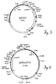
- Figure 2 is a schematic representation of plasmid pcDNEO, which includes the neo coding region (BamHI-BglII fragment) from plasmid pSV2neo inserted into the BamHI site of plasmid pcD; the Amp-R and pBR322Ori sequences from pBR322; and the polyA, 16S splice junctions and early promoter regions from SV40.
- neo coding region BamHI-BglII fragment
- Amp-R and pBR322Ori sequences from pBR322
- polyA, 16S splice junctions and early promoter regions from SV40.
- Figure 3 is a schematic representation of plasmid pXGH301 which includes the human growth hormone gene and the neo gene.
- Figure 4 is a flow chart of the method of the present invention.
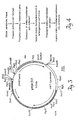
- Figure 5 is a schematic representation of plasmid pXEPO1.
- the solid black arc represents the pUC12 backbone and the arrow denotes the direction of transcription of the ampicillin resistance gene.
- the stippled arc represents the mouse metallothionein promoter (pmMT1).
- the unfilled arc interrupted by black boxes represents the human erythropoietin EPO gene (the black boxes denote exons and the arrow indicates the direction hEPO transcription). The relative positions of restriction endonuclease recognition sites are indicated.
- FIG. 6 is a schematic representation of plasmid pE3neoEPO.
- the positions of the human erythropoietin gene and the neo and amp resistance genes are indicated. Arrows indicate the directions of transcription of the various genes.
- pmMT1 denotes the mouse metallothionein promoter (driving hEPO expression)
- pTK denotes the Herpes Simplex Virus thymidine kinase promoter (driving neo expression).
- the dotted regions of the map mark the positions of human HGPRT sequences. The relative positions of restriction endonuclease recognition sites are indicated.
- Figure 7 is a schematic diagram of a strategy for transcriptionally activating the hEPO gene.
- Figure 8 is a schematic diagram of a strategy for transcriptionally activating the hEPO gene.
- Figure 9 shows the results of an assessment of long-term in vitro hGH production by transfected primary human skin fibroblasts (two strains, HF96-11 and HF96-23).
- Figure 10 is a graphic representation of human growth hormone (hGH) expression by transfected primary rabbit skin fibroblasts in vitro.
- hGH human growth hormone
- Figure 11 shows the results of an assay to detect serum levels of hGH over time in mice implanted with transfected rabbit fibroblasts expressing hGH.
- Figure 12 is a graphic representation of human growth hormone (hGH) expression in cells recovered from subrenal capsule implants.
- Figure 13a shows hematocrit (HCT) levels in control mice and mice implanted with transfected rabbit fibroblasts expressing hEPO.
- Figure 13b shows results of an assay to detect hEPO in the serum of mice implanted with transfected rabbit fibroblasts expressing hEPO.
- the present invention relates to transfected primary and secondary somatic cells of vertebrate origin, particularly of mammalian origin, transfected with exogenous DNA which encodes a therapeutic product, exogenous DNA which is itself a therapeutic product and/or exogenous DNA which causes the transfected cells to express a gene at a higher level than occurs in the corresponding nontransfected cell.
- primary or secondary cells of vertebrate, particularly mammalian, origin have been transfected with exogenous DNA encoding a therapeutic product and shown to produce the encoded therapeutic protein stably and reproducibly, both in vitro and in vivo , over extended periods of time.
- the transfected primary and secondary cells have been shown to express the encoded product in vivo at physiologically relevant levels, to be recoverable after implantation and, upon reculturing, to grow and display their preimplantation properties.
- transfected primary and secondary cells which include exogenous DNA encoding a desired product, (i.e., a translation product which is a therapeutic protein or an antigen against which antibodies are produced) and stably express the exogenous DNA. It is also possible, using the method described herein, to produce transfected primary and secondary cells which include exogenous DNA encoding other translation products (novel proteins not made in nature) or transcription products (e.g., anti-sense RNA or ribozymes) or exogenous DNA which itself is a therapeutic product (e.g., exogenous DNA which binds a regulatory protein present in the transfected cell).
- a desired product i.e., a translation product which is a therapeutic protein or an antigen against which antibodies are produced
- transfected primary and secondary cells which include exogenous DNA encoding other translation products (novel proteins not made in nature) or transcription products (e.g., anti-sense RNA or ribozymes) or exogenous DNA which itself is
- the method of the present invention includes the steps of: 1) providing a population of primary cells, obtained from the individual to whom the transfected primary cells will be administered or from another source; 2) introducing into the primary cells or into secondary cells derived from primary cells a DNA construct which includes exogenous DNA as described above and the necessary additional DNA sequences described above, producing transfected primary or secondary cells; 3) maintaining transfected primary or secondary cells under conditions appropriate for their propagation; 4) identifying a transfected primary or secondary cell; and 5) producing a colony from the transfected primary or secondary cell identified in (4) by maintaining it under appropriate culture conditions and for sufficient time for its propagation, thereby producing a cell strain derived from the (founder) cell identified in (4).
- exogenous DNA is introduced into genomic DNA by homologous recombination between DNA sequences present in the DNA construct and genomic DNA.
- a cell suspension containing primary or secondary cells is combined with exogenous DNA encoding a therapeutic product and DNA encoding a selectable marker, such as the neo gene.
- the two DNA sequences are present on the same DNA construct or on two separate DNA constructs.
- the resulting combination is subjected to electroporation, generally at 250-300 volts with a capacitance of 960 ⁇ Farads and an appropriate time constant (e.g., 14 to 20 m sec) for cells to take up the DNA construct.
- microinjection is used to introduce the DNA construct into primary or secondary cells. In either embodiment, introduction of the exogenous DNA results in production of transfected primary or secondary cells.
- heterogenous cell strains of the present invention the same steps are carried out as described for production of a clonal cell strain, except that a single transfected primary or secondary cell is not isolated and used as the founder cell. Instead, two or more transfected primary or secondary cells are cultured to produce a heterogenous cell strain.
- the subject invention also relates to a method of producing antibodies specific for the protein encoded by the exogenous DNA.
- transfected primary or secondary cells expressing an antigen against which antibodies are desired are introduced into an animal recipient (e.g., rabbit, mouse, pig, dog, cat, goat, guinea pig, sheep, non-human primate).
- the animal recipient produces antibodies against the antigen expressed, which may be an entire protein antigen or a peptide encoded by a fragment of the intact gene which encodes the entire antigen.
- Polyclonal sera is obtained from the animals. It is also possible to produce monoclonal antibodies through the use of transfected primary or secondary cells.
- Splenocytes are removed from an animal recipient of transfected primary or secondary cells expressing the antigen against which monoclonal antibodies are desired.
- the splenocytes are fused with myeloma cells, using known methods, such as that of Koprowski et al . (U.S. Patent No. 4,172,124) or Kohler et al ., ( Nature 256 :495-497 (1975)) to produce hybridoma cells which produce the desired monoclonal antibody.
- the polyclonal antisera and monoclonal antibodies produced can be used for the same purposes (e.g., diagnostic, preventive, therapeutic purposes) as antibodies produced by other methods.
- the present invention has wide applicability in treating abnormal or undesired conditions and can be used to provide a variety of products to an individual.
- it can be used to provide secreted proteins (with either predominantly systemic or predominantly local effects), membrane proteins (e.g., for imparting new or enhanced cellular responsiveness, facilitating removal of a toxic product or marking or targeting a cell) or intracellular proteins (e.g., for affecting gene expression or producing autolytic effects).
- secreted proteins with either predominantly systemic or predominantly local effects
- membrane proteins e.g., for imparting new or enhanced cellular responsiveness, facilitating removal of a toxic product or marking or targeting a cell
- intracellular proteins e.g., for affecting gene expression or producing autolytic effects.
- engineered DNA which binds or sequesters a cellular protein
- engineered RNA useful in an anti-sense approach to altering gene expression or to provide antigens against which immune response occurs in an individual (to prevent disease as by vaccination or to suppress an existing condition).
- the present invention is particularly advantageous in treating abnormal or undesired conditions in that it: 1) is curative (one gene therapy treatment has the potential to last a patient's lifetime); 2) allows precise dosing (the patient's cells continuously determine and deliver the optimal dose of the required protein based on physiologic demands, and the stably transfected cell strains can be characterized extensively in vitro prior to implantation, leading to accurate predictions of long term function in vivo ); 3) is simple to apply in treating patients; 4) eliminates issues concerning patient compliance (following a one-time gene therapy treatment, daily protein injections are no longer necessary); 5) reduces treatment costs (since the therapeutic protein is synthesized by the patient's own cells, investment in costly protein production and purification is unnecessary); and 6) is safe (the invention does not use infectious agents such as retroviruses to genetically engineer the patient's cells, thereby overcoming the safety and efficacy issues that have hampered other gene therapy systems).
- primary or secondary cells of vertebrate, particularly mammalian, origin have been transfected with exogenous DNA encoding EPO and shown to produce the encoded EPO reproducibly, both in vitro and in vivo , over extended periods of time.
- the transfected primary and secondary cells have been shown to express EPO in vivo at physiologically relevant levels.
- the EPO expressed has been shown to have the glycosylation pattern typical of EPO purified from human urine or recombinant human EPO.
- primary or secondary cells of vertebrate, particularly mammalian, origin have been transfected with exogenous DNA encoding hGH and shown to produce the encoded hGH reproducibly, both in vitro and in vivo , over extended periods of time.
- the transfected primary and secondary cells have been shown to express hGH in vivo at physiologically relevant levels.
- Applicants have also developed methods for producing transfected primary or secondary cells which stably express exogenous DNA encoding EPO, clonal cell strains and heterogenous cell strains of such transfected cells, methods of producing the clonal and heterogenous cell strains, and methods of using transfected cells expressing EPO to deliver the encoded product to an individual mammal at physiologically relevant levels.
- the constructs and methods herein described are useful, for example, for treating an individual (human) whose EPO production and/or function is in need of being increased or enhanced [e.g., is compromised or less than normal, or normal but the individual would benefit from enhancement, at least temporarily, of red blood cell production (e.g., during predialysis or dialysis therapy, during treatment of AIDS with AZT, after surgery, or during chemotherapy)].
- an individual human
- EPO production and/or function is in need of being increased or enhanced [e.g., is compromised or less than normal, or normal but the individual would benefit from enhancement, at least temporarily, of red blood cell production (e.g., during predialysis or dialysis therapy, during treatment of AIDS with AZT, after surgery, or during chemotherapy)].
- insulinotropin includes, e.g.
- GLP-1 glucagon-like peptide 1
- GLP-1 glucagon-like peptide 1
- carboxy-terminal amidated derivatives produced by in vivo amidating enzymes and derivatives which have amino acid alterations or other alterations which result in substantially the same biological activity or stability in the blood as that of a truncated GLP-1 or enhanced biological activity or stability.
- Applicants have also demonstrated that DNA can be introduced into primary or secondary vertebrate cells in a DNA construct or plasmid and integrated into the genome of the transfected primary or secondary cells by homologous recombination. That is, they have demonstrated gene targeting in primary and secondary mammalian cells. They have further demonstrated that the exogenous DNA has the desired function in the homologously recombinant (HR) cells and that correctly targeted cells can be identified on the basis of a detectable phenotype conferred by a selectable marker gene.
- HR homologously recombinant
- the present invention relates to a method of protein production using transfected primary, secondary or immortalized cells.
- the method involves transfecting primary cells, secondary cells or immortalized cells with exogenous DNA which encodes a therapeutic product or with DNA which is sufficient to target to and activate an endogenous gene which encodes a therapeutic product.
- Examples 18f, 19 and 21 describe protein production by targeting and activation of a selected endogenous gene.
- plasmid pcDNEO a selectable marker gene
- plasmid pXGH5 a gene encoding a therapeutic product
- pXGH301 a therapeutic product
- plasmid pXGH5 a therapeutic product
- pXGH301 a therapeutic product
- pXGH301 a therapeutic product
- pE3Neo a plasmid useful for targeting to a particular locus in the human genome and selection based upon a drug resistant phenotype
- This plasmid is designated pE3Neo and its integration into the cellular genomes at the HPRT locus produces cells which have an hprt - , 6-TG resistant phenotype and are also G418 resistant.
- pE3Neo functions properly in gene targeting in an established human fibroblast cell line (Example 18b), by demonstrating localization of the DNA introduced into established cells within exon 3 of the HPRT gene.
- Applicants demonstrate gene targeting in primary and secondary human skin fibroblasts using pE3Neo (Example 18c) and describe construction of a plasmid for targeted insertion of a gene encoding a therapeutic product (human growth hormone [hGH]) into the human genome (Example 18d).
- the subject application further demonstrates that modification of DNA termini enhances targeting of DNA into genomic DNA (Examples 18c and 18e) and construction of a variety of targeting plasmids.
- Applicants describe targeting plasmids for placing a human gene under the control of a murine promoter known to function in human cells (Examples 18f and 18i); for targeting to sequences flanking a gene and isolation of targeted secondary fibroblasts using a variety of screening and selection approaches (Examples 18g, 18h, 18j and 18k); for placing a human gene not normally expressed in the primary or secondary cells under the control of a promoter of nonhuman or human origin, to produce homologously recombinant primary or secondary cells which express the encoded product (Examples 18f-18k).
- the normal regulatory sequences upstream of a gene e.g. the human EPO gene
- the product of the targeting events is a chimeric transcription unit, in which a regulatory element and an operatively linked exon are positioned upstream of the desired endogenous gene to be activated.
- the product of transcription, splicing, and translation produces a chimeric protein in which amino acids encoded by exon 1 of the exogenous gene are fused to amino acids encoded by exons 2 and downstream exons in the endogenous gene.
- the product of the targeting event replaces the regulatory and exon 1 sequences of the endogenous gene with corresponding exogenous sequences.
- the product of transcription, splicing, and translation produces a chimeric protein similar to that described above.
- secretion of such proteins involves membrane translocation and removal of the signal peptide, in this case producing a normal protein lacking the chimeric signal peptide. In both cases the chimeric protein is now under the control of a desired regulatory element.
- Examples 18f-18h and 19 illustrate embodiments in which the normal regulatory sequences upstream of the human EPO gene are altered to allow expression of hEPO in primary or secondary fibroblast strains which do not express EPO in detectable quantities in their untransfected state.
- the product of targeting leaves the normal EPO protein intact, but under the control of the mouse metallothionein promoter.
- Examples 18i and 18j demonstrate the use of similar targeting constructs to activate the endogenous growth hormone gene in primary or secondary human fibroblasts.
- the products of targeting events are chimeric transcription units, in which the first exon of the human growth hormone gene is positioned upstream of EPO exons 2-5.
- the product of transcription (controlled by the mouse metallothionein promoter), splicing, and translation is a protein in which amino acids 1-4 of the hEPO signal peptide are replaced with amino acid residues 1-3 of hGH.
- the chimeric portion of this protein, the signal peptide, is removed prior to secretion from cells.
- the Examples provide methods for activating endogenous genes by gene targeting which do not require manipulation or other uses of the hEPO and hGH protein coding regions.
- normally inactive genes may be activated in cells that have properties desirable for in vivo protein delivery methods (e.g. gene therapy) and in vitro protein production (e.g., pharmaceutics).
- Figures 7 and 8 illustrate two strategies for transcriptionally activating the hEPO gene.
- the thin lines represent hEPO sequences; thick lines, mouse metallothionein I promoter; stippled box, 5' untranslated region of hGH; solid box, hGH exon 1; striped box, 10 bp linker from hEPO intron 1; cross-hatched box, 5' untranslated region of hEPO; and open boxes, hEPO coding sequences; HIII, HindIII site.
- exogenous DNA which encodes a therapeutic product (e.g., protein, ribozyme, nucleic acid) can be inserted at preselected sites in the genome of vertebrate (e.g., mammalian, both human and nonhuman) primary or secondary cells.
- a therapeutic product e.g., protein, ribozyme, nucleic acid
- the methods and DNA constructs described can be used for a wide variety of purposes.
- the method can be used to alter primary or secondary cells of vertebrate origin in order to repair, alter, delete or replace DNA already present in the recipient primary or secondary cell; to introduce into primary or secondary cells a gene or DNA sequence (at a preselected site) which encodes a therapeutic product or other desired product or is itself a therapeutic or other product; to add to or replace regulatory sequences present in the primary or secondary cell recipients; to knock out or remove an entire gene or gene portion present in primary or secondary cells; and to produce universal donor cells.
- the transfected primary or secondary cells may also include DNA encoding a selectable marker which confers a selectable phenotype upon them, facilitating their identification and isolation.
- Applicants have also developed methods for producing transfected primary or secondary cells which stably express exogenous DNA, clonal cell strains and heterogenous cell strains of such transfected cells, methods of producing the clonal and heterogenous cell strains, and methods of treating or preventing an abnormal or undesirable condition through the use of populations of transfected primary or secondary cells of the present invention.
- Primary and secondary cells to be transfected by the present method can be obtained from a variety of tissues and include all cell types which can be maintained in culture.
- primary and secondary cells which can be transfected by the present method include fibroblasts, keratinocytes, epithelial cells (e.g., mammary epithelial cells, intestinal epithelial cells), endothelial cells, glial cells, neural cells, formed elements of the blood (e.g., lymphocytes, bone marrow cells), muscle cells and precursors of these somatic cell types.
- Primary cells are preferably obtained from the individual to whom the transfected primary or secondary cells are administered. However, primary cells may be obtained from a donor (other than the recipient) of the same species or another species (e.g., mouse, rat, rabbit, cat, dog, pig, cow, bird, sheep, goat, horse).
- Transfected primary and secondary cells have been produced, with or without phenotypic selection, as described in Examples 5-7, and shown to express exogenous DNA encoding a therapeutic product including, e.g., EPO and insulinotropin.
- a therapeutic product including, e.g., EPO and insulinotropin.
- Immortalized cells can also be transfected by the present method and used for either gene therapy or protein production.
- Examples of immortalized human cell lines useful for protein production by the present method include, but are not limited to, HT1080, HeLa, MCF-7 breast cancer cells, K-562 leukemia cells, KB carcinoma cells and 2780AD ovarian carcinoma cells.
- Exogenous DNA incorporated into primary or secondary cells by the present method is: 1) DNA which encodes a translation or transcription product whose expression in primary or secondary cells is desired, such as a translation or transcription product useful to treat an existing condition or prevent it from occurring (eg., EPO or insulinotropin) and 2) DNA which does not encode a gene product but is itself useful, such as DNA useful to treat an existing condition or prevent it from occurring or 3) DNA which undergoes homologous recombination with genomic DNA of recipient cells and results in alteration of (increase or decrease in) expression of an endogenous gene.
- a translation or transcription product useful to treat an existing condition or prevent it from occurring eg., EPO or insulinotropin
- DNA transfected into primary or secondary cells can encode an entire desired product, or can encode, for example, the active or functional portion(s) of the product.
- the product can be, for example, a hormone, a cytokine, an antigen, an antibody, an enzyme, a clotting factor, a transport protein, a receptor, a regulatory protein, a structural protein, an anti-sense RNA, a ribozyme or a protein or a nucleic acid which does not occur in nature (i.e., a novel protein or novel nucleic acid).
- the DNA can be obtained from a source in which it occurs in nature or can be produced, using genetic engineering techniques or synthetic processes.
- the DNA transfected into primary or secondary cells can encode one or more therapeutic products.
- the exogenous DNA After transfection into primary or secondary cells, the exogenous DNA is stably incorporated into the recipient cell's genome (along with the additional sequences present in the DNA construct used), from which it is expressed or otherwise functions. Alternatively, the exogenous DNA may exist episomally within the transfected primary or secondary cells.
- DNA encoding the desired product can be introduced into cells under the control of an inducible promoter, with the result that cells produced or as introduced into an individual do not express the product but can be induced to do so (i.e., production is induced after the transfected cells are produced but before implantation or after implantation).
- DNA encoding the desired product can, of course, be introduced into cells in such a manner that it is expressed upon introduction (i.e., without induction).
- selectable markers can be incorporated into primary or secondary cells.
- a selectable marker which confers a selectable phenotype such as drug resistance, nutritional auxotrophy, resistance to a cytotoxic agent or expression of a surface protein
- selectable marker genes which can be used include neo, gpt, dhfr, ada, pac, hyg and hisd. The selectable phenotype conferred makes it possible to identify and isolate recipient primary or secondary cells.
- Selectable markers can be divided into two categories: positive selectable and negative selectable.
- positive selection cells expressing the positive selectable marker are capable of surviving treatment with a selective agent (such as neo, gpt, dhfr, ada, pac, hyg, mdrl and hisD).
- negative selection cells expressing the negative selectable marker are destroyed in the presence of the selective agent (e.g., tk, gpt).
- DNA constructs which include exogenous DNA and, optionally, DNA encoding a selectable marker, along with additional sequences necessary for expression of the exogenous DNA in recipient primary or secondary cells, are used to transfect primary or secondary cells in which the encoded product is to be produced.
- the DNA construct can also include targeting sequences for homologous recombination with host cell DNA.
- DNA constructs which include exogenous DNA sequences which do not encode a gene product (and are the therapeutic product) and, optionally, include DNA encoding a selectable marker can be used to transfect primary and secondary cells.
- infectious vectors such as retroviral, herpes, adenovirus, adenovirus-associated, mumps and poliovirus vectors, can be used for this purpose.
- a DNA construct which includes the exogenous DNA and additional sequences, such as sequences necessary for expression of the exogenous DNA can be used (e.g., plasmid pXGH5 or plasmid pXEPO1).
- a DNA construct can include an inducible promoter which controls expression of the exogenous DNA, making inducible expression possible.
- the DNA construct may include a bacterial origin of replication and bacterial antibiotic resistance markers, which allow for large-scale plasmid propagation in bacteria.
- a DNA construct which includes DNA encoding a selectable marker, along with additional sequences, such as a promoter, polyadenylation site and splice junctions, can be used to confer a selectable phenotype upon transfected primary or secondary cells (e.g., plasmid pcDNEO).
- the two DNA constructs are co-transfected into primary or secondary cells, using methods described herein.
- one DNA construct which includes exogenous DNA, a selectable marker gene and additional sequences e.g., those necessary for expression of the exogenous DNA and for expression of the selectable marker gene
- additional sequences e.g., those necessary for expression of the exogenous DNA and for expression of the selectable marker gene
- Such a DNA construct e.g., plasmid PXGH301, which includes the hGH gene and the neo gene, or plasmid pE3neoEPO which includes the EPO gene and the neo gene; these plasmids are described in Figures 3 and 6, respectively).
- Similar constructs which include exogenous DNA encoding insulinotropin and additional sequences (e.g., sequences necessary for insulinotropin expression) can be produced (e.g., plasmid pXGLP1; see Example 11).
- These constructs can also include DNA encoding a selectable marker, as well as other sequences, such as a promoter, a polyadenylation site, and splice junctions.
- the DNA construct includes the exogenous DNA and regulatory sequences necessary and sufficient for expression of the encoded product (e.g., EPO) upon entry of the DNA construct into recipient cells.
- EPO encoded product
- DNA constructs which include exogenous DNA encoding a desired product, targeting sequences for homologous recombination and, optionally, DNA encoding one or more selectable markers are used to transfect primary or secondary cells in which homologous recombination is to occur.
- DNA sequences necessary for expression of the exogenous DNA will generally be present as well.
- DNA constructs which include exogenous DNA sequences which do not encode a gene product (and are the desired product) and, optionally, include DNA encoding a selectable marker can also be used to transfect primary and secondary cells.
- the exogenous DNA, targeting sequences and selectable marker can be introduced into cells on a single DNA construct or on separate constructs.
- the total length of the DNA construct will vary according to the number of components (exogenous DNA, targeting sequences, selectable marker gene) and the length of each.
- the entire construct length will generally be at least 20 nucleotides.
- the construct will include a single component, the exogenous DNA.
- the exogenous DNA because of its homology, serves also to target integration into genomic DNA and additional targeting sequences are unnecessary.
- Such a construct is useful to knock out, replace or repair a resident DNA sequence, such as an entire gene, a gene portion, a regulatory element or portion thereof or regions of DNA which, when removed, place regulatory and structural sequences in functional proximity. It is also useful when the exogenous DNA is a selectable marker.
- the DNA construct includes exogenous DNA and one or more separate targeting sequences, generally located at both ends of the exogenous DNA sequence.
- Targeting sequences are DNA sequences normally present in the primary or secondary cell genome in the genome of the cells as obtained [e.g., an essential gene, a nonessential gene or noncoding DNA, or present in the genome through a previous modification].
- Such a construct is useful to integrate exogenous DNA encoding a therapeutic product, such as a hormone, a cytokine, an antigen, an antibody, an enzyme, a clotting factor, a transport protein, a receptor, a regulatory protein, a structural protein, an anti-sense RNA, a ribozyme or a protein or a nucleic acid which does not occur in nature.
- a therapeutic product such as a hormone, a cytokine, an antigen, an antibody, an enzyme, a clotting factor, a transport protein, a receptor, a regulatory protein, a structural protein, an anti-sense RNA, a ribozyme or a protein or a nucleic acid which does not occur in nature.
- exogenous DNA can encode one of the following: Factor VIII, Factor IX, erythropoietin, alpha-1 antitrypsin, calcitonin, glucocerebrosidase, growth hormone, low density lipoprotein (LDL) receptor, apolipoprotein E, IL-2 receptor and its antagonists, insulin, globin, immunoglobulins, catalytic antibodies, the interleukins, insulin-like growth factors, superoxide dismutase, immune responder modifiers, parathyroid hormone, interferons, nerve growth factors, tissue plasminogen activators, and colony stimulating factors.
- Such a construct is also useful to integrate exogenous DNA which is a therapeutic product, such as DNA sequences sufficient for sequestration of a protein or nucleic acid in the transfected primary or secondary cell, DNA sequences which bind to a cellular regulatory protein, DNA sequences which alter the secondary or tertiary chromosomal structure and DNA sequences which are transcriptional regulatory elements into genomic DNA of primary or secondary cells.
- exogenous DNA which is a therapeutic product, such as DNA sequences sufficient for sequestration of a protein or nucleic acid in the transfected primary or secondary cell, DNA sequences which bind to a cellular regulatory protein, DNA sequences which alter the secondary or tertiary chromosomal structure and DNA sequences which are transcriptional regulatory elements into genomic DNA of primary or secondary cells.
- the DNA construct includes exogenous DNA, targeting DNA sequences and DNA encoding at least one selectable marker.
- the order of construct components can be: targeting sequences-exogenous DNA-DNA encoding a selectable marker(s)-targeting sequences.
- one or more selectable markers are included in the construct, which makes selection based on a selectable phenotype possible. Cells that stably integrate the construct will survive treatment with the selective agent; a subset of the stably transfected cells will be HR cells, which can be identified by a variety of techniques, including PCR, Southern hybridization and phenotypic screening.
- the order of components in the DNA construct can be: targeting sequence-selectable marker 1 - targeting sequence - selectable marker 2.
- selectable marker 2 displays the property negative selection. That is, the gene product of selectable marker 2 can be selected against by growth in an appropriate media formulation containing an agent (typically a drug or metabolite analog) which kills cells expressing selectable marker 2. Recombination between the targeting sequences flanking selectable marker 1 with homologous sequences in the host cell genome results in the targeted integration of selectable marker 1, while selectable marker 2 is not integrated.
- Such recombination events generate cells which are stably transfected with selectable marker 1 but not stably transfected with selectable marker 2, and such cells can be selected for by growth in the media containing the selective agent which selects for selectable marker 1 and the selective agent which selects against selectable marker 2.
- exogenous DNA can encode one or more products, can be one or more therapeutic products or one or more of each, thus making it possible to deliver multiple products.
- vertebrate tissue is first obtained; this is carried out using known procedures, such as punch biopsy or other surgical methods of obtaining a tissue source of the primary cell type of interest.
- punch biopsy is used to obtain skin as a source of fibroblasts or keratinocytes.
- a mixture of primary cells is obtained from the tissue, using known methods, such as enzymatic digestion or explanting. If enzymatic digestion is used, enzymes such as collagenase, hyaluronidase, dispase, pronase, trypsin, elastase and chymotrypsin can be used.
- the resulting primary cell mixture can be transfected directly or it can be cultured first, removed from the culture plate and resuspended before transfection is carried out.
- Primary cells or secondary cells are combined with exogenous DNA to be stably integrated into their genomes and, optionally, DNA encoding a selectable marker, and treated in order to accomplish transfection.
- the exogenous DNA and selectable marker-encoding DNA are each on a separate construct (e.g., pXGH5 and pcDNEO, see Figures 1 and 2) or on a single construct (e.g., pXGH301 and pE3neoEPO, see Figure 3 and Figure 6).
- An appropriate quantity of DNA is used to ensure that at least one stably transfected cell containing and appropriately expressing exogenous DNA is produced. In general, 0.1 to 500 ⁇ g DNA is used.
- transfection is effected by electroporation, as described in the Examples. Electroporation is carried out at appropriate voltage and capacitance (and corresponding time constant) to result in entry of the DNA construct(s) into the primary or secondary cells. Electroporation can be carried out over a wide range of voltages (e.g., 50 to 2000 volts) and corresponding capacitance. As described herein, electroporation is very efficient if carried out at an electroporation voltage in the range of 250-300 volts and a capacitance of 960 ⁇ Farads. Total DNA of approximately 0.1 to 500 ⁇ g is generally used. As described in the Examples, total DNA of 60 ⁇ g and voltage of 250-300 volts with capacitance of 960 ⁇ Farads for a time constant 14-20 of msec. has been used and shown to be efficient.
- electroporation is carried out at appropriate voltage and capacitance (and corresponding time constant) to result in entry of the DNA construct(s) into the primary or secondary cells.
- primary or secondary cells are transfected using microinjection. See, for example, Example 5.
- known methods such as calcium phosphate precipitation, modified calcium phosphate precipitation and polybrene precipitation, liposome fusion and receptor-mediated gene delivery can be used to transfect cells.
- a stably transfected cell is isolated and cultured and subcultivated, under culturing conditions and for sufficient time, to propagate the stably transfected secondary cells and produce a clonal cell strain of transfected secondary cells.
- more than one transfected cell is cultured and subcultured, resulting in production of a heterogenous cell strain.
- Transfected primary or secondary cells undergo a sufficient number of doublings to produce either a clonal cell strain or a heterogenous cell strain of sufficient size to provide the therapeutic product (e.g., EPO) to an individual in effective amounts.
- a clonal cell strain or a heterogenous cell strain of sufficient size to provide the therapeutic product (e.g., EPO) to an individual in effective amounts.
- EPO therapeutic product
- 0.1 cm 2 of skin is biopsied and assumed to contain 100,000 cells; one cell is used to produce a clonal cell strain and undergoes approximately 27 doublings to produce 100 million transfected secondary cells. If a heterogenous cell strain is to be produced from an original transfected population of approximately 100,000 cells, only 10 doublings are needed to produce 100 million transfected cells.
- the number of required cells in a transfected clonal or heterogenous cell strain is variable and depends on a variety of factors, which include but are not limited to, the use of the transfected cells, the functional level of the exogenous DNA in the transfected cells, the site of implantation of the transfected cells (for example, the number of cells that can be used is limited by the anatomical site of implantation), and the age, surface area, and clinical condition of the patient.
- the site of implantation of the transfected cells for example, the number of cells that can be used is limited by the anatomical site of implantation
- the age, surface area, and clinical condition of the patient To put these factors in perspective, to deliver therapeutic levels of human growth hormone in an otherwise healthy 60 kg patient with isolated growth hormone deficiency, approximately one to five hundred million transfected fibroblasts would be necessary. This represents approximately the volume of cells prosent on the very tip of the patient's thumb.
- episomes DNA sequences that are present within the cell yet do not integrate into the genome are referred to as episomes.
- Recombinant episomes may be useful in at least three settings: 1) if a given cell type is incapable of stably integrating the exogenous DNA; 2) if a given cell type is adversely affected by the integration of DNA; and 3) if a given cell type is capable of improved therapeutic function with an episomal rather than integrated DNA.
- exogenous DNA in the form of episomes can be introduced into vertebrate primary and secondary cells.
- Plasmid pXGH301 can be converted into such an episome by the addition DNA sequences for the Epstein-Barr virus origin of replication and nuclear antigen [Yates, J.L. Nature 319 :780-7883 (1985)].
- vertebrate autonomously replicating sequences can be introduced into the construct (Weidle, U.H. Gene 73 (2):427-437 (1988).
- These and other episomally derived sequences can also be included in DNA constructs without selectable markers, such as pXGH5.
- the episomal exogenous DNA is then introduced into primary or secondary vertebrate cells as described in this application (if a selective marker is included in the episome a selective agent is used to treat the transfected cells).
- the transfected cells produced by the methods described above and in the Examples that follow, are introduced into an individual to whom the therapeutic product is to be delivered, using known methods.
- the clonal cell strain or heterogenous cell strain is then introduced into an individual, using known methods, using various routes of administration and at various sites (e.g., renal subcapsular, subcutaneous, central nervous system (including intrathecal), intravascular, intrahepatic, intrasplanchnic, intraperitoneal (including intraomental), or intramuscular implantation).
- the transfected cells produce the therapeutic product encoded by the exogenous DNA or are affected by the exogenous DNA itself.
- an individual who has been diagnosed with Hemophilia B a bleeding disorder that is caused by a deficiency in Factor IX, a protein normally found in the blood, is a candidate for a gene therapy cure.
- the patient has a small skin biopsy performed; this is a simple procedure which can be performed on an out-patient basis.
- the piece of skin approximately the size of a matchhead, is taken, for example, from under the arm and requires about one minute to remove.
- the sample is processed, resulting in isolation of the patient's cells (in this case, fibroblasts) and genetically engineered to produce the missing Factor IX. Based on the age, weight, and clinical condition of the patient, the required number of cells are grown in large-scale culture.
- the entire process usually requires 4-6 weeks and, at the end of that time, the appropriate number of genetically-engineered cells are introduced into the individual, once again as an out-patient (e.g., by injecting them back under the patient's skin).
- the patient is now capable of producing his or her own Factor IX and is no longer a hemophiliac.
- short stature can be treated by administering human growth hormone to an individual by implanting primary or secondary cells which express human growth hormone, or anemia can be treated by implanting primary or secondary cells which express EPO.
- transfected cells produced as described above, which contain insulinotropin-encoding DNA are delivered into an individual in whom insulin production, secretion, function and/or sensitivity is compromised. They are introduced into the individual by known methods and at various sites of administration (e.g., renal, subcapsular, subcutaneous, central nervous system (including intrathecal), intravascular, intrahepatic, intrasplanchnic, intraperitoneal (including intraomental) or intramuscular implantation). once implanted in the individual, the transfectedcells produce insulinotropin encoded by the exogenous DNA.
- sites of administration e.g., renal, subcapsular, subcutaneous, central nervous system (including intrathecal), intravascular, intrahepatic, intrasplanchnic, intraperitoneal (including intraomental) or intramuscular implantation.
- an individual in whom insulin production, secretion or sensitivity is impaired can receive therapy or preventive treatment through the implantation of transfected cells expressing exogenous DNA encoding insulinotropin produced as described herein.
- the cells to be genetically engineered are obtained as described above, processed in a similar manner to produce sufficient numbers of cells, and introduced back into the individual.
- the cells used will generally be patient-specific genetically-engineered cells. It is possible, however, to obtain cells from another individual of the same species or from a different species. Use of such cells might require administration of an immunosuppressant, alteration of histocompatibility antigens, or use of a barrier device to prevent rejection of the implanted cells.
- a barrier device is used to prevent rejection of implanted cells obtained from a source other than the recipient (e.g., from another human or from a non-human mammal such as a cow, dog, pig, goat, sheep or rodent).
- transfected cells of the present invention are placed within the barrier device, which is made of a material (e.g., a membrane such as Amicon XM-50) which permits the product encoded by the exogenous DNA to pass into the recipient's circulation or tissues but prevents contact between the cells and the recipient's immune system and thus prevents an immune resonse to (and possible rejection of) the cells by the recipient.
- DNA encoding hGH, EPO or insulinotropin can be introduced into an individual by direct injection, such as into muscle or other appropriate site.
- the DNA construct includes exogenous DNA encoding the therapeutic product (e.g., EPO, insulinotropin) and sufficient regulatory sequences for expression of the exogenous DNA in recipient cells. After injection into the individual, the DNA construct is taken up by some of the recipient cells.
- the DNA can be injected alone or in a formulation which includes a physiologially compatible carrier (e.g., a physiological buffer) and, optionally, other components, such as agents which allow more efficient entry of the DNA construct into cells, stabilize the DNA or protect the DNA from degradation.
- a physiologially compatible carrier e.g., a physiological buffer
- other components such as agents which allow more efficient entry of the DNA construct into cells, stabilize the DNA or protect the DNA from degradation.
- Transfected primary or secondary cells or cell strains as described herein have wide applicability as a vehicle or delivery system for therapeutic products, such as enzymes, hormones, cytokines, antigens, antibodies, clotting factors, anti-sense RNA, regulatory proteins, transcription proteins, receptors, structural proteins, ribozymes, novel (non-naturally occurring) proteins and nucleic acid products, and engineered DNA.
- therapeutic products such as enzymes, hormones, cytokines, antigens, antibodies, clotting factors, anti-sense RNA, regulatory proteins, transcription proteins, receptors, structural proteins, ribozymes, novel (non-naturally occurring) proteins and nucleic acid products, and engineered DNA.
- transfected primary or secondary cells can be used to supply a therapeutic protein, including, but not limited to, Factor VIII, Factor IX, erythropoietin, alpha-1 antitrypsin, calcitonin, glucocerebrosidase, growth hormone, low density lipoprotein (LDL), apolipoprotein E, receptor IL-2 receptor and its antagonists, insulin, globin, immunoglobulins, catalytic antibodies, the interleukins, insulin-like growth factors, superoxide dismutase, immune responder modifiers, parathyroid hormone and interferon, nerve growth factors, tissue plasminogen activators, and colony stimulating factors.
- transfected primary and secondary cells can be used to immunize an individual (i.e., as a vaccine).
- cell strains of the present invention can perhaps most conveniently be summarized as shown below.
- the cell strains can be used to deliver the following therapeutic products.
- transfected primary or secondary cells of the present invention can be used to administer therapeutic proteins (e.g., hormones, enzymes, clotting factors) which are presently administered intravenously, intra-muscularly or subcutaneously, which require patient cooperation and, often, medical staff participation.
- therapeutic proteins e.g., hormones, enzymes, clotting factors
- transfected primary or secondary cells there is no need for extensive purification of the polypeptide before it is administered to an individual, as is generally necessary with an isolated polypeptide.
- transfected primary or secondary cells of the present invention produce the therapeutic product as it would normally be produced.
- transfected primary or secondary cells of the present invention is that by controlling the number of cells introduced into an individual, one can control the amount of the product delivered to the body. In addition, in some cases, it is possible to remove the transfected cells if there is no longer a need for the product.
- a further advantage of treatment by use of transfected primary or secondary cells of the present invention is that production of the therapeutic product can be regulated, such as through the administration of zinc, steroids or an agent which affects translation or transcription of a protein, product or nucleic acid product or affects the stability of a nucleic acid product.
- GLP-1 derivatives include truncated derivatives GLP-1(7-37), GLP-1(7-36), GLP-1(7-35) GLP-1(7-34) and other truncated carboxy-terminal amidated derivatives and derivatives of GLP-1 which have amino acid substitutions, deletions, additions or other alterations (e.g., addition of a non-amino acid component) which result in biological activity or stability in the blood which is substantially the same as that of a truncated GLP-1 derivative or enhanced biological activity or stability in the blood (greater than that of a truncated GLP-1 derivative).
- GLP-1 derivative includes all of the above-described molecules.
- GLP-1 related peptide includes GLP-1 and GLP-1 derivatives.
- GLP-1 derivatives also known as insulinotropins or incretins, are normally secreted into the circulation by cells in the gastrointestinal tract. In vivo studies have demonstrated that these peptides function to stimulate insulin secretion and inhibit glucagon secretion from the endocrine pancreas, as well as increase insulin sensitivity in peripheral tissues [Goke. R. et al . (1991) Eur. J. Clin. Inv. 21 :135-144; Gutnia. M. et al . (1992) New Engl. J. Med.
- NIDDM non-insulin dependent diabetes mellitus
- gene targeting can be used to replace a gene's existing regulatory region with a regulatory sequence isolated from a different gene or a novel regulatory sequence synthesized by genetic engineering methods.
- regulatory sequences may be comprised of promoters, enhancers, Scaffold-attachment regions, negative regulatory elements, transcriptional initiation sites, regulatory protein binding sites or combinations of said sequences.
- sequences which affect the structure or stability of the RNA or protein produced may be replaced, removed, added, or otherwise modified by targeting, including polyadenylation signals, mRNA stability elements, splice sites, leader sequences for enhancing or modifying transport or secretion properties of the protein, or other sequences which alter or improve the function or stability of protein or RNA molecules).
- the targeting event may be a simple insertion of the regulatory sequence, placing the gene under the control of the new regulatory sequence (for example, inserting a new promoter or enhancer or both upstream of a gene).
- the targeting event may be a simple deletion of a regulatory element, such as the deletion of a tissue-specific negative regulatory element.
- the targeting event may replace an existing element; for example, a tissue-specific enhancer can be replaced by an enhancer that has broader or different cell-type specificity than the naturally-occurring elements. In this embodiment the naturally occurring sequences are deleted and new sequences are added.
- the identification of the targeting event may be facilitated by the use of one or more selectable marker genes that are contiguous with the targeting DNA, allowing for the selection of cells in which the exogenous DNA has integrated into the host cell genome.
- the identification of the targeting event may also be facilitated by the use of one or more marker genes exhibiting the property of negative selection, such that the negatively selectable marker is linked to the exogenous DNA, but configured such that the negatively selectable marker flanks the targeting sequence, and such that a correct homologous recombination event with sequences in the host cell genome does not result in the stable integration of the negatively selectable marker.
- Markers useful for this purpose include the Herpes Simplex Virus thymidine kinase (TK) gene or the bacterial xanthine-guanine phosphoribosyl-transferase (gpt) gene.
- Human fibroblasts can be obtained from a variety of tissues, including biopsy specimens derived from liver, kidney, lung and skin. The procedures presented here are optimized for the isolation of skin fibroblasts, which are readily obtained from individuals of any age with minimal discomfort and risk (embryonic and fetal fibroblasts may be isolated using this protocol as well). Minor modifications to the protocol can be made if the isolation of fibroblasts from other tissues is desired.
- Human skin is obtained following circumcision or punch biopsy.
- the specimen consists of three major components: the epidermal and dermal layers of the skin itself, and a fascial layer that adheres to the dermal layer.
- Fibroblasts can be isolated from either the dermal or fascial layers.
- tissue Approximately 3 cm 2 tissue is placed into approximately 10 ml of wash solution (Hank's Balanced Salt Solution containing 100 units/ml penicillin G, 100 ⁇ g/ml streptomycin sulfate, and 0.5 ⁇ g/ml Fungisone) and subjected to gentle agitation for a total of three 10-minute washes at room temperature. The tissue is then transferred to a 100 mm tissue culture dish containing 10 ml digestion solution (wash solution containing 0.1 units/ml collagenase A, 2.4 units/ml grade II Dispase).
- wash solution Hank's Balanced Salt Solution containing 100 units/ml penicillin G, 100 ⁇ g/ml streptomycin sulfate, and 0.5 ⁇ g/ml Fungisone
- the skin is adjusted such that the epidermis is facing down.
- the fascial tissue is separated from the dermal and epidermal tissue by blunt dissection.
- the fascial tissue is then cut into small fragments (less than 1 mm) and incubated on a rotating platform for 30 min at 37°C.
- the enzyme/cell suspension is removed and saved, an additional 10 cc of digestion solution is added to the remaining fragments of tissue, and the tissue is reincubated for 30 min at 37°C.
- the enzyme/cell suspensions are pooled, passed through a 15-gauge needle several times, and passed through a Cellector Sieve (Sigma) fitted with a 150-mesh screen.
- the cell suspension is centrifuged at 1100 rpm for 15 min at room temperature.
- the supernatant is aspirated and the disaggregated cells resuspended in 10 ml of nutrient medium (see below). Fibroblast cultures are initiated on tissue culture treated flasks (Corning) at a density of approximately 40,000 cells/cm 2 .
- Fascia is removed from skin biopsy or circumcision specimen as described above and the skin is cut into small fragments less than 0.5 cm 2 .
- the tissue is incubated with 0.25% trypsin for 60 min at 37°C (alternatively, the tissue can be incubated in trypsin for 18 hrs at 4°C). Under the dissecting microscope, the dermis and epidermis are separated. Dermal fibroblasts are then isolated as described above for fascial fibroblasts.
- the procedure is essentially as described above. Skin should be removed from areas that have been shaved and washed with a germicidal solution surgically prepared using accepted procedures.
- the primary culture When confluent, the primary culture is trypsinized using standard methods and seeded at approximately 10,000 cells/cm 2 .
- the cells are cultured at 37°C in humidified air containing 5% CO 2 .
- Human fibroblast nutrient medium (containing DMEM, high glucose with sodium pyruvate, 10-15% calf serum, 20 mM HEPES, 20 mM L-glutamine, 50 units/ml penicillin G, and 10 ⁇ g/ml streptomycin sulfate) is changed twice weekly.
- Rabbit fibroblast nutrient medium consists of a 1:1 solution of MCDB-110 (Sigma) with 20% calf serum and conditioned medium.
- Conditioned medium is essentially human fibroblast nutrient medium (with 15% calf serum) removed from rabbit fibroblasts grown in mass culture for 2-3 days.
- pXGH301 was constructed by a two-step procedure.
- the SaII-ClaI fragment from pBR322 (positions 23-651 in pBR322) was isolated and inserted into SaII-ClaI digested pcD NEO, introducing a BamHI site upstream of the SV40 early promoter region of pcDNEO.
- This plasmid, pBNEO was digested with BamHI, and the 2.1 kb fragment containing the neo gene under the control of the SV40 early promoter, was isolated and inserted into BamHI digested pXGH5.
- a plasmid with a single insertion of the 2.1 kb BamHI fragment was isolated in which neo and hGH are transcribed in the same direction relative to each other. This plasmid was designated pXGH301 ( Figure 3).
- the expression plasmid pXEPO1 has the hEPO gene under the transcriptional control of the mouse metallothionein (mMT) promoter.
- pXEPOl is constructed as follows: Plasmid pUC19 (ATCC #37254) is digested with KpnI and BanHI and ligated to a 0.7 kb KpnI-BgIII fragment containing the mouse metallothionein promoter [Hamer, D.H. and Walling, M., J. Mol. Appl. Gen. , 1:273-288 (1982). This fragment can also be isolated by known methods from mouse genomic DNA using PCR primers designed from analysis of mMT sequences available from Genbank; i.e. MUSMTI, MUSMTIP, MUSMTIPRM]. The resulting clone is designated pXQM2.
- the hEPO gene was isolated by from a bacteriophage lambda clone containing the entire hEPO gene. This bacteriophage was isolated by screening a human Sau3A-- partial genomic DNA library (Stratagene) constructed in the lambda vector LAMBDA DASH with 0.77 kb fragment of the human gene. This 0.77 kb fragment was amplified from human genomic DNA using the primers shown below in the polymerase chain reaction (PCR).
- PCR polymerase chain reaction
- the amplified fragment encompassing exons 4 and 5 of the human EPO gene, was radiolabelled and used to screen the human genomic DNA library. Phage with a 5.4 kb HindIII-BamHI fragment containing the entire human EPO gene were assumed to contain the entire gene, based on published DNA sequence and restriction enzyme mapping data [Lin, F-K., et al., Proc. Natl. Acad. Sci. USA, 82:7580-7584 (1985)].
- BstEII site is at position 580 in Genbank HUMERPA sequence; the BamHI site is 4.8 kb 3' of this site, outside of the sequenced region
- the purified fragment is made blunt-ended by treatment with the Klenow fragment of E. coli DNA polymerase and ligated to HincII digested pXQM2, which cuts in the pUC19-derived polylinker adjacent to the 3' side of the subcloned mMT promoter.
- hGH expressing clones were also generated by micro-injection.
- Plasmid pXGH301 was linearized by ScaI digestion (which cuts once within the amp r gene in the pUC12 backbone), purified by passage through an anion exchange column (QIAGEN Inc.), and injected into secondary human foreskin fibroblasts using 0.1 ⁇ m outer diameter glass needles.
- Several DNA concentrations were used, ranging from 2.5-20 ⁇ g pXGH301/ml.
- Twenty G418 resistant clones were isolated after microinjection into 2,100 cells (1 in 105 starting cells). The fraction of G418 cells, is approximately 1% of all cells treated.
- the methods described above may be used to obtain stable transfection of primary human fibroblasts with other DNA constructs, for example pE3neoEPO.
- Exponentially growing or early stationary phase fibroblasts are trypsinized and rinsed from the plastic surface with nutrient medium. An aliquot of the cell suspension is removed for counting, and the remaining cells are subjected to centrifugation. The supernatant is aspirated and the pellet is resuspended in 5 ml of electroporation buffer (20 mM HEPES pH 7.3, 137 mM NaCl, 5 mM KCl, 0.7 mM Na2HPO4, 6 mM dextrose). The cells are recentrifuged, the supernatant aspirated, and the cells resuspended in electroporation buffer containing 1 mg/ml acetylated bovine serum albumin. The final cell suspension contains approximately 3 x 10 6 cells/ml. Electroporation should be performed immediately following resuspension.
- Supercoiled plasmid DNA is added to a sterile cuvette with a 0.4 cm electrode gap (Bio-Rad). The final DNA concentration is generally at least 120 ⁇ g/ml. 0.5 ml of the cell suspension (containing approximately 1.5 x 10 6 cells) is then added to the cuvette, and the cell suspension and DNA solutions are gently mixed. Electroporation is performed with a Gene-Pulser apparatus (Bio-Rad). Capacitance and voltage are set at 960 ⁇ F and 250-300 V, respectively. As voltage increases, cell survival decreases, but the percentage of surviving cells that stably incorporate the introduced DNA into their genome increases dramatically. Given these parameters, a pulse time of approximately 14-20 msec should be observed.
- Electroporated cells are maintained at room temperature for approximately 5 min, and the contents of the cuvette are then gently removed with a sterile transfer pipette.
- the cells are added directly to 10 ml of prewarmed nutrient media (as above with 15% calf serum) in a 10 cm dish and incubated as described above. The following day, the media is aspirated and replaced with 10 ml of fresh media and incubated for a further 16-24 hrs.
- Subculture of cells to determine cloning efficiency and to select for G418-resistant colonies is performed the following day.
- Cells are trypsinized, counted and plated; typically, fibroblasts are plated at 10 3 cells/10 cm dish for the determination of cloning efficiency and at 1-2 x 10 4 cells/10 cm dish for G418 selection.
- Human fibroblasts are selected for G418 resistance in medium consisting of 300-400 ⁇ g/ml G418 (Geneticin, disulfate salt with a potency of approximately 50%; Gibco) in fibroblasts nutrient media (with 15% calf serum). Cloning efficiency is determined in the absence of G418. The plated cells are incubated for 12-14 days, at which time colonies are fixed with formalin, stained with crystal violet and counted (for cloning efficiency plates) or isolated using cloning cylinders (for G418 plates). Electroporation and selection of rabbit fibroblasts is performed essentially as described for human fibroblasts, with the exception of the selection conditions used. Rabbit fibroblasts are selected for G418 resistance in medium containing 1 gm/ml G418.
- Fibroblasts were isolated from freshly excised human foreskins. Cultures were seeded at 50,000 cells/cm in DMEM + 10% calf serum. When cultures became confluent fibroblasts were harvested by trypsinization and transfected by electroporation. Electroporation conditions were evaluated by transfection with the plasmid pcDNEO. A representative electroporation experiment using near optimal conditions (60 ⁇ g of plasmid pcDNEO at an electroporation voltage of 250 volts and a capacitance setting of 960 ⁇ Farads) resulted in one G418 coloney per 588 treated cells (0.17% of all cells treated), or one G418 colony per 71 clonable cells (1.4%).
- Stable transfectants also have been generated by electroporation of primary or secondary human fibroblasts with pXGH301, a DNA construct in which the neo and hGH genes are present on the same plasmid molecule (Example 3).
- pXGH301 a DNA construct in which the neo and hGH genes are present on the same plasmid molecule
- 1.5 x 10 6 cells were electroporated with 60 ⁇ g pXGH301 at 300 volts and 960 ⁇ Farads.
- G418 resistant colonies were isolated from transfected secondary fibroblasts at a frequency of 652 G418 resistant colonies per 1.5 x 10 treated cells (1 per 2299 treated cells). Approximately 59% of these colonies express hGH.
- hGH human growth hormone
- the media was aspirated, replaced with fresh media and assayed for hGH 24 hours later. Again all ten wells were expressing significant levels of hGH. Colony counts were performed for each of the ten wells at this time. An average of 11.5 colonies per large well was observed. If one assumes a minimum of one stable transfectant per well, a lower limit frequency of transfection of approximately 8-9% of clonable cells can be calculated.
- Fibroblasts were isolated from freshly excised human skin fibroblasts and cultured in DMEM + 10% calf serum. Electroporation (250 volts, 960 ⁇ Farads) with 60 ⁇ g of an equimolar mixture of pcDNEO and pXGH5 was performed and treated cells were selected in G418-containing medium (300 ⁇ g/ml G418). Colonies were isolated and expanded using standard methods, and the resulting primary cell strains were subcultured repeatedly in order to monitor the stability of hGH expression as a function of time in culture. Data derived from two such strains, HF96-11 and HF96-23, are shown in Figure 9.
- hGH levels (expressed as ⁇ g hGH/10 6 cells/24 hr) were plotted for various culture passage numbers from 9 to 28 passages.
- Fibroblasts were isolated from freshly excised rabbit skin and cultured in DMEM + 10% calf serum. Electroporation (250 volts, 960 ⁇ Farads) with 60 ⁇ g of an equimolar mixture of pcD NEO and pXGH5 was performed and treated cells were selected in G418-containing medium (1 mg/ml G418). Colonies were isolated using cloning cylinders and expanded using standard methods, and the resulting primary cell strains were subcultured repeatedly in order to monitor the stability of hGH expression as a function of time in culture. Data derived from one such strain, RF40/1-3 are shown in Figure 10.
- RF40/1-3 cells were maintained in the absence of selection in rabbit fibroblast nutrient medium and subcultured at a seeding density of 10,000 cells/cm 2 .
- Culture medium was changed 24 hr prior to harvesting the cells for passaging.
- an aliquot of the culture medium was removed for hGH assay and the cells were then harvested, counted, and reseeded.
- hGH concentration in the medium was determined using a commercially available immunoassay. (Nichols Institute)
- hGH levels (expressed as ⁇ g hGH/10 6 cells/24 hr) are plotted for culture passage numbers, 4, 8, 10, 13, 16, 19, and 22, corresponding to culture mean population doubling (mpd) levels of approximately 28 through 84.
- the results of these assays demonstrate that hGH expression remains remarkably constant throughout this extended in vitro culture interval and virtually identical to the levels observed at approximately 20 mpd in culture (47 ⁇ g hGH/10 6 cells/24 hr).
- the nude mouse provides a valuable system to study implants of genetically engineered cells for their ability to deliver therapeutically useful proteins to an animal's general circulation.
- the relative immune-incompetence of these animals may allow certain primary and secondary rabbit fibroblasts to survive in vivo for extended periods.
- mice are given intraperitoneal injection of Avertin (a solution of 2% w/v 2.2.2 tribromoethanol and 2% v/v 2-methyl, 2-butanol.) at a dose of 0.017 ml/g body weight.
- Avertin a solution of 2% w/v 2.2.2 tribromoethanol and 2% v/v 2-methyl, 2-butanol.
- the kidney generally the left kidney
- the skin, abdominal musculature, peritoneum, and perirenal fascia are retracted to expose the kidney.
- a small forcep is used to pull the kidney out of the abdominal cavity.
- a 27-gauge hypodermic needle is used to make a small opening in the renal capsule. Using a 20-gauge I.V.
- cells to be implanted typically 3 million cells in a volume of 5-10 ⁇ 1
- cells to be implanted typically 3 million cells in a volume of 5-10 ⁇ 1
- Care is taken to ensure that the cells are released distal to the opening in the renal capsule.
- the incision is closed with one staple through the musculature and the skin.
- Blood is collected after placing the mouse in a large beaker containing methoxyflurane until light anesthesia is achieved.
- the tip of a Pasteur pipette is placed between the eye and the periorbital space to collect blood from the orbital sinus.
- Serum hGH levels are determined using a commercially available kit (Nichol's Institute).
- a stringent test of the viability of implanted cells is their ability to grow and display their preimplantation properties when removed from animals and recultured in vitro.
- RF40/1-3 cells (3 x 10 6 cells per mouse) expressing 24.5 ⁇ g hGH/10 6 cells/24 hr were implanted under the subrenal capsule of nude mice. These cells had undergone 49 mpd in culture prior to implantation. After either 5 or 10 weeks in vivo , the animals were sacrificed, kidneys were harvested, and cells in the vicinity of the implant (clearly visible on the surface of the kidney) were microdissected out.
- the resulting tissue was subjected to a scaled-down version of a standard enzymatic cell dispersion protocol (Example 6), and the single cell suspension was placed in G418-containing medium.
- Cell culture data was obtained from 5 recultured implants from mice having 5-week-old implants, and 4 recultured implants from mice having 10-week-old implants.
- the recovered cells were propagated for 19 mpd in culture and the hGH levels were analyzed at various times after placing in culture, ranging from about 10 to about 50 days in culture. Results from these experiments are shown in Figure 12. Each point in Figure 12 represents the average (with standard deviation) of at least four separate recovery experiments.
- G418 r cells expressing high levels of hGH were recovered, with hGH expression being relatively stable over the 19 in vitro population doublings post-recovery.
- EXAMPLE 12 EXPRESSION OF HUMAN GROWTH HORMONE AND PRODUCTION OF HIGH TITER ANTI-hGH ANTI-SERA IN RABBITS IMPLANTED WITH TRANSFECTED AUTOLOGOUS CELLS
- the rabbit is anesthetized with an intramuscular injection of Ketamine-HCl (50 mg/kg body weight) and Xylazine-HCl (5 mg/kg body weight) until sedation is achieved.
- the animal is shaved in the area, for example, above the right pelvic joint and prepped for surgery using accepted procedures.
- Two arc-shaped incisions are made approximately 3 cm apart which are joined at the dorsal and ventral areas to form an oval-shaped patch.
- a serrated forcep is used to remove the epidermis and a small scissor is used to remove the underlying fascia.
- the dermis and fascia are placed in culture medium and the incision is closed with a 3-0 nylon suture. Rabbits are placed on a heating pad for recovery. Fascial fibroblasts are cultured, transfected, and selected as described in Example 1, 2, and 4.
- the rabbits are anesthetized as described above.
- the animal is placed in a right side lateral recumbent position exposing the area below the left rib cage.
- the animal is shaved and prepped according to accepted procedures.
- the left kidney is approached through a 6 cm dorso-ventral incision approximately 2 cm below the twelfth rib.
- the incision is carried through the abdominal musculature, peritoneum, and peri-renal fat and fascia.
- a Balfour retractor is used to maintain the abdominal opening while dissection of peri-renal fat and fascia is performed.
- Using an 11-gauge surgical blade a 2-3 mm incision is made at the surface of the renal capsule.
- cells to be implanted typically 100-200 million cells in a volume of 165-660 ⁇ l
- cells to be implanted typically 100-200 million cells in a volume of 165-660 ⁇ l
- Care is taken to ensure that the cells are released distal to the opening in the renal capsule.
- the peritoneal cavity is closed by suturing the peritoneum and abdominal muscle with 3-0 adsorbable chromic gut
- the skin is closed with a 3-0 nylon suture and a sterile gauze bandage and antibiotic ointment (Bactrin) is applied to the wound. Rabbits are placed on a heating pad for recovery.
- Systemic antibiotics Tribrissin
- IP intraperitoneal
- hGH expression levels and numbers of cells introduced into animals is listed in Table 1.
- serum hGH levels were readily detectable. By day 14, no hGH could be detected in any animal. The eventual disappearance of hGH in the serum of these animals was expected, since human growth hormone is known to be antigenic in rabbits. Serum samples were therefore assayed for anti-hGH antibodies (quantified by an anti-rabbit IgG ELISA). In each case, the decrease in serum hGH levels coincides with a rise in anti-hGH antibodies.
- Fibroblasts were isolated from freshly excised human skin fibroblasts and cultured in DMEM + 15% calf serum. Electroporation (250 volts, 960 ⁇ Farads) with 60 ⁇ g of an equimolar mixture of pcDNEO and pXEPO1 was performed on passage 1 cells and treated cells were selected in G418-containing medium (300 ⁇ g/ml G418). Colonies were isolated and expanded using standard methods. Data derived from an analysis of fifty-six individual clones is shown in Table 2. Cells were maintained in G418 (300 ⁇ g/ml G418) in DMEM + 15% calf serum and subcultured at a seeding density of 10,000 cells/cm 2 .
- hEPO concentration in the medium was determined using a commercially available ELISA (R & D Systems). hEPO levels are expressed as mU/10 6 cells/24 hr., and expression levels ranged from 69 to 55,591 mU/10 6 cells/24 hr. 19% of all G418-resistant colonies expressed detectable levels of hEPO.
- Clonally derived human fibroblasts isolated by co-transfection with pcDneo and pXEPO1 were analyzed for the glycosylation state of secreted hEPO.
- Media was collected from hEPO producing cells and immunoprecipitated with a mouse monoclonal antibody (Genzyme Corporation) specific for human erythropoietin.
- the immunoprecipitated material was subject to electrophoresis on a 12.5% polyacrylamide gel and transferred to a PVDF membrane (Millipore).
- the membrane was probed with the same anti-hEPO monoclonal antibody used for immunoprecipitation and was subsequently treated with an HRP-conjugated sheep anti-mouse IgG antisera (Cappel), followed by luminescent detection (ECL Western blotting detection kit; Amersham) to visualize hEPO through the production of a fluorescent product.
- Cappel horse anti-mouse IgG antisera
- ECL Western blotting detection kit Amersham
- hEPO produced by transfected human fibroblast clones was further analyzed to determine if the secreted material had both N- and O-linked glycosylation characteristic of natural human erythropoietin isolated from urine or recombinant hEPO produced by chinese hamster ovary cells.
- Western blot analysis was performed on the supernatent from a clonal strain of normal human fibroblasts co-transfected with pXEPO1 and pcDNEO. Samples of the supernatent were first treated with either endoglycosidase-F (New England Nuclear), neuraminidase Genzyme), or with O-glycanase (Genzyme).
- Treatment with endoglycosidase-F results in a shift in molecular weight from 34 kd to approximately 27 kd.
- Treatment with neuraminidase results in a barely detectable shift in band position, while treatment with O-glycanase further shifts the size of the immunoreactive band down to approximately 18.5 kd.
- Fibroblasts were isolated from freshly excised rabbit skin and cultured in DMEM 10% calf serum. Electroporation (250 volts, 960 ⁇ Farads) with 60 ⁇ g of an equimolar mixture of pcDNEO and pXEPO1 was performed and treated cells were selected in G418-containing rabbit fibroblast growth medium (1 mg/ml G418; Example 2). Colonies were isolated and expanded using standard methods, and the resulting secondary cell strains were analyzed for hEPO expression. Data derived from forty-nine independent rabbit fibroblast clones is shown in Table 3.
- Expression levels in these clones ranged from 43 to 2,900,000 mU/10 6 cells/24 hr., and 72% of all G418-resistant clones expressed detectable levels of hEPO.
- pE3Neo One orientation, with the direction of neo transcription opposite that of HPRT transcription was chosen and designated pE3Neo.
- pE3neo has a unique XhoI site at the junction of HPRT sequences and the 5' side of the neo gene.
- pE3neo is cut with XhoI and made blunt-ended by treatment with the Klenow fragment of E. coli DNA polymerase.
- a 5.1 kb EcoRI-HindIII fragment was isolated from plasmid pXEPOl (Example 3; Figure 5).
- the EcoRI site is located adjacent to the 5' side of the mMT promoter, and the HindIII site is located 5.1 kb away, 3' to the hEPO coding region.
- the purified Fragment is made blunt-ended by treatment with Klenow fragment of E. coli DNA polymerase and ligated to the XhoI digested and blunt-ended pE3neo fragment described above. After transformation into E.
- plasmid with one copy of the mMT-hEPO fragment inserted into pE3neo was identified by restriction enzyme analysis in which the hEPO gene is transcribed in the same orientation as the adjacent neo gene. This plasmid was designated pE3neoEPO.
- this fragment may also be used in gene targeting to direct the integration of the hEPO gene to the human HPRT locus.
- Fibroblasts were isolated from freshly excised human skin fibroblasts and cultured in DMEM + 15% calf serum. Electroporation (250 volts, 960 ⁇ Farads) with 60 ⁇ g of supercoiled pE3neoEPO was performed on passage 1 cells and treated cells were selected in G418-containing medium (300 ⁇ g/ml G418). Colonies were isolated and expanded using standard methods. Data derived from an analysis of twenty-six individual clones is shown in Table 4. Cells were maintained in G418 (300 ⁇ g/ml G418) in DMEM + 15% calf serum and subcultured at a seeding density of 10,000 cells/cm 2 .
- hEPO concentration in the medium was determined using a commercially available ELISA (R and D Systems). hEPO levels are expressed as mU hEPO/10 6 cells/24 hr, and expression levels ranged from 240 to 961,620 mU/10 6 cells/24 hr. 89% of all G418-resistant clones expressed detectable levels of hEPO.
- hEPO expressing human fibroblast clones are also isolated by electroporation with 60 ⁇ g of HindIII digested pE3neoEPO.
- hEPO expressing rabbit fibroblast clones are isolated using plasmid pE3neoEPO under identical transfection conditions, with the exception that rabbit fibroblast clones are selected in rabbit fibroblast growth medium (Example 2) containing 1 mg/ml G418.
- the mouse provides a valuable system to study implants of genetically engineered cells for their ability to deliver therapeutically useful proteins to an animal's general circulation.
- the relative immuneincompetence of nude mice allow xenogeneic implants to retain biologic function and may allow certain primary and secondary rabbit fibroblasts to survive in vivo for extended periods.
- mice are given intraperitoneal injection of Avertin at a dose of 0.0175 ml/g body weight.
- the kidney generally the left kidney
- the skin, abdominal musculature, peritoneum, and peri-renal fascia are retracted to expose the kidney.
- a small forcep is used to pull the kidney out of the abdominal cavity.
- a 27-gauge hypodermic needle is used to make a small opening in the renal capsule. Using a 20-gauge I.V.
- cells to be implanted typically 3 million cells in a volume of 5-10 ⁇ l
- cells to be implanted typically 3 million cells in a volume of 5-10 ⁇ l
- Care is taken to ensure that the cells are released distal to the opening in the renal capsule.
- the incision is closed with one staple through the musculature and the skin.
- Blood is collected after placing the mouse in a large beaker containing methoxyflurane until light anesthesia is achieved.
- the tip of a Pasteur pipette is placed between the eye and the periorbital space to collect blood from the orbital sinus.
- Serum hEPO levels are determined using a commercially available kit (R and D Systems).
- An aliquot of blood is also drawn into EDTA coated capillary tubes (Statspin, Norwood, MA) for determination of hematocrit levels.
- a clonal strain of rabbit skin fibroblasts was isolated by the methods described in Example 13.
- One clone, designated RF115-D4 was determined to be stably transfected with the human EPO gene and expressed approximately 786,000 mU hEPO/10 6 cells/24 hr.
- Three million cells were implanted into the subrenal capsule in each of 15 six nude mice. Approximately 400 ⁇ l of blood was drawn on days 1 and 7 after implantation and on every other day thereafter until day 21. During this time an equal volume of saline solution was injected after bleeding to prevent hypotonic shock. Blood was drawn weekly thereafter until day 63. An identical bleeding schedule was used on ten mice that had no cells implanted.
- Figure 13a shows the effect of these treatments on blood hematocrit (HCT), a commonly used indicator of red blood cell number, was measured in implanted and control animals.
- HCT blood hematocrit
- animals receiving implants of the hEPO expressing cells showed elevated HCT levels by day 7.
- hEPO immuno-reactive hEPO was readily detectable in the blood of implanted animals (the sensitivity of the hEPO ELISA has been determined to be 2 mU/ml by the kit's manufacturer (R and D Systems) and endogenous mouse EPO shows no cross-reactivity with the antibodies used in the ELISA kit). hEPO levels in the implanted animals dropped gradually, from 29 to 9 mU/ml, from days 7 to 63 postimplantation.
- GLP-1(7-37); encoded by base pairs 7214 to 7306 in Genbank sequence HUMGLUCG2] has been demonstrated to have insulinotropin activity in vivo.
- Plasmid pXGLP1 is constructed such that the GLP-1(7-37) moiety is fused at its N-terminus to a 26-amino acid signal peptide derived from human growth hormone for efficient transport through the endoplasmic reticulum.
- the fusion protein is cleaved immediately C-terminal to residue 26 prior to secretion, such that the secreted product consists solely of residues 7-37 of GLP-1.
- Expression of the signal peptide: GLP-1(1-37) fusion protein is controlled by the mouse metallothionein promoter.
- Plasmid pXGLP1 is constructed as follows: Plasmid PXGH5 [Selden, R.F. et al., Mol. Cell. Biol. 6:3173-3179 (1986)] is digested with SmaI and ligated to a double-stranded oligonucleotide containing a BgIII site (BgIII linkers; New England Biolabs). The ligated product is digested with BgIII and EcoRI and the 0.32 kb fragment corresponding to the 3'-untranslated region of the human growth hormone gene is isolated (with a BgIII linker attached to the SmaI site lying at position 6698 in Genbank entry HUMGHCSA).
- the hGH fragment can also be isolated by known methods from human genomic DNA using PCR primers designed to amplify the sequence between positions 6698 to 7321 in Genbank entry HUMGHCSA.
- a 1.45 EcoRI-BgIII fragment containing the mouse metallothionein (mMT) promoter [Hamer, D.H. and Walling, M., J. Mol. Appl. Gen. , 1 :273- 288 (1982)] is next isolated.
- the mouse metallothionein promoter may be isolated by known methods from mouse genomic DNA using PCR primers designed from analysis of mMT sequences available from Genbank (i.e. Genbank entries MUSMTI, MUSMTIP, and MUSMTIPRM).
- Plasmid pUC19 (ATCC #37254) is digested with EcoRI and treated with bacterial alkaline phosphatase. The treated plasmid is ligated with the hGH and mMT fragments described above. The resulting plasmid has a single copy of each the mouse metallothionein promoter and the 3'untranslated region of hGH joined at a BgIII site.
- This plasmid designated pX1 is digested with BgIII and the full-length linear product is purified by gel electrophoresis.
- Oligonucleotides 11.1 and 11.2 are used to amplify a DNA sequence encoding amino acids 7-37 of GLP-1 from human genomic DNA by PCR.
- the amplified product (104 bp) is purified and mixed with pXGH5 and oligonucleotides 11.2, 11.3, 11.4, and 11.5 and subject to PCR.
- Oligonucleotides 11.3 and 11.4 are complementary and correspond to the desired junction between the hGH signal peptide and GLP-1 amino acid residue 7.
- the 500 base pair amplification product contains 5'-untranslated, exon 1, intron 1, and part of exon 2 sequences from hGH (nucleotides 5168 to 5562 in Genbank entry HUMGHCSA) 15 fused to a sequence encoding GLP-l residues 7-37 followed by a stop codon.
- the fragment, by design, is flanked on both ends by BamHI sites.
- the fragment is cleaved with BamHI and ligated to the BgIII digest of pX1 described above. Ligation products are analysed to identify those with one copy of the hGH-GLP-1(7-37) fusion product inserted at the BgIII site separating the mMT promoter and the 3'-untranslated hGH sequence in pX1, such that GLP-1 residue 37 is distal to the mMT promoter.
- DNA encoding the signal peptides of the LDL receptor (amino acid residues 1-21), preproglucagon (amino acid residues 1-20), or human growth hormone (amino acid residues 1-26) may be synthesized by known methods and ligated in vitro to similarly synthesized DNA encoding amino acids 7-37 or 7-36 of GLP-1 (followed immediately by a stop codon).
- the sequences necessary to design and synthesize these molecules are readily available in Genbank entries HUMLDLR01 (human LDL receptor), HUMGLUCG2 (human GLP-1 and glucagon sequences), and HUMGHCSA (human growth hormone).
- the ligated product may be inserted into a suitable mammalian expression vector for use in human fibroblasts. Plasmid pMSG (Pharmacia LKB Biotechnology, Piscataway, NJ) is suitable for this purpose, having 5' and 3'untranslated sequences, a splice site, a polyA addition site, and an enhancer and promoter for use in human skin fibroblasts.
- the ligated product may be synthesized with an appropriate 5'-untranslated sequence and inserted into plasmid pX1 described above.
- a second insulinotropic GLP-1 derivative, GLP-1(7-36), can be expressed by substituting oligonucleotide 11.6 for oligonucleotide 11.2 described above. All subsequent cloning operations described above for construction of pXGLP1 are followed, such that the final product is lacking the C-terminal glycine residue characteristic of GLP-1(7-37). Alternatively, this terminal glycine residue may be removed in vivo by the activity of a peptidyl-glycine alpha-amidating enzyme to produce the insulinotropin GLP-1(7-36) amide.
- Plasmid pXGLP1 is co-transfected into primary human skin fibroblasts with plasmid pcDNEO exactly as described for pXEPO1 and pcDNEO in Example 13. Clones are selected in G418 containing medium, transferred to 96-well plates, and assayed for GLP-1(7-37) activity or immunoreactivity in cell supernatants. GLP-1(7-37) activity is determined by incubation of cell supernatants with rat insulinoma RINm5F cells and measuring the ability of the supernatants to induce insulin secretion from these cells using a commercially available insulin radioimmuno-assay (Coat-a-Count Insulin, DPC, Los Angeles, CA).
- GLP-1(7-37) antigen is determined using a commercially available antisera against GLP-1 (Peninsula Laboratories, Belmont, CA). GLF-1(7-37) positive clones are expanded for implantation into nude mice as described in Example 16 and blood samples are taken to monitor serum human GLP-1(7-37) levels.
- In vivo activity is monitored in fasting animals by determining the insulinogenic index after intraperitoneal injection of glucose (1 mg glucose per gram of body weight).
- glucose 1 mg glucose per gram of body weight
- implanted and non-implanted groups of 32 mice are fasted overnight, and 28 are injected with glucose.
- the 28 mice are arbitrarily assigned to seven groups of four, and blood sampling (for serum glucose and insulin) is performed on each group at 5, 10, 20, 30, 45, 60, or 90 minutes post-injection, with the non-glucose injected group serving as a fasting control.
- Increases in the postinjection insulinogenic index (the ration of insulin to glucose in the blood) in animals receiving GLP-1(7-37) expressing cells over non-implanted animals provides in vivo support for the insulinotropic activity of the expressed peptide.
- EXAMPLE 18 PRODUCTION OF TRANSFECTED CELL STRAINS BY GENE TARGETING
- Gene targeting occurs when transfecting DNA either integrates into or partially replaces chromosomal DNA sequences through a homologous recombinant event. While such events may occur in the course of any given transfection experiment, they are usually masked by a vast excess of events in which plasmid DNA integrates by non-homologous, or illegitimate, recombination.
- the human HPRT locus encodes the enzyme hypoxanthine-phosphoribosyl transferase.
- hprt - cells can be selected for by growth in medium containing the nucleoside analog 6-thioguanine (6-TG): cells with the wild-type (HPRT+) allele are killed by 6-TG, while cells with mutant (hprt - ) alleles can survive. Cells harboring targeted events which disrupt HPRT gene function are therefore selectable in 6-TG medium.
- the 6.9 kb HindIII fragment extending from positions 11,960-18,869 in the HPRT sequence (Genebank name HUMHPRTB; Edwards, A. et al ., Genomics 6 :593-608 (1990)) and including exons 2 and 3 of the HPRT gene, is subcloned into the HindIII site of pUC12.
- the resulting clone is cleaved at the unique XhoI site in exon 3 of the HPRT gene fragment and the 1.1 kb SalI-XhoI fragment containing the neo gene from pMClNeo (Stratagene) is inserted, disrupting the coding sequence of exon 3.
- the human fibrosarcoma cell line HT1080 (ATCC CCL 121) was transfected with pE3Neo by electroporation.
- HT1080 cells were maintained in HAT (hypoxanthine/ aminopterin/xanthine) supplemented DMEM with 15% calf serum (Hyclone) prior to electroporation. Two days before electroporation, the cells are switched to the same medium without aminopterin. Exponentially growing cells were trypsinized and diluted in DMEM/15% calf serum, centrifuged, and resuspended in PBS (phosphate buffered saline) at a final cell volume of 13.3 million cells per ml.
- HAT hypoxanthine/ aminopterin/xanthine
- DMEM 15% calf serum
- PBS phosphate buffered saline
- pE3Neo is digested with HindIII, separating the 8 kb HPRT-neo fragment from the pUC12 backbone, purified by phenol extraction and ethanol precipitation, and resuspended at a concentration of 600 ⁇ g/ml. 50 ⁇ l (30 ⁇ g) was added to the electroporation cuvette (0.4 cm electrode gap; Bio-Rad Laboratories), along with 750 ⁇ l of the cell suspension (10 million cells). Electroporation was at 450 volts, 250 ⁇ Farads (Bio-Rad Gene Pulser; Bio-Rad Laboratories).
- the contents of the cuvette were immediately added to DMEM with 15% calf serum to yield a cell suspension of 1 million cells per 25 ml media.
- 25 ml of the treated cell suspension was plated onto 150 mm diameter tissue culture dishes and incubated at 37°C, 5% CO 2 .
- a G418 solution was added directly to the plates to yield a final concentration of 800 ⁇ g/ml G418.
- Five days later the media was replaced with DMEM/15% calf serum/800 ⁇ g/ml G418.
- the media was replaced with DMEM/15% calf serum/800 ⁇ g/ml G418 and 10 ⁇ M 6-thioguanine. Colonies resistant to G418 and 6-TG were picked using cloning cylinders 14-16 days after the dual selection was initiated.
- pE3Neo is digested with HindIII, separating the 8 kb HPRT-neo fragment from the pUC12 backbone, and purified by phenol extraction and ethanol precipitation. DNA was resuspended at 2 mg/ml. Three million secondary human foreskin fibroblasts cells in a volume of 0.5 ml were electroporated at 250 volts and 960 ⁇ Farads, with 100 ⁇ g of HindIII pE3Neo (50 ⁇ l). Three separate transfections were performed, for a total of 9 million treated cells. Cells are processed and selected for G418 resistance as described in Example 6, except that 500,000 cells per 150 mm culture dish are plated for G418 selection.
- the culture medium is replaced with human fibroblast nutrient medium containing 400 ⁇ g/ml G418 and 10 ⁇ M 6-TG. Selection with the two drug combination is continued for 10 additional days. Plates are scanned microscopically to localize human fibroblast colonies resistant to both drugs. The fraction of G418 r t-TG r colonies is 4 per 9 million treated cells. These colonies constitute 0.0001% (or 1 in a million) of all cells capable of forming colonies. Control plates designed to determine the overall yield of G418 r colonies indicated that 2,850 G418 r colonies could be generated from the initial 9 x 10 6 treated cells. Thus, the ratio of targeted to non-targeted events is 4/2,850, or 1 to 712.
- Targeting was maximally stimulated (approximately 10-fold stimulation) when overhangs of 175 bp were used, with 1/80 G418 r colonies displaying restriction fragments that are diagnostic for targeting at HPRT (a total of 9 targeted clones were isolated).
- targeting is readily observed in normal human fibroblasts and the overall targeting frequency (the number of targeted clones divided by the total number of clones stably transfected to G418-resistance) can be stimulated by transfection with targeting constructs containing single-stranded overhanging tails, by the method as described in Example 18e.
- a variant of pE3Neo in which a gene of therapeutic interest is inserted within the HPRT coding region, adjacent to or near the neo gene, can be used to target a gene of therapeutic interest to a specific position in a recipient primary or secondary cell genome.
- Such a variant of pE3Neo can be constructed for targeting the hGH gene to the HPRT locus.
- pXGH5 is digested with EcoRI and the 4.1 kb fragment containing the hGH gene and linked mouse metallothionein (mMT) promoter is isolated.
- the EcoRI overhangs are filled in with the Klenow fragment from E . coli DNA polymerase.
- pE3Neo is digested with XhoI, which cuts at the junction of the neo fragment and HPRT exon 3 (the 3' junction of the insertion into exon 3).
- the XhoI overhanging ends of the linearized plasmid are filled in with the Klenow fragment from E . coli DNA polymerase, and the resulting fragment is ligated to the 4.1 kb blunt-ended hGH-mMT fragment.
- Bacterial colonies derived from the ligation mixture are screened by restriction enzyme analysis for a single copy insertion of the hGH-mMT fragment and one orientation, the hGH gene transcribed in the same direction as the neo gene, is chosen and designated pE3Neo/hGH.
- pE3Neo/hGH is digested with HindIII, releasing the 12.1 kb fragment containing HPRT, neo and mMT-hGH sequences. Digested DNA is treated and transfected into primary or secondary human fibroblasts as described in Example 18c.
- G418 r TG r colonies are selected and analyzed for targeted insertion of the mMT-hGH and neo sequences into the HPRT gene as described in Example 18c. Individual colonies are assayed for hGH expression using a commercially available immunoassay (Nichols Institute).
- plasmid pE3Neo (Example 18a) is digested with HindIII.
- This DNA can be used directly after phenol extraction and ethanol precipitation, or the 8 kb HindIII fragment containing only HPRT and the neo gene can be separated away from the pUC12 vector sequences by gel electrophoresis.
- ExoIII digestion of the HindIII digested DNA results in extensive exonucleolytic digestion at each end, initiating at each free 3' end, and leaving 5'-overhanging ends. The extent of exonucleolytic action and, hence, the length of the resulting 5'-overhangings, can be controlled by varying the time of ExoIII digestion.
- ExoIII digestion of 100 ⁇ g of HindIII digested pE3Neo is carried out according to the supplier's recommended conditions, for times of 30 sec, 1 min, 1.5 min, 2 min, 2.5 min, 3 min, 3.5 min, 4 min, 4.5 min, and 5 min.
- an aliquot from each time point, containing 1 ⁇ g of ExoIII treated DNA is treated with mung bean nuclease (Promega), under conditions recommended by the supplier, and the samples fractionated by gel electrophoresis.
- the difference in size between non-treated, HindIII digested pE3Neo and the same molecules treated with ExoIII and mung bean nuclease is measured. This size difference divided by two gives the average length of the 5'-overhang at each end of the molecule. Using the time points described above and digestion at 30°, the 5'-overhangs produced should range from 100 to 1,000 bases.
- ExoIII treated DNA total HindIII digest of pE3NEO
- 60 ⁇ g of ExoIII treated DNA total HindIII digest of pE3NEO
- the degree to which targeting is enhanced by each ExoIII treated preparation is quantified by counting the number of G418 r 6-TG r colonies and comparing these numbers to targeting with HindIII digested pE3Neo that was not treated with ExoIII.
- HindIII digested pE3Neo is treated with bacteriophase T7 gene 6 exonuclease (United States Biochemicals) for varying time intervals under the supplier's recommended conditions. Determination of the extent of digestion (average length of 3'-overhang produced per end) and electroporation conditions are as described for ExoIII treated DNA. The degree to which targeting is enhanced by each T7 gene 6 exonuclease treated preparation is quantified by counting the number of G418 r 6-TG r colonies and comparing these numbers to targeting with HindIII digested pE3NEO that was not treated with T7 gene 6 exonuclease.
- EPO erythropoietin
- a region lying exclusively upstream of the human EPO coding region can be amplified by PCR.
- Three sets of primers useful for this purpose were designed after analysis of the published human EPO [Genbank designation HUMERPA; Lin, F-K., et al ., Proc. Natl. Acad. Sci., USA 82 :7580-7584 (1985)]. These primer pairs can amplify fragments of 609, 603, or 590 bp.
- the three fragments overlap substantially and are interchangeable for the present purposes.
- the 609 bp fragment extending from -623 to -14 relative to the translation start site (HUMERPA nucleotide positions 2 to 610), is ligated at both ends with ClaI linkers.
- the resulting ClaI-linked fragment is digested with ClaI and inserted into the ClaI site of pBluescriptIISK/+ (Stratagene), with the orientation such that HUMERPA nucleotide position 610 is adjacent to the SalI site in the plasmid polylinker).
- This plasmid, p5'EPO can be cleaved, separately, at the unique FspI or SfiI sites in the EPO upstream fragment (HUMERPA nucleotide positions 150 and 405, respectively) and ligated to the mouse metallotheionein promoter.
- the 1.8 kb EcoRI-BglII from the mMT-I gene [containing no mMT coding sequences; Hamer, D.H. and Walling M., J. Mol. Appl. Gen.
- this fragment can also be isolated by known methods from mouse genomic DNA using PCR primers designed from analysis of mMT sequences available from Genbank; i.e., MUSMTI, MUSMTIP, MUSMTIPRM] is made blunt-ended by known methods and ligated with SfiI digested (also made blunt-ended) or FspI digested p5'EPO.
- the orientations of resulting clones are analyzed and those in which the former mMT BglII site is proximal to the SalI site in the plasmid polylinker are used for targeting primary and secondary human fibroblasts. This orientation directs mMT transcription towards HUMERPA nucleotide position 610 in the final construct.
- the resulting plasmids are designated p5'EPO-mMTF and p5'EPO-mMTS for the mMT insertions in the FspI and SfiI sites, respectively.
- Additional upstream sequences are useful in cases where it is desirable to modify, delete and/or replace negative regulatory elements or enhancers that lie upstream of the initial target sequence.
- EPO a negative regulatory element that inhibits EPO expression in extrahepatic and extrarenal tissues [Semenza, G.L. et al ., Mol. Cell. Biol. 10 :930-938 (1990)] can be deleted. A series of deletions within the 6 kb fragment are prepared. The deleted regions may be replaced with an enhancer with broad host-cell activity [e.g. an enhancer from the Cytomegalovirus (CMV)].
- CMV Cytomegalovirus
- a 6 kb BamHI-HindIII fragment normally lying upstream of the 609 bp fragment [Semenza, G. L. et al. , Mol. Cell. Biol. 10 :930-938 (1990)] can be isolated from genomic DNA by known methods. For example, a bacteriophage, cosmid, or yeast artificial chromosome library could be screened with the 609 bp PCR amplified fragment as a probe.
- the desired clone will have a 6 kb BamHI-HindIII fragment and its identity can be confirmed by comparing its restriction map from a restriction map around the human EPO gene determined by known methods.
- constructing a restriction map of the human genome upstream of the EPO gene using the 609 bp fragment as a probe can identify enzymes which generate a fragment originating between HUMERPA coordinates 2 and 609 and extending past the upstream BamHI site; this fragment can be isolated by gel electrophoresis from the appropriate digest of human genomic DNA and ligated into a bacterial or yeast cloning vector.
- the correct clone will hybridize to the 609 bp 5'EPO probe and contain a 6 kb BamHI-HindIII fragment.
- the isolated 6 kb fragment is inserted in the proper orientation into p5'EPO, p5'EPO-mMTF, or p5'EPO-mMTS (such that the HindIII site is adjacent to HUMERPA nucleotide position 2). Additional upstream sequences can be isolated by known methods, using chromosome walking techniques or by isolation of yeast artificial chromosomes hybridizing to the 609 bp 5'EPO probe.
- the cloning strategies described above allow sequences upstream of EPO to be modified in vitro for subsequent targeted transfection of primary and secondary human fibroblasts.
- the strategies describe simple insertions of the mMT promoter, as well as deletion of the negative regulatory region, and deletion of the negative regulatory region and replacement with an enhancer with broad host-cell activity.
- the plasmids are cut with restriction enzymes that free the insert away from the plasmid backbone.
- HindIII and SaII digestion releases a targeting fragment of 2.4 kb, comprised of the 1.8 kb mMT promoter flanked on the 5' and 3' sides by 405 bp and 204 base pairs, respectively, of DNA for targeting this construct to the regulatory region of the EPO gene.
- This DNA or the 2.4 kb targeting fragment alone is purified by phenol extraction and ethanol precipitation and transfected into primary or secondary human fibroblasts under the conditions described in Example 18c.
- Transfected cells are plated onto 150 mm dishes in human fibroblast nutrient medium, 48 hours later the cells are plated into 24 well dishes at a density of 10,000 cells/cm 2 [approximately 20,000 cells per well; if targeting occurs at a rate of 1 event per 10 6 clonable cells (Example 18c, then about 50 wells would need to be assayed to isolate a single expressing colony].
- Cells in which the transfecting DNA has targeted to the homologous region upstream of EPO will express EPO under the control of the mMT promoter. After 10 days, whole well supernatants are assayed for EPO expression using a commercially available immunoassay kit (Amgen).
- Clones from wells displaying EPO synthesis are isolated using known methods, typically by assaying fractions of the heterogenous populations of cells separated into individual wells or plates, assaying fractions of these positive wells, and repeating as needed, ultimately isolating the targeted colony by screening 96-well microtiter plates seeded at one cell per well.
- DNA from entire plate lysates can also be analyzed by PCR for amplification of a fragment using a mMT specific primer in conjunction with a primer lying upstream of HUMERPA nucleotide position 1. This primer pair should amplify a DNA fragment of a size precisely predicted based on the DNA sequence. Positive plates are trypsinized and replated at successively lower dilutions, and the DNA preparation and PCR steps repeated as needed to isolate targeted cells.
- the targeting schemes herein described may also be used to activate hGH expression in immortalized human cells (for example, HT1080 fibroblasts, HeLa, MCF-7 breast cancer cells, K-562 leukemia cells, KB carcinoma cells or 2780AD ovarian carcinoma cells) for the purposes of producing hGH for conventional pharmacologic delivery.
- immortalized human cells for example, HT1080 fibroblasts, HeLa, MCF-7 breast cancer cells, K-562 leukemia cells, KB carcinoma cells or 2780AD ovarian carcinoma cells
- p5'EPO-mMTF, p5'EPO-mMTS, and derivatives of such with the additional upstream 6 kb BamHI-HindIII fragment can be followed with the additional step of inserting the neo gene adjacent to the mMT promoter.
- a negative selection marker for example, gpt [from pMSG (Pharmacia) or another suitable source]
- gpt from pMSG (Pharmacia) or another suitable source
- G418 r colonies are isolated and screened by PCR amplification or restriction enzyme and Southern hybridization analysis of DNA prepared from pools of colonies to identify targeted colonies.
- G418 r colonies are placed in medium containing 6-thioxanthine to select against the integration of the gpt gene [Besnard, C. et al ., Mol. Cell. Biol . 7 :4139-4141 (1987)].
- the HSV-TK gene can be placed on the opposite side of the insert as gpt, allowing selection for neo and against both gpt and TK by growing cells in human fibroblast nutrient medium containing 400 ⁇ g/ml G418, 100 ⁇ M 6-thioxanthine, and 25 ⁇ g/ml gancyclovir.
- the double negative selection should provide a nearly absolute selection for true targeted events and Southern blot analysis provides an ultimate confirmation.
- the targeting schemes herein described may also be used to activate hEPO expression in immortalized human cells (for example,. HT1080 fibroblasts, HeLa, MCF-7 breast cancer cells, K-562 leukemia cells, KB carcinoma cells or 2780AD ovarian carcinoma cells) for the purposes of producing hEPO for conventional pharmacologic delivery.
- immortalized human cells for example,. HT1080 fibroblasts, HeLa, MCF-7 breast cancer cells, K-562 leukemia cells, KB carcinoma cells or 2780AD ovarian carcinoma cells
- the following example serves to illustrate one embodiment of the present invention, in which the normal regulatory sequences upstream of the human growth hormone gene are altered to allow expression of human growth hormone in primary or secondary human fibroblast strains.
- Targeting molecules similar to those described in Example 18f for targeting to the EPO gene regulatory region are generated using cloned DNA fragments derived from the 5' end of the human growth hormone N gene.
- An approximately 1.8 kb fragment spanning HUMGHCSA (Genbank Entry) nucleotide positions 3787-5432 (the positions of two EcoNI sites which generate a convenient sized fragment for cloning or for diagnostic digestion of subclones involving this fragment) is amplified by PCR primers designed by analysis of the HUMGHCSA sequence in this region. This region extends from the middle of hGH gene N intron 1 to an upstream position approximately 1.4 kb 5' to the translational start site.
- pUC12 is digested with EcoRI and BamHI, treated with Klenow to generate blunt ends, and recircularized under dilute conditions, resulting in plasmids which have lost the EcoRI and BamHI sites.
- This plasmid is designated pUC12XEB.
- HindIII linkers are ligated onto the amplified hGH fragment and the resulting fragment is digested with HindIII and ligated to HindIII digested pUC12XEB.
- the resulting plasmid, pUC12XEB-5'hGH is digested with EcoRI and BamHI, to remove a 0.5 kb fragment lying immediately upstream of the hGH transcriptional initiation site.
- the digested DNA is ligated to the 1.8 kb EcoRI-BglII from the mMT-I gene [containing no mMT coding sequences; Hamer, D.H. and Walling, M., J. Mol. Appl. Gen. 1 :273-288 (1982); the fragment can also be isolated by known methods from mouse genomic DNA using PCR primers designed from analysis of mMT sequences available from Genbank; i.e., MUSMTI, MUSMTIP, MUSMTIPRM].
- This plasmid p5'hGH-mMT has the mMT promoter flanked on both sides by upstream hGH sequences.
- the cloning strategies described above allow sequences upstream of hGH to be modified in vitro for subsequent targeted transfection of primary and secondary human fibroblasts.
- the strategy described a simple insertion of the mMT promoter.
- Other strategies can be envisioned, for example, in which an enhancer with broad host-cell specificity is inserted upstream of the inserted mMT sequence.
- the plasmids are cut with restriction enzymes that free the insert away from the plasmid backbone.
- HindIII digestion releases a targeting fragment of 2.9 kb, comprised of the 1.8 kb mMT promoter flanked on the 5' end 3' sides by DNA for targeting this construct to the regulatory region of the hGH gene.
- This DNA or the 2.9 kb targeting fragment alone is purified by phenol extraction and ethanol precipitation and transfected into primary or secondary human fibroblasts under the conditions described in Example 6. Transfected cells are plated onto 150 mm dishes in human fibroblast nutrient medium.
- the cells are plated into 24 well dishes at a density of 10,000 cells/cm 2 [approximately 20,000 cells per well; if targeting occurs at a rate of 1 event per 10 6 clonable cells (Example 18c), then about 50 wells would need to be assayed to isolate a single expressing colony].
- Cells in which the transfecting DNA has targeted to the homologous region upstream of hGH will express hGH under the control of the mMT promoter. After 10 days, whole well supernatants are assayed for hGH expression using a commercially available immunoassay kit (Nichols).
- Clones from wells displaying hGH synthesis are isolated using known methods, typically by assaying fractions of the heterogenous populations of cells separated into individual wells or plates, assaying fractions of these positive wells, and repeating as needed, ultimately isolated the targeted colony by screening 96-well microtiter plates seeded at one cell per well.
- DNA from entire plate lysates can also be analyzed by PCR for amplification of a fragment using a mMT specific primer in conjunction with a primer lying downstream of HUMGHCSA nucleotide position 5,432. This primer pair should amplify a DNA fragment of a size precisely predicted based on the DNA sequence. Positive plates are trypsinized and replated at successively lower dilutions, and the DNA preparation and PCR steps repeated as needed to isolate targeted cells.
- the targeting schemes herein described may also be used to activate hHGH expression in immortalized human cells (for example, HT1080 fibroblasts, HeLa, MCF-7 breast cancer cells, K-562 leukemia cells, KB carcinoma cells or 2780AD ovarian carcinoma cells) for the purposes of producing hGH for conventional pharmacologic delivery.
- immortalized human cells for example, HT1080 fibroblasts, HeLa, MCF-7 breast cancer cells, K-562 leukemia cells, KB carcinoma cells or 2780AD ovarian carcinoma cells
- the strategy for constructing p5'hGH-mMT can be followed with the additional step of inserting the neo gene adjacent to the mMT promoter.
- a negative selection marker for example, gpt [from pMSG (Pharmacia) or another suitable source]
- gpt from pMSG (Pharmacia) or another suitable source
- G418 r colonies are isolated and screened by PCR amplification or restriction enzyme and Southern hybridization analysis of DNA prepared from pools of colonies to identify targeted colonies.
- G418 r colonies are placed in medium containing thioxanthine to select against the integration of the gpt gene (Besnard, C. et al. , Mol. Cell.
- the HSV-TK gene can be placed on the opposite side of the insert as gpt, allowing selection for neo and against both gpt and TK by growing cells in human fibroblast nutrient medium containing 400 ⁇ g/ml G418, 100 ⁇ M 6-thioxanthine, and 25 ⁇ g/ml gancyclovir.
- the double negative selection should provide a nearly absolute selection for true targeted events.
- Southern hybridization analysis is confirmatory.
- the targeting schemes herein described may also be used to activate hGH expression in immortalized human cells (for example, HT1080 fibroblasts, HeLa, MCF-7 breast cancer cells, K-562 leukemia cells, KB carcinoma cells or 2780AD ovarian carcinoma cells) for the purposes of producing hGH for conventional pharmacologic delivery.
- immortalized human cells for example, HT1080 fibroblasts, HeLa, MCF-7 breast cancer cells, K-562 leukemia cells, KB carcinoma cells or 2780AD ovarian carcinoma cells
- EXAMPLE 19 CONSTRUCTION OF TARGETING PLASMIDS WHICH RESULT IN CHIMERIC TRANSCRIPTION UNITS IN WHICH HUMAN GROWTH HORMONE AND ERYTHROPOIETIN SEQUENCES ARE FUSED
- the following serves to illustrate two further embodiments of the present invention, in which the normal regulatory sequences upstream of the human EPO gene are altered to allow expression of hEPO in primary or secondary fibroblast strains which do not express EPO in detectable quantities in their untransfected state as obtained.
- the products of the targeting events are chimeric transcription units in which the first exon of the human growth hormone gene is positioned upstream of EPO exons 2-5.
- the product of transcription, splicing and translation is a protein in which amino acids 1-4 of the hEPO signal peptide are replaced with amino acid residues 1-3 of hGH.
- the two embodiments differ with respect to both the relative positions of the foreign regulatory sequences that are inserted and the specific pattern of splicing that needs to occur to produce the final, processed transcript.
- Plasmid pXEPO-10 is designed to replace exon 1 of hEPO with exon 1 of hGH by gene targeting to the endogenous hEPO gene on human chromosome 7.
- Plasmid pXEPO-10 is constructed as follows. First, the intermediate plasmid pT163 is constructed by inserting the 6 kb HindIII-BamHI fragment (see Example 18f) lying upstream of the hEPO coding region into HindIII-BamHI digested pBluescriptII SK+ (Stratagene, LaJolla, CA).
- the product of this ligation is digested with XhoI and HindIII and ligated to the 1.1 kb HindIII-XhoI fragment from pMClneoPolyA [Thomas, K. R. and Capecchi, M. R. Cell 51 : 503-512 (1987) available from Strategene, LaJolla, CA] to create pT163.
- Oligonucleotides 13.1 - 13.4 are utilized in polymerase chain reactions to generate a fusion fragment in which the mouse metallothionein 1 (mMT-I) promoter - hGH exon 1 sequences are additionally fused to hEPO intron 1 sequences.
- mMT-I mouse metallothionein 1
- oligonucleotides 13.1 and 13.2 are used to amplify the approximately 0.73 kb mMT-I promoter - hGH exon 1 fragment from pXGH5 ( Figure 1).
- oligonucleotides 13.3 and 13.4 are used to amplify the approximately 0.57 kb fragment comprised predominantly of hEPO intron 1 from human genomic DNA.
- the two amplified fragments are mixed and further amplified with oligonucleotides 13.1 and 13.4 to generate the final fusion fragment (fusion fragment 3) flanked by a SalI site at the 5' side of the mMT-I moiety and an XhoI site at the 3' side of the hEPO intron 1 sequence.
- Fusion fragment 3 is digested with XhoI and SalI and ligated to XhoI digested pT163.
- the ligation mixture is transformed into E. coli and a clone containing a single insert of fusion fragment 3 in which the XhoI site is regenerated at the 3' side of hEPO intron 1 sequences is identified and designated pXEPO-10.
- the non-boldface region of oligo 13.1 is identical to the mMT-I promoter, with the natural KpnI site as its 5' boundary.
- the boldface type denotes a SalI site tail to convert the 5' boundary to a SalI site.
- the boldface region of oligos 13.2 and 13.3 denote hGH sequences, while the non-boldface regions are intron 1 sequences from the hEPO gene.
- the non-boldface region of oligo 13.4 is identical to last 25 bases of hEPO intron 1.
- the boldface region includes an XhoI site tail to convert the 3' boundary of the amplified fragment to an XhoI site.
- Plasmid pXEPO-11 is designed to place, by gene targeting, the mMT-I promoter and exon 1 of hGH upstream of the hEPO structural gene and promoter region at the endogenous hEPO locus on human chromosome 7. Plasmid pXEPO-11 is constructed as follows. Oligonucleotides 13.1 and 13.5 - 13.7 are utilized in polymerase chain reactions to generate a fusion fragment in which the mouse metallothionein I (mMT-I) promoter - hGH exon 1 sequences are additionally fused to hEPO sequences from -1 to -630 relative to the hEPO coding region.
- mMT-I mouse metallothionein I
- oligonucleotides 13.1 and 13.5 are used to amplify the approximately 0.73 kb mMT-I promoter - hGH exon 1 fragment from pXGH5 ( Figure 1).
- oligonucleotides 13.6 and 13.7 are used to amplify, from human genomic DNA, the approximately 0.62 kb fragment comprised predominantly of hEPO sequences from -1 to -620 relative to the hEPO coding region.
- Both oligos 13.5 and 13.6 contain a 10 bp linker sequence located at the hGH intron 1 - hEPO promoter region, which corresponds to the natural hEPO intron 1 splice donor site.
- the two amplified fragments are mixed and further amplified with oligonucleotides 13.1 and 13.7 to generate the final fusion fragment (fusion fragment 6) flanked by a SalI site at the 5' side of the mMT-I moiety and an XhoI site at the 3' side of the hEPO promoter region.
- Fusion fragment 6 is digested with XhoI and SalI and ligated to XhoI digested pT163.
- the ligation mixture is transformed into E. coli and a clone containing a single insert of fusion fragment 6 in which the XhoI site is regenerated at the 3' side of hEPO promoter sequences is identified and designated pXEPO-11.
- the boldface regions of oligos 13.5 and 13.6 denote hGH sequences.
- the italicized regions correspond to the first 10 base pairs of hEPO intron 1.
- the remainder of the oligos correspond to hEPO sequences from -620 to -597 relative to the hEPO coding region.
- the non-boldface region of oligo 13.7 is identical to bases -1 to -24 relative to the hEPO coding region.
- the boldface region includes an XhoI site tail to convert the 3' boundary of the amplified fragment to an XhoI site.
- Plasmid pXEPO-10 can be used for gene targeting by digestion with BamHI and XhoI to release the 7.3 kb fragment containing the mMT-I/hGH fusion flanked on both sides by hEPO sequences.
- This fragment contains no hEPO coding sequences, having only sequences lying between -620 and approximately -6620 upstream of the hEPO coding region and hEPO intron 1 sequences to direct targeting to the human EPO locus.
- Targeting fragment 1 is transfected into primary or secondary human skin fibroblasts using conditions similar to those described in Example 18c.
- G418-resistant colonies are picked into individual wells of 96-well plates and screened for EPO expression by an ELISA assay (R&D Systems, Minneapolis MN).
- Cells in which the transfecting DNA integrates randomly into the human genome cannot produce EPO.
- Cells in which the transfecting DNA has undergone homologous recombination with the endogenous hEPO intron 1 and hEPO upstream sequences contain a chimeric gene in which the mMT-I promoter and non-transcribed sequences and the hGH 5' untranslated sequences and hGH exon 1 replace the normal hEPO promoter and hEPO exon 1 (see Figure 7).
- Non-hEPO sequences in targeting fragment 1 are joined to hEPO sequences down-stream of hEPO intron 1.
- the replacement of the normal hEPO regulatory region with the mMT-I promoter will activate the EPO gene in fibroblasts, which do not normally express EPO.
- the replacement of hEPO exon 1 with hGH exon 1 results in a protein in which the first 4 amino acids of the hEPO signal peptide are replaced with amino acids 1-3 of hGH, creating a functional, chimeric signal peptide which is removed by post-translation processing from the mature protein and is secreted from the expressing cells.
- Plasmid pXEPO-11 can be used for gene targeting by digestion with BamHI and XhoI to release the 7.4 kb fragment containing the mMT-I/hGH fusion flanked on both sides by hEPO sequences.
- This fragment contains no hEPO coding sequences, having only sequences lying between -1 and approximately -6620 upstream of the hEPO coding region to direct targeting to the human EPO locus.
- Targeting fragment 2 is transfected into primary or secondary human skin fibroblasts using conditions similar to those described in Example 18g. G418-resistant colonies are picked into individual wells of 96-well plates and screened for EPO expression by an ELISA assay (R&D Systems, Minneapolis, MN).
- Cells in which the transfecting DNA has undergone homologous recombination with the endogenous hEPO promoter and upstream sequences contain a chimeric gene in which the mMT-I promoter and non-transcribed sequences, hGH 5' untranslated sequences and hGh exon 1, and a 10 base pair linker comprised of the first 10 bases of hEPO intron 1 are inserted at the HindIII site lying at position -620 relative to the hEPO coding region (see Figure 8).
- the localization of the mMT-I promoter upstream of the normally silent hEPO promoter will direct the synthesis, in primary or secondary skin fibroblasts, of a message reading (5' to 3') non-translated metallothionein and hGH sequences, hGH exon 1, 10 bases of DNA identical to the first 10 base pairs of hEPO intron 1, and the normal hEPO promoter and hEPO exon 1 (-620 to +13 relative to the EPO coding sequence).
- the 10 base pair linker sequence from hEPO intron 1 acts as a splice donor site to fuse hGH exon 1 to the next downstream splice acceptor site, that lying immediately upstream of hEPO exon 2.
- a series of constructs related to pXEPO-10 and pXEPO-11 may be constructed, using known methods.
- the relative positions of the mMT-I promoter and hGH sequences, as well as the position at which the mMT-I/hGH sequences are inserted into hEPO upstream sequences are varied to create alternative chimeric transcription units that facilitate gene targeting, result in more efficient expression of the fusion transcripts, or have other desirable properties.
- Such constructs will give similar results, such that an hGH-hEPO fusion gene is placed under the control of an exogenous promoter by gene targeting to the normal hEPO locus.
- the 6 kb HindIII-BamHI fragment upstream of the hEPO gene has numerous restriction enzyme recognition sequences that can be utilized as sites for insertion of the neo gene and the mMT-I promoter/hGH fusion fragment.
- One such site a BglII site lying approximately 1.3 kb upstream of the HindIII site, is unique in this region and can be used for insertion of one or more selectable markers and a regulatory region derived from another gene that will serve to activate EPO expression in primary, secondary, or immortalized human cells.
- the intermediate plasmid pT164 is constructed by inserting the 6 kb HindIII-BamHI fragment (Example 18f) lying upstream of the hEPO coding region into HindIII-BamHI digested pBluescriptII SK+ (Stratagene, LaJolla, CA).
- Plasmid pMClneoPolyA [Thomas, K.R. and Capecchi, M.R. Cell 51 :503-512 (1987); available from Stratagene, LaJolla, CA] is digested with BamHI and XhoI, made blunt-ended by treatment with the Klenow fragment of E. coli DNA polymerase, and the resulting 1.1 kb fragment is purified.
- pT164 is digested with BglII and made blunt-ended by treatment with the Klenow fragment of E. coli DNA polymerase. The two preceding blunt-ended fragments are ligated together and transformed into competent E. coli. Clones with a single insert of the 1.1 kb neo fragment are isolated and analyzed by restriction enzyme analysis to identify those in which the BglII site recreated by the fusion of the blunt XhoI and BglII sites is localized 1.3 kb away from the unique HindIII site present in plasmid pT164. The resulting plasmid, pT165, can now be cleaved at the unique BglII site flanking the 5' side of the neo transcription unit.
- Oligonucleotides 13.8 and 13.9 are utilized in polymerase chain reactions to generate a fragment in which the mouse metallothionein I (mMT-I) promoter - hGH exon 1 sequences are additionally fused to a 10 base pair fragment comprising a splice donor site.
- the splice donor site chosen corresponds to the natural hEPO intron 1 splice donor site, although a larger number of splice donor sites or consensus splice donor sites may be used.
- the oligonucleotides (13.8 and 13.9) are used to amplify the approximately 0.73 kb mMT-I promoter - hGH exon 1 fragment from pXGH5 ( Figure 1).
- the amplified fragment (fragment 7) is digested with BglII and ligated to BglII digested pT165.
- the ligation mixture is transformed into E. coli and a clone, containing a single insert of fragment 7 in which the KpnI site in the mMT-I promoter is adjacent to the 5' end of the neo gene and the mMT-I promoter is oriented such that transcription is directed towards the unique HindIII site, is identified and designated pXEPO-12.
- the non-boldface region of oligo 13.8 is identical to the mMT-I promoter, with the natural KpnI site as its 5' boundary.
- the boldface type denotes a BglII site tail to convert the 5' boundary to a BglII site.
- the boldface region of oligos 13.9 denote hGH sequences.
- the italicized region corresponds to the first 10 base pairs of hEPO intron 1.
- the underlined BglII site is added for plasmid construction purposes.
- Plasmid pXEPO-12 can be used for gene targeting by digestion with BamHI and HindIII to release the 7.9 kb fragment containing the neo gene and the mMT-I/hGH fusion flanked on both sided by hEPO sequences.
- This fragment contains no hEPO coding sequences, having only sequences lying between approximately -620 and approximately -6620 upstream of the hEPO coding region to direct targeting upstream of the human EPO locus.
- Targeting fragment 3 is transfected into primary, secondary, or immortalized human skin fibroblasts using conditions similar to those described in Examples 18b and 18c.
- G418-resistant colonies are picked into individual wells of 96-well plates and screened for EPO expression by an ELISA assay (R&D Systems, Minneapolis MN).
- Cells in which the transfecting DNA integrates randomly into the human genome cannot produce EPO.
- Cells in which the transfecting DNA has undergone homologous recombination with the endogenous hEPO promoter and upstream sequences contain a chimeric gene in which the mMT-I promoter and non-transcribed sequences, hGH 5' untranslated sequences, and hGH exon 1, and a 10 base pair linker comprised of the first 10 bases of hEPO intron 1 are inserted at the BglII site lying at position approximately -1920 relative to the hEPO coding region.
- the localization of the mMT-I promoter upstream of the normally silent hEPO promoter will direct the synthesis, in primary, secondary, or immortalized human fibroblasts (or other human cells), of a message reading: (5' to 3') nontranslated metallothionein and hGH sequences, hGH exon 1, 10 bases of DNA identical to the first 10 base pairs of hEPO intron 1, and hEPO upstream region and hEPO exon 1 (from approximately -1920 to +13 relative to the EPO coding sequence).
- the 10 base pair linker sequence from hEPO intron 1 acts as a splice donor site to fuse hGH exon 1 to a downstream splice acceptor site, that lying immediately upstream of hEPO exon 2. Processing of the resulting transcript will therefore splice out the hEPO upstream sequences, promoter region, exon 1, and intron 1 sequences.
- post-transcriptional processing of the message may be improved by using in vitro mutagenesis to eliminate splice acceptor sites lying in hEPO upstream sequences between the mMT-I promoter and hEPO exon 1, which reduce level of productive splicing events needed create the desired message.
- hEPO exon 1 The replacement of hEPO exon 1 with hGH exon 1 results in a protein in which the first 4 amino acids of the hEPO signal peptide are replaced with amino acids 1-3 of hGH, creating a functional, chimeric signal peptide which is removed by post-translation processing from the mature protein and is secreted from the expressing cells.
- HMEC Human mammary epithelial cells
- HBSS Hanks Basic Salt Solution
- Enzyme solution consists of Collagenase A (Boehringer Mannheim, Indianapolis, IN) at 1 mg/ml and hyaluronidase (Sigma, St.
- the pellet is either resuspended in freezing media (MEBM + 20% calf serum + 10% DMSO) and frozen under liquid nitrogen or further digested to obtain single cells.
- trypsin 0.05% trypsin-EDTA, Gibco
- trypsinization is stopped with the addition of 2 ml calf serum and cells are spun down at 1200 rpm for 10 minutes.
- the cell pellet is resuspended in growth media, counted and seeded into uncoated culture vessels at 10,000 cells/cm 2 .
- HMEC are routinely cultured in serum-free growth media, MEGM, composed of MEBM supplemented with bovine insulin (10 ⁇ g/ml, Sigma), epidermal growth factor (10 ng/ml, Collaborative Research, Waltham, MA), bovine transferrin (5 ⁇ g/ml, Sigma), hydrocortisone (0.14 ⁇ M, Sigma), and bovine pituitary extract (35 ⁇ g/ml, Sigma).
- HMEC at early passage are trypsinized, resuspended in HBSS + 15% calf serum, and counted. Cells are then pelleted, resuspended in 10 ml of electroporation buffer (EPB) and pelleted once again. Cells are resuspended at 3 x 10 6 cells/ml EPB + bovine serum albumin (BSA, Sigma). Supercoiled plasmid DNA is added to a sterile cuvette with a 0.4 cm electrode gap (Bio Rad, Melville, NY). 0.5 ml of the cell suspension is added to the cuvette and mixed with the DNA using a sterile transfer pipette (Costar, Cambridge, MA).
- a Gene Pulser apparatus (Bio Rad, Melville, NY) is set at a capacitance of 960 ⁇ F and 150-400 V.
- cells are removed with a transfer pipette and added to 15 ml polypropylene conical centrifuge tubes containing MEGM.
- the cell suspension is added to tissue culture dishes at 1.5 x 10 6 cells per 100 mm dish for selection.
- Cells are re-fed after 48 hours with MEGM containing 50 ⁇ g/ml G418 (Geneticin, Gibco). Selection dishes are incubated for 14 - 28 days until distinct colonies of 2 mm 2 or larger are observed.
- cultures are re-fed twice weekly with MEGM + 50 ⁇ g/ml G418 after one week without refeeding. Colonies are isolated with cloning cylinders and transferred into 96 or 24 well plates.
- HMEC were transfected with pXGH301 (120 ⁇ g), pcDNEO (5 - 60 ⁇ g), or cotransfected with pXGH5 and pcDNEO (100 ⁇ g each) at voltages ranging from 150 - 400 V. Average hGH expression for 23 hGH expressing clones was 2.2 ⁇ g hGH/10 6 cells/24 hours. Table 9 summarizes the number of population doublings achieved for a sample of transfected and non-transfected clones. As the data indicate, stably transfected human mammary epithelial cell clones can be propagated for more than 20 population doublings and may be used further as cellular implants for in vivo protein delivery and gene therapy.
- the targeting construct pXEPO-13 was made to test the hypothesis that the endogenous hEPO gene could be activated in a human fibroblast cell.
- plasmid pT22.1 was constructed, containing 63 bp of genomic hEPO sequence upstream of the first codon of the hEPO gene fused to the mouse metallothionein-1 promoter (mMT-I). Oligonucleotides 22.1 to 22.4 were used in PCR to fuse mMT-I and hEPO sequences.
- 22.1 is a 21 base oligonucleotide homologous to a segment of the mMT-I promoter beginning 28 bp upstream of the mMT-I KpnI site
- 22.2 and 22.3 are 58 nucleotide complementary primers which define the fusion of hEPO and mMT-I sequences such that the fusion contains 28 bp of hEPO sequence beginning 35 bases upstream of the first codon of the hEPO gene, and mMT-I sequences beginning at base 29 of oligonucleotide 22.2, comprising the natural BglII site of mMT-I and extending 30 bases into mMT-I sequence
- 22.4 is 21 nucleotides in length and is homologous to hEPO sequence beginning 725 bp downstream of the first codon of the hEPO gene.
- primers were used to amplify a 1.4 kb DNA fragment comprising a fusion of mMT-I and hEPO sequences as described above.
- the resulting fragment was digested with KpnI (the PCR fragment contained two KpnI sites: a single natural KpnI site in the mMT-I promoter region and a single natural KpnI site in the hEPO sequence), and purified.
- the plasmid pXEPO1 ( Figure 5) was also digested with KpnI, releasing a 1.4 kb fragment and a 6.4 kb fragment. The 6.4 kb fragment was purified and ligated to the 1.4 kb KpnI PCR fusion fragment.
- pT22.1 The resulting construct was called pT22.1.
- a second intermediate, pT22.2 was constructed by ligating the approximately 6 kb HindIII-BamHI fragment lying upstream of the hEPO structural gene (see Example 18f) to BamHI and HindIII digested pBSIISK+ (Stratagene, LaJolla, CA).
- a third intermediate, pT22.3 was constructed by first excising a 1.1 kb XhoI/BamHI fragment from pMCINEOpolyA (Stratagene,, LaJolla, CA) containing the neomycin phosphotransferase gene. The fragment was then made blunt-ended with the Klenow fragment of DNA polymerase I (New England Biolabs).
- pT22.3 was then ligated to the HincII site of pBSIISK+ (similarly made blunt with DNA polymerase I) to produce pT22.3.
- a fourth intermediate, pT22.4 was made by purifying a 1.1 kb XhoI/HindIII fragment comprising the neo gene from pT22.3 and ligating this fragment to XhoI and HindIII digested pT22.2.
- pT22.4 thus contains the neo gene adjacent to the HindIII side of the BamHI-HindIII upstream hEPO fragment.
- pXEPO-13 was generated by first excising a 2.8 kb EcoRI/AccI fragment from pT22.1.
- the EcoRI site of this fragment defines the 5' boundary of the mMT-I promoter, while the AccI site of this fragment lies within hEPO exon 5.
- the AccI/EcoRI fragment contains a nearly complete hEPO expression unit, missing only a part of exon 5 and the natural polyadenylation site.
- This 2.8 kb EcoRI/AccI fragment was purified, made blunt-ended by treatment with the Klenow fragment of DNA polymerase I, and ligated to XhoI digested, blunt-ended, pT22.4.
- HT1080 cells were transfected with PvuI-BamHI digested pXEPO-13.
- pXEPO-13 digested in this way generates three fragments; a 1 kb vector fragment including a portion of the amp gene, a 1.7 kb fragment of remaining vector sequences and an 8.9 kb fragment containing hEPO, neo and mMT-I sequences.
- the 8.9 kb BamHI/PvuI fragment contained the following sequences in order from the BamHI site: 6.0 kb of upstream hEPO genomic sequence, the 1.1 kb neo transcription unit, the 0.7 kb mMT-I promoter and the 2.8 kb hEPO coding sequence truncated within exon 5.
- 45 ⁇ g of pEXPO-13 digested in this way was used in an electroporation of 12 million cells (electroporation conditions were described in Example 18b). This electroporation was repeated a total of eight times, resulting in electroporation of a total of 96 million cells.
- Cells were mixed with media to provide a cell density of 1 million cells per ml and 1 ml aliquots were dispensed into a total of 96, 150mm tissue culture plates (Falcon) each containing a minimum of 35 ml of DMEM/15% calf serum. The following day, the media was aspirated and replaced with fresh medium containing 0.8 mg/ml G418 (Gibco). After 10 days of incubation, the media of each plate was sampled for hEPO by ELISA analysis (R & D Systems). Six of the 96 plates contained at least 10 mU/ml hEPO. One of these plates, number 18, was selected for purification of hEPO expressing colonies.
- each of the 96, 150 mm plates contained approximately 600 G418 resistant colonies (an estimated total of 57,600 G418 resistant colonies on all 96 plates).
- the approximately 600 colonies on plate number 18 were trypsinized and replated at 50 cells/ml into 364 well plates (Sterilin). After one week of incubation, single colonies were visible at approximately 10 colonies per large well of the 364 well plates (Sterilin). After one week of incubation, single colonies were visible at approximately 10 colonies per large well of the 364 well plate (these plates are comprised of 16 small wells within each of the 24 large wells).
- Each well was screened for hEPO expression at this time. Two of the large wells contained media with at least 20 mU/ml hEPO.
- Well number A2 was found to contain 15 colonies distributed among the 16 small wells. The contents of each of these small wells were trypsinized and transferred to 16 individual wells of a 96 well plate. following 7 days of incubation the media from each of these wells was sampled for hEPO ELISA analysis. Only a single well, well number 10, contained hEPO. This cell strain was designated HT165-18A2-10 and was expanded in culture for quantitative hEPO analysis, RNA isolation and DNA isolation. Quantitative measurement of hEPO production resulted in a value of 2,500 milliunits/million cells/24 hours.
- a 0.2 kb DNA probe extending from the AccI site in hEPO exon 5 to the BglII site in the 3' untranslated region was used to probe genomic DNA from HT165-18A2-10 cells.
- the targeting construct, pXEPO-13, truncated at the AccI site in exon 5 does not contain these AccI/BglII sequences and, therefore, is diagnostic for targeting at the hEPO locus. Only cell strains that have recombined in a homologous manner with natural hEPO sequences would produce an hEPO mRNA containing sequence homologous to the AccI/BglII sequences.
- HT16518A2-10 was found to express an mRNA of the predicted size hybridizing with the 32-P labeled AccI/BglII hEPO probe on Northern blots. Restriction enzyme and Southern blot analysis confirmed that the neo gene and mMT-I promoter were targeted to one of the two hEPO alleles in HT165-18A2-10 cells.
Applications Claiming Priority (8)
| Application Number | Priority Date | Filing Date | Title |
|---|---|---|---|
| US78918891A | 1991-11-05 | 1991-11-05 | |
| US78784091A | 1991-11-05 | 1991-11-05 | |
| US787840 | 1991-11-05 | ||
| US789188 | 1991-11-05 | ||
| US91153392A | 1992-07-10 | 1992-07-10 | |
| US911533 | 1992-07-10 | ||
| EP96202037A EP0750044B1 (de) | 1991-11-05 | 1992-11-05 | DNA-Konstrukte und ihre Verwendung zur Transfektion von Zellen durch homologe Rekombination |
| EP92924367A EP0649464B1 (de) | 1991-11-05 | 1992-11-05 | Transfektion von vertebratenzellen beispielsweise durch homologe rekombination |
Related Parent Applications (1)
| Application Number | Title | Priority Date | Filing Date |
|---|---|---|---|
| EP96202037A Division EP0750044B1 (de) | 1991-11-05 | 1992-11-05 | DNA-Konstrukte und ihre Verwendung zur Transfektion von Zellen durch homologe Rekombination |
Publications (2)
| Publication Number | Publication Date |
|---|---|
| EP1221477A2 true EP1221477A2 (de) | 2002-07-10 |
| EP1221477A3 EP1221477A3 (de) | 2002-07-24 |
Family
ID=27419842
Family Applications (3)
| Application Number | Title | Priority Date | Filing Date |
|---|---|---|---|
| EP92924367A Revoked EP0649464B1 (de) | 1991-11-05 | 1992-11-05 | Transfektion von vertebratenzellen beispielsweise durch homologe rekombination |
| EP01204619A Withdrawn EP1221477A3 (de) | 1991-11-05 | 1992-11-05 | Transfektion von vertebratenzellen beispielsweise durch homologe rekombination |
| EP96202037A Expired - Lifetime EP0750044B1 (de) | 1991-11-05 | 1992-11-05 | DNA-Konstrukte und ihre Verwendung zur Transfektion von Zellen durch homologe Rekombination |
Family Applications Before (1)
| Application Number | Title | Priority Date | Filing Date |
|---|---|---|---|
| EP92924367A Revoked EP0649464B1 (de) | 1991-11-05 | 1992-11-05 | Transfektion von vertebratenzellen beispielsweise durch homologe rekombination |
Family Applications After (1)
| Application Number | Title | Priority Date | Filing Date |
|---|---|---|---|
| EP96202037A Expired - Lifetime EP0750044B1 (de) | 1991-11-05 | 1992-11-05 | DNA-Konstrukte und ihre Verwendung zur Transfektion von Zellen durch homologe Rekombination |
Country Status (19)
| Country | Link |
|---|---|
| US (7) | US5994127A (de) |
| EP (3) | EP0649464B1 (de) |
| JP (3) | JP3564126B2 (de) |
| KR (1) | KR100339316B1 (de) |
| AT (2) | ATE155810T1 (de) |
| AU (1) | AU669499B2 (de) |
| CA (1) | CA2122991A1 (de) |
| DE (2) | DE69221168T2 (de) |
| DK (2) | DK0750044T3 (de) |
| ES (2) | ES2106891T3 (de) |
| GR (1) | GR3025054T3 (de) |
| HK (1) | HK1014033A1 (de) |
| IL (1) | IL103649A0 (de) |
| MX (1) | MX9206376A (de) |
| MY (1) | MY119132A (de) |
| NZ (1) | NZ245015A (de) |
| PT (1) | PT101031B (de) |
| SG (1) | SG44652A1 (de) |
| WO (1) | WO1993009222A2 (de) |
Families Citing this family (163)
| Publication number | Priority date | Publication date | Assignee | Title |
|---|---|---|---|---|
| US6048729A (en) * | 1987-05-01 | 2000-04-11 | Transkaryotic Therapies, Inc. | In vivo protein production and delivery system for gene therapy |
| US7094400B1 (en) * | 1987-05-01 | 2006-08-22 | Shire Human Genetic Therapies, Inc. | Transkaryotic implantation |
| US5538722A (en) * | 1989-06-13 | 1996-07-23 | Stanford University | Isolation, growth, differentiation and genetic engineering of human muscle cells |
| CA2045175C (en) * | 1989-11-06 | 2003-03-18 | Arthur I. Skoultchi | Production of proteins using homologous recombination |
| DK0505500T3 (da) | 1989-12-22 | 1997-08-25 | Applied Research Systems | Modificering af endogen genekspression med regulatorisk element ved hjælp af homolog rekombination |
| NZ245015A (en) * | 1991-11-05 | 1995-12-21 | Transkaryotic Therapies Inc | Delivery of human growth hormone through the administration of transfected cell lines encoding human growth hormone, which are physically protected from host immune response; the transfected cells and their production |
| US5733761A (en) | 1991-11-05 | 1998-03-31 | Transkaryotic Therapies, Inc. | Protein production and protein delivery |
| US6692737B1 (en) | 1991-11-05 | 2004-02-17 | Transkaryotic Therapies, Inc. | In vivo protein production and delivery system for gene therapy |
| US6270989B1 (en) | 1991-11-05 | 2001-08-07 | Transkaryotic Therapies, Inc. | Protein production and delivery |
| US6063630A (en) | 1991-11-05 | 2000-05-16 | Transkaryotic Therapies, Inc. | Targeted introduction of DNA into primary or secondary cells and their use for gene therapy |
| FR2686899B1 (fr) * | 1992-01-31 | 1995-09-01 | Rhone Poulenc Rorer Sa | Nouveaux polypeptides biologiquement actifs, leur preparation et compositions pharmaceutiques les contenant. |
| US6531124B1 (en) | 1992-07-10 | 2003-03-11 | Transkaryotic Therapies, Inc. | In vivo production and delivery of insulinotropin for gene therapy |
| US6670178B1 (en) | 1992-07-10 | 2003-12-30 | Transkaryotic Therapies, Inc. | In Vivo production and delivery of insulinotropin for gene therapy |
| TW402639B (en) | 1992-12-03 | 2000-08-21 | Transkaryotic Therapies Inc | Protein production and protein delivery |
| DE69433034T3 (de) * | 1993-04-21 | 2010-04-15 | The University Court Of The University Of Edinburgh | Expression von heterologen genen nach einem ziel-expressionsprofil |
| SE9402642D0 (sv) * | 1994-08-05 | 1994-08-05 | Maria Anvret | Gene Therapy |
| US6419920B1 (en) | 1995-10-25 | 2002-07-16 | Trans Karyotic Therapies, Inc. | Hybrid matrix implants and explants |
| US5965125A (en) | 1995-10-25 | 1999-10-12 | Transkaryotic Therapies, Inc. | Hybrid matrix implants and explants |
| US5869041A (en) * | 1996-01-12 | 1999-02-09 | The Miriam Hospital | Delivery of bioactive compounds to an organism |
| WO1997029180A1 (en) * | 1996-02-06 | 1997-08-14 | Eli Lilly And Company | Diabetes therapy |
| US6458574B1 (en) | 1996-09-12 | 2002-10-01 | Transkaryotic Therapies, Inc. | Treatment of a α-galactosidase a deficiency |
| RU2179034C2 (ru) * | 1996-09-13 | 2002-02-10 | Транскариотик Терапиз, Инк. | ЛЕЧЕНИЕ В СЛУЧАЕ ДЕФИЦИТА α-ГАЛАКТОЗИДАЗЫ А |
| US6083725A (en) | 1996-09-13 | 2000-07-04 | Transkaryotic Therapies, Inc. | Tranfected human cells expressing human α-galactosidase A protein |
| BR9711844A (pt) | 1996-10-01 | 2000-01-18 | Geron Corp | Preparação de proteìna isolada, substancialmente pura ou recombinante, proteìna de trt isolada, substancialmente pura ou recombinante protéina de trt isolada, substancialmente pura ou recombinante, protéina de trt isolada ou uma sua variante, ou um seu fragmento, polinucleotìdeo, sintético, substancialmente puro ou recombinante, ácido nucléico isolado, codificando uma proteìna de trt, vetor de expressão, célula transfectada, animal não-humano, ou sua progênie, animal não-humano transgênico, anticorpo ou seu fragmento de ligação, anticorpo, especificamente imunorreativo sob condições imunologicamente reativas, para uma proteìna de trt ou seu fragmento imunogênico, processos de determinar se um composto ou tratamento é um modulador de uma atividade ou expressão de trt, de determinar se um composto de teste é um modulador de uma atividade de trt, de preparar telomerase recombinante, de detectar um produto genético de trt em uma amostra, de detectar a presença de pelo menos uma célula humana positiva de telomerase em uma amostra biológica compreendendo células humanas, de diagnosticar uma condição relacionada com a telomerase em um mamìfero, para aumentar a capacidade proliferativa de uma célula de vertebrado in vitro, para detectar a presença de yuma sequência de polinucleotìdeo codificando pelo menos uma parte de uma trt em uma amostra bilógica, para produzir um polinucleotìdeo, para detectar a expressão ou presença de uma trt em uma amostra biológica, usos de ummpolinucleotìdeo, de um agente que aumenta a expressão ou atividade de uma trt, de um inibidor da expressão ou atividade de telomerase de uma proteìna, variante ou fragmento, composição farmacêutica, polipeptìdeo substancialmente purificado, sequência de polinucleotìdeo isolado, complexo de telomerase, composição, telomerase substancialmente pura, polipeptìdeo de trt isolado, ácido nucléico isolado. |
| US5952226A (en) * | 1996-11-05 | 1999-09-14 | Modex Therapeutiques | Hypoxia responsive EPO producing cells |
| PT981637E (pt) | 1997-03-14 | 2005-09-30 | Biogen Idec Inc | Metodo para integrar genes em sitios especificos em celulas de mamifero atraves de recombinacao homologa e vectores para a realizacao do mesmo |
| US6172211B1 (en) | 1997-07-11 | 2001-01-09 | Boehringer Ingelheim International Gmbh | Nucleic acid encoding tag7 polypeptide |
| KR100622203B1 (ko) | 1997-07-23 | 2006-09-07 | 로셰 디아그노스틱스 게엠베하 | 내인성 유전자 활성화에 의해 사람 단백질을 생성시키는 사람 세포주를 확인하는 방법 |
| US6548296B1 (en) | 1997-07-23 | 2003-04-15 | Roche Diagnostics Gmbh | Methods for identifying human cell lines useful for endogenous gene activation, isolated human lines identified thereby, and uses thereof |
| CA2298412C (en) | 1997-07-23 | 2010-10-19 | Roche Diagnostics Gmbh | Identification of human cell lines for the production of human proteins by endogenous gene activation |
| CA2298015C (en) * | 1997-07-23 | 2015-01-27 | Roche Diagnostics Gmbh | Production of erythropoietin by endogenous gene activation |
| KR100641969B1 (ko) | 1997-07-23 | 2006-11-06 | 로셰 디아그노스틱스 게엠베하 | 내인성 유전자 활성화에 의한 에리트로포이에틴의제조방법 |
| US6241701B1 (en) | 1997-08-01 | 2001-06-05 | Genetronics, Inc. | Apparatus for electroporation mediated delivery of drugs and genes |
| US6216034B1 (en) | 1997-08-01 | 2001-04-10 | Genetronics, Inc. | Method of programming an array of needle electrodes for electroporation therapy of tissue |
| US6055453A (en) | 1997-08-01 | 2000-04-25 | Genetronics, Inc. | Apparatus for addressing needle array electrodes for electroporation therapy |
| US6740503B1 (en) | 1997-09-26 | 2004-05-25 | Athersys, Inc. | Compositions and methods for non-targeted activation of endogenous genes |
| US6897066B1 (en) | 1997-09-26 | 2005-05-24 | Athersys, Inc. | Compositions and methods for non-targeted activation of endogenous genes |
| ZA989497B (en) † | 1997-10-20 | 2000-04-19 | Roche Diagnostics Gmbh | Positive-negative selection in homologous recombination. |
| KR100390325B1 (ko) | 1997-12-03 | 2003-07-07 | 로셰 디아그노스틱스 게엠베하 | 높은 특이활성을 가진 에리트로포이에틴 |
| US6294655B1 (en) | 1998-04-03 | 2001-09-25 | Hyseq, Inc. | Anti-interleukin-1 receptor antagonist antibodies and uses thereof |
| US6426191B1 (en) | 1998-04-03 | 2002-07-30 | Hyseq, Inc. | Assays involving an IL-1 receptor antagonist |
| US6541623B1 (en) | 1998-04-03 | 2003-04-01 | Hyseq, Inc. | Interleukin—1 receptor antagonist and uses thereof |
| US6337072B1 (en) | 1998-04-03 | 2002-01-08 | Hyseq, Inc. | Interleukin-1 receptor antagonist and recombinant production thereof |
| US6893838B1 (en) | 1998-04-07 | 2005-05-17 | Genetics Institute Llc | DADD, death activator death domain protein |
| ATE286127T1 (de) * | 1998-04-24 | 2005-01-15 | Transkaryotic Therapies Inc | Verabreichung der therapeutischen proteine durch implantation der genetischen modifizierten zellen in das omentum |
| US6537806B1 (en) * | 1998-06-02 | 2003-03-25 | University Of Washington | Compositions and methods for treating diabetes |
| ATE369877T1 (de) * | 1998-06-26 | 2007-09-15 | Genetronics Inc | Synergismus von photodynamischen und elektropermeationseffekten auf die zellvitalität als ein neues cytotoxisches mittel |
| US7922709B2 (en) | 1998-07-13 | 2011-04-12 | Genetronics, Inc. | Enhanced delivery of naked DNA to skin by non-invasive in vivo electroporation |
| WO2000002621A1 (en) | 1998-07-13 | 2000-01-20 | Genetronics, Inc. | Skin and muscle-targeted gene therapy by pulsed electrical field |
| JP2002520101A (ja) | 1998-07-13 | 2002-07-09 | ジェネトロニクス、インコーポレーテッド | 電気的に補助される化粧用薬剤の局部送達法および装置 |
| US6678556B1 (en) * | 1998-07-13 | 2004-01-13 | Genetronics, Inc. | Electrical field therapy with reduced histopathological change in muscle |
| AU766693B2 (en) | 1998-11-24 | 2003-10-23 | Emory University | Transgenic circulating endothelial cells |
| DE19857609A1 (de) | 1998-12-14 | 2000-06-15 | Hannelore Ehrenreich | Verwendung von Erythropoietin zur Behandlung von cerebralen Ischämien des Menschen |
| WO2000065027A2 (en) | 1999-04-28 | 2000-11-02 | Genetics Institute, Inc. | Human gil-19/ae289 proteins and polynucleotides encoding same |
| EP2067788B1 (de) | 1999-05-18 | 2015-07-22 | Dyax Corp. | FAB-Fragmentbibliotheken und Verwendungsverfahren dafür |
| WO2001023427A1 (en) | 1999-09-29 | 2001-04-05 | The General Hospital Corporation | Polypeptide derivatives of parathyroid hormone (pth) |
| US7229793B1 (en) | 1999-11-19 | 2007-06-12 | Shire Human Genetic Therapies, Inc. | Constructs and cells for production of small peptides |
| US8168178B2 (en) | 1999-11-30 | 2012-05-01 | Curis, Inc. | Methods and compositions for regulating lymphocyte activity |
| US6951839B1 (en) | 1999-11-30 | 2005-10-04 | Curis, Inc. | Methods and compositions for regulating lymphocyte activity |
| WO2001042442A2 (en) | 1999-12-10 | 2001-06-14 | Cytos Biotechnology Ag | Activation of endogenous genes by genomic introduction of a replicon |
| TW201006846A (en) | 2000-03-07 | 2010-02-16 | Senomyx Inc | T1R taste receptor and genes encidung same |
| US20030084471A1 (en) | 2000-03-16 | 2003-05-01 | David Beach | Methods and compositions for RNA interference |
| EP1728862A1 (de) | 2000-03-16 | 2006-12-06 | Genetica, Inc. | Methoden und Zusammensetzungen zur Interferenz durch RNA |
| US20050100991A1 (en) * | 2001-04-12 | 2005-05-12 | Human Genome Sciences, Inc. | Albumin fusion proteins |
| EP1274720A4 (de) * | 2000-04-12 | 2004-08-18 | Human Genome Sciences Inc | Albumin enthaltende fusionsproteine |
| US6686188B2 (en) | 2000-05-26 | 2004-02-03 | Amersham Plc | Polynucleotide encoding a human myosin-like polypeptide expressed predominantly in heart and muscle |
| US6656700B2 (en) | 2000-05-26 | 2003-12-02 | Amersham Plc | Isoforms of human pregnancy-associated protein-E |
| US20040204379A1 (en) * | 2000-06-19 | 2004-10-14 | Cheng Seng H. | Combination enzyme replacement, gene therapy and small molecule therapy for lysosomal storage diseases |
| KR100407792B1 (ko) * | 2000-08-02 | 2003-12-01 | 한국생명공학연구원 | 인간 글루카곤 유사펩타이드를 융합파트너로 이용한재조합 단백질의 제조방법 |
| US20070135345A1 (en) * | 2000-09-18 | 2007-06-14 | Henriksen Dennis B | Use of GLP-2 for the treatment or prevention, of bone-related disorders |
| EP1326630B1 (de) | 2000-09-18 | 2008-05-28 | Sanos Bioscience A/S | Verwendung von glp-2-peptiden |
| US7371721B2 (en) * | 2000-09-18 | 2008-05-13 | Sanos Bioscience A/S | Use of GLP-2 and related compounds for the treatment, prevention, diagnosis, and prognosis of bone-related disorders and calcium homeostasis related syndromes |
| US20080249016A1 (en) * | 2000-09-18 | 2008-10-09 | Sanos Bioscience A/S | Use of GLP-2 in a combination treatment for bone-related disorders |
| US20020123474A1 (en) * | 2000-10-04 | 2002-09-05 | Shannon Mark E. | Human GTP-Rho binding protein2 |
| DE60144439D1 (de) | 2000-12-20 | 2011-05-26 | Hoffmann La Roche | Konjugate von erythropoietin (epo) mit polyethylenglykol (peg) |
| PA8536201A1 (es) | 2000-12-29 | 2002-08-29 | Kenneth S Warren Inst Inc | Protección y mejoramiento de células, tejidos y órganos que responden a la eritropoyetina |
| TW201022287A (en) | 2001-01-03 | 2010-06-16 | Senomyx Inc | T1R taste receptors and genes encoding same |
| DE60232785D1 (de) | 2001-03-14 | 2009-08-13 | Myriad Genetics Inc | Tsg101-gag-wechselwirkung und ihre verwendung |
| US7883856B2 (en) | 2001-04-05 | 2011-02-08 | Senomyx Inc. | Identification of bitter ligands that specifically activate human T2R receptors and related assays for identifying human bitter taste modulators |
| CA2446739A1 (en) * | 2001-05-25 | 2002-12-05 | Human Genome Sciences, Inc. | Chemokine beta-1 fusion proteins |
| EP2327985B2 (de) | 2001-06-26 | 2019-08-21 | Senomyx, Inc. | Hetero-oligomere T1R-Geschmacksrezeptoren und diese Rezeptoren exprimierende Zelllinien und deren Verwendung zur Identifizierung von Geschmacksverbindungen |
| US20040204669A1 (en) * | 2001-07-05 | 2004-10-14 | Hofmann Gunter A. | Apparatus for electroporation mediated delivery for drugs and genes |
| WO2003006482A2 (en) | 2001-07-10 | 2003-01-23 | Senomyx, Inc. | Use of specific t2r taste receptors to identify compounds that block bitter taste |
| CN1363654A (zh) * | 2001-07-19 | 2002-08-14 | 上海华谊生物技术有限公司 | 生产促胰岛素分泌肽glp-1(7-36)的基因工程菌以及生产glp-1(7-36)的方法 |
| WO2003009815A2 (en) * | 2001-07-25 | 2003-02-06 | Biomarin Pharmaceutical Inc. | Compositions and methods for modulating blood-brain barrier transport |
| US6867189B2 (en) * | 2001-07-26 | 2005-03-15 | Genset S.A. | Use of adipsin/complement factor D in the treatment of metabolic related disorders |
| US6642003B2 (en) | 2001-08-02 | 2003-11-04 | Cedars-Sinai Medical Center | Human glucose-dependent insulin-secreting cell line |
| US20040078837A1 (en) * | 2001-08-02 | 2004-04-22 | Shannon Mark E. | Four human zinc-finger-containing proteins: MDZ3, MDZ4, MDZ7 and MDZ12 |
| US20040143104A1 (en) * | 2001-08-08 | 2004-07-22 | Wadsworth Samuel C. | Methods of treating diabetes and other blood sugar disorders |
| JP2005509495A (ja) * | 2001-11-16 | 2005-04-14 | チルドレンズ メディカル センター コーポレーション | 臓器の機能の増強 |
| CA2484556A1 (en) * | 2001-12-21 | 2003-07-24 | Human Genome Sciences, Inc. | Albumin fusion proteins |
| CA2841097A1 (en) * | 2001-12-21 | 2003-07-24 | Human Genome Sciences, Inc. | Albumin and g-csf fusion proteins |
| WO2003054172A2 (en) * | 2002-01-17 | 2003-07-03 | Lonza Biologics Plc. | Glutamine-auxothrophic human cells capable of producing proteins and capable of growing in a glutamine-free medium |
| EP1585386A4 (de) | 2002-02-20 | 2009-09-02 | American Integrated Biolog Inc | Rekombinante produktion im speichel |
| US7141240B2 (en) * | 2002-03-12 | 2006-11-28 | Cedars-Sinai Medical Center | Glucose-dependent insulin-secreting cells transfected with a nucleotide sequence encoding GLP-1 |
| JP5570677B2 (ja) | 2002-04-25 | 2014-08-13 | シャイアー ヒューマン ジェネティック セラピーズ インコーポレイテッド | α−ガラクトシダーゼA欠損症の治療 |
| US7374930B2 (en) * | 2002-05-21 | 2008-05-20 | Expression Genetics, Inc. | GLP-1 gene delivery for the treatment of type 2 diabetes |
| US7459435B2 (en) | 2002-08-29 | 2008-12-02 | Hoffmann-La Roche Inc. | Treatment of disturbances of iron distribution |
| WO2004022593A2 (en) * | 2002-09-09 | 2004-03-18 | Nautilus Biotech | Rational evolution of cytokines for higher stability, the cytokines and encoding nucleic acid molecules |
| US7459436B2 (en) | 2002-11-22 | 2008-12-02 | Hoffmann-La Roche Inc. | Treatment of disturbances of iron distribution |
| US20050048041A1 (en) * | 2003-01-13 | 2005-03-03 | Rao Mahendra S. | Persistent expression of candidate molecule in proliferating stem and progenitor cells for delivery of therapeutic products |
| KR20060002745A (ko) * | 2003-01-13 | 2006-01-09 | 마헨드라 에스 라오 | 치료적 생성물의 전달을 위한 증식성 줄기세포 및전구세포에서의 후보 분자의 지속적 발현 |
| JP5568208B2 (ja) | 2003-01-14 | 2014-08-06 | デイナ ファーバー キャンサー インスティチュート,インコーポレイテッド | 癌治療感作物質 |
| US7595306B2 (en) * | 2003-06-09 | 2009-09-29 | Alnylam Pharmaceuticals Inc | Method of treating neurodegenerative disease |
| NZ597500A (en) | 2003-08-06 | 2014-08-29 | Senomyx Inc | Novel flavors, flavor modifiers, tastants, taste enhancers, umami or sweet tastants, and/or enhancers and use thereof |
| WO2005058347A1 (en) * | 2003-12-19 | 2005-06-30 | F. Hoffmann-La Roche Ag | Use of erythropoietin in the treatment of disturbances of iron distribution in chronic inflammatory intestinal diseases |
| US7846438B2 (en) | 2004-08-03 | 2010-12-07 | Biogen Idec Ma Inc. | Methods of promoting neurite outgrowth with soluble TAJ polypeptides |
| EP2338993B1 (de) | 2004-09-02 | 2013-06-12 | Yale University | Regulierung von Onkogenen durch mikro-RNAs |
| EP2932982B1 (de) | 2005-05-17 | 2018-10-03 | Amicus Therapeutics, Inc. | Verfahren zur Behandlung der Pompe-Krankheit mit 1-desoxynojirimycin und Derivaten |
| US8354384B2 (en) | 2005-06-23 | 2013-01-15 | Yale University | Anti-aging micrornas |
| WO2007030820A2 (en) | 2005-09-09 | 2007-03-15 | The Johns Hopkins University | Manipulation of regulatory t cell and dc function by targeting neuritin gene using antibodies, agonists and antagonists |
| CA2611349C (en) | 2005-10-20 | 2017-01-03 | Senomyx, Inc. | Chimeric human sweet-umami and umami-sweet taste receptors |
| CN101300489A (zh) | 2005-11-03 | 2008-11-05 | 红点生物公司 | Trpm5离子通道的高通量筛选实验 |
| DE102006004008A1 (de) | 2006-01-27 | 2007-08-02 | Hannelore Prof. Dr. Dr. Ehrenreich | Verfahren zur Behandlung und/oder Prophylaxe von Multipler Sklerose, sowie Verwendung von Erythropoietin zur Herstellung eines Arzneimittels zur intermittierenden Behandlung und/oder intermittierenden Prophylaxe von Multipler Sklerose |
| US8129334B2 (en) | 2006-03-31 | 2012-03-06 | The Regents Of The University Of California | Methods and compositions for treating neurodegenerative disorders and Alzheimer'S disease and improving normal memory |
| KR101387563B1 (ko) | 2006-04-21 | 2014-04-25 | 세노믹스, 인코포레이티드 | 고효능의 조미용 향료를 포함하는 식품 조성물 및 이의 제조 방법 |
| JP2007325571A (ja) * | 2006-06-09 | 2007-12-20 | Toto Ltd | 組換え哺乳類動物細胞を用いたタンパク質生産方法 |
| US20100159450A1 (en) | 2006-06-23 | 2010-06-24 | Susanne Wagner | Dpyd gene variants and use thereof |
| WO2008013861A2 (en) | 2006-07-27 | 2008-01-31 | Redpoint Bio Corporation | Screening assay for inhibitors of trpa1 activation by a lower alkyl phenol |
| EP2120998B1 (de) | 2006-11-28 | 2013-08-07 | HanAll Biopharma Co., Ltd. | Modifizierte erythropoetin-polypeptide und ihre verwendungen zur behandlung |
| JP5277243B2 (ja) | 2007-05-11 | 2013-08-28 | トーマス・ジェファーソン・ユニバーシティ | 神経変性疾患および障害を治療および阻止する方法 |
| WO2008141176A1 (en) | 2007-05-11 | 2008-11-20 | The Trustees Of The University Of Pennsylvania | Methods of treatment of skin ulcers |
| CA2688265C (en) | 2007-06-08 | 2021-02-16 | Wake Forest University Health Sciences | Selective cell therapy for the treatment of renal failure |
| EP2006388A1 (de) * | 2007-06-19 | 2008-12-24 | Helmholtz-Zentrum für Infektionsforschung GmbH | Nukleinsäurekonstrukte zur Inaktivierung und konditionalen Mutagenese von Genen |
| AR067537A1 (es) | 2007-07-17 | 2009-10-14 | Hoffmann La Roche | Purificacion de polipeptidos pegilados |
| AR067536A1 (es) | 2007-07-17 | 2009-10-14 | Hoffmann La Roche | Metodo para obtener una eritropoyetina mono-pegilada en una forma sustancialmente homogenea |
| WO2009010107A1 (en) | 2007-07-19 | 2009-01-22 | Hannelore Ehrenreich | Use of epo receptor activation or stimulation for the improvement of the edss score in patients with multiple sclerosis |
| EP2650009B8 (de) | 2007-08-01 | 2018-07-18 | The General Hospital Corporation | Screeningverfahren mit G-Protein-gekoppelten Rezeptoren und entsprechende Zusammensetzungen |
| WO2009022656A1 (ja) * | 2007-08-10 | 2009-02-19 | Toto Ltd. | 組換え哺乳動物細胞、組換え哺乳動物細胞の製造方法、および目的タンパク質の生産方法 |
| EP2195656A4 (de) | 2007-08-21 | 2011-10-19 | Senomyx Inc | Menschliche t2r-bitterkeit-rezeptoren und verwendungen davon |
| US20090176729A1 (en) * | 2007-12-14 | 2009-07-09 | Alnylam Pharmaceuticals, Inc. | Method of treating neurodegenerative disease |
| AU2009205978B2 (en) | 2008-01-18 | 2014-04-10 | Biomarin Pharmaceutical Inc. | Manufacture of active highly phosphorylated human lysosomal sulfatase enzymes and uses thereof |
| US20090291073A1 (en) * | 2008-05-20 | 2009-11-26 | Ward Keith W | Compositions Comprising PKC-theta and Methods for Treating or Controlling Ophthalmic Disorders Using Same |
| BRPI0919403A2 (pt) | 2008-09-26 | 2017-09-26 | Ambrx Inc | polipeptídeos modificados de eritropoetina animal e seus usos |
| WO2010037224A1 (en) | 2008-10-03 | 2010-04-08 | St. Michael's Hospital | Method for preventing and treating cardiovascular diseases with brca1 |
| KR102357077B1 (ko) * | 2008-11-12 | 2022-01-27 | 인리젠 | 분리된 신장 세포 및 이의 용도 |
| WO2010057015A1 (en) * | 2008-11-14 | 2010-05-20 | Wake Forest University Health Sciences | Kidney structures and methods of forming the same |
| WO2010057013A1 (en) * | 2008-11-14 | 2010-05-20 | Wake Forest University Health Sciences | Selective cell therapy for the treatment of renal failure |
| US20100178641A1 (en) * | 2009-01-12 | 2010-07-15 | Franklin David Damon | Apparel fastening learning apparatus |
| US8211655B2 (en) | 2009-02-12 | 2012-07-03 | Discoverx Corporation | Wild-type receptor assays |
| CA2766907A1 (en) | 2009-07-06 | 2011-01-13 | Novartis Ag | Self replicating rna molecules and uses thereof |
| US20120264688A1 (en) | 2009-09-23 | 2012-10-18 | Walter Hinderer | Process for the purification of recombinant human erythropoietin (epo), epo thus purified and pharmaceutical compositions comprising same |
| BR112012028949B1 (pt) | 2010-05-13 | 2020-11-17 | Chugai Seiyaku Kabushiki Kaisha | polipeptídeo análogo do hormônio paratireoideo, seu uso e método de produção, bem como ácido nucleico, vetor de expressão, célula e composição farmacêutica |
| AR082319A1 (es) | 2010-07-22 | 2012-11-28 | Biomarin Pharm Inc | Produccion de una n-acetilgalactosamina-6-sulfatasa humana activa altamente fosforilada y sus usos |
| JP5735650B2 (ja) | 2010-09-14 | 2015-06-17 | エフ.ホフマン−ラ ロシュ アーゲーF. Hoffmann−La Roche Aktiengesellschaft | Peg化エリスロポエチンを精製するための方法 |
| KR102238108B1 (ko) | 2010-11-10 | 2021-04-12 | 인리젠 | 장기 확대를 위한 주사가능 제제 |
| WO2012075337A2 (en) | 2010-12-01 | 2012-06-07 | Spinal Modulation, Inc. | Directed delivery of agents to neural anatomy |
| US8759313B2 (en) | 2011-08-03 | 2014-06-24 | The Charlotte-Mecklenburg Hospital Authority | Treatment of fibrosis using microRNA 19b |
| US20140227293A1 (en) | 2011-06-30 | 2014-08-14 | Trustees Of Boston University | Method for controlling tumor growth, angiogenesis and metastasis using immunoglobulin containing and proline rich receptor-1 (igpr-1) |
| CA2841047A1 (en) | 2011-07-06 | 2013-01-10 | Novartis Ag | Immunogenic compositions and uses thereof |
| CA2840989A1 (en) | 2011-07-06 | 2013-01-10 | Novartis Ag | Immunogenic combination compositions and uses thereof |
| JP2015522580A (ja) | 2012-07-06 | 2015-08-06 | ノバルティス アーゲー | 免疫学的組成物およびその使用 |
| US10066001B2 (en) | 2013-03-15 | 2018-09-04 | Apotex Inc. | Enhanced liquid formulation stability of erythropoietin alpha through purification processing |
| CN105473614B (zh) | 2013-08-20 | 2019-11-01 | 斯洛文尼亚莱柯制药股份有限公司 | 用于控制多肽的α-酰胺化和/或C-末端氨基酸分裂的细胞培养基和方法 |
| EP3194581A4 (de) | 2014-09-15 | 2018-04-25 | Children's Medical Center Corporation | Verfahren und zusammensetzungen zur erhöhung der somatischen zellkerntransfer(scnt)-effizienz durch entfernung der histon h3-lysin-trimethylierung |
| MA40871A (fr) | 2014-10-29 | 2017-09-05 | Novartis Ag | Expression directe d'anticorps |
| CA3027143A1 (en) | 2016-07-15 | 2018-01-18 | F. Hoffmann-La Roche Ag | Method for purifying pegylated erythropoietin |
| US11123372B2 (en) | 2016-07-29 | 2021-09-21 | Prokidney | Bioactive renal cells for the treatment of chronic kidney disease |
| PL3731873T3 (pl) | 2017-12-29 | 2022-04-25 | F. Hoffmann-La Roche Ag | Sposób dostarczania kompozycji pegylowanego białka |
| CN111801120A (zh) | 2017-12-29 | 2020-10-20 | 豪夫迈·罗氏有限公司 | 用于提供聚乙二醇化蛋白质组合物的方法 |
| SG11202005990RA (en) | 2017-12-29 | 2020-07-29 | Hoffmann La Roche | Process for providing pegylated protein composition |
| AU2021230209A1 (en) * | 2020-03-04 | 2022-09-29 | Metcela Inc. | Fibroblast having enhanced erythropoietin production ability |
| WO2022159414A1 (en) | 2021-01-22 | 2022-07-28 | University Of Rochester | Erythropoietin for gastroinfestinal dysfunction |
Citations (17)
| Publication number | Priority date | Publication date | Assignee | Title |
|---|---|---|---|---|
| GB2159172A (en) * | 1984-05-24 | 1985-11-27 | Damon Biotech Inc | Production of antibodies |
| WO1987000201A1 (en) * | 1985-07-05 | 1987-01-15 | Whitehead Institute For Biomedical Research | Epithelial cells expressing foreign genetic material |
| EP0236059A2 (de) * | 1986-02-27 | 1987-09-09 | Snow Brand Milk Products Co. Ltd. | Herstellung von erythropoietinproduzierenden Zellen und Verfahren zur Herstellung von Erythropoietin mit Verwendung dieser Zellen |
| EP0255231A1 (de) * | 1986-06-27 | 1988-02-03 | The Board Of Regents Of The University Of Washington | Menschliches Erythropoietin-Gen: hochstufige Expression in stabil transfektierten Säugerzellen |
| WO1988008306A1 (en) * | 1987-05-01 | 1988-11-03 | The General Hospital Corporation | Transkaryotic implantation |
| WO1989000239A1 (en) * | 1987-07-06 | 1989-01-12 | Komatsu Zenoah Kabushiki Kaisha | Choke for engines |
| WO1989001517A1 (en) * | 1987-08-07 | 1989-02-23 | Medical Res Council | Vector for integration site independent gene expression in mammalian host cells |
| WO1990011354A1 (fr) * | 1989-03-20 | 1990-10-04 | Institut Pasteur | Procede de remplacement specifique d'une copie d'un gene present dans le genome receveur par l'integration d'un gene different de celui ou se fait l'integration |
| WO1991009955A1 (en) * | 1989-12-22 | 1991-07-11 | Applied Research Systems Ars Holding N.V. | Endogenous gene expression modification with regulatory element |
| WO1991013151A1 (en) * | 1990-02-22 | 1991-09-05 | Biogen, Inc. | Improved expression of polypeptides |
| EP0452894A2 (de) * | 1990-04-19 | 1991-10-23 | Consiglio Nazionale Delle Ricerche | Menschliche Zellen für erhöhte Expression von inserierten Genen in episomaler rekombinanter DNS, ihre Herstellung und Verwendung |
| WO1991019796A1 (en) * | 1990-06-12 | 1991-12-26 | Baylor College Of Medicine | Method for homologous recombination in animal and plant cells |
| WO1992003917A1 (en) * | 1990-08-29 | 1992-03-19 | Genpharm International | Homologous recombination in mammalian cells |
| WO1992008796A1 (en) * | 1990-11-13 | 1992-05-29 | Immunex Corporation | Bifunctional selectable fusion genes |
| WO1992019255A1 (en) * | 1991-05-06 | 1992-11-12 | Cell Genesys, Inc. | Gene manipulation and expression using genomic elements |
| WO1992020808A1 (en) * | 1991-05-15 | 1992-11-26 | Cell Genesys, Inc. | Genomic modifications with homologous dna targeting |
| WO1993004169A1 (en) * | 1991-08-20 | 1993-03-04 | Genpharm International, Inc. | Gene targeting in animal cells using isogenic dna constructs |
Family Cites Families (38)
| Publication number | Priority date | Publication date | Assignee | Title |
|---|---|---|---|---|
| US4497796A (en) * | 1980-03-26 | 1985-02-05 | The Regents Of The University Of California | Gene transfer in intact mammals |
| US4396601A (en) * | 1980-03-26 | 1983-08-02 | The Regents Of The University Of Calif. | Gene transfer in intact mammals |
| EP0038765B1 (de) * | 1980-04-22 | 1987-09-02 | Institut Pasteur | Impfstoff gegen Hepatitis B Virus, Verfahren und transformierte eukaryotische Zellen zur Herstellung dieses Impfstoffes |
| NZ210501A (en) * | 1983-12-13 | 1991-08-27 | Kirin Amgen Inc | Erythropoietin produced by procaryotic or eucaryotic expression of an exogenous dna sequence |
| CA1215922A (en) * | 1984-05-25 | 1986-12-30 | Connaught Laboratories Limited | Microencapsulation of living tissue and cells |
| DE3688418T2 (de) * | 1985-02-13 | 1993-08-26 | Scios Nova Inc | Menschlicher metallothionein ii-promotor in saeugetierexpressionssystemen. |
| US4980286A (en) * | 1985-07-05 | 1990-12-25 | Whitehead Institute For Biomedical Research | In vivo introduction and expression of foreign genetic material in epithelial cells |
| US5118666A (en) * | 1986-05-05 | 1992-06-02 | The General Hospital Corporation | Insulinotropic hormone |
| GB8615942D0 (en) * | 1986-06-30 | 1986-08-06 | Animal & Food Research Council | Peptide production |
| US5219740A (en) * | 1987-02-13 | 1993-06-15 | Fred Hutchinson Cancer Research Center | Retroviral gene transfer into diploid fibroblasts for gene therapy |
| US5175255A (en) * | 1987-03-23 | 1992-12-29 | Amgen Inc. | Methods for purification of platelet-derived growth factor |
| US6048729A (en) * | 1987-05-01 | 2000-04-11 | Transkaryotic Therapies, Inc. | In vivo protein production and delivery system for gene therapy |
| US5166059A (en) * | 1987-06-16 | 1992-11-24 | The United States Of America As Represented By The Department Of Health And Human Services | Gene therapy using gene fusions for genetic or acquired disorders |
| EP0378576B1 (de) * | 1987-09-11 | 1995-01-18 | Whitehead Institute For Biomedical Research | Transduktionsveränderte fibroblasten und ihre anwendung |
| US4892538A (en) * | 1987-11-17 | 1990-01-09 | Brown University Research Foundation | In vivo delivery of neurotransmitters by implanted, encapsulated cells |
| DE3851153T2 (de) * | 1987-12-11 | 1995-01-05 | Whitehead Biomedical Inst | Genetische modifizierung von endothelialen zellen. |
| US4890286A (en) * | 1987-12-11 | 1989-12-26 | Sanyo Electric Co., Ltd. | Method and apparatus for decoding error correcting code |
| DE3802640A1 (de) * | 1988-01-29 | 1989-08-03 | Roehm Gmbh | Haarerhaltendes aescherverfahren |
| DE68927996T2 (de) * | 1988-02-05 | 1997-12-04 | Whitehead Biomedical Inst | Modifizierte hepatozyten und deren anwendung |
| WO1990006997A1 (en) * | 1988-12-13 | 1990-06-28 | United States Government As Represented By The Secretary Of The Department Of Health And Human Services | Genetically engineered endothelial cells and use thereof |
| US5082670A (en) * | 1988-12-15 | 1992-01-21 | The Regents Of The University Of California | Method of grafting genetically modified cells to treat defects, disease or damage or the central nervous system |
| HU9203905D0 (en) * | 1989-04-05 | 1993-04-28 | Glaxo Group Ltd | Method for producing ranitidine |
| GB8909218D0 (en) * | 1989-04-22 | 1989-06-07 | Medical Res Council | Improvements in or relating to enhancers |
| JP3244696B2 (ja) * | 1989-06-13 | 2002-01-07 | ザ ボード オブ トラスティーズ オブ ザ リランド スタンフォード ジュニア ユニバーシティー | ヒト筋肉細胞の単離、増殖および分化 |
| US5399346A (en) * | 1989-06-14 | 1995-03-21 | The United States Of America As Represented By The Department Of Health And Human Services | Gene therapy |
| US5194596A (en) * | 1989-07-27 | 1993-03-16 | California Biotechnology Inc. | Production of vascular endothelial cell growth factor |
| US5464764A (en) * | 1989-08-22 | 1995-11-07 | University Of Utah Research Foundation | Positive-negative selection methods and vectors |
| CA2045175C (en) * | 1989-11-06 | 2003-03-18 | Arthur I. Skoultchi | Production of proteins using homologous recombination |
| US5272071A (en) * | 1989-12-22 | 1993-12-21 | Applied Research Systems Ars Holding N.V. | Method for the modification of the expression characteristics of an endogenous gene of a given cell line |
| DK0568537T3 (da) * | 1990-10-31 | 1998-09-23 | Whitehead Biomedical Inst | Genetisk modificering af endotelceller |
| JPH05505944A (ja) * | 1990-12-26 | 1993-09-02 | ホワイトヘッド・インスティテュート・フォー・バイオメディカル・リサーチ | 修飾肝細胞およびその用途 |
| WO1992015676A1 (en) * | 1991-03-08 | 1992-09-17 | The Salk Institute For Biological Studies | Somatic cell gene therapy |
| US6054288A (en) * | 1991-11-05 | 2000-04-25 | Transkaryotic Therapies, Inc. | In vivo protein production and delivery system for gene therapy |
| NZ245015A (en) * | 1991-11-05 | 1995-12-21 | Transkaryotic Therapies Inc | Delivery of human growth hormone through the administration of transfected cell lines encoding human growth hormone, which are physically protected from host immune response; the transfected cells and their production |
| US6063630A (en) * | 1991-11-05 | 2000-05-16 | Transkaryotic Therapies, Inc. | Targeted introduction of DNA into primary or secondary cells and their use for gene therapy |
| US6670178B1 (en) * | 1992-07-10 | 2003-12-30 | Transkaryotic Therapies, Inc. | In Vivo production and delivery of insulinotropin for gene therapy |
| US6531124B1 (en) * | 1992-07-10 | 2003-03-11 | Transkaryotic Therapies, Inc. | In vivo production and delivery of insulinotropin for gene therapy |
| KR960037062A (ko) * | 1996-08-02 | 1996-11-19 | 오동덕 | 화농성 피부질환 치료제의 제조방법 |
-
1992
- 1992-11-04 NZ NZ245015A patent/NZ245015A/en not_active IP Right Cessation
- 1992-11-04 PT PT101031A patent/PT101031B/pt not_active IP Right Cessation
- 1992-11-05 DE DE69221168T patent/DE69221168T2/de not_active Revoked
- 1992-11-05 AU AU30698/92A patent/AU669499B2/en not_active Ceased
- 1992-11-05 WO PCT/US1992/009627 patent/WO1993009222A2/en not_active Application Discontinuation
- 1992-11-05 ES ES92924367T patent/ES2106891T3/es not_active Expired - Lifetime
- 1992-11-05 DK DK96202037T patent/DK0750044T3/da active
- 1992-11-05 JP JP50876793A patent/JP3564126B2/ja not_active Expired - Fee Related
- 1992-11-05 IL IL103649A patent/IL103649A0/xx not_active IP Right Cessation
- 1992-11-05 CA CA002122991A patent/CA2122991A1/en not_active Abandoned
- 1992-11-05 DK DK92924367.3T patent/DK0649464T3/da active
- 1992-11-05 DE DE69232728T patent/DE69232728T2/de not_active Expired - Fee Related
- 1992-11-05 EP EP92924367A patent/EP0649464B1/de not_active Revoked
- 1992-11-05 EP EP01204619A patent/EP1221477A3/de not_active Withdrawn
- 1992-11-05 SG SG1996005101A patent/SG44652A1/en unknown
- 1992-11-05 MX MX9206376A patent/MX9206376A/es active IP Right Grant
- 1992-11-05 AT AT92924367T patent/ATE155810T1/de not_active IP Right Cessation
- 1992-11-05 EP EP96202037A patent/EP0750044B1/de not_active Expired - Lifetime
- 1992-11-05 ES ES96202037T patent/ES2181841T3/es not_active Expired - Lifetime
- 1992-11-05 MY MYPI92002007A patent/MY119132A/en unknown
- 1992-11-05 AT AT96202037T patent/ATE221917T1/de not_active IP Right Cessation
-
1994
- 1994-05-04 KR KR10-1994-0701527A patent/KR100339316B1/ko not_active IP Right Cessation
- 1994-11-04 US US08/334,455 patent/US5994127A/en not_active Expired - Lifetime
-
1995
- 1995-05-22 US US08/446,909 patent/US6048524A/en not_active Expired - Lifetime
- 1995-05-22 US US08/446,911 patent/US6048724A/en not_active Expired - Lifetime
-
1997
- 1997-10-15 GR GR970402700T patent/GR3025054T3/el unknown
-
1998
- 1998-12-24 HK HK98115412A patent/HK1014033A1/xx not_active IP Right Cessation
-
1999
- 1999-06-08 US US09/328,130 patent/US6846676B2/en not_active Expired - Fee Related
- 1999-10-19 US US09/420,861 patent/US6355241B1/en not_active Expired - Lifetime
-
2002
- 2002-12-11 JP JP2002359926A patent/JP3733110B2/ja not_active Expired - Fee Related
-
2004
- 2004-07-07 US US10/885,899 patent/US7410799B2/en not_active Expired - Lifetime
- 2004-07-22 JP JP2004214021A patent/JP2005046147A/ja active Pending
-
2008
- 2008-06-24 US US12/145,324 patent/US20100143314A1/en not_active Abandoned
Patent Citations (17)
| Publication number | Priority date | Publication date | Assignee | Title |
|---|---|---|---|---|
| GB2159172A (en) * | 1984-05-24 | 1985-11-27 | Damon Biotech Inc | Production of antibodies |
| WO1987000201A1 (en) * | 1985-07-05 | 1987-01-15 | Whitehead Institute For Biomedical Research | Epithelial cells expressing foreign genetic material |
| EP0236059A2 (de) * | 1986-02-27 | 1987-09-09 | Snow Brand Milk Products Co. Ltd. | Herstellung von erythropoietinproduzierenden Zellen und Verfahren zur Herstellung von Erythropoietin mit Verwendung dieser Zellen |
| EP0255231A1 (de) * | 1986-06-27 | 1988-02-03 | The Board Of Regents Of The University Of Washington | Menschliches Erythropoietin-Gen: hochstufige Expression in stabil transfektierten Säugerzellen |
| WO1988008306A1 (en) * | 1987-05-01 | 1988-11-03 | The General Hospital Corporation | Transkaryotic implantation |
| WO1989000239A1 (en) * | 1987-07-06 | 1989-01-12 | Komatsu Zenoah Kabushiki Kaisha | Choke for engines |
| WO1989001517A1 (en) * | 1987-08-07 | 1989-02-23 | Medical Res Council | Vector for integration site independent gene expression in mammalian host cells |
| WO1990011354A1 (fr) * | 1989-03-20 | 1990-10-04 | Institut Pasteur | Procede de remplacement specifique d'une copie d'un gene present dans le genome receveur par l'integration d'un gene different de celui ou se fait l'integration |
| WO1991009955A1 (en) * | 1989-12-22 | 1991-07-11 | Applied Research Systems Ars Holding N.V. | Endogenous gene expression modification with regulatory element |
| WO1991013151A1 (en) * | 1990-02-22 | 1991-09-05 | Biogen, Inc. | Improved expression of polypeptides |
| EP0452894A2 (de) * | 1990-04-19 | 1991-10-23 | Consiglio Nazionale Delle Ricerche | Menschliche Zellen für erhöhte Expression von inserierten Genen in episomaler rekombinanter DNS, ihre Herstellung und Verwendung |
| WO1991019796A1 (en) * | 1990-06-12 | 1991-12-26 | Baylor College Of Medicine | Method for homologous recombination in animal and plant cells |
| WO1992003917A1 (en) * | 1990-08-29 | 1992-03-19 | Genpharm International | Homologous recombination in mammalian cells |
| WO1992008796A1 (en) * | 1990-11-13 | 1992-05-29 | Immunex Corporation | Bifunctional selectable fusion genes |
| WO1992019255A1 (en) * | 1991-05-06 | 1992-11-12 | Cell Genesys, Inc. | Gene manipulation and expression using genomic elements |
| WO1992020808A1 (en) * | 1991-05-15 | 1992-11-26 | Cell Genesys, Inc. | Genomic modifications with homologous dna targeting |
| WO1993004169A1 (en) * | 1991-08-20 | 1993-03-04 | Genpharm International, Inc. | Gene targeting in animal cells using isogenic dna constructs |
Non-Patent Citations (27)
| Title |
|---|
| BARR E ET AL: "SYSTEMIC DELIVERY OF RECOMBINANT PROTEINS BY GENETICALLY MODIFIED MYOBLASTS" SCIENCE, AMERICAN ASSOCIATION FOR THE ADVANCEMENT OF SCIENCE,, US, vol. 254, 6 December 1991 (1991-12-06), pages 1507-1509, XP002054119 ISSN: 0036-8075 * |
| CAPECCHI M R: "ALTERING THE GENOME BY HOMOLOGOUS RECOMBINATION" SCIENCE, AMERICAN ASSOCIATION FOR THE ADVANCEMENT OF SCIENCE,, US, vol. 244, June 1989 (1989-06), pages 1288-1292, XP002937369 ISSN: 0036-8075 * |
| CHANG D C ET AL: "HIGH EFFICIENCY GENE TRANSFECTION BY ELECTROPORATION USING A RADIO-FREQUENCY ELECTRIC FIELD" BIOCHIMICA ET BIOPHYSICA ACTA, vol. 1092, no. 2, 1991, pages 153-160, XP001077005 ISSN: 0006-3002 * |
| DATABASE BIOSIS [Online] BIOSCIENCES INFORMATION SERVICE, PHILADELPHIA, PA, US; 1989 KRAMEROVA I A ET AL: "EXPRESSION OF THE CLONED HUMAN ERYTHROPOIETIN GENE IN CHO CELLS" Database accession no. PREV198988037850 XP002199207 & BIOPOLIMERY I KLETKA, vol. 5, no. 2, 1989, pages 47-51, ISSN: 0233-7657 * |
| DATABASE MEDLINE [Online] BARR ET AL: "SYSTEMIC DELIVERY OF RECOMBINANT PROTEINS BY GENETICALLY MODIFIED MYOBLASTS" Database accession no. nlm1962212 XP002199213 & SCIENCE, vol. 254, December 1991 (1991-12), pages 1507-1509, * |
| DATABASE MEDLINE [Online] COREY ET AL: "ERYTHROPOIESIS IN MURINE LONG-TERM MARROW CULTURES FOLLOWING TRANSFER OF THE ERYTHROPOIETIN CDNA INTO MARROW STROMAL CELLS" Database accession no. nlm2303114 XP002199209 & EXPERIMENTAL HEMATOLOGY, vol. 18, 1990, pages 201-204, * |
| DATABASE MEDLINE [Online] DRUCKER ET AL: "CELL-SPECIFIC POST-TRANSLATIONALPROCESSING OF PREPROGLUCAGON EXPRESSED FROM A METALLOTHIOHEIN-GLUCAGON FUSION GENE" Database accession no. nlm3525530 XP002199210 & THE JOURNAL OF BIOLOGICAL CHEMISTRY, vol. 261, 1986, pages 9637-9643, * |
| DATABASE MEDLINE [Online] KEATING ET AL: "GENE TRANSFER BY ELECTROPORATION: A MODEL FOR GENE THERAPY" Database accession no. nlm2308997 XP002199212 & PROGRESS IN CLINICAL AND BIOLOGICAL RESEARCH, vol. 333, 1990, pages 491-498, * |
| DATABASE MEDLINE [Online] LEE ET AL: "GLUCAGON GENE 3'-FLANKING SEQUENCES DIRECT FORMATION OF PROGLUCAGON MESSENGER RNA 3'-ENDS IN ISLET AND NONISLET CELLS LINES" Database accession no. nlm2233738 XP002199211 & MOLECULAR ENDOCRINOLOGY, vol. 4, 1990, pages 800-806, * |
| DATABASE MEDLINE [Online] SMITH: "REGULATION OF HEMATOPOIESIS" Database accession no. nlm2293498 XP002199208 & THE YALE JOURNAL OF BIOLOGY AND MEDICINE, vol. 63, 1990, pages 371-380, * |
| FISHEL R ET AL: "BIOCHEMICAL STUDIES OF HOMOLOGOUS AND NONHOMOLOGOUS RECOMBINATION IN HUMAN CELLS" BIOCHIMIE (PARIS), vol. 73, no. 2-3, 1991, pages 257-268, XP001076616 ISSN: 0300-9084 * |
| FRIEDMANN T: "PROGRESS TOWARD HUMAN GENE THERAPY" SCIENCE, AMERICAN ASSOCIATION FOR THE ADVANCEMENT OF SCIENCE,, US, vol. 244, no. 4910, 16 June 1989 (1989-06-16), pages 1275-1281, XP000052917 ISSN: 0036-8075 * |
| HAMMER R E ET AL: "PARTIAL CORRECTION OF MURINE HEREDITARY GROWTH DISORDER BY GERM-LINE INCORPORATION OF A NEW GENE" NATURE, MACMILLAN JOURNALS LTD. LONDON, GB, vol. 311, no. 5981, 6 September 1984 (1984-09-06), pages 65-67, XP001026467 ISSN: 0028-0836 * |
| IMAGAWA S ET AL: "REGULATORY ELEMENTS OF THE ERYTHROPOIETIN GENE" BLOOD, W.B.SAUNDERS COMPAGNY, ORLANDO, FL, US, vol. 77, no. 2, 1991, pages 278-285, XP001061537 ISSN: 0006-4971 * |
| ITZHAKI J E ET AL: "TARGETED DISRUPTION OF A HUMAN INTERFERON-INDUCIBLE GENE DETECTED BY SECRETION OF HUMAN GROWTH HORMONE" NUCLEIC ACIDS RESEARCH, vol. 19, no. 14, 1991, pages 3835-3842, XP001076637 ISSN: 0305-1048 * |
| LUPTON S D ET AL: "DOMINANT POSITIVE AND NEGATIVE SELECTION USING A HYGROMYCIN PHOSPHOTRANSFERASE THYMIDINE KINASE FUSION GENE" MOLECULAR AND CELLULAR BIOLOGY, vol. 11, no. 6, 1991, pages 3374-3378, XP001076615 ISSN: 0270-7306 * |
| MANSOUR S L ET AL: "DISRUPTION OF THE PROTO-ONCOGENE INT-2 IN MOUSE EMBRYO-DERIVED STEMCELLS: A GENERAL STRATEGY FOR TARGETING MUTATIONS TO NON-SELECTABLEGENES" NATURE, MACMILLAN JOURNALS LTD. LONDON, GB, vol. 336, 24 November 1988 (1988-11-24), pages 348-352, XP002946550 ISSN: 0028-0836 * |
| MORGAN J R ET AL: "EXPRESSION OF AN EXOGENOUS GROWTH HORMONE GENE BY TRANSPLANTABLE HUMAN EPIDERMAL CELLS" SCIENCE (WASHINGTON D C), vol. 237, no. 4821, 1987, pages 1476-1479, XP001076608 ISSN: 0036-8075 * |
| MORGENSTERN J P ET AL: "ADVANCED MAMMALIAN GENE TRANSFER: HIGH TITRE RETROVIRAL VECTORS WITH MULTIPLE DRUG SELECTION MARKERS AND A COMPLEMENTARY HELPER-FREE PACKAGING CELL LINE" NUCLEIC ACIDS RESEARCH, OXFORD UNIVERSITY PRESS, SURREY, GB, vol. 18, no. 12, 1990, pages 3587-3596, XP000652091 ISSN: 0305-1048 * |
| PALMITER R D ET AL: "METALLOTHIONEIN-HUMAN GH FUSION GENES STIMULATE GROWTH OF MICE" SCIENCE, vol. 222, November 1983 (1983-11), pages 809-814, XP002928534 * |
| SCHARFMAN R ET AL: "LONG-TERM IN-VIVO EXPRESSION OF RETROVIRUS-MEDIATED GENE TRANSFER IN MOUSE FIBROBLAST IMPLANTS" PROCEEDINGS OF THE NATIONAL ACADEMY OF SCIENCES OF THE UNITED STATES, vol. 88, no. 11, 1991, pages 4626-4630, XP001076634 1991 ISSN: 0027-8424 * |
| SEDIVY J M ET AL: "POSITIVE GENETIC SELECTION FOR GENE DISRUPTION IN MAMMALIAN CELLS BY HOMOLOGOUS RECOMBINATION" PROCEEDINGS OF THE NATIONAL ACADEMY OF SCIENCES OF THE UNITED STATES, vol. 86, no. 1, 1989, pages 227-231, XP001076632 1989 ISSN: 0027-8424 * |
| SELDEN R F ET AL: "IMPLANTATION OF GENETICALLY ENGINEERED FIBROBLASTS INTO MICE: IMPLICATIONS FOR GENE THERAPY" SCIENCE, AMERICAN ASSOCIATION FOR THE ADVANCEMENT OF SCIENCE, US, vol. 236, 8 May 1987 (1987-05-08), pages 714-718, XP002066273 ISSN: 0036-8075 * |
| SHESELY E G ET AL: "CORRECTION OF A HUMAN BETA-S GLOBIN GENE BY GENE TARGETING" PROCEEDINGS OF THE NATIONAL ACADEMY OF SCIENCES OF THE UNITED STATES, vol. 88, no. 10, 1991, pages 4294-4298, XP001076635 1991 ISSN: 0027-8424 * |
| SITTLER A M ET AL: "TISSUE-SPECIFIC EXPRESSION OF THE RAT GROWTH HORMONE GENE IS DUE TO THE INTERACTION OF MULTIPLE PROMOTER NOT ENHANCER ELEMENTS" DNA AND CELL BIOLOGY, vol. 9, no. 7, 1990, pages 511-518, XP001056408 ISSN: 1044-5498 * |
| WU G Y ET AL: "RECEPTOR-MEDIATED GENE DELIVERY IN-VIVO PARTIAL CORRECTION OF GENETIC ANALBUMINEMIA IN NAGASE RATS" JOURNAL OF BIOLOGICAL CHEMISTRY, vol. 266, no. 22, 1991, pages 14338-14342, XP001076639 ISSN: 0021-9258 * |
| ZHENG H ET AL: "FIDELITY OF TARGETED RECOMBINATION IN HUMAN FIBROBLASTS AND MURINE EMBRYONIC STEM CELLS" PROCEEDINGS OF THE NATIONAL ACADEMY OF SCIENCES OF THE UNITED STATES, vol. 88, no. 18, 1991, pages 8067-8071, XP001076633 1991 ISSN: 0027-8424 * |
Also Published As
Similar Documents
| Publication | Publication Date | Title |
|---|---|---|
| EP0750044B1 (de) | DNA-Konstrukte und ihre Verwendung zur Transfektion von Zellen durch homologe Rekombination | |
| US6048729A (en) | In vivo protein production and delivery system for gene therapy | |
| US6537542B1 (en) | Targeted introduction of DNA into primary or secondary cells and their use for gene therapy and protein production | |
| US6670178B1 (en) | In Vivo production and delivery of insulinotropin for gene therapy | |
| US6565844B1 (en) | Protein production and protein delivery | |
| EP0672160B1 (de) | Exprimierungsaktivation und vervielfältigung von endogenem gen mit homologer rekombination | |
| US6054288A (en) | In vivo protein production and delivery system for gene therapy | |
| US6692737B1 (en) | In vivo protein production and delivery system for gene therapy | |
| AU710255B2 (en) | Transfection of vertebrate cells e.g. by homologous recombination | |
| US6531124B1 (en) | In vivo production and delivery of insulinotropin for gene therapy | |
| AU753372B2 (en) | Transfection of vertebrate cells e.g. by homologous recombination | |
| NZ270805A (en) | Transfected primary and secondary cells of vertebrate origin and their use in gene therapy |
Legal Events
| Date | Code | Title | Description |
|---|---|---|---|
| PUAI | Public reference made under article 153(3) epc to a published international application that has entered the european phase |
Free format text: ORIGINAL CODE: 0009012 |
|
| PUAL | Search report despatched |
Free format text: ORIGINAL CODE: 0009013 |
|
| AC | Divisional application: reference to earlier application |
Ref document number: 750044 Country of ref document: EP Ref document number: 649464 Country of ref document: EP |
|
| AK | Designated contracting states |
Kind code of ref document: A2 Designated state(s): AT BE CH DE DK ES FR GB GR IE IT LI LU MC NL SE |
|
| AK | Designated contracting states |
Kind code of ref document: A3 Designated state(s): AT BE CH DE DK ES FR GB GR IE IT LI LU MC NL SE |
|
| 17P | Request for examination filed |
Effective date: 20030107 |
|
| AKX | Designation fees paid |
Designated state(s): AT BE CH DE DK ES FR GB GR IE IT LI LU MC NL SE |
|
| 17Q | First examination report despatched |
Effective date: 20041214 |
|
| REG | Reference to a national code |
Ref country code: HK Ref legal event code: WD Ref document number: 1047296 Country of ref document: HK |
|
| STAA | Information on the status of an ep patent application or granted ep patent |
Free format text: STATUS: THE APPLICATION IS DEEMED TO BE WITHDRAWN |
|
| 18D | Application deemed to be withdrawn |
Effective date: 20100601 |