EP1069888B1 - Use of particulate contrast agents in diagnostic imaging for studying physiological parameters - Google Patents
Use of particulate contrast agents in diagnostic imaging for studying physiological parameters Download PDFInfo
- Publication number
- EP1069888B1 EP1069888B1 EP99915906A EP99915906A EP1069888B1 EP 1069888 B1 EP1069888 B1 EP 1069888B1 EP 99915906 A EP99915906 A EP 99915906A EP 99915906 A EP99915906 A EP 99915906A EP 1069888 B1 EP1069888 B1 EP 1069888B1
- Authority
- EP
- European Patent Office
- Prior art keywords
- contrast
- matrix
- temperature
- membrane material
- contrast medium
- Prior art date
- Legal status (The legal status is an assumption and is not a legal conclusion. Google has not performed a legal analysis and makes no representation as to the accuracy of the status listed.)
- Expired - Lifetime
Links
- 239000002872 contrast media Substances 0.000 title claims abstract description 91
- 238000002059 diagnostic imaging Methods 0.000 title description 5
- 239000012528 membrane Substances 0.000 claims abstract description 79
- 238000003384 imaging method Methods 0.000 claims abstract description 78
- 239000011159 matrix material Substances 0.000 claims abstract description 68
- 239000000463 material Substances 0.000 claims abstract description 63
- 238000000034 method Methods 0.000 claims abstract description 48
- 230000004044 response Effects 0.000 claims abstract description 34
- 239000011236 particulate material Substances 0.000 claims abstract description 12
- 239000002502 liposome Substances 0.000 claims description 164
- 150000001875 compounds Chemical class 0.000 claims description 54
- 230000005291 magnetic effect Effects 0.000 claims description 49
- 239000000203 mixture Substances 0.000 claims description 46
- BIABMEZBCHDPBV-MPQUPPDSSA-N 1,2-palmitoyl-sn-glycero-3-phospho-(1'-sn-glycerol) Chemical compound CCCCCCCCCCCCCCCC(=O)OC[C@H](COP(O)(=O)OC[C@@H](O)CO)OC(=O)CCCCCCCCCCCCCCC BIABMEZBCHDPBV-MPQUPPDSSA-N 0.000 claims description 44
- 238000010438 heat treatment Methods 0.000 claims description 39
- 230000000694 effects Effects 0.000 claims description 33
- 150000003904 phospholipids Chemical class 0.000 claims description 31
- 239000000126 substance Substances 0.000 claims description 31
- 150000002632 lipids Chemical class 0.000 claims description 27
- 230000005298 paramagnetic effect Effects 0.000 claims description 24
- XLYOFNOQVPJJNP-UHFFFAOYSA-N water Substances O XLYOFNOQVPJJNP-UHFFFAOYSA-N 0.000 claims description 24
- 230000001965 increasing effect Effects 0.000 claims description 22
- 206010028980 Neoplasm Diseases 0.000 claims description 21
- 206010020843 Hyperthermia Diseases 0.000 claims description 18
- 230000036031 hyperthermia Effects 0.000 claims description 18
- 230000035699 permeability Effects 0.000 claims description 18
- 229920000642 polymer Polymers 0.000 claims description 17
- QVGXLLKOCUKJST-UHFFFAOYSA-N atomic oxygen Chemical compound [O] QVGXLLKOCUKJST-UHFFFAOYSA-N 0.000 claims description 16
- 230000015556 catabolic process Effects 0.000 claims description 16
- 229910052760 oxygen Inorganic materials 0.000 claims description 16
- 239000001301 oxygen Substances 0.000 claims description 16
- 238000011282 treatment Methods 0.000 claims description 15
- HVVJCLFLKMGEIY-UHFFFAOYSA-N 2,3-dioctadecoxypropyl 2-(trimethylazaniumyl)ethyl phosphate Chemical compound CCCCCCCCCCCCCCCCCCOCC(COP([O-])(=O)OCC[N+](C)(C)C)OCCCCCCCCCCCCCCCCCC HVVJCLFLKMGEIY-UHFFFAOYSA-N 0.000 claims description 13
- 201000011510 cancer Diseases 0.000 claims description 13
- 239000008350 hydrogenated phosphatidyl choline Substances 0.000 claims description 13
- 238000002604 ultrasonography Methods 0.000 claims description 13
- 108090000790 Enzymes Proteins 0.000 claims description 12
- 102000004190 Enzymes Human genes 0.000 claims description 12
- 150000002500 ions Chemical class 0.000 claims description 11
- CURLTUGMZLYLDI-UHFFFAOYSA-N Carbon dioxide Chemical compound O=C=O CURLTUGMZLYLDI-UHFFFAOYSA-N 0.000 claims description 10
- -1 lanthanide metal compounds Chemical class 0.000 claims description 10
- 230000008685 targeting Effects 0.000 claims description 10
- 238000009792 diffusion process Methods 0.000 claims description 9
- 239000004094 surface-active agent Substances 0.000 claims description 9
- 150000008105 phosphatidylcholines Chemical class 0.000 claims description 8
- 208000024172 Cardiovascular disease Diseases 0.000 claims description 7
- HVYWMOMLDIMFJA-DPAQBDIFSA-N cholesterol Chemical compound C1C=C2C[C@@H](O)CC[C@]2(C)[C@@H]2[C@@H]1[C@@H]1CC[C@H]([C@H](C)CCCC(C)C)[C@@]1(C)CC2 HVYWMOMLDIMFJA-DPAQBDIFSA-N 0.000 claims description 6
- ZGSPNIOCEDOHGS-UHFFFAOYSA-L disodium [3-[2,3-di(octadeca-9,12-dienoyloxy)propoxy-oxidophosphoryl]oxy-2-hydroxypropyl] 2,3-di(octadeca-9,12-dienoyloxy)propyl phosphate Chemical compound [Na+].[Na+].CCCCCC=CCC=CCCCCCCCC(=O)OCC(OC(=O)CCCCCCCC=CCC=CCCCCC)COP([O-])(=O)OCC(O)COP([O-])(=O)OCC(OC(=O)CCCCCCCC=CCC=CCCCCC)COC(=O)CCCCCCCC=CCC=CCCCCC ZGSPNIOCEDOHGS-UHFFFAOYSA-L 0.000 claims description 6
- 239000003446 ligand Substances 0.000 claims description 6
- 102000004169 proteins and genes Human genes 0.000 claims description 6
- 108090000623 proteins and genes Proteins 0.000 claims description 6
- ABWHOEHSTSEVRD-UHFFFAOYSA-K 2-[bis[2-[carboxylatomethyl-[2-(methylamino)-2-oxoethyl]amino]ethyl]amino]acetate;dysprosium(3+) Chemical compound [Dy+3].CNC(=O)CN(CC([O-])=O)CCN(CC([O-])=O)CCN(CC([O-])=O)CC(=O)NC ABWHOEHSTSEVRD-UHFFFAOYSA-K 0.000 claims description 5
- 206010061218 Inflammation Diseases 0.000 claims description 5
- 229910002092 carbon dioxide Inorganic materials 0.000 claims description 5
- 230000004054 inflammatory process Effects 0.000 claims description 5
- 229910052688 Gadolinium Inorganic materials 0.000 claims description 4
- 239000001569 carbon dioxide Substances 0.000 claims description 4
- UIWYJDYFSGRHKR-UHFFFAOYSA-N gadolinium atom Chemical compound [Gd] UIWYJDYFSGRHKR-UHFFFAOYSA-N 0.000 claims description 4
- 229910052739 hydrogen Inorganic materials 0.000 claims description 4
- 239000001257 hydrogen Substances 0.000 claims description 4
- 150000002697 manganese compounds Chemical class 0.000 claims description 4
- 238000004519 manufacturing process Methods 0.000 claims description 4
- 239000011734 sodium Substances 0.000 claims description 4
- 208000023275 Autoimmune disease Diseases 0.000 claims description 3
- 229910052692 Dysprosium Inorganic materials 0.000 claims description 3
- 229920002472 Starch Polymers 0.000 claims description 3
- 229910052769 Ytterbium Inorganic materials 0.000 claims description 3
- 235000012000 cholesterol Nutrition 0.000 claims description 3
- KBQHZAAAGSGFKK-UHFFFAOYSA-N dysprosium atom Chemical compound [Dy] KBQHZAAAGSGFKK-UHFFFAOYSA-N 0.000 claims description 3
- 230000005293 ferrimagnetic effect Effects 0.000 claims description 3
- 230000005294 ferromagnetic effect Effects 0.000 claims description 3
- 150000002431 hydrogen Chemical class 0.000 claims description 3
- 229910052747 lanthanoid Inorganic materials 0.000 claims description 3
- 238000012544 monitoring process Methods 0.000 claims description 3
- 229910052708 sodium Inorganic materials 0.000 claims description 3
- 235000019698 starch Nutrition 0.000 claims description 3
- 239000008107 starch Substances 0.000 claims description 3
- NAWDYIZEMPQZHO-UHFFFAOYSA-N ytterbium Chemical compound [Yb] NAWDYIZEMPQZHO-UHFFFAOYSA-N 0.000 claims description 3
- 208000001132 Osteoporosis Diseases 0.000 claims description 2
- 150000002178 europium compounds Chemical class 0.000 claims description 2
- 229910044991 metal oxide Inorganic materials 0.000 claims description 2
- 150000004706 metal oxides Chemical class 0.000 claims description 2
- SLKDGVPOSSLUAI-PGUFJCEWSA-N 1,2-dihexadecanoyl-sn-glycero-3-phosphoethanolamine zwitterion Chemical compound CCCCCCCCCCCCCCCC(=O)OC[C@H](COP(O)(=O)OCCN)OC(=O)CCCCCCCCCCCCCCC SLKDGVPOSSLUAI-PGUFJCEWSA-N 0.000 claims 2
- 229960005160 dimyristoylphosphatidylglycerol Drugs 0.000 claims 2
- BPHQZTVXXXJVHI-AJQTZOPKSA-N ditetradecanoyl phosphatidylglycerol Chemical compound CCCCCCCCCCCCCC(=O)OC[C@H](COP(O)(=O)OC[C@@H](O)CO)OC(=O)CCCCCCCCCCCCC BPHQZTVXXXJVHI-AJQTZOPKSA-N 0.000 claims 2
- UFHFLCQGNIYNRP-UHFFFAOYSA-N Hydrogen Chemical compound [H][H] UFHFLCQGNIYNRP-UHFFFAOYSA-N 0.000 claims 1
- WTFXARWRTYJXII-UHFFFAOYSA-N iron(2+);iron(3+);oxygen(2-) Chemical compound [O-2].[O-2].[O-2].[O-2].[Fe+2].[Fe+3].[Fe+3] WTFXARWRTYJXII-UHFFFAOYSA-N 0.000 claims 1
- 150000003254 radicals Chemical class 0.000 claims 1
- 150000003623 transition metal compounds Chemical class 0.000 claims 1
- 230000008859 change Effects 0.000 abstract description 42
- 241000894007 species Species 0.000 abstract description 35
- 229940039231 contrast media Drugs 0.000 abstract description 2
- KILNVBDSWZSGLL-KXQOOQHDSA-N 1,2-dihexadecanoyl-sn-glycero-3-phosphocholine Chemical compound CCCCCCCCCCCCCCCC(=O)OC[C@H](COP([O-])(=O)OCC[N+](C)(C)C)OC(=O)CCCCCCCCCCCCCCC KILNVBDSWZSGLL-KXQOOQHDSA-N 0.000 description 45
- 210000001519 tissue Anatomy 0.000 description 44
- 239000002245 particle Substances 0.000 description 40
- 239000000243 solution Substances 0.000 description 33
- OKKJLVBELUTLKV-UHFFFAOYSA-N Methanol Chemical compound OC OKKJLVBELUTLKV-UHFFFAOYSA-N 0.000 description 30
- 239000007789 gas Substances 0.000 description 30
- 230000007704 transition Effects 0.000 description 28
- 238000001727 in vivo Methods 0.000 description 27
- BHPQYMZQTOCNFJ-UHFFFAOYSA-N Calcium cation Chemical compound [Ca+2] BHPQYMZQTOCNFJ-UHFFFAOYSA-N 0.000 description 26
- WQZGKKKJIJFFOK-GASJEMHNSA-N Glucose Natural products OC[C@H]1OC(O)[C@H](O)[C@@H](O)[C@@H]1O WQZGKKKJIJFFOK-GASJEMHNSA-N 0.000 description 24
- 239000008103 glucose Substances 0.000 description 24
- 238000002595 magnetic resonance imaging Methods 0.000 description 24
- HEDRZPFGACZZDS-UHFFFAOYSA-N Chloroform Chemical compound ClC(Cl)Cl HEDRZPFGACZZDS-UHFFFAOYSA-N 0.000 description 22
- 238000000338 in vitro Methods 0.000 description 21
- 230000035945 sensitivity Effects 0.000 description 21
- 239000006185 dispersion Substances 0.000 description 19
- 239000000523 sample Substances 0.000 description 19
- 239000003795 chemical substances by application Substances 0.000 description 17
- 210000004369 blood Anatomy 0.000 description 16
- 239000008280 blood Substances 0.000 description 16
- 208000037265 diseases, disorders, signs and symptoms Diseases 0.000 description 16
- 238000005481 NMR spectroscopy Methods 0.000 description 15
- 201000010099 disease Diseases 0.000 description 15
- 230000002829 reductive effect Effects 0.000 description 15
- 238000000502 dialysis Methods 0.000 description 14
- 230000003834 intracellular effect Effects 0.000 description 14
- 239000004417 polycarbonate Substances 0.000 description 14
- 239000011148 porous material Substances 0.000 description 14
- 238000004611 spectroscopical analysis Methods 0.000 description 14
- 238000005538 encapsulation Methods 0.000 description 13
- 230000036571 hydration Effects 0.000 description 13
- 238000006703 hydration reaction Methods 0.000 description 13
- 238000002347 injection Methods 0.000 description 13
- 239000007924 injection Substances 0.000 description 13
- 229920000515 polycarbonate Polymers 0.000 description 12
- 239000010409 thin film Substances 0.000 description 12
- 229940088598 enzyme Drugs 0.000 description 11
- 239000007788 liquid Substances 0.000 description 11
- 238000005259 measurement Methods 0.000 description 11
- 210000003205 muscle Anatomy 0.000 description 11
- 239000007864 aqueous solution Substances 0.000 description 10
- 239000013522 chelant Substances 0.000 description 10
- 239000000032 diagnostic agent Substances 0.000 description 10
- 229940039227 diagnostic agent Drugs 0.000 description 10
- 239000010408 film Substances 0.000 description 10
- 239000000499 gel Substances 0.000 description 10
- OAICVXFJPJFONN-UHFFFAOYSA-N Phosphorus Chemical compound [P] OAICVXFJPJFONN-UHFFFAOYSA-N 0.000 description 9
- 241000700159 Rattus Species 0.000 description 9
- 210000004027 cell Anatomy 0.000 description 9
- 238000003745 diagnosis Methods 0.000 description 9
- 230000000887 hydrating effect Effects 0.000 description 9
- 210000004185 liver Anatomy 0.000 description 9
- 210000000056 organ Anatomy 0.000 description 9
- 230000027455 binding Effects 0.000 description 8
- 210000004556 brain Anatomy 0.000 description 8
- 238000001514 detection method Methods 0.000 description 8
- 229910052751 metal Inorganic materials 0.000 description 8
- 239000002184 metal Substances 0.000 description 8
- 239000013598 vector Substances 0.000 description 8
- 238000004679 31P NMR spectroscopy Methods 0.000 description 7
- 230000000975 bioactive effect Effects 0.000 description 7
- 230000001419 dependent effect Effects 0.000 description 7
- 238000001125 extrusion Methods 0.000 description 7
- 238000002523 gelfiltration Methods 0.000 description 7
- XEEYBQQBJWHFJM-UHFFFAOYSA-N iron Substances [Fe] XEEYBQQBJWHFJM-UHFFFAOYSA-N 0.000 description 7
- 238000000576 coating method Methods 0.000 description 6
- 238000002360 preparation method Methods 0.000 description 6
- 210000000689 upper leg Anatomy 0.000 description 6
- 239000002616 MRI contrast agent Substances 0.000 description 5
- JLCPHMBAVCMARE-UHFFFAOYSA-N [3-[[3-[[3-[[3-[[3-[[3-[[3-[[3-[[3-[[3-[[3-[[5-(2-amino-6-oxo-1H-purin-9-yl)-3-[[3-[[3-[[3-[[3-[[3-[[5-(2-amino-6-oxo-1H-purin-9-yl)-3-[[5-(2-amino-6-oxo-1H-purin-9-yl)-3-hydroxyoxolan-2-yl]methoxy-hydroxyphosphoryl]oxyoxolan-2-yl]methoxy-hydroxyphosphoryl]oxy-5-(5-methyl-2,4-dioxopyrimidin-1-yl)oxolan-2-yl]methoxy-hydroxyphosphoryl]oxy-5-(6-aminopurin-9-yl)oxolan-2-yl]methoxy-hydroxyphosphoryl]oxy-5-(6-aminopurin-9-yl)oxolan-2-yl]methoxy-hydroxyphosphoryl]oxy-5-(6-aminopurin-9-yl)oxolan-2-yl]methoxy-hydroxyphosphoryl]oxy-5-(6-aminopurin-9-yl)oxolan-2-yl]methoxy-hydroxyphosphoryl]oxyoxolan-2-yl]methoxy-hydroxyphosphoryl]oxy-5-(5-methyl-2,4-dioxopyrimidin-1-yl)oxolan-2-yl]methoxy-hydroxyphosphoryl]oxy-5-(4-amino-2-oxopyrimidin-1-yl)oxolan-2-yl]methoxy-hydroxyphosphoryl]oxy-5-(5-methyl-2,4-dioxopyrimidin-1-yl)oxolan-2-yl]methoxy-hydroxyphosphoryl]oxy-5-(5-methyl-2,4-dioxopyrimidin-1-yl)oxolan-2-yl]methoxy-hydroxyphosphoryl]oxy-5-(6-aminopurin-9-yl)oxolan-2-yl]methoxy-hydroxyphosphoryl]oxy-5-(6-aminopurin-9-yl)oxolan-2-yl]methoxy-hydroxyphosphoryl]oxy-5-(4-amino-2-oxopyrimidin-1-yl)oxolan-2-yl]methoxy-hydroxyphosphoryl]oxy-5-(4-amino-2-oxopyrimidin-1-yl)oxolan-2-yl]methoxy-hydroxyphosphoryl]oxy-5-(4-amino-2-oxopyrimidin-1-yl)oxolan-2-yl]methoxy-hydroxyphosphoryl]oxy-5-(6-aminopurin-9-yl)oxolan-2-yl]methoxy-hydroxyphosphoryl]oxy-5-(4-amino-2-oxopyrimidin-1-yl)oxolan-2-yl]methyl [5-(6-aminopurin-9-yl)-2-(hydroxymethyl)oxolan-3-yl] hydrogen phosphate Polymers Cc1cn(C2CC(OP(O)(=O)OCC3OC(CC3OP(O)(=O)OCC3OC(CC3O)n3cnc4c3nc(N)[nH]c4=O)n3cnc4c3nc(N)[nH]c4=O)C(COP(O)(=O)OC3CC(OC3COP(O)(=O)OC3CC(OC3COP(O)(=O)OC3CC(OC3COP(O)(=O)OC3CC(OC3COP(O)(=O)OC3CC(OC3COP(O)(=O)OC3CC(OC3COP(O)(=O)OC3CC(OC3COP(O)(=O)OC3CC(OC3COP(O)(=O)OC3CC(OC3COP(O)(=O)OC3CC(OC3COP(O)(=O)OC3CC(OC3COP(O)(=O)OC3CC(OC3COP(O)(=O)OC3CC(OC3COP(O)(=O)OC3CC(OC3COP(O)(=O)OC3CC(OC3COP(O)(=O)OC3CC(OC3COP(O)(=O)OC3CC(OC3CO)n3cnc4c(N)ncnc34)n3ccc(N)nc3=O)n3cnc4c(N)ncnc34)n3ccc(N)nc3=O)n3ccc(N)nc3=O)n3ccc(N)nc3=O)n3cnc4c(N)ncnc34)n3cnc4c(N)ncnc34)n3cc(C)c(=O)[nH]c3=O)n3cc(C)c(=O)[nH]c3=O)n3ccc(N)nc3=O)n3cc(C)c(=O)[nH]c3=O)n3cnc4c3nc(N)[nH]c4=O)n3cnc4c(N)ncnc34)n3cnc4c(N)ncnc34)n3cnc4c(N)ncnc34)n3cnc4c(N)ncnc34)O2)c(=O)[nH]c1=O JLCPHMBAVCMARE-UHFFFAOYSA-N 0.000 description 5
- 238000009826 distribution Methods 0.000 description 5
- 239000011521 glass Substances 0.000 description 5
- UQSXHKLRYXJYBZ-UHFFFAOYSA-N iron oxide Inorganic materials [Fe]=O UQSXHKLRYXJYBZ-UHFFFAOYSA-N 0.000 description 5
- 238000013507 mapping Methods 0.000 description 5
- 239000001267 polyvinylpyrrolidone Substances 0.000 description 5
- 235000013855 polyvinylpyrrolidone Nutrition 0.000 description 5
- 108090000765 processed proteins & peptides Proteins 0.000 description 5
- 239000000047 product Substances 0.000 description 5
- 238000011002 quantification Methods 0.000 description 5
- 102000005962 receptors Human genes 0.000 description 5
- 108020003175 receptors Proteins 0.000 description 5
- 239000007787 solid Substances 0.000 description 5
- IJGRMHOSHXDMSA-UHFFFAOYSA-N Atomic nitrogen Chemical compound N#N IJGRMHOSHXDMSA-UHFFFAOYSA-N 0.000 description 4
- TWRXJAOTZQYOKJ-UHFFFAOYSA-L Magnesium chloride Chemical compound [Mg+2].[Cl-].[Cl-] TWRXJAOTZQYOKJ-UHFFFAOYSA-L 0.000 description 4
- 108091034117 Oligonucleotide Proteins 0.000 description 4
- 230000002776 aggregation Effects 0.000 description 4
- 238000004220 aggregation Methods 0.000 description 4
- 230000017531 blood circulation Effects 0.000 description 4
- 238000009529 body temperature measurement Methods 0.000 description 4
- 239000011248 coating agent Substances 0.000 description 4
- 238000011161 development Methods 0.000 description 4
- 239000003814 drug Substances 0.000 description 4
- 229940079593 drug Drugs 0.000 description 4
- 239000002961 echo contrast media Substances 0.000 description 4
- 239000000839 emulsion Substances 0.000 description 4
- 238000009472 formulation Methods 0.000 description 4
- 230000006870 function Effects 0.000 description 4
- 230000003993 interaction Effects 0.000 description 4
- 235000013980 iron oxide Nutrition 0.000 description 4
- VBMVTYDPPZVILR-UHFFFAOYSA-N iron(2+);oxygen(2-) Chemical class [O-2].[Fe+2] VBMVTYDPPZVILR-UHFFFAOYSA-N 0.000 description 4
- 238000001139 pH measurement Methods 0.000 description 4
- 230000010412 perfusion Effects 0.000 description 4
- 238000005191 phase separation Methods 0.000 description 4
- 239000002243 precursor Substances 0.000 description 4
- 230000008569 process Effects 0.000 description 4
- 102000004196 processed proteins & peptides Human genes 0.000 description 4
- 210000002966 serum Anatomy 0.000 description 4
- RYMZZMVNJRMUDD-HGQWONQESA-N simvastatin Chemical compound C([C@H]1[C@@H](C)C=CC2=C[C@H](C)C[C@@H]([C@H]12)OC(=O)C(C)(C)CC)C[C@@H]1C[C@@H](O)CC(=O)O1 RYMZZMVNJRMUDD-HGQWONQESA-N 0.000 description 4
- TXEYQDLBPFQVAA-UHFFFAOYSA-N tetrafluoromethane Chemical compound FC(F)(F)F TXEYQDLBPFQVAA-UHFFFAOYSA-N 0.000 description 4
- QFMZQPDHXULLKC-UHFFFAOYSA-N 1,2-bis(diphenylphosphino)ethane Chemical compound C=1C=CC=CC=1P(C=1C=CC=CC=1)CCP(C=1C=CC=CC=1)C1=CC=CC=C1 QFMZQPDHXULLKC-UHFFFAOYSA-N 0.000 description 3
- 229920002307 Dextran Polymers 0.000 description 3
- 206010021143 Hypoxia Diseases 0.000 description 3
- 241001465754 Metazoa Species 0.000 description 3
- 238000013494 PH determination Methods 0.000 description 3
- 229910019142 PO4 Inorganic materials 0.000 description 3
- 229930006000 Sucrose Natural products 0.000 description 3
- CZMRCDWAGMRECN-UGDNZRGBSA-N Sucrose Chemical compound O[C@H]1[C@H](O)[C@@H](CO)O[C@@]1(CO)O[C@@H]1[C@H](O)[C@@H](O)[C@H](O)[C@@H](CO)O1 CZMRCDWAGMRECN-UGDNZRGBSA-N 0.000 description 3
- JLPULHDHAOZNQI-JLOPVYAASA-N [(2r)-3-hexadecanoyloxy-2-[(9e,12e)-octadeca-9,12-dienoyl]oxypropyl] 2-(trimethylazaniumyl)ethyl phosphate Chemical compound CCCCCCCCCCCCCCCC(=O)OC[C@H](COP([O-])(=O)OCC[N+](C)(C)C)OC(=O)CCCCCCC\C=C\C\C=C\CCCCC JLPULHDHAOZNQI-JLOPVYAASA-N 0.000 description 3
- 230000008512 biological response Effects 0.000 description 3
- 230000008081 blood perfusion Effects 0.000 description 3
- 210000001124 body fluid Anatomy 0.000 description 3
- 239000010839 body fluid Substances 0.000 description 3
- 239000000872 buffer Substances 0.000 description 3
- 238000004090 dissolution Methods 0.000 description 3
- 238000002296 dynamic light scattering Methods 0.000 description 3
- 230000037149 energy metabolism Effects 0.000 description 3
- 210000003743 erythrocyte Anatomy 0.000 description 3
- 239000007850 fluorescent dye Substances 0.000 description 3
- 238000004108 freeze drying Methods 0.000 description 3
- 230000007954 hypoxia Effects 0.000 description 3
- 239000007927 intramuscular injection Substances 0.000 description 3
- 238000011835 investigation Methods 0.000 description 3
- 238000007896 magnetic source imaging Methods 0.000 description 3
- 239000002207 metabolite Substances 0.000 description 3
- 229910052756 noble gas Inorganic materials 0.000 description 3
- 230000003287 optical effect Effects 0.000 description 3
- 238000007911 parenteral administration Methods 0.000 description 3
- NBIIXXVUZAFLBC-UHFFFAOYSA-K phosphate Chemical compound [O-]P([O-])([O-])=O NBIIXXVUZAFLBC-UHFFFAOYSA-K 0.000 description 3
- 239000010452 phosphate Substances 0.000 description 3
- 150000008104 phosphatidylethanolamines Chemical class 0.000 description 3
- 239000004033 plastic Substances 0.000 description 3
- 229920003023 plastic Polymers 0.000 description 3
- 238000002600 positron emission tomography Methods 0.000 description 3
- 150000003384 small molecules Chemical class 0.000 description 3
- 239000005720 sucrose Substances 0.000 description 3
- 239000000725 suspension Substances 0.000 description 3
- 230000002123 temporal effect Effects 0.000 description 3
- 230000001225 therapeutic effect Effects 0.000 description 3
- 231100000331 toxic Toxicity 0.000 description 3
- 230000002588 toxic effect Effects 0.000 description 3
- 210000004881 tumor cell Anatomy 0.000 description 3
- 210000005166 vasculature Anatomy 0.000 description 3
- 238000001644 13C nuclear magnetic resonance spectroscopy Methods 0.000 description 2
- 238000004293 19F NMR spectroscopy Methods 0.000 description 2
- 238000005160 1H NMR spectroscopy Methods 0.000 description 2
- ZSDLYSDDELEVDD-UFTMZEDQSA-K 2-[[(2r)-2-[bis(carboxylatomethyl)amino]-3-[(4,4-diphenylcyclohexyl)oxy-oxidophosphoryl]oxypropyl]-[2-[bis(carboxylatomethyl)amino]ethyl]amino]acetate;gadolinium(3+);hydron Chemical compound [H+].[H+].[H+].[Gd+3].C1CC(OP([O-])(=O)OC[C@@H](CN(CCN(CC([O-])=O)CC([O-])=O)CC(=O)[O-])N(CC([O-])=O)CC([O-])=O)CCC1(C=1C=CC=CC=1)C1=CC=CC=C1 ZSDLYSDDELEVDD-UFTMZEDQSA-K 0.000 description 2
- QKNYBSVHEMOAJP-UHFFFAOYSA-N 2-amino-2-(hydroxymethyl)propane-1,3-diol;hydron;chloride Chemical compound Cl.OCC(N)(CO)CO QKNYBSVHEMOAJP-UHFFFAOYSA-N 0.000 description 2
- BZTDTCNHAFUJOG-UHFFFAOYSA-N 6-carboxyfluorescein Chemical compound C12=CC=C(O)C=C2OC2=CC(O)=CC=C2C11OC(=O)C2=CC=C(C(=O)O)C=C21 BZTDTCNHAFUJOG-UHFFFAOYSA-N 0.000 description 2
- 108091006112 ATPases Proteins 0.000 description 2
- 102000057290 Adenosine Triphosphatases Human genes 0.000 description 2
- 108010088751 Albumins Proteins 0.000 description 2
- 102000009027 Albumins Human genes 0.000 description 2
- 102000016289 Cell Adhesion Molecules Human genes 0.000 description 2
- 108010067225 Cell Adhesion Molecules Proteins 0.000 description 2
- 108010001857 Cell Surface Receptors Proteins 0.000 description 2
- 108020004414 DNA Proteins 0.000 description 2
- JVTAAEKCZFNVCJ-UHFFFAOYSA-M Lactate Chemical compound CC(O)C([O-])=O JVTAAEKCZFNVCJ-UHFFFAOYSA-M 0.000 description 2
- 241000283973 Oryctolagus cuniculus Species 0.000 description 2
- 102000016611 Proteoglycans Human genes 0.000 description 2
- 108010067787 Proteoglycans Proteins 0.000 description 2
- FAPWRFPIFSIZLT-UHFFFAOYSA-M Sodium chloride Chemical compound [Na+].[Cl-] FAPWRFPIFSIZLT-UHFFFAOYSA-M 0.000 description 2
- 230000002159 abnormal effect Effects 0.000 description 2
- 238000009825 accumulation Methods 0.000 description 2
- 210000003484 anatomy Anatomy 0.000 description 2
- 150000001450 anions Chemical class 0.000 description 2
- 239000005557 antagonist Substances 0.000 description 2
- TZCXTZWJZNENPQ-UHFFFAOYSA-L barium sulfate Chemical compound [Ba+2].[O-]S([O-])(=O)=O TZCXTZWJZNENPQ-UHFFFAOYSA-L 0.000 description 2
- 230000015572 biosynthetic process Effects 0.000 description 2
- 230000036760 body temperature Effects 0.000 description 2
- 239000007853 buffer solution Substances 0.000 description 2
- RYYVLZVUVIJVGH-UHFFFAOYSA-N caffeine Chemical compound CN1C(=O)N(C)C(=O)C2=C1N=CN2C RYYVLZVUVIJVGH-UHFFFAOYSA-N 0.000 description 2
- 244000309466 calf Species 0.000 description 2
- 230000000747 cardiac effect Effects 0.000 description 2
- 230000001413 cellular effect Effects 0.000 description 2
- 210000003710 cerebral cortex Anatomy 0.000 description 2
- 239000002738 chelating agent Substances 0.000 description 2
- 238000006243 chemical reaction Methods 0.000 description 2
- 230000000875 corresponding effect Effects 0.000 description 2
- 239000002178 crystalline material Substances 0.000 description 2
- 230000003247 decreasing effect Effects 0.000 description 2
- 235000014113 dietary fatty acids Nutrition 0.000 description 2
- 239000000539 dimer Substances 0.000 description 2
- BPHQZTVXXXJVHI-UHFFFAOYSA-N dimyristoyl phosphatidylglycerol Chemical compound CCCCCCCCCCCCCC(=O)OCC(COP(O)(=O)OCC(O)CO)OC(=O)CCCCCCCCCCCCC BPHQZTVXXXJVHI-UHFFFAOYSA-N 0.000 description 2
- 238000002593 electrical impedance tomography Methods 0.000 description 2
- 238000002565 electrocardiography Methods 0.000 description 2
- 239000003792 electrolyte Substances 0.000 description 2
- 238000000804 electron spin resonance spectroscopy Methods 0.000 description 2
- 210000002889 endothelial cell Anatomy 0.000 description 2
- 230000002255 enzymatic effect Effects 0.000 description 2
- 210000002919 epithelial cell Anatomy 0.000 description 2
- 238000002474 experimental method Methods 0.000 description 2
- 210000003722 extracellular fluid Anatomy 0.000 description 2
- 239000000194 fatty acid Substances 0.000 description 2
- 229930195729 fatty acid Natural products 0.000 description 2
- 150000004665 fatty acids Chemical class 0.000 description 2
- 238000005189 flocculation Methods 0.000 description 2
- 230000016615 flocculation Effects 0.000 description 2
- 238000001506 fluorescence spectroscopy Methods 0.000 description 2
- HZHFFEYYPYZMNU-UHFFFAOYSA-K gadodiamide Chemical compound [Gd+3].CNC(=O)CN(CC([O-])=O)CCN(CC([O-])=O)CCN(CC([O-])=O)CC(=O)NC HZHFFEYYPYZMNU-UHFFFAOYSA-K 0.000 description 2
- IZOOGPBRAOKZFK-UHFFFAOYSA-K gadopentetate Chemical compound [Gd+3].OC(=O)CN(CC([O-])=O)CCN(CC([O-])=O)CCN(CC(O)=O)CC([O-])=O IZOOGPBRAOKZFK-UHFFFAOYSA-K 0.000 description 2
- DPNNNPAKRZOSMO-UHFFFAOYSA-K gadoteridol Chemical compound [Gd+3].CC(O)CN1CCN(CC([O-])=O)CCN(CC([O-])=O)CCN(CC([O-])=O)CC1 DPNNNPAKRZOSMO-UHFFFAOYSA-K 0.000 description 2
- 239000007863 gel particle Substances 0.000 description 2
- 239000003102 growth factor Substances 0.000 description 2
- 239000008240 homogeneous mixture Substances 0.000 description 2
- 230000002977 hyperthermial effect Effects 0.000 description 2
- 238000010255 intramuscular injection Methods 0.000 description 2
- 238000001990 intravenous administration Methods 0.000 description 2
- 238000010253 intravenous injection Methods 0.000 description 2
- 229910052742 iron Inorganic materials 0.000 description 2
- 239000004973 liquid crystal related substance Substances 0.000 description 2
- 210000004072 lung Anatomy 0.000 description 2
- 239000011777 magnesium Substances 0.000 description 2
- 229910001629 magnesium chloride Inorganic materials 0.000 description 2
- 239000003550 marker Substances 0.000 description 2
- 102000006240 membrane receptors Human genes 0.000 description 2
- 239000004530 micro-emulsion Substances 0.000 description 2
- 238000000386 microscopy Methods 0.000 description 2
- 239000004005 microsphere Substances 0.000 description 2
- 238000012986 modification Methods 0.000 description 2
- 230000004048 modification Effects 0.000 description 2
- 229910052757 nitrogen Inorganic materials 0.000 description 2
- 150000002835 noble gases Chemical class 0.000 description 2
- 238000012633 nuclear imaging Methods 0.000 description 2
- QYSGYZVSCZSLHT-UHFFFAOYSA-N octafluoropropane Chemical compound FC(F)(F)C(F)(F)C(F)(F)F QYSGYZVSCZSLHT-UHFFFAOYSA-N 0.000 description 2
- 239000000813 peptide hormone Substances 0.000 description 2
- KAVGMUDTWQVPDF-UHFFFAOYSA-N perflubutane Chemical compound FC(F)(F)C(F)(F)C(F)(F)C(F)(F)F KAVGMUDTWQVPDF-UHFFFAOYSA-N 0.000 description 2
- 239000007793 ph indicator Substances 0.000 description 2
- 229940067605 phosphatidylethanolamines Drugs 0.000 description 2
- 230000035479 physiological effects, processes and functions Effects 0.000 description 2
- 238000012552 review Methods 0.000 description 2
- 150000003839 salts Chemical class 0.000 description 2
- 229920006395 saturated elastomer Polymers 0.000 description 2
- 238000002603 single-photon emission computed tomography Methods 0.000 description 2
- 239000011343 solid material Substances 0.000 description 2
- 210000000952 spleen Anatomy 0.000 description 2
- 238000012453 sprague-dawley rat model Methods 0.000 description 2
- 230000003019 stabilising effect Effects 0.000 description 2
- 238000003325 tomography Methods 0.000 description 2
- 238000000108 ultra-filtration Methods 0.000 description 2
- 238000012285 ultrasound imaging Methods 0.000 description 2
- 230000002792 vascular Effects 0.000 description 2
- NTOKCJHDRACAJH-UHFFFAOYSA-N 2-fluoro-5-hydroxy-3-(hydroxymethyl)-6-methylpyridine-4-carbaldehyde Chemical compound CC1=NC(F)=C(CO)C(C=O)=C1O NTOKCJHDRACAJH-UHFFFAOYSA-N 0.000 description 1
- GHCZTIFQWKKGSB-UHFFFAOYSA-N 2-hydroxypropane-1,2,3-tricarboxylic acid;phosphoric acid Chemical compound OP(O)(O)=O.OC(=O)CC(O)(C(O)=O)CC(O)=O GHCZTIFQWKKGSB-UHFFFAOYSA-N 0.000 description 1
- 125000004200 2-methoxyethyl group Chemical group [H]C([H])([H])OC([H])([H])C([H])([H])* 0.000 description 1
- RCIPRGNHNAEGHR-ZLHAWHIKSA-N 3-[(3s,6s,13s,16r,19r,22r,25r,28s)-6,13,19,22-tetrakis(2-amino-2-oxoethyl)-16-(hydroxymethyl)-25-[(4-hydroxyphenyl)methyl]-10-(11-methyltridecyl)-2,5,8,12,15,18,21,24,27-nonaoxo-1,4,7,11,14,17,20,23,26-nonazabicyclo[26.3.0]hentriacontan-3-yl]propanamide Chemical compound C([C@H]1NC(=O)[C@@H]2CCCN2C(=O)[C@H](CCC(N)=O)NC(=O)[C@H](CC(N)=O)NC(=O)CC(NC(=O)[C@H](CC(N)=O)NC(=O)[C@@H](CO)NC(=O)[C@@H](CC(N)=O)NC(=O)[C@@H](CC(N)=O)NC1=O)CCCCCCCCCCC(C)CC)C1=CC=C(O)C=C1 RCIPRGNHNAEGHR-ZLHAWHIKSA-N 0.000 description 1
- IDPINNAOTHGQQW-UHFFFAOYSA-N 6-fluoro-4,5-bis(hydroxymethyl)-2-methylpyridin-3-ol Chemical compound CC1=NC(F)=C(CO)C(CO)=C1O IDPINNAOTHGQQW-UHFFFAOYSA-N 0.000 description 1
- 229920001817 Agar Polymers 0.000 description 1
- QGZKDVFQNNGYKY-UHFFFAOYSA-O Ammonium Chemical compound [NH4+] QGZKDVFQNNGYKY-UHFFFAOYSA-O 0.000 description 1
- 241000271566 Aves Species 0.000 description 1
- 208000008035 Back Pain Diseases 0.000 description 1
- 241000894006 Bacteria Species 0.000 description 1
- 108010082845 Bacteriorhodopsins Proteins 0.000 description 1
- AFWTZXXDGQBIKW-UHFFFAOYSA-N C14 surfactin Natural products CCCCCCCCCCCC1CC(=O)NC(CCC(O)=O)C(=O)NC(CC(C)C)C(=O)NC(CC(C)C)C(=O)NC(C(C)C)C(=O)NC(CC(O)=O)C(=O)NC(CC(C)C)C(=O)NC(CC(C)C)C(=O)O1 AFWTZXXDGQBIKW-UHFFFAOYSA-N 0.000 description 1
- RZZPDXZPRHQOCG-OJAKKHQRSA-O CDP-choline(1+) Chemical compound O[C@@H]1[C@H](O)[C@@H](COP(O)(=O)OP(O)(=O)OCC[N+](C)(C)C)O[C@H]1N1C(=O)N=C(N)C=C1 RZZPDXZPRHQOCG-OJAKKHQRSA-O 0.000 description 1
- UXVMQQNJUSDDNG-UHFFFAOYSA-L Calcium chloride Chemical compound [Cl-].[Cl-].[Ca+2] UXVMQQNJUSDDNG-UHFFFAOYSA-L 0.000 description 1
- 241000282465 Canis Species 0.000 description 1
- 108010078791 Carrier Proteins Proteins 0.000 description 1
- 241000700199 Cavia porcellus Species 0.000 description 1
- 241001323490 Colias gigantea Species 0.000 description 1
- 241000186427 Cutibacterium acnes Species 0.000 description 1
- 102000004127 Cytokines Human genes 0.000 description 1
- 108090000695 Cytokines Proteins 0.000 description 1
- 102000052510 DNA-Binding Proteins Human genes 0.000 description 1
- 108700020911 DNA-Binding Proteins Proteins 0.000 description 1
- 230000004568 DNA-binding Effects 0.000 description 1
- 102100037709 Desmocollin-3 Human genes 0.000 description 1
- 238000004435 EPR spectroscopy Methods 0.000 description 1
- 108090000371 Esterases Proteins 0.000 description 1
- 229910052693 Europium Inorganic materials 0.000 description 1
- YCKRFDGAMUMZLT-UHFFFAOYSA-N Fluorine atom Chemical compound [F] YCKRFDGAMUMZLT-UHFFFAOYSA-N 0.000 description 1
- GHASVSINZRGABV-UHFFFAOYSA-N Fluorouracil Chemical compound FC1=CNC(=O)NC1=O GHASVSINZRGABV-UHFFFAOYSA-N 0.000 description 1
- 102000058063 Glucose Transporter Type 1 Human genes 0.000 description 1
- JZNWSCPGTDBMEW-UHFFFAOYSA-N Glycerophosphorylethanolamin Natural products NCCOP(O)(=O)OCC(O)CO JZNWSCPGTDBMEW-UHFFFAOYSA-N 0.000 description 1
- 229930186217 Glycolipid Natural products 0.000 description 1
- 206010019196 Head injury Diseases 0.000 description 1
- 229920002971 Heparan sulfate Polymers 0.000 description 1
- 101000880960 Homo sapiens Desmocollin-3 Proteins 0.000 description 1
- 101001074244 Homo sapiens Glycophorin-A Proteins 0.000 description 1
- 102100025306 Integrin alpha-IIb Human genes 0.000 description 1
- 101710149643 Integrin alpha-IIb Proteins 0.000 description 1
- LPHGQDQBBGAPDZ-UHFFFAOYSA-N Isocaffeine Natural products CN1C(=O)N(C)C(=O)C2=C1N(C)C=N2 LPHGQDQBBGAPDZ-UHFFFAOYSA-N 0.000 description 1
- 206010025323 Lymphomas Diseases 0.000 description 1
- 238000012307 MRI technique Methods 0.000 description 1
- FYYHWMGAXLPEAU-UHFFFAOYSA-N Magnesium Chemical compound [Mg] FYYHWMGAXLPEAU-UHFFFAOYSA-N 0.000 description 1
- 102000029749 Microtubule Human genes 0.000 description 1
- 108091022875 Microtubule Proteins 0.000 description 1
- 241001529936 Murinae Species 0.000 description 1
- 241000699660 Mus musculus Species 0.000 description 1
- 241000699670 Mus sp. Species 0.000 description 1
- MBBZMMPHUWSWHV-BDVNFPICSA-N N-methylglucamine Chemical class CNC[C@H](O)[C@@H](O)[C@H](O)[C@H](O)CO MBBZMMPHUWSWHV-BDVNFPICSA-N 0.000 description 1
- 229930182555 Penicillin Natural products 0.000 description 1
- JGSARLDLIJGVTE-MBNYWOFBSA-N Penicillin G Chemical compound N([C@H]1[C@H]2SC([C@@H](N2C1=O)C(O)=O)(C)C)C(=O)CC1=CC=CC=C1 JGSARLDLIJGVTE-MBNYWOFBSA-N 0.000 description 1
- CWRVKFFCRWGWCS-UHFFFAOYSA-N Pentrazole Chemical compound C1CCCCC2=NN=NN21 CWRVKFFCRWGWCS-UHFFFAOYSA-N 0.000 description 1
- 229920001710 Polyorthoester Polymers 0.000 description 1
- 102000044126 RNA-Binding Proteins Human genes 0.000 description 1
- 108700020471 RNA-Binding Proteins Proteins 0.000 description 1
- 241000283984 Rodentia Species 0.000 description 1
- 108091006296 SLC2A1 Proteins 0.000 description 1
- 208000006011 Stroke Diseases 0.000 description 1
- 108091023040 Transcription factor Proteins 0.000 description 1
- 102000040945 Transcription factor Human genes 0.000 description 1
- 102000004243 Tubulin Human genes 0.000 description 1
- 108090000704 Tubulin Proteins 0.000 description 1
- 241000700605 Viruses Species 0.000 description 1
- 229910021536 Zeolite Inorganic materials 0.000 description 1
- YDPHAUWMQUAYNS-UHFFFAOYSA-J [Tc+4].[O-]P([O-])=O.[O-]P([O-])=O Chemical compound [Tc+4].[O-]P([O-])=O.[O-]P([O-])=O YDPHAUWMQUAYNS-UHFFFAOYSA-J 0.000 description 1
- WEHXZAFXKZKZLV-UHFFFAOYSA-N [Tm].O=P1OCO1 Chemical compound [Tm].O=P1OCO1 WEHXZAFXKZKZLV-UHFFFAOYSA-N 0.000 description 1
- 230000005856 abnormality Effects 0.000 description 1
- 239000002253 acid Substances 0.000 description 1
- 230000002378 acidificating effect Effects 0.000 description 1
- 239000013543 active substance Substances 0.000 description 1
- 239000000556 agonist Substances 0.000 description 1
- 229910052783 alkali metal Inorganic materials 0.000 description 1
- 150000001340 alkali metals Chemical class 0.000 description 1
- 229910052784 alkaline earth metal Inorganic materials 0.000 description 1
- 102000004139 alpha-Amylases Human genes 0.000 description 1
- 108090000637 alpha-Amylases Proteins 0.000 description 1
- 229940024171 alpha-amylase Drugs 0.000 description 1
- VREFGVBLTWBCJP-UHFFFAOYSA-N alprazolam Chemical compound C12=CC(Cl)=CC=C2N2C(C)=NN=C2CN=C1C1=CC=CC=C1 VREFGVBLTWBCJP-UHFFFAOYSA-N 0.000 description 1
- 125000003277 amino group Chemical group 0.000 description 1
- JINBYESILADKFW-UHFFFAOYSA-N aminomalonic acid Chemical compound OC(=O)C(N)C(O)=O JINBYESILADKFW-UHFFFAOYSA-N 0.000 description 1
- 150000003863 ammonium salts Chemical class 0.000 description 1
- 238000004458 analytical method Methods 0.000 description 1
- 239000003963 antioxidant agent Substances 0.000 description 1
- 239000008135 aqueous vehicle Substances 0.000 description 1
- 229910001566 austenite Inorganic materials 0.000 description 1
- 230000008901 benefit Effects 0.000 description 1
- 150000001555 benzenes Chemical class 0.000 description 1
- 239000011230 binding agent Substances 0.000 description 1
- 239000003876 biosurfactant Substances 0.000 description 1
- 238000009530 blood pressure measurement Methods 0.000 description 1
- 238000007469 bone scintigraphy Methods 0.000 description 1
- 210000004958 brain cell Anatomy 0.000 description 1
- 210000005013 brain tissue Anatomy 0.000 description 1
- 229960001948 caffeine Drugs 0.000 description 1
- VJEONQKOZGKCAK-UHFFFAOYSA-N caffeine Natural products CN1C(=O)N(C)C(=O)C2=C1C=CN2C VJEONQKOZGKCAK-UHFFFAOYSA-N 0.000 description 1
- 239000001110 calcium chloride Substances 0.000 description 1
- 229910001628 calcium chloride Inorganic materials 0.000 description 1
- 159000000007 calcium salts Chemical class 0.000 description 1
- 150000001720 carbohydrates Chemical class 0.000 description 1
- 235000014633 carbohydrates Nutrition 0.000 description 1
- 229910052799 carbon Inorganic materials 0.000 description 1
- 150000001768 cations Chemical class 0.000 description 1
- 230000005779 cell damage Effects 0.000 description 1
- 208000037887 cell injury Diseases 0.000 description 1
- 210000003169 central nervous system Anatomy 0.000 description 1
- 230000002490 cerebral effect Effects 0.000 description 1
- 150000005829 chemical entities Chemical class 0.000 description 1
- 238000000701 chemical imaging Methods 0.000 description 1
- 238000002512 chemotherapy Methods 0.000 description 1
- 238000003776 cleavage reaction Methods 0.000 description 1
- 238000005354 coacervation Methods 0.000 description 1
- 239000000084 colloidal system Substances 0.000 description 1
- 230000000052 comparative effect Effects 0.000 description 1
- 230000000295 complement effect Effects 0.000 description 1
- 238000004624 confocal microscopy Methods 0.000 description 1
- 230000002596 correlated effect Effects 0.000 description 1
- 102000003675 cytokine receptors Human genes 0.000 description 1
- 108010057085 cytokine receptors Proteins 0.000 description 1
- 230000006378 damage Effects 0.000 description 1
- 239000008367 deionised water Substances 0.000 description 1
- 229910021641 deionized water Inorganic materials 0.000 description 1
- 230000008021 deposition Effects 0.000 description 1
- 230000027734 detection of oxygen Effects 0.000 description 1
- 238000002405 diagnostic procedure Methods 0.000 description 1
- 238000000113 differential scanning calorimetry Methods 0.000 description 1
- 239000003085 diluting agent Substances 0.000 description 1
- MWRBNPKJOOWZPW-CLFAGFIQSA-N dioleoyl phosphatidylethanolamine Chemical compound CCCCCCCC\C=C/CCCCCCCC(=O)OCC(COP(O)(=O)OCCN)OC(=O)CCCCCCC\C=C/CCCCCCCC MWRBNPKJOOWZPW-CLFAGFIQSA-N 0.000 description 1
- HNPSIPDUKPIQMN-UHFFFAOYSA-N dioxosilane;oxo(oxoalumanyloxy)alumane Chemical compound O=[Si]=O.O=[Al]O[Al]=O HNPSIPDUKPIQMN-UHFFFAOYSA-N 0.000 description 1
- 231100000673 dose–response relationship Toxicity 0.000 description 1
- 239000011363 dried mixture Substances 0.000 description 1
- 230000009977 dual effect Effects 0.000 description 1
- 239000000975 dye Substances 0.000 description 1
- 238000013399 early diagnosis Methods 0.000 description 1
- 230000002526 effect on cardiovascular system Effects 0.000 description 1
- 238000000537 electroencephalography Methods 0.000 description 1
- 210000002257 embryonic structure Anatomy 0.000 description 1
- 230000007613 environmental effect Effects 0.000 description 1
- 206010015037 epilepsy Diseases 0.000 description 1
- 150000002148 esters Chemical class 0.000 description 1
- OGPBJKLSAFTDLK-UHFFFAOYSA-N europium atom Chemical compound [Eu] OGPBJKLSAFTDLK-UHFFFAOYSA-N 0.000 description 1
- 238000001704 evaporation Methods 0.000 description 1
- 230000008020 evaporation Effects 0.000 description 1
- 230000001605 fetal effect Effects 0.000 description 1
- 239000012530 fluid Substances 0.000 description 1
- 238000000799 fluorescence microscopy Methods 0.000 description 1
- 239000011737 fluorine Substances 0.000 description 1
- 229910052731 fluorine Inorganic materials 0.000 description 1
- 229960002949 fluorouracil Drugs 0.000 description 1
- 125000000524 functional group Chemical group 0.000 description 1
- 238000002599 functional magnetic resonance imaging Methods 0.000 description 1
- 230000004927 fusion Effects 0.000 description 1
- LGMLJQFQKXPRGA-VPVMAENOSA-K gadopentetate dimeglumine Chemical compound [Gd+3].CNC[C@H](O)[C@@H](O)[C@H](O)[C@H](O)CO.CNC[C@H](O)[C@@H](O)[C@H](O)[C@H](O)CO.OC(=O)CN(CC([O-])=O)CCN(CC([O-])=O)CCN(CC(O)=O)CC([O-])=O LGMLJQFQKXPRGA-VPVMAENOSA-K 0.000 description 1
- 230000009477 glass transition Effects 0.000 description 1
- 230000002518 glial effect Effects 0.000 description 1
- 230000001434 glomerular Effects 0.000 description 1
- 229910001385 heavy metal Inorganic materials 0.000 description 1
- 230000002440 hepatic effect Effects 0.000 description 1
- WMIYKQLTONQJES-UHFFFAOYSA-N hexafluoroethane Chemical compound FC(F)(F)C(F)(F)F WMIYKQLTONQJES-UHFFFAOYSA-N 0.000 description 1
- 229930195733 hydrocarbon Natural products 0.000 description 1
- 150000002430 hydrocarbons Chemical class 0.000 description 1
- 230000002209 hydrophobic effect Effects 0.000 description 1
- 230000002102 hyperpolarization Effects 0.000 description 1
- 239000012216 imaging agent Substances 0.000 description 1
- 210000002865 immune cell Anatomy 0.000 description 1
- 230000008105 immune reaction Effects 0.000 description 1
- 238000010348 incorporation Methods 0.000 description 1
- 230000001939 inductive effect Effects 0.000 description 1
- 238000009616 inductively coupled plasma Methods 0.000 description 1
- 230000000977 initiatory effect Effects 0.000 description 1
- 210000002660 insulin-secreting cell Anatomy 0.000 description 1
- 238000002075 inversion recovery Methods 0.000 description 1
- NTHXOOBQLCIOLC-UHFFFAOYSA-N iohexol Chemical compound OCC(O)CN(C(=O)C)C1=C(I)C(C(=O)NCC(O)CO)=C(I)C(C(=O)NCC(O)CO)=C1I NTHXOOBQLCIOLC-UHFFFAOYSA-N 0.000 description 1
- 230000005865 ionizing radiation Effects 0.000 description 1
- 208000028867 ischemia Diseases 0.000 description 1
- 238000001155 isoelectric focusing Methods 0.000 description 1
- YWXYYJSYQOXTPL-SLPGGIOYSA-N isosorbide mononitrate Chemical compound [O-][N+](=O)O[C@@H]1CO[C@@H]2[C@@H](O)CO[C@@H]21 YWXYYJSYQOXTPL-SLPGGIOYSA-N 0.000 description 1
- 210000003127 knee Anatomy 0.000 description 1
- 150000002602 lanthanoids Chemical class 0.000 description 1
- 238000002356 laser light scattering Methods 0.000 description 1
- 238000012886 linear function Methods 0.000 description 1
- 208000019423 liver disease Diseases 0.000 description 1
- 238000004599 local-density approximation Methods 0.000 description 1
- 230000002132 lysosomal effect Effects 0.000 description 1
- 229920002521 macromolecule Polymers 0.000 description 1
- 229910052749 magnesium Inorganic materials 0.000 description 1
- 239000000696 magnetic material Substances 0.000 description 1
- 230000007257 malfunction Effects 0.000 description 1
- 230000003211 malignant effect Effects 0.000 description 1
- 230000007246 mechanism Effects 0.000 description 1
- 239000000155 melt Substances 0.000 description 1
- 230000028161 membrane depolarization Effects 0.000 description 1
- 230000037323 metabolic rate Effects 0.000 description 1
- 239000000693 micelle Substances 0.000 description 1
- 239000013081 microcrystal Substances 0.000 description 1
- 239000011859 microparticle Substances 0.000 description 1
- 210000004688 microtubule Anatomy 0.000 description 1
- 150000007522 mineralic acids Chemical class 0.000 description 1
- 230000004898 mitochondrial function Effects 0.000 description 1
- 239000000178 monomer Substances 0.000 description 1
- 230000000877 morphologic effect Effects 0.000 description 1
- 108700030603 mycosubtiline Proteins 0.000 description 1
- 230000002107 myocardial effect Effects 0.000 description 1
- 210000004165 myocardium Anatomy 0.000 description 1
- 239000002159 nanocrystal Substances 0.000 description 1
- 239000002105 nanoparticle Substances 0.000 description 1
- 238000001320 near-infrared absorption spectroscopy Methods 0.000 description 1
- 230000001338 necrotic effect Effects 0.000 description 1
- 230000001613 neoplastic effect Effects 0.000 description 1
- 230000001537 neural effect Effects 0.000 description 1
- 239000002353 niosome Substances 0.000 description 1
- 125000000449 nitro group Chemical group [O-][N+](*)=O 0.000 description 1
- 229940127264 non-peptide agonist Drugs 0.000 description 1
- 239000002405 nuclear magnetic resonance imaging agent Substances 0.000 description 1
- 238000001208 nuclear magnetic resonance pulse sequence Methods 0.000 description 1
- 238000009206 nuclear medicine Methods 0.000 description 1
- 150000007524 organic acids Chemical class 0.000 description 1
- 150000002896 organic halogen compounds Chemical class 0.000 description 1
- 239000003002 pH adjusting agent Substances 0.000 description 1
- 238000012856 packing Methods 0.000 description 1
- 239000002907 paramagnetic material Substances 0.000 description 1
- 230000001575 pathological effect Effects 0.000 description 1
- 229940049954 penicillin Drugs 0.000 description 1
- 229950003332 perflubutane Drugs 0.000 description 1
- 229960004065 perflutren Drugs 0.000 description 1
- 239000000546 pharmaceutical excipient Substances 0.000 description 1
- 239000008363 phosphate buffer Substances 0.000 description 1
- WTJKGGKOPKCXLL-RRHRGVEJSA-N phosphatidylcholine Chemical compound CCCCCCCCCCCCCCCC(=O)OC[C@H](COP([O-])(=O)OCC[N+](C)(C)C)OC(=O)CCCCCCCC=CCCCCCCCC WTJKGGKOPKCXLL-RRHRGVEJSA-N 0.000 description 1
- 150000003017 phosphorus Chemical class 0.000 description 1
- 229910052698 phosphorus Inorganic materials 0.000 description 1
- 239000011574 phosphorus Substances 0.000 description 1
- 230000002186 photoactivation Effects 0.000 description 1
- 230000004962 physiological condition Effects 0.000 description 1
- 229920003213 poly(N-isopropyl acrylamide) Polymers 0.000 description 1
- 229920000728 polyester Polymers 0.000 description 1
- 229920005597 polymer membrane Polymers 0.000 description 1
- 229920006324 polyoxymethylene Polymers 0.000 description 1
- 229920000036 polyvinylpyrrolidone Polymers 0.000 description 1
- 239000002244 precipitate Substances 0.000 description 1
- 238000001556 precipitation Methods 0.000 description 1
- 230000035935 pregnancy Effects 0.000 description 1
- 238000012545 processing Methods 0.000 description 1
- 230000001737 promoting effect Effects 0.000 description 1
- 229940055019 propionibacterium acne Drugs 0.000 description 1
- 230000004850 protein–protein interaction Effects 0.000 description 1
- 238000010791 quenching Methods 0.000 description 1
- 230000000171 quenching effect Effects 0.000 description 1
- 230000005855 radiation Effects 0.000 description 1
- 230000002285 radioactive effect Effects 0.000 description 1
- 239000012857 radioactive material Substances 0.000 description 1
- 238000001959 radiotherapy Methods 0.000 description 1
- 238000011084 recovery Methods 0.000 description 1
- 230000009467 reduction Effects 0.000 description 1
- 230000020988 regulation of intracellular pH Effects 0.000 description 1
- 230000015227 regulation of liquid surface tension Effects 0.000 description 1
- 230000010410 reperfusion Effects 0.000 description 1
- 238000011160 research Methods 0.000 description 1
- 230000007017 scission Effects 0.000 description 1
- 238000001338 self-assembly Methods 0.000 description 1
- 239000011780 sodium chloride Substances 0.000 description 1
- 159000000000 sodium salts Chemical class 0.000 description 1
- 210000004872 soft tissue Anatomy 0.000 description 1
- 230000007928 solubilization Effects 0.000 description 1
- 238000005063 solubilization Methods 0.000 description 1
- 238000000527 sonication Methods 0.000 description 1
- 238000001179 sorption measurement Methods 0.000 description 1
- 238000002798 spectrophotometry method Methods 0.000 description 1
- 229950008151 sprodiamide Drugs 0.000 description 1
- 239000003381 stabilizer Substances 0.000 description 1
- 230000003068 static effect Effects 0.000 description 1
- 208000005809 status epilepticus Diseases 0.000 description 1
- 150000003431 steroids Chemical class 0.000 description 1
- 239000000758 substrate Substances 0.000 description 1
- 235000000346 sugar Nutrition 0.000 description 1
- SFZCNBIFKDRMGX-UHFFFAOYSA-N sulfur hexafluoride Chemical compound FS(F)(F)(F)(F)F SFZCNBIFKDRMGX-UHFFFAOYSA-N 0.000 description 1
- 229960000909 sulfur hexafluoride Drugs 0.000 description 1
- NJGWOFRZMQRKHT-UHFFFAOYSA-N surfactin Natural products CC(C)CCCCCCCCCC1CC(=O)NC(CCC(O)=O)C(=O)NC(CC(C)C)C(=O)NC(CC(C)C)C(=O)NC(C(C)C)C(=O)NC(CC(O)=O)C(=O)NC(CC(C)C)C(=O)NC(CC(C)C)C(=O)O1 NJGWOFRZMQRKHT-UHFFFAOYSA-N 0.000 description 1
- NJGWOFRZMQRKHT-WGVNQGGSSA-N surfactin C Chemical compound CC(C)CCCCCCCCC[C@@H]1CC(=O)N[C@@H](CCC(O)=O)C(=O)N[C@@H](CC(C)C)C(=O)N[C@H](CC(C)C)C(=O)N[C@@H](C(C)C)C(=O)N[C@@H](CC(O)=O)C(=O)N[C@H](CC(C)C)C(=O)N[C@@H](CC(C)C)C(=O)O1 NJGWOFRZMQRKHT-WGVNQGGSSA-N 0.000 description 1
- 238000001356 surgical procedure Methods 0.000 description 1
- 238000003786 synthesis reaction Methods 0.000 description 1
- 229910052713 technetium Inorganic materials 0.000 description 1
- 150000003505 terpenes Chemical class 0.000 description 1
- 235000007586 terpenes Nutrition 0.000 description 1
- 150000003512 tertiary amines Chemical class 0.000 description 1
- 238000012360 testing method Methods 0.000 description 1
- 229910052716 thallium Inorganic materials 0.000 description 1
- BKVIYDNLLOSFOA-UHFFFAOYSA-N thallium Chemical compound [Tl] BKVIYDNLLOSFOA-UHFFFAOYSA-N 0.000 description 1
- 238000001931 thermography Methods 0.000 description 1
- 239000003053 toxin Substances 0.000 description 1
- 231100000765 toxin Toxicity 0.000 description 1
- 108700012359 toxins Proteins 0.000 description 1
- 238000011830 transgenic mouse model Methods 0.000 description 1
- 229910052723 transition metal Inorganic materials 0.000 description 1
- 150000003624 transition metals Chemical class 0.000 description 1
- 150000003626 triacylglycerols Chemical class 0.000 description 1
- 230000001960 triggered effect Effects 0.000 description 1
- 238000011144 upstream manufacturing Methods 0.000 description 1
- 210000004291 uterus Anatomy 0.000 description 1
- 210000001215 vagina Anatomy 0.000 description 1
- 238000012800 visualization Methods 0.000 description 1
- 239000008215 water for injection Substances 0.000 description 1
- 229920003169 water-soluble polymer Polymers 0.000 description 1
- 239000001993 wax Substances 0.000 description 1
- 239000010457 zeolite Substances 0.000 description 1
Images


Classifications
-
- A—HUMAN NECESSITIES
- A61—MEDICAL OR VETERINARY SCIENCE; HYGIENE
- A61B—DIAGNOSIS; SURGERY; IDENTIFICATION
- A61B5/00—Measuring for diagnostic purposes; Identification of persons
- A61B5/01—Measuring temperature of body parts ; Diagnostic temperature sensing, e.g. for malignant or inflamed tissue
-
- A—HUMAN NECESSITIES
- A61—MEDICAL OR VETERINARY SCIENCE; HYGIENE
- A61B—DIAGNOSIS; SURGERY; IDENTIFICATION
- A61B5/00—Measuring for diagnostic purposes; Identification of persons
- A61B5/41—Detecting, measuring or recording for evaluating the immune or lymphatic systems
- A61B5/414—Evaluating particular organs or parts of the immune or lymphatic systems
- A61B5/416—Evaluating particular organs or parts of the immune or lymphatic systems the spleen
-
- A—HUMAN NECESSITIES
- A61—MEDICAL OR VETERINARY SCIENCE; HYGIENE
- A61B—DIAGNOSIS; SURGERY; IDENTIFICATION
- A61B5/00—Measuring for diagnostic purposes; Identification of persons
- A61B5/45—For evaluating or diagnosing the musculoskeletal system or teeth
- A61B5/4519—Muscles
-
- A—HUMAN NECESSITIES
- A61—MEDICAL OR VETERINARY SCIENCE; HYGIENE
- A61K—PREPARATIONS FOR MEDICAL, DENTAL OR TOILETRY PURPOSES
- A61K49/00—Preparations for testing in vivo
- A61K49/06—Nuclear magnetic resonance [NMR] contrast preparations; Magnetic resonance imaging [MRI] contrast preparations
- A61K49/18—Nuclear magnetic resonance [NMR] contrast preparations; Magnetic resonance imaging [MRI] contrast preparations characterised by a special physical form, e.g. emulsions, microcapsules, liposomes
- A61K49/1806—Suspensions, emulsions, colloids, dispersions
- A61K49/1812—Suspensions, emulsions, colloids, dispersions liposomes, polymersomes, e.g. immunoliposomes
-
- A—HUMAN NECESSITIES
- A61—MEDICAL OR VETERINARY SCIENCE; HYGIENE
- A61K—PREPARATIONS FOR MEDICAL, DENTAL OR TOILETRY PURPOSES
- A61K49/00—Preparations for testing in vivo
- A61K49/22—Echographic preparations; Ultrasound imaging preparations ; Optoacoustic imaging preparations
- A61K49/222—Echographic preparations; Ultrasound imaging preparations ; Optoacoustic imaging preparations characterised by a special physical form, e.g. emulsions, liposomes
- A61K49/223—Microbubbles, hollow microspheres, free gas bubbles, gas microspheres
-
- A—HUMAN NECESSITIES
- A61—MEDICAL OR VETERINARY SCIENCE; HYGIENE
- A61B—DIAGNOSIS; SURGERY; IDENTIFICATION
- A61B5/00—Measuring for diagnostic purposes; Identification of persons
- A61B5/05—Detecting, measuring or recording for diagnosis by means of electric currents or magnetic fields; Measuring using microwaves or radio waves
- A61B5/055—Detecting, measuring or recording for diagnosis by means of electric currents or magnetic fields; Measuring using microwaves or radio waves involving electronic [EMR] or nuclear [NMR] magnetic resonance, e.g. magnetic resonance imaging
-
- A—HUMAN NECESSITIES
- A61—MEDICAL OR VETERINARY SCIENCE; HYGIENE
- A61B—DIAGNOSIS; SURGERY; IDENTIFICATION
- A61B8/00—Diagnosis using ultrasonic, sonic or infrasonic waves
- A61B8/48—Diagnostic techniques
- A61B8/481—Diagnostic techniques involving the use of contrast agent, e.g. microbubbles introduced into the bloodstream
Definitions
- This invention relates to the use of particulate contrast agents in diagnostic imaging procedures for studying physiological parameters of the subject under investigation.
- contrast agents In diagnostic imaging procedures, e.g. X-ray, MRI, ultrasound, light imaging and nuclear imaging, it has long been known to use contrast agents to facilitate visualization of particular organs or tissues or to identify diseased or malfunctioning regions, ie. generating morphological images.
- the present invention is concerned with the use of parenterally administered particulate contrast agents for the quantitative or qualitative study of physiological parameters within the human or non-animal (e.g. mammalian, avian or reptilian, but preferably mammalian) body.
- human or non-animal e.g. mammalian, avian or reptilian, but preferably mammalian
- Such parameters include for example pH, temperature, pressure, oxygen tension, carbon dioxide tension, ion tension/concentration the presence or concentration of other body metabolites or enzymes and cell surface properties, e.g. the presence or absence of various cell surface receptors.
- Parameters such as these may be indicative of the normal or abnormal functioning of the body as a whole or of a particular localized region, e.g. an organ which may or may not be tumorous, infected or otherwise malfunctioning.
- variations in such parameters may occur in response to drugs or other treatments administered to the body, e.g. hyperthermic treatment.
- quantitative, semi-quantitative or even qualitative determination of such parameters may be used to assess the need for a particular treatment or to monitor the success of a particular treatment.
- pH and temperature are particularly important as indicators of abnormality or malfunction.
- imaging techniques can be used to study physiological parameters, e.g. to diagnose disease.
- Typical non-imaging techniques include simple blood pressure measurements, electrocardiography or electrocephalography for detection of electric currents in the heart muscle and brain, respectively, and other simple tests performed in doctors' offices or hospitals.
- Today, a variety of imaging techniques are also used. The most frequently used methods include various X-ray based techniques, MRI, ultrasound and diagnostic methods based on radioactive materials (e.g. scintigraphy, PET and SPECT).
- Other diagnostic imaging methods include light imaging modalities, Overhauser MR (OMRI), oxygen imaging (OXI) which is based on OMRI, magnetic source imaging (MSI), applied potential tomography (APT) and imaging methods based on microwaves.
- OMRI Overhauser MR
- OXI oxygen imaging
- MSI magnetic source imaging
- APT applied potential tomography
- Contrast agents are today used to improve the image contrast in soft tissue examinations. Examples of such contrast agents include gas (negative contrast effect relative to tissue); barium sulphate suspensions; and iodinated agents including ionic monomeric agents, non-ionic monomers, ionic dimers and non-ionic dimers. Typical examples of commercial X-ray contrast agents are Omnipaque® and Visipague®.
- MRI is an imaging method generally based on interactions between radiowaves and body tissue water protons in a magnetic field.
- the contrast parameter or signal intensity is dependent on several factors including proton density, spin lattice (T,) and spin spin (T ) relaxation times of water protons.
- Typical commercial MRI contrast agents include Omniscan®, Magnevist® and ProHance®.
- Ultrasound is another valuable modality in diagnostic imaging as it does not involve the use of ionizing radiation.
- the patient In ultrasound examinations the patient is generally exposed to sound waves in the frequency of 1-10 MHz. These sound waves (or ultrasound waves) penetrate through or are reflected from the tissue. The transmitted or reflected sound waves are detected by a "microphone" and form the basis for development of a ultrasound image.
- Ultrasound imaging is a method of choice in pregnancy checks and birth control and diagnosis of cardiovascular and liver diseases.
- ultrasound contrast agents have been approved, there is as yet no general use of these agents. The main reason for this is the poor efficacy of the "first generation" agents.
- the ultrasound contrast agents currently under development are based on encapsulated gas because the reflection of sound from the liquid-gas interface is extremely efficient.
- Typical ultrasound contrast agents are gas encapsulated in a sugar matrix, in a shell of denaturated albumin/or partly denaturated albumin, in polymers, and in surfactants including phospholipids.
- a typical ultrasound contrast agent with high contrast efficacy consists of a fluorinated gas bubble (for example SF o or a perfluorcarbon such as perfluoropropane or perfluorobutane) coated with a mono or multilayer phospholipid membrane.
- the particle size will generally be around 4 micrometer with very few particles larger than 10 micrometer in diameter.
- the main indications for such a typical product in the future may be cardiac imaging (cardiac perfusion examinations) and liver imaging.
- Nuclear medicine imaging modalities are based upon administration of radioactive isotopes followed by detection of the isotopes, e.g. using gamma camera or positron emission tomography (PET).
- PET positron emission tomography
- the most frequently used examination is gamma camera detection of 99-technetium in the form of a chelate, for example a technetium phosphonate chelate for bone scintigraphy.
- Light imaging methods are performed using contrast agents that absorb and/or emit light (generally near infrared light).
- MSI methods may be performed without contrast agents; however, contrast agents based on magnetic materials improve this technique substantially.
- APT based methods can also be performed (like for example thallium scans) without use of contrast agents; again however, contrast agents based on physiologically acceptable ions or other agents with effect on conductivity improve the diagnostic utility of APT.
- tissue pH has been measured using near infrared reflectance spectroscopy ( J. Clin. Monit. 1996, 12, 387-95); intratumor pH has been measured using 19 F magnetic resonance spectroscopy ( Invest. Radiol. 1996, 31 , 680-9); 6-fluoropyridoxal polymer conjugates have been suggested as 19 F pH indicators for magnetic resonance spectroscopy ( Bioconjug. Chem. 1996, 7 , 536-40); spectral imaging microscopy has been used for simultaneous measurements of intracellular pH and Ca 2+ in insulin-secreting cells ( Am. J. Physiol.
- intracellular pH has been estimated in developing rodent embryos using computer imaging techniques ( Teratology , 1995, 52 , 160-8); biscarboxyethyl carboxyfluorescein has been evaluated as in vivo fluorescent pH indicator ( J. Photochem. Photobiol. B. 1995, 227 , 302-8); the effect of blood flow modification on intra- and extracellular pH has been measured by 31 P magnetic resonance spectroscopy in murine tumors ( Br. J. Cancer, 1995, 72 , 905-11); intracellular Ca 2+ , pH and mitochondrial function in cultures of rabbit corneal tissue have been studied by digitized fluorescence imaging (In Vitro Cell Biol. Anim.
- temperature has been measured by electron paramagnetic resonance spectroscopy ( J. Biomech. Eng. 1996, 118 , 193-200), an ytterbium chelate has been used as a temperature sensitive probe for MR spectroscopy ( Magn. Res. Med. 1996, 35 , 648-651), fast imaging techniques have been evaluated in MRI for temperature imaging ( J. Magn. Reson. B , 1996, 112 , 86-90), 31 P and 1 H magnetic resonance spectroscopy has been used to study relationship between brain temperature and energy utilization rate in vivo ( Pediatr. Res. 1995, 38, 919-925), local brain temperature has been estimated in vivo by 1 H NMR spectroscopy ( J.
- a magnetic resonance pulsed heat system for selectively heating a region of a subject that uses pulsed heat from focussed ultrasound equipment to destroy tumor tissue and MRI to provide fast scan images to monitor tissue and temperature with a diffusion sensitive pulse sequence
- a magnetic resonance pulsed heat system for selectively heating tissue - surgery is performed using localised heating of tissue guided by and monitored by temperature sensitive magnetic resonance imaging and body tissue is heated using a magnetic resonance imaging system having a source and a probe containing a magnetic imaging coil and heating imaging rf source
- apparatus for hyperthermia treatment of cancer comprising a combined hyperthermia and MRI probe to simultaneously heat a malignant area and monitor temperature, with a filter to isolate signals (WO91/07132); and a temperature measurement method using tomographic techniques of magnetic resonance imaging to measure the temperature of a region indirectly from an intensity change of magnetic resonance signal (US-A-5207222).
- particulate contrast agents may be produced in which the matrix or membrane material for the particles is responsive to a particular physiological parameter and the response is an increased matrix or membrane permeability or chemical or physical breakdown of the membrane or matrix material. This results in a change in the contrast efficacy of the contrast agent which may be correlated to that physiological parameter.
- the invention provides a method of imaging of an animate human or non-human animal body, which method comprises: administering parenterally to said body a particulate material comprising a matrix or membrane material and at least one magnetic resonance contrast generating species, which matrix or membrane material is responsive to a pre-selected physiological parameter and the response is an increased matrix or membrane permeability or chemical or physical breakdown of the membrane or matrix material whereby to alter the contrast efficacy of said species in response to a change in the value of said parameter; generating image data of at least part of said body in which said species is present; and generating therefrom a signal indicative of the value or variation of said parameter in said part of said body.
- the invention provides a parenterally administrable contrast medium for imaging of a physiological parameter, said medium comprising a particulate material comprising a matrix or membrane material and at least one magnetic resonance contrast generating species, said matrix or membrane material being responsive to said physiological parameter to cause the contrast efficacy of said contrast generating species to vary in response to said parameter and the response is an increased matrix or membrane permeability or chemical or physical breakdown of the membrane or matrix material.
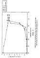
- the matrix or membrane material comprises a lipid or lipid mixture having a Tc value between 35 and 80°C, preferably between 37 and 45°C, more preferably between 38 and 43°C (Tc is defined as the gel-to-liquid crystalline phase temperature).
- the matrix or membrane material comprises peptides or one or more polymers.
- the invention provides the use of a contrast generating species for the manufacture of a particulate contrast medium for use in a method of diagnosis comprising generating a signal indicative of the value of said physiological parameter, the particles of said contrast medium comprising a matrix or membrane material and at least one magnetic resonance contrast generating species, said matrix or membrane material being responsive to said physiological parameter to cause the contrast efficacy of said contrast generating species to vary in response to said parameter and the response is an increased matrix or membrane permeability or chemical or physical breakdown of the membrane or matrix material.
- the image data generated may if desired be presented as a two or more dimensional spatial image, alternatively they may be presented as a temporal image, again in two or more dimensions.
- the data may simply provide one or more image values (e.g. numerical values) which either directly or indirectly may be used to provide quantitative or qualitative information (a signal) indicative of the value of the parameter under study.
- the image data may if desired be presented in visualizable form but alternatively they may simply be a set of data points which are collected and operated on to produce the signal without a visible image actually being generated.
- the signal indicative of the value of the parameter under study may likewise be generated in the form of a visible image, e.g.
- the signal provides a quantitative or at least semi-quantitative value for the parameter, e.g. either in a region of interest or in.a plurality of regions of interest in the body, for example providing a spatial and/or temporal map of the parameter within at least a portion of the body.
- Data relating to a physiological parameter may not necessarily also contain information relating to the anatomy of the animal body and thus, a further aspect of the invention relates to the combination of traditional anatomical imaging with physiological imaging to obtain two images, one containing information about a physiological parameter and the other containing anatomical information.
- the two images may be combined to give one image with both anatomical and physiological information.
- a method of imaging of an animate human or non-human animal body which method comprises:
- An image generated in response to a physiological parameter, a 'physiological image' may be formed using any of the imaging methods and or contrast media described herein. This physiological image can be combined with a conventional image obtained with or without a contrast agent.
- Suitable contrast agents for use with traditional anatomical imaging are well known in the art for all types of imaging techniques, MRI, X-ray, ultrasound, light and nuclear imaging etc. and many suitable contrast agents for anatomical imaging are discussed herein.
- the imaging technique used to obtain physiological data may be the same or different to the imaging technique used to obtain the anatomical image.
- the imaging technique will be the same, MRI being particularly suitable.
- Two separate contrast agents may be used, one for physiological imaging and one for traditional imaging.
- the two agents can be injected sequentially and the body scanned sequentially with respect to the appropriate imaging techniques and optionally the two images which are generated are then combined.
- a single multi-functional contrast agent may be used which is capable of providing both physiological and anatomical information.
- a multi-functional MRI contrast agent may be used, wherein one of its functions responds to a physiological parameter while a second function provides anatomical information.
- a multi-functional contrast agent may be used wherein the components of the agent function as contrast agents for different imaging techniques.
- the contrast agent may contain microbubbles to provide contrast in ultrasound imaging and paramagnetic complexes for MRI, being responsive to a physiological parameter according to the invention.
- the images obtained by scanning according to the two imaging techniques may be combined.
- MRI with hyperpolarised substances will tend to provide good physiological information relating to e.g. pH, temperature or pressure but little or no anatomical information.
- the hyperpolarised MR image is advantageously combined with an anatomical image, e.g. by superimposing the images.
- the two images may be generated separately or at the same time.
- physiological and anatomical imaging may be used to investigate all parts of the human or non-human animal body and any of the physiological parameters discussed herein, particularly pH and temperature.
- physiological parameter is temperature
- changes in the value of the parameter i.e. temperature changes, may be caused by intrinsic or extrinsic means.
- Intrinsic means will include cancer, cardiovascular disease and inflammation while extrinsic means include hyperthermia (external heating) treatment.
- the physiological contrast agent may be a contrast agent for hyperthermia.
- the imaging technique used in the method of the invention is MRI.
- signal strength or chemical shift or both may typically be studied.
- the particulate contrast agent used will accordingly be or contain a material capable of having a contrast or signal generating effect in MRI, e.g. a paramagnetic, ferromagnetic, ferrimagnetic or superparamagnetic material or a precursor therefor, or hyperpolarized NMR active (i.e. non zero nuclear spin) nuclei (e.g. noble gas or 13 C nuclei).
- the physiological parameter studied using the method of the invention may be any physiochemical parameter capable of affecting the matrix or membrane material of the contrast agent in such a way that the response to the physiological parameter is an increased matrix or membrane permeability or chemical or physical breakdown of the membrane or matrix material, e.g. pressure, temperature, pH, oxygen tension, carbon dioxide tension, enzyme activity, metabolite concentration, tissue electrical activity, tissue water diffusion, ion concentration, particularly Mg 2+ , Ca 2+ and Zn 2+ , etc.
- it will be selected from blood parameters, e.g. pressure, temperature and pH, in particular in the vasculature rather than the chambers of the heart.
- changes may be due to intrinsic factors such as disease or because of external factors, i.e. hyperthermia. It is not envisaged that the parameter be one which does not affect the membrane or matrix, for example flow rate or perfusion density.
- the contrast agent particles should comprise a membrane or matrix material which is responsive to the physiological parameter under investigation so as to alter the contrast efficacy of the contrast agent and the response is an increased matrix or membrane permeability or chemical or physical breakdown of the membrane or matrix material.
- the manner in which the membrane or matrix responds will depend on the particular combination of imaging modality, physiological parameter and contrast generating material selected.
- the response involves a change in membrane or matrix permeability to one or more species (e.g. water or gases) or chemical or physical breakdown of the membrane or matrix material.
- species e.g. water or gases
- Such a response may thus for example involve release from the particulate contrast agent of water-soluble contrast generating moieties that are capable of being taken up into the extracellular fluid outside the vasculature.
- physiological parameter responsive particulate contrast agents will be described in greater detail below.
- thermosensitive paramagnetic particulate compositions for temperature MRI-mapping of the human body.
- thermosensitive particulate compositions for temperature mapping in imaging guided hyperthermia treatment.
- the active contrast agent may be a paramagnetic, magnetic or fluorinated compound detectable by MRI.
- particulate compositions as contrast agents or as in vivo indicators or probes for detection of oxygen concentration/tension in the tissue using modalities such as MRI or Overhauser MRI.
- Another aspect of the present invention relates to particulate compositions as contrast agents or as in vivo indicators or probes in combination with a targeting ligand, wherein said targeting ligand targets cells or receptors selected from the group consisting of myocardial cells, endothelial cells, epithelial cells, tumor cells, brain cells, and the glycoprotein GPIIb/IIIa receptor, for detection of changes in physiological parameters and/or quantification/ semiquantification of physiological parameters relevant for diagnosis of disease.
- a targeting ligand targets cells or receptors selected from the group consisting of myocardial cells, endothelial cells, epithelial cells, tumor cells, brain cells, and the glycoprotein GPIIb/IIIa receptor, for detection of changes in physiological parameters and/or quantification/ semiquantification of physiological parameters relevant for diagnosis of disease.
- targeting ligands which can be used are:
- the particulate contrast agent may thus be used for quantification/semi-quantification of a physiological parameter which is relevant for diagnosis of disease.
- the particulate contrast agent may be triggered into giving a measurable signal difference either by the target parameter itself (e.g. the local temperature, pH or pressure or by binding to the particular cell surface receptors of interest) or by a chemical or biological response of the target parameter (e.g. release of enzymes or local variation in pH or temperature due to cellular reactions).
- the particulate agent may thus respond to, identify and/or quantitatively or semi-quantitatively determine bacteria, viruses, antibodies, enzymes, drugs, toxins, etc.
- Another aspect of the present invention relates to intravenous particulate compositions as contrast agents or as in vivo indicators or probes with long vascular half life (reduced liver uptake) for detection of changes in physiological parameters and/or quantification/semiquantification of physiological parameters relevant for diagnosis of disease.
- the particulate contrast agent used according to the invention may be a solid material, a porous material, a liquid crystal material, a gel, a plastic material, a material having one or more walls or membranes or liquid particles, e.g. emulsion droplets or gas based particles, e.g. micro bubbles.
- the particles can also be thermodynamically stabilised, e.g. micro emulsion droplets or surfactant micelles.
- the chemical composition of the particulate material can be one simple chemical compound or a mixture of two or more chemical compounds. Generally it will comprise two or more different chemical entities, at least one of which is a matrix or membrane forming material and at least one other of which is a magnetic resonance contrast generating species.
- the composition can consist of solid material(s) only or it may be a mixture of different solids/liquids/gases.
- the particulate will generally have a mean particle size (e.g. as determined by particle size analyzers such as laser light scattering apparatus or Coulter counters) in the range 0.001 to 20 ⁇ m, more preferably 0.01 to 10 ⁇ m, especially 0.05 to 7 ⁇ m.
- particle size analyzers such as laser light scattering apparatus or Coulter counters
- Such particles are often described in the literature as particles, colloids, emulsions, droplets, microcrystals, nanocrystals, microparticles, nanoparticles, vesicles, liposomes, bubbles, microspheres, microbubbles, coated particles, microballons and the like.
- polymer refers to any chemical compound with more than 10 repeating units.
- a polymer can be naturally occurring, synthetic, or semisynthetic.
- Semisynthetic polymers are polymers that are produced by synthetic modification of naturally occurring polymers.
- Compounds with 2 to 10 repeating units are herein generally referred to as “oligomers” and likewise may be natural, synthetic or semisynthetic.
- surface active compound or “surfactant” is used herein to refer to any chemical compound having at least one hydrophilic functional group and at least one hydrophobic (lipophilic) group. In a multiphase system, surface active compounds will commonly accumulate at the interface.
- lipid is used herein to refer to naturally-occurring compounds, synthetic compounds and semisynthetic compounds which are surface active compounds and have structures similar to fatty acids, waxes, mono-, di- or tri-glycerides, glycolipids, phospholipids, higher (C 10 or greater) aliphatic alcohols, terpenes and steroids.
- gas is used herein to refer to any compound or a mixture of compounds with sufficiently high vapor pressure to be at least partly in the gas phase at 37°C.
- Gaseous contrast generating species may be used in MRI.
- gases that change contrast property as a result of the physiological parameters in the surrounding tissue include: gases that change properties (e.g. lose hyperpolarization or change other magnetic properties) upon contact with body fluids or components, including dissolved components, thereof.
- gases include hydrogen, oxygen, nitrogen, noble gases (including hyperpolarized gases), carbon dioxide, fluorinated gases (e.g. sulphur hexafluoride, fluorohydrocarbons, perfluorocarbons and other fluorinated halogenated organic compounds in gas phase), and low molecular weight hydrocarbons.
- gases also include any pharmaceutically acceptable gas mixture like for example air and air/perfluorocarbon mixtures.
- the perfluorocarbon gas is selected from perfluoromethane, perfluoroethane, perfluoropropanes and perfluorobutanes.
- Any physiologically acceptable gas precursor can be used.
- suitable gas precursors are compounds that form a gas as a result of a chemical reaction (for example compounds sensitive to pH, for example carbonic acid, aminomalonic acid or other acceptable pH sensitive gas generating substances).
- Other suitable gas precursors are compounds that form a gas as a result of other physiological conditions like for example temperature, oxygen, enzymes or other physiological parameters/compounds relevant for body tissue (whether in the normal or diseased state) or which are activated to a gas forming state as a result of an interaction with an external stimulus (e.g. photoactivation, sono-activation etc.).
- the encapsulation material can be any material such as for example lipids, phospholipids, surfactants, proteins, oligomers and polymers. Such materials are chosen to respond to the pre-selected physiological parameter in such a way, that the response is an increased matrix or membrane permeability or chemical or physical breakdown of the membrane or matrix material, e.g. to dissolve, melt, collapse, weaken, increase porosity, or otherwise break down, change phase or change size (e.g. by aggregation due to change in surface charge, for example in response to local Ca 2+ and/or Mg 2+ concentration) in response to the physiological parameter, e.g.
- the contrast generating effect of the magnetic resonance contrast generating species may be dispersed (e.g. into the extracellular fluid space), switched on or increased (e.g. by generation of a contrast generating species such as a gas or by increasing water contact (for a positive (T 1 effect) MR contrast agent such as a gadolinium chelate)), or switched off or decreased (e.g.
- a negative (T 2 effect) MR contrast agent such as a dysprosium chelate, or by quenching of a radical or depolarization of a hyperpolarized nucleus or dissolution of a blood soluble gas).
- a porous solid matrix e.g. a zeolite, may be impregnated with the contrast generating species with the pore mouths then being closed off totally or partially using a material which breaks down, melts or dissolves when the relevant physiological parameter (e.g. pH, temperature, enzyme concentration) in the surrounding body fluid is above or below a pre-set value.
- the particulate contrast agent used according to the invention may respond to physiological parameters in several different ways.
- the particulate contrast agent may respond to physiological parameters by accumulation in the area where a certain value for a particular parameter is fulfilled, compared to areas where it is not.
- the particulate contrast agent responds by accumulation in areas where the physiological parameter value is not fulfilled.
- the particulate contrast agent responds to a given parameter by disintegration, the disintegration being dissolution or chemical breakdown.
- a particulate composition When a particulate composition responds by disintegration or transport, changes in contrast effect may be achieved by exposing otherwise invisible/shielded contrast agents or altering the distribution of contrast agents.
- Especially advantageous are particulate compositions where the contrast effect is gained by interaction with the environment.
- both transport of the contrast agent and transport of the actual environmental component may be utilized for detection of physiological parameters.
- An example is MRI contrast agents where an increased degree of water access/transport to the contrast agent leads to the measured contrast enhancement.
- response to a physiological parameter may be an increased rate of water transport in/out of the particulate.
- phase transitions may be utilized to induce leakage/transport.
- a solid particle/membrane may become leaky when it is melted, the process being sensitive to temperature.
- Phase transitions involving a gas phase may be used to respond to pressure as a physiological parameter.
- An especially useful aspect of the present invention is particles comprising liquid crystalline material as for example liposomes, niosomes or other vesicles. Liquid crystalline materials may undergo several different phase changes which may induce leakage and/or increase the transport rate of solutes or even breakdown of the particle.
- the gel to liquid crystalline phase transition of phospholipids may increase the liposome permeability and increase the transport rate or induce leakage of solutes on heating and hence temperature sensitivity.
- the lamellar to reversed hexagonal phase transition will also induce leakage since the liposomes require lipids in lamellar, gel or other layered phase structure.
- the lamellar to reversed hexagonal phase transition may be induced by pH, electrolytes, and changes in the chemical environment such as targeting, enzymes, antibodies etc.
- the suitable parameter to respond to may be tuned by selection of the membrane composition and processing.
- Other phase transitions such as lamellar to cubic phases, lamellar to microemulsion phases or lamellar to normal hexagonal phase may also be used to introduce leakage.
- Gel based particles or gel-surrounding particles may respond to a physiological parameter by, for example, a lowering of the viscosity of the gel.
- a physiological parameter for example, a lowering of the viscosity of the gel.
- Such viscosity lowering may for example be obtained by temperature, pH or electrolytes such as Ca 2+ or Mg 2+ and the particles are thus sensitive to these parameters.
- Such parameters may also induce phase separation in the gel particles, leading to leakage of liquid and phase separation of the polymer which comprises the gel.
- These mechanisms may in turn influence a parameter such as water leakage and exposure of, e.g. paramagnetic chelates to water and hence lead to a change in MRI contrast.
- Particles or membranes composed of solid polymer may also respond to physiological parameters. For instance temperature may change the glass transition temperature of the polymer, and hence induce phase transitions in the polymer membrane, which in turn may influence a parameter such as water transport which influences the contrast efficacy of the contrast agent.
- Particles which at least in part are composed of or stabilised by water soluble polymers e.g. peptides may respond to physiological parameters by alternation in the peptide conformation. For instance peptides may undergo an ⁇ - helix to ⁇ - sheet transition or vice versa and hence influence a parameter which in turn effects contrast. Also transitions to/from ⁇ - helix or ⁇ - sheet to random coil may influence a parameter such as membrane permeability, particle stability against aggregation/flocculation or even fusion, or particle dissolution or precipitation which in turn alters the contrast efficacy of the magnetic resonance contrast agent.
- a parameter such as membrane permeability, particle stability against aggregation/flocculation or even fusion, or particle dissolution or precipitation which in turn alters the contrast efficacy of the magnetic resonance contrast agent.
- Leakage may also be controlled by entities forming channels or other transport routes through the membrane of a particle. These channels may control the transport of molecules in/out of the particle, and be quite selective for, e.g., ions.
- the protein tubulin which forms microtubules in absence of Ca 2+ may induce a higher leakage in presence of Ca 2+ than in absence of Ca 2+ and hence be Ca 2+ sensitive.
- proteins/enzymes which may control the transport of substance in/out of a vesicle, include erythrocyte anion transporter, erythrocyte glucose transporter, Na + -K + ATPase (Na + /K + pump), Ca 2+ - ATPase (Ca 2+ pump) and Bacteriorhodopsin (H + - pump).
- biosurfactants such as iturins, esperine, bacillomycins, mycosubtilin, surfactin and similar substances may be used as membrane components to induce/prevent leakage by response to external parameters since these molecules may respond by changes in secondary and tertiary structure as well as self-assembly properties on influence from extrinsic parameters.
- the contrast generating species in MR contrast agents used according to the invention will generally be a paramagnetic, superparamagnetic, ferrimagnetic or ferromagnetic compound and/or a compound containing other non zero spin nuclei than hydrogen, e.g. 19 F, 13 C, 15 N, 29 Si, 31 P and certain noble gases, such as 129 Xe or 3 He.
- Preferred as paramagnetic compounds are stable free radicals, and compounds (especially chelates) of transition metal or lanthanide metals, e.g. manganese compounds, gadolinium chelates, ytterbium chelates and dysprosium chelates.
- Preferred magnetic e.g.
- superparamagnetic compounds are ⁇ -Fe.O., Fe.O and other iron/metal oxides with high magnetic susceptibility.
- Preferred fluorinated compounds are compounds with relative short 19 F T-relaxation times.
- Other preferred fluorinated compounds according to the present invention are fluorinated pH-probes, such as compounds described in EP-0447013 of Schering A.G. and ZK-150471 described by Y. Aoki in Invest. Radiol 1996, 34 , 680-689.
- Examples of MR contrast effective materials are well known from the patent literature, see for example the patent publications of Nycomed, Salutar, Sterling Winthrop, Schering, Squibb, Mallinckrodt, Guerbet and Bracco.
- contrast generating species useful in MR contrast agents for use according to the invention: species that change contrast property as a result of the physiological parameters in the surrounding tissue; and species that are inert to physiology but change contrast properties as a result of an interaction between coating material/encapsulation material and physiology.
- species that change contrast property as a result of the physiological parameters in the surrounding tissue and species that are inert to physiology but change contrast properties as a result of an interaction between coating material/encapsulation material and physiology.
- Typical examples here will be GdDTPA, GdDTPA-BMA, GdDOTA, GdHPDO3A, PrDO3A-derivatives and Tm chelates in thermosensitive liposomes or in pH-sensitive vesicles.
- Typical examples of species that change contrast property as a result of the physiological parameters in the surrounding tissue include: paramagnetic chelates that change relaxation properties and/or change chemical shift as a result of temperature, paramagnetic chelates that change coordination number and thereby relaxation properties and/or shift properties as a function of pH, paramagnetic compounds, for example manganese compounds (Mn(2+)/Mn(3+)), europium compounds (Eu(2+), Eu(3+)) and free radicals (radical, no radical) that change relaxation properties and/or shift properties as a result of oxygen tension/concentration or as a result of redox potential in the surrounding tissue, paramagnetic and magnetic compounds that change relaxation/shift properties as a result of enzymatic activity (for example with enzymatic cleavage of paramagnetic chelates from macromolecules conjugated thereto causing a change in correlation time and/or water coordination) and paramagnetic chelates that change properties as a result of concentration of ions in the tissue,
- Paramagnetic compounds have, according to the present invention, either an effect on the relaxation times (T 1 or T 2 ) or an effect on chemical shift.
- Typical compounds that change relaxation times are gadolinium chelates, manganese compounds and superparamagnetic iron oxides.
- Europium chelates are well-known chemical shift compounds. The effect on chemical shift is related to temperature.
- Magnetic resonance contrast generating species can be released from the matrix/encapsulation material as a result of increased temperature and thereby change their contrast property or distribute to other tissues than the particulate product.
- a change in contrast efficacy may occur due to an increased permeability of the matrix/encapsulation material, and, hence, to an increased rate of water transport across the matrix/encapsulation material, even if the agent itself does not leave the matrix/encapsulation material.
- Typical examples of temperature sensitive particulate materials are temperature sensitive liposomes, these being suitable for use with MRI. These liposomes take advantage of the fact that the membrane permeability is.markedly increased at the gel-to-liquid crystal phase transition temperature (T c ) of their membrane lipids. Also, possibly depending upon the membrane properties and the nature of the MR active agent, leakage of the agent may occur. Liposomes made from specific phospholipids or a specific blend of phospholipids may be stable up to 37°C but exhibit an increased water permeability or/and leak as they pass through an area of the body in which the temperature is raised, e.g. to 40 to 45°C, as a result of a disease process or an external heating.
- Table 1 shows the transition temperature of various saturated phosphatidylcholines.
- PC Phosphatidylcholines
- Tc Transition temperature 12:0 -1 13:0 14 14:0 23 15:0 33 16:0 41 17:0 48 18:0 55 19:0 60 20:0 66 21:0 72 22:0 75 23:0 79 24:0 80
- Table 2 shows the phase transition of various unsaturated phosphatidylcholines.
- PC Phosphatidylcholines
- Table 4 below shows the phase transition temperature for various saturated symmetric phosphatidylglycerols (PG) in the form of their sodium salts.
- Tables 1-4 are based on information from the product catalogue of Avanti Polar Lipid Inc., USA.
- thermosensitive liposomes may be selected to give products with the correct Tc for thermosensitive liposomes for diagnostic use.
- Typical blends for preparation of thermosensitive liposomes for diagnostic use are mixtures of dipalmitoylphosphatidylcholine (DPPC) and dipalmitoylphosphatidyl glycerol (DPPG) and distearylphosphatidylcholine (DSPC).
- DPPC dipalmitoylphosphatidylcholine
- DPPG dipalmitoylphosphatidyl glycerol
- DSPC distearylphosphatidylcholine
- Particulate contrast agents may also respond to temperature by utilizing the conformational temperature sensitivity of certain polymer systems.
- An example is poly(N-isopropyl acrylamide) which phase separates at 37°C. Hence particles comprising contrast agents will become leaky dependent on temperature (see Hoffmann et al. Macromol. Symp. 118 : 553-563 (1997)).
- temperature sensitive matrices/coatings are lipid suspensions/emulsions containing the contrast generating species or other particulate or particulate like formulations that release the magnetic resonance contrast generating species or change properties as a result of changes in temperature.
- the parameter under study is capable of manipulation, e.g. by treatment with drugs, external application of heat etc., it may be used to study the efficacy of such treatment or localized such treatment may be used to cause a change in contrast efficacy which in turn may be used to measure parameters such as organ perfusion.
- external application of heat at, near or upstream of an organ of interest may be used to cause release from the particles of a contrast agent which may diffuse into the organ and so to detect blood perfusion (or lack of perfusion) in that organ.
- a thermally sensitive particulate agent in connection with an external heating to follow the heat transport in parts of the body. Heat transport in vivo is directly connected to blood flow through the bioheat equation (J. Appl. Physiol. vol.
- thermosensitive particulate compositions may, after a controlled, localized external heating, give a measure of blood perfusion in an organ.
- thermosensitive MR-liposomes can in general be divided into three distinct regions:
- the absolute [Gd] can not be determined, unless the liposomal relaxivity in the tissue is known. However this may be fairly well approximated for the r relaxivity, but not the r * relaxivity, since this depends on tissue geometry. Nonetheless, the [Gd] in one region relative to another can be estimated as described above.
- the relative [Gd] is valuable information that can be used to adjust the signal enhancement in the image so that it reflects actual temperature changes. In order for this to be possible, one need to assume that a 'core region' exists where the temperature is above T. It is likely in a clinical situation that such a core region exists where the heating is most efficient surrounded by a 'penumbra' where heating is less efficient and the temperature distribution is less well defined. Now, by knowing the relative [Gd] in the core versus the penumbra, the image intensity can be adjusted to compensate for any difference in [Gd] in the two regions.
- R 1 R 1 0 + [Gd]*r 1 ;
- R 1 0 is the relaxation rate prior to contrast administration.
- the T 1 effect of the liposomes is detectable below T 0 , it is possible to map the local Gd concentration and consequently compensate for differences in contrast enhancement after heating due to local variations in Gd concentration.
- the R 2 or R 2 ' effect of the liposomes can also be used for this purpose.
- pH is an important physiological parameter associated to several severe diseases.
- the pH value is usually reduced during cancer diseases, cardiovascular diseases like for example stroke, osteoporosis, inflammations and autoimmune diseases.
- pH sensitive encapsulation for diagnostic agents involves the use of pH sensitive liposomes.
- the general strategy is to employ pH-sensitive groups in the liposomal membrane. Such typical groups have pKa values between 4 and 5.5.
- Phospholipids useful for preparation of pH-sensitive diagnostic agents include diheptadecanoyl phosphatidylcholine (DHPC) in admixture with DPPC and N-palmitoyl homocystein (PHC) in different ratios (see Eur. J. Pharm. Biopharm. 1993, 39 , 97-101 for a general review on temperature and pH-sensitive liposomes).
- DHPC diheptadecanoyl phosphatidylcholine
- PLC N-palmitoyl homocystein
- pH-sensitive encapsulation of contrast generating species involves the use of pH-sensitive surfactants like for example N -dodecyl-2-imidazole propionate (DIP) which has pKa of 6.8 (see for example Pharm. Res. 1993, 13, 404).
- DIP N -dodecyl-2-imidazole propionate
- pH-sensitive encapsulation of magnetic resonance contrast generating species involves the use of matrix materials and/or coating materials with pKa values in the range of 4.5-7.0 so that the material is soluble or partly soluble in the charged form and insoluble or partly insoluble in the non-charged form.
- Such compounds can be physiologically acceptable low molecular weight compounds or physiologically acceptable polymers.
- Still another means of pH-sensitive encapsulation involves the use of compounds that are chemically cleaved as a result of pH, for example polyorthoesters or polyacetals/ketals which are cleaved under acidic conditions.
- Liposomes comprising phosphatidyl ethanolamines (PE) as the central component are another example of liposomes which can undergo a phase transition and become leaky when pH is reduced. pH sensitive liposomes can also be achieved by incorporation of fatty acids into phospholipid membranes.
- PE phosphatidyl ethanolamines
- any charged particulate system where the charge is pH dependent and influences the packing of the membrane material can be used.
- Oxygen is critical for all types of cells, and diagnostic agents for determination of oxygen concentration/tension in tissue will be of great importance in diagnosis of diseases like cancer, cardiovascular diseases, autoimmune diseases and several diseases in the central nervous system.
- One type of oxygen or redox sensitive encapsulation/coating material is a material that has different solubility/diffusion properties dependent on the oxygen level or the redox status; for example compounds containing a nitro-group that is reduced in vivo to an amino-group which improves solubilization of the material in reductive/low oxygen surroundings.
- ion concentration sensitive encapsulation materials that may be used in this regard include phospholipids, surfactants and other ion chelating materials.
- Negatively charged liposomes will for example bind Ca(2+) and the membrane will change its diffusion properties and become more stiff.
- Ca 2+ /Mg 2+ sensitive particulate compositions are liposomes enriched with the dimeric phospholipid cardiolipin.
- a cardiolipin containing membrane may undergo a lamellar to reversed hexagonal phase transition upon addition of the divalent cations since these ions bind to the cardiolipin di-phosphatidyl group.
- Ca 2+ or Mg 2+ sensitivity may be obtained by using charge stabilised particles, e.g. solid particles, liquid particles e.g. emulsion droplets, gas particles e.g. microbubble dispersions or liposomes. Ca 2+ or Mg 2+ may thus induce aggregation or flocculation among the particles and by this means alter contrast effect.
- Ca 2+ or Mg 2+ sensitivity may also be obtained by using stabilising moieties for the particles which are chemically or physically influenced by Ca 2+ or Mg 2+ , for instance using surfactants which form water insoluble species when exposed to Ca 2+ or Mg 2+ and thus precipitate.
- Some particles or stabilising membranes surrounding particles may also respond with a phase transition when exposed to Ca 2+ or Mg 2+ .
- An example are liquid crystalline based particles e.g. liposomes, which may respond by a lamellar to reversed hexagonal phase transition upon addition of Ca 2+ or Mg 2+ .
- gel particles may respond easily to Ca 2+ or Mg 2+ by a significant lowering of viscosity or even phase separation of the polymer which forms basis for the gel on exposure to Ca 2+ or Mg 2+ . This viscosity reduction or phase separation may induce a change in contrast effect.
- Types of enzyme sensitive encapsulation material include matrices or coatings that are degraded by enzymes, for example simple esters of low molecular weight compounds or polyesters like polyacetic acid and others.
- Various metabolites may also change the properties of coating materials.
- Particulates can be made sensitive to for example antibodies based on enhanced leakage due to a phase transition induced by the chemical binding between membrane molecules and the antibody.
- liposomes comprising N-(dinitrophenylamino- ⁇ -caproyl)-phosphatidyl ethanolamine (DNP-cap-PE) become leaky due to a lamellar to reversed hexagonal phase transition when binding to anti-DNP.
- DNP-cap-PE N-(dinitrophenylamino- ⁇ -caproyl)-phosphatidyl ethanolamine
- Another example includes liposomes comprising human glycophorin A in dioleoyl phosphatidyl ethanolamine membranes. These liposomes become leaky when immobilized antibodies are added.
- a further aspect of the present invention is to use one of the above described particulate diagnostic agents together with another compound that has the potential to change the physiological parameter of interest or together with use of an external energy source to change the parameter of interest.
- thermosensitive MR diagnostic agents according to the invention in connection with an external heating and to follow the heating effect in parts of the body.
- Another example is to administer compounds that change pH in connection with a pH-sensitive particulate MR diagnostic agent according to the invention to follow the pH-profile in the area of interest.
- Still another example is to cause the subject under study to inhale oxygen, after administration of an oxygen sensitive MR diagnostic agent according to the invention, to follow oxygen uptake in tissue.
- the particulate diagnostic agent or a component thereof carries an overall charge
- it will conveniently be used in the form of a salt with a physiologically acceptable counterion, for example an ammonium, substituted ammonium, alkali metal or alkaline earth metal cation or an anion deriving from an inorganic or organic acid.
- a physiologically acceptable counterion for example an ammonium, substituted ammonium, alkali metal or alkaline earth metal cation or an anion deriving from an inorganic or organic acid.
- meglumine salts are particularly preferred.
- the diagnostic agents of the present invention may be formulated in conventional pharmaceutical or veterinary parenteral administration forms, e.g. suspensions, dispersions, etc., for example in an aqueous vehicle such as water for injections.
- compositions may further contain pharmaceutically acceptable diluents and excipients and formulation aids, for example stabilizers, antioxidants, osmolality adjusting agents, buffers, pH adjusting agents, etc.
- pharmaceutically acceptable diluents and excipients and formulation aids for example stabilizers, antioxidants, osmolality adjusting agents, buffers, pH adjusting agents, etc.
- the carrier medium is preferably isotonic or somewhat hypertonic.
- the particulate agent comprises a chelate or salt of an otherwise toxic metal species, e.g. a heavy metal ion
- a chelating agent e.g. as discussed by Schering in DE-A-3640708, or more preferably a slight excess of the calcium salt of such a chelating agent.
- the dosage of the diagnostic agents of the invention will depend upon the imaging modality, the contrast generating species and the means by which contrast enhancement occurs (e.g. with switching on or off of contrast, with dispersion of contrast out of the vascular space, etc).
- dosages will be between 1/10 and 10 times the dosage conventionally used for the selected contrast generating species or analogous species in the same imaging modality. Even lower doses may also be used.
- the present invention is particularly suitable for methods involving parenteral administration of the particulate material, e.g. into the vasculature or directly into an organ or muscle tissue, intravenous administration being especially preferred, it is also applicable where administration is not via a parenteral route, e.g. where administration is transdermal, nasal, sub-lingual or is into an externally voiding body cavity, e.g. the gi tract, the bladder, the uterus or the vagina. The present invention is deemed to extend to cover such administration.
- Liposomes containing GdHPDO3A ProHance, Bracco Spa, Milan, Italy
- GdDTPA-BMA Olethyl-choline
- HPS hydrogenated phosphatidylserine-sodium
- the phospholipid mixtures contained 5% or 10% (w/w) of the negatively charged HPS and DPPG components.
- the phospholipid mixtures were dissolved in a chloroform/ methanol mixture and the organic solution was evaporated to dryness under reduced pressure.
- DPPC/DPPG liposomes were formed by hydrating the lipid film with a pre-heated (55°C) aqueous solution (pH 7.4) of 250 mM GdDTPA-BMA or 250 mM Gd HPD03A.
- the HPC/HPS liposomes were prepared analogously but with a lipid hydration temperature of 70°C.
- the DPPC/DPPG and HPC/HPS liposomes where allowed to swell for 2 hours at 55 and 70°C respectively.
- the total lipid concentration was 50 mg/ml.
- the liposomes were subjected to 3 freeze-thaw cycles in liquid nitrogen. Differently sized liposomes were produced by sequential extrusion (Lipex ExtruderTM, Lipex Biomembranes Inc., Vancouver, Canada) through polycarbonate filters of various pore diameters. The extrusion temperature was 55 and 70°C for the DPPC/DPPG and HPC/HPS liposomes respectively. Untrapped metal chelate was removed by gel filtration or dialysis against isoosmotic and isoprotic glucose solution.
- the mean hydrodynamic diameter of the liposomes varied from 103 nm to 276 nm, as measured by photon correlation spectroscopy (ZetaSizer IV, Malvern Instruments Ltd., Malvern, England); the zeta potential was negative in the order of -25 mV, as determined by laser Doppler velocimetry at 25°C (ZetaSizer IV, Malvern Instruments Ltd., Malvern, England).
- the mean gel-to-liquid crystalline phase transition temperature (Tc) of the HPC/HPS and DPPC/DPPG preparations was 50 and 42°C, respectively, as determined by differential scanning calorimetry (DSC4, Perkin Elmer Inc., Norwalk, CT).
- Figure 1 of the accompanying drawings and Table 5 below show the temperature sensitivity of in vitro T 1 relaxivity (r 1 ) for liposome encapsulated GdDTPA-BMA and GdHPDO3A, respectively (0.47T).
- Figure 2 of the accompanying drawings shows the temperature response of the in vitro MR signal intensity for liposome encapsulated GdDTPA-BMA.
- FIG. 3 of the accompanying drawings shows a series of T 1 -w GRE images prior to and after heating of a gel phantom containing inserts of liposome encapsulated GdDTPA-BMA.
- Control 20 0.16 0.06 4.53 25 0.23 0.08 4.27 30 0.31 0.12 3.94 37 0.69 0.21 3.75 45 3.30 0.53 3.07 55 3.10 3.00 2.82 60 - 2.96 2.54
- DSPC/DPPC/DPPG weight ratio; 28.5/66.5/5) liposomes were prepared by the thin film hydration method.
- the phospholipids 500 mg were dissolved in a chloroform/methanol mixture and the organic solution was evaporated to dryness under reduced pressure.
- Liposomes were formed by hydrating the lipid film with a pre-heated (57"C) aqueous solution (pH ⁇ 7) of 250 mM GdDTPA-BMA (10 ml). The liposomes were subjected to 3 freeze-thaw cycles and allowed to swell for one and a half hours at 65"C. The liposome dispersion was extruded at 65°C through polycarbonate filters of various pore diameters.
- DPPC/DPPG/DPPE-PEG-2000 (weight ratio; 90/5/5) liposomes were prepared by the thin film hydration method.
- the phospholipids 500 mg were dissolved in a chloroform/methanol mixture and the organic solution was evaporated to dryness under reduced pressure.
- Liposomes were formed by hydrating the lipid film with a pre-heated (57 C) aqueous solution (pH ⁇ 7) of 250 mM GdDTPA-BMA (10 ml). The liposomes were subjected to 3 freeze-thaw cycles and allowed to swell for one and a half hours at 65°C. The liposome dispersion was extruded at 65°C through polycarbonate filters of various pore diameters.
- the liposome size (z-average) after extrusion was 132 nm.
- Untrapped GdDTPA-BMA was removed by dialysis against isoosmotic and isoprotic glucose solution.
- Table 7 shows the temperature sensitivity of the in vitro r (0.235T) in glucose 5% solution for liposome encapsulated GdDTPA-BMA. Temperature (°C) r 1 in glucose 5% (s -1 mM -1 ) 35 0.32 37 0.46 38 0.56 39.2 2.53 40 4.16 42 5.65
- DSPC/DPPC/DPPG weight ratio; 43/52/5) liposomes were prepared by the thin film hydration method.
- the phospholipids 500 mg were dissolved in a chloroform/methanol mixture and the organic solution was evaporated to dryness under reduced pressure.
- Liposomes were formed by hydrating the lipid film with a pre-heated (63"C) aqueous solution (pH ⁇ 7) of 250 mM GdDTPA-BMA (10 ml). The liposomes were subjected to 3 freeze-thaw cycles and allowed to swell for one and a half hours at 64 C. The liposome dispersion was extruded at 65 C through polycarbonate filters of various pore diameters.
- the liposome size (z-average) was 145 nm. Untrapped metal chelate was removed by dialysis against isoosmotic and isoprotic glucose solution.
- Table 8 shows the temperature sensitivity of the in vitro r (0.235T) in both glucose 5% solution and human serum for liposome encapsulated GdDTPA-BMA. Temperature (°C) r 1 in glucose 5% (s -1 mM -1 ) r 1 in serum (s -1 mM -1 ) 35 0.12 0.14 40 0.22 0.25 42 0.29 0.44 44 0.88 1.91 46 4.47 4.51 48 4.40 4.51 50 4.40 4.35
- DPPC/DPPG weight ratio; 95/5) liposomes were prepared by the thin film hydration method.
- the phospholipids 500 mg were dissolved in a chloroform/methanol mixture and the organic solution was evaporated to dryness under reduced pressure.
- Liposomes were formed by hydrating the lipid film with a pre-heated (52°C) aqueous solution (pH ⁇ 7) of 250 mM GdDTPA-BMA (10 ml). The liposomes were subjected to 3 freeze-thaw cycles and allowed to swell for one and a half hours at 55°C.
- the liposome dispersion was extruded at 62"C through polycarbonate filters of various pore diameters.
- Example 4 1.5 ml liposomes from Example 4 were mixed with 1.5 ml DPPC/DPPG liposomes prepared as Example 5. The mixture was diluted to 40 ml with glucose 5% solution.
- Table 10 shows the temperature sensitivity of the in vitro R 1 (0.235 T) in glucose 5% solution for the liposome mixture. Temperature ( C) R in glucose 5% (s - ) 35 2.46 38 2.61 39 2.83 40 3.87 41 7.11 42 7.17 44 10.9 46 14.0 48 14.0
- Liposomes were injected intramuscularly at a dosage of 0.02 mmol/kg.
- the left thigh muscle was heated with focused ultrasound whereas the right thigh muscle served as a control.
- Figures 4-5 show axial T 1 -w SE images of the thigh before and after liposome injection, respectively.
- Figures 6-8 are T 1 -w SE images after 2, 5, and 9 minutes of heating, respectively.
- Figure 9 represents the final image 15 minutes after termination of heating.
- the syringe containing the liposomal dispersion identical to that injected was included.
- Liposomes were injected intravenously into a rat (upper position) at a dosage of 0.10 mmol/kg.
- the rat in the lower position served as control (e.g. no injection nor heating).
- Figure 10 is the axial T 1 -w SE image of the liver 7 minutes after liposome injection. At 15 minutes post injection, the liver was heated by focused ultrasound (Figure 11).
- Figures 12-13 are T 1 -w SE images 16 and 21 minutes after initiation of heating, respectively. After termination of heating, the measured temperature in the liver was 51°C.
- liver signal intensity increases substantially after heating as compared to the control liver.
- the mean hydrodynamic diameter of the liposomes was measured to 165 nm by photon correlation spectroscopy (ZetaSizer IV, Malvern Instruments Ltd., Malvern, England).
- the in vitro T 1 -relaxation times of the paramagnetic liposomes were measured (0.235 T, Minispec PC-110b, Bruker GmbH, Rheinstetten, Germany) in different isoosmotic buffer solutions (0.05 M citrate-phosphate buffer and 0.05 M Tris-HCl buffer).
- the investigated pH range was 4-8.5.
- the buffered liposome dispersions were incubated at 37°C for 15 minutes.
- Table 11 shows the pH sensitivity of in vitro r 1 -relaxivity for liposome encapsulated GdDTPA-BMA. pH dependency of the r 1 (37 °C, 0.235 T) for liposomal GdDTPA-BMA pH r 1 (s -1 mM -1 ) 3.91 1.32 4.30 1.26 4.70 1.31 5.15 1.17 5.59 1.10 5.95 1.03 6.40 1.00 6.71 0.50 7.33 0.32 7.69 0.29 8.02 0.28 8.34 0.29 8.54 0.31
- DPPC/DPPG weight ratio; 95/5) liposomes were prepared by the thin film hydration method.
- the phospholipids 500mg were dissolved in a chloroform/methanol mixture and the organic solution was evaporated to dryness under reduced pressure.
- Liposomes were formed by hydrating the lipid film at 50°C with an aqueous solution (pH ⁇ 7) of 250 mM DyDTPA-BMA (sprodiamide, Nycomed Imaging AS, Oslo, Norway) (10ml). The liposomes were subjected to 3 freeze-thaw cycles and allowed to swell for one hour at 59°C. The liposome dispersion was extruded at 65°C through polycarbonate filters of various pore diameters.
- the liposome size (z-average) after extrusion was 153 nm. Untrapped DyDTPA-BMA was removed by dialysis against isoosmotic and isoprotic glucose solution. The temperature sensitivity of the MR contrast effect may be investigated.
- DPPC/DPPG weight ratio; 95/5) liposomes were prepared by the thin film hydration method.
- the phospholipids 500 mg were dissolved in a chloroform/methanol mixture and the organic solution was evaporated to dryness under reduced pressure.
- the liposomes were formed by hydrating the lipid film at 48°C with an aqueous solution of 50 mM GdDTPA-dextran (MW 156 kD), whose synthesis is described in: P Rongved et al., Carbohydr. Res ., 287 (1996) 77-89.
- the liposome dispersion was sonicated at 46°C using a sonicator tip.
- the liposome size (z-average) after sonication was 70 nm. Untrapped GdDTPA-dextran is removed by gel filtration or dialysis against isoosmotic and isoprotic glucose solution. The temperature sensitivity of the MR contrast effect may be investigated.
- Dibehenoyl-PC (22:0) (Table 1) liposomes may be prepared by the thin film hydration method.
- the phospholipids 500 mg are dissolved in a chloroform/methanol mixture and the organic solution is evaporated to dryness under reduced pressure.
- Liposomes are formed by hydrating the lipid film at 80°C with an aqueous solution (pH ⁇ 7) of 250 mM GdDTPA-BMA (10 ml).
- the liposomes are subjected to 3 freeze-thaw cycles and allowed to swell for one and half-hours at 80°C.
- the liposome dispersion is extruded at 80°C through polycarbonate filters of various pore diameters.
- Untrapped GdDTPA-BMA is removed by gel filtration or dialysis against isoosmotic and isoprotic glucose solution. The temperature sensitivity of the MR contrast effect may be investigated.
- SPIOs Superparamagnetic iron oxides
- HPC/HPS weight ratio; 90/10 liposomes were prepared by a modified thin film hydration method. Liposomes were formed by adding a homogeneous mixture of phospholipids (700 mg) to 10 ml of a pre-heated (55°C) aqueous dispersion of PEGylated SPIOs (6.10 mg iron/ml). The liposomes were allowed to swell for 30 minutes at 65°C. The liposome dispersion was extruded at 66°C through polycarbonate filters of various pore diameters. Untrapped SPIOs are removed by gel filtration or dialysis. The temperature sensitivity of the MR contrast effect may be investigated.
- Ultrasmall superparamagnetic iron oxides (USPIOs) encapsulated within HPC/HPS liposomes
- HPC/HPS weight ratio; 90/10 liposomes were prepared by a modified thin film hydration method. Liposomes were formed by adding a homogeneous mixture of phospholipids (700 mg) to 10 ml of a pre-heated (70°C) aqueous dispersion of USPIOs (3.63 mg iron/ml). The liposomes were allowed to swell for 90 minutes at 70°C. The liposome dispersion was extruded at 70°C through polycarbonate filters of various pore diameters. Untrapped USPIOs are removed by gel filtration or dialysis. The temperature sensitivity of the MR contrast effect may be investigated.
- SPIOs Superparamagnetic iron oxides
- ultrasmall-SPIOs encapsulated within pH-sensitive liposomes
- SPIOs or USPIOs encapsulated within pH-sensitive liposomes may be prepared in a manner analogous to that used in Example 9. Untrapped superparamagnetic material is removed by gel filtration or dialysis. The pH-sensitivity of the MR contrast effect may be investigated.
- DSPC/DMPG/cholesterol liposomes (molar ratio; 49:5:20) were prepared by a modified thin film hydration method.
- Liposomes were formed by adding a freeze-dried mixture of phospholipids (60 g) to a pre-heated (59°C) aqueous solution (pH ⁇ 6.3) of 250 mM GdDTPA-BMA/300 mM sucrose/10mM phosphate (300 ml). The liposomes were allowed to swell for 30 minutes at 59°C. The liposome dispersion was homogenized and extruded at high pressure through polycarbonate filters with a pore size of 400 nm.
- Untrapped GdDTPA-BMA was removed by ultrafiltration with a 300 mM sucrose/10 mM phosphate solution.
- the liposome size (z-average) after ultrafiltration was 110 nm.
- Liposomes were also lyophilized (2ml per vial) and reconstituted by addition of 2 ml of deionized water.
- the liposome size (z-average) after reconstitution was 119 nm.
- Table 12 summarizes the temperature sensitivity of the in vitro R 1 (0.235 T) for liposome encapsulated GdDTPA-BMA in a 300 mM sucrose/10 mM phosphate solution.
- Liposomes containing the following contrast agents may be prepared in a manner analogous to that used in Examples 1-5, 9 and 12. Untrapped Gd compound is removed by gel filtration or dialysis. The temperature sensitivity of the MR contrast effect may be investigated.
- DPPC/DPPG/DPPE-PEG-2000 and DPPC/DPPG liposomes containing GdDTPA-BMA were prepared in a manner analogous to that used in Examples 3 and 5, respectively.
- Table 13 summarizes the temperature evolution of the in vitro r 1 (0.235 T) in rat blood for both liposome formulations. Temperature (°C) r 1 (s -1 mM -1 ) DPPC/DPPG 120 nm r 1 (s -1 mM -1 ) DPPC/DPPG/DPPE-PEG 121 nm 35 0.260 0.244 37 0.479 0.391 39 0.659 0.588 40 1.18 0.823 41 2.45 1.26 42 4.06 2.87 43 5.18 3.65 44 4.57 4.06
- DPPC/DPPC liposomes containing GdDTPA-BMA were injected intramuscularly (im) into Sprague Dawley rats at a dosage of 20 ⁇ mol/kg.
- Table 15 shows the temperature response of the T 1 in muscle. All results are given as mean values.
- DPPC/DPPG/DPPE-PEG-2000 and DPPC/DPPG liposomes containing GdDTPA-BMA were injected intravenously (iv) into Sprague Dawley rats at a dosage of 100 ⁇ mol/kg.
- the Gd uptake in tissue is also shown as the percentage tissue uptake of the administered Gd dosage.
- Tables 18 and 19 summarize the temperature response of the blood T 1 after iv injection of DPPC/DPPG and DPPC/DPPG/DPPE-PEG-2000 liposomes, respectively.
- Time post injection (min) T 1 relaxation time (ms)
- Tissue uptake % of adm.
- DSPC/DPPC/DPPG liposomes containing GdDTPA-BMA were prepared analogously to Example 2.
- the liposome size (z-average) was 129 nm.
- MR imaging was performed at 2.0 T (Bruker Medspec) on a concentric spherical phantom in which the inner chamber contained liposomal GdDTPA-BMA diluted with an isotonic medium composed of glucose and 6.25% polyvinylpyrrolidone (conc. ⁇ 0.8 mM Gd), whilst the outer compartment was filled with saline.
- Microwave heating was performed at 434 MHz with a linear radio frequency antenna placed in the outer chamber. The microwave irradiation was applied simultaneously with the image acquisition.
- Blocks consisting of 10 diffusion-weighted spin-echo single shot EPI (DW-SE-EPI) images (b-factor from 3 to 864 s/mm 2 ), a set of SE-EPI images with inversion-recovery preparation (IR-SE-EPI) and gradient echo T 1 -weighted (T 1 W-GE) images were repeated until the temperature of the liposome sample reached 48°C (in appr. 110 min).
- T 1 W-GE images were acquired with TE/TR/flip: 5ms/30ms/50°.
- T 1 -maps were calculated from the set of 13 IR-SE-EPI images, measured with inversion times varying from 14.4 ms to 16 s.
- Plots of 1/T 1 (R 1 ) versus temperature were generated from a fixed region-of-interest within the phantom.
- the sample temperature was measured by a thermocouple immediately after acquisition of each block.
- the temperature distribution within the imaged slice was evaluated from ADC-maps.
- FIG 14. The temperature evolution of the measured R 1 for liposomal GdDTPA-BMA is summarized in Figure 14.
- a linear correlation was obtained between R 1 and temperature in the "transition region" 40.4-43.7°C (regression coefficient of 0.995).
- Figure 15 shows selected T 1 -W GE images of the phantom (a) before heating, (b) during heating; signal intensity distribution observed within liposome sample, and (c) after heating; homogeneous signal intensity distribution.
- Figure 16 shows the corresponding T 1 -maps at the same time points as for Figure 15.
- a corresponding temperature map could be derived from the T 1 -map at timepoint (b), as seen on Figure 17.
- the temperature map demonstrates the thermosensitivity of liposomal GdDTPA-BMA. (NB The temperature scale is only valid for the inner chamber containing liposomes).
- a static in vitro phantom composed of twelve glass vials (10 mm dia.) placed in a rectangular plastic container, was used for this study.
- the plastic container was filled with a viscous isotonic medium composed of glucose/25% (w/w) polyvinyl-pyrrolidone (PVP) and doped with GdDTPA-BMA to give a T 1 of about 430 ms at 1.5 T.
- Three of the vials contained a marker solution with a known T 1 value (about 630 ms).
- the remaining nine vials were filled with DPPC/DPPG-based GdDTPA-BMA liposomes (prepared in Example 18) dispersed in varying amounts of isotonic 10% PVP/glucose solution.
- the Gd concentration [Gd] in the liposome samples ranged from 0 to 5.2 mM Gd as determined by inductively coupled plasma atomic emission spectrophotometry.
- the phantom was imaged at room temperature in a quadrature knee coil at 1.5 T on a Philips NT system. The following imaging sequences were used:
- a linear correlation was obtained between [Gd] and matrix corrected R 1 ( ⁇ R 1 ) prior to heating (measured from TMIX sequence), with a regression coefficient of 0.996.
- the calculated liposomal r 1 was 0.11 mM -1 s -1 .
- the r 1 and r 2 were 3.23 and 3.75, respectively, giving an r 2 /r 1 ratio of 1.16.
- Figure 18 shows, prior to heating, a linear correlation between [Gd] and the ratio of the signal intensities of liposome sample and PVP gel (SI lip /SI gel ) using the T 1 -FFE sequence.
- the findings suggest that the T 1 - (and T 2 -) effect of liposome encapsulated GdDTPA-BMA prior to heating is significant enough to enable a relative assessment of liposomal Gd concentration.
- DPPE/PA liposomes containing GdDTPA-BMA were prepared analogously to Example 9.
- the mean hydrodynamic diameter of the liposomes was measured to 158 nm by photon correlation spectroscopy (Malvern PS/MW 4700, Malvern Instruments Ltd., Malvern, England).
- An in vitro phantom composed of thirteen glass vials (11 mm diameter) placed in a circular glass reactor, was used for this study.
- the glass reactor was filled with an agar gel (2 % w/v) doped with GdDTPA-BMA to give a T 1 of about 900 ms at 1.5 T.
- the glass vials were filled with isoosmotic buffer solutions with pHs ranging from 4.8 to 8.2.
- the phantom was constantly held at a temperature of 37°C by circulating heated water through the shell of the reactor with a circulating water pump. Liposomes were added successively to each vial with a time interval of 1 minute. The imaging was started 25 minutes after addition of liposomes to the first vial. The phantom was imaged at 1.5 T on a Philips NT system. The following imaging parameters were used: sequence: MIX-TSE; TR (ms): 800.0; TE (ms): 12.5; TI (ms): 500.0; flip (deg): 90; slice thickness (mm): 7.0; FoV (freq*phase, mm): 230.0*230.0. The scan cycles were repeated every minute for 20 minutes. Figure 20 shows the phantom 25 minutes after addition of liposomes to the first vial. The signal intensity increases with decreasing pH.
- the liposome dispersion is diluted ten times with water and transformed to an NMR tube.
- the r 1 relaxivity at 0.235 Tesla is measured using a Minispec NMR instrument at 37°C.
- the r1 relaxivitiy is low.
- the sample is then titrated by a 0.6 M MgCl 2 solution.
- Mg 2+ to cardiolipin ratio increases, the lamellar to H II phase transition is induced as described in F. Reiss-Husson, J . Mol . Biol ., 25 , 363, (1967).
- the break-down of the liposomal structure leads to contact between the GdDTPA-BMA and water, inducing a significant increase in the r1 relaxtivitiy.
- This experiment will demonstrate a Mg 2+ sensitive MRI contrast agent.
- Example 26 The experiment as described in Example 26 above is repeated, but the MgCl 2 solution is replaced by a CaCl 2 solution. Observations of a similar increase in relaxivity at a sufficiently high Ca 2+ concentration demonstrate a Ca 2+ sensitive MRI contrast agent.
- Gadolinium DTPA starch particles were prepared according to P. Rongved et al. in Carbohydrate Research 214 (1991) 325-330 substrate 9 to 12. The particles were suspended in 0.9 % NaCl solution before administration. The product can be used to diagnose diseases related to abnormal enzyme activity ( ⁇ -amylase and esterase); for example.
Landscapes
- Health & Medical Sciences (AREA)
- Life Sciences & Earth Sciences (AREA)
- Veterinary Medicine (AREA)
- Public Health (AREA)
- General Health & Medical Sciences (AREA)
- Animal Behavior & Ethology (AREA)
- Physics & Mathematics (AREA)
- Pathology (AREA)
- Engineering & Computer Science (AREA)
- Biomedical Technology (AREA)
- Heart & Thoracic Surgery (AREA)
- Medical Informatics (AREA)
- Molecular Biology (AREA)
- Surgery (AREA)
- Biophysics (AREA)
- Nuclear Medicine, Radiotherapy & Molecular Imaging (AREA)
- Radiology & Medical Imaging (AREA)
- Immunology (AREA)
- Epidemiology (AREA)
- Chemical & Material Sciences (AREA)
- Dentistry (AREA)
- Dispersion Chemistry (AREA)
- Oral & Maxillofacial Surgery (AREA)
- Orthopedic Medicine & Surgery (AREA)
- Rheumatology (AREA)
- Vascular Medicine (AREA)
- Acoustics & Sound (AREA)
- High Energy & Nuclear Physics (AREA)
- Medicines Containing Antibodies Or Antigens For Use As Internal Diagnostic Agents (AREA)
- Magnetic Resonance Imaging Apparatus (AREA)
- Medicinal Preparation (AREA)
- Measuring And Recording Apparatus For Diagnosis (AREA)
Applications Claiming Priority (5)
| Application Number | Priority Date | Filing Date | Title |
|---|---|---|---|
| GBGB9807840.5A GB9807840D0 (en) | 1998-04-09 | 1998-04-09 | Imaging |
| GB9807840 | 1998-04-09 | ||
| GBGB9828874.9A GB9828874D0 (en) | 1998-12-31 | 1998-12-31 | Imaging |
| GB9828874 | 1998-12-31 | ||
| PCT/GB1999/001100 WO1999052505A1 (en) | 1998-04-09 | 1999-04-09 | Use of particulate contrast agents in diagnostic imaging for studying physiological parameters |
Publications (2)
| Publication Number | Publication Date |
|---|---|
| EP1069888A1 EP1069888A1 (en) | 2001-01-24 |
| EP1069888B1 true EP1069888B1 (en) | 2004-09-22 |
Family
ID=26313464
Family Applications (1)
| Application Number | Title | Priority Date | Filing Date |
|---|---|---|---|
| EP99915906A Expired - Lifetime EP1069888B1 (en) | 1998-04-09 | 1999-04-09 | Use of particulate contrast agents in diagnostic imaging for studying physiological parameters |
Country Status (10)
| Country | Link |
|---|---|
| US (2) | US20050136002A1 (ja) |
| EP (1) | EP1069888B1 (ja) |
| JP (1) | JP2002511312A (ja) |
| CN (1) | CN1221250C (ja) |
| AT (1) | ATE276745T1 (ja) |
| AU (1) | AU760697B2 (ja) |
| CA (1) | CA2327816A1 (ja) |
| DE (1) | DE69920425T2 (ja) |
| RU (1) | RU2207808C2 (ja) |
| WO (1) | WO1999052505A1 (ja) |
Cited By (2)
| Publication number | Priority date | Publication date | Assignee | Title |
|---|---|---|---|---|
| WO2008033031A2 (en) | 2006-09-12 | 2008-03-20 | Epitarget As | Mri trackable drug delivery particles, uses and methods thereof |
| RU2454174C2 (ru) * | 2011-03-16 | 2012-06-27 | Федеральное государственное учреждение "Московский научно-исследовательский онкологический институт им. П.А. Герцена" Министерства здравоохранения и социального развития Российской Федерации (ФГУ "МНИОИ им. П.А. Герцена" Минздравсоцразвития России) | Способ диагностики опухолей шейки матки |
Families Citing this family (69)
| Publication number | Priority date | Publication date | Assignee | Title |
|---|---|---|---|---|
| US6120751A (en) * | 1997-03-21 | 2000-09-19 | Imarx Pharmaceutical Corp. | Charged lipids and uses for the same |
| AU2000255610A1 (en) * | 2000-06-28 | 2002-01-08 | Insightec-Image Guided Treatment Ltd. | Use of temperature sensitive liposomes |
| DE10107165A1 (de) | 2001-02-15 | 2002-08-29 | Max Planck Gesellschaft | Thermolabiles Liposom mit geregelter Freigabetemperatur |
| ITMI20011706A1 (it) * | 2001-08-03 | 2003-02-03 | Bracco Imaging Spa | Agenti di contrasto radiografici ionici e non ionici, utilizzabili per l'indagine diagnostica combinata tramite raggi-x e risonanza magnetic |
| US8518373B2 (en) * | 2001-08-03 | 2013-08-27 | Bracco Imaging Spa | Ionic and non-ionic radiographic contrast agents for use in combined X-ray and nuclear magnetic resonance diagnostics |
| US7672704B2 (en) * | 2002-09-11 | 2010-03-02 | Duke University | Methods and compositions for blood pool identification, drug distribution quantification and drug release verification |
| US7769423B2 (en) * | 2002-09-11 | 2010-08-03 | Duke University | MRI imageable liposomes for the evaluation of treatment efficacy, thermal distribution, and demonstration of dose painting |
| DE10242367A1 (de) | 2002-09-12 | 2004-03-18 | MAX-PLANCK-Gesellschaft zur Förderung der Wissenschaften e.V. | Thermolabiles Liposom mit geregelter Freigabetemperatur |
| US20040152975A1 (en) * | 2003-01-30 | 2004-08-05 | Ira Blevis | Image registration |
| ATE546091T1 (de) | 2003-04-15 | 2012-03-15 | Koninkl Philips Electronics Nv | Verfahren zur räumlich aufgelösten bestimmung physikalischer, chemischer und biologischer eigenschaften oder zustandsgrössen |
| EP1761766A1 (en) * | 2004-06-17 | 2007-03-14 | Koninklijke Philips Electronics N.V. | Combined ultrasonic imaging and spectroscopic molecular analysis |
| ES2413757T3 (es) * | 2005-04-19 | 2013-07-17 | Indiana University Research And Technology Corporation | Procedimiento para el análisis de la función renal |
| US20090028798A1 (en) * | 2005-08-11 | 2009-01-29 | Government Of The Usa, Represented By The Secretary, Dept.Of Health And Human Services | Method for determining redox status of a tissue |
| EP1940475A4 (en) * | 2005-09-28 | 2010-05-26 | Harvard College | HYPERPOLARIZED SOLID MATERIALS WITH LONG SPIN RECOVERY TIMES FOR USE AS IMAGING MEANS IN MAGNETIC RESONANCE TOMOGRAPHY |
| US20090004114A1 (en) * | 2005-12-08 | 2009-01-01 | Koninklijke Philips Electronics N. V. | System and Method for Monitoring in Vivo Drug Release Using Overhauser-Enhanced Nmr |
| WO2007070466A2 (en) * | 2005-12-10 | 2007-06-21 | The President And Fellows Of Harvard College | In situ hyperpolarization of imaging agents |
| US20090252686A1 (en) * | 2006-01-11 | 2009-10-08 | President And Fellows Of Harvard College | Ex Vivo Hyperpolarization of Imaging Agents |
| JP5437790B2 (ja) | 2006-04-28 | 2014-03-12 | チルドレンズ ホスピタル メディカル センター | 膜貫通薬物送達システムに適用するためのプロサポシン由来の融合タンパク質又はポリペプチドを含む組成物 |
| US7480525B2 (en) * | 2006-05-01 | 2009-01-20 | The Board Of Trustees Of The Leland Stanford Junior University | Positive contrast MRI using positive and negative field inhomogeneity |
| BRPI0711346A8 (pt) * | 2006-05-09 | 2015-10-06 | Koninklije Philips Electronics N V | Método para detectar um alvo em uma amostra suspeita de conter o alvo, e, uso de um magneto |
| US10369343B2 (en) | 2006-06-30 | 2019-08-06 | Biocompatibles Uk Limited | Apparatus and method to convey a fluid |
| JP5319121B2 (ja) | 2007-01-30 | 2013-10-16 | 株式会社東芝 | 診療支援システム及び診療支援装置 |
| EP2120943A2 (en) * | 2007-03-09 | 2009-11-25 | Government of the United States of America, Represented by the Secretary, Department of Health and Human Services | Nitroxide radical as a treatment for neurodegeneration |
| JP4279328B2 (ja) * | 2007-05-07 | 2009-06-17 | 株式会社日立製作所 | 超音波撮像システム |
| US20110020235A1 (en) * | 2007-05-17 | 2011-01-27 | Ralf Eugene Hurd | Mr methods of grading a tumor using an imaging medium that comprises hyperpolarised 13c-pyruvate |
| GB0711662D0 (en) * | 2007-06-15 | 2007-07-25 | Cancercure As | Magnetic resonance guided cancer treatment system |
| US20090004258A1 (en) * | 2007-06-27 | 2009-01-01 | National Health Research Institutes | Drug Release from Thermosensitive Liposomes by Applying an Alternative Magnetic Field |
| WO2009059449A1 (en) | 2007-11-05 | 2009-05-14 | Celsion Corporation | Novel thermosensitive liposomes containing therapeutic agents |
| US20090157069A1 (en) * | 2007-12-06 | 2009-06-18 | Curtis Tom | Systems and methods for thermal treatment of body tissue |
| EP2225724A1 (en) * | 2007-12-18 | 2010-09-08 | Koninklijke Philips Electronics N.V. | System for multimodality fusion of imaging data based on statistical models of anatomy |
| US7701215B2 (en) * | 2008-02-27 | 2010-04-20 | The Board Of Trustees Of The Leland Stanford Junior University | MR compatible rotating anode x-ray tube |
| WO2009123927A2 (en) * | 2008-03-31 | 2009-10-08 | Celtrast Llc | System and method for indirectly measuring calcium ion efflux |
| WO2009140026A2 (en) | 2008-04-18 | 2009-11-19 | Pharmacophotonics, Llc D/B/A Fast Diagnostics | Renal function analysis method and apparatus |
| US8591865B2 (en) | 2008-04-18 | 2013-11-26 | Pharmacophotonics, Inc. | Renal function analysis method and apparatus |
| GB0811856D0 (en) | 2008-06-27 | 2008-07-30 | Ucl Business Plc | Magnetic microbubbles, methods of preparing them and their uses |
| BRPI0913482A2 (pt) | 2008-09-10 | 2019-09-24 | Koninl Philips Electronics Nv | veículo adaptado para aplicação localizada de um agente biologicamente ativo, e, método para a aplicação guiada por mri de um agente bioativo a um indivíduo |
| US20100092390A1 (en) * | 2008-10-09 | 2010-04-15 | President And Fellows Of Harvard College | Methods for Making Particles Having Long Spin-Lattice Relaxation Times |
| AR076634A1 (es) * | 2008-11-21 | 2011-06-29 | Medgenesis Therapeutix Inc | Composiciones y metodo para tratar desordenes del sistema nervioso central |
| WO2010093635A2 (en) * | 2009-02-10 | 2010-08-19 | Celtrast Llc | Systems and methods for measuring and modeling in vivo manganese ion transport in a subject |
| EP2223719A1 (en) * | 2009-02-27 | 2010-09-01 | Koninklijke Philips Electronics N.V. | Therapeutic apparatus for treating a subject using magnetic nanoparticles |
| CA2764011C (en) * | 2009-06-05 | 2016-01-19 | Institut National D'optique | Hybridized optical-mri method and device for molecular dynamic monitoring of in vivo response to disease treatment |
| WO2011030249A1 (en) * | 2009-09-14 | 2011-03-17 | Koninklijke Philips Electronics N.V. | Apparatus and method for measuring the internal pressure of an examination object |
| US8761860B2 (en) | 2009-10-14 | 2014-06-24 | Nocimed, Llc | MR spectroscopy system and method for diagnosing painful and non-painful intervertebral discs |
| US8825131B2 (en) | 2009-10-14 | 2014-09-02 | Nocimed, Llc | MR spectroscopy system and method for diagnosing painful and non-painful intervertebral discs |
| US8664955B1 (en) * | 2010-02-22 | 2014-03-04 | Howard J. Halpern | High-isolation transmit/receive surface coils and method for EPRI |
| US9280718B2 (en) | 2010-11-24 | 2016-03-08 | Nocimed, Llc | Systems and methods for automated voxelation of regions of interest for magnetic resonance spectroscopy |
| RU2467316C1 (ru) * | 2011-06-23 | 2012-11-20 | Шлюмберже Текнолоджи Б.В. | Способ определения пространственного распределения и концентрации компонента в поровом пространстве пористого материала |
| RU2467315C1 (ru) * | 2011-06-23 | 2012-11-20 | Шлюмберже Текнолоджи Б.В. | Способ определения пространственного распределения и концентрации глины в образце керна |
| US8965094B2 (en) | 2012-04-14 | 2015-02-24 | Nocimed, Llc | Magnetic resonance spectroscopy pulse sequence, acquisition, and processing system and method |
| EP2844362B1 (en) * | 2012-04-30 | 2020-02-26 | Ge Healthcare As | Method for filling a container with a foamable composition |
| CN105283202B (zh) * | 2013-03-11 | 2019-04-23 | 安都磁学有限公司 | 用于淋巴结检测的低渗溶液 |
| CN105142680A (zh) * | 2013-03-15 | 2015-12-09 | 加利福尼亚大学董事会 | 基于低-z原子的肠ct造影材料 |
| MX2015013658A (es) * | 2013-03-27 | 2016-02-18 | Ge Healthcare As | Metodo y reactivo para preparar una composicion diagnostica. |
| JP6537498B2 (ja) * | 2013-05-24 | 2019-07-03 | ダンマークス テクニスケ ウニバーシテート | 撮像造影用組成物及びキット |
| RU2611762C1 (ru) * | 2015-12-10 | 2017-02-28 | Федеральное государственное бюджетное учреждение "Научный центр акушерства, гинекологии и перинатологии имени академика В.И. Кулакова" Министерства здравоохранения Российской Федерации | Метод функциональной магнитно-резонансной томографии для определения перфузионного кровотока при врожденных аномалиях матки с использованием парамагнитного контрастного агента |
| CN105590531B (zh) * | 2015-12-16 | 2019-01-25 | 中山大学附属第三医院 | 一种模拟肿瘤超声造影表现用于肿瘤诊治研究的仿体模型 |
| CN105374266B (zh) * | 2015-12-16 | 2018-11-13 | 中山大学附属第三医院 | 一种用于模拟肿瘤超声造影的仿体模型 |
| RU2618923C1 (ru) * | 2016-02-19 | 2017-05-11 | Федеральное государственное бюджетное учреждение "Научный центр акушерства, гинекологии и перинатологии имени академика В.И. Кулакова" Министерства здравоохранения Российской Федерации | Метод динамического сканирования при мультипараметрической магнитно-резонансной томографии с использованием парамагнитного контрастного агента для определения параметров кровотока в ткани матки при миоме и аденомиозе |
| WO2017147418A1 (en) | 2016-02-24 | 2017-08-31 | Ohio State Innovation Foundation | Methods and devices for contrast agent magnetic resonance imaging |
| US11484258B2 (en) * | 2016-06-13 | 2022-11-01 | Kyushu University, National University Corporation | Free radical consumption speed information acquisition method and NASH determination method |
| EP4289347A3 (en) | 2016-06-19 | 2024-03-06 | Aclarion, Inc. | Magnetic resonance spectroscopy system and method for diagnosing pain or infection associated with propionic acid |
| WO2019060298A1 (en) | 2017-09-19 | 2019-03-28 | Neuroenhancement Lab, LLC | METHOD AND APPARATUS FOR NEURO-ACTIVATION |
| US11717686B2 (en) | 2017-12-04 | 2023-08-08 | Neuroenhancement Lab, LLC | Method and apparatus for neuroenhancement to facilitate learning and performance |
| US11478603B2 (en) | 2017-12-31 | 2022-10-25 | Neuroenhancement Lab, LLC | Method and apparatus for neuroenhancement to enhance emotional response |
| US10809331B2 (en) * | 2018-03-05 | 2020-10-20 | QalibreMD | Liquid crystal thermometer for MRI |
| US11364361B2 (en) | 2018-04-20 | 2022-06-21 | Neuroenhancement Lab, LLC | System and method for inducing sleep by transplanting mental states |
| WO2020056418A1 (en) | 2018-09-14 | 2020-03-19 | Neuroenhancement Lab, LLC | System and method of improving sleep |
| US11786694B2 (en) | 2019-05-24 | 2023-10-17 | NeuroLight, Inc. | Device, method, and app for facilitating sleep |
| US20220409748A1 (en) * | 2021-06-25 | 2022-12-29 | Beacon Mri Ltd | Particles for use in hyperpolarization |
Family Cites Families (28)
| Publication number | Priority date | Publication date | Assignee | Title |
|---|---|---|---|---|
| US4136683A (en) * | 1976-03-25 | 1979-01-30 | Gordon Robert T | Intracellular temperature measurement |
| US4957939A (en) * | 1981-07-24 | 1990-09-18 | Schering Aktiengesellschaft | Sterile pharmaceutical compositions of gadolinium chelates useful enhancing NMR imaging |
| DE3484198D1 (de) * | 1983-07-29 | 1991-04-04 | Milton David Goldenberg | Verfahren zur erhoehung der zielspezifizitaet in antikoerperbestimmungen und zur raeumung von nichtzielspezifischen diagnostischen und therapeutischen faktoren. |
| US5720939A (en) * | 1985-08-15 | 1998-02-24 | Nycomed Imaging As | Method of contrast enhanced magnetic resonance imaging using magnetically responsive-particles |
| ZA852979B (en) * | 1984-04-27 | 1985-11-27 | Vestar Research Inc | Contrast agents for nmr imaging |
| US4728575A (en) * | 1984-04-27 | 1988-03-01 | Vestar, Inc. | Contrast agents for NMR imaging |
| GB8813144D0 (en) * | 1988-06-03 | 1988-07-06 | Nycomed As | Compositions |
| US4914608A (en) * | 1988-08-19 | 1990-04-03 | The United States Of America As Represented By The Department Of Health And Human Services | In-vivo method for determining and imaging temperature of an object/subject from diffusion coefficients obtained by nuclear magnetic resonance |
| JP2798450B2 (ja) * | 1989-12-08 | 1998-09-17 | 株式会社日立製作所 | 生体計測装置 |
| US5542935A (en) * | 1989-12-22 | 1996-08-06 | Imarx Pharmaceutical Corp. | Therapeutic delivery systems related applications |
| US5922304A (en) * | 1989-12-22 | 1999-07-13 | Imarx Pharmaceutical Corp. | Gaseous precursor filled microspheres as magnetic resonance imaging contrast agents |
| US5585112A (en) * | 1989-12-22 | 1996-12-17 | Imarx Pharmaceutical Corp. | Method of preparing gas and gaseous precursor-filled microspheres |
| US5776429A (en) * | 1989-12-22 | 1998-07-07 | Imarx Pharmaceutical Corp. | Method of preparing gas-filled microspheres using a lyophilized lipids |
| DE4318369C1 (de) * | 1993-05-28 | 1995-02-09 | Schering Ag | Verwendung von makrocyclischen Metallkomplexen als Temperatursonden |
| JP3759765B2 (ja) * | 1994-03-28 | 2006-03-29 | 第一製薬株式会社 | リポソーム |
| CN1148812A (zh) * | 1994-03-28 | 1997-04-30 | 尼科梅德成像有限公司 | “脂质体” |
| JPH07325908A (ja) * | 1994-06-01 | 1995-12-12 | Ge Yokogawa Medical Syst Ltd | 投影画像処理方法及び投影画像処理装置 |
| US5512294A (en) * | 1994-08-05 | 1996-04-30 | Li; King C. | Targeted polymerized liposome contrast agents |
| JP3559318B2 (ja) * | 1994-09-14 | 2004-09-02 | 株式会社東芝 | 磁気共鳴診断装置 |
| JP3519486B2 (ja) * | 1995-03-15 | 2004-04-12 | 株式会社東芝 | 磁気共鳴診断装置 |
| US5631141A (en) * | 1995-05-05 | 1997-05-20 | The Regents Of The University Of California | High resolution biosensor for in-situ microthermometry |
| US5643599A (en) * | 1995-06-07 | 1997-07-01 | President And Fellows Of Harvard College | Intracellular delivery of macromolecules |
| US6033645A (en) * | 1996-06-19 | 2000-03-07 | Unger; Evan C. | Methods for diagnostic imaging by regulating the administration rate of a contrast agent |
| JP3589505B2 (ja) * | 1995-06-09 | 2004-11-17 | 株式会社日立メディコ | 3次元画像処理表示装置 |
| JPH1080324A (ja) * | 1996-09-06 | 1998-03-31 | Sangi Co Ltd | 電子歯ブラシ |
| US6331289B1 (en) * | 1996-10-28 | 2001-12-18 | Nycomed Imaging As | Targeted diagnostic/therapeutic agents having more than one different vectors |
| US6143276A (en) * | 1997-03-21 | 2000-11-07 | Imarx Pharmaceutical Corp. | Methods for delivering bioactive agents to regions of elevated temperatures |
| JP2001524095A (ja) * | 1997-04-22 | 2001-11-27 | ニユコメド・イメージング・アクシエセルカペト | Nmr画像形成化合物 |
-
1999
- 1999-04-09 AU AU34329/99A patent/AU760697B2/en not_active Ceased
- 1999-04-09 AT AT99915906T patent/ATE276745T1/de not_active IP Right Cessation
- 1999-04-09 WO PCT/GB1999/001100 patent/WO1999052505A1/en active IP Right Grant
- 1999-04-09 RU RU2000128673/14A patent/RU2207808C2/ru not_active IP Right Cessation
- 1999-04-09 DE DE69920425T patent/DE69920425T2/de not_active Expired - Lifetime
- 1999-04-09 CN CNB998068322A patent/CN1221250C/zh not_active Expired - Fee Related
- 1999-04-09 EP EP99915906A patent/EP1069888B1/en not_active Expired - Lifetime
- 1999-04-09 CA CA002327816A patent/CA2327816A1/en not_active Abandoned
- 1999-04-09 JP JP2000543115A patent/JP2002511312A/ja active Pending
-
2004
- 2004-09-21 US US10/945,852 patent/US20050136002A1/en not_active Abandoned
-
2009
- 2009-01-15 US US12/354,275 patent/US20090191131A1/en not_active Abandoned
Cited By (2)
| Publication number | Priority date | Publication date | Assignee | Title |
|---|---|---|---|---|
| WO2008033031A2 (en) | 2006-09-12 | 2008-03-20 | Epitarget As | Mri trackable drug delivery particles, uses and methods thereof |
| RU2454174C2 (ru) * | 2011-03-16 | 2012-06-27 | Федеральное государственное учреждение "Московский научно-исследовательский онкологический институт им. П.А. Герцена" Министерства здравоохранения и социального развития Российской Федерации (ФГУ "МНИОИ им. П.А. Герцена" Минздравсоцразвития России) | Способ диагностики опухолей шейки матки |
Also Published As
| Publication number | Publication date |
|---|---|
| AU3432999A (en) | 1999-11-01 |
| US20090191131A1 (en) | 2009-07-30 |
| CA2327816A1 (en) | 1999-10-21 |
| WO1999052505A1 (en) | 1999-10-21 |
| CN1221250C (zh) | 2005-10-05 |
| AU760697B2 (en) | 2003-05-22 |
| US20050136002A1 (en) | 2005-06-23 |
| ATE276745T1 (de) | 2004-10-15 |
| CN1303273A (zh) | 2001-07-11 |
| EP1069888A1 (en) | 2001-01-24 |
| DE69920425T2 (de) | 2005-09-29 |
| RU2207808C2 (ru) | 2003-07-10 |
| JP2002511312A (ja) | 2002-04-16 |
| DE69920425D1 (de) | 2004-10-28 |
Similar Documents
| Publication | Publication Date | Title |
|---|---|---|
| EP1069888B1 (en) | Use of particulate contrast agents in diagnostic imaging for studying physiological parameters | |
| CA2451852C (en) | Contrast agents comprising metal particles encapsulated in microspheres for use in medical diagnostic imaging and therapy | |
| US7769423B2 (en) | MRI imageable liposomes for the evaluation of treatment efficacy, thermal distribution, and demonstration of dose painting | |
| Liu et al. | Label-free CEST MRI detection of citicoline-liposome drug delivery in ischemic stroke | |
| KR0177532B1 (ko) | 변형된 쌍극자 모멘트 자기 공명 영상화 방법 | |
| Feshitan et al. | Theranostic Gd (III)-lipid microbubbles for MRI-guided focused ultrasound surgery | |
| Rizzitelli et al. | Sonosensitive theranostic liposomes for preclinical in vivo MRI-guided visualization of doxorubicin release stimulated by pulsed low intensity non-focused ultrasound | |
| RU2477146C2 (ru) | Полимерный носитель лекарственных средств для доставки под визуальным контролем | |
| Lindner et al. | Paramagnetic thermosensitive liposomes for MR-thermometry | |
| Liao et al. | Paramagnetic perfluorocarbon-filled albumin-(Gd-DTPA) microbubbles for the induction of focused-ultrasound-induced blood–brain barrier opening and concurrent MR and ultrasound imaging | |
| Chan et al. | Diamagnetic chemical exchange saturation transfer (diaCEST) liposomes: physicochemical properties and imaging applications | |
| Liu et al. | A novel drug vehicle capable of ultrasound-triggered release with MRI functions | |
| JP4283675B2 (ja) | 偏極129xeを用いる慢性心不全のような心肺障害を評価するための方法を含む、生理学的状態、および/または器官もしくは系の機能のインビボ評価のための方法 | |
| WO2004023981A2 (en) | Methods and compositions for blood pool identification, drug distribution quantification and drug release verification | |
| WO2005097208A2 (en) | Tissue oxygenation measurements | |
| Peller et al. | MR characterization of mild hyperthermia-induced gadodiamide release from thermosensitive liposomes in solid tumors | |
| NIESMAN et al. | Liposome encapsulated MnCl2 as a liver specific contrast agent for magnetic resonance imaging | |
| Krombach et al. | MR imaging of spatial extent of microvascular injury in reperfused ischemically injured rat myocardium: value of blood pool ultrasmall superparamagnetic particles of iron oxide | |
| CN101878044A (zh) | 用于基于体磁化率效应的cest mri的非球形对比剂 | |
| Giustetto et al. | Release of a paramagnetic magnetic resonance imaging agent from liposomes triggered by low intensity non-focused ultrasound | |
| Yao et al. | A folate-conjugated dual-modal fluorescent magnetic resonance imaging contrast agent that targets activated macrophages in vitro and in vivo | |
| Wang et al. | In vitro characterization of phosphatidylglyceroglycerol‐based thermosensitive liposomes with encapsulated 1H MR T1‐shortening gadodiamide | |
| Feshitan et al. | Magnetic resonance properties of Gd (III)-bound lipid-coated microbubbles and their cavitation fragments | |
| RU2330609C1 (ru) | Способ магнитно-резонансной томографической диагностики ишемических нарушений коронарного кровообращения | |
| Masotti et al. | Intracerebral diffusion of paramagnetic cationic liposomes containing Gd (DTPA) 2-followed by MRI spectroscopy: assessment of patternc diffusion and time steadiness of a non-viral vector model |
Legal Events
| Date | Code | Title | Description |
|---|---|---|---|
| PUAI | Public reference made under article 153(3) epc to a published international application that has entered the european phase |
Free format text: ORIGINAL CODE: 0009012 |
|
| 17P | Request for examination filed |
Effective date: 20001107 |
|
| AK | Designated contracting states |
Kind code of ref document: A1 Designated state(s): AT BE CH CY DE DK ES FI FR GB GR IE IT LI LU MC NL PT SE |
|
| RAP1 | Party data changed (applicant data changed or rights of an application transferred) |
Owner name: AMERSHAM HEALTH AS |
|
| 17Q | First examination report despatched |
Effective date: 20030124 |
|
| GRAP | Despatch of communication of intention to grant a patent |
Free format text: ORIGINAL CODE: EPIDOSNIGR1 |
|
| GRAS | Grant fee paid |
Free format text: ORIGINAL CODE: EPIDOSNIGR3 |
|
| GRAA | (expected) grant |
Free format text: ORIGINAL CODE: 0009210 |
|
| RIN1 | Information on inventor provided before grant (corrected) |
Inventor name: SKURTVEIT, ROALD Inventor name: GOLMAN, KLAESAMERSHAM HEALTH R&D AB Inventor name: RONGVED, PAL Inventor name: BJORNERUD, ATLE Inventor name: KLAVENESS, JO Inventor name: FOSSHEIM, SIGRID, LISE |
|
| AK | Designated contracting states |
Kind code of ref document: B1 Designated state(s): AT BE CH CY DE DK ES FI FR GB GR IE IT LI LU MC NL PT SE |
|
| PG25 | Lapsed in a contracting state [announced via postgrant information from national office to epo] |
Ref country code: NL Free format text: LAPSE BECAUSE OF FAILURE TO SUBMIT A TRANSLATION OF THE DESCRIPTION OR TO PAY THE FEE WITHIN THE PRESCRIBED TIME-LIMIT Effective date: 20040922 Ref country code: LI Free format text: LAPSE BECAUSE OF FAILURE TO SUBMIT A TRANSLATION OF THE DESCRIPTION OR TO PAY THE FEE WITHIN THE PRESCRIBED TIME-LIMIT Effective date: 20040922 Ref country code: FI Free format text: LAPSE BECAUSE OF FAILURE TO SUBMIT A TRANSLATION OF THE DESCRIPTION OR TO PAY THE FEE WITHIN THE PRESCRIBED TIME-LIMIT Effective date: 20040922 Ref country code: CH Free format text: LAPSE BECAUSE OF FAILURE TO SUBMIT A TRANSLATION OF THE DESCRIPTION OR TO PAY THE FEE WITHIN THE PRESCRIBED TIME-LIMIT Effective date: 20040922 Ref country code: BE Free format text: LAPSE BECAUSE OF FAILURE TO SUBMIT A TRANSLATION OF THE DESCRIPTION OR TO PAY THE FEE WITHIN THE PRESCRIBED TIME-LIMIT Effective date: 20040922 Ref country code: AT Free format text: LAPSE BECAUSE OF FAILURE TO SUBMIT A TRANSLATION OF THE DESCRIPTION OR TO PAY THE FEE WITHIN THE PRESCRIBED TIME-LIMIT Effective date: 20040922 |
|
| REG | Reference to a national code |
Ref country code: GB Ref legal event code: FG4D |
|
| REG | Reference to a national code |
Ref country code: CH Ref legal event code: EP |
|
| REG | Reference to a national code |
Ref country code: IE Ref legal event code: FG4D |
|
| REF | Corresponds to: |
Ref document number: 69920425 Country of ref document: DE Date of ref document: 20041028 Kind code of ref document: P |
|
| PG25 | Lapsed in a contracting state [announced via postgrant information from national office to epo] |
Ref country code: SE Free format text: LAPSE BECAUSE OF FAILURE TO SUBMIT A TRANSLATION OF THE DESCRIPTION OR TO PAY THE FEE WITHIN THE PRESCRIBED TIME-LIMIT Effective date: 20041222 Ref country code: GR Free format text: LAPSE BECAUSE OF FAILURE TO SUBMIT A TRANSLATION OF THE DESCRIPTION OR TO PAY THE FEE WITHIN THE PRESCRIBED TIME-LIMIT Effective date: 20041222 Ref country code: DK Free format text: LAPSE BECAUSE OF FAILURE TO SUBMIT A TRANSLATION OF THE DESCRIPTION OR TO PAY THE FEE WITHIN THE PRESCRIBED TIME-LIMIT Effective date: 20041222 |
|
| PG25 | Lapsed in a contracting state [announced via postgrant information from national office to epo] |
Ref country code: ES Free format text: LAPSE BECAUSE OF FAILURE TO SUBMIT A TRANSLATION OF THE DESCRIPTION OR TO PAY THE FEE WITHIN THE PRESCRIBED TIME-LIMIT Effective date: 20050102 |
|
| REG | Reference to a national code |
Ref country code: CH Ref legal event code: PL |
|
| NLV1 | Nl: lapsed or annulled due to failure to fulfill the requirements of art. 29p and 29m of the patents act | ||
| PG25 | Lapsed in a contracting state [announced via postgrant information from national office to epo] |
Ref country code: LU Free format text: LAPSE BECAUSE OF NON-PAYMENT OF DUE FEES Effective date: 20050409 Ref country code: CY Free format text: LAPSE BECAUSE OF FAILURE TO SUBMIT A TRANSLATION OF THE DESCRIPTION OR TO PAY THE FEE WITHIN THE PRESCRIBED TIME-LIMIT Effective date: 20050409 |
|
| PG25 | Lapsed in a contracting state [announced via postgrant information from national office to epo] |
Ref country code: IE Free format text: LAPSE BECAUSE OF NON-PAYMENT OF DUE FEES Effective date: 20050411 |
|
| PG25 | Lapsed in a contracting state [announced via postgrant information from national office to epo] |
Ref country code: MC Free format text: LAPSE BECAUSE OF NON-PAYMENT OF DUE FEES Effective date: 20050430 |
|
| PLBE | No opposition filed within time limit |
Free format text: ORIGINAL CODE: 0009261 |
|
| STAA | Information on the status of an ep patent application or granted ep patent |
Free format text: STATUS: NO OPPOSITION FILED WITHIN TIME LIMIT |
|
| ET | Fr: translation filed | ||
| 26N | No opposition filed |
Effective date: 20050623 |
|
| PG25 | Lapsed in a contracting state [announced via postgrant information from national office to epo] |
Ref country code: PT Free format text: LAPSE BECAUSE OF NON-PAYMENT OF DUE FEES Effective date: 20050222 |
|
| REG | Reference to a national code |
Ref country code: DE Ref legal event code: R082 Ref document number: 69920425 Country of ref document: DE Representative=s name: J D REYNOLDS & CO., GB |
|
| PGFP | Annual fee paid to national office [announced via postgrant information from national office to epo] |
Ref country code: GB Payment date: 20130429 Year of fee payment: 15 Ref country code: DE Payment date: 20130429 Year of fee payment: 15 |
|
| PGFP | Annual fee paid to national office [announced via postgrant information from national office to epo] |
Ref country code: IT Payment date: 20130423 Year of fee payment: 15 Ref country code: FR Payment date: 20130506 Year of fee payment: 15 |
|
| REG | Reference to a national code |
Ref country code: DE Ref legal event code: R119 Ref document number: 69920425 Country of ref document: DE |
|
| GBPC | Gb: european patent ceased through non-payment of renewal fee |
Effective date: 20140409 |
|
| REG | Reference to a national code |
Ref country code: FR Ref legal event code: ST Effective date: 20141231 |
|
| REG | Reference to a national code |
Ref country code: DE Ref legal event code: R119 Ref document number: 69920425 Country of ref document: DE Effective date: 20141101 |
|
| PG25 | Lapsed in a contracting state [announced via postgrant information from national office to epo] |
Ref country code: GB Free format text: LAPSE BECAUSE OF NON-PAYMENT OF DUE FEES Effective date: 20140409 Ref country code: DE Free format text: LAPSE BECAUSE OF NON-PAYMENT OF DUE FEES Effective date: 20141101 |
|
| PG25 | Lapsed in a contracting state [announced via postgrant information from national office to epo] |
Ref country code: FR Free format text: LAPSE BECAUSE OF NON-PAYMENT OF DUE FEES Effective date: 20140430 |
|
| PG25 | Lapsed in a contracting state [announced via postgrant information from national office to epo] |
Ref country code: IT Free format text: LAPSE BECAUSE OF NON-PAYMENT OF DUE FEES Effective date: 20140409 |