EP0988029B1 - Device and methods for wound treatment - Google Patents
Device and methods for wound treatment Download PDFInfo
- Publication number
- EP0988029B1 EP0988029B1 EP98924538A EP98924538A EP0988029B1 EP 0988029 B1 EP0988029 B1 EP 0988029B1 EP 98924538 A EP98924538 A EP 98924538A EP 98924538 A EP98924538 A EP 98924538A EP 0988029 B1 EP0988029 B1 EP 0988029B1
- Authority
- EP
- European Patent Office
- Prior art keywords
- microspheres
- wound
- microsphere
- cells
- present
- Prior art date
- Legal status (The legal status is an assumption and is not a legal conclusion. Google has not performed a legal analysis and makes no representation as to the accuracy of the status listed.)
- Expired - Lifetime
Links
- 208000027418 Wounds and injury Diseases 0.000 title claims abstract description 122
- 206010052428 Wound Diseases 0.000 title claims abstract description 112
- 238000011282 treatment Methods 0.000 title claims description 47
- 238000000034 method Methods 0.000 title abstract description 33
- 239000004005 microsphere Substances 0.000 claims abstract description 240
- 239000000203 mixture Substances 0.000 claims abstract description 37
- 239000000463 material Substances 0.000 claims abstract description 25
- 239000003937 drug carrier Substances 0.000 claims abstract description 22
- 239000004793 Polystyrene Substances 0.000 claims description 32
- 229920002223 polystyrene Polymers 0.000 claims description 32
- 210000003205 muscle Anatomy 0.000 claims description 15
- 208000014674 injury Diseases 0.000 claims description 13
- 108010007568 Protamines Proteins 0.000 claims description 11
- 102000007327 Protamines Human genes 0.000 claims description 11
- 125000003178 carboxy group Chemical group [H]OC(*)=O 0.000 claims description 10
- 230000001413 cellular effect Effects 0.000 claims description 9
- WLZPCFOGJNCCRJ-UHFFFAOYSA-M 4-ethenyl-1-ethylpyridin-1-ium;bromide Chemical compound [Br-].CC[N+]1=CC=C(C=C)C=C1 WLZPCFOGJNCCRJ-UHFFFAOYSA-M 0.000 claims description 8
- 239000003814 drug Substances 0.000 claims description 7
- 108010039918 Polylysine Proteins 0.000 claims description 6
- 239000012528 membrane Substances 0.000 claims description 6
- 229920000656 polylysine Polymers 0.000 claims description 6
- 229940048914 protamine Drugs 0.000 claims description 5
- 229950008679 protamine sulfate Drugs 0.000 claims description 5
- 238000004519 manufacturing process Methods 0.000 claims description 4
- 125000002924 primary amino group Chemical group [H]N([H])* 0.000 claims description 4
- 229920002319 Poly(methyl acrylate) Polymers 0.000 claims description 3
- 229920001296 polysiloxane Polymers 0.000 claims description 3
- 150000003839 salts Chemical class 0.000 claims description 3
- 230000008733 trauma Effects 0.000 claims description 2
- 230000001684 chronic effect Effects 0.000 claims 1
- 230000029663 wound healing Effects 0.000 abstract description 56
- 230000009756 muscle regeneration Effects 0.000 abstract description 14
- 230000008569 process Effects 0.000 abstract description 12
- 230000001225 therapeutic effect Effects 0.000 abstract description 12
- 229960000074 biopharmaceutical Drugs 0.000 abstract 1
- 230000003902 lesion Effects 0.000 abstract 1
- 239000002831 pharmacologic agent Substances 0.000 abstract 1
- 210000004027 cell Anatomy 0.000 description 67
- 230000000694 effects Effects 0.000 description 57
- 239000003795 chemical substances by application Substances 0.000 description 34
- 241000700159 Rattus Species 0.000 description 31
- 239000000499 gel Substances 0.000 description 30
- 102000008186 Collagen Human genes 0.000 description 23
- 108010035532 Collagen Proteins 0.000 description 23
- 229920001436 collagen Polymers 0.000 description 23
- 239000000126 substance Substances 0.000 description 18
- 230000015572 biosynthetic process Effects 0.000 description 17
- CIWBSHSKHKDKBQ-JLAZNSOCSA-N Ascorbic acid Chemical compound OC[C@H](O)[C@H]1OC(=O)C(O)=C1O CIWBSHSKHKDKBQ-JLAZNSOCSA-N 0.000 description 16
- FAPWRFPIFSIZLT-UHFFFAOYSA-M Sodium chloride Chemical compound [Na+].[Cl-] FAPWRFPIFSIZLT-UHFFFAOYSA-M 0.000 description 16
- 230000035876 healing Effects 0.000 description 16
- QTBSBXVTEAMEQO-UHFFFAOYSA-N Acetic acid Chemical compound CC(O)=O QTBSBXVTEAMEQO-UHFFFAOYSA-N 0.000 description 15
- 210000003491 skin Anatomy 0.000 description 15
- 102000004420 Creatine Kinase Human genes 0.000 description 14
- 108010042126 Creatine kinase Proteins 0.000 description 14
- 210000003098 myoblast Anatomy 0.000 description 14
- 230000006378 damage Effects 0.000 description 12
- 239000011780 sodium chloride Substances 0.000 description 12
- 210000001519 tissue Anatomy 0.000 description 12
- 238000003786 synthesis reaction Methods 0.000 description 11
- 239000011324 bead Substances 0.000 description 10
- 239000002245 particle Substances 0.000 description 10
- 230000004913 activation Effects 0.000 description 9
- 230000004663 cell proliferation Effects 0.000 description 9
- 230000003247 decreasing effect Effects 0.000 description 9
- 210000002950 fibroblast Anatomy 0.000 description 9
- 238000009472 formulation Methods 0.000 description 9
- 229920000642 polymer Polymers 0.000 description 9
- 229960005070 ascorbic acid Drugs 0.000 description 8
- 235000010323 ascorbic acid Nutrition 0.000 description 8
- 239000011668 ascorbic acid Substances 0.000 description 8
- 239000006194 liquid suspension Substances 0.000 description 8
- 229920000609 methyl cellulose Polymers 0.000 description 8
- 239000001923 methylcellulose Substances 0.000 description 8
- 235000010981 methylcellulose Nutrition 0.000 description 8
- 210000004940 nucleus Anatomy 0.000 description 8
- 239000012736 aqueous medium Substances 0.000 description 7
- 230000008021 deposition Effects 0.000 description 7
- 238000002474 experimental method Methods 0.000 description 7
- 230000004927 fusion Effects 0.000 description 7
- 210000002540 macrophage Anatomy 0.000 description 7
- 239000000725 suspension Substances 0.000 description 7
- 239000003104 tissue culture media Substances 0.000 description 7
- 206010042496 Sunburn Diseases 0.000 description 6
- 208000025865 Ulcer Diseases 0.000 description 6
- 210000000170 cell membrane Anatomy 0.000 description 6
- 239000011162 core material Substances 0.000 description 6
- 238000001727 in vivo Methods 0.000 description 6
- 230000007246 mechanism Effects 0.000 description 6
- 230000021268 myoblast fusion Effects 0.000 description 6
- 230000001737 promoting effect Effects 0.000 description 6
- 231100000397 ulcer Toxicity 0.000 description 6
- 102000010834 Extracellular Matrix Proteins Human genes 0.000 description 5
- 108010037362 Extracellular Matrix Proteins Proteins 0.000 description 5
- 241000282412 Homo Species 0.000 description 5
- 210000001217 buttock Anatomy 0.000 description 5
- 210000002744 extracellular matrix Anatomy 0.000 description 5
- 210000002414 leg Anatomy 0.000 description 5
- 230000008439 repair process Effects 0.000 description 5
- 230000036573 scar formation Effects 0.000 description 5
- 239000000243 solution Substances 0.000 description 5
- 208000002874 Acne Vulgaris Diseases 0.000 description 4
- 239000006144 Dulbecco’s modified Eagle's medium Substances 0.000 description 4
- LFQSCWFLJHTTHZ-UHFFFAOYSA-N Ethanol Chemical compound CCO LFQSCWFLJHTTHZ-UHFFFAOYSA-N 0.000 description 4
- 241001465754 Metazoa Species 0.000 description 4
- QAOWNCQODCNURD-UHFFFAOYSA-L Sulfate Chemical compound [O-]S([O-])(=O)=O QAOWNCQODCNURD-UHFFFAOYSA-L 0.000 description 4
- 206010000496 acne Diseases 0.000 description 4
- 239000000443 aerosol Substances 0.000 description 4
- 230000004075 alteration Effects 0.000 description 4
- 230000003110 anti-inflammatory effect Effects 0.000 description 4
- 239000007864 aqueous solution Substances 0.000 description 4
- 210000000988 bone and bone Anatomy 0.000 description 4
- 230000019522 cellular metabolic process Effects 0.000 description 4
- 230000007423 decrease Effects 0.000 description 4
- 238000000338 in vitro Methods 0.000 description 4
- 208000015181 infectious disease Diseases 0.000 description 4
- 239000004816 latex Substances 0.000 description 4
- 229920000126 latex Polymers 0.000 description 4
- 230000000394 mitotic effect Effects 0.000 description 4
- 210000001087 myotubule Anatomy 0.000 description 4
- 239000002674 ointment Substances 0.000 description 4
- 230000000638 stimulation Effects 0.000 description 4
- 238000001356 surgical procedure Methods 0.000 description 4
- MWUXSHHQAYIFBG-UHFFFAOYSA-N Nitric oxide Chemical class O=[N] MWUXSHHQAYIFBG-UHFFFAOYSA-N 0.000 description 3
- 230000000961 alloantigen Effects 0.000 description 3
- 239000000969 carrier Substances 0.000 description 3
- 230000020411 cell activation Effects 0.000 description 3
- 238000005119 centrifugation Methods 0.000 description 3
- 210000003837 chick embryo Anatomy 0.000 description 3
- 239000002537 cosmetic Substances 0.000 description 3
- 238000001212 derivatisation Methods 0.000 description 3
- 239000006185 dispersion Substances 0.000 description 3
- 210000002615 epidermis Anatomy 0.000 description 3
- 210000000245 forearm Anatomy 0.000 description 3
- 230000006872 improvement Effects 0.000 description 3
- 230000000670 limiting effect Effects 0.000 description 3
- 230000001114 myogenic effect Effects 0.000 description 3
- 231100000252 nontoxic Toxicity 0.000 description 3
- 210000000056 organ Anatomy 0.000 description 3
- 239000002953 phosphate buffered saline Substances 0.000 description 3
- 230000035755 proliferation Effects 0.000 description 3
- 210000001243 pseudopodia Anatomy 0.000 description 3
- 230000009467 reduction Effects 0.000 description 3
- 230000035807 sensation Effects 0.000 description 3
- 230000002459 sustained effect Effects 0.000 description 3
- 229940124597 therapeutic agent Drugs 0.000 description 3
- 210000000689 upper leg Anatomy 0.000 description 3
- RZVAJINKPMORJF-UHFFFAOYSA-N Acetaminophen Chemical compound CC(=O)NC1=CC=C(O)C=C1 RZVAJINKPMORJF-UHFFFAOYSA-N 0.000 description 2
- 108091003079 Bovine Serum Albumin Proteins 0.000 description 2
- 206010017711 Gangrene Diseases 0.000 description 2
- 229930182566 Gentamicin Natural products 0.000 description 2
- CEAZRRDELHUEMR-URQXQFDESA-N Gentamicin Chemical compound O1[C@H](C(C)NC)CC[C@@H](N)[C@H]1O[C@H]1[C@H](O)[C@@H](O[C@@H]2[C@@H]([C@@H](NC)[C@@](C)(O)CO2)O)[C@H](N)C[C@@H]1N CEAZRRDELHUEMR-URQXQFDESA-N 0.000 description 2
- 208000002260 Keloid Diseases 0.000 description 2
- 102000011782 Keratins Human genes 0.000 description 2
- 108010076876 Keratins Proteins 0.000 description 2
- 208000004210 Pressure Ulcer Diseases 0.000 description 2
- 206010072170 Skin wound Diseases 0.000 description 2
- 208000002847 Surgical Wound Diseases 0.000 description 2
- 239000007983 Tris buffer Substances 0.000 description 2
- 102000004142 Trypsin Human genes 0.000 description 2
- 108090000631 Trypsin Proteins 0.000 description 2
- 239000004480 active ingredient Substances 0.000 description 2
- 239000013543 active substance Substances 0.000 description 2
- 238000013019 agitation Methods 0.000 description 2
- 230000001668 ameliorated effect Effects 0.000 description 2
- APKFDSVGJQXUKY-INPOYWNPSA-N amphotericin B Chemical compound O[C@H]1[C@@H](N)[C@H](O)[C@@H](C)O[C@H]1O[C@H]1/C=C/C=C/C=C/C=C/C=C/C=C/C=C/[C@H](C)[C@@H](O)[C@@H](C)[C@H](C)OC(=O)C[C@H](O)C[C@H](O)CC[C@@H](O)[C@H](O)C[C@H](O)C[C@](O)(C[C@H](O)[C@H]2C(O)=O)O[C@H]2C1 APKFDSVGJQXUKY-INPOYWNPSA-N 0.000 description 2
- 210000004102 animal cell Anatomy 0.000 description 2
- 239000003242 anti bacterial agent Substances 0.000 description 2
- 229940088710 antibiotic agent Drugs 0.000 description 2
- 230000003385 bacteriostatic effect Effects 0.000 description 2
- 238000001574 biopsy Methods 0.000 description 2
- 210000001124 body fluid Anatomy 0.000 description 2
- 230000015556 catabolic process Effects 0.000 description 2
- 230000007910 cell fusion Effects 0.000 description 2
- 239000006071 cream Substances 0.000 description 2
- 230000003436 cytoskeletal effect Effects 0.000 description 2
- 210000001151 cytotoxic T lymphocyte Anatomy 0.000 description 2
- 238000006731 degradation reaction Methods 0.000 description 2
- 230000001419 dependent effect Effects 0.000 description 2
- 206010012601 diabetes mellitus Diseases 0.000 description 2
- 229940079593 drug Drugs 0.000 description 2
- 239000012894 fetal calf serum Substances 0.000 description 2
- 238000002695 general anesthesia Methods 0.000 description 2
- 238000005469 granulation Methods 0.000 description 2
- 230000003179 granulation Effects 0.000 description 2
- 230000012010 growth Effects 0.000 description 2
- 230000006698 induction Effects 0.000 description 2
- 230000002427 irreversible effect Effects 0.000 description 2
- 210000001117 keloid Anatomy 0.000 description 2
- 230000035800 maturation Effects 0.000 description 2
- 238000005259 measurement Methods 0.000 description 2
- 239000002609 medium Substances 0.000 description 2
- 230000005012 migration Effects 0.000 description 2
- 238000013508 migration Methods 0.000 description 2
- 210000000663 muscle cell Anatomy 0.000 description 2
- 230000003000 nontoxic effect Effects 0.000 description 2
- 239000004033 plastic Substances 0.000 description 2
- 229920003023 plastic Polymers 0.000 description 2
- 238000007747 plating Methods 0.000 description 2
- 230000005855 radiation Effects 0.000 description 2
- 230000002829 reductive effect Effects 0.000 description 2
- 230000007363 regulatory process Effects 0.000 description 2
- 231100000241 scar Toxicity 0.000 description 2
- 230000037390 scarring Effects 0.000 description 2
- 210000002966 serum Anatomy 0.000 description 2
- 230000037380 skin damage Effects 0.000 description 2
- 239000007787 solid Substances 0.000 description 2
- 239000007921 spray Substances 0.000 description 2
- 230000004936 stimulating effect Effects 0.000 description 2
- 239000006228 supernatant Substances 0.000 description 2
- 208000024891 symptom Diseases 0.000 description 2
- 231100000041 toxicology testing Toxicity 0.000 description 2
- LENZDBCJOHFCAS-UHFFFAOYSA-N tris Chemical compound OCC(N)(CO)CO LENZDBCJOHFCAS-UHFFFAOYSA-N 0.000 description 2
- 239000012588 trypsin Substances 0.000 description 2
- 238000005303 weighing Methods 0.000 description 2
- AJDIZQLSFPQPEY-UHFFFAOYSA-N 1,1,2-Trichlorotrifluoroethane Chemical compound FC(F)(Cl)C(F)(Cl)Cl AJDIZQLSFPQPEY-UHFFFAOYSA-N 0.000 description 1
- TUSDEZXZIZRFGC-UHFFFAOYSA-N 1-O-galloyl-3,6-(R)-HHDP-beta-D-glucose Natural products OC1C(O2)COC(=O)C3=CC(O)=C(O)C(O)=C3C3=C(O)C(O)=C(O)C=C3C(=O)OC1C(O)C2OC(=O)C1=CC(O)=C(O)C(O)=C1 TUSDEZXZIZRFGC-UHFFFAOYSA-N 0.000 description 1
- NYPGBHKJFKQTIY-TYYBGVCCSA-N 2-cyanoethylazanium;(e)-4-hydroxy-4-oxobut-2-enoate Chemical compound NCCC#N.OC(=O)\C=C\C(O)=O NYPGBHKJFKQTIY-TYYBGVCCSA-N 0.000 description 1
- 229930183010 Amphotericin Natural products 0.000 description 1
- QGGFZZLFKABGNL-UHFFFAOYSA-N Amphotericin A Natural products OC1C(N)C(O)C(C)OC1OC1C=CC=CC=CC=CCCC=CC=CC(C)C(O)C(C)C(C)OC(=O)CC(O)CC(O)CCC(O)C(O)CC(O)CC(O)(CC(O)C2C(O)=O)OC2C1 QGGFZZLFKABGNL-UHFFFAOYSA-N 0.000 description 1
- APKFDSVGJQXUKY-KKGHZKTASA-N Amphotericin-B Natural products O[C@H]1[C@@H](N)[C@H](O)[C@@H](C)O[C@H]1O[C@H]1C=CC=CC=CC=CC=CC=CC=C[C@H](C)[C@@H](O)[C@@H](C)[C@H](C)OC(=O)C[C@H](O)C[C@H](O)CC[C@@H](O)[C@H](O)C[C@H](O)C[C@](O)(C[C@H](O)[C@H]2C(O)=O)O[C@H]2C1 APKFDSVGJQXUKY-KKGHZKTASA-N 0.000 description 1
- OYPRJOBELJOOCE-UHFFFAOYSA-N Calcium Chemical compound [Ca] OYPRJOBELJOOCE-UHFFFAOYSA-N 0.000 description 1
- 241000700198 Cavia Species 0.000 description 1
- 206010007882 Cellulitis Diseases 0.000 description 1
- 108010077544 Chromatin Proteins 0.000 description 1
- 208000017667 Chronic Disease Diseases 0.000 description 1
- 208000032544 Cicatrix Diseases 0.000 description 1
- 206010009900 Colitis ulcerative Diseases 0.000 description 1
- 206010011985 Decubitus ulcer Diseases 0.000 description 1
- 229920002307 Dextran Polymers 0.000 description 1
- KCXVZYZYPLLWCC-UHFFFAOYSA-N EDTA Chemical compound OC(=O)CN(CC(O)=O)CCN(CC(O)=O)CC(O)=O KCXVZYZYPLLWCC-UHFFFAOYSA-N 0.000 description 1
- 102000004190 Enzymes Human genes 0.000 description 1
- 108090000790 Enzymes Proteins 0.000 description 1
- 206010063560 Excessive granulation tissue Diseases 0.000 description 1
- 239000001263 FEMA 3042 Substances 0.000 description 1
- WQZGKKKJIJFFOK-GASJEMHNSA-N Glucose Natural products OC[C@H]1OC(O)[C@H](O)[C@@H](O)[C@@H]1O WQZGKKKJIJFFOK-GASJEMHNSA-N 0.000 description 1
- SXRSQZLOMIGNAQ-UHFFFAOYSA-N Glutaraldehyde Chemical compound O=CCCCC=O SXRSQZLOMIGNAQ-UHFFFAOYSA-N 0.000 description 1
- 241000590002 Helicobacter pylori Species 0.000 description 1
- 206010022714 Intestinal ulcer Diseases 0.000 description 1
- 208000032984 Intraoperative Complications Diseases 0.000 description 1
- ZDXPYRJPNDTMRX-VKHMYHEASA-N L-glutamine Chemical compound OC(=O)[C@@H](N)CCC(N)=O ZDXPYRJPNDTMRX-VKHMYHEASA-N 0.000 description 1
- 229930182816 L-glutamine Natural products 0.000 description 1
- COLNVLDHVKWLRT-QMMMGPOBSA-N L-phenylalanine Chemical compound OC(=O)[C@@H](N)CC1=CC=CC=C1 COLNVLDHVKWLRT-QMMMGPOBSA-N 0.000 description 1
- OUYCCCASQSFEME-QMMMGPOBSA-N L-tyrosine Chemical compound OC(=O)[C@@H](N)CC1=CC=C(O)C=C1 OUYCCCASQSFEME-QMMMGPOBSA-N 0.000 description 1
- 206010028980 Neoplasm Diseases 0.000 description 1
- 206010067482 No adverse event Diseases 0.000 description 1
- 206010033372 Pain and discomfort Diseases 0.000 description 1
- LRBQNJMCXXYXIU-PPKXGCFTSA-N Penta-digallate-beta-D-glucose Natural products OC1=C(O)C(O)=CC(C(=O)OC=2C(=C(O)C=C(C=2)C(=O)OC[C@@H]2[C@H]([C@H](OC(=O)C=3C=C(OC(=O)C=4C=C(O)C(O)=C(O)C=4)C(O)=C(O)C=3)[C@@H](OC(=O)C=3C=C(OC(=O)C=4C=C(O)C(O)=C(O)C=4)C(O)=C(O)C=3)[C@H](OC(=O)C=3C=C(OC(=O)C=4C=C(O)C(O)=C(O)C=4)C(O)=C(O)C=3)O2)OC(=O)C=2C=C(OC(=O)C=3C=C(O)C(O)=C(O)C=3)C(O)=C(O)C=2)O)=C1 LRBQNJMCXXYXIU-PPKXGCFTSA-N 0.000 description 1
- 102000057297 Pepsin A Human genes 0.000 description 1
- 108090000284 Pepsin A Proteins 0.000 description 1
- 241000233805 Phoenix Species 0.000 description 1
- 241000589516 Pseudomonas Species 0.000 description 1
- 241000700157 Rattus norvegicus Species 0.000 description 1
- 208000026137 Soft tissue injury Diseases 0.000 description 1
- 208000007107 Stomach Ulcer Diseases 0.000 description 1
- 201000006704 Ulcerative Colitis Diseases 0.000 description 1
- 206010046996 Varicose vein Diseases 0.000 description 1
- 230000002159 abnormal effect Effects 0.000 description 1
- 238000010521 absorption reaction Methods 0.000 description 1
- 230000009471 action Effects 0.000 description 1
- 230000001154 acute effect Effects 0.000 description 1
- 230000001464 adherent effect Effects 0.000 description 1
- 229940009444 amphotericin Drugs 0.000 description 1
- 229960003942 amphotericin b Drugs 0.000 description 1
- 230000036592 analgesia Effects 0.000 description 1
- 238000004458 analytical method Methods 0.000 description 1
- 230000000844 anti-bacterial effect Effects 0.000 description 1
- 239000002260 anti-inflammatory agent Substances 0.000 description 1
- 239000002246 antineoplastic agent Substances 0.000 description 1
- 210000001142 back Anatomy 0.000 description 1
- 230000009286 beneficial effect Effects 0.000 description 1
- AGSPXMVUFBBBMO-UHFFFAOYSA-N beta-aminopropionitrile Chemical compound NCCC#N AGSPXMVUFBBBMO-UHFFFAOYSA-N 0.000 description 1
- 210000004204 blood vessel Anatomy 0.000 description 1
- 230000037396 body weight Effects 0.000 description 1
- 230000010478 bone regeneration Effects 0.000 description 1
- 210000004556 brain Anatomy 0.000 description 1
- 239000011575 calcium Substances 0.000 description 1
- 229910052791 calcium Inorganic materials 0.000 description 1
- 150000001720 carbohydrates Chemical class 0.000 description 1
- 235000014633 carbohydrates Nutrition 0.000 description 1
- 238000004113 cell culture Methods 0.000 description 1
- 230000010261 cell growth Effects 0.000 description 1
- 230000000739 chaotic effect Effects 0.000 description 1
- 238000001311 chemical methods and process Methods 0.000 description 1
- 238000007385 chemical modification Methods 0.000 description 1
- 238000006243 chemical reaction Methods 0.000 description 1
- 230000035606 childbirth Effects 0.000 description 1
- 125000001309 chloro group Chemical class Cl* 0.000 description 1
- 210000003483 chromatin Anatomy 0.000 description 1
- 210000002808 connective tissue Anatomy 0.000 description 1
- 238000011109 contamination Methods 0.000 description 1
- 230000002596 correlated effect Effects 0.000 description 1
- 238000004132 cross linking Methods 0.000 description 1
- 210000004748 cultured cell Anatomy 0.000 description 1
- 210000004292 cytoskeleton Anatomy 0.000 description 1
- 229940127089 cytotoxic agent Drugs 0.000 description 1
- 210000004207 dermis Anatomy 0.000 description 1
- 230000003292 diminished effect Effects 0.000 description 1
- 230000003467 diminishing effect Effects 0.000 description 1
- LOKCTEFSRHRXRJ-UHFFFAOYSA-I dipotassium trisodium dihydrogen phosphate hydrogen phosphate dichloride Chemical compound P(=O)(O)(O)[O-].[K+].P(=O)(O)([O-])[O-].[Na+].[Na+].[Cl-].[K+].[Cl-].[Na+] LOKCTEFSRHRXRJ-UHFFFAOYSA-I 0.000 description 1
- 238000009826 distribution Methods 0.000 description 1
- 230000013020 embryo development Effects 0.000 description 1
- 230000012202 endocytosis Effects 0.000 description 1
- 230000002708 enhancing effect Effects 0.000 description 1
- 229940088598 enzyme Drugs 0.000 description 1
- 230000008472 epithelial growth Effects 0.000 description 1
- 210000003953 foreskin Anatomy 0.000 description 1
- 229960002518 gentamicin Drugs 0.000 description 1
- 239000011521 glass Substances 0.000 description 1
- 239000008103 glucose Substances 0.000 description 1
- PCHJSUWPFVWCPO-UHFFFAOYSA-N gold Chemical compound [Au] PCHJSUWPFVWCPO-UHFFFAOYSA-N 0.000 description 1
- 239000010931 gold Substances 0.000 description 1
- 229910052737 gold Inorganic materials 0.000 description 1
- 210000001126 granulation tissue Anatomy 0.000 description 1
- 239000003102 growth factor Substances 0.000 description 1
- 229960004198 guanidine Drugs 0.000 description 1
- PJJJBBJSCAKJQF-UHFFFAOYSA-N guanidinium chloride Chemical compound [Cl-].NC(N)=[NH2+] PJJJBBJSCAKJQF-UHFFFAOYSA-N 0.000 description 1
- 210000002216 heart Anatomy 0.000 description 1
- 229940037467 helicobacter pylori Drugs 0.000 description 1
- 208000014617 hemorrhoid Diseases 0.000 description 1
- 238000010562 histological examination Methods 0.000 description 1
- 239000000017 hydrogel Substances 0.000 description 1
- 230000001969 hypertrophic effect Effects 0.000 description 1
- 238000011534 incubation Methods 0.000 description 1
- 230000005764 inhibitory process Effects 0.000 description 1
- 230000003993 interaction Effects 0.000 description 1
- 230000031146 intracellular signal transduction Effects 0.000 description 1
- 230000001788 irregular Effects 0.000 description 1
- 210000002510 keratinocyte Anatomy 0.000 description 1
- 229960003299 ketamine Drugs 0.000 description 1
- VCMGMSHEPQENPE-UHFFFAOYSA-N ketamine hydrochloride Chemical compound [Cl-].C=1C=CC=C(Cl)C=1C1([NH2+]C)CCCCC1=O VCMGMSHEPQENPE-UHFFFAOYSA-N 0.000 description 1
- 210000003734 kidney Anatomy 0.000 description 1
- 239000003446 ligand Substances 0.000 description 1
- 210000004185 liver Anatomy 0.000 description 1
- 210000004705 lumbosacral region Anatomy 0.000 description 1
- 210000004072 lung Anatomy 0.000 description 1
- 210000001165 lymph node Anatomy 0.000 description 1
- 230000014759 maintenance of location Effects 0.000 description 1
- 230000003211 malignant effect Effects 0.000 description 1
- 210000001161 mammalian embryo Anatomy 0.000 description 1
- 230000010534 mechanism of action Effects 0.000 description 1
- 230000002503 metabolic effect Effects 0.000 description 1
- 239000011859 microparticle Substances 0.000 description 1
- 230000011278 mitosis Effects 0.000 description 1
- 229940105631 nembutal Drugs 0.000 description 1
- 239000012457 nonaqueous media Substances 0.000 description 1
- 229910000489 osmium tetroxide Inorganic materials 0.000 description 1
- 230000011164 ossification Effects 0.000 description 1
- 230000036407 pain Effects 0.000 description 1
- 238000010422 painting Methods 0.000 description 1
- 229960005489 paracetamol Drugs 0.000 description 1
- 230000036961 partial effect Effects 0.000 description 1
- 231100000915 pathological change Toxicity 0.000 description 1
- 230000036285 pathological change Effects 0.000 description 1
- 230000007170 pathology Effects 0.000 description 1
- 239000008188 pellet Substances 0.000 description 1
- WEXRUCMBJFQVBZ-UHFFFAOYSA-N pentobarbital Chemical compound CCCC(C)C1(CC)C(=O)NC(=O)NC1=O WEXRUCMBJFQVBZ-UHFFFAOYSA-N 0.000 description 1
- 229940111202 pepsin Drugs 0.000 description 1
- 210000002640 perineum Anatomy 0.000 description 1
- COLNVLDHVKWLRT-UHFFFAOYSA-N phenylalanine Natural products OC(=O)C(N)CC1=CC=CC=C1 COLNVLDHVKWLRT-UHFFFAOYSA-N 0.000 description 1
- 239000011148 porous material Substances 0.000 description 1
- 230000003389 potentiating effect Effects 0.000 description 1
- 239000000843 powder Substances 0.000 description 1
- 238000002360 preparation method Methods 0.000 description 1
- 102000004169 proteins and genes Human genes 0.000 description 1
- 108090000623 proteins and genes Proteins 0.000 description 1
- 150000003254 radicals Chemical class 0.000 description 1
- 238000002407 reforming Methods 0.000 description 1
- 230000001172 regenerating effect Effects 0.000 description 1
- 230000008929 regeneration Effects 0.000 description 1
- 238000011069 regeneration method Methods 0.000 description 1
- 230000008521 reorganization Effects 0.000 description 1
- 230000004043 responsiveness Effects 0.000 description 1
- 230000000979 retarding effect Effects 0.000 description 1
- 230000037387 scars Effects 0.000 description 1
- 239000008257 shaving cream Substances 0.000 description 1
- 210000002027 skeletal muscle Anatomy 0.000 description 1
- 210000004683 skeletal myoblast Anatomy 0.000 description 1
- 210000004872 soft tissue Anatomy 0.000 description 1
- 241000894007 species Species 0.000 description 1
- 238000005507 spraying Methods 0.000 description 1
- 238000012289 standard assay Methods 0.000 description 1
- 210000002784 stomach Anatomy 0.000 description 1
- 238000003860 storage Methods 0.000 description 1
- 229920002258 tannic acid Polymers 0.000 description 1
- LRBQNJMCXXYXIU-NRMVVENXSA-N tannic acid Chemical compound OC1=C(O)C(O)=CC(C(=O)OC=2C(=C(O)C=C(C=2)C(=O)OC[C@@H]2[C@H]([C@H](OC(=O)C=3C=C(OC(=O)C=4C=C(O)C(O)=C(O)C=4)C(O)=C(O)C=3)[C@@H](OC(=O)C=3C=C(OC(=O)C=4C=C(O)C(O)=C(O)C=4)C(O)=C(O)C=3)[C@@H](OC(=O)C=3C=C(OC(=O)C=4C=C(O)C(O)=C(O)C=4)C(O)=C(O)C=3)O2)OC(=O)C=2C=C(OC(=O)C=3C=C(O)C(O)=C(O)C=3)C(O)=C(O)C=2)O)=C1 LRBQNJMCXXYXIU-NRMVVENXSA-N 0.000 description 1
- 229940033123 tannic acid Drugs 0.000 description 1
- 235000015523 tannic acid Nutrition 0.000 description 1
- 230000009772 tissue formation Effects 0.000 description 1
- 230000000699 topical effect Effects 0.000 description 1
- 230000018405 transmission of nerve impulse Effects 0.000 description 1
- 230000032258 transport Effects 0.000 description 1
- OUYCCCASQSFEME-UHFFFAOYSA-N tyrosine Natural products OC(=O)C(N)CC1=CC=C(O)C=C1 OUYCCCASQSFEME-UHFFFAOYSA-N 0.000 description 1
- 208000027185 varicose disease Diseases 0.000 description 1
- 210000001835 viscera Anatomy 0.000 description 1
- 238000005406 washing Methods 0.000 description 1
- XLYOFNOQVPJJNP-UHFFFAOYSA-N water Substances O XLYOFNOQVPJJNP-UHFFFAOYSA-N 0.000 description 1
- 230000037314 wound repair Effects 0.000 description 1
Images


Classifications
-
- A—HUMAN NECESSITIES
- A61—MEDICAL OR VETERINARY SCIENCE; HYGIENE
- A61K—PREPARATIONS FOR MEDICAL, DENTAL OR TOILETRY PURPOSES
- A61K9/00—Medicinal preparations characterised by special physical form
- A61K9/48—Preparations in capsules, e.g. of gelatin, of chocolate
- A61K9/50—Microcapsules having a gas, liquid or semi-solid filling; Solid microparticles or pellets surrounded by a distinct coating layer, e.g. coated microspheres, coated drug crystals
-
- A—HUMAN NECESSITIES
- A61—MEDICAL OR VETERINARY SCIENCE; HYGIENE
- A61L—METHODS OR APPARATUS FOR STERILISING MATERIALS OR OBJECTS IN GENERAL; DISINFECTION, STERILISATION OR DEODORISATION OF AIR; CHEMICAL ASPECTS OF BANDAGES, DRESSINGS, ABSORBENT PADS OR SURGICAL ARTICLES; MATERIALS FOR BANDAGES, DRESSINGS, ABSORBENT PADS OR SURGICAL ARTICLES
- A61L26/00—Chemical aspects of, or use of materials for, wound dressings or bandages in liquid, gel or powder form
- A61L26/0061—Use of materials characterised by their function or physical properties
- A61L26/0076—Sprayable compositions
-
- A—HUMAN NECESSITIES
- A61—MEDICAL OR VETERINARY SCIENCE; HYGIENE
- A61L—METHODS OR APPARATUS FOR STERILISING MATERIALS OR OBJECTS IN GENERAL; DISINFECTION, STERILISATION OR DEODORISATION OF AIR; CHEMICAL ASPECTS OF BANDAGES, DRESSINGS, ABSORBENT PADS OR SURGICAL ARTICLES; MATERIALS FOR BANDAGES, DRESSINGS, ABSORBENT PADS OR SURGICAL ARTICLES
- A61L15/00—Chemical aspects of, or use of materials for, bandages, dressings or absorbent pads
- A61L15/16—Bandages, dressings or absorbent pads for physiological fluids such as urine or blood, e.g. sanitary towels, tampons
- A61L15/42—Use of materials characterised by their function or physical properties
- A61L15/44—Medicaments
-
- A—HUMAN NECESSITIES
- A61—MEDICAL OR VETERINARY SCIENCE; HYGIENE
- A61L—METHODS OR APPARATUS FOR STERILISING MATERIALS OR OBJECTS IN GENERAL; DISINFECTION, STERILISATION OR DEODORISATION OF AIR; CHEMICAL ASPECTS OF BANDAGES, DRESSINGS, ABSORBENT PADS OR SURGICAL ARTICLES; MATERIALS FOR BANDAGES, DRESSINGS, ABSORBENT PADS OR SURGICAL ARTICLES
- A61L26/00—Chemical aspects of, or use of materials for, wound dressings or bandages in liquid, gel or powder form
- A61L26/0009—Chemical aspects of, or use of materials for, wound dressings or bandages in liquid, gel or powder form containing macromolecular materials
- A61L26/0014—Chemical aspects of, or use of materials for, wound dressings or bandages in liquid, gel or powder form containing macromolecular materials obtained by reactions only involving carbon-to-carbon unsaturated bonds
-
- A—HUMAN NECESSITIES
- A61—MEDICAL OR VETERINARY SCIENCE; HYGIENE
- A61L—METHODS OR APPARATUS FOR STERILISING MATERIALS OR OBJECTS IN GENERAL; DISINFECTION, STERILISATION OR DEODORISATION OF AIR; CHEMICAL ASPECTS OF BANDAGES, DRESSINGS, ABSORBENT PADS OR SURGICAL ARTICLES; MATERIALS FOR BANDAGES, DRESSINGS, ABSORBENT PADS OR SURGICAL ARTICLES
- A61L26/00—Chemical aspects of, or use of materials for, wound dressings or bandages in liquid, gel or powder form
- A61L26/0009—Chemical aspects of, or use of materials for, wound dressings or bandages in liquid, gel or powder form containing macromolecular materials
- A61L26/0019—Chemical aspects of, or use of materials for, wound dressings or bandages in liquid, gel or powder form containing macromolecular materials obtained otherwise than by reactions only involving carbon-to-carbon unsaturated bonds
-
- A—HUMAN NECESSITIES
- A61—MEDICAL OR VETERINARY SCIENCE; HYGIENE
- A61L—METHODS OR APPARATUS FOR STERILISING MATERIALS OR OBJECTS IN GENERAL; DISINFECTION, STERILISATION OR DEODORISATION OF AIR; CHEMICAL ASPECTS OF BANDAGES, DRESSINGS, ABSORBENT PADS OR SURGICAL ARTICLES; MATERIALS FOR BANDAGES, DRESSINGS, ABSORBENT PADS OR SURGICAL ARTICLES
- A61L26/00—Chemical aspects of, or use of materials for, wound dressings or bandages in liquid, gel or powder form
- A61L26/0061—Use of materials characterised by their function or physical properties
-
- A—HUMAN NECESSITIES
- A61—MEDICAL OR VETERINARY SCIENCE; HYGIENE
- A61P—SPECIFIC THERAPEUTIC ACTIVITY OF CHEMICAL COMPOUNDS OR MEDICINAL PREPARATIONS
- A61P17/00—Drugs for dermatological disorders
- A61P17/02—Drugs for dermatological disorders for treating wounds, ulcers, burns, scars, keloids, or the like
-
- B—PERFORMING OPERATIONS; TRANSPORTING
- B41—PRINTING; LINING MACHINES; TYPEWRITERS; STAMPS
- B41J—TYPEWRITERS; SELECTIVE PRINTING MECHANISMS, i.e. MECHANISMS PRINTING OTHERWISE THAN FROM A FORME; CORRECTION OF TYPOGRAPHICAL ERRORS
- B41J2/00—Typewriters or selective printing mechanisms characterised by the printing or marking process for which they are designed
- B41J2/005—Typewriters or selective printing mechanisms characterised by the printing or marking process for which they are designed characterised by bringing liquid or particles selectively into contact with a printing material
- B41J2/01—Ink jet
- B41J2/135—Nozzles
- B41J2/16—Production of nozzles
- B41J2/1601—Production of bubble jet print heads
-
- B—PERFORMING OPERATIONS; TRANSPORTING
- B41—PRINTING; LINING MACHINES; TYPEWRITERS; STAMPS
- B41J—TYPEWRITERS; SELECTIVE PRINTING MECHANISMS, i.e. MECHANISMS PRINTING OTHERWISE THAN FROM A FORME; CORRECTION OF TYPOGRAPHICAL ERRORS
- B41J2/00—Typewriters or selective printing mechanisms characterised by the printing or marking process for which they are designed
- B41J2/005—Typewriters or selective printing mechanisms characterised by the printing or marking process for which they are designed characterised by bringing liquid or particles selectively into contact with a printing material
- B41J2/01—Ink jet
- B41J2/135—Nozzles
- B41J2/16—Production of nozzles
- B41J2/1621—Manufacturing processes
- B41J2/1623—Manufacturing processes bonding and adhesion
-
- B—PERFORMING OPERATIONS; TRANSPORTING
- B41—PRINTING; LINING MACHINES; TYPEWRITERS; STAMPS
- B41J—TYPEWRITERS; SELECTIVE PRINTING MECHANISMS, i.e. MECHANISMS PRINTING OTHERWISE THAN FROM A FORME; CORRECTION OF TYPOGRAPHICAL ERRORS
- B41J2/00—Typewriters or selective printing mechanisms characterised by the printing or marking process for which they are designed
- B41J2/005—Typewriters or selective printing mechanisms characterised by the printing or marking process for which they are designed characterised by bringing liquid or particles selectively into contact with a printing material
- B41J2/01—Ink jet
- B41J2/135—Nozzles
- B41J2/16—Production of nozzles
- B41J2/1621—Manufacturing processes
- B41J2/164—Manufacturing processes thin film formation
-
- H—ELECTRICITY
- H05—ELECTRIC TECHNIQUES NOT OTHERWISE PROVIDED FOR
- H05K—PRINTED CIRCUITS; CASINGS OR CONSTRUCTIONAL DETAILS OF ELECTRIC APPARATUS; MANUFACTURE OF ASSEMBLAGES OF ELECTRICAL COMPONENTS
- H05K3/00—Apparatus or processes for manufacturing printed circuits
- H05K3/38—Improvement of the adhesion between the insulating substrate and the metal
- H05K3/386—Improvement of the adhesion between the insulating substrate and the metal by the use of an organic polymeric bonding layer, e.g. adhesive
-
- A—HUMAN NECESSITIES
- A61—MEDICAL OR VETERINARY SCIENCE; HYGIENE
- A61K—PREPARATIONS FOR MEDICAL, DENTAL OR TOILETRY PURPOSES
- A61K9/00—Medicinal preparations characterised by special physical form
- A61K9/14—Particulate form, e.g. powders, Processes for size reducing of pure drugs or the resulting products, Pure drug nanoparticles
- A61K9/16—Agglomerates; Granulates; Microbeadlets ; Microspheres; Pellets; Solid products obtained by spray drying, spray freeze drying, spray congealing,(multiple) emulsion solvent evaporation or extraction
- A61K9/167—Agglomerates; Granulates; Microbeadlets ; Microspheres; Pellets; Solid products obtained by spray drying, spray freeze drying, spray congealing,(multiple) emulsion solvent evaporation or extraction with an outer layer or coating comprising drug; with chemically bound drugs or non-active substances on their surface
-
- A—HUMAN NECESSITIES
- A61—MEDICAL OR VETERINARY SCIENCE; HYGIENE
- A61L—METHODS OR APPARATUS FOR STERILISING MATERIALS OR OBJECTS IN GENERAL; DISINFECTION, STERILISATION OR DEODORISATION OF AIR; CHEMICAL ASPECTS OF BANDAGES, DRESSINGS, ABSORBENT PADS OR SURGICAL ARTICLES; MATERIALS FOR BANDAGES, DRESSINGS, ABSORBENT PADS OR SURGICAL ARTICLES
- A61L2300/00—Biologically active materials used in bandages, wound dressings, absorbent pads or medical devices
- A61L2300/40—Biologically active materials used in bandages, wound dressings, absorbent pads or medical devices characterised by a specific therapeutic activity or mode of action
- A61L2300/402—Anaestetics, analgesics, e.g. lidocaine
-
- A—HUMAN NECESSITIES
- A61—MEDICAL OR VETERINARY SCIENCE; HYGIENE
- A61L—METHODS OR APPARATUS FOR STERILISING MATERIALS OR OBJECTS IN GENERAL; DISINFECTION, STERILISATION OR DEODORISATION OF AIR; CHEMICAL ASPECTS OF BANDAGES, DRESSINGS, ABSORBENT PADS OR SURGICAL ARTICLES; MATERIALS FOR BANDAGES, DRESSINGS, ABSORBENT PADS OR SURGICAL ARTICLES
- A61L2300/00—Biologically active materials used in bandages, wound dressings, absorbent pads or medical devices
- A61L2300/40—Biologically active materials used in bandages, wound dressings, absorbent pads or medical devices characterised by a specific therapeutic activity or mode of action
- A61L2300/404—Biocides, antimicrobial agents, antiseptic agents
- A61L2300/406—Antibiotics
-
- A—HUMAN NECESSITIES
- A61—MEDICAL OR VETERINARY SCIENCE; HYGIENE
- A61L—METHODS OR APPARATUS FOR STERILISING MATERIALS OR OBJECTS IN GENERAL; DISINFECTION, STERILISATION OR DEODORISATION OF AIR; CHEMICAL ASPECTS OF BANDAGES, DRESSINGS, ABSORBENT PADS OR SURGICAL ARTICLES; MATERIALS FOR BANDAGES, DRESSINGS, ABSORBENT PADS OR SURGICAL ARTICLES
- A61L2300/00—Biologically active materials used in bandages, wound dressings, absorbent pads or medical devices
- A61L2300/40—Biologically active materials used in bandages, wound dressings, absorbent pads or medical devices characterised by a specific therapeutic activity or mode of action
- A61L2300/412—Tissue-regenerating or healing or proliferative agents
- A61L2300/414—Growth factors
-
- A—HUMAN NECESSITIES
- A61—MEDICAL OR VETERINARY SCIENCE; HYGIENE
- A61L—METHODS OR APPARATUS FOR STERILISING MATERIALS OR OBJECTS IN GENERAL; DISINFECTION, STERILISATION OR DEODORISATION OF AIR; CHEMICAL ASPECTS OF BANDAGES, DRESSINGS, ABSORBENT PADS OR SURGICAL ARTICLES; MATERIALS FOR BANDAGES, DRESSINGS, ABSORBENT PADS OR SURGICAL ARTICLES
- A61L2300/00—Biologically active materials used in bandages, wound dressings, absorbent pads or medical devices
- A61L2300/60—Biologically active materials used in bandages, wound dressings, absorbent pads or medical devices characterised by a special physical form
- A61L2300/62—Encapsulated active agents, e.g. emulsified droplets
- A61L2300/622—Microcapsules
-
- A—HUMAN NECESSITIES
- A61—MEDICAL OR VETERINARY SCIENCE; HYGIENE
- A61L—METHODS OR APPARATUS FOR STERILISING MATERIALS OR OBJECTS IN GENERAL; DISINFECTION, STERILISATION OR DEODORISATION OF AIR; CHEMICAL ASPECTS OF BANDAGES, DRESSINGS, ABSORBENT PADS OR SURGICAL ARTICLES; MATERIALS FOR BANDAGES, DRESSINGS, ABSORBENT PADS OR SURGICAL ARTICLES
- A61L2300/00—Biologically active materials used in bandages, wound dressings, absorbent pads or medical devices
- A61L2300/60—Biologically active materials used in bandages, wound dressings, absorbent pads or medical devices characterised by a special physical form
- A61L2300/64—Animal cells
-
- B—PERFORMING OPERATIONS; TRANSPORTING
- B41—PRINTING; LINING MACHINES; TYPEWRITERS; STAMPS
- B41J—TYPEWRITERS; SELECTIVE PRINTING MECHANISMS, i.e. MECHANISMS PRINTING OTHERWISE THAN FROM A FORME; CORRECTION OF TYPOGRAPHICAL ERRORS
- B41J2/00—Typewriters or selective printing mechanisms characterised by the printing or marking process for which they are designed
- B41J2/005—Typewriters or selective printing mechanisms characterised by the printing or marking process for which they are designed characterised by bringing liquid or particles selectively into contact with a printing material
- B41J2/01—Ink jet
- B41J2/135—Nozzles
- B41J2/14—Structure thereof only for on-demand ink jet heads
- B41J2002/14362—Assembling elements of heads
-
- H—ELECTRICITY
- H05—ELECTRIC TECHNIQUES NOT OTHERWISE PROVIDED FOR
- H05K—PRINTED CIRCUITS; CASINGS OR CONSTRUCTIONAL DETAILS OF ELECTRIC APPARATUS; MANUFACTURE OF ASSEMBLAGES OF ELECTRICAL COMPONENTS
- H05K1/00—Printed circuits
- H05K1/02—Details
- H05K1/03—Use of materials for the substrate
- H05K1/0393—Flexible materials
-
- H—ELECTRICITY
- H05—ELECTRIC TECHNIQUES NOT OTHERWISE PROVIDED FOR
- H05K—PRINTED CIRCUITS; CASINGS OR CONSTRUCTIONAL DETAILS OF ELECTRIC APPARATUS; MANUFACTURE OF ASSEMBLAGES OF ELECTRICAL COMPONENTS
- H05K3/00—Apparatus or processes for manufacturing printed circuits
- H05K3/0058—Laminating printed circuit boards onto other substrates, e.g. metallic substrates
-
- H—ELECTRICITY
- H05—ELECTRIC TECHNIQUES NOT OTHERWISE PROVIDED FOR
- H05K—PRINTED CIRCUITS; CASINGS OR CONSTRUCTIONAL DETAILS OF ELECTRIC APPARATUS; MANUFACTURE OF ASSEMBLAGES OF ELECTRICAL COMPONENTS
- H05K3/00—Apparatus or processes for manufacturing printed circuits
- H05K3/22—Secondary treatment of printed circuits
- H05K3/28—Applying non-metallic protective coatings
- H05K3/281—Applying non-metallic protective coatings by means of a preformed insulating foil
Definitions
- the present invention relates to wound treatment and, in particular, it concerns the use of a composition for the manufacture of a medicament for accelerating wound healing and enhancement of muscle regeneration with microspheres as a therapeutic agent.
- Wound healing is a complex process involving such factors as cells, extracellular matrix (ECM) components and the cellular microenvironment. Essentially, all wound healing involves the repair or replacement of damaged tissues. The precise nature of such repair or replacement depends upon the tissues involved, although all such processes involve certain basic principles. To illustrate these principles, cutaneous, or skin, wound healing will be described, it being understood that the discussion could also extend to all types of wound repair.
- ECM extracellular matrix
- Skin has multiple layers, including keratin, epidermis and dermis. If only the epidermis is damaged, as in most minor injuries, keratinocytes migrate from the edge of wound and eventually cover it, reforming the epidermis and keratin [D.R. Knighton and V.D. Fiegel, Invest. Radiol., 26 :604-611, 1991].
- U.S. Patent No. 4,772,591 discloses a method of accelerating the rate of wound healing by applying a combination of ascorbic acid, calcium, tyrosine or phenylalanine, and anti-inflammatory substances to the wound.
- U.S. Patent No. 4,590,212 discloses a method of applying acetaminophen to the wound.
- Many other patents have focussed upon other methods of accelerating the rate of wound healing. However, none of these methods has proven broadly effective.
- microspheres which are small, microscopic particles made of various materials, including plastics and long-chain carbohydrates.
- Many prior art applications are known for microspheres as carriers for various therapeutic agents.
- U.S. Patent No. 5,264,207 discloses microspheres as a carrier for a pharmaceutical or cosmetic substance.
- a composition containing the microspheres and the active substance is applied cutaneously, with the microspheres in effect enabling such a route of administration for the active substance.
- this reference does not teach or suggest using the microspheres themselves as a therapeutic substance.
- PCT Application Nos. WO96/13164 and WO94/13333 both disclose microspheres made of a material which catalyzes the production or release of certain therapeutic substances.
- PCT Application No. WO96/13164 discloses polymeric nitric oxide adducts which release nitric oxide when directly applied to damaged tissue.
- PCT Application No. WO94/13333 discloses particles which are chemically modified to have free radical activity in the wound environment. Again, neither reference teaches or suggests using the microspheres themselves as a therapeutic substance, without chemical modification of the microsphere material.
- microspheres used as pharmaceutical carriers were shown to influence the effect of the therapeutic substance itself.
- the activation of cytotoxic T lymphocytes by class I alloantigen immobilized on latex microspheres was studied.
- the class I alloantigen was clearly providing the stimulus itself, the extent of cell stimulation was increased by using particle sizes of 4 to 5 microns [M.F. Mescher, J. Immunol ., 149 :2402-2405, 1992].
- Such increased stimulation may demonstrate surface contact requirements for cytotoxic T lymphocytes.
- the optimum particle size may have increased the effect of class I alloantigen by providing an optimum surface area for cell contact. It should be emphasized, however, that these beads were still only carriers for the active
- US-A-5 092 883 relates to a method for promoting soft connective tissue growth and repair in mammals using charged beads.
- U.S. Patent No. 3,842,830 discloses glass microparticles which act to promote wound healing when directly applied to damaged tissue.
- U.S. Patent No. 5,092,883 discloses biodegradable positively-charged dextran beads with a similar ability to promote osteogenesis and healing of soft tissue injuries.
- none of these references teaches or suggests the promotion of regeneration of muscle by administration of microspheres to the wound.
- none of these references teaches microspheres which initially promote more rapid cell metabolism and proliferation, yet which have a limited, finite effect, so that rapid cell metabolism and proliferation are not permanently induced.
- the present invention relates to a composition, a device and a method for promoting wound healing by using microspheres.
- microspheres of the particular size range described herein are able to promote wound healing without the further addition or inclusion of any drug or other therapeutic substance. Indeed, as described below, these microspheres do not degrade or undergo other chemical alteration in order to produce their therapeutic effect.
- these microspheres are the active ingredient in the compositions of the present invention, rather than acting merely as a carrier for another, different active ingredient.
- compositions, a method and device of treating a wound of a subject consist essentially of an agent capable of forming a multi-point contact with a cellular membrane, the agent being substantially non-biodegradable during the period of treatment.
- the agent is a microsphere having charged surface groups.
- the composition consists essentially of an agent with charged surface groups, wherein the charge can be negative or positive.
- the microsphere material is selected from the group consisting of polystyrene, derivatized polystyrene, polymethyl acrylate, silicone, polylysine, and poly-N-ethyl-4-vinylpyridinium bromide.
- the charged surface groups are selected from the charged groups consisting of polystyrene, derivatized polystyrene, sulfate, poly-N-ethyl-4-vinylpyridinium bromide, protamine, protamine sulfate, protamine salts, polylysine and carboxyl.
- the microsphere has a diameter in a range of from about 0.01 microns to about 200 microns, more preferably in a range of from about 0.1 to about 100 microns, and most preferably from about 0.1 to about 20 microns.
- the composition also includes a pharmaceutically acceptable carrier for the microsphere.
- the pharmaceutically acceptable carrier is an aqueous solution.
- the pharmaceutically acceptable carrier is a gel forming material. More preferably, the gel forming material includes methyl cellulose.
- the microsphere is present at a concentration in a range of from about 0.0001 percent to 1.5 percent, weight per weight. More preferably, the microsphere is present at a concentration in a range of from about 0.001 percent to 1.0 percent, weight per weight. Most preferably, the microsphere is present at a concentration in a range of from about 0.01 percent to about 0.2 percent, weight per weight.
- compositions for promoting muscle regeneration in a subject including an agent capable of forming a multi-point contact with a muscle cell; preferably a microsphere having a surface group with a substantial charge which may be either positive or negative.
- a device for treating a wound including: (a) a composition including an agent being capable of forming a multi-point contact with a cellular membrane and a pharmaceutically acceptable carrier in which the agent is substantially insoluble; and (b) a container for containing the composition.
- the carrier is preferably selected from the group consisting of aqueous medium, aerosol carrier, ointment and bandage.
- the pharmaceutical carrier is selected from the group consisting of aqueous solution, gel forming material aerosol carrier and ointment.
- the pharmaceutical carrier is a gel forming material. More preferably, the gel forming material includes methyl cellulose.
- the container is a squeezable tube.
- the pharmaceutical carrier is an aqueous solution. More preferably, the container is a substantially sealed, sterile container. Most preferably, the substantially sealed, sterile container is an aerosol sprayer.
- a device for promoting muscle regeneration including: (a) a composition including an agent being capable of forming a multi-point contact with a cellular membrane and a pharmaceutically acceptable carrier in which the agent is substantially insoluble; and (b) a container for containing the composition.
- the method of the present invention may also be used cosmetically, to prevent excess scar formation in a cut or other wound to the skin such as the skin of the face, and to treat acne.
- microspheres of the particular size range described herein are able to promote wound healing without the further addition or inclusion of any drug or other therapeutic substance. Indeed, as described below, these microspheres do not degrade or undergo other chemical alteration in order to produce their therapeutic effect.
- the structure of these microspheres includes a core and at least one type of charged surface group which is present at least on the exterior of the core of the microsphere.
- suitable materials for the core include long chain polymers such as polystyrene, derivatized polystyrene, polylysine, poly-N-ethyl-4-vinylpyridinium bromide, polymethylacrylate (PMMA) and silicone.
- the material is selected from the group consisting of polystyrene and derivatized polystyrene.
- surface groups include but are not limited to sulfate, poly-N-ethyl-4-vinylpyridinium bromide, protamine, protamine sulfate, protamine salts, polylysine, carboxyl, polystyrene and derivatized polystyrene. More preferably, the surface group is selected from the group consisting of polylysine, polystyrene, derivatized polystyrene and carboxyl. These surface groups may be present as part of the core of the microsphere, or may be added later by such chemical processes as derivatization of the long-chain polymer.
- derivatization refers to the process of chemically altering, modifying or changing a molecule or a portion thereof.
- the structure of the microspheres includes a "core” and a "surface group”, which are two separate elements, even though the list of possible species of each of these elements overlaps.
- polystyrene can be both a surface group and a core material of the microsphere.
- microspheres produced from the polymer should be substantially insoluble in aqueous media, instead forming a suspension or dispersion in such media.
- the term “wound” includes any injury to any portion of the body of a subject including, but not limited to, acute conditions such as thermal bums, chemical bums, radiation bums, bums caused by excess exposure to ultraviolet radiation such as sunburn, damage to bodily tissues such as the perineum as a result of labor and childbirth, including injuries sustained during medical procedures such as episiotomies, trauma-induced injuries including cuts, those injuries sustained in automobile and other mechanical accidents, and those caused by bullets, knives and other weapons, and post-surgical injuries, as well as chronic conditions such as pressure sores, bedsores, conditions related to diabetes and poor circulation, and all types of acne. Areas of the body which can be treated with the present invention include, but are not limited to, skin, muscle and internal organs.
- the term “subject” refers to a human or lower animal on whom the present invention is practiced.
- the term “promoting” includes accelerating and enhancing.
- reducing scarring includes preventing or decreasing excess scar formation such as keloids and hypertrophic scars, as well decreasing the extent of scar tissue formation both externally such as on the skin of the subject, and internally such as adhesions.
- the method of the present invention may also be used cosmetically, to prevent excess scar formation in a cut or other wound to the skin such as the skin of the face, and to treat acne.
- the term “excess scar formation” includes any scarring which is cosmetically undesirable or unacceptable.
- microspheres can be beads, particles or globules which are either solid or hollow.
- these agents are dispersed in a pharmaceutically acceptable carrier medium in which the agents are substantially insoluble, as a suspension in aqueous medium for example, or in a non-aqueous medium such as an ointment or an aerosol spray.
- the shape of the agents can be regular, such as spherical or elliptical, or regular non-spherical shapes; or the shape of the particles can be non-regular, so that the surface is not a single continuous curve or so that the surface is not smooth.
- the agents can be a mixture of different polymers and can also be a mixture of different particles, beads or globules of different sizes.
- the agents can also have pores of different sizes.
- the long-chain polymer forming the agents can be cross-linked, which particularly favors the spherical shape of a microsphere, although such a shape can be obtained without cross-linking.
- the particles can have chaotic irregular forms, particularly if the polymer is not cross-linked.
- the particle can have any form, such as coiled, globular, extended and random coil.
- the polymer should not be biochemically reactive and should be non-biodegradable.
- the polymer is non-biodegradable substantially during the treatment period, so that it would remain undegraded during the period required for healing of the wound.
- non-biodegradable refers to agents which are not biodegradable during the treatment period, which is the period required for treatment of the wound.
- the agents should have the following properties:
- the effect of the agents of the present invention appears to be directly linked to the formation of multi-point contacts between the material of the agents and a portion of the cell such as the outer cell membrane, thereby forming an adherent surface for the cells to attach to.
- Such multi-point contacts are possible with many different polymers which permit charged groups to be accessible for interaction with molecules and portions of the outer cell membrane.
- the description below focuses on one type of agent, microspheres, it is understood that the present invention covers any material capable of forming such multi-point contacts.
- the microsphere has a diameter in a range of from about 0.01 microns to about 200 microns, more preferably in a range of from about 0.1 to about 100 microns, and most preferably from about 0.1 to about 20 microns.
- these preferred ranges are the best size for enabling uptake of the microspheres by macrophages infiltrating the wound area.
- the microspheres appear to actually attract and activate the macrophages through contact with at least a portion of the macrophages, probably the molecules of the outer cell membrane of the macrophage.
- the anti-inflammatory and anti-bacterial effects observed for the microspheres are thus presumably indirect effects, obtained through the activation of the macrophages or other cells.
- microspheres Another important property of the microspheres is the charge of the surface groups.
- the overall charge carried by certain preferred examples of microspheres was measured as a Z or zeta potential by electrophoretic mobility (milliVolts) by a ZetaMaster (Malvern Instruments, United Kingdom).
- the range of Z potentials measured in certain embodiments exemplified herein was from -29.58 mV to -79.76 mV.
- charged refers to a Z potential with an absolute value of at least about 1 mV, and preferably of at least about 10 mV, whether negative or positive.
- microspheres in the suspensions tested did not aggregate, coalesce, clump or undergo irreversible caking. Although the microspheres did settle somewhat over time, they were easily resuspended with gentle agitation.
- the present invention is exemplified herein by the use of microspheres which can be used to promote wound healing in general, as well as muscle regeneration.
- Wound healing and muscle regeneration both involve the repair of damaged tissue and the replacement of missing tissue.
- the migration and proliferation of specific types of cells must occur in an orderly and structured manner, which can be easily differentiated from the unrestrained growth of malignant tissues such as solid tumors.
- cells involved in wound healing and muscle repair must first become activated in order to perform their required roles in the healing process.
- the orderly, structured cell growth in proliferation which occurs in wound healing clearly demonstrates the presence of a highly organized regulatory process.
- the microspheres of the present invention do not appear to interfere with this complex, organized and structured process, since these microspheres clearly only quicken the pace of the overall healing process, as well as of specific steps within that process.
- the microspheres of the present invention do not cause the cells to exhibit a state of continuous, unrestrained metabolic activation, indicating that normal regulatory processes are not affected.
- the microspheres of the present invention do not cause unrestrained cellular activation.
- the addition of microspheres with negatively charged groups may have a therapeutic effect on wound healing by serving as an additional surface for the attachment and plating of cells.
- One hypothesis for the efficacy of the microspheres of the present invention is that the negatively charged groups enable the creation of multiple links between the solid surface of the microsphere and the cell membranes, which represent multi-point contacts between the material of the microsphere and the cell membrane. The formation of these links causes changes in the distribution and state of membrane ligands, cytoskeletal reorganization, activation of intracellular signal transduction and other biochemical changes, eventually leading to activation of the cell. Cell activation then leads to cell proliferation and production of growth factors, and of collagen and other components of the extracellular matrix. It should be noted that the present invention need not rely on any particular mechanism, since as demonstrated below, these microspheres clearly had a beneficial effect for wound treatment and healing in vivo .
- microspheres were tested in the Examples below. These microspheres were made of polystyrene, either with carboxyl or amino surface groups or without additional surface groups. The diameters of the microspheres ranged from about 0.1 to about 20 microns. The zeta potential of certain microspheres was also tested and demonstrated that the size of the sphere and the type of surface groups clearly had an effect on the amount of overall charge carried by each microsphere, which could have important effect on the ability of the microsphere to promote wound healing.
- microspheres Although certain specific types of microspheres are illustrated, it is understood that many other related types of microspheres could be used if the following characteristics were fulfilled.
- the microspheres should preferably be made from material which is non-biodegradable during the treatment period, most preferably polystyrene.
- the microspheres should preferably carry a substantial charge, more preferably an overall negative charge.
- the size of the microspheres is less critical, preferably the microspheres should be from about 0.1 to about 20 microns in diameter.
- the microspheres should be derivatized with carboxyl surface groups, although other negatively charged groups may also be used.
- these types of microspheres are given for illustrative purposes only and are not meant to be limiting in any way.
- the microspheres of the present invention clearly induced an initial increase in creatine phosphokinase (CPK) activity of cultured myoblasts, as shown in Figure 1.
- CPK creatine phosphokinase
- the untreated and treated cells both demonstrate the same level of CPK activity, indicating that the induction of increased CPK activity by the microspheres of the present invention is temporary.
- the experimental method was as follows.
- a primary culture of rat embryo skeletal muscle was prepared as described by Freshney [R. J. Freshney, Culture of Animal Cells, Willey, 1986, p. 117, 170-172]. Briefly, the muscles were dissected free of skin and bone and desegregated by warm trypsinization (0.25% trypsin at 36.5 °C). Contamination by fibroblasts was reduced by prcplating cells for 1 hour in an incubator with 5% CO 2 , 37 °C, since fibroblasts adhere to tissue culture plates first.
- Myoblasts were then seeded on 35 mm Petri dishes at a concentration of 50,000 cells per ml with 2 ml of media (Dulbecco modified Eagle medium : medium 199 at a 1:4 ratio), enriched by antibiotics, 10% vol/vol horse serum and 4% vol/vol chick embryonic extract.
- the chick embryonic extract was prepared from 10 day-old chick embryos according to R. J. Freshney, Culture of Animal Cells, Willey, 1986.
- the antibiotics included amphotericin and gentamicin, diluted as 1:1000 from the standard initial concentration of 2.5 mg/ml. After 24 hours, the media was decanted and replaced with new media containing 20% vol/vol foetal horse serum and 1% vol/vol chick embryonic extract.
- the cultured cells were then either treated with microspheres, starting at the time of plating, in media for 4-8 days or with media alone.
- the microspheres were either carboxylated polystyrene of 1, 2 or 4.5 microns in diameter, or polystyrene alone at 4.5 microns in diameter.
- the concentration of microspheres was either 10 6 or 10 7 per ml of media, with similar results obtained for both concentrations (not shown).
- creatine phosphokinase activity was measured by a standard assay ("Creatine Kinase", Worthington Enzyme Manual, Worthington Biochemical Corporation, Freehold, N.J., USA, 1972, pp. 54-55). Results are shown in Figure 1, as Units of CPK activity per mg of total cellular protein.
- Figure 1 clearly demonstrates the ability of the microspheres of the present invention to induce an initial increase of creatine phosphokinase activity, as compared to control cells.
- microsphere-treated cells show an initial increase of CPK activity as compared to control cells. This increase is particularly pronounced at days 5 and 6 of treatment.
- CPK activity in control cells is beginning to achieve parity with that of microsphere-treated cells.
- both control and microsphere-treated cells show similar levels of activity.
- Clearly microspheres promoted an initial increase of CPK activity in myoblasts, which leveled off after 8 days of treatment. Such increased CPK activity is correlated with biochemical maturation of myogenic cells.
- the microspheres promoted biochemical maturation of the cultured myoblasts.
- microspheres of the present invention were demonstrated to induce an initial increase in both cell proliferation and myoblast fusion, as compared to control (untreated) cells, as shown below.
- rat myoblasts Primary cultures of rat myoblasts were prepared as described in Example 1 above, except that the cells were grown on cover slips. Treated cells were incubated with microspheres in media, as further described below, while control cells were only given media.
- cells were fixed in ethanol/acetic acid (3: 1) and then stained by hematoxilin-eosin. The stained cells were then counted in a light microscope. The mitotic index was calculated as the proportion of cells in mitosis counted per 1000 cells.
- polystyrene microspheres which had sulfate surface groups were used, with a diameter of 0.18 microns, and a concentration of 10 7 microspheres/ml of media.
- a 20-fold increase in the mitotic index was observed after treatment for 24 hours with microspheres as compared to control cells. Specifically, the mitotic index of control cells was 1.25 ⁇ 0.7%, while that of microsphere-treated cells was 24.6 ⁇ 1.0%.
- microspheres promoted a large increase in the mitotic index of the myoblasts.
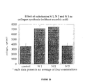
- microspheres The effect of microspheres on myoblast fusion was also examined. Results are given in Table 1. Generally, cells treated with microspheres exhibited about 150% fusion rate as compared to controls. However, the extent of this effect depended upon the type of microspheres and the length of treatment.
- microspheres tested are given in Table 1.
- the diameter of the microspheres is given in microns under "Diameter”.
- the surface groups on the polystyrene beads are given under "Surface Group”.
- Polystyrene beads without any further derivatization are "polystyrene”.
- Beads derivatized with either carboxyl or amino surface groups are described as “carboxy” and “amino”, respectively.
- concentration of beads is given as number of beads per ml of media under "Conc.”
- Cells were prepared, fixed and stained as for determining the rate of proliferation of myoblasts, described above. Cells were initially plated at the density given in Table 1 as cells per ml media, under the column "Initial Cells”. The measurements of myoblast fusion were made after the given number of days after treatment under "Days after Treatment”.
- the extent of fusion is calculated as the proportion of nuclei within multinuclear cells, or myosimplasts, related to the total amount of nuclei within the microscopic field, given as "Proportion of Fusion” for microsphere treated cells, and "Control Fusion” for control, untreated cells. At least 400 nuclei were counted for each experimental condition. The ratio of the extent of fusion in microsphere treated cells and control, untreated cells is given as "Relative Effect". If no value is given for a particular slot in Table 1, the value is the same as that in the row above.
- microspheres As can be seen from Table 1, all of the different types of microspheres promoted myoblast cell fusion, although the extent of the effect depended upon the diameter of the microsphere, the surface group on the microsphere, the number of days after treatment and the concentration.
- Myoblast fusion occurs when muscle tissue is formed during embryogenesis, and is also a very important step in muscle regeneration and repair of damaged muscle tissue.
- the ability of microspheres to promote such fusion clearly indicates the potential of these microspheres to promote muscle regeneration, as demonstrated in Example 5 below.
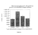
- collagen synthesis and deposition is an important step in the process of wound healing. Furthermore, the amount of collagen deposited in the wound is an important determinant of wound strength.
- the microspheres of the present invention clearly have a variety of effects on different cell types, as demonstrated in the preceding and following Examples, clearly one important determinant of the ability of a composition to promote wound healing is its effect on collagen synthesis and deposition.
- the microspheres of the present invention clearly promote collagen synthesis by cultured fibroblasts.
- Type I microspheres had a diameter of 4.5 microns, was made of carboxylated polystyrene and had a Z potential of about -29.96 mV.
- Type II microspheres had a diameter of 0.49 microns, were made of polystyrene alone and had a Z potential of about -34.5 mV.
- Type III microspheres had a diameter of 1.0 microns, were made of carboxylated polystyrene and had a Z potential of about -53.34 mV.
- the experimental method was as follows.
- Foreskin fibroblast cultures were grown in 75 cm 2 plastic flasks (Coming Glass Works, Coming, NY) in Dulbecco's modified Eagle medium (DMEM) containing 4.5 mg/ml glucose supplemented with 10% vol/vol fetal calf serum, 2 mM L-glutamine, 50 ⁇ g/ml gentamycin sulfate and 2.5 mg/ml amphotericin B. The cultures were incubated at 37°C in 5% CO 2 until confluent.
- DMEM Dulbecco's modified Eagle medium
- Fibroblasts were harvested using 0.25% trypsin/0.05% EDTA solution and subcultured in 24-well plates at a density of 200,000 cells/well with the same media for 24 hours, at which time treated cells were incubated with Type I, II or III microspheres. Control cells were incubated with media alone.
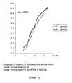
- Collagen synthesis was measured as follows. The cultured fibroblasts were preincubated in DMEM supplemented with 0.5% dialyzed fetal calf serum for 24 hours. Cells were labeled with 3 ⁇ Ci 2,3- 3 H-proline or 3,4- 3 H-proline solution containing ⁇ -aminopropionitrile fumarate (BAPN) at a final concentration of 100 ⁇ M, in the presence ( Figure 2A) or absence ( Figure 2B) of 10 ⁇ M ascorbic acid as indicated. Ascorbic acid promotes collagen synthesis in fibroblasts and is an important stimulation factor.
- BAPN ⁇ -aminopropionitrile fumarate
- Type I and Type II microspheres were able to stimulate collagen synthesis above the level seen in control (untreated) fibroblasts, both in the presence ( Figure 2A) and absence ( Figure 2B) of ascorbic acid.
- Type I microspheres had a greater effect relative to Type II microspheres in the presence of ascorbic acid, although both types had a similar effect in the absence of ascorbic acid.
- Type III microspheres did not have a detectable effect on collagen synthesis either in the presence or absence of ascorbic acid.
- Type I and Type II microspheres had an effect, while Type III microspheres did not, indicating that the specific size and material of the microspheres is important. Furthermore, both Type I and Type II microspheres elicited an effect even in the absence of ascorbic acid, indicating that these two types of microspheres can potentiate collagen synthesis even in the absence of other stimulatory factors. Thus, clearly both Type I and Type II microspheres have a substantial stimulatory effect on collagen synthesis.
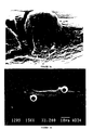
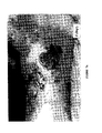
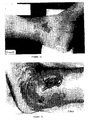
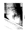
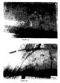
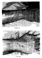
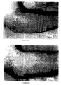
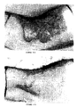
- rat myoblasts Primary cell cultures of rat myoblasts were prepared as described in Example 1 above. Cells were then incubated with polystyrene microspheres (treated cells) or without (control cells) for 48 hours. Cells were then fixed in 1% glutaraldehyde in phosphate buffered saline for 1-4 days, and rinsed in PBS. Cells were then transferred to a solution of 1% tannic acid and 1% guanidine HCl (1:1 ratio) in PBS for 1 hour. Specimens were post-fixed in 1% OsO 4 for 1 hour and dehydrated in graded ethanol and Freon 113 at room temperature. Specimens were then mounted on slides, coated with gold and examined in a JEOL T-300 scanning electron microscope at 2 kV.
- Figures 3A-3C illustrate the effect of the microspheres of the present invention on myoblast shape.
- the cell in Figure 3A has grown over the microsphere, so that part of the cell surface is convex rather than flat.
- Figures 3B and 3C show cells extending pseudopodia from a portion of the cell on which the microsphere rests.
- the pseudopod of the cell in Figure 3C is particularly pronounced, showing that the microspheres clearly influence myoblast shape.
- the formation and extension of a pseudopod clearly requires changes in the cytoskeletal structure, demonstrating that the microspheres also affect the cytoskeleton of the cell.
- the formation of such pseudopodia may be important for the migration of cells into the wound area.
- the stimulation of such pseudopodia by the microspheres indicates their ability to promote another important step in the wound healing process.
- the following description is a general device and method for application of the agents for wound healing.
- the agents such as microspheres, are preferably applied repeatedly to the wound to be treated.
- the frequency of application, and the concentration applied is dependent on the severity of the symptoms and on the responsiveness of the subject to the treatment. Persons of ordinary skill in the art can easily determine optimum concentrations, dosing methodologies and repetition rates.
- the microspheres were applied to the wound to be treated about once or twice per day, although of course other application rates are possible.
- the method includes the step of administering the agents such as microspheres, in a pharmaceutically acceptable carrier in which the agents are substantially insoluble, to a subject to be treated.
- pharmaceutically acceptable carriers include aqueous media for a suspension of agents, non-aqueous media such as ointments, creams and aerosol-forming material, as well as bandages soaked in, or otherwise containing, media with the agents.
- one particularly preferred pharmaceutically acceptable carrier is a gel forming material such as methyl cellulose, such that the microspheres are in a gel form.
- methyl cellulose is a particulary preferred example of such a gel forming material, it is understood that other such gel forming materials could also be used, especially if these gel forming materials were substantially physiologically inert.
- the bandages can be occlusive or non-occlusive.
- the agents which are in a pharmaceutically acceptable carrier can be described as a dispersion of agents.
- the agents are administered according to an effective dosing methodology, preferably until a predefined endpoint is reached, such as the absence of clinical symptoms in the subject.
- a predefined endpoint such as the absence of clinical symptoms in the subject.
- the closure of the wound to be treated is an example of such an endpoint.
- the device of the present invention includes a composition with one or more agents and a pharmaceutically acceptable carrier for the agents, and a container for containing the composition.
- the container is a substantially sealed, sterile container, such as an aerosol-dispersing pump or a spray can.
- the container is a substantially sterile bandage.
- the container is a squeezable tube or a gel-dispersing pump.
- the microspheres of the present invention can even be used without a carrier, simply by being directly placed on the wound, the extremely low concentrations of microspheres which are effective for wound healing are much easier to apply when present in a pharmaceutical carrier.
- the microspheres are present at a concentration in a range of from about 0.0001 percent to 1.5 percent, weight per weight. More preferably, the microspheres are present at a concentration in a range of from about 0.001 percent to 1.0 percent, weight per weight. Most preferably, the microspheres are present at a concentration in a range of from about 0.01 percent to about 0.2 percent, weight per weight.
- Such very low concentrations are most efficient to store, to transport, and especially to apply, when in a suitable pharmaceutical carrier contained within a suitable container.
- a preferred carrier and container combination is an aqueous or other liquid suspension contained within an aerosol-dispersing pump.
- a particularly preferred combination of a carrier and a container is a gel forming material, such methyl cellulose or any other suitable gel-forming substance, contained within a squeezable tube.
- the gel form is particularly preferred because it increases retention of the microspheres within the wound area, thereby enabling a lower amount of the gel formulation to exert the same wound healing effect. Such an effect was demonstrated in Example 9 below.
- particular preferred combinations of a carrier and a container are clearly efficient and convenient for the transportation, storage and application of the compositions of the present invention, especially at these very low preferred concentrations of microspheres.
- the agents such as microspheres
- the microspheres are first applied in a dispersion to the wound, by dripping, spraying, painting, washing or by any other suitable method of topical application.
- 30 sec to 2 minutes are allowed to elapse before the second step, in order to allow the microspheres to form initial contact with the wound.
- the second step includes applying an occlusive or non-occlusive bandage, or other suitable covering soaked in the liquid suspension containing the microspheres, to the wound. This substantially reduces or eliminates absorption of the microspheres by the bandage or covering. This method was used both in rats and humans for wound healing, as described in the Examples below.
- microspheres in the suspension did not aggregate, coalesce, clump or undergo irreversible caking. Although the microspheres did settle somewhat over time, they were easily resuspended with gentle agitation.
- the microspheres of the present invention promote various in vitro cell processes which are important for wound healing.
- in vitro and in vivo effects do not always correlate. Therefore, in vivo experiments were performed to assess the ability of the microspheres to promote wound healing in rats.
- Figures 4A-4D the microspheres of the present invention clearly promote wound healing in rats.
- Figure 5 is a graph of the rate at which the wound area decreases, showing that the microspheres of the present invention increase the rate at which such a decrease occurs.
- Table 2 shows that the microspheres promote muscle regeneration in rats. The experimental method was as follows.
- Tibialis anterior muscle Male Wistar rats, weighing between 300 and 400 g, were anesthetized by nembutal (5 mg/kg of body weight). An excision injury to the lateral parts of the Tibialis anterior muscle was performed as follows. First, a longitudinal incision was made in the skin to expose the Tibialis anterior muscle. Next, the partial excision of this muscle was made by a transverse cut of the muscle fibers, along about half of the muscle width. The excised piece was then cut out of the muscle, leaving a gap of about 5 mm by 5 mm in the muscle. In all rats the same amount of excised tissue (80 ⁇ 10 mg) was removed from precisely the same location in the muscle. The wound area was then dressed with 2 micron polystyrene microspheres in saline for treated rats, and saline alone for control rats. The wound area was measured for between 3 and 15 days following injury.
- Figures 4A-4D show pictures of wound areas prepared as described above.
- Figure 4A shows the wound of the control rat immediately after injury
- Figure 4B shows the equivalent wound of the rat to be treated
- Figures 4C and 4D show the same rats five days after injury.
- the wound of the control rat was treated with saline alone, and still has not completely healed.
- the wound of the treated rat, treated with microspheres has completely healed.
- the microspheres of the present invention promote faster wound healing.
- Figure 5 further illustrates the promotion of wound healing by the microspheres of the present invention.
- the wounds of control rats eventually heal, but at a much slower rate than the wounds of treated rats.
- the microspheres clearly increase the rate at which the wound area decreases and the wound heals.
- the microspheres of the present invention clearly promoted muscle regeneration, as measured by the number of "new" or incorporated nuclei in muscle fibers.
- the fact that such measurements were made on histological samples taken from rats treated in vivo also indicates that the microspheres promote muscle regeneration in vivo as well as in vitro .
- Figure 6 compares the effect of the microspheres of the present invention on wound healing with tissue culture media and saline in rats. Wounds were induced in rats as described above, and the rats were treated with saline alone (Figure 6A, ), tissue culture media alone ( Figure 6B, ), saline plus microspheres (Figure 6A, ) or tissue culture media plus microspheres ( Figure 6B, ). The rats were then photographed 4 days after wounding occurred. As can be seen from Figures 6A and 6B, the microspheres were able to induce a much more rapid rate of wound healing regardless of whether the carrier was saline or tissue culture media. Thus, tissue culture media was not responsible for any part of the effect of the microspheres of the present invention on wound healing.
- the first case study was that of a 66-year old female with ulcers in the left leg which refused to heal. The patient also had cellulitis of the left leg and varicose veins in both legs. Ulcers on the inner thigh of the patient were treated with Milton 2% which is a corrosive chlorine salt in water. Ulcers on the outer thigh of the patient were treated with 4.5 micron microspheres of the present invention made from polystyrene in tissue culture medium.
- Figure 7A shows the control wound at day 0, while Figure 7B shows the control wound after 4 months of treatment.
- Figure 7C shows the treated wound at day 0, while Figure 7D shows the treated wound after 4 months of treatment.
- Figures 7A and 7B the wound which had served as a control for Figure 7 above ( Figures 7A and 7B) was treated with the same microspheres as those used to treat the wound in Figures 7C and 7D.
- Figures 8A and 8B show the wound at day 0 of treatment with microspheres
- Figure 8B shows the wound after 21 days of treatment.
- the extent of the wound has decreased, even after such a short time period.
- the wound was superficial and clean, and was no longer producing exudations.
- the second case study was that of a 52-year old female who had a year-old infected wound on the front side of the left thigh.
- the wound was treated with 1% Milton for a week, debrided and then treated with the microspheres of Figures 7 and 8 for 10 days.
- Figure 9A shows the wound at day 0 of treatment
- Figure 9B shows the wound after 10 days of treatment.
- the wound After 10 days, the wound showed a significant improvement. It had decreased in extent to a small size, was clean and was no longer producing exudations, as can be seen from Figure 9B. Although the wound did not fully close during the relatively short treatment period, its effects had been significantly ameliorated.
- the third case study was that of a 19-year old male who was injured by a chemical spill in an industrial workplace accident.
- the chemicals in question sulfurides, caused severe bums and blistering on the right side of his neck and right hand.
- All wounds were treated with Silverol, a hydrogel with strong absorptive properties.
- the wounds on the right forearm were treated with the microspheres of case studies 1 and 2, while the remaining wounds were treated with Silverol.
- the results are shown in Figures 10A and 10B (control wound at day 0 and day 5, respectively), and in Figures 10C and 10D (treated wound at day 0 and day 5, respectively).
- the condition of the treated wound on the forearm had improved significantly over that of the remaining wounds, which were not treated with microspheres.
- the wound on the forearm had completely healed after 5 days of treatment with microspheres.
- the remaining wounds which were treated with Silverol had not healed completely.
- the microspheres clearly promoted wound healing, demonstrating a greater efficacy than currently available treatments.
- the fourth case study was of a 52-year old female who had sustained second-degree bums on the buttocks from a hot bath. Wounds on the left buttock were treated with Silverol, while those on the right buttock were treated with microspheres of the previous case studies. The results are shown, for microsphere-treated wounds only, in Figures 11A (day 0) and 11B (day 7) of treatment.
- the wounds on the right buttock which were treated with microspheres, had completely healed with good epithelial growth.
- the wounds on the left buttock, which were treated with Silverol had not completely healed and were closing relatively slowly.
- the microspheres were able to promote wound healing at a more rapid rate than conventional treatments.
- the fifth case study was of a 28 year old female who had suffered extensive and severe sunburn (data not shown). She was treated with the microspheres of the previous case studies. The patient reported both a significant reduction in discomfort and rapid healing of the sunburn.
- the microspheres used in the method and device of the present invention can both relieve discomfort and promote wound healing, although it should be noted that the relief of discomfort is probably a highly indirect effect of the microspheres rather than direct analgesia.
- the above patient report can only be inferred to include the apparent reduction in the sensation of discomfort from the sunburn.
- Such decreased discomfort probably does not demonstrate any ability of the microspheres to have a direct effect on the transmission of nerve impulses, or indeed to directly alter any of the many factors which lead to the sensation of discomfort. Instead, this effect is probably highly indirect, occurring as a result of the activation of macrophages, which in turn has anti-inflammatory effects, leading to the decreased sensation of discomfort by the patient.
- the sixth case study was of a 64 year old male with diabetis mellitus and a post-surgical wound on the left leg which had not healed with conventional treatment eight months after surgery.
- the microspheres of the previous case studies were applied twice daily for 17 days.
- the post-surgical wound was 7.5 cm by 8 cm and was 6 cm deep, which is the depth at which the bone is located in that portion of the leg, and was marked by gangrene of the soft tissue and of the bone.
- the gangrene was ameliorated and was totally eliminated after eight days (data not shown). Granulation appeared on the muscle and bone after three days of treatment. Bone regeneration was complete after ten days of treatment.
- the use of the agents, such as microspheres, according to the present invention has clearly been shown to have greater efficacy for the promotion of wound healing and muscle regeneration than currently available, prior art treatments.
- the method and device promotes, accelerates and enhances wound healing, as well as diminishing discomfort experienced by the subject.
- the microspheres of the present invention would be highly suitable for many different cosmetic applications.
- the microspheres of the present invention are contemplated as useful for the amelioration or even substantially complete cure of acne.
- the microspheres of the present invention could also be incorporated into shaving cream or gel, for the treatment of minor skin damage such as small nicks and cuts associated with shaving.
- the microspheres of the present invention are also contemplated as useful for the amelioration or even substantially complete cure of substantially any type of skin damage.
- microspheres of the present invention are additionally contemplated as being useful for the amelioration or even substantially complete cure of hemorrhoids and of intestinal ulcers, such as gastric ulcers, ulcers associated with ulcerative colitis and ulcers associated with Helicobacter pylori.
- methyl cellulose Hoechst A.G., Germany
- the methyl cellulose was used at a concentration in a range of from about 0.1 percent to about 5 percent weight per weight.
- the microspheres in gel format were found to be highly effective for wound healing, yet smaller amounts of the gel formulation could still exert the same healing effect because the gel formulation helped to retain the microspheres in the wound area.
- the experimental method was as follows.
- Hartley-derived albino guinea pigs weighing 300-400 grams were used in this study. All surgical procedures were performed under general anesthesia after administration of ketamine HCl (150 mg/kg, Parke-Davis, USA). After being anesthetized, four bilaterally symmetrical full-thickness skin segments measuring 2 cm by 2 cm were excised from the dorsum of each animal, two from the scapular region and two from the lumbar region.
- microspheres in gel form were applied to the wounds after surgery. Bandages were then applied to the wounds.
- the dressings were changed every 48 hours under general anesthesia, at which time all wounds were washed with warm saline to remove any debris and any remaining gel within the wound. All wounds were then measured and photographed.
- the microspheres in gel form were then applied to the wounds again, and fresh bandages were applied.
- the gel formulation was also placed on the bandages. All four wounds on each animal were treated with the same concentration of microspheres in gel form. The concentration of microspheres was 0.0027 percent weight per weight in 2.5% methyl cellulose.
- the microspheres themselves were polystyrene and were 4.3 microns in diameter.
- the wounds of some animals were treated with the same concentration of the same microspheres in a liquid suspension, which was also placed on the bandages.
- microspheres in the liquid suspension or in the gel formulation were applied in a sufficient volume to substantially fill the wound area, such that a smaller amount of the gel formulation than of the liquid suspension was applied.
Landscapes
- Health & Medical Sciences (AREA)
- Engineering & Computer Science (AREA)
- Chemical & Material Sciences (AREA)
- Veterinary Medicine (AREA)
- Life Sciences & Earth Sciences (AREA)
- Animal Behavior & Ethology (AREA)
- General Health & Medical Sciences (AREA)
- Public Health (AREA)
- Epidemiology (AREA)
- Materials Engineering (AREA)
- Manufacturing & Machinery (AREA)
- Chemical Kinetics & Catalysis (AREA)
- Medicinal Chemistry (AREA)
- Hematology (AREA)
- Microelectronics & Electronic Packaging (AREA)
- Bioinformatics & Cheminformatics (AREA)
- Pharmacology & Pharmacy (AREA)
- Organic Chemistry (AREA)
- Nuclear Medicine, Radiotherapy & Molecular Imaging (AREA)
- General Chemical & Material Sciences (AREA)
- Dermatology (AREA)
- Medicinal Preparation (AREA)
- Pharmaceuticals Containing Other Organic And Inorganic Compounds (AREA)
- Materials For Medical Uses (AREA)
- Basic Packing Technique (AREA)
- Replacement Of Web Rolls (AREA)
- Medicines That Contain Protein Lipid Enzymes And Other Medicines (AREA)
Applications Claiming Priority (3)
| Application Number | Priority Date | Filing Date | Title |
|---|---|---|---|
| US08/868,950 US5861149A (en) | 1997-06-04 | 1997-06-04 | Methods for wound treatment |
| US868950 | 1997-06-04 | ||
| PCT/IL1998/000260 WO1998055108A1 (en) | 1997-06-04 | 1998-06-04 | Device and methods for wound treatment |
Publications (3)
| Publication Number | Publication Date |
|---|---|
| EP0988029A1 EP0988029A1 (en) | 2000-03-29 |
| EP0988029A4 EP0988029A4 (en) | 2001-02-21 |
| EP0988029B1 true EP0988029B1 (en) | 2005-04-06 |
Family
ID=25352623
Family Applications (1)
| Application Number | Title | Priority Date | Filing Date |
|---|---|---|---|
| EP98924538A Expired - Lifetime EP0988029B1 (en) | 1997-06-04 | 1998-06-04 | Device and methods for wound treatment |
Country Status (12)
| Country | Link |
|---|---|
| US (2) | US5861149A (enExample) |
| EP (1) | EP0988029B1 (enExample) |
| JP (1) | JP4594454B2 (enExample) |
| KR (1) | KR100589064B1 (enExample) |
| AT (1) | ATE292454T1 (enExample) |
| AU (1) | AU764113B2 (enExample) |
| BR (1) | BRPI9809725B1 (enExample) |
| CA (1) | CA2292528C (enExample) |
| DE (1) | DE69829662T2 (enExample) |
| ES (1) | ES2241145T3 (enExample) |
| IL (1) | IL133162A0 (enExample) |
| WO (1) | WO1998055108A1 (enExample) |
Families Citing this family (75)
| Publication number | Priority date | Publication date | Assignee | Title |
|---|---|---|---|---|
| US6958148B1 (en) * | 1998-01-20 | 2005-10-25 | Pericor Science, Inc. | Linkage of agents to body tissue using microparticles and transglutaminase |
| US6919076B1 (en) | 1998-01-20 | 2005-07-19 | Pericor Science, Inc. | Conjugates of agents and transglutaminase substrate linking molecules |
| US6872384B1 (en) | 1998-02-23 | 2005-03-29 | Life Medical Sciences, Inc. | Treatment of trauma |
| US6660301B1 (en) | 1998-03-06 | 2003-12-09 | Biosphere Medical, Inc. | Injectable microspheres for dermal augmentation and tissue bulking |
| MXPA00000009A (es) * | 1998-06-22 | 2005-09-08 | Autologous Wound Therapy Inc | Solicitud para patente de utilidad para curativo de heridas mejorado enriquecido con plaquetas. |
| EP1041970B1 (en) * | 1998-10-23 | 2013-07-03 | Polyheal Ltd. | Compositions of microspheres for wound healing |
| US7338657B2 (en) * | 2001-03-15 | 2008-03-04 | Biosphere Medical, Inc. | Injectable microspheres for tissue construction |
| US6436424B1 (en) * | 2000-03-20 | 2002-08-20 | Biosphere Medical, Inc. | Injectable and swellable microspheres for dermal augmentation |
| AU2001249221A1 (en) | 2000-03-20 | 2001-10-03 | Biosphere Medical, Inc. | Injectable and swellable microspheres for tissue bulking |
| US20030212022A1 (en) * | 2001-03-23 | 2003-11-13 | Jean-Marie Vogel | Compositions and methods for gene therapy |
| CN101708165A (zh) | 2000-03-24 | 2010-05-19 | 生物领域医疗公司 | 用于主动栓塞术的微球体 |
| US6599526B2 (en) * | 2000-08-18 | 2003-07-29 | The University Of North Texas Health Science Center At Fort Worth | Pericardial anti-adhesion patch |
| US20030007951A1 (en) * | 2000-08-23 | 2003-01-09 | Richard Franklin | Treatment of trauma |
| AU2000269264A1 (en) * | 2000-08-23 | 2002-03-04 | Eidgenossische Technische Hochschule Zurich (Eth) | Treatment of trauma, and other indications |
| US9089407B2 (en) | 2001-04-23 | 2015-07-28 | Massachusetts Institute Of Technology | Antibacterial coatings that inhibit biofilm formation on implants |
| EP1390158A4 (en) * | 2001-04-23 | 2004-06-09 | Massachusetts Inst Technology | ANTIMICROBIAL POLYMER SURFACES |
| US7151139B2 (en) * | 2001-04-23 | 2006-12-19 | Massachusetts Institute Of Technology | Antimicrobial polymeric surfaces |
| FR2826278B1 (fr) * | 2001-06-20 | 2005-03-25 | Lipha | Utilisation d'agents antidiabetiques pour fabriquer un medicament ayant un effet cicatrisant |
| DE10141650C1 (de) | 2001-08-24 | 2002-11-28 | Lohmann Therapie Syst Lts | Transdermales Therapeutisches System mit Fentanyl bzw. verwandten Substanzen |
| ES2207387B1 (es) * | 2002-02-28 | 2005-07-16 | Consejo Sup. Investig. Cientificas | Composicion quimica de igf-i para el tratamiento y prevencion de enfermedades neurodegenerativas. |
| WO2004004671A1 (en) * | 2002-07-02 | 2004-01-15 | Procyte Corporation | Compositions containing peptide copper complexes and soft tissue fillers |
| CN1671400A (zh) * | 2002-07-03 | 2005-09-21 | 派瑞克科学公司 | 透明质酸组合物以及使用方法 |
| US8016881B2 (en) * | 2002-07-31 | 2011-09-13 | Icon Interventional Systems, Inc. | Sutures and surgical staples for anastamoses, wound closures, and surgical closures |
| US20040043947A1 (en) * | 2002-08-29 | 2004-03-04 | Gersh Steven A. | Methods of treating wounds or ulcers of the foot, ankle or lower extremity of the leg |
| US7465463B2 (en) * | 2002-09-04 | 2008-12-16 | Polyheal, Ltd. | Compositions comprising microspheres with anti-inflammatory properties for healing of ocular tissues |
| AU2003270802A1 (en) * | 2002-09-20 | 2004-04-08 | The Children's Hospital Of Philadelphia | Engineering of material surfaces |
| US8975295B2 (en) * | 2002-10-25 | 2015-03-10 | Precision Dermatology, Inc. | Modulation of zinc levels to improve tissue properties |
| JP2004298556A (ja) * | 2003-04-01 | 2004-10-28 | Mitsubishi Rayon Co Ltd | 高濃度炭酸温水浴による血管新生療法 |
| RU2257211C1 (ru) * | 2004-02-10 | 2005-07-27 | Дагестанская государственная медицинская академия | Способ лечения гнойных ран |
| US8697139B2 (en) | 2004-09-21 | 2014-04-15 | Frank M. Phillips | Method of intervertebral disc treatment using articular chondrocyte cells |
| US8663225B2 (en) | 2004-11-12 | 2014-03-04 | Medtronic, Inc. | Hydrogel bone void filler |
| EP1690558A1 (en) * | 2005-02-11 | 2006-08-16 | NOLabs AB | Device for treatment of diabetic disorders |
| US20060193818A1 (en) * | 2005-02-25 | 2006-08-31 | Southall Michael D | Compositions containing amines and use thereof |
| US8221046B2 (en) | 2005-02-25 | 2012-07-17 | Johnson & Johnson Consumer Companies, Inc. | Compositions containing amines and use thereof |
| US20060193777A1 (en) * | 2005-02-25 | 2006-08-31 | Southall Michael D | Method of screening compounds for potential efficacy for the treatment of signs of aging |
| US20060193814A1 (en) * | 2005-02-25 | 2006-08-31 | Eduardo Ruvolo | Compositions for the treatment of signs of aging |
| US8278359B2 (en) * | 2005-02-25 | 2012-10-02 | Johnson & Johnson Consumer Companies, Inc. | Compositions containing amines and use thereof to treat acne or reduce the appearance of oil or pores on the skin |
| US8226926B2 (en) | 2005-05-09 | 2012-07-24 | Biosphere Medical, S.A. | Compositions and methods using microspheres and non-ionic contrast agents |
| US20070049739A1 (en) * | 2005-08-30 | 2007-03-01 | Biomet Manufacturing Corp. | Method and system for extracting blood-derived growth factors |
| US7547434B2 (en) * | 2005-09-09 | 2009-06-16 | Johnson & Johnson Consumer Companies, Inc. | Compositions and methods for mitigating skin irritation |
| US20070092494A1 (en) * | 2005-10-26 | 2007-04-26 | Biomet Manufacturing Corp. | Composition for wound healing using lyophilized skin or skin-derived collagen |
| CA2634045C (en) * | 2005-12-20 | 2019-05-07 | Swiss-American Products, Inc. | Protease compositions for the treatment of damaged tissue |
| CU23388B6 (es) * | 2006-01-31 | 2009-07-16 | Ct Ingenieria Genetica Biotech | Composición farmacéutica de microesferas para prevenir la amputación del pie diabético |
| CU23558A1 (es) | 2006-02-28 | 2010-07-20 | Ct Ingenieria Genetica Biotech | Compuestos análogos a los secretagogos peptidicos de la hormona de crecimiento |
| US20100136072A1 (en) * | 2006-11-08 | 2010-06-03 | Massachusetts Institute Of Technology | Polymeric Coatings that Inactivate Viruses and Bacteria |
| US20090104160A1 (en) * | 2007-02-01 | 2009-04-23 | Moraga Biotechnology Corporation | Mobilization of Stem Cells After Trauma and Methods Therefor |
| WO2008139123A2 (fr) * | 2007-05-11 | 2008-11-20 | Galderma Research & Development | Preparations pharmaceutiques ou cosmetiques pour application topique et/ou parenterale, leurs procedes de preparation, et leurs utilisations |
| US20100323985A1 (en) * | 2007-05-11 | 2010-12-23 | Marc Moutet | Pharmaceutical or cosmetic preparations for topical and/or parenteral application, preparation methods thereof and use of same |
| USRE46397E1 (en) | 2007-11-07 | 2017-05-09 | Svip5 Llc | Slow release of organic salts of local anesthetics for pain relief |
| US20110244006A1 (en) * | 2007-11-29 | 2011-10-06 | The Trustees Of Columbia University In The City Of New York | Microsphere skin treatment |
| CA2710822C (en) | 2008-01-07 | 2022-06-14 | Johannes Gerhardus Maria Bolscher | Use of histatin derived peptides for promoting wound healing |
| US9115180B2 (en) | 2008-01-07 | 2015-08-25 | Rapid Pathogen Screening, Inc. | Use of peptides for promoting wound healing |
| WO2009097561A1 (en) * | 2008-01-30 | 2009-08-06 | Doheny Eye Institute | Wound closing compounds with additives |
| EP3574909A1 (en) | 2008-01-30 | 2019-12-04 | Imbed Biosciences, Inc. | Methods and compositions for wound healing |
| US9174839B2 (en) * | 2008-01-30 | 2015-11-03 | Imbed Biosciences, Inc. | Methods and compositions for wound healing |
| IL192262A (en) * | 2008-06-17 | 2016-05-31 | Z H T Eng Equipment And Tech Ltd | A polymer that releases drugs into the animal body, a medicinal product containing it and a method for its preparation |
| US20100197826A1 (en) * | 2009-01-30 | 2010-08-05 | Rajat Agrawal | Wound Closing Compounds With Additives |
| BRPI0900858A2 (pt) * | 2009-04-23 | 2011-05-03 | Clinica De Andrologia E Urologia Vera Cruz Ltda | composição farmacêutica carreadora de substáncias ativas |
| EP2498798A4 (en) * | 2009-11-10 | 2014-01-01 | Univ Columbia | COMPOSITIONS AND METHODS OF WOUND TREATMENT |
| DK2521535T3 (en) | 2010-01-05 | 2018-06-25 | Cell Construct I Llc | BIOMATERIALS MANUFACTURED FROM HUMAN HAIR |
| JP5787246B2 (ja) * | 2010-09-15 | 2015-09-30 | 国立大学法人京都大学 | 角化細胞遊走・増殖促進剤を含む、創傷治癒剤および褥瘡(床ずれ)治療薬 |
| NZ612801A (en) | 2010-12-27 | 2014-12-24 | Intellicell Biosciences Inc | Ultrasonic cavitation derived stromal or mesenchymal vascular extracts and cells derived therefrom obtained from adipose tissue and use thereof |
| US20130045269A1 (en) * | 2011-08-18 | 2013-02-21 | Sorush LLC | Formulations and methods for wound treatment |
| WO2013074526A2 (en) | 2011-11-15 | 2013-05-23 | Byocoat Enterprises, Inc. | Antimicrobial compositions and methods of use thereof |
| US20130225904A1 (en) * | 2012-02-29 | 2013-08-29 | University Of Virginia | System and method for magnetic control of an anesthetic |
| US20130310327A1 (en) | 2012-05-18 | 2013-11-21 | Rapid Pathogen Screening, Inc. | Histatin for Corneal Wound Healing and Ocular Surface Disease |
| WO2014041543A1 (en) * | 2012-09-13 | 2014-03-20 | Polyheal Ltd. | Improved wound healing compositions comprising microspheres |
| CA2908346C (en) * | 2013-06-28 | 2017-07-04 | Bsn Medical, Inc. | Orthopedic cast and splint bandages with encapsulated hardening medium and method |
| US9226890B1 (en) | 2013-12-10 | 2016-01-05 | Englewood Lab, Llc | Polysilicone base for scar treatment |
| WO2018023021A1 (en) | 2016-07-29 | 2018-02-01 | Imbed Biosciences Inc. | Methods and compositions for wound healing |
| JP6456897B2 (ja) | 2016-10-05 | 2019-01-23 | マルハニチロ株式会社 | 抗菌性及び創傷治癒促進性を有する創傷治癒剤 |
| KR101761073B1 (ko) * | 2016-11-15 | 2017-07-25 | 주식회사 메타바이오메드 | 폴리펩타이드 금속불활성화제, 이를 이용한 의료용 고분자 화합물 및 그 제조방법 |
| US9993578B1 (en) * | 2017-01-12 | 2018-06-12 | Lorstan Pharmaceutical, LLC | Silicone oil-in-water composition useful as an injectable filler and as a scaffold for collagen growth |
| WO2020111743A1 (ko) * | 2018-11-26 | 2020-06-04 | 한국해양과학기술원 | 지방 생성을 자극하는 펩타이드, 조성 및 방법 |
| EP3890756A4 (en) | 2018-12-03 | 2022-09-07 | Ross Ward | PERIVASCULAR CELLS OF INDIRECT ULTRASONIC CAVITATION AND THEIR USE |
Family Cites Families (20)
| Publication number | Priority date | Publication date | Assignee | Title |
|---|---|---|---|---|
| US3842830A (en) * | 1973-03-26 | 1974-10-22 | T Hargest | Surgical dressing and method of forming the same |
| US4380855A (en) * | 1980-01-18 | 1983-04-26 | University Of Rochester | Method for filling hollow shells with gas for use as laser fusion targets |
| US4273871A (en) * | 1980-03-03 | 1981-06-16 | Monsanto Company | Production of angiogenic factor by cell culture |
| DE3312431A1 (de) * | 1983-04-07 | 1984-10-11 | B. Braun Melsungen Ag, 3508 Melsungen | Pvp-iod-wundpuder mit synergistisch wirkender pudergrundlage |
| US4772591A (en) * | 1985-09-25 | 1988-09-20 | Peritain, Ltd. | Method for accelerated wound healing |
| FR2601371B1 (fr) * | 1986-07-11 | 1989-05-12 | Merieux Inst | Procede de traitement du collagene en vue, notamment, d'en faciliter la reticulation et collagene obtenu par application dudit procede |
| US4889530A (en) * | 1986-10-06 | 1989-12-26 | University Of Akron | Wound dressing containing acrylate or methacrylate hydrogel polymer |
| US4781921A (en) * | 1986-10-06 | 1988-11-01 | The University Of Akron | Hydrogels of quadrol methacrylate polymers |
| JPS63160661A (ja) * | 1986-12-24 | 1988-07-04 | テルモ株式会社 | 外用組成物 |
| US5658894A (en) * | 1989-04-23 | 1997-08-19 | The Trustees Of The University Of Pennsylvania | Compositions for inhibiting restenosis |
| US5092883A (en) * | 1988-12-28 | 1992-03-03 | Eppley Barry L | Method for promoting soft connective tissue growth and repair in mammals |
| FR2648465B1 (fr) * | 1989-06-15 | 1991-09-13 | Oreal | Procede de preparation de poly (beta)-alanine reticulee, en une seule etape, a partir d'acrylamide et d'un compose polyfonctionnel copolymerisable |
| FR2649888B1 (fr) * | 1989-07-18 | 1994-08-26 | Exsymol Sa | Produits pour applications cutanees, a effets cosmetiques ou/et therapeutiques |
| US5196185A (en) * | 1989-09-11 | 1993-03-23 | Micro-Collagen Pharmaceutics, Ltd. | Collagen-based wound dressing and method for applying same |
| DE4293451T1 (de) * | 1991-09-13 | 1994-09-08 | Gillette Canada | Polymerpartikel für zahnmedizinische Anwendungen |
| US5837747A (en) * | 1991-10-29 | 1998-11-17 | Vivorx, Inc. | Crosslinkable polysaccharides, polycations and lipids useful for encapsulation and drug release |
| GB9210574D0 (en) * | 1992-05-18 | 1992-07-01 | Ca Nat Research Council | Biotherapeutic cell-coated microspheres for wound/burn and prothesis implant applications |
| GB9225581D0 (en) * | 1992-12-08 | 1993-01-27 | Courtaulds Plc | Wound dressings |
| GB2281861B (en) * | 1993-09-21 | 1997-08-20 | Johnson & Johnson Medical | Bioabsorbable wound implant materials containing microspheres |
| US5519020A (en) * | 1994-10-28 | 1996-05-21 | The University Of Akron | Polymeric wound healing accelerators |
-
1997
- 1997-06-04 US US08/868,950 patent/US5861149A/en not_active Expired - Lifetime
-
1998
- 1998-06-04 WO PCT/IL1998/000260 patent/WO1998055108A1/en not_active Ceased
- 1998-06-04 CA CA002292528A patent/CA2292528C/en not_active Expired - Fee Related
- 1998-06-04 JP JP50199699A patent/JP4594454B2/ja not_active Expired - Lifetime
- 1998-06-04 AU AU76718/98A patent/AU764113B2/en not_active Ceased
- 1998-06-04 BR BRPI9809725A patent/BRPI9809725B1/pt not_active IP Right Cessation
- 1998-06-04 EP EP98924538A patent/EP0988029B1/en not_active Expired - Lifetime
- 1998-06-04 ES ES98924538T patent/ES2241145T3/es not_active Expired - Lifetime
- 1998-06-04 AT AT98924538T patent/ATE292454T1/de not_active IP Right Cessation
- 1998-06-04 IL IL13316298A patent/IL133162A0/xx active IP Right Grant
- 1998-06-04 KR KR1019997011345A patent/KR100589064B1/ko not_active Expired - Fee Related
- 1998-06-04 DE DE69829662T patent/DE69829662T2/de not_active Expired - Lifetime
- 1998-10-23 US US09/177,954 patent/US6086863A/en not_active Expired - Lifetime
Also Published As
| Publication number | Publication date |
|---|---|
| CA2292528C (en) | 2007-09-25 |
| US5861149A (en) | 1999-01-19 |
| IL133162A0 (en) | 2001-03-19 |
| AU7671898A (en) | 1998-12-21 |
| KR100589064B1 (ko) | 2006-06-13 |
| ATE292454T1 (de) | 2005-04-15 |
| EP0988029A4 (en) | 2001-02-21 |
| AU764113B2 (en) | 2003-08-07 |
| DE69829662D1 (de) | 2005-05-12 |
| KR20010013349A (ko) | 2001-02-26 |
| JP2002502413A (ja) | 2002-01-22 |
| US6086863A (en) | 2000-07-11 |
| BRPI9809725B1 (pt) | 2015-12-08 |
| DE69829662T2 (de) | 2006-02-02 |
| JP4594454B2 (ja) | 2010-12-08 |
| ES2241145T3 (es) | 2005-10-16 |
| WO1998055108A1 (en) | 1998-12-10 |
| BR9809725A (pt) | 2001-09-11 |
| EP0988029A1 (en) | 2000-03-29 |
| CA2292528A1 (en) | 1998-12-10 |
Similar Documents
| Publication | Publication Date | Title |
|---|---|---|
| EP0988029B1 (en) | Device and methods for wound treatment | |
| AU752255B2 (en) | Compositions of microspheres for wound healing | |
| AU731768B2 (en) | Composition and method for repairing neurological tissue | |
| AU2010303414B2 (en) | Methods and compositions for skin regeneration | |
| JP6618898B2 (ja) | 組織再生のための組織スキャフォールド材料および作製方法 | |
| US4725609A (en) | Method of promoting healing | |
| CN113924132B (zh) | 用于伤口护理的新型多糖基水凝胶支架 | |
| JP2011507859A (ja) | 外科的癒着の治療のための抗コネキシンポリヌクレオチドの使用 | |
| JPH05506861A (ja) | 損傷されたかまたは病気になった組織の局所的処置用の組成物および方法 | |
| IL133162A (en) | Device and methods for wound treatment | |
| Ramanan et al. | Giant cutaneous rhinosporidiosis. | |
| MXPA99011250A (en) | Device and methods for wound treatment | |
| WO2000030578A1 (en) | Enzymatic treatment and prevention of hypertrophic skin | |
| CN116271205A (zh) | 一种含黑磷纳米片成分的液体创可贴及其制备方法 | |
| AU757968B2 (en) | Methods for increasing vascularization and promoting wound healing | |
| MXPA00006308A (en) | Compositions of microspheres for wound healing | |
| JP2005263759A (ja) | 皮膚浸透性シート | |
| CN119746087A (zh) | 一种α-乳清蛋白纳米粒及其制备和应用 | |
| CN121003526A (zh) | 一种利用中强度脉冲超声波与氧化锌纳米颗粒-一型胶原水凝胶协同促进伤口愈合的方法 |
Legal Events
| Date | Code | Title | Description |
|---|---|---|---|
| PUAI | Public reference made under article 153(3) epc to a published international application that has entered the european phase |
Free format text: ORIGINAL CODE: 0009012 |
|
| 17P | Request for examination filed |
Effective date: 19991206 |
|
| AK | Designated contracting states |
Kind code of ref document: A1 Designated state(s): AT BE CH CY DE DK ES FI FR GB GR IE IT LI LU MC NL PT SE |
|
| A4 | Supplementary search report drawn up and despatched |
Effective date: 20010105 |
|
| AK | Designated contracting states |
Kind code of ref document: A4 Designated state(s): AT BE CH CY DE DK ES FI FR GB GR IE IT LI LU MC NL PT SE |
|
| 17Q | First examination report despatched |
Effective date: 20030814 |
|
| GRAP | Despatch of communication of intention to grant a patent |
Free format text: ORIGINAL CODE: EPIDOSNIGR1 |
|
| RIC1 | Information provided on ipc code assigned before grant |
Ipc: 7A 61P 17/02 B Ipc: 7A 61K 9/50 A |
|
| RIC1 | Information provided on ipc code assigned before grant |
Ipc: 7A 61P 17/02 B Ipc: 7A 61K 9/50 A |
|
| GRAS | Grant fee paid |
Free format text: ORIGINAL CODE: EPIDOSNIGR3 |
|
| GRAA | (expected) grant |
Free format text: ORIGINAL CODE: 0009210 |
|
| AK | Designated contracting states |
Kind code of ref document: B1 Designated state(s): AT BE CH CY DE DK ES FI FR GB GR IE IT LI LU MC NL PT SE |
|
| PG25 | Lapsed in a contracting state [announced via postgrant information from national office to epo] |
Ref country code: NL Free format text: LAPSE BECAUSE OF FAILURE TO SUBMIT A TRANSLATION OF THE DESCRIPTION OR TO PAY THE FEE WITHIN THE PRESCRIBED TIME-LIMIT Effective date: 20050406 Ref country code: LI Free format text: LAPSE BECAUSE OF FAILURE TO SUBMIT A TRANSLATION OF THE DESCRIPTION OR TO PAY THE FEE WITHIN THE PRESCRIBED TIME-LIMIT Effective date: 20050406 Ref country code: FI Free format text: LAPSE BECAUSE OF FAILURE TO SUBMIT A TRANSLATION OF THE DESCRIPTION OR TO PAY THE FEE WITHIN THE PRESCRIBED TIME-LIMIT Effective date: 20050406 Ref country code: CH Free format text: LAPSE BECAUSE OF FAILURE TO SUBMIT A TRANSLATION OF THE DESCRIPTION OR TO PAY THE FEE WITHIN THE PRESCRIBED TIME-LIMIT Effective date: 20050406 Ref country code: BE Free format text: LAPSE BECAUSE OF FAILURE TO SUBMIT A TRANSLATION OF THE DESCRIPTION OR TO PAY THE FEE WITHIN THE PRESCRIBED TIME-LIMIT Effective date: 20050406 Ref country code: AT Free format text: LAPSE BECAUSE OF FAILURE TO SUBMIT A TRANSLATION OF THE DESCRIPTION OR TO PAY THE FEE WITHIN THE PRESCRIBED TIME-LIMIT Effective date: 20050406 |
|
| REG | Reference to a national code |
Ref country code: GB Ref legal event code: FG4D |
|
| REG | Reference to a national code |
Ref country code: CH Ref legal event code: EP |
|
| REG | Reference to a national code |
Ref country code: IE Ref legal event code: FG4D |
|
| REF | Corresponds to: |
Ref document number: 69829662 Country of ref document: DE Date of ref document: 20050512 Kind code of ref document: P |
|
| PG25 | Lapsed in a contracting state [announced via postgrant information from national office to epo] |
Ref country code: LU Free format text: LAPSE BECAUSE OF NON-PAYMENT OF DUE FEES Effective date: 20050604 Ref country code: CY Free format text: LAPSE BECAUSE OF FAILURE TO SUBMIT A TRANSLATION OF THE DESCRIPTION OR TO PAY THE FEE WITHIN THE PRESCRIBED TIME-LIMIT Effective date: 20050604 |
|
| PG25 | Lapsed in a contracting state [announced via postgrant information from national office to epo] |
Ref country code: MC Free format text: LAPSE BECAUSE OF NON-PAYMENT OF DUE FEES Effective date: 20050630 |
|
| PG25 | Lapsed in a contracting state [announced via postgrant information from national office to epo] |
Ref country code: SE Free format text: LAPSE BECAUSE OF FAILURE TO SUBMIT A TRANSLATION OF THE DESCRIPTION OR TO PAY THE FEE WITHIN THE PRESCRIBED TIME-LIMIT Effective date: 20050706 Ref country code: GR Free format text: LAPSE BECAUSE OF FAILURE TO SUBMIT A TRANSLATION OF THE DESCRIPTION OR TO PAY THE FEE WITHIN THE PRESCRIBED TIME-LIMIT Effective date: 20050706 Ref country code: DK Free format text: LAPSE BECAUSE OF FAILURE TO SUBMIT A TRANSLATION OF THE DESCRIPTION OR TO PAY THE FEE WITHIN THE PRESCRIBED TIME-LIMIT Effective date: 20050706 |
|
| PG25 | Lapsed in a contracting state [announced via postgrant information from national office to epo] |
Ref country code: PT Free format text: LAPSE BECAUSE OF FAILURE TO SUBMIT A TRANSLATION OF THE DESCRIPTION OR TO PAY THE FEE WITHIN THE PRESCRIBED TIME-LIMIT Effective date: 20050908 |
|
| NLV1 | Nl: lapsed or annulled due to failure to fulfill the requirements of art. 29p and 29m of the patents act | ||
| REG | Reference to a national code |
Ref country code: CH Ref legal event code: PL |
|
| REG | Reference to a national code |
Ref country code: ES Ref legal event code: FG2A Ref document number: 2241145 Country of ref document: ES Kind code of ref document: T3 |
|
| PLBE | No opposition filed within time limit |
Free format text: ORIGINAL CODE: 0009261 |
|
| STAA | Information on the status of an ep patent application or granted ep patent |
Free format text: STATUS: NO OPPOSITION FILED WITHIN TIME LIMIT |
|
| ET | Fr: translation filed | ||
| 26N | No opposition filed |
Effective date: 20060110 |
|
| REG | Reference to a national code |
Ref country code: FR Ref legal event code: PLFP Year of fee payment: 19 |
|
| PGFP | Annual fee paid to national office [announced via postgrant information from national office to epo] |
Ref country code: DE Payment date: 20160601 Year of fee payment: 19 Ref country code: GB Payment date: 20160601 Year of fee payment: 19 Ref country code: IE Payment date: 20160609 Year of fee payment: 19 Ref country code: ES Payment date: 20160510 Year of fee payment: 19 |
|
| PGFP | Annual fee paid to national office [announced via postgrant information from national office to epo] |
Ref country code: FR Payment date: 20160516 Year of fee payment: 19 |
|
| PGFP | Annual fee paid to national office [announced via postgrant information from national office to epo] |
Ref country code: IT Payment date: 20160621 Year of fee payment: 19 |
|
| REG | Reference to a national code |
Ref country code: DE Ref legal event code: R119 Ref document number: 69829662 Country of ref document: DE |
|
| GBPC | Gb: european patent ceased through non-payment of renewal fee |
Effective date: 20170604 |
|
| REG | Reference to a national code |
Ref country code: IE Ref legal event code: MM4A |
|
| REG | Reference to a national code |
Ref country code: FR Ref legal event code: ST Effective date: 20180228 |
|
| PG25 | Lapsed in a contracting state [announced via postgrant information from national office to epo] |
Ref country code: IE Free format text: LAPSE BECAUSE OF NON-PAYMENT OF DUE FEES Effective date: 20170604 Ref country code: DE Free format text: LAPSE BECAUSE OF NON-PAYMENT OF DUE FEES Effective date: 20180103 Ref country code: GB Free format text: LAPSE BECAUSE OF NON-PAYMENT OF DUE FEES Effective date: 20170604 |
|
| PG25 | Lapsed in a contracting state [announced via postgrant information from national office to epo] |
Ref country code: FR Free format text: LAPSE BECAUSE OF NON-PAYMENT OF DUE FEES Effective date: 20170630 Ref country code: IT Free format text: LAPSE BECAUSE OF NON-PAYMENT OF DUE FEES Effective date: 20170604 |
|
| REG | Reference to a national code |
Ref country code: ES Ref legal event code: FD2A Effective date: 20181108 |
|
| PG25 | Lapsed in a contracting state [announced via postgrant information from national office to epo] |
Ref country code: ES Free format text: LAPSE BECAUSE OF NON-PAYMENT OF DUE FEES Effective date: 20170605 |