EP0969103A2 - Nukleinsäure-Sequenzanalyse durch die Methode der Parallelen Primerextension - Google Patents
Nukleinsäure-Sequenzanalyse durch die Methode der Parallelen Primerextension Download PDFInfo
- Publication number
- EP0969103A2 EP0969103A2 EP99200676A EP99200676A EP0969103A2 EP 0969103 A2 EP0969103 A2 EP 0969103A2 EP 99200676 A EP99200676 A EP 99200676A EP 99200676 A EP99200676 A EP 99200676A EP 0969103 A2 EP0969103 A2 EP 0969103A2
- Authority
- EP
- European Patent Office
- Prior art keywords
- primers
- polynucleotide
- interest
- annealed
- sequence
- Prior art date
- Legal status (The legal status is an assumption and is not a legal conclusion. Google has not performed a legal analysis and makes no representation as to the accuracy of the status listed.)
- Withdrawn
Links
Images

Classifications
-
- C—CHEMISTRY; METALLURGY
- C12—BIOCHEMISTRY; BEER; SPIRITS; WINE; VINEGAR; MICROBIOLOGY; ENZYMOLOGY; MUTATION OR GENETIC ENGINEERING
- C12Q—MEASURING OR TESTING PROCESSES INVOLVING ENZYMES, NUCLEIC ACIDS OR MICROORGANISMS; COMPOSITIONS OR TEST PAPERS THEREFOR; PROCESSES OF PREPARING SUCH COMPOSITIONS; CONDITION-RESPONSIVE CONTROL IN MICROBIOLOGICAL OR ENZYMOLOGICAL PROCESSES
- C12Q1/00—Measuring or testing processes involving enzymes, nucleic acids or microorganisms; Compositions therefor; Processes of preparing such compositions
- C12Q1/68—Measuring or testing processes involving enzymes, nucleic acids or microorganisms; Compositions therefor; Processes of preparing such compositions involving nucleic acids
- C12Q1/6869—Methods for sequencing
- C12Q1/6874—Methods for sequencing involving nucleic acid arrays, e.g. sequencing by hybridisation
-
- C—CHEMISTRY; METALLURGY
- C12—BIOCHEMISTRY; BEER; SPIRITS; WINE; VINEGAR; MICROBIOLOGY; ENZYMOLOGY; MUTATION OR GENETIC ENGINEERING
- C12Q—MEASURING OR TESTING PROCESSES INVOLVING ENZYMES, NUCLEIC ACIDS OR MICROORGANISMS; COMPOSITIONS OR TEST PAPERS THEREFOR; PROCESSES OF PREPARING SUCH COMPOSITIONS; CONDITION-RESPONSIVE CONTROL IN MICROBIOLOGICAL OR ENZYMOLOGICAL PROCESSES
- C12Q1/00—Measuring or testing processes involving enzymes, nucleic acids or microorganisms; Compositions therefor; Processes of preparing such compositions
- C12Q1/68—Measuring or testing processes involving enzymes, nucleic acids or microorganisms; Compositions therefor; Processes of preparing such compositions involving nucleic acids
- C12Q1/6844—Nucleic acid amplification reactions
- C12Q1/6858—Allele-specific amplification
Definitions
- the labeled deoxynucleotides can be used in conjunction with unlabeled dideoxynucleotides.
- This method is based upon the ability of an enzyme to add specific nucleotides onto the 3' hydroxyl end of a primer annealed to a template.
- the base pairing property of nucleic acids determines the specificity of nucleotide addition.
- the extension products are separated electrophoretically on a polyacrylamide gel and detected by an optical system utilizing laser excitation.
- This type of system utilizes the information obtained from multiple hybridizations of the polynucleotide of interest, using short oligonucleotides to determine the nucleic acid sequence (Drmanac, United States Patent No. 5,202,231). To reconstruct the sequence requires an extensive computer search algorithm to determine the optimal order of all fragments obtained from the multiple hybridizations.
- the hybridization is dependent upon the sequence composition of the duplex of the oligonucleotide and the polynucleotide of interest, so that GC-rich regions are more stable than AT-rich regions.
- GC-rich regions are more stable than AT-rich regions.
- false positives and false negatives during hybridization detection are frequently present and complicate sequence determination.
- sequence of the polynucleotide is not determined directly, but is inferred from the sequence of the known probe, which increases the possibility for error. A great need remains to develop efficient and accurate methods for nucleic acid sequence determination.
- the current invention pertains to methods for analyzing, and particularly for sequencing, a polynucleotide of interest, and an apparatus useful in analyzing a polynucleotide of interest.
- the nucleotide sequence of a polypeptide of interest is analyzed for the presence of mutations or alterations.
- the nucleotide sequence of a polypeptide of interest, for which the nucleotide sequence was not known previously is determined.
- the method comprises detecting single base extension events of a set of specific oligonucleotide primers, such that the label and position of each separate extension event defines a base in a polynucleotide of interest.
- a solid support is provided.
- An array of a set or several sets of consecutive oligonucleotide primers of a specified size having known sequences is attached at defined locations to the solid support.
- the oligonucleotide primers differ within each set by one base pair.
- the oligonucleotide primers either correspond to at least a part of the nucleotide sequence of one strand of the polynucleotide of interest, if the sequence is known, or represent a set of all possible nucleotide sequences for oligonucleotide primers of the specified size, if the sequence is not known.
- a polynucleotide of interest which may be DNA or RNA, or a fragment of the polynucleotide of interest, is annealed to the array of oligonucleotide primers under hybridization conditions, thereby generating "annealed primers".
- the annealed primers are subjected to single base extension reaction conditions, under which a nucleic acid polymerase and terminating nucleotides, such as dideoxynucleotides (ddNTPs) corresponding to the four known bases (A, G, T and C), are provided to the annealed primers.
- the terminating nucleotides can also comprise a terminating string of known polynucleotides, such as dinucleotides.
- extended primers are generated, in which a terminating nucleotide is added to each of the annealed primers.
- the terminating nucleotides can be provided to the annealed primers either simultaneously or sequentially.
- the terminating nucleotides are mutually distinguishable; i.e., at least one of the nucleotides is labelled to facilitate detection.
- the sequence of the polynucleotide of interest is analyzed by "reading" the oligonucleotide array: the identity and location of each terminating nucleotide within the array on the solid support is observed.
- the label and position of each terminating nucleotide on the solid support directly defines the sequence of the polynucleotide of interest that is being analyzed.
- the polynucleotide of interest is analyzed for the presence of specific mutations through the use of oligonucleotide primers that are not attached to a solid support.
- the oligonucleotide primers are tailored to anneal to the polynucleotide of interest at a point immediately preceding the mutation site(s). If more than one mutation site is examined, the oligonucleotide primers are designed to be mutually distinguishable: in a preferred embodiment, the oligonucleotide primers have different mobilities during gel electrophoresis. For example, oligonucleotides of different lengths are used.
- the annealed primers are subjected to single base extension reaction conditions, resulting in extended primers in which terminating nucleotides are added to each of the annealed primers.
- the terminating nucleotides are mutually distinguishable.
- the sequence of the polynucleotide of interest is analyzed by eluting the extended primers, performing gel electrophoresis, and "reading" the gel: the identity and location of each terminating nucleotides on the gel is observed using standard methods, such as with an automated DNA sequencer.
- the label and position of each terminating nucleotide on the gel directly defines the sequence of the polynucleotide of interest that is being analyzed, and indicates whether a mutation is present.
- the apparatus of the current invention comprises a solid support having an array of one or more sets of consecutive oligonucleotide primers with known sequences attached to it at defined locations, each oligonucleotide primer differing within each set by one base pair.
- the set of oligonucleotide primers either corresponds to at least a part of the nucleotide sequence of one strand of the polynucleotide of interest, if the sequence is known, or represents all possible nucleotide sequences for oligonucleotide primers of the specified size, if the sequence is not known.
- the current invention provides both direct information, due to the detection of a specific nucleotide addition, and indirect information, due to the known sequence of the annealed primer to which the specific base addition occurred, for the polynucleotide of interest.
- the ability to determine nucleic acid sequences is a critical element of understanding gene expression and regulation. In addition, as advances in molecular medicine continue, sequence determination will become a more important element in the diagnosis and treatment of disease.
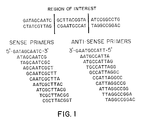
- Figure 1 depicts an example of a set of oligonucleotide primers comprising consecutive primers differing by one base pair at the growing end and capable of hybridizing successively along the relevant part(s) or the whole of the polynucleotide of interest.
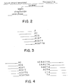
- Figure 2 is a schematic illustration of a single strand template bound to a primer which is in turn attached to a solid support.
- Figure 3 illustrates a set of consecutive oligonucleotide primers for a part of the polynucleotide of interest following immediately after the primer illustrated in Figure 2.
- Figure 4 illustrates the single base pair additions to all the primers illustrated in Figure 3, as well as the corresponding additions for the corresponding primers related to the complementary strand of the polynucleotide of interest.
- Figure 5 is a graphic depiction of the length of extended primers formed utilizing free oligonucleotide primers annealed to a polynucleotide of interest.
- Figures 6A, 6B and 6C are graphic depictions of electrophoretograms demonstrating the detection of the presence of a mutation in a polynucleotide of interest.
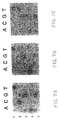
- Figures 7A, 7B and 7C depict the results of a DNA chip-based analysis for a five-base region within the third exon of the HPRT gene.
- the current invention pertains to methods for analyzing the nucleotide sequence of a polynucleotide of interest.
- the method comprises hybridizing all or a fragment of a polynucleotide of interest to oligonucleotide primers, conducting single base extension reactions, and detecting the single base extension events.
- the method can be used to analyze the sequence of a polypeptide of interest by examining the sequence for the presence of mutations or alterations in the nucleotide sequence, or by determining the sequence of a polypeptide of interest.
- polynucleotide of interest refers to the particular polynucleotide for which sequence information is wanted.
- Representative polynucleotides of interest include oligonucleotides, DNA or DNA fragments, RNA or RNA fragments, as well as genes or portions of genes.
- the polynucleotide of interest can be single- or double-stranded.
- template polynucleotide of interest is used herein to refer to the strand which is analyzed, if only one strand of a double-stranded polynucleotide is analyzed, or to the strand which is identified as the first strand, if both strands of a double-stranded polynucleotide are analyzed.
- complementary polynucleotide of interest is used herein to refer to the strand which is not analyzed, if only one strand of a double-stranded polynucleotide is analyzed, or to the strand which is identified as the second strand (i.e., the strand that is complementary to the first (template) strand), if both strands of a double-stranded polynucleotide are analyzed. Either one of the two strands can be analyzed. In a preferred embodiment, both strands of a double-stranded polypeptide of interest are analyzed in order to verify sequence information obtained from the template (first) strand by comparison with the complementary (second) strand.
- the methods of the current invention can be used to identify the presence of mutations or alterations in the nucleotide sequence of a polypeptide of interest.
- the sequence of the polynucleotide of interest is compared with the sequence of the native or normal polynucleotide.
- An "alteration" in the polynucleotide of interest refers to a deviation from the expected sequence (the sequence of the native or normal polynucleotide), including deletions, insertions, point mutations, frame-shifts, expanded oligonucleotide repeats, or other changes.
- the portion of the polynucleotide of interest that contains the alteration is known as the "altered" region.
- the methods can also be used to determine the sequence of a polypeptide of interest having a previously unknown nucleotide sequence.
- the polynucleotide of interest is analyzed by annealing the polynucleotide to an array comprising sets of oligonucleotide primers.
- the oligonucleotide primers in the array have a length N, where N is from about 7 to about 30 nucleotides, inclusive, and is preferably from 20 to 24 nucleotides, inclusive.
- Each oligonucleotide primer within each set differs by one base pair.
- the oligonucleotide primers can be prepared by conventional methods (see Sambrook et al., Molecular Cloning: A Laboratory Manual (2nd Ed, 1989)).
- the sets of oligonucleotide primers are arranged into an array, such that the position and nucleotide content of each oligonucleotide primer on the array is known.
- the size and nucleotide content of the oligonucleotide primers in the array depend on the polynucleotide of interest and the region of the polynucleotide of interest for which sequence information is desired.
- consecutive primers differing by one base pair at the growing end and capable of hybridizing successively along the relevant part(s) or the whole of the polynucleotide are used.
- An example of such a primer set is shown in Figure 1. If only one or a few specific positions of the polynucleotide sequence are examined for alterations, the necessary array of oligonucleotide primers covers only the mutation regions, and is therefore small.
- the necessary array is larger.
- the whole hypoxanthine-guanine phosphoribosyl-transferase (HPRT) gene can be covered by 900 primers, arranged in a 30 X 30 array; the whole p53 gene requires 700 primers.
- the array comprises consecutive oligonucleotide primers for the suspected mutation region of both the template polynucleotide of interest and the complementary polynucleotide of interest. If the polynucleotide of interest has not been sequenced previously, the array includes oligonucleotide primers comprising all possible N-mers.
- the array of sets of oligonucleotide primers is immobilized to a solid support at defined locations (i.e., known positions).
- the immobilized array is referred to as a "DNA chip", which is the apparatus of the current invention.
- the solid support can be a plate or chip of glass, silicon, or other material.
- the solid support can also be coated, such as with gold or silver. Coating may facilitate attachment of the oligonucleotide primers to the surface of the solid support.
- the oligonucleotide primers can be bound to the solid support by a specific binding pair, such as biotin and avidin or biotin and streptavidin.
- the primers can be provided with biotin handles in connection with their preparation, and then the biotin-labelled primers can be attached to a streptavidin-coated support.
- the primers can be bound by a linker arm, such as a covalently bonded hydrocarbon chain, such as a C 10-20 chain.
- the primers can also be bound directly to the solid support, such as by epoxide/amine coupling chemistry (see Eggers, M.D. et al., Advances in DNA sequencing Technology, SPIE conference proceedings, January 21, 1993).
- the solid support can be reused, as described in greater detail below.
- the polynucleotide of interest is analyzed by annealing the polynucleotide to one or more specific oligonucleotide primers that are not attached to a solid support; such oligonucleotide primers are referred to herein as "free oligonucleotide primers". If free oligonucleotide primers are used, the polynucleotide of interest can be attached to a solid support, such as magnetic beads.
- the free oligonucleotide primers have a length N, as described above, and are prepared by conventional methods (see Sambrook et al., Molecular Cloning: A Laboratory Manual (2nd Ed, 1989)).
- the size and nucleotide content of the free oligonucleotide primers depend on the polynucleotide of interest and the region of the polynucleotide of interest for which sequence information is desired.
- primers capable of hybridizing immediately adjacent to the relevant part(s) of the polynucleotide are used. If more than one position of the polynucleotide sequence is examined for alterations, the free oligonucleotide primers are mutually distinguishable: i.e., the oligonucleotide primers have different mobilities during gel electrophoresis. In a preferred embodiment, oligonucleotides of different lengths are used.
- an oligonucleotide primer of 10 nucleotides in length is designed to hybridize immediately adjacent to one putative mutation
- an oligonucleotide primer of 12 nucleotides in length is designed to hybridize immediately adjacent to a second putative mutation. Because the oligonucleotide primers are of different lengths, they will migrate to different positions on the gel. Thus, in this manner, the nucleotide content of each oligonucleotide primer can be identified by the position of the oligonucleotide primer on the gel.
- the polynucleotide of interest is hybridized to the array of oligonucleotide primers, or to the free nucleotide primers, under high stringency conditions, so that an exact match between the polynucleotide of interest and the oligonucleotide primers is obtained, without any base-pair mismatches (see Sambrook et al., Molecular Cloning: A Laboratory Manual (2nd Ed, 1989)).
- Figure 2 a schematic illustration of a hypothetical polynucleotide of interest annealed to an oligonucleotide primer that is attached to a solid support is shown schematically in Figure 2.
- TGCAACTA a part of the sequence of the polynucleotide of interest that follows immediately after the portion of the polynucleotide that is bound to the oligonucleotide primer on the array.
- Six corresponding consecutive primers are shown in Figure 3, i.e. primers ending with the pairing bases A, AC, ACG, etc.
- the polynucleotide of interest is double-stranded, it can be separated into two single strands either before or after the binding of the polynucleotide of interest to the array oligonucleotide primers. Both the template and the complementary polynucleotide of interest can be analyzed utilizing a single array.
- appropriate primers corresponding to the complementary polynucleotide of interest are also attached to the solid support in known positions.
- annealed primer refers to an oligonucleotide primer (either free or attached to a solid support) to which a polynucleotide of interest is hybridized. The annealed primers are subjected to a single base extension reaction.
- the "single base extension reaction”, as used herein, refers to a reaction in which the annealed primers are provided with a reaction mixture comprising a DNA polymerase, such as T7 polymerase, and terminating nucleotides under conditions such that single terminating nucleotides are added to each of the annealed primers.
- the term "terminating nucleotides”, as used herein, refers to either single terminating nucleotides, or units of nucleotides, the units preferably being dinucleotides. In a preferred embodiment, the terminating nucleotides are single dideoxynucleotides.
- the terminating nucleotides can comprise standard nucleotides, and/or nucleotide analogues.
- the terminating nucleotide added to each annealed primer is thus a base pairing with the template base on the polynucleotide of interest, and is added immediately adjacent to the growing end of the respective primer.
- An oligonucleotide primer to which a terminating nucleotide has been added through the single base extension reaction is termed an "extended primer".
- extended primer an oligonucleotide primer to which a terminating nucleotide has been added through the single base extension reaction.
- the terminating nucleotides preferably comprise dNTPs, and particularly comprise dideoxynucleotides (ddNTPs), but other terminating nucleotides apparent to the skilled person can also be used. If the terminating nucleotides are single nucleotides, then nucleotides corresponding to each of the four bases (A, T, G and C) are utilized in the single base extension reaction. If the terminating nucleotides are dinucleotide units, for example, then nucleotides corresponding to each of the sixteen possible dinucleotides are utilized.
- the nucleotides are mutually distinguishable. For example, if the solid support is coated with a free electron metal, such as with gold or silver, surface plasmon resonance (SPR) microscopy allows identification of each nucleotide, by the change of the refractive index at the surface caused by each base extension.
- SPR surface plasmon resonance
- at least one of the terminating nucleotides is labelled by standard methods to facilitate detection. Suitable labels include fluorescent dyes, chemiluminescence, and radionuclides. The number of nucleotides that are labelled can be varied. It is sufficient to use three labelled terminating nucleotides, the fourth terminating nucleotide being identified by its "non-label", if single nucleotides are added in the base extension reaction.
- each terminating nucleotide is observed. If free oligonucleotide primers are used, the extended primers are eluted and separated by gel electrophoresis, and the gel is then analyzed. If oligonucleotide primers attached to an array are used, the array itself is analyzed. The gel or array is analyzed by detecting the labelled, terminating nucleotides bound to the oligonucleotide primers. The labeled, terminating nucleotides are detected by conventional methods, such as by an optical system.
- a laser excitation source can be used in conjunction with a filter set to isolate the fluorescence emission of a particular type of terminating nucleotide.
- a photomultiplier tube, a charged-coupled device (CCD), or another suitable fluorescence detection metnod can be used to detect the emitted light from fluorescent terminating nucleotides.
- the sequence of the polynucleotide of interest can be analyzed from the label pattern observed on the array or on the gel, since the position of each different primer on the array or on the gel is known, and since the identity of each terminating nucleotide can be determined by its specific label.
- the label and position of each terminating nucleotide either within the array or on the gel will directly define the sequence of the polynucleotide of interest that is being analyzed. Mutations or alterations in the sequence of the polynucleotide of interest are indicated by alterations in the expected label pattern. For example, assume that the nucleotide sequence shown in Figure 2 contains a mutation: the third base C from the left is replaced by a G in the polynucleotide of interest.
- the top primer in Figure 3 will still be extended by a C as shown in Figure 4, whereas the next primer will be extended by a C rather than a G. Since this new, unexpected base C can be identified by its specific label and the respective primer location is known, the corresponding base mutation is identified as G.
- the following simple example illustrates the ability to obtain complete sequence information and to identify a mutation in a representative polynucleotide of interest.
- the example utilizes two labelled terminating nucleotides, which give complete sequence information.
- the presence of such a point mutation will affect the base pairing of the next few oligonucleotide primers to the polynucleotide of interest, and thereby the primer extensions obtained, such that the bases in the vicinity of the mutation (i.e., in the altered region) may not be accurately identified.
- the few bases that may be difficult to identify on the template polynucleotide of interest, as well as the changed base will be identified by the base extensions of the primers for the complementary polynucleotide of interest, as the analys-is of the complementary polynucleotide of interest approaches the mutation site from the opposite direction. In the nearest regions on either side of the alteration, the sequence determination is thereby provided by the oligonucleotide primers for one of the two strands.
- sequence of a polynucleotide of interest for which the sequence is previously known can be determined using methods similar to those described above in reference to identification of mutations utilizing an array of oligonucleotide primers. As before, the positions of the terminating nucleotides within the array will directly define the sequence position of each nucleotide in the polynucleotide of interest.
- one annealed primer is selected to be the "starting" annealed primer; it is supposed for purposes of analysis that the sequence of the polynucleotide of interest "starts" with this primer.
- the nucleotide which has been added to the starting annealed primer is detected using standard methods.
- a second annealed primer which has the same nucleotide sequence as the starting annealed primer, minus the 5' nucleotide and with the addition of the added nucleotide, is then selected.
- the terminating nucleotide which has been added to the second annealed primer is detected.
- the starting annealed primer is chosen to correspond to the first ten bases of the sequence.
- the terminating nucleotide of the starting annealed primer is then determined.
- bases 2-11 i.e., bases 2-10 of the starting annealed primer plus the terminating nucleotide extension
- bases 2-11 are matched to another annealed primer.
- This primer is the second annealed primer.
- the terminating nucleotide of the second annealed primer is then determined.
- the polynucleotide of interest and the terminating nucleotides can be removed from the DNA chip, so that the chip can be reused.
- the added terminating nucleotides are capable of being removed from the solid support after analysis of the polynucleotide of interest has been completed.
- the solid support with the immobilized oligonucleotide primers can be used for a new analysis.
- the nucleotides can be removed using standard methods, such as enzymatic cleavage or chemical degradation. Enzymatic cleavage, for example, would use a terminating nucleotide which can be removed by an enzyme.
- the single base extension reaction could result in addition to the oligonucleotide primers of RNA dideoxyTTP or RNA dideoxyCTP by reverse transcriptase or other polymerase.
- a C/T cleavage enzyme such as RNase A, can then be used to "strip" off the RNA dideoxynucleotides.
- sulfur-containing dideoxy-A or dideoxy-G can be used during the single extension reaction; a sulfur-specific esterase, which does not cleave phosphates can then be used to cleave off the dideoxynucleotides.
- a chemically degradable terminating nucleotide can be used for chemical degradation.
- a modified ribonucleotide having its 2'- and 3'-hydroxyl groups esterified, such as by acetyl groups can be used.
- the acetyl groups are removed by treatment with a base to expose the 2'- and 3'-hydroxyl groups.
- the ribose residue can then be degraded e by periodate oxidation, and the residual phosphate group removed from the annealed primer by treatment with a base and alkaline phosphatase.
- the method and apparatus of the current invention have uses in detecting mutations, deletions, expanded oligonucleotide repeats, and other genetic abnormalities.
- the current invention can be used to identify frame shifting mutations caused by insertions or deletions.
- carrier status of heritable diseases such as cystic fibrosis, ⁇ -thalassemia, ⁇ -1, Gaucher's disease, Tay Sach's disease, or Lesch-Nyham syndrome, can be easily determined using the current invention, because both the normal and the altered signals would be detected.
- mixtures of DNA molecules such as occur in HIV infected patients with drug resistance can be determined.
- the HIV virus may develop resistance against drugs like AZT by point mutations in the reverse transcriptase (RT) gene. When mutated viruses start to appear in the virus population, both the mutated gene and the normal (wild type) gene can be detected. The greater the proportion is of the mutant, the greater is the signal from the corresponding mutant terminating nucleotide.
- the current invention is further exemplified by the following Examples.
- HPRT hypoxanthine-guanine phosphoribosyl-transferase
- the polymerase chain reaction (see Sambrook et al., Molecular Cloning: A Laboratory Manual (2nd Ed, 1989), especially chapter 14) was utilized to amplify the polynucleotide of interest. During the reaction, one of the two PCR primers was tagged with a biotin group. Following amplification, the single strand template was captured with streptavidin coated magnetic beads. For a 50 ⁇ l PCR reaction, 25 ⁇ l of Dynal M-280 paramagnetic beads (Dynal A/S, Oslo, Norway) was used.
- the supernatant of the beads was removed and replaced with 50 ⁇ l of a binding and washing buffer (10 mM Tris-HCl (pH 7.5); 1 mM EDTA; 2 M NaCl).
- the PCR product was added to the beads and incubated at room temperature for 30 minutes for bead capture of the products.
- the single stranded polynucleotide of interest was isolated by the addition of 150 ⁇ l of 0.15 M NaOH for 5 minutes. The beads were captured, the supernatant was removed, and 150 ⁇ l of 0.15 M NaOH was again added for five minutes.
- the beads were washed once with 150 ⁇ l of 0.15 M NaOH and twice with 1X T7 annealing buffer (40 mM Tris-HCl (pH 7.5); 20 mM MgCl 2 ; 50 mM NaCl). The beads were finally suspended in 70 ⁇ l of water. This process both isolates the single-stranded polynucleotide of interest and removes any unincorporated dNTPs remaining after PCR.
- 1X T7 annealing buffer 40 mM Tris-HCl (pH 7.5); 20 mM MgCl 2 ; 50 mM NaCl.
- the oligonucleotide primers were annealed to the polynucleotide of interest by heating to 65°C for approximately two minutes and cooling to room temperature over approximately 20 minutes.
- the 10 ⁇ l reaction volume consisted of 7 ⁇ l of the polynucleotide of interest (0.5-1 pmol), 2 ⁇ l of 5X T7 annealing buffer, and 1 ⁇ l of extension primer (3-9 pmol).
- the extension reaction was then performed.
- 1 ⁇ l of DTT, 2 ⁇ l of T7 polymerase (diluted 1:8) and 1 ⁇ l of ddNTPs (final concentration of 0.5 uM) were added.
- the reaction proceeded at 37°C for two minutes, and then was stopped by the addition of 100 ⁇ l of washing buffer (1XSSPE, 0.1% SDS, 30% ethanol).
- the beads were washed twice with 150 ⁇ l of the washing buffer.
- the extension products were eluted by the addition of 5 ⁇ l of formamide andheated to 70°C for two minutes.
- the beads were captured by the magnet and the supernatant containing the extension products was collected and analyzed on a ABI 373 (Applied Biosystems, Inc.). Oligonucleotide primers of lengths varying from 10 to 17 were used. As shown in Figure 5, extension products were formed efficiently.
- Each ddNTP was labelled by a different fluorophore.
- ABI Dye Terminator dyes designed for taq polymerase were used: ddG is blue, ddA is green, ddT is yellow, and ddC is red.
- Four fluorescent ddNTPs were added to each reaction tube. The extension products were purified, gel separated, and analyzed on an ABI 373. Two different bases of exon 3 of the HPRT gene were analyzed: base 16534 (wild type is A) and base 16620 (wild type is C).
- Electrophoretograms shown in Figures 6A, 6B and 6C indicate that Patient A is wild type (C) at base 16620 (Figure 6A), patient B is a mutated individual (C-->T) at base 16620 ( Figure 6B), and patient C is a carrier at base 16620 (both C and T) ( Figure 6C).
- Each ddNTP was labelled by the same fluorophore.
- DuPont NEN fluorescein dyes (NEL 400-404) were used. Each ddNTP appears blue in the ABI 373. only one fluorescent ddNTP is added to each reaction tube.
- the extension products were purified, gel separated, and analyzed on an ABI 373. Four lanes on the gel must be used to analyze each base. Two different bases of exon 3 of the HPRT gene were analyzed: base 16534 (wild type is A) and base 16620 (wild type is C).
- the ddNTPs are labelled with a biotin group.
- Four separate reactions are performed, whereby only one of the four ddNTPs is biotinylated.
- a strepavidin (or avidin) coupled fluorescent group is attached to the biotinylated ddNTPs. Because the biotin group is small, uniform incorporation of the ddNTPs is expected and base-specific differences in extension are minimized.
- the fluorescent signal can be amplified because the biotin group can bind a steptavidin moiety coupled to multiple fluors.
- HPRT hypoxanthine-guanine phosphoribosyl-transferase
- Microscope glass slides were epoxysilanated at 80°C for eight hours using 25% 3' glycidoxy propyltriethoxysilane (Aldrich Chemical) in dry xylene (Aldrich Chemical) with a catalytic amount of diisopropylehylamine (Aldrich Chemical), according to Southern ( Nucl. Acids Res. 20 :1679 (1992), and Genomics 13 :1008 (1992)).
- the DNA chips were made by placing 0.5 ⁇ l drops of 5'-amino-linked oligonucleotides (50 ⁇ M, 0.1 M NaOH) at 37°C for six hours in a humid environment. The chips were washed in 50°C water for 15 minutes, dried and used.
- the annealing reaction consisted of adding 2.2 ⁇ l of single-stranded DNA (0.1 ⁇ M in T7 reaction buffer) to each grid position, heating the chip in a humid environment to 70°C and then cooling slowly to room temperature. A 1 ⁇ l drop of 0.1 M DTT, 3 units of Sequenase Version 2.0 (USB), 5 ⁇ Ci ⁇ - 32 P dNTP (3000 Ci/mmol) (DuPont NEN) and noncompeting unlabeled 18.5 ⁇ M ddNTPs (Pharmacia) were added to each grid position for three minutes. The reaction was stopped by washing in 75°C water, and analyzed on a PhosphorImager (Molecular Dynamics).
- Figures 7A, 7B and 7C depict the results of a DNA chip-based analysis for a five-base region within the third exon of the HPRT gene. The rows correspond to a particular base under investigation, and the columns correspond to the labeled base.
- Figure 7A demonstrates the wild type sequence (TCGAG)
- Figure 7B demonstrates a C-->T mutation
- Figure 7C demonstrates a C-->T mutation.
Landscapes
- Chemical & Material Sciences (AREA)
- Life Sciences & Earth Sciences (AREA)
- Proteomics, Peptides & Aminoacids (AREA)
- Organic Chemistry (AREA)
- Zoology (AREA)
- Wood Science & Technology (AREA)
- Health & Medical Sciences (AREA)
- Engineering & Computer Science (AREA)
- Microbiology (AREA)
- Biochemistry (AREA)
- Biotechnology (AREA)
- Molecular Biology (AREA)
- Biophysics (AREA)
- Analytical Chemistry (AREA)
- Physics & Mathematics (AREA)
- Immunology (AREA)
- Bioinformatics & Cheminformatics (AREA)
- General Engineering & Computer Science (AREA)
- General Health & Medical Sciences (AREA)
- Genetics & Genomics (AREA)
- Chemical Kinetics & Catalysis (AREA)
- Measuring Or Testing Involving Enzymes Or Micro-Organisms (AREA)
Applications Claiming Priority (3)
| Application Number | Priority Date | Filing Date | Title |
|---|---|---|---|
| SE9302152 | 1993-06-22 | ||
| SE9302152A SE501439C2 (sv) | 1993-06-22 | 1993-06-22 | Sätt och anordning för analys av polynukleotidsekvenser |
| EP94921363A EP0705349B1 (de) | 1993-06-22 | 1994-06-22 | NUKLEINSäURE-SEQUENZANALYSE DURCH DIE METHODE DER PARALLELEN PRIMEREXTENSION |
Related Parent Applications (1)
| Application Number | Title | Priority Date | Filing Date |
|---|---|---|---|
| EP94921363A Division EP0705349B1 (de) | 1993-06-22 | 1994-06-22 | NUKLEINSäURE-SEQUENZANALYSE DURCH DIE METHODE DER PARALLELEN PRIMEREXTENSION |
Publications (2)
| Publication Number | Publication Date |
|---|---|
| EP0969103A2 true EP0969103A2 (de) | 2000-01-05 |
| EP0969103A3 EP0969103A3 (de) | 2004-01-02 |
Family
ID=20390371
Family Applications (2)
| Application Number | Title | Priority Date | Filing Date |
|---|---|---|---|
| EP99200676A Withdrawn EP0969103A3 (de) | 1993-06-22 | 1994-06-22 | Nukleinsäure-Sequenzanalyse durch die Methode der parallelen Primerextension |
| EP94921363A Expired - Lifetime EP0705349B1 (de) | 1993-06-22 | 1994-06-22 | NUKLEINSäURE-SEQUENZANALYSE DURCH DIE METHODE DER PARALLELEN PRIMEREXTENSION |
Family Applications After (1)
| Application Number | Title | Priority Date | Filing Date |
|---|---|---|---|
| EP94921363A Expired - Lifetime EP0705349B1 (de) | 1993-06-22 | 1994-06-22 | NUKLEINSäURE-SEQUENZANALYSE DURCH DIE METHODE DER PARALLELEN PRIMEREXTENSION |
Country Status (9)
| Country | Link |
|---|---|
| EP (2) | EP0969103A3 (de) |
| JP (1) | JP3722833B2 (de) |
| AT (1) | ATE185843T1 (de) |
| AU (1) | AU698553B2 (de) |
| CA (1) | CA2164715A1 (de) |
| DE (1) | DE69421277T2 (de) |
| ES (1) | ES2141828T3 (de) |
| SE (1) | SE501439C2 (de) |
| WO (1) | WO1995000669A1 (de) |
Cited By (2)
| Publication number | Priority date | Publication date | Assignee | Title |
|---|---|---|---|---|
| WO2001068913A3 (en) * | 2000-03-13 | 2002-10-31 | Genset Sa | Nucleic acid detection method and system |
| WO2009031054A3 (en) * | 2007-06-29 | 2009-12-17 | Population Genetics Technologies Ltd. | Methods and compositions for isolating nucleic acid sequence variants |
Families Citing this family (89)
| Publication number | Priority date | Publication date | Assignee | Title |
|---|---|---|---|---|
| JPH07203998A (ja) * | 1994-01-26 | 1995-08-08 | Hamamatsu Photonics Kk | 核酸塩基配列決定方法 |
| GB9507238D0 (en) | 1995-04-07 | 1995-05-31 | Isis Innovation | Detecting dna sequence variations |
| US5763175A (en) * | 1995-11-17 | 1998-06-09 | Lynx Therapeutics, Inc. | Simultaneous sequencing of tagged polynucleotides |
| AU2320597A (en) | 1996-03-19 | 1997-10-10 | Molecular Tool, Inc. | Method for determining the nucleotide sequence of a polynucleotide |
| US6143529A (en) * | 1996-08-14 | 2000-11-07 | Exact Laboratories, Inc. | Methods for improving sensitivity and specificity of screening assays |
| US6096273A (en) | 1996-11-05 | 2000-08-01 | Clinical Micro Sensors | Electrodes linked via conductive oligomers to nucleic acids |
| US6566101B1 (en) | 1997-06-16 | 2003-05-20 | Anthony P. Shuber | Primer extension methods for detecting nucleic acids |
| US5888778A (en) * | 1997-06-16 | 1999-03-30 | Exact Laboratories, Inc. | High-throughput screening method for identification of genetic mutations or disease-causing microorganisms using segmented primers |
| JP4789294B2 (ja) * | 1997-08-22 | 2011-10-12 | ソレクサ・インコーポレイテッド | ローリングプライマーを用いたdna伸長および分析 |
| US6322968B1 (en) * | 1997-11-21 | 2001-11-27 | Orchid Biosciences, Inc. | De novo or “universal” sequencing array |
| JP2002506875A (ja) | 1998-03-16 | 2002-03-05 | ミレニアム ファーマシューティカルズ インク. | 第18p染色体関連障害の診断及び治療のための方法及び組成物 |
| US6872521B1 (en) | 1998-06-16 | 2005-03-29 | Beckman Coulter, Inc. | Polymerase signaling assay |
| US6245507B1 (en) | 1998-08-18 | 2001-06-12 | Orchid Biosciences, Inc. | In-line complete hyperspectral fluorescent imaging of nucleic acid molecules |
| US6150105A (en) | 1998-08-20 | 2000-11-21 | Genetic Assays, Inc. | Methods of screening nucleic acids for nucleotide variations |
| JP4794052B2 (ja) | 1999-04-09 | 2011-10-12 | ジェンザイム・コーポレーション | 癌の指標である核酸を検出するための方法 |
| JP3926625B2 (ja) | 1999-07-26 | 2007-06-06 | クリニカル・マイクロ・センサーズ・インコーポレイテッド | 電子検出を用いる核酸の配列決定 |
| US6528319B1 (en) | 1999-09-02 | 2003-03-04 | Amersham Biosciences Corp | Method for anchoring oligonucleotides to a substrate |
| US6586177B1 (en) | 1999-09-08 | 2003-07-01 | Exact Sciences Corporation | Methods for disease detection |
| US6849403B1 (en) | 1999-09-08 | 2005-02-01 | Exact Sciences Corporation | Apparatus and method for drug screening |
| AU7751600A (en) * | 1999-10-06 | 2001-05-10 | Amersham Biosciences Corp. | Method for detecting mutations using arrayed primer extension |
| US6596483B1 (en) | 1999-11-12 | 2003-07-22 | Motorola, Inc. | System and method for detecting molecules using an active pixel sensor |
| US7368233B2 (en) | 1999-12-07 | 2008-05-06 | Exact Sciences Corporation | Methods of screening for lung neoplasm based on stool samples containing a nucleic acid marker indicative of a neoplasm |
| US6919174B1 (en) | 1999-12-07 | 2005-07-19 | Exact Sciences Corporation | Methods for disease detection |
| DE10010280B4 (de) * | 2000-02-25 | 2006-08-10 | Epigenomics Ag | Verfahren zur Detektion von Cytosin-Methylierung in DNA Proben |
| DE10010282B4 (de) | 2000-02-25 | 2006-11-16 | Epigenomics Ag | Verfahren zur Detektion von Cytosin-Methylierung in DNA Proben |
| US6376191B1 (en) * | 2000-03-22 | 2002-04-23 | Mergen, Ltd. | Microarray-based analysis of polynucleotide sequence variations |
| US20040076956A1 (en) | 2000-04-06 | 2004-04-22 | Alexander Olek | Diagnosis of diseases associated with dna repair |
| US6803211B2 (en) | 2000-08-25 | 2004-10-12 | Pfizer Inc. | Methods and compositions for diagnosing and treating disorders involving angiogenesis |
| WO2002042490A1 (en) * | 2000-11-24 | 2002-05-30 | Asper OÜ | Method of optimising the sequences of synthetic nucleic acids |
| GB0105790D0 (en) * | 2001-03-08 | 2001-04-25 | Expresson Biosystems Ltd | Detecting binding of mRNA to an oligonnucleotide array using RNA dependent nucleic acid modifying enzymes |
| JP2004526448A (ja) | 2001-03-30 | 2004-09-02 | アプレラ コーポレイション | 非鋳型ヌクレオチド付加を用いた核酸分析 |
| US6653082B2 (en) | 2001-05-17 | 2003-11-25 | Baylor College Of Medicine | Substrate-bound cleavage assay for nucleic acid analysis |
| EP2354156A3 (de) | 2001-08-14 | 2011-11-16 | Sentigen Biosciences, Inc. | Nukleinsäuren und Proteine von Insekten OR83B-Duftrezeptorgenen und Verwendungen davon |
| DE10154317B4 (de) | 2001-10-26 | 2005-06-09 | Epigenomics Ag | Verfahren zum Nachweis von Cytosin-Methylierungen in immobilisierten DNA Proben |
| EP1340818A1 (de) | 2002-02-27 | 2003-09-03 | Epigenomics AG | Verfahren und Nukleinsäuren zur Analyse von Kolonkrebszellen |
| DE10245145B4 (de) * | 2002-09-27 | 2004-12-02 | IPK-Institut für Pflanzengenetik und Kulturpflanzenforschung | Verfahren zum Nachweis von SNPs auf polydimensionalen Microarrays |
| DE60328618D1 (de) | 2002-10-01 | 2009-09-10 | Epigenomics Ag | Verfahren für die behandlung von proliferativen erkrankungen von brustzellen |
| CA2528577C (en) | 2003-06-20 | 2012-08-07 | Illumina, Inc. | Methods and compositions for whole genome amplification and genotyping |
| AU2004252554B2 (en) | 2003-06-23 | 2012-01-19 | Epigenomics Ag | Methods and nucleic acids for analyses of colorectal cell proliferative disorders |
| EP1636381B1 (de) | 2003-06-23 | 2011-01-05 | Epigenomics AG | Verfahren und nukleinsäuren zur analyse von störungen der proliferation von kolonzellen |
| EP2354248B1 (de) | 2003-06-23 | 2018-01-03 | Epigenomics AG | Verfahren zur Analyse von Störungen der Proliferation kolorektaler Zellen |
| JP4824575B2 (ja) | 2003-12-01 | 2011-11-30 | エピゲノミクス アクチェンゲゼルシャフト | 前立腺細胞増殖性疾患の発症に関連する遺伝子発現の分析のための方法および核酸 |
| DE602004031405D1 (de) | 2003-12-11 | 2011-03-31 | Epigenomics Ag | Marker zur Prognose der Reaktion auf Therapie bzw. der Überlebensrate von Brustkrebspatienten |
| EP1778865A1 (de) | 2004-06-23 | 2007-05-02 | Epigenomics AG | Verfahren und nukleinsäuren zum nachweis der metastasierung proliferativer kolonzellerkrankungen |
| EP2281902A1 (de) | 2004-07-18 | 2011-02-09 | Epigenomics AG | Epigenetische Verfahren und Nukleinsäuren für den Nachweis von proliferativen Erkrankungen von Mammazellen |
| WO2006020617A1 (en) | 2004-08-09 | 2006-02-23 | Generation Biotech, Llc | Method for nucleic acid isolation and amplification |
| WO2006047787A2 (en) | 2004-10-27 | 2006-05-04 | Exact Sciences Corporation | Method for monitoring disease progression or recurrence |
| JP5745202B2 (ja) | 2004-12-02 | 2015-07-08 | エピゲノミックス アクチェンゲゼルシャフト | 前立腺細胞増殖障害の予後に関連する遺伝子発現分析のための方法および核酸 |
| KR20080011287A (ko) | 2005-04-15 | 2008-02-01 | 에피제노믹스 아게 | 세포 증식 질환을 분석하기 위한 방법 및 핵산 |
| WO2007044071A2 (en) | 2005-04-21 | 2007-04-19 | Exact Sciences Corporation | Analysis of heterogeneous nucleic acid samples |
| ES2446250T3 (es) | 2005-05-02 | 2014-03-06 | University Of Southern California | Marcadores de metilación del ADN asociados con el fenotipo metilador de islas CpG (CIMP) en el cáncer colorrectal humano |
| EP1934369A2 (de) | 2005-09-29 | 2008-06-25 | Epigenomics AG | Verfahren und nukleinsäuren zur analyse der mit gewebeklassifizierung assoziierten genexpression |
| WO2007057231A1 (en) | 2005-11-17 | 2007-05-24 | Epigenomics Ag | Method for the determination of the dna methylation level of a cpg position in identical cells within a tissue sample |
| AU2007237444B2 (en) | 2006-04-17 | 2013-05-23 | Epigenomics Ag | Methods and nucleic acids for the detection of colorectal cell proliferative disorders |
| WO2008008284A2 (en) | 2006-07-14 | 2008-01-17 | The Regents Of The University Of California | Cancer biomarkers and methods of use threof |
| WO2008011620A2 (en) | 2006-07-21 | 2008-01-24 | Epigenomics Ag | Methods and nucleic acids for analyses of cellular proliferative disorders |
| EP3184649A1 (de) | 2006-11-24 | 2017-06-28 | Epigenomics AG | Verfahren und nukleinsäuren zur analyse der genexpression, die mit der entwicklung von prostatazellproliferationserkrankungen assoziiert ist |
| DK2258871T3 (da) | 2007-01-19 | 2014-08-11 | Epigenomics Ag | Fremgangsmåder og nukleinsyrer til analyse af celleproliferative lidelser |
| CN101743326A (zh) * | 2007-05-14 | 2010-06-16 | 因赛特遗传学公司 | 筛选核酸中的单核苷酸变异的方法 |
| AU2008334901A1 (en) | 2007-12-11 | 2009-06-18 | Epigenomics Ag | Methods and nucleic acids for analyses of lung carcinoma |
| US20120184447A1 (en) | 2009-06-26 | 2012-07-19 | Reinhold Wasserkort | Methods and Nucleic Acids for Analysis of Bladder Cell Proliferative Disorders |
| US20110104695A1 (en) | 2009-11-05 | 2011-05-05 | Epigenomics Ag | Methods of predicting therapeutic efficacy of cancer therapy |
| EP3508854A1 (de) | 2010-04-27 | 2019-07-10 | The Regents of The University of California | Krebsbiomarker und verfahren zur verwendung davon |
| US8916344B2 (en) | 2010-11-15 | 2014-12-23 | Exact Sciences Corporation | Methylation assay |
| US8361720B2 (en) | 2010-11-15 | 2013-01-29 | Exact Sciences Corporation | Real time cleavage assay |
| CA2840149C (en) | 2011-07-08 | 2021-10-26 | Epigenomics Ag | Methods and nucleic acids for determining the prognosis of a cancer subject |
| CN105378108A (zh) | 2013-03-13 | 2016-03-02 | 雅培分子公司 | 用于分离核酸的系统和方法 |
| EP2971171A4 (de) | 2013-03-14 | 2016-11-02 | Abbott Molecular Inc | Multiplex-methylierungs-spezifische verstärkersysteme und -verfahren |
| ES2947873T3 (es) | 2013-03-14 | 2023-08-23 | Mayo Found Medical Education & Res | Detección de neoplasias |
| WO2015124921A1 (en) | 2014-02-19 | 2015-08-27 | The University Court Of The University Of Edinburgh | Methods and uses for determining the presence of inflammatory bowel disease |
| US11078539B2 (en) | 2014-03-31 | 2021-08-03 | Mayo Foundation For Medical Education And Research | Detecting colorectal neoplasm |
| US10184154B2 (en) | 2014-09-26 | 2019-01-22 | Mayo Foundation For Medical Education And Research | Detecting cholangiocarcinoma |
| EP3227687A4 (de) | 2014-12-05 | 2018-10-24 | Prelude, Inc. | Dcis-wiederauftreten und invasiver brustkrebs |
| EP3230744B1 (de) | 2014-12-12 | 2021-05-12 | Exact Sciences Development Company, LLC | Vorrichtungen und verfahren zur durchführung von methylierungserkennungstests |
| EP3274440B1 (de) | 2015-03-27 | 2026-03-04 | Exact Sciences Corporation | Nachweis von erkrankungen der speiseröhre |
| JP6871926B2 (ja) | 2015-08-31 | 2021-05-19 | マヨ ファウンデーション フォア メディカル エデュケーション アンド リサーチMayo Foundation for Medical Education and Research | 胃の新生物を検出すること |
| KR102811214B1 (ko) | 2015-10-30 | 2025-05-27 | 이그잭트 사이언시즈 코포레이션 | 복합 증폭 검출 분석 및 혈장으로부터 dna의 분리 및 검출 방법 |
| CA3022911A1 (en) | 2016-05-05 | 2017-11-09 | Exact Sciences Development Company, Llc | Detection of lung neoplasia by analysis of methylated dna |
| EP3978624B1 (de) | 2016-07-19 | 2025-03-26 | Exact Sciences Corporation | Methylierte kontroll-dna |
| AU2017318700B2 (en) | 2016-09-02 | 2022-09-22 | Exact Sciences Corporation | Detecting hepatocellular carcinoma |
| JP7289264B2 (ja) | 2017-01-27 | 2023-06-09 | エグザクト サイエンシーズ コーポレーション | メチル化dnaの分析による結腸腫瘍の検出 |
| CN118147307A (zh) | 2017-02-28 | 2024-06-07 | 梅约医学教育与研究基金会 | 用于表征来自人患者的样品的方法 |
| KR20250117473A (ko) | 2017-11-30 | 2025-08-04 | 메이오 파운데이션 포 메디칼 에쥬케이션 앤드 리써치 | 유방암 검출방법 |
| US10648025B2 (en) | 2017-12-13 | 2020-05-12 | Exact Sciences Development Company, Llc | Multiplex amplification detection assay II |
| EP3850368A4 (de) | 2018-09-14 | 2022-11-23 | Prelude Corporation | Verfahren zur auswahl zur behandlung von subjekten mit risiko auf invasiven brustkrebs |
| EP3880847B1 (de) | 2018-11-16 | 2025-09-03 | Oslo Universitetssykehus HF | Verfahren und zusammensetzungen zur charakterisierung von blasenkrebs |
| WO2021087275A1 (en) | 2019-10-31 | 2021-05-06 | Mayo Foundation For Medical Education And Research | Detecting ovarian cancer |
| CA3188230A1 (en) | 2020-08-19 | 2022-02-24 | John B. Kisiel | Detecting non-hodgkin lymphoma |
| WO2022165247A1 (en) | 2021-01-29 | 2022-08-04 | Mayo Foundation For Medical Education And Research | Detecting the presence or absence of multiple types of cancer |
Family Cites Families (5)
| Publication number | Priority date | Publication date | Assignee | Title |
|---|---|---|---|---|
| US5202231A (en) * | 1987-04-01 | 1993-04-13 | Drmanac Radoje T | Method of sequencing of genomes by hybridization of oligonucleotide probes |
| GB8810400D0 (en) * | 1988-05-03 | 1988-06-08 | Southern E | Analysing polynucleotide sequences |
| US5547839A (en) * | 1989-06-07 | 1996-08-20 | Affymax Technologies N.V. | Sequencing of surface immobilized polymers utilizing microflourescence detection |
| EP0834576B1 (de) * | 1990-12-06 | 2002-01-16 | Affymetrix, Inc. (a Delaware Corporation) | Detektion von Nukleinsäuresequenzen |
| US6004744A (en) * | 1991-03-05 | 1999-12-21 | Molecular Tool, Inc. | Method for determining nucleotide identity through extension of immobilized primer |
-
1993
- 1993-06-22 SE SE9302152A patent/SE501439C2/sv not_active IP Right Cessation
-
1994
- 1994-06-22 DE DE69421277T patent/DE69421277T2/de not_active Expired - Fee Related
- 1994-06-22 CA CA002164715A patent/CA2164715A1/en not_active Abandoned
- 1994-06-22 EP EP99200676A patent/EP0969103A3/de not_active Withdrawn
- 1994-06-22 JP JP50308095A patent/JP3722833B2/ja not_active Expired - Fee Related
- 1994-06-22 WO PCT/US1994/007086 patent/WO1995000669A1/en not_active Ceased
- 1994-06-22 ES ES94921363T patent/ES2141828T3/es not_active Expired - Lifetime
- 1994-06-22 AT AT94921363T patent/ATE185843T1/de not_active IP Right Cessation
- 1994-06-22 EP EP94921363A patent/EP0705349B1/de not_active Expired - Lifetime
- 1994-06-22 AU AU72121/94A patent/AU698553B2/en not_active Ceased
Cited By (4)
| Publication number | Priority date | Publication date | Assignee | Title |
|---|---|---|---|---|
| WO2001068913A3 (en) * | 2000-03-13 | 2002-10-31 | Genset Sa | Nucleic acid detection method and system |
| WO2009031054A3 (en) * | 2007-06-29 | 2009-12-17 | Population Genetics Technologies Ltd. | Methods and compositions for isolating nucleic acid sequence variants |
| US7635566B2 (en) | 2007-06-29 | 2009-12-22 | Population Genetics Technologies Ltd. | Methods and compositions for isolating nucleic acid sequence variants |
| US8241850B2 (en) | 2007-06-29 | 2012-08-14 | Population Genetics Technologies Ltd. | Methods and compositions for isolating nucleic acid sequence variants |
Also Published As
| Publication number | Publication date |
|---|---|
| EP0969103A3 (de) | 2004-01-02 |
| EP0705349A1 (de) | 1996-04-10 |
| SE9302152L (sv) | 1994-12-23 |
| EP0705349B1 (de) | 1999-10-20 |
| CA2164715A1 (en) | 1995-01-05 |
| JPH09501312A (ja) | 1997-02-10 |
| JP3722833B2 (ja) | 2005-11-30 |
| SE9302152D0 (sv) | 1993-06-22 |
| SE501439C2 (sv) | 1995-02-13 |
| WO1995000669A1 (en) | 1995-01-05 |
| AU7212194A (en) | 1995-01-17 |
| ATE185843T1 (de) | 1999-11-15 |
| DE69421277D1 (de) | 1999-11-25 |
| AU698553B2 (en) | 1998-10-29 |
| DE69421277T2 (de) | 2000-05-31 |
| ES2141828T3 (es) | 2000-04-01 |
Similar Documents
| Publication | Publication Date | Title |
|---|---|---|
| EP0705349B1 (de) | NUKLEINSäURE-SEQUENZANALYSE DURCH DIE METHODE DER PARALLELEN PRIMEREXTENSION | |
| US6153379A (en) | Parallel primer extension approach to nucleic acid sequence analysis | |
| JP3872812B2 (ja) | 段階連結および切断によるdna配列決定 | |
| JP4546582B2 (ja) | 平行オリゴヌクレオチド伸長によるdna配列決定 | |
| EP0640146B1 (de) | Dns sequenzierungsverfahren | |
| US7414115B2 (en) | Length determination of nucleic acid repeat sequences by discontinuous primer extension | |
| WO1992016657A1 (en) | Method of identifying a nucleotide present at a defined position in a nucleic acid | |
| WO2009052214A2 (en) | Sequence analysis using decorated nucleic acids | |
| US5470707A (en) | Hydrogen bond labeling and base sequence determination methods for DNA or RNA | |
| US5831065A (en) | Kits for DNA sequencing by stepwise ligation and cleavage | |
| JP2001521398A (ja) | 性状検査用dna | |
| US5817464A (en) | Fractionation method for nucleotide fragments | |
| US7001722B1 (en) | Parallel primer extension approach to nucleic acid sequence analysis | |
| WO1996002673A1 (en) | Method of using mobile priming sites for dna sequencing | |
| WO2026015752A1 (en) | Method of nucleic acid sequencing | |
| Lowe | ACID REPEAT SEQUENCES BY $8 $8 2i DISCONTINUOUS PRIMER EXTENSION | |
| WO1996027023A2 (en) | Method of using mobile priming sites for dna sequencing |
Legal Events
| Date | Code | Title | Description |
|---|---|---|---|
| PUAI | Public reference made under article 153(3) epc to a published international application that has entered the european phase |
Free format text: ORIGINAL CODE: 0009012 |
|
| 17P | Request for examination filed |
Effective date: 19990308 |
|
| AC | Divisional application: reference to earlier application |
Ref document number: 705349 Country of ref document: EP |
|
| AK | Designated contracting states |
Kind code of ref document: A2 Designated state(s): AT BE CH DE ES FR GB IT LI NL SE |
|
| PUAL | Search report despatched |
Free format text: ORIGINAL CODE: 0009013 |
|
| AK | Designated contracting states |
Kind code of ref document: A3 Designated state(s): AT BE CH DE ES FR GB IT LI NL SE |
|
| RAP1 | Party data changed (applicant data changed or rights of an application transferred) |
Owner name: BAYLOR COLLEGE OF MEDICINE Owner name: AMERSHAM BIOSCIENCES AB |
|
| 17Q | First examination report despatched |
Effective date: 20040528 |
|
| RAP1 | Party data changed (applicant data changed or rights of an application transferred) |
Owner name: BAYLOR COLLEGE OF MEDICINE Owner name: GE HEALTHCARE BIO-SCIENCES AB |
|
| STAA | Information on the status of an ep patent application or granted ep patent |
Free format text: STATUS: THE APPLICATION IS DEEMED TO BE WITHDRAWN |
|
| 18D | Application deemed to be withdrawn |
Effective date: 20071128 |