EP0689552B1 - Methode zur detektion von rissen in amniomembranen bei schwangeren säugetieren - Google Patents
Methode zur detektion von rissen in amniomembranen bei schwangeren säugetieren Download PDFInfo
- Publication number
- EP0689552B1 EP0689552B1 EP94911016A EP94911016A EP0689552B1 EP 0689552 B1 EP0689552 B1 EP 0689552B1 EP 94911016 A EP94911016 A EP 94911016A EP 94911016 A EP94911016 A EP 94911016A EP 0689552 B1 EP0689552 B1 EP 0689552B1
- Authority
- EP
- European Patent Office
- Prior art keywords
- amniotic fluid
- amniotic
- antigen
- molecular weight
- antibody
- Prior art date
- Legal status (The legal status is an assumption and is not a legal conclusion. Google has not performed a legal analysis and makes no representation as to the accuracy of the status listed.)
- Expired - Lifetime
Links
Images


Classifications
-
- C—CHEMISTRY; METALLURGY
- C07—ORGANIC CHEMISTRY
- C07K—PEPTIDES
- C07K14/00—Peptides having more than 20 amino acids; Gastrins; Somatostatins; Melanotropins; Derivatives thereof
- C07K14/435—Peptides having more than 20 amino acids; Gastrins; Somatostatins; Melanotropins; Derivatives thereof from animals; from humans
- C07K14/46—Peptides having more than 20 amino acids; Gastrins; Somatostatins; Melanotropins; Derivatives thereof from animals; from humans from vertebrates
- C07K14/47—Peptides having more than 20 amino acids; Gastrins; Somatostatins; Melanotropins; Derivatives thereof from animals; from humans from vertebrates from mammals
- C07K14/4701—Peptides having more than 20 amino acids; Gastrins; Somatostatins; Melanotropins; Derivatives thereof from animals; from humans from vertebrates from mammals not used
- C07K14/4715—Pregnancy proteins, e.g. placenta proteins, alpha-feto-protein, pregnancy specific beta glycoprotein
-
- C—CHEMISTRY; METALLURGY
- C07—ORGANIC CHEMISTRY
- C07K—PEPTIDES
- C07K16/00—Immunoglobulins [IG], e.g. monoclonal or polyclonal antibodies
- C07K16/18—Immunoglobulins [IG], e.g. monoclonal or polyclonal antibodies against material from animals or humans
-
- Y—GENERAL TAGGING OF NEW TECHNOLOGICAL DEVELOPMENTS; GENERAL TAGGING OF CROSS-SECTIONAL TECHNOLOGIES SPANNING OVER SEVERAL SECTIONS OF THE IPC; TECHNICAL SUBJECTS COVERED BY FORMER USPC CROSS-REFERENCE ART COLLECTIONS [XRACs] AND DIGESTS
- Y10—TECHNICAL SUBJECTS COVERED BY FORMER USPC
- Y10S—TECHNICAL SUBJECTS COVERED BY FORMER USPC CROSS-REFERENCE ART COLLECTIONS [XRACs] AND DIGESTS
- Y10S530/00—Chemistry: natural resins or derivatives; peptides or proteins; lignins or reaction products thereof
- Y10S530/806—Antigenic peptides or proteins
-
- Y—GENERAL TAGGING OF NEW TECHNOLOGICAL DEVELOPMENTS; GENERAL TAGGING OF CROSS-SECTIONAL TECHNOLOGIES SPANNING OVER SEVERAL SECTIONS OF THE IPC; TECHNICAL SUBJECTS COVERED BY FORMER USPC CROSS-REFERENCE ART COLLECTIONS [XRACs] AND DIGESTS
- Y10—TECHNICAL SUBJECTS COVERED BY FORMER USPC
- Y10S—TECHNICAL SUBJECTS COVERED BY FORMER USPC CROSS-REFERENCE ART COLLECTIONS [XRACs] AND DIGESTS
- Y10S530/00—Chemistry: natural resins or derivatives; peptides or proteins; lignins or reaction products thereof
- Y10S530/827—Proteins from mammals or birds
- Y10S530/85—Reproductive organs or embryos
- Y10S530/851—Placenta; amniotic fluid
Definitions
- the present invention relates to the method of detecting rupture of the amniotic membranes in pregnant mammals including humans using an immunoassay and reagents useful in such an assay.
- the method describes how to prepare a suitable protein antigen from amniotic fluid, gives criteria for the selection of this protein from the mixture of proteins present in amniotic fluid using the techniques of protein purification, gives criteria for assessing a sufficient degree of antigen purity for raising antibodies to the antigen and shows how the resultant antibodies can be used in immunoassays to detect the presence of amniotic fluid in the vagina and consequently to detect rupture of the amniotic membranes.
- the method also relates to the detection of amniotic fluid in other situations.
- Dyes and other chemicals have been injected into the amniotic fluid transabdominally and their appearance looked for in the vagina (Jiminez-Balderaz, E. A. above; Davidson, P. above). These methods differ from the present invention in that they are physical methods (Hellemans, P., et al. above; Bancerraf, B. R., above) or include the injection of dyes into the pregnant mammal (Meyer, B. A., et al., above). They are not immunoassays. Due to the possibility of allergic reactions, these techniques are not without risk (Davidson, K. M. above).
- proteins are present in amniotic fluid. These can derive from different tissues of origin. In particular, proteins have been described which originate from maternal serum (Sorensen, S. Clinica. Chimica. Acta. 202:199 (1991), the placenta, amniotic fluid cells, fetal urine, fetal lung and fetal skin. The fate of most of these proteins is unknown although it is known that proteins can move from one compartment to another in pregnancy or can be present in more than one body fluid. For example, alpha-fetoprotein and albumin are found in both and fetal serum as well as amniotic fluid (Huber, J. F., et al. above), and fetal fibronectin is found in placental membrane tissue as well as amniotic fluid.
- the invention therefore provides an assay for the detection of amniotic fluid in body fluids and in particular an assay for diagnosis of rupture of the amniotic membranes or leakage of amniotic fluid in pregnant mammals and in particular pregnant women including the step of detecting the presence of PROM antigen (PROM-Ag) as hereinafter described in vaginal fluid and thereby establishing that amniotic fluid is present in the vaginal fluid.
- PROM antigen PROM antigen
- amniotic fluid contains many proteins and it has now been surprisingly discovered by the present invention that a particular protein fraction (i.e. PROM-Ag) present in amniotic fluid may be used to differentiate amniotic fluid from other fluids present in the vagina by appropriate techniques for the detection of PROM-Ag described in detail hereinafter.
- PROM-Ag protein fraction
- False alarms are particularly prevalent when vaginal fluid may be leaked or emitted from the vagina after swimming or taking a bath, or if other body fluids such as maternal serum, exocoelemic fluid, tissue fluid, urine, semen or excessive vaginal fluid itself are present in larger than normal amounts.
- PROM-Ag is detectable in amniotic fluid and is not detectable in other fluids which may be present in the vagina.
- This detection of PROM-Ag by appropriate techniques which include isoelectric point, sub-unit molecular weight and native molecular weight will differentiate amniotic fluid from other fluids in the vagina.
- the best method of measuring PROM-Ag is by the use of immunological techniques or immunoassays as described hereinafter.
- development of an appropriate binding agent having specificity for PROM-Ag for example an antibody which may be polycloonal or monoclonal in nature, would be of assistance in detecting PROM-Ag.
- a specific polyclonal or monoclonal antibody for PROM-Ag will be extremely useful in detecting amniotic fluid. These antibodies will also be extremely useful in devising other methods for purifying PROM-Ag from amniotic fluid so that other monoclonal or polyclonal antibodies can be obtained by procedures well known to those skilled in the art.
- antibodies that may be used in the present invention can be raised in animals to proteins obtained from animals of a different species which are different to those found in the animal in which the antibodies are raised. Such antibodies may be polyclonal or monoclonal.
- the techniques for producing such antibodies are readily available in but not limited to those described in publications such as Kennett, R.H., McKearn, T.J., and Bechtol, K.B. eds., Monoclonal Antibodies, Hybridomas: A new dimension in biological analyses. Plenum Press, Inc., New York, pp. 361-419 (1980). Further, the procedures for isolating sensitised spleen cells are well known in the art (Kennett, R.H. et al., above).
- myeloma cell lines employed as starting materials for producing the hybridomas of the present invention which produces monoclonal antibodies specific to the PROM-Ag are not critical to the present invention.
- myeloma cell lines include SP2/0 Ag 14 (Fiscus, S.A., et al., J. Clin. Micro. 22: 395-401 (1985)), and FOX-NY (Taggert, R.T., et al., Science 219:1228-1230 (1983)), P3xG3 Ag 8 (Kohler, G., et al., J. Immunol. 6: 292 (1976)), 45.6T61.7. (Margulies, D., et al., Cold Spring Harbor Symp. Quant.
- hybridoma cell lines employing the sensitised spleen cells and well-known myelomas can be performed using well-known procedures (Kennett, R.H., et al., above, Taggart, R.T., above, Cole, S.P.C., et al., Mol. Cell. Bioch. 62:109-120 (1984)).
- the production of monoclonal antibody specific to PROM-Ag is not limited to that obtained from murine hybridomas and can include monoclonal antibodies obtained from human, rat'or other animal hybridomas.
- Antibody fragments such as (Fab) 2 , Fab and FV fragments, can be used in place of intact antibody molecules. Genetic engineering techniques can also be used to produce appropriate antibodies and fragments.
- PROM-Ag antigen and antibody specific thereto can be labelled with an enzyme in an ELISA or EIA such as horseradish peroxidase (hereinafter "HRPO"), alkaline phosphatase, urease and luciferase by procedures well known in the art.
- HRPO horseradish peroxidase
- the PROM-Ag and antibody specific thereto can be labelled with a fluorescent marker in an FIA such as fluorescein, rhodamine, Texas Red® (Molecular Probes, Inc.) or ANS (1-anilino-8-naphthalene sulfonate).
- FIA such as fluorescein, rhodamine, Texas Red® (Molecular Probes, Inc.) or ANS (1-anilino-8-naphthalene sulfonate).
- FIA such as fluorescein, rhodamine, Texas Red® (Molecular Probes, Inc.) or ANS (1-anilino-8
- labelling systems which may be used include insoluble particulate labels, especially particulate direct labels such as minute coloured latex particles, metallic sols (e.g. gold sol) and dye sols.
- Enzymes may be conjugated to antibodies or other proteins using any of a variety of coupling reactions.
- the reaction conditions vary depending upon the exact coupling reagent being used.
- the coupling reagents include, for example, di-isocyanates, di-aldehydes, carbodiimides, isothiocyanates, mercurics, imidoesters and bismaleimides. These coupling reactions are well known in the art. (Kennedy, J.H., et al., Clin, Chim. Acta 70:1 (1976)).
- enzyme-conjugated antibodies range from hormones such as human chorionic gonadotrophin (van Weemen, B.K. et al., Int. Arch. Allegy Appl. Immunol. 54:88 (1987)) to infectious diseases (Holmgren, J., et al., Infect. Immunity 7:759 (1977), and Voller, A., et al. The Enzyme-Linked Imminoabsorbent Assay (ELISA), Dynatech Laboratories, Inc. (1979)).
- hormones such as human chorionic gonadotrophin (van Weemen, B.K. et al., Int. Arch. Allegy Appl. Immunol. 54:88 (1987))
- infectious diseases Holmgren, J., et al., Infect. Immunity 7:759 (1977)
- ELISA Enzyme-Linked Imminoabsorbent Assay
- Fluorescent compounds may also be covalently linked to proteins using reagents similar to those employed with enzymes.
- An example of such a reagent is fluorescein isothiocyanate, wherein the fluorescein group is linked to the isothiocyanate coupling reagent.
- fluorescein isothiocyanate wherein the fluorescein group is linked to the isothiocyanate coupling reagent.
- Biotinylation of proteins is easily accomplished using the procedure of Goding, J.W., J. Immunol. Methods 39:285 (1980). Typically, 50-250 ⁇ g of biotin succinimide ester is required per. milligram of protein.
- Avidin is coupled to other proteins, such as HRPO. Avidin binds strongly to biotin. Uses for the avidin-biotin system are similar to those listed for enzyme-conjugated antibodies.
- Radiolabelled proteins can be obtained using a variety of reagents and labels.
- Examples of the well-known 125 I labelling kits include Enzymobead radioiodination reagent (Bio-Rad Laboratories) and Iodo-Gen® reagent (Peirce Chemicals) (Berson, S.A., et al., J.Clin, Invest. 35:170 (1956) and Skelley, D.S., Clin. Chem. 19:146 (1973)).
- Use may also be made of immunoassays based on agglutination including latex beads or chemiluminscence.
- the way to obtain an estimate of the amount of antigen present is to measure the test sample relative to a known quantity of standard antigen.
- solid supports useful in the present invention include polystyrene or polypropylene microtiter wells; polyethylene, polyvinyl, polypropylene, polycarbonate, polystyrene, or glass test tubes, capillary tubes, dipsticks, or beads, latex beads; nitrocellulose; nylon; cellulose; polyacrylamide; cross-linked dextrans and monocrystalline glass.
- Optimal conditions for coating a solid support are best determined by checkerboard titrations using reference reagents. At a minimum, one must test the concentration of antigen or antibody, the time of coating, temperature, buffer conditions and pH. Many antigens can be bound by passive absorption (Voller, A., et al., above). Practical aspects are well known to those skilled in the art and guides have been published relative thereto. (Voller, A., et al., above).
- the present invention therefore can be utilised in the following cases:
- the assay of the invention in a preferred form may include the following steps:
- the invention may also include within its scope a test system or kit for the use with the method described above.
- This may include a PROM-Ag antibody which is immobilised to an inert surface such as a test tube or other suitable vessel.
- the antibody which is suitably a monoclonal or polyclonal antibody may be physically or chemical bound to the inert surface.
- a further component of the reaction system may be a conjugated polyclonal or monoclonal antibody for PROM-Ag which has a suitable label attached thereto.
- conjugated polyclonal or monoclonal antibody may also be bound to the PROM-Ag and the label subsequently detected by any suitable means as described above.
- the label is an enzyme a suitable enzyme substrate may be used.
- RIA, FIA, agglutination, chemiluminescence, membrane or a dipstick detection may be used depending upon the label.
- rupture may also include within it scope a perforation of the amniotic membrane which does not lead to complete rupture or breakdown thereof. In some cases a perforation could well exist in the amniotic membrane resulting in leakage of amniotic fluid but without resulting in complete rupture or breakdown of the membrane.
- a protein that can be obtained from amniotic fluid that has now been characterised by a variety of physico-chemical methods and that these physico-chemical methods may be used to clearly differentiate this protein from others present in vaginal fluid.
- This protein (PROM-Ag) located in amniotic fluid has also been characterised by possessing the following physical properties:
- the invention includes within its scope a method of purification of PROM-Ag from a suitable source of amniotic fluid.
- proteins are purified by a single or sequential use of techniques which separate groups of proteins from one another depending on their possession of a particular physical or chemical property, biological or chemical activity or immunoreactivity.
- a particular method for purification of PROM-Ag from amniotic fluid includes the following steps:
- Term amniotic fluid specimens surplus to requirements for L/S (Lecithin-Sphingomyelin) ration evaluation and unmatched random maternal sera were obtained from the Mater Miseriocordiae Hospital in Brisbane, Australia. 2 mL samples of 50 random term amniotic fluids and 50 unmatched maternal sera were each bought to 50% saturation with ammonium sulphate, mixed end-over-end for one hour at room temperature, and then ultracentrifuged at 10,000 g for 30 min on a Bechman Ultracentrifuge L8-M.
- the precipitate or pellet was resuspended in 5.1 mL 0.02 M K 2 HPO 4 , pH 8.0 and the resulting solution (5 mL) was directly applied to a DEAE Affi-Gel Blue column (10 mL([BIORAD]) packed into a syringe and which had been equilibrated with ten column volumes of 0.02 M K 2 HPO 4 , pH 8.0.
- the columns were washed with 50 mL of the same buffer and the bound fraction eluted with a linear gradient of 0.02-0.8 M K 2 HPO 4 , pH 8.0. Unbound and bound fractions (10 mL) were collected and the columns were regenerated with 5 bed volumes of 2.0 M NaCl in 0.02 M K 2 HPO 4 , pH 8.0.
- fractions from the individual sample runs were analysed for protein subunit composition by SDS-PAGE according to the method of Laemmli (Nature 227:680 (1970)) with a vertical slab gel apparatus (BIORAD).
- Individual fractions 50 ⁇ L were mixed with 200 ⁇ L sample buffer (20 % glycerol (v/v), 2 % (w/v) SDS, 5 % (v/v) 2 ⁇ -mercaptoethanol, 0.00125 % (w/v) bromophenol blue and 12.5 % (v/v) 0.5M Tris-HCl, pH 6.8), boiled for 10 min at 100°C, and 10 ⁇ L aliquots were applied to the gel together with pre-stained molecular weight standards (BIORAD).
- Electrophoresis was performed in 10-24 % and 4-15 % linear gradient gels equilibrated with running buffer (125 mM Tris,0.96 M glycine pH 8.0 containing 0.5 % SDS(w/v)); proteins were visualised using a silver stain. All fractions from the first ten amniotic fluid and three maternal serum specimens were analysed by SDS-PAGE. Those fractions from ten amniotic fluids containing a protein (M.W. approx. 55,000 Da) which was absent from all maternal sera were then collected and analysed in subsequent specimens. In this regard it was noted that the bound fractions did not contain this protein.
- 0.10-0.15 mg of PROM-Ag was emulsified in complete Freund's adjuvant (1:1 v/v) and injected subcutaneoulsy into each mouse.
- Booster immunisations with 0.10-0.15 mg antigen prepared with incomplete Freund's adjuvant (1:1 v/v) were given by the same routes.
- the initial boost with antigen was given at six weeks post-immunisation with subsequent boosts prescribed at two-weekly intervals for 8 weeks.
- Antibody levels were monitored by collecting eye bleeds, 6 weeks post primary immunisation and then weekly until 13 weeks on immunised mice, and evaluating the serum by an indirect one-site enzyme immunoassay.
- the plate was washed three times with PBS [(Low Salt) L/S] Tween (136 mM NaCl; 3.2 mM Na 2 HPO 4 ; 1.5 mM KH 2 PO 4 ; 2.7 mM KCl containing 0.2 % (v/v) Tween-20) and blocked with 0.1 mL/well PBS(L/S)Tween containing 100 mM L-lysine and 0.5 % BSA for 2 hr at room temperature. Plates were washed 3 times with PBS(L/S)Tween, and 0.10 mL of mouse sera was then added to each well; blanks were similarly prepared but using 0.10 mL/well of sera collected from non-immunised BALB/c mice.
- mice were chosen for further treatment (see Results) and given a final subcutaneous boost of the same dose of antigen.
- the mouse was sacrificed by CO 2 asphyxiation and the spleen was removed under sterile conditions and placed in a 60 mm petri dish (FLOW) in RPMI-1640 culture medium (FLOW).
- the spleen was perfused with medium by injecting it with a 26-gauge needle at five sites, thereby forcing medium into the spleen to release the cells.
- the cells were transferred to a sterile centrifuge tube, centrifuged (250 x g for 10 min) and the supernatant removed.
- the cells were resuspended in 10 mL culture medium and counted on the Coulter Counter (Coulter). 1x10 7 NS1 cells grown in RPMI-1640/ 10 % % FCS were similarly centrifuged and resuspended in 10 mL RPMI-1640 and counted. Splenic lymphocytes were fused with NS1 myeloma cells at a ratio of 10:1 using 50 % PEG 4000 in RPMI-1640.
- Hybrids were grown in a selection medium containing HAT (10 mM hypoxanthine; 0.04 mM aminopterin; 1.6 mM thymidine) 2 x 10 5 feeder cells/mL, 10 % FCS, 100 U/mL penicillin, 50 U/mL streptomycin and 50 U/mL gentamycin in RPMI-1640 (Goding, 1986). 136 hybrids were obtained. Five clones secretin PROM-Ag antibodies were detected by indirect one-site enzyme immunoassay as described above. These hybrids were cloned three times by limited dilution.
- Clone 1A3/42 was grown as ascitic tumours in ten BALB/c mice; mice were injected with 0.5 mL pristane (SIGMA) intraperitoneally one week prior to injecting 1 mL of 2 x 10 5 /mL hybridoma cells prepared in RPMI-1640. Tumour formation was observed in five of the mice three weeks post-injection. Ascitic fluid was collected in each case mouse after insertion of an 18-gauge needle into the peritoneal cavity of the CO 2 asphyxiated mice and drawing out the fluid. A total of 8 mL of ascitic fluid was collected from five mice.
- SIGMA pristane
- Ascitic fluid was dilipidated by addition of lipoclean (Behring). Lipoclean was added to ascitic fluid in a ratio of 3:2 (v/v), mixed and stored at 4°C for 30 min. The solution was then centrifuged at 2500 rpm for 10 min and the aqueous fraction containing the antibody was collected, and stored at -70°C until required.
- a 100 mL hydroxyapatite (BIORAD) column (2.5 cm x 20 cm) was prepared and equilibrated with 10 column volumes of 50 mM sodium phosphate buffer (pH 7.0). 5 mL of delipidated ascitic fluid was applied to the column and the unbound material washed through with 5 column volumes of 50 mM sodium phosphate buffer (pH 7.0). The immunoglobulin fraction was eluted using a linear gradient of 50-500 mM sodium phosphate buffer (pH 7.0) and 5 mL fractions were collected.
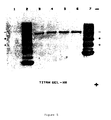
- the purification of immunoglobulin was monitored by agarose gel electrophoresis performed on a Helena electrophoresis unit.
- Samples (5.0 ⁇ L) were applied at the cathode end of the Titan HRE agarose gels (HELENA) and subjected to electrophoresis for 25-30 min 50 mM barbital buffer pH 8.6 for with a 100 V direct current potential.
- the proteins were fixed in 10% TCA for 10 min, and the gel dried and stained with 0.1% Coomassie Blue R in 20% methanol/10% acetic acid. The gel was destained in 20% methanol, 10% acetic acid.
- the antibody was recovered in fractions 29-34 (see Fig. 4) and pooled to give 30 mL antibody solution, containing 0.63 mg/mL protein. This solution was concentrated 10-fold using a stirred cell with a 10,000 M.W. cut-off membrane and stored in 0.2 mL aliquots at -70°C. The antibody was adjusted to 35 ⁇ g/mL with 100 mM sodium phosphate buffer, pH 5.0, prior to use for coating the plate wells for ELISA. Some of the purified IgG was adjusted to 2 mg/mL using 10 mM sodium carbonate buffer pH 9.6 and dialysed overnight at 4°C against the same buffer prior to conjugation with horse radish peroxidase (HRPO [SIGMA]).
- HRPO horse radish peroxidase
- Antibody solution (3.5 mL; 2 mg/mL in sodium carbonate buffer pH 9.6) was mixed with HRPO (1.15 mL; 2.02 mg/mL in 1 mM acetate buffer pH 4.4) to give a ratio of 4.0 mg IgG: 1.0 mg HRPO aldehyde and mixed at 20 rpm at room temperature in the dark for 2 hr.
- Sodium borohydride solution (0.1 mL; 4.0 mg/mL) was then added to the solution, followed by standing for 2 hr at 4°C.
- Amniotic fluid samples (15 ⁇ L of 0.02 mg protein/mL) in 45 ⁇ L sample buffer (20% glycerol (v/v), 2% (w/v) SDS, 5% (v/v) 2- ⁇ -mercaptoethanol, 0.00125% (w/v) bromophenol blue and 12.5% (v/v) 0.5M Tris-HCl, pH 6.8) were boiled for 10 minutes.
- SDS-polyacrylamide gel electrophoresis was performed on 7.5 and 12% gels according to the procedure of Laemmli, U.K., (Nature 227:680 (1970)). Calibration curves for molecular weight estimation were obtained from pre-stained standards (BIORAD) transferred to the membrane.
- Electrophoresis was performed on the Mini Protean II system (BIORAD) for 60 min at 180 V.
- Western blotting was performed according to the method of Towbin (1984) with some modifications; i.e. the use of Hybond membrane (AMERSHAM) and 48 mM Tris, 39 mM glycine, Ph 9.2 containing 20% (v/v) methanol as a blotting buffer.
- Electrophoretic transfer was performed at 15 V for 45 min using a Trans-Blot Electrophoretic Transfer Cell (BIORAD).
- Blots were soaked in 10 mM Tris., 0.9% NaCl (w/v), 100 mM L-lysine, 0.5% BSA (w/v) for 2 hr at room temperature and then washed in 3 x 10 min consecutive washes in 10 mM Tris, 0.9% NaCl (w/v) and 0.02% Tween-20 (v/v).
- the conjugate diluted in the same buffer (1/100), was added to the blot and left overnight at 4°C.
- Isoelectric focusing was performed on 1% agarose gels prepared with 3% sucrose and 2% Biolyte (v/v) 3.5-9.5 (BIORAD). Gels were pre-focused for 1 hr at 5 W, 1500 V, 150 mA with 1 M NaOH (cathode) and 0.005 M H 2 SO 4 (anode) buffers. Samples of amniotic fluid purified by Sephacryl S-200 column chromatography, and IEF standards (BIORAD) were applied to the gel on filter paper strips and focused for a further 2 hr. Proteins were transferred onto Hybond by placing the gel onto the surface and placing a weight onto the gel for 15 min. The antigen was detected on the blot by the procedure described for SDS-PAGE.
- the gel was stained for protein by initially drying the gel, fixing for 20 min using 35% methanol, 13% tricarboxylic acid, 3.5% sulfosalicylic acid, washing the gel in 30% methanol, 10% acetic acid and staining in Coomassie blue (0.2% w/v). The gel was destained in 30% methanol, 10% acetic acid.
- a column (2.5 cm x 100 cm) was packed with Sephacryl S-200 and equilibrated at 1.0 mL/min with 20 mM Tris-HCl (pH 7.0) for 12 hr. Following column equilibration (10 column volumes), gel filtration standards (BIORAD) were run and a standard curve constructed. Amniotic fluid (50 mL) was dialysed at 4°C against three 5.0 L changes of 20 mM Tris-HCl pH 7.0 and concentrated 20-fold on a stirred cell with a 10,000 MW cut-off membrane (Amicon).
- Nunc plates were activated for 4 hr at room temperature with 0.2 mL/well 0.2% glutaraldehyde prepared in 100 mM sodium phosphate buffer pH 5.0. The plates were washed three times in 100 mM sodium phosphate buffer pH 5.0 and incubated with 0.1 mL/well purified immunoglobulin (35 ⁇ g/mL in 50 mM sodium phosphate buffer pH 8.0) at 4°C overnight. Plates were washed three times in 0.9% NaCl and 0.1 mL per well of 100 mM lysine, 0.5% BSA, 0.02% azide was added and stored at 4°C for up to 14 days prior to use.
- the immunoglobulin coupled plates were washed three times in PBS [(high sait)HSJ/Tween wash buffer (3.2 mM Na 2 HPO 4 , 1.5 mM KH 2 PO 4 , 2.7 mM KCl, 272 mM NaCl, pH 7.4 containing 0.5% (v/v) Tween 20), followed by addition of the same buffer (25 ⁇ L per well) prior to sample application.
- 15 term amniotic fluids were pooled and serially-diluted 1/2-1/128 with PBS (HS)/Tween to allow the production of a standard curve. The concentration of the pool was given an arbitrary value of 100 U/L COVO Antigen.
- samples and standards were applied (0.1 mL/well) in duplicate and incubated at room temperature for 2 hr. Blanks containing PBS (HS)/Tween (0.1 mL/well) instead of sample, were also included.
- An amniotic fluid control was prepared from 20 random term amniotic fluids and run on every plate. After a further three washes in PBS (HS)/Tween, 50 ⁇ L IgG-HRPO conjugate was added to each well, and incubated at room temperature for 1 hr. After four washes with PBS (HS)/T, 0.1 mL of ABTS solution was allowed to react with the enzyme for 1 hr.
- OD sample Optical densities of samples (OD sample) were measured at a wavelength of 405 nm. Calibration curves were constructed from the amniotic fluid pool after subtraction of blank values (OD blank). The levels of antigen in individual specimens and controls were calculated from the standard curve.
- the PROM antigen level was measured in post-rupture of the membrane specimens (post-ROM) obtained from the following group of patients:-
- PROM antigen levels were measured in specimens of vaginal fluid collected from 50 women with intact membranes in the Labour Ward prior to artificial rupture of the membranes (pre-ROM). The specimens were regarded as negative controls if the women gave no history of fluid loss and liquor was not evident on clinical examination.
- PROM antigen levels were also measured in the sera obtained from 500 pregnant women who had blood samples collected routinely during pregnancy.
- vaginal speculum examination was performed and the posterior fornix was visualised. If fluid was present in the vagina, it was aspirated with a 2 mL syringe fitted with an aspiration cannula. Where fluid was not observed, the external cervical os was washed with 2 mL sterile saline and the fluid was then aspirated from the posterior fornix. Specimens were placed in sterile sample tubes and transported to the Laboratory where they were frozen at -70°C until analysis.
- amniotic fluid pool had a PROM antigen level of 36 U/L and gave a level of 18 U/L when diluted 1:1 with PBS (H/S)/Tween.
- Pre- and post-ROM (rupture of membrane) specimens of fluid were collected for analysis by an obstetrician from two separate groups of patients whose names were withheld from the Research staff and who were identified to them only by a randomly allocated number.
- first "blinded" group (Study A) specimens were obtained from six pregnant women who were booked for elective CS.
- vaginal fluid specimens were obtained, as described under "Procedure for vaginal fluid collection”.
- amniotic fluid was aspirated through intact membranes into a sterile syringe. Each specimen was numbered and sent to the Laboratory for analysis. Because there were two sets of triplets in the group, there were 10 post-ROM specimens.
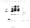
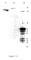
- Amniotic fluid supernatants (5 mL) were individually subjected to chromatography in 100 mL volume DEAE Affi-Gel Blue columns and serial 10 mL fractions were collected. These fractions were subjected to SDS-PAGE electrophoresis and fractions 4-10 were found to contain the PROM-Ag. These fractions were pooled and contained 1.4 mg of protein (Table 1). A typical SDS-PAGE result for this pool can be seen in Figure 1, lane 5.
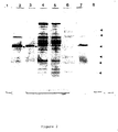
- the post-Affi-Gel Blue pools containing the antigen were concentrated 20-fold to 2.5 mL and individually applied to a pre-calibrated (2.5 x 7.5 cm) Sephacryl S-200 column. Serial 5 mL fractions were collected and subjected to SDS-PAGE electrophoresis to identify the protein at 55,000 Da. Fractions corresponding to molecular weight range of 310,000-350,000 Da were found to contain the protein giving an estimated native molecular weight of 330,000 Da. The sub-unit molecular weight from SDS-PAGE electrophoresis of 55,000 Da is consistent therefore with the native protein being a hexamer. Fractions containing the protein were pooled, yield 0.36 mg of protein (Table 1). A typical SDS-PAGE result is shown in Figure 2 which demonstrates that the protein is substantially pure with minor contaminants at sub-unit molecular weights of 82,000 and 30,000 Da, and one major contaminant of 57,000 Da.
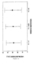
- mice Six BALB/c mice were immunized with PROM-Ag and were test bed on seven occasions from 6-15 weeks. These sera were tested for immunoreactivity to amniotic fluid using a one-site ELISA technique and compared with sera from non-immune BALB/c mice. These results are shown in Table 2. Two mice (#1,3) showed no immune response when assayed in this manner, three mice (#2,4,5) showed a partial response and one mouse (#6) demonstrated a 5-fold increase over the blank absorbance. This latter mouse was sacrificed and the spleen cells were fused with NS1 myeloma cells.
- Hybrids 1A3/42, 11B6/68, 2C5/48, 2C6/86 and 12D2/77; which reacted with amniotic fluid alone were cloned three times by limited dilution and retested for reactivity with amniotic fluid and maternal serum Figure 3
- Cell line 1A3/42 was selected for further analysis, and lines 11B6/68, 2C5/48, 2C6/86 and 12D2/77 were selected for storage in liquid nitrogen.
- This monoclonal antibody preparation was used both as a capture and, following conjugation with horseradish peroxidase, as the conjugate in the subsequent two-site ELISA assays.
- the conjugate was also used as a specific staining reagent to characterize the corresponding amniotic fluid protein by western blot analysis, and to test its identity to the original antigen purified from amniotic fluid.
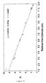
- Figure 6a shows the results in a western blot analysis following SDS-PAGE electrophoresis in 4-15% gradient gels in the presence of SDS/mercaptoethanol. Only one band of reactivity is seen and it is identical in three randomly selected term amniotic fluids. This band corresponds to a sub-unit molecular weight of 55,000 ⁇ 2,000 Da ( Figure 6b), a mass identical to that of the protein originally purified from amniotic fluid.
- the isoelectric point of the protein was also determined by western blot analysis (using monoclonal 1A3-HRPO conjugate) following isoelectric focusing in an agarose gel, using ampholines with a pH rage from 3-10. We used both purified protein and one native random term amniotic fluid for this analysis. The results are shown in Figure 7a. Only one major band of reactivity against purified protein is evident (Figure 7a) which corresponds to an isoelectric point of 4.9 ( Figure 7b).
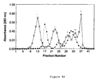
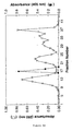
- the native molecular weight of the protein was determined by testing the reactivity of fractions eluted from a pre-calibrated Sephacryl S-200 column ( Figure 8a) using a two-site ELISA (a description of this assay follows). This assay uses monoclonal 1A3 as both the capture antibody and conjugate. The results are shown in Figure 8b. 2 mL of post DEAE Affi-Gel Blue amniotic fluid was loaded onto the column. Fractions 17-18, which corresponded to a molecular weight of 330,000 ⁇ 10,000 Da, reacted positively in the ELISA test ( Figure 8c). No other fractions were found to show reactivity. This native molecular weight corresponds with that found previously, during initial protein purification, and is consistent with monoclonal antibody 1A3 reacting with the antigen that was originally purified.
- amniotic fluids for use as a standard in the assay. This amniotic fluid pool was assigned a concentration of 100 U/L PROM-Ag. Randomly selected term amniotic fluids and maternal sera were used as specimens.
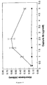
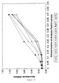
- Figure 9 is shown the effect of varying the concentration of capture antibody used to coat the plates. Response is measured by the development of colour at 40 nm. The mean (SEM) of three amniotic fluid and three maternal serum specimens are shown. The maternal sera do not react at any concentration but the response of amniotic fluid reaches a maximum at 2.5 ⁇ g/well. A reduced response is seen at higher antibody concentrations possibly due to steric hindrance or cross-reactivity problems giving a lower antigen binding capacity. Demonstrating the use of a narrower concentration range of capture antibody with a maximal response at 1.75 ⁇ g/well; this concentration was chosen for use in the optimized assay procedure.
- Figure 10 is shown the effect of different blocking agents used to suppress non-specific binding of non-specific proteins.
- Four reagents were compared: 100 mmol/L lysine, 0.5% (w/v) BSA in 100 mM sodium phosphate buffer pH 8.0, 100 mmol/L lysine in 100 mM sodium sulphate buffer pH 8.0; 5% (w/v) milk powder (Carnation Trim Milk Powder) in PBS and 1% (v/v) swine serum (GIBCO) in PBS.
- Serial dilutions (1:2, 1:5, 1:10 and 1:20) of the same pool of 10 amniotic fluids are compared.
- Blank wells contained sample buffer (PBS/Tween) only and were used to determine non-specific binding of antibody-HRPO conjugate to the well.
- the different blocking agents had little effect on the blank values but omission of BSA from the lysine solution, and the presence of swine serum and milk powder inhibited the binding of antigen when compared to the lysine/BSA blocking agent. Lysine/BSA was selected for use in the optimized procedure.
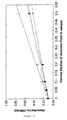
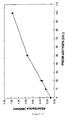
- Figure 11 is shown the effect of serial dilutions of antibody-HRPO conjugate.
- Results are the mean (SEM) of five randomly selected amniotic fluid specimens. After an initial plateau, response falls exponentially at dilutions greater than 1/150 indicating that the amount of tag antibody is becoming rate-limiting. A dilution of 1/150 was selected for use in the optimized assay procedure.
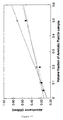
- Figure 12 is shown the effect on different sample diluents.
- Three reagents are compared: PBS/Tween-20, PBS and PBS/swine serum (1% [v/v]).
- Serial dilutions (1:2, 1:5, 1:10, 1:20 and 1:50) of the same pool of amniotic fluids are compared.
- Blank wells contain sample diluent only and are used to compare the effects of non-specific binding.
- the different sample diluents had little effect on the non-specific binding, but omission of Tween or addition of swine serum appeared to inhibit the binding of protein (PROM-Ag), when compared to the result using PBS/Tween reagent.
- PBS/Tween was selected for use in the optimized procedure.
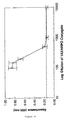
- FIG 13 is shown the effect of the pH of the sample diluent on non-specific binding and on antigen binding.
- PBS/Tween buffer at pH 2, 3, 4, 5, 6, 7 and 8 were prepared and used to dilute a pool of five amniotic fluids to 1:2, 1:4, 1:8, 1:16 and 1:32. Addition of buffer alone was used to assess non-specific binding. The pH appears to have little effect on non-specific binding. However, binding of the antigen is inhibited at acid pH presumably due to the masking of charged groups on the antigen or the antibody. This is less marked at low amniotic fluid dilutions probably due to the buffering effect of the amniotic fluid itself.
- sample diluent buffer pH 7.0 for use in the assay procedure.
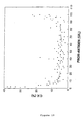
- a precision profile of 120 vaginal fluid specimens is shown in Figure 19.
- the assay shows good precision between 20-90 U/L with a CV of duplicates of less than 10%.
- the within-run precision was estimated from two pools analysed 50 times each. These pools gave values of 20 (1.2) mean (SD) and 50 (1.9) mean (SD) yielding CV's of 4% and 2% respectively.
- the between-run precision was estimated from two pools run 10 times each in four different rows. These pools gave values of 19.8 (1.5) mean (SD) and 48.8 (2.2) mean (SD) yielding CV's of 7.5% and 4.5% respectively.
- the NACC had no effect on PROM antigen levels in 40 term amniotic fluids showing that it did not inhibit the immunoreactivity of the PROM antigen. It was, however, active in lysing mucus as observed by visual inspection and enabled the detection of PROM antigen in 16 vaginal fluid specimens.
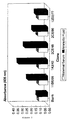
- the reference range for PROM antigen in amniotic fluid was investigated on amniotic fluid specimens obtained at different gestational ages during pregnancy. The results are shown in Figure 21.
- the level in the first trimester (26.5 (2.47) U/L) is significantly lower than that in the third trimester (30.2 (2.56) U/L).
- the levels during the second trimester 28.4 (2.03) U/L were similar to those of the third trimester.
- NACC N-Acetylcysteine
- Monoclonal antibody 1A3 referred to herein has been deposited at the European Collection of Animal Cell Cultures (ECACC) and has been allocated accession number 94031901. Typical total protein results at different stages of purification of amniotic fluid protein from five random term amniotic fluid. Fraction Volume (mL) Protein (mg/mL) Total Protein (mg) Total Activ. (U) Specific Activity (U/mg) Yield (%) Purification Amniotic Fluid 10.0 5.42 54.2 960 17.7 100 1 Ammon.
- Intefering Substances Mean (U/L) Range (U/L) Amniotic fluid (Control) 18 15-21 Haemolysed serum 10 6-12 Haemolysed serum (-Albumin) 16 14-21 Urine 18 16-20 Tap water 17 15-20 Prostaglandin E2 cream 18 16-21 Cord blood 11 7-14 Cord blood (-Albumin) 16 13-19 Blind study A.
- Pre-CS-PROM Antigen Level U/L
- Post-CS-ROM PROM Antigen Level U/L
- FIGURE 1 Amniotic fluid proteins following denaturation with SDS/mercaptoethanol during purification of PROM antigen (see arrows) on 4-15% SDS-PAGE gradient gels.
- FIGURE 2 Amniotic fluid proteins following denaturation with SDS/mercaptoethanol during purification of PROM antigen (see arrows) on 4-15% SDS-PAGE gradient gels.
- FIGURE 3 Reactivity of hybridoma supernatants with maternal serum and amniotic fluid by indirect on-site ELISA.
- FIGURE 4 Isolation of monoclonal immunoglobulin from ascitic fluid on a hydroxylapatite column with a 50-500 mM linear gradient of sodium phosphate buffer (pH 7.0).
- FIGURE 5 Agarose gel electrophoresis of monoclonal antibody following hydroxylapatite chromatography.
- FIGURE 6A Estimation of sub-unit molecular weight of PROM antigen by Western blot analysis with monoclonal 1A3-HRPO conjugate. Electrophoresis, following incubation with SDS/mercaptoethanol was performed on a 4-15% SDS-PAGE gradient gel.
- FIGURE 6B Standard curve constructed from FIGURE 6A.
- FIGURE 7A Estimation of isoelectric point of PROM antigen by western blot analysis with 1A3-HRPO conjugate. Isoelectric focusing was performed on an agarose gel pH range 3-10.
- FIGURE 7B Standard curve constructed from FIGURE 7A.
- FIGURE 8A Elution profile of post DEAE Affigel Blue fractions of amniotic fluid from a Sephocryl S-200 column.
- FIGURE 8B Estimation of the native molecular weight of PROM antigen by gel permeation chromatography on a Sephacryl S-200 column.
- FIGURE 8C Estimation of the native molecular weight of PROM antigen from molecular weight standards passed through a column of Sephacryl S-200.
- FIGURE 9 Effect of varying capture antibody concentration used on coating wells in the detection of three amniotic fluids and three maternal sera.
- FIGURE 10 Effect of four different blocking agents on the reactivity of serial dilutions of PROM antigen in an amniotic fluid pool.
- FIGURE 11 Effect of dilution of the 1A3-HRPO conjugate on the reactivity of five random amniotic fluids.
- FIGURE 12 Effect of three different sample diluents upon the reactivity of an amniotic fluid pool.
- FIGURE 13 Effect of sample diluent pH on the reactivity of serial dilutions of an amniotic pool.
- FIGURE 14 Effect of varying sample incubation time on the reactivity of three amniotic fluids.
- FIGURE 15 Effect of varying incubation time with 1A3-HRPO conjugate on reactivity on an amniotic fluid pool.
- FIGURE 16 Effect of varying incubation time with HRPO substrate on the reactivity of different dilutions of an amniotic fluid pool.
- FIGURE 17 Typical standard curve obtained with the optimized ELISA procedure. Mean (SD) of standards in five different runs.
- FIGURE 18 Dilution of three individual term amniotic fluids compared with the standard curve.
- FIGURE 19 Precision profile of 120 vaginal fluid specimens. CV is calculated from duplicates as the difference x 100/mean.
- FIGURE 20 Effect of pre-incubation with N-Acetyl cysteine on PROM antigen levels in 40 amniotic fluids.
- FIGURE 21 Reference range of PROM antigen levels in 1st, 2nd and 3rd trimester amniotic fluid.
Landscapes
- Health & Medical Sciences (AREA)
- Chemical & Material Sciences (AREA)
- Organic Chemistry (AREA)
- Life Sciences & Earth Sciences (AREA)
- Biochemistry (AREA)
- Biophysics (AREA)
- General Health & Medical Sciences (AREA)
- Genetics & Genomics (AREA)
- Medicinal Chemistry (AREA)
- Molecular Biology (AREA)
- Proteomics, Peptides & Aminoacids (AREA)
- Gynecology & Obstetrics (AREA)
- Immunology (AREA)
- Pregnancy & Childbirth (AREA)
- Reproductive Health (AREA)
- Toxicology (AREA)
- Zoology (AREA)
- Gastroenterology & Hepatology (AREA)
- Medicines Containing Antibodies Or Antigens For Use As Internal Diagnostic Agents (AREA)
- Peptides Or Proteins (AREA)
- Preparation Of Compounds By Using Micro-Organisms (AREA)
- Micro-Organisms Or Cultivation Processes Thereof (AREA)
- Investigating Or Analysing Materials By The Use Of Chemical Reactions (AREA)
- Investigating Or Analysing Biological Materials (AREA)
Claims (12)
- Isoliertes Antigen, erhältlich aus Fruchtwasser, charakterisiert durch:(i) ein Molekulargewicht einer Untereinheit von etwa 55.000 Dalton, gemessen durch SDS-PAGE mit Gelen verschiedener Acrylamid-Konzentrationen;(ii) eine native Molekulargewichtsanalyse, wie durch Gelpermeationschromatographie bestimmt, die ein anscheindendes Molekulargewicht von etwa 330.000 Dalton zeigt;(iii) eine beobachtete Bande mit einem isoelektrischen Punkt von etwa 4,9, erhalten durch isoelektrische Fokussierungsstudien; und(iv) Immunoreaktivität mit dem monoklonalen Antikörper 1A3, produziert von dem Hybridoma, das bei der European Animal Cell Culture Collection hinterlegt ist und die Zugangsnummer 94031901 aufweist, wobei der monoklonale Antikörper beim Test mit einem Sandwich-ELISA-Test keine Reaktivität in Bezug auf maternale Serumproteine aufweist, aber in Bezug auf das Fruchtwasser Reaktivität zeigt.
- Verfahren zum Reinigen eines Antigens aus Fruchtwasser, wie in Anspruch 1 beansprucht, beinhaltend die Schritte:(i) Reaktion des Fruchtwassers mit Ammoniumsulfat;(ii) Zentrifugieren des Produktes der Reaktion (i) und Erhalten des resultierenden Pellets oder Präzipitats;(iii) Resuspendieren des Pellets oder Präzipitats in Puffer und Durchleiten der resultierenden Lösung durch eine Säule aus DEAE-Affi-Gel Blue;(iv) Testen der ungebundenen Fraktionen nach Passage durch die Säule auf das Vorliegen eines Proteins eines Untereinheitsmolekulargewichts von etwa 55.000 Dalton durch SDS-PAGE-Elektrophorese;(v) Konzentrieren von Fraktionen mit dem Molekulargewicht von 55.000 Dalton und Überleiten der konzentrierten Fraktionen über eine Sephacryl S-200-Säule und Sammeln von Fraktionen mit einem Molekulargewicht von 330.000.
- Verfahren wie in Anspruch 2 beansprucht, wobei der Puffer 0,02 M K2HPO4 ist.
- Bindungsagens, immunoreaktiv mit dem Antigen aus Anspruch 1.
- Bindungsagens wie in Anspruch 4 beansprucht, wobei das Bindungsagens ein monoklonaler oder polyklonaler Antikörper ist.
- Monoklonaler Antikörper 1A3, hinterlegt bei der European Animal Cell Culture Collection unter der Zugangsnummer 94031901.
- Verfahren zur Detektion von Rissen der amniotischen Membran in schwangeren Säugetieren, einschließlich Menschen, beinhaltend die Schritte:(i) Entnehmen einer Probe von Vaginalflüssigkeit eines schwangeren weiblichen Individuums;(ii) zur Reaktion bringen der Probe mit einem Antikörper, abgeleitet von einem Antigen, das dadurch charakterisiert ist, dass es die folgenden physikalischen Eigenschaften aufweist:(a) ein Molekulargewicht einer Untereinheit von etwa 55.000 Dalton, gemessen durch SDS-PAGE mit Gelen verschiedener Acrylamid-Konzentrationen;(b) eine native Molekulargewichtsanalyse, wie durch Gelpermeationschromatographie bestimmt, die ein anscheindendes Molekulargewicht von etwa 330.000 Dalton zeigt;(c) eine beobachtete Bande mit einem isoelektrischen Punkt von etwa 4,9, erhalten durch isoelektrische Fokussierungsstudien; und(d) Immunoreaktivität mit dem monoklonaren Antikörper aus Anspruch 6, der beim Testen mit einem Sandwich-ELISA-Assay keine Reaktivität in Bezug auf maternale Serumproteine aufweist, aber in Bezug auf Fruchtwasser reaktiv ist; und(iii) Detektieren der Reaktivität in Schritt (ii).
- Verfahren zur Detektion, wie in Anspruch 7 beansprucht, wobei der Antikörper der in Anspruch 6 beanspruchte monoklonale Antikörper 1A3 ist.
- Verfahren zur Detektion wie in Anspruch 7 beansprucht, wobei der Antikörper auf einer inerten Oberfläche immobilisiert ist.
- Verfahren nach Anspruch 7, wobei die Signalamplifikation beinhaltet, dass der Antikörper ein gebundenes Enzym aufweist, das in Bezug auf ein Enzymsubstrat reaktiv ist.
- Verfahren nach Anspruch 10, wobei das Enzym Meerrettichperoxidase ist.
- Verfahren nach Anspruch 10, wobei das Enzymsubstrat Azinobis-(3-ethylbenzthiazolinsulfonsäure)diammoniumsalz ist.
Applications Claiming Priority (4)
| Application Number | Priority Date | Filing Date | Title |
|---|---|---|---|
| AUPL794593 | 1993-03-23 | ||
| AUPL794593 | 1993-03-23 | ||
| AUPL7945/93 | 1993-03-23 | ||
| PCT/AU1994/000144 WO1994021687A1 (en) | 1993-03-23 | 1994-03-23 | A method of detecting rupture of the amniotic membranes in pregant mammals |
Publications (3)
| Publication Number | Publication Date |
|---|---|
| EP0689552A1 EP0689552A1 (de) | 1996-01-03 |
| EP0689552A4 EP0689552A4 (de) | 1997-07-02 |
| EP0689552B1 true EP0689552B1 (de) | 2002-10-30 |
Family
ID=3776795
Family Applications (1)
| Application Number | Title | Priority Date | Filing Date |
|---|---|---|---|
| EP94911016A Expired - Lifetime EP0689552B1 (de) | 1993-03-23 | 1994-03-23 | Methode zur detektion von rissen in amniomembranen bei schwangeren säugetieren |
Country Status (6)
| Country | Link |
|---|---|
| US (1) | US5783396A (de) |
| EP (1) | EP0689552B1 (de) |
| JP (1) | JP3441076B2 (de) |
| AT (1) | ATE226959T1 (de) |
| DE (1) | DE69431621D1 (de) |
| WO (1) | WO1994021687A1 (de) |
Families Citing this family (8)
| Publication number | Priority date | Publication date | Assignee | Title |
|---|---|---|---|---|
| WO2002064633A1 (en) * | 2001-02-14 | 2002-08-22 | Victor Voroteliak | Monoclonal antibody against an unknown antigen from amniotic flu id and its method of preparation |
| US7399278B1 (en) | 2003-05-05 | 2008-07-15 | Los Angeles Biomedical Research Institute At Harbor-Ucla Medical Center | Method and system for measuring amniotic fluid volume and/or assessing fetal weight |
| US20050131287A1 (en) * | 2003-12-16 | 2005-06-16 | Kimberly-Clark Worldwide, Inc. | Detection of premature rupture of the amniotic membrane |
| US7863007B2 (en) * | 2005-09-15 | 2011-01-04 | Swiss Asian Property Limited | Marker for prolonged rupture of membranes |
| WO2010150804A1 (ja) * | 2009-06-26 | 2010-12-29 | 公立大学法人奈良県立医科大学 | 破水および羊水塞栓症の検査方法 |
| ES2690782T3 (es) | 2012-10-24 | 2018-11-22 | Nyu Winthrop Hospital | Biomarcador no invasivo para identificar sujetos en riesgo de parto prematuro |
| CA3075688A1 (en) | 2017-09-13 | 2019-03-21 | Progenity, Inc. | Preeclampsia biomarkers and related systems and methods |
| EP4070113A4 (de) | 2019-12-04 | 2023-12-20 | Biora Therapeutics, Inc. | Beurteilung von präeklampsie unter verwendung von tests für freien und dissoziierten plazentalen wachstumsfaktor |
Family Cites Families (6)
| Publication number | Priority date | Publication date | Assignee | Title |
|---|---|---|---|---|
| DE3780446T2 (de) * | 1987-03-24 | 1992-12-24 | Michael Silberman | Verfahren zur feststellung von schwangerschaftsstoerungen. |
| AU620351B2 (en) * | 1987-11-17 | 1992-02-20 | Adeza Biomedical Corporation | Vaginal sample test and reagents |
| US5281522A (en) * | 1988-09-15 | 1994-01-25 | Adeza Biomedical Corporation | Reagents and kits for determination of fetal fibronectin in a vaginal sample |
| WO1990010061A1 (en) * | 1989-03-02 | 1990-09-07 | Griffith University | Diamine oxidase and assay for rupture of amniotic membrane in pregnant mammals |
| FI86777C (fi) * | 1990-12-31 | 1992-10-12 | Medix Biochemica Ab Oy | Foerfarande foer diagnosticering av bristningen av fosterhinnorna samt reagensfoerpackning foer anvaendning daervid |
| US5514598A (en) * | 1993-11-30 | 1996-05-07 | Doody; Michael | Prenatal detection of meconium |
-
1994
- 1994-03-23 AT AT94911016T patent/ATE226959T1/de not_active IP Right Cessation
- 1994-03-23 US US08/513,902 patent/US5783396A/en not_active Expired - Fee Related
- 1994-03-23 DE DE69431621T patent/DE69431621D1/de not_active Expired - Lifetime
- 1994-03-23 WO PCT/AU1994/000144 patent/WO1994021687A1/en not_active Ceased
- 1994-03-23 JP JP52043194A patent/JP3441076B2/ja not_active Expired - Fee Related
- 1994-03-23 EP EP94911016A patent/EP0689552B1/de not_active Expired - Lifetime
Also Published As
| Publication number | Publication date |
|---|---|
| EP0689552A1 (de) | 1996-01-03 |
| JPH08508015A (ja) | 1996-08-27 |
| DE69431621D1 (de) | 2002-12-05 |
| WO1994021687A1 (en) | 1994-09-29 |
| JP3441076B2 (ja) | 2003-08-25 |
| EP0689552A4 (de) | 1997-07-02 |
| US5783396A (en) | 1998-07-21 |
| ATE226959T1 (de) | 2002-11-15 |
Similar Documents
| Publication | Publication Date | Title |
|---|---|---|
| US5756682A (en) | Assay for cardiac troponin I | |
| US6172198B1 (en) | PAPP-A, its immunodetection and uses | |
| EP0689552B1 (de) | Methode zur detektion von rissen in amniomembranen bei schwangeren säugetieren | |
| US4554256A (en) | Antigen associated with early detection of mammalian pregnancy | |
| US5108898A (en) | Use of fibronectin having a variable included type III repeat sequence as a marker for toxemia in pregnancy | |
| KR910000903B1 (ko) | 인체의 비-소 세포 폐암치료용 단클론의 항체 및 항원 | |
| AU2002366097B2 (en) | Compositions and methods for accurate early pregnancy diagnosis | |
| JP3742649B2 (ja) | 切迫分娩についての危険にある女性における膜の破裂を決定するための検定方法 | |
| US4530908A (en) | Diagnosis and treatment of fluke infections with monoclonal antibodies | |
| US4705748A (en) | Antigen associated with early detection of mammalian pregnancy | |
| US6187549B1 (en) | Protein as a diagnostic of cancer | |
| AU685096B2 (en) | A method of detecting rupture of the amniotic membranes in pregant mammals | |
| US4416866A (en) | Diagnosis and treatment of fluke infections with monoclonal antibodies | |
| AU2005323518B2 (en) | Enriched PAG-55 fraction and methods for early detection of pregnancy in ungulate animals | |
| SAJI et al. | Identification and Characterization of a Human Sperm Antigen Corresponding to Sperm‐Immobilizing Antibodies | |
| JPWO1997008549A1 (ja) | 腎疾患の検知法、診断薬及び診断用キット | |
| Naz | Effects of sperm-reactive antibodies present in human infertile sera on fertility of female rabbits | |
| JP2580028B2 (ja) | 抗ひと精子抗体、その製法および用途 | |
| Tornehave et al. | Immunohistochemical aspects of immunological cross-reaction and masking of epitopes for localization studies on pregnancy-associated plasma protein A | |
| Findlay et al. | The Nature and Role of Pregnancy‐Associated Antigens and the Endocrinology of Early Pregnancy in the Ewe | |
| AU726994B2 (en) | PAPP-A, its immunodetection and uses | |
| JPH03154867A (ja) | 1―メチルアデノシンの免疫学的測定方法,そのための測定試薬及び試薬キット | |
| JPH0892297A (ja) | μ−カルパイン80KサブユニットのN末端ペプチドに対する抗体及びこれを用いる免疫測定方法及び測定試薬 | |
| JPH06324046A (ja) | 悪性腫瘍の検出方法及びキット | |
| AU6370194A (en) | Papp-a, its immunodetection and uses |
Legal Events
| Date | Code | Title | Description |
|---|---|---|---|
| PUAI | Public reference made under article 153(3) epc to a published international application that has entered the european phase |
Free format text: ORIGINAL CODE: 0009012 |
|
| 17P | Request for examination filed |
Effective date: 19950906 |
|
| AK | Designated contracting states |
Kind code of ref document: A1 Designated state(s): AT BE CH DE DK ES FR GB GR IE IT LI NL PT SE |
|
| A4 | Supplementary search report drawn up and despatched |
Effective date: 19970514 |
|
| AK | Designated contracting states |
Kind code of ref document: A4 Designated state(s): AT BE CH DE DK ES FR GB GR IE IT LI NL PT SE |
|
| 17Q | First examination report despatched |
Effective date: 19990415 |
|
| GRAG | Despatch of communication of intention to grant |
Free format text: ORIGINAL CODE: EPIDOS AGRA |
|
| GRAG | Despatch of communication of intention to grant |
Free format text: ORIGINAL CODE: EPIDOS AGRA |
|
| GRAH | Despatch of communication of intention to grant a patent |
Free format text: ORIGINAL CODE: EPIDOS IGRA |
|
| GRAH | Despatch of communication of intention to grant a patent |
Free format text: ORIGINAL CODE: EPIDOS IGRA |
|
| GRAA | (expected) grant |
Free format text: ORIGINAL CODE: 0009210 |
|
| AK | Designated contracting states |
Kind code of ref document: B1 Designated state(s): AT BE CH DE DK ES FR GB GR IE IT LI NL PT SE |
|
| PG25 | Lapsed in a contracting state [announced via postgrant information from national office to epo] |
Ref country code: NL Free format text: LAPSE BECAUSE OF FAILURE TO SUBMIT A TRANSLATION OF THE DESCRIPTION OR TO PAY THE FEE WITHIN THE PRESCRIBED TIME-LIMIT Effective date: 20021030 Ref country code: LI Free format text: LAPSE BECAUSE OF FAILURE TO SUBMIT A TRANSLATION OF THE DESCRIPTION OR TO PAY THE FEE WITHIN THE PRESCRIBED TIME-LIMIT Effective date: 20021030 Ref country code: IT Free format text: LAPSE BECAUSE OF FAILURE TO SUBMIT A TRANSLATION OF THE DESCRIPTION OR TO PAY THE FEE WITHIN THE PRESCRIBED TIME-LIMIT;WARNING: LAPSES OF ITALIAN PATENTS WITH EFFECTIVE DATE BEFORE 2007 MAY HAVE OCCURRED AT ANY TIME BEFORE 2007. THE CORRECT EFFECTIVE DATE MAY BE DIFFERENT FROM THE ONE RECORDED. Effective date: 20021030 Ref country code: GR Free format text: LAPSE BECAUSE OF FAILURE TO SUBMIT A TRANSLATION OF THE DESCRIPTION OR TO PAY THE FEE WITHIN THE PRESCRIBED TIME-LIMIT Effective date: 20021030 Ref country code: FR Free format text: LAPSE BECAUSE OF FAILURE TO SUBMIT A TRANSLATION OF THE DESCRIPTION OR TO PAY THE FEE WITHIN THE PRESCRIBED TIME-LIMIT Effective date: 20021030 Ref country code: CH Free format text: LAPSE BECAUSE OF FAILURE TO SUBMIT A TRANSLATION OF THE DESCRIPTION OR TO PAY THE FEE WITHIN THE PRESCRIBED TIME-LIMIT Effective date: 20021030 Ref country code: BE Free format text: LAPSE BECAUSE OF FAILURE TO SUBMIT A TRANSLATION OF THE DESCRIPTION OR TO PAY THE FEE WITHIN THE PRESCRIBED TIME-LIMIT Effective date: 20021030 Ref country code: AT Free format text: LAPSE BECAUSE OF FAILURE TO SUBMIT A TRANSLATION OF THE DESCRIPTION OR TO PAY THE FEE WITHIN THE PRESCRIBED TIME-LIMIT Effective date: 20021030 |
|
| REF | Corresponds to: |
Ref document number: 226959 Country of ref document: AT Date of ref document: 20021115 Kind code of ref document: T |
|
| REG | Reference to a national code |
Ref country code: GB Ref legal event code: FG4D |
|
| RIC1 | Information provided on ipc code assigned before grant |
Free format text: 7C 07K 14/47 A, 7C 07K 1/36 B, 7C 12P 21/08 B, 7G 01N 33/53 B, 7G 01N 33/68 B |
|
| REG | Reference to a national code |
Ref country code: CH Ref legal event code: EP |
|
| REG | Reference to a national code |
Ref country code: IE Ref legal event code: FG4D |
|
| REF | Corresponds to: |
Ref document number: 69431621 Country of ref document: DE Date of ref document: 20021205 |
|
| PG25 | Lapsed in a contracting state [announced via postgrant information from national office to epo] |
Ref country code: SE Free format text: LAPSE BECAUSE OF FAILURE TO SUBMIT A TRANSLATION OF THE DESCRIPTION OR TO PAY THE FEE WITHIN THE PRESCRIBED TIME-LIMIT Effective date: 20030130 Ref country code: PT Free format text: LAPSE BECAUSE OF FAILURE TO SUBMIT A TRANSLATION OF THE DESCRIPTION OR TO PAY THE FEE WITHIN THE PRESCRIBED TIME-LIMIT Effective date: 20030130 Ref country code: DK Free format text: LAPSE BECAUSE OF FAILURE TO SUBMIT A TRANSLATION OF THE DESCRIPTION OR TO PAY THE FEE WITHIN THE PRESCRIBED TIME-LIMIT Effective date: 20030130 |
|
| PG25 | Lapsed in a contracting state [announced via postgrant information from national office to epo] |
Ref country code: DE Free format text: LAPSE BECAUSE OF FAILURE TO SUBMIT A TRANSLATION OF THE DESCRIPTION OR TO PAY THE FEE WITHIN THE PRESCRIBED TIME-LIMIT Effective date: 20030131 |
|
| PG25 | Lapsed in a contracting state [announced via postgrant information from national office to epo] |
Ref country code: GB Free format text: LAPSE BECAUSE OF NON-PAYMENT OF DUE FEES Effective date: 20030323 |
|
| PG25 | Lapsed in a contracting state [announced via postgrant information from national office to epo] |
Ref country code: IE Free format text: LAPSE BECAUSE OF NON-PAYMENT OF DUE FEES Effective date: 20030324 |
|
| NLV1 | Nl: lapsed or annulled due to failure to fulfill the requirements of art. 29p and 29m of the patents act | ||
| PG25 | Lapsed in a contracting state [announced via postgrant information from national office to epo] |
Ref country code: ES Free format text: LAPSE BECAUSE OF FAILURE TO SUBMIT A TRANSLATION OF THE DESCRIPTION OR TO PAY THE FEE WITHIN THE PRESCRIBED TIME-LIMIT Effective date: 20030429 |
|
| REG | Reference to a national code |
Ref country code: CH Ref legal event code: PL |
|
| EN | Fr: translation not filed | ||
| PLBE | No opposition filed within time limit |
Free format text: ORIGINAL CODE: 0009261 |
|
| STAA | Information on the status of an ep patent application or granted ep patent |
Free format text: STATUS: NO OPPOSITION FILED WITHIN TIME LIMIT |
|
| 26N | No opposition filed |
Effective date: 20030731 |
|
| GBPC | Gb: european patent ceased through non-payment of renewal fee |
Effective date: 20030323 |
|
| REG | Reference to a national code |
Ref country code: IE Ref legal event code: MM4A |