EP0614652B1 - Pansement pour l'hémostase - Google Patents
Pansement pour l'hémostase Download PDFInfo
- Publication number
- EP0614652B1 EP0614652B1 EP94103683A EP94103683A EP0614652B1 EP 0614652 B1 EP0614652 B1 EP 0614652B1 EP 94103683 A EP94103683 A EP 94103683A EP 94103683 A EP94103683 A EP 94103683A EP 0614652 B1 EP0614652 B1 EP 0614652B1
- Authority
- EP
- European Patent Office
- Prior art keywords
- pad
- hemostasis
- base material
- sticking
- layer
- Prior art date
- Legal status (The legal status is an assumption and is not a legal conclusion. Google has not performed a legal analysis and makes no representation as to the accuracy of the status listed.)
- Expired - Lifetime
Links
- 239000000463 material Substances 0.000 title claims description 170
- 230000023597 hemostasis Effects 0.000 title claims description 83
- 238000003825 pressing Methods 0.000 claims description 84
- 239000010410 layer Substances 0.000 claims description 38
- 239000012790 adhesive layer Substances 0.000 claims description 28
- 210000004369 blood Anatomy 0.000 claims description 22
- 239000008280 blood Substances 0.000 claims description 22
- 210000001367 artery Anatomy 0.000 claims description 20
- 208000032843 Hemorrhage Diseases 0.000 claims description 16
- 210000004204 blood vessel Anatomy 0.000 claims description 15
- 210000003462 vein Anatomy 0.000 claims description 11
- 230000000694 effects Effects 0.000 claims description 10
- 239000002344 surface layer Substances 0.000 claims description 10
- 206010020112 Hirsutism Diseases 0.000 claims description 2
- 230000008021 deposition Effects 0.000 claims 1
- 239000000853 adhesive Substances 0.000 description 22
- 230000001070 adhesive effect Effects 0.000 description 22
- 230000000740 bleeding effect Effects 0.000 description 14
- 239000004744 fabric Substances 0.000 description 14
- -1 acryl Chemical group 0.000 description 11
- 238000005259 measurement Methods 0.000 description 10
- 238000000034 method Methods 0.000 description 10
- 231100000862 numbness Toxicity 0.000 description 10
- 229920000742 Cotton Polymers 0.000 description 9
- 210000003017 ductus arteriosus Anatomy 0.000 description 8
- 210000002321 radial artery Anatomy 0.000 description 8
- 238000011084 recovery Methods 0.000 description 8
- 208000027418 Wounds and injury Diseases 0.000 description 7
- 210000000707 wrist Anatomy 0.000 description 7
- 239000004698 Polyethylene Substances 0.000 description 6
- 229920000297 Rayon Polymers 0.000 description 6
- 229920002678 cellulose Polymers 0.000 description 6
- 239000001913 cellulose Substances 0.000 description 6
- 230000008034 disappearance Effects 0.000 description 6
- 238000002347 injection Methods 0.000 description 6
- 239000007924 injection Substances 0.000 description 6
- 229920000573 polyethylene Polymers 0.000 description 6
- 239000002964 rayon Substances 0.000 description 6
- 238000002560 therapeutic procedure Methods 0.000 description 6
- 239000006260 foam Substances 0.000 description 5
- 239000011505 plaster Substances 0.000 description 5
- 210000003811 finger Anatomy 0.000 description 4
- 230000002439 hemostatic effect Effects 0.000 description 4
- 230000036556 skin irritation Effects 0.000 description 4
- 230000000638 stimulation Effects 0.000 description 4
- 238000012360 testing method Methods 0.000 description 4
- 210000002559 ulnar artery Anatomy 0.000 description 4
- 206010015150 Erythema Diseases 0.000 description 3
- 230000017531 blood circulation Effects 0.000 description 3
- 210000004177 elastic tissue Anatomy 0.000 description 3
- 231100000321 erythema Toxicity 0.000 description 3
- 238000000605 extraction Methods 0.000 description 3
- 230000002349 favourable effect Effects 0.000 description 3
- 210000001105 femoral artery Anatomy 0.000 description 3
- 210000002683 foot Anatomy 0.000 description 3
- 238000004806 packaging method and process Methods 0.000 description 3
- 229920002635 polyurethane Polymers 0.000 description 3
- 239000004814 polyurethane Substances 0.000 description 3
- 206010030113 Oedema Diseases 0.000 description 2
- 238000002845 discoloration Methods 0.000 description 2
- 238000011156 evaluation Methods 0.000 description 2
- 238000010030 laminating Methods 0.000 description 2
- 210000005036 nerve Anatomy 0.000 description 2
- 229920003023 plastic Polymers 0.000 description 2
- 239000004033 plastic Substances 0.000 description 2
- 239000000758 substrate Substances 0.000 description 2
- 229920001342 Bakelite® Polymers 0.000 description 1
- 241001133795 Brahea Species 0.000 description 1
- 241000197194 Bulla Species 0.000 description 1
- 206010011703 Cyanosis Diseases 0.000 description 1
- 206010014513 Embolism arterial Diseases 0.000 description 1
- 206010020772 Hypertension Diseases 0.000 description 1
- 239000004677 Nylon Substances 0.000 description 1
- 239000004743 Polypropylene Substances 0.000 description 1
- 230000001464 adherent effect Effects 0.000 description 1
- 239000002390 adhesive tape Substances 0.000 description 1
- 238000002583 angiography Methods 0.000 description 1
- 238000002399 angioplasty Methods 0.000 description 1
- 239000004637 bakelite Substances 0.000 description 1
- 238000005452 bending Methods 0.000 description 1
- 208000002352 blister Diseases 0.000 description 1
- 230000036772 blood pressure Effects 0.000 description 1
- 238000010241 blood sampling Methods 0.000 description 1
- 210000000988 bone and bone Anatomy 0.000 description 1
- 239000003795 chemical substances by application Substances 0.000 description 1
- 230000006835 compression Effects 0.000 description 1
- 238000007906 compression Methods 0.000 description 1
- 238000011109 contamination Methods 0.000 description 1
- 230000003247 decreasing effect Effects 0.000 description 1
- 238000003745 diagnosis Methods 0.000 description 1
- 238000000502 dialysis Methods 0.000 description 1
- 229920001971 elastomer Polymers 0.000 description 1
- 238000001631 haemodialysis Methods 0.000 description 1
- 230000000322 hemodialysis Effects 0.000 description 1
- 210000004072 lung Anatomy 0.000 description 1
- 238000004519 manufacturing process Methods 0.000 description 1
- 239000002184 metal Substances 0.000 description 1
- 238000012544 monitoring process Methods 0.000 description 1
- 231100000957 no side effect Toxicity 0.000 description 1
- 229920001778 nylon Polymers 0.000 description 1
- 239000002985 plastic film Substances 0.000 description 1
- 239000002984 plastic foam Substances 0.000 description 1
- 239000011120 plywood Substances 0.000 description 1
- 229920000728 polyester Polymers 0.000 description 1
- 229920001155 polypropylene Polymers 0.000 description 1
- 229920001296 polysiloxane Polymers 0.000 description 1
- 239000004800 polyvinyl chloride Substances 0.000 description 1
- 229920000915 polyvinyl chloride Polymers 0.000 description 1
- 239000000843 powder Substances 0.000 description 1
- 238000005096 rolling process Methods 0.000 description 1
- 239000005060 rubber Substances 0.000 description 1
- 238000007789 sealing Methods 0.000 description 1
- 125000006850 spacer group Chemical group 0.000 description 1
- 230000035488 systolic blood pressure Effects 0.000 description 1
- 239000004753 textile Substances 0.000 description 1
- 230000001225 therapeutic effect Effects 0.000 description 1
- 210000003813 thumb Anatomy 0.000 description 1
- 238000011282 treatment Methods 0.000 description 1
- 125000000391 vinyl group Chemical group [H]C([*])=C([H])[H] 0.000 description 1
- 229920002554 vinyl polymer Polymers 0.000 description 1
- XLYOFNOQVPJJNP-UHFFFAOYSA-N water Substances O XLYOFNOQVPJJNP-UHFFFAOYSA-N 0.000 description 1
Images


Classifications
-
- A—HUMAN NECESSITIES
- A61—MEDICAL OR VETERINARY SCIENCE; HYGIENE
- A61F—FILTERS IMPLANTABLE INTO BLOOD VESSELS; PROSTHESES; DEVICES PROVIDING PATENCY TO, OR PREVENTING COLLAPSING OF, TUBULAR STRUCTURES OF THE BODY, e.g. STENTS; ORTHOPAEDIC, NURSING OR CONTRACEPTIVE DEVICES; FOMENTATION; TREATMENT OR PROTECTION OF EYES OR EARS; BANDAGES, DRESSINGS OR ABSORBENT PADS; FIRST-AID KITS
- A61F13/00—Bandages or dressings; Absorbent pads
- A61F13/02—Adhesive bandages or dressings
- A61F13/0203—Adhesive bandages or dressings with fluid retention members
-
- A—HUMAN NECESSITIES
- A61—MEDICAL OR VETERINARY SCIENCE; HYGIENE
- A61B—DIAGNOSIS; SURGERY; IDENTIFICATION
- A61B17/00—Surgical instruments, devices or methods
- A61B17/12—Surgical instruments, devices or methods for ligaturing or otherwise compressing tubular parts of the body, e.g. blood vessels or umbilical cord
- A61B17/132—Tourniquets
- A61B17/1322—Tourniquets comprising a flexible encircling member
- A61B17/1325—Tourniquets comprising a flexible encircling member with means for applying local pressure
-
- A—HUMAN NECESSITIES
- A61—MEDICAL OR VETERINARY SCIENCE; HYGIENE
- A61F—FILTERS IMPLANTABLE INTO BLOOD VESSELS; PROSTHESES; DEVICES PROVIDING PATENCY TO, OR PREVENTING COLLAPSING OF, TUBULAR STRUCTURES OF THE BODY, e.g. STENTS; ORTHOPAEDIC, NURSING OR CONTRACEPTIVE DEVICES; FOMENTATION; TREATMENT OR PROTECTION OF EYES OR EARS; BANDAGES, DRESSINGS OR ABSORBENT PADS; FIRST-AID KITS
- A61F13/00—Bandages or dressings; Absorbent pads
- A61F2013/00089—Wound bandages
- A61F2013/00102—Wound bandages oblong
-
- A—HUMAN NECESSITIES
- A61—MEDICAL OR VETERINARY SCIENCE; HYGIENE
- A61F—FILTERS IMPLANTABLE INTO BLOOD VESSELS; PROSTHESES; DEVICES PROVIDING PATENCY TO, OR PREVENTING COLLAPSING OF, TUBULAR STRUCTURES OF THE BODY, e.g. STENTS; ORTHOPAEDIC, NURSING OR CONTRACEPTIVE DEVICES; FOMENTATION; TREATMENT OR PROTECTION OF EYES OR EARS; BANDAGES, DRESSINGS OR ABSORBENT PADS; FIRST-AID KITS
- A61F13/00—Bandages or dressings; Absorbent pads
- A61F2013/00089—Wound bandages
- A61F2013/00119—Wound bandages elastic
- A61F2013/00123—Wound bandages elastic with elastic indicator
-
- A—HUMAN NECESSITIES
- A61—MEDICAL OR VETERINARY SCIENCE; HYGIENE
- A61F—FILTERS IMPLANTABLE INTO BLOOD VESSELS; PROSTHESES; DEVICES PROVIDING PATENCY TO, OR PREVENTING COLLAPSING OF, TUBULAR STRUCTURES OF THE BODY, e.g. STENTS; ORTHOPAEDIC, NURSING OR CONTRACEPTIVE DEVICES; FOMENTATION; TREATMENT OR PROTECTION OF EYES OR EARS; BANDAGES, DRESSINGS OR ABSORBENT PADS; FIRST-AID KITS
- A61F13/00—Bandages or dressings; Absorbent pads
- A61F2013/00089—Wound bandages
- A61F2013/00217—Wound bandages not adhering to the wound
-
- A—HUMAN NECESSITIES
- A61—MEDICAL OR VETERINARY SCIENCE; HYGIENE
- A61F—FILTERS IMPLANTABLE INTO BLOOD VESSELS; PROSTHESES; DEVICES PROVIDING PATENCY TO, OR PREVENTING COLLAPSING OF, TUBULAR STRUCTURES OF THE BODY, e.g. STENTS; ORTHOPAEDIC, NURSING OR CONTRACEPTIVE DEVICES; FOMENTATION; TREATMENT OR PROTECTION OF EYES OR EARS; BANDAGES, DRESSINGS OR ABSORBENT PADS; FIRST-AID KITS
- A61F13/00—Bandages or dressings; Absorbent pads
- A61F2013/00361—Plasters
- A61F2013/00365—Plasters use
- A61F2013/00463—Plasters use haemostatic
- A61F2013/00468—Plasters use haemostatic applying local pressure
-
- A—HUMAN NECESSITIES
- A61—MEDICAL OR VETERINARY SCIENCE; HYGIENE
- A61F—FILTERS IMPLANTABLE INTO BLOOD VESSELS; PROSTHESES; DEVICES PROVIDING PATENCY TO, OR PREVENTING COLLAPSING OF, TUBULAR STRUCTURES OF THE BODY, e.g. STENTS; ORTHOPAEDIC, NURSING OR CONTRACEPTIVE DEVICES; FOMENTATION; TREATMENT OR PROTECTION OF EYES OR EARS; BANDAGES, DRESSINGS OR ABSORBENT PADS; FIRST-AID KITS
- A61F13/00—Bandages or dressings; Absorbent pads
- A61F2013/00361—Plasters
- A61F2013/00544—Plasters form or structure
- A61F2013/00604—Multilayer
- A61F2013/00617—Multilayer with different hardness
-
- A—HUMAN NECESSITIES
- A61—MEDICAL OR VETERINARY SCIENCE; HYGIENE
- A61F—FILTERS IMPLANTABLE INTO BLOOD VESSELS; PROSTHESES; DEVICES PROVIDING PATENCY TO, OR PREVENTING COLLAPSING OF, TUBULAR STRUCTURES OF THE BODY, e.g. STENTS; ORTHOPAEDIC, NURSING OR CONTRACEPTIVE DEVICES; FOMENTATION; TREATMENT OR PROTECTION OF EYES OR EARS; BANDAGES, DRESSINGS OR ABSORBENT PADS; FIRST-AID KITS
- A61F13/00—Bandages or dressings; Absorbent pads
- A61F2013/00361—Plasters
- A61F2013/00544—Plasters form or structure
- A61F2013/00621—Plasters form or structure cast
- A61F2013/00634—Plasters form or structure cast foam
-
- A—HUMAN NECESSITIES
- A61—MEDICAL OR VETERINARY SCIENCE; HYGIENE
- A61F—FILTERS IMPLANTABLE INTO BLOOD VESSELS; PROSTHESES; DEVICES PROVIDING PATENCY TO, OR PREVENTING COLLAPSING OF, TUBULAR STRUCTURES OF THE BODY, e.g. STENTS; ORTHOPAEDIC, NURSING OR CONTRACEPTIVE DEVICES; FOMENTATION; TREATMENT OR PROTECTION OF EYES OR EARS; BANDAGES, DRESSINGS OR ABSORBENT PADS; FIRST-AID KITS
- A61F13/00—Bandages or dressings; Absorbent pads
- A61F2013/00361—Plasters
- A61F2013/00655—Plasters adhesive
- A61F2013/00659—Plasters adhesive polymeric base
- A61F2013/00663—Plasters adhesive polymeric base acrylic
-
- A—HUMAN NECESSITIES
- A61—MEDICAL OR VETERINARY SCIENCE; HYGIENE
- A61F—FILTERS IMPLANTABLE INTO BLOOD VESSELS; PROSTHESES; DEVICES PROVIDING PATENCY TO, OR PREVENTING COLLAPSING OF, TUBULAR STRUCTURES OF THE BODY, e.g. STENTS; ORTHOPAEDIC, NURSING OR CONTRACEPTIVE DEVICES; FOMENTATION; TREATMENT OR PROTECTION OF EYES OR EARS; BANDAGES, DRESSINGS OR ABSORBENT PADS; FIRST-AID KITS
- A61F13/00—Bandages or dressings; Absorbent pads
- A61F2013/00361—Plasters
- A61F2013/00727—Plasters means for wound humidity control
- A61F2013/00731—Plasters means for wound humidity control with absorbing pads
-
- A—HUMAN NECESSITIES
- A61—MEDICAL OR VETERINARY SCIENCE; HYGIENE
- A61F—FILTERS IMPLANTABLE INTO BLOOD VESSELS; PROSTHESES; DEVICES PROVIDING PATENCY TO, OR PREVENTING COLLAPSING OF, TUBULAR STRUCTURES OF THE BODY, e.g. STENTS; ORTHOPAEDIC, NURSING OR CONTRACEPTIVE DEVICES; FOMENTATION; TREATMENT OR PROTECTION OF EYES OR EARS; BANDAGES, DRESSINGS OR ABSORBENT PADS; FIRST-AID KITS
- A61F13/00—Bandages or dressings; Absorbent pads
- A61F2013/00361—Plasters
- A61F2013/00795—Plasters special helping devices
- A61F2013/00817—Plasters special helping devices handles or handling tabs
-
- A—HUMAN NECESSITIES
- A61—MEDICAL OR VETERINARY SCIENCE; HYGIENE
- A61F—FILTERS IMPLANTABLE INTO BLOOD VESSELS; PROSTHESES; DEVICES PROVIDING PATENCY TO, OR PREVENTING COLLAPSING OF, TUBULAR STRUCTURES OF THE BODY, e.g. STENTS; ORTHOPAEDIC, NURSING OR CONTRACEPTIVE DEVICES; FOMENTATION; TREATMENT OR PROTECTION OF EYES OR EARS; BANDAGES, DRESSINGS OR ABSORBENT PADS; FIRST-AID KITS
- A61F13/00—Bandages or dressings; Absorbent pads
- A61F2013/00361—Plasters
- A61F2013/00897—Plasters package for individual plaster
-
- A—HUMAN NECESSITIES
- A61—MEDICAL OR VETERINARY SCIENCE; HYGIENE
- A61F—FILTERS IMPLANTABLE INTO BLOOD VESSELS; PROSTHESES; DEVICES PROVIDING PATENCY TO, OR PREVENTING COLLAPSING OF, TUBULAR STRUCTURES OF THE BODY, e.g. STENTS; ORTHOPAEDIC, NURSING OR CONTRACEPTIVE DEVICES; FOMENTATION; TREATMENT OR PROTECTION OF EYES OR EARS; BANDAGES, DRESSINGS OR ABSORBENT PADS; FIRST-AID KITS
- A61F13/00—Bandages or dressings; Absorbent pads
- A61F13/15—Absorbent pads, e.g. sanitary towels, swabs or tampons for external or internal application to the body; Supporting or fastening means therefor; Tampon applicators
- A61F13/84—Accessories, not otherwise provided for, for absorbent pads
- A61F2013/8488—Accessories, not otherwise provided for, for absorbent pads including testing apparatus
- A61F2013/8491—Accessories, not otherwise provided for, for absorbent pads including testing apparatus including test methods
Definitions
- the present invention relates to a sticking material for hemostasis, i.e. a hemostatic adhesive bandage or plaster.
- EP-A-0 067 622 describes a surgical plaster having a stretchable strip carrying a pad, the pad being sufficiently incompressible to insure that the pad will completely flatten and close a vein against escape of blood after a puncture of the vein or blood sampling.
- the pad is not compressible more than 25 % of its height by the elastic strip, and has a substantially non absorbing surface. While this surgical plaster is suitable to flatten a vein, it is not satisfactory to provide hemostasis after an arteric puncture.
- EP-A-O 141 693 describes a compressing hemostatic plaster comprising a body of such form, dimension and rigidity as to exert a compressive action on the point or region of hemorrhage while allowing free blood circulation, the body serving as support for a pad of a non-adherent hemostatic and/or cicatrising material in form of a powder, gel or tablet.
- the body (2) is fixed to or incorporated in a band (1) which itself may be chosen from woven or unwoven textile materials and may optionally be provided with an adhesive for application to the skin. While this plaster may be suitable to provide hemostasis of a vein, it is not satisfactory to provide hemostasis after an arteric puncture.
- arteric puncture catheter is widely used for the purpose of monitoring the blood pressure of artery during operation, and collecting the blood from the artery to check the conditions of the lungs by the blood conditions.
- the arterio puncture catheter is also applied to hemodialysis or the like.
- the arteriography is widely used not only for diagnosis, but also for the procedure of arterial embolism and percutaneous trans-catheter angioplasty.
- the injection needle, catheter, therapeutic tube or the like is removed from the ductus arteriosus, and the area from which it is removed (the puncture area) is pressed with fingers for at least about 10 minutes.
- a gauze is placed over the puncture area and a large pillow-like pad is put thereon, and then fastened to apply pressure to the ductus arteriosus for hemostasis. After the hemostasis is ascertained, the patient is returned to a ward or the like. After such therapies involving arterio puncture, the hemostasis treatments is made by doctors or nurses. However, the doctors or nurses have much work to do after the therapy, and it is desired to improve such hemostasis manner effectively.
- an adhesive layer is disposed at one surface of a base material made of a stretchable material having a recovery property.
- a pressing plate On the adhesive layer, there is placed a pressing plate and on this plate a pad with an adequate thickness, having a sufficient hardness and a sufficient elasticity to ensure complete flattening and closing of a vein when applied against a puncture area of said vein, the size of the pad being smaller than that of the pressing plate and such that it is usable to press both the puncture of the arterial blood vessel and the puncture of the skin surface at the same time.
- the sticking material obtained as above is fixed to the wound area in the vicinity of the puncture area by the base material having an adhesive, and the puncture of the arterial blood vessel is pressed tightly for hemostasis with the pad, the pressing action of the pad being strengthened by the pressing plate, while absorbing the bleeding blood from the puncture of the skin surface in the pad, to securely conduct the hemostasis.
- a pad is placed to cover the puncture area of the arterial blood vessel, a pressing plate larger in size than the pad is placed on the skin surface at the opposite side to the skin surface on which the pad is placed with the arterial blood vessel interposed, and then the pad and the pressing plate are fixed with a base material having an adhesive to surround the arterial blood vessel of the wound area, whereby the pad is pressed against the puncture area for hemostasis.
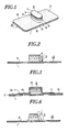
- Fig.1 is a perspective view of an embodiment of the sticking material for hemostasis of the present invention.
- Fig.2 is a cross-sectional view of the sticking material in Fig.1.
- Fig.3 is a cross-sectional view showing the sticking material for hemostasis as shown in Fig.1 in the packed condition.
- Fig.4 is an explanatory view showing the sticking material for hemostasis as shown in Fig. 1 in use.
- Fig.5 is an explanatory view showing the sticking material for hemostasis as shown in Fig. 1 when used for hemostasis of artery.
- Fig.6 is a cross-sectional view of another embodiment of the sticking material for hemostasis of the present invention.
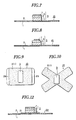
- Fig.7 is a cross-sectional view showing a further embodiment of the sticking material for hemostasis of the present invention.
- Fig.8 is a cross-sectional view showing still another embodiment of the sticking material for hemostasis of the present invention.
- Fig.9 is a plan view showing another modified embodiment of the sticking material for hemostasis of the present invention.
- Fig.10 is a plan view of a further modified embodiment of the sticking material for hemostasis of the present invention.
- Fig.11 is a perspective view showing another embodiment of the sticking material for hemostasis of the present invention.
- Fig.12 is a cross-sectional view of the sticking material as shown in Fig.11.
- Fig.13 is an explanatory view showing the sticking material for hemostasis as shown in Fig. 11 in use.
- Fig.14 is an explanatory view showing the sticking material for hemostasis of the present invention in another use.
- Fig.15 is a plan view showing another embodiment of the sticking material for hemostasis of the present invention.
- Fig.16 is a plan view showing a further embodiment of the sticking material for hemostasis of the present invention.
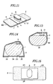
- Fig.17 is a perspective view showing another embodiment of the sticking material for hemostasis of the present invention.
- Fig.18 is a plan view showing still another embodiment of the sticking material for hemostasis of the present invention.
- Fig.19 is a plan view showing a further embodiment of the sticking material for hemostasis of the present invention.
- Fig.20 is a perspective view showing another embodiment of the sticking material for hemostasis of the present invention.
- Fig.21 is a perspective view showing still another embodiment of the sticking material for hemostasis of the present invention.
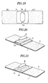
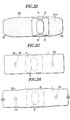
- Fig.22 is a plan view showing another embodiment of the sticking material for hemostasis of the present invention.
- Fig.23 is a plan view showing still another embodiment of the sticking material for hemostasis of the present invention.
- Fig.24 is a plan view showing the sticking material for hemostasis of the present invention when the substrate thereof is extended.
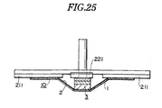
- Fig.25 is an explanatory view showing an apparatus for measuring the pressing force of the sticking material for hemostasis of the present invention.
- an adhesive layer 2 is formed on one surface of a base material 1.
- a base material various materials such as a plastic sheet, cloth or unwoven cloth, may be employed.
- the ones having stretchability are employed, when the base material is stuck over the puncture area as mentioned below while stretching it, it tends to shrink after sticking, whereby the pad as mentioned below will be more effectively pressed to the puncture area.
- the base material is preferably made of a stretchable material having a high recovery property, and the ones having a 100% extensibility, a stress exerted when extended to 100% of about 4,41 to 5,88 N/cm, and a recovery rate when extended to 100% of at least about 90% are effectively employed.
- Such a material includes, for example, fabrics having fine continuous filaments of polyurethane elastic fibers randomly laminated and bonded at the cross points.
- the adhesive of the adhesive layer 2 formed on the surface of the base material adhesives of acryl type, rubber type, silicone type, vinyl type, etc. may be appropriately employed. Particularly, when the acryl type adhesives are employed, stimulation against the skin will be reduced, such being preferable.
- the adhesive is provided on all or part of one surface of the base material so that it would be stuck and fixed to the wound area as mentioned below.
- a pad 3 is placed firmly.
- a pressing plate 21 of larger size than the pad 3 being inter posed between the pad and the adhesive layer of the base material 1 - see fig.11.
- This pad has a size such that when the injection needle, etc. is inserted into an arterial blood vessel 15 through the skin surface of the patient, it would cover both a puncture 16 of the arterial blood vessel and a puncture 17 of the skin surface at the same time for pressing as exemplified in Fig.5.
- the distance between both punctures 16 and 17 differs depending on the area of the body to be punctured, ages such as children or adult, or sex.
- the size of the pad is usually about 1 to 4 cm in the long side.
- the pad is formed in an appropriate shape such as rectangular, square, circle, oval, elliptical and polygon. In the case of oval, elliptical and rectangular, the ones having the ratio of the short side to the long side being up to about 1:4 are preferred. In the case of square and circle, the ratio is 1:1. Thus, the ratio is usually within the range of from about 1:1 to about 1:4.
- the pad When the pad is formed in a shape having corners, for example, rectangular, square and polygon, if the corner areas are formed in an arc shape, the stimulation against the skin surface is reduced when pressed, such being more preferable.
- both the puncture of the artery and the puncture at the skin surface are pressed in most cases, such being preferable.
- this pad is preferably one having a hardness capable of securely pressing the puncture, for example, one having not only a slight elasticity but also an appropriate compression strength so that it would not be readily deformed when pressed.
- This pad has such a thickness that it would press the puncture when put against the puncture area, and the thickness is at least 3 mm, and usually about 3 to 20 mm, although it differs depending on the applicable area. If the pad is too thin, pressing can not be effectively conducted.
- the sticking materials having a thin pad are suitable to hemostasis for dorsalis pedis artery, etc., and the ones having a thick pad are suitable to hemostasis for femoral artery, etc.
- the pad becomes thick the pressure will be strong, but the pad may in rare cases happen to be crushed horizontally or collapsed, such being not preferable. In many cases, preferred thickness is about 5 to 13 mm.
- This pad is formed in a multi-layer structure.
- a cushion layer 4, a lower layer 5, an upper layer 6 and a surface layer 7 are sequentially formed from the side of the adhesive layer 2.
- the cushion layer 4 has an elasticity at a level of a slight hardness to obtain pressing effect, and a sufficient recovery action so that no so-called settling phenomenon will result.
- the lower layer 5 subsequently formed is a layer exerting the pressing action and being capable of absorbing blood, and is softer and has less nerve than the cushion layer.
- the upper layer 6 is softer and has less nerve than the lower layer 5, so that it would be softly in contact with the puncture and the blood would be absorbed rapidly.
- the surface layer 7 is formed thin, to cover hairiness, of the upper layer 6 and facilitate peeling off of the coagulated blood.
- the cushion layer 4 is formed from plastic foam such as polyethylene foam having a large content of isolated cells, or compressed cotton such as compressed cotton of polyester unwoven cloth, etc.
- compressed cotton such as compressed cotton of cellulose unwoven cloth, or a laminated rayon cotton
- unwoven cloth such as rayon unwoven cloth
- perforated polyethylene film, nylon net, rayon net, a thin cellulose unwoven cloth, etc, are employed.
- An example effective for hemostasis of artery is the one wherein the cushion layer 4 is made of polyethylene foam having a large content of isolated cells and occupies about 50 to 70 % of the entire thickness, the lower layer 5 is made of cellulose unwoven compressed cotton and occupies about 20 to 30 % of the thickness, the upper layer 6 is made of a rayon unwoven cloth and occupies about 20 % of the thickness, and the surface layer 7 is a perforated polyethylene film.
- the lower layer 5 and the upper layer 6 may be formed in one layer by using a material having the properties required for both layers, to form a three-layer structure as a whole. Further, the lower layer 5, the upper layer 6 and the surface layer 7 may be formed in one layer to form a two-layer structure as a whole.
- This pad is usually located at the central portion of the base material so that the sticking material can be stuck over the puncture with the adhesive layer 2 provided on the substrate 1. However, depending on the site of application, the pad may be localized at one side of the base material for easy handling.
- this base material is sometimes formed in square, circle, oval, etc,. preferred is often a long strip shape.
- the pad When the pad is placed on the long strip-shaped base material, it is advisable to place the pad such that the long side of the pad extends in the short side direction (transverse direction) of the base material. Further, the pad covers in its long side of the puncture of arterial blood vessel and the puncture of the skin surface, whereby the pressing can readily be conducted. Thus, after that, the adhesive layer is stuck to the skin surface of the patient, whereby sure hemostasis can simply and securely be conducted.
- an elliptical pad is placed such that the long side of the pad would extend at right angle to the longitudinal direction of the long strip-shaped base material such being preferred for pressing.
- the pad may be placed obliquely at an angle other than the right angle.
- the pad is preferably placed with a space 9 of a length shorter than the thickness of the pad, i.e. a distance of usually about 3 to 10 mm, preferably about 6 mm, from each of both side edges 8, 8 of the base material.
- a space 9 of a length shorter than the thickness of the pad i.e. a distance of usually about 3 to 10 mm, preferably about 6 mm, from each of both side edges 8, 8 of the base material.
- a release paper It is advisable to cover the surface of the adhesive layer and pad of the base material of the sticking material for hemostasis 10 formed as above, with a release paper. It is preferred to pack the material with a bag for hygiene and easy handling.
- the one as shown in Fig.3 is obtained by covering the adhesive layer of the both sides of the base material with a release paper 12 having a folding strip 11 disposed, and further putting thereon a cap 14 made of a plastic having an enclosing portion 15 for the pad 3, whereby the pad can be hygienically maintained without distortion.
- Such a material may be sealed in a packaging bag obtained by weakly and removably adhering a pair of packaging sheets with a co-sealing agent, etc. or, as the case requires, may be sealed in such a packaging bag without putting on the cap (not shown in the drawing).
- the pad is attached to the puncture area such that the long side thereof extends in the direction that the needle is inserted at the puncture area, i.e. the direction that the ductus arteriosus 15 lies, and the needle or the catheter is removed while pressing the pad.
- one release paper is peeled off while pressing the pad, the adhesive layer is stuck to the skin such that it will lie across the direction that the ductus arteriosus lies while pulling the base material slightly, and then another release paper is picked at its folding strip and removed, and the adhesive layer is stuck to the skin while pulling the base material similarly to fix the sticking material to the skin.
- the pad is located over the puncture 16 of the ductus arteriosus under the skin and also covers the puncture 17 of the skin surface. Then, the pad is pressed downward by the shrink action of the stretched base material. Further, this pad presses the skin downwards strongly although the press is soft due to the multi-layer structure including the cushion layer. Thus, the area of the puncture 16 of the ductus arteriosus 15 is pressed concentratedly and strongly and closed, whereby the hemostasis can be securely conducted. By the pressing action, bleeding from the puncture 17 of the skin surface will result after removal of the needle. However, the blood will be immediately readily absorbed and held therein, whereby no blood will leak out.
- the hemostasis After a certain period of time while this article sticks the hemostasis is complete, and the article can be removed from the skin surface, about 5-30 minutes, generally about 10 - 20 minutes after stuck to the skin surface, whereby no contamination of the clothes, etc. by the bleeding will result.
- the sticking material for hemostasis as shown in Fig.6 is produced by making the shape of the pad 3 in a trapezoid shape wherein the area just above the adhesive layer of the base material is made larger than the other side. This sticking material makes it possible to press the pad against the puncture under more stable condition, whereby the pad will hardly be collapsed and the hemostasis will be effectively conducted.
- the corners of the surface layer 7 side of the pad 3 are cut off in an arc shape so that the material can adapt to the shape of the skin surface, whereby the contact with the skin will be milder.
- the surface layer 7 being in contact with the skin and the upper layer 6 of the pad are designed to be slightly smaller, whereby the stability when the pad is fixed is further increased and the pressing action will be made concentrated.
- a non-adhesive portion 29 is disposed by omitting the adhesive layer 2 at both edge areas of the base material or other sticking sheet.
- the non-adhesive portion 29 is readily grasped and thus the sticking material can readily be peeled off.
- the non-adhesive portion can also be disposed only at one edge area of the base material.
- the base material is formed in an X-shape, and a pad is placed at the central portion thereof. This base material is sometimes useful to fix and press the pad to the puncture area more securely.
- the sticking material for hemostasis as shown in Figs. 6 to 10 can be stuck to the wound portion for use according to the above explanation.
- This sticking material for hemostasis has a pressing plate 21 of a thin and tough strip-shaped member made of a plastic plate, a metal plate, a plywood or the like, between the adhesive layer 2 and the pad 3.
- This pressing plate is formed larger in size than the pad. It is advisable to form the pressing plate to have a length of about 3 to 10 cm in the longitudinal direction of the base material and the length is usually about 4 to 7 cm.
- the width of the pressing plate is made in the same size as the width of the base material and the elliptical-shaped pad is fixed on the pressing plate such that the long side of the pad would extend at the right angle to the longitudinal direction of the long strip-shaped base material.
- the pad may be placed in an oblique direction as has been mentioned above. This pad may be placed such that it is shifted towards one side of the pressing plate.
- the sticking material for hemostasis 22 can be used for hemostasis of artery in the same manner as above.
- the action of the stretched base material to shrink during the use is concentrated towards the pad by the large pressing plate, and serves to strongly press it downwards.
- the area to which the base material is not stuck becomes wide by means'of the pressing plate, it will rarely happen to disturb the blood circulation of the ulnar artery and the vein other than the punctured radial artery of the arm portion 23.
- the hemostasis can be conducted by adequately pressing the puncture area without causing cyanosis, etc., and the stimulation by the adhesive against the skin will be reduced, such being preferable.
- the pressing plate is bent when the sticking material for hemostasis is stuck, the pressing action by means of the pad may sometimes be further strengthened by the recovery action of the pressing plate.
- the pad 3 is placed on the adhesive layer 2 of the base material 1 as in the embodiment shown in Fig.1, and this pad is placed to cover the puncture of the blood vessel. Then, a pressing plate 24 is located on the skin surface at the opposite side of the skin surface having the pad placed, with the arterial blood vessel interposed, and both edge portions 25, 25 of the base material are stuck to the pressing plate to fix the sticking material to the wound area.
- This structure also provides secure hemostasis as has been made above.
- the width of the pressing plate 21 is made narrower than the width of the base material 2.
- the spacer areas 26, 26 at the both sides of the base material are folded to cover the both sides of the pressing plate, and will exert pressing action downwards and inner-obliquely.
- the pressing action is concentrated from the pressing plate towards the pad without scattering, thereby further preventing the thick pad from shifting horizontally of being collapsed.
- an adhesive layer is disposed only at both edge portions 27, 27 of the base material.
- This sticking material is used in the same manner as the embodiment shown in Fig.14 and, since the area of the adhesive layer is small, the pressing action and the stimulation action against the skin other than the puncture area are small, such being preferable in many cases.
- a concave portion 31 is disposed at the central portion of the side edge of the pressing plate along the longitudinal direction of the base material, and each corner is formed in an arc shape.
- this sticking material 22 if the concave portions 31, 31 of the pressing plate are picked up from the base material side, the longitudinal center line of the pad will be naturally known and thus this pad can be brought in contact with both punctures securely and readily.
- This concave portion 31 serves to further prevent the pressing plate from being in contact with the inserted needle, etc. Since both edge portions 32 in the longitudinal direction of the base material gradually become narrower in width, the base material can readily be stretched by pulling the base material at the portions 32.
- a convex portion 33 is disposed instead of the concave portion 31 of the pressing plate as shown in the above Fig.17.
- a V-shaped concave portion 34 is disposed. The convex portion 33 and the V-shaped concave portion 34 disposed on the pressing plate make it easy to find the position of the pad when stuck to the wound area as in the case of the concave portion 31.
- the pressing plate may have a protruded portion at one part thereof and a pad may be placed on the protruded portion.
- a bending protruded portion 36 is formed at the central portion of the pressing plate 21.
- a protruded portion 37 is formed by pressing at the central portion of the pressing plate. Such a protruded portion can be formed by stacking a small strip on a large strip.
- the pressing action can further be strengthened by the height of the protruded portion, whereby it is further easily applied to the hemostasis for femoral artery, etc. for which hemostasis is hardly applicable.
- the combination of the pad and the pressing plate is located at the central portion in the longitudinal direction of the base matérial.
- these pads, etc. may be placed in such a manner that these are shifted towards one side in the longitudinal direction.
- Such a sticking material is sometimes conveniently applicable to the ones wherein the blood vessel such as radial artery, ulnar artery of dorsalid pedis artery, is locally placed on the arm, etc.
- Figs. 23 and 24 Reference will be made to Figs. 23 and 24.
- Fig.23 distorted circular figures 38 are indicated at the surface of a stretchable subbase material opposite to the adhesive layer.
- the sticking material by pulling the base material, the base material expands and the distorted circular figures 38 will become circle figures 39 (Fig.24).
- circle figures 39 Fig.24.
- the desired pressing action can readily be obtained by the shrinking base material.
- various indications such as an equilateral triangle can be used in addition to the above circle figure.
- the bleeding stoppage to the ductus arteriosus is mentioned above.
- the blood extraction, dip of water, blood transfusion, artifical dialysis, etc. are carried out by puncturing the injector needle, catheter, medical tube, etc. into the venous blood through the surface of skin.
- the venipunctured portion After pulling the injector needle out, the venipunctured portion also starts bleeding, and it is therefore necessary to stop bleeding by pressing the absorbent cotton or gauze by finger strongly and pressing the absorbent cotton by adhesive tapes and/or flexible bandage, etc.
- Some patients are not in the position to press the punctured portion by fingers of other means.
- the aperture of the punctured portion of the blood vessel is also small, and the bleeding stoppage is relatively easy to the venous blood.
- the aperture of the punctured portion becomes large, which results in bleeding of a large quantity of blood, and the bleeding stoppage is more difficult than the one in case of the thin injector needle.
- the present invention can stop bleeding of artery. For example, when the relatively thick injector needle of 16 to 18 gauge (outer diameter 1,67 to 1,28 mm) is used for blood extraction and blood transfusion, sure bleeding stoppage is obtained. Of course, the bleeding stoppage is carried out more easily in case of using a thin injector needle.
- the adhesive layer of the product was stuck to a platform for measurement 211 made of Bakelite as shown in Fig.25, the pad is pressed with a plate 221 of 20 mm diameter, so that the surface of the plate would be adjusted to the surface of the platform for measurement with a rheometer, and the pressing force after 1 minute was measured.
- the measurement was conducted with respect to 10 samples.
- the pressing force was 35000 ⁇ 1200 N/m 2 (360 ⁇ 12 p/cm 2 )
- Product 1 was prepared and the results of measurement of the force of pressure, pressure sense numbness and change of palm color when this product is used, are shown below.
- Base material obtained by randomly laminating fine continuous filaments comprising 100 % of polyurethane elastic fibers, and bonding the crossing points of respective filaments.
- Adhesive layer Acryl type adhesive as coated to 38 + 2 g/m 2 Pressing plate: A poyvinylchloride plate having a thickness of 1 mm was cut in a size of 45 x 50 mm and stuck to the adhesive layer at the central portion of the sticking material, while adjusting the edges.
- Pad The surface layer is cellulose unwoven cloth having a thickness of 0.05 mm.
- the upper layer is rayon unwoven cloth having a thickness of 2 mm
- the lower layer is a compressed cotton of cellulose unwoven cloth having a thickness of 2 mm.
- the cushion layer is a sheet of a foam of polyethylene isolated cells having a thickness of 5 mm.
- Shape An elliptical shape having a long side of 27 mm, a short side of 15 mm and a thickness of 9 mm.
- This pad is fixed at the central portion of the pressing plate such that the long side of the pad extends in the transverse direction of the base material.
- the pad was put on the radial artery of left wrist while extending the sticking material to 140 mm, and then the sticking material was stuck in the same manner as in Fig.13 for 1 minute.
- Product 2 was prepared by using the same base material, adhesive and pad as product 1 provided that the pad was placed at the central portion of the base material.
- Pressing plate A polyvinylchloride plate with a thickness of 1,1 mm was cut in a size of 45 x 70 mm.
- the pad was put on the radial artery of left wrist and the pressing plate was located at the side opposite to the pad. Then, while extending the base material to 140 mm, it was stuck to the pressing plate in the same manner as in Fig.14 for 1 minute.
- a pouch of a catheter for measurement of cerebrospinal pressure connected-to a piezo-electric transducer, was non-invatively fixed.
- the sticking material was stuck so that the pad portion is located on the pouch. Then, the pressing force at the pad portion was measured.
- the force of pressure at the pressed area was read from the recording sheet and then converted to numerals (mm Hg). The average number thereof was determined.
- the pressure sense, numbness and change of palm color were evaluated by four steps of "non, weak, medium and strong". These steps were called 0, 1, 2 and 3 respectively, and the average number thereof was determined.
- Example 1 Contrast Force of pressure (average, mm Hg) 246 143 85 Pressure sense (average) 3,0 2,2 1,5 Numbness (average) 1,2 0,8 1,0 Change of palm color (average) 0,7 0,5 1,2
- Product 3 was prepared and the results of measurement of the pressing force, side effect (pressing feeling, numbness, discoloration of palm), the time required for disappearance of the pressure marks, and stimulus of the skin surface by changing the extension length of the base material variously are shown below:
- Base material The base material is obtained by randomly laminating fine continuous filaments comprising 100 % of polyurethane elastic fibers, and bonding the crossing points of respective filaments. The properties are shown as below: Weight 100 g/m 2 Stress at 50 % extension Longitudinal direction 4,9 N/cm/20 mm Latitudinal direction 3,63 N/cm/20 mm Recovery at 100 % extension Longitudinal direction 87 % Latitudinal direction 87 % Strength of extension Longitudinal direction 15,7 N/cm/20 mm Latitudinal direction 11,76 N/cm/20 mm Extension Longitudinal direction 370 % Latitudinal direction 380 %
- the above base material was cut in a rectangular shape of 40 mm x 126 mm having gradually narrowed edges, the latitudinal direction being a long side (Fig.17).
- Adhesive layer Acryl type adhesive was coated to 40 g/m 2 .
- Adhesive strength 1,96 N/cm/15mm Tack (rolling ball method) No. 31 Pressing plate
- Polypropylene plate of 1 mm thickness was cut in a square of 36 mm x 36 mm with arc-shaped corners, having a convex on an opposite side (Fig.17).
- the surface layer is cellulose unwoven cloth having a thickness of 0,05 mm.
- the upper layer is rayon unwoven cloth having a thickness of 3mm.
- the cushion layer is a sheet of a foam of polyethylene isolated cell having a thickness of 6 mm.
- this sheet of foam is as follows: density at glande 0,067 g/cm 3 compressed hardness 0,64 KG/cm 2 shape an elliptical shape having a long side of 27 mm, a short side of 15 mm and a thickness of 9 mm.
- This pad is fixed at the central portion of the pressing plate such that the long side of the pad extends in the transverse direction of the base material (Fig.17).
- the pads were put on the radial arteries of left and right wrists, and the sticking material was stuck for a minute under the following four conditions:
- Adhesion and peeling condition The condition of adhesion, and the residue of the adhesive is evaluated in peeling the adhesive bandage one hour later.
- Time required for disappearance of the pad mark After peeling the sticking material, pressure marks by the pad are observed by eyes and the time for disappearance of the pad marks is measured.
- Dermal irritation index The reaction of the skin with the adhesive attached was evaluated according to the following standard: four hours later and 24 hours later after peeling, respectively, and the dermal irritation index was determined.
- the adhering condition is favourable, since no peeling is found, and no residue of the adhesive is found after peeling.
- the side effect after one hour usage of the product 3 is very small, i.e., there is no problem in use.
- the base material When the base material is extended by 3 cm, the base material surrounds the periphery of the wrist, but no side effect is found. This is because the pressing plate allows the ulnar artery's blood to flow freely.
- the product 3 when used by extending about 2 cm, the product 3 can give the proper pressure action, and various side effects are very small even after the long use. Therefore, it serves as the sticking product of hemostasis of artery.
- the present invention it is possible to securely conduct the hemostasis of the puncture of artery, and effective hemostasis can be made to arteries located close to bones such as ulnar artery and dorsalis pedis artery in addition to radial artery. Further, the present invention is applicable to femoral artery. Effective hemostasis can be made to a vein also as has been applied to the artery. Further, the procedure of use is easy and the structure of the sticking material is simple, whereby the production can be made economically.
Landscapes
- Health & Medical Sciences (AREA)
- Life Sciences & Earth Sciences (AREA)
- General Health & Medical Sciences (AREA)
- Animal Behavior & Ethology (AREA)
- Vascular Medicine (AREA)
- Engineering & Computer Science (AREA)
- Biomedical Technology (AREA)
- Heart & Thoracic Surgery (AREA)
- Veterinary Medicine (AREA)
- Public Health (AREA)
- Surgery (AREA)
- Medical Informatics (AREA)
- Reproductive Health (AREA)
- Molecular Biology (AREA)
- Nuclear Medicine, Radiotherapy & Molecular Imaging (AREA)
- Materials For Medical Uses (AREA)
Claims (5)
- Pansement compresseur pour hémostase comprenant un matériau de base en forme de longue bande élastique étirable (1) ayant des propriétés de recouvrement avec une couche adhésive (2) disposée sur l'une des surfaces de ce dernier et portant un coussinet (3) de forme, dimension et rigidité appropriés, pour exercer une action de compression quand appliqué sur le point ou la région de l'hémorragie, de manière a assurer un aplatissement complet et une suture de la veine quand appliqué contre la partie piquée de la dite veine, caractérisé en ce que pour l'hémostase d'un vaisseau sanguin que ce soit une veine ou une artère, le corps est une plaque de pression (21) de taille plus large que le coussinet (3) et est interposée entre le coussinet (3) et la couche adhésive (2) du matériau de base (1) et que ce coussinet (3) a une structure multicouches comprenant, sur le dit matériau de base (1) par séquence, une couche d'amortissement (4) ayant un haut effet de pression en direction de l'épaisseur, une couche inférieure (5) qui absorbe le sang et a un effet de pression, une couche supérieure (6) qui absorbe rapidement le sang, et une couche de surface (7) qui prévient l'abondance des poils de la couche supérieure (6) et le dépôt de sang coagulé.
- Pansement compresseur pour hémostase conformément à la revendication 1 dans lequel le coussinet (3) a une forme elliptique ayant un côté long de 20 mm à 40 mm, un côté court de 10 mm à 20 mm et une épaisseur de 8 à 12 mm, et le coussinet est placé de telle manière que le côté long s'étend en direction transversale du matériau de base.
- Pansement compresseur pour hémostase conformément aux revendications 1 et 2, dans le quel le coussinet (3) à un rapport entre le côté court et le côté long de 1 :1 à 1 :4.
- Pansement compresseur pour hémostase conformément avec l'une quelconque des revendications 1 à 3, caractérisé en ce que chaque côté des bords des plaques de pression (21) s'étendant en direction longitudinale du matériau de base (1) sont pourvus d'une partie concave (31).
- Pansement compresseur pour hémostase conformément avec l'une quelconque des revendications 1 à 3, caractérisé en ce que chaque côté des bords des plaques de pression (21) s'étendant en direction longitudinale du matériau de base (1) sont pourvus d'une partie convexe (33).
Applications Claiming Priority (9)
| Application Number | Priority Date | Filing Date | Title |
|---|---|---|---|
| JP75028/93 | 1993-03-10 | ||
| JP7502893 | 1993-03-10 | ||
| JP5075028A JPH06254115A (ja) | 1993-03-10 | 1993-03-10 | 動脈止血用貼付材 |
| JP13983393A JP3328787B2 (ja) | 1993-05-20 | 1993-05-20 | 動脈止血用具 |
| JP139833/93 | 1993-05-20 | ||
| JP13983393 | 1993-05-20 | ||
| JP60144/94 | 1994-03-04 | ||
| JP6014494A JPH07241313A (ja) | 1994-03-04 | 1994-03-04 | 止血用貼付材 |
| JP6014494 | 1994-03-04 |
Publications (2)
| Publication Number | Publication Date |
|---|---|
| EP0614652A1 EP0614652A1 (fr) | 1994-09-14 |
| EP0614652B1 true EP0614652B1 (fr) | 2001-07-18 |
Family
ID=27297107
Family Applications (1)
| Application Number | Title | Priority Date | Filing Date |
|---|---|---|---|
| EP94103683A Expired - Lifetime EP0614652B1 (fr) | 1993-03-10 | 1994-03-10 | Pansement pour l'hémostase |
Country Status (3)
| Country | Link |
|---|---|
| US (1) | US5690610A (fr) |
| EP (1) | EP0614652B1 (fr) |
| DE (1) | DE69427716T2 (fr) |
Cited By (6)
| Publication number | Priority date | Publication date | Assignee | Title |
|---|---|---|---|---|
| US7468470B2 (en) | 2004-03-26 | 2008-12-23 | Schering Ag | Medicinal patch that leaves less adhesive residue when removed |
| EP3202380A1 (fr) | 2016-02-06 | 2017-08-09 | Finn Pedersen | Pansement |
| CN108201463A (zh) * | 2016-12-16 | 2018-06-26 | 美敦力公司 | 一种局部加压止血装置 |
| WO2019143683A1 (fr) * | 2018-01-16 | 2019-07-25 | Yu Andrew S | Bandage compressif |
| EP3752076A1 (fr) * | 2018-02-15 | 2020-12-23 | Università Campus Bio-Medico di Roma | Dispositif médical de compression progressive pour supporter et optimiser la cicatrice cutanée |
| USD929596S1 (en) | 2019-01-16 | 2021-08-31 | Andrew S. Yu | Bandage |
Families Citing this family (119)
| Publication number | Priority date | Publication date | Assignee | Title |
|---|---|---|---|---|
| US5653224A (en) * | 1991-06-10 | 1997-08-05 | Creative Integration & Design, Inc. | Nasal dilator with areas of adhesive engagement of varying strength |
| SE503107C2 (sv) * | 1994-07-11 | 1996-03-25 | Moelnlycke Ab | Dambinda samt förfarande för tillverkning av denna |
| DE4447557C2 (de) * | 1994-08-18 | 1997-10-23 | Harren Ernst Diethelm | Punktionsverschluß |
| US5891074A (en) * | 1996-08-22 | 1999-04-06 | Avitar, Inc. | Pressure wound dressing |
| SE9701935D0 (sv) * | 1997-05-23 | 1997-05-23 | Radi Medical Systems | Medical device |
| US6074356A (en) * | 1998-03-06 | 2000-06-13 | Starkey; Paul | Method and device for treatment of varicose veins |
| US5947123A (en) * | 1998-03-06 | 1999-09-07 | Shippert; Ronald D. | Nose splint with contoured nose contacting surface |
| US6313370B1 (en) * | 1999-04-29 | 2001-11-06 | Morton Hyson | Medicated wrap |
| US6316686B1 (en) | 2000-06-20 | 2001-11-13 | Timothy N. Byrd | Medical pressure dressing and process |
| GB2367245B (en) * | 2000-09-29 | 2004-11-17 | Johnson & Johnson Medical Ltd | Adaptable dressings |
| WO2002028333A1 (fr) * | 2000-10-03 | 2002-04-11 | Fsk Medical Ventures, Llc | Procede et dispositif de traitement de veines variqueuses |
| US6441265B1 (en) * | 2000-12-26 | 2002-08-27 | Souliya S. Chan | Wound dressing |
| US6856075B1 (en) | 2001-06-22 | 2005-02-15 | Hutchinson Technology Incorporated | Enhancements for adhesive attachment of piezoelectric motor elements to a disk drive suspension |
| US7147615B2 (en) | 2001-06-22 | 2006-12-12 | Baxter International Inc. | Needle dislodgement detection |
| US7101862B2 (en) | 2001-12-31 | 2006-09-05 | Area Laboratories, Llc | Hemostatic compositions and methods for controlling bleeding |
| US20040068290A1 (en) * | 2002-03-27 | 2004-04-08 | Datascope Investment Corp. | Device and method for compressing wounds |
| US7022098B2 (en) | 2002-04-10 | 2006-04-04 | Baxter International Inc. | Access disconnection systems and methods |
| US20040254513A1 (en) | 2002-04-10 | 2004-12-16 | Sherwin Shang | Conductive polymer materials and applications thereof including monitoring and providing effective therapy |
| US7052480B2 (en) | 2002-04-10 | 2006-05-30 | Baxter International Inc. | Access disconnection systems and methods |
| US7138088B2 (en) * | 2002-04-10 | 2006-11-21 | Baxter International Inc. | Access disconnection system and methods |
| US10155082B2 (en) | 2002-04-10 | 2018-12-18 | Baxter International Inc. | Enhanced signal detection for access disconnection systems |
| EP1358851B1 (fr) * | 2002-05-03 | 2005-08-10 | Lina Medical ApS | Dispositif destiné à la hémostase d'un vaisseau sanguin ouvert |
| US7846141B2 (en) | 2002-09-03 | 2010-12-07 | Bluesky Medical Group Incorporated | Reduced pressure treatment system |
| DE10327153A1 (de) * | 2003-06-13 | 2005-01-05 | Stefan Rüster | Kompressiver Schnellwundverband/kompressives Schnellwundpflaster/Kompressionsverband |
| US20080154168A1 (en) * | 2003-07-17 | 2008-06-26 | Thomas Placido Lutri | Surgical bandage for use with tissue adhesives and other medicaments |
| US20050015036A1 (en) * | 2003-07-17 | 2005-01-20 | Lutri Thomas Placido | Surgical bandage for use with tissue adhesives and other medicaments |
| US8029454B2 (en) | 2003-11-05 | 2011-10-04 | Baxter International Inc. | High convection home hemodialysis/hemofiltration and sorbent system |
| US20050125025A1 (en) * | 2003-12-05 | 2005-06-09 | Marcel Rioux | Styptic device |
| DE102004016591A1 (de) * | 2004-03-31 | 2005-10-27 | Schering Ag | Medizinische Pflaster mit verringertem Kleberrückstand |
| US20050256438A1 (en) * | 2004-05-13 | 2005-11-17 | Roberta Lombardozzi | Venipuncture bandage |
| US8592639B2 (en) * | 2004-09-16 | 2013-11-26 | Dsu Medical Corp. | Injection and hemostasis site |
| US8017826B2 (en) * | 2004-09-16 | 2011-09-13 | Dsu Medical Corporation | Injection and hemostasis site |
| US7012170B1 (en) * | 2004-11-16 | 2006-03-14 | Tomaioulo Theodore B | Puncture wound bandage |
| US7645252B2 (en) | 2006-05-16 | 2010-01-12 | Barbara Brooke Jennings-Spring | Body or plant part dressing |
| US7905852B2 (en) | 2006-05-16 | 2011-03-15 | Barbara Jennings-Spring | Skin-contacting-adhesive free dressing |
| US20080015482A1 (en) * | 2006-07-13 | 2008-01-17 | Utterberg David S | Resilient double pad hemostasis devices |
| US20080097270A1 (en) * | 2006-08-25 | 2008-04-24 | Utterberg David S | Resilient hemostasis devices |
| FR2909862B1 (fr) * | 2006-12-13 | 2009-02-06 | Christophe Tanvier | Pansement et collier a bille anti-hemorragie arterielle |
| DE102007044555A1 (de) * | 2007-07-18 | 2009-01-22 | Siemens Ag | Optische Koppelvorrichtung und Verfahren zu deren Herstellung |
| WO2009089390A2 (fr) | 2008-01-08 | 2009-07-16 | Bluesky Medical Group Inc. | Traitement durable de blessures par pression négative variable et procédé pour son contrôle |
| US8100290B2 (en) * | 2008-03-03 | 2012-01-24 | Edison Nation, Llc | Spooled adhesive bandage dispenser |
| US8945030B2 (en) | 2008-03-12 | 2015-02-03 | Bluesky Medical Group, Inc. | Negative pressure dressing and method of using same |
| US8114043B2 (en) | 2008-07-25 | 2012-02-14 | Baxter International Inc. | Electromagnetic induction access disconnect sensor |
| US20110238021A1 (en) * | 2008-09-26 | 2011-09-29 | Suzana Hillhouse | Transdermal delivery device and method |
| CA2747665C (fr) | 2008-12-17 | 2016-08-02 | Benrikal Services Inc. | Dispositif styptique |
| US10842502B2 (en) * | 2009-09-11 | 2020-11-24 | Tbi Innovations, Llc | Devices and systems to mitigate traumatic brain and other injuries caused by concussive or blast forces |
| JP5844730B2 (ja) * | 2010-03-29 | 2016-01-20 | テルモ株式会社 | イントロデューサーシース組立体 |
| CA2724866A1 (fr) * | 2010-10-07 | 2012-04-07 | Jason Turner | Tourniquet avec element absorbant jetable |
| US20120191128A1 (en) * | 2011-01-25 | 2012-07-26 | Wound Care 360?, Inc. | Vascular wound closing apparatus and method |
| US8764789B2 (en) * | 2011-04-15 | 2014-07-01 | CellAegis Devices Inc. | System for performing remote ischemic conditioning |
| JP5922883B2 (ja) * | 2011-06-01 | 2016-05-24 | ニチバン株式会社 | 静脈からの採血後の止血用自着包帯 |
| JP6144683B2 (ja) * | 2011-10-18 | 2017-06-07 | ヴォストラ−メド・アーゲー | 開いた血管を閉鎖するための閉鎖デバイス |
| EP2583646A1 (fr) * | 2011-10-18 | 2013-04-24 | Vostra-Med Ag | Dispositif de fermeture pour la fermeture de vaisseaux sanguins ouverts et utilisation de ce dispositif de fermeture |
| US20130204190A1 (en) * | 2012-02-07 | 2013-08-08 | Marie-Andrea I. Wilborn | Intravenous splint cover and associated methods |
| US9950143B2 (en) | 2012-02-07 | 2018-04-24 | Marie Andrea I. Wilborn | Intravenous splint cover and associated methods |
| US10456559B2 (en) * | 2012-02-07 | 2019-10-29 | Marie-Andrea I. Wilborn | Cannulated tube protector, apparatus operable to facilitate the flow of fluids through a cannulated site and apparatus operable to protect and maintain positioning of a catheter |
| USD733896S1 (en) * | 2012-05-04 | 2015-07-07 | Genadyne Biotechnologies, Inc. | Abdominal dressing |
| US20140031781A1 (en) * | 2012-07-12 | 2014-01-30 | Lydda Razon-Domingo | Pressure application for hemostatis |
| US9259212B2 (en) | 2012-07-24 | 2016-02-16 | Wound Care 360, LLC | Vascular wound closing apparatus and method |
| USD708338S1 (en) * | 2012-08-15 | 2014-07-01 | CellAegis Devices Inc. | Cuff for remote ischemic conditioning |
| USD705429S1 (en) * | 2012-08-29 | 2014-05-20 | Merit Medical Systems, Inc. | Medical compression bandage |
| USD705428S1 (en) * | 2012-08-29 | 2014-05-20 | Merit Medical Systems, Inc. | Medical compression bandage |
| EP2745785B1 (fr) | 2012-12-21 | 2017-06-21 | Vostra-Med Ag | Système de fermeture de récipient |
| GB201317746D0 (en) | 2013-10-08 | 2013-11-20 | Smith & Nephew | PH indicator |
| US9861531B2 (en) * | 2013-06-18 | 2018-01-09 | Sanko Tekstil Isletmeleri Sanayi Ve Ticaret Anonim Sirketi | Multi-function emergency bandage |
| US9517163B2 (en) * | 2013-07-03 | 2016-12-13 | 3K Anesthesia Innovations, Llp | Pre-stressed pressure device |
| US9332994B2 (en) | 2013-07-12 | 2016-05-10 | Vasoinnovations, Inc. | Apparatus and method to stop bleeding |
| US9949738B2 (en) | 2013-07-12 | 2018-04-24 | Vasoinnovations, Inc. | Method to stop bleeding, with short hemostasis duration using a low dose of anticoagulant |
| US10342551B2 (en) | 2013-07-12 | 2019-07-09 | Vasoinnovations Inc. | Method to stop bleeding, with short hemostasis duration using a low dose of anticoagulant |
| US10213214B2 (en) | 2013-07-12 | 2019-02-26 | Vasoinnovations, Inc. | Method to stop bleeding, with short hemostasis duration using a low dose of anticoagulant |
| US9668744B2 (en) | 2015-08-05 | 2017-06-06 | Vasoinnovations, Inc. | Apparatus and method to stop bleeding |
| US9308000B2 (en) | 2013-07-12 | 2016-04-12 | Vasoinnovations, Inc. | Method of transradial catheterization, device for ulnar artery compression, and method of use |
| US10888334B2 (en) | 2013-07-12 | 2021-01-12 | Vasoinnovations Inc. | Apparatus and method to stop bleeding |
| US11564697B2 (en) | 2013-07-12 | 2023-01-31 | Vasoinnovations Inc. | Apparatus and method to stop bleeding |
| USD733305S1 (en) | 2013-10-25 | 2015-06-30 | Medtronic Vascular, Inc. | Tissue compression apparatus |
| US9795391B2 (en) | 2013-10-25 | 2017-10-24 | Medtronic Vascular, Inc. | Tissue compression device with tension limiting strap retainer |
| US10092297B2 (en) | 2014-04-25 | 2018-10-09 | Medtronic Vascular, Inc. | Tissue compression device with fixation and tension straps |
| WO2016112342A1 (fr) * | 2015-01-09 | 2016-07-14 | Hemcon Medical Technologies, Inc. | Systèmes et procédés de traitement de lésion vasculaire percutanée |
| JP6806669B2 (ja) * | 2015-04-07 | 2021-01-06 | テルモ株式会社 | 止血器具 |
| FR3044893B1 (fr) * | 2015-12-09 | 2018-05-18 | Emile Droche | Pansement pour soins de la peau en milieu humide |
| CN109069712A (zh) | 2016-05-13 | 2018-12-21 | 史密夫及内修公开有限公司 | 启用传感器的伤口监测和治疗装置 |
| US10426490B2 (en) * | 2016-07-12 | 2019-10-01 | Reza Salimi | Transparent pressure dressing device and methods for enabling hemostasis of blood |
| US12178597B2 (en) | 2017-03-09 | 2024-12-31 | Smith & Nephew Plc | Device, apparatus and method of determining skin perfusion pressure |
| EP3592212B1 (fr) | 2017-03-09 | 2024-08-07 | Smith & Nephew plc | Pansement |
| EP3592230A1 (fr) | 2017-03-09 | 2020-01-15 | Smith & Nephew PLC | Appareil et procédé d'imagerie du sang dans une région cible de tissu |
| WO2018185555A2 (fr) * | 2017-04-04 | 2018-10-11 | Advent Access Pte. Ltd. | Systèmes, appareils, kits et méthodes pour des actes médicaux améliorés |
| JP7235673B2 (ja) | 2017-04-11 | 2023-03-08 | スミス アンド ネフュー ピーエルシー | センサ対応型創傷被覆材のための構成要素配置および応力緩和 |
| JP7150750B2 (ja) | 2017-05-15 | 2022-10-11 | スミス アンド ネフュー ピーエルシー | オイラービデオ倍率を使用した陰圧創傷療法システム |
| CA3062989A1 (fr) | 2017-05-15 | 2018-11-22 | Smith & Nephew Plc | Dispositif et procede d'analyse de plaies |
| WO2018234443A1 (fr) | 2017-06-23 | 2018-12-27 | Smith & Nephew Plc | Positionnement de capteurs pour la surveillance ou le traitement de plaie activé(e) par capteurs |
| GB201809007D0 (en) | 2018-06-01 | 2018-07-18 | Smith & Nephew | Restriction of sensor-monitored region for sensor-enabled wound dressings |
| GB201804502D0 (en) | 2018-03-21 | 2018-05-02 | Smith & Nephew | Biocompatible encapsulation and component stress relief for sensor enabled negative pressure wound therapy dressings |
| CN111093726B (zh) | 2017-08-10 | 2023-11-17 | 史密夫及内修公开有限公司 | 实施传感器的伤口监测或治疗的传感器定位 |
| CN111050671B (zh) * | 2017-09-01 | 2024-02-06 | 泰尔茂株式会社 | 线压迫垫 |
| GB201804971D0 (en) | 2018-03-28 | 2018-05-09 | Smith & Nephew | Electrostatic discharge protection for sensors in wound therapy |
| EP3681376A1 (fr) | 2017-09-10 | 2020-07-22 | Smith & Nephew PLC | Systèmes et procédés d'inspection d'encapsulation et de composants dans des pansements équipés de capteurs |
| GB201718870D0 (en) | 2017-11-15 | 2017-12-27 | Smith & Nephew Inc | Sensor enabled wound therapy dressings and systems |
| GB201718859D0 (en) | 2017-11-15 | 2017-12-27 | Smith & Nephew | Sensor positioning for sensor enabled wound therapy dressings and systems |
| CN111132605B (zh) | 2017-09-27 | 2023-05-16 | 史密夫及内修公开有限公司 | 用于实施传感器的负压伤口监测和治疗设备的pH感测 |
| US11839464B2 (en) | 2017-09-28 | 2023-12-12 | Smith & Nephew, Plc | Neurostimulation and monitoring using sensor enabled wound monitoring and therapy apparatus |
| CN111343950A (zh) | 2017-11-15 | 2020-06-26 | 史密夫及内修公开有限公司 | 实施传感器的集成伤口监测和/或治疗敷料和系统 |
| CN111954499B (zh) * | 2018-05-16 | 2023-09-22 | 泰尔茂株式会社 | 压迫设备 |
| GB201814011D0 (en) | 2018-08-29 | 2018-10-10 | Smith & Nephew | Componet positioning and encapsulation for sensor enabled wound dressings |
| EP3849401A1 (fr) | 2018-09-12 | 2021-07-21 | Smith & Nephew plc | Dispositif, appareil et procédé de détermination de pression de perfusion cutanée |
| EP3856104B1 (fr) | 2018-09-28 | 2025-08-06 | T.J.Smith And Nephew, Limited | Fibres optiques permettant la détection optique à travers des pansements de plaie |
| GB201816838D0 (en) | 2018-10-16 | 2018-11-28 | Smith & Nephew | Systems and method for applying biocompatible encapsulation to sensor enabled wound monitoring and therapy dressings |
| GB201820927D0 (en) | 2018-12-21 | 2019-02-06 | Smith & Nephew | Wound therapy systems and methods with supercapacitors |
| GB2595176B (en) | 2019-01-30 | 2023-02-15 | Smith & Nephew | Sensor integrated dressings and systems |
| WO2020187851A1 (fr) | 2019-03-18 | 2020-09-24 | Smith & Nephew Plc | Règles de conception pour substrats à capteur intégré |
| EP3941346A1 (fr) | 2019-03-19 | 2022-01-26 | Smith & Nephew plc | Systèmes et procédés pour la mesure de l'impédance d'un tissu |
| CN110353755B (zh) * | 2019-07-18 | 2020-10-16 | 李伟 | 止血贴 |
| GB201914443D0 (en) | 2019-10-07 | 2019-11-20 | Smith & Nephew | Sensor enabled negative pressure wound monitoring apparatus with different impedances inks |
| JP2023521954A (ja) | 2020-04-21 | 2023-05-26 | ティージェイ スミス アンド ネフュー リミテッド | 拡張現実オーバーレイを使用する創傷治療管理 |
| GB202007391D0 (en) | 2020-05-19 | 2020-07-01 | Smith & Nephew | Patient protection from unsafe electric current in sensor integrated dressings and systems |
| US11369387B2 (en) * | 2020-06-04 | 2022-06-28 | Eric Nivens | Tourniquet assembly |
| USD1023312S1 (en) * | 2022-03-18 | 2024-04-16 | Gc Corporation | Hemostatic agent material |
| USD991463S1 (en) * | 2022-10-11 | 2023-07-04 | Alexis Karina Sepulveda | Combination of tape with a pressure point device |
| WO2025059142A1 (fr) * | 2023-09-11 | 2025-03-20 | Biodaptive Advanced Materials, Llc | Pansement à adhérence de bord améliorée |
| CN118340937B (zh) * | 2024-06-18 | 2024-08-30 | 浙江理工大学 | 穿刺后可止血的膨体ptfe人造血管及制备方法和应用 |
Family Cites Families (19)
| Publication number | Priority date | Publication date | Assignee | Title |
|---|---|---|---|---|
| US2280506A (en) * | 1941-03-10 | 1942-04-21 | Richard T Betts | Surgical dressing |
| US3490448A (en) * | 1967-07-31 | 1970-01-20 | Harvey C Upham | Adhesive pressure pad |
| US3908645A (en) * | 1974-05-28 | 1975-09-30 | Minnesota Mining & Mfg | Ophthalmic pressure bandage |
| DE2515786C3 (de) * | 1975-04-11 | 1979-01-04 | Laerdal, Asmund S., Stavanger (Norwegen) | Druckverband |
| IT1161426B (it) * | 1978-01-06 | 1987-03-18 | Dotta Angelo | Confezione sigillata e sterilizzabile per bende adesive ad apertura rapida e razionale e mezzi per l'attuazione della stessa |
| US4390519A (en) * | 1978-05-19 | 1983-06-28 | Sawyer Philip Nicholas | Bandage with hemostatic agent and methods for preparing and employing the same |
| FR2499416B1 (fr) * | 1981-02-06 | 1986-05-23 | Thuasne & Cie | Coussin hemostatique |
| NO821782L (no) * | 1981-06-04 | 1982-12-06 | John Michael Simpson | Kirurgisk plaster. |
| IT1144223B (it) * | 1981-06-11 | 1986-10-29 | Angelo Dotta | Confezione sigillata per bende adesive |
| US4377159A (en) * | 1981-06-29 | 1983-03-22 | Minnesota Mining And Manufacturing Company | Pressure bandages and methods for making the same |
| FR2550938B1 (fr) * | 1983-08-29 | 1986-11-14 | Buisson Philippe | Pansement hemostatique compressif |
| US4561435A (en) * | 1984-04-04 | 1985-12-31 | Chesebrough-Ponds, Inc. | Wound dressing |
| US4743499A (en) * | 1987-05-20 | 1988-05-10 | Variseal Corporation | Hydrocolloid laminate |
| JPS641216U (fr) * | 1987-06-16 | 1989-01-06 | ||
| KR900004662Y1 (ko) * | 1988-02-08 | 1990-05-25 | 금광남 | 지혈용 1회용 테이프 |
| JP2646372B2 (ja) * | 1988-07-08 | 1997-08-27 | ニチバンプリント株式会社 | 止血用貼付材 |
| CH676197A5 (en) * | 1988-10-21 | 1990-12-28 | Flawa Schweiz Verband Wattefab | Variable pressure bandage - has cushioning pad at one end and system of fastening at other which allows pressure on wound to be changed as often as desired |
| DE59009846D1 (de) * | 1989-04-27 | 1995-12-14 | Flawa Schweiz Verband Wattefab | Kompresse zur Versorgung von Wunden. |
| JPH05245173A (ja) * | 1992-02-29 | 1993-09-24 | Nichiban Co Ltd | 止血用貼付材 |
-
1994
- 1994-03-10 EP EP94103683A patent/EP0614652B1/fr not_active Expired - Lifetime
- 1994-03-10 DE DE69427716T patent/DE69427716T2/de not_active Expired - Fee Related
- 1994-03-10 US US08/209,537 patent/US5690610A/en not_active Expired - Fee Related
Cited By (9)
| Publication number | Priority date | Publication date | Assignee | Title |
|---|---|---|---|---|
| US7468470B2 (en) | 2004-03-26 | 2008-12-23 | Schering Ag | Medicinal patch that leaves less adhesive residue when removed |
| EP3202380A1 (fr) | 2016-02-06 | 2017-08-09 | Finn Pedersen | Pansement |
| DE102016102106A1 (de) * | 2016-02-06 | 2017-08-10 | Finn S. Pedersen | Wundabdeckung |
| CN108201463A (zh) * | 2016-12-16 | 2018-06-26 | 美敦力公司 | 一种局部加压止血装置 |
| WO2019143683A1 (fr) * | 2018-01-16 | 2019-07-25 | Yu Andrew S | Bandage compressif |
| US11766361B2 (en) | 2018-01-16 | 2023-09-26 | Andrew S. Yu | Pressure bandage |
| EP3752076A1 (fr) * | 2018-02-15 | 2020-12-23 | Università Campus Bio-Medico di Roma | Dispositif médical de compression progressive pour supporter et optimiser la cicatrice cutanée |
| USD929596S1 (en) | 2019-01-16 | 2021-08-31 | Andrew S. Yu | Bandage |
| USD1088244S1 (en) | 2019-01-16 | 2025-08-12 | Andrew S. Yu | Bandage |
Also Published As
| Publication number | Publication date |
|---|---|
| US5690610A (en) | 1997-11-25 |
| DE69427716T2 (de) | 2002-06-13 |
| EP0614652A1 (fr) | 1994-09-14 |
| DE69427716D1 (de) | 2001-08-23 |
Similar Documents
| Publication | Publication Date | Title |
|---|---|---|
| EP0614652B1 (fr) | Pansement pour l'hémostase | |
| JP3355509B2 (ja) | 止血用具 | |
| JP3621429B2 (ja) | 組み合せた接着ストリップ及び透明手当用品の適用システム | |
| US3954109A (en) | Bandage to prevent local hematoma | |
| US11607228B2 (en) | Hemostasis pressure device | |
| EP0676183B1 (fr) | Bandages adhésifs conformes | |
| JPH07241313A (ja) | 止血用貼付材 | |
| JP3328787B2 (ja) | 動脈止血用具 | |
| JPH06254115A (ja) | 動脈止血用貼付材 | |
| CN205698230U (zh) | 一种止血敷贴 | |
| US20050256438A1 (en) | Venipuncture bandage | |
| CN220876845U (zh) | 一次性压脉带 | |
| JP3855223B2 (ja) | 止血用具 | |
| CN217244624U (zh) | 一种力度可调式动静脉内瘘止血器 | |
| JP2558818Y2 (ja) | 止血用貼付材 | |
| CN213607448U (zh) | 一种新型动脉压迫止血敷贴 | |
| CA2146615C (fr) | Bandages adhesifs souples et elastiques | |
| CN204636467U (zh) | 动脉、静脉采血后穿刺点压迫止血带 | |
| CN204839626U (zh) | 自动止血贴 | |
| JP3067117U (ja) | 穿刺針用止血シ―ト | |
| JPH08131476A (ja) | 止血用貼付材 | |
| JPH05245173A (ja) | 止血用貼付材 | |
| CN221490094U (zh) | 血管介入治疗股动脉穿刺防勒伤一次性压迫止血包 | |
| CN213098113U (zh) | 压紧结构及股动脉止血设备 | |
| JP2001017461A (ja) | 止血用貼材 |
Legal Events
| Date | Code | Title | Description |
|---|---|---|---|
| PUAI | Public reference made under article 153(3) epc to a published international application that has entered the european phase |
Free format text: ORIGINAL CODE: 0009012 |
|
| AK | Designated contracting states |
Kind code of ref document: A1 Designated state(s): DE FR GB |
|
| 17P | Request for examination filed |
Effective date: 19950216 |
|
| 17Q | First examination report despatched |
Effective date: 19960528 |
|
| GRAG | Despatch of communication of intention to grant |
Free format text: ORIGINAL CODE: EPIDOS AGRA |
|
| GRAG | Despatch of communication of intention to grant |
Free format text: ORIGINAL CODE: EPIDOS AGRA |
|
| GRAH | Despatch of communication of intention to grant a patent |
Free format text: ORIGINAL CODE: EPIDOS IGRA |
|
| GRAH | Despatch of communication of intention to grant a patent |
Free format text: ORIGINAL CODE: EPIDOS IGRA |
|
| GRAA | (expected) grant |
Free format text: ORIGINAL CODE: 0009210 |
|
| RTI1 | Title (correction) |
Free format text: STICKING MATERIAL FOR HEMOSTASIS |
|
| AK | Designated contracting states |
Kind code of ref document: B1 Designated state(s): DE FR GB |
|
| RAP2 | Party data changed (patent owner data changed or rights of a patent transferred) |
Owner name: NICHIBAN CO. LTD. |
|
| REF | Corresponds to: |
Ref document number: 69427716 Country of ref document: DE Date of ref document: 20010823 |
|
| ET | Fr: translation filed | ||
| REG | Reference to a national code |
Ref country code: GB Ref legal event code: IF02 |
|
| PGFP | Annual fee paid to national office [announced via postgrant information from national office to epo] |
Ref country code: FR Payment date: 20020312 Year of fee payment: 9 |
|
| PLBE | No opposition filed within time limit |
Free format text: ORIGINAL CODE: 0009261 |
|
| STAA | Information on the status of an ep patent application or granted ep patent |
Free format text: STATUS: NO OPPOSITION FILED WITHIN TIME LIMIT |
|
| 26N | No opposition filed | ||
| PGFP | Annual fee paid to national office [announced via postgrant information from national office to epo] |
Ref country code: GB Payment date: 20030305 Year of fee payment: 10 |
|
| PGFP | Annual fee paid to national office [announced via postgrant information from national office to epo] |
Ref country code: DE Payment date: 20030320 Year of fee payment: 10 |
|
| PG25 | Lapsed in a contracting state [announced via postgrant information from national office to epo] |
Ref country code: FR Free format text: LAPSE BECAUSE OF NON-PAYMENT OF DUE FEES Effective date: 20031127 |
|
| REG | Reference to a national code |
Ref country code: FR Ref legal event code: ST |
|
| PG25 | Lapsed in a contracting state [announced via postgrant information from national office to epo] |
Ref country code: GB Free format text: LAPSE BECAUSE OF NON-PAYMENT OF DUE FEES Effective date: 20040310 |
|
| PG25 | Lapsed in a contracting state [announced via postgrant information from national office to epo] |
Ref country code: DE Free format text: LAPSE BECAUSE OF NON-PAYMENT OF DUE FEES Effective date: 20041001 |
|
| GBPC | Gb: european patent ceased through non-payment of renewal fee |
Effective date: 20040310 |