EP0488900A1 - Protein mit Cytokinaktivität, Rekombinant-DNS, Expressionsvektor und Wirtszellen zu seiner Herstellung - Google Patents
Protein mit Cytokinaktivität, Rekombinant-DNS, Expressionsvektor und Wirtszellen zu seiner Herstellung Download PDFInfo
- Publication number
- EP0488900A1 EP0488900A1 EP91403243A EP91403243A EP0488900A1 EP 0488900 A1 EP0488900 A1 EP 0488900A1 EP 91403243 A EP91403243 A EP 91403243A EP 91403243 A EP91403243 A EP 91403243A EP 0488900 A1 EP0488900 A1 EP 0488900A1
- Authority
- EP
- European Patent Office
- Prior art keywords
- sequence
- protein
- cells
- recombinant dna
- protein according
- Prior art date
- Legal status (The legal status is an assumption and is not a legal conclusion. Google has not performed a legal analysis and makes no representation as to the accuracy of the status listed.)
- Granted
Links
Images


Classifications
-
- C—CHEMISTRY; METALLURGY
- C07—ORGANIC CHEMISTRY
- C07K—PEPTIDES
- C07K14/00—Peptides having more than 20 amino acids; Gastrins; Somatostatins; Melanotropins; Derivatives thereof
- C07K14/435—Peptides having more than 20 amino acids; Gastrins; Somatostatins; Melanotropins; Derivatives thereof from animals; from humans
- C07K14/52—Cytokines; Lymphokines; Interferons
- C07K14/521—Chemokines
- C07K14/523—Beta-chemokines, e.g. RANTES, I-309/TCA-3, MIP-1alpha, MIP-1beta/ACT-2/LD78/SCIF, MCP-1/MCAF, MCP-2, MCP-3, LDCF-1, LDCF-2
-
- A—HUMAN NECESSITIES
- A61—MEDICAL OR VETERINARY SCIENCE; HYGIENE
- A61P—SPECIFIC THERAPEUTIC ACTIVITY OF CHEMICAL COMPOUNDS OR MEDICINAL PREPARATIONS
- A61P35/00—Antineoplastic agents
-
- A—HUMAN NECESSITIES
- A61—MEDICAL OR VETERINARY SCIENCE; HYGIENE
- A61P—SPECIFIC THERAPEUTIC ACTIVITY OF CHEMICAL COMPOUNDS OR MEDICINAL PREPARATIONS
- A61P37/00—Drugs for immunological or allergic disorders
-
- A—HUMAN NECESSITIES
- A61—MEDICAL OR VETERINARY SCIENCE; HYGIENE
- A61K—PREPARATIONS FOR MEDICAL, DENTAL OR TOILETRY PURPOSES
- A61K38/00—Medicinal preparations containing peptides
-
- Y—GENERAL TAGGING OF NEW TECHNOLOGICAL DEVELOPMENTS; GENERAL TAGGING OF CROSS-SECTIONAL TECHNOLOGIES SPANNING OVER SEVERAL SECTIONS OF THE IPC; TECHNICAL SUBJECTS COVERED BY FORMER USPC CROSS-REFERENCE ART COLLECTIONS [XRACs] AND DIGESTS
- Y02—TECHNOLOGIES OR APPLICATIONS FOR MITIGATION OR ADAPTATION AGAINST CLIMATE CHANGE
- Y02A—TECHNOLOGIES FOR ADAPTATION TO CLIMATE CHANGE
- Y02A50/00—TECHNOLOGIES FOR ADAPTATION TO CLIMATE CHANGE in human health protection, e.g. against extreme weather
- Y02A50/30—Against vector-borne diseases, e.g. mosquito-borne, fly-borne, tick-borne or waterborne diseases whose impact is exacerbated by climate change
Definitions
- the subject of the present invention is a new protein exhibiting cytokine-type activity, the genetic engineering tools for producing it, namely a recombinant DNA, an expression vector carrying this recombinant DNA, the prokaryotic microorganisms and the eukaryotic cells containing this Recombinant DNA and a drug, useful in particular as an anticancer or immunomodulatory agent, containing this protein as an active principle.
- cytokines cellular elements and soluble substances secreted by them. These are proteins that provide communication between an emitting cell and a target cell belonging either to the immune system or to another biological system in the body. Cytokines generally have a so-called pleiotropic biological activity: they can have multiple actions on the target cell: proliferation, differentiation, cytolysis, activation, chemotaxis, etc.
- interleukin-2 or interferon- ⁇ used for the treatment of certain tumors by immunotherapy and myelopoietic factors such as GCSF (Granulocyte Colony Stimulating Factor) or GMCSF (Granulocyte Monocyte Colony Stimulating Factor) the differentiation of blood cells and thus allow the enrichment in the latter of the blood depleted in these following chemotherapy.
- GCSF Granulocyte Colony Stimulating Factor
- GMCSF Gram Monocyte Colony Stimulating Factor
- interleukin-1 the chemotaxis of neutrophils, following experiments showing activity of this type in vivo after injection.
- interleukin-1 stimulates in vivo the expression of another chemotactic cytokine towards neutrophils, interleukin-8 (initially called Neutrophil Chemotactic Factor NCF).
- This cytokine whose amino acid sequence was determined in 1987 after isolation and purification as well as by cloning and sequencing of its complementary DNA (K. Matsushima et al. 1988. J. Exp.
- cytokines are homologous to other cytokines already known at the time of its discovery: the cytokines produced by ⁇ granules in platelets such as PF4 (Platelet Factor 4) and PBP (Platelet Basic Protein).
- PF4 Platinum Factor 4
- PBP Platinum Basic Protein
- MCP-1 Monocyte Chemoattractant Protein 1 also called MCAF: Monocyte chemotactic and Activating Factor
- cytokine MCP-1 isolated from a glioma line by T. Yoshimura, reference above, as well as from a monocytic line by K. Matsushima et al, reference above, exists in two forms of apparent molecular masses 13 and 15 kDa, called MCP-1 ⁇ and MCP-1 ⁇ which seem to correspond to post-translational modifications [Y. Jiang et al., 1990, J. Biol. Chem., 265 , 1318-321].
- the cytokine MCP-1 has chemotactic activity for monocytes and basophils but not for neutrophils (EJ Leonard, 1990, Immunology Today, 11, 3, 97-101) and a stimulatory effect on the cytostatic activity of monocytes on certain tumor lines (K. Matsushima et al, 1989, J. Exp. Med., 169, 1485-1490).
- the present invention relates to a new protein exhibiting cytokine-type activity, characterized in that it comprises the following sequence (a1): and immediately upstream of the sequence (a1), part of the following sequence (a2): or a sequence different from said sequence (a2) by one or more amino acids and conferring on the protein the same activity, or in that it comprises the sequence (a2) and immediately downstream of (a2) the sequence (a1) or a sequence different from the sequence (a1) by one or more amino acids and conferring on the protein the same activity.
- the part of the sequence (a2) immediately upstream of the sequence (a1) can be chosen from the following sequences:
- the part of the sequence (a2) immediately upstream of the sequence (a1) is chosen from the sequence (a2) and the sequences (a3) and (a4) below: and or among the sequences which differ from one or more amino acids of the sequence (a2), (a3) or (a4), and confer on the protein the same activity.
- the sequence (a2) is particularly appreciated, the protein then optionally comprising an amino terminal block.
- the protein sequence and the aminoterminal blocking probably correspond to those of the protein exported from peripheral blood mononuclear cells under conditions stimulating the expression of cytokines.
- This protein has, with regard to its amino acid sequence, a certain resemblance to that of the cytokine MCP-1, and, like the latter it has a cytokine-like activity. She is a new member of the SIS family.
- This protein is preferably in a form which has an apparent molecular mass, determined by polyacrylamide gel electrophoresis in the presence of SDS of 9 ⁇ 2, 11 ⁇ 2, or 16 ⁇ 2 kDa.
- This protein is advantageously N-glycosylated in particular when it is in the form of an apparent molecular mass 16 ⁇ 2 kDa.
- This protein is advantageously O-glycosylated in particular when it is in the form of an apparent molecular mass of 11 ⁇ 2 kDa.
- This protein preferably has a degree of purity, determined by polyacrylamide gel electrophoresis in the presence of SDS and revelation with silver nitrate, greater than 90%, and in particular greater than 95%.
- the subject of the invention is also a recombinant DNA characterized in that it codes for the preceding protein, which can then be obtained from the cell lysate or, advantageously, for a precursor of the preceding protein.
- This precursor preferably comprises a signal sequence
- This signal sequence chosen according to the host cell, has the function of allowing the export of the recombinant protein outside the cytoplasm, which allows the recombinant protein to take a conformation close to that of the natural protein and considerably facilitates its purification.
- This signal sequence can be cleaved, either in a single step by a signal-peptidase released by the mature protein, or in several stages when this signal sequence comprises in addition to the sequence eliminated by the signal-peptidase, called signal peptide or pre sequence , a sequence later eliminated during one or more proteolytic events, called a pro sequence.
- this signal sequence can be either a sequence derived from a natural precursor of a protein exported by a prokaryotic microorganism (for example the signal peptide OMPa (Grayeb et al , 1984, EMBO Journal, 3, 2437-2442) or that of alkaline phosphatase (Michaelis et al, J. Bact. 1983, 154, 366-374), ie a non-endogenous sequence originating from a eukaryotic precursor (for example the signal peptide of one of the natural precursors of human growth hormone), or a synthetic signal peptide (for example that described in French patent application No. 2,636,643, of sequence:
- this signal sequence is preferably a sequence derived from a natural precursor of a protein secreted by these cells, for example for yeast the precursor of invertase (patent application EP-0123289) or the precursor of the prepro sequence of the alpha pheromone, (patent application DK 2484/84), or for Cryphonectria parasitica , that of the prepro sequence of endothiapepsin, of sequence:
- a signal sequence for expression in animal cells, use is made, as a signal sequence, of a signal sequence of an animal cell protein known to be exported - for example the signal peptide of one of the natural precursors of human growth hormone, already known to allow the secretion of interleukin-2 (cf. French patent application No. 2 619 711) -, or one of the three signal sequences explained below. and advantageously encoded by the following sequences (Nb1), (Nb2) and (Nb3): and
- the nucleotide sequence coding for the mature protein is for example the following sequence (Na2):
- the invention also relates to an expression vector which carries, with the means necessary for its expression, the recombinant DNA defined above.
- the recombinant DNA For expression in prokaryotic microorganisms, in particular in Escherichia coli , the recombinant DNA must be inserted into an expression vector comprising in particular an effective promoter, followed by a ribosome binding site upstream of the gene to be expressed. , as well as an efficient transcription stop sequence downstream of the gene to be expressed.
- This vector must also include an origin of replication and a selection marker. All these sequences must be chosen according to the host cell.
- the expression vector according to the invention carries the recombinant DNA defined above with the means necessary for its expression, as well as possibly the means necessary for its replication in eukaryotic cells and / or selection of transformed cells.
- this vector carries a selection marker, chosen for example to complement a mutation of the eukaryotic receptor cells, which allows the selection of cells which have integrated the recombinant DNA into a high copy number either in their genome or in a multicopy vector.
- the recombinant DNA For expression in eukaryotic cells such as yeast, for example Saccharomyces cerevisiae , the recombinant DNA should be inserted between, on the one hand, sequences recognized as an effective promoter, on the other hand, a transcription terminator.
- the promoter-coding-terminator assembly called the expression cassette, is cloned either into a single-copy or mimeographed plasmid vector for yeast, or integrated into the yeast genome by multicopy.
- the invention also relates to yeast which contains, with the means necessary for its expression, the recombinant DNA defined above.
- the invention also relates to a process for the preparation of the above protein, characterized in that it comprises a stage of culture of this yeast, followed by the isolation and purification of the recombinant protein.
- the expression vector according to the invention carries the recombinant DNA defined above with the means necessary for its expression, and optionally a selection marker and / or telomeric sequences. It is indeed possible to select the transformants having integrated a DNA of interest using a selection marker located either on the same vector as the DNA of interest, or on another vector, these two vectors being then introduced by cotransformation.
- the recombinant DNA of the invention can either be integrated into the genome of filamentous fungi, or conserved in extrachromosomal form thanks to sequences allowing the replication and partitioning of this DNA.
- the recombinant DNA is preferably inserted into a plasmid (for example derived from pBR322) comprising either a single expression unit in which is inserted the recombinant DNA of the invention and optionally a selection marker, in front of an effective promoter, ie two expression units.
- the first expression unit comprises the above recombinant DNA, preceded by an effective promoter (for example the SV40 early promoter).
- the sequence around the initiation ATG is preferably chosen according to the consensus sequence described by KOZAK (M. KOZAK (1978) Cell., 15, 1109-1123).
- An intronic sequence for example of the mouse ⁇ -globin intron, can be inserted upstream of the recombinant DNA as well as a sequence comprising a polyadenylation site, for example a polyadenylation sequence of SV40, in downstream of the recombinant gene.
- the second expression unit comprises a selection marker, for example a DNA sequence coding for dihydrofolate reductase (enzyme hereinafter abbreviated DHFR).
- the plasmid is transfected into animal cells, for example CHO dhfr ⁇ cells (unable to express DHFR).
- a line is selected for its resistance to methotrexate: it has integrated into its genome a high number of copies of the recombinant DNA and expresses the latter at a sufficient level.
- the invention also relates to animal cells containing, with the means necessary for its expression, this recombinant DNA.
- the latter may, for example, have been introduced into the cells by transfection with the above expression vector, by infection using a virus or a retro-virus carrying it, or by microinjection.
- Preferred animal cells are cells CHO, in particular the CHO dhfr ⁇ cells from which it is possible to obtain lines which are highly productive of the protein of the invention.
- COS cells also constitute an advantageous host for obtaining this protein.
- the invention also relates to a method for preparing the protein defined above, which comprises a stage of culturing the above animal cells, followed by the isolation and purification of the recombinant protein.
- the invention also relates to the recombinant protein capable of being obtained by a method which comprises a stage of culture of these animal cells, followed by the isolation and purification of the recombinant protein.
- the protein of the invention is a cytokine possessing chemotactic activity with respect to monocytes, cells which can inhibit the growth of tumors (BJ Rollins et al, 1991, Molecular and Cellular Biology, 11, 6, 3125-3131) and eliminate certain parasites such as Leishmania major (S. Stenger et al, 1991, Eur. J. Immunol., 21, 327-33).
- a subject of the invention is therefore also the medicament, useful in particular in oncology and in the treatment of certain infectious states, during which the immune defenses are weakened, following for example the presence of certain parasites (for example leishmaniasis, leprosy or shagas), which contains as active ingredient the protein or peptide defined above in a pharmaceutically acceptable excipient.
- certain parasites for example leishmaniasis, leprosy or shagas
- active agents for example one or more other cytokines.
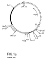
- FIG. 1a represents an assembly map of the plasmid pSE1, a plasmid for cloning in E. coli and for expression in animal cells, the sites having disappeared by ligation being noted in parentheses.
- the symbols used in this figure will be specified during the description of this plasmid (section 2).
- FIG. 1b represents the sequence of the synthetic fragment "HIndIII binding site" -HindIII used in the assembly of the plasmid pSE1.
- FIG. 2 represents the nucleotide sequence of the cDNA NC28 and opposite the deduced amino acid sequence, the three Met likely to initiate translation being underlined, the probable site of cleavage of the signal peptide being indicated by a vertical line and the potential site of N-glycosylation being underlined in dotted lines.
- FIG. 3 and FIG. 4 respectively represent the alignment according to the maximum homology according to the method of Needleman and Wunsch, 1970, J. Mol. Biol., 48, 443-453 of the amino acid sequence deduced from the cDNA NC28 (upper line) and the amino acid sequence deduced from the cDNA of the cytokine MCP-1 (lower line) and l alignment by this method of the cDNA NC28 (upper line) and the cDNA of the cytokine MCP-1 (lower line).
- FIG. 5 represents the sequence of fragment B used in the construction of the plasmid pEMR617, expression vector in yeast.
- Figures 6a, 6b and 6c relate to experiments demonstrating chemotactic activity.
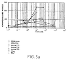
- FIG. 6a represents the number of cells per microscopic field as a function of the concentration expressed, in nM for the purified NC28 protein derived from yeast, the purified NC28 protein derived from COS cells, the C13, C16, C20 and fMLP peptides.
- FIG. 6b represents the number of cells per microcospic field as a function of the concentration expressed in ng / ml for the purified NC28 protein derived from yeast and the purified cytokine MCP-1 derived from COS cells.
- FIG. 6c represents the number of cells per microscopic field as a function of the concentration expressed in ng / ml, for the purified NC28 protein derived from yeast, the cytokine IL-8 and the peptide fMLP.
- SECTION 1 Culture and stimulation using PMA and PHA-P of peripheral blood mononuclear cells. Preparation of the messenger RNA used to make the complementary DNA library
- peripheral blood mononuclear cells PBMNC
- a fraction of cells enriched in peripheral blood mononuclear cells PBMNC with the following approximate composition: 70% lymphocytes, 25% monocytes and 5% granulocytes (counting of cells using the Coulter cell counter -Model S-Plus IV)
- the cells are collected in a 250 ml flask, then centrifuged for 10 min at 37 ° C. The supernatant is eliminated and the cell pellet is rinsed with 50 ml of medium based on glucose, mineral salts, amino acids and vitamins, called RPMI medium (RPMI 1640 medium from Gibco BRL), then again centrifuged under the same conditions.
- RPMI medium RPMI 1640 medium from Gibco BRL
- the cell pellet is then taken up with 500 ml of RPMI medium supplemented with 10% fetal calf serum (Gibco BRL- ref. 013-06290H), supplemented with 10 units of penicillin and 10 ⁇ g of streptomycin (penicillin solution / streptomycin from Gibco ref. 043-05140D) per ml of medium as well as L-glutamine (Gibco BRL-ref. 043-05030D) up to 2 mM final.
- RPMI medium supplemented with 10% fetal calf serum
- streptomycin penicillin solution / streptomycin from Gibco ref. 043-05140D
- L-glutamine Gibco BRL-ref. 043-05030D
- Part of the cell suspension is distributed for the separation of adherent cells and non-adherent cells, at a rate of approximately 100 ml per dish, in four large square culture dishes (245 x 245 x 20 mm-Nunc - ref 166508) and incubated for 1 h at 37 ° C. It is known in fact that most of the monocytic cells adhere to the culture dish while most of the lymphocyte cells remain in suspension.
- the non-adherent cells are aspirated using a pipette and cultured in culture flasks of the Falcon type with a surface area of 175 cm 2 in the presence of RPMI medium supplemented as described above, supplemented with 10 ng / ml of phorbol myristate- 2 acetate-3 (PMA) (Sigma-ref. P8139) and 5 ⁇ g / ml of phytohemaglutinin (PHA-P) (Sigma-ref. L8754), at 37 ° C in the presence of 5% CO2 for 24 h.
- PMA phorbol myristate- 2 acetate-3
- PHA-P phytohemaglutinin
- the rest of the cell suspension hereinafter called the total cells, is distributed into 4 large square culture dishes and incubated in the presence of RPMI medium supplemented as described above, supplemented with 10 ng / ml of PMA and 5 ⁇ g / ml of PHA-P at 37 ° C in the presence of 5% (v / v) of CO2 for 5 h for the first two dishes and 24 h for the other two.
- cycloheximide (Sigma ref. C6255) (translation inhibitor which increases the stability of cytokine RNAs) is added to the culture medium of these different cells. See Lindsten et al. , 1989, Science 244, 339-344) and the incubation is continued for 2 h at 37 ° C.
- Cell pellets A, NA, T (5 h) and T (24 h) are frozen and stored at -80 ° C.
- Each frozen cell pellet is suspended in the lysis buffer of the following composition: guanidine-thiocyanate 5M; Tris- (hydroxymethyl) -aminomethane 50mM pH 7.5 EDTA 10mM.
- the suspension is sonicated using an Ultra Turax sonicator No. 231 256 (Janke and Kunkel) at maximum power for 4 cycles of 20 s.
- ⁇ -mercaptoethanol is added up to 0.2M and a sonication cycle of 30 s is repeated.
- Lithium chloride is added up to 3M.
- the suspension is cooled to 4 ° C and allowed to stand at this temperature for 48 h.
- the RNA is then isolated by centrifugation for 60 min.
- RNA pellet is washed once with a 3M lithium chloride solution, recentrifuged, then taken up in a buffer of the following composition: SDS 1%, EDTA 5 mM and Tris HCl 10 mM pH 7.5, added with 1 mg / ml proteinase K (Boehringer Mannheim, GmbH). After incubation at 40 ° C for 1 h the RNA solution is extracted with a phenol / chloroform mixture. The RNA contained in the aqueous phase is precipitated at -20 ° C. using a final 0.3M ammonium acetate solution and 2.5 volumes of ethanol. Centrifuge at 15,000 xg for 30 min and keep the pellet.
- the pellet is taken up in 1 ml of 10 mM Tris-HCl composition buffer pH 7.5 1 mM EDTA, called TE buffer and suspended by vortexing.
- the oligo dT-cellulose type 3 (marketed by Collaborative Research Inc, Biomedicals Product Division) is prepared according to the manufacturer's recommendations.
- the RNA is deposited on the oligo dT-cellulose, stirred gently to suspend the beads, then heated for 1 min at 65 ° C.
- the suspension is adjusted to 0.5 M NaCl, then stirred gently for 10 min.
- the suspension is then centrifuged for 1 min at 1000 g, the supernatant is removed, the pellet is washed 2 times with 1 ml of TE buffer containing 0.5 M NaCl. The supernatants are eliminated.
- the elution of the polyadenylated fraction of RNA is obtained by suspending the beads in 1 ml of TE buffer, then heating this suspension at 60 ° C for 1 min, followed by stirring for 10 min on a tilting plate. Then centrifuged for 1 min at 1000 g, which allows the supernatant containing free messenger RNA in solution to be recovered.
- RNA-poly A+-A RNA-poly A +-NA
- RNA-polyA +-T RNA-poly A+-T
- the strategy implemented uses fragments obtained from pre-existing plasmids accessible to the public and fragments prepared by synthesis using the techniques now commonly used.
- the cloning techniques used are those described by T. Maniatis, EF. Fritsch and J. Sambrook in "Molecular Cloning, a Laboratory manual” (Cold Spring Harbor Laboratory, 1984).
- the synthesis of oligonucleotides is carried out using a Biosearch 8700 DNA synthesizer.
- FIG. 1 a The description below will be better understood with reference to FIG. 1 a .
- the cloning technique used is that described by Caput et al, (Primer-adapter technique: Caput et al, Proc. Natl. Acad. Sci. (USA), 1986, 83, 1670-1674 ).
- the polyA+ RNAs obtained at the end of section 1 are subjected to reverse transcription from a primer whose sequence is as follows:
- the cDNAs have at their 5 ′ end the GATCC sequence complementary to the BamHI cohesive end.
- RNA-DNA hybrids obtained by the action of reverse transcriptase are subjected to alkaline hydrolysis which makes it possible to get rid of the RNA.
- the single-stranded cDNAs are then subjected to a terminal transferase treatment, so as to add polydG at 3 ′ and purified by 2 cycles on a CL4B sepharose column.
- RNA-polyA+ originating from cells of the COS3 line (monkey kidney cell line expressing the T antigen of the SV40 virus: cf. Y. Gluzman, 1981, Cell, 23, 175- 182 prepared as described in Section 1.2).
- the non-hybridized cDNAs are isolated (fraction enriched in DNA complementary to the messenger RNAs specific for peripheral blood mononuclear cells).
- cDNAs are inserted in single-stranded form into the vector pSE1.
- a second oligonucleotide (the adapter) complementary to the primer is necessary to generate a BamHI site at the 5 ′ end of the cDNAs.
- the recombinant molecules are circularized by the action of the ligase of phage T4.
- the single-stranded regions are then repaired using DNA polymerase from phage T4.
- the pool of plasmids thus obtained is used to transform the E. coli MC 1061 strain (Casabadan and S. Cohen, J. Bact. (1980) 143, 971-980) by electroporation.
- RNA-poly A+ A 0.5 ⁇ g
- RNA-poly A+ NA 2 ⁇ g
- RNA-poly A+ T 5 h
- RNA-poly A+ T 24 h
- the complementary DNA is extended at 3 ′ with a “tail” of dG with 66 units of the terminal enzyme transferase (Pharmacia 27073001). Incubate for 30 min at 37 ° C., then add 4 ⁇ l of 0.5M EDTA.
- the complementary DNA is purified on two successive columns of 1 ml of CL4B sepharose (Pharmacia), equilibrated with a 30 mM NaOH / 2 mM EDTA solution.
- the first three radioactive fractions (approximately 80 ⁇ l each) are combined and precipitated with 1 / 10th of a volume of an ammonium acetate solution and 2.5 volumes of ethanol.
- the amount of complementary DNA is 1 ⁇ g.
- the complementary DNA pellet is suspended in 25 ⁇ l of TE buffer, 15 ⁇ g of RNA-polyA+ extracted from cells of the COS line are added, then 1 / 10th of a volume of a 3M NaCl solution, 2, 5 volumes of ethanol and allowed to precipitate at -20 ° C.
- the content of the capillary is diluted in 100 ⁇ l of TE buffer to which 300 ⁇ l of 50 mM sodium phosphate buffer pH 6.8 are added.
- the solution obtained is passed through a hydroxyapatite column (Biorad ref. 130.0520) at 60 ° C, equilibrated with this phosphate buffer.
- the single strand (non-hybridized complementary DNA) and the double strand (COS messenger RNA hybridized to complementary DNA) are separated by a phosphate buffer gradient of 0.1M to 0.2M through the column of hydroxyapatite.
- the fractions corresponding to the complementary single-stranded DNA are combined (25% by weight of the eluted cDNA, which corresponds to an enrichment of approximately 4 times in specific sequences of mononuclear cells of the peripheral blood), 20 ⁇ g of d are added.
- 'Transfer RNA the total volume is precipitated with 1 / 10th of a volume of a 10M ammonium acetate solution and 2.5 volumes of ethanol. Centrifuged, the pellet is dissolved in 200 ⁇ l of TE, the residual phosphate is removed on polyacrylamide P10, again precipitated with 1/10 th volume of a 10M ammonium acetate solution and 2.5 volumes d ethanol.
- the pellet is dissolved in 30 ⁇ l of a 30 mM NaOH solution; EDTA 2mM.
- the complementary DNA is loaded onto a 1 ml CL4B (Pharmacia) sepharose column, equilibrated with a 30 mM NaOH solution; 2mM EDTA, to remove the rest of the synthetic primer.
- the first 3 radioactive fractions of around 80 ⁇ l each are grouped together.
- the cDNA contained in these fractions is precipitated with 1/10 th volume of a 10 M ammonium acetate solution and 2.5 volume of ethanol.
- the amount of complementary DNA thus recovered is 20 ng.
- the pellet is dissolved in 33 ⁇ l of TE buffer, 5 ⁇ l (125 ng) of cloning vector pSE1, 1 ⁇ l (120 ng) of the adapter with the following sequence (comprising an ApaI site) are added: 10 ⁇ l of a 200 mM NaCl solution, incubated for 5 min at 65 ° C. and then the reaction mixture is allowed to cool to room temperature.
- the cloning vector and the single-stranded cDNA are ligated in a volume of 100 ⁇ l with 32.5 units of the enzyme DNA ligase from phage T4 (Pharmacia ref: 270 87002) overnight at 15 ° C.
- the proteins are eliminated by phenol extraction followed by chloroform extraction, then 1/10 th of a volume of a 10 mM ammonium acetate solution is added, then 2.5 volumes of ethanol. Centrifuged, the pellet is dissolved in the Tris acetate composition buffer, pH 7.9, 33 mM, potassium acetate 62.5 mM, 1 mM magnesium acetate and 1 mM dithiothreitol (DTT).
- the second strand of complementary DNA is synthesized in a volume of 30 ⁇ l with 30 units of the DNA polymerase enzyme of phage T4 (Pharmacia: ref.
- E. coli MC1061 cells (Clontech) are transformed with the recombinant DNA obtained previously by electroporation using the Biorad Gene Pulser device (Biorad) used at 2.5 kV under the conditions prescribed by the manufacturer, then makes the bacteria grow for 6 h 30 in medium known as LB medium (Sambrook, op cited) of composition: bacterotryptone 10 g / l; yeast extract 5 g / l; NaCl 10 g / l, supplemented with 100 ⁇ g / ml of ampicillin.
- LB medium Standardbrook, op cited
- colonies are transferred to a nylon membrane (Hybond N-Amersham) by depositing the membrane on the surface of the dish and setting marks by piercing the membrane using 'a needle. The membrane is then removed and deposited on the surface of a new Petri dish containing LB agar medium. It is left for a few hours at 37 ° C. to obtain the regrowth of the colonies. From this first membrane, two replicas are made on new membranes (previously deposited on LB agar medium to moisten them) by successive contacts with the first membrane. The replicas on membrane obtained are finally deposited on plates of agar LB medium and incubated overnight at 30 ° C.
- a nylon membrane Hybond N-Amersham
- the membrane replicas are deposited with the colonies upwards, on a sheet of Whatman 3MM saturated with a solution of composition: 0.5M NaOH; 1.5M NaCl, for 5 min, which makes it possible to lyse the bacteria and fix the DNA.
- the replicas on the membrane are then placed on a second sheet of Whatman 3MM, this time saturated with a neutralizing solution of composition: NaCl 1.5M; Tris-HCl pH8 0.5M, for 5 min.
- the replicas on the membrane are then immersed in a solution of 2 x SSC (composition of the SSC solution: 0.15M NaCl; 0.015M sodium citrate) and the bacterial debris is partially removed by gently rubbing with cotton wool. cleaning.
- replicas are then treated on a membrane with proteinase K (Boehringer Mannheim GmbH) at 100 ⁇ g / ml in a solution of composition: Tris-HCl 10mM pH8; 10mM EDTA; 50 mM NaCl; 0.1% SDS at a rate of 20 ml per membrane. Incubate for 30 min at 37 ° C with shaking.
- the replicas on the membrane are again immersed in a solution of 2 x SSC to permanently eliminate all traces of bacterial debris. They are finally put to dry on filter paper for a few minutes and then for 30 min under vacuum at + 80 ° C. There are thus obtained, for each box, two replicas on the membrane, hereinafter called replica 1 and replica 2.
- the monocytic line U937 ATCC 1593 is cultivated in RPMI medium supplemented with 10% fetal calf serum (Gibco BRL-ref. 013-06290H), supplemented with 10 units of penicillin and 10 ⁇ g of streptomycin per ml as well as 2 mM final L-glutamine.
- RPMI medium supplemented with penicillin, streptomycin and L-glutamine and 20 ng / ml of phorbol myristate-2 acetate-3 (PMA) (Sigma-ref. P8139) for 24h.
- the cells thus activated are scraped and centrifuged.
- the cell pellet obtained is called the U937P cell pellet.
- Half of the cells are additionally induced with cycloheximide (10 ⁇ g / ml) during the last two hours of culture.
- This inhibitor of protein synthesis makes unstable messenger RNAs (including cytokines) more stable (cf. T. Lindsten et al., 1989, Science, 244, 339-343).
- the cells thus activated are scraped and centrifuged.
- the cell pellet obtained is called the U937PC cell pellet.
- RNA is extracted and the polyA+ fraction is purified as described in section 1-2) a) and b). Two RNA-polyA+ fractions are thus obtained, called respectively polyA+1 fraction and polyA+2 fraction.
- probe 1 and probe 2 are synthesized from the two RNA-polyA+ fractions above, prepared as described below.
- 1 ⁇ g of polyA+ RNA is hybridized with 200 ng of oligo dT12 ⁇ 18 (Pharmacia) in 2 to 3 ⁇ l of Tris HCl composition buffer, pH 7.5, 50 mM and 1 mM EDTA by incubation for 2 min at 65 ° C and cooling to room temperature.
- the synthesis of the radiolabelled cDNA is carried out in a reaction volume of 10 ⁇ l in a buffer of composition: Tris-HCl 50mM pH8.3; MgCl2 5mM; 10mM dithiotreitol, containing 50 ⁇ M of dATP dGTP and dTTP, 10 ⁇ M of dCTP and 150 ⁇ Ci of dCTP ⁇ 32P (3000 Ci / mmol Amersham) and 40 units of RNasine (RNAse-Genofit inhibitor).
- the reaction is carried out at 46 ° C for 30 min in the presence of 10 to 20 units of reverse transcriptase (Genofit).
- the amount of cDNA is 60 to 100 ng with a specific activity of 1 x 10⁹dpm / ⁇ g.
- the membrane replicas are prehybridized for 2 h at 42 ° C in a buffer of composition: 50% formamide; 6 x SSC; 5 x Denhardt's solution; 0.1% SDS and 100 ⁇ g / ml of sonicated salmon sperm DNA, added after denaturing for 10 min at 100 ° C.
- the membrane replicas are hybridized for two days: replica 1 with probe 1 and replica 2 with probe 2, these probes being used at a concentration of 4 ng / ml in the above buffer.
- the 5 x Denhardt solution (cf. Sambrook, op. City) has the composition: Ficoll (type 400-Pharmacia) 1 g / l, polyvinylpyrrolidone 1 g / l and bovine serum albumin (BSA) 1 g / l.
- the prehybridization and hybridization are carried out in tubes in a hybridization oven (Hybaid) with respectively 25 ml and 10 ml of buffer per membrane.
- the replicas on the membrane are successively washed several times for 15 min at 20 ° C in the 2 x SSC composition buffer; 0.1% SDS, then twice for 15 min in a solution 0.1 x SSC, 0.1% SDS at 55 ° C, dried on Whatman 3MM paper and autoradiographed on Kodak XAR5 films.
- Interleukin-3 (Yang YC et al, 1986, 47, 3-10), Interleukin-4 (Yokoto T. et al, 1986, Proc. Ntl. Acad. Sci., 83, 5894-5898), Interleukin-5 (Hirano T. et al, 1986, Nature, 324, 73 -75), Interleukin-6 (May L. et al, 1986, Proc. Natl. Acad. Sci. USA, 83, 8957-8961), Interleukin-7 (Namen A.
- TGF ⁇ 1 Derynck R. et al, 1985, Nature, 316, 701-705
- bFGF Prats H. et al, 1989, Proc. Natl. Acad. Sci. USA, 86, 1836-1840
- Erythropoietin Jacobs K. et al, 1985, Nature, 313, 806-810
- BCGF BCGF (Sharma S. et al, 1987, Science, 235, 1489- 1492)
- MIF Weiser W. et al, 1989, Proc. Natl. Acad. Sci USA, 86, 7522-7526
- MCP-1 Yoshimura T.
- the mixture of oligonucleotides hybridizes with approximately 10% of the clones of the library.
- Clones giving a stronger autoradiographic signal with probe 2 than with probe 1, and not hybridizing with mixture C were partially sequenced as described in section 4 below.
- the clone NC28 contains the vector pSE1, which carries a cDNA between the ApaI and BamHI sites, hereinafter called cDNA NC28.
- the vector pSE1 which contains the origin of replication of phage f1, makes it possible to produce single-stranded DNA by culture of the clone NC28 in the presence of the bacteriophage M13K07 (Pharmacia- ref: 27-1524) in the following manner:
- the sequencing reactions are carried out using the United States Biochemical sequencing kit (ref: 70770), which uses the method of Sanger et al, Proc. Ntl. Acad. Sci. USA, 1977, 14, 5463-5467.
- the primers used are oligonucleotides of 18 nucleotides, complementary either to the vector pSE1 in the region immediately 5 ′ of the cDNA NC28, or to the sequence of the cDNA NC28.
- FIG. 2 represents the nucleotide sequence of the cDNA NC28 and opposite the deduced amino acid sequence, the three Met capable of initiating translation being underlined, the N-glycosylation site being underlined in dotted lines and the site of probable cleavage of the signal peptide being indicated by a vertical arrow.
- FIG. 3 and FIG. 4 respectively represent the alignment according to the maximum homology according to the method of Needleman and Wunsch, 1970, J. Mol. Biol., 48, 443-453 of the amino acid sequence deduced from the cDNA NC28 (upper line) and the amino acid sequence deduced from the cDNA of the cytokine MCP-1 (lower line) and l alignment by this method of the cDNA NC28 (upper line) and the cDNA of the cytokine MCP-1 (lower line).
- the cleavage site predicted by the PS software, at position 139-140 corresponds to that observed experimentally for the cytokine MCP-1 by E. Robinson et al, 1989, Proc. Ntl. Acad. Sci. USA, 86, 1850-1854.
- the ATG at position 71-73 of the cDNA NC28 corresponds to the ATG initiating the translation of the cytokine MCP-1
- COS cells are monkey kidney cells expressing the SV40 virus T antigen (Gluzman, Y. Cell 23 , 1981, 175-182). These cells, which allow the replication of the vectors containing the origin of replication of the DNA of the SV40 virus (case of the vector pSE1), constitute hosts of choice for the study of gene expression in animal cells.
- DMEM modified Eagle's medium from Gibco ref. 041-01965
- Gibco ref. 041-01965 modified Eagle's medium from Gibco ref. 041-01965
- DMEM modified Eagle's medium from Gibco ref. 041-01965
- Gibco fetal calf serum
- the culture medium is removed by aspiration and the cells are washed with 3 ml of PBS buffer (Phosphate Buffered Saline from GIBCO).
- PBS buffer Phosphate Buffered Saline from GIBCO.
- the following mixture is then added to it: 1000 ⁇ l of (DMEM + 10% fetal calf serum (Gibco)), 110 ⁇ l of diethylamino-ethyl-dextran of average molecular weight 500,000, at a concentration of 2 mg / ml (pharmacia ), 1.1 ⁇ l of 100mM chloroquine (Sigma) and 6 ⁇ g of plasmid DNA from clone NC28, prepared according to the alkaline lysis technique followed by purification of the plasmid DNA on a cesium chloride gradient (Sambrook et al , op cited).
- control COS cells were prepared by carrying out the operations described above with the DNA of the plasmid pSE1.
- the culture medium is removed by suction and the cells are washed twice with 3 ml of PBS buffer. 5 ml of MEM medium (Minimal Eagle's Medium) without methionine are added (Gibco - ref. 041-01900H), supplemented with 3 g / ml of glucose and 4 mM of glutamine. Incubate for 2 h at 37 ° C. The culture medium is removed and 2 ml of the same medium added with 200 ⁇ Ci of methionine 35S (ref. SJ1015 Amersham) are added. Incubate for 6 h at 37 ° C. The culture medium is removed, centrifuged for 5 min to remove cell debris and suspended cells, and the supernatant is kept. The adherent cells are rinsed twice with PBS buffer, scraped with a rubber scraper and centrifuged.
- MEM medium Minimal Eagle's Medium
- methionine 35S Ref. SJ1015 Amersham
- the autoradiogram shows the presence of four supernumerary bands for the cells transfected with the plasmid DNA of the clone NC28 compared to the control cells: two high intensity bands corresponding to apparent molecular masses of 9 ⁇ 2 and 16 ⁇ 2 kDa and two low intensity bands corresponding to apparent molecular masses of 11 ⁇ 2 and 17 ⁇ 2 kDa (the latter band is diffuse).
- the molecular mass for the mature protein of 76 amino acids is 8956 kDa, therefore close to the mass apparent molecular corresponding to the first of these bands.
- Protein labeling is carried out as in 2) above but in the presence of 10 ⁇ g / ml of tunicanycin (Sigma-ref. T7765), an agent inhibiting the N-glycosylation of proteins.
- the autoradiogram shows the presence of two supernumerary bands for the cells transfected with the plasmid DNA of the clone NC28, compared to the control cells: a high intensity band corresponding to an apparent molecular mass of 9 ⁇ 2 kDa and a band of low intensity corresponding to an apparent molecular mass of 11 ⁇ 2 kDa.
- DMEM modified Eagle medium according to Dubelcco hereinafter called DMEM (Dulbecco's Modified Eagle's medium from Gibco ref: 041-01965), which contains 0.6 g / l glutamine, 3.7 g / l NaHCO3 and is supplemented with fetal calf serum (Gibco) at a rate of 5% and then buffered with carbon dioxide. After approximately 16 h of culture at 37 ° C.
- DMEM modified Eagle medium according to Dubelcco hereinafter called DMEM (Dulbecco's Modified Eagle's medium from Gibco ref: 041-01965), which contains 0.6 g / l glutamine, 3.7 g / l NaHCO3 and is supplemented with fetal calf serum (Gibco) at a rate of 5% and then buffered with carbon dioxide. After approximately 16 h of culture at 37 ° C.
- Gibco fetal calf serum
- the mixture is removed from the cells and 35 ml of PBS buffer containing 7% of dimethyl sulfoxide (Spectroscopy quality, Merck is added thereto). ) After 1 min and 30 s of rotation at room temperature, the mixture is removed and the cells are washed twice with PBS and are incubated for 5 days at 37 ° C. in rotation in DMEM medium without phenol red. The supernatant is removed, with a volume of approximately 130 ml.
- fraction 1 and fraction 2 The recombinant protein is found in the fractions corresponding to two peaks, called fraction 1 and fraction 2, which give, respectively, by electrophoretic analysis in the presence of SDS, for one, a majority band of apparent molecular mass of 9 ⁇ 2 kDa and a minority band of apparent molecular mass of 11 ⁇ 2 kDa, for the other, a majority band of apparent molecular mass of 16 ⁇ 2 kDa and a series of weak bands of apparent molecular masses of between (16 and 18) ⁇ 2 kDa .
- the amino-terminal sequence was analyzed using an Applied Biosystem model 470A sequencer, coupled to an Applied Biosystem model 120A phenylthiohydantoic derivative analyzer.
- the purified protein 200 pmol controlled by amino acid analysis was deposited on the sequencer in the presence of 20 pmol of ⁇ -lactoglobulin, control protein.
- Fractions 1 and 2 were subjected to digestions using the 3 enzymes, N-glycanase, neuraminidase and O-glycanase (Genzyme), according to the protocol described in the technical file delivered with these enzymes.
- the majority band of apparent molecular mass of 9 ⁇ 2 kDa is not modified in the presence of N-glycanase, neither of neuraminidase, nor of O-glycanase.
- the minority band of apparent molecular mass 11 ⁇ 2 kDa is not modified by N-glycanase, but is modified by neuraminidase to a band of apparent molecular mass of 10 ⁇ 2 kDa, this band itself being modified by O-glycanase in a band merging with the majority band of apparent molecular mass of 9 ⁇ 2 kDa.
- the majority band of apparent molecular mass 16 ⁇ 2 kDa is not modified by neuraminidase or by O-glycanase, but after the action of N-glycanase, it merges with the majority band of apparent molecular mass of 9 ⁇ 2 kDa.
- NC28 protein is the first known member of the family of human SIS cytokines, which is N-glycosylated (A. Minty, 1991, Médecine Sciences, 7, 578).
- the cytokine MCP-1 exists in an O-glycosylated form but not in an N-glycosylated form (Jiang et al, 1990, J. Biol. Chem., 30, 18318).
- SECTION 7 Construction of an NC28 cDNA expression vector in yeast: the plasmid pEMR617 and transformation of a yeast strain using this plasmid
- fragment A The large fragment (hereinafter called fragment A) comprising the origin of replication and the 2 micron STB locus, the LEU2d gene, the ampicillin resistance gene, the origin of pBR322, the terminator of the PGK gene, the URA3 gene, the artificial promoter and the start of the alpha pheromone prepro region has been purified.
- fragment B The HindIII-BamHI fragment (hereinafter called fragment B) comprising the end of the prepro region of the alpha pheromone and the cDNA coding for the mature protein, flanked by the BamHI restriction site at 3 ′, was obtained by amplification by the PCR technique from the plasmid pSE1-NC28. The sequence of this fragment is specified in FIG. 5. The fragments A and B were ligated so as to obtain the plasmid pEMR617
- PCR polymerase chain reaction
- the PCR technique is based on the repetition of three stages, making it possible, after between 10 and 30 cycles, hundreds of thousands of copies of the original matrix, using a DNA polymerase from Thermus aquaticus, usually called Taq polymerase.
- the three steps are:
- Double stranded DNA is denatured to single stranded DNA by incubation at high temperature (92 ° C to 96 ° C) for approximately 2 min.
- primers are a pair of synthetic oligonucleotides which hybridize with the ends of the region to be amplified. The two primers hybridize with opposite strands. The primers are added in excess, so that the formation of the primer-template complex is favored.
- the step during which the Taq polymerase ensures the extension of the primer-template complex from 5 ′ to 3 ′ is carried out at 72 ° C.
- the product of interest appears in the third cycle and is then amplified significantly. During the course of the cycles, the amplification product quickly becomes the matrix with which the primers hybridize.
- the first oligonucleotide called primer 1, whose sequence is as follows: has two distinct regions: region 1, which contains the end of the prepro region of the modified pheromone compared to the natural sequence described by Kurjan et al, Cell, 1982, 30, 933-943 by a silent mutation which allows d '' introduce a HindIII site just before the coding part of region 1 (fifth nucleotide of region 1), and region 2, which is a region intended to hybridize with the coding region corresponding to the start of the mature protein of 76 amino acids (cf. section 4), of the non-coding strand of the part of the plasmid pSE1-NC28 which carries the cDNA NC28.
- region 1 contains the end of the prepro region of the modified pheromone compared to the natural sequence described by Kurjan et al, Cell, 1982, 30, 933-943 by a silent mutation which allows d '' introduce a HindIII site just before the coding part of region 1 (f
- the second oligonucleotide called primer 2, whose sequence is as follows: also consists of two distinct regions: region 1 which carries a BamHI site on the fifth nucleotide, and region 2, which carries a nucleotide sequence corresponding to the last codons of the coding part of the cDNA-NC28 and to the stop codon, with a mutation intended to suppress the HindIII site (silent mutation of the codon AAG into AAA). This region is intended to hybridize with the strand coding for the part of the plasmid pSE1-NC28 which carries the cDNA NC28.
- the plasmid pSE1 - NC28 which carries the cDNA coding for the protein NC28 is used as a template.
- 100 ng of the pSE1 - NC28 plamide are added to a tube, 100 ng of the primer 1, 100 ng of the primer 2, 5 ⁇ l of reaction mixture concentrated 10 times (final quantity: 67 mM Tris-HCl pH 8.8, 16.6 mM (NH4) 2SO4, 1 mM ⁇ mercaptoethanol, 6.7 mM EDTA, 0.15% Triton x 100, 2 mM MgCl2, 0.2 mM dNTP, 200 ng of gelatin) the volume of the mixture is then brought to 50 ml by adding water.
- 0.5 ⁇ l is added, ie 2.5 units of Taq Polymerase (Boehringer Manheim ref 1146 - 173). The mixture is then covered with paraffin to avoid evaporation of the aqueous solution.
- the DNA fragment thus amplified which has the expected size of approximately 250 bp, is then isolated and purified on 1% agarose gel, subjected for dialysis by chromatography on a column of polyacrylamide gel P10 (Pharmacia), then completely and simultaneously hydrolyzed by the enzymes HindIII and BamHI according to the usual techniques well known to those skilled in the art (Sambrook et al, op cited) in order to to form the HindIII and BamHI cohesive ends. After hydrolysis, the fragment is purified on column P10.
- the sequence of the fragment B obtained is represented in FIG. 5. It includes, in its part coding for the protein NC28, a silent mutation with respect to the cDNA NC28, indicated by an asterisk in FIG. 5.
- Fragments A and B were ligated so as to obtain the plasmid pEMR617.
- the strain EMY761 (Mat alpha, leu2, ura3, his3) described in patent EP-0408461 and which can be obtained by plasmid cleaning of the strain deposited at the CNCM on December 27, 1989 under the number I-1021, contains mutations ( leu2 and ura3 ), capable of being complemented by the defective selection marker LEU2d and the selection marker URA3, present in the plasmid pEMR617. It was transformed by the plasmid pEMR617 with selection for the prototrophy of leucine according to a variant of the transformation technique described by Beggs et al. (Beggs et al, 1978, Nature 275, 104-109) which consists in subjecting the yeasts to a protoplastization treatment in the presence of an osmotic stabilizer, sorbitol in 1 M concentration.
- composition and preparation of the main media used in the transformation protocol of the yeast strain EMY761 Composition and preparation of the main media used in the transformation protocol of the yeast strain EMY761
- the culture was replaced at 30 ° C with stirring for 24 h.
- the mixture was brought to the temperature of + 4 ° C for 30 min, then centrifuged for 30 min.
- the pellet was taken up in approximately 1 ml of cold acetone (+ 4 ° C) and centrifuged again for 30 min.
- the pellet after having been dried, is taken up in approximately 20 ⁇ l of a buffer called loading buffer consisting of 0.125 M Tris-HCl pH 6.8 SDS 4%, bromophenol blue 0.002%, glycerol 20%, ⁇ -mercaptoethanol 10% according to the protocol described by Laemmli (cited above).
- the pellet is dissolved by boiling for 15 min and then neutralized until the bromophenol blue turns blue.
- the samples are deposited on a polyacrylamide gel in the presence of SDS and subjected to electrophoresis
- N-glycosylation of a protein by yeast involves a simple N-glycosylation ("core glycosylation") in the endoplasmic reticulum and an N-hyperglycosylation ("outer-chain glycosylation") in the Golgi apparatus (RA Hitzeman et al, 1990, "Methods in Enzymology, No. 185", Academic Press, p. 421-440).
- core glycosylation N-glycosylation
- outer-chain glycosylation N-chain glycosylation
- simple N-glycosylation leads to a glycoprotein of homogeneous apparent molecular weight (a band) and N-hyperglycosylation to a heterogeneous apparent molecular weight glycoprotein (plurality of diffuse bands).
- certain proteins can be O-glycosylated by yeast (J. Zueco et al, 1986, Biochemica and Biophysica Acta, 884, 93-100).
- N-glycosylation can be demonstrated in several ways (P. Oleans et al, 1991, Methods in Enzymology, 194, 682-697). One of them consists in observing the decrease in the apparent molecular mass of the protein when it is treated with endoglycosydase H (Endo-bN-acetylglucosaminisidase: EC 32.1.96) which specifically cuts the N-linked carbohydrate chains .
- O-glycosylation can be presumed when the protein is resistant to endoglycosydase H and its apparent molecular mass decreases after treatment with ⁇ -mannosidase ( ⁇ -D-manoside mannohydrolase: EC 3.2.1.24), as described by Biemans et al, 1991, DNA and Cell Biology, 10, 191-200.
- the mixture was brought to the temperature of + 4 ° C for 30 min, then centrifuged for 30 min.
- the pellet was taken up in approximately 1 ml of cold acetone (+ 4 ° C) and centrifuged again for 30 min.
- the pellet is taken up in 40 ⁇ l of a solubilization buffer (composition: Tris-HCl pH 6.8 10 mM, ⁇ mercaptoethanol 2%, SDS 1%).
- the pellet is brought to 100 ° C. for 15 min.
- the immunoblot analysis shows for the induced sample not treated with endoglycosidase H the presence of several additional bands compared to the non-induced sample, the two main ones of which correspond to an apparent molecular mass of 9 ⁇ 2 and 11 ⁇ 2 kDa. These two bands are recognized by the immune serum. Diffuse bands of apparent molecular weight greater than 16 ⁇ 2 kDa are also highlighted.
- the diffuse bands corresponding to the molecular weight greater than 16 ⁇ 2 kDa tend to disappear after treatment with endoglycosidase H to give way to a band of apparent molecular mass of 16 ⁇ 2 kDa which may correspond to the precursor having kept the pro pheromone sequence.
- the two main bands of apparent molecular weights of 9 ⁇ 2 and 11 ⁇ 2 kDa are always present.
- composition and preparation of certain media used for sample preparation are compositions and preparation of certain media used for sample preparation:
- the amino-terminal sequence is determined according to the principle of Edman degradation (Kia-Ki Han et al, 1977, Biochemistry, 59, 557).
- the first amino-terminal amino acid is coupled to phenyl-isothiocyanate (PITC), then cleaved.
- PITC phenyl-isothiocyanate
- the derivative obtained is converted into a stable phenylthioidantoin amino acid, absorbing at 269 nm.
- the product of each cycle is analyzed by HPLC.
- Sequence 1 is the expected amino terminal sequence: that of the mature protein of 76 amino acids (cf. FIG. 2) described in section 4, the coding sequence of which is introduced into the vector pEMR617 described in section 7.
- Sequences 1 and 2 are the amino-terminal sequences of the mature protein of 76 amino acids cleaved respectively in its amino-terminal part of 2 and 18 amino acids.
- the carboxy-terminal residue of the peptide to be synthesized is attached to an insoluble polymer, then different amino acids are added.
- the polypeptide chain increases in size in the amino-terminal direction.
- the peptide is separated from the support by hydrofluoric acid and recovered in solution.
- the peptides corresponding to amino acids 90-109, 94-109 and 97-109 of the translated protein NC28 were synthesized, called respectively peptides C20, peptide C16 and peptide C13.
- the analysis of the amino acid composition is fully automated.
- the amino acid analyzer (model 420A, Applied Biosystems) performs the hydrolysis, then the derivation of the released amino acids.
- the separation derived amino acids is produced by a Brownlee 130A HPLC system connected in line with the 420A system.
- the amino-terminal sequence is determined according to the principle of Edman degradation.
- the first amino-terminal amino acid is coupled to phenyl-isocyanate (PITC), then cleaved.
- PITC phenyl-isocyanate
- the derivative obtained is converted into a stable phenylthioidantoin amino acid, absorbing at 254 nM.
- the product of each cycle is analyzed by HPLC.
- peptide 48-69 is coupled to a carrier protein Fisurella hemocyanin (KLH) by its carboxy-terminal cysteine residue with the m-maleimidobenzoyl-N-hydroxysuccinimide ester (MBS) as a coupling agent.
- KLH Fisurella hemocyanin
- MFS m-maleimidobenzoyl-N-hydroxysuccinimide ester
- Rabbits (New Zealand, males weighing approximately 2 kg) are immunized with the peptide 48-69 / KLH conjugate (amount corresponding to 800 ⁇ g of peptide) (rabbit N ° 43) or with 50 ⁇ g of NC28 protein (purified from yeast - section 9) (rabbit N ° 44) every two weeks according to the protocol described by Vaitukaitis, 1981, Methods in Enzymology, 73, 46.
- one volume of antigenic solution is emulsified by one volume of adjuvant complete by Freund (Sigma-ref. 4258).
- the boosters (3 for rabbit No. 43 and 6 for rabbit No. 44) are administered in Freund's incomplete adjuvant (Sigma-ref. 5506).
- the immune sera obtained are capable of recognizing the protein NC28 produced in yeast and in COS cells by immunodetection after electrophoresis on polyacrylamide gel in the presence of SDS.
- peripheral blood From the peripheral blood, most of the red cells are removed by sedimentation at 37 ° C. for 30 min in a solution containing 0.6% dextran T500 (Pharmacia -ref: 17-0320-01) and 0.09% NaCl. Subsequently the cells are deposited on top of a layer of Ficoll-Paque (Pharmacia) and centrifuged at 400 g for 30 min. Peripheral blood mononuclear cells (PBMNC) are present at the interface between Ficoll and the supernatant while residual red blood cells and polynuclear cells (mainly neutrophils) are found in the cell pellet.
- PBMNC Peripheral blood mononuclear cells
- HBSS solution Hanks Balanced Saline Solution (Gibco BRL-Ref. 041-04020 H), hereinafter called HBSS solution.
- the principle of isolation of monocytes has been described by A. Boyum, 1983, Scan. J. Immunol., 17, 429-436. It is summarized below.
- the method consists in separating the monocytes from the blood using an iodine gradient medium, Nycodenz (N, N′-Bis (2,3-dihydroxypropyl), [N- (2,3-dihydroxy propyl) acetamido] -5- triodoisophthalamide-2,4,6).
- Nycodenz N, N′-Bis (2,3-dihydroxypropyl), [N- (2,3-dihydroxy propyl) acetamido] -5- triodoisophthalamide-2,4,6).
- Nycodenz N, N′-Bis (2,3-dihydroxypropyl), [N- (2,3-dihydroxy propyl) acetamido] -5- triodoisophthalamide-2,4,6.
- Nycodenz N, N′-
- the clarified plasma is taken up to 3-4 mm above the interphase and the rest of the plasma and all of the NycoPrep solution are collected up to about 1 cm above the pellet cell, which does not collect lymphocytes.
- the suspension of monocytes collected is completed to a volume of 6-7 ml with a solution of composition: 0.9% NaCl 0.13% EDTA 1% BSA, then centrifuged for 7 min at 600 g.
- Monocytes are contaminated with platelets. To remove these, centrifuge then remove the supernatant and resuspend with the same solution, repeating these operations 3 times.
- the cells are resuspended in RPMI 1640 medium (Gibco) containing 0.5% bovine serum albumin (BSA)
- the modified Boyden chamber is used for chemotaxis measurement, sold by Neuroprobe (ref. AP48).
- the test samples diluted in an HBSS solution for the tests on the neutrophils, and the RPMI medium containing 0.5% BSA for the test on the monocytes are placed in the wells of the lower plate.
- a polycarbonate filter (pore size: 5 mm-Nuclepore ref. 155845) is placed on it with the shiny side at the bottom.
- the top plate is placed on the filter.
- the cells (50,000 for 50 ⁇ l buffer) are placed in the wells of the upper plate.
- the chamber is incubated at 37 ° C. in a humidified oven or in a box containing wet cotton for 1 h for the neutrophil test, and 3 h for the monocyte test.
- the filter is removed and the cells which are on the matt side (cells which have not migrated) are removed by wiping the filter and scraping it with a rubber scraper, these last two operations being repeated 1 time.
- the cells which have migrated are stained and fixed using the "Diff-quick" kit (Dade-ref. 130832). By microscopic observation, count the number of cells on the shiny side of the filter (cells that have migrated)
- FIG. 6a represents the number of cells per microscopic field as a function of the concentration expressed in nM for the purified NC28 protein derived from yeast, the purified NC28 protein derived from COS cells, the C13, C16 peptides, C20 and fMLP and in FIG. 6b which represents the number of cells per microscopic field as a function of the concentration expressed in ng / ml for the purified NC28 protein derived from yeast and the purified cytokine MCP-1 derived from COS cells.
- the purified NC28 protein derived from yeast and the purified NC28 protein derived from COS cells exhibit, after 3 h of incubation, a chemotaxis activity on monocytes significantly greater than that of fMLP, a peptide known to have a chemotaxis activity on monocytes (Schiffmann E. et al, J. Immunol. 114, 1831).
- the carboxyterminal peptides of the NC28 protein also have this activity.
- FIG. 6c represents the number of cells per microcospic field as a function of the concentration expressed in ng / ml, for the purified NC28 protein derived from yeast, the cytokine IL-8 and the peptide fMLP.
- NC28 protein is found to have only a weak chemotaxis activity on neutrophils (at high concentrations greater than 100 ng / ml), unlike the cytokine IL-8 and the peptide fMLP, both known to possess this activity (Yoshimura T. et al, 1987, Proc. Natl. Acad. Sci-USA, 84, 9233.1 and Schiffman E. et al, 1975, J. Immunol., 114, 1831).
- NC28 protein is therefore a powerful chemoattractant specific for monocytes.
Applications Claiming Priority (2)
| Application Number | Priority Date | Filing Date | Title |
|---|---|---|---|
| FR9014961 | 1990-11-29 | ||
| FR9014961A FR2669930A1 (fr) | 1990-11-29 | 1990-11-29 | Proteine presentant une activite de type cytokine, adn recombinant, vecteur d'expression et hotes permettant son obtention. |
Publications (2)
| Publication Number | Publication Date |
|---|---|
| EP0488900A1 true EP0488900A1 (de) | 1992-06-03 |
| EP0488900B1 EP0488900B1 (de) | 2001-02-28 |
Family
ID=9402736
Family Applications (1)
| Application Number | Title | Priority Date | Filing Date |
|---|---|---|---|
| EP91403243A Expired - Lifetime EP0488900B1 (de) | 1990-11-29 | 1991-11-29 | Protein mit Cytokinaktivität, Rekombinant-DNS, Expressionsvektor und Wirtszellen zu seiner Herstellung |
Country Status (7)
| Country | Link |
|---|---|
| US (1) | US6001649A (de) |
| EP (1) | EP0488900B1 (de) |
| JP (1) | JPH05503945A (de) |
| AT (1) | ATE199377T1 (de) |
| DE (1) | DE69132545D1 (de) |
| FR (1) | FR2669930A1 (de) |
| WO (1) | WO1992009629A1 (de) |
Cited By (14)
| Publication number | Priority date | Publication date | Assignee | Title |
|---|---|---|---|---|
| EP0767796A1 (de) * | 1994-05-16 | 1997-04-16 | Human Genome Sciences, Inc. | Monocytisches chemotaktisches protein 4 |
| WO1998002459A1 (en) * | 1996-07-15 | 1998-01-22 | Incyte Pharmaceuticals, Inc. | Human monocyte chemotactic proprotein |
| US5910431A (en) * | 1996-03-19 | 1999-06-08 | Human Genome Sciences, Inc. | Polynucleotides encoding chemokine α-2 |
| US6028169A (en) * | 1997-03-31 | 2000-02-22 | Human Genome Sciences, Inc. | Chemokine β-6 antagonists |
| US6075124A (en) * | 1994-05-16 | 2000-06-13 | Human Genome Sciences, Inc. | Human chemotactin protein |
| US6100389A (en) * | 1995-04-21 | 2000-08-08 | Human Genome Sciences, Inc. | Polynucleotides encoding a human chemotactic protein |
| US6139832A (en) * | 1995-02-08 | 2000-10-31 | Human Genome Sciences, Inc. | Leukocyte adhesion inhibitor-1 (LAI-1) Polypeptides |
| US6274342B1 (en) | 1996-09-18 | 2001-08-14 | Center For Blood Research, Inc. | Nucleic acid molecules encoding monocyte chemotactic protein 5 (MCP-5) molecules and uses therefor |
| EP1125948A1 (de) * | 1994-05-16 | 2001-08-22 | Human Genome Sciences, Inc. | Monocytisches chemotaktisches protein 4 |
| US6391589B1 (en) | 1994-08-23 | 2002-05-21 | Human Genome Sciences, Inc. | Human chemokine beta-10 mutant polypeptides |
| US6410268B1 (en) | 1996-03-18 | 2002-06-25 | Human Genome Sciences, Inc. | Polynucleotides encoding chemokine alpha-3 |
| US6586185B2 (en) | 2000-06-20 | 2003-07-01 | Switch Biotech Ag | Use of polypeptides or nucleic acids for the diagnosis or treatment of skin disorders and wound healing and for the identification of pharmacologically active substances |
| US7943741B2 (en) | 2002-05-01 | 2011-05-17 | Human Genome Sciences, Inc. | Antibodies that specifically bind to chemokine β-4 |
| US8491901B2 (en) | 2010-11-19 | 2013-07-23 | Toshio Imai | Neutralizing anti-CCL20 antibodies |
Families Citing this family (8)
| Publication number | Priority date | Publication date | Assignee | Title |
|---|---|---|---|---|
| US7005509B1 (en) * | 1995-02-17 | 2006-02-28 | Incyte Corporation | Chemokine PANEC-1 polynucleotides and compositions and methods related thereto |
| US6713605B1 (en) * | 1996-04-10 | 2004-03-30 | Kansas State University Research Foundation | Synthetic peptides that inhibit leukocyte superoxide anion production and/or attract leukocytes |
| US7265201B1 (en) * | 1995-06-23 | 2007-09-04 | Millennium Pharmaceuticals, Inc. | Human chemotactic cytokine |
| US6562347B1 (en) * | 1998-03-12 | 2003-05-13 | The United States Of America As Represented By The Department Of Health And Human Services | Chemokine-tumor antigen fusion proteins as cancer vaccines |
| US7157418B1 (en) | 1998-07-22 | 2007-01-02 | Osprey Pharmaceuticals, Ltd. | Methods and compositions for treating secondary tissue damage and other inflammatory conditions and disorders |
| AU2001291050A1 (en) * | 2000-09-15 | 2002-03-26 | The Government Of The United States Of America, As Represented By The Secretary Of The Department Of Health And Human Services | Viral chemokine-tumur antigen fusion proteins |
| US7754676B2 (en) * | 2000-09-15 | 2010-07-13 | The United States Of America As Represented By The Department Of Health And Human Services | Defensin-antigen fusion proteins |
| FR3031100A1 (fr) | 2014-12-31 | 2016-07-01 | Equipement De Chantiers Et Locations | Dispositif de chantier perfectionne pour le recyclage d’une eau de lavage |
Citations (2)
| Publication number | Priority date | Publication date | Assignee | Title |
|---|---|---|---|---|
| WO1990007863A1 (en) * | 1989-01-01 | 1990-07-26 | The United States Of America, Represented By The Secretary, United States Department Of Commerce | A polypeptide having human monocyte chemotactic factor activity and a dna encoding said polypeptide |
| WO1990008777A1 (en) * | 1989-01-31 | 1990-08-09 | The United States Of America, Represented By The Secretary, United States Department Of Commerce | HUMAN DERIVED MONOCYTE ATTRACTING PURIFIED PROTEIN PRODUCT USEFUL IN A METHOD OF TREATING INFECTION AND NEOPLASM IN HUMAN BODY, AND THE CLONING OF FULL LENGTH cDNA THEREOF |
Family Cites Families (2)
| Publication number | Priority date | Publication date | Assignee | Title |
|---|---|---|---|---|
| JPS5155297A (en) * | 1974-10-28 | 1976-05-14 | Toppan Moore Kk | Mihirakya bunryoinakirokubutsuno sakuseihoho |
| JPS6274652A (ja) * | 1985-09-30 | 1987-04-06 | Tokyo Kikai Seisakusho:Kk | 発送仕分処理システム |
-
1990
- 1990-11-29 US US07/927,391 patent/US6001649A/en not_active Expired - Fee Related
- 1990-11-29 FR FR9014961A patent/FR2669930A1/fr active Granted
-
1991
- 1991-11-29 WO PCT/FR1991/000949 patent/WO1992009629A1/fr unknown
- 1991-11-29 DE DE69132545T patent/DE69132545D1/de not_active Expired - Lifetime
- 1991-11-29 EP EP91403243A patent/EP0488900B1/de not_active Expired - Lifetime
- 1991-11-29 AT AT91403243T patent/ATE199377T1/de active
- 1991-11-29 JP JP4501436A patent/JPH05503945A/ja active Pending
Patent Citations (2)
| Publication number | Priority date | Publication date | Assignee | Title |
|---|---|---|---|---|
| WO1990007863A1 (en) * | 1989-01-01 | 1990-07-26 | The United States Of America, Represented By The Secretary, United States Department Of Commerce | A polypeptide having human monocyte chemotactic factor activity and a dna encoding said polypeptide |
| WO1990008777A1 (en) * | 1989-01-31 | 1990-08-09 | The United States Of America, Represented By The Secretary, United States Department Of Commerce | HUMAN DERIVED MONOCYTE ATTRACTING PURIFIED PROTEIN PRODUCT USEFUL IN A METHOD OF TREATING INFECTION AND NEOPLASM IN HUMAN BODY, AND THE CLONING OF FULL LENGTH cDNA THEREOF |
Cited By (25)
| Publication number | Priority date | Publication date | Assignee | Title |
|---|---|---|---|---|
| EP0767796A4 (de) * | 1994-05-16 | 1997-06-11 | Human Genome Sciences Inc | Monocytisches chemotaktisches protein 4 |
| US6867006B2 (en) | 1994-05-16 | 2005-03-15 | Human Genome Sciences, Inc. | Antibodies to human chemotactic protein |
| US6419917B1 (en) | 1994-05-16 | 2002-07-16 | Human Genome Sciences, Inc. | Human chemotactic protein |
| US6075124A (en) * | 1994-05-16 | 2000-06-13 | Human Genome Sciences, Inc. | Human chemotactin protein |
| EP0767796A1 (de) * | 1994-05-16 | 1997-04-16 | Human Genome Sciences, Inc. | Monocytisches chemotaktisches protein 4 |
| EP1125948A1 (de) * | 1994-05-16 | 2001-08-22 | Human Genome Sciences, Inc. | Monocytisches chemotaktisches protein 4 |
| US6391589B1 (en) | 1994-08-23 | 2002-05-21 | Human Genome Sciences, Inc. | Human chemokine beta-10 mutant polypeptides |
| US6485719B1 (en) | 1995-02-08 | 2002-11-26 | Human Genome Sciences, Inc. | Methods for inhibiting angiogenesis with leukocyte adhesion inhibitor-1 (LAI-1) polypeptides |
| US6139832A (en) * | 1995-02-08 | 2000-10-31 | Human Genome Sciences, Inc. | Leukocyte adhesion inhibitor-1 (LAI-1) Polypeptides |
| US6100389A (en) * | 1995-04-21 | 2000-08-08 | Human Genome Sciences, Inc. | Polynucleotides encoding a human chemotactic protein |
| US6410268B1 (en) | 1996-03-18 | 2002-06-25 | Human Genome Sciences, Inc. | Polynucleotides encoding chemokine alpha-3 |
| US6908986B2 (en) | 1996-03-18 | 2005-06-21 | Human Genome Sciences, Inc. | Chemokine alpha 3 |
| US7419662B2 (en) | 1996-03-18 | 2008-09-02 | Human Genome Sciences, Inc. | Methods of making chemokine alpha 3 antibodies |
| US6479633B1 (en) | 1996-03-19 | 2002-11-12 | Human Genome Sciences, Inc. | Chemokine alpha 2 |
| US5910431A (en) * | 1996-03-19 | 1999-06-08 | Human Genome Sciences, Inc. | Polynucleotides encoding chemokine α-2 |
| US7122639B2 (en) | 1996-03-19 | 2006-10-17 | Human Genome Sciences, Inc. | Chemokine alpha-2 antibodies |
| WO1998002459A1 (en) * | 1996-07-15 | 1998-01-22 | Incyte Pharmaceuticals, Inc. | Human monocyte chemotactic proprotein |
| US6274342B1 (en) | 1996-09-18 | 2001-08-14 | Center For Blood Research, Inc. | Nucleic acid molecules encoding monocyte chemotactic protein 5 (MCP-5) molecules and uses therefor |
| US6379926B1 (en) | 1997-03-31 | 2002-04-30 | Human Genome Sciences, Inc. | Polynucleotides encoding chemokine β-6 antagonists |
| US6028169A (en) * | 1997-03-31 | 2000-02-22 | Human Genome Sciences, Inc. | Chemokine β-6 antagonists |
| US6586185B2 (en) | 2000-06-20 | 2003-07-01 | Switch Biotech Ag | Use of polypeptides or nucleic acids for the diagnosis or treatment of skin disorders and wound healing and for the identification of pharmacologically active substances |
| US7943741B2 (en) | 2002-05-01 | 2011-05-17 | Human Genome Sciences, Inc. | Antibodies that specifically bind to chemokine β-4 |
| US8491901B2 (en) | 2010-11-19 | 2013-07-23 | Toshio Imai | Neutralizing anti-CCL20 antibodies |
| US9133273B2 (en) | 2010-11-19 | 2015-09-15 | Eisai R&D Management Co., Ltd. | Nucleic acids encoding neutralizing anti-CCL20 antibodies |
| US9809647B2 (en) | 2010-11-19 | 2017-11-07 | Eisai R&D Management Co., Ltd. | Neutralizing anti-CCL20 antibodies |
Also Published As
| Publication number | Publication date |
|---|---|
| DE69132545D1 (de) | 2001-04-05 |
| ATE199377T1 (de) | 2001-03-15 |
| WO1992009629A1 (fr) | 1992-06-11 |
| US6001649A (en) | 1999-12-14 |
| JPH05503945A (ja) | 1993-06-24 |
| EP0488900B1 (de) | 2001-02-28 |
| FR2669930A1 (fr) | 1992-06-05 |
| FR2669930B1 (de) | 1995-04-14 |
Similar Documents
| Publication | Publication Date | Title |
|---|---|---|
| EP0488900B1 (de) | Protein mit Cytokinaktivität, Rekombinant-DNS, Expressionsvektor und Wirtszellen zu seiner Herstellung | |
| EP0506574B1 (de) | Protein mit der Aktivität vom Typ von Cytokin, dafür kodierende rekombinante DNS, transformierte Zellen und Mikroorganismen | |
| CH672790A5 (de) | ||
| JP2682858B2 (ja) | 白血病抑制性因子 | |
| JP2553829B2 (ja) | 組換えコロニー刺激因子−1 | |
| JP2999579B2 (ja) | Dnaおよびその用途 | |
| JPH06506195A (ja) | サイトカイン誘導タンパク質、tsg−14、そのためのdnaコード化およびその使用 | |
| FR2514783A1 (fr) | Interferon immunitaire humain | |
| JP2003038179A (ja) | Cd53細胞表面抗原およびその使用 | |
| DK168048B1 (da) | Renset human interleukin-1, dets fremstilling og anvendelse i farmaceutiske praeparater | |
| KR20010015711A (ko) | 폴리펩티드, 그 폴리펩티드를 암호화하는 cDNA 및그들의 용도 | |
| EP0567575A1 (de) | Cytokin-induziertes protein, tsg-6, seine dna und verwendung | |
| EP0408461B1 (de) | Protein mit Urate-Oxidase-Aktivität, dafür kodierendes rekombinantes Gen, Expressionsvektor, Mikroorganismen und Transformanten | |
| EP0953051A1 (de) | Antivirale wirkung von chemokinantagonisten und deren anwendung, insbesondere gegen hiv | |
| JP2633295B2 (ja) | ヒトadfをコードする遺伝子 | |
| González et al. | Cloning and characterization of a gene coding for a protein (KAP) associated with the kinetoplast of epimastigotes and amastigotes of Trypanosoma cruzi | |
| FR2685919A1 (fr) | Proteine a activite de type cytokine, adn recombinant codant pour cette proteine, cellules animales transformees. | |
| FR2674537A1 (fr) | Proteine a activite de type cytokine, adn recombinant codant pour cette proteine, cellules animales transformees. | |
| JP2000510684A (ja) | インターロイキン−3(il−3)受容体アゴニスト | |
| KR20010030984A (ko) | 폴리펩티드, 그 폴리펩티드를 암호화하는 cDNA 및이들의 용도 | |
| Schall | Prolegomena to a molecular understanding of T cell function: A novel late activation gene and a new cytokine family | |
| JPH08131178A (ja) | 還元能を有するタンパク質、及び該タンパク質をコードするdna | |
| JPH10136983A (ja) | 新規なポリペプチド、その製造方法、そのポリペプチドをコードするdna、そのdnaからなるベクター、そのベクターで形質転換された宿主細胞、そのポリペプチドの抗体、およびそのペプチドまたは抗体を含有する薬学的組成物 | |
| JPH08301898A (ja) | 新規なポリペプチド、その製造方法、そのポリペプチドをコードするdna、そのdnaからなるベクター、そのベクターで形質転換された宿主細胞、そのポリペプチドの抗体、およびそのペプチドまたは抗体を含有する薬学的組成物 | |
| JPH08271A (ja) | 新規なポリペプチド、その製造方法、そのポリペプチドをコードするdna、そのdnaからなるベクター、そのベクターで形質転換された宿主細胞、そのポリペプチドの抗体、およびそのペプチドまたは抗体を含有する薬学的組成物 |
Legal Events
| Date | Code | Title | Description |
|---|---|---|---|
| PUAI | Public reference made under article 153(3) epc to a published international application that has entered the european phase |
Free format text: ORIGINAL CODE: 0009012 |
|
| AK | Designated contracting states |
Kind code of ref document: A1 Designated state(s): AT BE CH DE DK ES FR GB GR IT LI LU NL SE |
|
| 17P | Request for examination filed |
Effective date: 19921005 |
|
| RAP1 | Party data changed (applicant data changed or rights of an application transferred) |
Owner name: SANOFI |
|
| 17Q | First examination report despatched |
Effective date: 19980814 |
|
| RAP1 | Party data changed (applicant data changed or rights of an application transferred) |
Owner name: SANOFI |
|
| RAP1 | Party data changed (applicant data changed or rights of an application transferred) |
Owner name: SANOFI-SYNTHELABO |
|
| GRAG | Despatch of communication of intention to grant |
Free format text: ORIGINAL CODE: EPIDOS AGRA |
|
| GRAG | Despatch of communication of intention to grant |
Free format text: ORIGINAL CODE: EPIDOS AGRA |
|
| GRAG | Despatch of communication of intention to grant |
Free format text: ORIGINAL CODE: EPIDOS AGRA |
|
| GRAH | Despatch of communication of intention to grant a patent |
Free format text: ORIGINAL CODE: EPIDOS IGRA |
|
| RIC1 | Information provided on ipc code assigned before grant |
Free format text: 7C 07K 14/00 A, 7C 12P 21/02 B, 7C 12N 5/10 B, 7C 12N 1/19 B, 7C 12N 15/19 B, 7A 61K 38/00 B |
|
| GRAH | Despatch of communication of intention to grant a patent |
Free format text: ORIGINAL CODE: EPIDOS IGRA |
|
| GRAA | (expected) grant |
Free format text: ORIGINAL CODE: 0009210 |
|
| AK | Designated contracting states |
Kind code of ref document: B1 Designated state(s): AT BE CH DE DK ES FR GB GR IT LI LU NL SE |
|
| PG25 | Lapsed in a contracting state [announced via postgrant information from national office to epo] |
Ref country code: IT Free format text: LAPSE BECAUSE OF FAILURE TO SUBMIT A TRANSLATION OF THE DESCRIPTION OR TO PAY THE FEE WITHIN THE PRE;WARNING: LAPSES OF ITALIAN PATENTS WITH EFFECTIVE DATE BEFORE 2007 MAY HAVE OCCURRED AT ANY TIME BEFORE 2007. THE CORRECT EFFECTIVE DATE MAY BE DIFFERENT FROM THE ONE RECORDED.SCRIBED TIME-LIMIT Effective date: 20010228 Ref country code: GB Free format text: LAPSE BECAUSE OF FAILURE TO SUBMIT A TRANSLATION OF THE DESCRIPTION OR TO PAY THE FEE WITHIN THE PRESCRIBED TIME-LIMIT Effective date: 20010228 Ref country code: AT Free format text: LAPSE BECAUSE OF FAILURE TO SUBMIT A TRANSLATION OF THE DESCRIPTION OR TO PAY THE FEE WITHIN THE PRESCRIBED TIME-LIMIT Effective date: 20010228 Ref country code: NL Free format text: LAPSE BECAUSE OF FAILURE TO SUBMIT A TRANSLATION OF THE DESCRIPTION OR TO PAY THE FEE WITHIN THE PRESCRIBED TIME-LIMIT Effective date: 20010228 Ref country code: ES Free format text: THE PATENT HAS BEEN ANNULLED BY A DECISION OF A NATIONAL AUTHORITY Effective date: 20010228 Ref country code: GR Free format text: LAPSE BECAUSE OF NON-PAYMENT OF DUE FEES Effective date: 20010228 Ref country code: SE Free format text: LAPSE BECAUSE OF FAILURE TO SUBMIT A TRANSLATION OF THE DESCRIPTION OR TO PAY THE FEE WITHIN THE PRESCRIBED TIME-LIMIT Effective date: 20010228 |
|
| REF | Corresponds to: |
Ref document number: 199377 Country of ref document: AT Date of ref document: 20010315 Kind code of ref document: T |
|
| REG | Reference to a national code |
Ref country code: CH Ref legal event code: EP |
|
| REF | Corresponds to: |
Ref document number: 69132545 Country of ref document: DE Date of ref document: 20010405 |
|
| PG25 | Lapsed in a contracting state [announced via postgrant information from national office to epo] |
Ref country code: DE Free format text: LAPSE BECAUSE OF FAILURE TO SUBMIT A TRANSLATION OF THE DESCRIPTION OR TO PAY THE FEE WITHIN THE PRESCRIBED TIME-LIMIT Effective date: 20010529 |
|
| PG25 | Lapsed in a contracting state [announced via postgrant information from national office to epo] |
Ref country code: DK Free format text: LAPSE BECAUSE OF FAILURE TO SUBMIT A TRANSLATION OF THE DESCRIPTION OR TO PAY THE FEE WITHIN THE PRESCRIBED TIME-LIMIT Effective date: 20010531 |
|
| NLV1 | Nl: lapsed or annulled due to failure to fulfill the requirements of art. 29p and 29m of the patents act | ||
| GBV | Gb: ep patent (uk) treated as always having been void in accordance with gb section 77(7)/1977 [no translation filed] |
Effective date: 20010228 |
|
| PG25 | Lapsed in a contracting state [announced via postgrant information from national office to epo] |
Ref country code: LU Free format text: LAPSE BECAUSE OF NON-PAYMENT OF DUE FEES Effective date: 20011129 |
|
| PG25 | Lapsed in a contracting state [announced via postgrant information from national office to epo] |
Ref country code: LI Free format text: LAPSE BECAUSE OF NON-PAYMENT OF DUE FEES Effective date: 20011130 Ref country code: CH Free format text: LAPSE BECAUSE OF NON-PAYMENT OF DUE FEES Effective date: 20011130 Ref country code: BE Free format text: LAPSE BECAUSE OF NON-PAYMENT OF DUE FEES Effective date: 20011130 |
|
| PLBE | No opposition filed within time limit |
Free format text: ORIGINAL CODE: 0009261 |
|
| STAA | Information on the status of an ep patent application or granted ep patent |
Free format text: STATUS: NO OPPOSITION FILED WITHIN TIME LIMIT |
|
| 26N | No opposition filed | ||
| BERE | Be: lapsed |
Owner name: SANOFI-SYNTHELABO Effective date: 20011130 |
|
| REG | Reference to a national code |
Ref country code: CH Ref legal event code: PL |
|
| PG25 | Lapsed in a contracting state [announced via postgrant information from national office to epo] |
Ref country code: FR Free format text: LAPSE BECAUSE OF NON-PAYMENT OF DUE FEES Effective date: 20020730 |
|
| REG | Reference to a national code |
Ref country code: FR Ref legal event code: ST |
|
| REG | Reference to a national code |
Ref country code: FR Ref legal event code: ST |