EP0487418B1 - Dispositif à usage ophtalmologique formé d'un substrat polymérique comportant des groupements fluorés en surface, et procédé d'obtention - Google Patents
Dispositif à usage ophtalmologique formé d'un substrat polymérique comportant des groupements fluorés en surface, et procédé d'obtention Download PDFInfo
- Publication number
- EP0487418B1 EP0487418B1 EP91403133A EP91403133A EP0487418B1 EP 0487418 B1 EP0487418 B1 EP 0487418B1 EP 91403133 A EP91403133 A EP 91403133A EP 91403133 A EP91403133 A EP 91403133A EP 0487418 B1 EP0487418 B1 EP 0487418B1
- Authority
- EP
- European Patent Office
- Prior art keywords
- treatment
- fluorine
- lens
- lenses
- process according
- Prior art date
- Legal status (The legal status is an assumption and is not a legal conclusion. Google has not performed a legal analysis and makes no representation as to the accuracy of the status listed.)
- Expired - Lifetime
Links
Images


Classifications
-
- A—HUMAN NECESSITIES
- A61—MEDICAL OR VETERINARY SCIENCE; HYGIENE
- A61F—FILTERS IMPLANTABLE INTO BLOOD VESSELS; PROSTHESES; DEVICES PROVIDING PATENCY TO, OR PREVENTING COLLAPSING OF, TUBULAR STRUCTURES OF THE BODY, e.g. STENTS; ORTHOPAEDIC, NURSING OR CONTRACEPTIVE DEVICES; FOMENTATION; TREATMENT OR PROTECTION OF EYES OR EARS; BANDAGES, DRESSINGS OR ABSORBENT PADS; FIRST-AID KITS
- A61F2/00—Filters implantable into blood vessels; Prostheses, i.e. artificial substitutes or replacements for parts of the body; Appliances for connecting them with the body; Devices providing patency to, or preventing collapsing of, tubular structures of the body, e.g. stents
- A61F2/02—Prostheses implantable into the body
- A61F2/14—Eye parts, e.g. lenses or corneal implants; Artificial eyes
-
- G—PHYSICS
- G02—OPTICS
- G02C—SPECTACLES; SUNGLASSES OR GOGGLES INSOFAR AS THEY HAVE THE SAME FEATURES AS SPECTACLES; CONTACT LENSES
- G02C7/00—Optical parts
- G02C7/02—Lenses; Lens systems ; Methods of designing lenses
- G02C7/04—Contact lenses for the eyes
-
- A—HUMAN NECESSITIES
- A61—MEDICAL OR VETERINARY SCIENCE; HYGIENE
- A61F—FILTERS IMPLANTABLE INTO BLOOD VESSELS; PROSTHESES; DEVICES PROVIDING PATENCY TO, OR PREVENTING COLLAPSING OF, TUBULAR STRUCTURES OF THE BODY, e.g. STENTS; ORTHOPAEDIC, NURSING OR CONTRACEPTIVE DEVICES; FOMENTATION; TREATMENT OR PROTECTION OF EYES OR EARS; BANDAGES, DRESSINGS OR ABSORBENT PADS; FIRST-AID KITS
- A61F2/00—Filters implantable into blood vessels; Prostheses, i.e. artificial substitutes or replacements for parts of the body; Appliances for connecting them with the body; Devices providing patency to, or preventing collapsing of, tubular structures of the body, e.g. stents
- A61F2/02—Prostheses implantable into the body
- A61F2/14—Eye parts, e.g. lenses or corneal implants; Artificial eyes
- A61F2/16—Intraocular lenses
-
- A—HUMAN NECESSITIES
- A61—MEDICAL OR VETERINARY SCIENCE; HYGIENE
- A61L—METHODS OR APPARATUS FOR STERILISING MATERIALS OR OBJECTS IN GENERAL; DISINFECTION, STERILISATION OR DEODORISATION OF AIR; CHEMICAL ASPECTS OF BANDAGES, DRESSINGS, ABSORBENT PADS OR SURGICAL ARTICLES; MATERIALS FOR BANDAGES, DRESSINGS, ABSORBENT PADS OR SURGICAL ARTICLES
- A61L27/00—Materials for grafts or prostheses or for coating grafts or prostheses
- A61L27/14—Macromolecular materials
- A61L27/16—Macromolecular materials obtained by reactions only involving carbon-to-carbon unsaturated bonds
-
- A—HUMAN NECESSITIES
- A61—MEDICAL OR VETERINARY SCIENCE; HYGIENE
- A61L—METHODS OR APPARATUS FOR STERILISING MATERIALS OR OBJECTS IN GENERAL; DISINFECTION, STERILISATION OR DEODORISATION OF AIR; CHEMICAL ASPECTS OF BANDAGES, DRESSINGS, ABSORBENT PADS OR SURGICAL ARTICLES; MATERIALS FOR BANDAGES, DRESSINGS, ABSORBENT PADS OR SURGICAL ARTICLES
- A61L27/00—Materials for grafts or prostheses or for coating grafts or prostheses
- A61L27/14—Macromolecular materials
- A61L27/18—Macromolecular materials obtained otherwise than by reactions only involving carbon-to-carbon unsaturated bonds
-
- A—HUMAN NECESSITIES
- A61—MEDICAL OR VETERINARY SCIENCE; HYGIENE
- A61L—METHODS OR APPARATUS FOR STERILISING MATERIALS OR OBJECTS IN GENERAL; DISINFECTION, STERILISATION OR DEODORISATION OF AIR; CHEMICAL ASPECTS OF BANDAGES, DRESSINGS, ABSORBENT PADS OR SURGICAL ARTICLES; MATERIALS FOR BANDAGES, DRESSINGS, ABSORBENT PADS OR SURGICAL ARTICLES
- A61L27/00—Materials for grafts or prostheses or for coating grafts or prostheses
- A61L27/50—Materials characterised by their function or physical properties, e.g. injectable or lubricating compositions, shape-memory materials, surface modified materials
-
- C—CHEMISTRY; METALLURGY
- C08—ORGANIC MACROMOLECULAR COMPOUNDS; THEIR PREPARATION OR CHEMICAL WORKING-UP; COMPOSITIONS BASED THEREON
- C08J—WORKING-UP; GENERAL PROCESSES OF COMPOUNDING; AFTER-TREATMENT NOT COVERED BY SUBCLASSES C08B, C08C, C08F, C08G or C08H
- C08J7/00—Chemical treatment or coating of shaped articles made of macromolecular substances
- C08J7/12—Chemical modification
- C08J7/123—Treatment by wave energy or particle radiation
-
- C—CHEMISTRY; METALLURGY
- C08—ORGANIC MACROMOLECULAR COMPOUNDS; THEIR PREPARATION OR CHEMICAL WORKING-UP; COMPOSITIONS BASED THEREON
- C08J—WORKING-UP; GENERAL PROCESSES OF COMPOUNDING; AFTER-TREATMENT NOT COVERED BY SUBCLASSES C08B, C08C, C08F, C08G or C08H
- C08J7/00—Chemical treatment or coating of shaped articles made of macromolecular substances
- C08J7/12—Chemical modification
- C08J7/16—Chemical modification with polymerisable compounds
- C08J7/18—Chemical modification with polymerisable compounds using wave energy or particle radiation
-
- A—HUMAN NECESSITIES
- A61—MEDICAL OR VETERINARY SCIENCE; HYGIENE
- A61L—METHODS OR APPARATUS FOR STERILISING MATERIALS OR OBJECTS IN GENERAL; DISINFECTION, STERILISATION OR DEODORISATION OF AIR; CHEMICAL ASPECTS OF BANDAGES, DRESSINGS, ABSORBENT PADS OR SURGICAL ARTICLES; MATERIALS FOR BANDAGES, DRESSINGS, ABSORBENT PADS OR SURGICAL ARTICLES
- A61L2400/00—Materials characterised by their function or physical properties
- A61L2400/18—Modification of implant surfaces in order to improve biocompatibility, cell growth, fixation of biomolecules, e.g. plasma treatment
-
- A—HUMAN NECESSITIES
- A61—MEDICAL OR VETERINARY SCIENCE; HYGIENE
- A61L—METHODS OR APPARATUS FOR STERILISING MATERIALS OR OBJECTS IN GENERAL; DISINFECTION, STERILISATION OR DEODORISATION OF AIR; CHEMICAL ASPECTS OF BANDAGES, DRESSINGS, ABSORBENT PADS OR SURGICAL ARTICLES; MATERIALS FOR BANDAGES, DRESSINGS, ABSORBENT PADS OR SURGICAL ARTICLES
- A61L2430/00—Materials or treatment for tissue regeneration
- A61L2430/16—Materials or treatment for tissue regeneration for reconstruction of eye parts, e.g. intraocular lens, cornea
Definitions
- the present invention relates to a new device for ophthalmological use formed of polymeric substrates comprising on their surface groups containing fluorine atoms chemically bonded to polymers.
- It also relates to a method for obtaining such a device.
- the article by MATEO and RATNER (Investigative Ophthalmology and Visual Science, 3O, N ° 5, 1989) relates to the treatment of intraocular lenses made of poly (methyl methacrylate) (PMMA) by various plasmas, inducing the formation of a film on the surface of said lens.
- PMMA poly (methyl methacrylate)
- These films are obtained by treatment with perfluoropropane, ethylene oxide, 2-hydroxyethyl methacrylate (HEMA) and n-vinyl-2-pyrrolidone (NVP).
- HEMA 2-hydroxyethyl methacrylate
- NDP n-vinyl-2-pyrrolidone
- PCT International Applications WO 87 O1040 and WO 88.O8.287 relate to a similar method making it possible to cover an intraocular lens with a polymerized film based on fluorocarbon monomer such as perfluoropropane, perfluoropropene, hexafluoroethane or tetrafluoroethylene.
- fluorocarbon monomer such as perfluoropropane, perfluoropropene, hexafluoroethane or tetrafluoroethylene.
- a method for treating articles made of polymeric materials in order to present an improved hemocompatibility and a reduced thrombogenicity was the subject of the filing of the application FR 87 13 154.
- This method comprises the treatment of articles made of materials polymers containing hydrogen atoms by a plasma of non-polymerizable fluorinated molecules, under controlled conditions.
- This treatment leads to the replacement of hydrogen atoms on the surface by fluorine atoms, or groups CF, CF 2 or CF 3 , the total fluorine representing at most lO% of the atoms present on the surface.
- the materials which can be treated by this method are in particular polyethylene, polyvinyl chloride, or polyethylene terephthalate.
- the plasma preferably used is composed of tetrafluoromethane molecules.
- the treatment is carried out for durations ranging from a few seconds to ten minutes at a transmission power ranging from 0.1 watt to 2 watts per liter of reactor capacity.
- the articles treated are in particular catheters, syringes or vascular or cardiac prostheses.
- this method of treatment relates to devices specifically in contact with the blood, and not with liquids or tissues of the eye.
- the materials and their processing conditions are linked to the requirements of contact with the blood.
- the inventors have surprisingly shown that it is possible to treat devices for ophthalmological use with plasmas of fluorinated molecules, in order to increase their biocompatibility while avoiding the formation of additional layers as in the methods described in the prior art.
- the present invention therefore relates to a device for ophthalmological use, in particular an implant or intraocular lens comprising a polymeric substrate, said substrate containing hydrogen atoms and having mechanical qualities traditionally required for this type of device, characterized in that the hydrogen atoms present on the surface of said device have been replaced by fluorine atoms, or CF, CF 2 or CF 3 groups , the total fluorine representing at least 15% of the atoms present on this surface, advantageously at least 30% and preferably from 30 to 50% of the atoms present on this surface.
- the surface of the device is defined as the surface layer of approximately 15O angstroms thick.
- said polymeric substrate has optical qualities and is in particular poly (methyl methacrylate) (PMMA), 2-hydroxy ethyl methacrylate (HEMA), silicone, or a polysulfonate.
- PMMA poly (methyl methacrylate)
- HEMA 2-hydroxy ethyl methacrylate
- silicone or a polysulfonate.
- Such devices in addition to intraocular implants such as lenses or glaucoma implants, may also include threads for suturing.
- the present invention further relates to a process for obtaining devices as described above comprising the surface treatment of the device in its form suitable for its use, by a plasma of non-polymerizable fluorinated molecules, at a power Reactor emission between approximately 3 and 10 watts per liter of reactor capacity.
- the surface treatment time is between 1 and 20 minutes approximately.
- the emission power of the reactor is approximately 7 watts per liter and the treatment time approximately 10 minutes.
- the treatment can be carried out under vacuum, preferably at a pressure of between 0.1 and 1 Torr.
- the fluorinated molecules can be any non-polymerizable molecules suitable for this type of process, and in particular CF 4 and SF 6 which are readily available in the form of gases.
- This surface treatment process allows sterilization of the devices after treatment. This characteristic constitutes a non-negligible advantage in the practice of said method.
- the method according to the invention also has the advantage of not requiring high treatment temperatures, the temperatures at which these treatments are carried out in the plasmas being between 20 ° C. and 80 ° C. This range of temperatures allows these devices to be treated without significant heating, therefore without risk of dimensional alteration.
- the surface treatment by substitution of the hydrogen atoms that is to say without the formation of an additional layer, has the advantage, in addition to avoiding risks of delamination between the layer and the rest of the device, of not not modify the geometry or the dimensions and not cause morphological heterogeneity.
- the process which is the subject of the present application has good reproducibility and ensures the non-introduction of impurities on the surface of the treated device.
- FIGS. 1 to 17 are photographs obtained by scanning electron microscopy of lenses treated by the method according to the invention and brought into contact with leukocytes.
- EXAMPLE 1 Process for treating intraocular implants.
- the implants used in this example are lenses of various types, either with a non-machined and manually polished puck (samples A to C), or machined puck of convex plane (radius of curvature corresponding to a biconvex optic of 23 diopters) and polished manually (sample D to F).
- a "plate” indicator corresponds to the bottom of the container containing the samples during treatment has also been treated.
- the treatment is carried out in low temperature plasmas with fluorinated gases, in this case CF 4 , for 5 or 10 minutes in a 15 liter reactor emitting at 13.56 MHz.
- the powers applied are 50 or 100 watts depending on the samples.
- Example 1 The lenses treated in Example 1 were analyzed by the ESCA technique (Chemical Analysis by Electron Spectroscopy). The results obtained are summarized in Tables 1 to 4.
- Tables 1 and 2 correspond to the carbon, oxygen, fluorine and nitrogen contents as determined by ESCA analysis of the non-machined and machined pallets respectively.
- Tables 3 and 4 show the quantities of the various chemical functions on the surface of the machined and non-machined pucks, respectively, as determined by the ESCA method.
- Lenses A and D were treated in 5 minutes at 50 watts, lenses B and E were treated 10 minutes at 50 watts and lenses C and F were treated 5 minutes at 100 watts.
- Human blood is taken from a healthy donor.
- the leukocytes are obtained by sedimentation in Dextran and then in a Percoll gradient.
- the generation of the superoxide anion is determined by spectrophotometric measurement of the reduction of ferricytochrome C at 550 nm continuously for 2 hours at 37 ° C. on a Vmax Molecular Devices reader and recorded on an IMB PS / 2 microcomputer under Soft-Max software. .
- the study is carried out on human blood from three different donors.
- a lens of each type is analyzed by scanning electron microscopy.
- an anticoagulant solution (CDP, Center de Transfusion Sanguine de Lyon, France) of the following composition: citric acid lH 2 O O, 32g trisodium citrate 2H 2 O 2.58g sodium phosphate 2H 2 O O, 247g glucose lH 2 O 2.55g distilled water 100 ml
- the freshly drawn blood is brought into contact in sterile tubes with Dextran (Dextran T5O, Pharmacia-LKB Biotechnology AB), for approximately one hour at room temperature.
- Dextran Dextran T5O, Pharmacia-LKB Biotechnology AB
- the supernatant is decanted and centrifuged.
- the cells present in the centrifugation pellet are washed twice in a PBS solution (Dulbeco's PBS, GIBCO) then treated with a Percoll gradient (Pharmacia) in order to obtain the total leukocytes.
- a PBS solution Dulbeco's PBS, GIBCO
- a Percoll gradient Pharmacia
- leukocytes are washed twice in a PBS solution then counted and adjusted in a HEPES solution (Flow Laboratories) at the concentration of 1 million cells per milliliter.
- the order in which the lenses are placed is drawn.
- a leukocyte suspension containing 1 million cells per milliliter 100 ⁇ l of a leukocyte suspension containing 1 million cells per milliliter and 100 ⁇ l of a ferricytochrome C solution (SIGMA) are introduced.
- SIGMA ferricytochrome C solution
- the study is performed on blood from three different donors.
- the changes in optical density variations corresponding to the reduction of ferricytochrome by the superoxide anion are measured at 37 ° C., at a wavelength of 550 nm for two hours every minute, on a V MAX plate reader Molecular Devices and recorded on an IBM PS / 2 microcomputer using Soft-Max software.
- the kinetics of generation of the superoxide anion are evaluated by the parameter p, maximum slope of the curve (calculated on 25 points) corresponding to the speed of production of the superoxide anion.
- the surface of the lens is covered at 70% by cellular elements (fig. 1) generally adherent and some of which show excretion figures (fig. 2).
- the least adherent cells appear to be lymphocytes.
- the surface is not covered with any cellular element, it is free of any adhesion and cellular excretion (fig. 3). Note a surface condition devoid of any accident and uniformly smooth.
- the surface of the lens is only covered by 2 small cellular foci, located towards the edges (fig. 4).
- the rest of the lens is devoid of cellular elements, but the surface state presents imperfections in the form of deposits more or less organized in geometric figures (fig. 5), as well as a partial lifting of the surface layer with an emergence of rounded elements (fig. 6).
- the lens is not covered, it is free of all adhesion and cellular excretion (fig. 7).
- the surface is smooth except for the presence of a scratch which is certainly of an artefactual nature following the taking of the sample.
- the lens surface is completely covered by cellular elements and proteins (Fig. 8).
- the majority of the cells are very adherent and a large number of them have excreted; the product of their excretion lines the entire surface of the sample (fig. 9).
- the generation of the superoxide anion expressed by the maximum slope of the curve is increased in the wells in the presence of type G lenses, that is to say untreated lenses, compared to the wells containing no lens (well T) .
- lenses of type A, B, C, D, E and F do not modify the generation of the superoxide anion with respect to the control well (well T).
- the examination in scanning microscopy (carried out on an experiment), gives an account of a state of the surfaces after 2 hours of incubation in contact with leukocytes. While the untreated lens (G) is the site of significant cell adhesion / activation, on a complete protein film, the treated surfaces are generally, except for surface A, less covered with proteins and cellular elements.
- the object of the study is to evaluate by scanning electron microscopy, by quantitative or semi-quantitative criteria, the importance of the activation of inflammatory cells (human leukocytes) by different surfaces of intraocular lenses and after different times. of contact .
- the freshly drawn blood is brought into contact in sterile tubes with Dextran, for approximately one hour at room temperature.
- the supernatant is decanted and centrifuged.
- the cells present in the centrifugation pellet are washed twice in a PBS solution, then treated with a Percoll gradient in order to obtain the total leukocytes.
- leukocytes are washed twice in a PBS solution and then counted and adjusted in a HEPES / HBSS solution at the concentration of 1 million cells per milliliter.
- a lens of each type to be studied is sterile deposited (the face to be evaluated is not in contact with the bottom of the cup).
- the samples are then metallized with Gold-Palladium, then observed with a scanning electron microscope (HITACHI S 8OO) at an electron acceleration voltage of 10 KV.
- HITACHI S 8OO scanning electron microscope
- the method of removing the lenses at the end of the experiment differed during the 3 tests.
- test 1 the lenses were removed by the face in contact with the cells, which could cause tears or detachments of the cell mat, while for tests 2 and 3 they were removed by the face not in touch.
- Figures l2 and l3 relate to the recovery of B at 30 minutes and an hour.
- Figures 14 and 15 relate to lens C (30 minutes and an hour).
- Figures l6 and l7 show the overlap at 30 'and per hour.
- Lens A is very activating but differs from lens D by a shorter activation time which generally disappears after one hour of contact. There are also slightly fewer cells in the excretion phase.
- the lenses B and C are difficult to differentiate because they are both weakly activating (or activating after a fairly long contact time: test 3). Their cell coverage is low and on these lenses the cells are generally not very adherent.
- the lens D is the most activating with an activation which generally persists during the 4 hours of contact. It is characterized by a surface completely covered with cells which are mostly adherent and have excreted.
- the wettability was estimated by measuring the angle formed between a drop of water and a flat surface of the lenses.
Landscapes
- Health & Medical Sciences (AREA)
- Chemical & Material Sciences (AREA)
- General Health & Medical Sciences (AREA)
- Ophthalmology & Optometry (AREA)
- Veterinary Medicine (AREA)
- Life Sciences & Earth Sciences (AREA)
- Transplantation (AREA)
- Oral & Maxillofacial Surgery (AREA)
- Animal Behavior & Ethology (AREA)
- Public Health (AREA)
- Medicinal Chemistry (AREA)
- Heart & Thoracic Surgery (AREA)
- Chemical Kinetics & Catalysis (AREA)
- Cardiology (AREA)
- Engineering & Computer Science (AREA)
- Biomedical Technology (AREA)
- Vascular Medicine (AREA)
- Epidemiology (AREA)
- Dermatology (AREA)
- Organic Chemistry (AREA)
- Polymers & Plastics (AREA)
- General Chemical & Material Sciences (AREA)
- Physics & Mathematics (AREA)
- Toxicology (AREA)
- Optics & Photonics (AREA)
- General Physics & Mathematics (AREA)
- Materials For Medical Uses (AREA)
- Prostheses (AREA)
- Treatments Of Macromolecular Shaped Articles (AREA)
- Addition Polymer Or Copolymer, Post-Treatments, Or Chemical Modifications (AREA)
- Eyeglasses (AREA)
- Surface Treatment Of Optical Elements (AREA)
- Coating Of Shaped Articles Made Of Macromolecular Substances (AREA)
- Eye Examination Apparatus (AREA)
- Investigating Or Analysing Biological Materials (AREA)
Description
- La présente invention a pour objet un nouveau dispositif à usage ophtalmologique formé de substrats polymériques comportant à leur surface des groupements contenant des atomes de fluor liés chimiquement aux polymères.
- Elle a aussi pour objet un procédé pour l'obtention d'un tel dispositif .
- On connaît dans l'art antérieur des traitements de dispositifs à usage ophtalmologique , tels que des lentilles intraoculaires, afin d'augmenter leur biocompatibilité .
- Notamment ,l'article de MATEO et RATNER (Investigative Ophtalmology and Visual Science , 3O, N°5 , 1989 ) a pour objet un traitement de lentilles intraoculaires constituées de poly(méthyl méthacrylate) (PMMA) par divers plasmas, induisant la formation d'un film à la surface de ladite lentille.Ces films sont obtenus par traitement par du perfluoropropane, de l'oxyde d'éthylène, du 2-hydroxyéthyl méthacrylate (HEMA) et du n-vinyl-2-pyrrolidone (NVP). Selon les auteurs, le traitement par le perfluoropropane induit les dommages les plus faibles sur la couche de cellules endothéliales.
- Les demandes Internationales PCT WO 87 O1040 et WO 88.O8.287 ont pour objet une méthode similaire permettant de recouvrir une lentille intraoculaire par un film polymérisé à base de monomère fluorocarboné tel qu'un perfluoropropane , un perfluoropropène , un hexafluoroéthane ou un tétrafluoroéthylène .
- D'autres méthodes n'utilisant pas de traitement par plasmas ont fait l'objet de dépôts de demandes de brevets . Il s'agit notamment de méthodes de traitement du PMMA par une solution d'un composé fluorocarboné (US 4 655 77O), de traitement par un polymère de silicone contenant du vinyle (US 4731080), de traitement de polyméthylméthacrylate par de l'acide hyaluronique (EP-A-233 7O8), de traitement par des solutions contenant des silanes ( Demande PCT WO 8904329 ou de traitement de poly(méthyl méthacrylate) par un film de carbone (EP-A-28O.215).
- A la connaissance des inventeurs tous les traitements décrits dans l'art antérieur présentent des inconvénients notables en ce qui concerne la biocompatibilité, ou les propriétés mécaniques des substrats traités.
- Ces inconvénients , inhérents aux traitements utilisés sont principalement :
- la formation d'une couche à la surface de lentilles entraîne une augmentation de leurs épaisseurs qui altère leurs caractéristiques mécaniques et optiques. L'épaisseur peut d'autre part être augmentée de manière hétérogène.
- un manque d'adhérence entraînant des départs de matière ,
- la couche formée peut présenter, outre une faible biocompatibilité, une friabilité importante réduisant encore sa biocompatibilité ,
- l'immersion dans des solutions de traitement entraîne une contamination des surfaces des lentilles par des réactifs fixés de manière non covalente, ce qui requiert une étape de lavage.Les produits lavés à cette étape ne remplissent pas nécessairement les normes en vigueur en ophtalmologie, en ce qui concerne la présence d'impuretés.
- D'autre part, une méthode de traitement d'articles en matériaux polymériques pour présenter une hémocompatibilité améliorée et une thrombogénéicité diminuée a fait l'objet du dépôt de la demande FR 87 13 154. Cette méthode comprend le traitement d'articles constitués de matériaux polymériques contenant des atomes d'hydrogène par un plasma de molécules fluorées non polymérisables, dans des conditions ménagées . Ce traitement conduit au remplacement d'atomes d'hydrogène en surface par des atomes de fluor, ou des groupements CF, CF2 ou CF3 , le fluor total représentant au plus lO% des atomes présents en surface . Les matériaux pouvant être traités par cette méthode sont notamment le polyéthylène, le polychlorure de vinyle, ou le polyéthylène téréphtalate . Le plasma utilisé de manière préférentielle est composé de molécules de tétrafluorométhane.
- De manière avantageuse le traitement est effectué pendant des durées allant de quelques secondes à une dizaine de minutes à une puissance d'émission allant de O,l watt à 2 watts par litre de capacité de réacteur.
- Les articles traités sont en particulier des cathéters, des seringues ou des prothèses vasculaires ou cardiaques.
- Il est donc clair que cette méthode de traitement concerne des dispositifs en contact spécifiquement avec le sang , et non avec des liquides ou des tissus de l'oeil. Les matériaux et leurs conditions de traitement sont liés aux impératifs du contact avec le sang.
- De même , le brevet US-A-3.74O.325 mentionne la substitution d'atomes d'hydrogène par des atomes de fluor ou des groupements fluorés afin de rendre la surface d'un matériau plus résistante à la corrosion et aux souillures ou plus inerte chimiquement que celle du matériau non traité .
- Aucune application spécifique à des matériaux en contact avec des liquides ou des tissus de l'oeil n'est mentionnée .
- Les inventeurs ont montré de manière surprenante que l'on pouvait traiter des dispositifs à usage ophtalmologique par des plasmas de molécules fluorées , afin d'augmenter leur biocompatibilité tout en évitant la formation de couches supplémentaires comme dans les méthodes décrites dans l'art antérieur.
- La présente invention a donc pour objet un dispositif à usage ophtalmologique , notamment implant ou lentille intraoculaire comprenant un substrat polymérique, ledit substrat contenant des atomes d'hydrogène et ayant des qualités mécaniques traditionnellement requises pour ce type de dispositif, caractérisé par le fait que les atomes d'hydrogène présents à la surface dudit dispositif ont été remplacés par des atomes de fluor, ou des groupements CF, CF2 ou CF3 , le fluor total représentant au moins l5% des atomes présents sur cette surface , avantageusement au moins 3O% et de préférence de 3O à 5O% des atomes présents sur cette surface .
- La surface du dispositif est définie comme étant la couche superficielle d'environ l5O angströms d'épaisseur.
- De manière préférentielle, ledit substrat polymérique possède des qualités optiques et est notamment le poly(méthyl méthacrylate) (PMMA), le 2-hydroxy éthyl-méthacrylate (HEMA), le silicone , ou un polysulfonate.
- Ces dispositifs présentent des biocompatibilités améliorées par rapport à ceux décrits dans l'art antérieur, et du fait de l'absence de couches supplémentaires présentent en outre une épaisseur identique et une absence d'effritement en surface. Ces matériaux ont conservé des qualités optiques permettant la transmission des rayons lumineux dans une gamme de longueurs d'onde visible .
- De tels dispositifs, outre les implants intraoculaires tels que des lentilles ou les implants pour glaucomes, peuvent aussi comprendre des fils pour suture.
- La présente invention a d'autre part pour objet un procédé d'obtention des dispositifs tels que décrits précédemment comprenant le traitement de surface du dispositif se trouvant dans sa forme appropriée pour son utilisation, par un plasma de molécules fluorées non polymérisables, à une puissance d'émission du réacteur comprise entre 3 et 10 watts environ par litre de capacité de réacteur.
- Avantageusement, le temps de traitement de la surface est compris entre l et 20 minutes environ.
- Selon un mode de mise en oeuvre particulier de l'invention, la puissance d'émission du réacteur est d'environ 7 watts par litre et le temps de traitement d'environ lO minutes.
- Le traitement peut être effectué sous vide, préférentiellement à une pression comprise entre O,l et l Torr.
- Les molécules fluorées peuvent être toutes molécules non-polymérisables adaptées à ce type de procédé , et en particulier le CF4 et le SF6 qui sont facilement disponibles sous forme de gaz.
- Ce procédé de traitement de surface permet la stérilisation des dispositifs après le traitement . Cette caractéristique , constitue un avantage non négligeable dans la pratique dudit procédé.
- Le procédé selon l'invention possède aussi l'avantage de ne pas nécessiter de températures de traitement élevées, les températures auxquelles se font ces traitements dans les plasmas étant comprises entre 2O°C et 8O°C . Cette gamme de températures permet de traiter ces dispositifs sans échauffement notable , donc sans risque d'altération dimensionnelle.
- Le traitement en surface par substitution des atomes d'hydrogène , c'est-à-dire sans formation d'une couche supplémentaire , possède l'avantage outre d'éviter des risques de délaminage entre la couche et le reste du dispositif , de ne pas modifier la géométrie , ni les dimensions et de ne pas entraîner d'hétérogénéité morphologique.
- De plus,le procédé objet de la présente demande présente une bonne reproductibilité et assure la non-introduction d'impuretés à la surface du dispositif traité .
- Cette reproductibilité se retrouve tant au niveau qualitatif (nature des groupements greffés) que quantitatif (concentration relative des différents groupements). Il est aussi envisageable d'introduire un contrôle de routine pour confirmer la fiabilité du traitement , par exemple par mesure du mouillage ou par analyse des signaux du carbone et du fluor ce qui permet ainsi une vérification rapide de la qualité du traitement.
- La description qui suit donne à titre illustratif et non limitatif des exemples d'application de l'invention.
- Les figures l à l7 sont des photographies obtenues en microscopie électronique à balayage de lentilles traitées par le procédé selon l'invention et mises en contact avec des leucocytes.
- Les implants utilisés dans cet exemple sont des lentilles de divers types , soit à palet non usiné et non poli manuellement (échantillons A à C) , soit à palet usiné de plan convexe (rayon de courbure correspondant à une optique biconvexe de 23 dioptries) et poli manuellement(échantillon D à F).
- Un témoin "plaque" correspond au fond du récipient contenant les échantillons lors du traitement a aussi été traité .
- Le traitement est effectué dans des plasmas à basse température avec des gaz fluorés, en l'occurrence CF4, durant 5 ou lO minutes dans un réacteur de 15 litres émettant à 13,56 MHz. Les puissances appliquées sont de 5O ou lOO watts suivant les échantillons .
- Chaque essai a été réalisé trois fois.
- Les propriétés des lentilles ainsi traitées ont été analysées, par analyse chimique par spectroscopie des électrons (ESCA), et en ce qui concerne leur biocompatibilité, comme indiqué dans les exemples 2 et 3.
- Les lentilles traitées dans l'exemple l , ont été analysées par la technique ESCA (Analyse Chimique par Spectroscopie des Electrons). Les résultats obtenus sont résumés dans les tableaux l à 4.
- Les tableaux l et 2 correspondent aux contenus en carbone , oxygène, fluor et azote tels que déterminés par analyse ESCA respectivement des palets non usinés et usinés.
- Les tableaux 3 et 4 représentent les quantités des diverses fonctions chimiques à la surface respectivement des palets usinés et non usinés tels que déterminés par la méthode ESCA.
- Les lentilles A et D ont été traitées en 5 minutes à 5O watts, les lentilles B et E ont été traitées lO minutes à 5O watts et les lentilles C et F ont été traitées 5 minutes à lOO watts.
- Les résultats obtenus montrent que :
- Tous les échantillons analysés ont subi une modification importante de leur surface : greffage de fluor et apparition de nouvelles fonctions carbonées (-CF; -CF2; CF3).
- On constate sur tous les échantillons une présence d'azote, cet élément est probablement apporté par le traitement par le plasma, et peut être dû à un mauvais vide.
- En ce qui concerne l'analyse des surfaces traitées:
- le traitement des séries A et D conduit au taux de fluoration le plus faible;
- les deux autres traitements (séries B, E et C,F) conduisent à une fluoration nettement plus importante (F/C maximum pour le troisième traitement, c'est-à-dire les séries C et F).
- Toutefois, on observe une nette distinction entre la série des palets usinés et non usinés. Le taux de fluoration est, dans tous les cas, beaucoup plus élevé pour les palets non usinés .
- On note une homogénéité des résultats pour les trois échantillons de chaque série dans le cas des palets non usinés. Les compositions élémentaires sont homogènes d'un échantillon à l'autre; il en est de même pour la répartition des différentes fonctions carbonées.
- Les échantillons des séries B, E et C, F présentent des surfaces où l'on retrouve en plus grande concentration les fonctions CF2 et CF3 .
- Les résultats analysés ci-dessus montrent que malgré une homogénéité imparfaite dans certains cas , on obtient des lentilles présentant un taux de fluoration important.
- Du sang humain est prélevé chez un donneur sain.
- Les leucocytes sont obtenus par sédimentation dans du Dextran puis dans un gradient de Percoll.
- Par donneur, trois lentilles de chaque type sont testées par contact dans des puits d'une microplaque avec une suspension de lO5 cellules et une solution de ferricytochrome C .
- La génération de l'anion superoxyde est déterminée par mesure spectrophotométrique de la réduction du ferricytochrome C à 55O nm en continu pendant 2 heures à 37°C sur un lecteur Vmax Molecular Devices et enregistrée sur un microordinateur IMB PS/2 sous logiciel Soft-Max.
- L'étude est réalisée sur du sang humain provenant de trois donneurs différents.
- En fin d'expérimentation une lentille de chaque type est analysée en microscopie électronique à balayage.
- Par donneur, 5O ml de sang sont prélevés dans un flacon en verre contenant une solution anticoagulante (CDP,Centre de Transfusion Sanguine de Lyon , France ) de composition suivante :
acide citrique lH2O O,32g citrate trisodique 2H2O 2,58g phosphate monosodique 2H2O O,247g glucose lH2O 2,55g eau distillée qsp lOO ml - Le sang fraîchement prélevé est mis en contact dans des tubes stériles avec du Dextran (Dextran T5O, Pharmacia-LKB Biotechnology AB ), pendant environ une heure à température ambiante .
- Le surnageant est décanté et centrifugé.
- Les cellules présentes dans le culot de centrifugation sont lavées deux fois dans une solution de PBS (PBS Dulbeco's, GIBCO) puis traitées par un gradient de Percoll (Pharmacia) afin d'obtenir les leucocytes totaux.
- Ces leucocytes sont lavés deux fois dans une solution de PBS puis numérés et ajustés dans une solution HEPES (Laboratoires Flow) à la concentration de 1 million de cellules par millilitre.
- Dans une microplaque 96 puits (Falcon 3872 Primaria) sont déposées stérilement trois lentilles de chaque type à étudier ,la face à évaluer n'étant pas en contact avec le fond de la cupule.
- L'ordre dans lequel sont déposées les lentilles est tiré au sort.
- Dans chaque puits contenant une lentille sont introduits lOO µl d'une suspension leucocytaire à 1 million de cellules par millilitre et lOO µl d'une solution de ferricytochrome C(SIGMA).
- L'étude est réalisée sur du sang provenant de trois donneurs différents.
- Pour chaque série un "témoin plaque" (sans lentille) est réalisé.
- Les évolutions des variations de densité optique correspondant à la réduction du ferricytochrome par l'anion superoxyde sont mesurées à 37°C , à la longueur d'onde de 55O nm pendant deux heures toutes les minutes, sur un lecteur de plaques V MAX Molecular Devices et enregistrées sur un microordinateur IBM PS/2 sous logiciel Soft-Max.
- Les cinétiques de génération de l'anion superoxyde sont évaluées par le paramètre p, pente maximale de la courbe (calculée sur 25 points) correspondant à la vitesse de production de l'anion superoxyde.
- En fin d'expérimentation, après un contact de deux heures avec les leucocytes , un échantillon de chaque type de lentille est prélevé, fixé dans une solution de glutaraldéhyde cacodylate et analysé en microscopie électronique à balayage.
- Les valeurs des pentes maximales de cinétique de génération de l'anion superoxyde sont consignées sur les tableaux 5, 6 et 7 correspondant aux leucocytes de 3 sujets (n°1, 2 et 3).
- Dans les conditions de l'expérimentation, les valeurs moyennes (n=9) des pentes maximales (mDO/mn) des cinétiques des réactions lors de l'évaluation des produits sont :
A* O,71 ± O,27 B O,88 ± O,41 C O,82 ± O,32 D 0,92 ± 0,29 E 0,88 ± 0,35 F 0,86 ± 0,25 G 2 ± 0,40 Témoin plaque (T). l,l2 ± 0,37 * (n=8) - La surface de la lentille est recouverte à 7O% par des éléments cellulaires ( fig.1 ) généralement adhérents et dont certains montrent des figures d'excrétion (fig.2).
- Les cellules les moins adhérentes semblent être des lymphocytes .
- Peu d'éléments cellulaires ont adhéré à la surface de cette lentille. On ne remarque la présence que de trois petits foyers sur la totalité de la lentille, localisés vers les bords à l'intérieur desquels les cellules sont adhérentes, certaines ayant excrété .
- La surface n'est recouverte d'aucun élément cellulaire , elle est libre de toute adhésion et excrétion cellulaire (fig.3) . A noter un état de surface dépourvu de tout accident et uniformément lisse.
- La surface de la lentille n'est recouverte que par 2 petits foyers cellulaires , localisés vers les bords (fig.4) . Le reste de la lentille est dépourvu d'éléments cellulaires, mais l'état de surface présente des imperfections sous forme de dépôts plus ou moins organisés en figures géométriques (fig.5) , de même qu'un soulèvement partiel de la couche superficielle avec une émergence d'éléments arrondis (fig.6).
- Aucun élément cellulaire n'est présent à la surface de la lentille, mais on remarque les mêmes imperfections de surface que celles observées sur l'échantillon précédent, à savoir :
- des dépôts plus ou moins géométriques
- un soulèvement partiel de la surface avec émergence d'éléments arrondis .
- Sur cet échantillon le décollement de la couche superficielle se traduit également par des figures "étoilées".
- La lentille n'est pas recouverte, elle est libre de toute adhésion et excrétion cellulaire (fig.7). La surface est lisse excepté la présence d'une rayure qui est certainement de nature artéfactuelle suite au prélèvement de l'échantillon.
- La surface de la lentille est totalement recouverte par des éléments cellulaires et des protéines (Fig.8) . La majorité des cellules sont très adhérentes et un grand nombre d'entre elles ont excrété ; le produit de leur excrétion tapisse toute la surface de l'échantillon (fig.9).
- L'activation de leucocytes humains au contact de différentes surfaces intraoculaires a été étudiée:
- en suivant l'apparition d'un marqueur, l'anion superoxyde,
- en analysant la surface après 2 heures de contact en microscopie électronique à balayage.
- La génération de l'anion superoxyde exprimée par la pente maximale de la courbe est augmentée dans les puits en présence des lentilles de type G c'est-à-dire non traitées , par rapport aux puits ne contenant pas de lentille (puits T) . A l'opposé les lentilles de type A, B, C, D, E et F ne modifient pas la génération de l'anion superoxyde par rapport au puits témoin (puits T).
- Les traitements subis par les lentilles A, B, C, D, E et F, ont diminué la réactivité de leur surface vis-à-vis des leucocytes humains. Il n'est pas possible d'identifier au sein du groupe des surfaces traitées, un traitement spécifique.
- L'examen en microscopie à balayage (réalisé sur une expérience), rend compte d'un état des surfaces après 2 heures d'incubation au contact de leucocytes . Alors que la lentille non traitée (G) est le siège d'une importante adhésion/activation cellulaire , sur un film protéique complet, les surfaces traitées sont généralement, sauf pour la surface A, moins recouvertes de protéines et d'éléments cellulaires.
- Les surfaces C, E et F sont exemptes de toute adhésion cellulaire.
- L'objet de l'étude est d'évaluer en microscopie électronique à balayage , par des critères quantitatifs ou semi-quantitatifs, l'importance de l'activation des cellules inflammatoires ( leucocytes humains) par différentes surfaces de lentilles intraoculaires et après différents temps de contact .
- Quatre types de lentilles ont été testés dans ces essais :
- lentille A traitée par le CF4 à 5O watts durant lO minutes ;
- lentille B traitée par le CF4 à lOO watts durant 5 minutes ;
- lentille C traitée par le CF4 à lOO watts durant lO minutes;
- lentille D : lentille témoin non traitée.
- Ces quatre types de lentilles présentent un plan convexe .
- Par donneur, 5O ml de sang sont prélevés dans un flacon en verre contenant une solution anticoagulante (CPD).
- Le sang fraîchement prélevé est mis en contact dans des tubes stériles avec du Dextran, pendant environ une heure à la température ambiante .
- Le surnageant est décanté et centrifugé.
- Les cellules présentes dans le culot de centrifugation sont lavées deux fois dans une solution de PBS , puis traitées par un gradient de Percoll afin d'obtenir les leucocytes totaux .
- Ces leucocytes sont lavés deux fois dans une solution de PBS puis numérés et ajustés dans une solution HEPES/HBSS à la concentration de 1 million de cellules par millilitre.
- Dans une microplaque 96 puits on dépose stérilement une lentille de chaque type à étudier (la face à évaluer n'étant pas en contact avec le fond de la cupule).
- Dans chaque puits contenant une lentille sont introduits lOO µl d'une suspension leucocytaire à l million de cellules par millilitre.
- Après un temps de contact de 3O minutes, l heure, 2 heures et 4 heures à 37°C, les échantillons sont prélevés et fixés dans un mélange de glutaraldéhyde 4% tamponné au cacodylate de sodium O,2 M (v/v) pendant l jour.
- Ils sont ensuite rincés plusieurs fois au cacodylate de sodium O,2 M , puis subissent une déshydratation éthylique progressive suivie d'une déshydratation au Fréon.
- Les échantillons sont alors métallisés à l'Or-Palladium, puis observés au microscope électronique à balayage (HITACHI S 8OO) à une tension d'accélération des électrons de lO KV.
- Les solutions utilisées ont été répertoriées dans l'exemple 3.
- Le mode de prélèvement des lentilles en fin d'expérimentation a différé au cours des 3 essais .
- Dans l'essai l, les lentilles ont été prélevées par la face en contact avec les cellules , ce qui a pu occasionner des déchirements ou des décollements du tapis cellulaire, alors que pour les essais 2 et 3 elles ont été prélevées par la face non en contact.
- Elle est très rapidement activatrice; à 3O minutes (Fig.lO) elle est totalement recouverte de cellules très adhérentes dont certaines sont en phase d'excrétion et des débris cellulaires sont déjà visibles.
- En revanche cette activation ne se poursuit pas au cours du temps ; à l heure (figure ll) le tapis cellulaire a presque disparu.
- Elles sont moins activatrices et tout particulièrement la lentille C.
- Le recouvrement cellulaire est moindre et il existe dans les temps courts de nombreuses cellules ne développant qu'une faible interaction avec les lentilles.
- Les figures l2 et l3 sont relatives au recouvrement de B à 3O minutes et l heure .
- Les figures l4 et l5 concernent la lentille C (3O minutes et l heure ).
- Elle présente la surface la plus fortement activatrice vis-à-vis des cellules leucocytaires; le recouvrement de la lentille est presque total durant 2 heures au moins, les cellules présentes sont très adhérentes et de nombreuses cellules sont en phase d'excrétion.
- A 4 heures le tapis cellulaire a complètement disparu.
- Les figures l6 et l7 représentent le recouvrement à 3O′ et à l heure.
- Elle est très rapidement activatrice et elle le reste au moins jusqu'à une heure de contact; au-delà le tapis cellulaire a presque totalement disparu.
- Elles sont peu activatrices durant les 4 heures de contact ( faible recouvrement et il n'existe que peu de cellules très adhérentes et/ou ayant excrété).
- Elle est fortement activatrice et ceci durant les 4 heures de contact (le tapis cellulaire persiste à 4 heures ). Les cellules sont toutes très adhérentes, beaucoup ont excrété, il existe de nombreux débris cellulaires.
- Par rapport aux deux essais précédents, l'activation cellulaire est plus marquée pour chaque type de lentilles (les pourcentages de recouvrement sont plus importants et les temps d'activation plus longs ).
- Elle est fortement activatrice et la diminution du recouvrement cellulaire ne se produit qu'après 2 heures . Néanmoins il persiste une activation cellulaire à la 4ème heure .
- Elles ne sont pas activatrices pendant les temps courts mais le deviennent après un temps de contact prolongé (2 heures pour la lentille B et l heure pour la lentille C).
- Elle est toujours fortement activatrice ceci durant les 4 heures de contact et dès les 3O premières minutes.
- La mise en contact de 4 types de lentilles ayant reçu des traitements de surface différents, avec une lignée leucocytaire de trois sangs humains différents a permis d'établir une classification de ces matériaux en terme d'activation cellulaire . Cette classification a été obtenue au terme d'une expérimentation et d'examens réalisés en "aveugle ".
- La lentille A est très activatrice mais diffère de la lentille D par une durée d'activation moins longue qui disparaît en général après l heure de contact . Les cellules en phase d'excrétion sont également un peu moins nombreuses.
- Les lentilles B et C sont difficiles à différencier car elles sont toutes deux faiblement activatrices (ou activatrices après un temps de contact assez long : essai 3) . Leur recouvrement cellulaire est faible et sur ces lentilles les cellules sont généralement peu adhérentes.
- La lentille D est la plus activatrice avec une activation qui persiste en général pendant les 4 heures de contact . Elle se caractérise par une surface totalement recouverte de cellules en majorité adhérentes et ayant excrété.
- Les temps d'expérimentation les plus pertinents sont, compte tenu des conditions expérimentales, les durées de contact de 3O minutes et 6O minutes . Au-delà de 2 heures d'essai, il faut en effet intégrer la survie des leucocytes après les techniques d'isolement et d'expérimentation in vitro.
- Les quatre types de lentilles décrits à l'exemple 4 ont été testés.
- La mouillabilité a été estimée par mesure de l'angle formé entre une goutte d'eau et une surface plane des lentilles .
- Les résultats obtenus sont les suivants:
lentille A test l lOO° test 2 98° test 3 lOO° lentille B test l lO6° test 2 lOO° test 3 lO4° lentille C test l lO3° test 2 lOl° test 3 lO2° lentille D 7O-8O°. - On ne constate pas de différence notable entre les trois conditions de traitement par le plasma (A, B et C). Par contre, on observe une meilleure mouillabilité de ces trois types de lentilles par rapport aux lentilles non traitées (D).
TABLEAU 1 ANALYSE ESCA DES PALETS NON USINES Echantillon Elément en % C O F N 1 47,9 18,4 32,7 1,0 Série A 2 44,5 17,3 37,2 1,0 3 45,6 19,2 34,1 1,1 1 38,4 10,2 51,0 0,4 Série B 2 40,1 11,0 48,2 0,7 3 38,4 10,0 51,6 traces 1 36,8 8,9 54,3 traces Série C 2 36,8 9,0 53,9 0,3 3 45,8 14,7 38,2 1,3 TABLEAU 2 ANALYSE ESCA DES PALETS USINES Echantillon Elément(en %) C O F N Al 1 48,8 16,7 33,1 1,4 traces Série D 2 45,3 14,6 39,0 1,1 traces 3 58,3 22,2 16,5 1,5 1,4 1 45,7 17,9 35,6 0,9 traces Série E 2 53,1 20,0 25,8 1,1 traces 3 45,3 17,8 35,9 1,0 - 1 48,7 18,6 30,6 2,1 traces Série F 2 45,0 16,1 37,6 1,3 traces 3 37,9 9,0 51,7 0,8 0,6 PMMA(théo) 71,43 28,57 - - - PMMA(ESCA) 72,5 27,5 - - - 
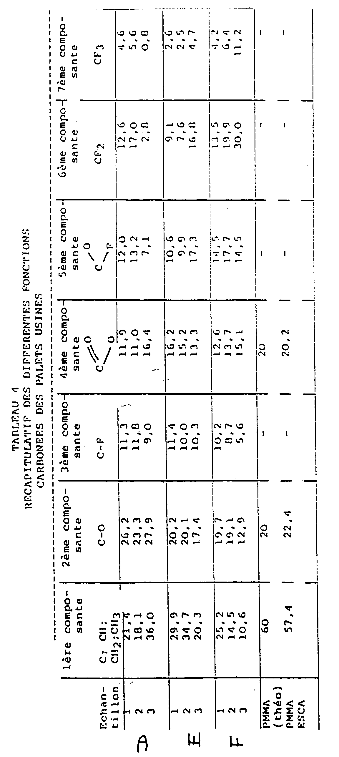
TABLEAU 5 Tableau des valeurs individuelles des pentes maximales (mDO/mn) des cinétiques de chaque réaction lors de l'évaluation des produits à étudier avec les leucocytes du sujet n°1 ESSAI PRODUITS ETUDIES A B C D E F G T n°1 0,391 0,491 0,565 0,544 0,457 0,757 2,039 0,922 n°2 0,485 0,494 0,500 0,764 0,562 0,454 2,190 0,919 n°3 0,475 0,465 0,570 0,675 0,409 0,585 1,788 0,825 Moyenne 0,450 0,483 0,545 0,661 0,476 0,599 2,010 0,889 TABLEAU 6 Tableau des valeurs individuelles des pentes maximales (mDO/mn) des cinétiques de chaque réaction lors de l'évaluation des produits à étudier avec les leucocytes du sujet n°2 ESSAI PRODUITS ETUDIES A B C D E F G T n°1 0,818 1,541 0,734 1,117 0,994 0,912 1,555 0,722 n°2 1,221 1,062 0,682 1,159 1,014 0,755 1,802 0,892 n°3 0,809 1,278 0,739 1,046 0,780 0,853 1,678 1,142 Moyenne 0,949 1,249 0,718 1,107 0,929 0,840 1,678 0,919 TABLEAU 7 Tableau des valeurs individuelles des pentes maximales (mDO/mn) des cinétiques de chaque réaction lors de l'évaluation des produits à étudier avec les leucocytes du sujet n°3 ESSAI PRODUITS ETUDIES A B C D E F G T n°1 - 0,584 0,981 0,823 1,172 1,138 1,946 1,278 n°2 0,791 1,251 1,484 0,686 1,389 1,153 2,054 1,744 n°3 0,691 0,768 1,096 1,446 1,137 1,158 2,933 1,651 Moyenne 0,741 0,868 1,187 0,985 1,233 1,150 2,311 1,158 TABLEAU 8 ESSAI 1 POURCENTAGE DE RECOUVREMENT CELLULES DE LA LIGNEE INFLAMMATOIRE DEURIS CELLULAIRES FILM PROTEIQUE très faiblement adhérentes ayant adhéré ayant excrété A 30 90% + +++ ++ + 1H < 5% + + + 2H 8 à 10% + + + 4H < 5% + + + B 30 75% +++ ++ + + 1 H 35% + ++ ++ ++ 2 H < 5 % + + + 4 H 55% +++ + + C 30 20% +++ + + + + 1 H 30% ++ ++ ++ + + 2 H < 5% + + + + 4 H < 5% + + + D 30 98% + +++ +++ ++ 1H 100% + +++ +++ ++ 2H 95% +++ +++ +++ 4H 0 TABLEAU 9 ESSAI 2 POURCENTAGE DE RECOUVREMENT CELLULES DE LA LIGNEE INFLAMMATOIRE DEBRIS CELLULAIRES FILM PROTEIQUE très faiblement adhérentes ayant adhéré ayant excrété A 30 90% + +++ + + ++ 1H 80% + +++ + + ++ 2H < 5% + + + + 4H < 5% + + + B 30 < 5 % + + + + 1 H < 5% + + + 2 H < 5 % + + + + 4H < 5% + + + + C 30 15 % ++ + + + 1H < 5% + + + 2H < 5% + + + + 4H 10% ++ + + + ++ D 30 100% ++ +++ +++ +++ + 1H 100% ++ +++ +++ +++ + 2H -- -- -- -- -- 4H 100% ++ +++ ++ ++ ++ TABLEAU 10 ESSAI 3 POURCENTAGE DE RECOUVREMENT CELLULES DE LA LIGNEE INFLAMMATOIRE DEBRIS CELLULAIRES FILM PROTEIQUE TRES FAIBLEMENT ADHERENTES ayant adhéré ayant excrété A 30 100% + +++ ++ ++ ++ 1H 40% ++ ++ ++ ++ 2H 98% + +++ ++ ++ 4H 40% ++ +++ ++ B 30 < 5 % + + + + 1 H 10% + + ++ 2 H 100 % ++ +++ + + 4 H 20% + ++ ++ ++ + C 30 < 5 % + + + + 1H 60% ++ +++ ++ ++ ++ 2H 10% + ++ ++ ++ ++ 4H 10% + ++ + + D 30 95 % + +++ ++ + 1H 100% + +++ ++ + 2H 100% + +++ +++ ++ 4H 100% ++ ++ ++ ++ ++
Claims (9)
- Dispositif à usage ophtalmologique, notamment implant ou lentille intraoculaire, réalisé en un polymère contenant des atomes d'hydrogène et ayant les qualités optiques et mécaniques traditionnellement requises pour ce type de dispositif, caractérisé en ce que les atomes d'hydrogène présents à la surface dudit polymère ont été remplacés par des atomes de fluor ou des groupements CF, CF2 ou CF3, le fluor total représentant au moins 15 % des atomes présents sur cette surface.
- Dispositif selon la revendication 1, caractérisé en ce que le polymère est du polyméthacrylate de méthyle (PMMA), un polymère de 2-hydroxy-éthyl méthacrylate (HEMA), du silicone ou un polysulfonate.
- Dispositif selon la revendication 1 ou la revendication 2, caractérisé en ce que le fluor total représente de 30 à 50 % des atomes présents à sa surface.
- Procédé d'obtention du dispositif selon l'une des revendications 1 à 3 comprenant le traitement de surface dudit dispositif se trouvant dans la forme appropriée pour son utilisation, par un plasma de molécules fluorées non polymérisables, le traitement étant réalisé à une puissance d'émission du réacteur comprise entre environ 3 et environ 10 watts par litre de capacité de reacteur.
- Procédé selon la revendication 4, caractérisé en ce que les molécules fluorées sont des gaz de tétrafluorure de carbone ou d'hexafluorure de soufre.
- Procédé selon la revendication 4, caractérisé en ce que le temps de traitement du dispositif est compris entre 1 et 20 minutes.
- Procédé selon la revendication 4, caractérisé en ce que la température de traitement est comprise entre 20 et 80 °C.
- Procédé selon la revendication 4, caractérisé en ce que le traitement est effectué sous vide à une pression comprise entre 0,1 et 1 Torr.
- Procédé selon l'une des revendications 4 à 8, caractérisé en ce que le dispositif est stérilisé après le traitement.
Applications Claiming Priority (2)
| Application Number | Priority Date | Filing Date | Title |
|---|---|---|---|
| FR9014536 | 1990-11-21 | ||
| FR9014536A FR2669226B1 (fr) | 1990-11-21 | 1990-11-21 | Dispositif a usage ophtalmologique forme d'un substrat polymerique comportant des groupements fluores en surface, et procede d'obtention. |
Publications (2)
| Publication Number | Publication Date |
|---|---|
| EP0487418A1 EP0487418A1 (fr) | 1992-05-27 |
| EP0487418B1 true EP0487418B1 (fr) | 1997-01-22 |
Family
ID=9402442
Family Applications (1)
| Application Number | Title | Priority Date | Filing Date |
|---|---|---|---|
| EP91403133A Expired - Lifetime EP0487418B1 (fr) | 1990-11-21 | 1991-11-20 | Dispositif à usage ophtalmologique formé d'un substrat polymérique comportant des groupements fluorés en surface, et procédé d'obtention |
Country Status (15)
| Country | Link |
|---|---|
| EP (1) | EP0487418B1 (fr) |
| JP (1) | JP3169659B2 (fr) |
| KR (1) | KR920009420A (fr) |
| AT (1) | ATE147996T1 (fr) |
| AU (1) | AU8801291A (fr) |
| BR (1) | BR9105058A (fr) |
| CA (1) | CA2055918C (fr) |
| DE (2) | DE69124343T2 (fr) |
| ES (1) | ES2032727T1 (fr) |
| FI (1) | FI915452L (fr) |
| FR (1) | FR2669226B1 (fr) |
| GR (1) | GR920300102T1 (fr) |
| MX (1) | MX9102165A (fr) |
| TW (1) | TW198099B (fr) |
| ZA (1) | ZA919128B (fr) |
Cited By (2)
| Publication number | Priority date | Publication date | Assignee | Title |
|---|---|---|---|---|
| US7232587B2 (en) | 2001-05-11 | 2007-06-19 | Bausch & Lomb Incorporated | Method for the passivation of an intraocular lens |
| EP2246386A1 (fr) | 2009-04-27 | 2010-11-03 | Université de Liège | Matériau fluoré |
Families Citing this family (6)
| Publication number | Priority date | Publication date | Assignee | Title |
|---|---|---|---|---|
| US5310571A (en) * | 1992-09-01 | 1994-05-10 | Allergan, Inc. | Chemical treatment to improve oxygen permeability through and protein deposition on hydrophilic (soft) and rigid gas permeable (RGP) contact lenses |
| ATE190336T1 (de) * | 1993-04-30 | 2000-03-15 | Alcon Lab Inc | Verfahren zur verminderung der klebrigkeit weicher acrylpolymere |
| US5603774A (en) * | 1993-09-27 | 1997-02-18 | Alcon Laboratories, Inc. | Method for reducing tackiness of soft acrylic polymers |
| DE19508772C2 (de) | 1995-03-01 | 1998-01-29 | Schering Ag | Verfahren und Verbindungen zur Detektion von Analyten mittels Remanenzmessung und deren Verwendung |
| FR2749170B1 (fr) * | 1996-05-30 | 1998-08-21 | Bellesort Stephane | Protheses dentaires modifiees en surface et procede d'obtention |
| WO2013011511A1 (fr) | 2011-07-18 | 2013-01-24 | Mor Research Applications Ltd. | Dispositif pour l'ajustement de la tension intraoculaire |
Family Cites Families (8)
| Publication number | Priority date | Publication date | Assignee | Title |
|---|---|---|---|---|
| US3674667A (en) * | 1969-07-23 | 1972-07-04 | Allis Chalmers Mfg Co | Process for increasing water repellency of cotton cloth |
| JPS5598232A (en) * | 1979-01-22 | 1980-07-26 | Agency Of Ind Science & Technol | Internal treatment of plastic tube member |
| US4508781A (en) * | 1982-06-07 | 1985-04-02 | The United States Of America As Represented By The Secretary Of Agriculture | Fluorination by inorganic fluorides in glow discharge |
| US4656083A (en) * | 1983-08-01 | 1987-04-07 | Washington Research Foundation | Plasma gas discharge treatment for improving the biocompatibility of biomaterials |
| US4655770A (en) * | 1985-06-06 | 1987-04-07 | Ioptex, Inc. | Surface passivated intraocular lens |
| JPH0796023B2 (ja) * | 1985-08-23 | 1995-10-18 | ワシントン リサ−チ フアンデイシヨン | 表面の性質が改良された高分子眼内レンズ |
| AU606068B2 (en) * | 1987-04-24 | 1991-01-31 | Washington Research Foundation | Intraocular lens with hydrophobic coating |
| FR2620624B1 (fr) * | 1987-09-23 | 1993-08-20 | Irap | Article en materiau polymere, modifie en surface pour presenter une hemocompatibilite amelioree et une thrombogeneicite diminuee, et son procede d'obtention |
-
1990
- 1990-11-21 FR FR9014536A patent/FR2669226B1/fr not_active Expired - Fee Related
-
1991
- 1991-11-18 ZA ZA919128A patent/ZA919128B/xx unknown
- 1991-11-19 FI FI915452A patent/FI915452L/fi not_active Application Discontinuation
- 1991-11-20 BR BR919105058A patent/BR9105058A/pt unknown
- 1991-11-20 EP EP91403133A patent/EP0487418B1/fr not_active Expired - Lifetime
- 1991-11-20 AU AU88012/91A patent/AU8801291A/en not_active Abandoned
- 1991-11-20 ES ES199191403133T patent/ES2032727T1/es active Pending
- 1991-11-20 AT AT91403133T patent/ATE147996T1/de not_active IP Right Cessation
- 1991-11-20 CA CA002055918A patent/CA2055918C/fr not_active Expired - Fee Related
- 1991-11-20 DE DE69124343T patent/DE69124343T2/de not_active Expired - Fee Related
- 1991-11-20 DE DE199191403133T patent/DE487418T1/de active Pending
- 1991-11-21 JP JP33263191A patent/JP3169659B2/ja not_active Expired - Fee Related
- 1991-11-21 MX MX9102165A patent/MX9102165A/es not_active IP Right Cessation
- 1991-11-21 KR KR1019910020778A patent/KR920009420A/ko not_active Ceased
-
1992
- 1992-04-02 TW TW081102585A patent/TW198099B/zh active
-
1993
- 1993-02-17 GR GR920300102T patent/GR920300102T1/el unknown
Cited By (3)
| Publication number | Priority date | Publication date | Assignee | Title |
|---|---|---|---|---|
| US7232587B2 (en) | 2001-05-11 | 2007-06-19 | Bausch & Lomb Incorporated | Method for the passivation of an intraocular lens |
| CN100406914C (zh) * | 2001-05-11 | 2008-07-30 | 鲍希和隆布公司 | 人工晶状体的钝化方法 |
| EP2246386A1 (fr) | 2009-04-27 | 2010-11-03 | Université de Liège | Matériau fluoré |
Also Published As
| Publication number | Publication date |
|---|---|
| KR920009420A (ko) | 1992-06-25 |
| CA2055918C (fr) | 2002-12-10 |
| FI915452A7 (fi) | 1992-05-22 |
| AU8801291A (en) | 1992-05-28 |
| DE69124343T2 (de) | 1997-05-15 |
| DE69124343D1 (de) | 1997-03-06 |
| GR920300102T1 (en) | 1993-02-17 |
| MX9102165A (es) | 1992-07-08 |
| FR2669226B1 (fr) | 1997-01-03 |
| TW198099B (fr) | 1993-01-11 |
| FR2669226A1 (fr) | 1992-05-22 |
| FI915452L (fi) | 1992-05-22 |
| ATE147996T1 (de) | 1997-02-15 |
| CA2055918A1 (fr) | 1992-05-22 |
| ES2032727T1 (es) | 1993-03-01 |
| JP3169659B2 (ja) | 2001-05-28 |
| FI915452A0 (fi) | 1991-11-19 |
| ZA919128B (en) | 1992-08-26 |
| JPH04314021A (ja) | 1992-11-05 |
| EP0487418A1 (fr) | 1992-05-27 |
| BR9105058A (pt) | 1992-06-23 |
| DE487418T1 (de) | 1992-10-15 |
Similar Documents
| Publication | Publication Date | Title |
|---|---|---|
| TWI768095B (zh) | 醫療器材及其製造方法 | |
| JP2783461B2 (ja) | ポリマー表面のフッ素化 | |
| EP3598212B1 (fr) | Lentille de contact et procédé de fabrication d'une telle lentille | |
| JP2566548B2 (ja) | 表面改質した外科用機器、器具、インプラント、コンタクトレンズおよびその類似物 | |
| US6582754B1 (en) | Coating process | |
| US5080924A (en) | Method of making biocompatible, surface modified materials | |
| Sheu et al. | A glow discharge treatment to immobilize poly (ethylene oxide)/poly (propylene oxide) surfactants for wettable and non-fouling biomaterials | |
| JP6954490B1 (ja) | 医療デバイスの製造方法 | |
| DE3875364T2 (de) | Okulares implantat und verfahren zu seiner herstellung. | |
| JP2000503044A (ja) | プラズマに誘導されたポリマー被覆 | |
| EP1913427B1 (fr) | Procede de fabrication de verres de contact | |
| EP0487418A1 (fr) | Dispositif à usage ophtalmologique formé d'un substrat polymérique comportant des groupements fluorés en surface, et procédé d'obtention | |
| US6143027A (en) | Polymeric intraocular lens material having improved surface properties and intraocular lens construction | |
| JPH012644A (ja) | 眼科用インプラントとその製造方法 | |
| Mateo et al. | Relating the surface properties of intraocular lens materials to endothelial cell adhesion damage. | |
| JP2002513948A (ja) | シリコーンヒドロゲルコンタクトレンズのプラズマ表面処理 | |
| EP0347267A1 (fr) | Hydrogel non réticulé, son procédé de préparation, son application en médecine ou en chirurgie, notamment comme implant oculaire | |
| EP0906075B1 (fr) | Protheses dentaires modifiees en surface et procede d'obtention | |
| AU606068B2 (en) | Intraocular lens with hydrophobic coating | |
| WO1996038503A1 (fr) | Composition souple a base de polymere de chlorure de vinyle, utilisation de cette composition pour la fabrication d'un article et article comprenant cette composition | |
| Nitschke et al. | Detachment of human endothelial cell sheets from thermo-responsive poly (NiPAAm-co-DEGMA) carriers | |
| McBriar | The plasma treatment of contact lenses | |
| TATEISHI et al. | Antithrombogenicity of poly (ether ether ketone) surface prepared by self-initiated surface graft polymerization of 2-methacryloyloxyethyl phosphorylcholine | |
| CN116370723A (zh) | 一种在金属密网支架表面构建磷酰胆碱涂层的方法 | |
| Mitzadeh et al. | Laser surface modification of polymers a novel technique for the preparation of blood compatible materials (II): in vitro assay |
Legal Events
| Date | Code | Title | Description |
|---|---|---|---|
| PUAI | Public reference made under article 153(3) epc to a published international application that has entered the european phase |
Free format text: ORIGINAL CODE: 0009012 |
|
| AK | Designated contracting states |
Kind code of ref document: A1 Designated state(s): AT BE CH DE DK ES FR GB GR IT LI LU NL SE |
|
| ITCL | It: translation for ep claims filed |
Representative=s name: STUDIO TORTA SOCIETA' SEMPLICE |
|
| 17P | Request for examination filed |
Effective date: 19920613 |
|
| TCNL | Nl: translation of patent claims filed | ||
| DET | De: translation of patent claims | ||
| TCAT | At: translation of patent claims filed | ||
| GBC | Gb: translation of claims filed (gb section 78(7)/1977) | ||
| REG | Reference to a national code |
Ref country code: ES Ref legal event code: BA2A Ref document number: 2032727 Country of ref document: ES Kind code of ref document: T1 |
|
| 17Q | First examination report despatched |
Effective date: 19940627 |
|
| RAP1 | Party data changed (applicant data changed or rights of an application transferred) |
Owner name: INSTITUT DE RECHERCHE APPLIQUEE SUR LES POLYMERES( Owner name: LABORATOIRES DOMILENS SOCIETE ANONYME |
|
| RAP1 | Party data changed (applicant data changed or rights of an application transferred) |
Owner name: ASSOCIATION POUR LES TRANSFERTS DE TECHNOLOGIE DU Owner name: LABORATOIRES DOMILENS SOCIETE ANONYME |
|
| GRAH | Despatch of communication of intention to grant a patent |
Free format text: ORIGINAL CODE: EPIDOS IGRA |
|
| GRAH | Despatch of communication of intention to grant a patent |
Free format text: ORIGINAL CODE: EPIDOS IGRA |
|
| GRAA | (expected) grant |
Free format text: ORIGINAL CODE: 0009210 |
|
| AK | Designated contracting states |
Kind code of ref document: B1 Designated state(s): AT BE CH DE DK ES FR GB GR IT LI LU NL SE |
|
| PG25 | Lapsed in a contracting state [announced via postgrant information from national office to epo] |
Ref country code: AT Effective date: 19970122 Ref country code: GR Free format text: LAPSE BECAUSE OF FAILURE TO SUBMIT A TRANSLATION OF THE DESCRIPTION OR TO PAY THE FEE WITHIN THE PRESCRIBED TIME-LIMIT Effective date: 19970122 Ref country code: DK Effective date: 19970122 |
|
| REF | Corresponds to: |
Ref document number: 147996 Country of ref document: AT Date of ref document: 19970215 Kind code of ref document: T |
|
| REG | Reference to a national code |
Ref country code: CH Ref legal event code: EP |
|
| REG | Reference to a national code |
Ref country code: CH Ref legal event code: NV Representative=s name: KIRKER & CIE SA |
|
| REF | Corresponds to: |
Ref document number: 69124343 Country of ref document: DE Date of ref document: 19970306 |
|
| GBT | Gb: translation of ep patent filed (gb section 77(6)(a)/1977) |
Effective date: 19970219 |
|
| ITF | It: translation for a ep patent filed | ||
| PG25 | Lapsed in a contracting state [announced via postgrant information from national office to epo] |
Ref country code: ES Free format text: LAPSE BECAUSE OF FAILURE TO SUBMIT A TRANSLATION OF THE DESCRIPTION OR TO PAY THE FEE WITHIN THE PRESCRIBED TIME-LIMIT Effective date: 19970723 |
|
| PG25 | Lapsed in a contracting state [announced via postgrant information from national office to epo] |
Ref country code: LU Free format text: LAPSE BECAUSE OF NON-PAYMENT OF DUE FEES Effective date: 19971120 |
|
| PLBE | No opposition filed within time limit |
Free format text: ORIGINAL CODE: 0009261 |
|
| STAA | Information on the status of an ep patent application or granted ep patent |
Free format text: STATUS: NO OPPOSITION FILED WITHIN TIME LIMIT |
|
| PG25 | Lapsed in a contracting state [announced via postgrant information from national office to epo] |
Ref country code: BE Free format text: LAPSE BECAUSE OF NON-PAYMENT OF DUE FEES Effective date: 19971130 |
|
| 26N | No opposition filed | ||
| BERE | Be: lapsed |
Owner name: ASSOCIATION POUR LES TRANSFERTS DE TECHNOLOGIE DU Effective date: 19971130 Owner name: S.A. LABORATOIRES DOMILENS Effective date: 19971130 |
|
| REG | Reference to a national code |
Ref country code: GB Ref legal event code: IF02 |
|
| PGFP | Annual fee paid to national office [announced via postgrant information from national office to epo] |
Ref country code: NL Payment date: 20071010 Year of fee payment: 17 Ref country code: DE Payment date: 20071130 Year of fee payment: 17 |
|
| PGFP | Annual fee paid to national office [announced via postgrant information from national office to epo] |
Ref country code: IT Payment date: 20071116 Year of fee payment: 17 |
|
| PGFP | Annual fee paid to national office [announced via postgrant information from national office to epo] |
Ref country code: SE Payment date: 20071105 Year of fee payment: 17 |
|
| PGFP | Annual fee paid to national office [announced via postgrant information from national office to epo] |
Ref country code: GB Payment date: 20071005 Year of fee payment: 17 Ref country code: FR Payment date: 20071105 Year of fee payment: 17 |
|
| PGFP | Annual fee paid to national office [announced via postgrant information from national office to epo] |
Ref country code: CH Payment date: 20081028 Year of fee payment: 18 |
|
| EUG | Se: european patent has lapsed | ||
| GBPC | Gb: european patent ceased through non-payment of renewal fee |
Effective date: 20081120 |
|
| PG25 | Lapsed in a contracting state [announced via postgrant information from national office to epo] |
Ref country code: NL Free format text: LAPSE BECAUSE OF NON-PAYMENT OF DUE FEES Effective date: 20090601 |
|
| NLV4 | Nl: lapsed or anulled due to non-payment of the annual fee |
Effective date: 20090601 |
|
| PG25 | Lapsed in a contracting state [announced via postgrant information from national office to epo] |
Ref country code: IT Free format text: LAPSE BECAUSE OF NON-PAYMENT OF DUE FEES Effective date: 20081120 |
|
| REG | Reference to a national code |
Ref country code: FR Ref legal event code: ST Effective date: 20090731 |
|
| PG25 | Lapsed in a contracting state [announced via postgrant information from national office to epo] |
Ref country code: DE Free format text: LAPSE BECAUSE OF NON-PAYMENT OF DUE FEES Effective date: 20090603 |
|
| PG25 | Lapsed in a contracting state [announced via postgrant information from national office to epo] |
Ref country code: GB Free format text: LAPSE BECAUSE OF NON-PAYMENT OF DUE FEES Effective date: 20081120 |
|
| REG | Reference to a national code |
Ref country code: CH Ref legal event code: PL |
|
| PG25 | Lapsed in a contracting state [announced via postgrant information from national office to epo] |
Ref country code: SE Free format text: LAPSE BECAUSE OF NON-PAYMENT OF DUE FEES Effective date: 20081121 |
|
| PG25 | Lapsed in a contracting state [announced via postgrant information from national office to epo] |
Ref country code: CH Free format text: LAPSE BECAUSE OF NON-PAYMENT OF DUE FEES Effective date: 20091130 Ref country code: LI Free format text: LAPSE BECAUSE OF NON-PAYMENT OF DUE FEES Effective date: 20091130 |
|
| PG25 | Lapsed in a contracting state [announced via postgrant information from national office to epo] |
Ref country code: FR Free format text: LAPSE BECAUSE OF NON-PAYMENT OF DUE FEES Effective date: 20081130 |