EP0455735B1 - Avidin-biotin assistiertes immunassayverfahren - Google Patents
Avidin-biotin assistiertes immunassayverfahren Download PDFInfo
- Publication number
- EP0455735B1 EP0455735B1 EP90903466A EP90903466A EP0455735B1 EP 0455735 B1 EP0455735 B1 EP 0455735B1 EP 90903466 A EP90903466 A EP 90903466A EP 90903466 A EP90903466 A EP 90903466A EP 0455735 B1 EP0455735 B1 EP 0455735B1
- Authority
- EP
- European Patent Office
- Prior art keywords
- antibody
- antigen
- immobilized
- avidin
- support
- Prior art date
- Legal status (The legal status is an assumption and is not a legal conclusion. Google has not performed a legal analysis and makes no representation as to the accuracy of the status listed.)
- Expired - Lifetime
Links
- 229960002685 biotin Drugs 0.000 title description 13
- 239000011616 biotin Substances 0.000 title description 13
- 238000003018 immunoassay Methods 0.000 title description 7
- 239000000427 antigen Substances 0.000 claims abstract description 82
- 102000036639 antigens Human genes 0.000 claims abstract description 79
- 108091007433 antigens Proteins 0.000 claims abstract description 79
- 108090000623 proteins and genes Proteins 0.000 claims abstract description 54
- 238000000034 method Methods 0.000 claims abstract description 53
- 102000004169 proteins and genes Human genes 0.000 claims abstract description 53
- 108090001008 Avidin Proteins 0.000 claims abstract description 42
- 102000004190 Enzymes Human genes 0.000 claims abstract description 38
- 108090000790 Enzymes Proteins 0.000 claims abstract description 38
- 210000001124 body fluid Anatomy 0.000 claims abstract description 31
- 239000010839 body fluid Substances 0.000 claims abstract description 31
- 239000000758 substrate Substances 0.000 claims abstract description 23
- 238000001514 detection method Methods 0.000 claims abstract description 21
- 238000012360 testing method Methods 0.000 claims abstract description 20
- 238000006243 chemical reaction Methods 0.000 claims abstract description 17
- 239000012530 fluid Substances 0.000 claims abstract description 15
- 239000007795 chemical reaction product Substances 0.000 claims abstract description 13
- 108091008324 binding proteins Proteins 0.000 claims abstract description 10
- 102000014914 Carrier Proteins Human genes 0.000 claims abstract 6
- 229920000936 Agarose Polymers 0.000 claims description 20
- 210000003296 saliva Anatomy 0.000 claims description 10
- 102000009109 Fc receptors Human genes 0.000 claims description 9
- 108010087819 Fc receptors Proteins 0.000 claims description 9
- 102000001706 Immunoglobulin Fab Fragments Human genes 0.000 claims description 8
- 108010054477 Immunoglobulin Fab Fragments Proteins 0.000 claims description 8
- 230000027455 binding Effects 0.000 claims description 8
- 238000003556 assay Methods 0.000 claims description 7
- 239000013060 biological fluid Substances 0.000 claims description 7
- 241000894006 Bacteria Species 0.000 claims description 5
- 239000012062 aqueous buffer Substances 0.000 claims description 5
- -1 polyethylene Polymers 0.000 claims description 5
- 241001430228 Clavibacter sepedonicus Species 0.000 claims description 4
- 239000004698 Polyethylene Substances 0.000 claims description 4
- 229920000573 polyethylene Polymers 0.000 claims description 4
- 230000009871 nonspecific binding Effects 0.000 claims description 3
- 239000004793 Polystyrene Substances 0.000 claims description 2
- 125000000524 functional group Chemical group 0.000 claims description 2
- 244000000003 plant pathogen Species 0.000 claims description 2
- 229920002223 polystyrene Polymers 0.000 claims description 2
- 239000011148 porous material Substances 0.000 claims description 2
- 241000713772 Human immunodeficiency virus 1 Species 0.000 claims 1
- 239000000419 plant extract Substances 0.000 claims 1
- 239000011159 matrix material Substances 0.000 abstract description 14
- 238000012216 screening Methods 0.000 abstract description 2
- 229940088598 enzyme Drugs 0.000 description 26
- 239000000243 solution Substances 0.000 description 20
- YBJHBAHKTGYVGT-ZKWXMUAHSA-N (+)-Biotin Chemical compound N1C(=O)N[C@@H]2[C@H](CCCCC(=O)O)SC[C@@H]21 YBJHBAHKTGYVGT-ZKWXMUAHSA-N 0.000 description 15
- 239000011324 bead Substances 0.000 description 15
- 239000000872 buffer Substances 0.000 description 13
- 239000003085 diluting agent Substances 0.000 description 10
- 210000002966 serum Anatomy 0.000 description 8
- 235000020958 biotin Nutrition 0.000 description 7
- 239000003153 chemical reaction reagent Substances 0.000 description 6
- LOKCTEFSRHRXRJ-UHFFFAOYSA-I dipotassium trisodium dihydrogen phosphate hydrogen phosphate dichloride Chemical compound P(=O)(O)(O)[O-].[K+].P(=O)(O)([O-])[O-].[Na+].[Na+].[Cl-].[K+].[Cl-].[Na+] LOKCTEFSRHRXRJ-UHFFFAOYSA-I 0.000 description 6
- 239000002953 phosphate buffered saline Substances 0.000 description 6
- 125000003277 amino group Chemical group 0.000 description 5
- 230000001580 bacterial effect Effects 0.000 description 5
- 239000000284 extract Substances 0.000 description 5
- 239000000203 mixture Substances 0.000 description 5
- 102000002260 Alkaline Phosphatase Human genes 0.000 description 4
- 108020004774 Alkaline Phosphatase Proteins 0.000 description 4
- 108010001336 Horseradish Peroxidase Proteins 0.000 description 4
- 230000000890 antigenic effect Effects 0.000 description 4
- 102000023732 binding proteins Human genes 0.000 description 4
- 238000011161 development Methods 0.000 description 4
- 201000010099 disease Diseases 0.000 description 4
- 208000037265 diseases, disorders, signs and symptoms Diseases 0.000 description 4
- 239000000499 gel Substances 0.000 description 4
- 238000011534 incubation Methods 0.000 description 4
- 239000000047 product Substances 0.000 description 4
- 230000035945 sensitivity Effects 0.000 description 4
- 239000000126 substance Substances 0.000 description 4
- 238000005406 washing Methods 0.000 description 4
- 241000196324 Embryophyta Species 0.000 description 3
- MHAJPDPJQMAIIY-UHFFFAOYSA-N Hydrogen peroxide Chemical compound OO MHAJPDPJQMAIIY-UHFFFAOYSA-N 0.000 description 3
- 108060003951 Immunoglobulin Proteins 0.000 description 3
- 241000194017 Streptococcus Species 0.000 description 3
- 239000011543 agarose gel Substances 0.000 description 3
- 125000003178 carboxy group Chemical group [H]OC(*)=O 0.000 description 3
- 238000007398 colorimetric assay Methods 0.000 description 3
- 102000018358 immunoglobulin Human genes 0.000 description 3
- 208000015181 infectious disease Diseases 0.000 description 3
- 239000000463 material Substances 0.000 description 3
- 210000002381 plasma Anatomy 0.000 description 3
- 210000001519 tissue Anatomy 0.000 description 3
- CXNVOWPRHWWCQR-UHFFFAOYSA-N 4-Chloro-ortho-toluidine Chemical compound CC1=CC(Cl)=CC=C1N CXNVOWPRHWWCQR-UHFFFAOYSA-N 0.000 description 2
- 208000030507 AIDS Diseases 0.000 description 2
- 108010010803 Gelatin Proteins 0.000 description 2
- 208000009889 Herpes Simplex Diseases 0.000 description 2
- FAPWRFPIFSIZLT-UHFFFAOYSA-M Sodium chloride Chemical compound [Na+].[Cl-] FAPWRFPIFSIZLT-UHFFFAOYSA-M 0.000 description 2
- 244000061456 Solanum tuberosum Species 0.000 description 2
- 235000002595 Solanum tuberosum Nutrition 0.000 description 2
- 241000191967 Staphylococcus aureus Species 0.000 description 2
- SXEHKFHPFVVDIR-UHFFFAOYSA-N [4-(4-hydrazinylphenyl)phenyl]hydrazine Chemical compound C1=CC(NN)=CC=C1C1=CC=C(NN)C=C1 SXEHKFHPFVVDIR-UHFFFAOYSA-N 0.000 description 2
- 150000001335 aliphatic alkanes Chemical class 0.000 description 2
- 239000012491 analyte Substances 0.000 description 2
- 230000008901 benefit Effects 0.000 description 2
- 238000012742 biochemical analysis Methods 0.000 description 2
- 230000000903 blocking effect Effects 0.000 description 2
- 239000003795 chemical substances by application Substances 0.000 description 2
- 239000003599 detergent Substances 0.000 description 2
- 239000008273 gelatin Substances 0.000 description 2
- 229920000159 gelatin Polymers 0.000 description 2
- 235000019322 gelatine Nutrition 0.000 description 2
- 235000011852 gelatine desserts Nutrition 0.000 description 2
- 150000004676 glycans Chemical class 0.000 description 2
- 229940072221 immunoglobulins Drugs 0.000 description 2
- NOESYZHRGYRDHS-UHFFFAOYSA-N insulin Chemical compound N1C(=O)C(NC(=O)C(CCC(N)=O)NC(=O)C(CCC(O)=O)NC(=O)C(C(C)C)NC(=O)C(NC(=O)CN)C(C)CC)CSSCC(C(NC(CO)C(=O)NC(CC(C)C)C(=O)NC(CC=2C=CC(O)=CC=2)C(=O)NC(CCC(N)=O)C(=O)NC(CC(C)C)C(=O)NC(CCC(O)=O)C(=O)NC(CC(N)=O)C(=O)NC(CC=2C=CC(O)=CC=2)C(=O)NC(CSSCC(NC(=O)C(C(C)C)NC(=O)C(CC(C)C)NC(=O)C(CC=2C=CC(O)=CC=2)NC(=O)C(CC(C)C)NC(=O)C(C)NC(=O)C(CCC(O)=O)NC(=O)C(C(C)C)NC(=O)C(CC(C)C)NC(=O)C(CC=2NC=NC=2)NC(=O)C(CO)NC(=O)CNC2=O)C(=O)NCC(=O)NC(CCC(O)=O)C(=O)NC(CCCNC(N)=N)C(=O)NCC(=O)NC(CC=3C=CC=CC=3)C(=O)NC(CC=3C=CC=CC=3)C(=O)NC(CC=3C=CC(O)=CC=3)C(=O)NC(C(C)O)C(=O)N3C(CCC3)C(=O)NC(CCCCN)C(=O)NC(C)C(O)=O)C(=O)NC(CC(N)=O)C(O)=O)=O)NC(=O)C(C(C)CC)NC(=O)C(CO)NC(=O)C(C(C)O)NC(=O)C1CSSCC2NC(=O)C(CC(C)C)NC(=O)C(NC(=O)C(CCC(N)=O)NC(=O)C(CC(N)=O)NC(=O)C(NC(=O)C(N)CC=1C=CC=CC=1)C(C)C)CC1=CN=CN1 NOESYZHRGYRDHS-UHFFFAOYSA-N 0.000 description 2
- 239000007788 liquid Substances 0.000 description 2
- 239000006166 lysate Substances 0.000 description 2
- 235000012015 potatoes Nutrition 0.000 description 2
- 239000002244 precipitate Substances 0.000 description 2
- 125000002924 primary amino group Chemical group [H]N([H])* 0.000 description 2
- 230000009257 reactivity Effects 0.000 description 2
- 239000007787 solid Substances 0.000 description 2
- 210000001138 tear Anatomy 0.000 description 2
- 210000002700 urine Anatomy 0.000 description 2
- 230000003612 virological effect Effects 0.000 description 2
- QRXMUCSWCMTJGU-UHFFFAOYSA-L (5-bromo-4-chloro-1h-indol-3-yl) phosphate Chemical compound C1=C(Br)C(Cl)=C2C(OP([O-])(=O)[O-])=CNC2=C1 QRXMUCSWCMTJGU-UHFFFAOYSA-L 0.000 description 1
- UJPKMTDFFUTLGM-UHFFFAOYSA-N 1-aminoethanol Chemical compound CC(N)O UJPKMTDFFUTLGM-UHFFFAOYSA-N 0.000 description 1
- HKAVADYDPYUPRD-UHFFFAOYSA-N 1h-pyrazine-2-thione Chemical compound SC1=CN=CC=N1 HKAVADYDPYUPRD-UHFFFAOYSA-N 0.000 description 1
- UAIUNKRWKOVEES-UHFFFAOYSA-N 3,3',5,5'-tetramethylbenzidine Chemical compound CC1=C(N)C(C)=CC(C=2C=C(C)C(N)=C(C)C=2)=C1 UAIUNKRWKOVEES-UHFFFAOYSA-N 0.000 description 1
- HSTOKWSFWGCZMH-UHFFFAOYSA-N 3,3'-diaminobenzidine Chemical compound C1=C(N)C(N)=CC=C1C1=CC=C(N)C(N)=C1 HSTOKWSFWGCZMH-UHFFFAOYSA-N 0.000 description 1
- ALKYHXVLJMQRLQ-UHFFFAOYSA-N 3-Hydroxy-2-naphthoate Chemical compound C1=CC=C2C=C(O)C(C(=O)O)=CC2=C1 ALKYHXVLJMQRLQ-UHFFFAOYSA-N 0.000 description 1
- LQILVUYCDHSGEU-UHFFFAOYSA-N 4-[(2,5-dioxopyrrol-1-yl)methyl]cyclohexane-1-carboxylic acid Chemical compound C1CC(C(=O)O)CCC1CN1C(=O)C=CC1=O LQILVUYCDHSGEU-UHFFFAOYSA-N 0.000 description 1
- RJEUJGHYCLSXBW-UHFFFAOYSA-N 4-[[3-(2,5-dioxopyrrolidin-1-yl)-2,5-dioxopyrrol-1-yl]methyl]cyclohexane-1-carboxylic acid Chemical compound C1CC(C(=O)O)CCC1CN1C(=O)C(N2C(CCC2=O)=O)=CC1=O RJEUJGHYCLSXBW-UHFFFAOYSA-N 0.000 description 1
- LVSPDZAGCBEQAV-UHFFFAOYSA-N 4-chloronaphthalen-1-ol Chemical compound C1=CC=C2C(O)=CC=C(Cl)C2=C1 LVSPDZAGCBEQAV-UHFFFAOYSA-N 0.000 description 1
- 241000228245 Aspergillus niger Species 0.000 description 1
- 208000035143 Bacterial infection Diseases 0.000 description 1
- ZYRLGMWOJQVXAW-UHFFFAOYSA-N CC(C)(CC1N2)SCC1NC2=O Chemical compound CC(C)(CC1N2)SCC1NC2=O ZYRLGMWOJQVXAW-UHFFFAOYSA-N 0.000 description 1
- 241000192125 Firmicutes Species 0.000 description 1
- 108010015776 Glucose oxidase Proteins 0.000 description 1
- 239000004366 Glucose oxidase Substances 0.000 description 1
- SXRSQZLOMIGNAQ-UHFFFAOYSA-N Glutaraldehyde Chemical compound O=CCCCC=O SXRSQZLOMIGNAQ-UHFFFAOYSA-N 0.000 description 1
- 241000714260 Human T-lymphotropic virus 1 Species 0.000 description 1
- 206010061598 Immunodeficiency Diseases 0.000 description 1
- 208000029462 Immunodeficiency disease Diseases 0.000 description 1
- 102000004877 Insulin Human genes 0.000 description 1
- 108090001061 Insulin Proteins 0.000 description 1
- 241000124008 Mammalia Species 0.000 description 1
- 241000699670 Mus sp. Species 0.000 description 1
- NQTADLQHYWFPDB-UHFFFAOYSA-N N-Hydroxysuccinimide Chemical compound ON1C(=O)CCC1=O NQTADLQHYWFPDB-UHFFFAOYSA-N 0.000 description 1
- 241000283973 Oryctolagus cuniculus Species 0.000 description 1
- 229910019142 PO4 Inorganic materials 0.000 description 1
- 241001505901 Streptococcus sp. 'group A' Species 0.000 description 1
- 101710120037 Toxin CcdB Proteins 0.000 description 1
- 239000007983 Tris buffer Substances 0.000 description 1
- 208000036142 Viral infection Diseases 0.000 description 1
- 229930003756 Vitamin B7 Natural products 0.000 description 1
- 238000001042 affinity chromatography Methods 0.000 description 1
- 125000003172 aldehyde group Chemical group 0.000 description 1
- 150000003931 anilides Chemical class 0.000 description 1
- 210000000628 antibody-producing cell Anatomy 0.000 description 1
- 230000001363 autoimmune Effects 0.000 description 1
- 208000022362 bacterial infectious disease Diseases 0.000 description 1
- 239000011230 binding agent Substances 0.000 description 1
- 230000008275 binding mechanism Effects 0.000 description 1
- 239000012472 biological sample Substances 0.000 description 1
- 230000006287 biotinylation Effects 0.000 description 1
- 238000007413 biotinylation Methods 0.000 description 1
- 210000004369 blood Anatomy 0.000 description 1
- 239000008280 blood Substances 0.000 description 1
- 150000001718 carbodiimides Chemical class 0.000 description 1
- 150000001720 carbohydrates Chemical class 0.000 description 1
- 150000001732 carboxylic acid derivatives Chemical class 0.000 description 1
- 210000002421 cell wall Anatomy 0.000 description 1
- 238000005119 centrifugation Methods 0.000 description 1
- 239000003593 chromogenic compound Substances 0.000 description 1
- 238000004737 colorimetric analysis Methods 0.000 description 1
- 239000013065 commercial product Substances 0.000 description 1
- 238000012875 competitive assay Methods 0.000 description 1
- 238000002967 competitive immunoassay Methods 0.000 description 1
- 150000001875 compounds Chemical class 0.000 description 1
- 230000021615 conjugation Effects 0.000 description 1
- 230000008878 coupling Effects 0.000 description 1
- 238000010168 coupling process Methods 0.000 description 1
- 238000005859 coupling reaction Methods 0.000 description 1
- ATDGTVJJHBUTRL-UHFFFAOYSA-N cyanogen bromide Chemical compound BrC#N ATDGTVJJHBUTRL-UHFFFAOYSA-N 0.000 description 1
- 238000003745 diagnosis Methods 0.000 description 1
- 238000002405 diagnostic procedure Methods 0.000 description 1
- ZWIBGKZDAWNIFC-UHFFFAOYSA-N disuccinimidyl suberate Chemical compound O=C1CCC(=O)N1OC(=O)CCCCCCC(=O)ON1C(=O)CCC1=O ZWIBGKZDAWNIFC-UHFFFAOYSA-N 0.000 description 1
- 239000000706 filtrate Substances 0.000 description 1
- 238000001914 filtration Methods 0.000 description 1
- 229940116332 glucose oxidase Drugs 0.000 description 1
- 235000019420 glucose oxidase Nutrition 0.000 description 1
- VOZRXNHHFUQHIL-UHFFFAOYSA-N glycidyl methacrylate Chemical compound CC(=C)C(=O)OCC1CO1 VOZRXNHHFUQHIL-UHFFFAOYSA-N 0.000 description 1
- 208000002672 hepatitis B Diseases 0.000 description 1
- 125000002887 hydroxy group Chemical group [H]O* 0.000 description 1
- 230000007813 immunodeficiency Effects 0.000 description 1
- 239000002198 insoluble material Substances 0.000 description 1
- 229940125396 insulin Drugs 0.000 description 1
- 230000003993 interaction Effects 0.000 description 1
- 125000002346 iodo group Chemical group I* 0.000 description 1
- 125000005439 maleimidyl group Chemical group C1(C=CC(N1*)=O)=O 0.000 description 1
- 238000004519 manufacturing process Methods 0.000 description 1
- FQPSGWSUVKBHSU-UHFFFAOYSA-N methacrylamide Chemical compound CC(=C)C(N)=O FQPSGWSUVKBHSU-UHFFFAOYSA-N 0.000 description 1
- 125000002496 methyl group Chemical group [H]C([H])([H])* 0.000 description 1
- 210000004080 milk Anatomy 0.000 description 1
- 239000008267 milk Substances 0.000 description 1
- 235000013336 milk Nutrition 0.000 description 1
- 230000004048 modification Effects 0.000 description 1
- 238000012986 modification Methods 0.000 description 1
- 229940126619 mouse monoclonal antibody Drugs 0.000 description 1
- 239000013642 negative control Substances 0.000 description 1
- JPXMTWWFLBLUCD-UHFFFAOYSA-N nitro blue tetrazolium(2+) Chemical compound COC1=CC(C=2C=C(OC)C(=CC=2)[N+]=2N(N=C(N=2)C=2C=CC=CC=2)C=2C=CC(=CC=2)[N+]([O-])=O)=CC=C1[N+]1=NC(C=2C=CC=CC=2)=NN1C1=CC=C([N+]([O-])=O)C=C1 JPXMTWWFLBLUCD-UHFFFAOYSA-N 0.000 description 1
- 244000052769 pathogen Species 0.000 description 1
- 230000001717 pathogenic effect Effects 0.000 description 1
- 230000037361 pathway Effects 0.000 description 1
- KHIWWQKSHDUIBK-UHFFFAOYSA-N periodic acid Chemical compound OI(=O)(=O)=O KHIWWQKSHDUIBK-UHFFFAOYSA-N 0.000 description 1
- NMHMNPHRMNGLLB-UHFFFAOYSA-N phloretic acid Chemical compound OC(=O)CCC1=CC=C(O)C=C1 NMHMNPHRMNGLLB-UHFFFAOYSA-N 0.000 description 1
- NBIIXXVUZAFLBC-UHFFFAOYSA-K phosphate Chemical compound [O-]P([O-])([O-])=O NBIIXXVUZAFLBC-UHFFFAOYSA-K 0.000 description 1
- 239000010452 phosphate Substances 0.000 description 1
- 239000004033 plastic Substances 0.000 description 1
- 229920003023 plastic Polymers 0.000 description 1
- 229920000642 polymer Polymers 0.000 description 1
- 229920001282 polysaccharide Polymers 0.000 description 1
- 239000005017 polysaccharide Substances 0.000 description 1
- 150000004804 polysaccharides Polymers 0.000 description 1
- 239000013641 positive control Substances 0.000 description 1
- 230000001376 precipitating effect Effects 0.000 description 1
- 108090000765 processed proteins & peptides Proteins 0.000 description 1
- 238000012545 processing Methods 0.000 description 1
- 238000000746 purification Methods 0.000 description 1
- 102000005962 receptors Human genes 0.000 description 1
- 108020003175 receptors Proteins 0.000 description 1
- 210000000582 semen Anatomy 0.000 description 1
- 239000011780 sodium chloride Substances 0.000 description 1
- 229910000162 sodium phosphate Inorganic materials 0.000 description 1
- 239000001488 sodium phosphate Substances 0.000 description 1
- 241000894007 species Species 0.000 description 1
- 230000009870 specific binding Effects 0.000 description 1
- 238000010561 standard procedure Methods 0.000 description 1
- 239000000725 suspension Substances 0.000 description 1
- 210000001179 synovial fluid Anatomy 0.000 description 1
- 238000012546 transfer Methods 0.000 description 1
- 239000012780 transparent material Substances 0.000 description 1
- LENZDBCJOHFCAS-UHFFFAOYSA-N tris Chemical compound OCC(N)(CO)CO LENZDBCJOHFCAS-UHFFFAOYSA-N 0.000 description 1
- RYFMWSXOAZQYPI-UHFFFAOYSA-K trisodium phosphate Chemical compound [Na+].[Na+].[Na+].[O-]P([O-])([O-])=O RYFMWSXOAZQYPI-UHFFFAOYSA-K 0.000 description 1
- 230000009385 viral infection Effects 0.000 description 1
- 239000011735 vitamin B7 Substances 0.000 description 1
- 235000011912 vitamin B7 Nutrition 0.000 description 1
- XLYOFNOQVPJJNP-UHFFFAOYSA-N water Substances O XLYOFNOQVPJJNP-UHFFFAOYSA-N 0.000 description 1
Images

Classifications
-
- B—PERFORMING OPERATIONS; TRANSPORTING
- B82—NANOTECHNOLOGY
- B82Y—SPECIFIC USES OR APPLICATIONS OF NANOSTRUCTURES; MEASUREMENT OR ANALYSIS OF NANOSTRUCTURES; MANUFACTURE OR TREATMENT OF NANOSTRUCTURES
- B82Y5/00—Nanobiotechnology or nanomedicine, e.g. protein engineering or drug delivery
-
- A—HUMAN NECESSITIES
- A61—MEDICAL OR VETERINARY SCIENCE; HYGIENE
- A61K—PREPARATIONS FOR MEDICAL, DENTAL OR TOILETRY PURPOSES
- A61K47/00—Medicinal preparations characterised by the non-active ingredients used, e.g. carriers or inert additives; Targeting or modifying agents chemically bound to the active ingredient
- A61K47/50—Medicinal preparations characterised by the non-active ingredients used, e.g. carriers or inert additives; Targeting or modifying agents chemically bound to the active ingredient the non-active ingredient being chemically bound to the active ingredient, e.g. polymer-drug conjugates
- A61K47/51—Medicinal preparations characterised by the non-active ingredients used, e.g. carriers or inert additives; Targeting or modifying agents chemically bound to the active ingredient the non-active ingredient being chemically bound to the active ingredient, e.g. polymer-drug conjugates the non-active ingredient being a modifying agent
- A61K47/68—Medicinal preparations characterised by the non-active ingredients used, e.g. carriers or inert additives; Targeting or modifying agents chemically bound to the active ingredient the non-active ingredient being chemically bound to the active ingredient, e.g. polymer-drug conjugates the non-active ingredient being a modifying agent the modifying agent being an antibody, an immunoglobulin or a fragment thereof, e.g. an Fc-fragment
- A61K47/6891—Pre-targeting systems involving an antibody for targeting specific cells
- A61K47/6897—Pre-targeting systems with two or three steps using antibody conjugates; Ligand-antiligand therapies
- A61K47/6898—Pre-targeting systems with two or three steps using antibody conjugates; Ligand-antiligand therapies using avidin- or biotin-conjugated antibodies
-
- G—PHYSICS
- G01—MEASURING; TESTING
- G01N—INVESTIGATING OR ANALYSING MATERIALS BY DETERMINING THEIR CHEMICAL OR PHYSICAL PROPERTIES
- G01N33/00—Investigating or analysing materials by specific methods not covered by groups G01N1/00 - G01N31/00
- G01N33/48—Biological material, e.g. blood, urine; Haemocytometers
- G01N33/50—Chemical analysis of biological material, e.g. blood, urine; Testing involving biospecific ligand binding methods; Immunological testing
- G01N33/53—Immunoassay; Biospecific binding assay; Materials therefor
- G01N33/543—Immunoassay; Biospecific binding assay; Materials therefor with an insoluble carrier for immobilising immunochemicals
- G01N33/544—Immunoassay; Biospecific binding assay; Materials therefor with an insoluble carrier for immobilising immunochemicals the carrier being organic
- G01N33/548—Carbohydrates, e.g. dextran
-
- G—PHYSICS
- G01—MEASURING; TESTING
- G01N—INVESTIGATING OR ANALYSING MATERIALS BY DETERMINING THEIR CHEMICAL OR PHYSICAL PROPERTIES
- G01N33/00—Investigating or analysing materials by specific methods not covered by groups G01N1/00 - G01N31/00
- G01N33/48—Biological material, e.g. blood, urine; Haemocytometers
- G01N33/50—Chemical analysis of biological material, e.g. blood, urine; Testing involving biospecific ligand binding methods; Immunological testing
- G01N33/53—Immunoassay; Biospecific binding assay; Materials therefor
- G01N33/566—Immunoassay; Biospecific binding assay; Materials therefor using specific carrier or receptor proteins as ligand binding reagents where possible specific carrier or receptor proteins are classified with their target compounds
-
- G—PHYSICS
- G01—MEASURING; TESTING
- G01N—INVESTIGATING OR ANALYSING MATERIALS BY DETERMINING THEIR CHEMICAL OR PHYSICAL PROPERTIES
- G01N33/00—Investigating or analysing materials by specific methods not covered by groups G01N1/00 - G01N31/00
- G01N33/48—Biological material, e.g. blood, urine; Haemocytometers
- G01N33/50—Chemical analysis of biological material, e.g. blood, urine; Testing involving biospecific ligand binding methods; Immunological testing
- G01N33/58—Chemical analysis of biological material, e.g. blood, urine; Testing involving biospecific ligand binding methods; Immunological testing involving labelled substances
- G01N33/581—Chemical analysis of biological material, e.g. blood, urine; Testing involving biospecific ligand binding methods; Immunological testing involving labelled substances with enzyme label (including co-enzymes, co-factors, enzyme inhibitors or substrates)
Definitions
- This invention relates to the detection for screening and diagnostic purposes of antibodies in body fluids such as saliva, urine, tears, serum or plasma.
- this invention relates to the detection of antibodies which are present in low amounts in such fluids or where small amounts of such fluids are available and yet the antibodies present have antigenic specificities characteristics of particular disease states, and are of diagnostic value.
- Body fluids of mammals such as serum, plasma, saliva, tears, urine, milk, seminal fluid, synovial fluid, etc. can contain antibodies which are useful in the diagnosis of diseases, including those of bacterial and viral infection and of autoimmune origin.
- Body fluids, such as saliva have significant advantage over serum and plasma as sources of these diagnostically valuable antibodies since they can be obtained without the facilities and hazards attendant with the taking of blood samples.
- concentration of antibodies is so low in these fluids as to make conventional tests for antibodies impractical.
- Saliva in particular, presents problems as a diagnostic indicator. These problems stem from the low concentration of antibodies in saliva and the inconvenience of collecting and processing quantities of saliva sufficient for a reliable diagnostic test, which can be quickly performed.
- GB-A-2 109 931 discloses an immunoassay system in which the filtrate from a column is analysed by means of a competitive assay.
- WO-A-86/03589 discloses an immunoassay utilising a column-like apparatus, trapping analytes by means of an immobilised binding agent specific for the analyte of interest.
- EP-A-0 060 700 discloses a competitive immunoassay system where the analyte of interest is trapped on a solid support by a biospecific agent.
- the avidin-biotin system is indicated as a possible label.
- the present invention is a method for detection of an antibody of interest in a body fluid containing said antibody of interest and other antibodies comprising the steps of providing an antibody-binding protein covalently bound to a support; contacting and incubating a test body fluid containing said antibody of interest and other antibodies with said protein-bound support whereby said protein reacts with said antibodies to immobilize said antibodies ; including said antibody of interest contacting and incubating a biotinylated antigen specific to said antibody of interest with the immobilized antibody of interest whereby said biotinylated antigen reacts with said immobilized antibody to immobilize said biotinylated antigen; contacting and incubating a solution of avidin, said avidin covalently linked to an enzyme, with the immobilized biotinylated antigen-immobilized antibody-protein bound complex whereby said enzyme-labelled avidin reacts with said complex to become immobilized; contacting and incubating said immobilized complex with a substrate solution wherein said enzyme linked to avidin induces
- the invention is a method for detection of antibody of interest in a body fluid containing said antibody and other antibodies, comprising the steps of providing an antibody-binding protein covalently bound to a support; contacting and incubating a test body fluid containing said antibody and other antibodies with a solution of an antigen specific for the antibody of interest, said antigen being biotinylated; contacting and incubating said combined solution with a solution of avidin, said avidin covalently linked to an enzyme; contacting and incubating said combined solution with the support such that a complex of antibody-biotinylated antigen- enzyme labelled avidin is immobilized by said support; contacting and incubating said immobilized complex with a substrate solution wherein said enzyme linked to avidin induces a reaction of said substrate and produces a detectable reaction product; and correlating said detectable reaction product to the presence of said antibody to be detected.
- a further aspect of the invention is a method for detecting an antigen in a test biological fluid containing said antigen, comprising the steps of providing an Fc receptor antibody-binding protein covalently bound to a support; providing a solution of test fluid containing said antigen and containing a small amount of antibody against said antigen in aqueous buffer; contacting and incubating said solution with said support such that a complex antibody and antigen is immobilized by said support; providing a solution of monoclonal antibody modified so that it is a Fab fragment, lacking the Fc portion which binds to the protein but retaining its ability to bind antigen, said antibody being biotinylated; contacting and incubating said solution with the immobilized complex such that an antibody-antigen-Fab antibody complex is immobilized by said support; providing a solution of avidin, said avidin covalently linked to an enzyme; contacting and incubating said solution with the immobilized antibody-antigen-Fab antibody complex whereby said enzyme-labelled avidin reacts with said complex to im
- the invention also contemplates a test kit comprising a support having covalently bound thereto an antibody-binding protein.
- the kit also comprises a biotinylated antigen specific to an antibody of interest (for detection of antibody), or a biotinylated monoclonal antibody modified to be a Fab fragment, lacking the Fc portion which binds to the protein but retaining ability to bind to an antigen of interest (for detection of antigen), enzyme-linked avidin solution and enzyme substrate.
- a preferred embodiment of the present invention comprises a method of rapidly isolating antibodies from a body fluid by passing the fluid through a porous matrix to which is chemically bonded the substance protein A, a protein which rapidly binds to antibody molecules.
- the antibodies are thus rapidly removed from the fluid as it filters through the matrix and the antibodies are concentrated in the matrix for the subsequent colorimetric assay for antigenic specificity.
- the porous matrix is a predetermined amount of a beaded agarose gel with covalently bonded protein A which is placed in a column made of transparent material.
- the column has a porous frit at the bottom to retain the agarose gel while allowing the body fluid to quickly filter through the gel.
- the amount of protein A-containing gel in the column is sufficient to bind an optimal amount of the antibodies in the body fluid and provide an adequate size surface for the subsequent colorimetric assay.
- Protein A is a protein isolated from the cell wall of the bacteria staphylococcus aureus (Cowan strain 1). Protein A (also called Type 1 Fc receptor) is available commercially as a protein isolated from this bacteria and also in a recombinant form expressed in other bacteria. Protein A can be covalently linked to agarose by any of several commonly used chemical reactions. Some examples of these techniques are the following: Amino groups or carboxyl groups are introduced into the agarose matrix by the cyanogen bromide method with diamino alkanes (introduction of amino group) or amino carboxylic alkanes (introduction of carboxyl groups). This method is described by M.
- proteins which rapidly and avidly bind antibodies are available, and can be used instead of protein A for the purposes described in this application.
- proteins include certain proteins isolated from group A streptococci (Type II Fc receptor), protein G (Type III Fc receptor) isolated from most human C and G streptococcus strains, Type IV Fc receptor isolated from some strain G streptococcus strains, and Types V, VI Fc receptors isolated from streptococcus zooepidimicus.
- Each of these proteins has certain advantages over the others in its strength of binding to different subclasses of immunoglobulins and immunoglobulins from different mammalian species.
- porous matrices are suitable for the purposes of this invention if they have low non-specific binding of proteins and functional groups such as hydroxyl groups or unique chemistries which allow the covalent binding of protein A or other proteins which bind antibodies.
- An example of such matrix is the methacrylamide, glycidyl methacrylate beaded polymer available from Röhm Pharma under the name "Eupergit C”.
- Biotin also known as Vitamin H
- Vitamin H has the following chemical structure:
- biotin molecules are bound per avidin molecule.
- enzymes such as horseradish peroxidase or alkaline phosphatase are chemically linked to the avidin molecule
- the avidin-biotin interaction can constitute a molecular bridge between the antigen of interest and the enzyme which is used to develop the color reaction that indicates the presence of the antigen. Because each avidin molecule has four biotin binding sites, the number of enzyme molecules per antibody molecule is increased in the presence of excess biotinylated antigen and the sensitivity of the assay is increased.
- the chemical bond between the antigen and biotin molecule can be formed by a number of different chemical reaction sequences which are available. These reactions typically utilize N-hydroxysuccinimidyl or iodo leaving groups to derivative amino groups in the protein structures of the antigen. Typical reagents are shown below:
- Carbohydrate antigens can be biotinylated by the reaction of biotin hydrazide with aldehyde groups produced in polysaccharide structures after reaction with periodate. The selection of the best biotinylation system among these and other available chemical reactions depends on the particular nature of the antigen being tested.
- the present invention represents a unique application of the use of the biotin-avidin reaction to link antigen with an enzyme detection system.
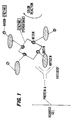
- the unique use lies in the fact that the antigen has been immobilized as it passes through a porous matrix containing specific antibody which has in turn been immobilized by protein A as shown in Figure 1 which illustrates schematically the system.
- the enzyme coupled to the avidin determines the colorimetric assay used in the detection of antigen, which ultimately is a test for the presence of certain types of antibody in the biological fluid.
- Two of the more commonly used enzymes are alkaline phosphatase and horseradish peroxidase. These enzymes may be chemically bonded and linked to the avidin molecule utilizing commonly used chemical pathways for protein; protein covalent linkage includes the use of glutaraldehyde or the use of other homobifunctional and heterobifunctional reagents such as disuccinimidyl suberate and succinimidyl-N-(4-carboxycyclohexylmethyl)-maleimide (S.
- horseradish peroxidase is used as the detection enzyme
- typical substrates would be diaminobenzidine, 4-chloro-1-naphthol, or 3,3′,5,5′-tetramethylbenzidine(7).
- a darkly colored polymeric material is formed. This colored product is fast, i.e., insoluble and precipitating on the solid support. The production of this colored precipitate indicates the presence of the enzyme and in the context of this invention the presence of antibodies specific for the antigen being investigated.
- alkaline phosphatase such as a mixture of 4-chloro-2-methylaniline and 3-hydroxy-2-naphthoic acid 4′ chloro-2′ methyl anilide phosphate, or a mixture of 5-bromo-4-chloro-3-indolyl-phosphate and nitroblue tetrazolium.
- the capture of antibodies by the Protein A attached to the insoluble matrix occurs in minutes with a total assay time of less than 15 minutes.
- the use of the column technique also lends speed and convenience to the present invention, and permits the concentration of antibodies from a large volume of fluid.
- Crosslinked agarose consisting of beads (75-300 ⁇ m (micron) diameter) to which protein A has been linked by the techniques indicated above (either by the producer or a commercial supplier, e.g., Bio Rad Corp. Product 1536154) is washed in phosphate buffered saline (PBS) and incubated for 12 hours at 4°C as a 25% suspension in PBS containing 0.75% gelatin, 0.3% TweenTM20 detergent. This reduces non-specific binding of proteins to the agarose. The agarose-protein A beads are again washed in PBS.
- PBS phosphate buffered saline
- a clean polystyrene column 10 (8 mm I.D. x 102 mm) with a volume of 6 milliliters and a porous (70 ⁇ m pore size) polyethylene disk (frit) 12 at the bottom is used (e.g., Pierce Chemical Co. product 29920).
- a cap 14 is used on the bottom tip of the column to control the flow of liquid through the column.
- Columns of other dimensions which fulfill the basic requirements of the invention are usable.
- Two hundred microliters of the prepared agarose-protein A beads 16 are placed on the frit at the bottom of the column with adequate PBS for transfer.
- a second frit 18 is placed on top of the settled agarose beads resulting in a 4 mm thick disk of agarose beads sandwiched between 2 porous frits.
- the column described here fulfills the essential requirement of a device which will (1) contain an adequate quantity of agarose-protein A beads, (2) allow easy observation of the beads, (3) allow the flow of a body fluid to be tested through the beads with adequate contact surface between solutes in the body fluid and the protein A attached to the beads, (4) allow the sequential addition and washing out of the reagents used to perform the colorimetric tests of the antibodies in the body fluid.
- aqueous buffer which is 0.25M in TRISTM (Tris (hydroxy methyl amino methane)
- TRISTM Tris (hydroxy methyl amino methane)
- 0.37% gelatin 0.30% TweenTM20, 0.075M NaCl, and 0.01M in sodium phosphate.
- This buffer is called "diluent buffer” and has a pH of about 8.0.
- the diluted body fluid is placed in the column on the top of the upper frit and allowed to drain through the column by removing the bottom cap (3 to 5 minutes for this step). The column is then washed twice with 3 ml. portions of diluent buffer (3 minutes per wash).
- the substrate solution can be 0.5 mg/ml. of 3,3 diaminobenzidine in PBS containing 0.00004 percent H2O2 (2 minutes). The extent of darkening of the gel at this point indicates the presence of antibodies in the biological fluid specific for the test antigen.
- An additional wash with water allows the test column to be capped and stored at refrigerator temperature with stable color development for several days.
- Controls consist of columns run at the same time with known positive and negative samples of body fluids or suitable solutions of positive and negative antibodies.
- Alternative or additional controls for the purpose of color comparison can be agarose without protein A (negative) or agarose with biotin coupled to it.
- Biotin which contains a carboxylic acid, can be coupled to agarose by the same chemical reactions used to couple protein A to agarose.
- These color-comparison-controls can be run in the same column as the test by simply placing the additional aliquots of agarose in the column with the agarose-protein A; each agarose aliquot is separated from the others by a polyethylene frit.
- Example I The procedure described in Example I for assaying 1 ml. of body fluid takes about 27 minutes. This time can be significantly reduced by the following modification: One milliliter of body fluid is combined with 2 ml. of diluent buffer. Ten micrograms of biotin-antigen complex are added followed by incubation for 2 minutes at room temperature. Twenty micrograms of avidin-peroxidase complex are added and the mixture is immediately passed through the column (3 minutes). This is followed by 2 washes with 3 ml. of diluent buffer (6 minutes) and color development with 2 ml. of diaminobenzidine-H2O2 substrate solution (2 minutes). The total assay time with this modified procedure is about 13 minutes and the sensitivity is equal to or greater than that described earlier.
- Example II Using the abbreviated column procedure of Example II, we have tested the saliva and serum of Acquired Immune Deficiency (AIDS) patients which are confirmed to have serum antibodies against the Human Immunodeficiency Virus-l (HIV-1) proteins (positive samples). Ten micrograms of biotinylated HIV viral lysate were used per column. Using 6 microliters of positive serum or 150 microliters of positive saliva, a near maximum dark brown color was obtained on the agarose beads using the diaminobenzidine substrate. Control serum and saliva from uninfected persons gave essentially colorless agarose beads with the same procedure.
- AIDS Acquired Immune Deficiency
- this invention should be useful for the detection of disease specific antibody in body fluids of patients with infection by such agents as Hepatitis B and Herpes Simplex Type I and Type II.
- Another format which fulfills the basic function of the column is to place the sandwich of frit-protein A-agarose-frit in a disposable plastic pipette tip.
- the frits wedge tightly against the walls of the tip, confining the agarose beads and allowing the flow of liquid through the beads.
- the tip When attached to the standard hand pipetting unit, the tip can be used to draw the body fluids and various reagents and washing fluids through the agarose beads.
- the present invention can be modified to directly detect the antigens of interest. Biotinylated antigen is omitted in this procedure.
- the biological fluid to be tested is added to blocking buffer in the column. Two micrograms of human antibody against the antigen(s) is also added to this solution in the blocking buffer. After 5-10 minutes incubation, there is added 2 micrograms of a mouse monoclonal antibody that has been modified so that it is a Fab fragment (lacking the Fc part of the molecule which binds to Protein A, but retaining its ability to bind antigen).
- D.W. Dresser "Assays for immunoglobulin-secreting cells," in D.M.
- the monoclonal antibody Fab fragment will only bind to the column, and thus color development will only be seen, if the antigen of interest is present.
- this technique captures antigen as an antigen-antibody complex by binding of antibody, including the antibody endogenously present in the biological sample. Also, larger volumes of biological fluid can be concentrated on a small detection surface.
- This technique can also be adapted to biological fluids which lack endogenous antibodies against the antigen of interest and where there are no other sources of human antibody specific for the antigen. This would be the case, for example, in a plant tissue extract to be assayed for antigens of a plant pathogen.
- two monoclonal antibodies specific for the antigens of the pathogen would be used. One antibody would be added to the extract to bind the antigen and mediate its binding to the immobilized Protein A. The second monoclonal would be made into a biotinylated Fab fragment and used as a detection antibody.
- C . sepedonicum infection produces bacterial ring rot in potatoes, a disease of significant commercial impact.
- Stem and tuber extracts of affected plants contain many of these gram-positive bacteria.
- a number of monoclonal antibodies have been produced in mice to the antigens of C. sepedonicum which can be used in an immunoassay for the organism. Extracts of suspect plant tissues are prepared according to standard techniques in fluid containing detergent to maximize solubility of the bacterial antigens. Insoluble materials are removed by filtration and centrifugation. Between 1 and 5 mL of this extract are added to the column described above.
- the column contains 1 mL of diluent buffer and 10 micrograms of a monoclonal antibody against a principal bacterial antigen. Ten micrograms of a second monoclonal antibody, biotinylated and present as a Fab fragment, are also present. This monoclonal antibody is specific for a second epitope on the same bacterial antigen. After 5 minutes incubation, the mixture is drained through the column. The column is washed with diluent buffer as described above. Avidin linked to enzyme is then passed through the column followed by washes as previously described in Examples I and II. Suitable chromogenic substrates for the enzyme are added and color development estimated in comparison to positive and negative controls to assess the presence of bacteria in the original tissues.
Landscapes
- Health & Medical Sciences (AREA)
- Life Sciences & Earth Sciences (AREA)
- Engineering & Computer Science (AREA)
- Immunology (AREA)
- Chemical & Material Sciences (AREA)
- Molecular Biology (AREA)
- Biomedical Technology (AREA)
- Hematology (AREA)
- Urology & Nephrology (AREA)
- Medicinal Chemistry (AREA)
- General Health & Medical Sciences (AREA)
- Biotechnology (AREA)
- Biochemistry (AREA)
- Food Science & Technology (AREA)
- Physics & Mathematics (AREA)
- Analytical Chemistry (AREA)
- Microbiology (AREA)
- Cell Biology (AREA)
- General Physics & Mathematics (AREA)
- Pathology (AREA)
- Bioinformatics & Cheminformatics (AREA)
- Nanotechnology (AREA)
- Pharmacology & Pharmacy (AREA)
- Medical Informatics (AREA)
- General Engineering & Computer Science (AREA)
- Crystallography & Structural Chemistry (AREA)
- Biophysics (AREA)
- Epidemiology (AREA)
- Animal Behavior & Ethology (AREA)
- Public Health (AREA)
- Veterinary Medicine (AREA)
- Heterocyclic Carbon Compounds Containing A Hetero Ring Having Oxygen Or Sulfur (AREA)
- Peptides Or Proteins (AREA)
- Measuring Or Testing Involving Enzymes Or Micro-Organisms (AREA)
- Apparatus Associated With Microorganisms And Enzymes (AREA)
Claims (27)
- Verfahren zur Bestimmung eines interessierenden Antikörpers in einem den interessierenden Antikörper und andere Antikörper enthaltenden Körperfluid, mit den Schritten:a. Bereitstellen eines kovalent an einen Träger gebundenen, Antikörper-bindenden Proteins;b. Kontaktieren und Inkubieren eines Testkörperfluids, enthaltend den interessierenden Antikörper und andere Antikörper, mit dem proteingebundenen Träger gemäß Schritt (a), wobei das Protein mit den Antikörpern unter Immobilisierung der Antikörper einschließlich des interessierenden Antikörpers reagiert;c. Bereitstellen einer Lösung eines gegen den interessierenden Antikörper spezifischen Antigens, wobei das Antigen biotinyliert ist;d. Kontaktieren und Inkubieren des biotinylierten Antigens gemäß Schritt (c) mit dem immobilisierten, interessierenden Antikörper gemäß Schritt (b), wobei das biotinylierte Antigen mit dem immobilisierten, interessierenden Antikörper unter Immobilisierung des biotinylierten Antigens reagiert;e. Bereitstellen einer Lösung von Avidin, wobei das Avidin kovalent an ein Enzym gebunden ist;f. Kontaktieren und Inkubieren der Lösung gemäß Schritt (e) mit dem immobilisierten, biotinylierten, antigenimmobilisierten, Antikörper-Protein-gebundenen Komplex gemäß Schritt (d), wobei das enzymmarkierte Avidin mit dem Komplex unter Immobiliserung des enzymmarkierten Avidins reagiert;g. Kontaktieren und Inkubieren des immobilisierten Komplexes gemäß Schritt (f) mit einer Substratlösung, wobei das an Avidin gebundene Enzym eine Reaktion mit dem Substrat induziert und ein bestimmbares Reaktionsprodukt auf dem immobilisiertem Komplex erzeugt; undh. Korrelieren des bestimmbaren Reaktionsproduktes auf dem immobilisiertem Komplex mit der Anwesenheit des zu bestimmenden, interessierenden Antikörpers.
- Verfahren nach Anspruch 1, bei dem das Körperfluid Speichel ist.
- Verfahren nach Anspruch 1, bei dem der Träger ein beliebiges poröses Material mit einer niedrigen nicht-spezifischen Bindung von Proteien und funktionellen Gruppen oder anderen chemischen Gegebenheiten, die die Bindung von antikörperbindenden Proteinen gestatten, ist.
- Verfahren nach Anspruch 3, bei dem der Träger Agarose ist.
- Verfahren nach Anspruch 4, bei dem man Farbvergleichskontrollen unter Verwendung zusätzlicher Aliquots an Agarose, voneinander getrennt mit Polyethylenscheiben, laufen läßt.
- Verfahren nach Anspruch 1, bei dem das Protein Protein A ist.
- Verfahren nach Anspruch 1, bei dem das Protein von Quellen abgeleitet ist, die aus der von den Typen II, III, IV, V und VI Fc-Rezeptoren gebildeten Gruppe ausgewählt sind.
- Verfahren nach Anspruch 1, bei dem nach dem Kontaktieren und Inkubieren des Testfluids mit dem proteingebundenen Träger der Träger gewaschen wird.
- Verfahren nach Anspruch 1, bei dem nach dem Kontaktieren und Inkubieren des biotinylierten Antigens mit dem immobilisiertem Antikörper der Träger gewaschen wird.
- Verfahren nach Anspruch 1, bei dem nach dem Kontaktieren und Inkubieren des enzymgebundenen Avidins mit dem immobilisierten Antigenkomplex der Träger gewaschen wird.
- Verfahren nach Anspruch 1, bei dem der Träger in einer offenendigen Kolonne festgehalten wird.
- Verfahren nach Anspruch 11, bei dem die Kolonne aus Polystyrol besteht.
- Verfahren nach Anspruch 11, bei dem der Träger von einer porösen Polyethylenscheibe gehalten wird.
- Verfahren nach Anspruch 1, bei dem die Testkörperflüssigkeit vor dem Auftragen mit einem wäßrigen Puffer verdünnt wird.
- Verfahren zum Bestimmen eines interessierenden Antikörpers in einem den interessierenden Antikörper und andere Antikörper enthaltenden Körperfluid, mit den Schritten:a. Bereitstellen eines kovalent an einen Träger gebundenen, antikörperbindenden Proteins,b. Kontaktieren und Inkubieren eines Testkörperfluids, enthaltend den interessierenden Antikörper und andere Antikörper, mit einer Lösung eines für den interessierenden Antikörper spezifischen Antigens, wobei das Antigen biotinyliert ist,c. Kontaktieren und Inkubieren der kombinierten Lösung gemäß Schritt (b) mit einer Lösung von Avidin, wobei das Avidin kovalent an ein Enzym gebunden ist,d. Kontaktieren und Inkubieren der kombinierten Lösung gemäß Schritt (c) mit einem Träger gemäß Schritt (a), so daß ein Komplex von Antikörper-biotinyliertem, Antigen-Enzym-markiertem Avidin durch den Träger immobilisiert wird,e. Kontaktieren und Inkubieren des immobilisierten Komplexes gemäß Schritt (d) mit einer Substratlösung, wobei das mit Avidin verknüpfte Enzym eine Reaktion mit dem Substrat induziert und ein bestimmbares Reaktionsprodukt auf dem immobilisiertem Komplex bildet; undf. Korrelieren des bestimmbaren Reaktionsproduktes auf dem immobiliserten Komplex mit der Anwesenheit des zu bestimmenden Antikörpers.
- Verfahren nach Anspruch 15, bei dem nach dem Kontaktieren und Inkubieren der kombinierten Lösung gemäß Schritt (c) mit dem Träger gemäß Schritt (a) der Träger gewaschen wird.
- Verfahren nach Anspruch 15, bei dem das Antigen HIV-1 ist.
- Verfahren nach Anspruch 1 oder 15, bei dem der Assay in einer Pipettespitze durchgeführt wird.
- Verfahren zum Bestimmen eines Antigens in einem das Antigen enthaltenden biologischen Testfluid, mit den Schritten:a. Bereitstellen eines Fc-Rezeptor-, Antikörper-bindenden Proteins, das kovalent an einen Träger gebunden ist;b. Bereitstellen einer das Antigen enthaltenden Lösung des Testfluids, enthaltend eine geringe Menge von Antikörpern gegen das Antigen in einem wäßrigen Puffer;c. Kontaktieren und Inkubieren der Lösung gemäß Schritt (b) mit dem Träger gemäß Schritt (a), so daß ein Komplex aus Antikörper und Antigen durch den Träger immobilisert wird;d. Bereitstellen einer Lösung eines monoklonalen Antikörpers, der so modifiziert ist, daß er ein Fab-Fragment ist, dem der Fc-Bereich fehlt, welcher an das Protein bindet, jedoch seine Fähigkeit zur Bindung des Antigens beibehält, wobei der Antikörper biotinyliert ist;e. Kontaktieren und Inkubieren der Lösung gemäß Schritt (d) mit dem immobilisiertem Komplex gemäß Schritt (c), so daß ein Antikörper-Antigen-Fab-Antikörperkomplex durch den Träger immobilisiert wird;f. Bereitstellen einer Lösung von Avidin, wobei das Avidin kovalent an ein Enzym gebunden ist;g. Kontaktieren und Inkubieren der Lösung gemäß Stufe (f) mit dem immobilisierten Antikörper-Antigen-Fab-Antikörperkomplex gemäß Stufe (e), wobei das enzymmarkierte Avidin mit dem Komplex unter Immobilisierung des enzymmarkierten Avidins reagiert;h. Kontaktieren und Inkubieren des immobilisierten Komplexes gemäß Stufe (g) mit einer Substratlösung, wobei das mit Avidin verbundene Enzym eine Reaktion mit dem Substrat induziert und ein bestimmbares Reaktionsprodukt auf dem immobilisierten Komplex bildet; undi. Korrelieren des bestimmbaren Reaktionsproduktes auf dem immobilisiertem Komplex mit der Anwesenheit des zu bestimmenden Antigens.
- Verfahren nach Anspruch 19, bei dem nach dem Kontaktieren und Inkubieren des enzymgebundenen Avidins der Träger gewaschen wird.
- Verfahren nach Anspruch 19, bei dem der anti-Antigen-Antikörper gemäß Schritt (b) und der anti-Antigen-Antikörper gemäß Schritt (d) für verschiedene Epitope des Antigens spezifisch sind.
- Verfahren nach Anspruch 19, bei dem der biologischen Testflüssigkeit endogene Antikörper gegen das Antigen fehlen.
- Verfahren nach Anspruch 22, bei dem das biologische Testfluid ein Pflanzenextrakt ist, der Antigene eines Pflanzenpathogens enthält.
- Verfahren nach Anspruch 23, bei dem das Antigen ein Bakterium ist.
- Verfahren nach Anspruch 24, bei dem das Bakterium Coryne-bacterium sepedonicum ist.
- Kit für die Bestimmung eines interessierenden Antikörpers in einem den interessierenden Antikörper und andere Antikörper enthaltenden Körperfluid, enthaltend:
ein antikörperbindendes Protein, das kovalent an einen Träger gebunden ist;
ein biotinyliertes Antigen, das gegen den interessierenden Antikörper spezifisch ist;
eine Lösung von enzymverknüpftem Avidin; und
ein Enzymsubstrat. - Kit für die Bestimmung eines interessierenden Antigens in einem das interessierende Antigen und andere Antigene enthaltenden Körperfluid, enthaltend:
ein Fc-Rezeptor-, antikörperbindendes Protein, das kovalent an einen Träger gebunden ist;
einen biotinylierten monoklonalen Antikörper, der zu einem Fab-Fragment modifiziert ist, dem der Fc-Bereich, der an das Protein bindet, fehlt, der jedoch die Fähigkeit zur Bindung des interessierenden Antigens beibehalten hat;
eine Lösung von enzymgebundenem Avidin; und
ein Enzymsubstrat.
Applications Claiming Priority (3)
| Application Number | Priority Date | Filing Date | Title |
|---|---|---|---|
| US30287789A | 1989-01-30 | 1989-01-30 | |
| US302877 | 1989-01-30 | ||
| PCT/US1990/000378 WO1990008957A1 (en) | 1989-01-30 | 1990-01-25 | Avidin-biotin assisted immunoassay |
Publications (2)
| Publication Number | Publication Date |
|---|---|
| EP0455735A1 EP0455735A1 (de) | 1991-11-13 |
| EP0455735B1 true EP0455735B1 (de) | 1994-09-14 |
Family
ID=23169594
Family Applications (1)
| Application Number | Title | Priority Date | Filing Date |
|---|---|---|---|
| EP90903466A Expired - Lifetime EP0455735B1 (de) | 1989-01-30 | 1990-01-25 | Avidin-biotin assistiertes immunassayverfahren |
Country Status (10)
| Country | Link |
|---|---|
| EP (1) | EP0455735B1 (de) |
| JP (1) | JPH04503109A (de) |
| AT (1) | ATE111604T1 (de) |
| AU (1) | AU640635B2 (de) |
| CA (1) | CA2045673A1 (de) |
| DE (1) | DE69012552T2 (de) |
| DK (1) | DK0455735T3 (de) |
| FI (1) | FI913615A0 (de) |
| NO (1) | NO912951L (de) |
| WO (1) | WO1990008957A1 (de) |
Cited By (1)
| Publication number | Priority date | Publication date | Assignee | Title |
|---|---|---|---|---|
| US6887377B2 (en) | 2001-02-01 | 2005-05-03 | Sigma-Aldrich Co. | Affinity matrices with enhanced visibility for molecular pull-down and immunoprecipitation applications |
Families Citing this family (17)
| Publication number | Priority date | Publication date | Assignee | Title |
|---|---|---|---|---|
| US5525338A (en) * | 1992-08-21 | 1996-06-11 | Immunomedics, Inc. | Detection and therapy of lesions with biotin/avidin conjugates |
| US5198340A (en) * | 1991-01-17 | 1993-03-30 | Genentech, Inc. | Assay for free igf-i, igf-ii, and gh levels in body fluids |
| US6087188A (en) * | 1992-11-13 | 2000-07-11 | Alk A/S | Two-site immunoassay for an antibody with chemiluminescent label and biotin bound ligand |
| IL104384A (en) * | 1993-01-13 | 1996-11-14 | Yeda Res & Dev | Method for screening catalytic non-enzyme polypeptides and proteins |
| US5504091A (en) * | 1993-04-23 | 1996-04-02 | American Home Products Corporation | Biotin esters of rapamycin |
| US7279561B1 (en) | 1993-04-23 | 2007-10-09 | Wyeth | Anti-rapamycin monoclonal antibodies |
| JPH08509499A (ja) | 1993-04-23 | 1996-10-08 | アボツト・ラボラトリーズ | ラパマイシン結合体及び抗体 |
| JPH11508036A (ja) * | 1995-06-07 | 1999-07-13 | アボツト・ラボラトリーズ | 抗−hiv−1または抗−hiv−2抗体を検出するための改良されたハプテン−ペプチド結合物 |
| DE19649390A1 (de) * | 1996-11-29 | 1998-06-04 | Boehringer Mannheim Gmbh | Antigenspezifischer IgG-Nachweis |
| US6150115A (en) * | 1997-04-04 | 2000-11-21 | Seikagaku Kogyo Kabushiki Kaisha | Quantitative determination method for heparan sulfate and diagnostic method using the same |
| US6379909B1 (en) | 1998-06-24 | 2002-04-30 | Alk-Abello A/S | Method of detecting an antibody in a liquid sample |
| ATE403866T1 (de) * | 1998-06-24 | 2008-08-15 | Alk Abello As | Verfahren zum nachweis von antikörpern in flüssigen proben |
| CA2384693A1 (en) * | 1999-09-13 | 2001-03-22 | Nissui Pharmaceutical Co., Ltd. | Kit for detecting or assaying subject substance and detection or assay method |
| WO2002090961A2 (en) * | 2001-05-03 | 2002-11-14 | Immunetics, Inc. | Systems and methods for detection of analytes in biological fluids |
| DE10153963A1 (de) * | 2001-11-06 | 2003-05-22 | Aventis Behring Gmbh Intellect | Testsystem zur Untersuchung einer biologischen Flüssigkeit |
| CN111208290A (zh) * | 2020-02-27 | 2020-05-29 | 江苏泽成生物技术有限公司 | 一种磁微粒化学发光法醛固酮测定试剂盒及使用方法 |
| CN114236131B (zh) * | 2021-11-05 | 2022-12-06 | 江苏省人民医院(南京医科大学第一附属医院) | 一种检测甲状腺过氧化物酶抗体及其亚型的试剂盒 |
Family Cites Families (7)
| Publication number | Priority date | Publication date | Assignee | Title |
|---|---|---|---|---|
| US4228237A (en) * | 1978-09-21 | 1980-10-14 | Calbiochem-Behring Corp. | Methods for the detection and determination of ligands |
| US4301139A (en) * | 1979-06-21 | 1981-11-17 | Ames-Yissum Ltd. | Multilayer column chromatography specific binding assay method, test device and test kit |
| US4425438A (en) * | 1981-03-13 | 1984-01-10 | Bauman David S | Assay method and device |
| AU8594382A (en) * | 1981-08-05 | 1983-02-10 | F. Hoffmann-La Roche Ag | Immunoassay using biotin-avidin-enzyme complex |
| JPS5856696A (ja) * | 1981-09-30 | 1983-04-04 | Amano Pharmaceut Co Ltd | カラムを用いる酵素免疫測定法 |
| US4535057A (en) * | 1982-07-26 | 1985-08-13 | Amf Incorporated | Immunoassay employing monoclonal herpes simplex antibody and biotin-avidin detection system |
| SE458643B (sv) * | 1984-12-07 | 1989-04-17 | Pharmacia Ab | Anordning med kolonnelement uppdelat i provparti och referenspatri |
-
1990
- 1990-01-25 EP EP90903466A patent/EP0455735B1/de not_active Expired - Lifetime
- 1990-01-25 DE DE69012552T patent/DE69012552T2/de not_active Expired - Fee Related
- 1990-01-25 CA CA002045673A patent/CA2045673A1/en not_active Abandoned
- 1990-01-25 WO PCT/US1990/000378 patent/WO1990008957A1/en active IP Right Grant
- 1990-01-25 AU AU50986/90A patent/AU640635B2/en not_active Ceased
- 1990-01-25 DK DK90903466.2T patent/DK0455735T3/da active
- 1990-01-25 JP JP2503517A patent/JPH04503109A/ja active Pending
- 1990-01-25 AT AT90903466T patent/ATE111604T1/de not_active IP Right Cessation
-
1991
- 1991-07-29 NO NO91912951A patent/NO912951L/no unknown
- 1991-07-29 FI FI913615A patent/FI913615A0/fi not_active Application Discontinuation
Non-Patent Citations (1)
| Title |
|---|
| PATENT ABSTRACTS OF JAPAN, vol. 4, no. 144 (C-27), 14 July 1980# * |
Cited By (3)
| Publication number | Priority date | Publication date | Assignee | Title |
|---|---|---|---|---|
| US6887377B2 (en) | 2001-02-01 | 2005-05-03 | Sigma-Aldrich Co. | Affinity matrices with enhanced visibility for molecular pull-down and immunoprecipitation applications |
| US7163633B2 (en) | 2001-02-01 | 2007-01-16 | Sigma-Aldrich Co. | Affinity matrices with enhanced visibility for molecular pull-down and immunoprecipitation applications |
| US7438806B2 (en) | 2001-02-01 | 2008-10-21 | Sigma-Aldrich Co. | Affinity matrices with enhanced visibility for molecular pull-down and immunoprecipitation applications |
Also Published As
| Publication number | Publication date |
|---|---|
| JPH04503109A (ja) | 1992-06-04 |
| DE69012552D1 (de) | 1994-10-20 |
| DE69012552T2 (de) | 1995-04-20 |
| ATE111604T1 (de) | 1994-09-15 |
| WO1990008957A1 (en) | 1990-08-09 |
| CA2045673A1 (en) | 1990-07-31 |
| NO912951D0 (no) | 1991-07-29 |
| NO912951L (no) | 1991-09-13 |
| EP0455735A1 (de) | 1991-11-13 |
| DK0455735T3 (da) | 1994-12-12 |
| AU5098690A (en) | 1990-08-24 |
| FI913615A0 (fi) | 1991-07-29 |
| AU640635B2 (en) | 1993-09-02 |
Similar Documents
| Publication | Publication Date | Title |
|---|---|---|
| EP0455735B1 (de) | Avidin-biotin assistiertes immunassayverfahren | |
| US5824268A (en) | Rapid self-contained assay format | |
| US4298685A (en) | Diagnostic reagent | |
| AU695419B2 (en) | Method for detecting antibodies | |
| JP3457324B2 (ja) | 同時でのサンプル抽出及び免疫原性反応を含む抗体又は抗原の検査のための迅速イムノアッセイ | |
| US4434236A (en) | Immunoassay wherein labeled antibody is displaced from immobilized analyte-analogue | |
| Yolken | Enzyme-linked immunosorbent assay (ELISA): a practical tool for rapid diagnosis of viruses and other infectious agents. | |
| US4474878A (en) | Sandwich EIA for antigen associated with hepatitis | |
| WO1996036878A9 (en) | Rapid self-contained assay format | |
| US4642285A (en) | Sandwich EIA for antigen | |
| JP2636331B2 (ja) | 抗原特異的な抗体の一段階測定法およびそれに適する試薬 | |
| JP2902581B2 (ja) | 測定すべき物質の定性的または/および定量的検出法 | |
| CA2046130A1 (en) | Biologically active reagent, analytical element and methods for use of the reagent | |
| EP0394398A1 (de) | Nanoteilchen mit gebundenen enzym und ligand zur verwendung bei analysen. | |
| JPH1164336A (ja) | 免疫クロマトグラフィーによる分析対象物の検出方法 | |
| US4729961A (en) | Process for the detection and assay by erythroadsorption | |
| EP0442231A1 (de) | Stabförmige Pobeeinrichtung zum Nachweis von Antikörpern | |
| JP2579972B2 (ja) | 膜親和濃縮免疫測定方法 | |
| EP0588958A1 (de) | Assay für den nachweis von spezifischen liganden | |
| CA2151690C (en) | Test kit and method for competitive specific binding assay | |
| CA2090392C (en) | Coupling of antigens and antibodies to non-fixed erythrocytes | |
| JP3337575B2 (ja) | 抗ストレプトリジンo抗体の決定方法 | |
| CA1301646C (en) | Method for diagnostic immunoassay by solid phase separation | |
| EP0152254A2 (de) | Immunotest mit chromogenischer Träger unter Verwendung von markierten Komplementbestandteilen | |
| US5061619A (en) | Immunoassay using antibody-antigen conjugates |
Legal Events
| Date | Code | Title | Description |
|---|---|---|---|
| PUAI | Public reference made under article 153(3) epc to a published international application that has entered the european phase |
Free format text: ORIGINAL CODE: 0009012 |
|
| 17P | Request for examination filed |
Effective date: 19910726 |
|
| AK | Designated contracting states |
Kind code of ref document: A1 Designated state(s): AT BE DE DK FR GB IT LU NL SE |
|
| 17Q | First examination report despatched |
Effective date: 19921125 |
|
| GRAA | (expected) grant |
Free format text: ORIGINAL CODE: 0009210 |
|
| AK | Designated contracting states |
Kind code of ref document: B1 Designated state(s): AT BE DE DK FR GB IT LU NL SE |
|
| REF | Corresponds to: |
Ref document number: 111604 Country of ref document: AT Date of ref document: 19940915 Kind code of ref document: T |
|
| REF | Corresponds to: |
Ref document number: 69012552 Country of ref document: DE Date of ref document: 19941020 |
|
| ITF | It: translation for a ep patent filed | ||
| REG | Reference to a national code |
Ref country code: DK Ref legal event code: T3 |
|
| ET | Fr: translation filed | ||
| EAL | Se: european patent in force in sweden |
Ref document number: 90903466.2 |
|
| PLBE | No opposition filed within time limit |
Free format text: ORIGINAL CODE: 0009261 |
|
| STAA | Information on the status of an ep patent application or granted ep patent |
Free format text: STATUS: NO OPPOSITION FILED WITHIN TIME LIMIT |
|
| 26N | No opposition filed | ||
| PGFP | Annual fee paid to national office [announced via postgrant information from national office to epo] |
Ref country code: DK Payment date: 19951031 Year of fee payment: 7 |
|
| PGFP | Annual fee paid to national office [announced via postgrant information from national office to epo] |
Ref country code: BE Payment date: 19951109 Year of fee payment: 7 |
|
| PGFP | Annual fee paid to national office [announced via postgrant information from national office to epo] |
Ref country code: FR Payment date: 19951123 Year of fee payment: 7 |
|
| PGFP | Annual fee paid to national office [announced via postgrant information from national office to epo] |
Ref country code: SE Payment date: 19951130 Year of fee payment: 7 |
|
| PGFP | Annual fee paid to national office [announced via postgrant information from national office to epo] |
Ref country code: LU Payment date: 19951201 Year of fee payment: 7 |
|
| PGFP | Annual fee paid to national office [announced via postgrant information from national office to epo] |
Ref country code: DE Payment date: 19951227 Year of fee payment: 7 |
|
| PGFP | Annual fee paid to national office [announced via postgrant information from national office to epo] |
Ref country code: GB Payment date: 19960116 Year of fee payment: 7 |
|
| PGFP | Annual fee paid to national office [announced via postgrant information from national office to epo] |
Ref country code: AT Payment date: 19960130 Year of fee payment: 7 |
|
| PGFP | Annual fee paid to national office [announced via postgrant information from national office to epo] |
Ref country code: NL Payment date: 19960131 Year of fee payment: 7 |
|
| PG25 | Lapsed in a contracting state [announced via postgrant information from national office to epo] |
Ref country code: LU Free format text: LAPSE BECAUSE OF NON-PAYMENT OF DUE FEES Effective date: 19970125 Ref country code: GB Effective date: 19970125 Ref country code: DK Effective date: 19970125 Ref country code: AT Effective date: 19970125 |
|
| REG | Reference to a national code |
Ref country code: DK Ref legal event code: EBP |
|
| PG25 | Lapsed in a contracting state [announced via postgrant information from national office to epo] |
Ref country code: SE Effective date: 19970126 |
|
| PG25 | Lapsed in a contracting state [announced via postgrant information from national office to epo] |
Ref country code: BE Effective date: 19970131 |
|
| BERE | Be: lapsed |
Owner name: EPITOPE INC. Effective date: 19970131 |
|
| PG25 | Lapsed in a contracting state [announced via postgrant information from national office to epo] |
Ref country code: NL Effective date: 19970801 |
|
| GBPC | Gb: european patent ceased through non-payment of renewal fee |
Effective date: 19970125 |
|
| PG25 | Lapsed in a contracting state [announced via postgrant information from national office to epo] |
Ref country code: FR Effective date: 19970930 |
|
| NLV4 | Nl: lapsed or anulled due to non-payment of the annual fee |
Effective date: 19970801 |
|
| PG25 | Lapsed in a contracting state [announced via postgrant information from national office to epo] |
Ref country code: DE Effective date: 19971001 |
|
| EUG | Se: european patent has lapsed |
Ref document number: 90903466.2 |
|
| REG | Reference to a national code |
Ref country code: FR Ref legal event code: ST |
|
| PG25 | Lapsed in a contracting state [announced via postgrant information from national office to epo] |
Ref country code: IT Free format text: LAPSE BECAUSE OF NON-PAYMENT OF DUE FEES Effective date: 20050125 |