EP0336399B1 - Endoskop zur Untersuchung der Gallen- und Pankreasgänge - Google Patents
Endoskop zur Untersuchung der Gallen- und Pankreasgänge Download PDFInfo
- Publication number
- EP0336399B1 EP0336399B1 EP89105987A EP89105987A EP0336399B1 EP 0336399 B1 EP0336399 B1 EP 0336399B1 EP 89105987 A EP89105987 A EP 89105987A EP 89105987 A EP89105987 A EP 89105987A EP 0336399 B1 EP0336399 B1 EP 0336399B1
- Authority
- EP
- European Patent Office
- Prior art keywords
- guide tube
- tube
- endoscope
- insertion unit
- tip
- Prior art date
- Legal status (The legal status is an assumption and is not a legal conclusion. Google has not performed a legal analysis and makes no representation as to the accuracy of the status listed.)
- Expired - Lifetime
Links
- 210000000277 pancreatic duct Anatomy 0.000 title claims description 89
- 210000000013 bile duct Anatomy 0.000 title claims description 85
- 238000003780 insertion Methods 0.000 claims description 163
- 230000037431 insertion Effects 0.000 claims description 163
- 210000001198 duodenum Anatomy 0.000 claims description 23
- 239000002504 physiological saline solution Substances 0.000 claims description 19
- FAPWRFPIFSIZLT-UHFFFAOYSA-M Sodium chloride Chemical compound [Na+].[Cl-] FAPWRFPIFSIZLT-UHFFFAOYSA-M 0.000 description 7
- 239000011780 sodium chloride Substances 0.000 description 7
- 238000002594 fluoroscopy Methods 0.000 description 6
- 239000002872 contrast media Substances 0.000 description 5
- 230000003287 optical effect Effects 0.000 description 4
- 230000010412 perfusion Effects 0.000 description 4
- XLYOFNOQVPJJNP-UHFFFAOYSA-N water Substances O XLYOFNOQVPJJNP-UHFFFAOYSA-N 0.000 description 3
- 208000027418 Wounds and injury Diseases 0.000 description 2
- 230000006378 damage Effects 0.000 description 2
- 239000012530 fluid Substances 0.000 description 2
- 239000007924 injection Substances 0.000 description 2
- 238000002347 injection Methods 0.000 description 2
- 208000014674 injury Diseases 0.000 description 2
- 238000000034 method Methods 0.000 description 2
- 238000001356 surgical procedure Methods 0.000 description 2
- 238000005452 bending Methods 0.000 description 1
- 238000004891 communication Methods 0.000 description 1
- 238000012790 confirmation Methods 0.000 description 1
- 238000003384 imaging method Methods 0.000 description 1
- 239000002184 metal Substances 0.000 description 1
- 210000000496 pancreas Anatomy 0.000 description 1
- 239000000843 powder Substances 0.000 description 1
- 230000002265 prevention Effects 0.000 description 1
Images


Classifications
-
- A—HUMAN NECESSITIES
- A61—MEDICAL OR VETERINARY SCIENCE; HYGIENE
- A61B—DIAGNOSIS; SURGERY; IDENTIFICATION
- A61B1/00—Instruments for performing medical examinations of the interior of cavities or tubes of the body by visual or photographical inspection, e.g. endoscopes; Illuminating arrangements therefor
- A61B1/012—Instruments for performing medical examinations of the interior of cavities or tubes of the body by visual or photographical inspection, e.g. endoscopes; Illuminating arrangements therefor characterised by internal passages or accessories therefor
- A61B1/015—Control of fluid supply or evacuation
-
- A—HUMAN NECESSITIES
- A61—MEDICAL OR VETERINARY SCIENCE; HYGIENE
- A61B—DIAGNOSIS; SURGERY; IDENTIFICATION
- A61B1/00—Instruments for performing medical examinations of the interior of cavities or tubes of the body by visual or photographical inspection, e.g. endoscopes; Illuminating arrangements therefor
- A61B1/012—Instruments for performing medical examinations of the interior of cavities or tubes of the body by visual or photographical inspection, e.g. endoscopes; Illuminating arrangements therefor characterised by internal passages or accessories therefor
- A61B1/018—Instruments for performing medical examinations of the interior of cavities or tubes of the body by visual or photographical inspection, e.g. endoscopes; Illuminating arrangements therefor characterised by internal passages or accessories therefor for receiving instruments
Definitions
- the present invention relates to an endoscope for bile duct and pancreatic duct for non-invasive endoscopic observation of bile duct and pancreatic duct.
- An endoscope of double-scope system is already known. This consists of a mother endoscope having an insertion unit, which can be inserted up to duodenum, and of a daughter endoscope having an insertion unit, which can be introduced by transpapillary insertion through a forceps channel of the above insertion unit.
- the interior of the bile duct and pancreatic duct can be observed by introducing the insertion unit of the daughter endoscope directly into papilla through forceps channel of the mother endoscope.
- the insertion unit of the daughter endoscope must have a path, through which physiological saline for perfusion can be sent, and, accordingly, the insertion unit must have larger diameter.
- the tip of the insertion unit of such daughter endoscope cannot be inserted conveniently into papilla unless it is bent toward the desired direction. Consequently, it is designed in such manner that the tip can be bent upward or downward.
- the tip of the insertion unit of the daughter endoscope is directly inserted into papilla.
- the tip must be bent.
- it is necessary to provide a path to send physiological saline for perfusion and a hole for accommodating small surgical tools.
- it must be relatively large in diameter (outer diameter: 3.5 mm).
- pancreatic duct which is smaller than bile duct, it is very difficult to place the tip of the insertion unit with outer diameter of 3.5 mm into the duct, and it requires skill. It is also very likely that the papilla is injured by the tip of the insertion unit.
- the object of the present invention is to provide an endoscope for bile duct and pancreatic duct, in which the insertion unit of the daughter endoscope can be easily and safely inserted into papilla as well as into bile duct and pancreatic duct and the insertion unit of the daughter endoscope is used only for observation with no need for bending and for providing the path to send saline.
- Another object of the invention is to offer an endoscope for bile duct and pancreatic duct, in which it is possible to inject physiological saline for perfusion and also to insert various types of tools.
- the first embodiment of the invention consists of a mother endoscope having an insertion unit to be inserted to duodenum, a forceps channel of the insertion unit and an insertion hole connected with the forceps channel, of a flexible guide tube, which is to be inserted into bile duct and pancreatic duct from papilla through the forceps channel and into which the insertion unit of the daughter endoscope is inserted, and of a flexible core bar, which is mounted removably on one end of the guide tube, is inserted into the guide tube and has such length that its tip does not protrude from the guide tube.
- the second embodiment of the invention consists of a mother endoscope, of which the insertion unit having a forceps channel can be inserted to duodenum, and of a daughter endoscope, of which the insertion unit is guided by the flexible guide tube from the forceps channel of the mother endoscope and is inserted into bile duct and pancreatic duct.
- Two or more fixing tubes with different diameters are concentrically provided to cover the insertion unit on the base end of the insertion unit of the daughter endoscope, and the fixing tube corresponding to the guide tube, which is selected through an adaptor according to the diameter of a guide tube selected from two or more guide tubes having different diameters and inserted into bile duct and pancreatic duct from the forceps channel of the mother endoscope, is connected with the guide tube.
- the third embodiment of the invention consists of a flexible guide tube, which can be inserted into bile duct and pancreatic duct through the forceps channel through the insertion hole of the mother endoscope with its insertion unit placed into duodenum, of a fixing tube to be mounted on the base end of the insertion unit of the daughter endoscope, and of an adaptor, both ends of which are mounted on the fixing tube and on the guide tube, into which the insertion unit of the daughter endoscope is inserted.
- the tip of the insertion unit inserted into the guide tube of the daughter endoscope is roughly aligned with the tip of the guide tube and does not protrude.
- the fourth embodiment of the invention consists of a flexible first guide tube to be inserted from the insertion hole of the mother endoscope through the forceps channel into bile duct and pancreatic duct, of a junction tube to be connected with the first guide tube, of a flexible second guide tube, which has larger diameter than the first guide tube, is guided by the junction tube and the first guide tube and is inserted into bile duct and pancreatic duct, of a fixing tube to be mounted on the base end of the insertion unit, which is placed into the second guide tube of the daughter endoscope, of an enclosure unit provided on the base end of the insertion unit of the daughter endoscope to close up the gap between the fixing tube and the insertion unit of the daughter endoscope, and of an adaptor, both ends of which are mounted on the second guide tube and the fixing tube after the first guide tube and the junction tube have been withdrawn from the second guide tube, guided by the first guide tube and inserted into bile duct and pancreatic duct.
- a core bar mounted on the mounting unit from the insertion hole of the mother endoscope is placed into the guide tube.
- This guide tube containing core bar is introduced into the forceps channel in order to insert the guide tube into bile duct and pancreatic duct.
- the tip of the guide tube containing core bar is inserted into bile duct or pancreatic duct through papilla by manipulating the forceps elevator. Since a flexible core bar is contained in the guide tube, neither twisting nor turning occurs within the forceps channel, and positive insertion can be achieved straightly toward the papilla.
- the core bar By fluoroscopy after the insertion, the core bar can be seen as image, and it is possible to confirm how far the guide tube has been inserted. If the core bar is withdrawn from the guide tube, it is posible to observe the interior of bile duct and pancreatic duct by placing the insertion unit of the daughter endoscope into the guide tube. In this case, it is possible to provide better observation through the supply of physiological saline through the gap between the insertion unit of the daughter endoscope and the guide tube.
- the tip of the insertion unit of the mother endoscope is introduced perorally into duodenum and the position of papilla is confirmed. Then, a guide tube having such diameter as easy to insert into papilla is inserted into the forceps channel, and the tip of the guide tube is inserted again toward the papilla by utilizing the forceps elevator mounted on the tip of the insertion unit of the mother endoscope. Because the guide tube has full flexibility, it can be inserted into bile duct or pancreatic duct through papilla without injuring it.
- the guide tube can be designed with smaller diameter because it is merely to accommodate the insertion unit of daughter endoscope for the purpose of observation only (Neither the passage nor the forceps channel is required.), and it can be inserted into papilla without difficulty.
- the guide tube is inserted into bile duct or pancreatic duct through papilla, the insertion unit of the daughter endoscope is placed into the guide tube.
- physiological saline can be supplied through the gap between the insertion unit of daughter endoscope and the guide tube.
- an appropriate tube is chosen from two or more fixing tubes, and these tubes are connected through the adaptor, and saline is supplied by the use of this adaptor.
- the guide tube with larger diameter is inserted into bile duct and pancreatic duct using a guide tube of smaller diameter, and the latter is then withdrawn.
- a fixing tube corresponding to the guide tube with larger diameter is selected, and these tube are connected through adaptor to have communication with each other, and various types of tools can be inserted by utilizing the adaptor.
- the papilla of duodenum is confirmed by the tip of the insertion unit of the mother endoscope. Then, the guide tube is introduced from the insertion hole into the forceps channel, and the tip of the guide tube is inserted toward papilla by manipulating the forceps elevator at the tip of the insertion unit.
- the insertion unit of the daughter endoscope is placed into the guide tube.
- the tip of the insertion unit is roughly aligned with the tip of the guide tube.
- bile duct or pancreatic duct is not injured by the tip of fiberscope.
- the papilla of duodenum is confirmed by the tip of the insertion unit of the mother endoscope. Then, the first guide tube with smaller diameter is placed from the insertion hole through the forceps channel, and the tip of the first guide tube is inserted toward papilla by manipulating the forceps elevator at the tip of the insertion unit.
- the junction tube is connected with this first guide tube, and the second guide tube is inserted into bile duct or pancreatic tube, being guided by these tubes.
- the first guide tube has such diameter as to facilitate the insertion even into pancreatic duct, it is possible to insert it easily using the mother endoscope with narrower thinner forceps channel.
- the mother endoscope having the forceps channel with smaller diameter is withdrawn, and the junction tube and the first guide tube are inserted into the forceps channel of another mother endoscope having forceps channel with larger diameter enough to accommodate the second guide tube.
- the mother endoscope can be replaced.
- the insertion unit of the daughter endoscope can be placed into the second guide tube.
- bile duct and pancreatic can be prevented from injury by the tip of the insertion unit through such arrangement that the tip of the insertion unit is roughly aligned with the tip of the second guide tube and does not protrude. Since the gap between the second guide tube and the insertion unit is relatively wide, a tool can be inserted or physiological saline can be supplied from the cross tube of the adaptor by utilizing this gap.
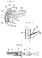
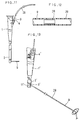
- Fig. 1 is a schematical view showing the entire unit according to the first embodiment of the invention
- Fig. 2 is an enlarged view of the tip of the insertion unit
- Fig. 3 is a drawing to explain how the guide tube is inserted into papilla.
- Fig. 4 is a cross-sectional view when the guide tube is inserted into pancreatic tube
- Fig. 5 is a cross-sectional view of an example of injection of contrast medium into guide tube
- Fig. 6 is a schematical front view of the daughter endoscope
- Fig. 7 is a cross-sectional view when the insertion unit of the daughter endoscope is inserted into the guide tube.
- Fig. 8 is a view showing the outline of the unit according to the second embodiment of the invention
- Fig. 8 is a view showing the outline of the unit according to the second embodiment of the invention
- FIG. 9 is a perspective view when the guide tube is inserted into the forceps channel of mother endoscope, and Fig. 10 is a cross-sectional view showing the details of the adaptor.
- Fig. 11 is a front view of the unit when junction tube is connected
- Fig. 12 is a cross-sectional view of the joint
- Fig. 13 is a drawing to explain how the endoscope is inserted after mother endoscope is withdrawn.
- Fig. 14 is a drawing to explain when the guide tube with larger diameter is inserted, and Fig. 15 explains how the insertion unit of the daughter endoscope is inserted.
- Fig. 16 is a cross-sectional view showing the detail of the adaptor
- Fig. 17 shows how the forceps are inserted.
- FIG. 18 is a front view showing how the mounting unit is removed and core bar is withdrawn after the guide tube is inserted to the desired position in bile duct or pancreatic duct in the third embodiment of the invention
- Fig. 19 is a front view showing how the insertion unit of the daughter endoscope is placed into the guide tube
- Fig. 20 is a cross-sectional view showing how the insertion unit of the daughter endoscope is mounted on the guide tube.
- Fig. 22 is a schematical view of the entire unit according to the fourth embodiment of the invention
- Fig. 22 is a front view showing how the mounting unit and the core bar are withdrawn after the first guide tube is placed into bile duct or pancreatic duct
- Fig. 23 is a cross-sectional view of the portion where junction tube is connected
- Fig. 24 is a front view showing how the second guide tube is guided by the first guide tube and junction tube and is inserted into bile duct or pancreatic duct
- Fig. 25 is a front view showing how the first guide tube and the junction tube are withdrawn after the second guide tube has been placed into bile duct or pancreatic duct
- Fig. 26 is a front view showing how the insertion unit of daughter endoscope is placed into the second guide tube.
- Fig. 27 is a cross-sectional view showing how the insertion unit of the daughter endoscope is mounted on the second guide tube
- Fig. 28 is a front view showing how the first mother endoscope is withdrawn
- Fig. 29 is a front view explaining how the insertion unit of the mother endoscope is inserted after the first mother endoscope is withdrawn.
- Fig. 30 is a cross-sectional view when daughter endoscope is connected with mother endoscope and forceps are inserted into pancreatic duct from the adaptor, and
- Fig. 31 and 32 are the front views showing the variations of the adaptor.
- Fig. 1 is a schematical view of the entire unit based on the invention, where the mother endoscope (1) has an insertion unit (2), and the tip of the insertion unit (2) is placed into duodenum.
- the forceps channel (3) is formed in the insertion unit (2), and the base (1A) has the insertion hole on the forceps channel to receive various types of tools.
- the symbol (1A) represents an ocular unit of the mother endoscope, and 1C is an optical cable connected with an external light source equipment (now shown). As shown in Fig.
- the tip of the insertion unit (2) is provided with an image guide window (5), a light guide window (6), a vent and water hole (7) and a forceps elevator (8) to bend the tool inserted from the forceps channel (3) to the desired direction.
- This forceps elevator (8) is operated by manipulating the base (1A).
- the optical cable (1C) sends light to the light guide window (6).
- a mounting unit (10) is removably mounted on one end of the flexible guide tube (9) inserted from the insertion hole (4) to the forceps channel (3), and the mounting unit (10) is furnished with a flexible core bar (11), which is placed into the guide tube (9) and its tip does not protrude from the guide tube (9).
- the insertion unit (2) of the mother endoscope (1) is first inserted into duodenum (100) perorally. After the position of papilla (101) has been confirmed by the ocular unit, the tip of the guide tube (9) is inserted into papilla (101) by manipulating the forceps elevator (8).
- the forceps channel (3) of the mother endoscope as used in this case has the diameter of 2.8 mm, and the diameter of the guide tube is 1.7 mm.
- pancreatic duct (102) adjoining to the papillae (101) and (101) the upper accessory pancreatic duct is thinner and the lower major pancreatic duct is thicker, whereas the guide tube (9) can be inserted into any of the papillae (101). Since the bile duct (103) is thickner than pancreatic duct (102), any tube, which can be inserted into the pancreatic tube (102), can be easily inserted into bile duct (103).
- the symbol (104) indicates pancreas.
- Fig. 3 shows how the guide tube (9) is inserted into the papilla below (101), and
- Fig. 4 gives how the guide tube (9) has been inserted into the pancreatic duct (102).
- the flexible core bar (11) is placed into the guide tube (9), which is also flexible. Since the tip of the core bar (11) does not protrude from the tip of the guide tube (9), the guide tube (9) can be inserted smoothly within the pancreatic duct (102). In case the guide tube (9) is bent toward the papilla (101) at the forceps elevator (8), the direction of the tip of the guide tube (9) can be conveniently arranged because of the presence of core bar (11).
- contrast medium (200) can be injected into the guide tube (9) by a syringe (12) connected with the mounting unit (10) as shown in Fig. 5.
- a syringe (12) connected with the mounting unit (10) as shown in Fig. 5.
- injecting the contrast medium (200) into the guide tube (9) it is possible to inject it after removing the mounting unit (10) and withdrawing the core bar (11) from the guide tube (9) or to place it in the guide tube (9) in advance.
- the syringe (12) is not necessarily used for the injection.
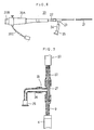
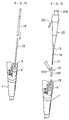
- Fig. 6 shows the daughter endoscope (20) having the insertion unit (21) with diameter of 0.8 mm, which can be placed into the guide tube (9) inserted into the insertion hole (4) of the mother endoscope (1).
- This daughter endoscope (20) also possesses the base (20A), an ocular unit (20B) and an optical cable (20C) connected with external light source equipment (not shown). It is also furnished with a fixing tube (22), and one end of T-shaped adaptor (23) is connected with this fixing tube (22). The length of the insertion unit (21) exposed from the other end of this T-shaped adaptor (23) is shorter than the length of the guide tube (9).
- a lure lock (25) is connected to the other end of the tube (24).
- a lure lock As shown in Fig. 5, it is possible to inject physiological saline into the guide tube (9) from this lure lock, using the syringe (12).
- An application example of such daughter endoscope (20) is given in Fig. 7. Specifically, the guide tube (9) containing core bar (11) is inserted to the desired position in bile duct (103) or pancreatic duct (102). Then, the mounting unit (10) is removed and the core bar (11) is withdrawn, and the insertion unit (21) of the daughter endoscope (20) is placed into the guide tube (9).
- T-shaped adaptor (23) To the base protruding from the insertion hole (4) of the guide tube (9), the other end of T-shaped adaptor (23) is connected.
- T-shaped adaptor (23) When T-shaped adaptor (23) is connected with the guide tube (9), the length of the insertion unit (21) is shorter than the guide tube (9). Accordingly, the tip of the insertion unit (21) is roughly aligned with the tip of the guide tube (9) or a little recessed, and the tip of the insertion unit (21) does not protrude from the tip of the guide tube (9). Since there is a gap between the insertion unit (21) and the guide tube (9), it is possible to supply physiological saline into this gap through the lure lock (25) and the tube (24).
- the daughter endoscope as used here needs not to have the function to bend or to provide the passage or forceps channel, and it needs only the function to observe. Therefore, the insertion unit (21) can be formed thinly as practical as possible (as thin as 0.8 mm), and it is also possible to provide a gap between the guide tube (9) and the insertion unit (21) to supply saline.
- the invention removably mount the flexible core bar (11) on one end of the flexible guide tube, and the core bar is of such length that its tip does not protrude from the guide tube (9).
- the tip of the guide tube (9) containing core bar (11) is protruded from the tip of the insertion unit (2) which has been placed into duodenum (100) through forceps channel (3) from the insertion hole (4) of mother endoscope (1). Then, the tip of the guide tube (9) can be correctly directed toward the papilla (101) by manipulating the forceps stand (8), and the insertion into the papilla (101) is also easily achievable because core bar (11) is contained.
- the flexible guide tube (9) Since it is the flexible guide tube (9) which comes into direct contact with the papilla (101) and with bile duct or pancreatic duct (103) or (102), there is no danger to injure them. Because the guide tube (9) contains core bar (11) in it, it is neither twisted nor turned within the forceps channel (3) and can be inserted smoothly into bile duct or pancreastic duct (103) or (102). In fluoroscopy, the presence of the core bar (11) makes it possible to find out how far the guide tube (9) has been inserted. Further, it is possible to supply physiological saline through the guide tube (9) into bile duct or pancreatic duct (103) or (102).
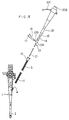
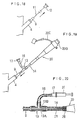
- Fig. 8 shows schematically the entire unit based on the second embodiment of the invention, which consists of a mother endoscope having the insertion unit (2) to be inserted as far as duodenum (100), a daughter endoscope furnished with an insertion unit (21) to be inserted into bile duct or pancreatic duct (103) or (102) from the insertion hole (4) of the forceps channel (3), and with a guide tube having smaller diameter.
- the tip of the insertion unit (2) which can be inserted to duodenum (100), is provided with an image guide window (5), a light guide window (6) and a vent and water hole (7), and with a forceps elevator (8) mounted on the forceps channel (3).
- bile duct or pancreatic duct (103) or (102) is observed by the mother endoscope (1), the guide tube (9) and the daughter endoscope (20), the guide tube (9) is inserted into the forceps channel (3) from the insertion hole (4) of the mother endoscope (1).
- the mounting unit (10) is mounted on the base end of this guide tube (9), and the flexible core bar (11) is mounted on this mounting unit (10).
- the tip of the core bar (11) is designed in such manner as not to protrude from the tip of the guide tube (9).
- the papilla (101) of duodenum (100) is observed (see Fig.
- pancreatic duct (102) or bile duct (103) When pancreatic duct (102) or bile duct (103) is photographed, the presence of the core bar (11) makes it possible to see how far the guide tube (7) has been inserted into the pancreatic duct (102) or bile duct (103). Also, it is possible to inject contrast medium into pancreatic duct (103) or bile duct (102) through the guide tube (9).
- the diameter of the guide tube (9) inserted into the pancreatic duct (103) or bile duct (102) is 1.7 mm.
- Core bar (11) is placed in the guide tube (9) because it contributes to the prevention of twisting in the forceps channel (3), to the easier insertion of guide tube (9) into the papilla (101) and also to the positive confirmation of the position of the tip by the image of core bar (11) by fluoroscopy.
- the guide tube (9) is formed with such flexibility that it can be easily inserted into papilla (101) and if metal powder is coated for clear imaging of the tip of the guide tube (9) by fluoroscopy, there is no need to insert the core bar (11).
- the guide tube (9) When the guide tube (9) is placed from the insertion hole (4) of the mother endoscope (1) into forceps channel (3) and the tip of the guide tube (9) is inserted into pancreatic duct (102), the mounting unit (10) is removed and the core bar (11) is withdrawn from the guide tube (9). Then, the insertion unit (21) without the passage for saline or the forceps channel of the daughter endoscope (20) is inserted into the guide tube (9).
- an adaptor (13) is furnished on the daughter endoscope to prevent the insertion unit (21) from excessive insertion.
- the adaptor (13) consists of a straight tube (13A) and a cross tube (13B), which is perpendicular to and connected with the straight tube (13A).
- One end of the straight tube (13A) is connected with a fixing tube (14) with smaller diameter selected from the fixing tubes (14) and (15) mounted on the daughter endoscope (20), and the other end of it is connected to the base end of the guide tube (9) (See Figs. 8 and 10.).
- the cross tube (13B) of the adaptor (13) is connected with the tube (16), and physiological saline is supplied through this tube (16).
- Physiological saline is injected by the syringe, which is connected by the lure lock (17) mounted on the other end of the tube (16), and saline is sent into bile duct or pancreatic duct (103) or (102) through the gap between the insertion unit (21) and the guide tube (9) passing through the tube (16), the cores tube (13B) and the straight tube (13A).
- the fixing tubes (14) and (15) are concentrically provided, and the fixing tube (15) with larger diameter is to be used when another guide tube (18) and the adaptor (19) are used. This will be described later.
- Both tubes (9) and (29) are inserted tinto the forceps channel (3′) and are taken out from the tube (29) through the insertion hole (4′). Then, guided by both tubes (9) and (29), the guide tube (18) with larger diameter is inserted into bile duct and pancreatic duct (103) and (102) (See Fig. 14.). When the tip of the guide tube (18) is guided by the guide tube (9), already inserted into bile duct and pancreatic duct (103) and (102), the tubes (9) and (29) are withdrawn.
- the insertion unit (21) of the daughter endoscope (20) is inserted into the guide tube (18) through the adaptor (19) as shown in Fig. 15.
- One end of the straight tube (19A) of the adaptor (19) is connected to the fixing tube (15), and the other end to the guide tube (18).
- the details are as shown in Fig. 16, and the communicating portion of the cross tube (19B) is closer to the guide tube (18) than to the tip of the fixing tube (14).
- this adaptor (19) When this adaptor (19) is mounted on the daughter endoscope (20) and on the guide tube (18), it is designed in such manner that the tip of the insertion unit (21) is roughly aligned with the tip of the guide tube (18) and does not to protrude.
- physiological saline can be supplied from the cross tube (19B) of the adaptor (19).
- various types of tools can be accommodated such as forceps (30) as shown in Fig. 17.
- the endoscope (1′) which may be regarded as the mother of the mother endoscope (1), was used in this case, whereas it is possible to insert the guide tube (18) with larger diameter after inserting the guide tube (9) with smaller diameter, regarding the endoscope (1′) as the mother from the beginning.
- two or more guide tube (9) and (18) are used in the second invention, and after the guide tube (9) with smaller diameter is first inserted into bile duct and pancreastic duct (103) and (102), the insertion unit (21) of the daughter endoscope (20) is placed into this guide tube (9).
- the guide tube (18) with larger diameter can be smoothly inserted into bile duct and pancreatic duct (103) and (102) because it is guided by the guide tube (9) with smaller diameter.
- the guide tubes (9) and (18) can be connected with the fixing tubes (14) and (15) mounted on the base of the insertion unit (21) of the daughter endoscope (20) through the adaptor (19). If designed well in advance, the tip of the insertion unit (21) does not protrude from the tip of the guide tubes (9) or (18), and it is possible to supply physiological saline or to insert various types of tools from the adaptor into the guide tubes (9) or (18).
- the third embodiment is also provided with the same configuration as shown in Fig. 8. If the guide tube (9) is inserted to the desired position as shown in Fig. 3 in such endoscope system, the mounting unit (10) is removed and the core bar (11) is withdrawn from the guide tube (9) as shown in Fig. 18. Then, the insertion unit (21) of the daughter endoscope (20) is placed into the guide tube (9), which is protruding from the insertion hole (4) (See Fig. 19.). The other end of the straight tube (13A) of the adaptor (13) is then mounted on the guide tube (9). As shown in Fig. 8.
- the tip of the insertion unit (21) of the daughter endoscope (20) is roughly aligned with the tip of the insertion unit (21) of the daughter endoscope (20), and the tip of the insertion unit (21) does not protrude from the tip of the guide tube (9). With this adaptor (13) mounted on the guide tube (9), the insertion unit (21) cannot be inserted further into the guide tube (9).
- bile duct (103) or pancreatic duct (102) When bile duct (103) or pancreatic duct (102) is observed with the insertion unit (21) of the daughter endoscope (20) placed into the guide tube (9), the observation by the insertion unit (21) can be performed more conveniently and clearly if physiological saline is supplied to bile duct (103) or pancreatic duct (102) by such means as the syringe (31) through the tube (16) and the lure lock (17) mounted on the cross tube (13B) of the adaptor (13). Since the insertion unit (21) requires neither a passage for saline nor a mechanism to bend the tip nor a forceps channel, its outer diameter can be designed as small as 0.8 mm.
- the diameter of the forceps channel (3) of the mother endoscope (1) was set to 2.8 mm in this case, the diameter of the guide tube (9) to 1.7 mm, and the diameter of the insertion unit (21) to 0.8 mm.
- Outer diameter of the guide tube (9) used in the present invention can be set to about 1.7 mm.
- its tip can be smoothly inserted into the papilla (101) and also into pancreatic duct (102), which is smaller in diameter than bile duct (103).
- the third embodiment it is possible by the third embodiment to easily insert the guide tube (9), having such diameter as to facilitate the insertion into papilla, into bile duct (103) or pancreatic duct (102) through the papilla (101), and there is no danger to injure the papilla (101) and the like.
- the guide tube (9) is inserted into bile duct (102) or pancreatic duct (103) through papilla (101) in easy and safe manner, the insertion unit (21) can be easily guided into bile duct (102) or pancreatic duct (103) only by placing the insertion unit (21) of the daughter endoscope (20) into the guide tube (9).
- the adaptor (13) serves as a stopper.
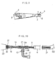
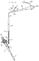
- the fourth embodiment according to the invention as shown in Figs. 21 to 32 consists of a mother endoscope (1) having a forceps channel (3) on the insertion unit (2) placed into duodenum (100), a first guide tube (32), inserted into the forceps channel (3) from the insertion hole (4) connected to the forceps channel (3) of the mother endoscope (1), a second guide tube (33), the diameter of which is larger than that of the first guide tube (32) and in which the first guide tube (32) can be inserted, and a daughter endoscope (20) having an insertion unit (21), which can be inserted into bile duct (103) or pancreatic duct (102).
- the insertion unit (2) of the mother endoscope (1) can be perorally inserted to duodenum (100), and inner diameter of the forceps channel (3) is set to 3.7 mm.
- the tip of the insertion unit (2) of this mother endoscope (1) is furnished with an image guide window (5), a light guide window (6), a vent and water hole (7) and a forceps elevator (8) mounted on the forceps channel (3).
- a mounting unit (10) is removably mounted, and the flexible core bar (11) to be inserted into the first guide tube (32) is mounted on this mounting unit (10).
- the tip of this core bar (11) is of such length that it does not protrude from the tip of the guide tube (32).
- the daughter endoscope (20) is provided with an ocular unit (20B) and an optical cable (20C) connected to the external light source equipment (not shown).
- a fixing tube (14) is mounted on the base, and one end of the adaptor (13) is mounted on this fixing tube (14).
- the adaptor (13) consists of a straight tube (13A) and a cross tube (13B) crossing with the straight tube (13A).
- the tube (16) is mounted on the cross tube (13B), and a lure lock (17) is mounted on this tube (16).
- the forceps channel (3) of the mother endoscope (1) has inner diameter of 3.7 mm
- the first guide tube (32) has outer diameter of 1.7 mm.
- the first guide tube (32) is passed through the forceps channel (3) from the insertion hole (4), and its tip is inserted into papilla (101) by manipulating the forceps elevator (8).
- the core bar (11) can be clearly identified by fluoroscopy. Also, it is possible to photograph by injecting contrast medium into the first guide tube (32).
- the mounting unit (10) is removed and the core bar (11) is withdrawn, leaving only the first guide tube (32) as shown in Fig. 22.
- the first guide tube (32) is connected with the junction tube (29) having the same diameter through a joint (see Fig. 23.). Guiding the flexible second guide tube (33) by the first guide tube (32) as shown in Fig. 24, the second guide tube (33) is inserted into bile duct (103) or pancreatic duct (102).
- the second guide tube (33) When the second guide tube (33) is inserted into bile duct (103) and pancreatic duct (102) in this way, the first guide tube (32) and the junction tube (29) are withdrawn as shown in Fig. 25. Then, the insertion unit (21) of the daughter endoscope (20) is placed into the second guide tube (33). The insertion unit (21) is stopped when the other end of the straight tube (13A) of the adaptor (13) is mounted on the second guide tube (33). In this status, as shown in Fig. 27, the tip of the insertion unit (21) is roughly aligned with the tip of the second guide tube (33) and does not protrude.
- the gap between the fixing tube (14) mounted on the base of the daughter endoscope (20) and the insertion unit (21) is closed up by the enclosure ring (26). Accordingly, even when fluid or tool is placed in the gap between the second guide tube (33) and the insertion unit (21) by means of the cross tube (13B), the fluid will not flow along toward the operator because it is blocked by the enclosure ring (26).
- inner diameter of the forceps channel (3) of the mother endoscope was set to 3.7 mm.
- the first guide tube (32) with diameter of 1.7 mm is placed into the forceps channel (3) with inner diameter of 3.7 mm and is inserted into bile duct (103) or pancreatic duct (102)
- the insertion will be difficult because there is too much idle space between the first guide tube (32) and the forceps channel (3).
- the first mother endoscope (1′) having the forceps channel (3) with inner diameter of 2.8 mm is used as shown in Fig.
- the first guide tube (32) is inserted from the insertion hole (4′) into bile duct (103) and pancreatic duct (102) according to the procedure as described above, and the junction tube (29) is connected to the first guide tube (32) protruding from the insertion hole (4′) through the joint (28).
- the junction tube (29) is connected, the first endoscope (1′) is withdrawn from the first guide tube (32) and the junction tube (29). Then, it is inserted into the tip of the insertion unit (2) of the mother endoscope (1) (the second mother endoscope) through the forceps channel with inner diameter of 3.7 mm, and this insertion unit (2) is inserted up to duodenum (100) (See Fig. 29.).
- the second guide tube (33) is inserted into bile duct (102) or pancreatic duct (103), being guided by the junction tube (29) and the first guide tube (32).
- the subsequent procedure is as described above.
- the mother endoscope (1) having the forceps channel (3) with inner diameter of 3.7 mm is inserted into duodenum (100)
- the forceps (30) are inserted from the cross tube (13B) of the adaptor (13).
- the forceps (30) are placed into pancreatic duct (103) through the gap between the second guide tube (33) and the insertion unit (21). It is shown in Fig. 30.
- the adaptor both ends of which are mounted on the fixing tube (14) and the second guide tube (33), may be provided with respect to the straight tube (13A) of the cross tube (13B) as shown in Fig. 31 and Fig. 32, whether it be a Y-shaped unit with a 3-way path in it or not, or two or more cross tubes (13B) may be provided.
- the forceps (30) or other tools are inserted from one of the cross tubes (13B), and physiological saline may be injected from the other cross tube (13B).
- the first guide tube (32) with smaller diameter is first placed into bile duct (103) or pancreatic duct (102) through the papilla (101) by the fourth invention, and the first guide tube (32) with smaller diameter is connected with the junction tube (29). Then, guided by these tubes (29) and (32), the second guide tube (33) with larger diameter is inserted into bile duct (103) or pancreatic duct (102). Therefore, the insertion is easily achievable and the flexibility of the first and second guide tubes (32) and (33) prevent bile duct (103) or pancreatic duct (102) as well as papilla (101) from injury.
- the second guide tube (33) can be inserted into bile duct (103) or pancreatic duct (102), guided by the first guide tube (32) and the junction tube (29). Since the first guide tube (32) is already inserted into bile duct (103) or pancreatic duct (102) in such case, even the second guide tube (33) with larger diameter can be smoothly inserted into bile duct (103) or pancreatic duct (102). After the second guide tube (33) is inserted, the first guide tube (32) and the junction tube (29) are withdrawn, and the other end of the adaptor (13) of the daughter endoscope (20) is mounted on this second guide tube (33).
Landscapes
- Health & Medical Sciences (AREA)
- Life Sciences & Earth Sciences (AREA)
- Surgery (AREA)
- Biomedical Technology (AREA)
- Medical Informatics (AREA)
- Optics & Photonics (AREA)
- Pathology (AREA)
- Radiology & Medical Imaging (AREA)
- Biophysics (AREA)
- Engineering & Computer Science (AREA)
- Physics & Mathematics (AREA)
- Heart & Thoracic Surgery (AREA)
- Nuclear Medicine, Radiotherapy & Molecular Imaging (AREA)
- Molecular Biology (AREA)
- Animal Behavior & Ethology (AREA)
- General Health & Medical Sciences (AREA)
- Public Health (AREA)
- Veterinary Medicine (AREA)
- Endoscopes (AREA)
- Media Introduction/Drainage Providing Device (AREA)
Claims (4)
- Endoskop zur Untersuchung der Gallen- und Pankreasgänge mit einem Mutterendoskop (1) und einem Tochterendoskop (20), das eine Einführungseinheit (21) hat, wobei das Mutterendoskop eine Einführungseinheit (2) zur Einführung in einen Zwölffingerdarm, einen Zangenkanal (3) in der Einführungseinheit (2) und eine Einführungsöffnung (4) hat, die mit dem Zangenkanal (3) in Verbindung steht,
gekennzeichnet durch
ein flexibles Führungsrohr (9), das eine Flexibilität hat, um von der Papille durch den Zangenkanal (3) von dessen Einführungsöffnung (4) in einen Gallengang und einen Pankreasgang eingeführt zu werden, und in das die Einführungseinheit (21) des Tochterendoskops einführbar ist, und einen flexiblen Kernstab (11), der lösbar an einem Ende des Führungsrohrs (9) befestigt und darin aufgenommen ist und an seinem von dem einen Ende des Führungsrohrs entfernten Ende eine Spitze hat, die eine solche Länge hat, daß sie nicht über das Rohr (9) hinaus vorsteht. - Endoskop nach Anspruch 1,
ferner gekennzeichnet durch ein Befestigungsrohr (14) zur Befestigung an dem Basisende der Einführungseinheit (21) des Tochterendoskops (20) und einen Adapter (13), dessen jeweilige Enden mit dem Befestigungsrohr (14) und dem Führungsrohr (9) übereinstimmen und in den ein Ende der Einführungseinheit (21) des Tochterendoskops (20) eingeführt wird, wobei das andere Ende der Einführungseinheit (21) des Tochterendoskops eine freie Spitze ist und der Adapter (13) so bemessen ist, daß er nicht in das Führungsrohr (9) paßt, und die Spitze der Einführungseinheit (21) des Tochterendoskops (20) grob auf die Spitze des entfernten Endes des Führungsrohres (9) ausgerichtet ist und nicht über diese übersteht, wenn der Adapter (13) an dem Führungsrohr (9) anliegt. - Endoskop nach Anspruch 1 oder 2,
ferner gekennzeichnet durch ein flexibles erstes Führungsrohr (32), das von der Einführungsöffnung des Mutterendoskops (1) durch den Zangenkanal (3) in den Gallengang oder Pankreasgang einsetzbar ist, ein Verbindungsrohr (29), das mit dem ersten Führungsrohr (32) verbunden ist, ein flexibles zweites Führungsrohr (33), das einen größeren Durchmesser hat als das erste Führungsrohr (32) und in den Gallengang oder Pankreasgang einsetzbar ist und von dem Verbindungsrohr (29) und dem ersten Führungsrohr (32) geführt wird,
ein Befestigungsrohr (14), das an dem Basisende der Einführungseinheit befestigbar ist und in das zweite Führungsrohr (33) des Tochterendoskops (20) eingeführt wird,
eine Endoskopeinheit (26), die an dem Basisende der Einführungseinheit (21) des Tochterendoskops (20) befestigt ist, um den Zwischenraum zwischen dem Befestigungsrohr (14) und der Einführungseinheit (21) des Tochterendoskops zu verschließen und einen Adapter (13), in dem sich wenigstens eine Drei-Wege-Bahn befindet, wobei der Adapter (13) ein geradeaus führendes Rohr (13A) und wenigstens ein querverlaufendes Rohr (13B) enthält, das mit dem geradeaus verlaufenden Rohr (13A) in Verbindung steht, wobei die jeweiligen Enden des geradeaus verlaufenden Rohres (13A) des Adapters (13) an dem zweiten Führungsrohr (33) und dem Befestigungsrohr (14) befestigt sind, nachdem das erste Führungsrohr (32) und das Verbindungsrohr (29) von dem zweiten Führungsrohr (33) zurückgezogen sind, wobei das letztere von dem ersten Führungsrohr (32) geführt und bin den Gallengang oder Pankreasgang eingeführt wurde, und wobei chirurgische Werkzeuge wie Zangen (30) oder physiologische Salzlösung oder dergleichen von dem Querrohr (13) des Adapters durch das zweite Führunsrohr (33) in den Gallengang oder Pankreasgang einführbar oder injizierbar sind. - Endoskop nach jedem der Ansprüche 1 bis 3,
ferner gekennzeichnet durch mehrere alternative flexible Führungsrohre (32, 33) unterschiedlicher Durchmesser zum Führen der Einführungseinheit (21) des Tochterendoskops (20) durch den Zangenkanal (3) des Mutterendoskops (1) in den Gallengang oder Pankreasgang,
mehrere alternative Adaptoren (13) verschiedener Durchmesser, wobei jeder Adapter (13) rohrförmig ist und gegenüberliegende Enden hat, und jeder Adapter an einem der gegenüberliegenden Enden zu dem jeweiligen Führungsrohr paßt,
und mehrere konzentrische Befestigungsrohre (14) unterschiedlicher Durchmesser, wobei jedes Befestigungsrohr zu dem anderen gegenüberliegenden Ende eines jeweiligen Adapters paßt und dadurch durch den Adapter die Einführungseinheit (21) des Tochterendoskops (20) mit dem Rest des Tochterendoskops (20) verbindet.
Applications Claiming Priority (8)
| Application Number | Priority Date | Filing Date | Title |
|---|---|---|---|
| JP63083590A JPH01254137A (ja) | 1988-04-05 | 1988-04-05 | 胆・膵管用内視鏡装置 |
| JP83587/88 | 1988-04-05 | ||
| JP63083589A JPH01254136A (ja) | 1988-04-05 | 1988-04-05 | 胆・膵管用内視鏡装置 |
| JP63083588A JPH01254135A (ja) | 1988-04-05 | 1988-04-05 | 胆・膵管用内視鏡装置 |
| JP83590/88 | 1988-04-05 | ||
| JP63083587A JPH01254134A (ja) | 1988-04-05 | 1988-04-05 | 胆・膵管用内視鏡装置 |
| JP83588/88 | 1988-04-05 | ||
| JP83589/89 | 1988-04-05 |
Publications (2)
| Publication Number | Publication Date |
|---|---|
| EP0336399A1 EP0336399A1 (de) | 1989-10-11 |
| EP0336399B1 true EP0336399B1 (de) | 1995-11-02 |
Family
ID=27466844
Family Applications (1)
| Application Number | Title | Priority Date | Filing Date |
|---|---|---|---|
| EP89105987A Expired - Lifetime EP0336399B1 (de) | 1988-04-05 | 1989-04-05 | Endoskop zur Untersuchung der Gallen- und Pankreasgänge |
Country Status (2)
| Country | Link |
|---|---|
| US (1) | US4979496A (de) |
| EP (1) | EP0336399B1 (de) |
Families Citing this family (85)
| Publication number | Priority date | Publication date | Assignee | Title |
|---|---|---|---|---|
| US7794424B2 (en) * | 1991-07-15 | 2010-09-14 | Paskar Larry D | Catheter with out-of-plane configurations |
| US5857996A (en) * | 1992-07-06 | 1999-01-12 | Catheter Imaging Systems | Method of epidermal surgery |
| US5342299A (en) * | 1992-07-06 | 1994-08-30 | Catheter Imaging Systems | Steerable catheter |
| US5486154A (en) * | 1993-06-08 | 1996-01-23 | Kelleher; Brian S. | Endoscope |
| US5746692A (en) * | 1994-05-05 | 1998-05-05 | Imagen Medical, Inc. | Catheter and endoscope system with distal protruding ball tip and method |
| US5662585A (en) * | 1994-05-05 | 1997-09-02 | Imagyn Medical, Inc. | Endoscope with protruding member and method of utilizing the same |
| US5807236A (en) * | 1994-05-05 | 1998-09-15 | Imagyn Medical Inc. | Catheter with guidewire and rounded enlargement and method |
| US5495851A (en) * | 1995-03-23 | 1996-03-05 | Roanoke Gastroenterology, P.C. | Use of endoscopic ultrasound and stimulated bilary drainage in the diagnosis of cholecystitis and microlithiasis |
| USD405881S (en) | 1996-01-16 | 1999-02-16 | Catheter Imaging Systems, Inc. | Handle for steerable catheter |
| US5860953A (en) * | 1995-11-21 | 1999-01-19 | Catheter Imaging Systems, Inc. | Steerable catheter having disposable module and sterilizable handle and method of connecting same |
| US6007531A (en) * | 1995-11-21 | 1999-12-28 | Catheter Imaging Systems, Inc. | Steerable catheter having disposable module and sterilizable handle and method of connecting same |
| USD398986S (en) | 1996-01-16 | 1998-09-29 | Catheter Imaging Systems, Inc. | Handle interface for steerable catheter |
| US5807239A (en) * | 1996-05-17 | 1998-09-15 | Conceptus, Inc. | Transcervical ostium access device and method |
| US6328730B1 (en) * | 1999-03-26 | 2001-12-11 | William W. Harkrider, Jr. | Endoluminal multi-luminal surgical sheath and method |
| US7555333B2 (en) | 2000-06-19 | 2009-06-30 | University Of Washington | Integrated optical scanning image acquisition and display |
| US20060178556A1 (en) | 2001-06-29 | 2006-08-10 | Intuitive Surgical, Inc. | Articulate and swapable endoscope for a surgical robot |
| US6817974B2 (en) | 2001-06-29 | 2004-11-16 | Intuitive Surgical, Inc. | Surgical tool having positively positionable tendon-actuated multi-disk wrist joint |
| US20060199999A1 (en) * | 2001-06-29 | 2006-09-07 | Intuitive Surgical Inc. | Cardiac tissue ablation instrument with flexible wrist |
| KR101087996B1 (ko) | 2002-12-06 | 2011-12-01 | 인튜어티브 서지컬 인코포레이티드 | 최소 침습 수술 기구 |
| US20040199052A1 (en) | 2003-04-01 | 2004-10-07 | Scimed Life Systems, Inc. | Endoscopic imaging system |
| EP1691666B1 (de) | 2003-12-12 | 2012-05-30 | University of Washington | Katheterskop-3d-führung und schnittstellensystem |
| US7922650B2 (en) * | 2004-03-23 | 2011-04-12 | Boston Scientific Scimed, Inc. | Medical visualization system with endoscope and mounted catheter |
| US11832793B2 (en) | 2004-03-23 | 2023-12-05 | Boston Scientific Scimed, Inc. | Vivo visualization system |
| US7922654B2 (en) | 2004-08-09 | 2011-04-12 | Boston Scientific Scimed, Inc. | Fiber optic imaging catheter |
| US20050222494A1 (en) * | 2004-04-06 | 2005-10-06 | Prescott James T | Dual-scope colonoscopy system with separate secondary colonoscope tool |
| WO2005110200A1 (en) * | 2004-05-13 | 2005-11-24 | Stryker Gi Ltd. | Connector for use with multilumen tubing |
| CN1984598A (zh) * | 2004-05-13 | 2007-06-20 | 斯特赖克Gi有限公司 | 用于切割和热封一次性多内腔管件的方法 |
| JP2008500110A (ja) * | 2004-05-25 | 2008-01-10 | ユー.エス. エンドスコピー グループ, インコーポレイテッド | 送達デバイス |
| US20060149127A1 (en) * | 2004-12-30 | 2006-07-06 | Seddiqui Fred R | Disposable multi-lumen catheter with reusable stylet |
| US8797392B2 (en) | 2005-01-05 | 2014-08-05 | Avantis Medical Sytems, Inc. | Endoscope assembly with a polarizing filter |
| US8872906B2 (en) | 2005-01-05 | 2014-10-28 | Avantis Medical Systems, Inc. | Endoscope assembly with a polarizing filter |
| US8289381B2 (en) | 2005-01-05 | 2012-10-16 | Avantis Medical Systems, Inc. | Endoscope with an imaging catheter assembly and method of configuring an endoscope |
| US8182422B2 (en) | 2005-12-13 | 2012-05-22 | Avantis Medical Systems, Inc. | Endoscope having detachable imaging device and method of using |
| US7530948B2 (en) | 2005-02-28 | 2009-05-12 | University Of Washington | Tethered capsule endoscope for Barrett's Esophagus screening |
| DE102005017204B4 (de) * | 2005-04-14 | 2012-03-22 | Lisa Laser Products Ohg Fuhrberg & Teichmann | Endoskop und Verfahren zum Einführen einer Lichtleitfaser in einen Arbeitskanal eines Endoskops |
| US20060235269A1 (en) * | 2005-04-15 | 2006-10-19 | The University Of Chicago | Method, apparatus and kit for bile or pancreatic duct endoscopy |
| US20070015989A1 (en) * | 2005-07-01 | 2007-01-18 | Avantis Medical Systems, Inc. | Endoscope Image Recognition System and Method |
| DE102005039601A1 (de) * | 2005-08-19 | 2007-02-22 | Karl Storz Gmbh & Co. Kg | Endoskop, insbesondere Duodenoskop für die Mutter-Baby Cholangioskopie |
| EP1951101A4 (de) * | 2005-11-08 | 2011-09-28 | Univ Columbia | Geräte und verfahren zur abgabe von einer oder mehreren substanzen und dergleichen in einen körper |
| US8537203B2 (en) | 2005-11-23 | 2013-09-17 | University Of Washington | Scanning beam with variable sequential framing using interrupted scanning resonance |
| WO2007063904A1 (ja) * | 2005-12-01 | 2007-06-07 | Olympus Medical Systems Corp. | ガイド用細長医療部材、及び細長医療装置 |
| WO2007087421A2 (en) | 2006-01-23 | 2007-08-02 | Avantis Medical Systems, Inc. | Endoscope |
| US7815564B2 (en) | 2006-02-21 | 2010-10-19 | Boston Scientific Scimed, Inc. | Positioning system for manipulating a channel within a medical device |
| US20070208220A1 (en) * | 2006-03-03 | 2007-09-06 | Wilson-Cook Medical Inc. | Endoscopic delivery apparatus having a catheter with radial grooves |
| EP1991314A2 (de) | 2006-03-03 | 2008-11-19 | University of Washington | Faseroptik-scanner mit multiumhüllung |
| US8287446B2 (en) | 2006-04-18 | 2012-10-16 | Avantis Medical Systems, Inc. | Vibratory device, endoscope having such a device, method for configuring an endoscope, and method of reducing looping of an endoscope |
| US7753843B2 (en) | 2006-05-09 | 2010-07-13 | Boston Scientific Scimed, Inc. | Medical device positioning system |
| US20070265494A1 (en) * | 2006-05-10 | 2007-11-15 | Boston Scientific Scimed Inc. | Flexible and retractable endoscope elevator |
| US20070265693A1 (en) * | 2006-05-15 | 2007-11-15 | Paskar Larry D | Coronary sinus catheter system and method |
| JP2009537284A (ja) | 2006-05-19 | 2009-10-29 | アヴァンティス メディカル システムズ インコーポレイテッド | 画像を作成しかつ改善するためのシステムおよび方法 |
| US7927272B2 (en) | 2006-08-04 | 2011-04-19 | Avantis Medical Systems, Inc. | Surgical port with embedded imaging device |
| US8096943B2 (en) * | 2006-12-04 | 2012-01-17 | University Of Washington Through Its Center For Commercialization | Flexible endoscope tip bending mechanism using optical fiber as compression member |
| US7655004B2 (en) | 2007-02-15 | 2010-02-02 | Ethicon Endo-Surgery, Inc. | Electroporation ablation apparatus, system, and method |
| US20080214890A1 (en) * | 2007-03-01 | 2008-09-04 | Olympus Medical Systems Corporation | Therapeutic method and therapeutic system used with steps for approaching to lesion using overtube |
| US8840566B2 (en) | 2007-04-02 | 2014-09-23 | University Of Washington | Catheter with imaging capability acts as guidewire for cannula tools |
| US8064666B2 (en) | 2007-04-10 | 2011-11-22 | Avantis Medical Systems, Inc. | Method and device for examining or imaging an interior surface of a cavity |
| WO2008137710A1 (en) | 2007-05-03 | 2008-11-13 | University Of Washington | High resolution optical coherence tomography based imaging for intraluminal and interstitial use implemented with a reduced form factor |
| WO2008141202A2 (en) * | 2007-05-11 | 2008-11-20 | Board Of Regents, The University Of Texas System | Medical scope carrier and scope as system and method |
| DE202007009713U1 (de) * | 2007-07-10 | 2007-09-06 | Karl Storz Gmbh & Co. Kg | Chirurgisches Instrumentensystem |
| US20090112063A1 (en) * | 2007-10-31 | 2009-04-30 | Bakos Gregory J | Endoscopic overtubes |
| US20090240143A1 (en) * | 2008-03-12 | 2009-09-24 | Mauna Kea Technologies | Method and an optical probe for in vivo imaging of a mucosa in a biliary or pancreatic system and a method for selectively operating a tissue sampling of a mucosa in a biliary or pancreatic system |
| US8888792B2 (en) | 2008-07-14 | 2014-11-18 | Ethicon Endo-Surgery, Inc. | Tissue apposition clip application devices and methods |
| JP5297732B2 (ja) * | 2008-09-12 | 2013-09-25 | オリンパスメディカルシステムズ株式会社 | 親子式内視鏡 |
| US20100125168A1 (en) * | 2008-11-14 | 2010-05-20 | Ethicon Endo-Surgery, Inc. | Methods and devices for endoscope control in a body cavity |
| US8157834B2 (en) | 2008-11-25 | 2012-04-17 | Ethicon Endo-Surgery, Inc. | Rotational coupling device for surgical instrument with flexible actuators |
| US8361066B2 (en) | 2009-01-12 | 2013-01-29 | Ethicon Endo-Surgery, Inc. | Electrical ablation devices |
| JP5313068B2 (ja) * | 2009-03-09 | 2013-10-09 | 富士フイルム株式会社 | 側視内視鏡装置 |
| US20110098704A1 (en) | 2009-10-28 | 2011-04-28 | Ethicon Endo-Surgery, Inc. | Electrical ablation devices |
| US9028483B2 (en) | 2009-12-18 | 2015-05-12 | Ethicon Endo-Surgery, Inc. | Surgical instrument comprising an electrode |
| US9233241B2 (en) | 2011-02-28 | 2016-01-12 | Ethicon Endo-Surgery, Inc. | Electrical ablation devices and methods |
| US9254169B2 (en) | 2011-02-28 | 2016-02-09 | Ethicon Endo-Surgery, Inc. | Electrical ablation devices and methods |
| WO2012125785A1 (en) | 2011-03-17 | 2012-09-20 | Ethicon Endo-Surgery, Inc. | Hand held surgical device for manipulating an internal magnet assembly within a patient |
| US9427255B2 (en) | 2012-05-14 | 2016-08-30 | Ethicon Endo-Surgery, Inc. | Apparatus for introducing a steerable camera assembly into a patient |
| US9078662B2 (en) | 2012-07-03 | 2015-07-14 | Ethicon Endo-Surgery, Inc. | Endoscopic cap electrode and method for using the same |
| US9545290B2 (en) | 2012-07-30 | 2017-01-17 | Ethicon Endo-Surgery, Inc. | Needle probe guide |
| US10314649B2 (en) | 2012-08-02 | 2019-06-11 | Ethicon Endo-Surgery, Inc. | Flexible expandable electrode and method of intraluminal delivery of pulsed power |
| US9572623B2 (en) | 2012-08-02 | 2017-02-21 | Ethicon Endo-Surgery, Inc. | Reusable electrode and disposable sheath |
| US9277957B2 (en) | 2012-08-15 | 2016-03-08 | Ethicon Endo-Surgery, Inc. | Electrosurgical devices and methods |
| US10098527B2 (en) | 2013-02-27 | 2018-10-16 | Ethidcon Endo-Surgery, Inc. | System for performing a minimally invasive surgical procedure |
| WO2014149734A1 (en) | 2013-03-15 | 2014-09-25 | Cook Medical Technolgoies Llc | Electrosurgical system with electrically active outer surface |
| US10500332B2 (en) * | 2015-11-03 | 2019-12-10 | Clph, Llc | Injection devices and systems and methods for using them |
| CN106932619A (zh) * | 2017-03-29 | 2017-07-07 | 国网新疆电力公司巴州供电公司 | 电表铅封安装辅助工具 |
| JP2021166562A (ja) * | 2018-04-16 | 2021-10-21 | オリンパス株式会社 | 医療デバイス、内視鏡システム |
| DE102020110835A1 (de) | 2020-04-21 | 2021-10-21 | Novatech Sa | Endoskopiereinrichtung und Endoskopierverfahren |
| CN120569152A (zh) * | 2022-10-12 | 2025-08-29 | 医疗机械人创新技术中心有限公司 | 一种用于一个或多个机器人系统的适配器 |
Family Cites Families (5)
| Publication number | Priority date | Publication date | Assignee | Title |
|---|---|---|---|---|
| JPS4831554B1 (de) * | 1968-12-24 | 1973-09-29 | ||
| FR2249641B3 (de) * | 1973-11-06 | 1977-08-05 | Wolf Gmbh Richard | |
| US4582067A (en) * | 1983-02-14 | 1986-04-15 | Washington Research Foundation | Method for endoscopic blood flow detection by the use of ultrasonic energy |
| DE3425427A1 (de) * | 1984-07-11 | 1986-01-16 | Olympus Winter & Ibe GmbH, 2000 Hamburg | Vorrichtung zur schaffung eines einfuehrungskanales fuer endoskope |
| US4586491A (en) * | 1984-12-14 | 1986-05-06 | Warner-Lambert Technologies, Inc. | Bronchoscope with small gauge viewing attachment |
-
1989
- 1989-04-03 US US07/332,859 patent/US4979496A/en not_active Expired - Lifetime
- 1989-04-05 EP EP89105987A patent/EP0336399B1/de not_active Expired - Lifetime
Also Published As
| Publication number | Publication date |
|---|---|
| US4979496A (en) | 1990-12-25 |
| EP0336399A1 (de) | 1989-10-11 |
Similar Documents
| Publication | Publication Date | Title |
|---|---|---|
| EP0336399B1 (de) | Endoskop zur Untersuchung der Gallen- und Pankreasgänge | |
| US5197457A (en) | Deformable and removable sheath for optical catheter | |
| JP6173917B2 (ja) | 光学案内型の医療用チューブおよび制御ユニットの組立品ならびに使用方法 | |
| US7883459B2 (en) | Endoscope and method for repairing the same | |
| DE69321963T2 (de) | Sterilisierbares endoskop mit einer trennbaren wegwerfbaren rohranordnung | |
| JPH0221041Y2 (de) | ||
| EP3490425B1 (de) | Flexibles endoskop | |
| EP2412297A1 (de) | Behandlungsendoskop mit haube und haube für das endoskop | |
| JPH0442930B2 (de) | ||
| WO2004026125A1 (en) | Endoscope | |
| CN115397300B (zh) | 用于医疗设备的远侧尖端 | |
| CN106793920A (zh) | 用于输尿管镜的附接装置 | |
| JPWO2015146652A1 (ja) | 腹腔内視鏡装置及び内視鏡システム | |
| CN109068963B (zh) | 器具插入辅助工具 | |
| WO2015142787A1 (en) | Multi-stage instrument connector | |
| CN112788977A (zh) | 内窥镜弯曲部 | |
| KR20200048692A (ko) | 내시경 일체형의 일회용 경막외 수술 장치 | |
| JP3034641B2 (ja) | 内視鏡 | |
| JP2018175291A (ja) | シースセット及び内視鏡システム | |
| JP2970887B2 (ja) | 内視鏡用ガイドカテーテル | |
| US20200100654A1 (en) | Minimal invasive medical device | |
| US12133630B2 (en) | Surgical sheath system | |
| JPH0438413B2 (de) | ||
| JPH0438412B2 (de) | ||
| JP2000014627A (ja) | 内視鏡 |
Legal Events
| Date | Code | Title | Description |
|---|---|---|---|
| PUAI | Public reference made under article 153(3) epc to a published international application that has entered the european phase |
Free format text: ORIGINAL CODE: 0009012 |
|
| AK | Designated contracting states |
Kind code of ref document: A1 Designated state(s): AT BE CH DE ES FR GB GR IT LI LU NL SE |
|
| RBV | Designated contracting states (corrected) |
Designated state(s): DE |
|
| 17P | Request for examination filed |
Effective date: 19900305 |
|
| 17Q | First examination report despatched |
Effective date: 19931202 |
|
| GRAA | (expected) grant |
Free format text: ORIGINAL CODE: 0009210 |
|
| AK | Designated contracting states |
Kind code of ref document: B1 Designated state(s): DE |
|
| REF | Corresponds to: |
Ref document number: 68924663 Country of ref document: DE Date of ref document: 19951207 |
|
| PLBE | No opposition filed within time limit |
Free format text: ORIGINAL CODE: 0009261 |
|
| STAA | Information on the status of an ep patent application or granted ep patent |
Free format text: STATUS: NO OPPOSITION FILED WITHIN TIME LIMIT |
|
| 26N | No opposition filed | ||
| PGFP | Annual fee paid to national office [announced via postgrant information from national office to epo] |
Ref country code: DE Payment date: 20030417 Year of fee payment: 15 |
|
| PG25 | Lapsed in a contracting state [announced via postgrant information from national office to epo] |
Ref country code: DE Free format text: LAPSE BECAUSE OF NON-PAYMENT OF DUE FEES Effective date: 20041103 |