EP0163249A2 - Gentechnologisches Verfahren zur Herstellung von Human-Interleukin-2 und Mittel zur Durchführung dieses Verfahrens - Google Patents
Gentechnologisches Verfahren zur Herstellung von Human-Interleukin-2 und Mittel zur Durchführung dieses Verfahrens Download PDFInfo
- Publication number
- EP0163249A2 EP0163249A2 EP85106279A EP85106279A EP0163249A2 EP 0163249 A2 EP0163249 A2 EP 0163249A2 EP 85106279 A EP85106279 A EP 85106279A EP 85106279 A EP85106279 A EP 85106279A EP 0163249 A2 EP0163249 A2 EP 0163249A2
- Authority
- EP
- European Patent Office
- Prior art keywords
- amino acids
- dna sequence
- terminal end
- sequence
- gene
- Prior art date
- Legal status (The legal status is an assumption and is not a legal conclusion. Google has not performed a legal analysis and makes no representation as to the accuracy of the status listed.)
- Granted
Links
- 238000000034 method Methods 0.000 title claims abstract description 21
- 101001002657 Homo sapiens Interleukin-2 Proteins 0.000 title claims abstract description 7
- 102000055277 human IL2 Human genes 0.000 title claims abstract description 6
- 108090000623 proteins and genes Proteins 0.000 claims abstract description 59
- 239000013612 plasmid Substances 0.000 claims abstract description 56
- 108091028043 Nucleic acid sequence Proteins 0.000 claims abstract description 46
- 238000010353 genetic engineering Methods 0.000 claims abstract description 10
- 150000001413 amino acids Chemical class 0.000 claims description 40
- 108010002350 Interleukin-2 Proteins 0.000 claims description 35
- 102000000588 Interleukin-2 Human genes 0.000 claims description 34
- 239000002773 nucleotide Substances 0.000 claims description 24
- 125000003729 nucleotide group Chemical group 0.000 claims description 24
- 102000004169 proteins and genes Human genes 0.000 claims description 23
- 108020004705 Codon Proteins 0.000 claims description 17
- 108091034117 Oligonucleotide Proteins 0.000 claims description 15
- 108020004414 DNA Proteins 0.000 claims description 14
- 108010054576 Deoxyribonuclease EcoRI Proteins 0.000 claims description 13
- JLCPHMBAVCMARE-UHFFFAOYSA-N [3-[[3-[[3-[[3-[[3-[[3-[[3-[[3-[[3-[[3-[[3-[[5-(2-amino-6-oxo-1H-purin-9-yl)-3-[[3-[[3-[[3-[[3-[[3-[[5-(2-amino-6-oxo-1H-purin-9-yl)-3-[[5-(2-amino-6-oxo-1H-purin-9-yl)-3-hydroxyoxolan-2-yl]methoxy-hydroxyphosphoryl]oxyoxolan-2-yl]methoxy-hydroxyphosphoryl]oxy-5-(5-methyl-2,4-dioxopyrimidin-1-yl)oxolan-2-yl]methoxy-hydroxyphosphoryl]oxy-5-(6-aminopurin-9-yl)oxolan-2-yl]methoxy-hydroxyphosphoryl]oxy-5-(6-aminopurin-9-yl)oxolan-2-yl]methoxy-hydroxyphosphoryl]oxy-5-(6-aminopurin-9-yl)oxolan-2-yl]methoxy-hydroxyphosphoryl]oxy-5-(6-aminopurin-9-yl)oxolan-2-yl]methoxy-hydroxyphosphoryl]oxyoxolan-2-yl]methoxy-hydroxyphosphoryl]oxy-5-(5-methyl-2,4-dioxopyrimidin-1-yl)oxolan-2-yl]methoxy-hydroxyphosphoryl]oxy-5-(4-amino-2-oxopyrimidin-1-yl)oxolan-2-yl]methoxy-hydroxyphosphoryl]oxy-5-(5-methyl-2,4-dioxopyrimidin-1-yl)oxolan-2-yl]methoxy-hydroxyphosphoryl]oxy-5-(5-methyl-2,4-dioxopyrimidin-1-yl)oxolan-2-yl]methoxy-hydroxyphosphoryl]oxy-5-(6-aminopurin-9-yl)oxolan-2-yl]methoxy-hydroxyphosphoryl]oxy-5-(6-aminopurin-9-yl)oxolan-2-yl]methoxy-hydroxyphosphoryl]oxy-5-(4-amino-2-oxopyrimidin-1-yl)oxolan-2-yl]methoxy-hydroxyphosphoryl]oxy-5-(4-amino-2-oxopyrimidin-1-yl)oxolan-2-yl]methoxy-hydroxyphosphoryl]oxy-5-(4-amino-2-oxopyrimidin-1-yl)oxolan-2-yl]methoxy-hydroxyphosphoryl]oxy-5-(6-aminopurin-9-yl)oxolan-2-yl]methoxy-hydroxyphosphoryl]oxy-5-(4-amino-2-oxopyrimidin-1-yl)oxolan-2-yl]methyl [5-(6-aminopurin-9-yl)-2-(hydroxymethyl)oxolan-3-yl] hydrogen phosphate Polymers Cc1cn(C2CC(OP(O)(=O)OCC3OC(CC3OP(O)(=O)OCC3OC(CC3O)n3cnc4c3nc(N)[nH]c4=O)n3cnc4c3nc(N)[nH]c4=O)C(COP(O)(=O)OC3CC(OC3COP(O)(=O)OC3CC(OC3COP(O)(=O)OC3CC(OC3COP(O)(=O)OC3CC(OC3COP(O)(=O)OC3CC(OC3COP(O)(=O)OC3CC(OC3COP(O)(=O)OC3CC(OC3COP(O)(=O)OC3CC(OC3COP(O)(=O)OC3CC(OC3COP(O)(=O)OC3CC(OC3COP(O)(=O)OC3CC(OC3COP(O)(=O)OC3CC(OC3COP(O)(=O)OC3CC(OC3COP(O)(=O)OC3CC(OC3COP(O)(=O)OC3CC(OC3COP(O)(=O)OC3CC(OC3COP(O)(=O)OC3CC(OC3CO)n3cnc4c(N)ncnc34)n3ccc(N)nc3=O)n3cnc4c(N)ncnc34)n3ccc(N)nc3=O)n3ccc(N)nc3=O)n3ccc(N)nc3=O)n3cnc4c(N)ncnc34)n3cnc4c(N)ncnc34)n3cc(C)c(=O)[nH]c3=O)n3cc(C)c(=O)[nH]c3=O)n3ccc(N)nc3=O)n3cc(C)c(=O)[nH]c3=O)n3cnc4c3nc(N)[nH]c4=O)n3cnc4c(N)ncnc34)n3cnc4c(N)ncnc34)n3cnc4c(N)ncnc34)n3cnc4c(N)ncnc34)O2)c(=O)[nH]c1=O JLCPHMBAVCMARE-UHFFFAOYSA-N 0.000 claims description 11
- 241000588724 Escherichia coli Species 0.000 claims description 10
- 230000004071 biological effect Effects 0.000 claims description 10
- 210000004899 c-terminal region Anatomy 0.000 claims description 8
- 238000004519 manufacturing process Methods 0.000 claims description 8
- 108700005078 Synthetic Genes Proteins 0.000 claims description 7
- 238000003776 cleavage reaction Methods 0.000 claims description 2
- 230000007017 scission Effects 0.000 claims description 2
- 125000003275 alpha amino acid group Chemical group 0.000 claims 7
- 239000012634 fragment Substances 0.000 abstract description 23
- WYURNTSHIVDZCO-UHFFFAOYSA-N Tetrahydrofuran Chemical compound C1CCOC1 WYURNTSHIVDZCO-UHFFFAOYSA-N 0.000 description 24
- 108091008146 restriction endonucleases Proteins 0.000 description 19
- 108090000765 processed proteins & peptides Proteins 0.000 description 14
- 102000004196 processed proteins & peptides Human genes 0.000 description 13
- OKKJLVBELUTLKV-UHFFFAOYSA-N Methanol Chemical compound OC OKKJLVBELUTLKV-UHFFFAOYSA-N 0.000 description 12
- YLQBMQCUIZJEEH-UHFFFAOYSA-N tetrahydrofuran Natural products C=1C=COC=1 YLQBMQCUIZJEEH-UHFFFAOYSA-N 0.000 description 12
- 229920001184 polypeptide Polymers 0.000 description 11
- 230000015572 biosynthetic process Effects 0.000 description 10
- 210000004027 cell Anatomy 0.000 description 10
- 238000003786 synthesis reaction Methods 0.000 description 10
- WEVYAHXRMPXWCK-UHFFFAOYSA-N Acetonitrile Chemical compound CC#N WEVYAHXRMPXWCK-UHFFFAOYSA-N 0.000 description 8
- 239000013598 vector Substances 0.000 description 7
- OISVCGZHLKNMSJ-UHFFFAOYSA-N 2,6-dimethylpyridine Chemical compound CC1=CC=CC(C)=N1 OISVCGZHLKNMSJ-UHFFFAOYSA-N 0.000 description 6
- 108091033380 Coding strand Proteins 0.000 description 6
- VHJLVAABSRFDPM-QWWZWVQMSA-N dithiothreitol Chemical compound SC[C@@H](O)[C@H](O)CS VHJLVAABSRFDPM-QWWZWVQMSA-N 0.000 description 6
- 238000010348 incorporation Methods 0.000 description 6
- 238000003780 insertion Methods 0.000 description 6
- 230000037431 insertion Effects 0.000 description 6
- XLYOFNOQVPJJNP-UHFFFAOYSA-N water Chemical compound O XLYOFNOQVPJJNP-UHFFFAOYSA-N 0.000 description 6
- TWRXJAOTZQYOKJ-UHFFFAOYSA-L Magnesium chloride Chemical compound [Mg+2].[Cl-].[Cl-] TWRXJAOTZQYOKJ-UHFFFAOYSA-L 0.000 description 5
- 239000000499 gel Substances 0.000 description 5
- WFDIJRYMOXRFFG-UHFFFAOYSA-N Acetic anhydride Chemical compound CC(=O)OC(C)=O WFDIJRYMOXRFFG-UHFFFAOYSA-N 0.000 description 4
- QGZKDVFQNNGYKY-UHFFFAOYSA-N Ammonia Chemical compound N QGZKDVFQNNGYKY-UHFFFAOYSA-N 0.000 description 4
- 102000012410 DNA Ligases Human genes 0.000 description 4
- 108010061982 DNA Ligases Proteins 0.000 description 4
- 102000004190 Enzymes Human genes 0.000 description 4
- 108090000790 Enzymes Proteins 0.000 description 4
- XSQUKJJJFZCRTK-UHFFFAOYSA-N Urea Chemical compound NC(N)=O XSQUKJJJFZCRTK-UHFFFAOYSA-N 0.000 description 4
- 230000001580 bacterial effect Effects 0.000 description 4
- 238000006243 chemical reaction Methods 0.000 description 4
- OPTASPLRGRRNAP-UHFFFAOYSA-N cytosine Chemical compound NC=1C=CNC(=O)N=1 OPTASPLRGRRNAP-UHFFFAOYSA-N 0.000 description 4
- BPHPUYQFMNQIOC-NXRLNHOXSA-N isopropyl beta-D-thiogalactopyranoside Chemical compound CC(C)S[C@@H]1O[C@H](CO)[C@H](O)[C@H](O)[C@H]1O BPHPUYQFMNQIOC-NXRLNHOXSA-N 0.000 description 4
- LYGJENNIWJXYER-UHFFFAOYSA-N nitromethane Chemical compound C[N+]([O-])=O LYGJENNIWJXYER-UHFFFAOYSA-N 0.000 description 4
- 229920002401 polyacrylamide Polymers 0.000 description 4
- 230000009466 transformation Effects 0.000 description 4
- 241000894006 Bacteria Species 0.000 description 3
- LFQSCWFLJHTTHZ-UHFFFAOYSA-N Ethanol Chemical compound CCO LFQSCWFLJHTTHZ-UHFFFAOYSA-N 0.000 description 3
- 108010086093 Mung Bean Nuclease Proteins 0.000 description 3
- ZMANZCXQSJIPKH-UHFFFAOYSA-N Triethylamine Chemical compound CCN(CC)CC ZMANZCXQSJIPKH-UHFFFAOYSA-N 0.000 description 3
- 239000000872 buffer Substances 0.000 description 3
- 238000010276 construction Methods 0.000 description 3
- 230000000694 effects Effects 0.000 description 3
- 238000001962 electrophoresis Methods 0.000 description 3
- 238000004128 high performance liquid chromatography Methods 0.000 description 3
- 108020004999 messenger RNA Proteins 0.000 description 3
- 238000012986 modification Methods 0.000 description 3
- 230000004048 modification Effects 0.000 description 3
- 238000000926 separation method Methods 0.000 description 3
- 238000004904 shortening Methods 0.000 description 3
- -1 succinoyl group Chemical group 0.000 description 3
- UHDGCWIWMRVCDJ-UHFFFAOYSA-N 1-beta-D-Xylofuranosyl-NH-Cytosine Natural products O=C1N=C(N)C=CN1C1C(O)C(O)C(CO)O1 UHDGCWIWMRVCDJ-UHFFFAOYSA-N 0.000 description 2
- QKNYBSVHEMOAJP-UHFFFAOYSA-N 2-amino-2-(hydroxymethyl)propane-1,3-diol;hydron;chloride Chemical compound Cl.OCC(N)(CO)CO QKNYBSVHEMOAJP-UHFFFAOYSA-N 0.000 description 2
- OPIFSICVWOWJMJ-AEOCFKNESA-N 5-bromo-4-chloro-3-indolyl beta-D-galactoside Chemical compound O[C@@H]1[C@@H](O)[C@@H](O)[C@@H](CO)O[C@H]1OC1=CNC2=CC=C(Br)C(Cl)=C12 OPIFSICVWOWJMJ-AEOCFKNESA-N 0.000 description 2
- ZKHQWZAMYRWXGA-UHFFFAOYSA-N Adenosine triphosphate Natural products C1=NC=2C(N)=NC=NC=2N1C1OC(COP(O)(=O)OP(O)(=O)OP(O)(O)=O)C(O)C1O ZKHQWZAMYRWXGA-UHFFFAOYSA-N 0.000 description 2
- 229920001817 Agar Polymers 0.000 description 2
- 229920000936 Agarose Polymers 0.000 description 2
- UHDGCWIWMRVCDJ-PSQAKQOGSA-N Cytidine Natural products O=C1N=C(N)C=CN1[C@@H]1[C@@H](O)[C@@H](O)[C@H](CO)O1 UHDGCWIWMRVCDJ-PSQAKQOGSA-N 0.000 description 2
- QOSSAOTZNIDXMA-UHFFFAOYSA-N Dicylcohexylcarbodiimide Chemical compound C1CCCCC1N=C=NC1CCCCC1 QOSSAOTZNIDXMA-UHFFFAOYSA-N 0.000 description 2
- 101710163270 Nuclease Proteins 0.000 description 2
- VYPSYNLAJGMNEJ-UHFFFAOYSA-N Silicium dioxide Chemical compound O=[Si]=O VYPSYNLAJGMNEJ-UHFFFAOYSA-N 0.000 description 2
- 210000001744 T-lymphocyte Anatomy 0.000 description 2
- 239000007983 Tris buffer Substances 0.000 description 2
- 238000010521 absorption reaction Methods 0.000 description 2
- 239000002253 acid Substances 0.000 description 2
- 239000008272 agar Substances 0.000 description 2
- 229910021529 ammonia Inorganic materials 0.000 description 2
- AVKUERGKIZMTKX-NJBDSQKTSA-N ampicillin Chemical compound C1([C@@H](N)C(=O)N[C@H]2[C@H]3SC([C@@H](N3C2=O)C(O)=O)(C)C)=CC=CC=C1 AVKUERGKIZMTKX-NJBDSQKTSA-N 0.000 description 2
- 229960000723 ampicillin Drugs 0.000 description 2
- 239000007864 aqueous solution Substances 0.000 description 2
- 239000007853 buffer solution Substances 0.000 description 2
- 239000004202 carbamide Substances 0.000 description 2
- 238000010367 cloning Methods 0.000 description 2
- 238000005520 cutting process Methods 0.000 description 2
- UHDGCWIWMRVCDJ-ZAKLUEHWSA-N cytidine Chemical compound O=C1N=C(N)C=CN1[C@H]1[C@H](O)[C@@H](O)[C@H](CO)O1 UHDGCWIWMRVCDJ-ZAKLUEHWSA-N 0.000 description 2
- 229940104302 cytosine Drugs 0.000 description 2
- 238000012217 deletion Methods 0.000 description 2
- 230000037430 deletion Effects 0.000 description 2
- 230000001419 dependent effect Effects 0.000 description 2
- HPYNZHMRTTWQTB-UHFFFAOYSA-N dimethylpyridine Natural products CC1=CC=CN=C1C HPYNZHMRTTWQTB-UHFFFAOYSA-N 0.000 description 2
- 239000013613 expression plasmid Substances 0.000 description 2
- 239000013604 expression vector Substances 0.000 description 2
- UYTPUPDQBNUYGX-UHFFFAOYSA-N guanine Chemical compound O=C1NC(N)=NC2=C1N=CN2 UYTPUPDQBNUYGX-UHFFFAOYSA-N 0.000 description 2
- 238000010438 heat treatment Methods 0.000 description 2
- 230000001900 immune effect Effects 0.000 description 2
- 238000009434 installation Methods 0.000 description 2
- 229910052740 iodine Inorganic materials 0.000 description 2
- 229910001629 magnesium chloride Inorganic materials 0.000 description 2
- 229930182817 methionine Natural products 0.000 description 2
- 125000001360 methionine group Chemical group N[C@@H](CCSC)C(=O)* 0.000 description 2
- PSHKMPUSSFXUIA-UHFFFAOYSA-N n,n-dimethylpyridin-2-amine Chemical compound CN(C)C1=CC=CC=N1 PSHKMPUSSFXUIA-UHFFFAOYSA-N 0.000 description 2
- 239000002777 nucleoside Substances 0.000 description 2
- 150000003833 nucleoside derivatives Chemical class 0.000 description 2
- OJMIONKXNSYLSR-UHFFFAOYSA-N phosphorous acid Chemical compound OP(O)O OJMIONKXNSYLSR-UHFFFAOYSA-N 0.000 description 2
- 125000002924 primary amino group Chemical group [H]N([H])* 0.000 description 2
- 239000000047 product Substances 0.000 description 2
- 125000006239 protecting group Chemical group 0.000 description 2
- 238000001243 protein synthesis Methods 0.000 description 2
- 239000000741 silica gel Substances 0.000 description 2
- 229910002027 silica gel Inorganic materials 0.000 description 2
- 239000000243 solution Substances 0.000 description 2
- RWQNBRDOKXIBIV-UHFFFAOYSA-N thymine Chemical compound CC1=CNC(=O)NC1=O RWQNBRDOKXIBIV-UHFFFAOYSA-N 0.000 description 2
- 230000014616 translation Effects 0.000 description 2
- LENZDBCJOHFCAS-UHFFFAOYSA-N tris Chemical compound OCC(N)(CO)CO LENZDBCJOHFCAS-UHFFFAOYSA-N 0.000 description 2
- WYTZZXDRDKSJID-UHFFFAOYSA-N (3-aminopropyl)triethoxysilane Chemical compound CCO[Si](OCC)(OCC)CCCN WYTZZXDRDKSJID-UHFFFAOYSA-N 0.000 description 1
- CPEONABTMRSIKA-UHFFFAOYSA-N 1,4$l^{2}-oxazinane Chemical compound C1COCC[N]1 CPEONABTMRSIKA-UHFFFAOYSA-N 0.000 description 1
- QTWJRLJHJPIABL-UHFFFAOYSA-N 2-methylphenol;3-methylphenol;4-methylphenol Chemical compound CC1=CC=C(O)C=C1.CC1=CC=CC(O)=C1.CC1=CC=CC=C1O QTWJRLJHJPIABL-UHFFFAOYSA-N 0.000 description 1
- 229940082584 3-(triethoxysilyl)propylamine Drugs 0.000 description 1
- WLHCBQAPPJAULW-UHFFFAOYSA-N 4-methylbenzenethiol Chemical compound CC1=CC=C(S)C=C1 WLHCBQAPPJAULW-UHFFFAOYSA-N 0.000 description 1
- BTJIUGUIPKRLHP-UHFFFAOYSA-N 4-nitrophenol Chemical compound OC1=CC=C([N+]([O-])=O)C=C1 BTJIUGUIPKRLHP-UHFFFAOYSA-N 0.000 description 1
- GMSFOOBDZWRQJZ-UHFFFAOYSA-N 5-(1H-indol-2-yl)-3-oxopent-4-enoic acid Chemical compound N1C(=CC2=CC=CC=C12)C=CC(=O)CC(=O)O GMSFOOBDZWRQJZ-UHFFFAOYSA-N 0.000 description 1
- ZCYVEMRRCGMTRW-UHFFFAOYSA-N 7553-56-2 Chemical compound [I] ZCYVEMRRCGMTRW-UHFFFAOYSA-N 0.000 description 1
- ZKHQWZAMYRWXGA-KQYNXXCUSA-J ATP(4-) Chemical compound C1=NC=2C(N)=NC=NC=2N1[C@@H]1O[C@H](COP([O-])(=O)OP([O-])(=O)OP([O-])([O-])=O)[C@@H](O)[C@H]1O ZKHQWZAMYRWXGA-KQYNXXCUSA-J 0.000 description 1
- 229930024421 Adenine Natural products 0.000 description 1
- GFFGJBXGBJISGV-UHFFFAOYSA-N Adenine Chemical compound NC1=NC=NC2=C1N=CN2 GFFGJBXGBJISGV-UHFFFAOYSA-N 0.000 description 1
- VEXZGXHMUGYJMC-UHFFFAOYSA-M Chloride anion Chemical compound [Cl-] VEXZGXHMUGYJMC-UHFFFAOYSA-M 0.000 description 1
- ZAMOUSCENKQFHK-UHFFFAOYSA-N Chlorine atom Chemical compound [Cl] ZAMOUSCENKQFHK-UHFFFAOYSA-N 0.000 description 1
- KCXVZYZYPLLWCC-UHFFFAOYSA-N EDTA Chemical compound OC(=O)CN(CC(O)=O)CCN(CC(O)=O)CC(O)=O KCXVZYZYPLLWCC-UHFFFAOYSA-N 0.000 description 1
- 102100031780 Endonuclease Human genes 0.000 description 1
- 241001646716 Escherichia coli K-12 Species 0.000 description 1
- FFEARJCKVFRZRR-BYPYZUCNSA-N L-methionine Chemical compound CSCC[C@H](N)C(O)=O FFEARJCKVFRZRR-BYPYZUCNSA-N 0.000 description 1
- QIVBCDIJIAJPQS-VIFPVBQESA-N L-tryptophane Chemical compound C1=CC=C2C(C[C@H](N)C(O)=O)=CNC2=C1 QIVBCDIJIAJPQS-VIFPVBQESA-N 0.000 description 1
- 108091081548 Palindromic sequence Proteins 0.000 description 1
- CWRVKFFCRWGWCS-UHFFFAOYSA-N Pentrazole Chemical compound C1CCCCC2=NN=NN21 CWRVKFFCRWGWCS-UHFFFAOYSA-N 0.000 description 1
- 108010021757 Polynucleotide 5'-Hydroxyl-Kinase Proteins 0.000 description 1
- 102000008422 Polynucleotide 5'-hydroxyl-kinase Human genes 0.000 description 1
- 108010092799 RNA-directed DNA polymerase Proteins 0.000 description 1
- 108091036333 Rapid DNA Proteins 0.000 description 1
- 229920005654 Sephadex Polymers 0.000 description 1
- 239000012507 Sephadex™ Substances 0.000 description 1
- 102100033130 T-box transcription factor T Human genes 0.000 description 1
- 101710086566 T-box transcription factor T Proteins 0.000 description 1
- AYFVYJQAPQTCCC-UHFFFAOYSA-N Threonine Natural products CC(O)C(N)C(O)=O AYFVYJQAPQTCCC-UHFFFAOYSA-N 0.000 description 1
- 239000004473 Threonine Substances 0.000 description 1
- QIVBCDIJIAJPQS-UHFFFAOYSA-N Tryptophan Natural products C1=CC=C2C(CC(N)C(O)=O)=CNC2=C1 QIVBCDIJIAJPQS-UHFFFAOYSA-N 0.000 description 1
- 150000007513 acids Chemical class 0.000 description 1
- 239000000654 additive Substances 0.000 description 1
- 230000000996 additive effect Effects 0.000 description 1
- 229960000643 adenine Drugs 0.000 description 1
- 238000005377 adsorption chromatography Methods 0.000 description 1
- 238000001042 affinity chromatography Methods 0.000 description 1
- 125000003277 amino group Chemical group 0.000 description 1
- MDFFNEOEWAXZRQ-UHFFFAOYSA-N aminyl Chemical compound [NH2] MDFFNEOEWAXZRQ-UHFFFAOYSA-N 0.000 description 1
- 238000004458 analytical method Methods 0.000 description 1
- HOPRXXXSABQWAV-UHFFFAOYSA-N anhydrous collidine Natural products CC1=CC=NC(C)=C1C HOPRXXXSABQWAV-UHFFFAOYSA-N 0.000 description 1
- 125000003178 carboxy group Chemical group [H]OC(*)=O 0.000 description 1
- 230000032823 cell division Effects 0.000 description 1
- 230000010261 cell growth Effects 0.000 description 1
- 238000005119 centrifugation Methods 0.000 description 1
- 238000012512 characterization method Methods 0.000 description 1
- 239000003795 chemical substances by application Substances 0.000 description 1
- 239000000460 chlorine Substances 0.000 description 1
- 229910052801 chlorine Inorganic materials 0.000 description 1
- 238000004587 chromatography analysis Methods 0.000 description 1
- 238000004140 cleaning Methods 0.000 description 1
- UTBIMNXEDGNJFE-UHFFFAOYSA-N collidine Natural products CC1=CC=C(C)C(C)=N1 UTBIMNXEDGNJFE-UHFFFAOYSA-N 0.000 description 1
- 239000002299 complementary DNA Substances 0.000 description 1
- 150000001875 compounds Chemical class 0.000 description 1
- 238000012790 confirmation Methods 0.000 description 1
- 238000001816 cooling Methods 0.000 description 1
- 229930003836 cresol Natural products 0.000 description 1
- 210000000805 cytoplasm Anatomy 0.000 description 1
- 239000003599 detergent Substances 0.000 description 1
- 238000006642 detritylation reaction Methods 0.000 description 1
- 229940079593 drug Drugs 0.000 description 1
- 239000003814 drug Substances 0.000 description 1
- 230000008030 elimination Effects 0.000 description 1
- 238000003379 elimination reaction Methods 0.000 description 1
- 230000002255 enzymatic effect Effects 0.000 description 1
- ZMMJGEGLRURXTF-UHFFFAOYSA-N ethidium bromide Chemical compound [Br-].C12=CC(N)=CC=C2C2=CC=C(N)C=C2[N+](CC)=C1C1=CC=CC=C1 ZMMJGEGLRURXTF-UHFFFAOYSA-N 0.000 description 1
- 229960005542 ethidium bromide Drugs 0.000 description 1
- 210000003527 eukaryotic cell Anatomy 0.000 description 1
- 230000029142 excretion Effects 0.000 description 1
- 108010052305 exodeoxyribonuclease III Proteins 0.000 description 1
- 238000011049 filling Methods 0.000 description 1
- 238000001914 filtration Methods 0.000 description 1
- 108020001507 fusion proteins Proteins 0.000 description 1
- 102000037865 fusion proteins Human genes 0.000 description 1
- 238000001502 gel electrophoresis Methods 0.000 description 1
- 238000001641 gel filtration chromatography Methods 0.000 description 1
- 238000005227 gel permeation chromatography Methods 0.000 description 1
- 230000002068 genetic effect Effects 0.000 description 1
- PJJJBBJSCAKJQF-UHFFFAOYSA-N guanidinium chloride Chemical compound [Cl-].NC(N)=[NH2+] PJJJBBJSCAKJQF-UHFFFAOYSA-N 0.000 description 1
- 238000011534 incubation Methods 0.000 description 1
- 239000000411 inducer Substances 0.000 description 1
- 230000006698 induction Effects 0.000 description 1
- 230000001939 inductive effect Effects 0.000 description 1
- PNDPGZBMCMUPRI-UHFFFAOYSA-N iodine Chemical compound II PNDPGZBMCMUPRI-UHFFFAOYSA-N 0.000 description 1
- 239000011630 iodine Substances 0.000 description 1
- 238000004255 ion exchange chromatography Methods 0.000 description 1
- 238000002955 isolation Methods 0.000 description 1
- 210000004962 mammalian cell Anatomy 0.000 description 1
- 239000003550 marker Substances 0.000 description 1
- 238000002844 melting Methods 0.000 description 1
- 125000002073 methionyl group Chemical group 0.000 description 1
- 239000000203 mixture Substances 0.000 description 1
- 238000010369 molecular cloning Methods 0.000 description 1
- 108020004707 nucleic acids Proteins 0.000 description 1
- 102000039446 nucleic acids Human genes 0.000 description 1
- 150000007523 nucleic acids Chemical class 0.000 description 1
- 230000003287 optical effect Effects 0.000 description 1
- YBYRMVIVWMBXKQ-UHFFFAOYSA-N phenylmethanesulfonyl fluoride Chemical compound FS(=O)(=O)CC1=CC=CC=C1 YBYRMVIVWMBXKQ-UHFFFAOYSA-N 0.000 description 1
- 230000026731 phosphorylation Effects 0.000 description 1
- 238000006366 phosphorylation reaction Methods 0.000 description 1
- 238000007747 plating Methods 0.000 description 1
- 238000002264 polyacrylamide gel electrophoresis Methods 0.000 description 1
- 108091033319 polynucleotide Proteins 0.000 description 1
- 102000040430 polynucleotide Human genes 0.000 description 1
- 239000002157 polynucleotide Substances 0.000 description 1
- 238000012545 processing Methods 0.000 description 1
- 125000006308 propyl amino group Chemical group 0.000 description 1
- 238000009419 refurbishment Methods 0.000 description 1
- 230000001105 regulatory effect Effects 0.000 description 1
- 238000012552 review Methods 0.000 description 1
- 229920006395 saturated elastomer Polymers 0.000 description 1
- ULVBNYNCYNALAV-UHFFFAOYSA-M sodium;dodecyl sulfate;prop-2-enamide Chemical compound [Na+].NC(=O)C=C.CCCCCCCCCCCCOS([O-])(=O)=O ULVBNYNCYNALAV-UHFFFAOYSA-M 0.000 description 1
- 239000007787 solid Substances 0.000 description 1
- 239000002904 solvent Substances 0.000 description 1
- 241000894007 species Species 0.000 description 1
- 239000000126 substance Substances 0.000 description 1
- 238000006467 substitution reaction Methods 0.000 description 1
- 239000006228 supernatant Substances 0.000 description 1
- GFYHSKONPJXCDE-UHFFFAOYSA-N sym-collidine Natural products CC1=CN=C(C)C(C)=C1 GFYHSKONPJXCDE-UHFFFAOYSA-N 0.000 description 1
- 230000002194 synthesizing effect Effects 0.000 description 1
- 125000001302 tertiary amino group Chemical group 0.000 description 1
- 238000012360 testing method Methods 0.000 description 1
- 150000003536 tetrazoles Chemical class 0.000 description 1
- 229940104230 thymidine Drugs 0.000 description 1
- 229940113082 thymine Drugs 0.000 description 1
- 108010087967 type I signal peptidase Proteins 0.000 description 1
- VNDYJBBGRKZCSX-UHFFFAOYSA-L zinc bromide Chemical class Br[Zn]Br VNDYJBBGRKZCSX-UHFFFAOYSA-L 0.000 description 1
Images

Classifications
-
- C—CHEMISTRY; METALLURGY
- C07—ORGANIC CHEMISTRY
- C07K—PEPTIDES
- C07K14/00—Peptides having more than 20 amino acids; Gastrins; Somatostatins; Melanotropins; Derivatives thereof
- C07K14/435—Peptides having more than 20 amino acids; Gastrins; Somatostatins; Melanotropins; Derivatives thereof from animals; from humans
- C07K14/52—Cytokines; Lymphokines; Interferons
- C07K14/54—Interleukins [IL]
- C07K14/55—IL-2
-
- Y—GENERAL TAGGING OF NEW TECHNOLOGICAL DEVELOPMENTS; GENERAL TAGGING OF CROSS-SECTIONAL TECHNOLOGIES SPANNING OVER SEVERAL SECTIONS OF THE IPC; TECHNICAL SUBJECTS COVERED BY FORMER USPC CROSS-REFERENCE ART COLLECTIONS [XRACs] AND DIGESTS
- Y02—TECHNOLOGIES OR APPLICATIONS FOR MITIGATION OR ADAPTATION AGAINST CLIMATE CHANGE
- Y02A—TECHNOLOGIES FOR ADAPTATION TO CLIMATE CHANGE
- Y02A50/00—TECHNOLOGIES FOR ADAPTATION TO CLIMATE CHANGE in human health protection, e.g. against extreme weather
- Y02A50/30—Against vector-borne diseases, e.g. mosquito-borne, fly-borne, tick-borne or waterborne diseases whose impact is exacerbated by climate change
Definitions
- the invention relates to a process for the production of human interleukin-2 and derived polypeptides with biological and immunological activity of human interleukin-2, chemically synthesized genes which encode these peptides and suitable vector constructions and host organisms for the expression of these polypeptides.
- IL-2 Human interleukin-2, hereinafter "IL-2", is a 133 amino acid polypeptide.
- the DNA sequence of the human IL-2 and its genetic engineering synthesis are described in the European patent application with the publication number EP 0 091 539 A1 (amino acid sequence II of FIG. 2b). This synthesis is based on an mRNA isolated from mammalian cells, which was converted into cDNA, incorporated into vectors and expressed in host cells, including E. coli.
- Bacteria especially E. coli, are preferred host cells for the genetic engineering of polypeptides for a number of reasons.
- Genes of eukaryotic cells that were obtained from mRNA by reverse transcriptase, as described in the patent application mentioned, are often only unsatisfactorily expressed after incorporation into plasmids and transformation in bacteria.
- the invention relates to: therefore a synthetic DNA sequence which codes for IL-2, which leads particularly advantageously in E. coli to the expression of a polypeptide with IL-2 activity.
- the invention further encompasses those DNA sequences which hybridize against the synthetic DNA sequence and which result from this by means of simple or multiple substitutions, deletions or inserts derived bases from bases.
- the exchange, insertion or deletion of codons or a combination thereof leads to peptides with one or more exchanged amino acids or to longer or shorter IL-2 derivatives, which are also the subject of the invention.
- the properties of these polypeptides with the biological or immunological activity of the IL-2 can be changed in such a way that the stability of the peptide is increased, the lipophilicity of the polypeptide is changed in favor of better solubility, the application as a drug is facilitated or the biological activity is increased.
- the genetic code is "degenerate", i.e. that only a single nucleotide sequence codes for two amino acids, while the remaining 18 genetically encodable amino acids are assigned 2 to 6 triplets.
- the host cells of different species do not always make the same use of the possible variations. There is an unmistakable variety of codon possibilities for the synthesis of the genes.
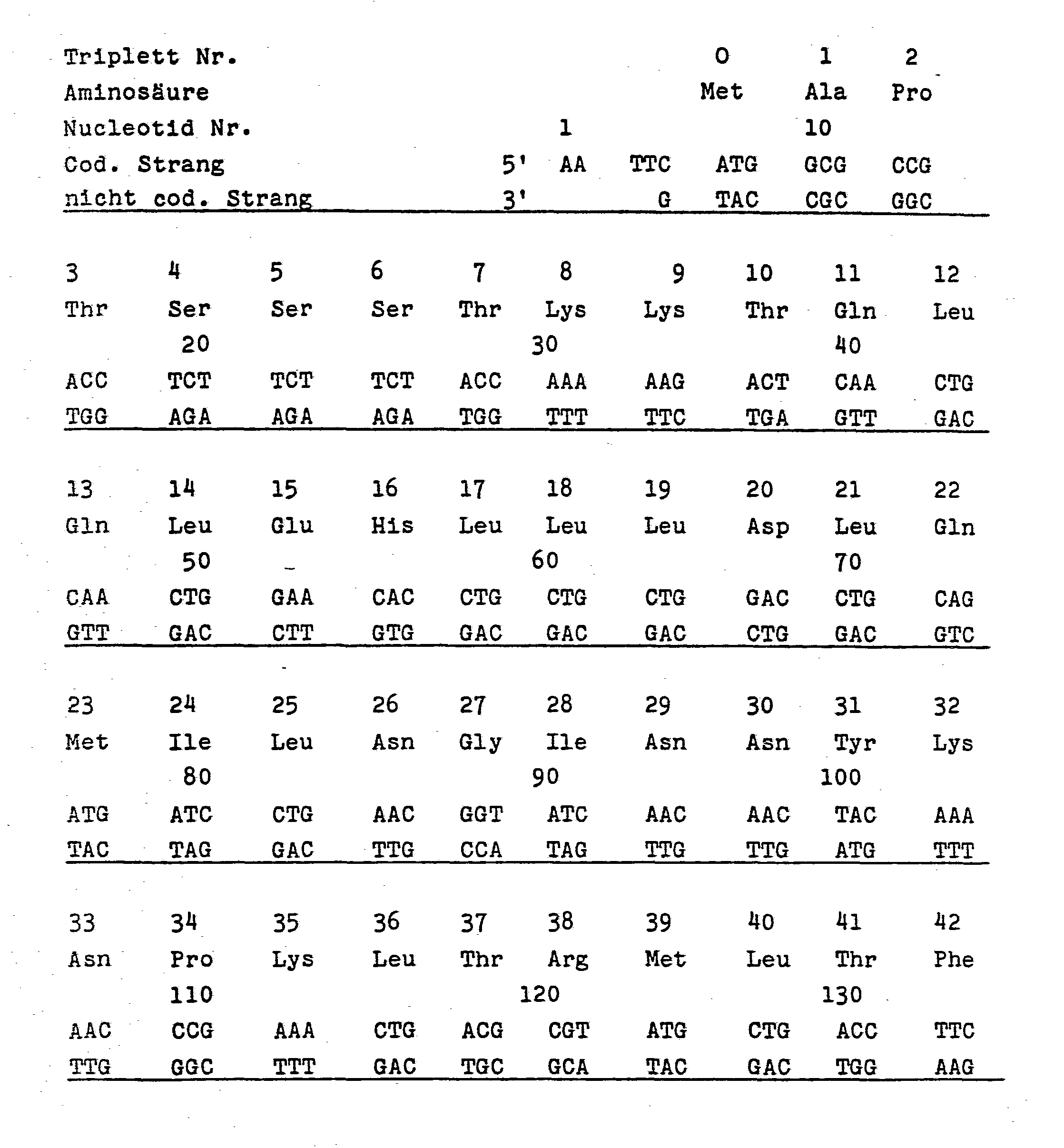
- DNA sequence I (Appendix), which codes for the entire amino acid sequence 1-133 of IL-2, and the DNA part-sequences used for the synthesis of Sequence I (Sequence II, Appendix, with the part-sequences IIa (IL 2-1) to II d (IL 2-IV) are particularly advantageous for the genetic engineering synthesis of IL-2.
- sequence II Sequence II
- IL 2-IV part-sequences IIa
- IL 2-IV part-sequences IIa
- recognition sequences allow not only the isolation of the DNA from a vector by cutting with the appropriate enzymes, for example Eco RI and SalI, but also numerous modifications of the DNA such as removal of the single-stranded areas, for example with mung bean nuclease, complete or partial filling of the shorter one In the end, for example with Kleenow polymerase, or the addition of suitable adapters or linkers.
- the overlapping ends are therefore extremely advantageous for the incorporation of the DNA according to the invention into expression vectors.
- codon for the amino acid methionine which is numbered 0 in DNA sequence I
- codon for the amino acid methionine which is numbered 0 in DNA sequence I
- there can be a presequence (also called signal or leader sequence) of a bacterial or other host-specific protein (review article: Perlman and Halvorson; J. Mol. Biol. 167 (1983), 391), which secretes the desired polypeptide caused by the cytoplasm and in this excretion process is cleaved from a signal peptidase naturally occurring in the host cell.
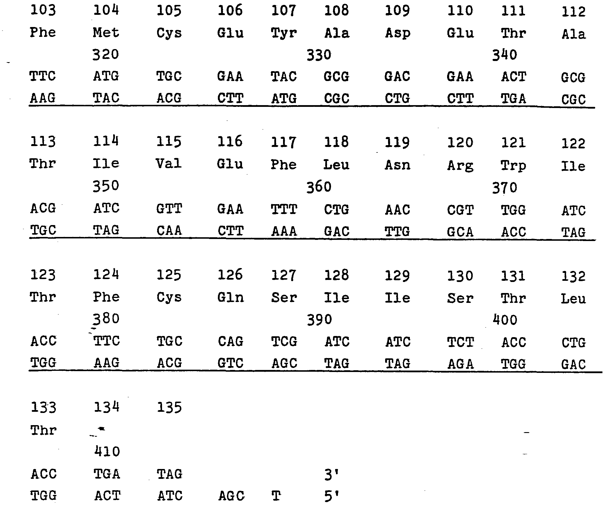
- one or preferably two stop triplets follow or follow the triplet 133 coding for threonine.
- DNA sequence I which comprises nucleotides 9 to 407 (amino acids 1-133), in connection with nucleotide sequence 1 to 8 (supernatant sequence according to Eco RI and Met codon) and two stop codons (nucleotides 408 to 413) and the protruding sequence according to Sall.
- the two stop codons represent a preferred embodiment of the invention ensure that even at high protein synthesis rates the molecules are "cut off" at the desired end and that no "fusion proteins" are formed.
- DNA sequence I can be constructed from 38 oligonucleotides of different lengths (see DNA sequence II) by first synthesizing them chemically and then linking them enzymatically by means of "sticky ends" of 4 to 9 nucleotides.
- DNA sequence I it was also taken into account that those amino acids to which several codons are assigned are not equivalent, but rather show different preferences in the respective host cell, such as E. coli. Furthermore, palindromic sequences were reduced to a minimum.
- DNA sequence I is thus easily accessible from relatively small building blocks, enables the subcloning of four gene fragments into well-known vectors and allows their combination to form the overall gene.
- the unique recognition sequences for restriction enzymes greatly facilitate the formation of extensions, changes and truncations of the protein molecule.
- Extensions are obtained after reaction with the respective restriction enzyme by adding suitable, chemically synthesized DNA molecules. Changes are obtained by cutting out individual sections of the DNA with suitable restriction enzymes and replacing them with other, chemically obtained DNA sequences. Shortenings can be obtained after cleavage with the respective restriction enzyme by reaction with nucleases.
- the biological activity of the extended, altered or shortened molecules can be checked in a biological test system, for example by inducing the cell growth of a T cell population, which is strictly dependent on the presence of the IL-2 in the medium for the multiplication.
- the synthetic genes or gene fragments are incorporated into cloning vectors, for example into commercially available plasmids such as pUC.12 or other generally available plasmids such as ptac 11, ptrp H1 and pKK 177.3, in a manner known per se.
- the chemically synthesized genes can also be provided beforehand with suitable chemically synthesized control regions which enable expression of the proteins.
- the gene fragments IL 2-1 to IL 2 - IV obtained according to the invention, the hybrid plasmids obtained therewith and the transformed host organisms are also new and the subject of the invention. The same applies to new DNA sequences modified from DNA sequence I. Further refinements of the invention are laid down in the patent claims.
- the synthesis of the gene building blocks is explained using the example of the gene building block Ia, which comprises nucleotides 1-17 of the coding strand.
- the nucleoside at the 3 'end in this case cytidine (nucleotide No. 17)
- silica gel (( R ) FRACTOSIL, Merck) covalently bound via the 3'-hydroxy function.
- the silica gel is first reacted with 3- (triethoxysilyl) propylamine with elimination of ethanol, an Si-0-Si bond being formed.
- the cytidine is reacted as N4-benzoyl-3'-0-succinoyl-5'-dimethoxytritylether in the presence of paranitrophenol and N, N'-dicyclohexylcarbodiimide with the modified carrier, the free carboxy group of the succinoyl group acylating the amino radical of the propylamino group.
- the base component is used as 5'-O-dimethoxytrityl nucleoside-3'-phosphonic acid monomethyl ester dialkylamide or chloride, the adenine being the N6-benzoyl compound, the cytosine being the N4-benzoyl compound, the Guanine as N2-isobutyryl compound and the thymine containing no amino group without a protective group are present.
- Phosphite here means deoxyribose-3'-monophosphorous acid monomethyl ester, the third valence being saturated by chlorine or a tertiary amino group, for example a morpholino radical.
- the yields of the individual synthesis steps can each be determined spectrophotometrically after the detritylation reaction b) by measuring the absorption of the dimethoxytrityl cation at a wavelength of 496 nm.
- the methylphosphate protective groups of the oligomer are cleaved off using p-thiocresol and triethylamine.
- the oligonucleotide is then separated from the solid support by treatment with ammonia for 3 hours. A 2-3 day treatment of the oligomers with concentrated ammonia removes the amino protective groups of the bases quantitatively.
- the raw so obtained product is purified by high pressure liquid chromatography (HPLC) or by polyacrylamide gel electrophoresis.
- the other gene building blocks Ib-IVj are also synthesized in a corresponding manner, the nucleotide sequence of which is evident from DNA sequence II.
- oligonucleotides Ia and Ib For phosphorylation of the oligonucleotides at the 5'-terminus, 1 nmol each of oligonucleotides Ia and Ib with 5 nmol of adenosine triphosphate with four units of T4 polynucleotide kinase in 20 ⁇ l of 50 mM Tris-HCl buffer (pH 7.6), 10 mM magnesium chloride and 10 mM dithiothreitol (DTT) for 30 minutes at 37 ° C (CC Richardson, Progress in Nucl. Acids Res. 2 (1972) 825). The enzyme is inhibited by heating to 95 g Gambminüti there ° C deactivated. The oligonucleotides Ia and Ib are then hybridized to one another by heating them in aqueous solution at 95 ° C. for 2 minutes and then slowly cooling them to 5 ° C.
- Tris-HCl buffer pH 7.6
- DTT
- the oligonucleotides Ic and Id as well as Ie and If are phosphorylated and hybridized in pairs.
- the oligonucleotides IIa with IIb etc. to IIi with IIj for the subfragment IL 2-111 with oligomers IIIa with IIIb etc. to IIIk with III1 and for the subfragment IL 2-IV with oligomers IVa IVb etc. to IVi phosphorylated with IVj and hybridized in pairs.
- the gene fragments IL 2-1 to IL 2-IV are purified by gel electrophoresis on a 10% polyacrylamide gel (without urea additive, 20.40 cm, 1 mm thick), using ⁇ X 174 DNA (BRL) as a marker substance, cut with Hinf I, or pBR 322, cut with Hae III, served.
- ⁇ X 174 DNA BNL
- the plasmid pUC 12 corresponds to the plasmid pUC 8 (Vieira et al., Gene 19 (1982) 259-268; Messing et al., Ibid. 269-276), but contains a somewhat enlarged polylinker with the additional restriction sites for the enzymes XbaI and SacI (Norrander et al., Gene 26 (1983) 101-106).
- the plasmid is incubated with the enzymes EcoRI and PstI. This cuts an approximately 40 base pair polynucleotide out of the plasmid.
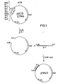
- the fragment IL 2-1 is inserted enzymatically into the opened plasmid as follows, the plasmid p 145/3 (FIG. 1) being formed:
- the DNA is ligated in a mixture of 50 mM Tris (pH 7.6), 5 mM ATP, 5 mM dithiothreitol (DTT), 5mM MgC1 2 and approx. 100 units of T4 DNA ligase for 16 hours at 12 ° C.
- the plasmid is then transformed into E. coli K 12 (JM 103), made competent with 70 mM CaCl 2 .
- Bacteria containing plasmid p 145/3 can be run on agar plates containing 50 ⁇ g / ml ampicillin, 1 mM isopropyl thiogalactoside (IPTG) and 2% 5-bromo-4-chloro-3-indolyl-ß-D- galactoside (X-Gal) can be discovered as a "white" colony.
- PUC 12 plasmid which is possibly still unchanged or not completely cut causes "blue" bacterial colonies. About five of the white bacterial clones are cultivated and the plasmids contained are isolated in a known manner (Maniatis).
- the size of the insert is checked by incubation with the restriction enzymes PstI and EcoRI and subsequent electrophoresis on 10% polyacrylamide gels.
- the insertion is then sequenced according to Maxam and Gilbert (Methods Enzymol. 65 (1980) 499) or Sanger and Coulson (J. Mol. Biol. 94 (1975) 441).
- the plasmid pUC 12 is reacted with the restriction enzymes Pst I and Xba I and, optionally after separation of the oligonucleotide formed, the fragment IL 2-11 is incorporated enzymatically.
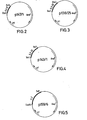
- the plasmid p 147/1 is formed (FIG. 2).
- the plasmid pUC 12 is reacted with the restriction enzymes Xba I and Sac I and the fragment IL 2-111 is incorporated enzymatically.
- the plasmid p 138/25 is formed (FIG. 3).
- the plasmid pUC 12 is cut with the restriction enzymes Sac I and Sal I and the fragment IL 2-IV is inserted enzymatically.
- the plasmid p 143/1 is formed (FIG. 4).
- the subfragments IL 2-1 to IL 2-IV are cut out again with the corresponding restriction enzymes and by electrophoresis on polyacrylamide separated. After immersing the gels in an aqueous solution of ethidium bromide, the bands are identified under UV light, cut out and the DNA eluted in a known manner (Maniatis).
- the subfragments IL 2-1 to IL 2-IV are as under 3 a) described enzymatically linked and incorporated into the plasmid pUC 12 opened by reaction with the restriction enzymes Eco RI and Sal I.
- the hybrid plasmid p 159/6 (FIG. 5) is obtained, the sequence of which is again confirmed by analysis.
- the expression plasmid pKK 177.3 (plasmid ptac 11, Amman et al., Gene 25 (1983) 167, in which a sequence which contains a Sal I site was synthetically incorporated into the Eco RI recognition site) is combined with the restriction enzymes Eco RI and Sal I open.
- the DNA sequence I with the restriction enzymes Eco RI and Sal I is cut out from the plasmid p 159/6 (FIG. 5) and placed on polyacrylamide or 2% low-melting agarose, separated from the plasmid DNA and the Insertion regained (maniatis).
- the expression plasmid p trp H1 (Amann et al., Gene 25 (1983) 167-178) contains the control elements of the Trp operons (promoter, operator) followed by a Hind III restriction site (Fig. 6).
- the protruding ends are broken down with mung bean nuclease according to the manufacturer's instructions (PL Biochemicals).
- p trp HI is opened with Hind III and the protruding ends are also removed with Mung Bean Nuclease.
- the DNA sequence I is then inserted "blunt-ended" with T 4 DNA ligase in the opened plasmid (FIG. 6).
- the start of protein synthesis is determined by the triplet ATG for methionine in position 0. Expression is induced by the absence of tryptophan and / or the presence of indolylacrylacetic acid.
- the plasmid p 159/6 was reacted with the restriction enzyme Sal I and then incubated in a manner known per se with exonuclease III and S 1 nuclease. After reaction with Eco RI, partial sequences of the IL-2 were now obtained which carry the overlapping sequences for Eco RI at one end and are blunt-ended at the other end. After addition of a chemically synthesized DNA, which is blunt-ended at one end and here has a stop codon as the first codon, but at the other end bears the overhang of a Sal I sequence, this shortened DNA sequence can be incorporated into the same expression vectors.
- a hybrid plasmid which contains the DNA sequence for the expression of Des- (Ala 1 ] -IL-2
- the DNA is made from the plasmid p 159/6 according to methods known per se with the restriction enzymes Eco RI and Hind III. Sequence I, including the polylinker part of the plasmid, was cut out, electrophoretically separated from the residual plasmid and cut with the restriction enzyme Aha II. The isolated Aha II-Hind III fragment is made "blunt-ended" as described in Example 5 b) and then cut with Sal I. Using the following adapter
- Competent E. coli cells are transformed with 0.1 to 1 ng of the hybrid plasmids containing sequence I or derivatives thereof and plated on agar plates containing ampicillin. Clones which contain the correctly integrated IL-2 gene sequence or derivatives thereof in the corresponding plasmids can then be identified by rapid DNA processing (Maniatis a.a.0.).
- the bacterial strains cultured to the desired optical density are incubated with a suitable inductor, for example IPTG, for a sufficiently long time, for example 2-4 hours.
- a suitable inductor for example IPTG
- the cells are then killed with 0.1% cresol and 0.1 mM benzylsulfonyl fluoride. After induction, a protein band with a molecular weight of approximately 15,000 daltons is found on an SDS-PAA gel.
- the cell mass is taken up in a buffer solution (50 mM Tris, 50 mM EDTA, pH 7.5) and mechanically disrupted, for example with a French press or ( R ) DYNO mill (Willy Bachofer, Basel ), whereupon the insoluble components are centrifuged off.
- a buffer solution 50 mM Tris, 50 mM EDTA, pH 7.5
- a part of the IL-2 protein remains in the residue after cell disruption and can be solubilized with 8 M urea, 6 M guanidinium hydrochloride, buffer solutions with detergents of ionic or non-ionic nature and similar solvents.
- the protein containing the IL-2 activity is purified from these solutions by customary methods. Ion exchange, adsorption, gel filtration or affinity chromatography on antibody columns are suitable. Sodium dodecyl sulfate-acrylamide gel or HPLC analysis controls the enrichment and purity of the product.
- IL-2 protein T cells For the biological characterization of the IL-2 protein T cells which are strictly dependent on the presence of IL-2 in the medium.
- the cell division rate of such cell lines is, within certain limits, proportional to the IL-2 concentration of the medium, which can therefore be determined by the absorption of 3 H-thymidine from the medium.
Landscapes
- Chemical & Material Sciences (AREA)
- Health & Medical Sciences (AREA)
- Life Sciences & Earth Sciences (AREA)
- Organic Chemistry (AREA)
- General Health & Medical Sciences (AREA)
- Molecular Biology (AREA)
- Biochemistry (AREA)
- Biophysics (AREA)
- Zoology (AREA)
- Genetics & Genomics (AREA)
- Medicinal Chemistry (AREA)
- Gastroenterology & Hepatology (AREA)
- Proteomics, Peptides & Aminoacids (AREA)
- Toxicology (AREA)
- Preparation Of Compounds By Using Micro-Organisms (AREA)
- Peptides Or Proteins (AREA)
- Saccharide Compounds (AREA)
- Medicines That Contain Protein Lipid Enzymes And Other Medicines (AREA)
- Micro-Organisms Or Cultivation Processes Thereof (AREA)
Abstract
Description
- Die Erfindung betrifft ein Verfahren zur Herstellung von Human-Interleukin-2 und davon abgeleitete Polypeptide mit biologischer und immunologischer Aktivität von Human-Interleukin-2, chemisch synthetisierte Gene, die diese Peptide codieren sowie geeignete Vektorkonstruktionen und Wirtsorganismen zur Expression dieser Polypeptide.
- Human-Interleukin-2, im folgenden "IL-2", ist ein Polypeptid aus 133 Aminosäuren. Die DNA-Sequenz des menschlichen IL-2 sowie dessen gentechnologische Synthese sind in der Europä- ichen Patentanmeldung mit der Veröffentlichungsnummer EP 0 091 539 Al beschrieben (Amino Acid Sequence II der Fig. 2b). Dieser Synthese liegt eine aus Säugetierzellen isolierte mRNA zugrunde, welche in cDNA überführt, in Vektoren eingebaut und in Wirtszellen, darunter auch E.coli, zur Expression gebracht wurde.
- Bakterien, insbesondere E.coli, sind aus einer Reihe von Gründen bevorzugte Wirtszellen für die gentechnische Produktion von Polypeptiden. Gene eukaryotischer Zellen, die wie in der genannten Patentanmeldung beschrieben - durch =Reverse Transcriptase aus mRNA erhalten wurden, werden häufig nach Einbau in Plasmide und Transformation in Bakterien nur unbefriedigend exprimiert. Die Erfindung betrifft :deshalb eine synthetische DNA-Sequenz, die für IL-2 codiert, welche besonders vorteilhaft in E.coli zur Expression eines Polypeptids mit IL-2-Aktivität führt. Ferner umfaßt die Erfindung solche DNA-Sequenzen, die gegen die synthetische DNA-Sequenz hybridieren und die sich aus dieser durch einfache oder multiple Substitionen, Deletionen oder Insertionen von Basen ableiten. Der Austausch, die Insertion oder die Deletion von Codons oder eine Kombination davon führt zu Peptiden mit einer oder mehreren ausgetauschten Aminosäure(n) oder zu längeren oder kürzeren IL-2-Derivaten, die ebenfalls Gegenstand der Erfindung sind. Diese Polypeptide mit der biologischen oder immunologischen Aktivität des IL-2 können in ihren Eigenschaften derart verändert sein, daß die Stabilität des Peptids erhöht ist, die Lipophilie des Polypeptids zugunsten einer besseren Löslichkeit verändert ist, die Applikation als Arzneimittel erleichtert ist oder aber die biologische Aktivität erhöht ist.
- Der genetische Code ist bekanntlich "entartet", d.h. daß nur für zwei Aminosäuren eine einzige Nucleotid-Sequenz codiert, während den restlichen 18 genetisch codierbaren Aminosäuren 2 bis 6 Tripletts zuzuordnen sind. Von den hierdurch gegebenen Variationsmöglichkeiten machen jedoch die Wirtszellen unterschiedlicher Spezies nicht immer den gleichen Gebrauch. Für die Synthese der Gene besteht somit eine unübersehbare Vielfalt von Codon-Möglichkeiten.
- Es wurde nun gefunden, daß die DNA-Sequenz I (Anhang), die für die gesamte Aminosäurensequenz 1-133 von IL-2 codiert, sowie die zur Synthese der Sequenz I benutzten DNA-Teilsequenzen (Sequenz II, Anhang, mit den Teilsequenzen IIa (IL 2-1) bis II d (IL 2-IV) besonders vorteilhaft für die gentechnoiogische Synthese von IL-2 sind. Am 5'-Ende des codierenden Stranges der DNA-Sequenz I befindet sich eine "überhängende" DNA-Sequenz, beispielsweise entsprechend der Restriktionsendonuclease Eco RI, am 3'-Ende des codierenden Stranges dagegen eine andere einzelsträngige, überhängende Sequenz, beispielsweise entsprechend dem Restriktionsenzym Sal I. Diese beiden unterschiedlichen Erkennungssequenzen gewährleisten die Insertion der DNA in Plasmide in der gewünschten Orientierung. Selbstverständlich können auch andere überstehende Sequenzen gewählt werden, die Schnittstellen im vorgesehenen Vektor entsprechen.
- Diese Erkennungssequenzen erlauben nicht nur die Reisolierung der DNA aus einem Vektor durch Schneiden mit den entsprechenden Enzymen, beispielsweise Eco RI und SalI, sondern auch zahlreiche Modifikationen der DNA wie Entfernung der einzelsträngigen Bereiche, beispielsweise mit Mung Bean-Nuclease, völliges oder teilweises Auffüllen des kürzeren Endes, beispielsweise mit Kleenow-Polymerase, oder die Addition geeigneter Adapter oder Linker. Die überlappenden Enden sind daher außerordentlich vorteilhaft für den Einbau der erfindungsgemäßen DNA in Expressionsvektoren.
- Zwischen diesen Erkennungssequenzen und den Codons für die Aminosäurefolge befindet sich am 5'-Ende des codierenden Stranges das Codon für die Aminosäure Methionin (das in der DNA-Sequenz I mit 0 beziffert ist). Alternativ hierzu kann eine Praesequenz (auch Signal- oder leader-Sequenz genannt), eines bakteriellen oder sonstigen wirtseigenen Proteins stehen (übersichtsartikel: Perlman und Halvorson; J. Mol. Biol. 167 (1983), 391), welche die Sekretion des gewünschten Polypeptids aus dem Cytoplasma bewirkt und bei diesem Exkretionsprozeß von einer in der Wirtszelle natürlich vorkommenden Signal-Peptidase abgespalten wird. Am Ende des codierenden Stranges folgt bzw. folgen dann auf das für Threonin codierende Triplett 133 ein bzw. vorzugsweise zwei Stop-Triplett(s). Im Anhang ist die DNA-Sequenz I, welche die Nucleotide 9 bis 407 (Aminosäuren 1-133) umfaßt, im Zusammenhang mit der Nucleotidsequenz 1 bis 8 (überstehende Sequenz gemäß Eco RI und Met-Codon) und zwei Stop-Codons (Nucleotide 408 bis 413) sowie der überstehenden Sequenz gemäß Sall dargestellt. Die zwei Stop-Codons stellen eine bevorzugte Ausführungsform der Erfindung dar. Sie gewährleisten, daß selbst bei hoher Proteinsyntheserate die Moleküle am gewünschten Ende "abgeschnitten" werden und keine "Fusionsproteine" gebildet werden.
- Drei interne singuläre Schnittstellen für die Restriktionsenzym Pst I, Xba I und Sac I (Nucleotide 69-74, 182-187 und 291-296 des codierenden Stranges der DNA-Sequenz I) ermöglichen die Subklonierung von vier Genfragmenten IL 2-1 bis IL 2-IV, die in gut untersuchten Klonierungsvektoren, wie etwa pUC 12, eingebaut werden können. Zusätzlich wurden innerhalb des Strukturgens eine Reihe von weiteren singulären Erkennungssequenzen für Restriktionsenzyme eingebaut, die einerseits einen Zugang zu Teilsequenzen des IL-2 schaffen und andererseits die Durchführung von Variationen erlauben:

- Bei der DNA-Sequenz I wurde weiterhin berücksichtigt, daß bei denjenigen Aminosäuren, denen mehrere Codons zuzuordnen sind, diese nicht gleichwertig sind, sondern vielmehr in der jeweiligen Wirtszelle wie E. coli unterschiedliche Präferenzen zeigen. Weiterhin wurden palindromische Sequenzen auf ein Mindestmaß reduziert.
- Die Genstruktur der DNA-Sequenz I ist somit leicht aus relativ kleinen Bausteinen zugänglich, ermöglicht die Subklonierung von vier Genfragmenten in gut bekannte Vektoren und erlaubt deren Kombination zum Gesamtgen. Die singulären Erkennungssequenzen für Restriktionsenzyme erleichtern außerordentlich die Bildung von Verlängerungen, Veränderungen und Verkürzungen des Proteinmoleküls.
- Verlängerungen werden nach Umsetzung mit dem jeweiligen Restriktionsenzym durch Addition geeigneter, chemisch synthetisierter DNA-Moleküle erhalten. Veränderungen werden durch Herausschneiden einzelner Abschnitte der DNA mit geeigneten Restriktionsenzymen und Ersetzen durch andere, chemisch gewonnene DNA-Sequenzen erhalten. Verkürzungen können nach Spaltung mit dem jeweiligen Restriktionsenzym durch Umsetzung mit Nucleasen erhalten werden.
- Die biologische Aktivität der verlängerten, veränderten oder verkürzten Moleküle kann in einem biologischen Testsystem geprüft werden, beispielsweise durch Induktion des Zellwachstums einer T-Zell-Population, die von der Anwesenheit des IL-2 im Medium für die Vermehrung streng abhängig ist. Der Einbau der synthetischen Gene bzw. Genfragmente in Klonierungsvektoren, beispielsweise in handelsübliche Plasmide wie pUC.12 bzw. andere allgemein zugängliche Plasmide wie ptac 11, ptrp H1 und pKK 177.3, erfolgt in an sich bekannter Weise. Auch können die chemisch synthetisierten Gene zuvor mit geeigneten chemisch synthetisierten Kontrollregionen versehen werden, die eine Expression der Proteine ermöglichen. Hierzu kann auf das Lehrbuch von Maniatis (Molecular Cloning, Maniatis et al., Cold Spring Harbor, 1982) verwiesen werden. Die Transformation der so erhaltenen Hybridplasmide in geeignete Wirtsorganismen, vorteilhaft E. coli, ist ebenfalls an sich bekannt und in dem vorstehend genannten Lehrbuch eingehend beschrieben.
- Die erfindungsgemäß erhaltenen Genfragmente IL 2-1 bis IL 2-IV, die damit erhaltenen Hybridplasmide und die transformierten Wirtsorganismen sind ebenfalls neu und Gegenstand der Erfindung. Dasselbe gilt für aus der DNA-Sequenz I abgewandelte neue DNA-Sequenzen. Weitere Ausgestaltungen der Erfindung sind in den Patentansprüchen niedergelegt.
- In den folgenden Beispielen werden noch einige Ausgestaltungen der Erfindung im einzelnen erläutert, woraus sich die Vielzahl der möglichen Abwandlungen und Kombinationen für den Fachmann ergeben. Prozentangaben beziehen sich hierbei auf das Gewieht, wenn nichts anderes angegeben ist.
- Am Beispiel des Genbausteins Ia, der die Nucleotide 1-17 des codierenden Strangs umfaßt, wird die Synthese der Genbausteine erläutert. Nach bekannten Methoden (M.J. Galt et al., Nucleic Acids Res. 8 (1980) 1081-1096)) wird das am 3'-Ende stehende Nucleosid, im vorliegenden Falle also Cytidin (Nucleotid Nr. 17), an Kieselgel ((R)FRACTOSIL, Firma Merck) über die 3'-Hydroxyfunktion covalent gebunden. Hierzu wird zunächst das Kieselgel unter Abspaltung von Ethanol mit 3-(Triethoxysilyl)-propylamin umgesetzt, wobei eine Si-0-Si-Bindung entsteht. Das Cytidin wird als N4-Benzoyl-3'-0-succinoyl-5'-dimethoxytritylether in Gegenwart von Paranitrophenol und N,N'-Dicyclohexylcarbodiimid mit dem modifizierten Träger umgesetzt, wobei die freie Carboxygruppe der Succinoylgruppe den Aminorest der Propylaminogruppe acyliert.
- In den folgenden Syntheseschritten wird die Basenkomponente als 5'-O-Dimethoxytrityl-nucleosid-3'-phosphorig- säuremonomethylester-dialkylamid oder -chlorid eingesetzt, wobei das Adenin als N6-Benzoyl-Verbindung, das Cytosin als N4-Benzoyl-Verbindung, das Guanin als N2-Isobutyryl-Verbindung und das keine Aminogruppe enthaltende Thymin ohne Schutzgruppe vorliegen.
- 50 mg des polymeren Trägers, der 2 µmol Cytosin gebunden enthält, werden nacheinander mit den folgenden Agentien behandelt:
- a) Nitromethan,
- b) gesättigte Zinkbromidlösung in Nitromethan mit 1 % Wasser,
- c) Methanol,
- d) Tetrahydrofuran,
- e) Acetonitril,
- f) 40 µmol des entsprechenden Nucleosidphosphits und 200 µmol Tetrazol in 0,5 ml wasserfreiem Acetonitril (5 Minuten),
- g) 20 % Acetanhydrid in Tetrahydrofuran mit 40 % Lutidin und 10 % Dimethylaminopyridin (2 Minuten),
- h) Tetrahydrofuran,
- i) Tetrahydrofuran mit 20 % Wasser und 40 % Lutidin,
- j) 3 % Jod in Kollidin/Wasser/Tetrahydrofuran im Volumenverhältnis 5:4:1,
- k) Tetrahydrofuran und
- 1) Methanol.
- Unter "Phosphit" wird hierbei der Desoxyribose-3'-mono- phosphorigsäure-monomethylester verstanden, wobei die dritte Valenz durch Chlor oder eine tertiäre Aminogruppe, beispielsweise einen Morpholinorest, abgesättigt ist. Die Ausbeuten der einzelnen Syntheseschritte können jeweils nach der Detritylierungsreaktion b) spektrophotometrisch durch Messung der Absorption des Dimethoxytritylkations bei einer Wellenlänge von 496 nm bestimmt werden.
- Nach abgeschlossener Synthese des Oligonucleotids werden die Methylphosphatschutzgruppen des Oligomers mit Hilfe von p-Thiokresol und Triethylamin abgespalten.
- Anschließend wird durch 3-stündige Behandlung mit Ammoniak das Oligonucleotid vom festen Träger abgetrennt. Eine 2- bis 3-tägige Behandlung der Oligomeren mit konzentriertem Ammoniak spaltet die Aminoschutzgruppen der Basen quantitativ ab. Das so erhaltene Rohprodukt wird durch Hochdruckflüssigkeitschromatographie (HPLC) oder durch Polyacrylamid-Gelelektrophorese gereinigt.
- Ganz entsprechend werden auch die übrigen Genbausteine Ib-IVj synthetisiert, deren Nucleotidfolge aus der DNA-Sequenz II hervorgeht.
- 2. Enzymatische Verknüpfung der einzelsträngigen Oligonucleotide zu den Genfragmenten IL 2-1 bis IL 2-IV.
- Zur Phosphorylierung der Oligonucleotide am 5'-Terminus wurde je 1 nmol der Oligonucleotide Ia und Ib mit 5 nmol Adenosintriphosphat mit vier Einheiten T4-Polynucleotid-Kinase in 20 µl 50 mM Tris-HCl-Puffer (pH 7,6), 10 mM Magnesiumchlorid und 10 mM Dithiothreitol (DTT) 30-Minuten bei 37°C behandelt (C.C. Richardson, Progress in Nucl. Acids Res. 2 (1972) 825). Das Enzym wird durch fünfminütiges Erhitzen auf 95°C desaktiviert. Anschließend werden die Oligonucleotide Ia und Ib gageneinander hybridisiert, indem man sie in wäßriger Lösung 2 Minuten auf 95°C erhitzt und dann langsam auf 5°C abkühlt.
- Analog werden die 0ligonucleotide Ic und Id sowie Ie und If phosphoryliert und paarweise hybridisiert. Für das Subfragment IL 2-II werden die Oligonucleotide IIa mit IIb usw. bis IIi mit IIj, für das Subfragment IL 2-111 die Oligomeren IIIa mit IIIb usw. bis IIIk mit III1 und für das Subfragment IL 2-IV die Oligomeren IVa mit IVb usw. bis IVi mit IVj phosphoryliert und paarweise hybridisiert.
- Die so erhaltenen drei Oligonucleotidpaare für das Genfragment IL 2-1, die fünf Oligonucleötidpaare für die Genfragmente IL 2-11 und IL 2-IV sowie die sechs Oligonucleotidpaare für das Genfragment IL 2-111 werden jeweils wie folgt ligiert:
- Die doppelsträngigen Nucleotide werden vereinigt und in jeweils 40 µl 50 mM Tris-HCl-Puffer, 20 mM Magnesiumchlorid und 10 mM DTT mit Hilfe von 100 Einheiten T4-DNA-Ligase bei 15°C im Laufe von 16 Stunden ligiert.
- Die Reinigung der Genfragmente IL 2-1 bis IL 2-IV erfolgt durch Gelelektrophorese auf einem 10%igen Polyacrylamidgel (ohne Harnstoffzusatz, 20 . 40 cm, 1 mm Dicke), wobei als Markersubstanz ØX 174 DNA (Fa. BRL), geschnitten mit Hinf I, oder pBR 322, geschnitten mit Hae III, diente.
- Das Plasmid pUC 12 entspricht dem Plasmid pUC 8 (Vieira et al., Gene 19 (1982) 259-268; Messing et al., ibid. 269-276), enthält allerdings einen etwas vergrößerten Polylinker mit den zusätzlichen Restriktionsstellen für die Enzyme XbaI und SacI (Norrander et al., Gene 26 (1983) 101-106). Das Plasmid wird mit den Enzymen EcoRI und PstI inkubiert. Hierdurch wird ein etwa 40 Basenpaare großes Polynucleotid aus dem Plasmid herausgeschnitten. Die Abtrennung dieses Frag- ments vom geöffneten Plasmid gelingt entweder durch Chromatographie an (R)SEPHADEX G50 oder durch Elektrophorese an 1,5 %iger Agarose in bekannter Weise (Maniatis). Auf eine Abtrennung des Fragments kann auch verzichtet werden, da Plasmide mit der Insertion von IL 2-1 leicht durch Ausplattieren erkannt werden können (wie im folgenden ausgeführt wird):
- In das geöffnete Plasmid wird das Fragment IL 2-1 wie folgt enzymatisch eingesetzt, wobei das Plasmid p 145/3 (Figur 1) gebildet wird:
- Die DNA wird in einer Mischung aus 50 mM Tris (pH 7,6), 5 mM ATP, 5 mM Dithiothreitol (DTT), 5mM MgC12 und ca. 100 Einheiten T4 DNA-Ligase 16 Stunden bei 12°C ligiert. Anschließend wird das Plasmid in E.coli K 12 (JM 103), kompetent gemacht mit 70 mM CaCl2, transformiert.
- Bakterien, die das Plasmid p 145/3 enthalten, können auf Agar-Platten mit 50 µg/ml Ampicillin, 1 mM Isopropyl-Thiogalactosid (IPTG) und 2 % 5-Brom-4-chlor-3-indolyl-ß-D-galactosid (X-Gal) als "weiße" Kolonie entdeckt werden. Eventuell noch unverändertes oder nicht vollständig geschnittenes Plasmid pUC 12 verursacht "blaue" Bakterienkolonien. Etwa fünf der weißen Bakterienklone werden kultiviert und die enthaltenen Plasmide in bekannter Weise (Maniatis) isoliert.
- Die Größe der Insertion wird durch Inkubation mit den Restriktionsenzymen PstI und EcoRI und anschließende Elektrophorese auf 10%igem Polyacrylamidgelen überprüft. Hierauf erfolgt eine Sequenzierung der Insertion nach Maxam und Gilbert (Methods Enzymol. 65 (1980) 499) oder Sanger und Coulson (J. Mol. Biol. 94 (1975) 441).
- Analog zu a) wird das Plasmid pUC 12 mit den Restriktionsenzymen Pst I und Xba I umgesetzt und, ggf. nach Abtrennung des entstandenen Oligonucleotids, das Fragment IL 2-11 enzymatisch eingebaut. Es entsteht das Plasmid p 147/1 (Figur 2).
- Analog zu a) wird das Plasmid pUC 12 mit den Restriktionsenzymen Xba I und Sac I umgesetzt und das Fragment IL 2-111 enzymatisch eingebaut. Es entsteht das Plasmid p 138/25 (Figur 3).
- Analog zu a) wird das Plasmid pUC 12 mit den Restriktionsenzymen Sac I und Sal I geschnitten und das Fragment IL 2-IV enzymatisch eingebaut. Es entsteht das Plasmid p 143/1 (Figur 4).
- Nach Vermehrung der Plasmide p 145/3, P 147/1, p 138/25 und p 143/1 und Bestätigung der Sequenz werden die Subfragmente IL 2-1 bis IL 2-IV erneut mit den entsprechenden Restriktionsenzymen herausgeschnitten und durch Elektrophorese auf Polyacrylamid abgetrennt. Nach Eintauchen der Gele in eine wäßrige Lösung von Ethidiumbromid werden die Banden unter UV-Licht identifiziert, herausgeschnitten und die DNA in bekannter Weise eluiert (Maniatis).
- Die Subfragmente IL 2-1 bis IL 2-IV werden wie unter 3 a) beschrieben enzymatisch verknüpft und in das durch Umsetzen mit den Restriktionsenzymen Eco RI und Sal I geöffnete Plasmid pUC 12 eingebaut. Man erhält das Hybridplasmid p 159/6 (Figur 5), dessen Sequenz nochmals durch Analyse bestätigt wird.
- Das Expressionsplasmid pKK 177.3 (Plasmid ptac 11, Amman et al., Gene 25 (1983) 167, bei dem in die Eco RI-Erkennungsstelle synthetisch eine Sequenz eingebaut wurde, die eine Sal I-Schnittstelle enthält) wird mit den Restriktionsenzymen Eco RI und Sal I geöffnet. Aus dem Plasmid p 159/6 (Figur 5) wird die DNA-Sequenz I mit den Restriktionsenzymen Eco RI und Sal I herausgeschnitten und auf Polyacrylamid oder 2%ige niedrig-schmelzende (low melting) Agarose gegeben, von derPlasmid-DNA abgetrennt und die Insertion wiedergewonnen (Maniatis). Durch Ligation des aufgeschnittenen Plasmids pKK 177.3 mit der DNA-Sequenz I wird ein Hybridplasmid geschaffen, bei dem der Insertion eine Expressions- bzw. Regulationsregion vorgeschaltet ist. Nach Zugabe eines geeigneten Induktors wie IPTG wird eine mRNA gebildet, die zur Expression des Polypeptids entsprechend der DNA-Sequenz I führt.
- Das Expressionsplasmid p trp H1 (Amann et al., Gene 25 (1983) 167-178) enthält die Kontrollelemente des Trp-Operons (Promotor, Operator), gefolgt von einer Hind III-Restriktionsstelle (Fig. 6).
- Nach Isolieren der DNA-Sequenz I aus p 159/6 werden die überstehenden Enden mit Mung Bean Nuclease nach Angaben des Herstellers (PL Biochemicals) abgebaut. p trp Hl wird mit Hind III geöffnet und die überstehenden Enden ebenfalls mit Mung Bean Nuclease entfernt. Die DNA-Sequenz I wird dann "stumpfendig" mit T 4 DNA-Ligase in das geöffnete Plasmid eingebaut (Fig. 6).
- Durch das Triplett ATG für Methionin in Position 0 wird der Start für die Proteinsynthese festgelegt. Die Expression wird durch Abwesenheit von Tryptophan und/oder Anwesenheit von Indolylacrylessigsäure induziert.
- Zur Darstellung von verkürzten Proteinmolekülen mit biologischer Aktivität des IL-2 wurde das Plasmid p 159/6 mit dem Restriktonsenzym Sal I umgesetzt und anschließend in an sich bekannter Weise mit Exonuclease III und S 1-Nuclease inkubiert. Nach Umsetzung mit Eco RI wurden nun Teilsequenzen des IL-2 erhalten, die an einem Ende die überlappenden Sequenzen für Eco RI tragen und am anderen Ende stumpfendig sind. Nach Addition einer chemisch synthetisierten DNA, die an einem Ende stumpfendig ist und hier als erstes Codon ein Stop-Codon aufweist, am anderen Ende dagegen den Überhang einer Sal I-Sequenz trägt, kann diese verkürzte DNA-Sequenz in die gleichen Expressionsvektoren eingebaut werden.
- Zur Konstruktion eines Hybridplasmids, das die DNA-Sequenz für die Expression von Des-(Ala1]-IL-2 enthält, wird aus dem Plasmid p 159/6 nach an sich bekannten Methoden mit den Restriktionsenzymen Eco RI und Hind III die DNA-Sequenz I inklusive Polylinkerteil des Plasmids herausgeschnitten, elektrophoretisch vom Restplasmid abgetrennt und mit dem Restriktionsenzym Aha II nachgeschnitten. Das isolierte Aha II-Hind III-Fragment wird wie in Beispiel 5 b) beschrieben "stumpfendig" gemacht und dann mit Sal I nachgeschnitten. Mit Hilfe des folgenden Adaptors
- 5' AA TTC ATG 3'
- 3' G TAC 5'
erhält man nach Ligation mit dem mit Eco RI und Sal I geöffneten Plasmid pKK 177.3 ein neues Hybridplasmid, das das Gen zur Herstellung von Des-[Alal]-IL-2 enthält. - Kompetente E. coli-Zellen werden mit 0,1 bis 1 ng der Hybridplasmide, die die Sequenz I oder Derivate davon enthalten, transformiert und auf Ampicillin enthaltende Agarplatten plattiert. Anschließend lassen sich Klone, die die korrekt integrierte IL-2-Gensequenz oder Derivate davon in den entsprechenden Plasmiden enthalten, durch DNA-Schnellaufarbeitung identifizieren (Maniatis a.a.0.).
- Nach Transformation der Hybridplasmide mit der DNA-Sequenz I oder Derivaten davon in E. coli wird ein Polypeptid exprimiert, das außer der IL-2-Aminosäuresequenz bzw. abgewandelte Sequenzen am Aminoterminus noch eine zusätzliche Methionylgruppe trägt.
- Die zur gewünschten optischen Dichte kultivierten Bakterienstämme werden mit einem geeigneten Induktor, beispielsweise IPTG, hinreichend lange, beispielsweise 2-4 Stunden, inkubiert. Anschließend werden die Zellen mit 0,1 % Kresol und 0,1 mM Benzylsulfonylfluorid abgetötet. Auf einem SDS-PAA-Gel ist nach der Induktion eine Proteinbande mit einem Molgewicht von etwa 15 000 Dalton festzustellen. Nach Zentrifugieren oder Filtrieren wird die Zellmasse in einer Pufferlösung (50 mM Tris, 50 mM EDTA, pH 7,5) aufgenommen und mechanisch aufgeschlossen, beispielsweise mit einer French-Presse bzw. (R)DYNO-Mühle (Fa. Willy Bachofer, Basel), worauf die unlöslichen Bestandteile abzentrifugiert werden.
- Ein Teil des IL-2-Proteins verbleibt nach dem Zellaufschluß im Rückstand und kann mit 8 M Harnstoff, 6 M Guanidinium-Hydrochlorid, Pufferlösungen mit Detergentien ionischer oder nicht-ionischer Art und ähnlichen Lösemitteln solubilisiert werden. Aus diesen Lösungen wird das die IL-2-Aktivität enthaltende Protein nach üblichen Verfahren gereinigt. Geeignet sind Ionenaustauscher-, Adsorptions-, Gelfiltrationssäulen oder Affinitätschromatographie an Antikörpersäulen. Durch Natriumdodecylsulfat-Acrylamidgel- oder HPLC-Analytik werden Anreicherung und Reinheit des Produktes kontrolliert.
- Zur biologischen Charakterisierung des IL-2-Proteins werden T-Zellen verwendet, die strikt abhängig von der Anwesenheit von IL-2 im Medium sind. Die Zellteilungsrate solcher Zell-Linien ist in gewissen Grenzen proportional zur IL-2-Konzentration des Mediums, die daher über die Aufnahme von 3H-Thymidin aus dem Medium festgestellt werden kann.
-


Claims (36)
Priority Applications (1)
| Application Number | Priority Date | Filing Date | Title |
|---|---|---|---|
| AT85106279T ATE65797T1 (de) | 1984-05-29 | 1985-05-22 | Gentechnologisches verfahren zur herstellung von human-interleukin-2 und mittel zur durchfuehrung dieses verfahrens. |
Applications Claiming Priority (2)
| Application Number | Priority Date | Filing Date | Title |
|---|---|---|---|
| DE3419995 | 1984-05-29 | ||
| DE19843419995 DE3419995A1 (de) | 1984-05-29 | 1984-05-29 | Gentechnologisches verfahren zur herstellung von human-interleukin-2 und mittel zur durchfuehrung dieses verfahrens |
Publications (3)
| Publication Number | Publication Date |
|---|---|
| EP0163249A2 true EP0163249A2 (de) | 1985-12-04 |
| EP0163249A3 EP0163249A3 (en) | 1988-05-11 |
| EP0163249B1 EP0163249B1 (de) | 1991-07-31 |
Family
ID=6237111
Family Applications (1)
| Application Number | Title | Priority Date | Filing Date |
|---|---|---|---|
| EP85106279A Expired - Lifetime EP0163249B1 (de) | 1984-05-29 | 1985-05-22 | Gentechnologisches Verfahren zur Herstellung von Human-Interleukin-2 und Mittel zur Durchführung dieses Verfahrens |
Country Status (11)
| Country | Link |
|---|---|
| EP (1) | EP0163249B1 (de) |
| JP (1) | JPS6199A (de) |
| AT (1) | ATE65797T1 (de) |
| AU (1) | AU589896B2 (de) |
| DE (2) | DE3419995A1 (de) |
| DK (1) | DK166681B1 (de) |
| ES (1) | ES8605813A1 (de) |
| GR (1) | GR851291B (de) |
| IE (1) | IE57996B1 (de) |
| PT (1) | PT80542B (de) |
| ZA (1) | ZA854031B (de) |
Cited By (10)
| Publication number | Priority date | Publication date | Assignee | Title |
|---|---|---|---|---|
| EP0147819A3 (en) * | 1983-12-23 | 1987-09-23 | F. Hoffmann-La Roche & Co. Aktiengesellschaft | Purification of recombinant interleukin-2 |
| EP0219839A3 (de) * | 1985-10-22 | 1988-05-04 | Hoechst Aktiengesellschaft | Derivat des Interleukin-2, seine Herstellung und Verwendung |
| EP0233578A3 (de) * | 1986-02-10 | 1988-08-03 | Otsuka Pharmaceutical Co., Ltd. | Polypeptid mit Interleukin-2-Aktivitäten |
| EP0200280A3 (en) * | 1985-01-18 | 1988-08-24 | Cetus Corporation | Oxidation resistant il-2 muteins and their production, formulations containing such muteins, and dna sequences and expression vectors coding for such muteins and corresponding transformed host cells |
| EP0288809A1 (de) * | 1987-04-16 | 1988-11-02 | Hoechst Aktiengesellschaft | Bifunktionelle Proteine |
| EP0259160A3 (de) * | 1986-09-04 | 1988-11-30 | Schering Biotech Corporation | Synthetische menschliche alpha-Interleukin-1-Gene |
| AT389892B (de) * | 1986-05-12 | 1990-02-12 | Hoffmann La Roche | Dna-sequenz mit einem gen, das fuer human-interleukin-2 codiert |
| FR2643646A1 (fr) * | 1989-02-27 | 1990-08-31 | Pasteur Institut | Expression de sequences de nucleotides codant pour des vesicules a gaz |
| US6403096B1 (en) | 1996-12-23 | 2002-06-11 | University Of Southern California | Vasopermeability enhancing peptide of human interleukin-2 and immunoconjugates thereof |
| US6955807B1 (en) | 1998-05-15 | 2005-10-18 | Bayer Pharmaceuticals Corporation | IL-2 selective agonists and antagonists |
Families Citing this family (5)
| Publication number | Priority date | Publication date | Assignee | Title |
|---|---|---|---|---|
| US5496924A (en) * | 1985-11-27 | 1996-03-05 | Hoechst Aktiengesellschaft | Fusion protein comprising an interleukin-2 fragment ballast portion |
| US5831022A (en) * | 1986-02-18 | 1998-11-03 | Hoffmann-La Roche Inc. | Purification of recombinant human IL-1α |
| CA1339757C (en) | 1987-04-16 | 1998-03-17 | Robert F. Halenbeck | Production of purified biologically active, bacterially produced recombinant human csf-1 |
| US4929700A (en) * | 1987-04-16 | 1990-05-29 | Cetus Corporation | Production of purified, biologically active, bacterially produced recombinant human CSF-1 |
| US5162507A (en) * | 1987-05-11 | 1992-11-10 | Cetus Corporation | Process for recovering purified, oxidized, renatured recombinant interleukin-2 from microorganisms |
Family Cites Families (8)
| Publication number | Priority date | Publication date | Assignee | Title |
|---|---|---|---|---|
| FR2483592A1 (fr) * | 1980-06-02 | 1981-12-04 | Stein Industrie | Dispositif de reduction des contraintes thermiques sur un echangeur de chaleur |
| DE3377363D1 (en) * | 1982-03-31 | 1988-08-18 | Ajinomoto Kk | Gene coding for interleukin-2 polypeptide, recombinant dna carrying said gene, cell lines possessing the recombinant dna,and method for producing interleukin-2 using said cells |
| CA1341562C (en) * | 1982-03-31 | 2007-11-27 | Tadatsugu Taniguchi | Gene coded for interleukin-2 polypeptide, recombinant dna carrying the said gene, a living cell line possessing the recombinant dna, and method for producing interleukin-2 using the said cell |
| AU579089B2 (en) * | 1983-02-08 | 1988-11-17 | Biogen, Inc. | Human interleukin-2-like polypeptides |
| ZA842025B (en) * | 1983-03-21 | 1984-11-28 | Hoffmann La Roche | Interleuken-2 |
| WO1985000817A1 (en) * | 1983-08-10 | 1985-02-28 | Amgen | Microbial expression of interleukin ii |
| FR2559782B1 (fr) * | 1984-02-16 | 1986-07-18 | Transgene Sa | Vecteur d'expression dans les levures de l'interleukine-2, levures transformees et procede de preparation de l'interleukine-2 |
| DE3574731D1 (de) * | 1984-05-08 | 1990-01-18 | Genetics Inst | Ein menschlicher t-zellwachstumsfaktor. |
-
1984
- 1984-05-29 DE DE19843419995 patent/DE3419995A1/de not_active Withdrawn
-
1985
- 1985-05-22 EP EP85106279A patent/EP0163249B1/de not_active Expired - Lifetime
- 1985-05-22 DE DE8585106279T patent/DE3583634D1/de not_active Expired - Fee Related
- 1985-05-22 AT AT85106279T patent/ATE65797T1/de not_active IP Right Cessation
- 1985-05-27 ES ES543508A patent/ES8605813A1/es not_active Expired
- 1985-05-27 GR GR851291A patent/GR851291B/el unknown
- 1985-05-28 ZA ZA854031A patent/ZA854031B/xx unknown
- 1985-05-28 DK DK237985A patent/DK166681B1/da not_active IP Right Cessation
- 1985-05-28 IE IE1319/85A patent/IE57996B1/en not_active IP Right Cessation
- 1985-05-28 AU AU43070/85A patent/AU589896B2/en not_active Ceased
- 1985-05-28 JP JP60113324A patent/JPS6199A/ja active Pending
- 1985-05-29 PT PT80542A patent/PT80542B/pt not_active IP Right Cessation
Cited By (13)
| Publication number | Priority date | Publication date | Assignee | Title |
|---|---|---|---|---|
| EP0147819A3 (en) * | 1983-12-23 | 1987-09-23 | F. Hoffmann-La Roche & Co. Aktiengesellschaft | Purification of recombinant interleukin-2 |
| EP0200280A3 (en) * | 1985-01-18 | 1988-08-24 | Cetus Corporation | Oxidation resistant il-2 muteins and their production, formulations containing such muteins, and dna sequences and expression vectors coding for such muteins and corresponding transformed host cells |
| EP0219839A3 (de) * | 1985-10-22 | 1988-05-04 | Hoechst Aktiengesellschaft | Derivat des Interleukin-2, seine Herstellung und Verwendung |
| EP0233578A3 (de) * | 1986-02-10 | 1988-08-03 | Otsuka Pharmaceutical Co., Ltd. | Polypeptid mit Interleukin-2-Aktivitäten |
| AT389892B (de) * | 1986-05-12 | 1990-02-12 | Hoffmann La Roche | Dna-sequenz mit einem gen, das fuer human-interleukin-2 codiert |
| EP0259160A3 (de) * | 1986-09-04 | 1988-11-30 | Schering Biotech Corporation | Synthetische menschliche alpha-Interleukin-1-Gene |
| EP0481536A1 (de) * | 1986-09-04 | 1992-04-22 | Schering Biotech Corporation | Synthetische Gene für menschliche Interleukin-1-Alpha |
| EP0288809A1 (de) * | 1987-04-16 | 1988-11-02 | Hoechst Aktiengesellschaft | Bifunktionelle Proteine |
| FR2643646A1 (fr) * | 1989-02-27 | 1990-08-31 | Pasteur Institut | Expression de sequences de nucleotides codant pour des vesicules a gaz |
| WO1990010071A1 (fr) * | 1989-02-27 | 1990-09-07 | Institut Pasteur | Expression de sequences de nucleotides codant pour des vesicules a gaz |
| US6403096B1 (en) | 1996-12-23 | 2002-06-11 | University Of Southern California | Vasopermeability enhancing peptide of human interleukin-2 and immunoconjugates thereof |
| US6955807B1 (en) | 1998-05-15 | 2005-10-18 | Bayer Pharmaceuticals Corporation | IL-2 selective agonists and antagonists |
| US7105653B2 (en) | 1998-05-15 | 2006-09-12 | Shanafelt Armen B | IL-2 selective agonists and antagonists |
Also Published As
| Publication number | Publication date |
|---|---|
| DK237985A (da) | 1985-11-30 |
| AU4307085A (en) | 1985-12-05 |
| ATE65797T1 (de) | 1991-08-15 |
| IE57996B1 (en) | 1993-06-02 |
| EP0163249B1 (de) | 1991-07-31 |
| ES8605813A1 (es) | 1986-01-16 |
| JPS6199A (ja) | 1986-01-06 |
| ES543508A0 (es) | 1986-01-16 |
| DK166681B1 (da) | 1993-06-28 |
| DE3419995A1 (de) | 1985-12-05 |
| GR851291B (de) | 1985-11-25 |
| EP0163249A3 (en) | 1988-05-11 |
| IE851319L (en) | 1985-11-29 |
| DK237985D0 (da) | 1985-05-28 |
| ZA854031B (en) | 1986-01-29 |
| AU589896B2 (en) | 1989-10-26 |
| PT80542A (de) | 1985-06-01 |
| DE3583634D1 (de) | 1991-09-05 |
| PT80542B (pt) | 1987-09-30 |
Similar Documents
| Publication | Publication Date | Title |
|---|---|---|
| EP0171024B1 (de) | Gentechnologisches Verfahren zur Herstellung von Hirudinen und Mittel zur Durchführung dieses Verfahrens | |
| EP0161504B1 (de) | Herstellung von Polypeptiden mit Human-Gammainterferon-Aktivität | |
| EP0211299B1 (de) | Fusionsproteine, Verfahren zu ihrer Herstellung und ihre Verwendung | |
| DE3785864T2 (de) | Verfahren zur Herstellung von menschlichem Epidermalwachstumsfaktor und dessen Analogen. | |
| EP0163249B1 (de) | Gentechnologisches Verfahren zur Herstellung von Human-Interleukin-2 und Mittel zur Durchführung dieses Verfahrens | |
| DE3486216T2 (de) | Hybrid-DNS-Synthesis von reifen insulinähnlichen Wachstumsfaktoren. | |
| EP0282042A2 (de) | Neue Fusionsproteine und deren Reinigung | |
| EP0291804A2 (de) | Proteine mit TNF-Wirkung | |
| AT389892B (de) | Dna-sequenz mit einem gen, das fuer human-interleukin-2 codiert | |
| DE3523634C2 (de) | ||
| EP0115613B1 (de) | DNA-Sequenzen, deren Herstellung, diese Sequenzen enthaltende Plasmide und deren Verwendung zur Synthese eukaryotischer Genprodukte in Prokaryoten | |
| EP0133282B1 (de) | Herstellung von Polypeptiden mit einem Säureamid-Carboxyterminus | |
| EP0292763A2 (de) | Gentechnologisches Verfahren zur Herstellung von Angiogeninen | |
| EP0306870A2 (de) | Gamma-Interferon-Derivate, Herstellungsverfahren, Vektoren dafür und Arzneimittel daraus | |
| EP0177827B1 (de) | Synthetische Signalsequenz zum Transport von Proteinen in Expressionssystemen | |
| EP0136472B1 (de) | Herstellung von Sekretin | |
| EP0244627A2 (de) | Expressionsvektoren zur Gewinnung von Polypeptiden | |
| EP0173149B1 (de) | Synthetische Regulationsregion | |
| DE68919085T2 (de) | Verfahren zur herstellung eines polypeptids welches als menschlicher neutrophiler chemotaktischer faktor aktiv ist. | |
| EP0198415B1 (de) | Veränderung der DNA-Sequenz zwischen Shine-Dalgarno-Sequenz und Startcodon des trp-Operons zur Steigerung der Proteinexpression | |
| EP0155590A2 (de) | Gentechnologisches Verfahren zur Herstellung von Human-Gamma-interferon und Mitter zur Durchfürung dieses Verfahrens | |
| EP0261552A1 (de) | Gentechnisches Verfahren zur Herstellung von Salm-Calcitonin sowie Mittel zur Durchführung dieses Verfahrens | |
| DE3925183A1 (de) | Proteine mit tnf-wirkung |
Legal Events
| Date | Code | Title | Description |
|---|---|---|---|
| PUAI | Public reference made under article 153(3) epc to a published international application that has entered the european phase |
Free format text: ORIGINAL CODE: 0009012 |
|
| AK | Designated contracting states |
Designated state(s): AT BE CH DE FR GB IT LI LU NL SE |
|
| PUAL | Search report despatched |
Free format text: ORIGINAL CODE: 0009013 |
|
| AK | Designated contracting states |
Kind code of ref document: A3 Designated state(s): AT BE CH DE FR GB IT LI LU NL SE |
|
| 17P | Request for examination filed |
Effective date: 19880716 |
|
| 17Q | First examination report despatched |
Effective date: 19900508 |
|
| GRAA | (expected) grant |
Free format text: ORIGINAL CODE: 0009210 |
|
| AK | Designated contracting states |
Kind code of ref document: B1 Designated state(s): AT BE CH DE FR GB IT LI LU NL SE |
|
| REF | Corresponds to: |
Ref document number: 65797 Country of ref document: AT Date of ref document: 19910815 Kind code of ref document: T |
|
| ITF | It: translation for a ep patent filed | ||
| REF | Corresponds to: |
Ref document number: 3583634 Country of ref document: DE Date of ref document: 19910905 |
|
| ET | Fr: translation filed | ||
| GBT | Gb: translation of ep patent filed (gb section 77(6)(a)/1977) | ||
| PLBE | No opposition filed within time limit |
Free format text: ORIGINAL CODE: 0009261 |
|
| STAA | Information on the status of an ep patent application or granted ep patent |
Free format text: STATUS: NO OPPOSITION FILED WITHIN TIME LIMIT |
|
| 26N | No opposition filed | ||
| EPTA | Lu: last paid annual fee | ||
| EAL | Se: european patent in force in sweden |
Ref document number: 85106279.4 |
|
| PGFP | Annual fee paid to national office [announced via postgrant information from national office to epo] |
Ref country code: FR Payment date: 19970410 Year of fee payment: 13 |
|
| PGFP | Annual fee paid to national office [announced via postgrant information from national office to epo] |
Ref country code: CH Payment date: 19970414 Year of fee payment: 13 |
|
| PGFP | Annual fee paid to national office [announced via postgrant information from national office to epo] |
Ref country code: SE Payment date: 19970424 Year of fee payment: 13 Ref country code: NL Payment date: 19970424 Year of fee payment: 13 Ref country code: GB Payment date: 19970424 Year of fee payment: 13 |
|
| PGFP | Annual fee paid to national office [announced via postgrant information from national office to epo] |
Ref country code: BE Payment date: 19970507 Year of fee payment: 13 Ref country code: AT Payment date: 19970507 Year of fee payment: 13 |
|
| PGFP | Annual fee paid to national office [announced via postgrant information from national office to epo] |
Ref country code: LU Payment date: 19970620 Year of fee payment: 13 |
|
| PGFP | Annual fee paid to national office [announced via postgrant information from national office to epo] |
Ref country code: DE Payment date: 19970724 Year of fee payment: 13 |
|
| PG25 | Lapsed in a contracting state [announced via postgrant information from national office to epo] |
Ref country code: LU Free format text: LAPSE BECAUSE OF NON-PAYMENT OF DUE FEES Effective date: 19980522 Ref country code: GB Free format text: LAPSE BECAUSE OF NON-PAYMENT OF DUE FEES Effective date: 19980522 Ref country code: AT Free format text: LAPSE BECAUSE OF NON-PAYMENT OF DUE FEES Effective date: 19980522 |
|
| PG25 | Lapsed in a contracting state [announced via postgrant information from national office to epo] |
Ref country code: SE Free format text: LAPSE BECAUSE OF NON-PAYMENT OF DUE FEES Effective date: 19980523 |
|
| PG25 | Lapsed in a contracting state [announced via postgrant information from national office to epo] |
Ref country code: LI Free format text: LAPSE BECAUSE OF NON-PAYMENT OF DUE FEES Effective date: 19980531 Ref country code: FR Free format text: LAPSE BECAUSE OF NON-PAYMENT OF DUE FEES Effective date: 19980531 Ref country code: CH Free format text: LAPSE BECAUSE OF NON-PAYMENT OF DUE FEES Effective date: 19980531 Ref country code: BE Free format text: LAPSE BECAUSE OF NON-PAYMENT OF DUE FEES Effective date: 19980531 |
|
| BERE | Be: lapsed |
Owner name: HOECHST A.G. Effective date: 19980531 |
|
| PG25 | Lapsed in a contracting state [announced via postgrant information from national office to epo] |
Ref country code: NL Free format text: LAPSE BECAUSE OF NON-PAYMENT OF DUE FEES Effective date: 19981201 |
|
| GBPC | Gb: european patent ceased through non-payment of renewal fee |
Effective date: 19980522 |
|
| REG | Reference to a national code |
Ref country code: CH Ref legal event code: PL |
|
| EUG | Se: european patent has lapsed |
Ref document number: 85106279.4 |
|
| NLV4 | Nl: lapsed or anulled due to non-payment of the annual fee |
Effective date: 19981201 |
|
| PG25 | Lapsed in a contracting state [announced via postgrant information from national office to epo] |
Ref country code: DE Free format text: LAPSE BECAUSE OF NON-PAYMENT OF DUE FEES Effective date: 19990302 |
|
| REG | Reference to a national code |
Ref country code: FR Ref legal event code: ST |