EP0120376B1 - Verfahren zur Durchführung von Hybridisierungsreaktionen - Google Patents
Verfahren zur Durchführung von Hybridisierungsreaktionen Download PDFInfo
- Publication number
- EP0120376B1 EP0120376B1 EP84102594A EP84102594A EP0120376B1 EP 0120376 B1 EP0120376 B1 EP 0120376B1 EP 84102594 A EP84102594 A EP 84102594A EP 84102594 A EP84102594 A EP 84102594A EP 0120376 B1 EP0120376 B1 EP 0120376B1
- Authority
- EP
- European Patent Office
- Prior art keywords
- probe
- protein
- polyamine
- polynucleotide sequence
- enzyme
- Prior art date
- Legal status (The legal status is an assumption and is not a legal conclusion. Google has not performed a legal analysis and makes no representation as to the accuracy of the status listed.)
- Expired - Lifetime
Links
Images

Classifications
-
- C—CHEMISTRY; METALLURGY
- C07—ORGANIC CHEMISTRY
- C07H—SUGARS; DERIVATIVES THEREOF; NUCLEOSIDES; NUCLEOTIDES; NUCLEIC ACIDS
- C07H21/00—Compounds containing two or more mononucleotide units having separate phosphate or polyphosphate groups linked by saccharide radicals of nucleoside groups, e.g. nucleic acids
-
- C—CHEMISTRY; METALLURGY
- C12—BIOCHEMISTRY; BEER; SPIRITS; WINE; VINEGAR; MICROBIOLOGY; ENZYMOLOGY; MUTATION OR GENETIC ENGINEERING
- C12Q—MEASURING OR TESTING PROCESSES INVOLVING ENZYMES, NUCLEIC ACIDS OR MICROORGANISMS; COMPOSITIONS OR TEST PAPERS THEREFOR; PROCESSES OF PREPARING SUCH COMPOSITIONS; CONDITION-RESPONSIVE CONTROL IN MICROBIOLOGICAL OR ENZYMOLOGICAL PROCESSES
- C12Q1/00—Measuring or testing processes involving enzymes, nucleic acids or microorganisms; Compositions therefor; Processes of preparing such compositions
- C12Q1/68—Measuring or testing processes involving enzymes, nucleic acids or microorganisms; Compositions therefor; Processes of preparing such compositions involving nucleic acids
- C12Q1/6813—Hybridisation assays
-
- Y—GENERAL TAGGING OF NEW TECHNOLOGICAL DEVELOPMENTS; GENERAL TAGGING OF CROSS-SECTIONAL TECHNOLOGIES SPANNING OVER SEVERAL SECTIONS OF THE IPC; TECHNICAL SUBJECTS COVERED BY FORMER USPC CROSS-REFERENCE ART COLLECTIONS [XRACs] AND DIGESTS
- Y10—TECHNICAL SUBJECTS COVERED BY FORMER USPC
- Y10S—TECHNICAL SUBJECTS COVERED BY FORMER USPC CROSS-REFERENCE ART COLLECTIONS [XRACs] AND DIGESTS
- Y10S435/00—Chemistry: molecular biology and microbiology
- Y10S435/803—Physical recovery methods, e.g. chromatography, grinding
-
- Y—GENERAL TAGGING OF NEW TECHNOLOGICAL DEVELOPMENTS; GENERAL TAGGING OF CROSS-SECTIONAL TECHNOLOGIES SPANNING OVER SEVERAL SECTIONS OF THE IPC; TECHNICAL SUBJECTS COVERED BY FORMER USPC CROSS-REFERENCE ART COLLECTIONS [XRACs] AND DIGESTS
- Y10—TECHNICAL SUBJECTS COVERED BY FORMER USPC
- Y10S—TECHNICAL SUBJECTS COVERED BY FORMER USPC CROSS-REFERENCE ART COLLECTIONS [XRACs] AND DIGESTS
- Y10S435/00—Chemistry: molecular biology and microbiology
- Y10S435/81—Packaged device or kit
Definitions
- the invention relates to a method for carrying out hybridization reactions and a detection means suitable for this method.
- Hybridization is a means of determining the identity of base sequences in two different polynucleotide chains. Two DNA chains with complementary base sequences form a double-strand structure with one another when cooled in solution, and a DNA chain and an RNA chain with a complementary structure behave in the same way to form a DNA-RNA hybrid. Hybridization can therefore solve numerous problems in nucleotide chemistry, for example the detection and isolation of specific genes or gene fragments, the mapping of gene sequences and the like. In the context of genetic engineering in particular, the discovery of certain sought genes plays a crucial role and hybridization makes this possible.
- nucleic acid chain with the structure complementary to the nucleic acid sought, which is marked in a suitable manner for the retrieval.
- this labeling has usually been carried out by enzymatic introduction of radioisotope-carrying nucleotide triphosphates into the complementary nucleic acid used for the search.
- a major disadvantage of this method is the need to work with radioactive substances, which both requires extensive safety precautions and requires complex equipment.
- Nucleotide analogs have recently been synthesized that contain covalently linked biotin on a pyrimidine or purine ring. These nucleotide derivatives are substrates for RNA and DNA polymerases and the polynucleotides formed under the action of these enzymes hybridize specifically with their complementary sequences. In connection with an affinity (e.g. avidin peroxidase) or an immunological (e.g. biotin antibody in combination with a second peroxidase-carrying antibody) detection system, these samples can be used as an alternative to radioactively labeled nucleic acids. Biotin residues have also been linked to RNA via cytochrome C and the hybridizations have been electron-microscopically localized using the avidin detection system (Chromosoma / Berl. 53, 107-117 (1975)).
- the object of the invention is therefore to provide such a method and means.
- GB 82109408 relates to detection techniques which involve hybridizing a nucleic acid sequence to a labeled sample.
- the label comprises an enzyme, which, however, is only connected to the sample after hybridization.
- a method according to the present invention for detecting a target polynucleotide sequence in polynucleotide material by contacting the material under hybridization conditions with a sample containing a polynucleotide sequence complementary to the target sequence and then Using the sample to detect the target sequence in the material is characterized, on the one hand, by the sample being reacted in solution of the single-strand complementary polynucleotide sequence with a detectable positively charged nucleic acid binding protein or polyamine and covalently linking the resulting complex was formed with a crosslinking agent.
- the method is characterized in that the sample is formed by reacting in solution the single strand complementary polynucleotide sequence with a detectable protein modified with a positively charged protein or polyamine and covalently linking the resulting complex to a crosslinking agent has been.
- the polyamine can be a polyethyleneimine.
- the invention provides the reaction product of an enzyme with a polyethyleneimine, the reaction product having the ability to be covalently linked to a crosslinking agent on a polynucleotide sequence.
- the present invention provides a method of making an enzyme-polyamine conjugate, the method comprising covalently attaching benzoquinone molecules to an enzyme and reacting the resulting intermediate with a polyamine.
- positively charged proteins or polyamines are understood to be those which either have this property in themselves or, if they are negatively charged or neutral, have been modified with a correspondingly positively charged protein or polyamine. This also results in the surprisingly found fact that any protein can be converted into a DNA-binding protein by modifying it with a positively charged protein or polyamine.
- polymers and oligomers of amino acids are suitable as positively charged proteins with nucleic acid binding proteins.
- Polyamines are also suitable. Preferred among these are the naturally occurring so-called "polyamines" such as spermidine, spearmin, putrescine and the like, some of which are actually oligoamines. However, synthetic polyamines are also suitable.
- the single-stranded polynucleotide can also be of natural or synthetic origin, preferably it is a DNA or RNA.
- DNA and RNA are also suitable as hybridization reaction partners, so that the hybridization reaction can itself concern DNA with DNA, DNA with RNA and RNA with RNA.
- the cross-linking of the complex formed from single-stranded polynucleotide and the positively charged nucleic acid-binding protein or polyamine is carried out using conventional cross-linking agents which are able to connect the functional groups of the protein with the functional groups of the polynucleic acid.
- Difunctional crosslinking agents such as dialdehydes, for example glutaraldehyde, diepoxides and generally compounds which carry two functional groups suitable for alkylation in aqueous solution are particularly suitable.
- Typical groups suitable for this are activated carboxyl groups such as acid chlorides, acid bromides, acid anhydrides, halogen atoms activated by adjacent double bonds and the like.
- the protein contained therein must be determinable, e.g. B. by a marker.
- the labeling of the protein or polyamine can either take place before the complex formation reaction is carried out or can be carried out on the complex which has already been formed and, if appropriate, has already been crosslinked.
- the marking is carried out according to the methods of protein chemistry known for this purpose, which are familiar to the person skilled in the art and need not be explained in more detail here. Both radioactive labeling and labeling with dyes, dye components or biologically active proteins such as e.g. B. enzymes into consideration.
- the protein or polyamine used for complex formation it is also possible to detect the protein or polyamine by an immunological reaction, in which case the immune reaction partner must be labeled or detected with a second antibody.
- the determinable groups as such can either be directly determinable as with radioactive or dye labels, or indirectly, e.g. B. due to their enzymatic activity, as is known from the enzyme immunoassay method.
- the solution for carrying out the complex formation reaction between protein or polyamine and single-stranded polynucleotide should have a low ion activity.
- the ionic strength should preferably be less than 50 mM.
- the hybridization reaction itself is carried out according to the methods known for this by simply combining the covalently cross-linked complex of the single-stranded polynucleotide and the positively charged nucleic acid-binding protein or polyamine with a solution which contains the complementary nucleic acid sought and incubation, advantageously at a somewhat elevated temperature .
- the visualization and isolation of the hybrids formed is likewise carried out according to methods known to those skilled in the art.
- the invention provides a sample suitable for use in hybridization reactions, characterized in that the sample is reacted in solution with a single-stranded polynucleotide sequence that is complementary to a desired target polynucleotide sequence, either with a detectable one , positively charged nucleic acid binding protein or polyamine, or with a detectable protein which has been modified with a positively charged protein or polyamine and was formed by covalently linking the resulting complex with a crosslinking agent.
- the reagent preferably contains histone H1 as the protein component.
- Histone H1 as the protein component.
- a mass ratio of protein and nucleic acid of about 1: 1 is also preferred.
- the invention provides a method and a reagent for carrying out hybridization reactions which uses a chemically produced compound as a sample, which is obtained in high yield (more than 90%), does not require any polymerases for its preparation and the separation of precursors and makes end products redundant.
- the time required is very short, the polynucleotides for the sample preparation do not have to be used in a highly pure state.
- the standard methods for blot hybridization known to the person skilled in the art can be used unchanged.
- Histone H1 (21 Kd) was isolated by perchloric acid extraction from calf thymus seeds as already described in BBA Volume 62, pages 608 to 609, 1962. Lyophilized histone H1 was dissolved in water (5 mg / ml) and stored at -20 ° C.
- Biotinyl-N-hydroxysuccinimide ester was prepared as described in Methods Enzymology, volume 62, pages 308 to 315, 1979, 250 mg biotin (Merck), 250 ⁇ Ci (1.4 ⁇ g) 3 H-biothin (NEN), 150 mg of N-hydroxysuccinimide (Merck) are dissolved in 3 ml of dimethylformamide (DMF) and 200 mg of dicyclohexylcarbodiimide are added. The mixture is stirred at 20 ° C for 16 hours. The precipitate is filtered off and the filtrate is dried in vacuo. The residue is washed with ether, recrystallized from isopropanol and used for biotin labeling.
- DMF dimethylformamide
- Histone H1 was biotinylated in various ways with increasing amounts of BHSE. 5 ⁇ g BHSE (dissolved in 10 ⁇ l DMF), 25 ⁇ g BHSE (in 10 ⁇ l DMF) and 250 ⁇ g (in 10 l DMF) become the same amount of histone H1 (1 mg in 300 ⁇ l 50 mM NaHCO 3 ) contained solution added and incubated for 1 hour at 20 ° C. After dialysis against 5 mM sodium phosphate (pH 6.8), the three samples are stored at -20 ° C. The concentrations of the biotin labeled histone H1 solutions are determined by comparison with unmodified histone H1 by SDS polyacrylamide gel electrophoresis. The number of biotin residues in the three preparations was estimated by the tritium marker contained in BHSE and gave 2.7 and 20 per histone H1 molecule.
- Circular DNA as well as large linear DNA molecules (Phage-Lamda DNA) were linearized or cleaved with restriction endonucleases to an average size of approximately 4 kb. In the course of the investigation, it was found that smaller DNA fragments give stronger hybridization signals. Therefore, the DNA was cleaved with enzymes such as SAU 3 A or Hae III. The cleaved DNA was used without further purification. 1 ⁇ g DNA (in no more than 20 ⁇ l cleavage buffer) was diluted with 180 ⁇ l freshly prepared 5 mM sodium phosphate buffer (pH 6.8), denatured by heat (100 ° C, 3 minutes) and cooled in an ice bath for 3 minutes.
- Histone H1 (between 0.8 and 1.0 pg in approximately 5 ⁇ l phosphate buffer) was added first, followed by 20 ⁇ l of a 2.5% glutaraldehyde solution. The sample was incubated at 30 ° C for 10 minutes and placed directly in the hybridization vessel containing the nitrocellulose paper and the hybridization solution.
- the DNA to be examined was cleaved with restriction endonucleases, fractionated by agarose gel electrophoresis and transferred to nitrocellulose paper by the Southern method (J. Mol. Biol. 98, 503-517, 1975).
- the filters were dried and developed using a biotin detection system.
- the nitrocellulose papers were first 20 minutes with a 10 ⁇ g / ml poly-1-lysine HBR, MW 220,000 (Sigma), 0.1 M Tris HCl (pH 7.5) solution and then incubated with a solution containing 3% bovine serum albumin (BSA), 0.1 M Tris HCl (pH 7.4) for 50 minutes and subsequently with the avidin-peroxidase solution (1 M NaCl, 0.1 M Tris HCl, pH 7.4, 0.1% Triton X-100, 0.1% BSA and 1 ⁇ g / ml avidin peroxidase, EY Laboratories, California).
- BSA bovine serum albumin
- a Drosophila virilis insert-bearing A clone was cleaved with various restriction endonucleases, separated according to size, transferred to nitrocellulose paper and simultaneously with a reagent according to the invention from A-DNA and 125 J-histone H1 and a reagent according to the invention from a plasmid (the Drosophila virilis Contains sequences of the ⁇ clone) and biotin histone H1 hybridized. Eco R1, Bam H1, Hind III and Sal I were used as restriction endonucleases. By exposure to an X-ray film and subsequent development with a biotin detection system, it was possible to identify the individual fragments that had been obtained with the various enzymes, each of which contained the complementary polynucleic acid. This enabled rapid mapping of the gene.
- the brown colored fractions (about 1.8 ml) were combined and the coupling reaction started by raising the pH by adding 180 ⁇ l of 1 M NaHC0 3 and adding 2.7 ⁇ l (133 ⁇ g) of a polyethyleneimine solution (Polymin G 35 BASF).
- the reaction mixture was kept in the dark at 37 ° C. for 14 hours and then dialyzed against 5 mM sodium phosphate, pH 6.8 and stored at 3 ° C. (solution A).
- the protein concentration of solution A was approx. 7 ⁇ g / pl. No loss of activity was observed during three months of storage.
- the size and composition of the peroxidase conjugates were determined by SDS-polyacrylamide gel electrophoresis using 125 I-polyethyleneimine (labeled with Bolton-Hunter reagent) and by comparing Coomassie blue-stained gels with the corresponding autoradiograms. It was found that all peroxidase molecules were modified.
- the conjugates had the following sizes: 50 kDa 10%; 100 kDa 60%; 150 kDa 20%; 200 kDa 5%; 250 kDa 2%. All conjugates had a peroxidase: polyethyleneimine molar ratio of 1.
- Circular, double-stranded DNA molecules were linearized with a restriction endonuclease and used without further purification.
- 1 ⁇ g DNA in 20 ⁇ l 5 mM sodium phosphate pH 6.8 was heat denatured (100 ° C, 3 minutes) and cooled on ice for 3 minutes.
- First 20 ⁇ l of solution A were added, then 6 ⁇ l of a 5% glutardialdehyde solution.
- the sample was incubated at 37 ° C. for 10 min and then either added directly to the hybridization reaction which contained nitrocellulose paper and hybridization solution or precipitated by adding 28 ⁇ l of a solution which contained 40% 8000 polyethylene glycol (Signa).
- Nitrocellulose paper (Schleicher & Schull) with immobilized DNA was soaked for one hour at 38 ° C in 10x Denhardt's solution, 4xSET (1 xSET is 0.15 M NaCl, 0.03 M Tris-HCl, pH 8, 1 mM EDTA) and 0.1 % SDS and then transferred to plastic containers containing between 2 and 20 ml blank hybridization mixture (50% formamide, 2xDenhardt's solution, 4xSET, 0.1% SDS and 30 pg / ml depth tRNA), incubated with shaking at 38 ° C for one hour and then further incubated at 38 ° C for 2 to 16 hours after addition of the probe.
- 4xSET 1 xSET is 0.15 M NaCl, 0.03 M Tris-HCl, pH 8, 1 mM EDTA
- SDS 0.1 % SDS
- the hybridization was carried out in the presence of polyethylene glycol 8000.
- the hybridization mixture was modified by using 12% (weight / volume) polyethylene glycol solution in formamide instead of formamide alone.
- the nitrocellulose paper was 60 minutes at 38 ° C with two changes of a solution containing 50% formamide, 0.4% SDS and 0.5xSSC (1xSSC is 0.15 M NaCI, 0.015 M sodium citrate) and 20 minutes at 20 ° C with two times Exchange of a solution containing 2xSSC washed. Then the filters were incubated at 20 ° C in the dark with staining solution.
- Peroxidase was visualized with 10 ml of a solution of 100 mM Tris-HCl, pH 7.4, containing 10 mM imidazole, 2 ml of ethanol in which 6 mg of 3,3'-dianisidine had been dissolved and 10 ⁇ l of 30% H 2 O. 2nd After staining, the filters were washed and stored in Tris-HCl imidazole buffer.
- panel a shows an agarose gel electrophoretogram (stained with ethidium bromide) of pHBV 14.1 DNA which had been cleaved with various restriction endonucleases, from left to right using: BamHl, Bg1II, Aval, Hpall, Pstl. Each order contained 100ng DNA.
- the column on the far right contains size markings of the following lengths in kb: 23.1, 9.4, 6.6, 4.4, 2.3, 2.2, 2.0, 1.1, 0.75, 0.56 , 0.38.
- Table b shows a nitrocellulose paper after transfer of the DNA shown in Table a and hybridization with 0.5 ⁇ g of pBR322 DNA cleaved with EcoRI, which was coupled with peroxidase (10 ⁇ l from solution A) and with anisidine-H 2 0 2 were colored. (Example 7).
- Panel c shows a replica filter which was hybridized with HBV DNA sequences which were labeled with peroxidase.
- the insert (3.2 kb length) of pHBV 14.1 was subjected to agarose gel electrophoresis after cleavage with Pstl and eluted, 0.5 pg of insert DNA obtained in this way was labeled with peroxidase (10 ⁇ l of solution A). (Example 8).
- Panel d shows a replica filter which had been hybridized with HBV DNA (labeled with peroxidase) and with pBR322 DNA (labeled with alkaline phosphatase).
- HBV DNA labeled with peroxidase
- pBR322 DNA labeled with alkaline phosphatase
- the filter paper was first incubated with the substrate solution for phosphatase, which resulted in a blue color and then with anisidine for the visualization of the peroxidase (brown bands). Hybridization volumes and times were 15 ml and 3 hours, respectively. (Example 9).
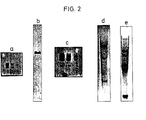
- panel a shows the result of the following experiment: pDv1, a pBR322 plasmid which contains an insert from D. virilis, was cleaved with EcoRI and Pvull, four fragments being formed: 2 D, virilis fragments (2.8 and 2.4 kb long) and 2 pBR322 fragments (2.3 and 2.06 kb long).
- Serial dilutions 500, 100 and 20 pg per application
- the two bands in the middle lane contain 24 and 21 pg DNA, respectively.
- the fragments in the two bands are 2.3 and 2.06 kb long. (Example 10).
- Panel b shows a nitrocellulose paper after transfer of 0.75 pg of EcoRI-digested and electrophoretically separated DNA from D. melanogaster Kc cells and three hours of hybridization in a volume of 5 ml with 0.5 ⁇ g of a pBR322 clone linearized with Pst I with a 4.8 kb insert, which carried the alcohol dehydrogenase gene and was coupled with peroxidase (10 ul of solution A). The DNA fragments in the band are 4.8 kb long. (Example 11).
- Table c) shows the results of a titration experiment similar to a), but here the hybridization was carried out in the presence of 6% polyethylene glycol.
- the two bands in the far right row contain 4.7 and 4.1 pg DNA, respectively. (Example 12).
- Panel d shows the results of an experiment in which 10 ⁇ g of chicken embryo DNA was digested with SacI, subjected to agarose gel electrophoresis and then transferred to a nitrocellulose filter paper.
- the hybridization was carried out with 2.5 ⁇ g of EcoRl-like pBR322 plasmid, containing 2.5 kb coding sequences for erb A and erb B of chicken erythroblastosis virus, which had been coupled with peroxidase (50 ⁇ l of solution A).
- the hybridization time was 14 hours, the volume 5 ml.
- the hybridization mixture contained 6% polyethylene glycol.
- the bands correspond to the following sizes in kb: 8.1, 7.5, 5.0, 3.0, 2.5 (Example 13).
- Panel e shows the results of an experiment in which 15 ⁇ m human DNA from HeLa cells were cleaved with Hind III, separated in size by agarose gel electrophoresis and transferred to a nitrocellulose filter paper and hybridized for 15 hours (in a volume of 5 nil) were using 2.5 * g of a pKT218 plasmid linearized with EcoRI, which contained the coding regions of the albumin gene (2 kb) and was coupled with peroxidase (50 ⁇ l) solution A).
- the hybridization mixture contained 6% polyethylene glycol.
- the bands obtained correspond to the following sizes in kb: 6.2, 4.2, 1.8. (Example 14).
- Example 4 a conjugate of alkaline phosphatase and polyethyleneimide was prepared.
- 3 mg (350 ⁇ l) alkaline phosphatase from calf intestine (grade 1, Boehringer Mannheim) were dialyzed against 0.1 M sodium phosphate, pH 6.0.
- 90 * l of the p-benzoquinone solution, as in Example 4 were added and incubated in the dark at 37 ° C. for one hour.
- gel chromatography (6 ml column Sephadex G 25)
- the fractions containing phosphatase were combined (about 900 pl) and mixed with 100 pl M NaHC0 3 and 20 ug polyethyleneimine.
- the mixture was incubated for 18 hours at 37 ° C. in the dark, dialyzed against 5 mM sodium phosphate pH 6.8 and stored at 3 ° C. solution B).
- the probe was produced, as described in Example 4, using solution B instead of solution A.
- the hybridization was also carried out as described in example 4.
- the alkaline phosphatase was visualized by adding 15 ml of a solution containing nitro blue tetrazoline (NBT) and 5-bromo-4-chloro-3-indoxyl-phosphate-4-toluidine salt (BCIP) and prepared as follows : 5 mg of NBT were suspended in 5 ml of 0.1 M Tris-HCl pH 9.5, 0.1 M NaCl, 5 mM MgCl 2 (buffer A), stirred vigorously for one minute and centrifuged briefly. The supernatant was decanted off in 10 ml of buffer A, which had been preheated to 37 ° C.
- NBT nitro blue tetrazoline
- BCIP 5-bromo-4-chloro-3-indoxyl-phosphate-4-toluidine salt
Description
- Die Erfindung betrifft ein Verfahren zur Durchführung von Hybridisierungsreaktionen und ein für dieses Verfahren geeignetes Nachweismittel.
- Die Hybridisierung ist ein Mittel zum Feststellen der Identität von Basensequenzen bei zwei verschiedenen Polynukleotidketten. Zwei DNA-Ketten mit komplementären Basensequenzen bilden eine Doppelstrangstruktur miteinander bei der Abkühlung in Lösung und genauso verhalten sich eine DNA-Kette und eine RNA-Kette mit komplementärer Struktur unter Bildung eines DNA-RNA-Hybrids. Durch Hybridisierung lassen sich daher zahlreiche Probleme der Nukleotidchemie lösen, beispielsweise die Auffindung und Isolierung bestimmter Gene oder Genbruchstücke, die Kartierung von Gensequenzen und dergleichen. Insbesondere im Rahmen der Gentechnologie spielt die Auffindung bestimmter gesuchter Gene eine entscheidende Rolle und Hybridisierung ermöglicht dies.
- Zur Durchführung der Hybridisierungsreaktion verwendet man zweckmäßig eine Nukleinsäurekette mit der zur gesuchten Nukleinsäure komplementären Struktur, welche in geeigneter Weise für die Wiederauffindung markiert ist. Üblicherweise erfolgt diese Markierung bisher durch enzymatische Einführung von Radioisotopen-tragenden Nukleotidtriphosphaten in die für die Suche verwendete komplementäre Nukleinsäure. Ein erheblicher Nachteil dieses Verfahrens besteht in der Notwendigkeit, mit radioaktiven Substanzen zu arbeiten, was sowohl umfangreiche Sicherheitsvorkehrungen erfordert als auch aufwendige Apparaturen voraussetzt.
- Man hat daher bereits Versuche unternommen, die radioaktive Markierung durch andere Markierungen zu ersetzen.
- So wurden neuerdings Nukleotidanaloge synthetisiert, die kovalent verknüpftes Biotin an einem Pyrimidin- oder Purinring enthalten. Diese Nukleotidderivate sind Substrate für RNS- und DNS-Polymerasen und die unter der Einwirkung dieser Enzyme gebildeten Polynukleotide hybridisieren spezifisch mit ihren Komplementärsequenzen. In Verbindung mit einem Affinitäts- (z. B. Avidin-Peroxidase) oder einem immunologischen (z. B. Biotin-Antikörper in Verbindung mit einem zweiten Peroxidase tragenden Antikörper) Nachweissystem können diese Proben als Alternative zu radioaktiv markierten Nukleinsäuren verwendet werden. Auch wurden schon Biotinreste mit RNS über Cytochrom C verknüpft und die Hybridisierungen elektronenmikroskopisch mit dem Avidin-Nachweissystem lokalisiert (Chromosoma/ Berl. 53, 107-117 (1975)).
- Dieses Verfahren ist jedoch immer noch relativ aufwendig, da es mehrere Syntheseschritte erfordert. Es besteht daher unverändert ein Bedarf nach einem einfacheren Hybridisierungsverfahren und einem Hybridisierungsreagenz, welches sich als Probe eignet und einfach hergestellt werden kann und die Empfindlichkeit der Hybridisierungstechnik erhöht.
- Aufgabe der Erfindung ist es daher, ein derartiges Verfahren und Mittel zu schaffen.
- GB 82109408 betrifft Nachweistechniken, die das Hybridisieren einer Nukleinsäuresequenz mit einer markierten Probe einschließen. Die Markierung umfaßt ein Enzym, das jedoch mit der Probe erst nach der Hybridisierung verbunden wird.
- In Chemical Abstract, 1982, 96: 30348 wird engas, Formaldehyd-induziertes Verbinden von Protein mit RNS in Teilchen eines stäbchenförmigen Virus beschrieben.
- Ein Verfahren gemäß der vorliegenden Erfindung zum Nachweist einer Target-Polynukleotid-Sequenz in Polynukleotid-Material durch In-Kontakt-Bringen des Materials unter Hybridisierungsbedingungen mit einer Probe, enthaltend eine Polynukleotid-Sequenz, die komplementär zu der Target-Sequenz ist, und durch anschließendes Verwenden der Probe zum Nachweis derTarget-Sequenz in dem Material, ist zum einen dadurch gekennzeichnet, daß die Probe durch Reaktion in Lösung der Einzelstrang-Komplementär-Polynukleotid-Sequenz mit einem nachweisbaren positive geladenen Nukleinsäure-bindenden Protein oder Polyamin und kovalentes Verbinden des resultierenden Komplexes mit einem Vernetzungsmittel gebildet wurde. Unter einem anderen Aspekt ist das Verfahren dadurch gekennzeichnet, daß die Probe durch Reaktion in Lösung der Einzelstrang-Komplementär-Polynukleotid-Sequenz mit einem nachweisbaren Protein, das mit einem positiv geladenen Protein oder Polyamin modifiziert wurde und kovalentes Verbinden der resultierenden Komplexes mit einem Vernetzungsmittel gebildet wurde. Das Polyamin kann ein Polyethylenimin sein.
- Unter einem anderen Aspekt liefert die Erfindung das Reaktionsprodukt eines Enzyms mit einem Polyethylenimin, wobei das Reaktionsprodukt die Fähigkeit besitzt kovalent mit einem Vernetzungsmittel an eine Polynukleotid-Sequenz verbunden zu werden.
- Unter einem noch anderen Aspekt liefert die vorliegende Erfindung ein Verfahren zur Herstellung eines Enzym-Polyamin-Konjugats, wobei das Verfahren das kovalente Anbringen von Benzochinon-Molekülen an ein Enzym und das Reagieren des resultierenden Zwischenstoffes mit einem Polyamin umfaßt.
- Als positiv geladene Proteine oder Polyamine werden im Rahmen der Erfindung solche verstanden, die entweder diese Eigenschaft an sich besitzen oder, falls sie negativ geladen oder neutral sind, mit einem entsprechend positiv geladenen Protein oder Polyamin modifiziert wurden. Hieraus ergibt sich auch die überraschend aufgefundene Tatsache, daß sich jedes beliebige Protein in ein DNA-bindendes Protein überführen läßt, indem man es mit einem positiv geladenen Protein oder Polyamin modifiziert.
- Die Erfindung liefert somit ein einfaches Verfahren zur Erzeugung von Hydridisierungsproben, das es erlaubt, auch große Proteinmoleküle als Markierungen einzuführen. Das Verfahren baut auf der Tatsache auf, daß ein positiv geladenes Proten oder Polyamin durch elektrostatische Wechselwirkungen an eine Nukleinsäure bei geringer Ionenstärke bindet und somit Vernetzungsreaktionen zwischen Nukleinsäure und Protein oder Polyamin begüngstigt. Vorzugsweise wird einzelsträngige DNS und ein Lysin-reiches DNS-bindendes chromosomales Protein, wie z. B. Histon H1, benutzt. Der hohe Lysin-Gehalt derartiger Protein erfüllt zwei Anforderungen:
- 1. Es wird eine sehr enge Bindung zu Nukleinsäuren erhalten und daher eine sehr wirksame Vernetzung mit geringen Konzentrationen an Vernetzungsmittel wie z. B. Glutaraldehyd innerhalb kurzer Reaktionszeiten ermöglicht. So hergestellte Protein-Nukleinsäure-Verbindungen (Proben) sind nicht einzelsträngige DNS markiert mit Protein, sondern stellen kovalent verbundene Einzelstrang-DNS-Protein-Einheiten dar mit einem Protein: DNS-Massenverhältnis von nahezu 1. Nach Hydridisierung kann man jetzt die Komplementär-sequenzen nachweisen durch Suche nach dem Protein (z. B. mit einem Antikörper).
- 2. Proteine oder Polyamine können chemisch einfach modifiziert werden. Im Falle des bevorzugten Histon H1 erfolgt die Modifizierung z. B. an der -Aminogruppe der Lysinreste. Dabei können Markierungsgruppen oder -verbindungen direkt oder über Brückenbildnerverbildungen wie z. B. cyclische N-Succinimidester an die Aminogruppe fixiert werden. Eine Markierung der Lysin-Aminogruppe hat wenig Einfluß auf die Nukleinsäurebindende Eigenschaft des Proteins, da nicht alle Lysinreste für starke Nukleinsäure-Proteinwechselwirkungen benötigt werden. Mit dieser Methode kann man viele Histon H1-Moleküle (jetzt markiert) mit Einzelstrang-DNS vernetzen und Marker mit einer großen Zahl von Markierungen erhalten, die nach Hybridisierung mit einem Affinitäts- oder immunologischen Nachweissystem leicht zu bestimmen sind. In Blot Hybridisierungsexperimenten kann gezeigt werden, daß unter identischen Reaktionsbedingungen markierte Proben entsprechend der erfindungsgemäßen Methode mit ihren Komplementär-Sequenzen mit hoher Spezifität reagieren und hierin nicht unterscheidbar sind von Nick-translatierten oder endmarkierten Proben.
- Als positiv geladene, mit Nukleinsäuren-bindenden Protein eignen sich im Rahmen der Erfindung prinzipiell alle positiv geladenen Polymeren und Oligomeren von Aminosäuren, natürlichen oder synthetischen Ursprungs, wie beispielsweise Polylysin, chromosomale Proteine einschließlich der Histone, einzelstrangspezifisch bindende Proteine wie z. B. Gen-32-Protein. Ebenfalls geeignet sind Polyamine. Bevorzugt werden unter diesen die natürlich vorkommenden sogenannten "Polyamine" wie Spermidin, Spearmin, Putrescin und dergleichen, die teilweise eigentlich Oligoamine darstellen. Geeignet sind aber auch synthetische Polyamine.
- Das einzelsträngige Polynukleotid kann ebenfalls natürlichen oder synthetischen Ursprungs sein, vorzugsweise handelt es sich um eine DNS oder RNS. Als Hybridisierungsreaktionspartner kommen ebenfalls DNS und RNS in Frage, so daß die Hybridisierungsreaktion selbst DNS mit DNS, DNS mit RNS und RNS mit RNS betreffen kann.
- Die Vernetzung des gebildeten Komplexes aus einzelsträngigem Polynukleotid und dem positiv geladenen Nukleinsäure bindenden Protein oder Polyamin erfolgt mit üblichen Vernetzungsmitteln, welche die funktionellen Gruppen des Proteins mit den funktionellen Gruppen der Polynukleinsäure zu verbinden vermögen. Besonders geeignet sind difunktionelle Vernetzungsmittel wie Dialdehyde, beispielsweise Glutaraldehyd, Diepoxide und generell Verbindungen, welche zwei zur Alkylierung in wässriger Lösung geeignete funktionelle Gruppen tragen. Typische hierfür geeignete Gruppen sind aktivierte Carboxylgruppen wie Säurechloride, Säurebromide, Säureanhydride, durch benachbarte Doppelbindungen aktivierte Halogenatome und dergleichen.
- Um das Hybridisierungsprodukt sichtbar zu machen, muß das darin enthaltene Protein bestimmbar sein, z. B. durch eine Markierung.
- Die Markierung des Proteins bzw. Polyamins kann entweder schon vor Durchführung der Komplexbildungsreaktion erfolgen oder am bereits gebildeten und gegebenenfalls auch bereits vernetzten Komplex durchgeführtwerden. Die Durchführung der Markierung erfolgt nach den hierfür bekannten Methoden der Proteinchemie, die dem Fachmann geläufig sind und hier nicht näher erläutert werden müssen. Als Markierungssystem kommt sowohl eine radioaktive Markierung als auch eine Markierung mit Farbstoffen, Farbstoffkomponenten oder biologisch aktiven Proteinen wie z. B. Enzymen in Betracht.
- Anstelle einer Markierung des für die Komplexbildung verwendeten positiv geladenen Nukleinsäurebindenden Proteins oder Polyamins ist es auch möglich, das Protein oder Polyamin durch eine immunologische Reaktion nachzuweisen, wobei der Immunreaktionspartner in diesem Falle markiert sein muß oder mit einem zweiten Antikörper nachgewiesen wird. Die bestimmbaren Gruppen als solche können entweder direkt bestimmbar sein wie bei radio aktiven- oder Farbstoffmarkierungen, oder indirekt, z. B. aufgrund ihrer enzymatischen Aktivität, wie dies aus dem Enzym-Immuno-Assay-Verfahren bekannt ist.
- Die Lösung für die Durchführung der Komplexbildungsreaktion zwischen Protein oder Polyamin und einzelsträngigem Polynukleotid soll eine geringe lonenaktivität aufweisen. Vorzugsweise soll die Ionenstärke niedriger als 50 mM sein.
- Die Hybridisierungsreaktion selbst erfolgt nach den hierfür bekannten Methoden durch einfaches Zusammengeben des erfindungsgemäß erhaltenen kovalent vernetzten Komplexes aus dem einzelsträngigen Polynukleotid und dem positiv geladenen Nukleinsäure-bindenden Protein oder Polyamin mit einer Lösung, welche die gesuchte komplementäre Nukleinsäure enthält und Inkubation, zweckmäßig bei etwas erhöhter Temperatur. Die Sichtbarmachung und Isolierung der gebildeten Hybride erfolgt ebenfalls nach dem Fachmann hierfür bekannten Methoden.
- Unter einem anderen Gesichtspunkt liefert die Erfindung eine Probe, die zur _Verwendung in Hybridisierungsreaktionen geeignet ist, dadurch gekennzeichnet, daß die Probe durch Reaktion in Lösung einer einzelsträngigen Polynukleotid-Sequenz, die komplementär zu einer gewünschten Target-Polynukleotid-Sequenz ist, entweder mit einem nachweisbaren, positiv geladenen Nukleinsäure-bindenden Protein oder Polyamin oder mit einem nachweisbaren Protein, das mit einem positive geladenen Protein oder Polyamin modifiziert wurde, und durch kovalentes Verbinden des resulierenden Komplexes mit einem Vernetzungsmittel gebildet wurde.
- Vorzugsweise enthält das Reagenz als Proteinkomponente Histon H1. Ebenfalls bevorzugt ist ein Massenverhältnis von Protein und Nukleinsäure von etwa 1:1.
- Durch die Erfindung wird ein Verfahren und ein Reagenz für die Durchführung von Hybridisierungsreaktionen zur Verfügung gestellt, welches als Probe eine chemisch hergestellte Verbindung verwendet, die mit hoher Ausbeute (mehr als 90%) anfällt, keine Polymerasen zu ihrer Herstellung benötigt und die Abtrennung von Vorläufern und Endprodukten überflüssig macht. Der Zeitaufwand ist sehr gering, die Polynukleotide für die Probenbereitung müssen nicht im hochreinen Zustand eingesetzt werden. Die dem Fachmann bekannten Standardmethoden für die Blot-Hybridisierung können unverändert benutz werden.
- Die folgenden Beispiele erläutern das Verfahren der Erfindung unter Verwendung von markierten Polynukleotid-Histon, H1-Verbindungen als Proben für Hybridisierungsreaktionen.
- Histon H1 (21 Kd) wurde durch Perchlorsäure-Extraktion aus Kälberthymuskernen isoliert wie bereits beschrieben in BBA Band 62, Seite 608 bis 609, 1962. Lyophilisiertes Histon H1 wurde in Wasser gelöst (5 mg/ml) und bei -20°C aufbewahrt.
- Biotinyl-N-hydroxysuccinimidester (BHSE) wurde hergestellt wie beschrieben in Methods Enzymology, Band 62, Seite 308 bis 315, 1979, 250 mg Biotin (Merck), 250 µ Ci (1,4 µg) 3H-Biothin (NEN), 150 mg N-Hydroxysuccinimid (Merck) werden in 3 ml Dimethylformamid (DMF) gelöst und 200 mg Dicyclohexylcarbodiimid zugegeben. Die Mischung wird 16 Stunden bei 20°C gerührt. Der Niederschlag wird abfiltriert und das Filtrat im Vakuum getrocknet. Der Rückstand wird mit Äther gewaschen, aus Isopropanol umkristallisiert und so für die Biotinmarkierung eingesetzt.
- Histon H1 wurde in verschiedener Weise mit steigenden Mengen an BHSE biotinyliert. Je 5 µg BHSE (gelöst in 10 µl DMF), 25 µg BHSE (in 10 µl DMF) und 250 µg (in 10 l DMF) werden zu einer die gleiche Menge Histon H1 (je 1 mg in 300 µl 50 mM NaHCO3) enthaltenen Lösung zugegeben und 1 Stunde bei 20°C inkubiert. Nach Dialyse gegen 5 mM Natriumphosphat (pH 6,8) werden die drei Proben bei -20°C aufbewahrt. Die Konzentrationen der Biotin markierten Histon H1-Lösungen werden durch Vergleich mit nicht modifiziertem Histon H1 durch SDS-Polyacrylamidgel-Elektrophorese bestimmt. Die Zahl der Biotinreste in den drei Präparationen wurde durch den in BHSE enthaltenen Tritium-Marker abgeschätzt und ergab 2,7 bzw. 20 pro Histon H1 Molekül.
- Zirkuläre DNS (Plasmide) wie auch große lineare DNS-Moleküle (Phage-Lamda DNS) wurden mit Restriktionsendonukleasen linearisiert oder gespalten zu einer Durchschnittsgröße von ungefähr 4 kb. Im Verlauf der Untersuchung wurde festgestellt, daß kleinere DNS- Fragmente stärkere Hybridisierungssignale ergeben. Daher wurde die DNS mit Enzymen wie SAU 3 A oder Hae III gespalten. Die gespaltene DNS wurde ohne weitere Reinigung eingesetzt. 1 µg DNS (in nicht mehr als 20 µl Spaltpuffer) wurde mit 180 µl frisch bereitetem 5 mM Natriumphosphatpuffer (pH 6,8) verdünnt, durch Hitze denaturiert (100°C, 3 Minuten) und 3 Minuten im Eisbad abgekült. Zuerst wurde Histon H1 (zwischen 0,8 und 1,0 pg in ungefähr 5 µl Phosphatpuffer) zugegeben und danach 20 µl einer 2,5 %igen Glutaraldehydlösung. Die Probe wurde 10 Minuten bei 30°C inkubiert und direkt in das das Nitrocellulosepapier und die Hybridisierungslösung enthaltene Hybridisierungsgefäß gegeben.
- Die zu untersuchende DNS wurde mit Restriktionsendonukleasen gespalten, durch Agarosegel-Elektrophorese fraktioniert und nach der Southern-Methode (J. Mol. Biol. 98, 503-517, 1975) auf Nitrocellulose-Papier übertragen. Die Papiere werden 1 Stunde bei 37°C in 10xDenhardts-Lösung (BBA, 23, 641 bis 645, 1966), 4xSET (1xSET=0,15 M NaCI, 0,03 M TrisxHCI, pH 8, 1 mM EDTA) eingeweicht und in Plastikwannen überführt, die zwischen 10 und 20 ml reiner Hybridisierungsmischung (50% deionisiertes Formamid, 2xDenhardts-Lösung, 4xSET, 0,1% SDS und 20 pg/ml Hefe T-RNS) enthalten, für eine Stunde bei 37°C inkubiert und dann nach Zugabe der Probe 16 bis 20 Stunden bei 37°C leicht geschwenkt.
- Die Nitrocellulose-Papiere wurden dann 60 Minuten bei 37°C mit zwei Wechseln einer 50% Formamid, 0,2% SDS und 5xSSC (einmal SSC=0,15 M NaCI, 0,015 M Natriumcitrat) enthaltenen Lösung und 40 Minuten bei 20°C mit zwei Wechseln einer zweimal SSC enthaltenen Lösung gewaschen. Die Filter wurden getrocknet und mit einem Biotin-Nachweissystem entwikkelt.
- Die Nitrocellulosepapiere wurden zuerst 20 Minuten mit einer 10 µg/ml Poly-1-Lysin HBR, MW 220.000 (Sigma), 0,1 M Tris HCI (pH 7,5) enthaltenen Lösung und dann mit einer 3% Rinderserumalbumin (BSA), 0,1 M Tris HCI (pH 7,4) enthaltenen Lösung 50 Minuten inkubiert und nachfolgend 60 Minuten lang mit der Avidin-Peroxidase-Lösung (1 M NaCl, 0,1 M Tris HCI, pH 7,4, 0,1% Triton X-100, 0,1% BSA und 1 µg/ml Avidin-Peroxidase, E. Y. Laboratories, California). Nach 20 Minuten Waschen mit zwei Wechseln einer 1 M NaCI, 0,1 M Tris HCI, pH 7,4, 0,1% BSA und 0,1% Triton X-100 enthaltenen Lösung, wurden die Papiere mit der Färbelösung (10 ml 0,1 M Tris HCI, pH 7,4, 2 ml Äthanol in den 6 mg 3,3'-Dianisidin gelöst wurden, 6 µl 30% H2O2) inkubiert. Die Farbentwicklung (braune Banden) findet in 5 bis 10 Minuten statt. Alle Inkubationsschritte wurden bei 20°C unter leichtem Schwenken ausgeführt.
- Es wurde, wie in Beispiel 1 beschrieben gearbeitet, jedoch wurde anstelle einer Biotin-Markierung eine Markierung mit 125J angewendet. Die Markierung von Histon H1 wurde wie folgt vorgenommen:
- Bei 0°C wurden zu 1mCi festem Mono-jodo (125J)-Bolton-Hunter Reagenz (2000 Ci/mMol) (NEN) 60 µl einer zwischen 120 und 150 µg Histon H1 und 100 mM Natriumborat (pH 8,9) enthaltenen Lösung zugegeben und für eine Stunde stehen gelassen. Danach wurden 10 µl 1 M Glycin in 0,5 M Natriumborat zugegeben. Die Lösung wurde durch eine mit 50 µg Histon H1 konditionierten Säule (silanisierte Pasteurpipette) gelfiltriert (Sephadex G 100: Molekularsiebmat. auf Basis von vernetztem Dextran) in 5 mM Natriumphosphat, pH 6,8. 125J-Histon H1 enthaltene Fraktionen wurden bei -20°C aufbewährt.
- Für Genkartierung wurde ein Drosophila virilis Insert tragender A-Clon mit verschiedenen Restriktionsendonukleasen gespalten, nach Größe getrennt, auf Nitrocellulosepapier übertragen und gleichzeitig mit einem erfindungsgemäßen Reagenz aus A-DNS und 125J-Histon H1 und einem erfindungsgemäßen Reagenz aus einem Plasmid (das Drosophila virilis Sequenzen des λ-Clones enthält) und Biotin-Histon H1 hybridisiert. Als Restriktionsendonukleasen wurden Eco R1, Bam H1, Hind III und Sal I verwendet. Durch Belichtung eines Röntgenfilmes und anschließende Entwicklung mit einem Biotin-Nachweise-System konnten die einzelnen Bruckstücke, die mit den verschiedenen Enzymen erhalten worden waren, identifiziert werden, welche jeweils die komplementäre Polynukleinsäure enthielten. Auf diese Weise war eine rasche Kartierung des Gens möglich.
- 20 mg (5x10³ Einheiten) Meerrettichperoxidase (Grad 1 Boehringer Mannheim) wurden in 220 µl von 90 mM Natriumphosphat, pH 6,0 gelöst und dann mit 60 p einer Lösung enthaltend 30 mg p-Benzochinon in 1 ml Ethanol versetzt. Die Mischung wurde eine Stunde bei 37°C im Dunklen reagieren gelassen. Peroxidase mit kovalent gebundenen Benzochinonmolekülen wurde von nicht umgesetztem Benzochinon durch Gelfiltration abgetrennt unter Verwendung einer 6 ml Säule Sephadex G 100 in 0,15 M NaCI ohne Puffer. Die braungefärbten Fraktionen (etwa 1,8 ml) wurden vereinigt und die Kupplungsreaktion in Gang gesetzt durch Anhebung des pH-Wertes durch Zusatz von 180 µl 1 M NaHC03 und Zusatz von 2,7 pl (133 µg) einer Polyethyleniminlösung (Polymin G 35 BASF). Die Reaktionsmischung wurde 14 Stunden bei 37°C im Dunkeln gehalten und dann gegen 5 mM Natriumphosphat, pH 6,8, dialysiert und bei 3°C gelagert (Lösung A). Die Proteinkonzentration der Lösung A betrug ca. 7 µg/pl. Während dreimonatiger Lagerung wurde kein Aktivitätsverlust beobachtet.
- Größe und Zusammensetzung der Peroxidasekonjugate wurde durch SDS-Polyacrylamid Gelelektrophorese unter Verwendung von 125-I-Polyethylenimin (markiert mit Bolton-Hunter-Reagenz) und durch Vergleich von Coomassie-blaugefärbten Gelen mit den entsprechenden Autoradiogrammen bestimmt. Dabei ergab sich, daß alle Peroxidasemoleküle modifiziert vorlagen. Die Konjugate hatten folgende Größen: 50 kDa 10%; 100 kDa 60%; 150 kDa 20%; 200 kDa 5%; 250 kDa 2%. Alle Konjugate wiesen ein Molverhältnis Peroxidase:Polyethylenimin von 1 auf.
- Zirkulaire, doppelsträngige DNA-Moleküle wurden mit einer Restriktionsendonuklease linearisiert und ohne weitere Reinigung verwendet. 1 µg DNA in 20 µl 5 mM Natriumphosphat pH 6,8 wurde hitzedenaturiert (100°C, 3 Minuten) und 3 Minuten auf Eis abgekült. Zuerst wurden 20 µl Lösung A zugesetzt, danach 6 µl einer 5%igen Glutardialdehydlösung. Die Probe wurde 10 min bei 37°C inkubiert und dann entweder direkt der Hybridisierungsreaktion zugesetzt, welche Nitrocellulosepapier und Hybridisierungslösung enthielt oder durch Zugabe von 28 µl einer Lösung, welche 40% Polyethylenglykol 8000 (Signa) enthielt, gefällt. Die Mischung wurde 6 Minuten zentrifugiert und der Niederschlag in 10 µl, 1,5 M L-Lysin in 5 mM Natriumphosphat, pH 6,8 aufgelöst und für die Hybridisierung eingesetzt. DNA-Bindungsuntersuchungen mit Gelfiltrationssäulen (Sepharose CL-6B) zeigten, daß etwa 25% der Peroxidaseaktivität kovalent an die DNA gebunden war, was einem Protein:DNA Massenverhältnis von etwa 30 entspricht.
- Nitrocellulosepapier (Schleicher & Schüll) mit immobilisierter DNAwurden eine Stunde bei 38°C eingeweicht in 10xDenhardt's Lösung, 4xSET (1 xSET ist 0,15 M NaCI, 0,03 M Tris-HCI, pH 8, 1 mM EDTA) und 0,1% SDS und dann in Kunststoffbehälter überführt, welche zwischen 2 und 20 ml Blank Hybridisierungsmischung enthielten (50% Formamid, 2xDenhardt's Lösung, 4xSET, 0,1% SDS und 30 pg/ml Hiefe tRNA), unter Schütteln eine Stunde bei 38°C inkubiert und dann nach Zugabe der Sonde 2 bis 16 Stunden bei 38°C weiterinkubiert. Für die Analyse von Hühnchen-und Human-Einfachkopie-Gensequenzen erfolgte die Hybridisierung in Gegenwart von Polyethylenglykol 8000. Die Hybridisierungsmischung wurde modifiziert durch Verwendung von 12% (Gewicht/Volumen) Polyethylenglykollösung in Formamid anstelle von Formamid alleine. Die Nitrocellulosepapier wurden 60 Minuten bei 38°C mit zweimaligem Austausch einer Lösung enthaltend 50% Formamid, 0,4% SDS und 0,5xSSC (1xSSC ist 0,15 M NaCI, 0,015 M Natriumcitrat) und 20 Minuten bei 20°C mit zweimaligem Austausch einer Lösung enthaltend 2xSSC gewaschen. Dann wurden die Filter bei 20°C im Dunklen mit Färbelösung inkubiert. Peroxidase wurde visualisiert mit 10 ml einer Lösung von 100 mM Tris-HCI, pH 7,4, enthaltend 10 mM Imidazol, 2 ml Ethanol, in welchem 6 mg 3,3'-Dianisidin aufgelöst worden waren und 10 µl 30% H2O2. Nach dem Anfärben wurden die Filter gewaschen und in Tris-HCI-Imidazolpuffer aufgewahrt.
- In Fig. 1 zeigt Tafel a ein Agarosegel-Elektrophoretogramm (mit Ethidiumbromid angefärbt) von pHBV 14.1 DNA, die mit verschiedenen Restriktionsendonukleasen gespalten worden war und zwar von links nach rechts mit: BamHl, Bg1II, Aval, Hpall, Pstl. Jeder Auftrag enthielt 100 ng DNA. Die Spalte ganz rechts enthält Größenmarkierungen folgenden Längen in kb: 23,1, 9,4, 6,6, 4,4, 2,3, 2,2, 2,0, 1,1, 0,75, 0,56, 0,38. (Beispiel 6) Tafel b zeigt ein Nitrocellulose- papier nach Transfer der in Tafel a gezeigten DNA und Hybridisierung mit 0,5 µg von mit EcoRI gespaltener pBR322 DNA, die mit Peroxidase (10 µl von Lösung A) gekuppelt und mit Anisidin-H202 gefärbt wurden. (Beispiel 7).
- Tafel c zeigt ein Replikafilter, welches mit HBV DNA-Sequenzen hybridisiert wurde, die mit Peroxidase markiert waren. Das Insert (3,2 kb Länge) von pHBV 14,1 wurde nach Spaltung mit Pstl der Agarosegel-Elektrophorese unterzogen und eluiert, 0,5 pg so erhaltener Insert DNA wurde mit Peroxidase markiert (10 µl von Lösung A). (Beispiel 8).
- Tafel d zeigt ein Replikafilter, welches mit HBV DNA (mit Peroxidase markiert) und mit pBR322 DNA (mit alkalischer Phosphatase markiert) hybridisiert worden war. 0,5 pg der Insert HBV DNA wurden mit Peroxidase gekuppelt (10 µl Lösung A) und 0,5 µg linearisierte pBR322 DNA wurde mit alkalischer Phosphatase markiert (10 µl Lösung B gemäß Beispiel 5).
- Nach der Hybridisierung wurde das Filterpapier zuerst mit der Substratlösung für Phosphatase inkubiert, was in einer blauen Färbung resultierte und dann mit Anisidin für die Sichtbarmachung der Peroxidase (braune Banden). Die Hybridisierungsvolumina und -zeiten betrugen jeweils 15 ml bzw. 3 Stunden. (Beispiel 9).
- In Fig. 2 zeigt Tafel a das Ergebnis folgenden Versuchs: pDv1, ein pBR322 Plasmid, welches ein Insert von D. virilis enthält, wurde mit EcoRI und Pvull gespalten, wobei vier Fragmente gebildet wurden: 2 D, virilis Fragmente (2,8 und 2,4 kb lang) und 2 pBR322 Fragmente (2,3 und 2,06 kb lang). Serienverdünnungen (500, 100 und 20 pg je Auftrag) wurden elektrophoriert, geblotted und hybridisiert mit 0,5 µg von mit EcoRI liniearisiertem pBR322, welches mit Peroxidase (10 µl von Lösung A) 16 Stunden in einem Volumen von 2,0 ml hybridisiert worden war. Die zwei Banden in der mittleren Spur enthalten 24 bzw. 21 pg DNA. Die Fragmente in den beiden Banden sind 2,3 bzw. 2,06 kb lang. (Beispiel 10).
- Tafel b) zeigt ein Nitrocellulosepapier nach Transfer von 0,75 pg von mit EcoRI gespaltener une elektrophoretisch aufgetrennter DNA aus D. melanogaster Kc Zellen und dreistündige Hybridisierung in einem Volumen von 5 ml mit 0,5 µg eines mit Pst I linearisierten pBR322 Clons mit einem 4,8 kb Insert, welcher das Alkoholdehydrogenasegen trug und mit Peroxidase (10 µl von Lösung A) gekuppelt war. Die DNA-Fragmente in der Bande sind 4.8 kb lang. (Beispiel 11).
- Tafel c) zeigt die Ergenisse eines Titrationsexperiments ähnlich a), hier wurde die Hybridisierung jedoch in Gegenwart von 6% Polyethylenglykol vorgenommen. Die zwei Banden in der ganz rechten Reihe enthalten 4,7 bzw. 4,1 pg DNA. (Beispiel 12).
- Tafel d) zeigt die Ergebnisse eines Versuchs, bei dem 10 µg von Hühnerembryo-DNA mit Sacl gespalten, der Agarosegel-Elektrophorese unterworfen und dann auf ein Nitrocellulosefilterpapier übertragen worden waren. Die Hybridisierung wurde mit 2,5 µg von mit EcoRl likearisiertem pBR322 Plasmid, enthaltend 2,5 kb codierende Sequenzen für erb A und erb B von Hühnererythroblastosevirus, welches mit Peroxidase (50 µl von Lösung A) gekuppelt worden war, durchgeführt. Die Hybridisierungsdauer betrug 14 Stunden, das Volumen 5 ml. Die Hybridisierungsmischung enthielt 6% Polyethylenglykol. Die Banden entsprechen folgenden Größen in kb: 8,1, 7,5, 5,0, 3,0, 2,5 (Beispiel 13).
- Tafel e) zeigt die Ergebnisse eines Versuchs, bei dem 15 um menschlicher DNA aus HeLa-Zellen mit Hind III gespalten, durch Agarosegel-Elektrophorese der Größe nach aufgetrennt und auf ein Nitrocellulosefilterpapier übertragen und 15 Stunden (in einem Volumen von 5 nil) hybridisiert worden waren unter Verwendung von 2,5 *g eines mit EcoRl linearisierten pKT218-Plasmids, welches die codierenden Regionen des Albumingens (2 kb) enthielt und mit Peroxidase (50 µl) Lösung A) gekuppelt war. Die Hybridisierungsmischung enthielt 6% Polyethylenglykol. Die erhaltenen Banden entsprechenden der folgenden Größen in kb: 6,2, 4,2, 1,8. (Beispiel 14).
- In der beigefügten Zeichnung sind die erfindungsgemäß erhaltenen Ergebnisse der Beispiele 6 bis 14 anhand der erhaltenen Elektrophoretogramme näher erläutert. In der Zeichnung stellen dar:
- Fig. 1 die Ergebnisse von BlotHybridisierungsversuchen unter Verwendung erfindungsgemäß markierter Sonden.
- Fig. 2 die Ergebnisse von Titrations- und Genom-Blot-Experimenten.
- Wie im Beispiel 4 beschrieben, wurde ein Konjugat von alkalischer Phosphatase und Polyethylenimid hergestellt. Hierzu wurden 3 mg (350 pl) alkalische Phosphatase aus Kälberdarm (Grad 1, Boehringer Mannheim) gegen 0,1 M Natriumphosphat, pH 6,0 dialysiert. 90 *l der p-Benzochinonlösung, wie im Beispiel 4, wurden zugesetzt und im Dunkeln eine Stunde bei 37°C inkubiert. Nach Gelchromatographie (6 ml Kolonne Sephadex G 25) wurden die Phosphatase enthaltenden Fraktionen vereinigt (etwa 900 pl) und mit 100 pl M NaHC03 und 20 ug Polyethylenimin versetzt. Die Mischung wurde 18 Stunden bei 37°C im Dunkeln inkubiert, gegen 5 mM Natriumphosphat pH 6,8 dialysiert und bei 3°C gelagert Lösung B).
- Die Herstellung der Sonde erfolgte, wie im Beispiel 4 beschrieben, unter Verwendung von Lösung B anstelle von Lösung A. Auch die Hybridisierung erfolgte wie im Beispiel 4 beschrieben.
- Die alkalische Phosphatase wurde visualisiert durch Zusatz von 15 ml einer Lösung, welche Nitroblau-Tetrazolin (NBT) und 5-Brom-4-chlor-3- indoxyl-phosphat-4-toluidin-Salz (BCIP) enthielt und wie folgt hergestellt worden war: 5 mg NBT wurden in 5 ml 0,1 M Tris-HCI pH 9,5, 0,1 M NaCI, 5 mM MgCI2 (Puffer A) suspendiert, eine Minute heftig gerührt und kurz zentrifugiert. Der Überstand wurde in 10 ml Puffer A abdekantiert, der auf 37°C vorgewärmt worden war. Dann wurden 2,5 mg BCIP in 50 µl Dimethylformamid gelöst und unter Rühren in die NBT-Lösung eingetropft. Nach der Farbentwicklung wurden die Filter gewaschen und in 100 mM Tris-HCI pH 7,4, enthaltend 5 mM EDTA, gelagert.
Claims (28)
Applications Claiming Priority (2)
| Application Number | Priority Date | Filing Date | Title |
|---|---|---|---|
| DE3310337A DE3310337A1 (de) | 1983-03-22 | 1983-03-22 | Verfahren zur durchfuehrung von hybridisierungsreaktionen |
| DE3310337 | 1983-03-22 |
Publications (2)
| Publication Number | Publication Date |
|---|---|
| EP0120376A1 EP0120376A1 (de) | 1984-10-03 |
| EP0120376B1 true EP0120376B1 (de) | 1990-10-24 |
Family
ID=6194276
Family Applications (1)
| Application Number | Title | Priority Date | Filing Date |
|---|---|---|---|
| EP84102594A Expired - Lifetime EP0120376B1 (de) | 1983-03-22 | 1984-03-09 | Verfahren zur Durchführung von Hybridisierungsreaktionen |
Country Status (5)
| Country | Link |
|---|---|
| US (1) | US5053326A (de) |
| EP (1) | EP0120376B1 (de) |
| JP (2) | JPS60501488A (de) |
| DE (2) | DE3310337A1 (de) |
| WO (1) | WO1984003717A1 (de) |
Families Citing this family (27)
| Publication number | Priority date | Publication date | Assignee | Title |
|---|---|---|---|---|
| US4873187A (en) * | 1986-03-13 | 1989-10-10 | Digene Diagnostics, Incorporated | Bifunctional DNA-protein conjugating agent |
| JPS638396A (ja) * | 1986-06-30 | 1988-01-14 | Wakunaga Pharmaceut Co Ltd | ポリ標識化オリゴヌクレオチド誘導体 |
| GB8621337D0 (en) * | 1986-09-04 | 1986-10-15 | Agricultural Genetics Co | Non-radioactive nucleic acid hybridization probes |
| CA1297432C (en) * | 1987-07-29 | 1992-03-17 | Gulilat Gebeyehu | Nucleic acid capture reagent |
| JP2597698B2 (ja) * | 1987-10-28 | 1997-04-09 | ハワード・フローレイ・インスティテュト・オブ・イクスペリメンタル・フィジオロジー・アンド・メディシン | オリゴヌクレオチド‐ポリアミド コンジュゲート |
| US5187260A (en) * | 1988-09-06 | 1993-02-16 | Sharifa Karali | Process for the preparation of a high purity protamine-DNA complex and process for use of same |
| US5849482A (en) * | 1988-09-28 | 1998-12-15 | Epoch Pharmaceuticals, Inc. | Crosslinking oligonucleotides |
| USRE38416E1 (en) | 1988-09-28 | 2004-02-03 | Epoch Biosciences, Inc. | Cross-linking oligonucleotides |
| US5824796A (en) * | 1988-09-28 | 1998-10-20 | Epoch Pharmaceuticals, Inc. | Cross-linking oligonucleotides |
| US5138045A (en) * | 1990-07-27 | 1992-08-11 | Isis Pharmaceuticals | Polyamine conjugated oligonucleotides |
| ES2148144T3 (es) * | 1990-11-20 | 2000-10-16 | Dade Behring Marburg Gmbh | Procedimiento para la estabilizacion de conjugados de enzima. |
| US6136601A (en) * | 1991-08-21 | 2000-10-24 | Epoch Pharmaceuticals, Inc. | Targeted mutagenesis in living cells using modified oligonucleotides |
| JP2651317B2 (ja) * | 1992-06-18 | 1997-09-10 | 扶桑薬品工業株式会社 | 核酸の検出法 |
| CA2169166C (en) * | 1993-08-10 | 2000-02-15 | Akio Matsuhisa | Method of detecting nucleic acid |
| US5582988A (en) * | 1994-09-15 | 1996-12-10 | Johnson & Johnson Clinical Diagnostics, Inc. | Methods for capture and selective release of nucleic acids using weakly basic polymer and amplification of same |
| GB9425138D0 (en) | 1994-12-12 | 1995-02-08 | Dynal As | Isolation of nucleic acid |
| US5783453A (en) * | 1995-06-29 | 1998-07-21 | Chiron Diagnostics Corporation | Non-separation specific binding chemiluminescent assay |
| JP4741124B2 (ja) * | 2001-09-20 | 2011-08-03 | 株式会社大気社 | 換気扇改良構造、及び、換気扇改良方法 |
| US7824910B2 (en) * | 2001-11-29 | 2010-11-02 | Nippon Shokubai Co., Ltd. | Method of transducing a protein into cells |
| US20030104623A1 (en) * | 2001-11-29 | 2003-06-05 | Nippon Shokubai Co., Ltd. | Method of introducing a protein into cells |
| US6927029B2 (en) | 2001-12-03 | 2005-08-09 | Agilent Technologies, Inc. | Surface with tethered polymeric species for binding biomolecules |
| EP1461084A2 (de) * | 2001-12-11 | 2004-09-29 | The Board Of Trustees Of The Leland Stanford Junior University | Guanidinium transport-reagenzien und konjugate |
| EP1468116A2 (de) | 2002-01-16 | 2004-10-20 | Dynal Biotech ASA | Verfahren zur isolierung von nukleinsäuren und proteinen aus einer einzigen probe |
| GB0229287D0 (en) * | 2002-12-16 | 2003-01-22 | Dna Res Innovations Ltd | Polyfunctional reagents |
| EP1687406B1 (de) * | 2003-11-10 | 2010-01-27 | Geneohm Sciences, Inc. | Nukleinsäurenachweisverfahren mit erhöhter empfindlichkeit |
| US8680290B2 (en) * | 2004-09-03 | 2014-03-25 | Syngenta Limited | Isoxazoline derivatives and their use as herbicides |
| WO2006065598A2 (en) * | 2004-12-13 | 2006-06-22 | Geneohm Sciences, Inc. | Fluidic cartridges for electrochemical detection of dna |
Family Cites Families (4)
| Publication number | Priority date | Publication date | Assignee | Title |
|---|---|---|---|---|
| US4152411A (en) * | 1977-07-27 | 1979-05-01 | Akzona Incorporated | High specific activity labeled substances |
| FR2422956A1 (fr) * | 1978-04-13 | 1979-11-09 | Pasteur Institut | Procede de detection et de caracterisation d'un acide nucleique ou d'une sequence de celui-ci, et reactif enzymatique pour la mise en oeuvre de ce procede |
| US4302204A (en) * | 1979-07-02 | 1981-11-24 | The Board Of Trustees Of Leland Stanford Junior University | Transfer and detection of nucleic acids |
| US4556643A (en) * | 1982-07-26 | 1985-12-03 | Agracetus | Assay method and probe for polynucleotide sequences |
-
1983
- 1983-03-22 DE DE3310337A patent/DE3310337A1/de not_active Withdrawn
-
1984
- 1984-03-09 WO PCT/DE1984/000051 patent/WO1984003717A1/de unknown
- 1984-03-09 JP JP59501284A patent/JPS60501488A/ja active Granted
- 1984-03-09 EP EP84102594A patent/EP0120376B1/de not_active Expired - Lifetime
- 1984-03-09 DE DE8484102594T patent/DE3483435D1/de not_active Expired - Lifetime
-
1988
- 1988-06-15 JP JP63147910A patent/JPH0667958B2/ja not_active Expired - Lifetime
-
1989
- 1989-04-17 US US06/724,307 patent/US5053326A/en not_active Expired - Lifetime
Also Published As
| Publication number | Publication date |
|---|---|
| JPH0259720B2 (de) | 1990-12-13 |
| US5053326A (en) | 1991-10-01 |
| JPS60501488A (ja) | 1985-09-12 |
| JPH0667958B2 (ja) | 1994-08-31 |
| DE3483435D1 (de) | 1990-11-29 |
| DE3310337A1 (de) | 1984-09-27 |
| EP0120376A1 (de) | 1984-10-03 |
| JPH01124400A (ja) | 1989-05-17 |
| WO1984003717A1 (en) | 1984-09-27 |
Similar Documents
| Publication | Publication Date | Title |
|---|---|---|
| EP0120376B1 (de) | Verfahren zur Durchführung von Hybridisierungsreaktionen | |
| DE2915082C2 (de) | ||
| DE3750198T3 (de) | Hybridisierungssonden. | |
| DE3546312C2 (de) | ||
| DE3020528C2 (de) | ||
| EP0161504B1 (de) | Herstellung von Polypeptiden mit Human-Gammainterferon-Aktivität | |
| DE2429398A1 (de) | Verfahren zum unloeslichmachen von aktiven proteinen | |
| NO841725L (no) | Fremgangsmaate og stoffer til bruk ved analyse og paavisning av genetisk materiale | |
| DE3523634A1 (de) | Ss-urogastron-gen, rekombinant-plasmide, transformanten, deren herstellung und herstellung von ss-urogastron | |
| EP0595167B1 (de) | Spezifische Gensonden und Verfahren zur Diagnostik von Candida albicans | |
| EP0133282B1 (de) | Herstellung von Polypeptiden mit einem Säureamid-Carboxyterminus | |
| EP1831246B1 (de) | Sonde zum nachweis von nukleinsäuren | |
| EP0296484A2 (de) | Promotoren zur Expression von Fremd-DNA in methylotrophen Bakterien, deren Gewinnung und deren Verwendung | |
| EP0314904A1 (de) | Verfahren und Reagenz zur Durchführung von Hybridisierungsreaktionen | |
| EP0286958A2 (de) | Verfahren und Mittel zur Bestimmung von Nucleinsäuren | |
| DE2337312C3 (de) | Verfahren zur Isolierung und Reinigung von Dehydrogenasen | |
| EP0303155A2 (de) | Verfahren zum Nachweis von Nukleinsäure-Sequenzen | |
| DE4226657A1 (de) | TYP II-Restriktionsendonuklease SexAI | |
| EP0214548B1 (de) | DNA-Sequenzen, rekombinante DNA-Moleküle und Verfahren zur Herstellung des Enzyms Mutarotase aus Acinetobacter calcoaceticus | |
| DE4339533C2 (de) | Nachweisverfahren für hdm-2 spezifische Antikörper | |
| DD254029A1 (de) | Verfahren und mittel zur bestimmung der aminoglucosid-phosphortransferaseaktivitaet | |
| DE3640382C2 (de) | ||
| DE3717437A1 (de) | Verfahren zur herstellung eines hmg-coa-reduktase-aehnlichen proteins | |
| DE3829535C2 (de) | ||
| DE3528392A1 (de) | Restriktionsendonuklease mfl i |
Legal Events
| Date | Code | Title | Description |
|---|---|---|---|
| PUAI | Public reference made under article 153(3) epc to a published international application that has entered the european phase |
Free format text: ORIGINAL CODE: 0009012 |
|
| AK | Designated contracting states |
Designated state(s): DE FR GB SE |
|
| 17P | Request for examination filed |
Effective date: 19850328 |
|
| 111L | Licence recorded |
Free format text: 0100 AMERSHAM INTERNATIONAL PLC |
|
| GRAA | (expected) grant |
Free format text: ORIGINAL CODE: 0009210 |
|
| AK | Designated contracting states |
Kind code of ref document: B1 Designated state(s): DE FR GB SE |
|
| REF | Corresponds to: |
Ref document number: 3483435 Country of ref document: DE Date of ref document: 19901129 |
|
| ET | Fr: translation filed | ||
| GBT | Gb: translation of ep patent filed (gb section 77(6)(a)/1977) | ||
| PLBE | No opposition filed within time limit |
Free format text: ORIGINAL CODE: 0009261 |
|
| STAA | Information on the status of an ep patent application or granted ep patent |
Free format text: STATUS: NO OPPOSITION FILED WITHIN TIME LIMIT |
|
| 26N | No opposition filed | ||
| EAL | Se: european patent in force in sweden |
Ref document number: 84102594.3 |
|
| REG | Reference to a national code |
Ref country code: GB Ref legal event code: IF02 |
|
| PGFP | Annual fee paid to national office [announced via postgrant information from national office to epo] |
Ref country code: GB Payment date: 20030305 Year of fee payment: 20 |
|
| PGFP | Annual fee paid to national office [announced via postgrant information from national office to epo] |
Ref country code: SE Payment date: 20030306 Year of fee payment: 20 |
|
| PGFP | Annual fee paid to national office [announced via postgrant information from national office to epo] |
Ref country code: FR Payment date: 20030310 Year of fee payment: 20 |
|
| PGFP | Annual fee paid to national office [announced via postgrant information from national office to epo] |
Ref country code: DE Payment date: 20030320 Year of fee payment: 20 |
|
| PG25 | Lapsed in a contracting state [announced via postgrant information from national office to epo] |
Ref country code: GB Free format text: LAPSE BECAUSE OF EXPIRATION OF PROTECTION Effective date: 20040308 |
|
| REG | Reference to a national code |
Ref country code: GB Ref legal event code: PE20 |
|
| EUG | Se: european patent has lapsed |